Advanced K-ε-GG DiGly Peptide Enrichment Protocol: A Comprehensive Guide for Deep Ubiquitinome Analysis
This article provides a comprehensive guide to the K-ε-GG diGly peptide enrichment protocol, a cornerstone method for mass spectrometry-based ubiquitinome analysis.
Advanced K-ε-GG DiGly Peptide Enrichment Protocol: A Comprehensive Guide for Deep Ubiquitinome Analysis
Abstract
This article provides a comprehensive guide to the K-ε-GG diGly peptide enrichment protocol, a cornerstone method for mass spectrometry-based ubiquitinome analysis. Tailored for researchers and drug development professionals, it covers the foundational principles of the ubiquitin code and the tryptic digestion process that generates the diagnostic diGly remnant. The guide details a step-by-step, optimized workflow for immunoaffinity enrichment, from cell culture and lysis to peptide fractionation and purification, applicable to both cell lines and complex tissues like brain. It further addresses critical troubleshooting and optimization strategies to enhance sensitivity and specificity, and concludes with a validation framework comparing methodological advances such as Data-Independent Acquisition (DIA) against traditional approaches, highlighting its application in uncovering biologically relevant ubiquitination signatures in stress response, circadian biology, and aging.
Decoding the Ubiquitin Code: The Foundation of K-ε-GG DiGly Proteomics
The Ubiquitin Proteasome System and its Central Role in Cellular Homeostasis
The Ubiquitin-Proteasome System (UPS) is a highly conserved and selective protein degradation pathway that is fundamental to maintaining cellular homeostasis in eukaryotes. This system ensures the precise regulation of key regulatory proteins, thereby enabling cells to dynamically respond to internal and external stimuli [1]. The UPS operates through a coordinated enzymatic cascade that tags target proteins with ubiquitin, marking them for degradation by the 26S proteasome. This process is indispensable for regulating vital cellular processes including the cell cycle, DNA repair, immune responses, and synaptic activity [2]. Dysregulation of the UPS is implicated in the pathogenesis of numerous diseases, particularly neurodegenerative disorders, cancer, and immune diseases, highlighting its critical role in cellular physiology [3] [2].
Beyond its canonical role in protein degradation, ubiquitination serves diverse non-proteolytic functions. The versatility of ubiquitin signaling arises from the complexity of ubiquitin conjugates, which can range from a single ubiquitin monomer to polymer chains of varying lengths and linkage types [4]. For instance, K48-linked polyubiquitin chains primarily target substrates for proteasomal degradation, whereas K63-linked chains are involved in non-proteolytic processes such as activating protein kinases in the NF-κB pathway and regulating autophagy [4]. Understanding this complex ubiquitin code is essential for deciphering its role in cellular homeostasis.
The Ubiquitination Cascade and Proteasomal Degradation
The Enzymatic Cascade of Ubiquitination
Protein ubiquitination is a reversible post-translational modification mediated by a sequential action of three enzymes, as illustrated in Figure 1:
- E1 Ubiquitin-Activating Enzymes: Initiate the process by activating ubiquitin in an ATP-dependent manner, forming a thioester bond with ubiquitin.
- E2 Ubiquitin-Conjugating Enzymes: Accept the activated ubiquitin from the E1 enzyme.
- E3 Ubiquitin Ligases: Confer substrate specificity by catalyzing the transfer of ubiquitin from the E2 enzyme to a lysine residue on the target protein [4].
The human genome encodes approximately 2 E1 enzymes, 40 E2 enzymes, and over 600 E3 ligases, enabling tremendous specificity in substrate selection [4]. Deubiquitinating enzymes (DUBs) counter-regulate this process by cleaving ubiquitin from substrates, adding a dynamic layer of control to ubiquitin signaling [3].
The 26S Proteasome and Protein Degradation
The 26S proteasome is a massive multi-subunit complex responsible for the degradation of ubiquitinated proteins. It consists of:
- A 20S core particle that contains the proteolytic active sites.
- One or two 19S regulatory particles that recognize polyubiquitinated substrates, remove the ubiquitin chains, unfold the protein, and translocate it into the core for degradation [5].
This degradation process is highly specific and ATP-dependent, ensuring the controlled turnover of cellular proteins to maintain homeostasis.
Table 1: Major Ubiquitin Chain Linkages and Their Primary Functions
| Linkage Type | Primary Function | Biological Context |
|---|---|---|
| K48-linked | Targets substrates for proteasomal degradation [4] | Most abundant proteolytic signal in cells [4] |
| K63-linked | Regulates protein-protein interactions, kinase activation, autophagy [4] | NF-κB pathway, DNA repair [4] |
| M1-linked (Linear) | Inflammatory signaling, NF-κB pathway [4] | Immune response regulation [4] |
| K6-, K11-, K27-, K29-, K33-linked | Diverse non-proteolytic functions; less characterized [4] | Endoplasmic reticulum-associated degradation (ERAD), transcription, trafficking [4] |
Figure 1. The Ubiquitin-Proteasome System Cascade. This diagram illustrates the sequential enzymatic process of ubiquitination, from ubiquitin activation by E1 to substrate ligation by E3, culminating in proteasomal degradation of the ubiquitinated protein.
K-ε-GG DiGly Peptide Enrichment: A Core Proteomic Protocol
The identification of ubiquitination sites on a proteome-wide scale relies heavily on mass spectrometry (MS) following the enrichment of ubiquitinated peptides. The K-ε-GG diGly remnant enrichment protocol has become the cornerstone of modern ubiquitinome analysis [6]. This methodology capitalizes on the unique signature that ubiquitination leaves after tryptic digestion.
Principle of the Protocol
Upon tryptic digestion of ubiquitinated proteins, the C-terminus of ubiquitin is cleaved, leaving a diglycine (diGly) remnant covalently attached to the modified lysine (K-ε-GG) on the target peptide [6] [7]. This diGly remnant constitutes a specific affinity handle that can be recognized by highly specific monoclonal antibodies, allowing for the immunoaffinity purification of these modified peptides from complex protein digests [6]. This method enriches for peptides originating from ubiquitin and ubiquitin-like modifiers (e.g., NEDD8, ISG15); however, more than 95% of K-ε-GG-modified sites are derived from ubiquitin [7].
Detailed Experimental Workflow
The standard workflow for ubiquitinome analysis using diGly remnant enrichment is outlined below and visualized in Figure 2.
Step 1: Cell Lysis and Protein Extraction
- Harvest cells and lyse using a denaturing lysis buffer (e.g., containing 8 M Urea or 1% SDS) to inactivate endogenous deubiquitinases (DUBs) and preserve the ubiquitination state.
- Quantify total protein concentration using an assay compatible with detergents (e.g., Pierce 660 nm assay).
Step 2: Protein Digestion
- Reduce disulfide bonds with dithiothreitol (DTT) and alkylate with iodoacetamide (IAA).
- Digest proteins first with Lys-C (optional but recommended for improved efficiency) followed by trypsin to generate peptides. Trypsin cleaves after lysine and arginine, generating peptides with the K-ε-GG motif [6].
Step 3: Peptide Clean-up and Quantification
- Desalt the digested peptides using C18 solid-phase extraction cartridges (e.g., Sep-Pak).
- Lyophilize and reconstitute peptides. Quantify peptide yield.
Step 4: DiGly Peptide Enrichment
- Use a commercial PTMScan Kit (e.g., Ubiquitin Remnant Motif (K-ε-GG) Kit, Cell Signaling Technology) or equivalent antibodies.
- Incubate 1-10 mg of total peptide digest with the anti-K-ε-GG antibody (typically 31.25 µg antibody per 1 mg peptide input is optimal [6]) for a minimum of 2 hours at 4°C with gentle mixing.
- Capture the antibody-peptide complexes using Protein A/G beads.
- Wash beads extensively to remove non-specifically bound peptides.
Step 5: Elution and Sample Preparation for MS
- Elute the enriched diGly peptides from the beads using a low-pH elution buffer (e.g., 0.15% TFA).
- Desalt the eluate using C18 StageTips or similar micro-columns.
- Lyophilize and reconstitute in MS loading solvent (e.g., 0.1% formic acid).
Step 6: Liquid Chromatography-Mass Spectrometry (LC-MS/MS) Analysis
- Separate peptides using reverse-phase liquid chromatography coupled online to a high-resolution mass spectrometer.
- For maximum depth and quantitative accuracy, Data-Independent Acquisition (DIA) is recommended over Data-Dependent Acquisition (DDA). An optimized DIA method using 46 precursor isolation windows and a fragment scan resolution of 30,000 has been shown to identify over 35,000 distinct diGly peptides in a single measurement [6].
Step 7: Data Analysis
- Process raw MS data using software (e.g., Spectronaut, DIA-NN, MaxQuant) against a appropriate spectral library.
- Search data against a protein sequence database to identify and quantify diGly peptides.
Figure 2. K-ε-GG DiGly Peptide Enrichment Workflow. The core protocol for ubiquitinome analysis involves protein digestion, immunoaffinity enrichment of diGly-modified peptides, and high-resolution mass spectrometry.
The Scientist's Toolkit: Key Research Reagent Solutions
Table 2: Essential Reagents for K-ε-GG DiGly Ubiquitinome Analysis
| Reagent / Material | Function / Application | Example Product / Note |
|---|---|---|
| Anti-K-ε-GG Antibody | Immunoaffinity enrichment of ubiquitinated peptides; core of the protocol. | PTMScan Ubiquitin Remnant Motif Kit (CST) [6] |
| Proteasome Inhibitor (MG132) | Increases ubiquitinated protein levels by blocking degradation; used pre-lysis to enhance signal. | Treatment (e.g., 10 µM, 4 hrs) prior to cell harvesting [6] |
| Denaturing Lysis Buffer | Inactivates DUBs and proteases to preserve the native ubiquitination state. | 8 M Urea or 1% SDS-based buffers [8] |
| Trypsin / Lys-C | Proteolytic enzymes for generating peptides with K-ε-GG remnant. | Sequencing grade enzymes recommended [8] |
| C18 Cartridges/StageTips | For desalting and cleaning up peptide samples pre- and post-enrichment. | Sep-Pak C18 Cartridges, C18 StageTips [8] |
| High-Resolution Mass Spectrometer | Enables identification and quantification of thousands of diGly sites. | Orbitrap-based platforms recommended for DIA [6] |
Advanced Applications and Quantitative Data
The diGly enrichment protocol has enabled systems-wide investigations into ubiquitin signaling in diverse biological contexts. The adoption of Data-Independent Acquisition (DIA) mass spectrometry has particularly revolutionized the field, doubling the number of diGly peptides identifiable in a single measurement (to over 35,000 distinct sites) and significantly improving quantitative accuracy and data completeness compared to traditional DDA methods [6]. This technical advance allows researchers to capture ubiquitination dynamics with unprecedented depth.
Table 3: Quantitative Ubiquitinome Findings from Recent Studies
| Biological Context | Key Quantitative Finding | Methodology | Reference |
|---|---|---|---|
| Aging Mouse Brain | 29% of altered ubiquitylation sites changed independently of protein abundance, indicating altered PTM stoichiometry with age. | DIA-MS with diGly enrichment [7] | [7] |
| ER Stress in CHO Cells | Identification of >4,000 ubiquitinated peptides; >900 proteins showed altered ubiquitination under ER stress and proteasome inhibition. | Label-free LC-MS/MS with diGly enrichment [8] | [8] |
| Circadian Biology | Discovery of hundreds of cycling ubiquitination sites on membrane receptors and transporters, linking ubiquitination to metabolic regulation. | Optimized DIA-MS workflow [6] | [6] |
| TNFα Signaling | Comprehensive capture of known and novel ubiquitination sites in a key signaling pathway. | DIA-MS with comprehensive spectral library [6] | [6] |
Concluding Remarks
The Ubiquitin-Proteasome System is an indispensable regulator of cellular homeostasis, and the K-ε-GG diGly peptide enrichment protocol stands as a powerful and refined tool for its study. The detailed methodology outlined here, combined with advanced MS acquisition strategies like DIA, provides researchers with a robust framework for conducting in-depth ubiquitinome analyses. As the field progresses, this core protocol will continue to be foundational for uncovering the intricate roles of ubiquitination in health and disease, driving discoveries in basic biology and the development of novel therapeutic strategies aimed at modulating the UPS.
The systematic identification of protein ubiquitination sites has been revolutionized by the exploitation of a specific tryptic signature: the diGly (K-ε-GG) remnant. When trypsin cleaves ubiquitin-modified proteins, it recognizes arginine and lysine residues within the ubiquitin molecule and the protein substrate. A key cleavage occurs between arginine 74 and glycine 75 in the C-terminal tail of ubiquitin. This digestion leaves a covalent remnant of the last two glycine residues (glycine 75 and glycine 76) of ubiquitin attached via an isopeptide bond to the epsilon-amino group of the modified lysine in the substrate protein. This generates a tryptic peptide with a characteristic K-ε-GG modification, exhibiting a defined mass shift of +114.1 Da [9] [10].
This diGly remnant serves as a mass spectrometry (MS)-detectable signature that enables the precise mapping of ubiquitination sites. The approach has become the foundation for large-scale ubiquitinome profiling, transforming the study of ubiquitin signaling from single-substrate characterization to systems-wide analysis [9] [6]. The following diagram illustrates the process of diGly remnant generation.
Enrichment Methodologies for DiGly Peptides
The low stoichiometry of endogenous ubiquitination means that K-ε-GG peptides are typically obscured by the overwhelming background of unmodified peptides in a tryptic digest. Consequently, specific enrichment strategies are essential for their comprehensive identification [9] [4]. The table below summarizes the primary methodologies used for enriching ubiquitinated proteins or diGly-modified peptides.
Table 1: Comparison of Methodologies for Enriching Ubiquitinated Substrates and Peptides
| Methodology | Principle | Advantages | Limitations | Typical Application |
|---|---|---|---|---|
| Immunoaffinity Enrichment (K-ε-GG) [9] [11] [6] | Antibodies specifically bind the K-ε-GG remnant on tryptic peptides. | High specificity; applicable to endogenous ubiquitination; enables direct site mapping. | Antibody cost; potential competition from highly abundant diGly peptides (e.g., from ubiquitin chains). | Global ubiquitinome profiling; targeted site mapping on individual proteins. |
| Ubiquitin Tagging (e.g., His/Strep) [4] | Affinity-tagged ubiquitin (e.g., 6xHis) is expressed in cells; conjugated proteins are purified. | Relatively low-cost; good for proof-of-concept studies. | May not mimic endogenous ubiquitination; artifacts possible; histidine-rich proteins co-purify. | Initial discovery screens in engineered cell lines. |
| Ubiquitin-Binding Domain (UBD) [4] | Tandem UBDs (TUBEs) bind polyubiquitin chains with high affinity. | Enriches under physiological conditions; can protect ubiquitin chains from deubiquitinases. | Lower specificity compared to anti-K-ε-GG; enriches proteins rather than sites. | Studying ubiquitinated protein complexes and ubiquitin chain biology. |
Immunoaffinity enrichment using anti-K-ε-GG antibodies has emerged as the most powerful and direct method for ubiquitination site identification. This peptide-level enrichment consistently outperforms protein-level enrichments, yielding a greater than fourfold increase in the levels of modified peptides detected from individual proteins like HER2 and DVL2 [11]. The optimal setup for such enrichments typically uses 1 mg of peptide material and ~31 µg of anti-diGly antibody, providing an effective balance between yield and depth of coverage [6].
Experimental Protocols
Standard Protocol for DiGly Peptide Enrichment and MS Analysis
This protocol details the steps from cell lysis to mass spectrometry analysis for global ubiquitinome profiling, incorporating best practices for sensitivity and depth [9] [6] [12].
I. Sample Preparation and Protein Digestion
- Cell Lysis and Denaturation: Lyse cells or tissue in a suitable denaturing buffer (e.g., 8 M urea, 50 mM Tris-HCl, pH 8.0). Avoid guanidine hydrochloride as it inhibits trypsin [12].
- Protein Reduction and Alkylation:
- Reduce disulfide bonds with 5 mM dithiothreitol (DTT) at 37°C for 1 hour.
- Alkylate cysteine residues with 15 mM iodoacetamide (IAA) at room temperature for 30 minutes in the dark [12].
- Protein Digestion:
- Dilute the sample with 50 mM Tris-HCl (pH 8.0) to reduce the urea concentration to below 2 M.
- Digest proteins using trypsin (e.g., Sequencing Grade Modified Trypsin) at a 1:50 (w/w) enzyme-to-protein ratio, overnight at 37°C. For challenging, tightly folded proteins, a Trypsin/Lys-C mix is recommended, as Lys-C remains active in higher urea concentrations, improving digestion efficiency and reducing missed cleavages [12].
- Peptide Desalting: Acidify the digest with trifluoroacetic acid (TFA) to a final concentration of 1%. Desalt the peptides using a C18 solid-phase extraction (SPE) cartridge or StageTips. Dry the purified peptides in a vacuum concentrator.
II. DiGly Peptide Immunoaffinity Enrichment
- Reconstitution: Resuspend the dried peptide pellet in 1X IAP (Immunoaffinity Purification) buffer (e.g., from CST PTMScan Kit).
- Enrichment: Incubate the peptide solution with anti-K-ε-GG antibody-conjugated beads for 2 hours at 4°C with gentle agitation. The optimal input is 1 mg of peptide material with ~31 µg of antibody [6].
- Washing: Pellet the beads and wash multiple times with 1X IAP buffer, followed by a final wash with HPLC-grade water to remove salts and detergents.
- Elution: Elute the bound K-ε-GG peptides from the beads using two washes of 0.15% TFA.
- Post-Enrichment Cleanup: Desalt the eluted peptides using C18 ZipTips or StageTips. The enriched peptides are now ready for LC-MS/MS analysis. As little as 25% of the total enriched material may be sufficient for a single injection, depending on instrument sensitivity [6].
III. Liquid Chromatography and Mass Spectrometry Analysis
- Chromatography: Separate the enriched peptides using reverse-phase nano-flow liquid chromatography (nano-LC).
- Mass Spectrometry:
- Data-Dependent Acquisition (DDA): Suitable for initial discovery and library generation. It selects the most intense precursors for fragmentation.
- Data-Independent Acquisition (DIA): Recommended for superior quantitative accuracy and data completeness. DIA fragments all ions in predefined m/z windows simultaneously, leading to more reproducible identification of over 35,000 distinct diGly peptides in a single measurement [6].
Advanced Workflow: DIA-Based Ubiquitinome Analysis
For the most comprehensive and quantitative analysis, a DIA-based workflow is recommended. The following diagram outlines this advanced strategy.
This workflow involves generating a deep, sample-specific spectral library using DDA from fractionated peptides, which is then used to interrogate single-shot DIA runs. This method doubles the number of diGly peptides identified in a single run compared to standard DDA and significantly improves quantitative reproducibility, with 45% of peptides showing a coefficient of variation (CV) below 20% [6].
The Scientist's Toolkit: Essential Research Reagents
Table 2: Essential Reagents for DiGly Peptide Enrichment and Analysis
| Item | Function / Role | Example / Specification |
|---|---|---|
| Anti-K-ε-GG Antibody [9] [11] [6] | Immunoaffinity enrichment of diGly-modified tryptic peptides. | Monoclonal antibody specific for the ubiquitin remnant motif (e.g., PTMScan Ubiquitin Remnant Motif Kit). |
| Sequencing-Grade Trypsin [12] | Highly specific protease for generating peptides with C-terminal Arg/Lys. Ensures clean digestion with minimal autolysis. | Sequencing Grade Modified Trypsin (reductively methylated). |
| Trypsin/Lys-C Mix [12] | Maximizes proteolytic efficiency, particularly for difficult-to-digest proteins, reducing missed cleavages. | Trypsin/Lys-C Mix, Mass Spectrometry Grade. |
| Strong Cation Exchange (SCX) or bRP Chromatography [6] | Pre-fractionation of complex peptide mixtures pre- or post-enrichment to reduce complexity and increase depth. | HPLC system with SCX or basic reversed-phase (bRP) column. |
| C18 StageTips / ZipTips [12] | Micro-solid phase extraction for desalting and concentrating peptide samples before LC-MS/MS. | C18 membrane-based pipette tips. |
| Nano-LC System | High-sensitivity separation of enriched peptides prior to mass spectrometry. | Nano-flow reversed-phase HPLC system. |
| High-Resolution Mass Spectrometer | Accurate mass measurement and fragmentation of diGly peptides for identification and quantification. | Orbitrap-based mass spectrometer (e.g., Orbitrap Exploris, Fusion Lumos). |
Advances in Ubiquitinome Analysis
The field has moved beyond simple identification to robust quantification and systems-wide analysis. Key advancements include:
- Data-Independent Acquisition (DIA): DIA-MS has been tailored for diGly proteomics, overcoming the dynamic range and stochastic sampling limitations of DDA. By using optimized window schemes and high-resolution fragment scanning, DIA can consistently identify and accurately quantify over 35,000 diGly sites from a single injection of enriched peptides from just 1 mg of protein digest [6].
- Linkage-Specific Analysis: While the standard anti-K-ε-GG antibody enriches peptides from all ubiquitin and ubiquitin-like modifications (with UBL-derived sites constituting <6% of identifications [6]), linkage-specific antibodies are available to enrich for proteins modified with specific Ub chain types (e.g., K48 or K63-linked chains) [4].
- Application to Biological Signaling: This sensitive workflow has been successfully applied to map dynamics in pathways like TNFα signaling and to uncover extensive, daily oscillations in the ubiquitinome across the circadian cycle, revealing hundreds of cycling sites on membrane receptors and transporters [6].
The anti-K-ε-GG antibody has revolutionized the study of ubiquitin signaling by enabling the large-scale enrichment and identification of ubiquitination sites through mass spectrometry-based proteomics. This antibody recognizes the di-glycine (diGly) remnant left on trypsinized peptides after digestion of ubiquitinated proteins [13] [9]. However, a critical consideration for researchers employing this powerful tool is its potential cross-reactivity with identical diGly remnants generated by other ubiquitin-like proteins (UBLs), principally NEDD8 and ISG15 [13] [14]. This Application Note delineates the specificity profile of the anti-K-ε-GG antibody, providing quantitative data on cross-reactivity and detailed protocols to ensure accurate interpretation of ubiquitinome datasets.
Molecular Basis of Anti-K-ε-GG Specificity
The Shared DiGly Motif and Its Origin
The core recognition motif for the anti-K-ε-GG antibody is the di-glycine adduct covalently attached to the ε-amine of a modified lysine residue. This motif is not unique to ubiquitin but is a common feature of several UBLs due to conserved C-terminal sequences [13] [9].
- Ubiquitin: The C-terminal sequence of mature ubiquitin is LRLRGG. Trypsin cleaves after the two arginine residues (R74 and R72 in some constructs), liberating the target peptide and leaving the C-terminal Gly-Gly motif attached to the modified lysine on the substrate peptide [9].
- NEDD8: The C-terminal sequence is LRGG, functionally identical to ubiquitin's LRLRGG for trypsin cleavage, generating an indistinguishable K-ε-GG remnant [15].
- ISG15: This UBL consists of two ubiquitin-like domains connected by a short linker. Its C-terminal sequence is LRLRGG, identical to ubiquitin. Consequently, trypsin digestion of ISGylated proteins produces peptides with the exact same K-ε-GG signature [16] [17] [18].
This shared biochemistry means that a standard diGly enrichment protocol will co-isolate peptides modified by ubiquitin, NEDD8, and ISG15. The inability of the classic anti-K-ε-GG antibody to distinguish between these modifications is a fundamental limitation that must be accounted for in experimental design and data analysis [13] [15].
Quantitative Assessment of Antibody Cross-Reactivity
While the anti-K-ε-GG antibody recognizes diGly peptides from all three UBLs, the proportion of identified sites originating from each source varies significantly under different physiological conditions. The following table summarizes the typical distribution of diGly peptides identified in standard proteomics experiments.
Table 1: Distribution of diGly Peptides Originating from Ubiquitin, NEDD8, and ISG15
| Ubiquitin-like Modifier | C-terminal Sequence | Typical Contribution to diGly Peptide Identifications | Key Contextual Factors |
|---|---|---|---|
| Ubiquitin | LRLRGG | ~94-95% [13] [6] | Constitutively active pathway; dominant under basal conditions. |
| NEDD8 | LRGG | Low (<6% combined) [6] [15] | Canonical NEDD8 targets (e.g., cullins) may be underrepresented in diGly datasets [15]. |
| ISG15 | LRLRGG | Low (<6% combined) [6] | Highly inducible by type I interferon during viral/bacterial infection [16] [17]. |
It is crucial to note that the contribution of ISG15-derived diGly peptides can increase dramatically in specific contexts, such as during the innate immune response to infection or upon interferon stimulation, which strongly upregulates the ISG15 conjugation machinery [16] [17]. Under such conditions, assuming all diGly peptides are from ubiquitin can lead to substantial misinterpretation of the data.
Experimental Workflows for Specific Ubiquitin Identification
To overcome the specificity limitation of the standard anti-K-ε-GG antibody, researchers can employ several strategic experimental workflows. The diagram below illustrates the two primary approaches discussed in this section.
Protocol: Specific Ubiquitin Site Identification Using LysC Digestion
This protocol leverages the differential enzymatic cleavage patterns of trypsin and LysC to generate ubiquitin-specific remnants [15].
1. Cell Lysis and Protein Digestion:
- Lyse cells or tissue in a denaturing buffer (e.g., 8 M Urea, 50 mM Tris-HCl, pH 8.0) supplemented with 10 mM N-Ethylmaleimide (NEM) to inhibit deubiquitinating enzymes [13].
- Reduce disulfide bonds with 5 mM dithiothreitol (DTT) and alkylate with 10 mM iodoacetamide (IAA).
- Digest the protein extract first with LysC (Wako, 1:100 enzyme-to-substrate ratio) for 3-4 hours at 25°C in a suitable buffer (e.g., 50 mM Tris-HCl, pH 8.5). LysC cleaves specifically C-terminal to lysine residues.
- Following LysC digestion, the sample is diluted to reduce urea concentration, and trypsin (Sigma, sequencing grade) is added (1:50 ratio) for overnight digestion at 37°C.
2. Peptide Desalting:
- Acidify the digested peptide mixture to pH < 3 using trifluoroacetic acid (TFA).
- Desalt the peptides using a C18 solid-phase extraction cartridge (e.g., Waters Sep-Pak) according to the manufacturer's instructions. Elute peptides with 30-50% acetonitrile in 0.1% TFA and lyophilize.
3. Immunoaffinity Enrichment:
- Reconstitute the lyophilized peptides in Immunoaffinity Purification (IAP) Buffer (50 mM MOPS-NaOH, pH 7.2, 10 mM Na2HPO4, 50 mM NaCl).
- Incubate the peptide solution with an antibody specific for the extended ubiquitin remnant (e.g., the Ubiquitin Branch Motif 2 (UBM2) antibody) conjugated to protein A/G beads for 2 hours at 4°C [15].
- Pellet the beads by gentle centrifugation and sequentially wash with: a) IAP buffer, b) HPLC-grade water, and c) 50 mM Tris-HCl, pH 7.5. Perform all washes on ice.
4. Peptide Elution and MS Analysis:
- Elute the enriched peptides from the beads twice with 50-100 µL of 0.15% TFA.
- Acidify the combined eluates and desalt using C18 StageTips.
- Analyze the enriched peptides by LC-MS/MS using Data-Dependent Acquisition (DDA) or Data-Independent Acquisition (DIA) for maximum depth and quantitative accuracy [6].
Protocol: Genetic Validation Using Ubiquitin Mutants
This approach uses genetic manipulation to dissect the origin of diGly signals.
1. Cell Line Engineering:
- Generate a cell line stably expressing a mutant form of ubiquitin where arginine 74 is substituted with lysine (R74K) [15]. In this mutant, trypsin cleaves after R72 but not at position 74, preventing the generation of the characteristic diGly remnant on ubiquitinated substrates.
- As a control, generate a cell line expressing wild-type ubiquitin.
2. Sample Processing and diGly Enrichment:
- Culture the engineered cells and treat them according to the experimental design.
- Lyse the cells, digest the proteins with trypsin, and enrich for diGly peptides using the standard anti-K-ε-GG antibody protocol [13].
- Analyze the enriched peptides by LC-MS/MS.
3. Data Interpretation:
- Any diGly peptides identified in the Ubiquitin R74K cell line under basal conditions must originate from NEDD8 or ISG15, as the ubiquitin-derived diGly remnant is not produced.
- Comparing the diGly proteomes of the wild-type and R74K ubiquitin cell lines allows for the specific assignment of ubiquitin versus non-ubiquitin diGly sites.
The Scientist's Toolkit: Essential Reagents
Table 2: Key Research Reagent Solutions for diGly Proteomics
| Reagent / Solution | Function | Specification / Notes |
|---|---|---|
| Anti-K-ε-GG Antibody [13] [6] | Immunoaffinity enrichment of diGly-modified peptides from tryptic digests. | Available as monoclonal antibody (clone). Critical for ubiquitinome studies. |
| UBM2 (Extended Remnant) Antibody [15] | Specific enrichment of ubiquitin-derived peptides following LysC digestion. | Key tool for discriminating ubiquitination from other UBL modifications. |
| NEM (N-Ethylmaleimide) [13] | Irreversible cysteine protease inhibitor. Preserves ubiquitin/UBL conjugates during lysis by inhibiting DUBs. | Add fresh to lysis buffer (e.g., 5-10 mM final concentration). |
| Proteasome Inhibitor (MG132) [6] | Blocks degradation of proteasome substrates, leading to accumulation of K48-linked ubiquitinated proteins and other substrates. | Use at 10-20 µM for 4-6 hours before lysis to deepen ubiquitinome coverage. |
| LysC Protease [15] | Endoproteinase that cleaves C-terminal to lysine. Used to generate extended ubiquitin-specific remnants. | Wako, Mass Spectrometry Grade. |
| Stable Isotope Labeling (SILAC) [13] | Metabolic labeling for quantitative proteomics; allows accurate comparison of ubiquitination sites between conditions. | Use heavy Lys (K8) and Arg (R10) in experimental media. |
The standard anti-K-ε-GG antibody is a powerful but non-specific tool that enriches for the diGly remnant shared by ubiquitin, NEDD8, and ISG15. While ubiquitin-derived peptides constitute the vast majority of identifications under basal conditions, the contribution from NEDD8 and ISG15 can be biologically significant and context-dependent. The experimental workflows and reagents detailed herein provide a roadmap for researchers to validate the specificity of their findings, discriminate between these highly similar post-translational modifications, and ensure the accurate characterization of the ubiquitinome.
Ubiquitination (or ubiquitylation) is a crucial post-translational modification (PTM) in which a small, 76-amino acid protein called ubiquitin is attached to target proteins [19] [20]. This process regulates virtually every aspect of cellular function, ranging from protein degradation to signal transduction, DNA repair, and immune responses [19] [21]. The clinical importance of understanding the molecular rules of the writers (E1–E2–E3 enzymes), erasers (deubiquitylating enzymes, DUBs), and readers of ubiquitylation is evident in the now major field of targeted protein degradation [22]. This article explores the key biological processes governed by ubiquitination, with particular focus on the context of K-ε-GG diGly peptide enrichment protocol research, providing researchers with both fundamental knowledge and practical methodological guidance.
Fundamentals of the Ubiquitination Cascade
The ubiquitination pathway is a tightly regulated, ATP-dependent biological process carried out by a complex cascade of three key enzymes [19] [23].
Table 1: Enzymatic Components of the Ubiquitination Cascade
| Enzyme Class | Number in Humans | Primary Function | Key Features |
|---|---|---|---|
| E1 (Activating Enzyme) | 2 [24] [21] | Activates ubiquitin in an ATP-dependent manner | Establishes a thioester bond with ubiquitin [19] |
| E2 (Conjugating Enzyme) | 30-35 [24] [21] [20] | Accepts activated ubiquitin from E1 | Characterized by a conserved ubiquitin-conjugating catalytic (UBC) fold [20] |
| E3 (Ligase Enzyme) | ~600 [19] [24] [21] | Transfers ubiquitin to specific substrate proteins | Provides substrate specificity; includes RING, HECT, and U-box domains [24] |
The process consists of three essential steps [19] [23]:
- Activation: The E1 ubiquitin-activating enzyme activates ubiquitin in an ATP-dependent two-step reaction, forming a thioester bond between its catalytic cysteine and the C-terminal carboxyl group of ubiquitin.
- Conjugation: The activated ubiquitin is transferred to the catalytic cysteine of an E2 ubiquitin-conjugating enzyme via a transesterification reaction.
- Ligation: E3 ubiquitin ligases catalyze the final transfer of ubiquitin to a substrate protein, creating an isopeptide bond between the C-terminal glycine of ubiquitin and a lysine residue on the substrate.
Figure 1: The Ubiquitination Enzymatic Cascade. This three-step process involves E1 (activation), E2 (conjugation), and E3 (ligation) enzymes working sequentially to attach ubiquitin to substrate proteins.
Biological Functions of Ubiquitination
Protein Degradation via the Proteasome
The best-characterized function of ubiquitination is the targeting of proteins for degradation by the 26S proteasome [19] [23]. Proteins tagged with Lys48-linked polyubiquitin chains are recognized by the proteasome, unfolded, and degraded into small peptides, recycling amino acids for future protein synthesis [24] [21]. This process serves as a critical quality control mechanism for intracellular proteins, rapidly removing unwanted, damaged, or misfolded proteins to maintain cellular homeostasis [23].
Non-Degradative Signaling Functions
Beyond proteasomal targeting, ubiquitination serves numerous non-degradative signaling functions:
DNA Damage Repair: Monoubiquitination and K6-linked polyubiquitination regulate DNA damage repair processes. For example, the E3 ligase Rad18 mediates monoubiquitination of proliferating cell nuclear antigen (PCNA), facilitating recruitment of DNA polymerases [21].
Inflammatory Signaling: K63-linked polyubiquitination plays crucial roles in immune and inflammatory signaling pathways, including the TLR, RLR, and STING-dependent signaling pathways that modulate the tumor microenvironment [24] [21].
Kinase Activation and Endocytosis: Monoubiquitination and K63-linked polyubiquitination control protein activity, interactions, and subcellular distribution. These modifications act as signals for endocytosis and trafficking of cellular vesicles to lysosomes [19].
Autophagy Regulation: Ubiquitination controls multiple steps in autophagy, with various ubiquitin chains serving as selective labels on protein aggregates and dysfunctional organelles to promote their autophagy-dependent degradation [25].
Table 2: Ubiquitin Chain Linkages and Their Primary Functions
| Linkage Type | Primary Function | Cellular Process |
|---|---|---|
| K48 | Target proteins for proteasomal degradation [19] [24] | Protein turnover, homeostasis |
| K63 | Activation of signaling pathways [19] [24] | DNA repair, endocytosis, immune signaling |
| K6 | DNA damage repair [21] | Genomic stability |
| K11 | Endoplasmic reticulum-associated degradation [24] | Protein quality control |
| K27 | Mitochondrial autophagy [21] | Mitophagy, organelle quality control |
| K29 | Protein modification [24] | Signaling, lysosomal degradation |
| M1 | Linear ubiquitination | NF-κB signaling, inflammation |
Pathophysiological Significance and Clinical Applications
Dysregulation of ubiquitination pathways is implicated in numerous human diseases. In von-Hippel Lindau (VHL) disease, loss-of-function mutations in the VHL tumor suppressor (an E3 ubiquitin ligase) result in uncontrolled growth and tumor formation [19]. In cancer, altered ubiquitination affects tumor metabolism, the immunological tumor microenvironment, and cancer stem cell stemness maintenance [21]. Neurodegenerative disorders such as Parkinson's disease are associated with protein misfolding and ubiquitin-positive aggregates [23].
The clinical significance of targeting ubiquitination is evident in the development of therapeutics such as proteasome inhibitors (bortezomib, carfilzomib) for multiple myeloma [24] [21]. Emerging technologies including proteolysis-targeting chimeras (PROTACs) leverage the ubiquitin system for targeted protein degradation, offering promising avenues for drug development [24] [22].
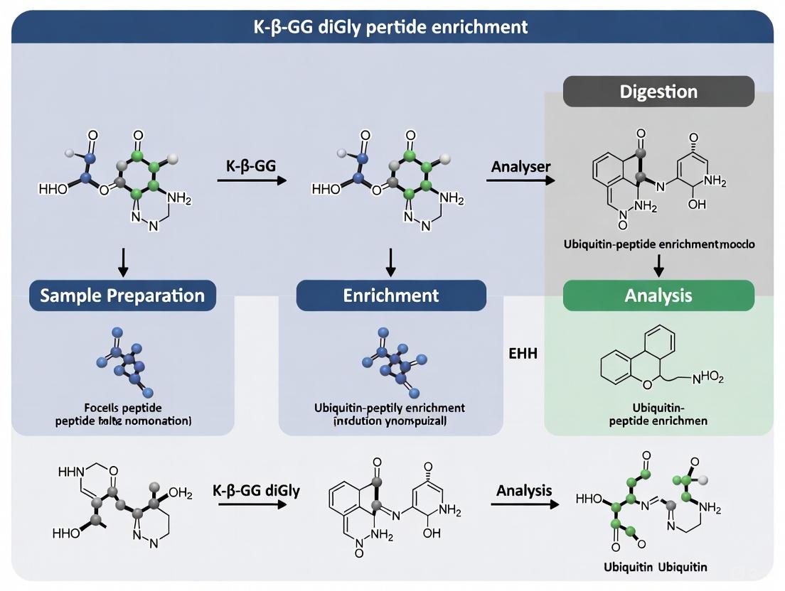
K-ε-GG DiGly Peptide Enrichment Protocol for Ubiquitination Site Mapping
Principle and Significance
When ubiquitinated proteins are digested with trypsin, they leave a 114.04 Da diglycine remnant on the target lysine residue, creating a "K-ε-diglycine" (K-ε-GG) motif that can be used to unambiguously identify the site of ubiquitination in mass spectrometry experiments [26]. Efficient immunopurification of diGly peptides combined with sensitive detection by mass spectrometry has revolutionized the identification of ubiquitination sites, enabling researchers to detect over 23,000 diGly peptides from human cell lysates upon proteasome inhibition [26] [27].
Materials and Reagents
Table 3: Essential Research Reagents for DiGly Peptide Enrichment
| Reagent / Kit | Manufacturer | Function |
|---|---|---|
| PTMScan Ubiquitin Remnant Motif (K-ε-GG) Kit | Cell Signaling Technologies | Antibody-based enrichment of diGly-modified peptides [26] |
| Anti-diglycine remnant (K-ε-GG) antibody | Commercial sources | Immunoaffinity purification of diGly peptides [27] |
| Bortezomib | UBPbio | Proteasomal inhibitor to accumulate ubiquitinated proteins [26] |
| Lysyl Endopeptidase (LysC) | Wako Pure Chemicals | Protein digestion enzyme |
| Trypsin, TPCK Treated | ThermoFisher | Protein digestion enzyme |
| Sep-Pak tC18 Cartridge | Waters | Peptide desalting and fractionation |
| Orbitrap Mass Spectrometer | ThermoFisher | High-sensitivity detection of diGly peptides |
Detailed Experimental Workflow
Figure 2: Experimental Workflow for DiGly Peptide Enrichment. This protocol enables comprehensive mapping of ubiquitination sites from biological samples.
Step 1: Sample Preparation and Proteasome Inhibition
- Culture cells in appropriate medium (e.g., HeLa or U2OS cells in DMEM supplemented with 10% FBS) [26].
- Treat cells with proteasome inhibitor (e.g., 10 μM Bortezomib) for 4-6 hours to accumulate ubiquitinated proteins [26] [23].
- Harvest cells by centrifugation and wash with ice-cold PBS.
Step 2: Protein Extraction and Digestion
- Lyse cells in appropriate buffer (e.g., 4% SDS, 100 mM Tris-HCl pH 7.6) with protease inhibitors [26].
- Reduce disulfide bonds with 1,4-dithioerythritol (10 mM, 30 min, 45°C) and alkylate with iodoacetamide (20 mM, 30 min, room temperature in the dark) [26].
- Precipitate proteins using methanol/chloroform method or filter-aided sample preparation (FASP).
- Digest proteins first with LysC (1:50 enzyme:substrate, 4h, 37°C) followed by trypsin (1:50 enzyme:substrate, overnight, 37°C) [26].
Step 3: Peptide Fractionation and Cleanup
- Desalt peptides using Sep-Pak tC18 cartridges [26].
- For deep ubiquitinome analysis, perform offline high-pH reverse-phase fractionation prior to diGly enrichment [26] [27].
- Fractionate peptides into 8-12 fractions using a step gradient of ammonium formate or ammonium hydroxide (pH 10) [26].
Step 4: DiGly Peptide Enrichment
- Reconstitute peptide fractions in immunoaffinity purification (IAP) buffer [26] [27].
- Incubate with anti-K-ε-GG antibody (typically 2-4 μg antibody per 1 mg of total peptide input) for 1.5-2 hours at 4°C with gentle rotation [26] [27].
- Use protein A/G beads to capture antibody-diGly peptide complexes.
- Wash beads extensively with IAP buffer and then with water [26].
- Elute diGly peptides with 0.15% trifluoroacetic acid [26].
Step 5: Mass Spectrometric Analysis
- Analyze enriched diGly peptides by nanoflow LC-MS/MS using systems such as EASY-nanoLC 1200 coupled to Orbitrap Fusion Lumos mass spectrometer [26].
- Use data-dependent acquisition with higher-energy collisional dissociation (HCD) fragmentation.
- Specifically trigger MS/MS fragmentation for peptides with a 114.0429 Da mass shift, corresponding to the diGly remnant [26].
Critical Protocol Considerations
- Protein Input: For optimal results, use at least 1 mg of total protein input to identify thousands of ubiquitination sites [26].
- Antibody Cross-linking: Cross-link antibodies to beads to prevent antibody leakage and improve specificity [27].
- Fractionation Depth: Offline high-pH reverse-phase fractionation prior to enrichment significantly increases the number of identified ubiquitination sites [26] [27].
- Controls: Include negative controls without antibody to assess nonspecific binding.
- Quantification: Incorporate SILAC or TMT labeling for quantitative ubiquitinome studies [27].
Ubiquitination represents a versatile regulatory mechanism that governs fundamental cellular processes through both degradative and non-degradative signaling. The development of robust proteomic methods, particularly K-ε-GG diGly peptide enrichment, has dramatically advanced our ability to comprehensively map ubiquitination sites and understand the complex ubiquitin code. These technical advances, combined with growing clinical interest in targeting ubiquitination pathways for therapeutic intervention, highlight the continued importance of ubiquitination research across basic science and drug development fields. As technologies evolve, including emerging fragment-based drug discovery approaches and novel PROTAC designs, our ability to precisely manipulate the ubiquitin system will continue to expand, offering new opportunities for research and therapeutic development.
The functional diversity of the proteome is dramatically expanded through post-translational modifications (PTMs), with over 500 unique types documented to date [28]. Among these, phosphorylation, acetylation, and SUMOylation represent crucial regulatory modifications that control virtually all cellular processes, including gene expression, signal transduction, cell division, and stress responses [29] [28]. These PTMs do not function in isolation; rather, they engage in intricate cross-talk, forming combinatorial networks that enable sophisticated regulation of protein function. SUMOylation, the reversible attachment of Small Ubiquitin-like MOdifier (SUMO) proteins to lysine residues, serves as a key node in these networks, integrating signals from phosphorylation and acetylation pathways to fine-tune cellular responses [29] [30] [31].
Understanding these interconnected PTM networks requires advanced proteomic technologies, particularly mass spectrometry (MS)-based approaches. However, MS analysis of PTMs presents significant challenges due to their low abundance and labile nature [28]. The development of enrichment strategies, such as the K-ε-GG diGly peptide enrichment protocol for ubiquitination and SUMOylation studies, has been instrumental in enabling comprehensive PTM analysis [32] [28]. These methodologies provide the foundation for deciphering the complex interplay between major PTMs, revealing how they collectively orchestrate precise control of cellular signaling pathways.
SUMOylation Basics and Analytical Challenges
The SUMOylation Machinery
SUMOylation involves a conserved enzyme cascade that conjugates SUMO proteins (∼12 kD) to specific lysine residues on target proteins. In the model plant Arabidopsis thaliana, this process begins with the SUMO-activation enzyme E1, a heterodimer of SAE1a/b and SAE2, which activates SUMO in an ATP-dependent manner [29]. The activated SUMO is then transferred to the single E2 SUMO-conjugation enzyme (SCE1). Finally, E3 SUMO ligases, such as SIZ1 and HIGH PLOIDY2 (HPY2)/MMS21, enhance substrate specificity and conjugation efficiency [29]. This modification is reversible through the action of deSUMOylating proteases, with Arabidopsis encoding approximately 16 such enzymes divided into two classes: Class I ubiquitin-like proteases (ULPs) and Class II DeSumoylating Isopeptidase (DeSI) family proteases [29].
The SUMO paralog landscape varies across organisms. Mammalian cells express three major SUMO paralogs: SUMO-1, SUMO-2, and SUMO-3. SUMO-2 and SUMO-3 share 95% sequence identity but only 45% homology with SUMO-1, leading to distinct substrate specificities and functional impacts [33]. Genetic studies have proven that SUMOylation is essential for plant survival, affecting diverse aspects of metabolism including biotic and abiotic stress tolerance, cell proliferation, protein stability, and gene expression [29].
Technical Challenges in SUMO Proteomics
The MS-based analysis of SUMOylation faces unique challenges compared to other PTMs. Unlike ubiquitination, where tryptic digestion generates a characteristic diGly (GG) remnant on modified lysines that is easily detected by MS, tryptic digestion of SUMO conjugates leaves a much longer ∼25 amino acid remnant attached to substrate lysines [29]. This large remnant complicates mass spectra interpretation and peptide identification. To overcome this limitation, researchers have developed specialized strategies including:
- SUMO mutagenesis: Introducing mutations near the C-terminus of SUMO to create sites amenable to proteolytic digestion that generate shorter remnant sequences [29]
- Multi-step purification: Implementing tandem enrichment steps to purify SUMO conjugates before MS analysis [29]
- Combinatorial peptide ligands: Developing artificial binders that specifically target SUMO remnant sequences for enrichment [33]
These technical innovations have progressively improved our capacity to study the SUMOylome, though comprehensive mapping of SUMO signaling pathways remains challenging, particularly for low-abundance species and specific paralogs like SUMO-1 [29] [33].
Figure 1: The SUMOylation Enzyme Cascade. SUMOylation occurs through a sequential enzymatic cascade involving E1 activation, E2 conjugation, and E3 ligation, which can be reversed by deSUMOylating proteases.
SUMOylation-Phosphorylation Interplay
System-Wide Evidence of Cross-Talk
A groundbreaking system-wide phosphoproteomics study revealed the extensive global cross-talk between SUMOylation and phosphorylation [31]. Using quantitative phosphoproteomics with SILAC (Stable Isotope Labeling by Amino Acids in Cell Culture) labeling, researchers analyzed phosphorylation changes in response to altered SUMOylation levels.当他们抑制SUMOylation时,他们发现许多蛋白质的磷酸化水平发生了显著变化(定义为至少2倍上调或下调)。值得注意的是,这些磷酸化蛋白质中有相当一部分是已知的SUMO底物,这表明SUOylation对磷酸化具有广泛的调节作用 [31]。
Table 1: Phosphoproteome Changes in Response to SUMOylation Inhibition
| Experimental Condition | Number of Phosphoproteins Altered | Key Functional Pathways Affected | Reference |
|---|---|---|---|
| SUMO1 knockdown | 34 proteins with ≥2-fold phosphorylation change | Cell cycle regulation, DNA damage response | [31] |
| SUMO2/3 knockdown | 41 proteins with ≥2-fold phosphorylation change | Transcription, RNA processing, cell proliferation | [31] |
| Ginkgolic acid treatment (SUMO inhibition) | Multiple phosphoproteins altered | Mitotic progression, chromosome segregation | [31] [34] |
Casein Kinase II: A Key Node in PTM Cross-Talk
The α subunit of casein kinase II (CKII) was identified as a novel SUMOylation target, providing a specific mechanism for SUMOylation-modulated phosphorylation [31]. CKII is a constitutively active serine/threonine kinase that phosphorylates numerous substrates involved in cell cycle regulation. SUMOylation of CKIIα was shown to affect the phosphorylation of its substrates, creating a functional link between these two PTM systems. This SUMOylation-phosphorylation interplay contributes to cell cycle control, with SUMOylation inhibition causing mitotic arrest and chromosome missegregation [31] [34].
The functional consequences of this cross-talk extend to multiple cellular processes. SUMOylation regulates chromosome segregation through mechanisms that may involve phosphorylation-dependent pathways. UBC9 knockout cells (lacking the sole SUMO E2 conjugating enzyme) displayed severe mitotic defects despite minimal DNA damage, underscoring the importance of SUMOylation in cell division independent of its role in DNA repair [34].
Phosphorylation-Regulated SUMOylation
The cross-talk between phosphorylation and SUMOylation is bidirectional. Multiple studies have identified phosphorylation-dependent SUMOylation motifs in various transcription factors, including heat shock factors, MEF2A, GATA-1, and ERRγ [31]. In these proteins, phosphorylation at specific sites creates a recognition motif for SUMO modification. Conversely, phosphorylation can also inhibit SUMOylation, as demonstrated with the AIB1 protein where phosphorylation by the MAPK pathway prevents its SUMO modification [31].
CK2-mediated phosphorylation of SUMO-interaction motifs (SIMs) represents another mechanism of cross-talk. Phosphorylation of serine or threonine residues adjacent to the hydrophobic core of SIMs in proteins like PML, Daxx, and PIAS family members enhances their binding to SUMO through electrostatic interactions with basic residues in SUMO (K39 in SUMO1 and K35 in SUMO2) [30]. These phosphoSIMs allow kinase activity to directly regulate SUMO-dependent protein interactions.
SUMOylation-Acetylation Interplay
The Acetylation Switch in SUMO Signaling
An elegant acetylation switch mechanism controls SUMO-mediated protein interactions [30]. Acetylation of SUMO paralogs at specific lysine residues—K37 in SUMO1 and K33 in SUMO2—neutralizes positive charges within the basic interface that is critical for binding to SUMO-interaction motifs (SIMs). This acetylation prevents binding to SIMs in PML, Daxx, and PIAS family members, effectively acting as an off-switch for SUMO-SIM interactions [30].
Table 2: Acetylation Sites in SUMO Paralogs and Their Functional Consequences
| SUMO Paralog | Acetylation Site | Effect on SIM Binding | Functional Consequences | Reference |
|---|---|---|---|---|
| SUMO1 | K37 | Abolishes binding to PML, Daxx, PIAS | Affects PML nuclear body assembly, gene silencing | [30] |
| SUMO2 | K33 | Abolishes binding to PML, Daxx, PIAS | Attenuates SUMO-mediated gene repression | [30] |
| SUMO2 | K42 | Unknown | Potential regulatory role | [30] |
The structural basis for this regulation lies in the electrostatic interactions between acidic residues in SIMs and the basic interface on SUMO. Canonical SIMs contain a hydrophobic core flanked by acidic residues that interact with basic residues (K37, K39, K46 in SUMO1) through salt bridges. Acetylation neutralizes the positive charge on K37/K33, disrupting these electrostatic interactions and reducing binding affinity [30]. Notably, this acetylation-dependent switch exhibits selectivity, as it does not affect the interaction between SUMO and RanBP2, indicating partner-specific regulation [30].
Enzymatic Control of the Acetylation Switch
The SUMO acetylation switch is dynamically regulated by opposing enzymatic activities. Histone deacetylases (HDACs) control the deacetylation of SUMO, thereby modulating the dynamics of SUMO-SIM interactions [30]. This acetylation switch expands the regulatory repertoire of SUMO signaling by adding another layer of reversible control that determines the selectivity and dynamics of SUMO-SIM interactions. The balance between acetyltransferases and deacetylases allows cells to fine-tune SUMO-dependent processes in response to changing conditions.
Figure 2: The SUMO Acetylation Switch. Acetylation of SUMO at K37 (SUMO1) or K33 (SUMO2) neutralizes basic charges required for binding to SIM-containing proteins, while deacetylases reverse this modification to restore binding capability.
Advanced Methodologies for Studying PTM Interplay
Enrichment Strategies for SUMOylated Peptides
Comprehensive analysis of PTM interplay requires sophisticated enrichment methodologies to overcome the challenges of low abundance and dynamic regulation. For SUMOylation studies, both affinity-based and chemical enrichment strategies have been developed [28].
Traditional approaches have utilized genetically engineered SUMO variants with affinity tags (e.g., 6xHis-SUMO1H89R) to facilitate purification of SUMO conjugates under normal and stress conditions [29]. For instance, this method identified 357 putative SUMO1 targets in Arabidopsis, with 76% associated with nuclear functions [29]. Similarly, studies of SUMOylation during Pseudomonas syringae infection identified 261 SUMO conjugates, highlighting roles in transcription, RNA processing, detoxification, and chromatin remodeling [29].
A breakthrough combinatorial peptide enrichment strategy has recently been developed specifically for endogenous SUMO-1 profiling [33]. This innovative approach uses phage display to identify peptide ligands that target the C-terminal regions of SUMO-1 remnants ("DVIEVYQEQTGG" and "QTGG"). The method employs both a linear 12-mer and a cystine-linked cyclic 7-mer peptide ligand to achieve high specificity and coverage [33]. This technology enabled the identification of 1312 SUMOylation sites in HeLa cells and 1365 sites in Alzheimer's disease mouse brain tissue, representing the most comprehensive exploration of endogenous SUMO-1 proteomics to date [33].
Mass Spectrometry and Fragmentation Techniques
Advanced mass spectrometry techniques are crucial for confident PTM site localization. Electron activated dissociation (EAD) has emerged as a powerful fragmentation method that generates information-rich spectra even for long peptides or those with labile modifications [32]. In comparative studies, EAD provided superior peptide backbone sequence coverage compared to traditional collision-induced dissociation (CID), enabling confident assignment of modification sites in challenging peptides up to 48 residues long [32].
Quantitative approaches such as SILAC (Stable Isotope Labeling by Amino Acids in Cell Culture) enable precise measurement of PTM dynamics in response to cellular perturbations [31]. When combined with immunoaffinity enrichment using PTMScan kits, these methods provide sensitive and specific analysis of modification sites [32]. For example, phosphotyrosine enrichment followed by EAD-MS/MS analysis identified 269 phosphorylated peptides with 96% containing one or more tyrosine phosphorylations [32].
Figure 3: Workflow for Comprehensive PTM Analysis. The general protocol for studying PTM interplay involves sample preparation, proteolytic digestion, PTM-specific enrichment, chromatographic separation, mass spectrometry analysis, and bioinformatic data interpretation.
The Scientist's Toolkit: Essential Research Reagents
Table 3: Key Research Reagents for Studying PTM Interplay
| Reagent/Technology | Specific Application | Function and Utility | Reference |
|---|---|---|---|
| 6xHis-SUMO1H89R mutant | SUMO conjugate enrichment | Enables purification of SUMO conjugates; H89R mutation allows tryptic cleavage with shorter remnant | [29] |
| PTMScan HS Ubiquitin/SUMO Remnant Motif (K-ε-GG) Kit | Enrichment of ubiquitinated and SUMOylated peptides | Immunoaffinity purification of modified peptides using remnant motif antibodies | [32] |
| Combinatorial peptide ligands (linear 12-mer + cyclic 7-mer) | Endogenous SUMO-1 enrichment | Phage-display derived ligands specifically targeting SUMO-1 C-terminal remnants | [33] |
| SILAC (Stable Isotope Labeling by Amino Acids in Cell Culture) | Quantitative proteomics | Metabolic labeling for accurate quantification of PTM changes across conditions | [31] |
| EAD (Electron Activated Dissociation) | PTM site localization | Mass spectrometry fragmentation technique for confident modification site assignment | [32] |
| Ginkgolic acid | SUMOylation inhibition | Natural product inhibitor of SUMO-activating E1 enzyme | [31] |
Detailed Experimental Protocols
Protocol 1: Comprehensive Analysis of SUMOylation-Phosphorylation Cross-Talk
This protocol outlines a systematic approach for investigating the interplay between SUMOylation and phosphorylation using quantitative phosphoproteomics.
Materials:
- SILAC labeling kits (Light and Heavy isotopes)
- SUMOylation inhibitors (e.g., ginkgolic acid)
- Lysis buffer: 8M urea, 200mM HEPES (pH 8.5), protease and phosphatase inhibitors
- PTMScan Phospho-Tyrosine Kit (Cell Signaling Technology, #98522)
- Mass spectrometer with EAD capability (e.g., ZenoTOF 7600 system)
Procedure:
- Cell Culture and Treatment:
- Culture HEK 293T cells in SILAC medium (light and heavy labels) for at least six doubling times
- Treat cells with 100 μM ginkgolic acid or DMSO control for 6 hours
- Harvest cells by centrifugation at 400 × g and wash with cold PBS
Protein Extraction and Digestion:
- Lyse cells in urea lysis buffer with phosphatase and protease inhibitors
- Reduce proteins with 4.5 mM DTT (30 min, 55°C) and alkylate with 10 mM iodoacetamide (30 min, room temperature in dark)
- Digest with trypsin (1:50 w/w) overnight at 37°C
Phosphopeptide Enrichment:
- Desalt peptides using C18 Sep-Pak columns
- Enrich phosphotyrosine-containing peptides using PTMScan HS Phospho-Tyrosine Kit per manufacturer's instructions
- Elute bound peptides with 0.15% TFA and desalt using StageTips
LC-MS/MS Analysis:
- Separate peptides using nano-liquid chromatography (Evosep One system with 30 samples per day method)
- Analyze using ZenoTOF 7600 system with EAD fragmentation
- Use data-dependent acquisition with electron potential of 7 eV and 20 ms reaction time
Data Processing:
- Process raw data using PEAKS Studio software
- Search against appropriate proteome database with following parameters:
- Precursor mass tolerance: 10 ppm
- Fragment mass tolerance: 0.05 Da
- Fixed modifications: carbamidomethylation (C)
- Variable modifications: phosphorylation (S,T,Y), oxidation (M), acetylation (protein N-term)
- Filter results for phosphorylation site localization confidence (AScore ≥ 15)
Protocol 2: Endogenous SUMO-1 Enrichment Using Combinatorial Peptide Ligands
This protocol describes the novel combinatorial peptide strategy for global profiling of endogenous SUMO-1 modifications.
Materials:
- Custom synthesized linear 12-mer and cyclic 7-mer peptide ligands
- Anti-adhesive polymer coating materials
- Lysis buffer: 8M urea, 200mM HEPES (pH 8.5), protease inhibitors
- End-over-end rotator for gentle mixing
- StageTips with C18 material for sample cleanup
Procedure:
- Sample Preparation:
- Lyse cells or tissue in urea buffer and sonicate (3 × 20 seconds at 15W)
- Centrifuge at 20,000 × g for 15 minutes and collect supernatant
- Determine protein concentration using BCA assay
- Reduce, alkylate, and digest proteins as described in Protocol 1
Combinatorial Peptide Enrichment:
- Reconstitute dried peptides in 1 mL of IP buffer (50 mM MOPS, 10 mM Na₂HPO₄, 50 mM NaCl, pH 7.2)
- Incubate with combinatorial peptide ligand beads for 2 hours at 4°C with end-over-end rotation
- Wash beads twice with IP buffer followed by three washes with water
- Elute bound SUMO-1 modified peptides with 0.15% TFA
LC-MS/MS Analysis and Data Interpretation:
- Analyze enriched peptides using high-resolution LC-MS/MS as described in Protocol 1
- For database searching, include the following SUMO-1-specific modifications:
- SUMO-1 remnant mass (K+ 326 m/z for pyro-QTGG)
- Variable modifications for other relevant PTMs
- Validate SUMOylation sites using appropriate scoring thresholds and manual verification
The intricate interplay between SUMOylation, phosphorylation, and acetylation represents a sophisticated regulatory network that enables precise control of cellular processes. The cross-talk between these PTMs occurs through multiple mechanisms, including phosphorylation-dependent SUMOylation motifs, SUMOylation-modulated kinase activity, and acetylation-controlled SUMO-SIM interactions. These networks allow cells to integrate diverse signals and generate appropriate responses to changing conditions.
Methodological advances, particularly in enrichment technologies and mass spectrometry, have dramatically improved our ability to study these complex PTM networks. The development of combinatorial peptide ligands for endogenous SUMO-1 enrichment represents a significant breakthrough, enabling comprehensive mapping of this previously elusive modification [33]. Similarly, the application of EAD fragmentation has improved confident localization of modification sites, even in challenging peptides [32].
Future research directions will likely focus on expanding our understanding of the temporal dynamics of PTM cross-talk and developing single-cell proteomic methods to examine cell-to-cell variability in PTM networks. Additionally, the application of these advanced methodologies to disease models, such as the identification of SUMO-1 upregulation in Alzheimer's disease mouse brain tissue [33], promises to uncover novel therapeutic targets and diagnostic biomarkers. As these technologies continue to evolve, they will undoubtedly reveal new layers of complexity in the intricate interplay between phosphorylation, acetylation, and SUMOylation.
A Step-by-Step Protocol for Robust DiGly Peptide Enrichment and Ubiquitinome Profiling
Within the framework of research focused on optimizing K-ε-GG diGly peptide enrichment protocols, the initial step of sample preparation is paramount. The preservation of the native ubiquitinome during cell lysis is a significant challenge, as the process itself can activate deubiquitinating enzymes (DUBs) and proteases, leading to rapid reversal of ubiquitination and general protein degradation [29]. This protocol details a method for cell lysis under fully denaturing conditions, leveraging N-Ethylmaleimide (NEM) to irreversibly inhibit DUBs [35]. By instantly denaturing cellular proteins and inhibiting DUB activity, this procedure ensures the accurate capture and subsequent mass spectrometric analysis of the endogenous ubiquitinome, providing a reliable foundation for downstream diGly peptide enrichment and quantification.
The Role of Lysis Conditions in Ubiquitinome Stability
Ubiquitination is a reversible post-translational modification, and its interplay with deubiquitination is a key regulatory point in cellular processes [29]. The enzyme-catalyzed SUMOylation cascade, which shares operational similarities with the ubiquitination pathway, underscores the importance of rapid and irreversible inhibition to preserve PTM states during analysis [29]. During cell lysis, the disruption of cellular compartments releases active DUBs which can swiftly remove ubiquitin from modified proteins. Similarly, proteases can degrade target proteins altogether. Standard, non-denaturing lysis buffers are insufficient for ubiquitinome studies because this window of activity allows for significant loss of diGly peptides.
The following pathway illustrates the threat to ubiquitinome integrity and the point of NEM intervention during sample preparation:
Figure 1: NEM Inhibition of DUB Activity to Preserve Ubiquitinome
Materials and Reagents
Research Reagent Solutions
The following table lists the essential materials required for the successful execution of this protocol.
Table 1: Key Research Reagents and Their Functions
| Reagent/Solution | Function and Rationale |
|---|---|
| N-Ethylmaleimide (NEM) | A cell-permeable, irreversible cysteine protease inhibitor. It covalently modifies thiol groups, effectively inhibiting a broad spectrum of deubiquitinating enzymes (DUBs) [35]. |
| Protease Inhibitor Cocktail | A mixture of inhibitors targeting various classes of proteases (e.g., serine, aspartic, and metalloproteases) to prevent general protein degradation during lysis. |
| Urea or SDS Lysis Buffer | A denaturing agent (e.g., 6-8 M Urea or 1-2% SDS) that instantly unfolds proteins, inactivating enzymes and disrupting protein-protein interactions to preserve PTMs. |
| Tris-HCl Buffer (pH 7.5-8.0) | Provides a buffering system to maintain a stable pH during the lysis procedure, which is critical for the efficacy of NEM and other inhibitors [35]. |
| Triton X-100 or NP-40 | A non-ionic detergent used in conjunction with denaturants to ensure complete membrane solubilization and protein extraction, though its use may be optional in strongly denaturing buffers [35]. |
| Dithiothreitol (DTT) | A reducing agent used to quench the NEM reaction after lysis is complete, preventing non-specific alkylation downstream [35]. |
Detailed Experimental Protocol
Preparation of Lysis Buffer
It is critical to prepare the lysis buffer fresh before use. A suggested formulation is below.
Table 2: Denaturing Lysis Buffer Composition
| Component | Final Concentration |
|---|---|
| Tris-HCl, pH 7.5 | 50 mM |
| Urea | 6 M |
| NaCl | 150 mM |
| N-Ethylmaleimide (NEM) | 20 mM |
| Protease Inhibitor Cocktail (without EDTA) | 1X |
| Triton X-100 | 1% |
Note: Urea can be substituted with 2% SDS for even more stringent denaturation. If using SDS, ensure compatibility with downstream protein digestion and cleanup steps.
Cell Lysis and NEM Inhibition Workflow
The entire lysis and inhibition process is designed for speed and efficiency to minimize any pre-lysis DUB activity. The following workflow outlines the key steps:
Figure 2: Workflow for Denaturing Cell Lysis with NEM Inhibition
Step-by-Step Procedure
- Lysis Buffer Preparation: Prepare the denaturing lysis buffer as described in Table 2. Ensure all components are fully dissolved. Keep the buffer on ice.
- Cell Harvesting: For adherent cells, quickly aspirate the culture medium from the dish. For cell pellets, ensure they are kept on ice.
- Lysis and Inhibition: Immediately add a sufficient volume of the pre-chilled lysis buffer directly to the cells (e.g., 1 mL per 10⁷ cells). Swiftly scrape adherent cells or vortex pellet cells to ensure rapid and complete lysis. It is this immediate contact with NEM in the denaturing buffer that inactivates DUBs [35].
- Incubation: Incubate the lysate on ice for 20 minutes to ensure complete protein extraction and NEM inhibition.
- Quenching: Add DTT to a final concentration of 100 mM from a 1 M stock solution to quench any unreacted NEM. This step is crucial to prevent unwanted alkylation in subsequent processing steps [35].
- Clarification: Transfer the lysate to a microcentrifuge tube and centrifuge at 14,000 × g for 15 minutes at 4°C to pellet insoluble debris, including DNA [35].
- Protein Handling: Carefully collect the supernatant, which contains the solubilized proteins. The sample can now be quantified and is ready for downstream steps such as protein cleanup, digestion, and finally, enrichment of K-ε-GG diGly peptides.
Critical Parameters and Troubleshooting
- NEM Concentration and Stability: A concentration of 20 mM NEM is standard for effective DUB inhibition [35]. NEM is labile in aqueous solution; always use a fresh stock prepared in ethanol or water immediately before use.
- pH of the Lysis Buffer: The alkylation efficiency of NEM is highest at a slightly basic pH (7.5-8.0). Verify the pH of the lysis buffer after adding all components.
- Denaturation Stringency: For particularly challenging samples with high DUB activity, using SDS-based lysis may be more effective than urea. Ensure subsequent steps include a robust protein cleanup protocol (e.g., filter-aided sample preparation) to remove SDS before digestion.
- Quenching with DTT: Do not omit the DTT quenching step. Unquenched NEM will interfere with downstream reduction and alkylation steps required for mass spectrometry sample preparation.
Protein ubiquitination is a fundamental post-translational modification (PTM) involved in virtually all cellular processes, including signaling, regulation, and degradation. The analysis of ubiquitination has been revolutionized by antibodies that recognize the diglycine (K-ε-GG) remnant left on lysine residues after tryptic digestion of ubiquitinated proteins. This specific recognition enables the immunopurification of modified peptides for subsequent mass spectrometric analysis, a methodology that has dramatically improved the detection of endogenous ubiquitination sites. The commercialization of these highly specific anti-K-ε-GG antibodies has transformed the field, allowing researchers to identify thousands of ubiquitination sites and analyze changes in their abundance following biological or chemical perturbations.
The complete and specific proteolytic cleavage of protein samples into peptides is crucial for the success of every shotgun LC-MS/MS experiment. For ubiquitinome studies, the efficiency of protein digestion directly impacts the yield of diGly-containing peptides and consequently, the depth of ubiquitination site coverage. Inefficient digestion can result in missed cleavages, incomplete peptide recovery, and ultimately, reduced sensitivity in detecting low-abundance ubiquitination events. Therefore, optimizing digestion protocols is paramount for comprehensive ubiquitinome analyses, particularly when studying dynamic biological systems or working with limited sample material.
The Role of Trypsin in DiGly Peptide Generation
Fundamental Properties and Cleavage Specificity
Trypsin remains the protease of choice for most bottom-up proteomics workflows, including ubiquitinome studies. This serine protease cleaves proteins at the carboxyl side of arginine and lysine residues, making it ideally suited for generating peptides with C-terminal lysine residues that can carry the diGly remnant. The diGly modification itself (a Gly-Gly moiety attached to the ε-amine of a lysine side chain) remains on the peptide after tryptic cleavage, creating the epitope recognized by anti-K-ε-GG antibodies. The typical tryptic digestion protocol involves protein denaturation, reduction of disulfide bonds, alkylation of cysteine residues, and enzymatic digestion, often performed overnight to ensure complete cleavage.
Optimized Trypsin Digestion Protocol for DiGly Studies
Reagents Needed:
- Sequencing grade modified trypsin (TPCK-treated)
- Denaturation buffer (8 M urea or 2 M guanidine HCl)
- Reduction buffer (5-10 mM dithiothreitol, DTT)
- Alkylation buffer (10-20 mM iodoacetamide, IAM)
- Tris-HCl buffer (50 mM, pH 7.5-8.5)
- Calcium chloride (1-2 mM, optional)
Procedure:
- Protein Denaturation: Dilute protein extract to 1-2 mg/mL in denaturation buffer (8 M urea, 50 mM Tris-HCl, pH 7.5). Incubate at room temperature for 30 minutes.
- Reduction: Add DTT to a final concentration of 5 mM. Incubate at 37°C for 45 minutes.
- Alkylation: Add IAM to a final concentration of 10-15 mM. Incubate at room temperature for 30 minutes in the dark.
- Dilution and Digestion: Dilute the sample 4-fold with 50 mM Tris-HCl (pH 7.5) to reduce urea concentration. Add trypsin at a 1:50 (enzyme:substrate) ratio.
- Incubation: Incubate at 37°C for 12-16 hours (overnight).
- Quenching: Acidify the digestion with formic acid (final concentration 0.5-1%) to stop the reaction.
Note: For targeted protein quantification where known surrogate peptides are monitored, digestion times can be significantly reduced to 90 minutes or less by increasing trypsin concentration, without adversely affecting yield [36].
Performance and Limitations in Ubiquitinome Studies
Traditional trypsin digestion protocols have enabled significant advances in ubiquitinome research, but they present certain limitations. Trypsin activity can be inhibited by common denaturants such as guanidine HCl at concentrations above 2 M, necessitating dilution or desalting steps prior to digestion. Additionally, the enzyme shows reduced efficiency for cleavage sites flanked by acidic residues, potentially leading to missed cleavages that complicate MS spectra and reduce quantitative accuracy. These limitations become particularly problematic when analyzing complex samples or proteins resistant to proteolytic digestion, highlighting the need for alternative or complementary enzymatic approaches.
Lys-C as a Complementary and Alternative Protease
Enzymatic Properties and Advantages
Lysyl endopeptidase (Lys-C) is a proteolytic enzyme that cleaves specifically at the carboxyl side of lysine residues. Unlike trypsin, Lys-C retains high enzymatic activity under strong denaturing conditions, including 4-6 M guanidine HCl or 8 M urea. This property allows proteins to remain denatured throughout the digestion process, improving enzyme access to cleavage sites and resulting in more complete digestion of protease-resistant proteins. For diGly peptide generation, Lys-C offers the particular advantage of producing peptides that still contain the diGly modification on C-terminal lysine residues, similar to trypsin.
Optimized Lys-C Digestion Protocol for DiGly Studies
Reagents Needed:
- Lys-C protease (MS grade)
- Denaturation buffer (6 M guanidine HCl, 50 mM Tris-HCl, pH 7.5)
- Reduction buffer (3.5 mM DTT)
- Alkylation buffer (8.5 mM iodoacetamide)
- Methionine (10-20 mM, as scavenger)
Procedure:
- Protein Denaturation: Dilute protein to desired concentration in 6 M guanidine HCl, 50 mM Tris-HCl, pH 7.5.
- Reduction: Add DTT to 3.5 mM final concentration. Incubate at 37°C for 60 minutes.
- Alkylation: Add IAM to 8.5 mM final concentration. Incubate at 37°C for 15 minutes in the dark.
- Dilution: Dilute sample 3-fold with 50 mM Tris-HCl (pH 7.5) to reduce guanidine HCl concentration to ≤2 M.
- Digestion: Add Lys-C at 1:50-1:100 enzyme-to-substrate ratio.
- Incubation: Incubate at 37°C for 6-8 hours.
- Quenching: Acidify with formic acid to stop reaction.
Note: The inclusion of methionine as a scavenger helps minimize artifactual oxidation during digestion, and maintaining neutral pH (7.5) reduces method-induced deamidation [37] [38].
Applications and Benefits for Challenging Samples
Lys-C has proven particularly valuable for analyzing proteolytically resistant proteins and achieving complete sequence coverage in antibody characterization studies. For instance, when a stable, orally administered single-domain antibody (Ab-1) could not be adequately characterized using tryptic digestion (resulting in only 91% sequence coverage), switching to a Lys-C-based protocol enabled 100% sequence coverage, allowing comprehensive product quality attribute assessment [37]. This demonstrates the particular utility of Lys-C for challenging molecules that may resist conventional tryptic digestion.
Comparative Analysis of Digestion Approaches
Quantitative Comparison of Digestion Efficiency
Table 1: Comparative Performance of Trypsin, Lys-C, and Tandem Digestion Methods
| Parameter | Trypsin Alone | Lys-C Alone | Tandem Lys-C/Trypsin |
|---|---|---|---|
| Cleavage specificity | C-terminal to K/R | C-terminal to K | C-terminal to K, then K/R |
| Activity in denaturants | Reduced above 2 M GuHCl | Retained in 4-6 M GuHCl | Retained in initial step |
| Typical missed cleavage rate | Moderate to high | Low | Lowest |
| Optimal digestion time | 12-16 hours | 6-8 hours | 4-6 hours (Lys-C) + 12-16 hours (trypsin) |
| Sequence coverage | Variable | High for K-rich regions | Most comprehensive |
| Compatibility with diGly enrichment | Excellent | Excellent | Excellent |
Tandem Lys-C/Trypsin Digestion for Superior Results
A large-scale quantitative assessment of different in-solution protein digestion protocols revealed that tandem Lys-C/trypsin proteolysis provides superior cleavage efficiency compared to trypsin digestion alone [39]. This sequential approach leverages the complementary strengths of both enzymes: Lys-C performs initial cleavage under strong denaturing conditions, followed by tryptic digestion to further divide the resulting peptides. The protocol for this method involves:
- Initial Digestion: Digest with Lys-C (1:100 ratio) in 2 M guanidine HCl for 4 hours at 37°C.
- Dilution: Dilute the sample to reduce guanidine HCl concentration to <0.5 M.
- Secondary Digestion: Add trypsin (1:50 ratio) and continue digestion overnight at 37°C.
- Quenching: Acidify with formic acid to stop the reaction.
This combinatorial approach has been shown to significantly reduce missed cleavages, particularly at sequence stretches flanked by charged basic and acidic residues, resulting in more accurate protein quantification and extending the number of peptides suitable for SRM assay development [39].
Advanced Applications and Workflow Integration
Integration with DiGly Enrichment and Mass Spectrometry
Table 2: Optimized Workflow Parameters for Deep Ubiquitinome Analysis
| Workflow Step | Recommended Conditions | Performance Outcome |
|---|---|---|
| Protein Input | 5-10 mg per SILAC channel | Enables identification of >20,000 diGly sites |
| Pre-fractionation | Basic RP into 8-10 fractions | Reduces complexity, improves depth |
| Antibody Amount | 31-62 μg per enrichment | Optimal for 1-2 mg peptide input |
| Enrichment Specificity | Cross-linked antibody beads | Improved specificity, reduced non-specific binding |
| MS Analysis | DIA with optimized settings | 35,000+ diGly peptides in single measurements |
Recent advances in ubiquitinome analysis have demonstrated that combining optimized digestion protocols with improved enrichment and mass spectrometry methods can dramatically increase coverage. Researchers have achieved identification of approximately 20,000 distinct endogenous ubiquitination sites in a single SILAC experiment using moderate protein input (5 mg) through systematic optimization of the entire workflow [40]. Key improvements include offline fractionation prior to enrichment, antibody cross-linking, and optimized peptide input requirements.
Data-Independent Acquisition for Enhanced Ubiquitinome Coverage
The implementation of data-independent acquisition (DIA) methods has further advanced ubiquitinome studies by improving sensitivity and quantitative accuracy. One recent study developed a workflow combining diGly antibody-based enrichment with optimized Orbitrap-based DIA, utilizing spectral libraries containing more than 90,000 diGly peptides [6]. This approach identified 35,000 diGly peptides in single measurements of proteasome inhibitor-treated cells—double the number and quantitative accuracy achievable with conventional data-dependent acquisition (DDA). The DIA method also demonstrated superior reproducibility, with 45% of diGly peptides showing coefficients of variation below 20% across replicates.
Practical Implementation Guide
Researcher's Toolkit: Essential Reagents and Materials
Table 3: Key Research Reagent Solutions for DiGly Peptide Studies
| Reagent/Material | Function/Purpose | Recommendations |
|---|---|---|
| Anti-K-ε-GG Antibody | Immunoaffinity enrichment of diGly peptides | PTMScan Ubiquitin Remnant Motif Kit (Cell Signaling Technology) |
| Trypsin, MS Grade | Primary proteolytic digestion | TPCK-treated, modified to reduce autolysis |
| Lys-C Protease | Alternative digestion, especially for resistant proteins | MS grade, retains activity in denaturants |
| Trypsin/Lys-C Mix | Combined cleavage specificities | Promega Trypsin/Lys-C Mix (discontinued but similar alternatives available) |
| Denaturation Buffers | Protein unfolding for enzyme access | 8 M urea or 6 M guanidine HCl with appropriate buffers |
| Reduction/Alkylation | Disulfide bond breakage and capping | DTT (reduction) and IAM (alkylation) |
| Fractionation Columns | Sample complexity reduction | Basic reversed-phase (bRP) columns for offline fractionation |
Workflow Visualization
Optimizing protein digestion strategies is fundamental to successful diGly peptide analysis and deep ubiquitinome coverage. While trypsin remains the workhorse protease for most applications, Lys-C offers distinct advantages for challenging samples, particularly those resistant to proteolysis or requiring maintenance of denaturing conditions. The tandem Lys-C/trypsin approach represents the current gold standard for digestion efficiency, significantly reducing missed cleavages and improving quantitative accuracy. When integrated with optimized enrichment protocols, advanced fractionation methods, and modern DIA mass spectrometry, these digestion strategies enable the comprehensive analysis of ubiquitination signaling at a systems-wide scale, opening new possibilities for understanding the intricate role of ubiquitination in cellular regulation and disease pathogenesis.
In-depth analysis of the ubiquitinome via K-ε-GG diGly peptide enrichment faces a significant analytical hurdle: the extreme dynamic range and low stoichiometry of endogenous ubiquitination. Under normal physiological conditions, the abundance of ubiquitinated peptides is vanishingly small compared to their non-modified counterparts, making their confident detection and quantification exceptionally challenging [4]. Without extensive fractionation, the mass spectrometer is overwhelmed by co-eluting peptides, leading to under-sampling and stochastic identification of low-abundance diGly peptides [6].
High-pH Reverse-Phase (HpH RP) Chromatography has emerged as a powerful pre-enrichment fractionation technique to mitigate this complexity. It serves as a highly orthogonal first separation dimension to the conventional low-pH reverse-phase chromatography used in online LC-MS/MS [41]. By fractionating the proteome digest based on hydrophobicity under basic conditions, HpH RP distributes the peptide mixture into multiple simplified subsets, thereby reducing sample complexity for subsequent diGly immunoenrichment and dramatically increasing the depth of ubiquitinome coverage [42] [43].
Strategic Advantages of HpH RP Fractionation
Enhanced Separation Orthogonality and Peak Capacity
The principal advantage of HpH RP fractionation lies in its orthogonality to standard low-pH LC-MS methods. While both techniques separate peptides based on hydrophobicity, the shift in pH from acidic (pH ~2.5) to basic (pH ~10) alters the ionization state of acidic and basic residues, effectively changing the hydrophobicity of peptides and resulting in different elution profiles [41]. This orthogonality was systematically evaluated, confirming that high-pH and low-pH RPLC provide complementary separation mechanisms ideal for two-dimensional separation [41].
When applied to ubiquitinome analysis, this orthogonality directly translates to significantly improved proteome coverage. In a recent methodology paper, a simple one-dimensional analysis without fractionation identified only 3,344 proteins with 23,093 peptides from nuclear extracts. However, when coupled with a 24-fraction HpH RP fractionation system, detection increased dramatically to 8,896 proteins with 138,417 peptides—representing a 2.7-fold improvement in protein identifications [42]. For diGly proteomics specifically, researchers have achieved remarkable depths of coverage—generating spectral libraries containing over 90,000 diGly peptides by implementing a 96-fraction HpH RP approach prior to immunoenrichment [6].
Quantitative Performance for Low-Abundance DiGly Peptides
The sensitivity gains from HpH RP fractionation are particularly crucial for detecting low-abundance ubiquitination events. By reducing sample complexity prior to enrichment, the technique minimizes ion suppression effects and increases the identification rates of rare modifications. The signal enhancement is substantial; micro-flow HpH RP fractionation has been shown to increase peptide signals by up to 18-fold while maintaining high quantitative precision [42].
This sensitivity is critical for comprehensive ubiquitinome mapping, as it enables the detection of low-stoichiometry modifications that would otherwise be missed. The improved dynamic range allows researchers to detect diGly peptides across a wider concentration spectrum, from highly abundant ubiquitination sites to rare regulatory modifications that often represent the most biologically interesting targets [6].
Optimized HpH RP Fractionation Protocol for DiGly Proteomics
Reagent Preparation and System Setup
Mobile Phase Preparation:
- Mobile Phase A (MPA): 20 mM Ammonium Formate in water, pH adjusted to 10.0 using ammonium hydroxide [41]. Filter through a 0.22 µm membrane before use.
- Mobile Phase B (MPB): 20 mM Ammonium Formate in acetonitrile, pH adjusted to 10.0 [41].
- Alternative Buffer System: For enhanced system stability, ammonium bicarbonate can be substituted as the buffer salt [42].
Column Selection: An XBridge Protein BEH C4 column (300 Å, 3.5 µm, 2.1 mm × 250 mm) or equivalent wide-pore column is recommended for intact protein separation. For peptide-level fractionation, C18 material with 300 Å pore size provides optimal performance [41].
System Configuration: The protocol utilizes an offline fractionation approach on a standard HPLC system capable of high-pressure operation (e.g., Thermo Accela HPLC system). A fraction collector is required for precise fraction collection [41].
Step-by-Step Fractionation Procedure
Sample Preparation:
- Digest proteins to peptides using standard trypsin/Lys-C protocols.
- Desalt peptides using C18 solid-phase extraction cartridges and dry completely via vacuum centrifugation.
- Reconstitute peptides in MPA to a concentration of 1-2 µg/µL [41].
Chromatographic Separation:
- Column Equilibration: Equilibrate column with 95% MPA, 5% MPB for at least 10 column volumes at a flow rate of 150 µL/min.
- Sample Loading: Load 500 µg to 1 mg of peptide material onto the column [6] [41].
- Gradient Elution: Implement a 60-minute linear gradient from 10% to 70% MPB following sample loading.
- Column Regeneration: Wash column with 90% MPB for 10 minutes before re-equilibration [41].
Fraction Collection:
- Collect 24-96 fractions based on desired depth of coverage and available sample amount.
- For deeper coverage, collect 96 fractions and concatenate into 8-12 pooled fractions to minimize runs while maintaining separation efficiency [6].
- Dry fractions completely via vacuum centrifugation before resuspension for subsequent diGly enrichment [41].
Integration with Downstream DiGly Enrichment
Following fractionation, resuspend dried peptide fractions in appropriate buffer for diGly immunoprecipitation. The standard immunoaffinity enrichment protocol using anti-K-ε-GG antibodies should be performed on each fraction separately before pooling for LC-MS/MS analysis [6]. For quantitative studies, isobaric labeling (TMT or iTRAQ) can be incorporated after fractionation and enrichment to enable multiplexed analysis [44] [13].
Quantitative Performance Assessment
Table 1: Impact of HpH RP Fractionation on Proteomic Coverage
| Parameter | Without Fractionation | With HpH RP Fractionation | Improvement Factor |
|---|---|---|---|
| Proteins Identified | 3,344 | 8,896 | 2.7x |
| Peptides Identified | 23,093 | 138,417 | 6.0x |
| Signal Intensity | Baseline | Up to 18-fold increase | 18x |
| Low-Abundance Protein Detection | Limited | 2-fold increase in detection | 2x |
Table 2: Performance of DIA-based DiGly Proteomics with HpH RP Pre-fractionation
| Performance Metric | DDA with Fractionation | DIA with Fractionation | Advantage |
|---|---|---|---|
| Distinct DiGly Peptides (Single Run) | ~20,000 | 35,111 ± 682 | ~75% increase |
| Quantitative Reproducibility (CV <20%) | 15% of peptides | 45% of peptides | 3-fold improvement |
| Total DiGly Peptides Across Replicates | 24,000 | 48,000 | 2-fold increase |
The Scientist's Toolkit: Essential Research Reagents
Table 3: Essential Research Reagents for HpH RP Fractionation and DiGly Proteomics
| Reagent/Equipment | Function/Application | Key Specifications |
|---|---|---|
| XBridge Protein BEH C4 Column | First-dimension separation of peptides | 300 Å pore size, 3.5 µm particles, 2.1 × 250 mm |
| Anti-K-ε-GG Antibody | Immunoaffinity enrichment of diGly peptides | Specific to ubiquitin remnant motif (Cell Signaling Technology) |
| Ammonium Formate | Volatile salt for mobile phase preparation | MS-compatible, 20 mM concentration, pH 10 |
| SepPak tC18 Cartridges | Peptide desalting and cleanup | 500 mg sorbent capacity for 30 mg protein digest |
| Stable Isotope Amino Acids (SILAC) | Metabolic labeling for quantification | ¹³C₆-lysine, ¹⁵N₂-arginine for heavy labeling |
Workflow Visualization
Technical Considerations and Optimization Strategies
Methodological Refinements for Enhanced Performance
Fractionation Granularity: The depth of coverage is directly proportional to the number of fractions collected. For comprehensive ubiquitinome mapping, 96 fractions provide optimal resolution, though 24 fractions offer a practical balance between depth and throughput for many applications [6]. Concatenation strategies—pooling early, middle, and late eluting fractions—can reduce analytical time while maintaining most separation benefits [6] [43].
Sample Loading Optimization: For limited samples, micro-flow systems enable effective fractionation with only 30-60 µg of peptide material while maintaining robust performance [42]. The system described by Zurawska et al. demonstrates that micro-flow HpH RP fractionation significantly increases detection of low-abundance proteins without requiring milligram quantities of starting material [42].
K48-Peptide Interference Management: A critical consideration for ubiquitinome studies is the overabundance of K48-linked ubiquitin-chain derived diGly peptides, which can dominate signal and impair detection of lower-abundance modifications. Strategic separation and pooling of fractions containing these highly abundant peptides significantly improves overall coverage [6].
Integration with Advanced Mass Spectrometry Approaches
The combination of HpH RP fractionation with Data-Independent Acquisition (DIA) mass spectrometry represents a particularly powerful approach for ubiquitinome analysis. As demonstrated in a landmark Nature Communications study, this combination enables identification of approximately 35,000 diGly peptides in single measurements—doubling the coverage achievable with Data-Dependent Acquisition (DDA) methods while significantly improving quantitative accuracy [6]. The reproducibility of this approach is exceptional, with 45% of diGly peptides exhibiting coefficients of variation below 20% across replicates [6].
High-pH Reverse-Phase Chromatography as a pre-enrichment fractionation strategy dramatically enhances the depth and quantitative accuracy of ubiquitinome studies using K-ε-GG diGly peptide enrichment. By effectively reducing sample complexity prior to immunoaffinity purification, this orthogonal separation approach enables researchers to overcome the dynamic range limitations that have traditionally constrained ubiquitination analysis. The method provides robust, reproducible performance across diverse sample types—from cell culture models to clinical specimens—making it an indispensable tool for researchers exploring the complex landscape of ubiquitin signaling in health and disease. When integrated with modern DIA mass spectrometry and sophisticated data analysis tools, HpH RP fractionation empowers systems-level investigations of ubiquitin biology that were previously technically unattainable.
Within the framework of K-ε-GG diGly peptide enrichment protocol research, the step of core immunoaffinity enrichment is foundational. This process, which involves the cross-linking of specific antibodies to solid supports like magnetic beads, is critical for the selective isolation of ubiquitinated peptides or soluble HLA complexes from complex biological mixtures [45] [4]. The efficiency and specificity of this enrichment directly govern the sensitivity and reproducibility of subsequent mass spectrometric analyses, enabling profound insights into the ubiquitin-modified proteome (ubiquitinome) or immunopeptidome [7] [45]. This article delineates detailed application notes and protocols for antibody-bead cross-linking, focusing on the key parameters that ensure optimal performance in high-sensitivity proteomic studies.
Cross-Linking Reagents and Buffer Systems
The choice of cross-linker and the corresponding buffer conditions are paramount for forming stable, covalent bonds between the antibody and the bead matrix while preserving the antibody's antigen-binding capability.
Table 1: Common Cross-Linking Reagents and Conditions
| Cross-Linker | Reactive Groups | Spacer Arm | Cross-Linking Buffer | Cross-Linking Time & Temperature | Quenching Reagent |
|---|---|---|---|---|---|
| DMP (Dimethyl Pimelimidate) [45] | Imidoester (amine-reactive) | ~9.2 Å | 200 mM Triethanolamine, pH 8.3 [45] | 60 min at 4°C (rolling/rotation) [45] | 100 mM Tris-HCl, pH 8 (15 min) [45] |
| BS³ (Bis(sulfosuccinimidyl)suberate) [46] | NHS ester (amine-reactive) | ~11.4 Å | 20 mM Sodium Phosphate, 0.15 M NaCl, pH 7-9 [46] | 30 min at Room Temperature [46] | 1 M Tris-HCl, pH 7.5 (15 min) [46] |
| DSSO (Disuccinimidyl sulfoxide) [47] | NHS ester (amine-reactive, MS-cleavable) | 10.1 Å [47] | Phosphate-Buffered Saline (PBS) [47] | 60 min at 25°C [47] | 1 M Tris, pH 8 (15 min) [47] |
Detailed Cross-Linking Protocol
The following protocol is adapted from established methodologies for cross-linking antibodies to magnetic beads for the enrichment of soluble HLA and ubiquitinated proteins [45] [46].
MagReSyn Bead Preparation and Antibody Binding
- Begin by thoroughly resuspending MagReSyn Protein A Max beads to ensure a homogenous suspension [45].
- Transfer the required bead slurry (e.g., 50 µL per sample for plasma volumes of 500 µL or less) to a low-bind microcentrifuge tube [45].
- Place the tube on a magnetic separator for approximately 30 seconds until the supernatant is clear. Carefully aspirate and discard the storage buffer without disturbing the bead pellet [45].
- Wash the beads three times with 1 mL of PBS, pH 7. Between each wash, use the magnetic separator to clear the supernatant [45].
- After the final wash, resuspend the beads in a suitable volume of PBS. Add the recommended amount of antibody (e.g., 100 µg of W6/32 for HLA Class I enrichment) to the prepared beads [45].
- Incubate the bead-antibody mixture at 4°C for 1 hour with gentle rolling or rotation to facilitate the binding of the antibody Fc region to the Protein A on the beads [45].
Cross-Linking Reaction
- Following incubation, place the tube on the magnetic separator and discard the supernatant containing any unbound antibody [45].
- Wash the bead-antibody complex three times with 1 mL of cross-linking wash buffer (e.g., 200 mM Triethanolamine, pH 8.3 for DMP) to remove amines and adjust the pH for optimal cross-linking [45].
- Resuspend the washed beads in 100 µL of cross-linking wash buffer [45].
- Add 1 mL of the prepared cross-linker solution (e.g., 5 mM DMP in cross-linking buffer) to the beads [45].
- Incubate at 4°C for 1 hour with gentle rolling or rotation. This step covalently links the antibody to the beads [45].
- Quench the reaction by adding a quenching buffer to a final concentration of 100 mM (e.g., 122 µL of 1M Tris-HCl, pH 8) and incubate for an additional 15 minutes. This step neutralizes any unreacted cross-linker [45].
- Wash the cross-linked beads three times with 200 µL of PBST (PBS with 0.1% Tween-20) or your chosen immunoprecipitation buffer to remove quenching salts and reaction by-products [45] [46]. The beads are now ready for sample incubation.
Performance and Quantitative Data
Adherence to optimized cross-linking and incubation parameters yields highly reproducible and efficient enrichment, as demonstrated in recent studies.
Table 2: Enrichment Performance Metrics from Recent Studies
| Application | Sample Input | Bead & Antibody System | Key Performance Output | Reproducibility |
|---|---|---|---|---|
| sHLA Immunopeptidomics (SAPrIm 2.0) [45] | 100 µL - 1 mL plasma | 50 µL MagReSyn Protein A Max, 100 µg W6/32 [45] | ~1,200 to 4,000 immunopeptides identified [45] | High reproducibility across technical and biological replicates [45] |
| In Vivo XL-MS (DSBSO) [48] | K562 cells (0.25 mg protein) | 25 µL Cytiva NHS-magnetic beads, DBCO-amine [48] | >5,000 crosslinks and 393 PPIs identified [48] | Effective enrichment with low background of linear peptides [48] |
| Ubiquitinome Analysis [7] | Mouse brain tissue | Anti-K-ε-GG antibody [7] | 7,031 ubiquitylation sites quantified [7] | 29% of altered sites showed changes independent of protein abundance [7] |
The Scientist's Toolkit: Research Reagent Solutions
Table 3: Essential Materials for Immunoaffinity Enrichment
| Item | Specific Example | Function in Protocol |
|---|---|---|
| Magnetic Beads | MagReSyn Protein A Max [45] | Solid support for antibody immobilization via Protein A interaction. |
| Cross-Linking Agent | Dimethyl Pimelimidate (DMP) [45] | Forms stable covalent bonds between antibody and bead matrix. |
| Antibody | W6/32 (anti-HLA Class I) [45] | Provides specificity for the target molecule (e.g., HLA complex). |
| Lysis & Wash Buffer | PBS with protease inhibitors [45] | Maintains protein complex integrity and reduces non-specific binding. |
| Elution Buffer | Acidic conditions (e.g., low pH TFA) or MS-compatible solvents [4] | Releases bound peptides from the antibody-bead complex for MS analysis. |
| Solid-Phase Extraction | SDB-XC StageTips [45] | Desalting and cleanup of enriched peptides prior to LC-MS/MS. |
Experimental Workflow Visualization
The following diagram illustrates the logical flow and key steps of the core immunoaffinity enrichment protocol.
Diagram 1: Core Immunoaffinity Enrichment Workflow. This diagram outlines the sequential steps from bead preparation to peptide elution, highlighting critical incubation parameters.
Integration with K-ε-GG DiGly Enrichment
The cross-linked antibody-bead complexes created through this protocol are directly applicable for the enrichment of K-ε-GG-containing peptides, which are tryptic remnants of ubiquitinated proteins [7] [4]. The use of cross-linked beads is crucial here, as it prevents the co-elution of antibody heavy and light chains during peptide recovery, which would otherwise severely interfere with the mass spectrometric detection of low-abundance endogenous peptides [46]. The high specificity achieved by optimized cross-linking enables the study of ubiquitination dynamics, as evidenced by research where this approach quantified over 7,000 ubiquitylation sites in the mouse brain, revealing significant age-related changes independent of protein abundance [7].
Post-Enrichment Cleanup and Desalting for High-Purity MS Sample Preparation
In K-ε-GG diGly peptide enrichment protocols for ubiquitin research, post-enrichment cleanup is not merely an optional step but a fundamental requirement for successful mass spectrometric analysis. The enrichment process, while essential for isolating ubiquitylated peptides from complex biological samples, inevitably introduces various contaminants that can severely compromise LC-MS/MS performance and results reliability. These contaminants include salts, detergents, and polymeric species from immunoprecipitation buffers and affinity beads, which cause ion suppression, reduced sensitivity, and chromatographic interference [49] [7].
The importance of effective cleanup is magnified when working with precious ubiquitylation samples following diGly remnant enrichment, as these samples typically represent low-abundance proteoforms that require maximum sensitivity for detection and characterization. Without proper contaminant removal, even the most sophisticated enrichment protocols fail to deliver their full potential, leading to incomplete proteoform characterization and potentially misleading biological conclusions about ubiquitin signaling pathways [49] [7].
Cleanup Technique Comparison and Selection Guidelines
Technical Performance of Common Cleanup Methods
| Method | Mechanism | Detergent Removal | Recovery Efficiency | Automation Compatibility | Sample Volume Range | Processing Time |
|---|---|---|---|---|---|---|
| SP2 (Carboxylate-Magnetic Beads) | HILIC/ERLIC interactions | Excellent (SDS, Triton, NP-40) | >80% for most peptides | Excellent (magnetic handling) | 1-100 μL | ~30 minutes |
| Solid-Phase Extraction (SPE) | Reversed-phase (C18) | Poor (concentrates polymers) | 70-90% | Good (96-well format) | 10-500 μL | 45-60 minutes |
| Filter-Aided Sample Prep (FASP) | Size exclusion (MWCO) | Moderate | 60-80% | Limited | 10-100 μL | 3-4 hours (including digestion) |
| Precipitation (Acetone/Methanol) | Solubility shift | Variable | Often low for hydrophilic peptides | Moderate | 50-1000 μL | 2-3 hours (including drying) |
Table 1: Performance comparison of major cleanup methods for proteomic samples. Data compiled from multiple sources [49] [50] [51].
Analytical Metrics Across Techniques
| Method | Phosphopeptide Compatibility | Glycopeptide Compatibility | Desalting Efficiency | LLOQ Improvement | Reproducibility (%CV) |
|---|---|---|---|---|---|
| SP2 | Excellent (no significant loss) | Excellent (no significant loss) | >95% salt removal | 5-10x vs. contaminated samples | <10% |
| SPE (C18) | Good (some hydrophilic losses) | Good | >90% salt removal | 3-5x vs. contaminated samples | 10-15% |
| FASP | Moderate | Moderate | >85% salt removal | 2-4x vs. contaminated samples | 12-20% |
| Precipitation | Poor (significant losses) | Poor (significant losses) | 70-80% salt removal | Variable | 18-25% |
Table 2: Analytical performance characteristics of cleanup methods for modified peptide analysis [50].
Selection Guidelines for diGly Enrichment Workflows
The optimal cleanup strategy following K-ε-GG diGly enrichment depends on several experimental factors:
- For maximum sensitivity with low-abundance ubiquitylation sites: SP2 provides superior recovery of hydrophilic diGly-containing peptides [50].
- For high-throughput studies: Automated SP2 or 96-well SPE formats enable parallel processing of multiple samples [50] [51].
- When detergent contamination is suspected: SP2 specifically addresses SDS, Triton, and NP-40 removal without concentrating these interferents [50].
- For phosphopeptide or glycopeptide co-analysis: SP2 maintains modified peptide recovery while effectively cleaning diGly-enriched samples [50].
Detailed Protocol: SP2 Cleanup for diGly-Enriched Peptides
Principle and Applications
The SP2 (Solid Phase Paramagnetic) cleanup method utilizes carboxylate-modified magnetic beads to capture peptides through hydrophilic and electrostatic interactions in high organic solvent conditions. This approach effectively removes detergents, salts, and polymeric contaminants while maintaining excellent recovery of diGly-modified peptides. The method is particularly valuable after K-ε-GG enrichment because it preserves the hydrophilic diGly remnant motif that can be challenging to recover with standard reversed-phase methods [50].
Materials and Reagents
Research Reagent Solutions
| Item | Function in Protocol | Critical Parameters |
|---|---|---|
| Carboxylate-Magnetic Beads | Peptide capture through HILIC/ERLIC interactions | Use 1:1 mixture of hydrophilic & hydrophobic beads; 50 μg/μL stock in water |
| HPLC-grade Acetonitrile | Create high organic binding environment | Must be ≥99.9% purity to prevent interference |
| LC-MS Grade Water | Final peptide elution and reconstitution | Low organic content for optimal LC-MS compatibility |
| Formic Acid | Acidify elution solvent for improved ionization | 0.1-1.0% in final sample for MS compatibility |
| Low-Binding Tubes | Sample processing to minimize adsorption | Polypropylene with protein-low binding surface |
Table 3: Essential reagents for SP2 cleanup protocol [50].
Step-by-Step Procedure
Bead Preparation:
- Vortex carboxylate-modified magnetic bead stock thoroughly to ensure homogeneous suspension.
- Transfer 6 μL of bead suspension (approximately 300 μg total beads) to a low-binding 1.7 mL microcentrifuge tube.
- Place tube on magnetic rack for 1 minute, then remove and discard supernatant.
- Remove tube from magnetic rack and resuspend beads in the diGly-enriched peptide sample (typically 5-20 μL volume).
Peptide Binding:
- Add 100% HPLC-grade acetonitrile to the bead-sample mixture to achieve exactly 95% final acetonitrile concentration.
- Triturate the mixture by pipetting 8-10 times until beads are fully suspended.
- Allow the mixture to settle for 8 minutes at room temperature to ensure complete peptide binding.
- Place tube on magnetic rack for 2 minutes to capture beads, then carefully aspirate and discard supernatant.
Bead Washing:
- Remove tube from magnetic rack and add 180 μL of 100% acetonitrile to completely cover beads.
- Briefly triturate to resuspend beads, then return tube to magnetic rack for 1 minute.
- Aspirate and discard wash supernatant completely.
- Repeat acetonitrile wash step once to ensure thorough contaminant removal.
Peptide Elution:
- Remove tube from magnetic rack and add 60 μL of 2% acetonitrile in LC-MS grade water containing 0.1% formic acid.
- Triturate thoroughly to resuspend beads, ensuring complete contact between beads and elution solvent.
- Allow mixture to stand for 3 minutes to maximize peptide elution.
- Place tube on magnetic rack for 2 minutes to capture beads.
- Carefully transfer the cleaned peptide supernatant to a new low-binding vial for LC-MS analysis.
Critical Optimization Parameters
- Acetonitrile Concentration: Precise 95% acetonitrile during binding is crucial for efficient peptide capture [50].
- Binding Time: Minimum 8-minute incubation ensures complete peptide adsorption to beads.
- Elution Solvent: Aqueous low-organic solvent (2% acetonitrile) maximizes peptide recovery while maintaining MS compatibility.
- Bead-to-Peptide Ratio: 300 μg beads for up to 50 μg peptide material maintains optimal binding capacity.
Workflow Integration and Quality Control
Strategic Workflow Placement
Diagram 1: Cleanup placement in diGly workflow.
Quality Control Assessment
Implement these QC measures to ensure optimal cleanup performance:
- Peptide Recovery Quantification: Use fluorometric peptide assays (e.g., Pierce Quantitative Fluorometric Peptide Assay) to assess recovery efficiency [50].
- Contaminant Monitoring: Monitor MS signal in early LC gradient for detergent-related ions (SDS ~265 m/z).
- Process Blank: Include blank samples processed through entire workflow to identify background contamination.
- Internal Standards: Use stable isotope-labeled diGly peptides when available to normalize recovery variations.
Troubleshooting Common Issues
Performance Challenges and Solutions
Diagram 2: Cleanup troubleshooting guide.
Method Adaptation for Specific Scenarios
- Low Sample Amounts (<5 μg): Scale down bead amount proportionally (3 μL bead stock per 5 μg peptides) and reduce final elution volume to 20-30 μL [50] [51].
- Phospho/diGly Dual Enrichment: SP2 cleanup maintains phosphopeptide recovery while effectively cleaning diGly-enriched samples [50].
- Automated Processing: Adapt to liquid handlers using magnetic separation modules for improved reproducibility [50].
Implementation of robust post-enrichment cleanup using the SP2 method provides a critical enhancement to K-ε-GG diGly peptide enrichment workflows, enabling high-sensitivity detection of ubiquitylation sites by effectively removing contaminants that compromise MS analysis. The detailed protocol presented here offers researchers a reliable, automatable approach to maximize the value of precious ubiquitylation samples, ultimately supporting more comprehensive characterization of the ubiquitin code in biological and clinical research.
The immunoenrichment of peptides containing a K-ε-diglycine (diGly) remnant has become the cornerstone of modern ubiquitinome research. This methodology capitalizes on the signature motif left on modified lysine residues after tryptic digestion of ubiquitinated proteins, enabling system-wide investigation of ubiquitin signaling [6] [52]. The diGly antibody-based approach has catalyzed numerous quantitative, systems-wide studies, allowing researchers to profile ubiquitination across diverse biological contexts, from cultured cell lines to complex animal tissues [6]. This application note delineates the optimized protocols and comparative performance of this workflow across two fundamental sample types: commonly used cultured cells (HeLa and U2OS) and complex tissues (mouse brain), providing researchers with a framework for selecting and implementing appropriate methodologies for their experimental systems.
The fundamental workflow for diGly proteome analysis involves sample preparation, tryptic digestion to generate diGly-containing peptides, immunoaffinity enrichment, and high-sensitivity mass spectrometry analysis [52]. Significant technological advancements have dramatically improved the depth and quantitative accuracy of ubiquitinome profiling. The implementation of data-independent acquisition (DIA) mass spectrometry has proven particularly transformative, enabling identification of approximately 35,000 diGly peptides in single measurements of proteasome inhibitor-treated cells—nearly double the identification rate achievable with traditional data-dependent acquisition methods [6]. Additional refinements including offline high-pH reverse-phase fractionation prior to enrichment, improved wash steps to reduce non-specific binding, and optimized peptide fragmentation settings have collectively enhanced specificity and coverage [53] [52]. These developments now permit unprecedented investigation of ubiquitin signaling networks across diverse biological systems.
Figure 1: Core workflow for K-ε-GG diGly peptide enrichment and analysis, applicable to both cultured cells and complex tissues. Key steps include sample-specific preparation, protein digestion, immunoaffinity enrichment, and high-sensitivity mass spectrometry.
Application to Cultured Cell Models
Protocol for Cultured Cell Ubiquitinome Analysis
Cell Culture and Treatment:
- Culture HeLa or U2OS cells in appropriate media (DMEM with 10% FBS) [52].
- For quantitative experiments, implement SILAC labeling using heavy lysine and arginine isotopes for at least six cell doublings to ensure complete labeling [52].
- Apply experimental treatments (e.g., 10 µM MG132 proteasome inhibitor for 4-8 hours or 10 µM bortezomib for 8 hours) [6] [52].
Cell Lysis and Protein Preparation:
- Lyse cell pellets in ice-cold 50 mM Tris-HCl (pH 8.2) with 0.5% sodium deoxycholate (DOC) [52].
- Boil lysates at 95°C for 5 minutes, followed by sonication [52].
- Quantify protein using BCA assay, ensuring ≥2 mg total protein for successful enrichment [52].
- Reduce proteins with 5 mM DTT (30 minutes, 50°C), alkylate with 10 mM iodoacetamide (15 minutes, dark), and digest with Lys-C (1:200 ratio, 4 hours) followed by trypsin (1:50 ratio, overnight, 30°C) [52].
Peptide Fractionation and Enrichment:
- Pre-fractionate peptides using high-pH reverse-phase C18 chromatography (300 Å, 50 µm material) [53] [52].
- Elute into 3 fractions using 10 mM ammonium formate (pH 10) with increasing acetonitrile concentrations (7%, 13.5%, 50%) [52].
- Lyophilize fractions completely before enrichment [52].
- Perform immunoenrichment using ubiquitin remnant motif (K-ε-GG) antibodies conjugated to protein A agarose beads [52].
- Use optimal antibody-to-peptide ratio (31.25 µg antibody per 1 mg peptides) for maximum efficiency [6].
Mass Spectrometry Analysis:
- Analyze enriched peptides using Orbitrap mass spectrometer with optimized DIA methods [6].
- Employ 46 precursor isolation windows with MS2 resolution of 30,000 for optimal diGly peptide detection [6].
- Inject only 25% of total enriched material due to high sensitivity of optimized workflow [6].
Performance Metrics in Cell Culture Models
Table 1: Quantitative Performance of diGly Workflow in Cultured Cell Models
| Cell Line | Treatment | Identification Depth | Quantitative Precision | Special Considerations |
|---|---|---|---|---|
| HeLa [53] [52] | MG132 (10µM, 4h) | >23,000 diGly peptides | CV <20% for 45% of peptides (DIA) | Separate K48-rich fractions to reduce competition |
| HEK293 [6] | MG132 (10µM, 4h) | 35,111 diGly peptides (DIA) | 77% of peptides with CV <50% | Optimal with 1mg peptide input |
| U2OS [54] [6] | Untreated | ~10,000 diGly peptides | Suitable for basal ubiquitination | Lower depth than inhibited cells |
| U2OS [6] | MG132 (10µM, 4h) | >20,000 diGly peptides | Improved dynamic range with DIA | Library merging boosts IDs 30% |
The exceptional depth achievable in cultured cells enables investigation of diverse biological processes. For example, applying this workflow to TNF signaling comprehensively captured known ubiquitination sites while adding many novel ones [6]. Similarly, systems-wide investigation of ubiquitination across the circadian cycle uncovered hundreds of cycling ubiquitination sites and dozens of cycling ubiquitin clusters within individual membrane protein receptors and transporters [6].
Application to Complex Tissues (Mouse Brain)
Protocol for Mouse Brain Ubiquitinome Analysis
Tissue Collection and Homogenization:
- Rapidly dissect brain regions of interest and snap-freeze in liquid nitrogen [55].
- Grind frozen tissue using a pre-cooled mortar and pestle under liquid nitrogen [55].
- Lyse tissue in ice-cold buffer containing 100 mM Tris-HCl (pH 8.5), 12 mM sodium DOC, and 12 mM sodium N-lauroylsarcosinate [52].
- Sonicate lysate for 10 minutes at 4°C, followed by boiling at 95°C for 5 minutes [52].
Fractionation for Soluble and Insoluble Proteins:
- For studies involving protein aggregates (e.g., neurodegenerative disease models), fractionate using mild lysis buffer (50 mM Tris/HCl pH 7, 150 mM NaCl, 1% Triton X-100) [55].
- Centrifuge at 14,000 rpm for 20 minutes at 4°C to separate soluble (supernatant) and insoluble (pellet) fractions [55].
- Solubilize insoluble fraction aggregates using specialized protocols [55].
- Denature soluble fraction proteins with 8 M urea lysis buffer [55].
Protein Digestion and Peptide Preparation:
- Quantitate protein using BCA assay, aiming for ≥2 mg total protein per enrichment [52].
- Reduce, alkylate, and digest proteins following the same protocol as for cultured cells [52].
- Pre-fractionate using high-pH reverse-phase chromatography as described for cells [53].
Enrichment and Mass Spectrometry:
- Enrich diGly peptides using the same immunoenrichment protocol optimized for cultured cells [52].
- Utilize DIA mass spectrometry methods for superior quantification accuracy across tissue samples [6].
Performance Metrics in Brain Tissue
Table 2: diGly Workflow Performance in Mouse Brain Tissue Studies
| Study Focus | Sample Processing | Identification Depth | Key Biological Insights | Technical Considerations |
|---|---|---|---|---|
| Brain Aging [7] | Whole brain | 7,031 ubiquitylation sites quantified | 29% of sites altered independently of protein abundance | Revealed organ-specific aging signature |
| Huntington's Disease [55] | Soluble/Insoluble fractionation | Comprehensive site mapping | Differential Huntingtin ubiquitination (K6, K9 in mHtt) | Fractionation reveals compartment-specific regulation |
| Circadian Biology [6] | Brain regions | Deep coverage achievable | Not specified in available excerpt | DIA enables high sensitivity in tissue |
| Method Optimization [53] | Whole brain | ~10,000 diGly sites | Validated protocol for in vivo samples | Requires efficient lysis and homogenization |
Brain tissue applications have revealed critical biological insights, particularly in neuroscience research. For example, ubiquitinome analysis of aging mouse brains demonstrated that 29% of quantified ubiquitylation sites were affected independently of protein abundance, indicating genuine alterations in modification stoichiometry with age [7]. In Huntington's disease research, this workflow identified distinct ubiquitination patterns between wild-type and mutant Huntingtin protein, with mutant Huntingtin showing preferential ubiquitination at K6 and K9 in the insoluble fraction [55].
Comparative Analysis Across Sample Types
Technical Considerations for Different Sample Types
The successful application of diGly enrichment across sample types requires careful consideration of several technical factors:
Sample Input and Complexity:
- Cultured cells typically provide more controlled protein input (1-2 mg sufficient for deep coverage) [6].
- Complex tissues often require greater starting material to account for cellular heterogeneity [7] [55].
- Brain tissue exhibits particularly high lipid content, necessitating efficient delipidation during preparation [52].
Extraction Efficiency:
- Cultured cells allow complete recovery with simple lysis buffers containing DOC [52].
- Brain tissue requires more aggressive solubilization using dual detergents (DOC + N-lauroylsarcosinate) [52].
- Fractionation of tissues into soluble and insoluble pools enables analysis of aggregate-associated proteins [55].
Biological Variability:
- Cultured cells offer low biological variability, especially with clonal lines [6].
- Brain tissue exhibits higher inter-individual variability, requiring larger sample sizes (typically n ≥ 4) [55].
- Age, diet, and genetic background significantly influence tissue ubiquitinomes [7].
Figure 2: Decision pathway for sample-specific methodological adjustments in diGly enrichment protocols. Critical parameters differ between cultured cells and complex tissues, though the core workflow remains consistent after initial preparation.
Selection Guidelines for Experimental Design
Choose Cultured Cells When:
- Investigating specific signaling pathways or cellular processes
- High-depth coverage is paramount (>30,000 diGly sites)
- Controlled manipulation of cellular environment is required
- Testing drug effects or genetic manipulations [56]
Choose Complex Tissues When:
- Studying physiological processes in their native context
- Investigating tissue-specific ubiquitination signatures
- Modeling human diseases with complex pathophysiology
- Examining compartment-specific protein regulation [7] [55]
Research Reagent Solutions
Table 3: Essential Reagents for K-ε-GG diGly Peptide Enrichment Workflow
| Reagent Category | Specific Product/Composition | Function in Workflow | Application Notes |
|---|---|---|---|
| diGly Antibody | Ubiquitin Remnant Motif (K-ε-GG) Kit (Cell Signaling Technology) [6] | Immunoaffinity enrichment of diGly peptides | Core reagent; 31.25 µg per 1mg peptides optimal [6] |
| Cell Lysis Buffer | 50 mM Tris-HCl (pH 8.2), 0.5% sodium deoxycholate [52] | Protein extraction from cultured cells | Includes boiling and sonication steps |
| Tissue Lysis Buffer | 100 mM Tris-HCl (pH 8.5), 12 mM DOC, 12 mM N-lauroylsarcosinate [52] | Efficient extraction from complex tissues | Essential for complete brain tissue solubilization |
| Fractionation Resin | High-pH RP C18 material (300 Å, 50 µm) [52] | Offline peptide fractionation | Improves depth by reducing complexity prior to IP |
| Protease Inhibitors | Protease inhibitor cocktails (Roche) [55] | Prevent protein degradation during extraction | Critical for preserving native ubiquitination states |
| Proteasome Inhibitors | MG132 (10 µM, 4h) or Bortezomib (10 µM, 8h) [6] [52] | Stabilize ubiquitinated proteins | Increases identification depth 2-3 fold in cells |
| Digestion Enzymes | Trypsin and Lys-C [52] | Protein digestion to generate diGly peptides | Sequential digestion improves efficiency |
The K-ε-GG diGly peptide enrichment protocol represents a robust and versatile platform for ubiquitinome profiling across diverse sample types. While the core methodology remains consistent, critical adjustments in sample preparation, fractionation, and MS acquisition enable optimal performance in both reductionist cell culture models and physiologically relevant complex tissues. The continued refinement of this workflow—particularly through implementation of DIA mass spectrometry and optimized pre-fractionation strategies—has dramatically enhanced detection sensitivity and quantitative accuracy, opening new avenues for investigating ubiquitin signaling in health and disease. Researchers can confidently apply these standardized protocols to compare ubiquitination dynamics across experimental systems, from controlled cell culture environments to complex tissue contexts that preserve native physiological states.
Maximizing Depth and Reproducibility: Troubleshooting and Advanced Optimization of the DiGly Workflow
Optimizing Antibody and Peptide Input Ratios for Maximum Yield
In the context of K-ε-GG diGly peptide enrichment protocol research, optimizing antibody and peptide input ratios is not merely a procedural step but a fundamental determinant of experimental success. This enrichment technique, central to ubiquitinome studies, relies on the specific immunoaffinity capture of peptides containing the diGly remnant motif (K-ε-GG) left after tryptic digestion of ubiquitylated proteins [29] [7]. The efficiency of this capture directly impacts the depth and quality of ubiquitinome data, particularly when studying complex biological processes such as brain aging, stress responses, and therapeutic interventions [29] [7] [56].
Achieving maximum yield requires precise stoichiometric control between the affinity reagent (typically an anti-K-ε-GG antibody) and the target peptides in a digested protein sample. Imbalanced ratios lead to either unsaturated binding sites (underutilizing precious antibody) or incomplete peptide capture (reducing yield and introducing bias). This application note details systematic strategies to identify these optimal ratios, framed within the rigorous demands of diGly enrichment proteomics.
Key Principles and Quantitative Optimization Strategies
Foundational Concepts for Ratio Optimization
The optimization process is governed by several key principles. The binding affinity of the antibody for the K-ε-GG motif defines the fundamental interaction, while the abundance and diversity of ubiquitylated peptides in the sample determine the required antibody capacity [7]. The reaction volume and incubation time must be sufficient for the interaction to reach equilibrium, and the presence of competing proteins in the lysate necessitates the use of blocking agents to minimize non-specific binding [57] [58].
Structured Optimization Approaches
The table below summarizes the quantitative data and key findings from relevant studies on optimizing molecular input ratios for complex biologics.
Table 1: Experimental Strategies for Optimizing Input Ratios
| System/Goal | Optimal Ratio Found | Key Experimental Parameters | Outcome & Yield Impact |
|---|---|---|---|
| BsAb Chain Assembly [59] | Heavy Chain (HC) : Light Chain (LC) : scFv-Fc = 1 : >1 : 1 (in polycistronic vector) | • Use of IRES or 2A peptides for coordinated expression.• Analysis of correctly assembled products via chromatography. | 2A peptide system yielded the highest titer and correct assembly proportion. |
| Dual-Promoter System for IgG [59] | Two symmetric eCMV promoters in a single plasmid. | • Transient expression in Expi293-F and ExpiCHO-S cells.• Comparison against single-promoter and dual-plasmid systems. | Achieved preferential LC expression, increased correct assembly, and markedly enhanced protein yields. |
| Quad-Vector for BsAbs [59] | Single plasmid encoding four chains (HC1, HC2, LC1, LC2). | • Transient and stable expression in CHO cells.• Assessment of assembly accuracy and mispaired products. | Higher assembly accuracy, lower mispairing, and more stable expression in stable cell lines. |
| Antibody Blocking/Pep. Comp. [57] [60] [58] | Antibody : Blocking Peptide = 1 : 5 to 1 : 10 (by weight) | • Pre-incubation of antibody with immunizing peptide.• Comparison of signal in Western Blot/IHC with and without peptide. | Confirmed antibody specificity by abolished specific signal, reducing false positives. |
These strategies highlight a universal theme: precise control over stoichiometry, whether at the cellular expression level or in an in vitro binding reaction, is critical for maximizing the yield and fidelity of the desired product.
Detailed Experimental Protocols
Protocol 1: Pre-emptive Optimization via Transient Expression Testing
This protocol is used to define the optimal plasmid DNA ratios for expressing complex antibodies before stable cell line development, ensuring correct chain assembly and high yield [59].
Table 2: Reagents for Transient Expression Testing
| Reagent/Material | Function/Description |
|---|---|
| Expi293-F or ExpiCHO-S Cells | Mammalian host cells for transient protein expression. |
| Opti-MEM Reduced-Serum Medium | Low-protein medium for preparing DNA-lipid complexes. |
| Linear PEI (Polyethylenimine) | Cationic polymer that condenses DNA, facilitating transfection. |
| Plasmid DNA Vectors | Vectors encoding heavy chains, light chains, and selection markers. |
| Cell Culture Shaker Flasks | For suspension cell culture with adequate gas exchange. |
Methodology:
- Vector Construction: Design a single plasmid (Quad-vector) encoding all required polypeptide chains (e.g., two heavy and two light chains for a BsAb), each under the control of a strong, independent promoter (e.g., eCMV) [59].
- Transient Transfection: For a Quad-vector system, transfect Expihydro cells using a standardized protocol. For a multi-vector system, co-transfect vectors at different mass ratios (e.g., 1:1:1:1, 2:1:2:1) to identify the balance that minimizes mispairing.
- Harvest and Titer Assessment: Collect the cell culture supernatant 5-7 days post-transfection. Quantify the total antibody titer using protein A HPLC or ELISA.
- Product Quality Analysis: Analyze the purified antibody using techniques like CE-SDS (non-reduced) to assess the percentage of correctly assembled product versus mispaired species.
- Ratio Selection: The DNA ratio or the single-vector design that yields the highest titer of correctly assembled product is selected for downstream stable cell line development.
Protocol 2: Determining Optimal Antibody-to-Peptide Ratio for DiGly Enrichment
This protocol outlines a quantitative method for titrating the anti-K-ε-GG antibody against a defined quantity of diGly-modified peptides from a complex digest to achieve maximum enrichment efficiency.
Table 3: Reagents for Antibody-to-Peptide Ratio Optimization
| Reagent/Material | Function/Description |
|---|---|
| Anti-K-ε-GG Motif Antibody | Immunoaffinity capture reagent (e.g., agarose/bead-conjugated). |
| Cell Lysate Digest | Tryptic digest of ubiquitylated proteins, providing K-ε-GG peptides. |
| PBS (pH 7.4) with 0.1% Tween-20 | Wash buffer to remove non-specifically bound peptides. |
| LC-MS Grade Water and Acetonitrile | For peptide elution and MS sample preparation. |
| Formic Acid (FA) | Acidifying agent for peptide elution and MS compatibility. |
Methodology:
- Sample Preparation: Generate a consistent, complex background of tryptic peptides from a relevant cell line or tissue. The number of endogenous K-ε-GG sites can be used as an internal standard, or a known quantity of synthetic K-ε-GG peptide can be spiked in [7].
- Antibody Titration: Aliquot a fixed volume of anti-K-ε-GG antibody resin into multiple microcentrifuge tubes. A good starting point is 5 µL of settled beads per condition.
- Incubation with Lysate: To each tube, add a fixed amount of the peptide digest (e.g., 1 mg total peptide weight) and bring all reactions to the same volume with IP lysis/wash buffer. Include a negative control with an irrelevant antibody or bare resin.
- Enrichment Workflow:
- Rotate the mixtures for 2 hours at 4°C to ensure binding equilibrium.
- Centrifuge and carefully remove the supernatant (the flow-through fraction).
- Wash the beads 3-4 times with 1 mL of ice-cold PBS.
- Elute the bound peptides with 50 µL of 0.1% formic acid or a low-pH elution buffer.
- LC-MS/MS Analysis and Calculation:
- Analyze both the enriched samples and the flow-through fractions by LC-MS/MS.
- Quantify the number of unique K-ε-GG peptides identified and their total spectral counts.
- Calculate the enrichment efficiency:
(Spectra in Enriched / (Spectra in Enriched + Spectra in Flow-through)) * 100. - Plot the enrichment efficiency against the antibody-to-peptide ratio. The optimal ratio is at the point where efficiency plateaus, indicating saturation of the antibody's binding capacity.
The Scientist's Toolkit: Research Reagent Solutions
Table 4: Essential Materials for DiGly Enrichment and Antibody Optimization
| Item | Function in Workflow |
|---|---|
| Anti-K-ε-GG Motif Antibody | Key immunoaffinity reagent for specific enrichment of ubiquitylated tryptic peptides [29] [7]. |
| Nickel-Nitrilotriacetic Acid (Ni-NTA) Agarose | For enrichment of His-tagged SUMO conjugates in related PTM studies, illustrating alternative tag-based enrichment [29]. |
| Immunizing/Blocking Peptide | Peptide antigen used to validate antibody specificity in peptide competition assays [57] [58]. |
| Dual-Promoter Single Plasmid System | Vector architecture designed to coordinate expression of multiple protein chains (e.g., HC/LC), improving correct assembly and yield [59]. |
| Bidirectional Promoter (BiDi) | Genetic tool enabling synchronized expression of two genes from a single plasmid, optimizing stoichiometry [59]. |
| IRES and 2A Peptide Systems | Polycistronic vector elements for expressing multiple genes from a single mRNA, ensuring fixed stoichiometric ratios [59]. |
Workflow and Pathway Visualizations
DiGly Enrichment and Optimization Logic
Vector Strategy for Stoichiometry
Addressing Challenges with Abundant Ubiquitin Chain Peptides (e.g., K48)
The analysis of the ubiquitin-modified proteome (ubiquitinome) via mass spectrometry (MS) is fundamental to understanding diverse cellular processes. A significant technical challenge in this field is the overrepresentation of peptides derived from endogenous K48-linked polyubiquitin chains, which can dominate the analytical signal and impair the detection of lower-abundance ubiquitination events from substrate proteins [6]. This application note details a refined K-ε-GG diGly peptide enrichment protocol that incorporates advanced fractionation and data acquisition strategies to mitigate this issue, enabling more comprehensive and unbiased ubiquitinome analysis.
The K48 Ubiquitin Chain Challenge
Ubiquitination is a post-translational modification where a 76-amino acid ubiquitin (Ub) protein is covalently attached to substrate proteins. Ub itself contains seven lysine (K) residues and an N-terminus that can be modified, leading to polyUb chains of different linkages [14]. Among these, K48-linked Ub chains are the most abundant type in cells and primarily target substrate proteins for degradation by the 26S proteasome [14] [61].
During sample preparation for MS-based ubiquitinome analysis, proteins are digested with trypsin. This cleavage generates a signature diGly (K-ε-GG) remnant on modified lysines, which serves as the key handle for antibody-based enrichment [6] [62]. A major challenge arises because tryptic digestion of endogenous K48-linked polyUb chains produces a single, highly abundant K48-linked diGly peptide [6]. This specific peptide can compete for binding sites during the immunoaffinity enrichment process and, due to its extreme abundance, dominate the MS signal, thereby obscuring the detection of thousands of lower-abundance ubiquitination sites from cellular substrates [6]. This effect is further exacerbated when proteasome activity is inhibited (e.g., with MG132), leading to the accumulation of K48-linked chains and worsening the signal suppression [6].
Table 1: Key Characteristics of K48-Linked Ubiquitin Chains
| Characteristic | Description | Impact on Ubiquitinome Analysis |
|---|---|---|
| Cellular Abundance | Most abundant polyubiquitin linkage type [14] | Generates a highly dominant signature peptide post-digestion |
| Primary Function | Targets proteins for proteasomal degradation [14] [61] | Levels increase upon proteasome inhibition, worsening interference |
| Tryptic Peptide | Single, specific K-ε-GG peptide from the chain backbone [6] | Can saturate enrichment antibodies and dominate MS spectra, masking other sites |
Optimized DiGly Enrichment and Analysis Workflow
To overcome the challenge of abundant K48 chain peptides, an optimized workflow employing pre-enrichment fractionation and Data-Independent Acquisition (DIA) mass spectrometry is recommended. The core of this strategy is to separate the problematic K48-diGly peptide from the bulk of the proteome digest before the immunoaffinity enrichment step [6].
The following diagram illustrates the optimized protocol, highlighting the critical fractionation step.
Detailed Protocol
Step 1: Sample Preparation and Digestion
- Culture and treat cells as required by the experimental design.
- Lyse cells using a denaturing buffer (e.g., 8 M Urea, 50 mM Tris-HCl, pH 8.0) to preserve PTMs.
- Reduce disulfide bonds with 5 mM dithiothreitol (DTT) at 37°C for 30 minutes and alkylate with 15 mM iodoacetamide (IAA) at room temperature in the dark for 30 minutes.
- Digest proteins first with LysC (1:100 enzyme-to-protein ratio) for 3-4 hours at 37°C, followed by dilution and trypsin digestion (1:50 ratio) overnight at 37°C.
- Desalt the resulting peptides using C18 solid-phase extraction cartridges and lyophilize.
Step 2: Basic Reversed-Phase (bRP) Fractionation
- Reconstitute the peptide digest in a basic pH buffer (e.g., 10 mM triethylammonium bicarbonate, pH 8.5).
- Separate peptides using bRP chromatography on a C18 column over a shallow acetonitrile gradient (e.g., 5-35% over 60 minutes) while collecting 96 fractions.
- Use a small aliquot of each fraction for analytical LC-MS to identify the 5-10 fractions containing the intense K48-diGly peptide signal.
- Pool the K48-diGly-rich fractions separately. Concatenate the remaining fractions into 8-10 pools for downstream processing [6].
Step 3: diGly Peptide Immunoaffinity Enrichment
- Reconstitute each peptide pool (including the separate K48 pool) in immunoaffinity purification (IAP) buffer.
- For each enrichment, use 31.25 µg of anti-K-ε-GG antibody and 1 mg of total peptide input per 1/8th of a commercial vial [6].
- Incubate the peptide mixture with the antibody for 1.5-2 hours at 4°C with gentle agitation.
- Wash the antibody-bound complexes extensively with IAP buffer and then with water. Elute the diGly peptides with 0.1-0.2% trifluoroacetic acid (TFA) or 0.1% formic acid.
- Desalt the eluted peptides using C18 StageTips prior to MS analysis.
Step 4: Mass Spectrometry Data Acquisition (DIA)
- Analyze the enriched diGly peptides on a high-resolution Orbitrap mass spectrometer.
- Utilize a DIA (Data-Independent Acquisition) method. The recommended parameters include:
- MS2 Resolution: 30,000
- Number of Precursor Isolation Windows: 46 (optimized for diGly precursor distribution) [6]
- This optimized DIA method provides superior sensitivity and quantitative accuracy compared to traditional Data-Dependent Acquisition (DDA) for ubiquitinome analysis [6].
Step 5: Data Processing
- Generate a comprehensive spectral library for diGly peptides. This can be constructed from deep fractionated DDA runs of the cell type of interest or using publicly available resources.
- Process the raw DIA data using software (e.g., Spectronaut, DIA-NN, or Skyline) to match the acquired spectra against the spectral library for identification and quantification.
Expected Results and Data Analysis
Implementing this optimized workflow significantly improves the depth of ubiquitinome coverage. As demonstrated in the referenced study, this approach enabled the identification of over 35,000 distinct diGly peptides in a single measurement of MG132-treated cells, doubling the number typically obtained with DDA methods [6]. The quantitative accuracy is also enhanced, with over 45% of identified diGly peptides showing a coefficient of variation (CV) below 20% across technical replicates [6].
Table 2: Performance Comparison of DDA vs. Optimized DIA for diGly Proteomics
| Performance Metric | Standard DDA Approach | Optimized DIA with Fractionation |
|---|---|---|
| DiGly Peptides Identified (Single Shot) | ~20,000 [6] | ~35,000 [6] |
| Quantitative Precision (CV < 20%) | ~15% of peptides [6] | ~45% of peptides [6] |
| Interference from K48 Peptide | High | Effectively mitigated |
| Required Sample Amount | Higher for equivalent coverage | Lower; 25% of enriched material injected [6] |
The Scientist's Toolkit: Key Research Reagents
Table 3: Essential Reagents for DiGly Ubiquitinome Analysis
| Reagent / Material | Function | Example / Note |
|---|---|---|
| Anti-K-ε-GG Antibody | Immunoaffinity enrichment of ubiquitinated peptides following tryptic digestion. | PTMScan Ubiquitin Remnant Motif (K-ε-GG) Kit (Cell Signaling Technology) [6] |
| High-Purity Trypsin | Proteolytic enzyme that generates the signature diGly (K-ε-GG) remnant on modified lysines. | Sequencing-grade trypsin is essential for efficient and specific digestion. |
| UBE2D E2 Enzyme | For in vitro generation of E2~Ub thioester intermediates to study E3 ligase activity. | Used in pulse-chase assays to monitor ubiquitin transfer [63]. |
| Proteasome Inhibitor (MG132) | Blocks proteasomal degradation, leading to accumulation of ubiquitinated proteins. | Used to increase ubiquitin signal but requires K48-peptide management [6]. |
| Linkage-Specific Ub Antibodies | Enrichment or detection of polyubiquitin chains with a specific linkage type. | e.g., K48-linkage specific antibody [14]. |
| HECT E3 Ligase (UBR5) | A specific E3 ligase known to form K48-linked ubiquitin chains. | Used as a model enzyme to study K48-chain formation mechanisms [63]. |
Within the framework of K-ε-GG diGly peptide enrichment protocol research, the selection of an optimal tandem mass spectrometry (MS/MS) fragmentation technique is paramount for achieving high-confidence identifications. Collision-based methods like Higher-Energy Collisional Dissociation (HCD) and electron-activated dissociation (EAD) methods, such as Electron-Transfer Dissociation (ETD), provide complementary fragmentation patterns that enhance sequence coverage and modification localization [64] [65]. This application note provides a detailed, quantitative comparison of HCD and EAD techniques and offers structured protocols for their implementation in ubiquitination-focused proteomic workflows, leveraging the K-ε-GG remnant motif for enrichment.
Quantitative Performance Comparison of Fragmentation Techniques
The effectiveness of HCD, CID, and ETD has been systematically evaluated for LC-FT MS/MS analysis of peptides. The table below summarizes key performance metrics from comparative studies, which are critical for selecting the appropriate method in ubiquitination research.
Table 1: Quantitative Comparison of Fragmentation Techniques for Peptide Identification
| Performance Metric | HCD | CID (High Res) | CID (Low Res) | ETD |
|---|---|---|---|---|
| Peptide Identifications (Relative Contribution) | Provides a large contribution, but CID provided the largest in one study [64] | Largest contribution in one study [64] | Not recommended due to high error | Identified ~50% fewer peptides than CID in one study [65] |
| Sequence Consecutive Residues (de novo) | Afforded more sequence consecutive residues (≥7 AA) than CID or ETD [64] | Lower than HCD [64] | Information Not Available | Lower than HCD [64] |
| Error Rate in Peptide Sequencing | 22% error rate [66] | 22% error rate (High Res) [66] | 37% error rate [66] | Information Not Available |
| Reproducibility | High [66] | High [66] | Lower than HCD/CID-High [66] | Information Not Available |
| Optimal Peptide Charge State | Effective across a wide range [64] | Less effective for high charge state (e.g., >+5) peptides [64] | Information Not Available | More effective for higher charge states; can identify 60% of peptides with +2 charge [65] |
For researchers focusing on ubiquitination, where the K-ε-GG remnant peptide is the analyte, the choice of fragmentation method significantly impacts the quality of data. HCD generates predominantly b- and y-ions and is highly effective for generating consecutive sequence information, which is beneficial for de novo sequencing [64] [67]. In contrast, ETD produces c- and z-ions without disrupting labile post-translational modifications (PTMs), making it exceptionally suitable for preserving and localizing modifications like phosphorylation and ubiquitination [65] [68]. The combined use of HCD and ETD has been shown to increase the sequence coverage for an average tryptic peptide to over 90%, demonstrating the power of a complementary approach [65].
Experimental Protocols for Ubiquitinome Analysis
The following section outlines a comprehensive protocol for analyzing the ubiquitinome, from sample preparation to data analysis, with an emphasis on the critical fragmentation step.
Basic Protocol 1: Preparation of Peptide Samples from Cell Lysates
This protocol covers the enrichment of K-ε-GG-modified peptides from complex cell lysates.
Materials:
- Lysis Buffer (e.g., 8 M Urea, 100 mM Tris-HCl, pH 8.0)
- Protease Inhibitors (e.g., EDTA-free cocktail)
- Protease (e.g., Trypsin, Lys-C)
- DiGly Remnant Motif Enrichment Antibodies (e.g., PTM Scan Anti-K-ε-GG)
- Protein A/G Agarose Beads
- Desalting Columns (e.g., C18 StageTips)
Procedure:
- Cell Lysis and Protein Digestion: Lyse cells or tissue in a denaturing lysis buffer. Reduce and alkylate proteins using standard methods (e.g., DTT and Iodoacetamide). Digest the proteins into peptides first with Lys-C (for 3-4 hours) and then with trypsin (overnight) [7].
- Peptide Desalting: Desalt the resulting peptide mixture using a C18 solid-phase extraction column to remove salts and detergents.
- K-ε-GG Peptide Enrichment:
- Incubate the desalted peptide sample with anti-K-ε-GG antibody-conjugated beads for 1-2 hours at 4°C with gentle agitation [14] [7].
- Wash the beads several times with ice-cold PBS or IP buffer to remove non-specifically bound peptides.
- Elute the bound K-ε-GG peptides using a low-pH elution buffer (e.g., 0.1-0.2% Trifluoroacetic acid).
- Post-Enrichment Cleanup: Desalt the enriched peptide eluate using C18 StageTips or similar micro-columns. Dry the samples in a vacuum concentrator and reconstitute in a mobile phase (e.g., 0.1% Formic Acid) for LC-MS/MS analysis.
Basic Protocol 2: LC-MS/MS Analysis with HCD and ETD
This protocol describes the instrumental setup for alternating HCD and ETD fragmentation to maximize peptide identifications.
Materials:
- Nanoflow Liquid Chromatography System
- High-Resolution Mass Spectrometer (e.g., Orbitrap-based instrument) with HCD and ETD capability
- Analytical LC Column (e.g., 50 cm, 75 μm i.d., C18-bonded silica)
Procedure:
- Liquid Chromatography:
- Separate the enriched peptides using a nanoflow LC system with a C18 column.
- Employ a linear gradient from mobile phase A (e.g., 0.1% Formic Acid in water) to B (e.g., 80% Acetonitrile, 0.1% Formic Acid) over 60-120 minutes.
- Mass Spectrometry Data Acquisition:
- Operate the mass spectrometer in data-dependent acquisition mode.
- Acquire full-scan MS1 spectra in the Orbitrap analyzer at a high resolution (e.g., 60,000-120,000).
- Select the most intense precursor ions (e.g., top 10-20) for MS/MS fragmentation.
- For HCD: Fragment ions with a normalized collision energy (NCE) of 28-32%. Acquire MS/MS spectra in the Orbitrap at a resolution of 15,000-30,000 [64].
- For ETD: Use fluoranthene as the anion precursor for the electron transfer reaction. Optimize the reaction time (e.g., 50-150 ms for +2 peptides, longer for higher charge states). Enable supplemental activation (SA) for charge state +2 peptides [64] [65].
- Implement dynamic exclusion to prevent repeated sequencing of the same abundant ions.
- Liquid Chromatography:
The following workflow diagram illustrates the key stages of this protocol.
The Scientist's Toolkit: Essential Reagents and Materials
Successful ubiquitination analysis requires specific reagents and materials. The following table lists key solutions for the featured experiments.
Table 2: Key Research Reagent Solutions for K-ε-GG Peptidomics
| Item | Function/Application | Key Characteristics |
|---|---|---|
| Anti-K-ε-GG Antibody | Immunoaffinity enrichment of ubiquitinated peptides from digests [14] [7] | High specificity for the diGly remnant motif left after tryptic digestion of ubiquitylated proteins. |
| Stable Isotope-Labeled Internal Standards | Accurate quantification of peptides in complex matrices [69] | Improves accuracy and precision in LC-MS/MS bioanalysis; essential for pharmacokinetic studies. |
| FT Mass Spectrometer | High-resolution and high-mass-accuracy analysis of peptides and fragments [64] [68] | Enables use of accurate fragment mass information, increasing peptide identifications by ~50% [64]. |
| C18 Desalting Columns | Sample cleanup and preparation for LC-MS/MS [68] | Removes salts, detergents, and other interfering compounds from peptide samples. |
| Protease Inhibitors | Stabilization of peptides during sample collection and processing [69] | Prevents enzymatic degradation of peptides, preserving the sample's integrity. |
The strategic integration of K-ε-GG peptide enrichment with complementary fragmentation methods like HCD and ETD provides a powerful framework for comprehensive ubiquitinome profiling. HCD offers robust peptide identification and superior sequence continuity, while ETD excels at preserving labile PTMs and provides complementary fragmentation pathways. By adopting the detailed protocols and leveraging the essential tools outlined in this application note, researchers can significantly enhance the depth, accuracy, and reliability of peptide identification in ubiquitination research and beyond.
Strategies to Minimize Non-Specific Binding and Improve Enrichment Specificity
Within the broader research on K-ε-GG diGly peptide enrichment protocols, achieving high specificity is paramount for accurate ubiquitinome profiling. Non-specific binding during immunoaffinity enrichment represents a significant challenge that can obscure genuine biological signals and lead to erroneous conclusions. This application note details refined methodologies, grounded in systematic optimization, to minimize non-specific interactions and improve the specificity of ubiquitin remnant motif enrichment. These protocols are essential for researchers investigating ubiquitin signaling in contexts ranging from fundamental biology to drug discovery, where precise identification of ubiquitination sites informs mechanistic understanding and therapeutic targeting.
Quantitative Optimization of Enrichment Parameters
Systematic optimization of key parameters is critical for maximizing yield and specificity in diGly peptide enrichment. The following table summarizes the findings from a comprehensive titration study that evaluated the relationship between antibody amount, peptide input, and ubiquitination site identification.
Table 1: Optimization Parameters for K-ε-GG Peptide Enrichment
| Antibody Amount (μg) | Peptide Input (mg) | Total Identified K-ε-GG Sites | Recommended Application Context |
|---|---|---|---|
| 31 μg | 5 mg | ~20,000 sites | Standard SILAC experiments with moderate input [40] |
| 62 μg | Not specified | Increase vs. lower amounts | Conditions requiring higher capacity [40] |
| 125 μg | Not specified | Increase vs. lower amounts | Further increases in site identification [40] |
| 250 μg | Not specified | No substantial gain vs. 125μg | Not cost-effective; diminishing returns [40] |
These data demonstrate that a balanced ratio of antibody to peptide input is crucial. While increasing antibody amount enhances enrichment capacity up to a point (125μg), diminishing returns occur at higher concentrations (250μg), making this inefficient for most applications [40]. The optimized condition of 31μg antibody with 5mg peptide input enables identification of approximately 20,000 ubiquitination sites in a single experiment, representing a 10-fold improvement over earlier methods [40].
Core Experimental Protocol for High-Specificity Enrichment
Cell Lysis and Trypsin Digestion
Resuspend cell pellets in 4°C denaturing lysis buffer (8 M urea, 50 mM Tris-HCl pH 7.5, 150 mM NaCl, 1 mM EDTA) supplemented with protease inhibitors (2 μg/mL aprotinin, 10 μg/mL leupeptin, 1 mM PMSF) and 50 μM PR-619 deubiquitinase inhibitor [40]. Following lysis, centrifuge at 20,000 × g for 15 minutes at 4°C to remove insoluble material. Determine protein concentration using BCA assay. For each sample, combine 5 mg of protein, reduce with 5 mM dithiothreitol (45 minutes, room temperature), and alkylate with 10 mM iodoacetamide (30 minutes, room temperature in darkness) [40]. Dilute lysates to 2 M urea with 50 mM Tris-HCl (pH 7.5), then digest overnight at 25°C with sequencing-grade trypsin (1:50 enzyme-to-substrate ratio) [40]. Acidify digested peptides with formic acid and desalt using C18 solid-phase extraction cartridges.
Offline Basic pH Reversed-Phase Fractionation
Fractionate desalted peptides using basic pH reversed-phase chromatography to reduce sample complexity prior to enrichment [40]. Resuspend samples in basic RP solvent A (2% acetonitrile, 5 mM ammonium formate, pH 10) and separate using a C18 column (Zorbax 300 Extend-C18, 9.4 × 250 mm) with a 64-minute gradient from 2% to 60% solvent B (90% acetonitrile, 5 mM ammonium formate, pH 10) at 3 mL/min flow rate [40]. Collect 80 fractions in a 96-well plate, then pool non-contiguously into 8 final fractions (e.g., combine fractions 1, 9, 17, 25, 33, 41, 49, 57, 65, 73) to minimize fractionation artifacts [40]. Dry pooled fractions completely by SpeedVac concentration.
Antibody Cross-Linking for Reduced Non-Specific Binding
Cross-linking the anti-K-ε-GG antibody to beads significantly reduces antibody leaching and non-specific peptide co-elution, a major source of background interference. Wash anti-K-ε-GG antibody beads three times with 100 mM sodium borate (pH 9.0) [40]. Resuspend beads in 20 mM dimethyl pimelimidate (DMP) in sodium borate and rotate at room temperature for 30 minutes [40]. Wash twice with 200 mM ethanolamine (pH 8.0), then incubate in ethanolamine for 2 hours at 4°C with rotation to block unreacted sites [40]. Wash cross-linked beads three times with ice-cold IAP buffer (50 mM MOPS pH 7.2, 10 mM sodium phosphate, 50 mM NaCl), resuspend in IAP buffer, and store at 4°C for immediate use [40].
K-ε-GG Peptide Enrichment and Cleanup
Resuspend each dried peptide fraction in 1.5 mL IAP buffer and incubate with 31 μg cross-linked anti-K-ε-GG antibody beads for 1 hour at 4°C with rotation [40]. Wash beads four times with 1.5 mL ice-cold PBS to remove non-specifically bound peptides. Elute K-ε-GG peptides with two applications of 50 μL 0.15% trifluoroacetic acid [40]. Desalt eluted peptides using C18 StageTips and analyze by LC-MS/MS.
Figure 1: High-Specificity diGly Peptide Enrichment Workflow. The optimized protocol emphasizes antibody cross-linking and pre-enrichment fractionation to minimize non-specific binding.
The Scientist's Toolkit: Essential Research Reagents
Table 2: Essential Reagents for High-Specificity diGly Proteomics
| Reagent/Equipment | Function/Specificity | Key Characteristics |
|---|---|---|
| Anti-K-ε-GG Antibody | Immunoaffinity enrichment of diglycine remnant on lysine | High specificity for K-ε-GG motif; minimal cross-reactivity [40] [62] |
| Dimethyl Pimelimidate (DMP) | Antibody-bead cross-linker | Reduces antibody leaching and non-specific peptide carryover [40] |
| PR-619 | Pan-deubiquitinase inhibitor | Preserves endogenous ubiquitination during lysis [40] |
| C18 SPE Cartridges | Sample desalting and cleanup | High recovery for hydrophilic peptides [40] |
| Basic pH RP Chromatography | Pre-enrichment fractionation | Reduces sample complexity; improves identification depth [40] |
| Strep-Tactin/His-NTA Resins | Alternative enrichment matrices | For tagged-ubiquitin approaches; different non-specific binding profiles [14] |
Comparative Assessment of Enrichment Methodologies
The following diagram illustrates the decision pathway for selecting appropriate enrichment strategies based on research objectives and sample type, highlighting specificity considerations for each approach.
Figure 2: Decision Pathway for Ubiquitin Enrichment Method Selection. The anti-K-ε-GG antibody approach is generally preferred for endogenous site identification when the research question does not specifically require linkage-type analysis.
Troubleshooting and Quality Control Measures
Addressing Common Specificity Issues
If high background persists after implementing the cross-linking protocol, verify DMP cross-linking efficiency by testing the supernatant for antibody leaching during enrichment. Increase wash stringency by adding 0.1% SDS to PBS wash buffers, though this may slightly decrease yield. Monitor non-specific binding by including control samples without cross-linked antibody beads. For samples with exceptional complexity, increase off-line fractionation from 8 to 12-16 fractions, though this extends analysis time. When working with tissue samples, ensure complete homogenization in denaturing buffer to inactivate endogenous proteases and DUBs that can cause ubiquitin remnant loss.
Validation of Enrichment Specificity
Incorporate quality control steps including: (1) monitoring the percentage of K-ε-GG-containing peptides in the enriched fraction (should exceed 70%), (2) analyzing a subset of identified sites by immunoblotting validation, and (3) comparing against negative control enrichments performed with isotype control antibodies. Quantitative consistency across technical replicates (Pearson correlation >0.9 for label-free quantification) further indicates robust enrichment. These measures ensure that the optimized protocol effectively minimizes non-specific binding while maximizing genuine ubiquitinome coverage.
Quantitative proteomics is indispensable for elucidating changes in protein expression, modifications, and interactions in response to disease, environmental stressors, and biological stimuli [70]. The selection of an appropriate quantification method is a critical first step in experimental design, balancing factors of precision, throughput, cost, and biological context. This application note provides a structured comparison of three foundational techniques—SILAC, TMT, and Label-Free—and integrates them with an advanced protocol for the enrichment of K-ε-GG diGly peptides, a key method for ubiquitinome studies. Designed for researchers and drug development professionals, this guide aims to facilitate robust, reproducible, and in-depth proteomic analysis.
Section 1: Comparative Analysis of Quantitative Proteomics Methods
The table below summarizes the core characteristics of the three major quantitative proteomics techniques to guide method selection.
Table 1: Core Characteristics of Quantitative Proteomics Methods
| Feature | SILAC (Stable Isotope Labeling by Amino acids in Cell culture) | TMT (Tandem Mass Tags) | Label-Free |
|---|---|---|---|
| Quantification Principle | Metabolic labeling with stable isotopes in vivo [70] | Chemical labeling with isobaric tags on digested peptides [70] | Direct measurement from raw MS data [70] |
| Multiplexing Capacity | Low (2-3 plex) [70] [71] | High (up to 18 plex) [70] | Virtually unlimited [70] |
| Quantification Basis | Precoder ion intensity [71] | Reporter ion intensity [70] | Peak intensity or spectral counting [70] |
| Sample Preparation Throughput | Lower (requires cell culture) | Higher (can label post-harvest) | Highest |
| Typical CVs for Ubiquitinome | ~15-20% (for phosphosites) [72] | Higher variability for phospho/ubiquitin sites [72] | >20% (for phosphosites) [72] |
| Best Applications | Cell culture models; dynamic protein turnover; signaling pathways [70] [72] | High-throughput screening of multiple conditions [70] | Large cohort studies; samples unsuitable for labeling [70] |
A systematic comparison of these techniques revealed that SILAC achieves the highest precision, especially for post-translational modifications like phosphorylation, making it the preferred method for analyzing cellular signaling in cell culture models [72]. While label-free approaches achieve superior proteome coverage, they are outperformed by label-based methods regarding technical variability [72]. TMT shows outstanding multiplexing capability but may exhibit the lowest coverage and most missing values, particularly when experimental replicates are distributed over several TMT plexes [72].
Table 2: Performance Metrics from a Comparative Study in a Colorectal Cancer Cell Line
| Performance Metric | SILAC | TMT | Label-Free |
|---|---|---|---|
| Coverage (Proteins/Phosphosites) | Intermediate | Lowest | Superior [72] |
| Technical Variability | Highest Precision [72] | Intermediate (for proteins) | Highest Variability [72] |
| Performance for Phosphosite Quantification | Outstanding [72] | Lower performance [72] | Lower performance [72] |
| Missing Values | Few | Most [72] | Intermediate |
Section 2: Integrated Protocol for K-ε-GG DiGly Peptide Enrichment and Quantification
This section details a refined and optimized workflow for the enrichment and analysis of endogenous ubiquitination sites using K-ε-GG antibodies, compatible with SILAC, TMT, or label-free quantification.
Sample Preparation and Digestion
Cell Culture and Lysis:
- Grow cells in SILAC media (e.g., light, medium, and heavy labels) for at least six cell doublings to ensure complete isotope incorporation [40]. For ubiquitinome studies, treat cells with a proteasome inhibitor (e.g., 10 µM MG132 for 4 hours) to stabilize ubiquitinated proteins [6].
- Lyse cells in a denaturing buffer (e.g., 8 M Urea, 50 mM Tris-HCl, pH 7.5) supplemented with protease and deubiquitinase inhibitors [40].
- Determine protein concentration using a BCA assay [40].
Reduction, Alkylation, and Digestion:
- Reduce proteins with 5 mM dithiothreitol (45 min, room temperature) and alkylate with 10 mM iodoacetamide (30 min, room temperature in the dark) [40].
- Dilute the urea concentration to 2 M and digest proteins first with Lys-C (1:200 w/w, 4 hours) followed by trypsin (1:50 w/w, overnight at 25-30°C) [40] [52].
- Acidify peptides with trifluoroacetic acid (TFA) to a final concentration of 0.5-1% and desalt using C18 solid-phase extraction cartridges [40] [52].
Peptide Pre-fractionation
- Basic Reversed-Phase (bRP) Chromatography:
- To achieve deep ubiquitinome coverage, fractionate the peptide sample using bRP HPLC at high pH (e.g., pH 10) prior to diGly enrichment [40] [53].
- Resuspend the desalted peptide pellet in basic RP solvent A (e.g., 5 mM ammonium formate, pH 10). Separate peptides on a C18 column using a shallow gradient of increasing acetonitrile [40].
- Critical Step: Collect 80-96 fractions and pool them in a non-contiguous manner into 8-12 final fractions. This pooling strategy, such as combining fractions 1, 9, 17..., increases the robustness of the analysis by distributing chemical interference [40]. For MG132-treated samples, the highly abundant K48-linked ubiquitin chain-derived diGly peptide should be isolated in separate fractions to prevent it from dominating the MS signal [6].
K-ε-GG DiGly Peptide Immunoenrichment
Antibody Bead Preparation:
- Use a commercial anti-K-ε-GG antibody conjugated to protein A agarose beads.
- Cross-linking: To prevent antibody leaching and reduce background, cross-link the antibody to the beads. Wash beads with 100 mM sodium borate (pH 9.0) and incubate with 20 mM dimethyl pimelimidate (DMP) for 30 minutes at room temperature. Block the reaction with 200 mM ethanolamine (pH 8.0) [40].
Peptide Enrichment:
- Resuspend each pooled bRP fraction in 1.5 mL of ice-cold Immunoaffinity Purification (IAP) buffer [40].
- For optimal yield, incubate the peptide fraction with 31-62 µg of cross-linked anti-K-ε-GG antibody for 1 hour at 4°C with rotation [40] [6].
- Wash the beads four times with 1.5 mL of ice-cold PBS or IAP buffer.
- Elute diGly peptides with two aliquots of 50 µL of 0.15% TFA [40].
- Desalt the eluted peptides using C18 StageTips or micro-columns prior to mass spectrometry analysis [40].
Mass Spectrometry and Data Analysis
Data Acquisition:
- For maximum sensitivity and quantitative accuracy, use Data-Independent Acquisition (DIA). An optimized DIA method with 46 windows and a fragment scan resolution of 30,000 has been shown to double the number of diGly peptides identified in a single run compared to Data-Dependent Acquisition (DDA), with significantly improved reproducibility [6].
- If using DDA, a standard high-resolution LC-MS/MS method on an Orbitrap instrument is applicable.
Data Processing and Metadata Integration:
- Process the raw data using software such as MaxQuant, Spectronaut, or DIA-NN.
- Leverage Metadata: Use the metadata integration feature in MaxQuant (v2.7.0+) to create a Sample and Data Relationship Format (SDRF) file. This standardizes metadata annotation, facilitating seamless downstream analysis in Perseus and improving the reproducibility and reusability of your dataset [73].
- In Perseus, use the "Read SDRF" function to automatically annotate the MaxQuant output tables with sample information, enabling straightforward filtering, normalization, and statistical analysis [73].
The following diagram summarizes the integrated experimental workflow.
Section 3: The Scientist's Toolkit: Essential Reagents and Materials
Table 3: Key Research Reagent Solutions for DiGly Proteomics
| Item | Function / Application | Example / Specification |
|---|---|---|
| SILAC Media Kits | Metabolic labeling of proteins in cell culture for precise quantification [70] [71] | Media deficient in Lys/Arg, supplemented with light/heavy Lys and Arg isotopes (e.g., Lys0/Arg0, Lys8/Arg10) [40] |
| TMT/Isobaric Tag Kits | Chemical labeling of peptides for multiplexed quantification of multiple samples in a single run [70] | TMTpro (16-plex) or iTRAQ (4-8 plex) reagents |
| Anti-K-ε-GG Antibody | Immunoaffinity enrichment of ubiquitin-derived diGly peptides from complex digests [40] [6] | PTMScan Ubiquitin Remnant Motif (K-ε-GG) Kit |
| High-pH RP Chromatography | Offline fractionation of complex peptide mixtures to reduce complexity and increase depth of coverage [40] [53] | C18 column (300 Å, 5 μm) with pH-stable packing material |
| UHPLC System | High-resolution separation of peptides prior to MS injection for optimal peak capacity and sensitivity [71] | Vanquish Neo UHPLC System |
| Orbitrap Mass Spectrometer | High-resolution and high-mass-accuracy analysis for peptide identification and quantification [6] [71] | Orbitrap Exploris 480 or Orbitrap Eclipse Tribrid |
| Data Analysis Software | Identification, quantification, and statistical analysis of proteomics data; supports SILAC, TMT, and label-free workflows [73] [74] | MaxQuant, Perseus, Proteome Discoverer |
Section 4: Method Selection and Concluding Workflow
Choosing the optimal quantification method depends on the specific research question and experimental constraints. The following decision pathway aids in selecting the most appropriate technique.
Conclusion: The integration of robust quantification methods like SILAC, TMT, and Label-Free with a highly optimized K-ε-GG diGly enrichment protocol enables the systematic and in-depth exploration of the ubiquitinome. Adherence to the detailed protocols for fractionation, antibody cross-linking, and DIA mass spectrometry, as outlined herein, allows researchers to routinely identify and quantify tens of thousands of ubiquitination sites. This powerful integrated approach provides a solid foundation for uncovering novel biology regulated by ubiquitination in areas ranging from signal transduction to circadian regulation and drug mechanism-of-action studies.
Benchmarking Performance and Biological Validation: From Data Acquisition to Disease Insights
Comprehensive analysis of the ubiquitinome presents a significant challenge in mass spectrometry-based proteomics. The core of this challenge lies in the effective detection and quantification of K-ε-GG (diGly) remnant peptides, which serve as a signature for protein ubiquitination. The low stoichiometry of endogenous ubiquitination, combined with the vast dynamic range of protein abundance in complex biological samples, demands acquisition methods that are both highly sensitive and reproducible. For decades, Data-Dependent Acquisition (DDA) has been the workhorse method for discovery proteomics. However, the emergence of Data-Independent Acquisition (DIA) has introduced a powerful alternative that addresses several limitations inherent to DDA. This application note provides a systematic comparison of these two acquisition strategies, with a specific focus on their performance in diGly proteomics applications relevant to biomarker discovery, drug target validation, and fundamental ubiquitin signaling research.
Fundamental Principles of DDA and DIA
Understanding the fundamental operational differences between DDA and DIA is crucial for selecting the appropriate acquisition strategy.
Data-Dependent Acquisition (DDA): The Traditional Approach
In Data-Dependent Acquisition (DDA), the mass spectrometer operates in a targeted discovery mode. It first performs a full MS1 scan to measure all intact peptide precursors eluting at a given moment. Then, in real-time, it selects only the most abundant precursor ions from the MS1 scan for subsequent isolation and fragmentation (MS2). This intensity-based selection means that low-abundance precursors, which are common in diGly enrichments, are frequently overlooked, leading to stochastic and incomplete data acquisition [75] [76] [77].
Data-Independent Acquisition (DIA): The Comprehensive Alternative
In Data-Independent Acquisition (DIA), the acquisition process is systematic and unbiased. Instead of selecting individual precursors, the instrument cycles through a series of predefined, consecutive mass-to-charge (m/z) windows that cover the entire MS1 mass range. All precursor ions within each window are simultaneously isolated and fragmented, regardless of their intensity. This ensures that every detectable analyte, including low-abundance diGly peptides, is fragmented and measured in every run, creating a permanent digital record of the sample [76] [78] [79].
The core differences in their operation are visualized below.
Head-to-Head Performance Comparison
Extensive benchmarking studies using standardized samples reveal consistent and significant performance advantages for DIA across key metrics relevant to diGly proteomics.
Table 1: Quantitative Performance Comparison of DDA and DIA in Proteomic Analyses
| Performance Metric | Data-Dependent Acquisition (DDA) | Data-Independent Acquisition (DIA) | Reference |
|---|---|---|---|
| Typical Protein/Peptide ID | ~396 proteins in tear fluid~20,000 diGly peptides (single run) | ~701 proteins in tear fluid~35,000 diGly peptides (single run) | [75] [6] |
| Data Completeness | ~42-69% (High missing data) | ~78.7-99% (Minimal missing data) | [75] [77] |
| Quantitative Reproducibility (CV) | Median CV: 17.3-22.3%>15% intragroup CV | Median CV: 9.8-10.6%<10% intragroup CV | [75] [80] |
| Quantitative Accuracy | Lower accuracy in spike-in studiesProne to interference | Superior accuracy in spike-in studiesHigh consistency in dilution series | [75] [81] |
| Dynamic Range | Limited coverage of low-abundance proteins | 2-fold increase in low-abundance protein IDs; extends dynamic range by an order of magnitude | [6] [77] |
Key Performance Differentiators
- Proteome Depth and Sensitivity: DIA's unbiased acquisition strategy fundamentally overcomes the under-sampling problem of DDA. In a direct comparison for tear fluid proteomics, DIA identified 701 unique proteins compared to 396 by DDA [75]. This advantage is even more pronounced in diGly proteomics, where a specialized DIA workflow identified 35,000 distinct diGly peptides in a single measurement—nearly double the number identified by DDA [6].
- Data Completeness and Reproducibility: The stochastic nature of DDA precursor selection leads to significant missing data when comparing multiple samples (ranging from 31% to over 50% missing values) [75] [77]. In contrast, DIA analyzes all ions in every run, resulting in data matrices that are 78.7% to 93% complete [75] [77]. This high reproducibility is reflected in significantly lower coefficients of variation (CVs) for DIA (9.8-10.6%) compared to DDA (17.3-22.3%) [75].
- Quantitative Accuracy: Benchmarking using gold standard spike-in samples with known protein ratios has demonstrated DIA's superior quantification accuracy. DIA not only outperformed DDA in reproducibility and specificity but also showed higher accuracy in quantifying low-abundance proteins [81]. This precision is critical for reliable biomarker discovery and validation.
Application-Optimized DIA Protocol for diGly Ubiquitinome Analysis
The following section details a refined, high-performance protocol for ubiquitinome analysis using DIA mass spectrometry, optimized based on recent methodological improvements.
Sample Preparation and diGly Peptide Enrichment
Cell Lysis and Digestion:
- Lyse cells or tissue in a denaturing buffer (e.g., 8 M Urea, 50 mM Tris-HCl, pH 7.5) supplemented with protease and deubiquitinase inhibitors (e.g., 50 µM PR-619) to preserve ubiquitination states [40] [6].
- Reduce proteins with 5 mM dithiothreitol (DTT) and alkylate with 10 mM iodoacetamide.
- Dilute the urea concentration to 2 M and digest overnight at 25°C with sequencing-grade trypsin at a 1:50 enzyme-to-substrate ratio [40].
- Desalt the resulting peptides using a C18 solid-phase extraction cartridge and dry completely.
Offline Basic Reversed-Phase Fractionation:
- Rationale: Pre-fractionation prior to enrichment reduces sample complexity and increases the depth of coverage by separating the highly abundant K48-linked ubiquitin-chain derived diGly peptide, which can compete for antibody binding sites [6] [53].
- Resuspend the peptide pellet in basic reversed-phase solvent A (e.g., 2% Acetonitrile, 5 mM Ammonium Formate, pH 10).
- Separate peptides using a Zorbax 300 Extend-C18 column with a 64-minute gradient from 2% to 60% solvent B (90% Acetonitrile, 5 mM Ammonium Formate, pH 10) [40].
- Collect 80 fractions and pool them in a non-contiguous manner into 8-10 fractions to maximize peptide diversity in each pool [40] [6]. Dry the pooled fractions.
Anti-K-ε-GG Immunoaffinity Enrichment:
- Cross-link the anti-K-ε-GG antibody to protein A/G beads using dimethyl pimelimidate (DMP) to prevent antibody leaching and reduce background [40].
- Resuspend each dried peptide fraction in 1.5 mL of ice-cold Immunoaffinity Purification (IAP) buffer.
- For each enrichment, incubate the peptide fraction (from 1-5 mg total protein input) with 31-62 µg of cross-linked antibody for 1 hour at 4°C with rotation [40] [6].
- Wash the beads extensively with 1.5 mL of ice-cold PBS four times.
- Elute the bound diGly peptides with two rounds of 50 µL of 0.15% Trifluoroacetic Acid (TFA).
- Desalt the eluate using C18 StageTips and dry ready for LC-MS/MS analysis [40].
Optimized DIA Mass Spectrometry Acquisition
Liquid Chromatography:
- Separate the enriched diGly peptides using a nanoflow liquid chromatography system with a C18 analytical column (e.g., 75 µm x 25 cm) and a linear gradient (e.g., 2-30% Acetonitrile over 120 minutes).
DIA Method Configuration:
- MS1 Survey Scan: Acquire at a resolution of 120,000.
- DIA Windows: Use variable window widths to optimize distribution. A method with 46 windows of variable width across a 400-1000 m/z range is effective [6].
- Fragmentation: Use higher-energy collisional dissociation (HCD) with normalized collision energy optimized for diGly peptides (often ~28-32%).
- MS2 Scans: Acquire fragment ions in the Orbitrap at a resolution of 30,000 [6].
- Cycle Time: Aim to keep the total cycle time under 3 seconds to ensure sufficient data points across chromatographic peaks.
The complete workflow, from sample preparation to data acquisition, is summarized below.
Data Analysis and Interpretation
- Spectral Library Generation: For DIA data analysis, a project-specific spectral library is highly recommended. Generate this by combining DDA runs of pre-fractionated, enriched samples. Alternatively, use a hybrid approach by merging DDA data with a library-free analysis (e.g., DirectDIA or DIA-Umpire) of the DIA files themselves to expand the library [81] [6].
- DIA Data Extraction: Process the raw DIA files using specialized software (e.g., Spectronaut, Skyline, or DIA-NN) against the spectral library. This extracts the fragment ion chromatograms for identified diGly peptides, enabling precise label-free quantification [81] [79].
- Downstream Analysis: Perform statistical analysis to identify significantly regulated diGly sites. The high completeness of DIA data matrices facilitates robust differential analysis and systems-level interpretation, such as pathway enrichment and network analysis.
Table 2: Key Research Reagent Solutions for DIA-based Ubiquitinome Analysis
| Item | Function/Application | Example/Notes |
|---|---|---|
| Anti-K-ε-GG Antibody | Immunoaffinity enrichment of ubiquitin remnant peptides | PTMScan Ubiquitin Remnant Motif Kit (Cell Signaling Technology); critical for specificity [40] [6]. |
| Cross-linking Reagent | Immobilizes antibody to beads to reduce contamination | Dimethyl Pimelimidate (DMP) [40]. |
| Deubiquitinase Inhibitors | Preserves endogenous ubiquitination states during lysis | PR-619; included in lysis buffer to prevent artefactual loss of diGly peptides [40] [6]. |
| Basic RP Chromatography | Offline fractionation to reduce complexity and increase coverage | Zorbax 300 Extend-C18 column; non-contiguous pooling strategy is key [40] [6]. |
| Spectral Library Software | Peptide identification and quantification from DIA data | Spectronaut, Skyline, DIA-NN; essential for targeted extraction of complex DIA data [81] [79]. |
The collective evidence from rigorous comparative studies firmly establishes Data-Independent Acquisition (DIA) as the superior mass spectrometry method for deep, reproducible, and accurate ubiquitinome analysis. By systematically capturing a complete digital map of all fragment ions in a sample, DIA effectively overcomes the stochasticity and bias inherent to Data-Dependent Acquisition (DDA). The performance advantages of DIA—particularly in data completeness, quantitative reproducibility, and sensitivity for low-abundance diGly peptides—make it exceptionally well-suited for probing the dynamics of ubiquitin signaling in complex biological systems, biomarker discovery, and evaluating the efficacy of targeted protein degradation therapeutics. While DIA requires more sophisticated data analysis and spectral libraries, its ability to generate permanent, re-minable datasets provides unparalleled long-term value. For researchers aiming to maximize discovery potential in diGly proteomics, DIA represents the current state-of-the-art acquisition strategy.
Building Comprehensive Spectral Libraries for >90,000 DiGly Peptide Identifications
The systematic characterization of protein ubiquitination is essential for understanding its pivotal role in cellular regulation, stress response, and disease pathogenesis [4]. The K-ε-GG diGly remnant motif, generated after tryptic digestion of ubiquitinated proteins, serves as a key analytical handle for mass spectrometry (MS)-based ubiquitinome profiling [6] [7]. However, the low stoichiometry of endogenous ubiquitination and the complexity of ubiquitin chain architectures present significant challenges for comprehensive analysis [4]. This application note details integrated proteomic workflows that leverage data-independent acquisition (DIA) mass spectrometry and advanced spectral library strategies to achieve unprecedented depth in diGly peptide identification, enabling the reliable quantification of over 90,000 unique ubiquitination sites [6].
Key Methodologies and Workflows
Deep Spectral Library Generation for DiGly Peptide Identification
The construction of a comprehensive spectral library is a critical prerequisite for sensitive DIA analysis of the ubiquitinome. A robust protocol for generating an in-depth diGly spectral library involves the following key steps [6]:
- Cell Culture and Proteasome Inhibition: Treat human cell lines (e.g., HEK293, U2OS) with a proteasome inhibitor such as MG132 (10 µM for 4 hours) to stabilize ubiquitinated substrates and enhance the detection of K48-linked ubiquitin chains.
- Protein Extraction and Digestion: Lyse cells and digest extracted proteins to peptides using trypsin. This cleavage leaves a characteristic diGly remnant (K-ε-GG) on modified lysine residues.
- High-Fractionation for Library Depth: Separate the resulting peptides using basic reversed-phase (bRP) chromatography into 96 fractions. To manage the overabundance of K48-linked ubiquitin-chain derived diGly peptides that can compete for antibody binding, isolate and pool these fractions separately.
- DiGly Peptide Enrichment: Enrich diGly-containing peptides from the pooled fractions using anti-K-ε-GG motif antibodies. The optimal ratio is 31.25 µg of antibody per 1 mg of peptide input material.
- Library Data Acquisition via DDA: Analyze the enriched diGly peptides using liquid chromatography-tandem mass spectrometry (LC-MS/MS) with data-dependent acquisition (DDA) to build an extensive spectral library.
This meticulous approach has been successfully used to compile spectral libraries containing over 90,000 distinct diGly peptides, forming the foundation for highly sensitive subsequent DIA analyses [6].
Optimized Data-Independent Acquisition (DIA) for Ubiquitinome Profiling
Following library generation, DIA provides superior quantitative accuracy and data completeness for single-shot ubiquitinome analyses.
- DIA Method Optimization: Tailor DIA parameters to the unique properties of diGly peptides, which are often longer and carry higher charge states. An optimized method using 46 precursor isolation windows and a fragment scan resolution of 30,000 has been shown to improve identifications by 13% compared to standard proteomic methods [6].
- Spectral Library Searching: Search the acquired DIA data against the comprehensive spectral library using specialized software (e.g., SpectraST). Employ a hybrid library strategy, merging the DDA-derived library with a direct DIA search, to maximize identifications [6] [82] [83].
- Rigorous False Discovery Rate (FDR) Control: Estimate FDR using a decoy spectra approach, where shuffled peptide sequences and their associated fragment ion lists create a decoy library searched in parallel with the target library [84].
This optimized DIA-based diGly workflow enables the identification of over 35,000 distinct diGly peptides in a single measurement, doubling the identification yield of traditional DDA methods while significantly improving quantitative reproducibility [6].
Experimental Workflow Diagram
The following diagram illustrates the integrated workflow from sample preparation to data analysis:
Key Research Reagents and Solutions
The following table details essential reagents and materials critical for implementing the described ubiquitinome profiling workflow.
Table 1: Key Research Reagent Solutions for DiGly Proteomics
| Reagent / Material | Function / Application | Key Details / Optimization Notes |
|---|---|---|
| Anti-K-ε-GG Antibody | Immunoaffinity enrichment of diGly-modified peptides post-trypsinization. | Critical for sensitivity; optimal use is 31.25 µg antibody per 1 mg peptide input [6]. |
| Proteasome Inhibitor (MG132) | Stabilizes ubiquitinated proteins by blocking proteasomal degradation, increasing diGly peptide yield. | Used at 10 µM for 4 hours in cell culture to enhance ubiquitinated substrate detection [6]. |
| Spectral Library Search Software (e.g., SpectraST) | Enables peptide identification from DIA data by matching against reference spectral libraries. | Open-source tool; allows creation of custom libraries and hybrid search strategies for maximum coverage [82] [83]. |
| Pan-Human Spectral Library (e.g., DPHL v.2) | Provides a comprehensive reference of peptide spectra for human proteome and ubiquitinome DIA analysis. | DPHL v.2 contains >600,000 peptide precursors from 24 human tissue types, enabling deep coverage [85]. |
Performance and Validation Data
The optimized DIA workflow for diGly peptide analysis demonstrates marked improvements in key performance metrics compared to traditional DDA methods.
Table 2: Quantitative Performance Comparison of DDA vs. DIA for DiGly Proteomics
| Performance Metric | Data-Dependent Acquisition (DDA) | Data-Independent Acquisition (DIA) |
|---|---|---|
| Typical DiGly Peptide IDs (Single Run) | ~20,000 peptides | ~35,000 peptides [6] |
| Quantitative Reproducibility (Median CV) | 15% of peptides with CV < 20% | 45% of peptides with CV < 20% [6] |
| Data Completeness | Higher rates of missing values across samples | Fewer missing values, greater consistency [6] |
| Required Sample Input | Higher amounts often needed for fractionation | Lower input required; 25% of enriched material injected [6] |
Biological Validation in Signaling and Chronobiology
The power of this workflow is demonstrated by its application to complex biological questions. When applied to TNFα signaling, the method comprehensively captured known ubiquitination sites while adding many novel ones, providing a more complete picture of this key signaling pathway [6]. Furthermore, a systems-wide investigation of ubiquitination across the circadian cycle uncovered hundreds of cycling ubiquitination sites. This revealed that individual membrane protein receptors and transporters often contain dozens of cycling ubiquitin clusters, highlighting novel connections between ubiquitin-mediated proteostasis and metabolic regulation [6].
Applications in Disease and Biological Research
The detailed characterization of the ubiquitinome provided by these methods offers profound insights into disease mechanisms and the biology of aging.
- Mechanism of Drug Action: Integrated ubiquitinome and proteostasis analysis revealed that the anti-diabetic drug metformin suppresses global protein ubiquitination, including all major ubiquitin linkage types. This inhibition leads to reduced histone H4-K92 ubiquitination, impairing DNA damage repair and providing a novel mechanistic link to metformin's regulation of the cell cycle [56].
- Brain Aging and Dietary Intervention: DIA-based ubiquitinome profiling of the aging mouse brain showed that 29% of quantified ubiquitylation sites were altered independently of changes in core protein abundance, indicating genuine changes in PTM stoichiometry with age. Strikingly, these age-associated ubiquitination signatures were found to be partially modifiable by dietary restriction, offering potential insights into mechanisms of protein homeostasis impairment and highlighting potential biomarkers of brain aging [7].
The integration of robust diGly peptide enrichment, deep spectral library generation, and optimized DIA mass spectrometry creates a powerful pipeline for ubiquitinome research. The detailed protocols and reagents outlined in this application note provide a proven path for researchers to achieve system-wide coverage of over 90,000 diGly peptides. This technical advancement enables the precise quantification of ubiquitination dynamics in response to cellular signals, during disease progression, and in response to pharmacological interventions, thereby cracking the complex molecular code of ubiquitin signaling.
Validation through Genetic and Pharmacological Perturbations (e.g., Proteasome Inhibition)
Within the framework of K-ε-GG diGly peptide enrichment protocol research, validation of findings is a critical step that transitions from mere identification of ubiquitination events to understanding their functional significance. The enrichment of K-ε-GG-modified peptides via specific antibodies allows for the large-scale profiling of ubiquitination sites by mass spectrometry (MS). However, to decipher the biological meaning of these modifications, researchers must employ targeted validation strategies incorporating both genetic and pharmacological perturbations [4]. These approaches confirm the identity of true ubiquitination substrates and reveal the functional consequences of ubiquitination within specific biological contexts, such as stress response, protein degradation, and cell death pathways [29] [86] [7]. This application note provides detailed protocols for validating ubiquitination findings through genetic manipulations and pharmacological interventions, particularly proteasome inhibition, offering a standardized framework for researchers in proteomics and drug development.
Key Validation Approaches
Genetic Perturbation Strategies
Genetic perturbation involves directly manipulating gene expression or function to observe consequent changes in the ubiquitination system. This approach establishes causal relationships between specific enzymes and their substrates.
Table 1: Genetic Perturbation Methods for Ubiquitination Validation
| Method | Key Features | Experimental Readouts | Considerations |
|---|---|---|---|
| Gene Knockout/Knockdown | Complete (KO) or partial (KD) elimination of gene function; Targets E3 ligases, DUBs, or substrates | • Changes in global ubiquitination patterns • Altered substrate stability • Phenotypic consequences | • KO provides definitive results but may cause compensatory mechanisms • KD (siRNA/shRNA) allows acute depletion |
| Site-Directed Mutagenesis | Substitution of specific lysine residues on substrate proteins; Mutation of catalytic sites in enzymes | • Loss of ubiquitination on specific sites • Functional characterization of specific modifications | • K-to-R (Lysine to Arginine) mimics non-ubiquitinatable state • Essential for mapping functional ubiquitination sites |
| Tagged Ubiquitin Expression | Expression of epitope-tagged Ub (His, HA, Flag) in cells | • Affinity enrichment of ubiquitinated proteins • Identification of ubiquitination sites by MS | • May not fully replicate endogenous Ub function • Enables purification of ubiquitinated conjugates [4] |
| Catalytic Mutants | Mutation of critical catalytic residues in ubiquitinating enzymes | • Determination of enzyme-substrate relationships • Functional assessment of enzyme activity | • E115Q mutation in ChaC1 ablates γ-glutamylcyclotransferase activity [86] |
Pharmacological Perturbation Strategies
Pharmacological perturbation utilizes small molecule inhibitors or activators to rapidly and reversibly modulate components of the ubiquitination machinery, offering temporal control that genetic methods lack.
Table 2: Pharmacological Agents for Ubiquitination Validation
| Agent Category | Specific Examples | Molecular Target | Application in Validation |
|---|---|---|---|
| Proteasome Inhibitors | Bortezomib, Ixazomib, Delanzomib, MG132 | 26S proteasome | • Induce accumulation of polyubiquitinated proteins • Identify proteasomal substrates • Study ubiquitination dynamics [86] [7] |
| Deubiquitinase (DUB) Inhibitors | PR-619, P5091, VLX1570 | Multiple DUBs or specific DUB families | • Stabilize ubiquitination events • Identify DUB substrates • Probe DUB function |
| E1 Inhibitors | TAK-243, PYR-41 | Ubiquitin-activating enzyme | • Global suppression of ubiquitination • Confirm ubiquitin-dependent processes |
| GSH-Depleting Agents | Auranofin, ChaC1 overexpression | Glutathione system | • Create synthetic lethality with other stressors • Study redox-sensitive ubiquitination [86] |
Experimental Protocols
Protocol 1: Validation Through Genetic Manipulation of Ubiquitination Enzymes
This protocol describes how to validate ubiquitination substrates by genetically manipulating specific E3 ligases or deubiquitinases (DUBs), using resources from the K-ε-GG enrichment workflow.
Materials:
- Cell line of interest
- Plasmid constructs for overexpression or knockout reagents (CRISPR-Cas9, siRNA)
- Antibodies: target protein-specific, ubiquitin-linkage specific, anti-K-ε-GG
- Lysis buffer (e.g., RIPA buffer with protease and deubiquitinase inhibitors)
- Protein A/G beads, Ni-NTA beads (for His-tagged ubiquitin pulldown)
- MS equipment and reagents
Procedure:
- Genetic Perturbation:
- For E3 ligase/DUB overexpression: Transfect cells with plasmid encoding the enzyme of interest.
- For knockout: Perform CRISPR-Cas9 editing to generate E3/DUB knockout cell lines.
- For knockdown: Transfert cells with siRNA or shRNA targeting the enzyme.
Cell Lysis and Protein Extraction:
- Lyse cells in RIPA buffer supplemented with 10 μM PR-619 (DUB inhibitor) and protease inhibitors.
- Centrifuge at 14,000 × g for 15 min at 4°C; collect supernatant for analysis.
Immunoprecipitation (IP):
- Incubate cell lysates (500 μg) with 1-2 μg of target protein antibody overnight at 4°C.
- Add Protein A/G beads and incubate for 2 h at 4°C.
- Wash beads 3× with lysis buffer; elute proteins with 2× Laemmli buffer.
Western Blot Analysis:
- Resolve proteins by SDS-PAGE; transfer to PVDF membrane.
- Probe with anti-K-ε-GG antibody (1:1000) to detect ubiquitination.
- Reprobe with target protein antibody to confirm equal loading.
Mass Spectrometry Validation:
- For MS analysis of ubiquitination sites, perform IP under denaturing conditions (1% SDS).
- Enrich K-ε-GG peptides using anti-K-ε-GG antibody-conjugated beads.
- Analyze by LC-MS/MS to identify and quantify ubiquitination sites.
Protocol 2: Validation Through Pharmacological Proteasome Inhibition
This protocol utilizes proteasome inhibitors to validate ubiquitination events that target proteins for degradation, causing accumulation of polyubiquitinated species.
Materials:
- Cell line of interest
- Proteasome inhibitors (e.g., MG132, Bortezomib, Lactacystin)
- Dimethyl sulfoxide (DMSO)
- Cycloheximide (for protein synthesis inhibition)
- Antibodies as in Protocol 1
Procedure:
- Proteasome Inhibition Treatment:
- Prepare fresh stock solutions of proteasome inhibitors in DMSO.
- Treat cells at 70-80% confluence with inhibitor (e.g., 10 μM MG132) or DMSO vehicle control for 4-6 h.
- For time-course experiments, collect samples at 0, 2, 4, and 8 h post-treatment.
Protein Stability Assessment (Cycloheximide Chase):
- Treat cells with cycloheximide (100 μg/mL) to inhibit new protein synthesis.
- Harvest cells at various time points (0, 1, 2, 4, 8 h) after cycloheximide treatment.
- Analyze target protein levels by Western blotting to determine half-life.
Detection of Ubiquitinated Proteins:
- Perform IP as described in Protocol 1, steps 2-4.
- Alternatively, for global ubiquitination analysis, directly probe whole cell lysates with anti-ubiquitin or linkage-specific antibodies.
Quantitative Mass Spectrometry:
- Enrich K-ε-GG peptides from inhibitor-treated and control cells.
- Perform label-based (TMT, SILAC) or label-free quantitative MS.
- Identify significantly increased ubiquitination sites following proteasome inhibition.
Case Study: Integrated Validation in HCC Research
A comprehensive study investigating therapeutic vulnerabilities in hepatocellular carcinoma (HCC) demonstrates the power of integrating genetic and pharmacological perturbations [86]. Researchers employed a two-phase screening approach:
ChaC1-Activity-Directed Screening: Overexpression of ChaC1 (glutathione-degrading enzyme) sensitized HCC cells to auranofin-induced cell death, establishing a synthetic lethal interaction.
Proteasome Inhibitor Screening: Identification of proteasome inhibitors (bortezomib, ixazomib, delanzomib) as potent inducers of endogenous ChaC1 expression via ATF4-dependent transcriptional activation.
Key validation experiments included:
- Catalytic Mutant Analysis: An enzyme-dead ChaC1 mutant (E115Q) failed to sensitize cells to auranofin, confirming the requirement for enzymatic activity.
- Pathway Inhibition: Neither apoptosis inhibitors (zVAD) nor necroptosis inhibitors (Nec-1) rescued cell death, indicating a non-canonical cell death mechanism.
- Redox Manipulation: N-acetylcysteine (NAC) completely blocked cell death, confirming redox-dependent cell death.
This integrated approach identified a synergistic combination of auranofin and proteasome inhibitors for HCC treatment, demonstrating how systematic validation can reveal novel therapeutic strategies.
The Scientist's Toolkit
Table 3: Essential Research Reagents for Ubiquitination Validation
| Reagent/Category | Specific Examples | Function/Application |
|---|---|---|
| Epitope-Tagged Ubiquitin | His-Ub, HA-Ub, Flag-Ub, Strep-Ub | Affinity purification of ubiquitinated proteins; identification of substrates and sites [4] |
| Linkage-Specific Antibodies | K48-linkage specific, K63-linkage specific, M1-linkage specific | Detection and enrichment of specific ubiquitin chain types; understanding signaling outcomes |
| K-ε-GG Site-Specific Antibodies | Commercial anti-K-ε-GG antibodies | Immunoaffinity enrichment of ubiquitinated peptides for MS analysis; Western blot detection |
| Proteasome Inhibitors | Bortezomib, MG132, Carfilzomib, Ixazomib | Accumulation of polyubiquitinated proteins; identification of proteasomal substrates [86] [7] |
| DUB Inhibitors | PR-619 (pan-DUB inhibitor), P5091 (USP7 inhibitor) | Stabilization of ubiquitination events; identification of DUB substrates |
| Genetic Manipulation Tools | CRISPR-Cas9 systems, siRNA/shRNA, cDNA overexpression constructs | Targeted perturbation of ubiquitination enzymes; establishment of functional relationships |
| Ubiquitin-Activating Enzyme (E1) Inhibitor | TAK-243 (MLN7243) | Global shutdown of ubiquitination; confirmation of ubiquitin-dependent processes |
| Stress Inducers | Heat shock, oxidative stress (H₂O₂), proteotoxic stress | Investigation of context-specific ubiquitination changes [29] |
Signaling Pathways and Experimental Workflows
Ubiquitination Validation Pathway
Proteasome Inhibition Mechanism
Data Presentation and Analysis
Quantitative Analysis of Ubiquitination Changes
Table 4: Representative Data from Aging Mouse Brain Ubiquitylome Study [7]
| Protein Category | Young Mouse Brain\n(Ubiquitination Level) | Old Mouse Brain\n(Ubiquitination Level) | Fold Change | Significance (p-value) |
|---|---|---|---|---|
| Myelin Sheath Proteins | 1.0 ± 0.15 | 2.3 ± 0.21 | 2.3 | p < 0.001 |
| Mitochondrial Proteins | 1.0 ± 0.12 | 1.8 ± 0.18 | 1.8 | p < 0.01 |
| Synaptic Proteins | 1.0 ± 0.14 | 0.6 ± 0.09 | 0.6 | p < 0.001 |
| GTPase Complex | 1.0 ± 0.11 | 1.7 ± 0.16 | 1.7 | p < 0.01 |
| Chaperones/Cochaperones | 1.0 ± 0.13 | 2.1 ± 0.23 | 2.1 | p < 0.001 |
Proteasome Inhibition Effects
Table 5: Effects of Proteasome Inhibition on Ubiquitination and Related Pathways [86] [7]
| Parameter Measured | Control | Proteasome Inhibitor Treatment | Time Course | Functional Outcome |
|---|---|---|---|---|
| Global PolyUb Accumulation | Baseline | 3.5-5.0 fold increase | 4-8 hours | Impaired protein degradation |
| ChaC1 mRNA Level | 1.0 ± 0.2 | 4.2 ± 0.5 fold increase | 12-24 hours | Glutathione depletion |
| ATF4 Protein Level | 1.0 ± 0.15 | 3.1 ± 0.4 fold increase | 6-12 hours | ER stress response activation |
| DDIT4 Induction | Not detected | 8-10 fold induction | 6-8 hours | Pro-death signaling |
| Cell Viability | 100% | 25-40% remaining | 48 hours | Synergistic lethality with auranofin |
The ubiquitin-proteasome system (UPS) serves as a critical post-translational regulatory mechanism that governs protein stability, localization, and function. Within circadian biology and brain aging research, understanding ubiquitination dynamics provides crucial insights into temporal protein homeostasis. The K-ε-GG diGly peptide enrichment protocol has emerged as a foundational methodology for investigating ubiquitination landscapes, enabling precise identification of ubiquitination sites through mass spectrometry-based proteomics. This case study examines the application of this technique within circadian regulation and age-related neurological decline, framing our findings within a broader thesis on diGly remnant enrichment methodologies.
Background and Significance
Ubiquitination in Circadian Timekeeping
The circadian clock represents an endogenous timekeeping system that orchestrates 24-hour rhythms in physiology and behavior. At its molecular core, the clock operates through transcription-translation feedback loops (TTFLs) where rhythmic protein degradation provides temporal precision [5]. The UPS ensures that core clock proteins such as PERIOD (PER) and CRYPTOCHROME (CRY) are cleared at precise times within the circadian cycle, maintaining oscillatory precision [5]. E3 ubiquitin ligases, including β-TrCP and FBXL3, recognize specific phosphorylated motifs on clock proteins, facilitating their polyubiquitination and subsequent proteasomal degradation [5] [87]. This regulated turnover creates the necessary time delays in feedback loops that establish approximately 24-hour rhythms.
Ubiquitination Alterations in Brain Aging
Brain aging represents a complex process characterized by progressive decline in protein homeostasis (proteostasis). Recent evidence demonstrates that aging has a profound impact on protein ubiquitylation in the mammalian brain [7]. Mass spectrometry analyses reveal that 29% of quantified ubiquitylation sites in mouse brain are altered with aging, independent of changes in total protein abundance, indicating significant changes in modification stoichiometry [7]. These alterations correlate with functional decline, as evidenced by connections between specific ubiquitination patterns and age-related neurodegenerative conditions, including Alzheimer's disease and frontotemporal dementia [7] [88].
Key Quantitative Findings
Age-Related Ubiquitination Changes in Mouse Brain
Table 1: Ubiquitination Changes in the Aging Mouse Brain
| Parameter | Young Brain | Aged Brain | Change | Significance |
|---|---|---|---|---|
| Significant ubiquitylation sites affected | - | - | 29% | Independent of protein abundance |
| Sites with increased ubiquitylation | - | - | Majority | p < 0.05 |
| Sites with decreased ubiquitylation | - | - | Minority | p < 0.05 |
| Attribution to reduced proteasome activity | - | - | 35% | iPSC-derived neuron models |
| Myelin sheath-associated proteins | Baseline | Increased ubiquitylation | Enriched | GO analysis |
| Synaptic compartment proteins | Baseline | Decreased ubiquitylation | Enriched | GO analysis |
| Mitochondrial proteins | Baseline | Increased ubiquitylation | Enriched | GO analysis |
A comprehensive analysis of mouse brain tissue revealed striking age-related alterations in the ubiquitin landscape. The changes displayed compartment-specific patterns, with proteins localized to myelin sheath, mitochondrion, and GTPase complex showing increased ubiquitylation, while synaptic compartment proteins demonstrated decreased ubiquitylation [7]. Notably, multiple proteins associated with neurodegeneration, including APP and TUBB5, exhibited prominent increases in ubiquitylation independent of protein level changes [7].
Circadian-Ubiquitination Interplay
Table 2: Ubiquitination Components in Circadian Regulation
| Component | Type | Circadian Target | Functional Outcome |
|---|---|---|---|
| β-TrCP1/β-TrCP2 | E3 Ligase | PER | Degradation & circadian timing |
| FBXL3 | E3 Ligase | CRY | Degradation & period length |
| SINAT5 | RING-type E3 Ligase | LHY (Arabidopsis) | Protein stability |
| ZEITLUPE (ZTL) | F-box Protein | TOC1, PRR5 (Plants) | Clock protein stability |
| USP2 | Deubiquitinase | Clock proteins | Counteracts ubiquitination |
| USP14 | Deubiquitinase | Clock proteins | Counteracts ubiquitination |
The ubiquitination machinery demonstrates remarkable specificity in circadian regulation. In Drosophila, the E3 ubiquitin ligase Jetlag (jet) mediates light-dependent degradation of TIMELESS (tim), thereby resetting the molecular clock in response to photic cues [5]. Genetic studies in humans have identified mutations in UPS components associated with circadian rhythm sleep disorders, including a CRY2 mutation (A260T) that increases affinity for the E3 ubiquitin ligase FBXL3, leading to Familial Advanced Sleep Phase Disorder [87].
Experimental Protocols
K-ε-GG DiGly Peptide Enrichment Protocol for Ubiquitinome Analysis
Principle: This protocol leverages the tryptic cleavage of ubiquitinated proteins, which generates peptides containing a diGly (GG) remnant on modified lysine residues. These K-ε-GG-modified peptides are enriched using specific antibodies for mass spectrometry analysis.
Reagents Required:
- Tissue lysis buffer (8 M urea, 100 mM NH₄HCO₃, protease inhibitors)
- Reduction buffer (10 mM DTT)
- Alkylation buffer (50 mM iodoacetamide)
- Sequencing-grade trypsin
- anti-K-ε-GG antibody-conjugated beads
- C18 solid-phase extraction columns
- Mass spectrometry buffers (0.1% formic acid in water, 0.1% formic acid in acetonitrile)
Procedure:
- Tissue Homogenization: Homogenize brain tissue (10-20 mg) in ice-cold lysis buffer using a mechanical homogenizer. Centrifuge at 20,000 × g for 15 minutes at 4°C and collect supernatant.
- Protein Digestion: Reduce proteins with 10 mM DTT (30 minutes, 56°C) and alkylate with 50 mM iodoacetamide (30 minutes, room temperature in darkness). Dilute urea concentration to 1.5 M with 100 mM NH₄HCO₃. Digest with trypsin (1:50 enzyme-to-substrate ratio) overnight at 37°C.
- Peptide Desalting: Acidify digested peptides with 1% trifluoroacetic acid (TFA) and desalt using C18 columns according to manufacturer's instructions. Lyophilize and resuspend in immunoaffinity purification (IAP) buffer (50 mM MOPS pH 7.2, 10 mM Na₂HPO₄, 50 mM NaCl).
- K-ε-GG Peptide Enrichment: Incubate peptides with anti-K-ε-GG antibody-conjugated beads for 2 hours at 4°C with gentle rotation. Wash beads three times with IAP buffer and twice with HPLC-grade water.
- Peptide Elution: Elute K-ε-GG-modified peptides with 0.1% TFA. Desalt using C18 StageTips and lyophilize for mass spectrometry analysis.
- LC-MS/MS Analysis: Reconstitute peptides in 0.1% formic acid and analyze by LC-MS/MS using data-independent acquisition (DIA) or data-dependent acquisition (DDA) methods.
Technical Notes:
- Include control samples from young and aged animals for comparative analysis
- Use label-free quantification or isobaric labeling for multiplexed experiments
- Validate key ubiquitination sites by parallel reaction monitoring (PRM)
Circadian Ubiquitination Assay in Model Systems
Principle: This protocol assesses ubiquitination dynamics of core clock proteins across circadian time, utilizing genetic and pharmacological manipulation of UPS components.
Reagents Required:
- Synchronized cell cultures (U2OS, NIH3T3, or primary neurons)
- Proteasome inhibitor (MG132, 10 μM)
- E1 ubiquitin-activating enzyme inhibitor (PYR-41, 50 μM)
- Plasmid constructs expressing ubiquitin mutants (K48-only, K63-only)
- Lysis buffer (1% NP-40, 150 mM NaCl, 50 mM Tris pH 7.4, protease inhibitors)
- Protein A/G agarose beads
- Antibodies against core clock proteins (PER1/2, CRY1/2, BMAL1)
Procedure:
- Cell Synchronization: Synchronize cells using 100 nM dexamethasone or serum shock (50% horse serum for 2 hours). Collect samples at 4-hour intervals over 24-28 hours.
- Pharmacological Inhibition: Treat parallel cultures with UPS inhibitors at specific circadian phases. Include DMSO-treated controls.
- Co-immunoprecipitation: Lyse cells in NP-40 buffer. Incubate 500 μg total protein with 1-2 μg primary antibody overnight at 4°C. Add Protein A/G beads and incubate for 2 hours. Wash beads 3-4 times with lysis buffer.
- Ubiquitination Detection: Elute immunoprecipitated proteins with 2× Laemmli buffer. Analyze by SDS-PAGE and western blotting using anti-ubiquitin and target protein antibodies.
- Data Analysis: Quantify band intensities and normalize to input controls. Plot ubiquitination levels across circadian time.
Technical Notes:
- Perform experiments under constant conditions (serum-free media, constant darkness) after synchronization
- Include controls for non-specific antibody binding (IgG control)
- Consider using tandem ubiquitin binding entities (TUBEs) for enhanced ubiquitinated protein recovery
Signaling Pathways and Molecular Relationships
Ubiquitin-Proteasome Pathway in Circadian Protein Turnover
Figure 1: Ubiquitin-Proteasome Pathway in Circadian Protein Turnover. This diagram illustrates the hierarchical enzymatic cascade of ubiquitination targeting core clock proteins, culminating in proteasomal degradation. E1 activating enzymes, E2 conjugating enzymes, and E3 ligases work sequentially to attach ubiquitin chains to target proteins. K48-linked polyubiquitination directs substrates to the 26S proteasome for degradation, while deubiquitinating enzymes (DUBs) can reverse this process. Source: [5]
Circadian-Ubiquitination Crosstalk in Brain Aging
Figure 2: Circadian-Ubiquitination Crosstalk in Brain Aging. This diagram illustrates the interconnected pathways through which aging simultaneously disrupts ubiquitination profiles and circadian function, creating a vicious cycle that promotes neurodegeneration. Key findings include that 29% of ubiquitination sites are altered in aged brain independently of protein abundance, and 35% of these changes are attributable to reduced proteasome activity. Therapeutic interventions targeting REV-ERBα or employing dietary restriction can partially counteract these changes. Source: [7] [88]
The Scientist's Toolkit: Research Reagent Solutions
Table 3: Essential Research Reagents for Ubiquitination-Circadian Studies
| Reagent Category | Specific Examples | Research Application | Key Function |
|---|---|---|---|
| K-ε-GG Enrichment Antibodies | Anti-K-ε-GG monoclonal antibody (Cell Signaling Technology #5562) | Ubiquitinome profiling by LC-MS/MS | Immunoaffinity enrichment of diGly-modified peptides from tryptic digests |
| Ubiquitination Inhibitors | PYR-41 (E1 inhibitor), MLN4924 (NAE inhibitor) | Blocking ubiquitination cascades | Inhibition of ubiquitin-activating enzyme or NEDD8-activating enzyme |
| Proteasome Inhibitors | MG132, Bortezomib, Carfilzomib | Stabilizing ubiquitinated proteins | Reversible or irreversible inhibition of 26S proteasome activity |
| E3 Ligase Modulators | SMER3 (SCFMet30 inhibitor), BC-1215 (FBXO3 inhibitor) | Targeted protein stabilization | Specific inhibition of particular E3 ligase complexes |
| Deubiquitinase Inhibitors | PR-619 (pan-DUB inhibitor), P5091 (USP7 inhibitor) | Studying deubiquitination effects | Broad-spectrum or specific DUB inhibition to stabilize ubiquitination |
| Circadian Synchronization Agents | Dexamethasone, Forskolin, Serum | Cell cycle and clock synchronization | Harmonizing cellular oscillators for time-course experiments |
| SUMOylation Tools | 6xHis-SUMO1H89R mutants, SENP inhibitors | Studying SUMO-ubiquitin crosstalk | Investigation of competing ubiquitin-like modification pathways |
| Metabolic Labeling Reagents | SILAC amino acids ([²H₈]L-lysine, [¹³C₆]L-lysine) | Protein turnover measurements | Quantitative mass spectrometry for synthesis and degradation kinetics |
Discussion and Future Perspectives
The integration of K-ε-GG diGly enrichment protocols with circadian biology and aging research has revealed unprecedented insights into temporal protein regulation. The quantitative data presented herein demonstrates that ubiquitination represents the most prominently altered post-translational modification in the aging brain, with distinct patterns emerging in specific cellular compartments [7]. The conservation of these ubiquitination signatures across species further underscores their biological significance in age-related processes.
Future research directions should focus on several key areas:
- Temporal Ubiquitinomics: Applying diGly enrichment protocols to tissues collected across circadian time to establish comprehensive maps of ubiquitination dynamics.
- Cell-Type Specific Resolution: Isolating specific neural cell types (neurons, astrocytes, microglia) to resolve cell-type-specific ubiquitination changes in aging.
- Intervention Strategies: Leveraging the discovery that dietary restriction modifies the brain ubiquitylome to develop nutritional approaches targeting ubiquitination pathways [7].
- Therapeutic Development: Exploiting the connection between circadian proteins like REV-ERBα and NAD+ metabolism to develop novel interventions for neurodegenerative diseases [88].
The continued refinement of diGly enrichment methodologies, coupled with advanced mass spectrometry techniques, will undoubtedly yield deeper understanding of how ubiquitination dynamics at the intersection of circadian biology and brain aging can be targeted for therapeutic benefit.
Protein ubiquitination is a crucial post-translational modification that regulates diverse cellular functions, including protein degradation, signal transduction, and DNA repair [14] [89]. The versatility of ubiquitin signaling arises from the complexity of ubiquitin conjugates, which can range from single ubiquitin monomers to polymers with different lengths and linkage types [14]. To decipher the molecular mechanisms of ubiquitination, researchers have developed various enrichment strategies to isolate ubiquitinated proteins or peptides for mass spectrometry analysis. This application note provides a comparative analysis of three primary enrichment methodologies: tagged-ubiquitin approaches, ubiquitin-binding domain (UBD)-based methods, and the anti-K-ε-GG (diGly) antibody-based technique, with experimental protocols for implementation.
Ubiquitin Enrichment Methodologies
Tagged-Ubiquitin Approaches
Principle: Tagged-ubiquitin approaches involve genetically engineering cells to express ubiquitin fused to an affinity tag (e.g., His, FLAG, HA, Strep). The tagged ubiquitin incorporates into the cellular ubiquitination machinery, allowing purification of ubiquitinated substrates under denaturing conditions using affinity resins specific to the tag [14].
Experimental Protocol:
- Generate cell lines stably expressing tagged ubiquitin (e.g., 6×His-tagged Ub or Strep-tagged Ub)
- Culture cells under experimental conditions and harvest cell pellets
- Lyse cells in denaturing buffer (e.g., 8 M urea, 50 mM Tris-HCl, pH 7.5, 150 mM NaCl) to disrupt non-covalent interactions
- Perform affinity purification using tag-specific resins:
- For His-tagged Ub: Use Ni-NTA agarose with wash buffers containing imidazole
- For Strep-tagged Ub: Use Strep-Tactin resin
- Elute bound ubiquitinated proteins
- Digest proteins with trypsin for MS analysis
- Identify ubiquitination sites by detecting the 114.043 Da mass shift on modified lysine residues [14]
Advantages: This approach efficiently removes most non-ubiquitinated proteins and allows identification of ubiquitination sites. It is relatively low-cost and easy to implement in cell culture systems [14] [90].
Limitations: Tagged ubiquitin may not completely mimic endogenous ubiquitin behavior, potentially introducing artifacts. The method has low identification efficiency and cannot be applied to tissues or clinical samples without genetic manipulation [14] [90].
Ubiquitin-Binding Domain (UBD)-Based Approaches
Principle: UBD-based approaches utilize proteins or protein domains that naturally recognize and bind ubiquitin (e.g., some E3 ubiquitin ligases, deubiquitinating enzymes, and ubiquitin receptors) to enrich endogenously ubiquitinated proteins [14].
Experimental Protocol:
- Prepare cell lysates under native or denaturing conditions
- Incubate lysates with immobilized UBDs (e.g., tandem-repeated ubiquitin-binding entities)
- Wash beads to remove non-specifically bound proteins
- Elute bound ubiquitinated proteins using denaturing conditions or competitive elution with free ubiquitin
- Process eluates for MS analysis [14]
Advantages: UBD-based approaches do not require genetic manipulation or expensive antibodies, enabling enrichment of endogenous ubiquitinated proteins. Some UBDs show linkage specificity, allowing isolation of ubiquitinated proteins with specific chain types [14] [90].
Limitations: These methods have lower affinity for monoubiquitinated proteins and relatively low identification efficiency. They may yield high background due to co-purification of UBD-associated proteins [14] [90].
Anti-K-ε-GG (diGly) Antibody-Based Approach
Principle: Trypsin digestion of ubiquitinated proteins leaves a di-glycine (diGly) remnant on modified lysine residues. Specific antibodies recognizing this K-ε-GG motif enable highly specific enrichment of ubiquitinated peptides directly from complex protein digests [40] [6].
Experimental Protocol:
- Culture cells under experimental conditions (e.g., treat with 5 μM MG132 for 4 hours to enhance ubiquitination)
- Lyse cells in denaturing buffer (8 M urea, 50 mM Tris-HCl, pH 7.5, 150 mM NaCl) with protease and deubiquitinase inhibitors
- Reduce proteins with 5 mM DTT and alkylate with 10 mM iodoacetamide
- Digest proteins with trypsin (enzyme:substrate ratio 1:50) overnight at 25°C
- Desalt peptides using C18 solid-phase extraction cartridges
- Perform off-line basic reversed-phase fractionation to reduce complexity (optional)
- Cross-link anti-K-ε-GG antibodies to protein A/G beads using dimethyl pimelimidate
- Incubate peptides with cross-linked antibody beads (31 μg antibody per 1 mg peptide input) for 1 hour at 4°C
- Wash beads with ice-cold PBS
- Elute K-ε-GG peptides with 0.15% trifluoroacetic acid
- Desalt peptides using C18 StageTips before MS analysis [40] [6]
Advantages: The anti-diGly approach identifies a large number of ubiquitination sites with high efficiency and can be applied to any biological sample, including tissues and clinical specimens. Modern implementations can identify >20,000 ubiquitination sites in a single experiment [40] [6].
Limitations: The method cannot distinguish between ubiquitination and modification by other ubiquitin-like proteins (e.g., NEDD8, ISG15). It also cannot provide information on ubiquitin chain topology and requires expensive antibodies [90].
Comparative Analysis
Table 1: Comparison of Ubiquitin Enrichment Methodologies
| Method | Throughput | Sensitivity | Specificity | Applications | Identifications | Cost |
|---|---|---|---|---|---|---|
| Tagged-Ubiquitin | Medium | Low-Medium | Medium | Cell culture systems | Hundreds to low thousands of sites | Low |
| UBD-Based | Medium | Low | Low-Medium | All samples | Hundreds of proteins | Low-Medium |
| Anti-diGly Antibody | High | High | High | All samples | >20,000 sites in single experiment | High |
Table 2: Functional Characteristics of Enrichment Methods
| Method | Endogenous Processing | Linkage Information | Chain Architecture | Site Identification |
|---|---|---|---|---|
| Tagged-Ubiquitin | No (requires genetic manipulation) | Possible with linkage-specific tags | No | Yes |
| UBD-Based | Yes | Possible with linkage-specific UBDs | Limited | No |
| Anti-diGly Antibody | Yes | No | No | Yes |
The Scientist's Toolkit
Table 3: Essential Research Reagent Solutions
| Reagent | Function | Example Applications |
|---|---|---|
| Anti-diGly Antibody | Enrichment of ubiquitinated peptides from tryptic digests | Large-scale ubiquitinome profiling by MS [40] [6] |
| Linkage-Specific Ub Antibodies | Detection and enrichment of ubiquitin chains with specific linkages | Studying specific ubiquitin signaling pathways [14] |
| Tandem Ubiquitin-Binding Entities (TUBEs) | Protection of ubiquitin chains from deubiquitinases and enrichment of polyubiquitinated proteins | Analysis of endogenous ubiquitination in tissues [14] |
| Recombinant E1, E2, and E3 Enzymes | In vitro ubiquitination assays | Validation of specific ubiquitination events [91] |
| Ubiquitin-Activating Enzyme (E1) Inhibitors | Block global ubiquitination | Studying consequences of disrupted ubiquitination [14] |
| Proteasome Inhibitors (MG132) | Accumulation of ubiquitinated proteins | Enhancing detection of ubiquitinated species [40] [6] |
| Deubiquitinase (DUB) Inhibitors (PR-619) | Prevent removal of ubiquitin during sample processing | Preservation of ubiquitination states [40] |
Methodology Integration and Workflow
Figure 1: Integrated Workflow for Ubiquitin Enrichment Methodologies
The choice of ubiquitin enrichment method depends on the specific research questions and sample types. Tagged-ubiquitin approaches are suitable for cell culture studies where genetic manipulation is feasible. UBD-based methods enable analysis of endogenous ubiquitination in diverse sample types but with lower sensitivity. The anti-diGly antibody approach provides the highest sensitivity for ubiquitination site mapping but cannot elucidate chain architecture. A combined approach using multiple methods offers the most comprehensive analysis of the ubiquitinome.
Conclusion
The K-ε-GG diGly enrichment protocol, especially when integrated with modern mass spectrometry techniques like DIA, has revolutionized our capacity to interrogate the ubiquitinome with unprecedented depth and precision. This guide synthesizes the critical steps from foundational knowledge to advanced optimization, empowering researchers to reliably profile tens of thousands of ubiquitination sites. The methodological refinements in sample preparation, fractionation, and acquisition have direct implications for understanding fundamental biology, from stress responses and circadian regulation to the mechanisms of brain aging, as demonstrated in recent studies. Future directions will focus on further increasing sensitivity for rare modifications, precisely defining ubiquitin chain topology, and translating these powerful proteomic insights into the discovery of novel therapeutic targets and biomarkers for cancer, neurodegenerative diseases, and beyond.
References
- 1. The dual role of the ubiquitin-proteasome system in ... [https://pubmed.ncbi.nlm.nih.gov/41151053/]
- 2. The Ubiquitin-Proteasome System in Brain Disorders [https://pubmed.ncbi.nlm.nih.gov/41264052/?utm_source=FeedFetcher&utm_medium=rss&utm_campaign=None&utm_content=1x_POZXjwvkcsarQzozji89pJTuITeRBIY9P7KAo12saGGWkxF&fc=None&ff=20251120191010&v=2.18.0.post22+67771e2]
- 3. chemical structures and their potential for drug discovery [https://pubs.rsc.org/en/content/articlelanding/2025/np/d5np00004a]
- 4. Current methodologies in protein ubiquitination characterization [https://cellandbioscience.biomedcentral.com/articles/10.1186/s13578-022-00870-y]
- 5. The ubiquitin-proteasome system in circadian regulation [https://www.frontiersin.org/journals/neuroscience/articles/10.3389/fnins.2025.1632905/full]
- 6. Data-independent acquisition method for ubiquitinome ... [https://www.nature.com/articles/s41467-020-20509-1]
- 7. Aging and diet alter the protein ubiquitylation landscape in ... [https://www.nature.com/articles/s41467-025-60542-6]
- 8. Characterization of the Ubiquitin‐Modified Proteome of ... [https://pmc.ncbi.nlm.nih.gov/articles/PMC11612545/]
- 9. Characterizing Ubiquitination Sites by Peptide-based ... [https://pmc.ncbi.nlm.nih.gov/articles/PMC3518114/]
- 10. Tryptic digestion of ubiquitin standards reveals an ... [https://pubmed.ncbi.nlm.nih.gov/17370265/]
- 11. Peptide Level Immunoaffinity Enrichment Enhances ... [https://www.sciencedirect.com/science/article/pii/S1535947620342663]
- 12. Protease Digestion for Mass Spectrometry | Protein ... [https://www.promega.com/resources/guides/protein-analysis/protease-digestion-for-mass-spec/]
- 13. Ubiquitin diGLY proteomics as an approach to identify and ... [https://pmc.ncbi.nlm.nih.gov/articles/PMC6791129/]
- 14. Current methodologies in protein ubiquitination ... [https://pmc.ncbi.nlm.nih.gov/articles/PMC9373315/]
- 15. Proteome-wide identification of NEDD8 modification sites ... [https://www.sciencedirect.com/science/article/pii/S2211124720316247]
- 16. Mechanisms of USP18 specificity toward ISG15 revealed ... [https://pmc.ncbi.nlm.nih.gov/articles/PMC12221285/]
- 17. Proteomics Mapping of the ISGylation Landscape in Innate ... [https://www.frontiersin.org/journals/immunology/articles/10.3389/fimmu.2021.720765/full]
- 18. Identification and characterization of a novel ISG15 ... [https://www.nature.com/articles/srep12704]
- 19. Biochemistry, Ubiquitination - StatPearls - NCBI Bookshelf - NIH [https://www.ncbi.nlm.nih.gov/books/NBK556052/]
- 20. Ubiquitin [https://en.wikipedia.org/wiki/Ubiquitin]
- 21. The role of ubiquitination in tumorigenesis and targeted ... [https://www.nature.com/articles/s41392-020-0107-0]
- 22. Focus on ubiquitylation and protein degradation [https://www.nature.com/articles/s41594-025-01672-9]
- 23. Protein Degradation using the Ubiquitin-Proteasome ... [https://www.thermofisher.com/us/en/home/life-science/cell-analysis/cell-analysis-learning-center/protein-degradation-resource-center/protein-degradation-using-ubiquitin-proteasome-pathway.html]
- 24. Emerging drug development technologies targeting ... [https://pmc.ncbi.nlm.nih.gov/articles/PMC7112620/]
- 25. Ubiquitin signaling and autophagy [https://www.sciencedirect.com/science/article/pii/S0021925820418393]
- 26. Detection of Protein Ubiquitination Sites by Peptide and... Enrichment [https://app.jove.com/t/59079/detection-protein-ubiquitination-sites-peptide-enrichment-mass]
- 27. Refined preparation and use of anti-diglycine remnant ( K - ε - GG )... [https://pubmed.ncbi.nlm.nih.gov/23266961/]
- 28. Advances in enrichment methods for mass spectrometry ... [https://www.sciencedirect.com/science/article/abs/pii/S0021967322005453]
- 29. Recent advances in proteomic workflows to interrogate the ... [https://pmc.ncbi.nlm.nih.gov/articles/PMC12138179/]
- 30. An Acetylation Switch Regulates SUMO-Dependent Protein ... [https://pmc.ncbi.nlm.nih.gov/articles/PMC3614008/]
- 31. SUMOylation-regulated Protein Phosphorylation, Evidence ... [https://pmc.ncbi.nlm.nih.gov/articles/PMC3149328/]
- 32. Confident PTM site localization of immunoaffinity-enriched ... [https://sciex.com/tech-notes/life-science-research/proteomics/confident-ptm-site-localization-of-immunoaffinity-enriched-pepti]
- 33. a novel combinatorial peptide enrichment strategy for global ... [https://pubs.rsc.org/en/content/articlehtml/2025/sc/d4sc07379g]
- 34. SUMOylation is enriched in the nuclear matrix and required ... [https://www.sciencedirect.com/science/article/pii/S0021925823025759]
- 35. Membrane protein structure in live cells [https://pmc.ncbi.nlm.nih.gov/articles/PMC5862532/]
- 36. Cheaper, faster, simpler trypsin digestion for high- ... [https://pmc.ncbi.nlm.nih.gov/articles/PMC10716584/]
- 37. Developing New Lys-C Peptide Mapping Methods ... [https://pmc.ncbi.nlm.nih.gov/articles/PMC10536426/]
- 38. Developing New Lys-C Peptide Mapping Methods with ... [https://www.mdpi.com/1424-8247/16/9/1327]
- 39. Large-scale quantitative assessment of different in-solution ... [https://pubmed.ncbi.nlm.nih.gov/23017020/]
- 40. Refined Preparation and Use of Anti-diglycine Remnant (K- ... [https://pmc.ncbi.nlm.nih.gov/articles/PMC3591673/]
- 41. Two-Dimensional Separation Using High-pH and Low- ... [https://pmc.ncbi.nlm.nih.gov/articles/PMC6516780/]
- 42. A micro-flow, high-pH, reversed-phase peptide ... [https://pubmed.ncbi.nlm.nih.gov/37577163/]
- 43. Protein-Based Analysis [https://bpmsf.ucsd.edu/protein-based-analysis/index.html]
- 44. Histone PTM Quantification: Label-Free vs... - Creative Proteomics [https://www.creative-proteomics.com/resource/histone-ptm-quantification-label-free-vs-label-based.htm]
- 45. SAPrIm 2.0: a semi-automated protocol for mid-throughput ... [https://pmc.ncbi.nlm.nih.gov/articles/PMC12058715/]
- 46. Immunoprecipitation Crosslinking [https://www.thermofisher.com/us/en/home/references/protocols/proteins-expression-isolation-and-analysis/immunoprecipitation-protocol/immunoprecipitation-crosslinking.html]
- 47. Protocol for structural modeling of antibody to human ... [https://pmc.ncbi.nlm.nih.gov/articles/PMC10914520/]
- 48. In vivo crosslinking and effective 2D enrichment for ... [https://www.nature.com/articles/s42004-025-01644-6]
- 49. Sample preparation and cleanup methods for clinical top- ... [https://pmc.ncbi.nlm.nih.gov/articles/PMC12433189/]
- 50. SP2: Rapid and Automatable Contaminant Removal from ... [https://pmc.ncbi.nlm.nih.gov/articles/PMC6571012/]
- 51. Bottom-Up Proteomics: Advancements in Sample ... [https://www.mdpi.com/1422-0067/24/6/5350]
- 52. Detection of Protein Ubiquitination Sites by Peptide ... [https://www.jove.com/t/59079/detection-protein-ubiquitination-sites-peptide-enrichment-mass]
- 53. Improvement of ubiquitylation site detection by Orbitrap ... [https://www.sciencedirect.com/science/article/pii/S1874391917303718]
- 54. Quantitative Ubiquitylome Analysis Reveals the Specificity ... [https://pmc.ncbi.nlm.nih.gov/articles/PMC8665411/]
- 55. Global Proteome and Ubiquitinome Changes in the ... [https://pmc.ncbi.nlm.nih.gov/articles/PMC6731087/]
- 56. Multi-dimensional Proteomics Reveal Metformin's Impact ... [https://www.sciencedirect.com/science/article/pii/S1535947625005390]
- 57. Blocking with immunizing peptide protocol [https://www.abcam.com/en-us/technical-resources/protocols/blocking-with-immunizing-peptides]
- 58. Blocking Peptide Protocol for Western Blot (WB) [https://www.alomone.com/info/peptide-blocking-protocol-for-western-blot?srsltid=AfmBOopo2zt9hdhyegMsjJbU5N_uvi9sDjfVIt227UQsJW2oiGwbc5tF]
- 59. Platform Strategies for Complex Antibody Expression [https://www.bioworkshops.com/html/news/3815.html]
- 60. Peptide Competition Protocol [https://www.novusbio.com/support/support-by-application/peptide-competition/protocol.html?srsltid=AfmBOoryKOaxTM6P9gYLtHTzJrDUP_BNYSosQrKzdNckghka1HfDP0Nw]
- 61. Structural basis for the recognition of K48-linked Ub chain ... [https://pmc.ncbi.nlm.nih.gov/articles/PMC6443662/]
- 62. Systematic and Quantitative Assessment of the Ubiquitin- ... [https://www.sciencedirect.com/science/article/pii/S1097276511006757]
- 63. Structural snapshots along K48-linked ubiquitin chain ... [https://www.nature.com/articles/s41589-023-01414-2]
- 64. Effectiveness of CID, HCD, and ETD with FT MS/MS for ... [https://pmc.ncbi.nlm.nih.gov/articles/PMC3166380/]
- 65. Comprehensive Comparison of Collision Induced ... [https://pmc.ncbi.nlm.nih.gov/articles/PMC2664833/]
- 66. Comparison of higher energy collisional dissociation and ... [https://pubmed.ncbi.nlm.nih.gov/25821083/]
- 67. Algorithms for de-novo sequencing of peptides by tandem ... [https://www.sciencedirect.com/science/article/abs/pii/S0003267023005512]
- 68. Electron-Activated Tandem Mass Spectrometry Analysis of ... [https://pmc.ncbi.nlm.nih.gov/articles/PMC8034365/]
- 69. How to Overcome 9 Key Peptide Drug Development ... [https://labtesting.wuxiapptec.com/2025/10/10/how-to-overcome-9-key-peptide-drug-development-challenges/]
- 70. Label-Free vs. Label-Based Quantitative Proteomics [https://silantes.com/label-vs-label-free-quantitative-proteomics/]
- 71. SILAC Quantitation | Thermo Fisher Scientific - US [https://www.thermofisher.com/us/en/home/industrial/mass-spectrometry/proteomics-mass-spectrometry/quantitative-proteomics-mass-spectrometry/silac-quantitation.html]
- 72. Systematic Comparison of Label-Free, SILAC, and TMT ... [https://pubmed.ncbi.nlm.nih.gov/31814417/]
- 73. Facilitating analysis and dissemination of proteomics data ... [https://www.nature.com/articles/s41467-025-64089-4]
- 74. Benchmarking SILAC Proteomics Workflows and Data ... [https://www.sciencedirect.com/science/article/pii/S1535947625000787]
- 75. a comparative study of DIA and DDA mass spectrometry [https://www.sciencedirect.com/science/article/pii/S2667145X25000240]
- 76. DIA vs DDA Mass Spectrometry [https://www.creative-proteomics.com/ngpro/resource-dia-vs-dda-mass-spectrometry-a-comprehensive-comparison.html]
- 77. What's the difference between DIA & DDA proteomics? [https://blog.cellsignal.com/deep-proteome-profiling-with-dia]
- 78. Data-independent acquisition (DIA): an emerging ... [https://pmc.ncbi.nlm.nih.gov/articles/PMC8674493/]
- 79. 5 Reasons Why DIA Can Elevate Your Proteomics Research [https://biognosys.com/5-reasons-why-dia-can-elevate-your-proteomics-research/]
- 80. In-Depth Analysis of Data Characteristics and Comparative ... [https://sciety-labs.elifesciences.org/articles/by?article_doi=10.21203/rs.3.rs-7430109/v1]
- 81. Reproducibility, Specificity and Accuracy of Relative ... [https://pmc.ncbi.nlm.nih.gov/articles/PMC6944235/]
- 82. Building Consensus Spectral Libraries for Peptide ... [https://pmc.ncbi.nlm.nih.gov/articles/PMC2637392/]
- 83. Development and Validation of a Spectral Library ... [https://www.nist.gov/publications/development-and-validation-spectral-library-searching-method-peptide-identification]
- 84. Spectral Library Search [https://www.bioinfor.com/spectral-library-search/]
- 85. DPHL v.2: An updated and comprehensive DIA pan-human ... [https://pmc.ncbi.nlm.nih.gov/articles/PMC10382975/]
- 86. ChaC1-based drug screenings identify a synergistic lethal ... [https://www.nature.com/articles/s41420-025-02838-6]
- 87. Genomics of circadian rhythms in health and disease [https://genomemedicine.biomedcentral.com/articles/10.1186/s13073-019-0704-0]
- 88. Circadian clock protein linked to brain aging ... [https://source.washu.edu/2025/10/circadian-clock-protein-linked-to-brain-aging-neurodegenerative-disease/]
- 89. Dissecting the ubiquitin pathway by mass spectrometry - PMC [https://pmc.ncbi.nlm.nih.gov/articles/PMC1828906/]
- 90. from ubiquitinated protein to ubiquitin chain architecture | Cell ... [https://cellandbioscience.biomedcentral.com/articles/10.1186/s13578-022-00870-y/tables/1]
- 91. Site-directed multivalent conjugation of antibodies to ... [https://www.nature.com/articles/s41551-024-01342-z]