Advanced Methods for Discovering Novel Ubiquitination Sites: A Guide for Researchers and Drug Developers
This article provides a comprehensive overview of the evolving methodologies for discovering novel protein ubiquitination sites, a critical post-translational modification with vast implications in cell regulation and disease.
Advanced Methods for Discovering Novel Ubiquitination Sites: A Guide for Researchers and Drug Developers
Abstract
This article provides a comprehensive overview of the evolving methodologies for discovering novel protein ubiquitination sites, a critical post-translational modification with vast implications in cell regulation and disease. Tailored for researchers, scientists, and drug development professionals, the content spans from foundational principles of the ubiquitin-proteasome system to cutting-edge experimental and computational techniques. It covers high-throughput mass spectrometry, enrichment strategies, AI-based prediction tools, and the application of fragment-based drug discovery. The article also addresses common challenges in site identification and validation, offers a comparative analysis of available methods, and discusses the direct translation of these discoveries into targeted cancer therapeutics and novel drug development.
Understanding the Ubiquitin Code: Fundamentals and Biological Significance
The Ubiquitin-Proteasome System (UPS) is a highly conserved mechanism fundamental to cellular protein homeostasis in eukaryotic cells. It operates as the primary pathway for the targeted degradation of most short-lived proteins, thereby influencing a vast array of cellular processes. These processes include, but are not limited to, the regulation of the cell cycle, immune responses, transcription, and the elimination of misfolded proteins [1] [2]. At its core, the UPS orchestrates the covalent attachment of a small, 76-amino acid protein called ubiquitin to specific substrate proteins. This modification, known as ubiquitination, can target a protein for degradation by the 26S proteasome or alter its function, localization, or interaction with other molecules [1] [2].
The UPS cascade involves a sequential action of three key enzymes: ubiquitin-activating enzymes (E1), ubiquitin-conjugating enzymes (E2), and ubiquitin ligases (E3). This enzymatic cascade facilitates the precise tagging of target proteins with ubiquitin. The process is counterbalanced by a family of proteases known as deubiquitinases (DUBs), which can remove ubiquitin modifications, providing a dynamic and reversible layer of regulation [2]. The specificity of the UPS is largely governed by the E3 ubiquitin ligases, which recognize specific substrate proteins, and the DUBs, which fine-tune ubiquitin signals. Dysregulation of this system is implicated in numerous human diseases, including cancer, neurodegenerative disorders like Alzheimer's disease, and autoimmune conditions, making its components attractive targets for therapeutic intervention [1] [3].
The Core Enzymatic Cascade: E1, E2, and E3
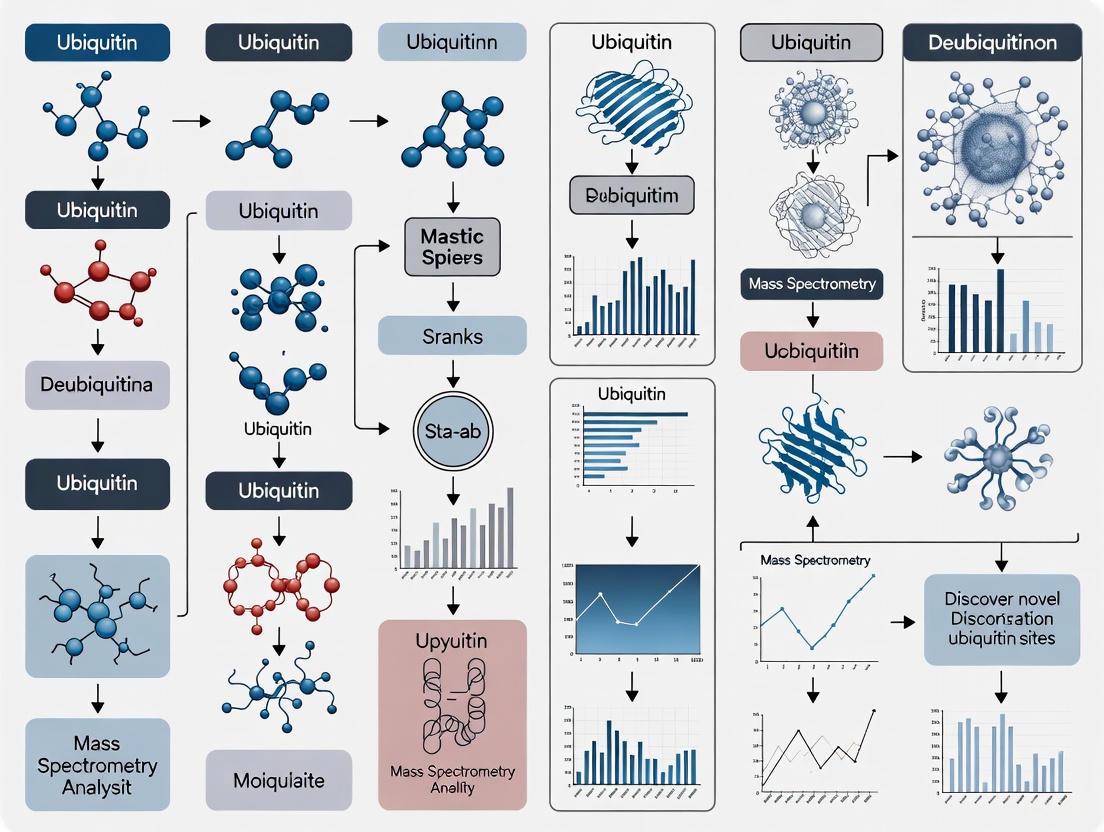
The ubiquitination process is a sequential enzymatic cascade that results in the attachment of ubiquitin to a lysine residue on a substrate protein. The following diagram illustrates this core pathway and its key outcomes.
E1: Ubiquitin-Activating Enzyme
The ubiquitination cascade is initiated by the E1 ubiquitin-activating enzyme in an ATP-dependent process. The E1 enzyme first binds to ATP-Mg²⁺ and ubiquitin, catalyzing the acyl-adenylation of the C-terminus of ubiquitin. This results in a ubiquitin-adenylate (Ub-AMP) intermediate. Subsequently, a catalytic cysteine residue within the E1 active site attacks this complex, forming a high-energy thioester bond between E1 and ubiquitin, with AMP released as a byproduct. Throughout this process, the E1 enzyme can be bound to two ubiquitin molecules, with the second ubiquitin molecule believed to facilitate conformational changes necessary for the subsequent step [1]. The E1 enzyme then recruits an E2 conjugating enzyme, setting the stage for the next step in the cascade.
E2: Ubiquitin-Conjugating Enzyme
The E2 ubiquitin-conjugating enzyme (also known as ubiquitin carrier protein) accepts the activated ubiquitin from the E1 enzyme through a transthioesterification reaction. In this step, a catalytic cysteine on the E2 enzyme attacks the thioester bond linking ubiquitin to the E1, resulting in the transfer of ubiquitin to the E2's active site cysteine, forming a new E2-ubiquitin thioester complex. This transfer involves a complex intermediate wherein both E1 and E2 enzymes undergo a series of conformational changes to bind with one another [1]. The E2 enzyme then complexes with an E3 ubiquitin ligase, which will ultimately facilitate the transfer of ubiquitin to the target protein.
E3: Ubiquitin Ligase
The E3 ubiquitin ligase is the pivotal component that confers substrate specificity to the ubiquitination system. It simultaneously binds to the E2-ubiquitin complex and the target substrate protein, catalyzing the final transfer of ubiquitin. This transfer occurs through the formation of an isopeptide bond between the C-terminus of ubiquitin and a lysine residue on the substrate protein [2]. E3 ligases can be single or multi-subunit enzymes. Their ability to recognize specific substrates is often regulated by post-translational modifications of the substrate itself, such as phosphorylation [2]. The human genome encodes hundreds of E3 ligases, allowing for the precise regulation of a vast number of specific proteins. The attachment of a chain of ubiquitin molecules (a polyubiquitin chain) typically serves as the signal for recognition and degradation by the 26S proteasome [2].
Table 1: Core Enzymes of the Ubiquitin-Proteasome System
| Enzyme | Key Function | Reaction Catalyzed | Key Structural Features |
|---|---|---|---|
| E1 (Ubiquitin-activating enzyme) | Initiates ubiquitination cascade | Ubiquitin adenylation & E1-thioester formation | Catalytic cysteine; binds ATP-Mg²⁺ and ubiquitin [1] |
| E2 (Ubiquitin-conjugating enzyme) | Accepts and carries ubiquitin | Transthioesterification with E1; coordinates with E3 | Catalytic cysteine; E3 binding domain [1] [2] |
| E3 (Ubiquitin ligase) | Provides substrate specificity | Isopeptide bond formation between ubiquitin and substrate lysine | Substrate recognition domain (e.g., TPR domain); E2 binding domain [2] [4] |
Regulation by Deubiquitinases (DUBs)
Deubiquitinases (DUBs) are a class of proteases that function as the essential counterbalance to the ubiquitination process. They cleave ubiquitin moieties from substrate proteins and from polyubiquitin chains, thereby reversing the signal created by E1-E2-E3 activity. This activity allows DUBs to edit ubiquitin chains, recycle ubiquitin, and rescue proteins from proteasomal degradation, making them critical for maintaining the dynamic equilibrium of ubiquitin signaling in the cell [2]. DUBs are categorized into five main subfamilies based on their catalytic mechanisms: ubiquitin-specific proteases (USP), ubiquitin C-terminal hydrolases (UCH), ovarian tumor proteases (OTU), Machado-Joseph disease protein domain proteases (MJD), and JAMM/MPN domain-associated metallopeptidases (JAMM) [2]. The action of DUBs is crucial for a variety of cellular functions, including regulating protein stability, controlling inflammatory responses, and managing cell death pathways. For instance, recent research has revealed that the deubiquitinase OTULIN regulates tau expression and RNA metabolism in neurons, a finding with significant implications for Alzheimer's disease treatment [3].
Experimental Methods for UPS and Ubiquitination Site Research
The study of the UPS and the identification of ubiquitination sites require a combination of biochemical, genetic, and computational approaches. The following sections detail key methodologies used in the field.
High-Throughput Screening for DUB Inhibitors
The identification of selective DUB inhibitors is critical for probing DUB biological function and exploring therapeutic potential. A robust protocol for high-throughput screening (HTS) of DUB inhibitors utilizes a fluorogenic ubiquitin-rhodamine assay [5] [6]. The workflow for this screening approach is detailed below.
The core of this protocol involves the following steps [5] [6]:
- Protein Expression and Purification: Recombinant DUB enzymes are expressed and purified to ensure a consistent enzyme source for the screening assay.
- Assay Optimization and Miniaturization: A fluorogenic substrate, ubiquitin-rhodamine110-glycine (Ub-Rho), is used. Upon cleavage by an active DUB, the rhodamine group is released, producing a measurable fluorescent signal. The assay conditions (buffer, pH, ionic strength) and enzyme kinetics are optimized. The assay is then miniaturized to a 384-well plate format to enable high-throughput processing.
- Primary Screening: A library of small molecules is screened in parallel against a panel of DUBs. Compounds are incubated with the DUB enzyme and the Ub-Rho substrate, and fluorescence is measured over time. Compounds that inhibit DUB activity result in a reduction of the fluorescent signal.
- Dose-Response Analysis: Hit compounds from the primary screen are subjected to dose-response analysis to determine their potency (IC50 values).
- Selectivity Profiling: Promising inhibitors are screened against the panel of DUBs to identify selective inhibitors that target a specific DUB without affecting others.
- Probe Validation: Selective inhibitors are then used as chemical probes in cellular and biochemical assays to elucidate the biological function of the target DUB.
Computational Prediction of Ubiquitination Sites
Given the experimental challenges in identifying ubiquitination sites, computational prediction has become an indispensable tool. GPS-Uber is a hybrid-learning framework developed for the prediction of both general and E3-specific ubiquitination sites [7]. The algorithm was trained on a large dataset of 121,742 ubiquitination sites and uses a model that integrates deep neural networks (DNN), convolutional neural networks (CNN), and penalty logistic regression (PLR). For E3-specific prediction, transfer learning was applied to 1,117 experimentally identified E3-specific sites, allowing the tool to predict substrates for 182 individual E3s across 111 predictors [7]. This tool allows researchers to prioritize lysine residues for experimental validation based on the probability of their ubiquitination.
Studying E3-Substrate Interactions
Understanding the specific interactions between an E3 ligase and its substrate is fundamental. Techniques such as Hydrogen Deuterium Exchange Mass Spectrometry (HDX-MS) can reveal dynamic conformational changes in E3 ligases upon binding substrates or co-chaperones. For example, HDX-MS was used to demonstrate that binding of the co-chaperone Hsp70 to the TPR domain of the E3 ligase CHIP induces allosteric changes that extend to its U-box domain, thereby regulating its E3-ligase activity [4]. This provides critical insights into how E3 activity is modulated beyond simple substrate recognition.
Table 2: Key Experimental Reagents and Resources for UPS Research
| Reagent/Resource | Type | Function/Application | Example/Reference |
|---|---|---|---|
| Fluorogenic Ub-Rho Substrate | Biochemical Probe | High-throughput screening for DUB enzyme activity; cleavage produces fluorescent signal [5] [6] | Ubiquitin-rhodamine110-glycine |
| Recombinant DUBs/E1/E2/E3 | Protein | Essential purified enzymes for in vitro ubiquitination or deubiquitination assays [5] [4] | Purified CHIP (E3), UBE1 (E1) [1] [4] |
| UBE1 (E1 Enzyme) | Recombinant Protein | Essential for initiating in vitro ubiquitination reactions by activating ubiquitin [4] | Commercially available from Boston Biochem [4] |
| Small Molecule Inhibitors | Chemical Probe | Functional modulation of UPS components (e.g., DUB inhibition) for mechanistic studies [3] [5] | UC495 (OTULIN Inhibitor) [3] |
| GPS-Uber | Bioinformatics Tool | In silico prediction of general and E3-specific ubiquitination sites on substrate proteins [7] | http://gpsuber.biocuckoo.cn/ |
| hUbiquitome / UbPred | Database / Algorithm | Public resource of human ubiquitination enzymes and substrates; prediction of ubiquitination sites [8] | UbPred random forest predictor |
UPS in Disease and Therapeutic Targeting
Dysregulation of the UPS is a hallmark of numerous human diseases. In cancer, mutations in E3 ligases like MDM2 (a regulator of the tumor suppressor p53) or overexpression of certain DUBs can lead to uncontrolled cell proliferation. In neurodegenerative diseases such as Alzheimer's disease, the accumulation of toxic proteins is a key feature. Recent groundbreaking research has identified the deubiquitinase OTULIN as a master regulator of tau protein expression, the main component of neurofibrillary tangles in Alzheimer's [3]. Surprisingly, while initial hypotheses suggested OTULIN would affect tau clearance, complete knockout of the OTULIN gene led to the disappearance of tau because its mRNA was not produced, revealing a novel role for OTULIN in regulating gene expression and RNA metabolism [3]. This paradigm-shifting discovery opens new therapeutic avenues for Alzheimer's and related tauopathies, suggesting that partial inhibition of OTULIN could reduce pathological tau without completely eliminating it, potentially offering a therapeutic window [3].
Table 3: Disease Associations of UPS Components
| Disease Category | Example Disease | Associated UPS Component(s) | Molecular Consequence |
|---|---|---|---|
| Neurodegenerative | Alzheimer's Disease | Deubiquitinase OTULIN [3] | Increased tau expression and phosphorylation |
| Neurodegenerative | X-linked Infantile Spinal Muscular Atrophy (XL-SMA) | UBE1 (E1 enzyme) missense mutations [1] | Impaired degradation of MAP1B, neuronal cell death |
| Neuromuscular | Parkinson's Disease | E3 Ubiquitin Ligases (e.g., Parkin) [7] | Accumulation of damaged proteins, neuronal toxicity |
| Autoimmune & Inflammatory | Inflammatory Arthritis, Lupus, VEXAS Syndrome | Dysregulated UPS activity [1] | Disrupted immune cell signaling and homeostasis |
| Cancer | Various Cancers | Mutations in E3s (e.g., MDM2); DUB overexpression [7] | Stabilization of oncoproteins; loss of tumor suppressors |
The Ubiquitin-Proteasome System represents one of the most sophisticated and critical regulatory networks in cell biology. Its core enzymatic cascade—E1, E2, and E3—works in concert to direct the precise modification of target proteins with ubiquitin, while deubiquitinases provide essential reversibility and fine-tuning. The continued development of advanced experimental methods, including high-throughput screening for DUB inhibitors and sophisticated computational prediction tools like GPS-Uber, is dramatically accelerating our ability to discover novel ubiquitination sites and understand the intricate regulation of this system. Furthermore, the association of UPS dysregulation with a wide spectrum of human diseases, underscored by recent paradigm-shifting discoveries such as OTULIN's role in regulating tau expression, highlights the immense therapeutic potential of targeting this pathway. Future research will undoubtedly continue to unravel the complexities of the UPS, leading to novel diagnostic and therapeutic strategies for some of the most challenging human diseases.
The ubiquitin system constitutes a vital post-translational modification (PTM) network that regulates virtually all aspects of eukaryotic cell biology, from protein degradation to cell signaling, DNA repair, and immune responses [9] [10]. This versatility stems from the remarkable structural diversity of ubiquitin modifications, which can assume various forms including mono-ubiquitination, multiple mono-ubiquitination, and diverse polyubiquitin chains that differ in length, linkage type, and overall architecture [9] [11]. The specificity of ubiquitin signaling is governed by an enzymatic cascade involving E1 (activating), E2 (conjugating), and E3 (ligating) enzymes, with the human genome encoding approximately 2 E1s, 40 E2s, and over 600 E3s that provide exquisite specificity [9] [11] [10]. This sophisticated enzymatic machinery enables the precise attachment of ubiquitin to substrate proteins, creating a complex "ubiquitin code" that is interpreted by specialized effector proteins to determine substrate fate and function [10].
Understanding this diverse ubiquitination landscape is paramount for elucidating fundamental biological processes and developing novel therapeutic strategies. Dysregulation of ubiquitin signaling underlies numerous pathologies, including cancer, neurodegenerative diseases, and immune disorders, making components of the ubiquitin system attractive drug targets [9] [10]. This technical guide comprehensively details the molecular architectures, biological functions, and experimental methodologies for characterizing diverse ubiquitin modifications, with particular emphasis on their relevance to discovering novel ubiquitination sites and their functional implications.
Molecular Architecture of Ubiquitin Modifications
Mono-ubiquitination and Multiple Mono-ubiquitination
Mono-ubiquitination describes the covalent attachment of a single ubiquitin molecule to a substrate protein, typically occurring at lysine residues via an isopeptide bond between the C-terminal glycine (G76) of ubiquitin and the ε-amino group of the substrate lysine [9]. This modification can regulate diverse non-proteolytic processes including protein activity, protein-protein interactions, and subcellular localization [10]. Multiple mono-ubiquitination occurs when a substrate is modified by single ubiquitin moieties at multiple distinct lysine residues, creating a ubiquitination pattern that can be recognized by specific effector proteins containing ubiquitin-binding domains (UBDs) [9]. Historically, mono-ubiquitination was primarily associated with histone regulation and membrane trafficking, but recent studies have expanded its functional repertoire to include roles in DNA repair, transcription, and kinase activation [12].
Polyubiquitin Chain Architectures
Polyubiquitin chains are classified into three major categories based on their linkage patterns:
Table 1: Classification of Polyubiquitin Chain Architectures
| Chain Type | Structural Definition | Key Characteristics | Examples |
|---|---|---|---|
| Homotypic Chains | Uniform linkage through the same acceptor site | Single linkage type throughout chain | K48-linked, K63-linked |
| Mixed Chains | Multiple linkage types with each ubiquitin modified at one site | Sequential arrangement of different linkages | M1/K63, K11/K48 |
| Branched Chains | Ubiquitin subunits simultaneously modified on ≥2 different sites | Complex topology with branch points | K11/K48, K48/K63, K29/K48 |
Homotypic chains represent the best-characterized category, with each chain type exhibiting distinct structural properties and biological functions [11]. For instance, K48-linked ubiquitin chains represent the most abundant linkage in cells and primarily target substrate proteins for degradation by the 26S proteasome [9]. In contrast, K63-linked chains predominantly regulate protein-protein interactions in processes such as NF-κB pathway activation, DNA damage response, and autophagy [9]. M1-linked linear chains, generated through N-terminal methionine linkage, play crucial roles in inflammatory signaling and NF-κB activation [10].
Branched ubiquitin chains constitute a particularly complex category characterized by the presence of ubiquitin molecules modified at two or more distinct sites, creating intricate polymeric structures with specialized functions [11]. These chains significantly expand the coding potential of ubiquitin signaling and have been implicated in diverse cellular processes, including cell cycle regulation and signal transduction [11]. For example, K11/K48-branched chains assembled by the APC/C complex during mitosis can enhance substrate targeting to the proteasome, while K48/K63-branched chains generated by E3 ligase pairs like TRAF6 and HUWE1 regulate NF-κB signaling [11].
Figure 1: Classification of Ubiquitin Modification Types. Ubiquitin signals are categorized based on their structural complexity, ranging from single modifications to complex chain architectures with distinct functional consequences.
Biological Functions of Ubiquitin Signals
The diverse ubiquitination landscapes described above enable the regulation of an extraordinary range of cellular processes. The specific biological outcome of ubiquitination depends on multiple factors, including the type of modification, the cellular context, and the presence of specific effector proteins that interpret the ubiquitin code.
Table 2: Biological Functions of Major Ubiquitin Linkage Types
| Linkage Type | Primary Biological Functions | Key Effectors/Pathways |
|---|---|---|
| K48 | Proteasomal degradation, cell cycle control | 26S proteasome, Ub-binding proteins |
| K63 | NF-κB signaling, DNA repair, endocytosis, autophagy | TAB2/3, RAP80, ESCRT complex |
| K11 | ER-associated degradation, cell cycle regulation | Proteasome, Cdc48/p97 |
| K29 | Proteasomal degradation, transcriptional regulation | E3 ligase HUWE1, Ufd2 |
| K33 | T-cell receptor signaling, kinase regulation | T-cell receptor pathway |
| K6 | DNA damage response, mitochondrial regulation | BRCA1/BARD1 complex |
| K27 | Neuroinflammatory signaling, lysosomal targeting | UCHL3, HOIP complex |
| M1 (linear) | NF-κB activation, inflammatory responses | HOIP complex, NEMO |
The functional specialization of ubiquitin linkages enables precise control over cellular homeostasis. For instance, while K48-linked chains predominantly target proteins for proteasomal degradation, thereby controlling the half-lives of regulatory proteins and eliminating misfolded proteins, K63-linked chains function as scaffolds in signal transduction pathways by facilitating the assembly of protein complexes [9] [10]. The NF-κB signaling pathway exemplifies how different ubiquitin linkages cooperate: M1-linked and K63-linked chains activate upstream signaling components, while K48-linked chains terminate signaling by targeting inhibitory proteins for degradation [10].
Branched ubiquitin chains often function as enhanced or specialized signals that can determine the efficiency or specificity of substrate recognition. For example, K11/K48-branched chains assembled by the APC/C complex during mitosis appear to promote more efficient proteasomal targeting of cell cycle regulators compared to homotypic K48 chains [11]. Similarly, the sequential formation of K48/K63-branched chains on the apoptosis regulator TXNIP converts a non-degradative K63-linked signal into a degradative one, providing a mechanism for signal termination [11]. This conversion strategy represents an efficient means of regulating the activation and inactivation dynamics of signaling proteins controlled by ubiquitylation events [11].
The functional complexity of the ubiquitin system is further enhanced by crosstalk between different ubiquitin linkages and other post-translational modifications. For instance, ubiquitination can be modulated by phosphorylation, acetylation, and other modifications on either the substrate or ubiquitin itself, creating sophisticated regulatory networks that enable cells to respond precisely to changing environmental conditions [9].
Analytical Methodologies for Ubiquitination Studies
Enrichment Strategies for Ubiquitinated Proteins
The low stoichiometry of protein ubiquitination under normal physiological conditions necessitates effective enrichment strategies prior to analysis. Three principal approaches have been developed to isolate ubiquitinated proteins from complex biological samples:
Ubiquitin Tagging-Based Approaches utilize genetically engineered ubiquitin containing affinity tags such as 6×His or Strep-tag for purification [9]. In this methodology, cells are engineered to express tagged ubiquitin, which becomes incorporated into the endogenous ubiquitination machinery. Following cell lysis, ubiquitinated proteins are enriched using affinity resins such as Ni-NTA for His-tag or Strep-Tactin for Strep-tag [9]. This approach enabled the pioneering identification of 110 ubiquitination sites on 72 proteins in Saccharomyces cerevisiae and has been refined through systems like the Stable Tagged Ubiquitin Exchange (StUbEx) for human cells [9]. While relatively straightforward and cost-effective, this method risks generating artifacts as tagged ubiquitin may not perfectly mimic endogenous ubiquitin, and genetic manipulation limits application to clinical samples [9].
Ubiquitin Antibody-Based Approaches employ antibodies such as P4D1 and FK1/FK2 that recognize all ubiquitin linkages to immunoprecipitate endogenously ubiquitinated proteins without genetic manipulation [9]. This strategy is particularly valuable for studying ubiquitination in animal tissues or clinical samples. Furthermore, linkage-specific antibodies (e.g., for K48, K63, M1 linkages) enable the selective enrichment of proteins modified with particular chain types, providing insight into the functional specialization of ubiquitin signals [9]. For instance, linkage-specific antibodies revealed abnormal accumulation of K48-linked polyubiquitination on tau proteins in Alzheimer's disease [9]. Limitations include the high cost of high-quality antibodies and potential non-specific binding.
Ubiquitin-Binding Domain (UBD)-Based Approaches exploit natural ubiquitin receptors, such as tandem UBA domains or specific subunits of the proteasome, to affinity-purify ubiquitinated proteins [9]. These domains can exhibit general ubiquitin binding or linkage-specific preferences, making them valuable tools for interrogating specific aspects of the ubiquitin code. For example, the UbIA-MS method uses chemically synthesized diubiquitin of specific linkages to enrich and identify linkage-selective interactors from cell lysates [13].
Mass Spectrometry-Based Proteomics
Advanced mass spectrometry (MS) techniques represent the cornerstone of modern ubiquitin research, enabling comprehensive identification of ubiquitination sites, quantification of ubiquitin chain linkages, and characterization of ubiquitin architectures [9]. Following enrichment, ubiquitinated proteins are typically digested with trypsin, which generates a characteristic di-glycine (Gly-Gly) remnant on modified lysines with a mass shift of 114.04 Da, serving as a diagnostic feature for ubiquitination site identification by MS [9].
Recent methodological advances have significantly enhanced the sensitivity and scope of ubiquitin proteomics:
- Ubiquitin-AQUA: Absolute quantification using synthetic stable isotope-labeled ubiquitin peptides enables precise measurement of specific ubiquitin linkage abundances [12].
- DiGly antibody enrichment: Antibodies specifically recognizing the diGly remnant improve the detection sensitivity of ubiquitination sites by MS [9].
- UbIA-MS: Ubiquitin Interactor Affinity Enrichment-Mass Spectrometry uses chemically synthesized diubiquitin to profile linkage-specific interactors proteome-wide [13].
- Middle-down MS: This approach allows characterization of mixed and branched ubiquitin chains by analyzing larger peptide fragments [11].
Figure 2: Experimental Workflow for Ubiquitin Proteomics. The general pipeline for mass spectrometry-based analysis of ubiquitination includes sample preparation, enrichment of ubiquitinated proteins, tryptic digestion generating diGly signatures, LC-MS/MS analysis, and bioinformatic data interpretation.
Computational Prediction of Ubiquitination Sites
To complement experimental approaches, computational tools have been developed to predict ubiquitination sites from protein sequence features. Ubigo-X represents a recent advance in this area, employing ensemble learning with image-based feature representation and weighted voting [14]. This tool integrates three sub-models:
- Single-Type sequence-based features: Amino acid composition, physicochemical properties, and one-hot encoding
- k-mer sequence-based features: Capture local sequence context around potential modification sites
- Structure- and function-based features: Secondary structure, solvent accessibility, and signal peptide cleavage sites
Ubigo-X achieved an area under the curve (AUC) of 0.85-0.94 in independent testing, outperforming existing prediction tools, particularly for balanced datasets [14]. Such computational approaches provide valuable prioritization for experimental validation, especially for low-abundance ubiquitination events that challenge MS-based detection.
The Scientist's Toolkit: Key Research Reagents and Methodologies
Table 3: Essential Research Reagents and Methodologies for Ubiquitin Studies
| Reagent/Methodology | Key Features | Primary Applications | Considerations |
|---|---|---|---|
| His/Strep-tagged Ubiquitin | Affinity purification tags | High-throughput substrate identification | Potential artifacts, cannot use in tissues |
| Linkage-specific Antibodies | Recognize specific chain types | Enrichment and detection of specific linkages | High cost, specificity validation required |
| Ubiquitin Mutants (K0, K-to-R) | Block specific chain extensions | Functional studies of specific linkages | May perturb normal ubiquitin landscape |
| Diubiquitin Probes | Chemically synthesized defined linkages | Interaction proteomics (UbIA-MS) | Requires specialized synthesis expertise |
| Activity-Based DUB Probes | Covalently trap deubiquitinases | DUB activity profiling and identification | Requires active enzyme forms |
| Tandem UBD Affinity Reagents | High-affinity ubiquitin binders | Enrichment of endogenous ubiquitinated proteins | May exhibit linkage preferences |
| Ubigo-X Prediction Tool | Ensemble machine learning | Computational ubiquitination site prediction | AUC 0.85-0.94, species-neutral |
Technical Applications: Ubi-Tagging for Protein Engineering
Beyond its biological functions, the ubiquitin system has inspired innovative protein engineering applications. Ubi-tagging is a recently developed technology that exploits the ubiquitination machinery for site-specific, multivalent conjugation of antibodies to various payloads [15]. This modular approach enables rapid (30-minute) generation of homogeneous antibody conjugates for diagnostic and therapeutic applications.
The ubi-tagging system employs three key components:
- Donor ubi-tag (Ubdon): Contains free C-terminal glycine with the conjugating lysine mutated to arginine to prevent homodimer formation
- Acceptor ubi-tag (Ubacc): Contains the corresponding conjugation lysine with blocked C-terminus (ΔGG or His-tag)
- Specific E1/E2/E3 enzymes: Determines linkage specificity (e.g., K48-specific gp78RING-Ube2g2)
This platform has been successfully applied to generate bispecific T-cell engagers, fluorescently labeled Fab' fragments, and nanobody-antigen conjugates with high efficiency (93-96% conversion) and minimal impact on protein stability or antigen binding [15]. The technology demonstrates how understanding fundamental ubiquitin biochemistry enables innovative solutions to longstanding challenges in biotherapeutics development.
The diverse ubiquitination landscapes encompassing mono-ubiquitination, polyubiquitin chains of various linkages, and complex branched architectures represent a sophisticated regulatory system that controls virtually all aspects of cell physiology. The continuing development of advanced mass spectrometry methods, linkage-specific reagents, computational prediction tools, and innovative applications like ubi-tagging is rapidly expanding our ability to decipher the complex language of ubiquitin signaling. As these methodologies become increasingly sophisticated and accessible, they promise to accelerate both fundamental discoveries of ubiquitin-mediated processes and the development of novel therapeutic strategies targeting the ubiquitin system in human disease.
Ubiquitination in Cellular Homeostasis and Disease Pathogenesis
Ubiquitination is a crucial post-translational modification process that involves the covalent attachment of a small, 76-amino acid protein called ubiquitin to substrate proteins [16]. This highly conserved enzymatic cascade regulates virtually all aspects of cellular physiology in eukaryotic organisms. The process is mediated by a sequential action of three enzymes: ubiquitin-activating enzymes (E1), ubiquitin-conjugating enzymes (E2), and ubiquitin ligases (E3) [17] [16]. The human genome encodes approximately 2 E1 enzymes, over 35 E2 enzymes, and more than 600 E3 ligases, which provide tremendous specificity in substrate recognition [17] [18].
Ubiquitination serves as a fundamental mechanism for maintaining cellular protein homeostasis by targeting proteins for proteasomal degradation, but its functional repertoire extends far beyond protein turnover [17]. This modification regulates diverse cellular processes including cell cycle progression, DNA damage repair, signal transduction, and immune responses [17] [19]. The versatility of ubiquitination signals stems from the ability of ubiquitin itself to form various polymer chains through its internal lysine residues or N-terminal methionine [17]. Different chain linkage types encode distinct functional consequences for the modified substrate, creating a sophisticated ubiquitin code that determines protein fate and function [17].
Dysregulation of ubiquitination pathways contributes to the pathogenesis of numerous human diseases, particularly cancers and neurodegenerative disorders [17] [18]. Consequently, the ubiquitin-proteasome system has emerged as an attractive therapeutic target, exemplified by the clinical success of proteasome inhibitors in treating multiple myeloma and mantle cell lymphoma [17] [18]. This whitepaper provides an in-depth technical examination of ubiquitination in cellular homeostasis and disease pathogenesis, with particular emphasis on methodologies for discovering novel ubiquitination sites and their applications in drug development.
The Ubiquitination Machinery
The Enzymatic Cascade
The ubiquitination process initiates with E1 ubiquitin-activating enzymes, which activate ubiquitin in an ATP-dependent manner through the formation of a ubiquitin-adenylate intermediate, followed by transfer of ubiquitin to the E1 active-site cysteine via a thioester bond [17] [16]. The activated ubiquitin is then transferred to a cysteine residue of an E2 conjugating enzyme through a trans-thioesterification reaction [17]. Finally, an E3 ubiquitin ligase facilitates the transfer of ubiquitin from the E2 to a lysine residue on the substrate protein, forming an isopeptide bond between the C-terminal glycine of ubiquitin and the ε-amino group of the substrate lysine [17] [16].
E3 ubiquitin ligases fall into three major structural classes based on their catalytic mechanisms. RING (Really Interesting New Gene) and U-box E3s function as scaffolds that simultaneously bind E2~Ub and substrate, facilitating direct ubiquitin transfer without a covalent E3-ubiquitin intermediate [17] [18]. In contrast, HECT (Homologous to E6AP C-terminus) E3s form a thioester intermediate with ubiquitin on a catalytic cysteine residue before transferring it to the substrate [17] [18]. RBR (RING-between-RING) E3s employ a hybrid mechanism, combining aspects of both RING and HECT-type catalysis [17].
Diversity of Ubiquitin Signals
Ubiquitin modification can take several forms, each with distinct functional consequences. Monoubiquitination involves attachment of a single ubiquitin molecule and typically regulates protein activity, localization, or interactions [16]. Multi-monoubiquitination occurs when multiple lysine residues on a single substrate are modified with individual ubiquitin molecules [20]. Polyubiquitination involves the formation of ubiquitin chains through linkage between the C-terminus of one ubiquitin and specific lysine residues (K6, K11, K27, K29, K33, K48, K63) or the N-terminal methionine (M1) of another ubiquitin molecule [17] [20].
Table: Ubiquitin Linkage Types and Their Primary Functions
| Linkage Type | Primary Functions |
|---|---|
| K48-linked | Targets substrates for proteasomal degradation [17] |
| K63-linked | Regulates protein-protein interactions, signaling pathways, endocytosis, and DNA repair [17] |
| K11-linked | Cell cycle regulation and proteasomal targeting [17] |
| K6-linked | DNA damage repair [17] |
| K27-linked | Controls mitochondrial autophagy [17] |
| K29-linked | Cell cycle regulation and stress response [17] |
| K33-linked | T-cell receptor-mediated signaling [17] |
| M1-linked (linear) | NF-κB inflammatory signaling [17] |
The complexity of ubiquitin signaling is further enhanced by the formation of heterotypic chains (containing multiple linkage types) and branched chains, which likely expand the coding capacity of ubiquitin signals [17]. Additionally, ubiquitination interacts with other post-translational modifications such as phosphorylation, acetylation, and SUMOylation, creating sophisticated regulatory networks [17].
Diagram 1: The ubiquitination enzymatic cascade. The process involves sequential action of E1 (activation), E2 (transference), and E3 (conjugation/ligation) enzymes, ultimately leading to substrate ubiquitination.
Deubiquitination
The ubiquitination process is reversible through the action of deubiquitinating enzymes (DUBs), which cleave ubiquitin from modified substrates [17]. The human genome encodes approximately 100 DUBs belonging to several structural families, with the ubiquitin-specific proteases (USPs) representing the largest group [18]. DUBs perform multiple cellular functions including processing of ubiquitin precursors, editing of ubiquitin-protein conjugates, and recycling ubiquitin at the proteasome [17]. The balance between ubiquitination and deubiquitination creates dynamic regulation of protein ubiquitination status, allowing cells to rapidly respond to changing physiological conditions.
Methodologies for Ubiquitination Site Discovery
Mass Spectrometry-Based Approaches
Mass spectrometry has revolutionized the identification and quantification of ubiquitination sites on a proteome-wide scale. The primary MS-based strategy exploits the characteristic di-glycine (di-Gly) remnant that remains attached to modified lysine residues after tryptic digestion [19]. This di-Gly modification produces a distinct mass shift of 114.0429 Da, enabling precise identification and localization of ubiquitination sites based on peptide fragment masses [19].
Antibody-Based Enrichment Strategies
Early proteomic studies utilized antibodies that recognize ubiquitin for enrichment of ubiquitinated proteins prior to MS analysis. While this approach enabled identification of ubiquitinated proteins, it typically yielded limited information about specific modification sites [20]. A major advancement came with the development of di-glycine-lysine-specific antibodies that specifically recognize the tryptic remnant of ubiquitination [19]. This technology enables direct immunoenrichment of ubiquitinated peptides from complex tryptic digests, dramatically improving the depth and coverage of ubiquitin site mapping.
A landmark study utilizing this approach identified 11,054 endogenous ubiquitination sites on 4,273 human proteins, providing unprecedented insight into the scope and diversity of the ubiquitin-modified proteome [19]. The methodology involves cell lysis under denaturing conditions in the presence of N-ethylmaleimide to inhibit deubiquitinases, followed by protein digestion, peptide purification, and immunoenrichment with di-Gly-lysine-specific antibodies [19]. The enriched peptides are then fractionated and analyzed by high-resolution liquid chromatography-tandem mass spectrometry (LC-MS/MS).
Diagram 2: Workflow for mass spectrometry-based ubiquitination site mapping using di-glycine remnant antibody enrichment.
Quantitative Ubiquitin Proteomics
Combining di-Gly remnant enrichment with stable isotope labeling by amino acids in cell culture (SILAC) enables quantitative assessment of ubiquitination dynamics in response to cellular perturbations [19]. This powerful approach has revealed that ubiquitination site occupancy spans over four orders of magnitude, with the median ubiquitination site occupancy being three orders of magnitude lower than that of phosphorylation [21]. Furthermore, quantitative studies have demonstrated that inhibition of proteasomal function with MG-132 not only increases ubiquitination on degradation targets but also decreases ubiquitination at many sites with non-proteasomal functions, revealing complex feedback regulation within the ubiquitin system [19] [21].
Recent advances in quantitative ubiquitin proteomics have enabled measurement of both ubiquitination site occupancy (stoichiometry) and turnover rates, providing a systems-level view of ubiquitination dynamics [21]. These studies have revealed that sites in structured protein regions exhibit longer half-lives and stronger upregulation by proteasome inhibitors than sites in unstructured regions [21]. Additionally, researchers have discovered a surveillance mechanism that rapidly deubiquitinates all ubiquitin-specific E1 and E2 enzymes, protecting them against accumulation of bystander ubiquitylation [21].
Computational Prediction Methods
While mass spectrometry provides experimental identification of ubiquitination sites, computational approaches offer complementary strategies for large-scale prediction of ubiquitination sites. Traditional machine learning methods such as Support Vector Machines (SVM) have been employed with features including amino acid composition, evolutionary information, position-specific scoring matrices, and physicochemical properties [22]. However, these methods typically rely on hand-engineered features which may introduce bias and incompletely represent relevant biological information.
More recently, deep learning approaches have been applied to ubiquitination site prediction, overcoming limitations of traditional feature engineering [22]. A multimodal deep architecture has been developed that integrates three complementary representations of protein sequences: (1) raw protein sequence fragments, (2) physicochemical properties, and (3) evolutionary profiles from position-specific scoring matrices (PSSM) [22]. This approach uses convolutional neural networks to extract relevant features directly from the input representations, eliminating the need for manual feature engineering.
The deep learning framework was trained on the Protein Lysine Modification Database (PLMD), containing 121,742 ubiquitination sites from 25,103 proteins [22]. After removing homologous sequences, the final dataset contained 60,879 ubiquitination sites from 17,406 proteins. The model achieved 66.4% specificity, 66.7% sensitivity, and 66.43% accuracy, outperforming existing prediction tools [22]. This demonstrates the power of deep learning for large-scale ubiquitination site prediction, particularly as the volume of experimentally identified sites continues to grow.
Table: Comparison of Ubiquitination Site Detection Methods
| Method | Principle | Throughput | Advantages | Limitations |
|---|---|---|---|---|
| Di-Gly antibody MS | Immunoenrichment of ubiquitinated peptides with MS detection [19] | High | Identifies endogenous sites; enables quantification; comprehensive coverage | Cannot distinguish ubiquitin from NEDD8/ISG15; requires specific equipment |
| Tagged ubiquitin | Affinity purification using epitope-tagged ubiquitin [20] | High | Efficient enrichment; can express in specific cell types | May not fully mimic endogenous ubiquitin; genetic manipulation required |
| Linkage-specific antibodies | Antibodies recognizing specific ubiquitin linkages [20] | Medium | Provides linkage information; works with endogenous proteins | Limited to characterized linkages; antibody quality variable |
| Computational prediction | Machine learning on sequence and structural features [22] | Very high | Low cost; applicable to any protein sequence | Predictive only; requires experimental validation |
The Scientist's Toolkit: Research Reagent Solutions
Table: Essential Research Reagents for Ubiquitination Studies
| Reagent Type | Specific Examples | Function and Applications |
|---|---|---|
| Ubiquitin Antibodies | P4D1, FK1/FK2 (pan-ubiquitin); linkage-specific antibodies (K48, K63, etc.) [20] | Immunoblotting, immunofluorescence, and immunoprecipitation of ubiquitinated proteins; linkage-specific antibodies enable characterization of chain topology |
| Tagged Ubiquitin Systems | His-tagged Ub, Strep-tagged Ub, HA-Ub [20] | Affinity purification of ubiquitinated proteins; can be expressed in cells to facilitate enrichment and identification of ubiquitination substrates |
| Activity Assays | Auto-ubiquitination kits [23] | In vitro analysis of E1, E2, and E3 enzyme activity; high-throughput screening for ubiquitination inhibitors |
| Proteasome Inhibitors | Bortezomib, Carfilzomib, MG-132 [19] [18] | Block proteasomal degradation, causing accumulation of ubiquitinated proteins; tools for studying ubiquitination dynamics and identifying proteasomal substrates |
| DUB Inhibitors | PR-619, P22077, etc. | Inhibit deubiquitinating enzymes, stabilizing ubiquitination signals; useful for studying transient ubiquitination events |
| di-Gly Remnant Antibodies | Commercial di-glycine-lysine antibodies [19] | Immunoenrichment of ubiquitinated peptides for mass spectrometry-based ubiquitinome analysis |
| UBD-Based Reagents | Tandem Ubiquitin-Binding Entities (TUBEs) [20] | Affinity reagents for enriching ubiquitinated proteins while protecting against deubiquitination and proteasomal degradation |
Ubiquitination in Disease and Therapeutic Targeting
Ubiquitination in Cancer
Dysregulation of ubiquitination pathways is implicated in numerous cancers, with mutations in ubiquitin system components identified across cancer types [17] [18]. E3 ubiquitin ligases such as MDM2 (negative regulator of p53) are frequently overexpressed in cancers, leading to excessive degradation of tumor suppressor proteins [17]. Conversely, many tumor suppressors function as E3 ligases or components of ubiquitin-regulated complexes, and their inactivation promotes tumorigenesis [18].
The clinical success of proteasome inhibitors (bortezomib, carfilzomib) in treating multiple myeloma and mantle cell lymphoma validated the ubiquitin-proteasome system as a therapeutic target in oncology [17] [18]. These drugs cause accumulation of polyubiquitinated proteins, disrupting protein homeostasis and ultimately triggering apoptosis in malignant cells [18]. Interestingly, cancer cells appear more sensitive to proteasome inhibition than normal cells, although the precise mechanisms underlying this selective vulnerability remain under investigation [18].
Emerging Therapeutic Strategies
Beyond proteasome inhibitors, several innovative strategies are being developed to target ubiquitination pathways for cancer therapy:
E1/E2/E3-Targeted Inhibitors
Significant efforts have focused on developing specific inhibitors of ubiquitination cascade enzymes. MLN4924 (Pevonedistat) is a selective inhibitor of NEDD8-activating enzyme (NAE1) that blocks the neddylation pathway required for activation of cullin-RING ligases (CRLs) [17]. By inhibiting CRL activity, MLN4924 stabilizes numerous CRL substrates involved in cell cycle progression and DNA replication, causing DNA re-replication and apoptosis in cancer cells [17]. This compound has entered clinical trials for treatment of various malignancies.
Small molecule inhibitors of E3 ligases such as Nutlin (MDM2 inhibitor) have shown promise in preclinical models by stabilizing p53 and activating apoptosis in cancer cells retaining wild-type p53 [17]. Additionally, fragment-based screening and DNA-encoded compound libraries are being employed to identify novel inhibitors of E2 and E3 enzymes [18].
PROTAC Technology
Proteolysis-Targeting Chimeras (PROTACs) represent a revolutionary approach to targeted protein degradation [18]. These bifunctional molecules consist of a target-binding warhead connected to an E3 ligase-recruiting ligand via a chemical linker. By bringing the target protein into proximity with an E3 ubiquitin ligase, PROTACs induce target ubiquitination and subsequent proteasomal degradation [18]. This technology enables selective degradation of disease-causing proteins that may be difficult to target with conventional inhibitors, expanding the druggable proteome.
Ubiquitin Variants (UbVs)
Protein engineering approaches have generated ubiquitin variants that function as specific inhibitors of E3 ligases or DUBs [18]. These engineered ubiquitin molecules can block enzyme-substrate interactions, modulating specific ubiquitination events without globally disrupting ubiquitination. For example, UbVs have been developed that selectively inhibit HECT E3 ligases including NEDD4L [18].
Novel Frontiers: Small Molecule Ubiquitination
Recent research has revealed that ubiquitin ligases can modify not only proteins but also drug-like small molecules. A 2025 study demonstrated that the E3 ligase HUWE1 can ubiquitinate compounds previously reported as HUWE1 inhibitors [24]. These compounds, containing primary amino groups, were modified with ubiquitin through the canonical enzymatic cascade [24]. This discovery expands the substrate realm of ubiquitination to include exogenous small molecules and opens possibilities for harnessing the ubiquitin system to transform therapeutic compounds into novel chemical modalities within cells.
Diagram 3: Therapeutic strategies targeting the ubiquitin-proteasome system in cancer. Multiple approaches are being developed to exploit different components of the ubiquitination machinery for cancer therapy.
The field of ubiquitination research has evolved from fundamental biochemical studies to comprehensive systems-level analyses and therapeutic applications. Technical advances in mass spectrometry, particularly di-Gly remnant-based enrichment, have enabled quantitative mapping of ubiquitination sites across the proteome, revealing the astonishing scope and complexity of ubiquitin signaling [19] [21]. Concurrently, computational methods have advanced from feature-based machine learning to deep learning architectures capable of integrating multiple modalities for large-scale ubiquitination site prediction [22].
Future challenges include developing improved methods for characterizing atypical ubiquitin linkages, quantifying ubiquitin chain architecture, and understanding the spatial regulation of ubiquitination within cellular compartments. Additionally, there is a need for better tools to distinguish ubiquitination from modifications by other ubiquitin-like proteins (NEDD8, ISG15) that generate identical di-Gly remnants after tryptic digestion [19].
The therapeutic targeting of ubiquitination pathways continues to advance with novel modalities including PROTACs, molecular glues, and ubiquitin variants expanding the toolbox for modulating protein stability [18]. The recent discovery that drug-like small molecules can serve as ubiquitination substrates further expands the potential applications of ubiquitination in biotechnology and medicine [24]. As our understanding of ubiquitination in cellular homeostasis and disease pathogenesis deepens, so too will opportunities for developing innovative therapies for cancer, neurodegenerative disorders, and other diseases linked to ubiquitin pathway dysregulation.
Ubiquitination is a reversible post-translational modification (PTM) that regulates nearly all aspects of eukaryotic biology, including proteasome and lysosome degradation, gene transcription, DNA repair and replication, intracellular trafficking, stress response, and cell-cycle regulation [25]. The process involves a cascade of enzymes (E1 activating, E2 conjugating, and E3 ligase enzymes) that attach the 76-amino acid ubiquitin protein to lysine residues on target proteins [25]. Ubiquitination can occur as monoubiquitination or polyubiquitination, with different chain linkages conferring distinct functional consequences for the modified protein [26].
The identification of ubiquitination sites represents a critical step toward understanding the biological role of this modification in cellular regulation and disease pathogenesis. However, researchers face three fundamental challenges in ubiquitination site discovery: accurately determining modification stoichiometry, capturing dynamic turnover rates, and deciphering the complexity of ubiquitin chain architectures. This technical guide examines these core challenges and outlines current methodological frameworks for addressing them, providing researchers with a comprehensive resource for advancing studies of ubiquitin-dependent signaling systems.
Quantitative Landscape of Ubiquitination
Stoichiometry and Occupancy Challenges
A primary challenge in ubiquitination site analysis lies in its remarkably low stoichiometry compared to other post-translational modifications. Recent global, site-resolved analyses reveal that ubiquitylation site occupancy spans over four orders of magnitude, yet the median ubiquitylation site occupancy is three orders of magnitude lower than that of phosphorylation [21]. This low occupancy presents significant detection challenges, as conventional analytical methods often lack the sensitivity to capture these modification events.
The distribution of ubiquitination sites follows a distinct pattern, with the lowest 80% and the highest 20% occupancy sites exhibiting distinct properties [21]. High-occupancy sites are particularly concentrated in the cytoplasmic domains of solute carrier (SLC) proteins, suggesting specialized regulatory functions for these membrane transporters [21]. This occupancy disparity necessitates specialized enrichment and detection strategies tailored to the specific occupancy range of interest within experimental designs.
Table 1: Key Quantitative Properties of Ubiquitination Sites
| Property | Finding | Biological Significance |
|---|---|---|
| Site Occupancy | Spans over four orders of magnitude; median 3 orders lower than phosphorylation | Explains detection challenges; indicates tight regulatory control |
| Occupancy Distribution | Distinct properties between lowest 80% and highest 20% occupancy sites | Suggests different regulatory mechanisms for high vs. low occupancy sites |
| Structural Correlation | Sites in structured regions exhibit longer half-lives than unstructured regions | Links protein structure to ubiquitination dynamics and function |
| Enzyme Protection | Rapid deubiquitylation of E1 and E2 enzymes prevents bystander ubiquitylation | Reveals quality control mechanism in ubiquitination machinery |
Dynamics and Turnover Rates
The turnover rate of ubiquitination sites represents another critical dimension of the ubiquitin code, with direct implications for their biological functions. Research demonstrates that occupancy, turnover rate, and regulation by proteasome inhibitors are strongly interrelated [21]. These attributes collectively distinguish sites primarily involved in proteasomal degradation from those participating in cellular signaling pathways.
The cellular environment implements a surveillance mechanism that rapidly and site-indiscriminately deubiquitylates all ubiquitin-specific E1 and E2 enzymes, protecting them against accumulation of bystander ubiquitylation [21]. This specialized regulatory system ensures the proper functioning of the ubiquitination machinery itself, highlighting the layered complexity of the ubiquitin system.
Analytical Frameworks for Ubiquitination Studies
Mass Spectrometry-Based Approaches
Mass spectrometry has emerged as the cornerstone technology for ubiquitination site identification and quantification. The fundamental strategy involves purifying the protein of interest, generating peptides through proteolytic digestion (typically with trypsin), and analyzing the resulting peptides by mass spectrometry [27] [28]. Trypsin cleaves after the carboxyl-terminal arginine in ubiquitin, leaving only the two terminal glycine residues attached to the modified lysine in the target protein [28]. The resulting 114-Dalton mass increase serves as a diagnostic signature for ubiquitination sites [28].
Advanced proteomic workflows now integrate multiple enrichment and quantification strategies to address ubiquitination complexity. Stable Isotope Labeling with Amino acids in Cell culture (SILAC) enables precise relative quantification, while tandem mass tag (TMT) methods allow multiplexing of up to 10 samples simultaneously [29]. To overcome signal compression limitations in TMT experiments, LC-MS3 approaches with synchronous precursor selection (MultiNotch MS3) significantly improve quantification accuracy by co-isolating and co-fragmenting multiple MS2 fragment ions [29]. These advanced mass spectrometry configurations are particularly valuable for capturing the dynamic range of ubiquitination stoichiometry.
Diagram 1: Mass spectrometry workflow for ubiquitination site identification. The process involves sample preparation, protein extraction, proteolytic digestion, enrichment of ubiquitinated peptides, LC-MS/MS analysis, and computational data processing for site identification.
Specialized Enrichment Technologies
Enrichment strategies are essential for overcoming the low stoichiometry of ubiquitination. Recent advances include Tandem Ubiquitin Binding Entities (TUBEs), which are engineered protein domains with nanomolar affinities for polyubiquitin chains [26]. These specialized reagents protect ubiquitin chains from deubiquitinating enzymes and proteasomal degradation during sample processing, significantly improving detection sensitivity [26].
The development of chain-specific TUBEs represents a particularly significant advancement, enabling researchers to discriminate between different ubiquitin linkage types. For example, K48-linked chains primarily target proteins for proteasomal degradation, while K63-linked chains regulate signal transduction and protein trafficking [26]. Research demonstrates that K63-TUBEs can specifically capture inflammatory agent L18-MDP-induced RIPK2 ubiquitination, while K48-TUBEs selectively enrich for RIPK2 PROTAC-induced ubiquitination [26]. This linkage-specific resolution provides critical functional insights that pan-selective enrichment methods cannot deliver.
Table 2: Research Reagent Solutions for Ubiquitination Studies
| Reagent/Tool | Type | Primary Function | Application Examples |
|---|---|---|---|
| TUBEs (Pan-selective) | Affinity reagent | Broad ubiquitin chain enrichment; protects from DUBs | General ubiquitination profiling; stabilization of ubiquitinated proteins |
| Chain-specific TUBEs | Linkage-specific affinity reagent | Selective enrichment of specific ubiquitin linkages | Differentiating K48 (degradation) vs K63 (signaling) ubiquitination |
| UbPred | Computational tool | Predicts ubiquitination sites from protein sequences | Preliminary site identification; guiding experimental design |
| Ubigo-X | Machine learning model | Ensemble learning for ubiquitination site prediction | Species-neutral ubiquitination site prediction |
| Mutant Ubiquitins | Biological reagent | Dominant-negative approach to study chain specificity | Identifying functional roles of specific ubiquitin linkages |
Computational Prediction of Ubiquitination Sites
Machine Learning Frameworks
The experimental challenges and costs associated with ubiquitination site mapping have stimulated the development of computational prediction tools. Machine learning approaches now offer valuable complementary strategies for identifying potential ubiquitination sites. The Ubigo-X tool exemplifies recent advances, employing ensemble learning with image-based feature representation and weighted voting to predict ubiquitination sites [14]. This approach integrates three sub-models: Single-Type sequence-based features, k-mer sequence-based features, and structure-based and function-based features, achieving an area under the curve (AUC) of 0.85 on balanced independent test data [14].
Comparative analyses reveal that deep learning approaches generally outperform classical machine learning methods for ubiquitination site prediction, with the best-performing models achieving a 0.902 F1-score, 0.8198 accuracy, 0.8786 precision, and 0.9147 recall [25]. Interestingly, model performance shows a positive correlation with the length of amino acid fragments, suggesting that utilizing entire protein sequences can yield more accurate predictions [25]. These computational tools serve as valuable preliminary screening methods before committing to resource-intensive experimental verification.
Specialized Experimental Models and Systems
Kinetic Analysis of Ubiquitination
Understanding the kinetics of ubiquitination is essential for deciphering its regulatory functions, particularly in the context of targeted protein degradation. Recent methodological advances enable the determination of kinetics for small-molecule-induced ubiquitination, a crucial capability for the development of proteolysis-targeting chimeras (PROTACs) [30]. These systems allow researchers to fit essential activator kinetic models to ubiquitination data, characterizing the affinities between bifunctional degraders, target proteins, and E3 ligases in binary complexes, ternary complexes, and full ubiquitination complexes [30].
Mathematical modeling of ubiquitination kinetics reveals that protein degradation mainly follows Michaelis-Menten formulation with a time delay caused by ubiquitination and deubiquitination processes [31]. This nonlinear degradation kinetics significantly influences system dynamics, promoting oscillations in biological networks and enlarging the parameter space for oscillatory behavior [31]. However, the time delay inherent in ubiquitination and deubiquitination generally suppresses oscillations, reducing amplitude and increasing frequency [31]. These insights highlight the importance of considering both enzymatic kinetics and system architecture when studying ubiquitin-driven processes.
Integration with Phosphorylation Analysis
Ubiquitination frequently functions within integrated PTM networks, most notably with phosphorylation. Quantitative proteomic approaches now enable simultaneous analysis of these interconnected modification systems [29] [32]. Two canonical pathways exemplify this integration: (1) substrates are phosphorylated to generate "phosphodegrons" recognized by SCF complexes, which then promote ubiquitination; and (2) E3 ligases themselves are phosphorylated, leading to their activation through various mechanisms [29].
Diagram 2: Integrated ubiquitination and phosphorylation signaling pathways. Two canonical pathways show how phosphorylation can either create recognition motifs (phosphodegrons) on substrates for E3 ligase binding or directly activate E3 ligases to promote substrate ubiquitination, leading to diverse functional outcomes.
The field of ubiquitination site discovery continues to evolve with increasingly sophisticated methodologies addressing the fundamental challenges of stoichiometry, dynamics, and chain complexity. The integration of advanced mass spectrometry platforms, specialized enrichment tools, computational predictions, and kinetic models provides researchers with a powerful toolkit for deciphering the ubiquitin code.
Future methodological developments will likely focus on improving spatial resolution through subcellular ubiquitination mapping, enhancing temporal resolution for capturing rapid ubiquitination dynamics, and expanding linkage-specific tools beyond the well-characterized K48 and K63 chains. As these technologies mature, they will further illuminate the intricate roles of ubiquitination in cellular regulation and disease pathogenesis, accelerating drug discovery efforts particularly in the targeted protein degradation arena. The continued refinement of these specialized methodologies promises to unlock deeper insights into the complex world of ubiquitin signaling, providing researchers with an expanding arsenal of tools for probing this essential regulatory system.
A Toolkit for Discovery: Experimental and Computational Methods for Site Identification
Ubiquitination, the covalent attachment of a small regulatory protein to substrate proteins, is a fundamental post-translational modification (PTM) that governs critical cellular processes including protein degradation, signal transduction, and DNA repair [9] [33]. The discovery of novel ubiquitination sites is therefore paramount to understanding both normal physiology and disease pathogenesis, such as cancer and neurodegenerative disorders [9] [34]. However, the low stoichiometry of ubiquitinated species within the complex cellular milieu and the diverse architectures of ubiquitin chains present significant analytical challenges [9]. Consequently, effective enrichment of ubiquitinated proteins or peptides is an indispensable prerequisite for their subsequent identification and characterization, typically via mass spectrometry (MS). This technical guide provides an in-depth examination of the three cornerstone experimental strategies for ubiquitin enrichment: antibody-based methods, ubiquitin-binding domain (UBD) approaches, and tagged ubiquitin systems. Framed within the context of discovering novel ubiquitination sites, this review synthesizes current methodologies, detailed protocols, and emerging innovations to equip researchers with the knowledge to select and implement the most appropriate strategy for their specific research objectives.
The selection of an enrichment strategy directly influences the specificity, depth, and biological relevance of ubiquitination data. The three primary methods—tagged ubiquitin, antibody-based, and UBD-based enrichment—operate at different levels (protein versus peptide) and offer distinct advantages and limitations, which are systematically compared in Table 1.
Table 1: Comprehensive Comparison of Ubiquitin Enrichment Strategies
| Enrichment Strategy | Principle | Key Advantages | Major Limitations | Typical Application in Novel Site Discovery |
|---|---|---|---|---|
| Tagged Ubiquitin [9] | Ectopic expression of ubiquitin fused to an affinity tag (e.g., His, Strep, HA). | Relatively low cost; effectively reduces background from non-ubiquitinated proteins. | Cannot mimic endogenous ubiquitin perfectly; genetic manipulation required; infeasible for clinical tissues. | Initial screening and validation of ubiquitinated substrates in engineered cell lines. |
| Antibody-Based [35] [9] | Immunoaffinity purification using antibodies against ubiquitin or the K-ε-GG remnant. | Can profile endogenous ubiquitination; applicable to any sample type; high sensitivity for site identification. | High cost; antibody sequence recognition bias; cannot distinguish from NEDD8/ISG15 (anti-K-ε-GG). | Global, site-specific profiling of ubiquitination across diverse sample types, including tissues. |
| UBD-Based [9] [36] | Utilization of natural or engineered protein domains with high affinity for ubiquitin chains. | Purifies endogenous proteins; no tag or antibody needed; can be engineered for linkage specificity. | Lower affinity for monoubiquitination; can have high background; lower identification efficiency. | Enrichment of ubiquitinated proteins and analysis of ubiquitin chain topology. |
The following diagram illustrates the logical decision-making process for selecting an appropriate enrichment strategy based on key experimental parameters, helping researchers align their methodology with project goals.
Antibody-Based Enrichment Strategies
Antibody-based methods are among the most widely used and sensitive techniques for ubiquitin profiling, particularly for site-specific identification. These approaches can be deployed at two levels: enriching intact ubiquitinated proteins or enriching tryptic peptides derived from them.
Protein-Level Immunoprecipitation
This classical approach utilizes antibodies raised against ubiquitin itself (e.g., P4D1, FK1, FK2) or specific ubiquitin chain linkages (e.g., K48, K63) to immunoprecipitate ubiquitinated proteins from complex cell lysates [9] [37]. The enriched proteins can then be separated by SDS-PAGE, and the high molecular weight smears—characteristic of polyubiquitinated proteins—can be excised, subjected to in-gel tryptic digestion, and analyzed by LC-MS/MS [35]. This method preserves information about the protein substrate but provides relatively low efficiency for mapping the exact site of ubiquitination.
Peptide-Level Immunoaffinity Enrichment (K-ε-GG)
A transformative advancement in the field was the development of antibodies specifically recognizing the di-glycine (K-ε-GG) remnant left attached to the modified lysine residue after tryptic digestion of ubiquitinated proteins [35]. This method has become the gold standard for large-scale ubiquitin site mapping due to its high sensitivity and specificity.
Experimental Protocol for K-ε-GG Peptide Immunoaffinity Enrichment [35]:
- Cell Lysis and Proteolysis: Lyse cells in a suitable buffer (e.g., RIPA). Reduce, alkylate, and digest the protein lysate to peptides using trypsin.
- Peptide Immunoaffinity Enrichment: Incubate the peptide mixture with anti-K-ε-GG antibody beads. As demonstrated in a 2013 study, this consistently yielded a greater than fourfold higher level of modified peptides than protein-level affinity purification-mass spectrometry (AP-MS) approaches [35].
- Wash and Elution: Wash the beads extensively to remove non-specifically bound peptides. Elute the bound K-ε-GG-modified peptides.
- LC-MS/MS Analysis: Desalt and analyze the eluted peptides by liquid chromatography coupled to tandem mass spectrometry.
The power of this technique is evident from its application, which led to the identification of thousands of ubiquitination sites from just 1 mg of input material and enabled the characterization of inducible ubiquitination on members of the T-cell receptor complex [35]. A key limitation, however, is that the K-ε-GG remnant is identical to that generated by the ubiquitin-like modifiers NEDD8 and ISG15, potentially leading to false-positive assignments without additional controls [9] [38].
Ubiquitin-Binding Domain (UBD) Based Strategies
UBDs are natural protein modules that non-covalently interact with ubiquitin. Their exploitation provides a powerful, tag-free method for enriching endogenous ubiquitinated proteins.
Principle and Native UBDs
Proteins containing UBDs, such as some E3 ligases, deubiquitinases (DUBs), and Ub receptors, can be utilized to bind and enrich ubiquitinated proteins [9]. However, the affinity of a single UBD is often low, limiting its utility for purification. To overcome this, Tandem Ubiquitin-Binding Entities (TUBEs) were developed. TUBEs consist of multiple UBDs fused in tandem, conferring a much higher affinity for ubiquitin chains and protecting ubiquitinated substrates from deubiquitination and proteasomal degradation during purification [37].
Engineered Tandem Hybrid UBDs (ThUBDs)
Recent innovations have focused on engineering artificial UBDs with superior properties. One study systematically evaluated UBD affinities and constructed Tandem Hybrid UBDs (ThUBDs) by combining different high-affinity UBDs [36]. Two constructs, ThUDQ2 and ThUDA20, demonstrated markedly higher affinity than naturally occurring UBDs and displayed almost unbiased high affinity to all seven lysine-linked ubiquitin chains [36]. When used for MS-based profiling, ThUBDs enabled the identification of 7487 putative ubiquitinated proteins from mammalian cells, showcasing their utility for deep ubiquitome coverage without genetic manipulation or antibodies [36].
Tagged Ubiquitin Strategies
This approach involves the genetic engineering of ubiquitin to include an affinity tag, allowing for stringent purification of ubiquitin-conjugated proteins.
Standard Tagged Ubiquitin Workflow
Researchers typically generate cell lines that stably express ubiquitin tagged with an epitope (e.g., HA, FLAG) or protein tag (e.g., His, Strep, GST) [9]. After treating cells under desired conditions, lysates are prepared, and the tagged ubiquitin along with its conjugated substrates are purified using the appropriate affinity resin (e.g., Ni-NTA for His-tag, Strep-Tactin for Strep-tag). The purified proteins can be identified by MS. A variation of this is the Stable Tagged Ubiquitin Exchange (StUbEx) system, where endogenous ubiquitin is replaced with His-tagged ubiquitin, facilitating the identification of hundreds of ubiquitination sites [9].
Limitations and Considerations
While cost-effective and widely used, this method has critical caveats. The introduced tag may alter ubiquitin's structure or function, potentially leading to artifacts [9]. Furthermore, histidine-rich or endogenously biotinylated proteins can co-purify, increasing background noise. Most importantly, this strategy is restricted to genetically tractable cell systems and cannot be applied to primary tissues or clinical samples, limiting its translational relevance [9].
Emerging Antibody-Free Methodologies
To circumvent the limitations of antibodies, such as cost and sequence bias, novel chemical biology approaches are being developed.
The AFUP Strategy
A notable antibody-free method for Ubiquitination Profiling (AFUP) has been proposed, which selectively labels and enriches ubiquitinated peptides based on their unique chemical properties [38]. The workflow involves:
- Blocking: All free amine groups on proteins are blocked with formaldehyde.
- Deubiquitination: Treatment with deubiquitinases (USP2 and USP21) removes ubiquitin, regenerating the free ε-amine specifically at the site of modification.
- Click Labeling: The newly exposed ε-amines are biotinylated with NHS-SS-biotin.
- Enrichment and Analysis: Biotinylated peptides are captured on streptavidin beads, eluted, and analyzed by LC-MS/MS.
This innovative strategy identified 349 ± 7 ubiquitination sites from 0.8 mg of HeLa lysate with excellent reproducibility and, when combined with fractionation, over 7,000 sites in 293T cells, proving to be a robust and cost-effective complementary tool for ubiquitomics [38].
The Scientist's Toolkit: Essential Research Reagents
Successful implementation of ubiquitin enrichment protocols requires a suite of reliable reagents. The following table details key materials and their functions.
Table 2: Key Research Reagent Solutions for Ubiquitin Enrichment
| Reagent / Tool | Function / Application | Examples / Notes |
|---|---|---|
| Anti-K-ε-GG Antibody [35] | Immunoaffinity enrichment of ubiquitinated peptides for MS-based site mapping. | Central to high-sensitivity site identification; check for cross-reactivity with NEDD8/ISG15. |
| Linkage-Specific Ub Antibodies [9] | Enrichment or detection of ubiquitin chains with specific linkages (e.g., K48, K63). | Critical for deciphering the functional consequences of ubiquitination. |
| TUBEs / ThUBDs [36] [37] | High-affinity enrichment of endogenous ubiquitinated proteins; protect chains from DUBs. | Engineered ThUBDs show broad linkage affinity and high yield. |
| Tagged Ubiquitin Plasmids [9] | Expression of His-, HA-, or Strep-tagged Ub in cells for substrate purification. | Essential for tagged-ubiquitin approaches; available from multiple cDNA repositories. |
| Deubiquitinases (DUBs) [38] | Hydrolyze ubiquitin from substrates; used in antibody-free methods like AFUP. | USP2 and USP21 catalytic domains are commonly used for their broad specificity. |
| Proteasome Inhibitors [35] [34] | Stabilize ubiquitinated proteins by blocking their degradation (e.g., MG132, Bortezomib). | Used during cell treatment to increase the yield of labile ubiquitinated species. |
Integrated Experimental Workflow
A typical, comprehensive workflow for ubiquitin site discovery integrates cell culture, sample preparation, enrichment, and mass spectrometry, as visualized below.
The experimental enrichment of ubiquitinated species is a dynamic and critical frontier in proteomics, directly enabling the discovery of novel ubiquitination sites. Antibody-based methods, particularly K-ε-GG immunoaffinity enrichment, remain the most sensitive for site-specific profiling. UBD-based approaches offer a powerful, tag-free alternative for studying endogenous proteins and chain architecture. Tagged ubiquitin systems provide a straightforward method for substrate identification in manipulable cell lines. The ongoing development of sophisticated tools like engineered ThUBDs and chemical biology methods like AFUP continues to push the boundaries of sensitivity, specificity, and applicability. The choice of strategy must be guided by the specific biological question, the model system, and the desired depth of information. As these methodologies mature and integrate, they will undoubtedly accelerate our understanding of the vast ubiquitin code and its implications in health and disease.
Protein ubiquitination is a pivotal post-translational modification (PTM) that regulates diverse cellular functions, including protein degradation, activity, and localization [9]. The versatility of ubiquitination stems from the complexity of ubiquitin (Ub) conjugates, which can range from a single Ub monomer to polymers of different lengths and linkage types [9]. For researchers focused on discovering novel ubiquitination sites, understanding the molecular mechanism of ubiquitination signaling requires sophisticated strategies to characterize not only the modification sites but also the linkage type and architecture of Ub chains [9]. The dysregulation of ubiquitination pathways leads to many pathologies, including cancer and neurodegenerative diseases, making the comprehensive mapping of ubiquitination sites a critical endeavor in both basic research and drug development [9].
Mass spectrometry has emerged as the cornerstone technology for ubiquitin-modified proteome analysis, enabling the large-scale identification and quantification of ubiquitination sites [39]. This technical guide details the integrated workflows from specific enrichment of ubiquitinated peptides to their confident identification and data analysis, providing a framework for researchers designing studies aimed at novel ubiquitination site discovery.
Ubiquitinated Peptide Enrichment Methodologies
Since ubiquitinated peptides are typically of low abundance compared to their unmodified counterparts, effective enrichment is a critical first step in any ubiquitination site discovery pipeline [9] [39]. The choice of enrichment strategy significantly impacts the depth and reliability of ubiquitin site identification.
Table 1: Comparison of Ubiquitinated Peptide Enrichment Strategies
| Method | Principle | Advantages | Limitations | Typical Applications |
|---|---|---|---|---|
| Immunoaffinity (diGly Antibody) | Antibodies specifically recognize the diGly remnant left on lysine after tryptic digestion [40]. | High specificity; applicable to endogenous ubiquitination; suitable for tissue samples [9]. | Cannot distinguish ubiquitination from other diGly modifications; antibody cost [9]. | Global ubiquitin site profiling from complex samples including clinical tissues [40]. |
| Ubiquitin Tagging (e.g., His/Strep) | Ectopic expression of affinity-tagged ubiquitin (e.g., 6×His, Strep) in cells [9]. | Relatively low cost; easy implementation; good for cellular systems [9]. | Cannot mimic endogenous Ub perfectly; genetic manipulation required; potential artifacts [9]. | High-throughput screening of ubiquitinated substrates in cultured cell lines [9]. |
| Ubiquitin-Binding Domain (UBD) | Uses proteins or domains that naturally bind ubiquitin (e.g., from DUBs, E3 ligases) [9]. | Captures endogenous ubiquitination; can potentially preserve linkage information [9]. | Lower affinity of single UBDs; may require tandem domains for efficiency [9]. | Enrichment of specific ubiquitin chain linkages when using linkage-selective UBDs [9]. |
| Tandem Enrichment (SCASP-PTM) | Serial enrichment of multiple PTMs (Ub, phosphorylation, glycosylation) from a single sample without intermediate desalting [41]. | Efficient use of precious sample; comprehensive PTM profiling [41]. | Protocol complexity; potential for cross-contamination between PTM fractions [41]. | Multi-PTM profiling from limited sample material; systems-level analysis of PTM crosstalk [41]. |
Specialized Enrichment Protocol: SCASP-PTM for Tandem PTM Enrichment
A recently developed protocol, SCASP-PTM (SDS-cyclodextrin-assisted sample preparation-post-translational modification), enables the tandem enrichment of ubiquitinated, phosphorylated, and glycosylated peptides from a single sample in a serial manner [41]. The key steps include:
- Protein Extraction and Digestion: Proteins are extracted using a specialized SDS-cyclodextrin buffer system to ensure efficient solubilization and denaturation. This is followed by standard reduction, alkylation, and tryptic digestion procedures [41].
- Serial Enrichment without Desalting: Unlike many traditional workflows, the SCASP-PTM method allows for the direct enrichment of ubiquitinated peptides from the protein digest without a desalting step. The flowthrough from this step is then directly used for the subsequent enrichment of either phosphorylated or glycosylated peptides [41].
- Cleanup and MS Analysis: After the serial enrichment, the isolated PTM peptides are desalted prior to mass spectrometric analysis to ensure compatibility with LC-MS systems [41].
This approach is particularly valuable for discovery-phase research where sample quantity is limited and a broad view of the PTM landscape is desired.
Mass Spectrometric Analysis and Data Acquisition
Following enrichment, ubiquitinated peptides are analyzed by liquid chromatography tandem mass spectrometry (LC-MS/MS). High-resolution mass spectrometers are essential for accurately identifying the characteristic mass shift (+114.04 Da) on the modified lysine residues, corresponding to the diGly remnant [9] [39].
The identification of the precise site of ubiquitination relies on MS/MS fragmentation, which reveals sequence information and allows localization of the modification to specific lysine residues [39]. For discovery-focused projects aimed at finding novel sites, data-independent acquisition (DIA) methods are increasingly employed alongside traditional data-dependent acquisition (DDA) because DIA provides comprehensive, reproducible recording of all fragment ions in a sample, which is valuable for retrospective analysis [41].
A significant challenge in ubiquitination site analysis is the presence of multi-ubiquitination, where ubiquitin chains are attached to a single lysine residue, resulting in complex fragmentation patterns [39]. Advanced techniques like top-down mass spectrometry or high-resolution tandem MS are being applied to better capture and sequence intact polyubiquitin chains, thereby improving the characterization of chain topology [39].
Data Processing and Visualization Workflows
The raw data generated from LC-MS/MS runs requires sophisticated computational processing to confidently identify ubiquitination sites and, where possible, determine Ub chain linkage.
Integrated Data Analysis Platforms
Workflow systems that combine small, reusable building blocks into larger, experiment-specific analysis pipelines provide the flexibility needed to handle diverse experimental setups in ubiquitination research [42]. A prominent example is the integration of OpenMS tools into the KNIME (Konstanz Information Miner) platform [42].
- OpenMS is an open-source software framework for computational mass spectrometry that provides numerous tools for signal processing, identification, and quantification, including adapters to established search engines like MASCOT, OMSSA, and X!Tandem [42].
- KNIME is an open-source integration platform that offers a powerful and flexible workflow system with advanced data analytics, visualization, and reporting capabilities [42].
The combination allows users to create automated pipelines that process raw MS data (signal processing, quantification, identification) and seamlessly connect to KNIME's data-mining and visualization nodes for downstream analysis [42]. Such workflows can be shared with collaborators as preconfigured ZIP files, ensuring reproducibility [42].
Visualization for Quality Control and Interpretation
Data visualization is a crucial component at every stage of the ubiquitin proteomics workflow, providing core capabilities for data inspection, evaluation, and sharing [43]. Interactive visualizations allow researchers to interact with and explore their data from different angles without manually re-generating plots, streamlining scientific discovery [43].
Effective visualization strategies in untargeted omics include:
- Volcano plots to visualize treatment impacts and significantly altered ubiquitination sites [43].
- Cluster heatmaps to extract and highlight patterns within the ubiquitination data [43].
- Network visualizations to organize and showcase relations between ubiquitinated proteins and other cellular components [43].
Quality control visualization is equally important. Customizable dashboards can be developed to monitor instrument performance parameters, helping laboratories take proactive measures to maintain instruments and ultimately reduce downtime [44].
The Scientist's Toolkit: Essential Research Reagents and Materials
Table 2: Key Research Reagent Solutions for Ubiquitination Site Mapping
| Reagent/Material | Function | Example Applications |
|---|---|---|
| diGly Remnant Antibodies | Immunoaffinity enrichment of ubiquitinated peptides after tryptic digestion; recognizes the characteristic Gly-Gly remnant on lysine [40]. | Global ubiquitinome profiling from complex biological samples including cell lines and tissues [40]. |
| Linkage-Specific Ub Antibodies | Enrich ubiquitinated proteins with specific chain linkages (e.g., K48, K63) [9]. | Studying the functional role of specific ubiquitin chain types in pathways like proteasomal degradation (K48) or signaling (K63) [9]. |
| Recombinant Ubiquitin (Wild-type & Mutants) | Used in in vitro ubiquitination assays to study enzyme specificity and chain formation [39]. | Mechanistic studies of E3 ligase activity and specificity; reconstitution of ubiquitination cascades [39]. |
| Affinity Tags (His, Strep) | Genetic fusion to ubiquitin for purification of ubiquitinated substrates from cellular systems [9]. | High-throughput screening of ubiquitination substrates in cultured cells; StUbEx (Stable Tagged Ub Exchange) systems [9]. |
| Recombinant E1, E2, E3 Enzymes | Core enzymatic components for in vitro ubiquitination reactions [39]. | Biochemical characterization of ubiquitination mechanisms; identification of direct substrates for specific E3 ligases [39]. |
| Activity-Based Probes | Chemical probes that covalently bind ubiquitin or deubiquitinases (DUBs) for enrichment or activity profiling [39]. | Profiling active DUBs in cell lysates; chemical proteomics strategies for ubiquitination analysis [39]. |
| SILAC/TMT Kits | For stable isotope labeling enabling quantitative comparison of ubiquitination levels across different experimental conditions [39]. | Differential ubiquitination studies to identify sites altered by drug treatments, disease states, or genetic perturbations [39]. |
Comprehensive mass spectrometry workflows integrating specific enrichment strategies, advanced instrumentation, and sophisticated data analysis platforms have dramatically accelerated the discovery of novel ubiquitination sites. The continued refinement of methods like the SCASP-PTM protocol for tandem PTM enrichment and the development of more accurate computational tools promise to further deepen our understanding of the complex ubiquitin code. For researchers in both academic and drug development settings, mastering these workflows is essential for uncovering the role of ubiquitination in fundamental biological processes and disease mechanisms, ultimately paving the way for novel therapeutic interventions targeting the ubiquitin-proteasome system.
Protein ubiquitination is a fundamental, reversible post-translational modification (PTM) that regulates a vast array of cellular processes, including protein degradation, signal transduction, cell cycle control, and DNA repair [25]. This modification involves the covalent attachment of a small, 76-amino acid protein, ubiquitin, to lysine residues on target substrate proteins [45]. The versatility of ubiquitination signaling arises from its complexity; it can manifest as monoubiquitination, multi-ubiquitination, or polyubiquitination chains with various linkage types, each dictating distinct functional outcomes for the modified protein [9]. Given its pivotal role, dysregulation of the ubiquitin system is implicated in the pathogenesis of numerous human diseases, including cancer, neurodegenerative disorders, and autoimmune diseases [25] [45].
Traditional experimental methods for identifying ubiquitination sites, such as mass spectrometry (MS) and immunoprecipitation (IP), are often costly, time-consuming, and labor-intensive [25] [9]. Furthermore, the low stoichiometry of ubiquitination and the complexity of Ub chains make experimental detection particularly challenging [9]. To address these limitations, the field has witnessed a surge in computational approaches designed to predict ubiquitination sites from protein sequence and structural features. This whitepaper explores the current landscape of machine learning (ML) and deep learning (DL) tools for ubiquitination site prediction, with a focused examination of two advanced tools: Ubigo-X and EUP. These tools exemplify how modern computational strategies are overcoming previous barriers, offering researchers powerful, species-neutral platforms for high-throughput discovery in ubiquitination research and drug development.
Methodological Foundations: From Feature Engineering to Deep Learning
The evolution of computational tools for ubiquitination site prediction has transitioned from conventional machine learning relying on hand-crafted features to sophisticated deep learning models that automatically learn relevant representations.
Feature Representation and Conventional ML Approaches
Early and some contemporary ML models depend on expertly engineered features derived from protein sequences. These features provide the input vectors for classifiers like Support Vector Machines (SVM) and Random Forests (RF) [25]. Key feature categories include:
- Amino Acid Composition (AAC) and Physicochemical Properties (PCP): These features represent the distribution and biochemical characteristics of amino acids surrounding the target lysine, which are critical for enzyme recognition [14] [25].
- k-mer Composition (e.g., CKSAAP): This method captures local sequence order information by counting the frequency of k-spaced amino acid pairs [25].
- Structure-Based Features: Features such as secondary structure, relative solvent accessibility (RSA), and absolute solvent-accessible area (ASA) provide information on the structural environment of a lysine residue, which influences its accessibility to ubiquitinating enzymes [14].
- Evolutionary Information: Features like position-specific scoring matrices (PSSMs) derived from multiple sequence alignments encapsulate evolutionary conservation, a strong indicator of functional sites.
The Shift to Deep and Transfer Learning
Deep learning models have demonstrated superior performance by automating feature extraction from raw sequences or simplified inputs [25]. Key advancements include:
- End-to-End Sequence Models: Models like DeepUni use Convolutional Neural Networks (CNNs) to learn relevant patterns directly from amino acid sequences, bypassing the need for manual feature engineering [25].
- Image-Based Feature Representation: Ubigo-X innovatively transforms sequence-based features into image-like formats, which are then processed by powerful CNN architectures like ResNet34 [14] [46].
- Transfer Learning and Protein Language Models: EUP leverages a pre-trained protein language model, ESM2, to extract rich, contextual features from protein sequences. ESM2 embeddings capture evolutionary information, structural patterns, and functional constraints learned from millions of diverse protein sequences [47]. This approach is particularly powerful for cross-species prediction and in scenarios with limited labeled training data.
- Multimodal and Ensemble Architectures: State-of-the-art tools combine multiple feature types and models. For instance, MMUbiPred integrates one-hot encoding, embeddings, and physicochemical properties within a unified deep-learning framework to enhance prediction robustness [48].
Table 1: Key Feature Types Used in Ubiquitination Site Prediction
| Feature Category | Specific Examples | Description | Biological Significance |
|---|---|---|---|
| Sequence-Based | Amino Acid Composition (AAC), k-mer (CKSAAP) | Quantifies the occurrence of amino acids or short peptides in a sequence window. | Reveals local sequence motifs recognized by E3 ligases. |
| Evolutionary | PSSM, ESM2 Embeddings | Encodes conservation and evolutionary constraints from multiple sequences. | Identifies functionally important, conserved residues. |
| Physicochemical | AAindex, One-Hot Encoding | Represents biochemical properties (e.g., hydrophobicity, charge) of amino acids. | Captures biophysical constraints for enzyme binding. |
| Structure-Based | Secondary Structure, RSA/ASA | Describes the local 3D structural environment of the lysine residue. | Determines solvent accessibility and structural fit for modification. |
In-Depth Analysis of Featured Tools: Ubigo-X and EUP
Ubigo-X: Ensemble Learning with Image-Based Features
Ubigo-X is a novel prediction tool that distinguishes itself through an ensemble learning strategy with image-based feature representation and weighted voting [14] [46].
Experimental Protocol and Methodology:
- Data Curation: The model was trained on a large dataset sourced from the Protein Lysine Modification Database (PLMD 3.0), comprising 53,338 ubiquitination and 71,399 non-ubiquitination sites after sequence redundancy filtering with CD-HIT [14] [46].
- Multi-Model Feature Extraction: Ubigo-X develops three distinct sub-models, each processing different feature types:
- Single-Type SBF: Utilizes AAC, AAindex, and one-hot encoding.
- Co-Type SBF: Applies k-mer encoding to the features from the Single-Type SBF model.
- Structure-Function Based Features (S-FBF): Incorporates secondary structure, RSA/ASA, and signal peptide cleavage sites.
- Model Training and Ensemble: The S-FBF model is trained using XGBoost, a gradient-boosting algorithm. In contrast, the Single-Type SBF and Co-Type SBF features are transformed into image-based features and trained using a ResNet34 deep learning architecture. The final prediction is generated by combining the outputs of these three models via a weighted voting strategy [14] [46].
Ubigo-X Ensemble Architecture
EUP: Cross-Species Prediction with Protein Language Models
EUP addresses the critical challenge of generalizability and data scarcity across different species by leveraging a large pre-trained protein language model.
Experimental Protocol and Methodology:
- Data Sourcing and Curation: EUP was constructed using ubiquitination data from the CPLM 4.0 database, which includes 182,120 experimentally verified ubiquitination sites from multiple species (e.g., Homo sapiens, Mus musculus, Arabidopsis thaliana). A key step involves rigorous data cleaning, including de-homology and balancing of ubiquitination and non-ubiquitination sites [47].
- Feature Extraction with ESM2: Instead of hand-crafted features, EUP uses the ESM2 model to generate high-dimensional, contextual feature representations for each lysine site and its surrounding sequence. This captures deep evolutionary and functional information [47].
- Dimensionality Reduction with cVAE: A conditional Variational Autoencoder (cVAE) is employed to reduce the high-dimensional ESM2 features into a lower-dimensional, informative latent representation. This step helps in denoising and creating a more robust feature set [47].
- Species-Neutral Prediction: A downstream deep neural network classifier is built on this latent representation. This architecture allows EUP to perform accurate cross-species ubiquitination site prediction, effectively "dismantling the species barrier" [47].
EUP Cross-Species Prediction Workflow
Performance Benchmarking and Comparative Analysis
Evaluating the performance of computational tools requires rigorous testing on balanced and imbalanced independent datasets. Key metrics include Area Under the Curve (AUC), Accuracy (ACC), and Matthews Correlation Coefficient (MCC), the latter being particularly informative for imbalanced data.
Table 2: Performance Comparison of Ubiquitination Prediction Tools
| Tool | Core Methodology | Independent Test Data | AUC | Accuracy | MCC | Key Strength |
|---|---|---|---|---|---|---|
| Ubigo-X [14] [46] | Ensemble (XGBoost + ResNet34) + Weighted Voting | Balanced (PhosphoSitePlus) | 0.85 | 0.79 | 0.58 | High AUC & ACC on balanced data |
| Imbalanced (1:8 ratio) | 0.94 | 0.85 | 0.55 | Robust on imbalanced data | ||
| EUP [47] | ESM2 + cVAE + DNN | Cross-Species Independent Test | Not Reported | 77.25% | Not Reported | Cross-species generalization |
| MMUbiPred [48] | Multimodal Deep Learning | Independent Human Test | 0.87 | 77.25% | 0.54 | Integrates diverse sequence representations |
| Deep Learning Model [25] | Hybrid (Sequence + Hand-crafted features) | Benchmark Dataset | Not Reported | 81.98% | Not Reported | Combines raw sequence and expert features |
The data reveals that Ubigo-X achieves state-of-the-art performance on balanced datasets, with high AUC and accuracy, while also demonstrating remarkable robustness on imbalanced data, which is common in real-world biological scenarios [14]. EUP and MMUbiPred, meanwhile, showcase the power of modern deep learning approaches for building generalizable and cross-species predictive models [47] [48]. The performance of these tools underscores a significant advancement over conventional ML methods, which often struggle with generalizability and data imbalance.
The Scientist's Toolkit: Research Reagent Solutions
Table 3: Essential Resources for Ubiquitination Site Research
| Resource Name | Type | Function in Research | Example Use Case |
|---|---|---|---|
| PLMD 3.0 / CPLM 4.0 [14] [47] | Database | Repository of experimentally verified protein lysine modifications, including ubiquitination sites. | Source of high-quality, curated data for training and testing computational models. |
| PhosphoSitePlus [14] | Database | Repository of PTM sites, used as an independent dataset for validating prediction tool performance. | Benchmarking model generalizability on unseen data. |
| ESM2 [47] | Protein Language Model | Provides deep contextual and evolutionary feature representations from protein sequences. | Used as a feature extractor in EUP to enable cross-species prediction. |
| StUbEx System [9] | Experimental Method (Ub Tagging) | Enriches ubiquitinated proteins from cell lysates using His-tagged Ub for mass spectrometry analysis. | Generating new experimental data for model training or validation. |
| Linkage-Specific Ub Antibodies [9] | Research Reagent (Antibody) | Enriches for ubiquitinated proteins with specific chain linkages (e.g., K48, K63) via immunoprecipitation. | Experimental validation of predicted sites and linkage type analysis. |
Computational prediction tools have become indispensable in the ubiquitination researcher's arsenal. The advancements embodied by tools like Ubigo-X and EUP—through ensemble learning, image-based feature representation, and protein language models—demonstrate a significant leap in predictive accuracy, robustness, and generalizability. These tools effectively bridge the gap between high-cost, low-throughput experimental methods and the need for proteome-wide ubiquitination site discovery.
The integration of these predictive models into a cohesive research workflow, where computational predictions are prioritized for subsequent experimental validation, dramatically accelerates the pace of discovery. This is particularly crucial for understanding disease mechanisms and identifying novel therapeutic targets, such as in cancer and neurodegenerative diseases. Future developments will likely focus on integrating predictive models with structural biology tools like AlphaFold, predicting the functional consequences of ubiquitination, and designing targeted interventions for diseases driven by ubiquitination dysregulation. As these computational methodologies continue to mature, they will undoubtedly play an increasingly central role in deconvoluting the complex landscape of ubiquitin signaling.
The ubiquitin-proteasome system (UPS) serves as the primary pathway for regulated intracellular protein degradation in eukaryotic cells, playing crucial roles in maintaining cellular homeostasis, DNA repair, stress response, and controlling proliferation [49]. This system involves a coordinated enzymatic cascade wherein ubiquitin-activating (E1), conjugating (E2), and ligase (E3) enzymes work together to tag target proteins with polyubiquitin chains, marking them for recognition and degradation by the 26S proteasome [49] [50]. The diversity of E3 ligases (over 600 in humans) provides specificity to this process, determining which proteins are selected for ubiquitination [51] [52]. In recent years, revolutionary technologies have emerged that exploit this natural degradation machinery for therapeutic purposes, most notably through the development of proteolysis-targeting chimeras (PROTACs) [49] [52]. Simultaneously, fragment-based drug discovery (FBDD) has evolved as a powerful approach for identifying novel chemical starting points against challenging targets, including components of the UPS and proteins targeted for degradation [53] [54]. The convergence of these two technologies represents a cutting-edge frontier in drug discovery, enabling researchers to address previously "undruggable" targets through targeted protein degradation.
Core Principles of PROTAC Technology
Mechanistic Framework of PROTACs
PROTACs are heterobifunctional molecules consisting of three fundamental components: a ligand that binds to the protein of interest (POI), an E3 ubiquitin ligase-recruiting ligand, and a chemical linker that connects these two moieties [55] [49]. The mechanism of action involves the formation of a ternary complex where the PROTAC molecule simultaneously engages both the target protein and an E3 ubiquitin ligase, bringing them into spatial proximity. This induced proximity enables the transfer of ubiquitin from the E2 enzyme to lysine residues on the surface of the target protein [55]. Once polyubiquitinated (typically with at least four ubiquitin molecules connected via K48 linkages), the target protein is recognized and degraded by the 26S proteasome [49] [50]. Following degradation, the PROTAC molecule is released and can catalytically facilitate additional rounds of degradation, enabling efficacy at sub-stoichiometric concentrations [55] [49].
Table 1: Key Advantages of PROTAC Technology Over Traditional Inhibition
| Feature | Traditional Small Molecule Inhibitors | PROTAC Degraders |
|---|---|---|
| Mode of Action | Occupies active site; requires sustained high occupancy for effect | Catalytic; induces degradation; can act sub-stoichiometrically |
| Target Scope | Limited to proteins with functional binding pockets | Can target proteins without functional pockets (scaffolding, structural) |
| Resistance Issues | Vulnerable to mutations in active site or overexpression | Potentially overcomes resistance via target elimination |
| Dosing Requirements | Often require high, sustained concentrations | Lower concentrations possible due to catalytic mechanism |
| Selectivity | Binds to orthosteric sites, potentially leading to off-target effects | Enhanced selectivity through ternary complex formation |
Evolution and Current Status of PROTAC Development
PROTAC technology has undergone significant evolution since its initial conceptualization. The first generation, developed by the Crews group in 2001, utilized peptide-based motifs to recruit E3 ligases [49] [50]. These early proof-of-concept molecules demonstrated the feasibility of targeted protein degradation but faced challenges with cell permeability and stability. The field transformed with the introduction of small molecule-based PROTACs in 2008, which employed synthetic ligands for E3 ligases such as MDM2, opening new possibilities for drug development [49]. A critical advancement came with the exploitation of E3 ligase recruiters including von Hippel-Lindau (VHL) and cereblon (CRBN) ligands, greatly expanding the toolbox for PROTAC design [49] [50].
The clinical translation of PROTAC technology has progressed rapidly. As of 2025, there are over 30 PROTAC candidates in various stages of clinical trials, including 19 in Phase I, 12 in Phase II, and 3 in Phase III [55]. Notable examples include ARV-110 and ARV-471 from Arvinas, which target the androgen receptor and estrogen receptor for prostate and breast cancer respectively, and have shown encouraging results in clinical trials [55] [50]. The continued expansion of the PROTAC landscape includes developing compounds targeting diverse proteins such as STAT3, BTK, IRAK4, and epigenetic regulators [55] [49].
Diagram 1: PROTAC Mechanism of Action. This diagram illustrates the catalytic cycle by which PROTAC molecules mediate target protein ubiquitination and degradation via the ubiquitin-proteasome system.
Fundamentals of Fragment-Based Drug Discovery
Conceptual Framework and Methodological Approaches
Fragment-based drug discovery is a systematic approach for identifying lead compounds through screening small, low molecular weight molecules (typically <300 Da) against biological targets [53] [51]. Unlike traditional high-throughput screening (HTS) that employs large, complex libraries of drug-like molecules, FBDD utilizes fragment libraries that offer more efficient coverage of chemical space with far fewer compounds [53]. Fragments typically adhere to the "Rule of 3" (molecular weight <300 Da, logP ≤3, ≤3 hydrogen bond donors, ≤3 hydrogen bond acceptors, and ≤3 rotatable bonds), which ensures favorable physicochemical properties [53] [51].
The screening process employs highly sensitive biophysical techniques capable of detecting weak binding affinities (typically in the mM to μM range). Common methods include:
- Surface Plasmon Resonance (SPR): Measures binding kinetics in real-time without labeling [53] [51]
- Nuclear Magnetic Resonance (NMR): Provides structural information on binding interactions [53]
- Differential Scanning Fluorimetry (DSF): Detects changes in protein thermal stability upon fragment binding [53] [51]
- X-ray Crystallography: Directly visualizes fragment binding modes within protein pockets [53] [51]
Following initial screening, fragment hits undergo systematic optimization through structure-guided chemistry approaches including fragment linking, growing, and merging to develop higher affinity lead compounds while maintaining favorable drug-like properties [53] [54].
Covalent Fragment Strategies
An important extension of FBDD involves the use of covalent fragments, which contain reactive functional groups ("warheads") that form irreversible or reversible covalent bonds with nucleophilic residues on target proteins [53] [51]. Common warheads include acrylamides, chloroacetamides, and α,β-unsaturated esters that target cysteine residues, with emerging chemistries expanding to lysine, tyrosine, and histidine targeting [53]. The primary advantage of covalent fragment screening is the stabilization of otherwise weak fragment-target interactions, simplifying hit detection and validation through mass spectrometry-based methods [53] [51].
Table 2: Comparison of FBDD Screening Methodologies
| Method | Principle | Throughput | Information Obtained | Key Advantages |
|---|---|---|---|---|
| Surface Plasmon Resonance (SPR) | Measures changes in refractive index at biosensor surface | Medium to High | Binding kinetics (kon, koff), affinity | Label-free, real-time kinetics, low false-positive rate |
| Nuclear Magnetic Resonance (NMR) | Detects changes in nuclear spin states | Low to Medium | Binding site location, structural data | Can detect very weak binders, provides structural information |
| Differential Scanning Fluorimetry (DSF) | Measures protein thermal stability changes | High | Thermal shift (ΔTm) | Low protein consumption, suitable for initial screening |
| X-ray Crystallography | Direct visualization of electron density | Low | Atomic-resolution structure of complex | Detailed structural information for optimization |
| Mass Spectrometry | Detects mass changes from covalent modification | Medium to High | Binding stoichiometry, site of modification | Ideal for covalent fragments, works with complex mixtures |
Integration of FBDD and PROTAC Technologies
Synergistic Applications in Degrader Discovery
The integration of FBDD and PROTAC technologies creates a powerful synergy that addresses several challenges in degrader development [54]. FBDD provides an efficient pathway for identifying novel ligands against target proteins, particularly for challenging targets that lack established small-molecule binders [54]. This is especially valuable for PROTAC development because the catalytic mechanism of PROTACs can tolerate weaker binding affinities for the target protein compared to traditional inhibitors, making fragment-derived ligands particularly suitable [54].
Several successful PROTAC degraders have emerged from FBDD approaches, including:
- CST905: A BRAF^(V600E)-targeting PROTAC discovered through FBDD [54]
- DT2216: A BCL-XL degrader in Phase I clinical trials for advanced liquid and solid tumors [54]
- MS143: An AKT-targeting PROTAC derived from fragment screening [54]
- SIAIS100: A BCR-ABL PROTAC developed using FBDD methodologies [54]
The structural information obtained from protein-fragment complexes is particularly valuable for PROTAC design, as it informs optimal vectors for linker attachment—a critical factor in forming productive ternary complexes [54]. Furthermore, FBDD approaches are being applied to expand the repertoire of available E3 ligase ligands beyond the commonly used CRBN and VHL recruiters, addressing a significant limitation in current PROTAC development [54] [52].
Diagram 2: FBDD-PROTAC Workflow Integration. This diagram outlines the sequential process of integrating fragment-based drug discovery with PROTAC development, from initial screening to optimized degrader molecules.
Experimental Protocols for FBDD in PROTAC Development
Fragment Screening Protocol for Novel POI Ligands
Objective: Identify fragment hits against a protein of interest for subsequent development into PROTAC warheads.
Materials and Reagents:
- Purified target protein (≥95% purity, correctly folded)
- Fragment library (500-2000 compounds, Rule of 3 compliant)
- SPR chips (e.g., CM5 for Biacore) or crystallization plates
- NMR buffers and deuterated solvents
- DSF- compatible fluorescent dye (e.g., SYPRO Orange)
Procedure:
- Library Preparation: Prepare fragment stocks in DMSO (typically 100-200 mM) and dilute in appropriate assay buffer immediately before screening.
- Primary Screening:
- SPR Method: Immobilize target protein on CMS chip surface via amine coupling to achieve 5-10 kDa response. Screen fragments at 0.5-1 mM concentration in single-cycle kinetics mode.
- DSF Method: Mix protein (1-5 μM) with fragments (1 mM final) and SYPRO Orange dye in 96-well plate. Perform thermal ramping from 25°C to 95°C while monitoring fluorescence.
- X-ray Crystallography: Soak pre-grown protein crystals in fragment solutions (10-50 mM) for 2-24 hours before flash-freezing.
- Hit Validation: Confirm primary hits in dose-response format (SPR) or by isothermal titration calorimetry (ITC). Determine binding affinity (KD) and ligand efficiency (LE > 0.3 kcal/mol/heavy atom preferred).
- Structural Characterization: Obtain high-resolution (<2.5 Å) co-crystal structures of protein-fragment complexes for structure-based design.
Ternary Complex Assay for PROTAC Efficiency
Objective: Evaluate the formation and stability of POI-PROTAC-E3 ligase ternary complexes.
Materials and Reagents:
- Purified POI and E3 ligase complex (e.g., CRBN-DDB1)
- PROTAC molecules (serial dilutions in DMSO)
- SPR biosensor with streptavidin chip
- Biotinylated POI or E3 ligase
Procedure:
- Immobilization: Immobilize biotinylated POI or E3 ligase on streptavidin-coated SPR chip surface.
- Ternary Complex Analysis:
- Inject PROTAC at varying concentrations (1 nM - 10 μM) over immobilized component.
- Follow with injection of binding partner (soluble E3 ligase or POI).
- Monitor binding responses indicative of ternary complex formation.
- Data Analysis: Calculate cooperative factors by comparing binary vs. ternary binding responses. Determine optimal linker lengths that maximize cooperativity.
Advanced Applications and Emerging Frontiers
Pro-PROTACs and Conditional Activation Strategies
Recent advances in PROTAC technology include the development of pro-PROTACs (also called latent PROTACs)—inactive precursors designed to release active PROTAC molecules under specific physiological or experimental conditions [55]. This prodrug approach addresses challenges related to precision targeting, duration of action, and potential on-target off-tissue toxicity [55]. Several protection strategies have been employed:
Photocaged PROTACs (opto-PROTACs): These molecules incorporate photolabile groups (e.g., DMNB, DEACM, NPOM) that prevent critical interactions with either the E3 ligase or target protein [55]. Upon irradiation with specific wavelengths (typically 365 nm for UV-responsive groups), the caging group is removed, releasing the active PROTAC and enabling spatiotemporal control of protein degradation [55]. This approach has been successfully applied to PROTACs targeting BRD4, BTK, and the estrogen receptor [55].
Other Conditional Strategies: Beyond light activation, researchers are developing PROTACs responsive to enzymatic activity, pH changes, or reactive oxygen species, potentially enabling tissue-specific or disease-context-dependent activation [55].
Expanding the E3 Ligase Landscape
While current PROTAC development heavily relies on CRBN and VHL recruiters (accounting for >98% of clinical candidates), expanding the repertoire of E3 ligases is critical for overcoming resistance mechanisms and accessing tissue-specific degradation [52]. MDM2 has emerged as a promising alternative E3 ligase with dual functionality—it can be harnessed for degrading target proteins or directly targeted for degradation itself in cancer therapies [52]. FBDD approaches are particularly valuable for discovering novel E3 ligase ligands, as demonstrated by recent efforts targeting underutilized E3 ligases such as RNF114, DCAF16, and others [54] [52].
Artificial Intelligence in PROTAC Design
The integration of artificial intelligence is revolutionizing PROTAC development through predictive modeling and molecular simulations [55]. Several AI platforms have been specifically developed for PROTAC design:
- AIMLinker: A deep encoder-decoder neural network that generates novel linker moieties by extracting structural fragment information [55]
- ShapeLinker: Implements fragment-linking using reinforcement learning on a SMILES generator to design PROTAC linkers [55]
- DeepPROTAC: Processes ligands and binding pockets through graph convolutional networks (GCNs) for feature extraction to enable rational PROTAC design [55]
These computational approaches complement FBDD by enabling virtual screening of potential PROTAC configurations and predicting ternary complex formation efficiency before chemical synthesis.
The Scientist's Toolkit: Essential Research Reagents and Materials
Table 3: Key Research Reagents for FBDD and PROTAC Development
| Reagent/Material | Function/Application | Key Considerations |
|---|---|---|
| Fragment Libraries | Primary screening for novel binders | Rule of 3 compliance, diversity, solubility (>1 mM in DMSO) |
| SPR Biosensors (e.g., Biacore) | Label-free binding kinetics assessment | Chip choice (CMS, SA, NTA), regeneration conditions, throughput |
| X-ray Crystallography Platforms | Structural characterization of complexes | Protein crystallizability, resolution requirements, throughput |
| E3 Ligase Recruitment Ligands | CRBN (thalidomide analogs), VHL, MDM2, cIAP | Binding affinity, selectivity, functional activity |
| Linker Toolkits | Connecting POI and E3 ligands for PROTAC assembly | Length (5-15 atoms), composition (PEG, alkyl), physicochemical properties |
| Ubiquitination Assay Components | Evaluating efficiency of target ubiquitination | E1, E2, ubiquitin, ATP, detection antibodies |
| Cell-Based Degradation Assay Systems | Confirming PROTAC activity in cellular context | Target expression, degradation readout (Western, HiBit), time course |
| Ternary Complex Assay Components | Assessing cooperative binding | Purified proteins, biophysical methods (SPR, ITC, FRET) |
The convergence of fragment-based drug discovery and PROTAC technologies represents a powerful paradigm shift in targeted protein degradation and ubiquitination research. The synergistic application of these approaches enables researchers to address previously intractable targets, including those without defined active sites or known small-molecule binders. As the field advances, key areas of development will include expanding the E3 ligase repertoire, optimizing ternary complex formation through structural insights, and developing conditional activation strategies to enhance therapeutic precision.
The integration of artificial intelligence with experimental screening methods promises to accelerate the design cycle for PROTAC development, while advances in chemical biology continue to provide novel tools for probing ubiquitination mechanisms. For researchers investigating novel ubiquitination sites, the combined FBDD-PROTAC platform offers a systematic approach to both target validation and therapeutic development, ultimately contributing to a deeper understanding of the ubiquitin-proteasome system and its manipulation for research and therapeutic purposes.
The ubiquitin-proteasome system is a crucial regulatory mechanism in eukaryotic cells, controlling protein stability, activity, localization, and interactions [9]. This system employs a cascade of enzymatic reactions wherein a small protein, ubiquitin, is covalently attached to substrate proteins via a coordinated sequence of E1 (activating), E2 (conjugating), and E3 (ligating) enzymes [56]. In vitro ubiquitination assays provide researchers with a controlled system to dissect this complex biochemistry, enabling the validation of enzyme specificity and substrate preferences without the confounding variables present in cellular environments.
The versatility of ubiquitination has expanded considerably with recent discoveries demonstrating that non-proteinaceous biomolecules—including carbohydrates, lipids, nucleic acids, and even drug-like small molecules—can also serve as ubiquitination substrates [57] [24]. This expansion underscores the critical importance of well-characterized in vitro systems for validating these novel ubiquitination events. For researchers investigating novel ubiquitination sites, in vitro reconstitution offers unparalleled precision for mapping modification sites, determining linkage specificity, and identifying the minimal enzymatic components required for catalysis [57] [58].
Core Principles of Ubiquitination Biochemistry
The Enzymatic Cascade
Ubiquitination proceeds through a three-step enzymatic cascade that activates and transfers ubiquitin to specific substrates:
- E1 Activation: E1 ubiquitin-activating enzymes activate ubiquitin in an ATP-dependent reaction, forming a thioester bond between the E1 catalytic cysteine and the C-terminus of ubiquitin [59] [56].
- E2 Conjugation: Activated ubiquitin is transferred to a cysteine residue in an E2 conjugating enzyme, forming a similar thioester intermediate [56].
- E3 Ligation: E3 ubiquitin ligases facilitate the final transfer of ubiquitin to substrate targets, with different E3 families employing distinct mechanisms [56]. RING-type E3s function as scaffolds that promote direct ubiquitin transfer from E2 to substrate, while HECT and RBR-family E3s form an obligate thioester intermediate with ubiquitin before substrate modification [58].
Ubiquitin Linkage Diversity
Ubiquitination generates remarkable diversity through different modification types:
- Monoubiquitination: Single ubiquitin modification that regulates processes like endocytosis and histone function [59].
- Multi-monoubiquitination: Multiple single ubiquitin attachments to different lysines on the same substrate [9].
- Polyubiquitination: Ubiquitin chains formed through isopeptide bonds between the C-terminus of one ubiquitin and a specific lysine (K6, K11, K27, K29, K33, K48, K63) or the N-terminal methionine (M1) of another ubiquitin [9]. Different chain linkages create distinct topological surfaces recognized by specific effector proteins, ultimately determining the functional outcome of the modification [9] [56].
Table 1: Common Polyubiquitin Chain Linkages and Their Primary Functions
| Linkage Type | Known Primary Functions |
|---|---|
| K48-linked | Proteasomal degradation [9] |
| K63-linked | NF-κB signaling, kinase activation, DNA repair [9] |
| M1-linked (linear) | NF-κB signaling, inflammation [57] |
| K11-linked | ER-associated degradation, cell cycle regulation [9] |
| K27-linked | Immune signaling, mitophagy [9] |
| K29-linked | Proteasomal degradation, Wnt signaling [9] |
| K33-linked | Kinase modulation, trafficking [9] |
| K6-linked | DNA damage response, mitophagy [9] |
Establishing Core In Vitro Ubiquitination Assays
Standard Reaction Setup
The foundational in vitro ubiquitination assay reconstitutes the enzymatic cascade using purified components. A typical 25 μL reaction contains the following components incubated at 37°C for 30-60 minutes [60]:
Table 2: Standard In Vitro Ubiquitination Reaction Components
| Component | Working Concentration | Function |
|---|---|---|
| 10X Reaction Buffer | 1X (50 mM HEPES, pH 8.0, 50 mM NaCl, 1 mM TCEP) | Maintains optimal pH and reducing conditions |
| E1 Enzyme | 100 nM | Activates ubiquitin |
| E2 Enzyme | 1 μM | Accepts ubiquitin from E1 |
| E3 Ligase | 1 μM | Facilitates substrate-specific ubiquitin transfer |
| Ubiquitin | ~100 μM | Ubiquitin donor |
| MgATP Solution | 10 mM | Energy source for E1 activation |
| Substrate | 5-10 μM | Target protein or molecule for ubiquitination |
Reactions are typically terminated by addition of SDS-PAGE sample buffer for direct analysis or with EDTA/DTT for downstream applications [60]. Controls should include reactions without ATP (negative control) and with known substrate-E3 pairs (positive control).
Figure 1: The Ubiquitination Enzymatic Cascade. The three-enzyme cascade proceeds through activation (E1), conjugation (E2), and ligation (E3) steps to modify substrate proteins with ubiquitin.
Detection and Analysis Methods
Multiple detection strategies enable visualization and verification of ubiquitination products:
- SDS-PAGE with Coomassie Staining: Reveals all protein species in the reaction; successful ubiquitination appears as higher molecular weight smears or ladders above substrate bands [60].
- Western Blotting: Anti-ubiquitin antibodies confirm ubiquitination, while anti-substrate antibodies verify specific substrate modification. Anti-E3 antibodies distinguish substrate ubiquitination from E3 autoubiquitination [60].
- Mass Spectrometry: MALDI-TOF MS assays enable high-throughput characterization of E2/E3 activities, identification of optimal enzyme pairs, and discovery of non-canonical ubiquitination activities [58]. This approach can quantify reduction in free ubiquitin pools or directly detect ubiquitin-nucleophile adducts [58].
Advanced Applications for Specific Research Questions
Determining Enzyme Specificity
Understanding E3 ligase specificity is fundamental to mapping ubiquitination pathways. Several approaches enable specificity determination:
Peptide-based screening using peptides with varying central amino acids (e.g., Ac-EGxGN-NH2, where x = K, S, T, R) reveals amino acid preferences. Research on HOIL-1 demonstrated strong specificity for Ser over Thr and no activity toward Lys, identifying His510 as a critical residue enabling O-linked ubiquitination while prohibiting Lys modification [57].
MALDI-TOF MS-based discharge assays efficiently characterize E2 and E3 specificity by incubating enzymes with excess nucleophiles (lysine, serine, threonine) and detecting resulting ubiquitin adducts [58]. This approach identified UBE2Q1, UBE2Q2, and UBE2J2 as E2s with non-canonical specificity for serine and threonine residues [58].
Investigating Non-Proteinaceous Ubiquitination
The expanding realm of non-proteinaceous ubiquitination requires specialized adaptations of standard assays:
Glycoconjugate ubiquitination assays have revealed that HOIL-1 can ubiquitinate various di- and monosaccharides in vitro [57]. These assays employ similar reconstitution approaches but require specialized detection methods, as traditional immunoblotting may not recognize non-protein ubiquitination. HOIL-1's broad saccharide activity has been harnessed to generate preparative amounts of ubiquitinated sugars as tools for studying non-proteinaceous ubiquitination [57].
Small-molecule ubiquitination represents the most recent expansion of ubiquitination substrates. Studies on HUWE1 demonstrated that drug-like compounds (BI8622, BI8626) containing primary amino groups can be ubiquitinated by their target ligase [24]. Detection required size-exclusion chromatography with UV detection at wavelengths where the compounds absorb but proteins do not, followed by MS/MS confirmation of compound modification at the ubiquitin C-terminus [24].
Functional Screening Approaches
Luminescent ubiquitination assays provide quantitative, high-throughput screening methods superior to conventional western blot-based detection. These enabled screening of hundreds of purified yeast proteins to identify novel substrates of the Rsp5 E3 ligase [61].
DNA-encoded library (DEL) functional screens represent a cutting-edge approach that simultaneously evaluates small molecules and protein targets for ubiquitination susceptibility. This method uses DNA hybridization to pre-associate encoded small molecules with encoded protein targets, enabling identification of optimal small molecule/protein pairs for CRBN E3 ligase-mediated ubiquitination in a single experiment [62].
The Scientist's Toolkit: Essential Research Reagents
Table 3: Essential Reagents for In Vitro Ubiquitination Studies
| Reagent / Tool | Function / Application | Examples / Notes |
|---|---|---|
| Recombinant Enzymes | Reconstituting ubiquitination cascade | E1 (UBA1), E2s (UBE2L3, UBE2D3), E3s (HOIL-1, HUWE1, Rsp5) [57] [61] [24] |
| Ubiquitin Variants | Specific linkage formation, detection | Wild-type ubiquitin, mutant ubiquitins (K48R, K63R, etc.), tagged ubiquitins (His, Strep, HA, Flag) [9] [60] |
| Reaction Buffers | Maintaining optimal enzyme activity | HEPES (pH 8.0), NaCl, reducing agent (TCEP/DTT), MgATP [60] |
| Detection Antibodies | Verifying ubiquitination | Pan-ubiquitin antibodies (P4D1, FK1/FK2), linkage-specific antibodies (K48-, K63-specific) [9] |
| Affinity Resins | Enriching ubiquitinated substrates | Ni-NTA (His-tag), Strep-Tactin (Strep-tag), anti-ubiquitin beads [9] [62] |
| Mass Spectrometry | Identifying sites, linkage types | MALDI-TOF for enzyme characterization, LC-MS/MS for site mapping [58] |
Figure 2: Experimental Workflow for In Vitro Ubiquitination Assays. A systematic approach from experimental design through analysis ensures comprehensive characterization of ubiquitination events.
Troubleshooting and Methodological Considerations
Common Technical Challenges
Several technical challenges frequently arise in ubiquitination assays:
E3 Autoubiquitination: Many E3 ligases undergo robust autoubiquitination that can mask substrate modification. This can be distinguished using anti-substrate antibodies or by employing catalytically impaired E3 mutants that maintain substrate binding [60].
Low Ubiquitination Efficiency: Inefficient substrate modification may result from suboptimal E2-E3 pairing, insufficient reaction time, or lack of necessary co-factors. For example, HOIL-1 requires M1-linked di-ubiquitin for allosteric activation [57].
Substrate Verification: Confirming that the intended substrate is modified rather than co-purifying proteins requires careful controls including substrate omission and use of catalytically dead E3 variants.
Specific Applications in Novel Ubiquitination Discovery
For researchers focused on discovering novel ubiquitination sites, several methodological adaptations are particularly valuable:
Tagged Ubiquitin Systems: Expression of affinity-tagged ubiquitin (His, Strep, HA) in cells enables purification of ubiquitinated proteins for identification of modification sites by mass spectrometry [9]. However, these tags may alter ubiquitin structure and function, potentially introducing artifacts [9].
Ubiquitin Binding Domain (UBD)-based Enrichment: Tandem UBDs with higher affinity than single domains can enrich endogenously ubiquitinated proteins without genetic manipulation, though non-specific binding remains a concern [9].
Linkage-Specific Tools: The expanding repertoire of linkage-specific antibodies and UBDs enables researchers to determine both the presence of ubiquitination and the specific chain topology involved in regulating their protein of interest [9].
Emerging Technologies and Future Directions
The field of in vitro ubiquitination continues to evolve with several promising technological developments:
MALDI-TOF MS Platforms: These assays are increasingly adapted for high-throughput screening of ubiquitin enzyme activities, substrate preferences, and cooperative behaviors [58]. The ability to quantitatively monitor ubiquitin discharge and chain formation makes this platform ideal for both basic research and drug discovery applications.
DNA-Encisted Functional Selections: The combination of DEL technology with ubiquitination cascades enables multiplexed screening of small molecule/protein pairs for ubiquitination susceptibility [62]. This approach promises to accelerate the discovery of molecular glue degraders and PROTACs that redirect E3 ligase activity toward novel substrates.
Engineered Enzyme Variants: Constitutively active E3 variants, such as those developed for HOIL-1, simplify in vitro generation of ubiquitinated molecules for use as tools and standards [57]. Similar engineering approaches applied to other E3 ligases may facilitate production of diverse ubiquitination products.
As the ubiquitin field continues to expand beyond protein substrates to include diverse biomolecules and small molecules, well-designed in vitro ubiquitination assays will remain essential for validating these novel modifications and understanding their physiological relevance [57] [24]. The methodologies outlined in this guide provide researchers with a foundation for investigating the ever-widening scope of the ubiquitin code.
Navigating Technical Hurdles: Optimization Strategies for Reliable Detection
Protein ubiquitination is a critical post-translational modification (PTM) that regulates myriad cellular processes, including protein degradation, DNA repair, and cell signaling [63]. Despite its biological significance, the study of ubiquitination presents a formidable challenge: the substoichiometric nature of the modification results in low abundance of modified peptides within the complex cellular milieu. This low signal against a high background of non-modified peptides severely limits detection and necessitates powerful enrichment strategies. The fundamental objective in ubiquitinomics is to maximize the "speech-to-noise ratio"—a concept borrowed from audio engineering where the goal is to enhance the desired signal (ubiquitinated peptides) while minimizing background interference (non-modified peptides) [64]. This technical guide frames advanced enrichment methodologies within the broader thesis of discovering novel ubiquitination sites, providing researchers with the tools to overcome the central challenge of low abundance in ubiquitinomics research.
Core Principles: Understanding Ubiquitination and Background "Noise"
The Ubiquitination Machinery
Ubiquitination involves the covalent attachment of ubiquitin, a 76-amino acid protein (8.5 kDa), to lysine residues on substrate proteins [63] [65]. This enzymatic cascade involves three key enzymes: ubiquitin-activating (E1), ubiquitin-conjugating (E2), and ubiquitin-ligase (E3) enzymes, with E3 ligases conferring substrate specificity [63]. The human genome encodes over 600 E3 ligases, enabling tremendous diversity in substrate recognition [24]. Recently, the substrate realm has expanded beyond proteins to include drug-like small molecules, as demonstrated by HUWE1-mediated ubiquitination of primary amine-containing compounds [24].
In ubiquitinomics, "background noise" manifests as several types of interference that obscure the target signal:
- Broadband Noise: Non-specific binding of highly abundant non-modified peptides during enrichment [64]
- Narrowband Noise: Specific non-ubiquitin PTMs that compete for binding sites [64]
- Irregular Noise: Variable sample preparation artifacts and instrumental drift [64]
The table below quantifies the typical enrichment challenge, based on ubiquitinomics studies of pituitary adenoma tissues:
Table 1: Quantitative Profile of Ubiquitinomics Analysis in Tissue Samples
| Parameter | Pituitary Adenoma Study [65] | Control Pituitary Study [63] |
|---|---|---|
| Total Identified Ubiquitinated Sites | 111 sites | 158 sites |
| Total Ubiquitinated Proteins | 94 proteins | 108 proteins |
| Sites with Decreased Ubiquitination | 102 sites (92%) | Not specified |
| Proteins with Decreased Ubiquitination | 85 proteins (90%) | Not specified |
| Key Signaling Pathways Altered | Vesicle pathway, Protein secretion pathway | PI3K-AKT signaling, Hippo signaling, Ribosome, Nucleotide excision repair |
Advanced Enrichment Strategies for Ubiquitinated Peptides
Antibody-Based Enrichment Methodologies
The most widely adopted strategy for ubiquitinome enrichment employs anti-ubiquitin antibodies specifically recognizing the diglycine (K-ε-GG) remnant left on tryptic peptides after proteolytic digestion [63]. This method leverages the distinct mass shift (114.04 Da) caused by the GG remnant to enable precise identification and localization of ubiquitylation sites [63]. The typical workflow involves:
Protein Extraction and Digestion: Tissue samples are ground with liquid nitrogen and lysed with urea-based buffer (8 M urea, 1% protease inhibitor cocktail) followed by sonication and centrifugation [65]. Proteins are reduced with dithiothreitol, alkylated with iodoacetamide, and digested with trypsin [65].
Pan-Antibody-Based PTM Enrichment: Tryptic peptides are incubated with pre-washed anti-K-ε-GG antibody beads in NETN buffer (100 mM NaCl, 1 mM EDTA, 50 mM Tris-HCl, 0.5% NP-40, pH 8.0) at 4°C overnight with gentle shaking [65]. Beads are subsequently washed, and bound peptides are eluted with 0.1% trifluoroacetic acid [65].
Table 2: Research Reagent Solutions for Ubiquitin Enrichment
| Research Reagent | Function in Experimental Protocol |
|---|---|
| Anti-K-ε-GG Antibody Beads | Immunoaffinity enrichment of ubiquitinated peptides containing the diglycine remnant |
| NETN Buffer (100 mM NaCl, 1 mM EDTA, 50 mM Tris-HCl, 0.5% NP-40, pH 8.0) | Binding buffer that reduces non-specific interactions during immunoaffinity purification |
| Urea Lysis Buffer (8 M urea, 1% protease inhibitor cocktail) | Efficient protein denaturation and extraction while preserving PTMs |
| Sequence-Grade Trypsin | Proteolytic digestion of proteins while maintaining the K-ε-GG modification |
| C18 SPE Columns/ZipTips | Desalting and concentration of peptides prior to LC-MS/MS analysis |
Emerging Techniques and Considerations
Recent advancements have introduced several refinements to ubiquitin enrichment protocols:
- 4D-Label-Free Mass Spectrometry: Incorporates ion mobility separation as a fourth dimension, enhancing resolution and sensitivity [65].
- Crosslinking Strategies: Improve retention of non-covalent ubiquitin interactions during analysis.
- HUWE1-Mediated Small Molecule Ubiquitination: Reveals that drug-like compounds with primary amino groups can be ubiquitinated, suggesting new avenues for probe development [24].
The experimental workflow for a comprehensive ubiquitinomics study can be visualized as follows:
Diagram 1: Ubiquitinomics Experimental Workflow
Quantitative Analysis and Data Interpretation
Mass Spectrometry Acquisition Parameters
Modern ubiquitinomics relies on sophisticated LC-MS/MS configurations for comprehensive ubiquitinome profiling:
Table 3: Quantitative MS Parameters for Ubiquitinomics
| Parameter | Specification | Rationale |
|---|---|---|
| LC System | nanoElute UHPLC (Bruker Daltonics) | High-resolution separation |
| Analytical Column | 25-cm length, 75/100 μm i.d. reversed-phase | Optimal peptide resolution |
| Gradient | 70 min from 6% to 24% solvent B (ACN) | Sufficient separation complexity for ubiquitinated peptides |
| MS Instrument | timsTOF Pro (Bruker Daltonics) | 4D capabilities with PASEF technology |
| Scan Range | 100-1700 m/z | Comprehensive fragment ion detection |
| Dynamic Exclusion | 30 s | Prevention of redundant sequencing |
Bioinformatics and Pathway Analysis
Following MS data acquisition, bioinformatic analysis is crucial for extracting biological insights. Key steps include:
- Database Search: MS/MS data are processed using MaxQuant against the human SwissProt database, with carbamidomethylation as fixed modification and oxidation plus ubiquitin remnants as variable modifications [65].
- Motif Identification: Analysis of amino acid patterns surrounding ubiquitination sites reveals consensus motifs [63].
- Pathway Enrichment: Tools like Gene Ontology and KEGG pathway analysis identify biological processes and pathways significantly enriched with ubiquitinated proteins [65] [63].
The network of signaling pathways affected by ubiquitination can be complex, as revealed in studies of pituitary adenomas:
Diagram 2: Ubiquitination-Affected Signaling Pathways
Case Study: Application in Silent Corticotroph Adenoma Research
A recent investigation into silent corticotroph adenomas (SCAs) exemplifies the power of advanced ubiquitinomics [65]. Researchers employed 4D label-free mass spectrometry to compare ubiquitination patterns between SCAs and functioning corticotroph adenomas (FCAs), identifying 111 typically different ubiquitinated sites corresponding to 94 proteins [65]. Notably, 102 sites (92%) showed decreased ubiquitination in SCAs, mapping to 85 proteins [65]. Pathway analysis revealed enrichment in vesicle and protein secretion pathways, suggesting a mechanism for the observed reduction in ACTH secretion in SCAs [65].
The functional validation of ATP7A (K333) exemplifies the complete research pipeline. Following identification of decreased ATP7A ubiquitination in SCAs, researchers performed in vitro validation in AtT20 cells [65]. Both ATP7A siRNA and the protein inhibitor omeprazole significantly increased ACTH secretion in cell supernatant compared to control groups (p<0.05), confirming the functional role of ATP7A ubiquitination in regulating ACTH secretion [65].
The field of ubiquitinomics continues to evolve with several promising frontiers:
- Single-Cell Ubiquitinomics: Adaptation of enrichment strategies for single-cell analysis will reveal cell-to-cell heterogeneity in ubiquitination patterns.
- Spatial Ubiquitinomics: Integration with spatial proteomics technologies to map ubiquitination within tissue architecture.
- Chemical Biology Approaches: Development of activity-based probes that exploit the newly discovered capacity of E3 ligases like HUWE1 to modify drug-like small molecules [24].
In conclusion, overcoming the challenge of low abundance in ubiquitination research requires sophisticated enrichment techniques that maximize signal-to-noise ratio. The integration of antibody-based enrichment, advanced mass spectrometry, and bioinformatic analysis has enabled remarkable progress in mapping the ubiquitinome and understanding its functional consequences. As these technologies continue to advance, they will undoubtedly uncover novel ubiquitination sites and expand our understanding of this crucial regulatory system in health and disease.
The identification of ubiquitination sites is a critical endeavor in molecular biology, essential for understanding cellular regulation, protein degradation, and disease mechanisms. In the post-genomic era, computational models have become indispensable tools for large-scale prediction of ubiquitination sites, yet these models face a fundamental challenge: inherent data imbalance. Experimental datasets of ubiquitination sites typically contain vastly more non-ubiquitinated lysine residues than ubiquitinated ones, creating a severe class distribution skew that can mislead machine learning algorithms [66]. For instance, one comprehensive dataset contains 182,120 experimentally verified ubiquitination sites compared to 1,109,668 non-ubiquitination sites—a ratio of approximately 1:6 [66]. This imbalance poses significant obstacles to developing accurate predictive models that can reliably identify the rare but biologically crucial ubiquitination events.
The consequences of ignoring data imbalance are profound in both methodological and practical terms. Models trained on imbalanced data tend to develop a bias toward the majority class (non-ubiquitinated sites), achieving deceptively high accuracy by simply predicting "non-ubiquitinated" for most inputs while failing to identify the ubiquitination sites that are often of greatest biological interest [67]. This bias limits the practical utility of computational tools for researchers seeking to identify novel ubiquitination sites experimentally. As the field moves toward multi-species prediction and attempts to identify species-specific ubiquitination patterns, addressing data imbalance becomes increasingly critical for developing robust, generalizable models [66].
Understanding Ubiquitination and the Data Landscape
Ubiquitination Biology and Experimental Detection
Ubiquitination is a reversible post-translational modification involving the covalent attachment of ubiquitin molecules to target proteins, primarily through isopeptide bonds at lysine residues [17] [20]. This modification is mediated by an enzymatic cascade consisting of E1 (activating), E2 (conjugating), and E3 (ligating) enzymes, and can be reversed by deubiquitinating enzymes (DUBs) [20]. Ubiquitination regulates diverse cellular processes including protein degradation (primarily via K48-linked chains), DNA damage repair (K6-linked), cell cycle regulation (K11 and K29-linked), and inflammatory signaling (K63 and M1-linked) [17]. The functional diversity of ubiquitination signals makes accurate site prediction particularly valuable for understanding cellular mechanisms in health and disease.
Traditional experimental methods for ubiquitination site identification include site-directed mutagenesis and mass spectrometry-based approaches [68]. Mass spectrometry techniques have evolved from data-dependent acquisition (DDA) to more advanced data-independent acquisition (DIA) methods, with the latter demonstrating significantly improved sensitivity—capable of identifying over 35,000 distinct diGly peptides in single measurements [69]. Other enrichment strategies utilize antibody-based approaches targeting the diGly remnant left after tryptic digestion of ubiquitinated proteins, Ub-tagging methods with epitope tags like His or Strep, and Ub-binding domain (UBD) based approaches [20]. Each method has tradeoffs between specificity, throughput, and applicability to different biological contexts.
The Data Imbalance Problem in Ubiquitination Datasets
The imbalance between ubiquitinated and non-ubiquitinated sites in training data arises from both biological and technical factors. Biologically, only a subset of lysine residues in the proteome serves as ubiquitination sites. Technically, experimental limitations in detecting low-abundance ubiquitination events further skew the observable distribution. The recently developed EUP tool, which aims for cross-species ubiquitination site prediction, highlights this challenge with its training data containing approximately six non-ubiquitination sites for every ubiquitination site [66].
This imbalance causes machine learning models to optimize for overall accuracy at the expense of sensitivity to the minority class, a particularly problematic outcome in ubiquitination research where the rare positive cases are often the primary interest [67]. Without corrective strategies, models may achieve apparently high performance metrics while failing to provide biological insights—a concern especially relevant for clinical and drug discovery applications where missing true ubiquitination events could have significant consequences.
Table 1: Evaluation Metrics for Imbalanced Classification in Ubiquitination Site Prediction
| Metric | Formula | Advantages for Imbalanced Data | Typical Use Case |
|---|---|---|---|
| F1 Score | 2 × (Precision × Recall)/(Precision + Recall) | Balances precision and recall | When both false positives and false negatives matter |
| Precision | True Positives/(True Positives + False Positives) | Measures prediction reliability | When false positives are costly |
| Recall (Sensitivity) | True Positives/(True Positives + False Negatives) | Measures coverage of actual positives | When false negatives are costly |
| ROC-AUC | Area under ROC curve | Measures overall separability across thresholds | When class distributions are relatively balanced |
| PR-AUC | Area under Precision-Recall curve | Focuses on minority class performance | When positive class is rare |
| MCC | (TP×TN - FP×FN)/√((TP+FP)(TP+FN)(TN+FP)(TN+FN)) | Balanced measure for both classes | When comprehensive assessment is needed |
Technical Strategies for Addressing Data Imbalance
Data-Level Approaches: Resampling Techniques
Oversampling Methods
Oversampling techniques balance class distributions by increasing the number of minority class instances. The most basic approach involves random oversampling—duplicating existing ubiquitination site examples—though this carries a risk of overfitting [67]. More sophisticated synthetic oversampling techniques generate new examples through interpolation.
The Synthetic Minority Over-sampling Technique (SMOTE) creates synthetic ubiquitination site examples by interpolating between feature vectors of similar minority class instances [70]. This approach has been successfully applied in chemical sciences, including materials design and catalyst development, where it improved model performance on minority classes [70]. For example, in predicting mechanical properties of polymer materials, SMOTE combined with Extreme Gradient Boosting (XGBoost) resolved class imbalance and enhanced prediction accuracy [70]. Similarly, in catalyst design, SMOTE addressed uneven data distribution to improve hydrogen evolution reaction catalyst screening [70].
Advanced SMOTE variants address specific limitations: Borderline-SMOTE focuses generation on minority instances near class boundaries where misclassification is most likely [70]; SVM-SMOTE uses support vector machines to identify regions where synthetic samples should be generated; and ADASYN (Adaptive Synthetic Sampling) adaptively creates more samples for minority instances that are harder to learn [67]. These methods assume continuous feature spaces, requiring modifications like SMOTE-NC (Synthetic Minority Over-sampling Technique for Nominal and Continuous) for mixed data types common in biological datasets [67].
Table 2: Resampling Techniques for Ubiquitination Site Prediction
| Technique | Mechanism | Advantages | Limitations | Application Context |
|---|---|---|---|---|
| Random Oversampling | Duplicates minority instances | Simple implementation | Risk of overfitting | Small ubiquitination datasets |
| SMOTE | Generates synthetic minority instances | Reduces overfitting risk | May generate noisy samples | Continuous feature spaces |
| Borderline-SMOTE | Focuses on boundary instances | Improves decision boundaries | Sensitive to noise | Complex class boundaries |
| ADASYN | Adaptively weights instance difficulty | Focuses on hard examples | May over-emphasize outliers | Difficult-to-learn sites |
| Random Undersampling | Removes majority instances | Reduces computational cost | Loss of information | Large datasets with redundancy |
| Tomek Links | Cleans boundary instances | Improves class separation | Limited balancing effect | Data preprocessing |
Undersampling and Hybrid Approaches
Undersampling addresses imbalance by reducing the majority class (non-ubiquitinated sites). While simple random undersampling risks discarding potentially valuable information, more intelligent approaches like Tomek Links and Edited Nearest Neighbors (ENN) remove majority class instances that are noisy or borderline, effectively "cleaning" the dataset [67]. The Neighborhood Cleaning Rule (NCR), employed in the EUP ubiquitination prediction tool, is particularly effective for removing noisy majority examples that might otherwise confuse the classification model [66].
Hybrid approaches combine oversampling and undersampling to balance benefits and limitations. For instance, SMOTE+ENN first applies SMOTE to generate synthetic minority examples then uses ENN to remove examples from both classes that are misclassified by their neighbors. This approach can yield cleaner class clusters and improve model performance.
Algorithm-Level Approaches: Model-Centric Solutions
Cost-Sensitive Learning and Weighting
Cost-sensitive learning directly incorporates the different misclassification costs for majority and minority classes during model training. Rather than adjusting the training data distribution, this approach assigns higher penalties for misclassifying minority class instances (ubiquitination sites), steering the model to pay more attention to these critical examples [67].
A common implementation involves class weighting, where the loss function weights errors on the minority class more heavily. Most machine learning libraries (Scikit-learn, XGBoost, LightGBM) support automatic class weighting, typically inversely proportional to class frequencies. This approach has been successfully applied in chemical sciences and drug discovery contexts similar to ubiquitination research [70]. For instance, in the search for histone deacetylase 8 (HDAC8) inhibitors, researchers combined SMOTE with random forests to address compound activity imbalance [70].
Ensemble Methods
Ensemble methods combine multiple models to improve overall performance, with several variants specifically designed for imbalanced data:
- Balanced Random Forests incorporate balanced bootstrap sampling, ensuring each tree is trained on a balanced subset of the data [67]
- BalancedBagging combines bagging with undersampling, creating balanced subsets for training multiple base estimators
- EasyEnsemble adaptively constructs multiple balanced subsets and aggregates their predictions, often combined with boosting algorithms like AdaBoost [67]
These ensemble approaches have demonstrated particular effectiveness for complex patterns in data, making them well-suited for the intricate sequence determinants of ubiquitination sites.
Specialized Loss Functions
Advanced loss functions specifically designed for class imbalance provide an alternative to data manipulation:
- Focal Loss downweights easy-to-classify examples, focusing training on hard negatives that are more informative for boundary refinement [67]. Originally developed for object detection in computer vision, focal loss has proven effective in biological sequence classification tasks.
- AUC Optimization Loss directly optimizes the area under the ROC curve, improving the model's ranking performance regardless of threshold selection [67].
These loss functions are particularly valuable when dataset manipulation is undesirable or impractical, such as with very small datasets where resampling might introduce significant artifacts.
Emerging Approaches and Ubiquitination-Specific Solutions
Recent advances in ubiquitination site prediction have introduced novel approaches to the data imbalance challenge. The EUP (ESM2-based Ubiquitination Site Prediction) tool employs a multi-faceted strategy combining random undersampling of the majority class with the Neighborhood Cleaning Rule for data denoising [66]. This approach addresses both class imbalance and data quality simultaneously.
Furthermore, EUP leverages pretrained protein language models (ESM2) for feature extraction, capturing evolutionary information and biological constraints that help the model generalize despite data limitations [66]. The use of conditional Variational Autoencoders (cVAE) further improves feature representation learning from imbalanced data by creating a constrained latent space that better captures ubiquitination-related features [66].
Table 3: Algorithmic Approaches for Imbalanced Ubiquitination Data
| Approach | Mechanism | Implementation Examples | Advantages |
|---|---|---|---|
| Class Weighting | Higher loss penalties for minority misclassification | Scikit-learn, XGBoost, LightGBM | No data manipulation needed |
| Cost-Sensitive Learning | Incorporates misclassification costs | Cost-sensitive SVM, Cost-sensitive Neural Networks | Directly optimizes business metrics |
| Ensemble Methods | Combines multiple balanced models | Balanced Random Forest, EasyEnsemble, BalancedBagging | Robust to noise and variance |
| Focal Loss | Downweights easy examples | Deep learning implementations | Focuses on hard examples |
| Threshold Moving | Adjusts decision threshold | ROC curve analysis, Precision-Recall optimization | Simple post-processing method |
Experimental Protocols for Ubiquitination Site Prediction
Protocol 1: SMOTE-Enhanced Ubiquitination Site Prediction
This protocol outlines a comprehensive workflow for predicting ubiquitination sites using SMOTE to address data imbalance, adapted from successful applications in chemical sciences [70].
Step 1: Data Collection and Preprocessing
- Collect experimentally verified ubiquitination sites from databases such as CPLM 4.0 (https://cplm.biocuckoo.cn/) or PhosphoSitePlus
- Retrieve corresponding protein sequences from UniProt (https://www.uniprot.org)
- Encode sequences using appropriate feature extraction methods (e.g., PseAAC, CKSAAP, PSPM, or embeddings from protein language models like ESM2) [68] [66]
- Split data into training, validation, and test sets, maintaining the same imbalance ratio in validation/test sets as the real-world distribution
Step 2: Feature Selection
- Apply feature selection algorithms (LASSO, mRMR, or IPMA) to reduce dimensionality and remove redundant features [68]
- Select optimal feature subset using cross-validation performance
Step 3: SMOTE Application
- Apply SMOTE or variant (Borderline-SMOTE, ADASYN) only to the training set
- Generate synthetic ubiquitination site examples to achieve desired balance ratio (typically 1:1 to 1:2 minority:majority ratio)
- Validate synthetic examples through visualization (PCA/t-SNE) or similarity analysis
Step 4: Model Training and Validation
- Train classifier (SVM, Random Forest, or Neural Network) on balanced training set
- Validate on imbalanced validation set using appropriate metrics (F1, PR-AUC, MCC)
- Optimize hyperparameters using cross-validation
Step 5: Evaluation and Interpretation
- Evaluate final model on held-out test set with original imbalance
- Analyze feature importance to identify sequence determinants of ubiquitination sites
- Perform motif analysis on correctly predicted ubiquitination sites
Protocol 2: Ensemble-Based Ubiquitination Site Prediction with EUP
This protocol describes the data handling and modeling approach based on the EUP tool, which specifically addresses cross-species ubiquitination site prediction under data imbalance [66].
Step 1: Multi-Species Data Collection
- Compile ubiquitination sites from multiple species (Arabidopsis thaliana, Homo sapiens, Saccharomyces cerevisiae, etc.) from CPLM 4.0 database
- Map sites to UniProt sequences and extract lysine-centered sequence windows
- Perform strict de-homology processing to remove similar sequences between training and test sets
Step 2: ESM2 Feature Extraction
- Extract feature representations for each lysine site using ESM2 protein language model (version: esm2t363B_UR50D)
- Use the last hidden layer representations (dimensionality: 2560) as feature vectors
- Batch lysine sites from identical sequences together to maintain contextual relationships
Step 3: Data Denoising and Balancing
- Apply Neighborhood Cleaning Rule (NCR) to remove noisy majority class examples
- Use random undersampling to reduce majority class size while maintaining data quality
- Implement conditional Variational Autoencoder (cVAE) to learn balanced latent representations
Step 4: Model Architecture and Training
- Design Res-VAE architecture with residual connections for stable training
- Combine reconstruction loss, KL divergence, and classification loss in multi-task learning
- Train downstream classifiers on balanced latent representations
- Employ species-specific adaptation layers for cross-species prediction
Step 5: Cross-Species Evaluation
- Evaluate model performance on held-out species
- Identify conserved versus species-specific ubiquitination determinants
- Visualize attention weights to interpret model predictions
The Scientist's Toolkit: Research Reagent Solutions
Table 4: Essential Research Resources for Ubiquitination Site Prediction
| Resource Type | Specific Examples | Function | Application Context |
|---|---|---|---|
| Ubiquitination Databases | CPLM 4.0, PhosphoSitePlus, UniProt | Provide experimentally verified sites for training and validation | Model development and benchmarking |
| Feature Extraction Tools | PseAAC, CKSAAP, PSPM, ESM2 embeddings | Convert protein sequences to machine-readable features | Data preprocessing |
| Imbalance Handling Algorithms | SMOTE, ADASYN, NCR, Class Weighting | Address class distribution skew | Data preprocessing and model training |
| Machine Learning Frameworks | Scikit-learn, XGBoost, PyTorch, TensorFlow | Implement and train prediction models | Model development |
| Ubiquitination Prediction Tools | EUP, UbiSitePred | Ready-to-use prediction servers | Experimental validation planning |
| Evaluation Metrics | F1-score, PR-AUC, MCC, Balanced Accuracy | Assess model performance appropriately | Model validation and selection |
Addressing data imbalance is not merely a technical preprocessing step but a fundamental consideration in developing robust ubiquitination site prediction models. The strategies discussed—from resampling techniques like SMOTE to algorithmic approaches like cost-sensitive learning and ensemble methods—provide a toolkit for researchers to overcome the inherent skew in ubiquitination datasets. The continued development of specialized methods, such as the conditional VAE approach in EUP, demonstrates the ongoing innovation in this space.
Future directions point toward integration of multiple strategies—combining data-level and algorithm-level approaches—and species-adaptive models that can leverage information from data-rich organisms to inform predictions in less-studied species. As protein language models continue to advance, their ability to learn meaningful representations from unlabeled sequences may further mitigate data imbalance challenges. For researchers investigating ubiquitination, systematic application of these imbalance handling strategies will be essential for developing predictive models that truly advance our understanding of this crucial post-translational modification.
Ubiquitination is a crucial post-translational modification that regulates virtually all aspects of eukaryotic cell biology. The complexity of ubiquitin signaling arises from the ability of ubiquitin to form diverse polymeric chains through its seven lysine residues (K6, K11, K27, K29, K33, K48, K63) or N-terminal methionine (M1). These chains can be homotypic (single linkage type), mixed (alternating linkages), or branched (multiple linkages from a single ubiquitin moiety). The specific architecture of a ubiquitin chain—determined by its linkage type, length, and branching pattern—creates distinct structural surfaces that dictate downstream cellular outcomes, a concept known as the "ubiquitin code" [71] [72] [73].
Deciphering this code is fundamental to understanding sophisticated regulatory mechanisms in both health and disease. Branched ubiquitin chains, where a single ubiquitin moiety is modified at two or more positions, significantly expand the signaling capacity of the ubiquitin system and constitute a substantial fraction (10-20%) of cellular polyubiquitin [74] [72]. Among the various branched chain types, K11/K48 and K48/K63 branched chains have emerged as critical regulators of processes ranging from cell cycle progression to proteasomal degradation and NF-κB signaling [71] [74]. This technical guide comprehensively details the contemporary methodologies enabling researchers to determine ubiquitin chain linkage and architecture, providing an essential resource for advancing ubiquitin signaling research and drug discovery.
Key Methodological Approaches for Ubiquitin Chain Analysis
The experimental toolbox for ubiquitin chain analysis encompasses biochemical, proteomic, and structural biology approaches, each with distinct applications, advantages, and limitations. The selection of an appropriate method depends on the specific research question, available resources, and required sensitivity and specificity.
Table 1: Comparison of Key Methods for Ubiquitin Chain Analysis
| Method | Key Principle | Applications | Key Advantages | Limitations |
|---|---|---|---|---|
| Ubiquitin Mutant Panel [75] | Systematic use of ubiquitin mutants (K-to-R, K-only) in enzymatic assays | Linkage type determination for homotypic chains | Simple, accessible, cost-effective; does not require specialized instrumentation | Limited resolution for complex/mixed chains; qualitative rather than quantitative |
| Ub-AQUA/PRM Mass Spectrometry [71] | Quantitative MS with isotopically labeled signature peptides as internal standards | Absolute quantification of all 8 linkage types simultaneously; branched chain analysis | Highly sensitive and quantitative; comprehensive linkage coverage | Requires specialized MS equipment and expertise |
| Ubiquitin Interactor Pull-Down [73] | Use of immobilized ubiquitin chains to enrich specific binders from cell lysates | Identification of linkage-, length-, and branch-specific ubiquitin-binding proteins | Functional readout of biological recognition; can reveal new biology | Indirect assessment of chain architecture |
| Structural Biology (Cryo-EM) [74] | High-resolution structural analysis of ubiquitin chains bound to macromolecular complexes | Elucidation of molecular recognition mechanisms for complex chains | Provides direct structural information and mechanistic insights | Technically challenging; low throughput |
| Chemical Biology & Synthesis [72] | Chemical and enzymatic synthesis of defined ubiquitin chain architectures | Generation of well-defined chain standards and probes | Enables study of pure chain species not easily isolated from cells | Synthesis can be technically demanding |
Foundational Biochemical Method: Ubiquitin Mutant Panels
The use of ubiquitin mutant panels remains a foundational biochemical approach for determining ubiquitin chain linkage. This method employs two sets of ubiquitin mutants: lysine-to-arginine (K-to-R) mutants, which prevent chain formation through specific lysines, and "K-only" mutants, which contain only a single lysine residue among all possible linkage sites [75].
Experimental Protocol:
- Reaction Setup: Set up nine parallel ubiquitination reactions in microcentrifuge tubes containing:
- 2.5 µL 10X E3 Ligase Reaction Buffer (500 mM HEPES pH 8.0, 500 mM NaCl, 10 mM TCEP)
- 1 µL ubiquitin (wild-type, K-to-R mutant, or K-only mutant) at ~100 µM final concentration
- 2.5 µL MgATP solution (10 mM final concentration)
- Substrate protein (5-10 µM final concentration)
- 0.5 µL E1 enzyme (100 nM final concentration)
- 1 µL E2 enzyme (1 µM final concentration)
- E3 ligase (1 µM final concentration)
- dH2O to 25 µL total volume [75]
Incubation: Incubate reactions at 37°C for 30-60 minutes.
Reaction Termination: Add 25 µL 2X SDS-PAGE sample buffer or 0.5 µL EDTA (20 mM final) or 1 µL DTT (100 mM final) for downstream applications.
Analysis: Separate reaction products by SDS-PAGE, transfer to membrane, and perform western blotting with anti-ubiquitin antibody.
Data Interpretation: In the K-to-R mutant panel, the reaction containing the mutant lacking the specific lysine required for chain formation will show only monoubiquitination instead of polyubiquitin chains. This identifies the essential lysine for chain formation. The K-only mutant panel provides verification, as only the mutant retaining the relevant lysine will support chain formation [75].
Figure 1: Experimental workflow for ubiquitin linkage determination using mutant panels
Advanced Analytical Techniques for Complex Architectures
Mass Spectrometry-Based Quantification: Ub-AQUA/PRM
Mass spectrometry-based approaches represent the gold standard for comprehensive ubiquitin chain analysis. The Ubiquitin-Absolute QUantification (Ub-AQUA) method coupled with Parallel Reaction Monitoring (PRM) enables direct and highly sensitive measurement of all eight ubiquitin linkage types simultaneously [71].
Experimental Workflow:
- Sample Preparation: Digest ubiquitinated proteins or purified ubiquitin chains with trypsin, which cleaves ubiquitin after arginine residues and generates signature peptides specific to each linkage type.
Internal Standards: Add known quantities of isotopically labeled synthetic peptides (AQUA peptides) corresponding to the tryptic signature peptides for all eight linkage types.
LC-MS/MS Analysis: Analyze samples using liquid chromatography coupled to a high-resolution mass spectrometer (e.g., Q Exactive series) operating in PRM mode.
Quantification: Calculate the absolute amounts of each linkage type by comparing the peak areas of endogenous peptides to their corresponding heavy isotope-labeled internal standards [71].
The Ub-AQUA/PRM method offers exceptional sensitivity and accuracy across a wide dynamic range, making it particularly valuable for quantifying low-abundance linkage types and detecting subtle changes in ubiquitin chain stoichiometry. This approach has been successfully adapted for the quantification of branched ubiquitin chains, including K48/K63 branched species, by targeting signature peptides unique to these complex architectures [71].
Determining Chain Length: Ub-ProT Method
Beyond linkage type, ubiquitin chain length represents another critical determinant of ubiquitin signaling. The Ubiquitin chain Protection from Trypsinization (Ub-ProT) method enables measurement of ubiquitin chain length on specific substrates, addressing the limitation that gel mobility alone cannot determine chain length for proteins with multiple ubiquitylation sites [71].
Experimental Protocol:
- Substrate Isolation: Purify ubiquitylated substrates using tandem ubiquitin-binding entities (TUBEs) or other affinity enrichment methods.
Chain Protection: Incubate with a "chain protector" that specifically binds to and protects ubiquitin chains from proteolytic cleavage.
Limited Trypsin Digestion: Perform controlled trypsin digestion under conditions where unprotected regions are cleaved but protected ubiquitin chains remain intact.
Mass Spectrometry Analysis: Analyze the resulting peptides by MS to determine the number of ubiquitin moieties in the chain based on the characteristic cleavage pattern and protected fragments [71].
Structural Biology Approaches: Cryo-EM of Complexes
Recent advances in cryo-electron microscopy (cryo-EM) have enabled direct visualization of complex ubiquitin chains bound to their cellular machinery, providing unprecedented insights into molecular recognition mechanisms. This approach has been successfully applied to elucidate how the human 26S proteasome recognizes K11/K48-branched ubiquitin chains [74].
Key Structural Insights:
- Multivalent Recognition: Cryo-EM structures revealed that K11/K48-branched ubiquitin chains engage in a tripartite binding interface with the 26S proteasome, involving previously unknown ubiquitin-binding sites.
- Novel Binding Sites: The structures identified a K11-linked ubiquitin binding site at the groove formed by RPN2 and RPN10, in addition to the canonical K48-linkage binding site formed by RPN10 and RPT4/5.
- Branch-Point Recognition: RPN2 recognizes alternating K11-K48 linkages through a conserved motif, explaining the molecular mechanism underlying preferential recognition of K11/K48-branched chains as a priority degradation signal [74].
Specialized Methods for Branched Ubiquitin Chain Analysis
Synthesis of Defined Branched Ubiquitin Chains
Investigating the biology of branched ubiquitin chains requires access to well-defined chain architectures. Multiple sophisticated synthesis strategies have been developed to generate these complex structures.
Enzymatic Assembly Methods:
- Sequential Ligation: Begin with a C-terminally truncated proximal ubiquitin (Ub1-72) or blocked ubiquitin (UbD77). Ligate mutant distal ubiquitins sequentially using linkage-specific enzymes for each branch [72].
Ub-Capping Approach: Initiate assembly with an M1-linked dimer containing a proximal Ub1-72, K48R, K63R mutant. Following K48 and K63 ligation to the distal ubiquitin, use the M1-specific deubiquitinase OTULIN to remove the proximal cap, exposing the native C-terminus for further chain extension [72].
Photo-Controlled Assembly: Utilize chemically synthesized ubiquitin moieties with target lysine residues protected by photolabile 6-nitroveratryloxycarbonyl (NVOC) groups. Perform alternating cycles of elongation, UV deprotection, and subsequent elongation to build branched tetramers [72].
Chemical and Semisynthetic Approaches:
- Total Chemical Synthesis: Employ native chemical ligation (NCL) of solid-phase peptide synthesis (SPPS)-generated fragments to produce ubiquitin chains with precise control over structure and incorporated modifications [72].
- Genetic Code Expansion: Incorporate noncanonical amino acids via amber stop codon suppression in E. coli to generate functionalized ubiquitin monomers for precise chain assembly through click chemistry or other bioorthogonal approaches [72].
Figure 2: Strategic approaches for synthesizing defined branched ubiquitin chains
Branch-Specific Interactor Screening
Identifying proteins that specifically recognize branched ubiquitin chains is essential for understanding their unique biological functions. Ubiquitin interactor screens using immobilized chains of defined architecture can reveal branch-specific binders.
Experimental Protocol:
- Chain Preparation: Synthesize and purify homotypic chains (Ub2, Ub3) and branched chains (Br Ub3) using enzymatic methods. For K48/K63 branched chains, utilize the K48-branching activity of Ubc1 combined with K48- and K63-specific E2 enzymes (CDC34 and Ubc13/Uev1a) [73].
Immobilization: Add a serine/glycine linker with a single cysteine residue after the C-terminus of the proximal ubiquitin. Conjugate biotin molecules using cysteine-maleimide chemistry. Verify complete biotin conjugation using intact mass spectrometry [73].
Pulldown Assay: Incubate immobilized ubiquitin chains with cell lysate in the presence of deubiquitinase inhibitors (chloroacetamide or N-ethylmaleimide) to prevent chain disassembly.
Interactor Identification: Elute bound proteins and identify by liquid chromatography-mass spectrometry (LC-MS). Analyze chain-type enrichment patterns by statistical comparison [73].
This approach has identified the first K48/K63 branch-specific ubiquitin interactors, including histone ADP-ribosyltransferase PARP10/ARTD10, E3 ligase UBR4, and huntingtin-interacting protein HIP1, validated by surface plasmon resonance (SPR) [73].
Emerging Frontiers: Non-Protein Ubiquitination
Recent research has revealed that ubiquitination extends beyond protein substrates to include diverse non-protein molecules. Studying these atypical modifications requires adaptation of existing methods and development of new tools.
Key Developments:
- Small Molecule Ubiquitination: The ubiquitin ligase HUWE1 can ubiquitinate drug-like small molecules containing primary amino groups, as demonstrated with inhibitors BI8622 and BI8626. This modification occurs through the canonical catalytic cascade and can be detected both in vitro and in cells [24].
Saccharide Ubiquitination: The RBR E3 ubiquitin ligase HOIL-1 can ubiquitinate serine/threonine residues and various saccharides (mono- and disaccharides) in vitro. HOIL-1 contains a critical catalytic histidine residue (His510) that enables O-linked ubiquitination while prohibiting ubiquitin discharge onto lysine sidechains [76].
Tool Development: Engineered, constitutively active HOIL-1 variants simplify in vitro generation of ubiquitinated saccharides, providing essential tool compounds and standards for this emerging field [76].
Detection methods for non-protein ubiquitination include fractionation by size-exclusion chromatography to separate modified compounds, MS/MS analyses of modified ubiquitin C-terminal peptides, and cellular detection using engineered systems [24] [76].
The Scientist's Toolkit: Essential Research Reagents
Table 2: Key Research Reagents for Ubiquitin Chain Analysis
| Reagent Category | Specific Examples | Function and Application |
|---|---|---|
| Ubiquitin Mutants [75] | K-to-R mutants (K6R, K11R, K27R, K29R, K33R, K48R, K63R); K-only mutants | Determination of ubiquitin chain linkage types through systematic in vitro conjugation assays |
| Linkage-Specific Enzymes [72] [73] | E2 enzymes: UBE2N/UBE2V1 (K63), UBE2R1/UBE2K (K48), Ubc1 (K48-branching); DUBs: OTUB1 (K48-specific), AMSH (K63-specific) | Synthesis and validation of defined ubiquitin chain architectures; UbiCRest linkage confirmation |
| Mass Spectrometry Standards [71] | Isotopically labeled AQUA peptides for all 8 linkage types | Absolute quantification of ubiquitin linkage stoichiometry via Ub-AQUA/PRM mass spectrometry |
| Affinity Reagents [77] [73] | Tandem Ubiquitin-Binding Entities (TUBEs), linkage-specific antibodies/affimers, biotinylated ubiquitin chains | Enrichment of ubiquitinated substrates; pull-down assays for interactor screening; immunodetection |
| Activity-Based Probes [72] | DUB substrates with quenched fluorescence, ubiquitin chain-based activity probes | Assessment of DUB linkage specificity and activity; monitoring ubiquitin chain processing |
| Structural Biology Tools [74] | Engineered substrate complexes (e.g., Sic1PY-Ubn), pre-formed proteasome complexes with RPN13:UCHL5 | Cryo-EM studies of ubiquitin chain recognition by macromolecular complexes like the proteasome |
The expanding methodological landscape for deciphering complex ubiquitin chains has dramatically enhanced our ability to interrogate the sophisticated language of the ubiquitin code. From foundational biochemical approaches using ubiquitin mutant panels to cutting-edge mass spectrometry, structural biology, and chemical biology techniques, researchers now possess an unprecedented capacity to determine ubiquitin chain linkage, length, and architecture with remarkable precision.
These advanced methodologies have been particularly transformative for studying branched ubiquitin chains and non-protein ubiquitination, revealing new layers of complexity in ubiquitin signaling. The continued development of branch-specific binders, improved synthesis platforms for complex chain architectures, and more sensitive detection methods will further accelerate discovery in this rapidly evolving field. As these tools become more accessible and integrated with functional studies, they will undoubtedly uncover novel biology and create opportunities for therapeutic intervention in the many diseases characterized by disrupted ubiquitin signaling.
The eukaryotic proteome represents a sophisticated regulatory network where protein function is precisely controlled through post-translational modifications (PTMs). Among the hundreds of known PTMs, ubiquitination has emerged as a master regulator that communicates extensively with other modification pathways. This crosstalk creates a complex combinatorial code that enables cells to mount finely-tuned responses to internal and external cues [78] [79]. The integrative nature of PTM crosstalk is particularly evident in signaling pathways governing critical cellular processes, where ubiquitination interacts with phosphorylation, SUMOylation, acetylation, and other modifications to determine protein fate and function [79] [80].
Understanding these interconnected networks requires specialized methodologies that can capture the dynamic and context-dependent nature of PTM crosstalk. This technical guide provides a comprehensive framework for investigating ubiquitination crosstalk within the broader context of discovering novel ubiquitination sites, offering integrated experimental approaches for researchers seeking to unravel the complexity of the ubiquitin code and its functional partnerships with other PTM pathways.
Theoretical Framework: Classification and Mechanisms of PTM Crosstalk
Basic Modules of PTM Crosstalk
Crosstalk between ubiquitination and other PTMs operates through defined molecular mechanisms that can be systematically categorized. The PTM machinery consists of "writers" (enzymes that add modifications), "erasers" (enzymes that remove modifications), and "readers" (protein domains that recognize specific modifications) [78]. These components form the basis for two primary modes of PTM crosstalk:
Table 1: Fundamental Modes of PTM Crosstalk
| Crosstalk Mode | Mechanistic Basis | Functional Outcome | Representative Example |
|---|---|---|---|
| Intra-protein | Multiple PTMs on the same protein molecule | Creates signaling hubs that integrate signals from different pathways | Histone H3 modifications regulating chromatin state [78] |
| Inter-protein | PTMs on separate proteins that influence each other | Enables decentralized signal processing across pathways | Kinase activation loop phosphorylation affecting downstream substrate ubiquitination [78] |
| Positive | One PTM triggers another PTM event | Amplifies signals and creates feed-forward loops | Phosphorylation promoting proximal ubiquitination in "phosphodegrons" [78] |
| Negative | One PTM blocks another PTM event | Provides regulatory constraints and mutual exclusivity | O-GlcNAcylation protecting from phosphorylation-mediated degradation [78] |
Specific Ubiquitination Crosstalk Partnerships
Ubiquitination engages in specialized crosstalk relationships with specific PTMs, each with distinct functional consequences:
Ubiquitination-Phosphorylation Crosstalk: This represents one of the most extensively characterized PTM partnerships. Phosphorylation can create recognition motifs for E3 ubiquitin ligases, as exemplified by phosphodegron sequences where phosphorylation directly promotes subsequent ubiquitination and proteasomal degradation [78]. Conversely, ubiquitination can regulate kinase activation or substrate availability, creating sophisticated feedback loops in signaling pathways [79].
Ubiquitination-SUMOylation Crosstalk: SUMOylation frequently serves as a signal that antagonizes or competes with ubiquitination on the same lysine residues. This competitive crosstalk can determine protein stability, as SUMO modification often protects proteins from ubiquitin-mediated degradation [80]. Additionally, SUMO-targeted ubiquitin ligases (STUbLs) specifically recognize SUMOylated proteins and promote their ubiquitination, creating a sequential PTM pathway [79].
Ubiquitination-Acetylation Crosstalk: These modifications directly compete for lysine residues, creating a molecular switch that can determine protein fate. Acetylation can block ubiquitination by occupying the same lysine residue, thereby stabilizing proteins that would otherwise be targeted for degradation [79]. This competitive relationship integrates metabolic signals with protein turnover, as acetylation levels are sensitive to cellular metabolite concentrations [78].
Diagram 1: Ubiquitination crosstalk mechanisms with other PTMs. Intra-protein crosstalk integrates multiple modification signals on a single protein, while inter-protein crosstalk enables pathway-level communication between different proteins in a network.
Proteomic Methodologies for System-Wide Ubiquitination Analysis
Enrichment Strategies for Ubiquitinated Proteins
Comprehensive analysis of ubiquitination crosstalk requires specialized enrichment techniques to overcome the low stoichiometry of this modification. Multiple affinity-based strategies have been developed, each with distinct advantages and limitations for specific research contexts:
Table 2: Comparison of Ubiquitin Enrichment Methodologies
| Methodology | Principle | Throughput | Key Advantages | Primary Limitations |
|---|---|---|---|---|
| Ubiquitin Tagging | Expression of epitope-tagged ubiquitin (His, Flag, Strep) in cells [9] | Medium | Easy implementation; cost-effective | Cannot mimic endogenous ubiquitin perfectly; artifacts possible [9] |
| Antibody-Based Enrichment | Use of anti-ubiquitin antibodies (P4D1, FK1/FK2) or linkage-specific antibodies [9] | High | Works with endogenous ubiquitin; applicable to clinical samples [9] | High cost; potential non-specific binding [9] |
| TUBE Technology | Tandem ubiquitin-binding entities with high affinity for polyubiquitin chains [81] | Medium-high | Protects ubiquitinated proteins from deubiquitination; linkage-specific versions available [81] | Requires optimization for different sample types |
| UBD-Based Approaches | Ubiquitin-binding domains from native proteins used as affinity reagents [9] | Low-medium | Can target specific ubiquitin chain types | Lower affinity with single domains; requires tandem constructs |
The TUBE (Tandem Ubiquitin Binding Entities) platform deserves special emphasis as it simultaneously addresses several technical challenges in ubiquitin proteomics. TUBEs protect polyubiquitinated proteins from deubiquitination and proteasomal degradation during sample processing, allowing detection of low-abundance species [81]. Furthermore, different TUBE types with preferential affinities for specific polyubiquitin linkages (K48, K63, etc.) enable researchers to profile specialized ubiquitin signals [81].
Mass Spectrometry and Bioinformatics Integration
Advanced mass spectrometry (MS) instrumentation forms the cornerstone of modern ubiquitin proteomics, enabling identification of ubiquitination sites through detection of the characteristic 114.04 Da mass shift on modified lysine residues [9]. When integrated with the enrichment strategies above, MS-based proteomics has identified tens of thousands of ubiquitination sites across the proteome [78].
Bioinformatic analysis represents a critical component in crosstalk investigation. Resources like PTMcode compile manually validated examples of intra-protein PTM crosstalk and predict potential crosstalk events based on structural proximity, same-residue competition, and amino acid co-evolution [78]. These computational approaches guide experimental design by prioritizing potential crosstalk nodes for functional validation.
Experimental Platforms for Functional Validation of Ubiquitination Crosstalk
Cell-Based Assay Systems
Functional validation of ubiquitination crosstalk requires experimental systems that recapitulate physiological conditions while allowing precise manipulation and measurement of PTM events:
Ubiquitin Ligase Profiling (ULP) Assay: This cell-based platform enables high-throughput screening of E3 ligase activity in physiological conditions. The system utilizes three-plasmid co-transfection in HEK293 cells to reconstitute functional ubiquitination cascades, with luciferase reporter readouts that quantify ligase activity [82]. The ULP assay successfully detected autoubiquitination of Rnf8, Chfr, and Traf6 E3 ligases, with signal generation dependent on functional E3 enzymes [82]. Application of this platform to compound screening identified selective inhibitors of Rnf8, demonstrating its utility for pharmacological dissection of ubiquitination pathways [82].
NanoBiT-TUBE Luminescence Assay: This innovative approach combines nanolucidase complementation technology with TUBEs to create a live-cell assay for monitoring substrate ubiquitination. The system provides real-time resolution of ubiquitination kinetics without requiring exogenous or modified ubiquitin moieties [83]. Researchers have successfully applied this method to characterize compounds with varying activities toward GSPT1 ubiquitination, demonstrating its sensitivity and quantitative capabilities [83].
Diagram 2: Integrated workflow for system-wide ubiquitination crosstalk analysis. The pipeline progresses from biological sample preparation through targeted enrichment, mass spectrometric identification, bioinformatic prioritization, and functional validation of candidate crosstalk events.
Protein Engineering Approaches
Cutting-edge protein engineering methodologies provide powerful tools for dissecting ubiquitination crosstalk mechanisms with unprecedented precision:
Unnatural Amino Acid Incorporation: This technique enables site-specific incorporation of chemical moieties at defined positions in ubiquitin or ubiquitination machinery, allowing precise perturbation of the ubiquitin system [84].
Expressed Protein Ligation (EPL): EPL facilitates semisynthesis of ubiquitin and ubiquitin-like proteins with defined modifications, enabling production of homogeneously modified proteins for biochemical and structural studies [84].
Phage and Yeast Display: These high-throughput selection platforms enable engineering of ubiquitin variants (Ubvs) with enhanced specificity for particular components of the ubiquitination machinery, creating targeted inhibitors and sensors [84].
Research Reagent Solutions for Ubiquitination Studies
Table 3: Essential Research Tools for Ubiquitination Crosstalk Investigation
| Reagent Category | Specific Examples | Research Application | Technical Considerations |
|---|---|---|---|
| Affinity Reagents | Agarose-TUBE2, Linkage-specific antibodies (K48, K63), Ni-NTA resin | Enrichment of ubiquitinated proteins from complex mixtures | TUBEs offer protection from DUBs; linkage antibodies enable chain-type specific analysis [9] [81] |
| Cell-Based Systems | Ubiquitin Ligase Profiling (ULP) assay, NanoBiT-TUBE assay, Assay Ready Cells | Functional validation in physiological contexts | Cryopreserved Assay Ready Cells provide screening consistency; luciferase reporters enable HTS [82] [83] |
| Chemical Tools | PR619 (DUB inhibitor), Proteasome inhibitors (MG132, Bortezomib), E1 inhibitor (PYR-41) | Pathway perturbation to stabilize ubiquitinated proteins | PR619 protects ubiquitin chains during processing; proteasome inhibition accumulates polyubiquitinated proteins [81] |
| Protein Engineering Tools | Unnatural amino acids, Intein systems for EPL, Display libraries | Mechanistic dissection of specific crosstalk events | Enables site-specific interrogation of modification sites; generates defined ubiquitin chain architectures [84] |
Detailed Experimental Protocols
TUBE-Based Ubiquitinome Isolation for Crosstalk Analysis
This protocol adapts the methodology successfully employed to identify substrates of the MuRF1 ubiquitin ligase in primary cardiomyocytes [81]:
Cell Lysis and Proteome Extraction:
- Harvest cells by scraping and trituration in lysis buffer (50 mM Tris-HCl, pH 7.5, 0.15 M NaCl, 1 mM EDTA, 1% NP-40, 10% glycerol)
- Supplement with 50 µM PR619 (deubiquitinase inhibitor) and 1× HALT protease/phosphatase inhibitor
- Clarify lysate by centrifugation at 15,000 × g for 15 minutes at 4°C
TUBE Affinity Enrichment:
- Incubate 1 mg total protein with 60 µL pre-washed Agarose-TUBE2 beads for 18 hours at 4°C with gentle rotation
- Wash beads thoroughly with TBST (20 mM Tris-HCl, pH 8.0, 0.15 M NaCl, 0.1% Tween-20)
- Elute ubiquitinated proteins with 0.2 M glycine, pH 2.5, and neutralize with 1 M Tris, pH 9.0
Proteomic Sample Preparation:
- Reduce samples with 5 mM DTT (30 minutes, 56°C) and alkylate with 15 mM iodoacetamide (30 minutes, room temperature in dark)
- Digest with trypsin (1:50 enzyme-to-protein ratio) overnight at 37°C
- Desalt peptides using C18 solid-phase extraction before LC-MS/MS analysis
MS Data Acquisition and Analysis:
- Acquire data using high-resolution tandem mass spectrometry with HCD fragmentation
- Search data against appropriate protein databases using search engines that incorporate the 114.04 Da ubiquitin remnant signature (Gly-Gly) on lysine residues
- Apply stringent false discovery rate thresholds (<1%) for ubiquitination site localization
Ubiquitin Ligase Profiling Assay Protocol
This protocol details the cell-based system for quantifying E3 ligase activity and its modulation, adapted from the ULP assay validation study [82]:
Assay Ready Cell Preparation:
- Co-transfect HEK293 cells with three plasmids: (1) E3 ligase expression vector, (2) ubiquitin donor construct, (3) luciferase reporter
- Use MaxCyte electroporation for high transfection efficiency and consistency
- Aliquot and cryopreserve cells as Assay Ready banks (up to 2×10^10 cells per electroporation)
Compound Screening Workflow:
- Rapidly thaw Assay Ready Cells (37°C water bath) and resuspend at 1×10^6 cells/mL in warm assay media
- Dispense 10 µL cells per well into 384-well assay-ready compound plates
- Incubate plates for 20 hours in tissue culture incubator (37°C, 5% CO2)
- Equilibrate plates to room temperature and add luciferase assay reagents
- Measure luminescence after 30 minutes incubation in the dark
Data Analysis and Hit Selection:
- Normalize luminescence signals to control wells (DMSO vehicle and catalytically dead E3 mutants)
- Identify primary hits based on predetermined thresholds (e.g., ≥40% signal inhibition)
- Confirm dose-response relationships and selectivity through counter-screening against related E3 ligases
The investigation of ubiquitination crosstalk with other PTMs has evolved from focused studies of individual proteins to system-wide analyses enabled by technological advances in enrichment strategies, mass spectrometry, and functional screening platforms. The integrated approaches outlined in this technical guide provide a roadmap for researchers seeking to contextualize novel ubiquitination sites within the broader framework of PTM networks.
As these methodologies continue to mature, several emerging trends promise to further accelerate this field: the development of more linkage-specific ubiquitin tools, improved computational prediction of crosstalk nodes, and the application of single-cell proteomics to resolve cell-to-cell heterogeneity in PTM networks. Furthermore, the growing appreciation of ubiquitination crosstalk in disease pathogenesis, particularly in cancer and neurodegenerative disorders, underscores the translational potential of research in this area [85] [80].
By adopting the integrated experimental strategies detailed in this guide, researchers can advance from cataloging ubiquitination sites to understanding their functional significance within the complex circuitry of cellular signaling networks, ultimately enabling targeted manipulation of these pathways for therapeutic benefit.
Benchmarking and Validation: From Predictive Models to Clinical Translation
The discovery of novel ubiquitination sites is a critical endeavor in molecular biology, underpinning our understanding of cellular regulation, protein degradation, and disease mechanisms. Ubiquitination, a reversible post-translational modification (PTM) where ubiquitin molecules attach to lysine residues on target proteins, plays a cardinal role in diverse cellular functions including protein degradation, signal transduction, DNA repair, and cell cycle regulation [86] [87]. Disruptions in ubiquitination processes are implicated in various diseases, including cancer, neurodegenerative disorders, and inflammatory conditions [25] [87]. While traditional experimental methods like mass spectrometry (MS) for identifying ubiquitination sites are costly, time-consuming, and labor-intensive [25] [88], computational predictors offer a scalable and efficient alternative. However, the proliferation of these computational tools necessitates rigorous benchmarking to guide researchers and clinicians in selecting appropriate methods for their specific applications. This technical guide examines the performance metrics, species-specific considerations, and experimental protocols essential for the rigorous benchmarking of computational predictors in the context of ubiquitination site discovery.
Benchmarking Frameworks and Performance Metrics
General Benchmarking Principles for Computational Predictors
Robust benchmarking of computational predictors requires careful experimental design to avoid circularity and bias. A significant concern in previous evaluations has been the artificial inflation of performance estimates when training data is skewed toward pathogenic or benign variants or when training data is re-used in evaluation [89]. To facilitate unbiased benchmarking, researchers are increasingly using population-level cohorts of genotyped and phenotyped participants that have not been used in predictor training. For instance, the UK Biobank and All of Us cohorts have been employed to evaluate correlations of computational variant effect predictors with associated human traits based on rare missense variants [89].
Performance evaluation depends on whether the trait of interest is binary or quantitative. For binary traits, the area under the balanced precision-recall curve (AUBPRC) is an appropriate metric, where precision represents the fraction of correct positive predictions and recall represents the fraction of detected participants with the given trait. For quantitative traits, the Pearson Correlation Coefficient (PCC) effectively assesses the correspondence between predicted variant impact and trait value [89]. To estimate uncertainty in these performance measures, bootstrap resampling (e.g., 10,000 iterations) with participant replacement and recalculation of performance measures generates a distribution from which means and 95% confidence intervals can be extracted.
Statistical significance of performance differences between predictors is evaluated by calculating empirical p-values for every pairwise combination for each gene-trait combination. Storey's q-values then measure the false discovery rate (FDR), with a predictor considered to significantly outperform another at FDR < 10% [89].
Performance Metrics for Ubiquitination Site Prediction
For ubiquitination site prediction, several performance metrics are commonly used, each providing different insights into predictor capability:
- Accuracy (ACC): Measures the proportion of correctly classified ubiquitination sites out of total predictions [14] [25].
- Area Under the Curve (AUC): Represents the area under the Receiver Operating Characteristic curve, evaluating the trade-off between true positive and false positive rates across different classification thresholds [14].
- Precision: Quantifies the proportion of true positive predictions among all positive predictions made by the model [25].
- Recall (Sensitivity): Measures the model's ability to correctly identify all relevant instances of ubiquitination sites [25].
- F1-Score: The harmonic mean of precision and recall, providing a balanced assessment when class distribution is uneven [25].
- Matthew's Correlation Coefficient (MCC): A robust metric that considers all four confusion matrix categories (true positives, true negatives, false positives, false negatives), particularly effective for imbalanced datasets [90] [14].
Table 1: Performance Metrics for Ubiquitination Site Predictors
| Metric | Formula | Interpretation | Strengths |
|---|---|---|---|
| Accuracy | (TP+TN)/(TP+TN+FP+FN) | Overall correctness | Intuitive, works well with balanced classes |
| Precision | TP/(TP+FP) | Reliability of positive predictions | Important when false positives are costly |
| Recall | TP/(TP+FN) | Completeness of positive predictions | Important when false negatives are costly |
| F1-Score | 2×(Precision×Recall)/(Precision+Recall) | Balance between precision and recall | Useful with uneven class distributions |
| AUC-ROC | Area under ROC curve | Overall performance across thresholds | Threshold-independent, good for class imbalance |
| MCC | (TP×TN-FP×FN)/√((TP+FP)(TP+FN)(TN+FP)(TN+FN)) | Correlation between observed and predicted | Robust with imbalanced datasets |
Benchmarking Results for Ubiquitination Site Predictors
Recent benchmarking efforts have revealed performance variations across ubiquitination site prediction tools. One comprehensive study comparing ten machine learning approaches in three categories (feature-based conventional ML, end-to-end sequence-based deep learning, and hybrid feature-based DL models) found that deep learning approaches generally outperformed classical machine learning methods [25]. The best-performing model achieved a 0.902 F1-score, 0.8198 accuracy, 0.8786 precision, and 0.9147 recall using both raw amino acid sequences and hand-crafted features [25].
Notably, the performance of deep learning methods showed a positive correlation with the length of amino acid fragments, suggesting that utilizing entire sequences can lead to more accurate predictions [25]. This finding has important implications for future model development and benchmarking protocols.
Table 2: Comparative Performance of Recent Ubiquitination Site Predictors
| Predictor | Approach | Key Features | Reported Performance |
|---|---|---|---|
| ResUbiNet [87] | Deep Learning | ProtTrans, AA-index, BLOSUM62, Transformer, Multi-kernel CNN | Superior to hCKSAAP_UbSite, RUBI, MDCapsUbi, MusiteDeep in cross-validation and external tests |
| Ubigo-X [14] | Ensemble Learning | Image-based feature representation, weighted voting | AUC: 0.85, ACC: 0.79, MCC: 0.58 (balanced data); AUC: 0.94 (imbalanced data) |
| UBIPred [90] | Random Forest | Grey system model, functional domain, subcellular localization | Accuracy: 90.13%, MCC: 80.34% (cross-validation); Accuracy: 87.71% (independent test) |
| Deep Learning Framework [25] | Hybrid Deep Learning | Raw sequences + hand-crafted features | F1-score: 0.902, Accuracy: 0.8198, Precision: 0.8786, Recall: 0.9147 |
| EBMC [88] | Bayesian Network | Physicochemical properties | AUROC ≥ 0.6 across six datasets, performs better with larger data |
Species-Specific Considerations in Predictor Performance
Challenges in Cross-Species Prediction
The generalization of ubiquitination site predictors across species presents significant challenges due to evolutionary divergence in sequence preferences, structural constraints, and enzymatic machinery. Species-specific predictors often outperform general models when applied to their target organisms, highlighting the importance of taxonomic considerations in both predictor development and application [25]. The transfer of predictions between distant species is particularly problematic due to differences in:
- Ubiquitination-conjugating enzyme specificity
- Sequence motif preferences
- Structural accessibility of lysine residues
- Evolutionary conservation of modification sites
Approaches for Cross-Species Prediction
Recent advances have aimed to improve cross-species prediction capabilities. The Enhanced cross-species prediction of ubiquitination sites (EUP) tool employs integrated gradients analysis to identify important features for ubiquitination site predictions in Homo sapiens and other species [91]. Similarly, DeepTL-Ubi utilizes transfer learning to improve predictive performance for species with small sample sizes by leveraging knowledge from data-rich organisms [25].
For plant-specific applications, models trained on Brassica species have demonstrated the potential of deep learning architectures for species classification using genomic signatures like codon usage bias [92]. These approaches highlight the growing recognition of species-specific considerations in ubiquitination site prediction.
Experimental Protocols for Benchmarking
Dataset Preparation and Curation
Robust benchmarking begins with careful dataset preparation. For ubiquitination site prediction, positive datasets are typically extracted from databases like UniProt using specific queries such as "annotation: (type:crosslnk ubiquitinyl lysine) AND reviewed: yes" [90] [87]. Negative datasets containing non-ubiquitinated sites are then generated through filtering processes to remove sequences with significant similarity to positive examples, typically using tools like CD-HIT-2D with a 70% sequence identity threshold [90].
The standard benchmarking dataset construction workflow involves:
- Sequence Segment Extraction: Protein sequence segments are extracted from long chains of amino acid residues, typically centered on lysine (K) residues.
- Feature Matrix Generation: Physicochemical properties (PCPs) for each segment are calculated based on values from AAindex database by averaging PCP values of all amino acids in each segment.
- Prediction Matrix Generation: Segments are transformed into fixed-length feature vectors using approaches like pseudo amino acid composition (PseAAC) [90].
Dataset Preparation Workflow
Cross-Validation Strategies
To ensure reliable performance estimation, proper cross-validation strategies are essential. K-fold cross-validation (typically 5-fold or 10-fold) is widely used, where the dataset is randomly partitioned into K equal folds, with each fold serving as the validation set exactly once while the remaining K-1 folds are used for training [92]. This approach provides a robust estimate of model generalization performance while maximizing data utilization.
For ubiquitination site prediction, it is crucial to implement cross-validation at the protein level rather than the instance level to avoid overoptimistic performance estimates. This means that all sites from the same protein should appear exclusively in either training or validation folds, preventing information leakage from highly similar sequences.
Independent Testing and External Validation
The most rigorous benchmarking includes independent testing on completely held-out datasets not used during model training or cross-validation. For example, in evaluating ResUbiNet, 3,419 ubiquitination sites from 1,352 proteins were separated as test samples, while the remaining 6,118 sites were used for training [87]. Similarly, UBIPred used 300 positive and 300 negative proteins as an independent test dataset isolated from the training process [90].
External validation using different data sources provides additional evidence of generalizability. For instance, Ubigo-X was independently tested using PhosphoSitePlus data (65,421 ubiquitination and 61,222 non-ubiquitination sites) after sequence filtering [14].
Current State-of-the-Art Tools and Architectures
Deep Learning Architectures for Ubiquitination Site Prediction
Recent advances in ubiquitination site prediction have been dominated by sophisticated deep learning architectures that leverage multiple information sources and advanced neural network components:
ResUbiNet represents a novel deep learning architecture that utilizes a protein language model (ProtTrans), amino acid properties from AAindex, and evolutionary information from BLOSUM62 matrix for sequence embedding [87]. Its architecture incorporates multiple state-of-the-art components:
- Transformer blocks to capture long-range position-dependent relationships in protein sequences
- Multi-kernel convolutions to extract features at different spatial scales simultaneously
- Residual connections to facilitate training of deeper networks
- Squeeze-and-excitation sub-blocks to recalibrate feature maps by emphasizing informative channels
Ubigo-X employs an ensemble learning approach with image-based feature representation and weighted voting [14]. It develops three sub-models:
- Single-Type sequence-based features using amino acid composition, amino acid index, and one-hot encoding
- k-mer sequence-based features using the same features via k-mer encoding
- Structure-based and function-based features using secondary structure, solvent accessibility, and signal peptide cleavage sites
ResUbiNet Architecture Overview
Traditional Machine Learning Approaches
While deep learning has dominated recent developments, traditional machine learning methods continue to provide competitive performance, particularly with well-curated feature sets:
UBIPred utilizes a random forest classifier with features extracted from sequence conservation information through a grey system model, along with functional domain annotation and subcellular localization [90]. This approach achieved 90.13% accuracy with Matthew's correlation coefficient of 80.34% in cross-validation [90].
Efficient Bayesian Multivariate Classifier (EBMC) has demonstrated strong performance for ubiquitination site prediction using physicochemical properties, achieving AUROC values greater than or equal to 0.6 across six different datasets, with a tendency to perform better with larger data [88].
Research Reagent Solutions
Table 3: Essential Research Resources for Ubiquitination Site Discovery
| Resource Type | Specific Resource | Function in Research | Application Context |
|---|---|---|---|
| Databases | UniProt | Source of experimentally verified ubiquitination sites | Dataset curation for training and testing predictors [90] [87] |
| Databases | AAindex | Repository of physicochemical amino acid properties | Feature extraction for traditional ML and deep learning models [88] [87] |
| Databases | dbPTM | Comprehensive PTM resource including ubiquitination sites | Benchmark dataset construction and performance comparison [25] |
| Databases | PhosphoSitePlus | PTM resource for independent testing | External validation of predictor performance [14] |
| Software Tools | CD-HIT-2D | Sequence similarity analysis | Removing redundant sequences during dataset preparation [90] |
| Software Tools | PSI-BLAST | Generating position-specific scoring matrices | Evolutionary information extraction for feature engineering [90] |
| Computational Frameworks | ProtTrans | Protein language model | Generating sequence embeddings without need for multiple sequence alignments [87] |
| Computational Frameworks | Transformer Networks | Capturing long-range dependencies in sequences | Modeling position-dependent relationships in ubiquitination [87] |
The field of computational ubiquitination site prediction continues to evolve rapidly, with several promising research directions emerging. First, the integration of protein structural information, either experimentally determined or computationally predicted, could significantly enhance prediction accuracy by accounting for spatial accessibility of lysine residues. Second, multi-task learning frameworks that simultaneously predict multiple post-translational modifications could leverage shared underlying biological principles. Third, explainable AI approaches will be crucial for interpreting model predictions and generating biologically testable hypotheses about ubiquitination mechanisms.
In conclusion, rigorous benchmarking of computational predictors for ubiquitination sites requires careful attention to performance metrics, species-specific considerations, and experimental protocols. As deep learning architectures continue to advance, incorporating protein language models and sophisticated neural network components, performance is expected to further improve. However, traditional machine learning approaches with well-designed feature sets remain competitive, particularly for applications with limited training data. By adhering to robust benchmarking practices and leveraging the growing ecosystem of research resources, researchers can select appropriate predictors for their specific biological questions, ultimately accelerating our understanding of ubiquitination mechanisms and their roles in health and disease.
Integrating Computational and Experimental Data for High-Confidence Validation
The discovery of novel ubiquitination sites is critical for understanding cellular regulation and developing new therapeutic strategies. While experimental methods provide direct evidence, they are often costly and time-consuming. Conversely, computational predictions offer high-throughput screening but require experimental validation for confirmation. This whitepaper presents a comprehensive framework for integrating cutting-edge computational tools with rigorous experimental protocols to achieve high-confidence validation of novel ubiquitination sites. By leveraging ensemble learning strategies, protein language models, and structured validation pipelines, researchers can significantly accelerate the discovery process while maintaining scientific rigor. We provide detailed methodologies, visualization tools, and practical resources to guide researchers, scientists, and drug development professionals in implementing this integrated approach within their ubiquitination research workflows.
Protein ubiquitination is a crucial post-translational modification (PTM) that regulates diverse cellular functions, including protein degradation, DNA repair, signal transduction, and cell cycle control [93] [94]. This enzymatic process involves the covalent attachment of ubiquitin molecules to target proteins, primarily at lysine residues, through a three-step cascade involving E1 (activating), E2 (conjugating), and E3 (ligating) enzymes [95]. The identification of ubiquitination sites is fundamental to understanding cellular regulatory mechanisms and their dysregulation in various diseases, including cancer, neurodegenerative disorders, and immune system pathologies [94] [96].
Traditional experimental methods for identifying ubiquitination sites include mass spectrometry (MS), immunoprecipitation techniques, and ubiquitin antibody-based assays [93] [95]. While these approaches provide direct evidence of modification sites, they face significant limitations: they are time-consuming, resource-intensive, expensive, often plagued by unstable experimental outcomes, and challenged by uncontrolled protein degradation [66] [95]. Additionally, the dynamic, rapid, and reversible nature of ubiquitination further complicates its detection through conventional biological experimental methods [95].
Computational prediction methods have emerged as powerful complementary approaches that can overcome these limitations. Artificial intelligence (AI) approaches for PTM site prediction offer complementary advantages to traditional experimental methods, providing high-throughput and cost-effective screening that can prioritize candidate sites for further validation [97]. The general workflow for computational prediction involves data preparation from curated databases, feature extraction from protein sequences, model training using machine learning algorithms, validation through cross-testing, and finally deployment for prediction tasks [97]. By integrating these computational predictions with targeted experimental validation, researchers can achieve high-confidence identification of novel ubiquitination sites more efficiently than through either approach alone.
Computational Prediction Methods
Modern Prediction Tools and Architectures
Recent advances in computational methods for ubiquitination site prediction have leveraged diverse machine learning approaches, from traditional classifiers to sophisticated deep learning architectures. Table 1 summarizes the key features and performance metrics of contemporary prediction tools.
Table 1: Computational Tools for Ubiquitination Site Prediction
| Tool Name | Core Methodology | Features Used | Performance Metrics | Key Advantages |
|---|---|---|---|---|
| Ubigo-X [93] [14] | Ensemble learning with weighted voting | Sequence-based features (AAC, AAindex, one-hot, k-mer), structure-based features (secondary structure, RSA/ASA) | AUC: 0.85, ACC: 0.79, MCC: 0.58 (balanced data) | Integrates multiple feature types; image-based feature representation |
| EUP [66] | ESM2 protein language model with conditional VAE | Pretrained protein language model embeddings | Specializes in cross-species prediction | Identifies evolutionarily conserved features; low inference latency |
| UbiPred [95] | Support Vector Machine (SVM) | 31 physicochemical properties | Early pioneering method | Established feasibility of computational prediction |
| Knowledge Distillation Model [96] | Teacher-student framework with NLP | Protein sequence embeddings | Accuracy: 86.3%, AUC: 0.926 (A. thaliana) | Species-specific optimization for Arabidopsis thaliana |
| RF-based Predictor [95] | Random Forest | CKSAAP encoding | AUC: 0.86 (A. thaliana) | Robust against outlier observations |
The field has evolved from early methods like UbiPred, which utilized support vector machines with manually selected physicochemical properties [95], to contemporary approaches that leverage deep learning and ensemble strategies. For instance, Ubigo-X employs an innovative ensemble approach that combines three sub-models: Single-Type sequence-based features (using AAC, AAindex, and one-hot encoding), Co-Type sequence-based features (using k-mer encoding), and structure-based and function-based features (including secondary structure and solvent accessibility) [93] [14]. These diverse feature sets are integrated through a weighted voting strategy, with the sequence-based features transformed into image-based representations and processed using Resnet34 architecture, while the structural features are handled with XGBoost [93].
EUP represents another significant advancement by leveraging the ESM2 protein language model, which captures evolutionary information and structural insights without relying on hand-crafted features [66]. This approach uses conditional variational autoencoders to reduce the high-dimensional ESM2 embeddings to a lower-dimensional latent representation, which then feeds into downstream prediction models. This architecture has demonstrated superior performance in cross-species prediction while maintaining low inference latency, making it particularly valuable for researchers working with non-model organisms [66].
Implementation Workflow
The computational prediction workflow follows a systematic pipeline that transforms raw protein sequences into validated ubiquitination site predictions. The following diagram illustrates this comprehensive process:
Computational Prediction Workflow
The workflow begins with comprehensive data collection from specialized databases such as UniProt, PLMD (Protein Lysine Modification Database), PhosphoSitePlus, and dbPTM [97]. These repositories provide experimentally verified ubiquitination sites that serve as ground truth for model training. For example, Ubigo-X was trained on PLMD 3.0 data comprising 53,338 ubiquitination and 71,399 non-ubiquitination sites [93] [14], while EUP utilized CPLM 4.0 database containing 182,120 verified ubiquitination sites across multiple species [66].
Data preprocessing is critical for model performance and involves removing redundant sequences and minimizing bias. Commonly used tools include CD-HIT and CD-HIT-2d for sequence similarity filtering, typically employing a 30% identity cutoff to reduce redundancy while maintaining sufficient training data [93]. Additional balancing techniques such as random under-sampling or the Neighborhood Cleaning Rule (NCR) may be applied to address the natural imbalance between ubiquitination and non-ubiquitination sites [66].
Feature encoding transforms protein sequences into numerical representations that machine learning models can process. Diverse encoding strategies have been developed:
- Sequence-based features: Amino acid composition (AAC), composition of k-spaced amino acid pairs (CKSAAP), position-specific scoring matrices, and one-hot encoding [93] [95]
- Evolutionary features: Amino acid index (AAindex) capturing biochemical and physicochemical properties [93]
- Structure-based features: Secondary structure, relative solvent accessibility (RSA), absolute solvent-accessible area (ASA) [93]
- Language model embeddings: ESM2 representations that capture evolutionary, structural, and functional information [66]
Model training and validation employs various machine learning architectures depending on the feature types and prediction goals. Traditional classifiers include Random Forests, Support Vector Machines (SVM), and XGBoost, while deep learning approaches utilize Convolutional Neural Networks (CNNs), Residual Networks (ResNet), and sophisticated frameworks like Ubigo-X's ensemble system [93] [14]. Performance validation typically uses k-fold cross-validation and independent testing on held-out datasets, with metrics including area under the curve (AUC), accuracy (ACC), Matthew's correlation coefficient (MCC), precision, and recall [93] [95].
The final prediction and ranking phase generates ubiquitination probabilities for candidate lysine residues, which are then prioritized based on confidence scores, evolutionary conservation, and functional context to identify the most promising candidates for experimental validation [66].
Experimental Validation Techniques
Conventional Experimental Methods
Traditional experimental approaches for ubiquitination site identification provide the foundational ground truth data for computational method development and validation. Mass spectrometry (MS) techniques, particularly when combined with liquid chromatography (LC-MS/MS), enable high-throughput identification of ubiquitination sites by detecting characteristic mass shifts and fragmentation patterns corresponding to ubiquitin-modified peptides [95]. Immunoprecipitation methods using ubiquitin-specific antibodies allow selective enrichment of ubiquitinated proteins prior to MS analysis, significantly enhancing detection sensitivity [95]. Additionally, assays measuring E3 ligase activity provide functional validation of ubiquitination events by demonstrating the enzymatic capability to catalyze ubiquitin transfer to specific substrate proteins [66].
While these conventional methods have proven essential for establishing reference datasets, they face significant limitations including being time-consuming, resource-intensive, plagued by unstable experimental outcomes, and challenged by uncontrolled protein degradation pathways that can obscure results [66]. Furthermore, the dynamic, rapid, and reversible nature of ubiquitination makes capture and detection particularly challenging compared to more stable protein modifications [95]. These limitations highlight the necessity for complementary computational approaches that can prioritize candidates and guide experimental efforts.
Integrated Validation Framework
A robust validation framework for novel ubiquitination sites requires multiple complementary techniques to establish high-confidence identifications. The following workflow illustrates the integrated experimental approach:
Experimental Validation Workflow
The integrated validation framework begins with computational screening and candidate ranking to prioritize the most promising ubiquitination sites for experimental follow-up. This prioritization considers prediction confidence scores, evolutionary conservation across species, functional context within protein domains, and relevance to specific biological pathways or disease models [66].
Initial biochemical validation typically employs mass spectrometry with anti-ubiquitin immunoprecipitation to confirm the physical presence of ubiquitin modifications at predicted sites. For example, in the discovery of MARUbylation - a dual modification process combining ADP-ribosylation and ubiquitylation - researchers used specialized tools to confirm that this process occurs inside cells and leads to further reactions in cellular signaling pathways [94]. Site-directed mutagenesis of predicted lysine residues followed by ubiquitination assays provides functional validation, where mutation of ubiquitinated lysines should abolish or significantly reduce ubiquitin modification.
Structural analysis and complex formation studies examine the spatial feasibility of ubiquitination at predicted sites. Molecular docking simulations can model the three-dimensional structure of ubiquitin ligase complexes with target proteins to assess whether candidate lysine residues are positioned within accessible distance (typically 3-4 Å) from the ubiquitin transfer machinery [98]. Molecular dynamics simulations, such as the 100 ns Amber simulations used in BioPROTAC design, evaluate the stability of ubiquitination complex formation and maintenance of favorable reaction distances over extended timeframes [98].
Functional assays and pathway mapping place validated ubiquitination sites within their biological context. E3 ligase activity assays demonstrate the enzymatic capability to ubiquitinate specific target proteins, while pathway analysis techniques such as immunofluorescence staining, protein turnover assays, and transcriptional readouts can establish the functional consequences of ubiquitination events [94] [66]. For example, in the MARUbylation study, researchers established the importance of this dual modification in immune signaling pathways, particularly in fighting off infections [94].
The framework culminates in therapeutic exploration that investigates the translational potential of validated ubiquitination sites. Chemical inhibition studies using existing drugs (such as PARP inhibitors in the case of MARUbylation) or novel compounds can establish therapeutic relevance, while disease association studies examine correlations between ubiquitination site variants and pathological conditions [94].
Integrated Data Synthesis Strategy
Confidence Scoring System
A systematic confidence scoring framework is essential for evaluating and ranking validated ubiquitination sites based on the strength of supporting evidence from both computational and experimental approaches. Table 2 outlines a proposed multi-tiered scoring system that assigns weighted values to different types of validation evidence.
Table 2: Confidence Scoring System for Validated Ubiquitination Sites
| Evidence Type | Specific Method | Confidence Score | Interpretation |
|---|---|---|---|
| Computational Prediction | Single method with AUC <0.80 | 1 | Low confidence - requires further validation |
| Single method with AUC >0.85 | 2 | Medium confidence | |
| Ensemble methods with AUC >0.90 | 3 | High confidence | |
| Mass Spectrometry | Single peptide identification | 2 | Medium confidence |
| Multiple peptide identification | 3 | High confidence | |
| Synthetic reference verification | 4 | Very high confidence | |
| Functional Validation | E3 ligase activity assay | 3 | High confidence |
| Mutagenesis with functional effect | 4 | Very high confidence | |
| Structural Evidence | Molecular docking | 2 | Medium confidence |
| Molecular dynamics (≥100 ns) | 3 | High confidence | |
| Experimental structure | 4 | Very high confidence | |
| Cross-species Conservation | Limited conservation | 1 | Low confidence |
| Moderate conservation | 2 | Medium confidence | |
| High conservation | 3 | High confidence |
The cumulative confidence score across categories provides a quantitative measure of validation strength, with thresholds established for preliminary reporting (score 5-7), confident assignment (score 8-11), and high-confidence validation (score ≥12). This systematic approach enables researchers to prioritize follow-up studies and communicate the level of certainty associated with novel ubiquitination site identifications.
Data Integration Framework
The integration of computational predictions and experimental data requires a structured framework that enables continuous model refinement and validation. The cyclic nature of this integration process ensures that each validation cycle enhances the predictive accuracy of subsequent computational screens:
Initial Computational Screening: Deploy ensemble prediction tools (Ubigo-X, EUP) to generate prioritized candidate lists from proteome-wide screens.
Targeted Experimental Validation: Apply focused mass spectrometry, mutagenesis, and functional assays to top-ranked candidates based on prediction confidence, functional relevance, and disease association.
Results Integration and Model Retraining: Incorporate confirmed ubiquitination sites as positive training examples and disproven predictions as negative examples to refine computational models.
Expanded Prediction and Validation: Apply refined models to broader protein sets or additional species, continuing the validation cycle.
This integrated framework effectively addresses the species-specific challenges in ubiquitination prediction, where patterns of ubiquitination sites are not conserved across different organisms [95] [96]. By iteratively incorporating species-specific validation data, computational models can be progressively optimized for particular research contexts, whether working with model organisms like Arabidopsis thaliana or targeting human-specific therapeutic applications.
The knowledge distillation approach recently applied to A. thaliana ubiquitination prediction exemplifies this integrative strategy, where a "Teacher model" pre-trained on multi-species data guides a more compact, species-specific "Student model," with the Teacher generating pseudo-labels that enhance the Student's learning and prediction robustness [96]. This architecture achieved superior performance with 86.3% accuracy and an AUC of 0.926, demonstrating the power of integrated knowledge transfer in ubiquitination site prediction [96].
The Scientist's Toolkit
Research Reagent Solutions
Table 3: Essential Research Reagents for Ubiquitination Studies
| Reagent/Category | Specific Examples | Function/Application |
|---|---|---|
| Computational Tools | Ubigo-X, EUP, DeepUbi, UbiPred | Ubiquitination site prediction from protein sequences |
| Databases | UniProt, PLMD, CPLM, PhosphoSitePlus, dbPTM | Source of experimentally validated sites for training and validation |
| Feature Encoding Tools | ESM2, AAindex, CKSAAP, One-hot encoding | Convert protein sequences to machine-readable features |
| Experimental Validation Kits | Ubiquitin antibodies, Mass spectrometry kits, E3 ligase activity assays | Experimental confirmation of ubiquitination sites |
| Structural Biology Tools | AlphaFold 3.0, MODELLER, HADDOCK, Amber | Protein structure prediction and molecular docking |
| Specialized Reagents | PARP inhibitors, Proteasome inhibitors, Ubiquitin-activating enzyme inhibitors | Functional perturbation of ubiquitination pathways |
Protocol Implementation
Integrated Ubiquitination Site Discovery Protocol
Phase 1: Computational Prediction
- Data Collection: Retrieve protein sequences of interest from UniProt and cross-reference with ubiquitination databases (PLMD, CPLM) for known sites.
- Feature Encoding: Generate comprehensive feature sets using ESM2 embeddings (via EUP webserver) or multiple encoding schemes (AAC, AAindex, CKSAAP) for ensemble tools like Ubigo-X.
- Model Prediction: Submit sequences to multiple prediction tools (minimum 2-3) and compare results.
- Candidate Prioritization: Rank predicted sites based on consensus confidence scores, evolutionary conservation, and functional context.
Phase 2: Experimental Validation
- Initial Mass Spectrometry Screening: Express target proteins in appropriate cell lines, perform ubiquitin immunoprecipitation, and analyze by LC-MS/MS to confirm modification at predicted sites.
- Mutagenesis Confirmation: Generate lysine-to-arginine mutants for top candidate sites and repeat ubiquitination assays to verify site specificity.
- Functional Characterization: Assess functional consequences through protein turnover assays, co-immunoprecipitation of interaction partners, or transcriptional readouts of pathway activity.
- Structural Validation: For mechanistic studies, perform molecular docking and dynamics simulations (100 ns) to evaluate spatial feasibility of ubiquitin transfer.
Phase 3: Data Integration
- Results Compilation: Document both confirmed and disproven predictions in standardized format.
- Model Refinement: Incorporate new validation data into future computational predictions through transfer learning or model retraining.
- Therapeutic Exploration: Investigate disease associations and therapeutic targeting potential for high-confidence validated sites.
The integration of computational prediction and experimental validation represents a powerful paradigm for accelerating the discovery of novel ubiquitination sites. Computational tools have evolved from early machine learning models based on hand-crafted features to sophisticated ensemble methods and protein language models that achieve impressive predictive accuracy (AUC >0.90 in advanced implementations) [93] [96]. These tools provide valuable prioritization of candidate sites for experimental follow-up, significantly reducing the time and resources required for traditional discovery approaches.
Experimental methods have similarly advanced, with structural biology tools like AlphaFold 3.0 enabling accurate protein structure prediction [98] and specialized techniques facilitating the discovery of novel ubiquitination-related processes such as MARUbylation [94]. The integration of these computational and experimental approaches within a structured validation framework creates a positive feedback cycle where each validated site enhances the predictive power of computational models for future discoveries.
This integrated approach is particularly valuable for drug development professionals targeting the ubiquitin system for therapeutic intervention. As researchers increasingly recognize the importance of ubiquitination in diseases ranging from cancer to neurodegenerative disorders [94] [96], the ability to efficiently identify and validate novel ubiquitination sites opens new avenues for therapeutic development. Existing drugs such as PARP inhibitors already demonstrate the clinical potential of targeting ubiquitination-related pathways [94], and more targeted therapies are likely to emerge as our understanding of the ubiquitin code expands.
Moving forward, the field will benefit from continued development of species-specific predictors, enhanced feature representation methods, and streamlined experimental validation protocols. By adopting the integrated framework presented in this technical guide, researchers can systematically advance our understanding of ubiquitination signaling while accelerating the discovery of novel therapeutic targets within this crucial regulatory system.
Protein ubiquitination is a crucial post-translational modification that regulates diverse cellular functions, including protein degradation, DNA repair, and signal transduction [99] [9]. The dysregulation of ubiquitination pathways is implicated in numerous diseases, particularly cancer and neurodegenerative disorders, making the discovery of novel ubiquitination sites a critical focus in biomedical research [18] [9]. This technical guide provides a comparative analysis of current methodologies for ubiquitination site discovery, evaluating their throughput, cost, and specificity to inform research and drug development efforts. The content is framed within the context of a broader thesis on advancing ubiquitination research through methodological innovation, addressing the needs of researchers, scientists, and drug development professionals who require a comprehensive understanding of available technological platforms.
Current Methodologies for Ubiquitination Detection
Experimental Approaches
Affinity Capture-Based Platforms Affinity-based methods utilize molecular capture agents to enrich ubiquitinated proteins from complex biological samples. The ThUBD (Tandem hybrid Ubiquitin Binding Domain)-coated plate technology represents a significant advancement in this category, enabling unbiased, high-affinity capture of proteins modified with all types of ubiquitin chains [99]. This platform demonstrates a 16-fold wider linear range for capturing polyubiquitinated proteins compared to previous TUBE (Tandem Ubiquitin Binding Entity)-based methods, with detection sensitivity as low as 0.625 μg [99]. The high-density 96-well plate format facilitates high-throughput analysis of both global ubiquitination profiles and target-specific ubiquitination status, making it particularly valuable for dynamic monitoring of ubiquitination in PROTAC drug development [99].
Mass Spectrometry-Based Proteomics Mass spectrometry (MS) enables high-throughput identification and quantification of ubiquitinated proteins and their modification sites [100] [9]. Following trypsin digestion, ubiquitination sites are identified through the characteristic 114.043 Da mass shift corresponding to the di-glycine remnant attached to modified lysine residues [100] [9]. Early MS approaches faced limitations in sensitivity and requirement for large initial amounts of proteome samples, but advances in enrichment strategies and instrumentation have significantly improved identification capabilities [100]. Current LC/LC-MS/MS platforms can identify hundreds to thousands of ubiquitination sites with femtomolar or even sub-femtomolar sensitivity, though they still require sophisticated instrumentation and expertise [100].
Cell-Based Screening Systems Cell-based high-throughput screening methods have emerged as powerful tools for identifying modulators of E3 ubiquitin ligases. The Ubiquitin Ligase Profiling (ULP) assay utilizes a three-plasmid co-transfection system in HEK293 cells to detect physiological E3 ligase activity [82]. Another innovative approach integrates the ubiquitin-reference technique (URT) with a Dual-Luciferase system, where a linear fusion protein containing ubiquitin between a protein of interest and a reference protein moiety is co-translationally cleaved to produce equimolar amounts of the target protein and reference [101]. This system achieves excellent assay quality (Z-factor = 0.69) and effectively corrects for variation in cell-seeding densities, making it suitable for high-throughput compound screening [101].
Computational Prediction Methods
Computational approaches for ubiquitination site prediction have advanced significantly, leveraging machine learning and deep learning algorithms to identify potential modification sites from protein sequence and structural features [90] [93] [88]. These methods offer a cost-effective alternative to experimental approaches, particularly for initial screening and hypothesis generation.
Traditional Machine Learning Methods Early computational tools utilized various feature extraction methods and machine learning algorithms. UbiPred employed a support vector machine (SVM) with informative physicochemical properties, while CKSAAP_UbSite used composition of k-spaced amino acid pairs as features [88]. The Efficient Bayesian Multivariate Classifier (EBMC) has demonstrated superior performance for larger datasets, achieving AUCs greater than or equal to 0.6 across multiple benchmark datasets [88].
Advanced Deep Learning Platforms Recent developments incorporate deep learning architectures for improved prediction accuracy. Ubigo-X represents a novel ensemble approach that combines sequence-based, structure-based, and function-based features through a weighted voting strategy [93]. This tool transforms protein sequence features into image formats suitable for deep learning, achieving an AUC of 0.85, accuracy of 0.79, and Matthews correlation coefficient of 0.58 on balanced test data [93]. Other deep learning tools include DeepUbi, which utilizes convolutional neural networks with multiple feature encodings, and DeepTL-Ubi, which employs transfer learning for multi-species ubiquitination site prediction [93].
Comparative Analysis of Performance Metrics
Table 1: Comparison of Ubiquitination Detection Methodologies
| Methodology | Throughput | Cost Considerations | Specificity & Sensitivity | Key Applications |
|---|---|---|---|---|
| ThUBD-coated Plates | High (96-well format) | Moderate (specialized reagents) | 16-fold improvement in sensitivity vs. TUBE; unbiased toward ubiquitin chain types | PROTAC development; dynamic ubiquitination monitoring; target-specific ubiquitination studies |
| Mass Spectrometry | Variable (depends on instrumentation) | High (instrument acquisition, maintenance, expertise) | Identifies exact modification sites; sensitivity to femtomolar level; requires enrichment to reduce background | Comprehensive ubiquitinome mapping; identification of novel ubiquitination sites; quantitative ubiquitination profiling |
| Cell-Based URT-Dual-Luciferase | High (384-well format possible) | Moderate (reagents, cell culture) | Z-factor = 0.69; excellent correction for sample variation | High-throughput screening for E3 ligase modulators; functional studies of ubiquitination dynamics |
| Computational Prediction | Very high (genome-scale analysis) | Low (computational resources) | AUC up to 0.94 for Ubigo-X; species-neutral prediction | Preliminary screening; guiding experimental design; analysis of uncharacterized proteomes |
Throughput Comparison
Throughput capabilities vary significantly across methodologies, influencing their suitability for different research phases. Affinity-based platforms like ThUBD-coated plates support high-throughput analysis in 96-well formats, enabling parallel processing of multiple samples [99]. Cell-based systems such as the ULP assay and URT-Dual-Luciferase can be miniaturized to 384-well formats, further increasing throughput for drug screening applications [101] [82]. Mass spectrometry offers variable throughput depending on instrumentation, with modern LC/LC-MS/MS systems capable of analyzing thousands of peptides in a single run, though sample preparation remains a bottleneck [100] [9]. Computational methods provide the highest throughput potential, enabling genome-scale analysis of ubiquitination sites across entire proteomes [90] [93].
Cost-Benefit Analysis
The economic considerations for ubiquitination detection methodologies encompass both initial setup costs and ongoing operational expenses. Mass spectrometry represents the highest cost approach, requiring significant capital investment in instrumentation, specialized expertise, and maintenance [100] [9]. Affinity-based and cell-based methods involve moderate costs, primarily associated with specialized reagents, plates, and cell culture systems [99] [101] [82]. Computational prediction offers the most cost-effective solution, requiring primarily computational resources and publicly available software tools, though validation through experimental approaches remains essential [90] [93] [88].
Specificity and Sensitivity
Method specificity and sensitivity directly impact data reliability and biological relevance. ThUBD-coated plates provide unbiased recognition of all ubiquitin chain types with significantly improved sensitivity compared to previous affinity methods [99]. Mass spectrometry offers unparalleled specificity by identifying exact modification sites through characteristic mass signatures, though enrichment steps are critical to reduce background interference [100] [9]. Cell-based systems like the URT-Dual-Luciferase assay provide functional context and excellent statistical quality (Z-factor = 0.69), enabling detection of biologically relevant ubiquitination events [101]. Computational methods demonstrate variable performance across different tools and datasets, with advanced platforms like Ubigo-X achieving AUC values up to 0.94 on imbalanced data [93].
Table 2: Technical Specifications of Key Methodologies
| Methodology | Detection Principle | Sample Requirements | Key Limitations | Suitable for Clinical Samples |
|---|---|---|---|---|
| ThUBD-coated Plates | Protein binding to immobilized ThUBD | Complex proteome samples | Limited to in vitro analysis | Yes, with sample processing |
| Mass Spectrometry | Mass-to-charge ratio of peptides | Enriched ubiquitinated proteins | Requires large initial protein amounts; complex data analysis | Limited (genetic manipulation not feasible) |
| Cell-Based URT-Dual-Luciferase | Luminescence signal ratio | Transfected cells | Requires plasmid construction and transfection | No (requires genetic manipulation) |
| Computational Prediction | Algorithmic analysis of sequence features | Protein sequence data | Lower accuracy than experimental methods; requires experimental validation | Yes, if sequence data available |
Experimental Protocols
ThUBD-coated Plate Assay Protocol
Materials and Reagents
- ThUBD fusion protein (expressed and purified from recombinant E. coli)
- High-binding 96-well plates (Corning 3603 type)
- Blocking buffer (5% BSA in PBS)
- Proteome samples (cell lysates or tissue extracts)
- Detection antibody (ThUBD-HRP conjugate)
- Wash buffer (PBS with 0.1% Tween-20)
- Chemiluminescent substrate
Methodology
- Plate Coating: Coat 96-well plates with 1.03 μg ± 0.002 of ThUBD fusion protein per well and incubate overnight at 4°C.
- Blocking: Block plates with 5% BSA for 2 hours at room temperature to reduce non-specific binding.
- Sample Incubation: Add proteome samples (0.625 μg to 20 μg) to wells and incubate for 2 hours at room temperature with gentle shaking.
- Washing: Wash plates three times with wash buffer to remove unbound proteins.
- Detection: Incubate with ThUBD-HRP conjugate for 1 hour, followed by chemiluminescent substrate addition.
- Signal Measurement: Quantify ubiquitinated proteins using a plate reader [99].
URT-Dual-Luciferase Screening Protocol
Plasmid Construction
- Construct pRUF(RL-UbR48-FL)-RHOB plasmid containing:
- N-terminal triple FLAG-tagged Renilla luciferase (RL)
- Ubiquitin K48R mutant (UbR48)
- Triple FLAG-tagged firefly luciferase (FL)
- Target substrate (e.g., RHOB)
Cell-Based Screening
- Cell Transfection: Co-transfect HEK293T cells with pRUF plasmid and E3 ligase expression vector (e.g., SMURF1).
- Compound Treatment: Seed transfected cells into 384-well plates containing test compounds.
- Incubation: Incubate plates for 20 hours in a tissue culture incubator.
- Luciferase Assay: Add Dual-Glo Luciferase Assay reagents and measure both Firefly and Renilla luciferase activities.
- Data Analysis: Calculate FL/RL ratio to determine ubiquitination-dependent degradation [101].
Mass Spectrometry Ubiquitinome Analysis
Sample Preparation and Enrichment
- Cell Lysis: Lyse cells in denaturing buffer (e.g., 6M guanidine-HCl) to preserve ubiquitination status.
- Ubiquitinated Protein Enrichment:
- His-tag Method: Express His-tagged ubiquitin in cells, followed by purification under denaturing conditions using Ni-NTA resin.
- Antibody-based Method: Use anti-ubiquitin antibodies (e.g., FK2) to immunoprecipitate ubiquitinated proteins.
- Digestion: Digest enriched proteins with trypsin to generate peptides.
- Peptide Fractionation: Fractionate peptides using strong cation exchange chromatography or high-pH reverse-phase chromatography.
LC-MS/MS Analysis
- Chromatography: Separate peptides using reverse-phase nano-LC.
- Mass Spectrometry: Analyze eluted peptides using tandem mass spectrometry.
- Data Processing: Search MS/MS spectra against protein databases, filtering for ubiquitination signature (GG remnant, +114.043 Da on lysine) [100] [9].
Signaling Pathways and Experimental Workflows
Ubiquitination Cascade and Detection Methodologies
Research Reagent Solutions
Table 3: Essential Research Reagents for Ubiquitination Studies
| Reagent Category | Specific Examples | Function & Application | Considerations |
|---|---|---|---|
| Capture Agents | ThUBD fusion protein, TUBEs, Ubiquitin-binding domains | Enrichment of ubiquitinated proteins from complex mixtures; plate coating for high-throughput assays | ThUBD shows 16-fold improved sensitivity vs. TUBEs; unbiased toward chain types [99] |
| Affinity Tags | His-tag, Strep-tag, FLAG, HA | Purification of ubiquitinated proteins when fused to ubiquitin; enables enrichment under denaturing conditions | His-tag purification may co-enrich histidine-rich proteins; Strep-tag avoids this issue [9] |
| Antibodies | P4D1, FK1/FK2 (pan-ubiquitin), linkage-specific antibodies | Immunoprecipitation of endogenous ubiquitinated proteins; Western blot detection | Linkage-specific antibodies enable study of chain topology; availability limited for atypical chains [9] |
| Cell-Based Systems | URT-Dual-Luciferase constructs, Ubiquitin Ligase Profiling plasmids | Functional assessment of E3 ligase activity and compound screening in physiological context | Provides biological relevance; requires optimization of transfection and expression [101] [82] |
| Computational Tools | Ubigo-X, UbPred, CKSAAP_UbSite, DeepUbi | Prediction of ubiquitination sites from sequence features; prioritization for experimental validation | Ubigo-X integrates multiple features with weighted voting; performance varies by species [90] [93] [88] |
The landscape of methodologies for ubiquitination site discovery has expanded significantly, offering researchers multiple pathways for investigation with complementary strengths. Affinity-based methods like ThUBD-coated plates provide robust, high-throughput platforms for quantitative ubiquitination analysis, particularly valuable in drug discovery contexts [99]. Mass spectrometry remains the gold standard for comprehensive ubiquitinome mapping and site identification, though it requires substantial resources and expertise [100] [9]. Cell-based systems bridge the gap between in vitro assays and physiological relevance, enabling functional screening of E3 ligase modulators [101] [82]. Computational prediction methods offer scalable, cost-effective tools for initial screening and hypothesis generation, with performance continuously improving through advanced machine learning approaches [90] [93] [88].
The selection of appropriate methodologies should be guided by research objectives, resource constraints, and required throughput. Integrated approaches that combine computational prediction with experimental validation often provide the most efficient strategy for comprehensive ubiquitination site discovery. As the field advances, continued refinement of these methodologies promises to accelerate our understanding of ubiquitination signaling and facilitate the development of targeted therapeutics for ubiquitination-related diseases.
The ubiquitin-proteasome system (UPS) represents a sophisticated enzymatic cascade that governs the regulated degradation of intracellular proteins, thereby controlling essential cellular processes including cell cycle progression, DNA repair, and apoptosis. This system employs a hierarchical enzymatic machinery: ubiquitin-activating enzymes (E1) initiate the process by activating ubiquitin in an ATP-dependent manner, which is then transferred to ubiquitin-conjugating enzymes (E2), and finally delivered to target proteins by ubiquitin ligases (E3), which provide substrate specificity [102] [103]. The polyubiquitinated substrates are subsequently recognized and degraded by the 26S proteasome, a multi-subunit complex comprising a 20S core particle with proteolytic activity and one or two 19S regulatory caps that facilitate substrate recognition, deubiquitination, and translocation [103] [104]. The dynamic reversibility of ubiquitination is maintained by approximately 100 deubiquitinating enzymes (DUBs) that cleave ubiquitin from modified substrates, thereby fine-tuning protein stability and function [103] [104]. The critical importance of the UPS in maintaining cellular homeostasis is underscored by the frequent dysregulation of its components in human malignancies, making it an attractive target for therapeutic intervention in cancer [102] [105] [103].
The diagram below illustrates the core components and flow of the ubiquitin-proteasome system:
Mapping the Ubiquitinome: Methodologies for Discovery
Comprehensive mapping of ubiquitination events is fundamental to understanding UPS biology and identifying novel therapeutic targets. Modern proteomic approaches have revolutionized our ability to profile ubiquitination sites on a proteome-wide scale, with several core methodologies emerging as critical tools in the researcher's arsenal.
Affinity Enrichment Strategies for Ubiquitinated Proteins
The low stoichiometry of endogenous protein ubiquitination necessitates efficient enrichment strategies prior to mass spectrometry analysis. Three primary approaches have been developed, each with distinct advantages and limitations [9]:
Ubiquitin Tagging-Based Approaches: These methods involve expressing ubiquitin fused to affinity tags (e.g., His, Strep, or HA) in living cells. The tagged ubiquitin is incorporated into cellular pathways, allowing purification of ubiquitinated proteins using appropriate resins (Ni-NTA for His-tag, Strep-Tactin for Strep-tag) [9]. While cost-effective and relatively straightforward, this approach may introduce artifacts as tagged ubiquitin does not completely mimic endogenous ubiquitin, and it is infeasible for clinical tissue samples [9].
Antibody-Based Enrichment: This strategy utilizes antibodies that recognize the diglycine (K-ε-GG) remnant left on tryptic peptides after trypsin digestion of ubiquitinated proteins [9] [63]. More recently, the UbiSite antibody was developed to recognize a 13-amino-acid remnant specific to ubiquitin after LysC digestion, reducing bias toward certain sequences and distinguishing ubiquitination from other ubiquitin-like modifications [106]. Linkage-specific antibodies (e.g., for K48, K63 chains) enable investigation of chain topology [9]. This approach works without genetic manipulation, making it suitable for clinical specimens, though antibody cost and potential non-specific binding remain limitations [9].
Ubiquitin-Binding Domain (UBD)-Based Approaches: Engineered proteins containing multiple ubiquitin-associated domains (e.g., tandem UBA domains, Ubiquitin Interaction Motifs) can capture ubiquitinated proteins with high affinity [9] [107]. For instance, a recombinant protein with four tandem UBA domains from UBQLN1 (GST-qUBA) successfully isolated polyubiquitinated proteins from human 293T cells, identifying 294 endogenous ubiquitination sites on 223 proteins without proteasome inhibition or ubiquitin overexpression [107].
Experimental Workflow for Ubiquitin Remnant Profiling
The most widely adopted method for ubiquitinome profiling involves diGly remnant enrichment coupled with quantitative mass spectrometry. The detailed workflow is as follows:
Cell Culture and Treatment: Culture cells in SILAC (Stable Isotope Labeling by Amino Acids in Cell Culture) media for metabolic labeling. Treat light-labeled cells with the experimental condition (e.g., DNA damage agents, inhibitor compounds) while heavy-labeled cells serve as controls [108].
Proteasome Inhibition (Condition-Dependent): For comprehensive analysis of degradative ubiquitination, pre-treat cells with proteasome inhibitors (e.g., MG132) for 4-6 hours before harvesting. This prevents rapid degradation of ubiquitinated proteins, allowing their accumulation and detection [108]. Note that proteasome inhibition may diminish non-degradative ubiquitination events due to ubiquitin pool depletion [108].
Protein Extraction and Digestion: Lyse cells in denaturing buffer (e.g., 8M urea, 2M thiourea) to preserve ubiquitination status and inhibit DUBs. Reduce disulfide bonds with dithiothreitol (DTT) and alkylate with iodoacetamide. Digest proteins with sequencing-grade trypsin (1:50 w/w) at 37°C for 16 hours [108] [63].
Peptide Immunoprecipitation: Desalt tryptic peptides and incubate with anti-K-ε-GG antibody-conjugated beads (typically 1-2 μg antibody per 100 μg peptide) for 12-16 hours at 4°C with gentle rotation [108] [63]. Wash beads extensively to remove non-specifically bound peptides.
Mass Spectrometry Analysis: Elute bound peptides and analyze by LC-MS/MS using a high-resolution instrument (e.g., Q-Exactive, Orbitrap Fusion). Use a 2-4 hour gradient for peptide separation. Operate the mass spectrometer in data-dependent acquisition mode, with MS1 scans at 60,000 resolution and MS2 scans at 15,000 resolution [108].
Data Processing: Search MS/MS data against appropriate protein databases using software such as MaxQuant. Enable the diglycine (K-ε-GG) modification as a variable modification. Apply false discovery rate (FDR) thresholds of <1% at both peptide and protein levels [108] [63].
The following diagram illustrates this comprehensive experimental workflow:
Table 1: Comparison of Ubiquitin Enrichment Methodologies
| Method | Principle | Advantages | Limitations | Representative Applications |
|---|---|---|---|---|
| Ubiquitin Tagging | Expression of epitope-tagged ubiquitin (His, Strep, HA) | Easy implementation; relatively low cost; good for cell lines | Cannot be used in tissues; potential artifacts from tagged ubiquitin; co-purification of endogenous biotinylated proteins | Identification of 110 ubiquitination sites in yeast [9]; 753 sites in human cells [9] |
| Antibody-Based (diGly) | Immunoaffinity enrichment of tryptic peptides with K-ε-GG remnant | Applicable to any biological sample; no genetic manipulation required; can use linkage-specific antibodies | Potential sequence bias; high antibody cost; cannot distinguish ubiquitination from other UBL modifications | Identification of 33,500 ubiquitination sites in DNA damage response [108]; profiling of human pituitary tissues [63] |
| UBD-Based Affinity | High-affinity capture by engineered ubiquitin-binding domains | Endogenous ubiquitination; no trypsin bias; preserves ubiquitin chain architecture | Low throughput; challenging elution conditions; potential disruption of weak interactions | Identification of 294 endogenous ubiquitination sites from 293T cells [107] |
| UbiSite Approach | Antibody recognizing 13-aa remnant after LysC digestion | Ubiquitin-specific; reduced sequence bias; distinguishes from other UBLs | Limited validation in diverse systems; relatively new method | Identification of >63,000 ubiquitination sites on >9,000 proteins [106] |
The Scientist's Toolkit: Essential Research Reagents
Table 2: Key Research Reagents for UPS Studies
| Reagent Category | Specific Examples | Function/Application | Considerations |
|---|---|---|---|
| Proteasome Inhibitors | Bortezomib, Carfilzomib, MG132, Ixazomib | Inhibit proteasomal activity to stabilize ubiquitinated substrates; cancer therapeutics | MG132 useful in research; clinical inhibitors have different pharmacologic properties [108] [103] |
| E1 Inhibitors | TAK-243, PYZD-4409 | Block ubiquitin activation; broad UPS disruption | Low specificity; only two E1 enzymes in humans; limited therapeutic window [102] |
| E2 Inhibitors | CC0651 and derivatives | Target specific E2 enzymes (e.g., UBE2R1/CDCR34); more selective than E1 inhibition | Emerging chemical tools; limited repertoire available [102] |
| E3 Modulators | MDM2 inhibitors (Nutlins), PROTACs | Target specific substrate recognition; high specificity potential | >600 E3 ligases in humans; opportunity for precise therapeutic intervention [102] [84] |
| DUB Inhibitors | b-AP15, VLX1570, PR-619 | Block deubiquitination; increase degradation of specific substrates | Varying specificity across DUB families; therapeutic potential in hematologic cancers [103] [104] |
| Ubiquitin Variants (UbVs) | Engineered ubiquitin mutants | Selective inhibitors of specific E3 ligases or DUBs; protein engineering tools | High specificity; research tools with potential therapeutic applications [84] |
| Enrichment Reagents | Anti-K-ε-GG antibodies, TUBEs, GST-qUBA | Isolation of ubiquitinated proteins/peptides for proteomics | Different biases and applications; choice depends on experimental goals [9] [106] [107] |
Case Studies: UPS-Targeting Drugs in Clinical Practice and Development
Proteasome Inhibitors: From Bench to Bedside
The clinical validation of UPS-targeting cancer therapy arrived with the proteasome inhibitor bortezomib (Velcade), which received FDA approval in 2003 for relapsed/refractory multiple myeloma and later for mantle cell lymphoma [103] [104]. Bortezomib is a dipeptidyl boronic acid that reversibly inhibits the chymotrypsin-like activity of the 20S proteasome's β5 subunit [103]. The therapeutic efficacy of proteasome inhibition in hematologic malignancies stems from the accumulation of polyubiquitinated proteins, disrupting multiple oncogenic signaling pathways and ultimately inducing apoptosis in malignant cells, which typically exhibit higher protein turnover rates than normal cells [103] [104].
Despite its clinical success, bortezomib has limitations including peripheral neuropathy, drug resistance development, and limited efficacy in solid tumors [103]. These challenges prompted the development of second-generation proteasome inhibitors:
- Carfilzomib: An epoxyketone that irreversibly inhibits the β5 subunit, approved in 2012 for multiple myeloma [103] [104].
- Ixazomib: The first oral proteasome inhibitor, approved in 2015 for multiple myeloma, offering improved convenience for patients [103] [104].
In the context of solid tumors, bortezomib demonstrated preclinical activity in gastric cancer models by suppressing proliferation in vitro and in vivo, particularly in GC cells with lower NF-κB activation [102]. Additionally, MG132 was shown to reverse multidrug resistance in gastric cancer cells by promoting drug-induced apoptosis and inhibiting p-glycoprotein expression [102]. However, a phase II clinical trial revealed that bortezomib as a single agent was inactive in advanced or metastatic gastric cancer, highlighting the need for combination approaches in solid tumors [102].
Emerging Frontiers: E1, E2, and E3 Targeting
Beyond proteasome inhibition, therapeutic targeting of ubiquitin-conjugating enzymes represents an emerging frontier in cancer therapy:
E1 Inhibition: TAK-243 (also known as MLN7243) is a first-in-class E1 inhibitor that blocks ubiquitin activation by forming a covalent adduct with ubiquitin, inducing cancer cell death and attenuating tumor growth in xenograft models across various cancer types [102]. However, systemic E1 inhibition faces challenges due to low specificity and potential toxicity from global UPS disruption.
E2 Targeting: The development of E2 inhibitors has proven challenging due to the conserved nature of E2 active sites. However, novel approaches are emerging, such as inhibitors targeting UBE2T, which controls Wnt/β-catenin signaling in gastric cancer by blocking RACK1 ubiquitination [102]. Additionally, genetic silencing of UBE2D1 was shown to reduce SMAD4 ubiquitination, inhibiting migration of gastric cancer cells [102].
E3 Ligase Modulation: With over 600 E3 ligases in humans, this class offers exceptional opportunities for therapeutic specificity. In gastric cancer, approximately 66 E3 enzymes have been implicated, with 40 demonstrating oncogenic functions and 26 acting as tumor suppressors [102]. Notable examples include:
PROTACs: Revolutionizing Targeted Protein Degradation
Proteolysis-Targeting Chimeras (PROTACs) represent a paradigm shift in drug discovery by hijacking the UPS to selectively degrade target proteins [102]. These bifunctional molecules consist of three elements: a warhead that binds the protein of interest (POI), a linker, and an E3 ligase recruiter. By bringing the E3 ligase into proximity with the POI, PROTACs induce its polyubiquitination and subsequent proteasomal degradation [102]. This technology offers several advantages over traditional inhibitors:
- Ability to target "undruggable" proteins
- Catalytic mode of action (sub-stoichiometric efficiency)
- Potential to overcome resistance mutations Multiple PROTAC candidates have entered clinical trials for various cancers, demonstrating the clinical translation of this innovative approach [102].
Deubiquitinating Enzyme (DUB) Inhibitors
DUBs have emerged as promising therapeutic targets due to their frequent dysregulation in cancer and role in stabilizing oncoproteins [103] [104]. Several DUB inhibitor classes are under investigation:
- b-AP15 and VLX1570: These compounds inhibit proteasome-associated DUBs (USP14 and UCHL5) and show potent anticancer activity, particularly in multiple myeloma models [103].
- Selective USP Inhibitors: Multiple programs are developing specific inhibitors for ubiquitin-specific proteases, the largest DUB subfamily [104]. The therapeutic potential of DUB inhibition is enhanced by their ability to synergize with proteasome inhibitors and overcome bortezomib resistance in hematologic malignancies [103].
Table 3: Clinical and Preclinical UPS-Targeting Agents in Cancer
| Drug/Agent | Target | Mechanism | Cancer Indication | Development Status |
|---|---|---|---|---|
| Bortezomib | 20S proteasome (β5 subunit) | Reversible inhibition of chymotrypsin-like activity | Multiple myeloma, mantle cell lymphoma | FDA-approved (2003) |
| Carfilzomib | 20S proteasome (β5 subunit) | Irreversible inhibition | Multiple myeloma | FDA-approved (2012) |
| Ixazomib | 20S proteasome | Reversible inhibition; oral bioavailability | Multiple myeloma | FDA-approved (2015) |
| TAK-243 | Ubiquitin-activating enzyme (E1) | Blocks ubiquitin activation | Various solid and hematologic tumors | Phase I clinical trials |
| PROTACs | E3 ligases + protein of interest | Induces targeted protein degradation | Multiple cancer types | Several in clinical trials |
| b-AP15/VLX1570 | Proteasomal DUBs (USP14, UCHL5) | Inhibits deubiquitination | Multiple myeloma | Preclinical/early clinical |
| MDM2 inhibitors | MDM2 E3 ligase | Blocks p53 degradation | Cancers with wild-type p53 | Clinical development |
The ubiquitin-proteasome system represents a richly complex network of therapeutic targets for cancer treatment, as evidenced by the clinical success of proteasome inhibitors in hematologic malignancies. The continuing evolution of proteomic technologies for ubiquitinome mapping—from tagged ubiquitin expression systems to advanced antibody-based enrichment methods—has dramatically expanded our understanding of UPS biology and its dysregulation in cancer. These discovery tools have enabled the identification of specific E3 ligases and DUBs with roles in tumorigenesis, providing new targets for therapeutic intervention.
Future directions in UPS-targeted therapy include the development of more selective agents, particularly E3 ligase modulators and DUB inhibitors with improved therapeutic indices. The emerging PROTAC technology represents a particularly promising approach that leverages the UPS for targeted protein degradation, potentially expanding the druggable proteome. Additionally, combination strategies that pair UPS-targeted agents with conventional chemotherapy, radiation, immunotherapy, or other targeted therapies may overcome resistance mechanisms and expand efficacy to solid tumors. As our understanding of ubiquitin signaling complexity deepens through advanced proteomic mapping, so too will our ability to precisely target this system for therapeutic benefit in cancer treatment.
Conclusion
The field of ubiquitination site discovery is advancing rapidly, driven by synergies between sophisticated mass spectrometry, powerful AI-driven computational models, and innovative chemical biology approaches. The integration of these methodologies is crucial for overcoming historical challenges of low abundance and complex chain architecture. As computational tools become more accurate and accessible, and experimental techniques more sensitive, we are moving toward a comprehensive mapping of the 'ubiquitinome'. This progress holds immense promise for biomedical research, enabling the identification of novel disease biomarkers and the development of next-generation therapeutics, particularly targeted protein degraders and specific E3 ligase modulators, that precisely manipulate the ubiquitin system for therapeutic benefit.
References
- 1. Ubiquitin-activating enzyme - Wikipedia [https://en.wikipedia.org/wiki/Ubiquitin-activating_enzyme]
- 2. Ubiquitin/Proteasome | Cell Signaling Technology [https://www.cellsignal.com/pathways/ubiquitin-proteasome-pathway]
- 3. Novel Discovery Reveals How Brain Protein OTULIN ... [https://www.wjtv.com/business/press-releases/ein-presswire/869876629/novel-discovery-reveals-how-brain-protein-otulin-controls-tau-expression-and-could-transform-alzheimers-treatment]
- 4. Protein–Protein Interactions Modulate the Docking-Dependent ... [https://pmc.ncbi.nlm.nih.gov/articles/PMC4638040/]
- 5. Identification of deubiquitinase inhibitors via high-throughput ... [https://pubmed.ncbi.nlm.nih.gov/34723213/]
- 6. Identification of deubiquitinase inhibitors via high ... [https://www.sciencedirect.com/science/article/pii/S266616672100602X]
- 7. GPS-Uber - Ubiquitin-protein ligase enzymes-substrate ... [https://gpsuber.biocuckoo.cn/]
- 8. Signal-Seeker™: Useful Links [https://www.cytoskeleton.com/about-signal-seeker-useful-links]
- 9. Current methodologies in protein ubiquitination ... [https://pmc.ncbi.nlm.nih.gov/articles/PMC9373315/]
- 10. The ubiquitin system: from cell signalling to disease ... [https://www.nature.com/articles/s41418-020-00703-w]
- 11. Emerging functions of branched ubiquitin chains [https://www.nature.com/articles/s41421-020-00237-y]
- 12. A Perturbed Ubiquitin Landscape Distinguishes Between ... [https://pmc.ncbi.nlm.nih.gov/articles/PMC3098606/]
- 13. An Interaction Landscape of Ubiquitin Signaling [https://www.sciencedirect.com/science/article/pii/S1097276517300047]
- 14. Ubigo-X: Protein ubiquitination site prediction using ... [https://www.sciencedirect.com/science/article/pii/S2001037025002910]
- 15. Site-directed multivalent conjugation of antibodies to ... [https://www.nature.com/articles/s41551-024-01342-z]
- 16. (Ubiquitylation) | News-Medical Ubiquitination [https://www.news-medical.net/life-sciences/Ubiquitination-(Ubiquitylation).aspx]
- 17. detection techniques - PMC Ubiquitination [https://pmc.ncbi.nlm.nih.gov/articles/PMC10625345/]
- 18. Emerging drug development technologies targeting ... [https://pmc.ncbi.nlm.nih.gov/articles/PMC7112620/]
- 19. A Proteome-wide, Quantitative Survey of In Vivo ... [https://pmc.ncbi.nlm.nih.gov/articles/PMC3205876/]
- 20. Current methodologies in protein ubiquitination characterization [https://cellandbioscience.biomedcentral.com/articles/10.1186/s13578-022-00870-y]
- 21. Global, site-resolved analysis of ubiquitylation occupancy ... [https://www.sciencedirect.com/science/article/pii/S0092867424003155]
- 22. Large-scale prediction of protein ubiquitination sites using a ... [https://pmc.ncbi.nlm.nih.gov/articles/PMC6249717/]
- 23. What is Auto-ubiquitinylation Kit? Uses, How It Works & Top... [https://www.linkedin.com/pulse/what-auto-ubiquitinylation-kit-uses-how-works-top-companies-h3sve]
- 24. Selective ubiquitination of drug-like small molecules by the ... [https://www.nature.com/articles/s41467-025-63442-x]
- 25. Machine learning-based approaches for ubiquitination site ... [https://bmcbioinformatics.biomedcentral.com/articles/10.1186/s12859-023-05581-w]
- 26. Unraveling chain specific ubiquitination in cells using ... [https://www.nature.com/articles/s41598-025-07242-9]
- 27. Identification of ubiquitination sites and determination of ... [https://pubmed.ncbi.nlm.nih.gov/16338362/]
- 28. Identification of Ubiquitination Sites and Determination ... [https://www.sciencedirect.com/science/article/abs/pii/S0076687905990186]
- 29. Quantifying Ubiquitin Signaling - PMC - PubMed Central [https://pmc.ncbi.nlm.nih.gov/articles/PMC4441763/]
- 30. A Method for Determining the Kinetics of Small-Molecule- ... [https://www.sciencedirect.com/science/article/pii/S2472555222067028]
- 31. Roles of Protein Ubiquitination and Degradation Kinetics in ... [https://pmc.ncbi.nlm.nih.gov/articles/PMC3323547/]
- 32. Review Quantifying Ubiquitin Signaling [https://www.sciencedirect.com/science/article/pii/S1097276515001343]
- 33. Ubiquitin [https://en.wikipedia.org/wiki/Ubiquitin]
- 34. Discovery of the ubiquitin proteasome system and its ... [https://www.nature.com/articles/4401740]
- 35. Peptide Level Immunoaffinity Enrichment Enhances ... [https://pmc.ncbi.nlm.nih.gov/articles/PMC3879610/]
- 36. Enhanced Purification of Ubiquitinated Proteins by ... [https://www.sciencedirect.com/science/article/pii/S1535947620336288]
- 37. What Is Used to Enrich Ubiquitin Proteins? [https://www.mtoz-biolabs.com/what-is-used-to-enrich-ubiquitin-proteins.html]
- 38. Antibody-free approach for ubiquitination profiling by ... [https://www.sciencedirect.com/science/article/abs/pii/S0003267023000983]
- 39. Protein Ubiquitination: Assays, Sites & Proteomics Methods [https://www.creative-proteomics.com/resource/protein-ubiquitination-detection-assays-site-identification-proteomics.htm]
- 40. LC-MS/MS Analysis to Study the Ubiquitin-Modified ... [https://pubmed.ncbi.nlm.nih.gov/39460922/]
- 41. Protocol for tandem enrichment of ubiquitinated, ... [https://www.sciencedirect.com/science/article/pii/S2666166725003508]
- 42. Workflows for automated downstream data analysis and ... [https://pmc.ncbi.nlm.nih.gov/articles/PMC4415483/]
- 43. Effective data visualization strategies in untargeted ... [https://pmc.ncbi.nlm.nih.gov/articles/PMC11610048/]
- 44. An open-source toolkit to visualize mass spectrometer data [https://www.sciencedirect.com/science/article/pii/S2667145X21000328]
- 45. A protein sequence-based deep transfer learning ... [https://www.nature.com/articles/s41467-024-48446-3]
- 46. Ubigo-X: Protein ubiquitination site prediction using ... [https://pubmed.ncbi.nlm.nih.gov/40727425/]
- 47. EUP: Enhanced cross-species prediction of ubiquitination ... [https://journals.plos.org/ploscompbiol/article?id=10.1371/journal.pcbi.1013268]
- 48. Multimodal deep learning for predicting protein ... [https://pubmed.ncbi.nlm.nih.gov/40917649/]
- 49. PROTAC: a revolutionary technology propelling small ... [https://www.frontiersin.org/journals/oncology/articles/10.3389/fonc.2025.1676414/full]
- 50. Cellular parameters shaping pathways of targeted protein ... [https://www.nature.com/articles/s42003-025-08104-w]
- 51. Targeting the ubiquitin system by fragment-based drug ... [https://www.frontiersin.org/journals/molecular-biosciences/articles/10.3389/fmolb.2022.1019636/full]
- 52. Dual functionality of MDM2 in PROTACs expands the horizons ... [https://biomarkerres.biomedcentral.com/articles/10.1186/s40364-025-00826-7]
- 53. Targeting the ubiquitin system by fragment-based drug ... [https://pmc.ncbi.nlm.nih.gov/articles/PMC9580268/]
- 54. Review of the impact of fragment-based drug design on ... [https://www.sciencedirect.com/science/article/abs/pii/S0165993624000219]
- 55. Recent Advances in the Development of Pro-PROTAC for ... [https://pmc.ncbi.nlm.nih.gov/articles/PMC12473374/]
- 56. Ubiquitination Regulators Discovered by Virtual Screening ... [https://www.frontiersin.org/journals/cell-and-developmental-biology/articles/10.3389/fcell.2021.665646/full]
- 57. The RBR E3 ubiquitin ligase HOIL-1 can ... - PubMed Central [https://pmc.ncbi.nlm.nih.gov/articles/PMC11962058/]
- 58. MALDI-TOF Mass Spectrometry for interrogating ubiquitin ... [https://www.frontiersin.org/journals/molecular-biosciences/articles/10.3389/fmolb.2023.1184934/full]
- 59. In vitro assays to study protein ubiquitination in transcription [https://www.sciencedirect.com/science/article/abs/pii/S1046202302000270]
- 60. Protocol for In Vitro Ubiquitin Conjugation Reaction [https://www.rndsystems.com/resources/protocols/vitro-ubiquitin-conjugation-reaction]
- 61. A High Throughput Screen to Identify Substrates for the ... [https://www.sciencedirect.com/science/article/pii/S0021925820563709]
- 62. A method to identify small molecule/protein pairs susceptible ... [https://pubs.rsc.org/en/content/articlehtml/2025/sc/d5sc01251a]
- 63. Quantitative Analysis of Ubiquitinated Proteins in Human ... [https://www.frontiersin.org/journals/endocrinology/articles/10.3389/fendo.2019.00328/full]
- 64. Reduce background noise: 9 tips for clear audio in 2025 [https://www.descript.com/blog/article/how-to-reduce-background-noise]
- 65. Quantitative Ubiquitinomics Revealed Abnormal ... [https://pmc.ncbi.nlm.nih.gov/articles/PMC9130458/]
- 66. EUP: Enhanced cross-species prediction of ubiquitination ... [https://pmc.ncbi.nlm.nih.gov/articles/PMC12266453/]
- 67. Imbalanced Dataset: Strategies to Fix Skewed Class ... [https://labelyourdata.com/articles/imbalanced-dataset]
- 68. UbiSitePred: A novel method for improving the accuracy of ... [https://www.sciencedirect.com/science/article/abs/pii/S0169743918305227]
- 69. Data-independent acquisition method for ubiquitinome ... [https://www.nature.com/articles/s41467-020-20509-1]
- 70. A review of machine learning methods for imbalanced data ... [https://pubs.rsc.org/en/content/articlehtml/2025/sc/d5sc00270b]
- 71. Methods to measure ubiquitin chain length and linkage [https://www.sciencedirect.com/science/article/abs/pii/S0076687918305159]
- 72. Emerging tools and methods to study cell signalling ... [https://pmc.ncbi.nlm.nih.gov/articles/PMC12224918/]
- 73. K48- and K63-linked ubiquitin chain interactome reveals ... [https://www.life-science-alliance.org/content/7/8/e202402740]
- 74. Structural basis of K11/K48-branched ubiquitin chain ... [https://www.nature.com/articles/s41467-025-64719-x]
- 75. Protocol for Determining Ubiquitin Chain Linkage [https://www.rndsystems.com/resources/protocols/determine-ubiquitin-chain-linkage]
- 76. The RBR E3 ubiquitin ligase HOIL-1 can ... [https://www.life-science-alliance.org/content/8/6/e202503243]
- 77. The Molecular Toolbox for Linkage Type-Specific Analysis ... [https://pubmed.ncbi.nlm.nih.gov/40192223/]
- 78. Decoding Post-Translational Modification Crosstalk With ... [https://pmc.ncbi.nlm.nih.gov/articles/PMC8430371/]
- 79. Crosstalk between Ubiquitination and Other Post-translational ... [https://pmc.ncbi.nlm.nih.gov/articles/PMC7748009/]
- 80. Crosstalk between SUMOylation and other post-translational ... [https://cmbl.biomedcentral.com/articles/10.1186/s11658-024-00624-3]
- 81. A Novel Method for the Identification of Physiological E3 ... [https://pmc.ncbi.nlm.nih.gov/articles/PMC3758785/]
- 82. A Novel Cell-Based Assay for Ubiquitin Drug Discovery [https://maxcyte.com/resource/a-novel-cell-based-assay-for-ubiquitin-drug-discovery/]
- 83. A Novel Luminescence-Based High-Throughput Approach ... [https://www.sciencedirect.com/science/article/pii/S2472555222065571]
- 84. Protein Engineering in the Ubiquitin System [https://www.sciencedirect.com/science/article/pii/S0031699724009578]
- 85. Ubiquitination, E3 Ligases and Drug Discovery – Novel ... [https://www.ddw-online.com/ubiquitination-e3-ligases-and-drug-discovery-novel-technologies-for-a-challenging-pathway-757-201208/]
- 86. A Novel Deep Learning Architecture for Ubiquitination Site ... [https://pubmed.ncbi.nlm.nih.gov/41235098/]
- 87. ResUbiNet: A Novel Deep Learning Architecture for ... [https://pmc.ncbi.nlm.nih.gov/articles/PMC12606654/]
- 88. Computational methods for ubiquitination site prediction using ... [https://bmcbioinformatics.biomedcentral.com/articles/10.1186/s12859-016-0959-z]
- 89. Benchmarking computational variant effect predictors by their ... [https://genomebiology.biomedcentral.com/articles/10.1186/s13059-024-03314-7]
- 90. Computational Prediction of Ubiquitination Proteins Using ... [https://pmc.ncbi.nlm.nih.gov/articles/PMC7235393/]
- 91. EUP: Enhanced cross-species prediction of ubiquitination ... [https://journals.plos.org/ploscompbiol/article/figures?id=10.1371/journal.pcbi.1013268]
- 92. Deep learning decodes species-specific codon usage ... [https://www.nature.com/articles/s41598-025-18814-0]
- 93. Ubigo-X: Protein ubiquitination site prediction using ... [https://pmc.ncbi.nlm.nih.gov/articles/PMC12303043/]
- 94. Researchers discover new way to study protein, could lead ... [https://news.ohsu.edu/2025/03/31/researchers-discover-new-way-to-study-protein-could-lead-to-new-treatments-for-immune-system-diseases]
- 95. Computational prediction of protein ubiquitination sites ... [https://www.sciencedirect.com/science/article/abs/pii/S1476927118309654]
- 96. Enhancing Arabidopsis thaliana ubiquitination site ... [https://www.sciencedirect.com/science/article/abs/pii/S1046202324002238]
- 97. A Systematic Review of Computational Methods for Protein ... [https://link.springer.com/article/10.1007/s11831-025-10444-z]
- 98. BioPROTAC Design | HZAU-China - iGEM 2025 [https://2025.igem.wiki/hzau-china/protein-design]
- 99. A High-Throughput Method for Specific, Rapid, Precise and ... [https://www.sciencedirect.com/science/article/abs/pii/S0039914025015681]
- 100. Dissecting the ubiquitin pathway by mass spectrometry - PMC [https://pmc.ncbi.nlm.nih.gov/articles/PMC1828906/]
- 101. A cell-based high-throughput screening method ... [https://pmc.ncbi.nlm.nih.gov/articles/PMC6393603/]
- 102. UPS: Opportunities and challenges for gastric cancer ... [https://pmc.ncbi.nlm.nih.gov/articles/PMC10106573/]
- 103. Repurposing old drugs as new inhibitors of the ubiquitin ... [https://www.sciencedirect.com/science/article/abs/pii/S1044579X19304110]
- 104. Ubiquitin-Specific Protease Inhibitors for Cancer Therapy [https://www.mdpi.com/2218-273X/15/2/240]
- 105. Study unravels the complex interplay of the UPS in cancer ... [https://www.news-medical.net/news/20250224/Study-unravels-the-complex-interplay-of-the-UPS-in-cancer-development-and-treatment.aspx]
- 106. Comprehensive mapping of ubiquitination | Nature Methods [https://www.nature.com/articles/s41592-018-0122-z]
- 107. A Data Set of Human Endogenous Protein Ubiquitination ... [https://www.sciencedirect.com/science/article/pii/S1535947620302292]
- 108. Quantitative Proteomic Atlas of Ubiquitination and Acetylation ... [https://pmc.ncbi.nlm.nih.gov/articles/PMC4560960/]