Irwin Rose and the Ubiquitin Revolution: How a Biochemical Discovery Transformed Cellular Biology and Drug Development
This article details the pivotal role of Irwin Rose in the discovery of ubiquitin-mediated protein degradation, a fundamental cellular process for which he, Aaron Ciechanover, and Avram Hershko were awarded...
Irwin Rose and the Ubiquitin Revolution: How a Biochemical Discovery Transformed Cellular Biology and Drug Development
Abstract
This article details the pivotal role of Irwin Rose in the discovery of ubiquitin-mediated protein degradation, a fundamental cellular process for which he, Aaron Ciechanover, and Avram Hershko were awarded the 2004 Nobel Prize in Chemistry. Aimed at researchers, scientists, and drug development professionals, it explores the foundational research that uncovered the ubiquitin-proteasome system (UPS), from the initial observation of energy-dependent proteolysis to the identification of the E1-E2-E3 enzymatic cascade. The article further examines the methodological breakthroughs that enabled this discovery, the troubleshooting of experimental challenges, and the validation of the UPS's physiological significance. Finally, it synthesizes the profound clinical implications of this knowledge, highlighting the development of targeted therapies like proteasome inhibitors for multiple myeloma and the ongoing pursuit of novel treatments for cancer and neurodegenerative diseases.
The Foundational Puzzle: Unraveling the Mystery of Energy-Dependent Protein Degradation
Prior to 1980, the field of intracellular proteolysis was defined by a fundamental biochemical curiosity: the unexpected energy dependence of protein breakdown within cells. The hydrolysis of peptide bonds is an exergonic process, and there was no apparent thermodynamic rationale for requiring adenosine triphosphate (ATP) to fuel it. This paradoxical observation suggested the existence of a complex, energy-requiring regulatory mechanism that was entirely unknown to science. The resolution of this puzzle through the discovery of the ubiquitin-proteasome system not only addressed a longstanding mystery but also revolutionized our understanding of cellular regulation, earning Avram Hershko, Aaron Ciechanover, and Irwin Rose the 2004 Nobel Prize in Chemistry [1] [2].
The pre-1980 paradigm was characterized by limited understanding of the mechanisms governing protein turnover. While Melvin Simpson first demonstrated ATP-dependent proteolysis in 1953 through isotopic labeling studies, the following 25 years yielded few insights into the underlying processes [1]. The scientific community broadly recognized that damaged or abnormal proteins were rapidly cleared from cells, and that enzymes catalyzing rate-limiting steps in metabolic pathways were generally short-lived, but the molecular machinery responsible remained elusive [1]. This article examines the state of knowledge before the ubiquitin discovery, the key experimental approaches that advanced the field, and the essential contributions of Irwin Rose that helped unravel this biochemical mystery.
Historical Context and Key Observations
The Foundation of ATP-Dependent Proteolysis
The energy requirement for intracellular protein degradation presented a conceptual challenge that intrigued biochemists for decades. By the late 1970s, researchers had begun to suspect that energy dependence reflected some form of energy-dependent regulation of proteolytic systems, but the precise nature of this regulation remained obscure [1]. Several key observations set the stage for the groundbreaking work that would follow:
- Recognition of Selective Degradation: Goldberg's group demonstrated that damaged or abnormal proteins were selectively and rapidly cleared from cells, suggesting a sophisticated quality control mechanism [1].
- Short-Lived Regulatory Proteins: Enzymes that catalyzed rate-limiting steps in metabolic pathways were found to be generally short-lived, with amounts responsive to metabolic conditions [1].
- Lysosome-Independent Pathways: The discovery that reticulocyte lysates (which lack lysosomes) exhibited ATP-dependent proteolysis of denatured proteins indicated the existence of non-lysosomal degradation pathways [1].
Table 1: Key Historical Milestones in Understanding ATP-Dependent Proteolysis (Pre-1980)
| Year | Discovery | Key Researchers | Significance |
|---|---|---|---|
| 1953 | ATP-dependent proteolysis | Simpson | First demonstration of energy requirement for protein degradation |
| 1970s | Selective degradation of abnormal proteins | Goldberg's group | Established protein quality control concept |
| Mid-1970s | Non-lysosomal ATP-dependent proteolysis | Etlinger and Goldberg | Discovered lysosome-independent pathway in reticulocytes |
| 1977 | ATP-dependent proteolytic factor (APF-1) | Hershko and Ciechanover | Identified heat-stable factor required for proteolysis |
The Competing Proteolytic Systems
Before the discovery of the ubiquitin system, researchers primarily recognized two major proteolytic pathways in cells:
- Lysosomal Proteolysis: The dominant model for intracellular protein degradation involving acidic compartments containing multiple hydrolases [1].
- ATP-Dependent Proteases: Simple bacterial proteases such as Lon that directly used ATP for proteolytic activity [3] [4].
The bacterial Lon protease represented the prevailing model of ATP-dependent proteolysis before the ubiquitin system's discovery. Lon was known to be a homo-oligomeric ATP-dependent protease highly conserved in archaea, eubacteria, and eukaryotic organelles [3]. Unlike the ubiquitin system, Lon consisted of identical subunits carrying both ATPase and protease domains, with a single polypeptide containing:
- An N-terminal domain for protein substrate binding
- An ATPase domain with Walker A and B motifs
- A substrate sensor and discriminatory domain
- A C-terminal proteolytic domain containing the active site [3]
This simpler model of ATP-dependent proteolysis, where a single enzyme complex directly coupled ATP hydrolysis to protein degradation, formed the conceptual backdrop against which the more complex ubiquitin system would be discovered.
Experimental Models and Methodological Approaches
The Reticulocyte Lysate System
The critical breakthrough in unraveling the ATP-dependence paradox came from the selection of an appropriate experimental model. Hershko and Ciechanover employed rabbit reticulocyte lysates, which provided a cell-free system amenable to biochemical fractionation [1] [5]. This system offered several advantages:
- Lack of Lysosomes: Reticulocytes naturally lack lysosomes, allowing researchers to study non-lysosomal proteolytic pathways in isolation [1].
- High Proteolytic Activity: These lysates exhibited vigorous ATP-dependent proteolysis of denatured proteins [1].
- Biochemical Tractability: The system could be fractionated and reconstituted, enabling identification of essential components [1].
The experimental workflow typically involved:
- Preparation of reticulocyte lysates from rabbit blood
- ATP depletion or supplementation to assess energy dependence
- Fractionation of lysates using chromatographic techniques
- Reconstitution of proteolytic activity by combining fractions
- Use of radiolabeled protein substrates to quantify degradation [1]
Fractionation and Reconstitution Strategies
The power of biochemical fractionation was crucial to dissecting the ATP-dependent proteolytic system. Hershko and Ciechanover's approach involved:
- Separation into Complementary Fractions: The reticulocyte lysate system was separated into two fractions (I and II) that had to be recombined to generate ATP-dependent proteolysis [1].
- Identification of Essential Factors: Fraction I contained a single required component, a small, heat-stable protein they termed APF-1 (ATP-dependent Proteolysis Factor 1) [1].
- Characterization of APF-2: Fraction II contained a high molecular weight fraction (APF-2) that was stabilized by ATP and required for reconstitution of proteolytic activity [1].
Table 2: Key Research Reagents in the Discovery of ATP-Dependent Proteolysis
| Research Reagent | Composition/Type | Function in Experiments |
|---|---|---|
| Reticulocyte Lysate | Cell extract from rabbit reticulocytes | Source of ATP-dependent proteolytic activity; lacking lysosomes |
| ATPγS (Adenosine 5'-O-[γ-thio]triphosphate) | Non-hydrolyzable ATP analog | Used to test ATP dependence and identify ATP-stabilized components |
| Fraction I | Biochemical fraction containing APF-1 | Provided ubiquitin (initially as APF-1) for conjugation |
| Fraction II | Biochemical fraction containing APF-2 | Contained proteolytic activity and conjugation machinery |
| 125I-labeled APF-1 | Radioiodinated APF-1 | Tracer for tracking conjugation to proteins in Fraction II |
| ATP Depletion Systems | Apyrase or hexokinase/glucose | Used to deplete ATP and study ATP-dependent processes |
Figure 1: Experimental Workflow for Dissecting ATP-Dependent Proteolysis. This diagram illustrates the key steps in fractionating reticulocyte lysates to identify essential components of the ATP-dependent proteolytic system.
Irwin Rose's Intellectual and Technical Contributions
The Fox Chase Collaboration
Irwin "Ernie" Rose brought to this problem the perspective of a distinguished mechanistic enzymologist with particular expertise in studying reaction mechanisms using isotopic labeling and stereochemical approaches [1] [6]. His collaboration with Hershko began at a Fogarty Foundation meeting in 1977, where they discovered their mutual interest in ATP-dependent proteolysis [1]. This began a 10-year collaboration that saw Rose hosting the Israeli research group every summer at the Institute for Cancer Research at the Fox Chase Cancer Center in Philadelphia [1].
Rose's contributions extended far beyond providing laboratory space. He served as both intellectual contributor and patron, bringing rigorous enzymological thinking to the problem of ATP-dependent proteolysis [1]. His background in studying the stereochemistry of enzyme-catalyzed reactions and the role of magnesium in cellular equilibria provided valuable perspectives for investigating the mysterious energy requirement of proteolysis [6].
Key Experimental Insights
Rose's laboratory made several critical observations that advanced understanding of the ATP-dependence mechanism:
- Covalent Attachment Discovery: A postdoctoral fellow in Rose's laboratory, Art Haas, found that the association of 125I-labeled APF-1 with proteins in fraction II was surprisingly covalent in nature [1].
- ATP Dependence Characterization: The Rose laboratory helped demonstrate that the conjugation required low concentrations of ATP and was reversed upon ATP removal [1].
- Complex Stability Studies: They established that the APF-1-protein complex survived high pH treatment, confirming the covalent nature of the linkage [1].
Perhaps Rose's most significant contribution was fostering the intellectual environment where the identity of APF-1 could be established. Discussions between his postdoctoral fellows led to the recognition that APF-1 was identical to the previously known protein ubiquitin [1] [5]. This connection, made by Wilkinson, Urban, and Haas in Rose's laboratory, linked the ATP-dependent proteolytic system with the previously mysterious ubiquitin protein that had been discovered by Goldstein but whose function was unknown [1] [5].
The Emerging Model: Pre-1980 Understanding
By the eve of the ubiquitin discovery, the emerging model of ATP-dependent proteolysis included several key elements:
- Two-Component System: The requirement for both a small heat-stable factor (APF-1/ubiquitin) and a larger ATP-stabilized component (APF-2, later recognized as the proteasome) [1].
- Covalent Modification: The demonstration that APF-1 formed covalent attachments to multiple proteins in an ATP-dependent manner [1].
- Multiple Modifications: Evidence that substrate proteins were modified with multiple molecules of APF-1, suggesting a signaling mechanism [1].
However, critical questions remained unanswered in the pre-1980 paradigm:
- Was the covalent modification actually required for proteolysis?
- Were the modified proteins enzymes of the system or substrates destined for degradation?
- What was the nature of the ATP requirement - was it for phosphorylation, binding energy, or some other process?
- How did this system achieve specificity for particular protein substrates?
Figure 2: The Pre-1980 Model of ATP-Dependent Proteolysis. This diagram illustrates the understanding of the ATP-dependent proteolytic system just before the full elucidation of the ubiquitin system. The covalent conjugation of multiple APF-1 (ubiquitin) molecules to protein substrates was recognized as ATP-dependent, but the complete mechanism remained unresolved.
The pre-1980 paradigm of ATP-dependent proteolysis represented a field poised for revolutionary change. Researchers had established the biochemical curiosity of energy-dependent protein degradation and had developed the experimental tools to investigate it. The reticulocyte lysate system had been fractionated into essential components, and the covalent modification of proteins by APF-1/ubiquitin had been observed. Irwin Rose's contributions as a mechanistic enzymologist and collaborative partner had been instrumental in reaching this point.
What remained was to connect these observations into a coherent mechanism and to understand the broader biological significance of this pathway. The stage was set for the groundbreaking work that would reveal the ubiquitin-mediated proteolytic system - a discovery that would transform our understanding of cellular regulation and open new avenues for therapeutic intervention. The solution to the biochemical curiosity of ATP-dependent proteolysis would ultimately reveal one of the most sophisticated regulatory systems in eukaryotic cells, rivaling phosphorylation in its importance for controlling protein function and fate.
The discovery of ubiquitin-mediated protein degradation fundamentally altered our understanding of cellular regulation, revealing a sophisticated system that rivals transcription and translation in its importance to cellular homeostasis [5]. This paradigm emerged not from a single laboratory, but from the unique collaboration of three distinct scientific minds: Avram Hershko, Aaron Ciechanover, and Irwin Rose. Their combined expertise conquered a long-standing biochemical curiosity—the puzzling energy requirement for intracellular proteolysis, a process that thermodynamically should not require ATP [1]. For much of the 20th century, protein degradation was considered a nonspecific, scavenging operation, largely attributed to lysosomal activity. The collaborative work of this trio would overturn this perception, unveiling a highly specific, regulated system of protein destruction that governs countless cellular processes.
The significance of their discovery was formally recognized with the 2004 Nobel Prize in Chemistry "for the discovery of ubiquitin-mediated protein degradation" [7]. Within this collaborative effort, Irwin "Ernie" Rose served as the consummate enzymologist, whose mechanistic insights and experimental rigor proved indispensable to deciphering the complex biochemical pathway. This whitepaper examines Rose's particular contributions within the collaborative framework, focusing on the intellectual and methodological rigor he brought to bear on one of cell biology's most fundamental processes.
The Investigators: Complementary Scientific Minds
The collaboration's strength derived from the complementary expertise of its participants, each bringing a distinct skillset to a shared biological problem.
Avram Hershko (Technion, Haifa): As the project's initiator, Hershko's research interest centered on the enigma of ATP-dependent intracellular proteolysis. Having studied under Gordon Tomkins at UCSF, he established his laboratory at the Technion where he focused on biochemical fractionation of the reticulocyte lysate system [1] [8].
Aaron Ciechanover (Technion, Haifa): Hershko's graduate student, Ciechanover immersed himself in the laboratory work, performing the intricate fractionations that would separate the proteolytic system into its functional components [8]. His meticulous experimental work provided the essential raw data for their discoveries.
Irwin Rose (Fox Chase Cancer Center): The pivotal enzymologist, Rose brought a profound understanding of reaction mechanisms and kinetic analysis [1] [6]. His laboratory at Fox Chase became the sabbatical destination for the Israeli scientists, fostering a decade of productive collaboration where Rose acted as "a patron and intellectual contributor far beyond what might be indicated by his authorship on the papers" [1].
Table 1: Key Investigators and Their Primary Contributions
| Investigator | Institutional Affiliation | Primary Expertise | Key Contribution |
|---|---|---|---|
| Irwin Rose | Fox Chase Cancer Center | Mechanistic Enzymology | Experimental design, mechanistic interpretation, isotopic methods |
| Avram Hershko | Technion Institute | Biochemical Fractionation | System development, project direction, biological context |
| Aaron Ciechanover | Technion Institute | Cellular Biochemistry | Experimental execution, fractionation studies, complex analysis |
The Experimental Journey: Key Methodologies and Insights
Establishing a Model System and Initial Fractionation
The collaborative research program began by addressing a fundamental metabolic observation: intracellular proteolysis requires ATP, despite the exergonic nature of peptide bond hydrolysis [1]. The team adopted a reticulocyte (immature red blood cell) lysate system, which lacks lysosomes, thereby focusing on non-lysosomal proteolysis [8]. Initial work by Hershko and Ciechanover demonstrated that the ATP-dependent proteolytic activity could be separated into two essential fractions: Fraction I and Fraction II [1]. Fraction I contained a single, heat-stable component termed APF-1 (ATP-dependent Proteolysis Factor 1), later identified as ubiquitin [1] [9]. Fraction II contained a high molecular weight, ATP-stabilized component that would later be recognized as the proteasome [1].
The Seminal Covalent Conjugation Discovery
The critical breakthrough came when the team, now including Rose and his postdoctoral fellow Art Haas, investigated the interaction between APF-1 and Fraction II components. The experimental protocol was elegantly direct, yet its findings were revolutionary [1]:
- Incubation: 125I-labeled APF-1 was incubated with Fraction II in the presence of ATP.
- Analysis: The reaction mixture was analyzed using SDS-PAGE (Sodium Dodecyl Sulfate–Polyacrylamide Gel Electrophoresis).
- Key Observation: APF-1 was promoted to high molecular weight forms, suggesting an association with proteins in Fraction II.
- Stability Tests: The complex was found to be stable to treatment with NaOH, a finding that "to everyone's surprise" indicated a covalent linkage [1].
This covalent attachment was a novel concept in post-translational modification. The requirement for ATP and the reversibility of the conjugation process led the authors to hypothesize that this modification was a targeting signal for proteolysis [1].
Identifying APF-1 as Ubiquitin and the Polyubiquitin Chain
The connection to a known biological molecule came through interdisciplinary conversation. Researchers in Rose's laboratory, including Keith Wilkinson, Michael Urban, and Art Haas, recognized the similarity between APF-1 and a previously characterized protein, ubiquitin, which was known to be conjugated to histone H2A [1] [5]. They subsequently demonstrated that APF-1 and ubiquitin were identical [5]. This identification merged two previously separate fields: chromatin biology and protein degradation.
A second key paper from the team in 1980 demonstrated that substrate proteins were modified by multiple molecules of ubiquitin [1]. This process of polyubiquitination was shown to be processive, with enzymes preferring to add ubiquitins to existing conjugates. Later work would establish that a chain of ubiquitins linked through lysine 48 is the canonical signal for proteasomal degradation [1].
Rose's Enzymological Toolkit
Rose's specific contribution was his deep knowledge of enzyme kinetics and mechanism, which provided the intellectual framework for interpreting the experimental data. His prior work had involved:
- Isotope Trapping: A method used to determine the rate of enzyme-substrate dissociation [9].
- Stereochemical Analysis: Studying the three-dimensional course of enzyme-catalyzed reactions [6].
- Equilibrium Analysis: Precisely determining the state of magnesium and ATP in cells [6].
This enzymological perspective was crucial in recognizing that the covalent ubiquitin conjugation required a cascade of enzymes (E1, E2, E3), a prediction that was soon confirmed [1] [5].
Figure 1: Experimental Workflow Leading to Ubiquitin Discovery. This diagram outlines the key methodological steps taken by Rose, Hershko, and Ciechanover to identify the ubiquitin-mediated proteolysis system.
The Ubiquitin-Proteasome System: Mechanism and Regulation
The collaborative work elucidated a sophisticated biochemical pathway. The ubiquitin-proteasome system (UPS) comprises a sequential enzymatic cascade that labels target proteins for destruction.
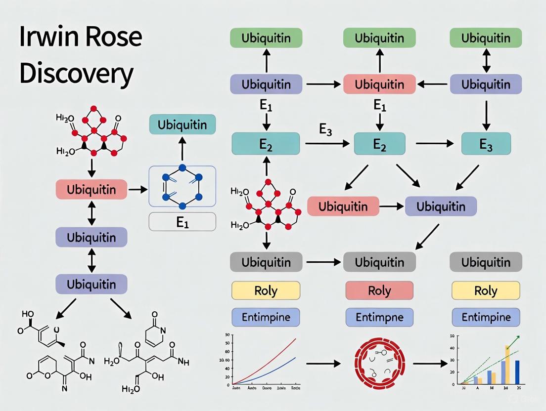
- Activation (E1): Ubiquitin is activated in an ATP-dependent manner by the E1 ubiquitin-activating enzyme, forming a thioester bond.
- Conjugation (E2): The activated ubiquitin is transferred to an E2 ubiquitin-conjugating enzyme.
- Ligation (E3): An E3 ubiquitin ligase recognizes specific substrate proteins and facilitates the transfer of ubiquitin from E2 to a lysine residue on the substrate, forming an isopeptide bond.
- Chain Elongation: Repeated cycles add multiple ubiquitins, forming a polyubiquitin chain.
- Degradation: The polyubiquitinated substrate is recognized by the 26S proteasome, a large proteolytic complex, which degrades the target protein into small peptides.
- Recycling: Ubiquitin is cleaved from the protein remnant and recycled.
Table 2: The Enzymatic Cascade of Ubiquitin-Mediated Proteolysis
| Enzyme | Designation | Primary Function | Key Discovery |
|---|---|---|---|
| E1 | Ubiquitin-Activating Enzyme | ATP-dependent activation of ubiquitin | Identified as part of the covalent conjugation system [5] |
| E2 | Ubiquitin-Conjugating Enzyme | Carries activated ubiquitin | Resolved through biochemical fractionation [5] |
| E3 | Ubiquitin Ligase | Confers substrate specificity | Recognized as a specificity component [5] |
| 26S Proteasome | Proteolytic Complex | Degrades ubiquitin-tagged proteins | Initially observed as high molecular weight APF-2 [1] |
The E3 ubiquitin ligases, of which there are hundreds, provide the specificity that allows the UPS to selectively target individual proteins for degradation at precise times, enabling exquisite regulation of cellular processes [1] [2].
Figure 2: The Ubiquitin-Proteasome System Pathway. This diagram illustrates the sequential enzymatic cascade (E1-E2-E3) that results in polyubiquitination of target proteins and their subsequent degradation by the proteasome.
The Scientist's Toolkit: Essential Research Reagents and Materials
The discovery of the ubiquitin system relied on a specific set of biochemical tools and reagents. The following table details key components used in the foundational experiments.
Table 3: Key Research Reagents and Materials in Ubiquitin Discovery
| Reagent/Material | Function in Research | Experimental Role |
|---|---|---|
| Reticulocyte Lysate | A cell-free system derived from immature red blood cells | Provided a source of ATP-dependent proteolytic activity free from lysosomal contamination [8] |
| ATP (Adenosine Triphosphate) | Energy source for enzymatic reactions | Required for both the conjugation of ubiquitin and the final proteolytic step [1] |
| 125I-labeled APF-1/Ubiqutin | Radioactively tagged protein factor | Enabled tracking and visualization of covalent conjugation to high molecular weight proteins [1] |
| SDS-PAGE | Analytical separation technique | Used to resolve and identify high molecular weight ubiquitin-protein conjugates [1] |
| Heat-Stable Proteins (Fraction I) | Crude protein fraction | Source of APF-1/Ubiquitin; heat stability was a key purification step [9] |
| Ion-Exchange Chromatography | Protein separation method | Critical for fractionating the reticulocyte lysate into functional components (I and II) [1] |
Implications and Therapeutic Applications
The discovery of ubiquitin-mediated proteolysis by Rose, Hershko, and Ciechanover transcended basic biochemistry, fundamentally reshaping our understanding of cellular regulation. The system is now known to be indispensable for controlling the cell cycle, DNA repair, transcription, immune response, and apoptosis by rapidly eliminating key regulatory proteins [5] [2]. The UPS functions as a quality control mechanism, disposing of damaged or misfolded proteins [2].
Dysregulation of the ubiquitin system underlies numerous human diseases. Defects in UPS components are implicated in various cancers, neurodegenerative disorders (such as Parkinson's and Alzheimer's), and genetic syndromes [2]. This mechanistic understanding has directly fueled drug development, most successfully with proteasome inhibitors like bortezomib (Velcade), which is now a standard treatment for multiple myeloma [2]. Ongoing research explores targeting specific E3 ubiquitin ligases for therapies in cancer and autoimmune diseases, demonstrating the vast translational potential arising from this foundational biochemical discovery [2].
The reticulocyte cell-free system stands as a cornerstone in the history of biochemical discovery, providing the essential experimental platform that enabled the elucidation of ubiquitin-mediated protein degradation. This immature red blood cell model, characterized by its simplicity and lack of internal organelles, offered researchers an unparalleled tool for dissecting complex cellular processes free from the confounding variables of intact cellular systems. It was within this meticulously prepared reticulocyte lysate that Irwin Rose, alongside Avram Hershko and Aaron Ciechanover, conducted the pioneering experiments that uncovered the ubiquitin-proteasome system, earning them the Nobel Prize in Chemistry in 2004 [1] [10]. Their work demonstrated that the reticulocyte system contained all necessary components for ATP-dependent protein degradation—a phenomenon that had puzzled scientists since the 1950s when energy-dependent intracellular proteolysis was first observed [10].
The critical importance of this cell-free system lies in its ability to be biochemically fractionated and reconstituted, allowing for the precise identification of individual components required for protein degradation. In the late 1970s, the researchers made the crucial observation that the reticulocyte extract could be separated into two complementary fractions (I and II), each inactive alone but capable of restoring ATP-dependent proteolysis when recombined [10]. This fractionation approach led directly to the identification of a heat-stable polypeptide initially termed APF-1 (ATP-dependent Proteolysis Factor 1), which was later identified as ubiquitin [1] [10]. The subsequent discovery that this small protein was covalently attached to target proteins in an ATP-dependent reaction revolutionized understanding of cellular regulation and opened an entirely new field of biochemical investigation.
Reticulocyte Biology and System Fundamentals
Biological Basis of Reticulocytes as an Experimental Model
Reticulocytes represent the penultimate stage in erythrocyte maturation, emerging after the expulsion of the nucleus from orthochromatic normoblasts. These immature red blood cells retain residual ribosomal RNA and mitochondrial elements but lack genomic DNA and most complex organelles, making them ideal for studying translational control and protein degradation without the complicating factor of ongoing transcription [11]. The standard reticulocyte count in healthy human peripheral blood ranges from 0.5% to 2.0% of total erythrocytes, or approximately 25,000 to 85,000 cells per microliter [12]. These cells circulate for approximately 24-48 hours before maturing into fully developed erythrocytes, during which time they progressively lose their remaining protein synthetic machinery [11] [12].
The unique biochemical composition of reticulocytes provides the foundation for their utility in cell-free systems. Unlike mature erythrocytes, reticulocytes maintain active protein synthesis and degradation machinery, including the complete ubiquitin-proteasome system [10]. Their relatively simple cytoplasmic content, dominated by hemoglobin, reduces biochemical complexity while retaining essential regulatory systems. Furthermore, the absence of a nucleus and lysosomes eliminates confounding degradation pathways, allowing researchers to focus specifically on the energy-dependent ubiquitin-proteasome pathway [1]. This unique combination of simplicity and retained biochemical activity makes the reticulocyte lysate an exceptionally powerful tool for reconstituting and analyzing specific cellular processes in a controlled environment.
Historical Significance in Ubiquitin Discovery
The reticulocyte model proved indispensable for the groundbreaking work of Irwin Rose and his colleagues, who utilized this system to decipher the biochemical mechanism of energy-dependent protein degradation. Their approach exemplifed the power of biochemical fractionation applied to a well-chosen cell-free system. The key breakthrough came when they demonstrated that the reticulocyte lysate could be separated into two fractions, I and II, with the active principle in fraction I (APF-1, later identified as ubiquitin) being a small, heat-stable protein [10]. Subsequent experiments in 1980 revealed the astonishing finding that APF-1 was covalently attached to target proteins in an ATP-dependent manner—the first evidence of what would become known as ubiquitination [1].
Table 1: Key Discoveries Enabled by the Reticulocyte Cell-Free System
| Year | Discovery | Experimental Approach | Significance |
|---|---|---|---|
| 1977-1978 | ATP-dependent proteolysis requires multiple factors | Fractionation of reticulocyte lysate | Identification of essential components for protein degradation [10] |
| 1980 | Covalent attachment of APF-1 to target proteins | Incubation of ¹²⁵I-labeled APF-1 with fraction II | First evidence of ubiquitin-protein conjugation [1] |
| 1980 | Multiple APF-1 molecules conjugate to single proteins | Analysis of high molecular weight complexes | Discovery of polyubiquitination as degradation signal [10] |
| 1981-1983 | Three-enzyme ubiquitination cascade | Biochemical reconstitution with purified components | Elucidation of E1-E2-E3 mechanism [10] |
The reticulocyte system continued to yield critical insights as Rose, Hershko, and Ciechanover developed the "multistep ubiquitin-tagging hypothesis" between 1981-1983, identifying the three enzyme classes (E1, E2, E3) responsible for the ubiquitination cascade [10]. This foundational work, made possible by the reticulocyte model, revealed a protein regulatory mechanism of comparable importance to phosphorylation and other reversible post-translational modifications.
Technical Establishment of the Reticulocyte Cell-Free System
Reticulocyte Production and Harvest
The initial step in establishing a functional reticulocyte cell-free system involves inducing reticulocytosis in laboratory animals, typically rabbits, and harvesting the immature red blood cells. The standard protocol involves rendering the animals anemic through a carefully controlled regimen of injections with acetylphenylhydrazine (APH) over several days, which stimulates enhanced erythropoiesis [13]. On day 8 post-induction, the animals are bled, and the blood is collected through cheesecloth filtration to remove debris and clots while maintaining the sample on ice to preserve biochemical activity [13].
The harvested blood undergoes centrifugation at 2000 RPM for 10 minutes at 4°C to separate the cellular components from plasma and other soluble factors [13]. The resulting pellet containing the reticulocyte-enriched erythrocyte population is then subjected to multiple washing steps to remove residual plasma proteins and contaminants that could interfere with subsequent fractionation procedures. This careful harvesting process yields a population of cells in which reticulocytes are significantly enriched compared to normal blood, providing the raw material for generating the cell-free extract.
Table 2: Reticulocyte Harvest Protocol Based on Historical Methods
| Step | Parameters | Purpose | Key Considerations |
|---|---|---|---|
| Reticulocyte Induction | Acetylphenylhydrazine injections over 3 days | Stimulate bone marrow to produce immature RBCs | Optimal dosing critical for yield [13] |
| Harvest Timing | Day 8 post-induction | Collect blood at peak reticulocyte production | Timing affects system activity [13] |
| Initial Processing | Cheesecloth filtration, maintenance on ice | Remove debris while preserving activity | Temperature control essential [13] |
| Separation | Centrifugation at 2000 RPM for 10 min | Pellet RBCs/retics from plasma | Gentle spin preserves cell integrity [13] |
| Washing | Multiple cycles with isotonic buffer | Remove plasma contaminants | Maintains osmotic balance [13] |
Lysate Preparation and Fractionation Techniques
The preparation of active reticulocyte lysate requires careful disruption of the harvested cells while preserving the integrity and function of the proteolytic machinery. The standard methodology involves resuspending the washed reticulocyte pellet in an appropriate lysis buffer, typically containing 20-40 mM HEPES (pH 7.4-7.6), 100 mM potassium acetate, 2 mM magnesium acetate, and 2 mM dithiothreitol (DTT) to maintain reducing conditions [13]. The exact composition may vary depending on the specific application, but maintenance of isotonic conditions and physiological pH is critical for preserving enzymatic activity.
Several lysis methods can be employed, with freeze-thaw cycling and osmotic shock being particularly effective for reticulocytes due to their relative fragility compared to nucleated cells [14]. The lysate is then clarified through centrifugation at 10,000-30,000 × g for 30 minutes at 4°C to remove membrane fragments, cytoskeletal components, and other insoluble material [13]. The resulting supernatant contains the soluble cytoplasmic factors necessary for protein synthesis and degradation, including the ubiquitin-proteasome system.
The critical breakthrough in the ubiquitin discovery came from the fractionation of this crude lysate into complementary fractions. Using chromatographic techniques such as ion-exchange or gel-filtration chromatography, researchers separate the lysate into two primary fractions: Fraction I containing the low molecular weight components (including ubiquitin/APF-1) and Fraction II containing the higher molecular weight components (including the E1, E2, and E3 enzymes and the proteasome) [10]. The separation is validated by demonstrating that ATP-dependent proteolytic activity is lost upon fractionation but restored when the fractions are recombined.
Core Reaction System and Reconstitution
The reconstitution of ubiquitin-dependent proteolytic activity from fractionated reticulocyte lysate requires specific reaction conditions optimized to support the multi-step enzymatic process. The standard reaction mixture includes both Fraction I and Fraction II, an energy regeneration system (typically ATP with creatine phosphate and creatine phosphokinase), magnesium ions, and the substrate protein of interest [10]. The reaction is buffered at physiological pH (7.2-7.6) and incubated at 37°C to mimic intracellular conditions.
The ubiquitination and degradation process can be monitored through various methods. Early experiments utilized ¹²⁵I-labeled APF-1 (ubiquitin) to demonstrate covalent attachment to proteins in Fraction II [1]. Alternatively, radiolabeled substrate proteins can be used to track their time-dependent degradation through the appearance of acid-soluble radioactivity or immunoblotting. The essential role of ATP in the process can be confirmed through control reactions containing non-hydrolyzable ATP analogs or ATP-depleting systems, which should abolish both ubiquitin conjugation and substrate degradation.
The successful reconstitution of the system demonstrates that all essential components for ubiquitin-mediated proteolysis are present in the fractionated reticulocyte lysate and provides a functional assay for further purification and characterization of individual factors. This approach ultimately led to the identification and purification of the E1 ubiquitin-activating enzyme, multiple E2 ubiquitin-conjugating enzymes, and various E3 ubiquitin ligases that confer substrate specificity [10].
The Scientist's Toolkit: Essential Research Reagents
Establishing a functional reticulocyte cell-free system requires carefully selected reagents that preserve biochemical activity while enabling specific experimental manipulations. The following table details essential components and their functions based on historically successful protocols and contemporary applications.
Table 3: Essential Research Reagents for Reticulocyte Cell-Free Systems
| Reagent Category | Specific Components | Function | Technical Considerations |
|---|---|---|---|
| Lysis Buffers | HEPES (20-40 mM, pH 7.4-7.6), KOAc (100 mM), Mg(OAc)₂ (2 mM), DTT (2 mM) | Maintain isotonic conditions during cell disruption; preserve enzymatic activity | DTT concentration critical for reducing environment; Mg²⁺ essential for ubiquitination [13] |
| Protease Inhibitors | PMSF (0.5 mM), protease inhibitor cocktails | Prevent nonspecific proteolysis during extract preparation | Use minimal effective concentrations to avoid interfering with studied proteolytic systems [13] |
| Energy Regeneration System | ATP (1-2 mM), creatine phosphate, creatine phosphokinase | Provide sustained ATP supply for energy-dependent ubiquitination and degradation | ATP concentration affects both ubiquitin conjugation and proteasomal degradation [10] |
| Fractionation Media | Sucrose or glycerol gradients (5-20%) | Separate cellular components by density and size | Sucrose concentration affects organelle integrity; gradients optimized for target components [14] |
| Ubiquitination Cofactors | Purified ubiquitin, E1, E2, E3 enzymes (for reconstitution experiments) | Enable specific ubiquitination of target substrates | Commercial preparations now available; purity affects experimental specificity [10] |
| Detection Reagents | ¹²⁵I-labeled ubiquitin, radiolabeled protein substrates, ubiquitin antibodies | Monitor conjugation and degradation processes | Antibody specificity critical for reducing background; radiolabeling provides high sensitivity [1] |
Biochemical Pathways Elucidated Through the Reticulocyte System
The reticulocyte cell-free system provided the experimental platform for delineating the complete ubiquitin-proteasome pathway, from initial ubiquitin activation to final substrate degradation. The biochemical cascade begins with ubiquitin activation by the E1 enzyme in an ATP-dependent reaction, forming a thioester bond between ubiquitin and E1 [10]. The activated ubiquitin is then transferred to an E2 conjugating enzyme, which collaborates with an E3 ubiquitin ligase to catalyze the formation of an isopeptide bond between the C-terminus of ubiquitin and a lysine residue on the target protein.
A critical insight from the reticulocyte system was the discovery of polyubiquitination, whereby multiple ubiquitin molecules are attached to a single substrate protein, often forming chains through linkage between the C-terminus of one ubiquitin and lysine 48 of another [1] [10]. This polyubiquitin chain serves as the recognition signal for the 26S proteasome, which unfolds the substrate protein in an ATP-dependent process and degrades it to small peptides while releasing reusable ubiquitin molecules.
This pathway, first reconstructed using the reticulocyte cell-free system, represents one of the most sophisticated regulatory mechanisms in eukaryotic cells. The specificity of the system resides primarily in the hundreds of different E3 ubiquitin ligases that recognize distinct subsets of substrate proteins, often in response to specific signals such as phosphorylation or allosteric modifications [10]. The proteasome itself represents a massive protease complex whose activity is compartmentalized within a barrel-shaped structure to prevent uncontrolled protein degradation within the cell [10].
Contemporary Applications and Methodological Advancements
While the reticulocyte cell-free system played a historical role in the discovery of the ubiquitin-proteasome pathway, its utility continues in contemporary research with both methodological refinements and expanded applications. Modern implementations of the system often utilize commercially prepared reticulocyte lysates that offer greater consistency and convenience while retaining the essential biochemical activities. These systems have been adapted for high-throughput screening of ubiquitin pathway modulators, studies of protein-protein interactions in the ubiquitination cascade, and analysis of degradation signals (degrons) that target specific proteins for destruction [2].
The principles established through the reticulocyte system have found broad application in drug development, particularly in the creation of proteasome inhibitors for cancer therapy. Bortezomib (Velcade), the first proteasome inhibitor approved for human use, was developed based on understanding gained from studying the proteasome in cell-free systems [2]. This drug has demonstrated significant efficacy in multiple myeloma and other hematological malignancies, validating the therapeutic potential of modulating the ubiquitin-proteasome pathway.
Recent technical advancements have enhanced the capabilities of reticulocyte-based systems, including the incorporation of non-natural amino acids for studying ubiquitin chain topology, the development of fluorescence-based degradation assays for real-time monitoring, and the integration of artificial intelligence approaches for predicting degradation kinetics [15]. These innovations build upon the foundational work conducted by Irwin Rose and his colleagues, demonstrating the enduring value of the reticulocyte cell-free system as both an historical milestone and a contemporary research tool.
The legacy of the reticulocyte model extends beyond the ubiquitin field, serving as a prototype for the development of other cell-free systems derived from various organisms including E. coli, wheat germ, yeast, and cultured mammalian cells [13] [16]. Each of these systems offers unique advantages for specific applications, but all share the fundamental approach of using biochemical fractionation and reconstitution to dissect complex cellular processes—a methodology whose power was definitively established through the pioneering work with the reticulocyte model.
The discovery of ATP-dependent proteolysis factor 1 (APF-1), later identified as ubiquitin, represents a cornerstone of modern cell biology, fundamentally altering our understanding of how cellular processes are regulated. This discovery emerged from a long-standing biochemical paradox: why would intracellular protein degradation, an inherently energy-liberating process, require ATP hydrolysis? [1] [17] For decades, the lysosome was believed to be the primary site of protein turnover. However, observations that cells could distinguish between long-lived and short-lived proteins, and that ATP dependence persisted even in reticulocyte extracts which lack lysosomes, pointed to the existence of a separate, non-lysosomal proteolytic pathway [1] [17]. It was within this context of intellectual curiosity that the collaboration between Avram Hershko, Aaron Ciechanover, and Irwin Rose began to unravel this mystery. Their work, for which they were awarded the 2004 Nobel Prize in Chemistry, revealed a sophisticated enzymatic system centered on a small, heat-stable protein [1] [2]. Irwin Rose's role, particularly during Hershko's sabbatical in his laboratory at the Fox Chase Cancer Center, was that of an essential intellectual contributor and patron, providing critical insights that helped frame the novel interpretations emerging from the data [1] [18].
Key Experiments and Methodologies
Initial Fractionation and the Discovery of a Heat-Stable Factor
The experimental journey to identify APF-1 began with a reconstitution approach using a cell-free system derived from reticulocytes, which Goldberg's group had shown exhibited ATP-dependent proteolysis of abnormal proteins [1]. Hershko and Ciechanover separated the reticulocyte lysate into two complementary fractions (I and II) using DEAE-cellulose chromatography [1] [17].
- Fraction I: Contained hemoglobin and other non-adherent components.
- Fraction II: Contained the bulk of other cellular proteins.
When assayed separately, neither fraction could support ATP-dependent proteolysis. However, when recombined, degradation of the test substrate was restored [1]. The key breakthrough came when they attempted to purify the essential component from Fraction I. After conventional separation methods failed due to interference from abundant hemoglobin, the researchers took an unconventional step: they boiled Fraction I [17]. Most proteins, including hemoglobin, denatured and hardened upon boiling. Astonishingly, the required factor remained soluble and fully active in the supernatant, revealing its remarkable heat stability [17]. This property was crucial for its initial purification and identification. The factor was named APF-1 (ATP-dependent Proteolysis Factor 1) [1].
The Covalent Modification Breakthrough
A series of elegant experiments followed to determine APF-1's mechanism of action. The researchers labeled APF-1 with a radioactive iodine-125 (¹²⁵I) tag and incubated it with Fraction II in the presence of ATP [1]. When the reaction mixture was analyzed by SDS-polyacrylamide gel electrophoresis (SDS-PAGE), a surprising pattern emerged.
Table: Key Observations from the Covalent Modification Experiments
| Experimental Condition | Observation | Interpretation |
|---|---|---|
| ¹²⁵I-APF-1 + Fraction II, no ATP | Radioactivity migrated as free APF-1. | No association occurred without energy. |
| ¹²⁵I-APF-1 + Fraction II, with ATP | Multiple radioactive bands of higher molecular weight appeared. | APF-1 formed complexes with other proteins. |
| Treatment with denaturants (e.g., NaOH) | Complexes remained stable. | APF-1 was covalently linked to target proteins. |
This covalent association was a revelation. It suggested that proteins were not simply bound to a protease but were being chemically tagged prior to degradation. The bond was later identified as an isopeptide bond between the C-terminal glycine of ubiquitin and lysine ε-amino groups on the target proteins [1] [17]. Irwin Rose's background as a mechanistic enzymologist was instrumental in guiding the interpretation of these complex bonding patterns, moving the field beyond the initial assumption that APF-2 (a high molecular weight fraction) might be a kinase [1].
The "Aha" Moment: APF-1 is Ubiquitin
The discovery that APF-1 was the previously known protein ubiquitin came from a confluence of observations across laboratories. Ubiquitin (so named for its ubiquitous presence in eukaryotic cells) had been discovered earlier but its physiological function was unknown [1]. A key conversation occurred between postdoctoral fellows in Rose's lab, including Arthur Haas and Michael Urban, who noted the similarity between the covalent attachment of APF-1 and the known conjugation of ubiquitin to histone H2A [1]. This led to a collaborative experiment which definitively showed that APF-1 and ubiquitin were identical [1]. This finding connected a mysterious biochemical activity with a known protein, instantly providing a new and vital functional context for ubiquitin.
Figure 1: Experimental Workflow Leading to APF-1 Identification. This diagram outlines the key biochemical fractionation and reconstitution steps that revealed the essential role of the heat-stable APF-1 and its covalent conjugation to cellular proteins.
The Ubiquitin Conjugation Pathway
Following the identification of APF-1 as ubiquitin, the Hershko, Ciechanover, and Rose team meticulously dissected the enzymatic pathway responsible for its conjugation. They discovered that three distinct classes of enzymes were required to attach ubiquitin to protein substrates [17].
- E1 (Ubiquitin-Activating Enzyme): This enzyme activates ubiquitin in an ATP-dependent reaction, forming a high-energy thioester bond between its active-site cysteine and the C-terminus of ubiquitin.
- E2 (Ubiquitin-Conjugating Enzyme): The activated ubiquitin is then transferred to a cysteine residue on an E2 enzyme.
- E3 (Ubiquitin Ligase): Finally, an E3 enzyme facilitates the transfer of ubiquitin from E2 to a lysine residue on the target protein, forming the stable isopeptide bond. E3s provide the critical substrate specificity to the system.
Further work, notably by Hershko and Varshavsky, revealed that proteins targeted for degradation are typically modified with a chain of ubiquitin molecules (a polyubiquitin chain) linked through Lysine 48 (K48) of one ubiquitin to the C-terminus of the next [1] [17]. This polyubiquitin chain serves as the recognized signal for the 26S proteasome, a massive proteolytic complex that degrades the tagged protein, releasing ubiquitin for reuse [1].
Figure 2: The Ubiquitin-Proteasome Pathway. This diagram illustrates the sequential action of E1, E2, and E3 enzymes in conjugating ubiquitin to a target protein, forming a polyubiquitin chain that directs the substrate to the 26S proteasome for degradation.
The Scientist's Toolkit: Key Research Reagents
The discovery of the ubiquitin system was enabled by a specific set of biochemical tools and reagents. The following table details some of the most critical components used in the foundational experiments.
Table: Essential Research Reagents in the Discovery of APF-1/Ubiquitin
| Reagent / Material | Function in the Experiment |
|---|---|
| Reticulocyte Lysate | A cell-free system derived from immature red blood cells; provided a source of ATP-dependent, non-lysosomal proteolytic activity for biochemical fractionation [1] [17]. |
| DEAE-Cellulose Chromatography | An ion-exchange chromatography method used to separate the reticulocyte lysate into two essential fractions (I and II), enabling the identification of the required components [1]. |
| ATP (Adenosine Triphosphate) | The essential energy source required to drive both the proteolytic process and the covalent conjugation of APF-1/ubiquitin to protein substrates [1]. |
| ¹²⁵I-labeled APF-1 | Radioactively labeled APF-1 (ubiquitin); allowed researchers to trace the fate of APF-1 and discover its covalent attachment to multiple high-molecular-weight proteins via SDS-PAGE [1]. |
| Heat Treatment (Boiling) | A critical purification step; exploited the exceptional heat stability of ubiquitin to separate it from the bulk of other proteins (like hemoglobin) in Fraction I [17]. |
| ts85 Mammalian Cell Line | A temperature-sensitive mutant cell line; provided genetic evidence in a living system that a functional ubiquitin system (specifically, a temperature-sensitive E1 enzyme) was essential for the degradation of short-lived proteins [17]. |
The identification of APF-1 as ubiquitin and the elucidation of the ubiquitin-proteasome system revolutionized cell biology. It revealed that regulated protein degradation is as critical a regulatory mechanism as transcription and translation for controlling cellular processes [18]. This system governs the precise turnover of key proteins involved in the cell cycle, DNA repair, transcriptional regulation, and immune and stress responses [18] [19].
The profound biological significance of this pathway is underscored by its direct relevance to human disease and drug development. Dysregulation of ubiquitin-mediated proteolysis is implicated in cancer, neurodegenerative diseases, and immune disorders [2] [19]. For instance, the proteasome inhibitor Bortezomib (Velcade) was developed based on this foundational knowledge and is now a standard treatment for multiple myeloma, validating the ubiquitin system as a viable therapeutic target [2] [19].
In conclusion, the journey to identify APF-1 is a testament to the power of curiosity-driven basic research. The collaborative effort, significantly enhanced by Irwin Rose's enzymological insight, solved a long-standing metabolic paradox and unveiled a universal regulatory mechanism. What began as a simple heat-stable factor in a reticulocyte extract is now recognized as a central controller of cell life and death, with enduring implications for our understanding of biology and the treatment of disease.
The year 1980 marked a revolutionary turning point in our understanding of cellular regulation with the publication of two seminal papers by Aaron Ciechanover, Avram Hershko, and Irwin Rose. Their discovery of energy-dependent intracellular proteolysis fundamentally challenged existing models which could not explain why the hydrolysis of peptide bonds, an exergonic process, would require ATP consumption [1]. This apparent paradox hinted at a far more complex regulatory mechanism than previously imagined. The researchers unveiled a system where proteins are marked for destruction through covalent attachment of a small protein tag, a process they initially characterized using ATP-dependent proteolysis factor 1 (APF-1), later identified as ubiquitin [1] [10]. This "kiss of death" mechanism, for which they received the Nobel Prize in Chemistry in 2004, established an entirely new paradigm for post-translational control of protein fate, every bit as important as phosphorylation or acetylation [1] [10] [20]. This article examines this groundbreaking discovery, with particular focus on the intellectual and experimental contributions of Irwin Rose, whose mechanistic enzymology expertise and collaborative spirit were instrumental in framing this new understanding of cellular regulation.
Historical and Intellectual Context
The Energy Paradox in Protein Degradation
Prior to the landmark 1980 discovery, the scientific understanding of intracellular proteolysis was limited. The prevailing knowledge recognized simple protein-degrading enzymes like trypsin and lysosomal proteolysis, neither of which required energy [10]. However, observations dating back to Simpson's 1953 studies consistently demonstrated that intracellular proteolysis in mammalian cells was energy-dependent [1]. This was biochemically perplexing because the hydrolysis of peptide bonds is inherently exergonic, offering no thermodynamic rationale for ATP consumption [1]. Goldberg's group later advanced this understanding by showing that damaged or abnormal proteins were rapidly cleared from cells in an energy-dependent manner, suggesting that energy dependence reflected sophisticated regulation of proteolytic systems [1]. This conundrum—the unexplained ATP requirement for proteolysis—formed the central mystery that Hershko, Ciechanover, and Rose set out to resolve.
The Confluence of Collaborative Minds
The collaboration between Ciechanover, Hershko, and Rose was uniquely positioned to tackle this problem. Avram Hershko had developed an interest in protein degradation during his postdoctoral work with Gordon Tompkins at UCSF and continued this work at the Technion-Israel Institute of Technology [1]. Aaron Ciechanover joined Hershko's laboratory as a graduate student after completing his military service [1]. Irwin Rose brought to this collaboration a distinguished background as a mechanistic enzymologist, known for his studies on proton transfer reactions and isotopic labeling to examine enzymatic mechanisms [1]. His interest in protein degradation was long-standing, stemming from earlier conversations with his Yale colleague Simpson about the ATP dependence of proteolysis [1].
The collaboration began in earnest when Hershko and Rose met at a Fogarty Foundation meeting in 1977 and discovered their mutual interests [1]. Rose subsequently invited Hershko to his laboratory at the Fox Chase Cancer Center in Philadelphia, initiating a decade of collaborative work that saw the Israeli scientists spending summers there [1]. Rose's role extended far beyond typical sabbatical hosting; he served as both patron and intellectual contributor, providing critical insights from his extensive background in enzymatic mechanisms [2]. His laboratory at the Institute for Cancer Research became the nurturing environment where key discoveries were made, underscoring his indispensable role in the ubiquitin story.
The Seminal 1980 Experiments: Methodology and Findings
Preliminary Fractionation and APF-1 Identification
Prior to the 1980 publications, Hershko and Ciechanover had established a critical experimental foundation. Using a cell-free extract from reticulocytes (immature red blood cells) that exhibited ATP-dependent proteolysis of abnormal proteins, they developed a fractionation approach that separated the system into two complementary fractions [1] [10]. When isolated, each fraction was inactive, but when recombined, ATP-dependent proteolysis was restored [10]. This elegant approach enabled them to identify the active component in Fraction I as a small, heat-stable polypeptide with a molecular weight of approximately 9,000 Da, which they termed APF-1 (ATP-dependent Proteolysis Factor 1) [1] [10]. Unbeknownst to them at the time, this protein was identical to ubiquitin, a polypeptide previously isolated by Gideon Goldstein in 1975 but whose function remained unknown [1] [21].
Table 1: Key Research Reagents in the Ubiquitin Discovery
| Reagent/Technique | Function in Experiments | Key Insight Provided |
|---|---|---|
| Reticulocyte Cell-Free Extract [1] | ATP-dependent proteolysis model system; amenable to biochemical fractionation | Provided functional reconstitution system for decomposition analysis |
| Biochemical Fractionation (I & II) [1] [10] | Separation of proteolysis machinery into complementary components | Revealed multi-component nature of the system; allowed identification of APF-1 |
| ATP Depletion/Replenishment [1] | Modulation of energy availability in the experimental system | Confirmed energy dependence; revealed conjugate formation regulation |
| 125I-labeled APF-1 [1] | Radioactive tagging for tracking APF-1 fate | Enabled visualization of covalent attachment to high molecular weight proteins |
| Heat Treatment [1] | Stability test for APF-1 | Confirmed APF-1's remarkable stability, characteristic of ubiquitin |
The Critical 1980 Investigations
The two 1980 PNAS papers represented the decisive breakthrough. The first paper, led by Ciechanover et al., addressed the fundamental mechanism of APF-1 action [1]. Using 125I-labeled APF-1, they demonstrated that in the presence of Fraction II and ATP, APF-1 was promoted to high molecular weight forms. A crucial observation came when postdoctoral fellow Art Haas found that this association survived high pH treatment, suggesting something unprecedented: the attachment of APF-1 to proteins in Fraction II was covalent [1].
Further characterization revealed this covalent bond was stable to NaOH treatment, and APF-1 was bound to many different proteins as judged by SDS/PAGE [1]. The researchers established that conjugation required low ATP concentrations and was reversible upon ATP removal [1]. Critically, they noted that the nucleotide and metal ion requirements for conjugation mirrored those for proteolysis, strongly suggesting the processes were functionally linked [1]. This explained why some investigators failed to observe APF-1 requirements—when Fraction II was prepared without ATP depletion, APF-1 was already present in high molecular weight conjugates that could be disassembled by amidases to liberate free APF-1 [1].
The second 1980 paper by Hershko et al. provided the definitive link between ubiquitination and proteolysis [1]. They demonstrated that authentic protein substrates of the system were heavily modified, with multiple molecules of APF-1 attached to each substrate molecule [1]. This polyubiquitination was processive, with the conjugating machinery preferring to add additional ubiquitin molecules to existing conjugates rather than initiating new ones [1]. This work provided the first evidence for enzyme-catalyzed conjugation, hinting at the existence of what would later be termed ubiquitin ligases [1].
Table 2: Key Experimental Findings from the 1980 PNAS Papers
| Experimental Finding | Methodology | Interpretation/Significance |
|---|---|---|
| Covalent Attachment [1] | 125I-APF-1 formed high MW complexes stable to high pH and NaOH | APF-1 forms covalent conjugates with target proteins; revolutionary concept |
| ATP Dependence [1] | ATP depletion prevented conjugate formation; reversal upon ATP restoration | Energy required for conjugation step, explaining the energy paradox |
| Multiplicity of Target Proteins [1] | SDS/PAGE showed APF-1 bound to numerous proteins in Fraction II | Ubiquitination system has broad substrate specificity |
| Polyubiquitination [1] | Multiple APF-1 molecules conjugated per substrate molecule | Established concept of polyubiquitin chains as degradation signal |
| Processive Nature [1] | Conjugation preferred adding to existing conjugates over new substrates | Suggested cooperative mechanism for chain elongation |
Diagram Title: Ubiquitin-Proteasome Pathway Enzymatic Cascade
The E1-E2-E3 Enzymatic Cascade and Irwin Rose's Mechanistic Insights
Following the seminal 1980 observations, between 1981-1983, Ciechanover, Hershko, Rose and their teams developed the "multistep ubiquitin-tagging hypothesis," defining the E1-E2-E3 enzymatic cascade that governs ubiquitin conjugation [10] [22]. This hierarchical system consists of:
- E1 (Ubiquitin-Activating Enzymes): A single E1 or small family activates ubiquitin in an ATP-dependent reaction, forming a high-energy thioester bond between its active-site cysteine and ubiquitin's C-terminal glycine [21] [22].
- E2 (Ubiquitin-Conjugating Enzymes): Activated ubiquitin is transferred to the active-site cysteine of an E2 enzyme via trans-thioesterification [21] [22].
- E3 (Ubiquitin Ligases): Hundreds of different E3 enzymes catalyze the final transfer of ubiquitin from E2 to a lysine residue on the target protein, forming an isopeptide bond [10] [21] [22]. E3s provide substrate specificity, determining which proteins are ubiquitinated.
Irwin Rose's background as a mechanistic enzymologist proved invaluable in deciphering this cascade [1]. His expertise in using isotopic labeling and analyzing enzyme mechanisms helped the team design critical experiments to elucidate the energy-dependent steps and covalent intermediates. Rose's intellectual contribution was particularly evident in the conceptual framing of the system as a coordinated enzymatic cascade rather than a simple binary modification [2]. His laboratory at Fox Chase provided not just physical resources but an environment rich in mechanistic thinking, where the fundamental enzymological principles governing ubiquitin transfer could be rigorously explored.
Evolution and Expanding Applications of the Ubiquitin Concept
From Protein Degradation to Multifunctional Signaling
The initial conception of ubiquitin as solely a degradation signal has dramatically expanded. We now recognize that the type of ubiquitin chain determines the functional outcome [21] [22]. While K48-linked polyubiquitin chains typically target proteins for proteasomal degradation, other chain types (e.g., K63-linked, K11-linked, K6-linked, M1-linked) and monoubiquitination regulate diverse non-proteolytic processes including endocytic trafficking, inflammation, translation, DNA repair, and kinase activation [21]. Furthermore, ubiquitination is now known to occur not only on lysine residues but also on cysteine, serine, threonine residues, and protein N-termini through distinct chemical bonds [21].
Recent research has revealed that ubiquitin can even be conjugated to non-protein substrates, including lipids, sugars, and nucleotides, expanding the ubiquitin concept beyond protein modification [23] [24]. These discoveries suggest ubiquitin may serve as a scaffold for recruiting effector proteins in various signaling contexts, dramatically broadening the potential cellular roles of this modification system [24].
Therapeutic Applications and Clinical Relevance
The ubiquitin-proteasome system has emerged as a valuable therapeutic target. Defects in ubiquitin-mediated degradation are implicated in various diseases, including cervical cancer, cystic fibrosis, and neurodegenerative disorders like Parkinson's disease [2] [20]. The proteasome inhibitor Bortezomib (Velcade, PS-341) was developed based on this understanding and has been approved for treating multiple myeloma, validating the clinical significance of this pathway [2]. Current research focuses on developing more specific inhibitors targeting particular E3 ligases or deubiquitinating enzymes to achieve greater therapeutic specificity with reduced side effects [2].
The 1980 discovery of ubiquitin-mediated protein degradation by Ciechanover, Hershko, and Rose fundamentally transformed our understanding of cellular regulation. Their work revealed not merely a degradation mechanism but a sophisticated language of post-translational control that rivals phosphorylation in its importance and complexity. Irwin Rose's contributions as a collaborator and mechanistic enzymologist were instrumental in framing the enzymatic logic of the system and guiding its biochemical characterization. From the initial observation of covalent APF-1 attachment to the current understanding of a multifaceted signaling system, the ubiquitin paradigm continues to evolve, offering profound insights into cell biology and providing novel therapeutic avenues for combating disease.
Mechanistic Breakthroughs: Deciphering the Ubiquitin-Proteasome System's Enzymatic Cascade
The discovery of the ubiquitin-proteasome system, a cornerstone of modern cell biology, was propelled not by sophisticated genomics but by classical and rigorous biochemistry. The 2004 Nobel Prize in Chemistry awarded to Aaron Ciechanover, Avram Hershko, and Irwin Rose crowned a series of elegant experiments that decoded the cell's primary mechanism for targeted protein degradation. Central to this breakthrough was the strategic application of three key experimental approaches: biochemical fractionation to separate and identify essential components, functional reconstitution to validate their roles, and radiolabeling to trace the covalent fate of a small protein tag. This guide details the specific methodologies, rooted in the work of Irwin Rose and his colleagues, that uncovered this sophisticated regulatory system. Their approach provides a timeless template for deconstructing complex cellular machinery.
The Biological Puzzle and Irwin Rose's Intellectual Context
In the late 1970s, a central paradox puzzled biochemists: while the hydrolysis of a peptide bond is an exergonic (energy-releasing) reaction, the degradation of intracellular proteins was known to require adenosine triphosphate (ATP) [1]. This energy dependence suggested the process was far more complex than simple proteolysis. Irwin "Ernie" Rose, then at the Fox Chase Cancer Center, had been intrigued by this problem since the 1950s, when his colleague at Yale, Melvin Simpson, first demonstrated the ATP dependence of proteolysis [1] [25]. Rose's background as a distinguished enzymologist, with deep expertise in the stereochemistry and mechanisms of enzymatic reactions [6], primed him to appreciate the complexity of this energy-dependent process.
The collaborative team was uniquely positioned to tackle this problem. Avram Hershko had been studying energy-dependent protein degradation, and after meeting Rose at a conference in 1977, he began a seminal sabbatical in Rose's laboratory [1]. This began a decade of summer collaborations, with Rose acting as both an intellectual contributor and a patron of the research [1]. Their initial model system was a cell-free extract derived from rabbit reticulocytes (immature red blood cells), chosen because it catalyzed the ATP-dependent breakdown of abnormal proteins and, crucially, lacked lysosomes, thus focusing on the non-lysosomal proteolytic pathway [1] [10].
Core Experimental Strategies
The elucidation of the ubiquitin pathway serves as a masterclass in the use of fractionation, reconstitution, and radiolabeling. The following diagram illustrates the overarching experimental workflow that led to the discovery.
Fractionation and Reconstitution: Deconstructing the System
The initial, critical breakthrough came from the decision to separate the reticulocyte lysate into its constituent parts. Hershko and Ciechanover used chromatography to resolve the lysate into two fractions, designated simply as Fraction I and Fraction II [1] [10]. Individually, each fraction was inactive; ATP-dependent proteolysis only occurred when they were recombined [10]. This reconstitution assay provided a powerful functional readout to track the essential components.
Further analysis revealed the identity of the key factors:
- Fraction I was found to contain a single, heat-stable essential component, a small protein they named APF-1 (ATP-dependent Proteolysis Factor 1) [1] [10].
- Fraction II contained a high molecular weight, ATP-stabilized factor (APF-2), which was later understood to be the proteasome itself, as well as the enzymatic machinery necessary for conjugation [1].
This fractionation-reconstitution strategy was the foundational step that made all subsequent discoveries possible, as it allowed for the isolation and individual characterization of the system's parts.
Radiolabeling: Visualizing the Covalent Signal
With APF-1 identified as crucial, the next question was its mechanism of action. Here, the expertise in Rose's laboratory was instrumental. The team, including postdoctoral fellow Art Haas, began using ¹²⁵I-labeled APF-1 as a radioactive tracer [1].
The key experiment involved incubating the labeled APF-1 with Fraction II and ATP. The researchers observed a dramatic shift: the labeled APF-1 was promoted to high molecular weight forms. To their astonishment, this association was covalent—it survived treatment with sodium hydroxide, which would disrupt non-covalent bonds [1] [5]. This finding was the "smoking gun." It demonstrated that APF-1 was not merely a cofactor but was itself chemically linked to target proteins prior to their degradation. The radiolabel was essential for visualizing this conjugation event, which would have been otherwise invisible.
Subsequent work showed that authentic protein substrates were modified by multiple molecules of APF-1, a process termed polyubiquitination [1] [10]. The radiolabeling approach allowed the team to trace the fate of ubiquitin and establish the multi-step enzymatic cascade involving E1, E2, and E3 enzymes [5] [10].
Detailed Experimental Protocols
Protocol 1: Fractionation of Reticulocyte Lysate for ATP-Dependent Proteolysis
This protocol is adapted from the methods used to identify the essential components of the system [1] [10].
- Principle: Separate a active reticulocyte lysate chromatographically into complementary fractions that are individually inactive but regain proteolytic function upon recombination.
Materials:
- Reticulocyte lysate (from rabbit blood)
- Chromatography resin (e.g., DEAE-cellulose)
- Chromatography buffer (e.g., 20 mM Tris-HCl, pH 7.5, 1 mM DTT)
- Salt gradient (e.g., 0-0.5 M KCl in chromatography buffer)
- ATP-regenerating system (ATP, Mg²⁺, creatine phosphate, creatine kinase)
- Radiolabeled substrate protein (e.g., ³H- or ¹²⁵I-labeled denatured lysozyme)
Procedure:
- Prepare Lysate: Centrifuge reticulocyte lysate at high speed (e.g., 100,000 × g) to remove membranes and obtain a clear soluble extract.
- Load Column: Apply the soluble lysate to a DEAE-cellulose column pre-equilibrated with low-salt chromatography buffer.
- Elute Fractions: Elute proteins using a linear salt gradient (0-0.5 M KCl). Collect multiple fractions.
- Desalt Fractions: Desalt individual fractions into a low-ionic-strength buffer compatible with the functional assay.
- Assay for Proteolysis: Incubate each fraction individually and in pairwise combinations with the radiolabeled substrate, an ATP-regenerating system, and buffer. A typical reaction would run for 1-2 hours at 37°C.
- Measure Degradation: Precipitate non-degraded protein with trichloroacetic acid (TCA) and measure the radioactivity in the soluble fraction (degraded peptides) by scintillation counting.
Expected Outcome: Two main pools of fractions will be identified. One pool (Fraction I) will contain the small, heat-stable protein APF-1 (ubiquitin). The other (Fraction II) will contain the larger enzymatic machinery and the proteasome. Neither will show significant activity alone, but their combination will restore robust, ATP-dependent proteolysis.
Protocol 2: Radiolabeling and Detection of Ubiquitin Conjugation
This protocol details the critical experiment that demonstrated the covalent attachment of ubiquitin to target proteins [1].
- Principle: Use iodinated ubiquitin to trace its covalent, ATP-dependent conjugation to proteins in Fraction II via SDS-PAGE autoradiography.
Materials:
- ¹²⁵I- labeled APF-1/Ubiquitin (The key reagent, prepared using Iodogen or Chloramine-T method)
- Fraction II (APF-1 depleted)
- ATP-regenerating system
- Incubation buffer
- SDS-PAGE equipment
- X-ray film or Phosphorimager
Procedure:
- Reaction Setup: Combine the following in a microtube:
- Fraction II (as a source of enzymes)
- ¹²⁵I-labeled Ubiquitin (0.1-1 µg)
- ATP-regenerating system (2 mM ATP, 5 mM MgCl₂, 10 mM creatine phosphate, 0.1 U creatine kinase)
- Incubation buffer to final volume.
- Incubation: Incubate at 37°C for 10-30 minutes.
- Termination and Denaturation: Stop the reaction by adding SDS-PAGE sample buffer and boiling for 5 minutes.
- Analysis: Resolve the proteins by SDS-PAGE.
- Visualization: Dry the gel and expose it to X-ray film or a phosphorimager screen to detect the radiolabeled proteins.
- Reaction Setup: Combine the following in a microtube:
Expected Outcome: In the presence of ATP, a "ladder" or "smear" of high molecular weight radioactive bands will be visible on the autoradiogram. These represent ¹²⁵I-ubiquitin covalently linked to various endogenous proteins in Fraction II. This ladder will be absent in control reactions lacking ATP.
Quantitative Data from Foundational Experiments
The following tables summarize key quantitative findings from the original research, providing a template for data presentation in similar biochemical studies.
Table 1: Characteristics of Fractions Isolated from Reticulocyte Lysate
| Fraction Name | Key Component(s) Identified | Essential Characteristics | Activity in Proteolysis Assay |
|---|---|---|---|
| Unfractionated Lysate | All components | N/A | Full ATP-dependent activity [10] |
| Fraction I | APF-1 (Ubiquitin) | Heat-stable, small protein (~8.5 kDa) | Inactive alone [1] [10] |
| Fraction II | E1, E2, E3 enzymes; Proteasome (APF-2) | High molecular weight complexes; ATP-stabilized | Inactive alone [1] [10] |
| Fraction I + II | All components | N/A | Full activity restored upon recombination [1] [10] |
Table 2: Key Parameters for Radiolabeled Ubiquitin Conjugation Assay
| Experimental Parameter | Condition/Measurement | Interpretation/Significance |
|---|---|---|
| ATP Dependence | Conjugation required ~2 mM ATP [1] | Explained the energy requirement of intracellular proteolysis |
| Stability of Bond | Stable to NaOH (high pH) treatment [1] | Confirmed covalent isopeptide bond, not non-covalent association |
| Multiplicity | Multiple ubiquitin molecules conjugated per target protein substrate [1] | Discovery of polyubiquitination as the proteolytic signal |
| Inhibitor Sensitivity | Conjugation was inhibited by alkylating agents (e.g., N-ethylmaleimide) [1] | Suggested the involvement of enzyme active sites with cysteine residues |
The Scientist's Toolkit: Key Research Reagents
The discovery of the ubiquitin system relied on a minimal but powerful set of biochemical tools. The following table catalogs the essential reagents used in these landmark experiments.
Table 3: Key Research Reagents in the Discovery of Ubiquitin-Mediated Proteolysis
| Reagent / Solution | Function in the Experimental Workflow |
|---|---|
| Reticulocyte Lysate | A cell-free system rich in the ubiquitin-proteasome machinery and devoid of lysosomes, serving as the starting material for fractionation [1] [10]. |
| ATP-Regenerating System | Maintains a constant, high level of ATP in prolonged incubations, which is crucial for both the conjugation process and the proteasome's activity [1]. |
| ¹²⁵I-labeled APF-1/Ubiquitin | A radioactive tracer that enabled the visual detection of the covalent conjugation of ubiquitin to protein targets via autoradiography, a cornerstone of the discovery [1]. |
| Chromatography Resins (e.g., DEAE) | Used for the fractionation of the complex lysate into functionally distinct sub-fractions (I and II) based on properties like charge [1]. |
| Denatured/Abnormal Proteins | Acted as preferred substrates for the pathway in assays, mimicking the natural "damaged protein" targets of the system [1]. |
The Ubiquitin Conjugation Pathway: A Visual Synthesis
The culmination of the fractionation, reconstitution, and radiolabeling experiments was the elucidation of a complete enzymatic pathway. The following diagram synthesizes these findings into the modern understanding of the ubiquitin conjugation cascade.
The work of Irwin Rose, Avram Hershko, and Aaron Ciechanover is a powerful testament to the enduring power of classical biochemistry. By systematically breaking down a complex cellular process using fractionation, proving the function of its parts through reconstitution, and illuminating a hidden covalent interaction with radiolabeling, they unveiled the ubiquitin system. This discovery transformed our understanding of the cell, revealing that controlled protein degradation is as crucial as protein synthesis for regulation. The methodologies they refined remain foundational for probing complex biochemical pathways, from signal transduction to quality control, proving that a rigorous, step-wise experimental approach remains one of the most powerful tools in scientific discovery.
The period spanning 1978 to 1980 marked a pivotal transition in our understanding of cellular proteolysis, culminating in the identification of ATP-dependent proteolysis factor 1 (APF-1) as the previously known but functionally enigmatic protein, ubiquitin. This discovery, forged through the collaborative efforts of Avram Hershko, Aaron Ciechanover, and Irwin Rose, connected a discrete biochemical activity to a universal cellular component, ultimately revealing the ubiquitin system as a central regulatory mechanism in eukaryotic cell biology. The intellectual and technical contributions of Irwin Rose were particularly instrumental in designing the critical experiments that demonstrated the covalent nature of APF-1-protein conjugation, a finding that provided the necessary conceptual bridge to prior observations of ubiquitin in other biological contexts. This whitepaper delineates the experimental journey and detailed methodologies that led to this fundamental discovery, which now underpins numerous therapeutic strategies in modern drug development.
In the late 1970s, a fundamental paradox puzzled cell biologists: the hydrolysis of peptide bonds is an exergonic process, yet intracellular proteolysis required metabolic energy in the form of ATP [1] [10]. This observation suggested the existence of a previously uncharacterized, energy-dependent regulatory step in the protein degradation pathway. The laboratory of Avram Hershko, in collaboration with Irwin Rose, sought to resolve this paradox using a biochemical fractionation approach.
A critical breakthrough came with the adoption of a reticulocyte (immature red blood cell) lysate system, which lacks lysosomes but efficiently degrades abnormal proteins in an ATP-dependent manner [17]. Fractionation of this lysate revealed that the proteolytic machinery consisted of at least two separable components, designated Fraction I and Fraction II [1] [26]. The active component within Fraction I was a small, heat-stable polypeptide that retained its biological activity even after boiling, a property that proved crucial for its isolation. This factor was named APF-1 (ATP-dependent Proteolysis Factor 1) [26].
Table 1: Key Characteristics of the Reticulocyte Lysate System and APF-1
| Parameter | Description |
|---|---|
| Biological Source | Rabbit reticulocytes |
| Key Advantage | Lacks lysosomes; exhibits robust ATP-dependent proteolysis |
| Critical Fraction | Fraction I (containing APF-1) |
| APF-1 Property | Heat-stable polypeptide (≈ 8.6 kDa) |
| Functional Assay | ATP-dependent degradation of radiolabeled substrate proteins |
Initial hypotheses suggested APF-1 might function as a protease activator [1]. However, a series of elegant experiments masterminded during Hershko and Ciechanover's sabbaticals in Irwin Rose's laboratory at the Fox Chase Cancer Center would radically alter this perception and set the stage for connecting APF-1 to a known protein.
Experimental Breakthrough: The Covalent Modification Hypothesis
The turning point in the understanding of APF-1's function emerged from experimental work conducted in Irwin Rose's laboratory. Rose, a renowned mechanistic enzymologist, provided key intellectual guidance that shifted the investigation toward the novel concept of covalent protein modification [1].
Detailed Experimental Protocol: Covalent Conjugation Assay
The following methodology was used to demonstrate the covalent attachment of APF-1 to target proteins [1] [17].
- Preparation of Radiolabeled APF-1: APF-1 was isolated from Fraction I of the reticulocyte lysate and labeled with Iodine-125 (¹²⁵I).
- Incubation with Fraction II and ATP: The ¹²⁵I-APF-1 was incubated with Fraction II of the lysate in the presence of ATP and Mg²⁺.
- Control Reaction: A parallel incubation was performed in the absence of ATP.
- Analysis by SDS-PAGE: The reaction mixtures were analyzed by sodium dodecyl sulfate-polyacrylamide gel electrophoresis (SDS-PAGE), a technique that separates proteins by molecular weight under denaturing conditions.
- Autoradiography: The gels were subjected to autoradiography to visualize the location of the radioactive ¹²⁵I-APF-1.
Key Findings and Interpretation
The results were striking. In the presence of ATP, the autoradiograph showed multiple radioactive bands of higher molecular weights, rather than a single band at the expected size of free APF-1 [1] [17]. This indicated that ¹²⁵I-APF-1 had formed stable associations with multiple proteins in Fraction II. Crucially, these complexes survived treatment with denaturing agents like SDS and high pH (NaOH), providing definitive evidence that APF-1 was covalently bound to the target proteins [1]. This finding was counterintuitive; instead of being cleaved, the substrate proteins were first being enlarged by the attachment of one or more APF-1 molecules prior to their degradation.
The experimental workflow and logical progression of this discovery is summarized below.
This discovery of covalent attachment reframed the entire problem. The role of ATP was not to activate a protease directly, but to drive the conjugation of a small protein tag (APF-1) onto protein substrates, which presumably served as a recognition signal for the true protease [10]. This new model immediately suggested a potential connection to other known protein modification systems.
The Connection: APF-1 is Ubiquitin
The revelation that APF-1 formed covalent conjugates with proteins triggered a search for precedents. A key insight came from discussions among postdoctoral researchers in the Rose lab, including Keith Wilkinson, Michael Urban, and Arthur Haas [1]. They noted the similarity between the APF-1 conjugates and a previously documented but poorly understood phenomenon: the covalent attachment of a small protein called ubiquitin to histone H2A in chromatin, forming a conjugate known as protein A24 [1] [5].
Experimental Protocol: Identity Confirmation
The experimental steps to confirm the identity of APF-1 and ubiquitin were straightforward but definitive [1] [17].
- Source Material: Authentic ubiquitin was obtained, thanks to the generosity of its discoverer, Gideon Goldstein [1].
- Functional Comparison: The authentic ubiquitin was tested in the established ATP-dependent proteolysis assay to see if it could substitute for APF-1.
- Biochemical Analysis: The biochemical properties, including molecular weight and amino acid composition, of APF-1 and ubiquitin were directly compared.
Key Findings and Interpretation
The results were clear: authentic ubiquitin could fully replace APF-1 in reconstituting ATP-dependent protein degradation in the fractionated reticulocyte system [1]. Furthermore, the biochemical properties of the two molecules matched perfectly. This conclusively demonstrated that APF-1 and ubiquitin were one and the same protein [1] [17] [26].
This connection was transformative. Ubiquitin, first identified in 1975 and named for its ubiquitous presence in tissues and organisms, had no known function [21]. The work of Hershko, Ciechanover, and Rose provided the missing link, assigning a central biochemical role to this highly conserved protein. The known widespread distribution of ubiquitin immediately suggested that this protein-tagging system was of general importance to all eukaryotic cells [10].
Table 2: The Connection Between APF-1 and Ubiquitin
| Aspect | APF-1 | Ubiquitin |
|---|---|---|
| Origin of Name | "ATP-dependent Proteolysis Factor 1" | From Latin ubique, meaning "everywhere" |
| Known Function (Pre-1980) | Component of ATP-dependent proteolytic system | Unknown; found conjugated to histone H2A |
| Molecular Weight | ≈ 8.6 kDa | 8.6 kDa (76 amino acids) |
| Key Property | Heat-stable | Heat-stable |
| Conjugation | Covalent, isopeptide bond to proteins | Covalent, isopeptide bond to proteins |
| Conclusion | APF-1 is the protein ubiquitin |
The Scientist's Toolkit: Key Research Reagents and Materials
The discovery of the ubiquitin system was enabled by a specific set of biological tools and reagents. The following table details the essential components used in the pivotal experiments.
Table 3: Key Research Reagent Solutions for Ubiquitin Pathway Analysis
| Reagent / Material | Function in the Experimental System |
|---|---|
| Rabbit Reticulocyte Lysate | A cell-free extract providing the source of the ubiquitin-proteasome machinery; chosen for its high activity and lack of lysosomes [17] [26]. |
| ATP (Adenosine Triphosphate) | The essential energy source required for the activation of ubiquitin and its subsequent conjugation to protein substrates [1] [10]. |
| Radiolabeled Substrate Proteins | Proteins (e.g., lysozyme) tagged with radioactive isotopes (e.g., ¹²⁵I); allowed researchers to track the degradation or conjugation fate of substrates via autoradiography [21] [17]. |
| Chromatography Resins | Used for fractionating the reticulocyte lysate into complementary Fractions I and II, enabling identification of individual components [10] [26]. |
| Anti-Ubiquitin Antibodies | Immunochemical tools developed later to isolate and identify ubiquitin-protein conjugates from living cells, confirming the physiological relevance of the cell-free findings [10]. |
| Heat-Stable Fraction (APF-1/Ubiquitin) | The key, heat-resistant component isolated from Fraction I; its stability allowed for easy purification from other thermolabile proteins [17] [26]. |
The identification of APF-1 as ubiquitin was far more than a simple renaming exercise. It connected a discrete biochemical pathway to a universal cellular molecule, instantly elevating the perceived importance of regulated protein degradation. The subsequent elucidation of the enzymatic cascade (E1-E2-E3) by the same researchers laid the foundation for understanding how this system achieves its remarkable specificity [27].
For the drug development community, this discovery opened an entirely new universe of therapeutic targets. The ubiquitin-proteasome system (UPS) controls the stability of a vast array of key regulatory proteins, including tumor suppressors, oncoproteins, transcription factors, and cell cycle regulators [2] [27]. Defects in the UPS are implicated in numerous diseases, most notably cancer and neurodegenerative disorders [2] [26]. The development of proteasome inhibitors, such as Bortezomib (Velcade), for the treatment of multiple myeloma stands as a direct clinical validation of this foundational research [2]. Ongoing efforts are now focused on targeting more specific components of the system, particularly the E3 ubiquitin ligases, to achieve greater therapeutic precision with reduced side effects [2] [28]. The journey from an obscure factor called APF-1 to the central regulatory system of ubiquitin exemplifies how fundamental biochemical research, driven by intellectual curiosity and collaborative spirit, can revolutionize biology and medicine.
The discovery of the E1-E2-E3 enzymatic cascade represents a foundational pillar of modern cell biology, elucidating a sophisticated mechanism for the post-translational regulation of protein fate. This trilogy of activation, conjugation, and ligation forms the core of the ubiquitin-proteasome system (UPS), which governs the targeted degradation of intracellular proteins [29]. The identification of this pathway emerged from investigations into a long-standing biochemical curiosity: the unexpected energy requirement for intracellular proteolysis in mammalian cells, a phenomenon first observed by Simpson in 1953 [1]. The hydrolysis of peptide bonds is inherently exergonic, presenting a thermodynamic puzzle that remained unresolved for decades. The collaborative work of Irwin Rose, Avram Hershko, and Aaron Ciechanover, for which they received the Nobel Prize in Chemistry in 2004, provided the seminal insights that cracked this code, revealing an ATP-dependent proteolytic system far more complex than previously imagined [1] [21] [2]. This whitepaper delineates the core components and mechanisms of the E1-E2-E3 pathway, framed within the context of its groundbreaking discovery and its profound implications for therapeutic intervention.
Historical Foundation: The Role of Irwin Rose and the Discovery of Ubiquitination
The elucidation of the ubiquitin pathway is a textbook example of scientific curiosity, collaborative spirit, and biochemical rigor. The critical collaboration began when Avram Hershko and Irwin "Ernie" Rose met at a Fogarty Foundation meeting in 1977, discovering their shared interest in ATP-dependent proteolysis [1]. Rose subsequently invited Hershko to his laboratory at the Institute for Cancer Research in Philadelphia, initiating a prolific, decade-long collaboration that included Hershko's graduate student, Aaron Ciechanover [1]. Rose's role was that of an intellectual patron and mechanistic enigmologist, whose expertise in isotopic labeling and enzymatic mechanisms proved invaluable [1].
Working with a reticulocyte lysate system, which lacks lysosomes, the team separated the ATP-dependent proteolytic machinery into two essential fractions: Fraction I and Fraction II [1]. Their critical breakthrough came with the identification of a small, heat-stable protein in Fraction I, which they termed APF-1 (ATP-dependent Proteolysis Factor 1). In a series of seminal 1980 papers, they demonstrated that APF-1 was covalently attached to multiple proteins in Fraction II in an ATP-dependent manner [1]. This observation—the covalent attachment of one protein to another as a regulatory signal—was astounding. Subsequent work revealed that APF-1 was, in fact, the previously identified protein ubiquitin [1] [21]. They further showed that authentic proteolytic substrates were heavily modified by multiple molecules of APF-1/ubiquitin, presaging the discovery of polyubiquitin chains as the degradation signal [1]. This foundational work established the core concept of a protein modification system involving covalent attachment of a small protein tag, a process every bit as important as phosphorylation or acetylation [1].
Table 1: Key Discoveries in the Early Elucidation of the Ubiquitin Pathway
| Year | Discovery | Key Researchers | Significance |
|---|---|---|---|
| 1953 | ATP dependence of intracellular proteolysis | Simpson | Posed the central biochemical question [1] |
| 1975 | Identification of ubiquitin | Goldstein | Discovery of the protein initially named "ubiquitous immunopoietic polypeptide" [21] |
| 1980 | Covalent attachment of APF-1 (Ubiquitin) to proteins; Multi-ubiquitin chain formation on substrates | Ciechanover, Hershko, & Rose | Defined the core mechanism of the ubiquitin-proteasome system [1] |
| 1980s | Identification of APF-1 as ubiquitin | Haas, et al. | Connected the new proteolysis factor to a known protein [1] |
| 2004 | Nobel Prize in Chemistry | Ciechanover, Hershko, & Rose | Recognition of the discovery of ubiquitin-mediated protein degradation [21] [2] |
The Core Enzymatic Trilogy
The ubiquitination process is a sequential enzymatic cascade that results in the attachment of ubiquitin to substrate proteins. This pathway governs the specificity and timing of a vast array of cellular processes, most famously targeting proteins for degradation by the proteasome [21] [29].
Step 1: Activation by E1 Enzymes
The cascade initiates with the E1 ubiquitin-activating enzyme. This step is ATP-dependent and involves the activation of ubiquitin for conjugation. The E1 enzyme catalyzes a two-step reaction: first, it forms a ubiquitin-adenylate intermediate; second, it transfers the activated ubiquitin to its own active-site cysteine residue, resulting in a high-energy thioester bond [21] [29]. This reaction is characterized by the release of AMP and inorganic pyrophosphate. The human genome encodes two E1 enzymes capable of activating ubiquitin: UBA1 and UBA6, with UBA1 being the major form [21].
Step 2: Conjugation by E2 Enzymes
The activated ubiquitin is subsequently transferred from the E1 to the active-site cysteine of an E2 ubiquitin-conjugating enzyme via a trans-thioesterification reaction [21] [29]. The E2 enzyme must bind simultaneously to both the E1 and the ubiquitin molecule to facilitate this transfer. Humans possess approximately 35 different E2 enzymes, which are characterized by a highly conserved ubiquitin-conjugating (UBC) catalytic fold [21]. E2s represent a key branching point in the cascade, as a single E1 can charge multiple different E2s, thereby beginning the process of functional diversification.
Step 3: Ligation by E3 Enzymes
The final and most diverse step is catalyzed by the E3 ubiquitin ligase, which is primarily responsible for imparting substrate specificity to the entire system [29] [30]. E3s facilitate the transfer of ubiquitin from the E2 to a lysine residue on the target protein, forming an isopeptide bond [21]. There are two major mechanistic classes of E3 ligases:
- HECT-type E3s: These possess an intrinsic catalytic activity. They form a transient thioester intermediate with ubiquitin (transferring it from the E2 to their own active-site cysteine) before finally ligating it to the substrate [31] [30].
- RING-type E3s: These act as scaffold proteins, simultaneously binding the E2~Ub complex and the substrate. They catalyze the direct transfer of ubiquitin from the E2 to the substrate without forming a covalent E3-Ub intermediate [30].
The human genome encodes an estimated 600-1000 E3 ligases, allowing for exquisite control over the degradation of a vast array of specific protein substrates [30].
Diagram 1: The E1-E2-E3 Ubiquitination Cascade. This diagram illustrates the sequential transfer of ubiquitin from the E1 activating enzyme to the E2 conjugating enzyme, and finally to the protein substrate via the E3 ligase, which confers substrate specificity.
Experimental Protocols: Recapitulating Key Discoveries
The initial characterization of the ubiquitin pathway relied on classical biochemistry, primarily using a cell-free system derived from reticulocytes. The following protocol outlines the core methodology that enabled the discovery of the E1-E2-E3 cascade.
Protocol: Fractionation of the Ubiquitin-Proteasome System from Reticulocyte Lysate
This protocol is adapted from the seminal work of Ciechanover, Hershko, and Rose [1].
Objective: To separate and reconstitute the components of the ATP-dependent proteolytic system to identify the roles of E1 (APF-1/Ubiquitin), E2, and E3 enzymes.
Materials and Reagents:
- Reticulocyte Lysate: Freshly prepared or commercially available, as a source of the ubiquitin-proteasome machinery [1].
- ATP-Regenerating System: Including ATP, creatine phosphate, and creatine phosphokinase to maintain steady ATP levels [1].
- Radiolabeled Substrate Protein: e.g., (^{125})I-labeled lysozyme or denatured bovine serum albumin, to track proteolysis [1].
- Chromatography Resins: DEAE-cellulose and hydroxyapatite for fractionation [1].
- APF-1 (Ubiquitin): The heat-stable fraction can be prepared from Fraction I [1].
- Proteasome Inhibitors: e.g., MG132, to distinguish degradation from ubiquitination events (developed later) [2].
Methodology:
- Lysate Preparation and ATP Depletion: Pre-incubate reticulocyte lysate in the absence of ATP to deplete endogenous ATP and dissociate ubiquitin from endogenous E2s and E3s. This step was crucial for demonstrating the requirement for added APF-1/ubiquitin [1].
- Biochemical Fractionation:
- Subject the lysate to chromatography on DEAE-cellulose.
- The flow-through contains Fraction I, which is heat-stable and was later identified as containing free ubiquitin (APF-1) [1].
- The fraction eluting with high salt contains Fraction II, which includes the E2 enzymes, E3 ligases, and the 26S proteasome (originally termed APF-2) [1].
- Reconstitution Assay for Proteolysis:
- Incubate the radiolabeled substrate with Fraction I, Fraction II, and an ATP-regenerating system.
- Measure the release of acid-soluble radioactivity over time to quantify proteolysis.
- Critical controls: Omit ATP, Fraction I, or Fraction II to establish the requirement for each component [1].
- Conjugation Assay:
- Use (^{125})I- labeled APF-1/ubiquitin instead of a labeled substrate.
- Incubate with Fraction II and ATP.
- Analyze reaction mixtures by SDS-PAGE and autoradiography to detect the formation of high molecular weight ubiquitin-protein conjugates, which indicates successful E1-E2-E3 activity [1].
The Scientist's Toolkit: Key Research Reagents
The study and manipulation of the ubiquitin pathway rely on a specific set of chemical and biological tools.
Table 2: Essential Research Reagents for Ubiquitin Pathway Studies
| Reagent / Solution | Function / Description | Experimental Application |
|---|---|---|
| Reticulocyte Lysate | A cell-free extract rich in the ubiquitin-proteasome system components [1]. | Served as the primary source for the biochemical fractionation and reconstitution of the UPS. |
| ATP-Regenerating System | Maintains constant, high levels of ATP in enzymatic reactions. | Essential for all in vitro ubiquitination and degradation assays to fuel the E1-mediated activation step [1]. |
| MG132 / PS-341 (Bortezomib) | Potent, reversible proteasome inhibitor. | Used to inhibit the proteasome, allowing for the accumulation of ubiquitinated proteins and facilitating their study. PS-341 is an FDA-approved drug for multiple myeloma [2]. |
| E1 Inhibitor (e.g., PYR-41) | Small molecule inhibitor of the E1 ubiquitin-activating enzyme. | Blocks the entire ubiquitination cascade upstream, used to probe the dependence of a cellular process on ubiquitination [29]. |
| Specific E3 Inhibitors (e.g., Nutlins) | Small molecules that inhibit the interaction between the E3 MDM2 and its substrate p53. | Used to stabilize p53 and activate apoptosis in cancer cells; prime examples of targeted therapy exploiting the UPS [29]. |
| HA-Ubiquitin Plasmid | Expression vector for epitope-tagged ubiquitin. | Enables immunoprecipitation of ubiquitinated proteins from cell lysates for identification and analysis of endogenous substrates. |
| TUBEs (Tandem Ubiquitin-Binding Entities) | Engineered proteins with high affinity for polyubiquitin chains. | Used to protect polyubiquitinated proteins from deubiquitinating enzymes (DUBs) and proteasomal degradation during extraction, enhancing their detection [30]. |
Visualization of Pathway Logic and Specificity
The specificity of ubiquitination is largely determined by the E3 ligase, which recognizes specific motifs or features on the substrate protein, known as degrons.
Diagram 2: E3 Ligase Substrate Recognition via Diverse Degrons. E3 ubiquitin ligases identify their specific protein substrates by binding to particular degradation signals or degrons, which can be linear sequences, post-translationally modified residues, or structural features.
The E1-E2-E3 trilogy represents one of the most sophisticated and precise regulatory systems in eukaryotic cell biology. What began as an investigation into an energy-dependent biochemical curiosity, pioneered by the intellectual partnership of Irwin Rose, Avram Hershko, and Aaron Ciechanover, has unfolded into a field of paramount importance [1] [2]. The pathway's centrality to cellular homeostasis is underscored by its involvement in regulating cell cycle progression, DNA repair, signaling transduction, and apoptosis [2] [30]. Consequently, dysregulation of the ubiquitin pathway is implicated in numerous human diseases, including cancer, neurodegenerative disorders, and immune pathologies [2] [29] [28].
The understanding of this enzymatic cascade has opened up a new frontier in drug discovery. Therapeutic strategies are being actively developed to target specific nodes of the pathway, particularly the E3 ligases, due to their substrate specificity which offers the potential for highly targeted interventions [29]. For instance, the proteasome inhibitor Bortezomib (Velcade) is already a mainstay in the treatment of multiple myeloma, validating the UPS as a druggable target [2]. Furthermore, small molecule inhibitors of E3 ligases like MDM2 (e.g., Nutlins) are in clinical development to reactivate the tumor suppressor p53 in cancers [29]. The continued elucidation of the E1-E2-E3 pathway, building on the foundational work of its discoverers, promises to yield a new generation of therapeutics for some of medicine's most challenging diseases.
Polyubiquitination, specifically through lysine-48 (K48) linkages, serves as the primary eukaryotic signal for targeting proteins to the 26S proteasome for degradation. This in-depth technical guide explores the core mechanisms, structural determinants, and experimental methodologies underlying this essential post-translational modification. Framed within the seminal contributions of Irwin Rose, whose collaborative work was foundational in discovering the ubiquitin-proteasome system, this review details the enzymatic cascade, chain topology specificity, and recognition mechanisms that define this 'kiss of death' signal. With implications for numerous disease pathologies, particularly cancer and neurodegenerative disorders, understanding polyubiquitination provides critical insights for therapeutic intervention strategies targeting the ubiquitin-proteasome pathway.
The discovery of the ubiquitin-proteasome system represents a paradigm shift in understanding controlled intracellular proteolysis. The pivotal research conducted in the late 1970s and early 1980s by Aaron Ciechanover, Avram Hershko, and Irwin Rose revealed that ATP-dependent protein degradation involved a complex enzymatic system, contrary to the prevailing assumption that proteolysis occurred without energy input [1] [10]. Their collaborative work, much of it performed during sabbaticals spent by the Israeli scientists in Rose's laboratory at the Fox Chase Cancer Center in Philadelphia, established the fundamental principles of ubiquitin-mediated protein degradation [1].
Rose's background as a mechanistic enzymologist and his interest in the ATP dependence of proteolysis, dating back to conversations with his Yale colleague Melvin Simpson, positioned him as an essential intellectual contributor to this discovery [1]. The groundbreaking 1980 papers published in PNAS reported that a small, heat-stable protein termed APF-1 (later identified as ubiquitin) was covalently attached to target proteins in an ATP-dependent manner [1] [10]. This conjugation system, they discovered, involved multiple APF-1 molecules forming a polymer on the target protein—a phenomenon they termed polyubiquitination [10]. This 'kiss of death'标记 targets proteins for destruction by cellular 'waste disposers' later identified as proteasomes.
The three-step enzymatic cascade involving E1, E2, and E3 enzymes that Rose helped elucidate represents a fundamental regulatory mechanism that controls virtually all aspects of eukaryotic cell biology, from cell cycle progression to DNA repair [10] [32]. For these foundational contributions, Rose, Ciechanover, and Hershko were awarded the Nobel Prize in Chemistry in 2004.
The Ubiquitin-Proteasome System: Core Mechanisms
The Enzymatic Cascade
The ubiquitination process involves a sequential enzymatic cascade that conjugates ubiquitin to substrate proteins:
- E1 Ubiquitin-Activating Enzyme: Activates ubiquitin in an ATP-dependent manner by forming a thioester bond with the C-terminus of ubiquitin [33].
- E2 Ubiquitin-Conjugating Enzyme: Accepts activated ubiquitin from E1 via transesterification [33].
- E3 Ubiquitin Ligase: Cooperates with E2 to facilitate specific transfer of ubiquitin to substrate lysine residues, with hundreds of E3s providing substrate specificity [33] [10].
The cascade results in an isopeptide bond between the C-terminal glycine of ubiquitin and the ε-amino group of a substrate lysine [33].
Polyubiquitin Chain Architectures
Ubiquitin contains seven lysine residues (K6, K11, K27, K29, K33, K48, K63) and an N-terminal methionine (M1) that can serve as linkage sites for polyubiquitin chain formation [33] [34]. These different linkage types create structurally distinct chains that are recognized as discrete cellular signals, often referred to as the 'ubiquitin code' [32].
Table 1: Polyubiquitin Chain Linkages and Their Primary Functions
| Linkage Type | Structural Features | Primary Cellular Functions |
|---|---|---|
| K48 | Compact, closed conformations | Proteasomal degradation [33] [35] |
| K11 | Extended structures | Proteasomal degradation, cell cycle regulation [33] |
| K63 | Extended, open conformations | DNA repair, kinase activation, endocytosis [33] [34] |
| M1 (Linear) | Rigid, extended structures | NF-κB activation, inflammatory signaling [33] [32] |
| K6 | Variable conformations | DNA damage response, mitophagy [36] |
| K27 | Not well characterized | Endoplasmic reticulum-associated degradation [33] |
| K29 | Not well characterized | Proteasomal degradation (non-canonical) [34] |
| K33 | Not well characterized | T-cell regulation, kinase modulation [33] |
Structural Basis of Polyubiquitin Recognition
The specificity of polyubiquitin signaling is achieved through recognition by proteins containing ubiquitin-binding domains (UBDs). Over 20 families of UBDs have been identified, including UBA, UIM, NZF, and CUE domains [34]. These domains typically bind to a conserved hydrophobic patch on ubiquitin centered around Ile44 [34] [35].
Different chain architectures present distinct surfaces for recognition by UBDs:
- K48-linked chains predominantly adopt closed conformations with sequestered hydrophobic patches [34]
- K63-linked chains maintain extended structures that expose hydrophobic patches [34]
- Recognition specificity is enhanced through avidity effects from multiple UBDs or combined substrate-ubiquitin interactions [34] [36]
Diagram Title: Ubiquitin-Proteasome System Enzymatic Cascade
K48-Linked Polyubiquitin as the Proteasomal Degradation Signal
Structural Determinants of K48-Linked Chains
K48-linked polyubiquitin chains serve as the primary signal for proteasomal degradation [33] [35]. Key structural features enable this specific function:
- Chain Length Specificity: Tetra-ubiquitin (Ub4) represents the minimal efficient degradation signal, though longer chains enhance proteosomal targeting [35]
- Closed Conformation: Under physiological conditions, K48-linked di- and tetra-ubiquitin chains exist in equilibrium between closed conformations with sequestered hydrophobic patches and more open states [34]
- Hydrophobic Patches: The recognition surface centered on Ile44 becomes available in specific chain conformations for binding proteasomal receptors [34] [35]
Recent structural studies using macrocyclic peptide inhibitors revealed that K48-linked tetra-ubiquitin wraps around these peptides in a ring-like arrangement, with main interactions occurring inside a central hole lined with hydrophobic surface patch residues of three consecutive ubiquitin units [35].
Proteasomal Recognition and Substrate Processing
The 26S proteasome recognizes polyubiquitinated substrates through intrinsic receptors and shuttling factors:
- Proteasomal Ubiquitin Receptors: RPN10 and RPN13 directly bind polyubiquitin chains through ubiquitin-interacting motifs (UIMs) and pleckstrin-like receptor for ubiquitin (PRU) domains, respectively [34]
- Shuttle Factors: Proteins like RAD23 and DSK2 contain UBA domains that bind polyubiquitin chains and deliver substrates to the proteasome [34]
- Deubiquitination: Proteasomal-associated DUBs remove ubiquitin chains before substrate translocation, recycling ubiquitin molecules [34]
Once recognized, substrates are unfolded, translocated into the proteolytic core, and degraded into small peptides, while ubiquitin chains are disassembled and recycled [10].
Experimental Methods for Studying Polyubiquitination
Analyzing E3 Ligase Activity with TUBEs
Tandem Ubiquitin Binding Entities (TUBEs) provide a homogeneous assay system for monitoring polyubiquitin chain formation and E3 ligase activity [37]:
- Principle: TUBEs contain multiple ubiquitin-associated domains (UBA) that selectively bind polyubiquitin chains over mono-ubiquitin, enabling detection of chain formation in the presence of mono-ubiquitin [37]
- Assay Format: Proximity between protein substrate and TUBEs increases as polyubiquitin chains form, providing a measurable signal [37]
- Applications: Rapid characterization and quantitation of E3 ligase activity for biochemical studies and drug discovery [37]
Structural Analysis Techniques
Multiple biophysical approaches elucidate polyubiquitin chain structure and recognition:
- X-ray Crystallography: Reveals atomic-level details of ubiquitin chain conformations and interactions with receptors or inhibitors [35]
- NMR Spectroscopy: Provides information on solution dynamics, binding interfaces, and conformational changes upon complex formation [35]
- Biochemical Assays: Deubiquitinase (DUB) protection assays and linkage-specific binding studies determine functional interactions [35]
Diagram Title: Experimental Workflow for Polyubiquitination Studies
The Scientist's Toolkit: Essential Research Reagents
Table 2: Key Reagents for Studying Polyubiquitination and Proteasomal Degradation
| Reagent / Tool | Composition / Type | Primary Research Applications |
|---|---|---|
| TUBEs (Tandem Ubiquitin Binding Entities) | Multiple UBA domains in tandem | Selective precipitation and detection of polyubiquitin chains; protecting chains from DUBs [37] |
| Linkage-Specific Antibodies | Monoclonal antibodies specific to ubiquitin linkages | Immunoblot detection of specific polyubiquitin chain types; immunohistochemistry [32] |
| DUB Inhibitors | Small molecules or ubiquitin variants | Preserving endogenous ubiquitin chains; studying DUB function [34] |
| E1/E2/E3 Enzyme Sets | Recombinant ubiquitin enzymes | Reconstituting ubiquitination cascades in vitro; screening assays [33] [38] |
| Proteasome Inhibitors | Small molecules (e.g., bortezomib, MG132) | Blocking substrate degradation; studying proteasome function [35] [32] |
| Activity-Based Probes | Covalent inhibitors with detection tags | Profiling DUB activity; visualizing active enzymes in cells [34] |
| Ubiquitin Mutants (K-only, R mutants) | Site-specific ubiquitin mutants | Determining linkage specificity; studying chain assembly mechanisms [33] [35] |
| Chain-Shutting Receptors | Recombinant UBA domain proteins | Isolating specific polyubiquitinated substrates; binding specificity studies [34] |
Pathophysiological Implications and Therapeutic Targeting
Ubiquitin System Dysregulation in Disease
Defects in the ubiquitin-proteasome system contribute to numerous human diseases:
- Cancer: Unregulated ubiquitin-mediated degradation of tumor suppressors (e.g., p53) or cell cycle regulators promotes tumorigenesis [10] [32]
- Neurodegenerative Disorders: Impaired clearance of toxic protein aggregates in Alzheimer's, Parkinson's, and Huntington's diseases [39] [32]
- Developmental Disorders: Mutations in E3 ligases or DUBs cause congenital abnormalities [32]
- Immune Dysfunction: Disrupted Met1-linked ubiquitin signaling affects NF-κB pathway and inflammatory responses [32]
Therapeutic Strategies and Emerging Approaches
Several therapeutic classes target the ubiquitin-proteasome pathway:
- Proteasome Inhibitors: Bortezomib, carfilzomib, and ixazomib are FDA-approved for multiple myeloma [35]
- Ubistatins: Small molecules that bind polyubiquitin and prevent proteasomal recognition [35]
- Macrocyclic Peptides: Selective inhibitors of K48-linked tetra-ubiquitin recognition with in vivo antitumor efficacy [35]
- PROTACs (Proteolysis-Targeting Chimeras): Bifunctional molecules that recruit E3 ligases to target proteins for degradation [35]
The development of linkage-specific inhibitors represents a promising frontier for targeting pathological ubiquitination while preserving essential ubiquitin-dependent processes.
K48-linked polyubiquitination remains the quintessential 'kiss of death' signal for proteasomal degradation, a discovery rooted in the collaborative work of Irwin Rose, Avram Hershko, and Aaron Ciechanover. The structural specificity of polyubiquitin chains, particularly K48-linked tetra-ubiquitin, creates a recognizable signal for proteasomal targeting through specialized receptors and shuttling factors. Advanced experimental tools including TUBEs, linkage-specific antibodies, and structural analysis techniques continue to elucidate the complexity of ubiquitin signaling. With profound implications for cancer, neurodegeneration, and immune disorders, therapeutic targeting of specific aspects of the ubiquitin-proteasome system represents a promising approach for treating diverse diseases while highlighting the enduring legacy of foundational biochemical research.
The elucidation of the 26S proteasome is inextricably linked to the groundbreaking work of Irwin Rose and his collaborators, Avram Hershko and Aaron Ciechanover. In the late 1970s and early 1980s, their series of epoch-making biochemical studies, conducted during sabbaticals at the Fox Chase Cancer Center, solved a long-standing paradox in cell biology: why the breakdown of proteins within the cell requires energy, while other forms of proteolysis do not [10]. Through meticulous fractionation of reticulocyte (immature red blood cell) extracts, they discovered that a heat-stable polypeptide they termed APF-1 (ATP-dependent proteolysis factor 1) was essential for this energy-dependent process [1] [10]. Their critical 1980 publications reported two astounding observations: first, that APF-1 formed covalent bonds with target proteins, and second, that multiple APF-1 molecules could be attached to a single protein, a phenomenon they termed polyubiquitination [1] [10]. This "kiss of death" mechanism was the triggering signal for degradation. APF-1 was soon identified as the previously known protein ubiquitin, and Rose, Hershko, and Ciechanover subsequently developed the multistep ubiquitin-tagging hypothesis, defining the E1, E2, and E3 enzyme cascade [1] [2]. This foundational work, for which they were awarded the Nobel Prize in Chemistry in 2004, revealed the 26S proteasome as the endpoint of the ubiquitin system, the principal proteolytic machine responsible for regulated protein degradation in eukaryotic cells [40] [10] [41].
The Structural Architecture of the 26S Proteasome
The 26S proteasome is a large, ~2.5 MDa, multi-catalytic ATP-dependent protease complex [42]. It is composed of two major sub-complexes: the 20S core particle (CP), which contains the proteolytic active sites, and the 19S regulatory particle (RP), which recognizes ubiquitinated proteins and prepares them for degradation [40] [42] [43].
The 20S Core Particle (CP)
The 20S CP is a barrel-shaped structure formed by the axial stacking of four heteroheptameric rings: two identical outer α-rings and two identical inner β-rings, creating an αββα arrangement [42]. The outer α-rings, composed of subunits α1-α7, form a narrow, gated pore that regulates substrate entry. This gate is formed by the N-terminal tails of the α-subunits and is normally closed, preventing unregulated access to the degradation chamber [42]. The inner β-rings, composed of subunits β1-β7, contain the proteolytic active sites. Three of these subunits are catalytic:
- β1 exhibits caspase-like (peptidyl-glutamyl-hydrolyzing) activity.
- β2 exhibits trypsin-like activity.
- β5 exhibits chymotrypsin-like activity [42].
These catalytic subunits are synthesized as proproteins, and their activation requires the autocatalytic removal of an N-terminal propeptide to expose the catalytic N-terminal threonine residue [42]. The interior of the CP is composed of three consecutive chambers: the two antechambers formed by the α- and β-rings, and the central catalytic chamber formed by the two β-rings, where the substrate is degraded into short peptides [42].
The 19S Regulatory Particle (RP)
The 19S RP caps one or both ends of the 20S CP and can be further divided into two sub-complexes, the Base and the Lid [40] [42].
- The Base: The base subcomplex contains a ring of six distinct AAA-ATPase subunits (Rpt1-Rpt6 in yeast). This ring is the mechanical motor of the proteasome, using ATP hydrolysis to unfold substrates and translocate them into the CP [40]. The base also contains three non-ATPase subunits: Rpn1, Rpn2, and Rpn13. Rpn1 and Rpn2 are large scaffold proteins, while Rpn13, along with Rpn10, functions as a ubiquitin receptor, recognizing substrates tagged for degradation [40] [42].
- The Lid: The lid subcomplex is composed of nine subunits (Rpn3, Rpn5-Rpn9, Rpn11, Rpn12, and Rpn15/Sem1 in yeast) and acts as a scaffold that braces the base [40] [42]. A crucial component of the lid is Rpn11, a Zn²⁺-dependent deubiquitinase (DUB) that removes the ubiquitin chains from substrates immediately before their translocation into the CP, allowing ubiquitin to be recycled [40]. The proteasome also associates with two additional DUBs, Ubp6 (USP14 in mammals) and Uch37, which edit ubiquitin chains and regulate degradation efficiency [40].
Table 1: Key Subunits of the 26S Proteasome and Their Functions
| Subcomplex | S. cerevisiae Subunit | H. sapiens Subunit | Function |
|---|---|---|---|
| Base | Rpn1 | PSMD2/S2 | Ubp6 and ubiquitin/UBL binding |
| Rpn2 | PSMD1/S1 | Structural scaffold | |
| Rpn13 | ADRM1 | Ubiquitin receptor | |
| Rpt1-Rpt6 | PSMC2/S7, etc. | AAA+ ATPase motor for unfolding/translocation | |
| Lid | Rpn11 | PSMD14/Poh1 | Deubiquitinase (DUB), removes ubiquitin before degradation |
| Rpn3, Rpn5-Rpn9, Rpn12 | PSMD3/S3, etc. | Structural scaffold | |
| Additional | Rpn10 | PSMD4/S5a | Ubiquitin receptor, bridges base and lid |
| Ubp6 | Usp14 | Associated DUB, chain editing | |
| Uch37 | Uch37 | Associated DUB, chain editing | |
| 20S CP | β1 | PSMB6 | Caspase-like activity |
| β2 | PSMB7 | Trypsin-like activity | |
| β5 | PSMB5 | Chymotrypsin-like activity |
The Functional Mechanism of Substrate Degradation
The degradation of a protein by the 26S proteasome is a complex, multi-step process that involves recognition, commitment, unfolding, and proteolysis.
Substrate Recognition and the Role of the Ubiquitin Code
Proteins are targeted for degradation by the covalent attachment of a chain of ubiquitin molecules. This polyubiquitin chain, linked through lysine 48 of ubiquitin, serves as the primary degradation signal [1]. The proteasome possesses multiple ubiquitin receptors (e.g., Rpn10, Rpn13, and Rpn1) that recognize this signal, providing a robust and redundant system for substrate capture [40].
Conformational Changes and Substrate Commitment
Recent cryo-electron microscopy (cryo-EM) studies have revealed that the 26S proteasome is a highly dynamic machine that samples a range of conformational states. These states, termed s1 (substrate-free), s2, s3 (substrate-committed/processing), and s4 in yeast, correspond to different functional steps [40]. The transition from the s1 to the s3 state involves a major structural rearrangement where the lid subcomplex rotates and the Rpn11 deubiquitinase is positioned directly over the entry portal to the ATPase motor. This ensures that deubiquitination is coupled to substrate translocation, preventing the futile degradation of ubiquitin and committing the substrate to degradation [40].
Substrate Unfolding and Translocation
Once a substrate is engaged and deubiquitinated, the AAA-ATPase motor of the base subcomplex goes to work. The six Rpt subunits form a heterohexameric ring that uses the energy from ATP hydrolysis to exert a mechanical pulling force on the substrate. Conserved loops within the central pore of the ATPase ring engage the substrate polypeptide and translocate it in a stepwise manner, unraveling its tertiary structure [40]. The unfolded polypeptide is then threaded through the gated pore of the 20S CP, which is opened by the insertion of the C-terminal tails of the Rpt subunits into pockets in the α-ring [42] [43].
Proteolytic Cleavage
The unfolded polypeptide chain is ushered into the central catalytic chamber of the 20S CP, where it is cleaved by the β1, β2, and β5 subunits. The resulting short peptides (typically 7-9 amino acids long) are released from the proteasome and further degraded by cellular peptidases to recycle the amino acids [10].
The following diagram illustrates the complete ubiquitin-proteasome pathway, from substrate tagging to degradation.
Experimental Protocols for Key Discoveries
The elucidation of the ubiquitin-proteasome system relied on classic biochemical techniques. Below is a detailed methodology for the foundational experiment that demonstrated the energy-dependent covalent attachment of ubiquitin (APF-1) to proteins.
Key Experiment: Demonstrating ATP-Dependent Ubiquitin Conjugation
This protocol is based on the seminal 1980 experiments by Ciechanover, Hershko, and Rose [1] [10].
Objective: To demonstrate that the degradation of proteins in a cell-free system requires energy and is facilitated by the covalent attachment of ubiquitin (APF-1) to target proteins.
Materials and Reagents:
- Reticulocyte Lysate: A cell-free extract derived from immature red blood cells, which is rich in the ubiquitin-proteasome system components.
- ATP (Adenosine Triphosphate) and ATP-regenerating System: e.g., Creatine Phosphate and Creatine Phosphokinase, to maintain constant ATP levels.
- Radioiodinated APF-1 ([¹²⁵I]APF-1): Purified ubiquitin labeled with ¹²⁵I for detection.
- Fraction II: The high molecular weight fraction from reticulocyte lysate chromatography, containing the proteasome and E3 ligases [1].
- Test Substrate: e.g., Radiolabeled bovine serum albumin or other model protein.
- SDS-PAGE (Sodium Dodecyl Sulfate-Polyacrylamide Gel Electrophoresis) Apparatus.
- Autoradiography Supplies.
Methodology:
- System Reconstitution: Set up multiple reaction mixtures containing:
- Fraction II.
- Radioiodinated [¹²⁵I]APF-1.
- An ATP-regenerating system (experimental) or an ATP-depleting system (control, e.g., with apyrase).
- Incubation: Incubate the reactions at 37°C for a defined time period (e.g., 30 minutes).
- Termination and Denaturation: Stop the reactions by adding SDS-PAGE sample buffer and boil the samples to denature the proteins. Note: The covalent bond between APF-1 and target proteins is stable under these denaturing conditions, which was a key surprise in the original discovery [1].
- Analysis: Resolve the proteins by SDS-PAGE. Dry the gel and expose it to X-ray film for autoradiography.
Expected Results and Interpretation:
- In the presence of ATP, a significant amount of the [¹²⁵I]APF-1 will be found in high molecular weight regions of the gel, indicating its covalent conjugation to numerous proteins in Fraction II.
- In the control reaction without ATP, little to no high molecular weight conjugates will be observed.
- This experiment directly demonstrates the ATP-dependent formation of a covalent bond between ubiquitin and target proteins, a cornerstone of the ubiquitin-proteasome system. Follow-up experiments adding a specific radiolabeled substrate (e.g., lysozyme) would show that the substrate itself becomes conjugated to multiple APF-1 molecules, illustrating polyubiquitination [1].
Table 2: Essential Research Reagents for Ubiquitin-Proteasome System Studies
| Research Reagent | Function in Experimental Research |
|---|---|
| Reticulocyte Lysate | A cell-free system containing all necessary cytosolic components (E1, E2, E3, proteasome) for reconstituting ubiquitination and degradation in vitro. |
| ATP-regenerating System | Maintains a constant, high level of ATP in the reaction, which is crucial for the energy-dependent steps of ubiquitination and proteasomal degradation. |
| Proteasome Inhibitors (e.g., MG132, Bortezomib) | Specifically inhibit the proteolytic activity of the 20S core particle. Used to block substrate degradation and accumulate ubiquitinated proteins, allowing for the study of upstream processes. |
| Ubiquitin-Activating Enzyme (E1) Inhibitor (e.g., PYR-41) | Inhibits the initial step of ubiquitin activation, blocking the entire ubiquitination cascade. A key tool for dissecting the pathway. |
| Antibodies against Ubiquitin | Used in Western blotting and immunoprecipitation to detect and isolate ubiquitinated proteins from complex cellular mixtures. |
| Recombinant E1, E2, and E3 Enzymes | Purified components used in fully reconstituted in vitro ubiquitination assays to study the specific activity and selectivity of individual enzymes. |
The Legacy of Irwin Rose: From Basic Science to Clinical Application
The fundamental knowledge of the ubiquitin-proteasome system, pioneered by Irwin Rose and his colleagues, has directly translated into novel therapeutic strategies for human disease, particularly in cancer. The most direct application has been the development of proteasome inhibitors. The drug bortezomib (Velcade), a dipeptide boronic acid inhibitor, was developed based on the understanding that rapidly dividing cancer cells are particularly reliant on the proteasome to manage their high protein turnover and eliminate misfolded proteins [2]. Bortezomib is now a first-line therapy for multiple myeloma and mantle cell lymphoma [2] [41].
More recently, Rose's discovery of the E1-E2-E3 enzyme cascade has inspired a revolutionary new class of drugs known as PROTACs (Proteolysis-Targeting Chimeras) [44] [45]. PROTACs are heterobifunctional small molecules that consist of one ligand that binds a protein of interest (POI) linked to another ligand that recruits an E3 ubiquitin ligase. This creates a ternary complex where the POI is placed in proximity to the E3 ligase, leading to its ubiquitination and subsequent degradation by the proteasome [44]. This approach can target proteins previously considered "undruggable" by traditional inhibitors. The first PROTACs targeting proteins like the androgen receptor (ARV-110) and estrogen receptor (ARV-471) have shown encouraging results in clinical trials for prostate and breast cancer, respectively [44]. The following diagram illustrates the catalytic mechanism of a PROTAC.
The 26S proteasome stands as a masterpiece of cellular machinery, combining high promiscuity with exceptional substrate selectivity to control the eukaryotic proteome. Its intricate structure, featuring a gated 20S core particle and a dynamic 19S regulatory cap, facilitates a multi-step process of recognition, deubiquitination, ATP-driven unfolding, and compartmentalized proteolysis. The foundational work of Irwin Rose and his collaborators did more than just solve a biochemical paradox; it unveiled an entire system of cellular regulation that rivals protein synthesis in its complexity and importance. Their discovery of ubiquitin-mediated degradation has blossomed into a field that continues to yield profound insights into cell biology and has directly catalyzed the development of transformative cancer therapies, from proteasome inhibitors to the pioneering PROTAC technology. The legacy of this research is a powerful testament to the enduring impact of basic scientific inquiry on human health.
Overcoming Experimental Hurdles: Troubleshooting a Novel Biochemical Pathway
The serendipitous discovery that ATP depletion dramatically alters the composition of Fraction II stands as a pivotal moment in the elucidation of the ubiquitin-proteasome system. This experimental observation, made in the collaborative environment fostered by Irwin Rose, directly revealed the existence of pre-formed ubiquitin-protein conjugates and provided the critical evidence for a covalent tagging mechanism in ATP-dependent proteolysis. This technical analysis examines the experimental journey that led to this discovery, detailing the methodological rigor and intellectual cross-pollination that characterized the work done in Rose's laboratory. We reconstruct the key experiments, present quantitative data from the original studies, and provide visual tools to aid contemporary researchers in understanding this foundational mechanism that continues to influence modern drug discovery.
Prior to the groundbreaking work of Ciechanover, Hershko, and Rose, the field of protein degradation was confronted with a significant biochemical paradox: while the hydrolysis of peptide bonds is thermodynamically favorable, intracellular proteolysis required substantial energy input in the form of ATP [1]. This energy requirement suggested the existence of a complex regulatory mechanism far beyond the simple lysosomal degradation or ATP-dependent proteases known at the time.
Irwin Rose's unique contribution as a mechanistic enzymologist provided essential perspective on this problem. His background in using isotopic labeling to examine chemical mechanisms, combined with his long-standing interest in the ATP dependence of proteolysis dating back to his conversations with Simpson at Yale, positioned him to guide the investigation toward a biochemical solution [1]. The collaboration with Hershko and Ciechanover, which began after a Fogarty Foundation meeting in 1977 and continued through summer visits to Rose's laboratory at the Fox Chase Cancer Center, created an intellectual environment where such paradoxes could be systematically dissected [1].
The experimental system of choice—a reticulocyte lysate fractionated by chromatography—provided the crucial platform for these discoveries. This cell-free extract, which lacks lysosomes but exhibits ATP-dependent proteolysis of abnormal proteins, enabled the biochemical fractionation that would ultimately reveal the ubiquitin system [1] [10].
The Fractionation Breakthrough: Separating the Proteolytic Machinery
Initial Fractionation Protocol
The critical experimental approach involved fractionating reticulocyte lysates using chromatography to separate components essential for ATP-dependent proteolysis [10]. The original methodology can be summarized as follows:
- Lysate Preparation: Reticulocyte lysates were prepared from immature red blood cells, known to contain high activity for ATP-dependent proteolysis.
Chromatographic Separation: The lysate was subjected to column chromatography, resulting in two complementary fractions:
- Fraction I: Contained a heat-stable polypeptide later identified as ubiquitin (initially termed APF-1)
- Fraction II: Contained higher molecular weight components including the proteolytic activity
Reconstitution Assay: Neither fraction alone could support ATP-dependent proteolysis; only when recombined was proteolytic activity restored [10].
Table 1: Key Fractions in the Ubiquitin Discovery Experiments
| Fraction | Key Components | Functional Role | Experimental Behavior |
|---|---|---|---|
| Fraction I | APF-1 (Ubiquitin) | Protein tag | Heat-stable; essential for reconstitution |
| Fraction II | E1, E2, E3 enzymes; Proteasome | Conjugation & degradation | Labile; required ATP for full activity |
| Reconstituted System | All components | Complete proteolysis | Degraded abnormal proteins in ATP-dependent manner |
The APF-1 Discovery
The active component in Fraction I was identified as a small, heat-stable protein designated APF-1 (ATP-dependent Proteolysis Factor 1). Key characteristics of APF-1 included [1]:
- Molecular weight of approximately 9,000 Da
- Remarkable heat stability
- Requirement for ATP-dependent proteolysis
- Ability to become covalently attached to protein substrates
This factor would later be identified as ubiquitin through collaborative work involving a postdoctoral fellow in Rose's laboratory, Art Haas, who recognized the similarity between APF-1 and the previously characterized protein ubiquitin [1].
The Fraction II Puzzle: Experimental Anomaly and Insight
The ATP Depletion Experiment
The critical insight emerged from a methodological variation in the preparation of Fraction II. Researchers observed that the apparent requirement for APF-1 (ubiquitin) in reconstitution experiments varied depending on how Fraction II was prepared [1]:
- Standard Preparation: When Fraction II was prepared without ATP depletion, it contained sufficient APF-1 to support proteolysis without supplementation.
- ATP-Depleted Preparation: When ATP was first depleted from the lysate before chromatographic preparation of Fraction II, the resulting fraction lacked APF-1 and required addition of exogenous APF-1 for proteolytic activity.
This discrepancy suggested that the ATP status of the lysate during fraction preparation fundamentally altered the composition or state of Fraction II components.
Revelation of Pre-formed Conjugates
The resolution to this puzzle came from a series of experiments using ¹²⁵I-labeled APF-1, which demonstrated that ATP promoted the association of APF-1 with high molecular weight components in Fraction II [1]. Surprisingly, this association was found to be covalent, stable to NaOH treatment, and involved attachment to multiple different proteins as judged by SDS/PAGE.
The key realization was that Fraction II prepared without ATP depletion contained pre-formed conjugates of APF-1 (ubiquitin) covalently linked to proteins. When ATP was present during preparation, APF-1 remained conjugated to proteins in Fraction II. These conjugates could be disassembled by amidases in the fraction, liberating free APF-1. When ATP was depleted before fractionation, the conjugates did not form, and APF-1 was separated from Fraction II during chromatography.
Table 2: Impact of ATP Status on Fraction II Composition
| Experimental Condition | APF-1 in Fraction II | Conjugates in Fraction II | Proteolytic Activity |
|---|---|---|---|
| ATP present during preparation | Sufficient (as conjugates) | Present | Yes (without added APF-1) |
| ATP depleted during preparation | Insufficient | Absent | Only with added APF-1 |
| ATP added to depleted fraction | Becomes conjugated | Formed de novo | Yes (with conjugation) |
Mechanistic Elucidation: The Covalent Tagging Hypothesis
Experimental Validation
The covalent attachment hypothesis was tested and validated through several critical experiments:
- Radiolabeling Studies: ¹²⁵I-labeled APF-1 was shown to form covalent conjugates with proteins in Fraction II in an ATP-dependent manner [1].
- Stability Analysis: The conjugates were stable to high pH treatment, indicating a covalent bond rather than non-covalent association.
- Multiple Attachment: Multiple molecules of APF-1 could be attached to a single substrate protein, a phenomenon termed polyubiquitination [1] [10].
- Functional Correlation: The nucleotide and metal ion requirements for conjugation matched those for proteolysis, suggesting a functional relationship.
The Enzyme Cascade
Subsequent work identified the enzymatic cascade responsible for conjugate formation:
- E1 (Ubiquitin-Activating Enzyme): Activates ubiquitin in an ATP-dependent reaction
- E2 (Ubiquitin-Conjugating Enzyme): Accepts activated ubiquitin from E1
- E3 (Ubiquitin Ligase): Facilitates transfer of ubiquitin to substrate proteins
This cascade explained the ATP requirement at the activation step and provided a mechanism for the covalent attachment observed in the Fraction II experiments.
Diagram 1: The ubiquitin conjugation cascade. The process involves E1-mediated activation, E2 conjugation, and E3-mediated substrate recognition, culminating in covalent attachment of ubiquitin to target proteins.
Technical Protocols: Reconstructing Key Experiments
ATP Depletion and Fraction II Preparation
Objective: To prepare Fraction II with and without pre-formed ubiquitin conjugates by manipulating ATP levels during fractionation.
Materials:
- Reticulocyte lysate
- Chromatography system (DEAE-cellulose or equivalent)
- ATP-regenerating system (for +ATP condition)
- Apyrase or hexokinase/glucose (for ATP depletion)
- Buffer system (Tris-HCl, sucrose, magnesium acetate, DTT)
Procedure:
- Divide reticulocyte lysate into two equal portions.
- For +ATP sample: Add ATP-regenerating system (2mM ATP, 10mM creatine phosphate, 0.2mg/mL creatine kinase).
- For -ATP sample: Add apyrase (0.1U/μL) to deplete ATP, incubate 30min at 37°C.
- Subject both samples to identical chromatographic separation.
- Collect Fraction II equivalent from both preparations.
- Assess conjugate content by Western blotting with anti-ubiquitin antibodies or by monitoring ability to support proteolysis without ubiquitin addition.
Validation: ATP-depleted Fraction II should require exogenous ubiquitin for proteolytic activity, while +ATP Fraction II should contain pre-formed conjugates and support proteolysis without ubiquitin addition.
Conjugate Detection and Characterization
Objective: To detect and characterize covalent ubiquitin-protein conjugates in Fraction II.
Materials:
- ¹²⁵I-labeled ubiquitin (APF-1)
- Fraction II preparation
- ATP-regenerating system
- SDS-PAGE system
- Phosphorimager or autoradiography equipment
Procedure:
- Incubate Fraction II with ¹²⁵I-ubiquitin and ATP-regenerating system.
- At time intervals, remove aliquots and stop reaction with SDS sample buffer.
- Separate proteins by SDS-PAGE.
- Visualize radiolabeled conjugates by autoradiography.
- For stability analysis, treat samples with NaOH (0.1N final) before electrophoresis.
Expected Results: Time-dependent formation of high molecular weight conjugates stable to alkaline treatment, demonstrating covalent attachment.
The Scientist's Toolkit: Essential Research Reagents
Table 3: Key Research Reagents for Ubiquitin-Proteasome System Studies
| Reagent | Function | Application in Discovery |
|---|---|---|
| Reticulocyte Lysate | Cell-free proteolytic system | Source of ubiquitin and proteasome components |
| ATP-Regenerating System | Maintains ATP levels | Required for ubiquitin activation and conjugate formation |
| ATP-Depleting Enzymes | Removes ATP from systems | Revealed existence of pre-formed conjugates |
| ¹²⁵I-labeled Ubiquitin | Radiolabeled tag | Enabled tracking of conjugate formation |
| Chromatography Media | Fractionates lysate components | Separated Fraction I and II |
| Heat-Stable Fraction | Source of ubiquitin (APF-1) | Identified the essential tagging molecule |
| Proteasome Inhibitors | Blocks proteasomal degradation | Confirmed proteasome role in final degradation step |
Legacy and Research Applications
Impact on Understanding Cellular Regulation
The resolution of the Fraction II puzzle fundamentally transformed our understanding of cellular regulation by revealing:
- Targeted Protein Degradation: The ubiquitin system provides specificity in protein degradation, unlike bulk proteolytic mechanisms.
- Reversible Covalent Modification: While phosphorylation was known as a reversible regulatory modification, ubiquitination represented a novel form of covalent modification that could target proteins for degradation.
- Energy Coupling: The ATP requirement at the activation step couples energy to a highly specific recognition process rather than the proteolytic step itself.
Modern Research Applications
The principles revealed by these experiments continue to influence contemporary research:
- Protac Technology: The understanding of E3 ligase specificity is now harnessed in PROTACs (Proteolysis-Targeting Chimeras) that direct specific proteins to the ubiquitin-proteasome system for degradation.
- Cancer Therapeutics: Proteasome inhibitors (e.g., Bortezomib) directly target the final step of this pathway and are approved for multiple myeloma treatment [2].
- Neurological Disease: Understanding ubiquitin-mediated degradation has illuminated pathological mechanisms in Parkinson's and Alzheimer's diseases [2].
Diagram 2: Research legacy and applications. The solution to the Fraction II puzzle established foundational concepts that enabled therapeutic innovations and deeper understanding of disease mechanisms.
The solution to the Fraction II puzzle through ATP depletion experiments represents a classic example of how methodological variations can reveal profound biological insights. The observation that ATP status during fraction preparation altered the apparent requirement for ubiquitin led directly to the discovery of covalent ubiquitin-protein conjugates and the modern understanding of targeted protein degradation. Irwin Rose's contributions as a collaborator and mechanistic thinker helped guide this discovery from a puzzling experimental anomaly to a fundamental biological principle. The continued expansion of ubiquitin-related research and therapeutics stands as testament to the importance of these careful biochemical observations and the collaborative environment that made them possible.
The elucidation of the ubiquitin-proteasome system represents a paradigm shift in our understanding of cellular regulation. At the heart of this discovery lay a fundamental biochemical challenge: distinguishing the enzymatic machinery from its protein substrates. This article examines how the collaborative work of Irwin Rose, Avram Hershko, and Aaron Ciechanover overcame this conceptual hurdle through rigorous biochemical fractionation and innovative experimental design. Their resolution of this distinction not only revealed the ubiquitin tagging mechanism but also established a new framework for understanding controlled protein degradation. Within the context of Irwin Rose's broader contributions to enzymology, we explore the experimental pathways and key reagents that enabled this breakthrough, with implications for modern drug development targeting ubiquitin pathway components.
For much of the 20th century, intracellular protein degradation was considered a nonspecific, lysosomal process that required no metabolic energy. This view was fundamentally challenged by observations that cellular proteolysis depended on ATP, creating a biochemical paradox since peptide bond hydrolysis is exergonic [1]. Melvin Simpson first demonstrated this ATP dependence in 1953, and for the next 25 years, the mechanism remained elusive [1]. Irwin Rose, then at Yale University, learned of Simpson's findings and maintained a long-standing interest in this biochemical curiosity, periodically returning to the problem despite making little progress [1].
The collaboration between Rose, Hershko, and Ciechanover began in 1977 after they discovered their mutual interests at a Fogarty Foundation meeting [1]. This partnership would eventually unravel the mystery through a series of elegant biochemical studies conducted during Hershko and Ciechanover's sabbaticals at Rose's laboratory at the Fox Chase Cancer Center in Philadelphia [10]. Their work would ultimately reveal that the energy requirement stemmed not from proteolysis itself, but from the preceding labeling process that marked specific proteins for destruction [10].
The APF-1 Enigma: Factor or Substrate?
Initial Fractionation Studies
The research team utilized a cell-free extract from rabbit reticulocytes (immature red blood cells) that exhibited ATP-dependent degradation of abnormal proteins [1]. Through chromatographic separation to remove interfering hemoglobin, they discovered the system could be divided into two complementary fractions (I and II), both required for ATP-dependent proteolysis [10]. Fraction I contained a single essential component—a small, heat-stable protein they termed APF-1 (ATP-dependent Proteolysis Factor 1) [1].
Table 1: Key Research Reagents in Early Ubiquitin Studies
| Reagent/Component | Function/Role | Initial Interpretation | Final Identification |
|---|---|---|---|
| Reticulocyte Lysate | Cell-free experimental system | Source of ATP-dependent proteolytic machinery | Complete ubiquitin-proteasome system |
| Fraction I | Required fraction for proteolysis | Contains essential factor | Source of free ubiquitin (APF-1) |
| Fraction II | Required fraction for proteolysis | Contains proteolytic machinery | Contains E1, E2, E3 enzymes and proteasome |
| APF-1 | Heat-stable protein in Fraction I | ATP-dependent proteolysis factor | Ubiquitin protein tag |
| ATP | Energy source | Required for proteolysis activation | Required for ubiquitin activation and conjugation |
The Covalent Binding Breakthrough
The critical breakthrough came in 1980 with two seminal papers published in PNAS. The researchers observed that ^125^I-labeled APF-1 formed high-molecular-weight complexes upon incubation with Fraction II and ATP [1]. Art Haas, a postdoctoral fellow in Rose's laboratory, made the crucial observation that this association was covalent—it survived high pH treatment [1]. This finding was unexpected and initially puzzling, as the team had hypothesized that APF-2 (a high molecular weight fraction later identified as containing the proteasome) might contain a kinase that phosphorylated APF-1 or an ATP-dependent binding protein [1].
The discovery of covalent attachment immediately suggested a new conceptual framework. As Irwin Rose later reflected, "The covalent attachment of proteins by the attachment of other proteins is one such example... this modification is a targeting mechanism used to move proteins around in the cell" [1]. The modification they observed would later be recognized as the prototype for a family of protein-based posttranslational modifications that includes ubiquitin, Nedd8, SUMO, and ISG15 [1].
Figure 1: Experimental Workflow Revealing Covalent Modification
Resolution of the Enzyme-Substrate Distinction
Identification of the Enzymatic Cascade
The observation of covalent APF-1 attachment prompted a systematic characterization of the enzymatic components. Between 1981-1983, the team identified and described a cascade of three enzyme classes that mediate ubiquitin conjugation [10]:
- E1 (Ubiquitin-activating enzyme): Activates ubiquitin in an ATP-dependent manner
- E2 (Ubiquitin-conjugating enzyme): Transfers activated ubiquitin from E1 to the target protein
- E3 (Ubiquitin ligase): Confers substrate specificity by recognizing target proteins and facilitating ubiquitin transfer from E2
This multistep ubiquitin-tagging hypothesis represented a complete biochemical pathway for target selection. The researchers recognized that the energy requirement (ATP hydrolysis) occurred at the initial activation step, explaining the long-standing paradox of energy-dependent proteolysis [10].
Recognition of Ubiquitin as the Tagging Molecule
The identity of APF-1 remained uncertain until a crucial insight emerged from discussions between researchers. Wilkinson, Urban, and Haas recognized the similarity between APF-1 and a previously characterized protein called ubiquitin [1]. Ubiquitin had been first discovered by Gideon Goldstein in search of thymopoietin and had been found conjugated to histone H2A in chromatin [1] [5]. This connection was confirmed when authentic ubiquitin samples were shown to function identically to APF-1 in the proteolytic system [1].
This identification was transformative—it connected a previously mysterious component of the energy-dependent proteolysis system with a known protein of undefined function. The researchers now understood that APF-1/ubiquitin was not part of the enzymatic machinery but rather the substrate of that machinery—the tag that marked other proteins for degradation.
Table 2: Key Characteristics of Ubiquitin (APF-1)
| Property | Characteristic | Significance |
|---|---|---|
| Size | 76 amino acids; 8.5 kDa | Small, heat-stable tag |
| Structure | β-grasp fold | Conserved protein modification domain |
| Conservation | Universally present in eukaryotes | Essential biological function |
| Thermal Stability | Heat-stable | Survived biochemical fractionation |
| Conjugation | C-terminal glycine forms isopeptide bond | Covalent attachment to substrate lysines |
Methodological Approaches and Experimental Design
Critical Experimental Protocols
The elucidation of the ubiquitin system relied on several key methodological approaches that enabled researchers to distinguish enzymes from substrates:
Biochemical Fractionation Protocol:
- Preparation of reticulocyte lysate from immature red blood cells
- Chromatographic separation to remove hemoglobin
- Separation into Fraction I and II using ion-exchange chromatography
- Reconstitution experiments by recombining fractions
- Identification of essential components through add-back experiments
Covalent Conjugation Assay:
- Radioiodination of APF-1 (^125^I-labeling)
- Incubation with Fraction II and ATP
- Analysis by SDS-PAGE and autoradiography
- Stability tests under various pH conditions
- Demonstration of ATP dependence through nucleotide omission
Polyubiquitination Detection:
- Incubation of target proteins with complete system
- Demonstration of multiple ubiquitin molecules per substrate
- Processivity studies showing preference for chain elongation
- Correlation between extent of ubiquitination and degradation rate
The Scientist's Toolkit: Essential Research Reagents
Table 3: Key Research Reagents in Ubiquitin Pathway Elucidation
| Reagent/Material | Function in Experimental Design |
|---|---|
| Reticulocyte Lysate | Provided complete cell-free system for biochemical dissection |
| ATPγS (non-hydrolyzable ATP analog) | Established ATP hydrolysis requirement |
| ^125^I-labeled APF-1 | Enabled tracking of ubiquitin fate through radiometry |
| Ion-Exchange Chromatography Resins | Allowed separation of Fraction I and II |
| Heat Treatment (90°C) | Demonstrated thermal stability of APF-1/ubiquitin |
| Specific Substrate Proteins (e.g., lysozyme) | Provided defined targets for ubiquitination studies |
| Proteasome Inhibitors | Later tools to separate ubiquitination from degradation |
Figure 2: Ubiquitin Pathway Enzyme-Substrate Relationships
Implications for Contemporary Drug Development
The resolution of the enzyme-substrate distinction in the ubiquitin system has profound implications for pharmaceutical research and therapeutic development. Understanding that E3 ubiquitin ligases confer substrate specificity while the proteasome executes degradation has enabled targeted therapeutic strategies:
Proteasome Inhibitors: Drugs like Bortezomib (Velcade) inhibit the proteasome, preventing degradation of polyubiquitinated proteins. This approach is particularly effective in multiple myeloma, where it disrupts protein homeostasis in rapidly dividing cells [2]. The specificity arises not from the inhibitor itself but from the differential sensitivity of malignant cells to proteotoxic stress.
Ubiquitin Pathway Targets: Current research focuses on developing:
- E1 inhibitors to globally block ubiquitination
- Specific E3 ligase inhibitors to target particular pathways
- DUB (deubiquitinating enzyme) inhibitors to stabilize specific substrates
- Molecular glues that modulate E3 substrate specificity
The conceptual framework established by Rose, Hershko, and Ciechanover—distinguishing the tagging system (enzymes) from the tag (ubiquitin) and the degradation machinery (proteasome)—continues to guide therapeutic innovation in protein homeostasis disorders.
The challenge of distinguishing enzymes from substrates in the early ubiquitin studies represents a classic example of how biochemical rigor and conceptual clarity can transform our understanding of cellular regulation. Irwin Rose's contributions, grounded in his expertise in enzyme mechanisms and reaction specificity, were instrumental in resolving this distinction. His collaborative work with Hershko and Ciechanover not only elucidated a fundamental biological pathway but also demonstrated the power of systematic biochemical analysis to overcome interpretive challenges.
The recognition that ubiquitin served as a reversible, covalently attached targeting signal—rather than as a stoichiometric factor or enzyme—established a new paradigm in posttranslational modification. This insight has reverberated through cell biology, revealing analogous protein-based modification systems and creating new avenues for therapeutic intervention. The resolution of this enzyme-substrate distinction stands as a testament to the importance of methodological rigor and conceptual clarity in biochemical discovery.
The 2004 Nobel Prize in Chemistry, awarded for the discovery of ubiquitin-mediated protein degradation, was not merely a triumph of individual intellect but a validation of a uniquely collaborative and permissive research environment. The laboratory of Irwin "Ernie" Rose at the Fox Chase Cancer Center served as the essential incubator for this paradigm-shifting discovery. While the key researchers—Avram Hershko and Aaron Ciechanover—were from the Technion-Israel Institute of Technology, it was within Rose's lab at Fox Chase that their seminal work crystallized [46] [10]. This environment provided not just physical space and resources, but an intellectual ethos that championed curiosity-driven research on fundamental biochemical problems, even those outside the immediate scope of cancer biology. The elucidation of the ubiquitin-proteasome system, now recognized as a regulator of virtually all cellular processes, from cell cycle to apoptosis, owes its genesis to an environment that valued biochemical rigor and collaborative problem-solving [32]. This article explores how the specific intellectual and operational environment of Rose's laboratory was instrumental in facilitating one of the most important biological discoveries of the late 20th century.
The Fox Chase Environment: Cultivation of a Collaborative Model
A Confluence of Minds and Expertise
The collaboration was rooted in a shared fascination with a fundamental biochemical paradox: why did the breakdown of proteins within cells require energy, when the hydrolysis of peptide bonds is an exergonic process? Rose had been pondering this since his time at Yale in the 1950s, inspired by his colleague Melvin Simpson's earlier work on ATP dependence in proteolysis [1] [47]. Hershko, pursuing a similar question, met Rose at a Fogarty Foundation conference in 1977, discovering their mutual interests [1]. This led to a decade-long collaborative partnership, where Hershko and his graduate student, Aaron Ciechanover, would spend summers and sabbaticals working in Rose's lab at Fox Chase [46] [1].
Rose's laboratory offered a critical blend of expertise and perspective. Hershko and Ciechanover brought a fractionated cell-free system from reticulocytes (immature red blood cells) that was capable of ATP-dependent proteolysis [10]. Rose contributed his profound skills as a mechanistic enzymologist and a culture of rigorous biochemical inquiry [1]. His own work had focused on enzyme mechanisms and the use of isotopic labeling, a background that proved invaluable in dissecting the complex enzymatic cascade they were uncovering [1] [47]. As one contemporary account notes, Rose was "a patron and intellectual contributor far beyond what might be indicated by his authorship on the papers," highlighting his role in fostering the intellectual climate as much as the experimental work [1].
An Unstructured, Curiosity-Driven Approach
A defining characteristic of the Fox Chase environment was its commitment to basic, fundamental science without immediate pressure for therapeutic application. Research was driven by intellectual curiosity about a core biochemical problem. In his own recollections, Rose noted that his interest in protein breakdown began in 1955, and despite having "no reputation in protein breakdown, having never published in the field," he was free to pursue this line of inquiry at Fox Chase [47]. This long-term, undirected investment in a basic scientific question was a crucial enabler. The laboratory was not pursuing the ubiquitin system as a known target; rather, it was meticulously probing an unexplained cellular phenomenon, allowing for a truly discovery-driven research process.
Table 1: Key Factors of the Fox Chase Intellectual Environment
| Environmental Factor | Description | Impact on Discovery |
|---|---|---|
| Cross-Institutional Collaboration | Hershko and Ciechanover's extended sabbaticals at Fox Chase [46] | Combined distinct expertise (cell systems & enzymology) to tackle a complex problem. |
| Mechanistic Enzymology Focus | Rose's background in enzyme mechanisms and isotopic labeling [1] | Provided the analytical rigor needed to decipher a multi-step enzymatic pathway. |
| Culture of Intellectual Openness | Unselfish collaboration and open discussion within the lab [1] | Fostered a environment where postdocs could make critical connections (e.g., Ubiquitin-APF-1 link). |
| Support for Basic Research | Freedom to pursue fundamental questions like energy-dependent proteolysis [47] | Allowed for investigation of a biochemical curiosity without immediate translational pressure. |
Deciphering the Ubiquitin Code: Key Experimental Breakthroughs at Fox Chase
The collaborative work at Fox Chase unfolded through a series of critical experiments that moved from a phenomenological observation to a detailed mechanistic model. The following experimental workflow details the key steps that led to the discovery of ubiquitin-mediated protein degradation.
Key Experimental Protocols and Methodologies
Fractionation of the Reticulocyte Lysate System
The initial experimental protocol relied on a cell-free extract from rabbit reticulocytes, which Goldberg's group had shown catalyzed the ATP-dependent breakdown of abnormal proteins [1] [10]. The key methodology involved chromatographic fractionation of this lysate.
- Objective: To isolate the essential components required for ATP-dependent proteolysis.
- Methodology: The reticulocyte lysate was separated into two fractions (I and II) using chromatography. Independently, neither fraction was active.
- Critical Observation: ATP-dependent proteolytic activity was restored only upon recombining both fractions [10]. This indicated that the system comprised multiple essential factors.
- Identification of APF-1: Further analysis revealed that the active component in Fraction I was a small, heat-stable protein, which the researchers termed APF-1 (ATP-dependent Proteolysis Factor 1) [10]. This protein would later be identified as ubiquitin.
Discovery of Covalent Modification
The pivotal breakthrough came from experiments designed to understand the function of APF-1.
- Objective: To determine how APF-1 functioned in the proteolytic pathway.
- Methodology: Researchers incubated ¹²⁵I-labeled APF-1 with Fraction II in the presence of ATP. The reaction mixtures were then analyzed using SDS/PAGE to monitor the migration of the radioactive label [1].
- Critical Observation: The labeled APF-1 was promoted to high molecular weight forms. A postdoctoral fellow, Art Haas, found these complexes were stable to high pH treatment, indicating a covalent linkage between APF-1 and proteins in Fraction II [1]. This was the first evidence of the ubiquitin-protein conjugation that is central to the system.
- Interpretation: The researchers correctly hypothesized that this covalent attachment was required for targeting proteins for degradation [1].
Identification of APF-1 as Ubiquitin
This crucial connection was made through the collaborative, interdisciplinary environment of the Fox Chase laboratory.
- Context: The discovery of covalent protein modification by APF-1 was unusual. A discussion among postdoctoral fellows—Arthur Haas, Keith Wilkinson (from the Rose/Hershko lab), and Michael Urban (a neighboring chromatin researcher)—led to the key insight [1] [47].
- Critical Insight: Urban pointed out the precedent of a small protein, ubiquitin, being covalently linked to histone H2A [1] [47].
- Experimental Validation: The team obtained authentic ubiquitin and demonstrated that APF-1 and ubiquitin were identical, thereby connecting the ATP-dependent proteolysis pathway to a previously known protein of unknown function [1].
Elucidation of the Enzymatic Cascade
With ubiquitin identified, the researchers used biochemical reconstitution experiments to dissect the enzymatic machinery.
- Objective: To identify the enzymes responsible for activating and conjugating ubiquitin to target proteins.
- Methodology: Through further fractionation of the reticulocyte lysate and in vitro reconstitution of ubiquitin conjugation and degradation, the researchers identified three key enzyme activities [10].
- The E1-E2-E3 Paradigm: They established the multi-step mechanism:
- E1 (Ubiquitin-activating enzyme): Activates ubiquitin in an ATP-dependent reaction, forming a high-energy thioester bond.
- E2 (Ubiquitin-conjugating enzyme): Accepts the activated ubiquitin from E1.
- E3 (Ubiquitin ligase): Recognizes specific protein substrates and facilitates the transfer of ubiquitin from E2 to a lysine residue on the target protein, forming an isopeptide bond [10] [32].
- Impact: This "multistep ubiquitin-tagging hypothesis," developed between 1981-1983, provided the fundamental framework for understanding the system [10].
Table 2: The Scientist's Toolkit: Key Research Reagents and Materials
| Reagent/Material | Function in the Discovery Process |
|---|---|
| Rabbit Reticulocyte Lysate | A cell-free system amenable to biochemical fractionation; the source of the ubiquitin-proteasome machinery [1]. |
| Chromatography Resins | Used to separate the lysate into complementary fractions (I and II), enabling identification of essential components [10]. |
| ¹²⁵I-labeled APF-1 (Ubiquitin) | Radioactive labeling allowed for tracking of APF-1 and led to the discovery of its covalent attachment to protein substrates [1]. |
| Adenosine Triphosphate (ATP) | The essential energy source for the reaction; its requirement was the central biochemical paradox being investigated [1] [10]. |
| Authentic Ubiquitin Sample | Provided by Gideon Goldstein; allowed for direct comparison and identification of APF-1 as ubiquitin [1]. |
The Ubiquitin-Proteasome System: Mechanism and Biological Significance
The collaborative work at Fox Chase revealed a sophisticated protein-targeting pathway. The following diagram illustrates the core mechanism of the ubiquitin-proteasome system as it is understood today, based on the foundational discoveries made by Ciechanover, Hershko, and Rose.
The core function of this system is to selectively target proteins for degradation by the proteasome, a large proteolytic complex [10]. The polyubiquitin chain serves as the recognition signal for the proteasome, which unfolds the target protein, degrades it into short peptides, and recycles ubiquitin [10] [32]. This discovery solved the long-standing energy paradox: ATP is not required for peptide bond hydrolysis per se, but for the regulation of the process—specifically, for the activation of ubiquitin and the precise, enzyme-driven labeling of target proteins [10].
The discovery of ubiquitin-mediated protein degradation at Rose's Fox Chase laboratory stands as a powerful testament to the importance of cultivating the right intellectual environment for scientific breakthrough. The convergence of distinct research expertise (Hershko and Ciechanover's biological system with Rose's enzymological rigor), sustained within a culture that championed collaboration, intellectual openness, and fundamental curiosity, created the perfect incubator for a discovery of Nobel stature. The resulting elucidation of the ubiquitin system has had profound and far-reaching consequences, revolutionizing our understanding of cellular regulation and providing new therapeutic avenues for treating cancer and neurodegenerative diseases [2] [32]. The story underscores that for truly transformative science, the environment in which research is conducted can be as critical as the question being asked.
This technical guide examines the crucial role of immunochemical method validation within the groundbreaking ubiquitin-proteasome system research initiated by Irwin Rose and colleagues. Their seminal work, which earned the 2004 Nobel Prize in Chemistry, established the necessity of rigorous antibody-based validation for physiological relevance in biochemical discoveries. We explore the historical context of Rose's contributions to establishing ubiquitin-mediated protein degradation and detail contemporary validation protocols that extend these principles to modern drug development and research applications. This whitepaper provides detailed methodologies and structured data presentation to assist researchers in implementing robust immunochemical validation frameworks that maintain scientific rigor while adapting to technological advancements.
The discovery of the ubiquitin-proteasome system by Irwin Rose, Avram Hershko, and Aaron Ciechanover represented a paradigm shift in understanding cellular protein regulation [1] [10]. Their late 1970s-early 1980s research demonstrated that intracellular protein degradation was not a passive process but an energy-dependent, highly selective mechanism central to eukaryotic cell function [10]. This system, which Rose helped elucidate during sabbaticals at Fox Chase Cancer Center, employs a small regulatory protein called ubiquitin to mark specific proteins for destruction by cellular "waste disposers" called proteasomes [46] [48].
A critical turning point in this research emerged when the team transitioned from biochemical observations to physiological validation using immunochemical methods [10]. The initial discovery that the heat-stable polypeptide APF-1 (later identified as ubiquitin) formed covalent conjugates with target proteins required confirmation in physiological systems [1]. This necessitated developing immunochemical approaches to demonstrate that ubiquitin-mediated degradation occurred in intact cells, not just cell-free extracts [10]. As Rose and colleagues progressed from characterizing the biochemical pathway to investigating its physiological roles, they developed immunochemical methods using antibodies to ubiquitin to isolate ubiquitin-protein conjugates from cells where proteins had been pulse-labeled with radioactive amino acids [10]. This approach confirmed that cells genuinely degrade faulty proteins using the ubiquitin system, with up to 30% of newly synthesized proteins being destroyed via proteasomes due to quality control failures [10].
Table 1: Key Historical Milestones in Ubiquitin Research and Methodological Evolution
| Year | Discovery | Methodological Advancement | Validation Approach |
|---|---|---|---|
| 1977-1978 | ATP-dependent proteolysis in reticulocyte extracts [1] | Biochemical fractionation | Functional reconstitution from separated fractions |
| 1980 | Covalent binding of APF-1/ubiquitin to proteins [1] | Radiolabeling (¹²⁵I-APF-1) | Detection of high molecular weight conjugates |
| 1980 | Polyubiquitination phenomenon [10] | Multi-step enzymatic analysis | Demonstration of processive ubiquitin chain formation |
| 1981-1983 | E1-E2-E3 enzyme system [1] | "Covalent affinity" purification [1] | Enzyme activity assays and cascade reconstitution |
| Mid-1980s | Physiological relevance in intact cells [10] | Immunochemical methods | Antibody-based isolation of ubiquitin conjugates |
The evolution of ubiquitin research demonstrates a fundamental principle in discovery science: initial biochemical observations must transition through method validation to establish physiological relevance. This whitepaper expands upon Rose's foundational work by detailing contemporary immunochemical validation techniques essential for researchers and drug development professionals investigating complex biological systems.
Core Principles of Immunochemical Validation
Defining Validation in Biological Context
Validation of an immunochemical method represents "the confirmation by examination and the provision of objective evidence that the particular requirements for a specific intended use are fulfilled" [49]. In practical terms, this requires demonstrating that antibodies and associated detection methods are specific, selective, and reproducible within their intended experimental context [50]. For ubiquitin research and related fields, this validation bridges the gap between in vitro biochemical observations and physiologically relevant mechanisms.
The intended use of a method must be carefully specified before validation, as this determines which parameters require assessment [49]. For example, an antibody suitable for detecting denatured ubiquitin-conjugated proteins in Western blot may not recognize native protein complexes in immunohistochemistry, as epitopes may be hidden or altered by fixation [50]. This distinction proved crucial in ubiquitin research, where the same protein tag functions differently in cell-free systems versus intact cells.
The Five Pillars of Antibody Validation
Contemporary antibody validation relies on multiple orthogonal approaches to ensure reliability. The International Working Group for Antibody Validation (IWGAV) established five foundational pillars for this process [51] [52]:
- Genetic strategies: Using knockout or knockdown cells to confirm signal loss
- Independent antibody correlation: Comparing multiple antibodies against different epitopes
- Immunoprecipitation with mass spectrometry: Direct identification of bound targets
- Orthogonal validation: Comparing with non-antibody-based methods
- Recombinant expression: Expressing target protein as positive control
Table 2: Five Pillars of Antibody Validation with Applications to Ubiquitin Research
| Validation Pillar | Technical Approach | Key Advantages | Relevance to Ubiquitin Research |
|---|---|---|---|
| Genetic strategies (KO/KD) | siRNA, CRISPR-Cas9 | Confirms target specificity | Essential for validating ubiquitin ligase specificity |
| Independent antibodies | Multiple epitope targeting | Controls for epitope accessibility | Critical for distinguishing ubiquitin chain linkages |
| IP-MS | Target immunoprecipitation with mass spec | Identifies direct binding partners | Confirms substrates of ubiquitin ligase complexes |
| Orthogonal methods | Correlation with MS/transcriptomics | Antibody-independent confirmation | Validates physiological ubiquitination levels |
| Recombinant expression | Heterologous protein expression | Controls for molecular weight | Confirms ubiquitin-conjugate size patterns |
These validation strategies address common pitfalls in antibody-based research, including nonspecific binding and non-reproducible results between lots [50]. For example, a study validating antibodies against the Met tyrosine kinase receptor found two different lots of the same monoclonal antibody showing completely different staining patterns—one nuclear and one membranous/cytoplasmic—with extremely poor correlation (R²=0.038) [50]. Such findings underscore the critical importance of rigorous validation, particularly when research findings may transition to clinical applications.
Methodological Framework: Experimental Protocols
Robustness Assessment for Immunochemical Methods
Robustness represents "the ability of a method to remain unaffected by small variations in method parameters" [49]. This assessment should occur early in method development, as results inform the acceptable parameter ranges specified in final protocols.
Procedure:
- Identify critical parameters in the procedure (e.g., incubation times, temperatures, antibody concentrations)
- Perform the assay with systematic variations in these parameters using identical sample sets
- If measured concentrations remain unchanged despite variations, incorporate appropriate intervals into the protocol (e.g., "30 ± 3 minutes")
- If variations systematically alter results, reduce the magnitude of changes until no dependence is observed
- Document all acceptable ranges in the final protocol
For ubiquitin-related immunochemical methods, critical parameters typically include ubiquitin preservation (avoiding deubiquitinating activity), epitope accessibility (addressed through antigen retrieval), and conjugate stability (preventing dissociation during processing) [50].
Precision and Reproducibility Measurement
Precision is defined as "the closeness of agreement between independent test results obtained under stipulated conditions" [49]. Three types of precision must be assessed:
- Repeatability: Minimal variation when all factors are held constant
- Intermediate precision: Variation within laboratories with changing factors (different days, operators, equipment)
- Reproducibility: Variation between laboratories
Procedure for precision assessment:
- Select samples representing low, medium, and high analyte concentrations
- Analyze multiple aliquots of each sample across the intended precision conditions
- Calculate mean, standard deviation, and coefficient of variation for each level
- Compare results against acceptability criteria (typically <15-20% CV)
For ubiquitin conjugate detection, this is particularly important due to the dynamic nature of ubiquitination and the potential for rapid changes during sample processing [10].
Orthogonal Validation Using Proteomics and Transcriptomics
Orthogonal validation compares results from antibody-based methods with antibody-independent quantification methods across multiple sample types [52]. This approach was used systematically in the Human Protein Atlas project, validating over 6,000 antibodies.
Procedure for orthogonal validation:
- Select a panel of cell lines with variable expression of the target protein
- Quantify protein levels using immunochemical methods (e.g., Western blot)
- Quantify same proteins using mass spectrometry-based proteomics (TMT or PRM)
- Compare results using correlation analysis (Pearson correlation)
- Establish acceptability criteria (typically r > 0.5)
Alternatively, transcriptomics data can serve as a surrogate for protein levels when proteomics is unavailable, though this requires confirmation of correlation for the specific target [52]. This method successfully validated 46 of 53 antibodies in a systematic study when compared with proteomics data [52].
Specificity Confirmation Through Knockout/Knockdown
Genetic approaches provide the most definitive evidence of antibody specificity by removing the target protein and confirming signal loss [51] [52].
Procedure for knockout validation:
- Generate or obtain cell lines with target gene knockout (CRISPR-Cas9) or knockdown (RNAi)
- Prepare parallel samples from knockout and wild-type cells
- Perform immunochemical analysis (Western blot, IHC, etc.)
- Confirm absence of signal in knockout samples
- Document any remaining signals as potential off-target binding
This method is particularly valuable for ubiquitin system components, as it can distinguish between specific antibodies and those with cross-reactivity to related ubiquitin-like modifiers [52].
Immunoprecipitation with Mass Spectrometry (IP-MS)
IP-MS combines antibody-based target enrichment with direct protein identification, providing unambiguous confirmation of specificity [51].
Procedure for IP-MS validation:
- Perform immunoprecipitation using the validation antibody
- Separate captured proteins by SDS-PAGE
- Excise protein bands and digest with trypsin
- Analyze peptides by liquid chromatography-mass spectrometry
- Identify all proteins present in the immunoprecipitate
- Confirm enrichment of intended target over background proteins
This approach has revealed that commercially available antibodies frequently precipitate off-target proteins, with one study showing only 25% of antibodies tested were specific for their intended targets [52].
Research Reagent Solutions for Ubiquitin Research
Table 3: Essential Research Reagents for Ubiquitin-Focused Immunochemical Studies
| Reagent Category | Specific Examples | Function and Application | Validation Considerations |
|---|---|---|---|
| Ubiquitin antibodies | Anti-ubiquitin (linkage-specific), anti-polyubiquitin | Detection of ubiquitinated proteins; distinction of chain linkages | Specificity for ubiquitin vs ubiquitin-like proteins; linkage preference |
| Proteasome inhibitors | MG132, Bortezomib (PS-341) | Block degradation of ubiquitinated proteins; accumulate substrates | Specificity for proteasome vs other proteases; cellular permeability |
| Deubiquitinase inhibitors | PR-619, P2201 | Prevent deubiquitination; preserve ubiquitin signals | Specificity for DUB classes; cellular toxicity at working concentrations |
| E1/E2/E3 reagents | Recombinant enzymes, specific inhibitors | Manipulate ubiquitination cascades; identify direct substrates | Enzyme activity verification; specificity profiling |
| Ubiquitin variants | Mutant ubiquitin (K48R, K63R, etc.) | Study chain-type specific functions; dominant-negative approaches | Purity and functionality; proper folding confirmation |
| Positive control lysates | Cells treated with proteasome inhibitors | Known ubiquitin conjugate sources; assay performance verification | Consistent ubiquitination levels; storage stability |
Quantitative Assessment and Data Analysis
Establishing Limits of Quantification
The limits of quantification (LOQ) define the concentration range where analyte can be reliably measured with acceptable precision and accuracy [49]. For ubiquitin conjugate detection, this is particularly challenging due to the heterogeneous nature of the targets.
Procedure for LOQ determination:
- Prepare samples with known amounts of target analyte
- Analyze multiple replicates across the expected concentration range
- Calculate accuracy (relative error) and precision (coefficient of variation) at each level
- Establish lower LOQ as the lowest concentration with <20% CV and ±20% accuracy
- Establish upper LOQ as the highest concentration meeting these criteria
Dilutional Linearity and Parallelism Assessment
Dilutional linearity demonstrates that samples above the upper LOQ can be diluted into the working range while maintaining accurate measurement [49]. Parallelism assesses whether endogenous analyte in biological matrix behaves comparably to reference standards in substitute matrix.
Procedure for dilutional linearity:
- Start with sample containing high analyte concentration
- Prepare serial dilutions in appropriate matrix
- Measure analyte concentration in each dilution
- Calculate observed vs expected concentrations
- Accept if recovery remains within 80-120% across working range
These parameters are crucial for ubiquitin research, as conjugate levels can vary dramatically between experimental conditions and sample types [10].
The pioneering work of Irwin Rose and colleagues on the ubiquitin-proteasome system established not only a fundamental biological mechanism but also a methodological paradigm for transitioning from biochemical discovery to physiological validation. Their progressive approach—beginning with cell-free systems, identifying essential components, characterizing biochemical mechanisms, and finally implementing immunochemical validation in physiological contexts—provides a template for rigorous scientific investigation.
Contemporary researchers must build upon this foundation by implementing systematic validation frameworks that address the specific challenges of their experimental systems. The five pillars of antibody validation, combined with rigorous assessment of precision, robustness, and quantification limits, provide a comprehensive approach to ensuring research reproducibility and reliability. As the field advances with new technologies for detecting protein post-translational modifications and protein complex dynamics, these core validation principles remain essential for distinguishing genuine biological signals from methodological artifacts.
For drug development professionals, these validation approaches take on additional significance, as decisions regarding therapeutic target investment and clinical development strategies depend on reproducible, physiologically relevant data. The transition of proteasome inhibitors from basic research (MG132) to clinical application (Bortezomib) for multiple myeloma treatment exemplifies how rigorous validation of ubiquitin-proteasome system components can translate to therapeutic advances [2]. By adhering to these technical refinements in immunochemical method development, researchers honor the legacy of Rose's meticulous approach while advancing the scientific frontier with appropriately validated tools.
Physiological and Clinical Validation: From Cellular Mechanism to Disease Therapy
The Ubiquitin-Proteasome System (UPS) represents one of the most sophisticated and versatile regulatory mechanisms in eukaryotic cell biology. This intricate system, responsible for the controlled degradation of cellular proteins, governs an astonishing array of fundamental processes including cell cycle progression, DNA damage repair, gene transcription, and signal transduction. The discovery of this system, for which Irwin Rose, Avram Hershko, and Aaron Ciechanover were awarded the 2004 Nobel Prize in Chemistry, emerged from seemingly simple biochemical curiosity about why intracellular proteolysis required energy [1] [48]. Their seminal work, conducted largely through summer collaborations at Fox Chase Cancer Center, unveiled not merely a degradation pathway but an entirely new language of cellular regulation based on covalent protein modification [46] [2].
The physiological relevance of the UPS extends far beyond mere protein disposal. It provides the cell with a dynamic, responsive, and highly specific mechanism for adjusting protein abundance, altering protein function, and responding to environmental cues. This guide examines the validation of this physiological relevance across three critical cellular domains—cell cycle control, DNA repair mechanisms, and cellular signaling pathways—while framing these modern understandings within the context of Rose's foundational contributions. The mechanistic insights first described by Rose and colleagues have since blossomed into a field of study that intersects virtually every aspect of cell biology and has provided crucial platforms for therapeutic intervention in diseases ranging from cancer to neurodegenerative disorders [53] [48].
Historical Context: Irwin Rose and the Discovery of Ubiquitin-Mediated Proteolysis
The journey to understanding the UPS began with a fundamental paradox in biochemistry: why would the hydrolysis of peptide bonds, an exergonic process, require ATP hydrolysis? This question had puzzled scientists since Melvin Simpson's 1953 observations of energy-dependent proteolysis [1]. The collaboration between Rose, Hershko, and Ciechanover was uniquely positioned to address this paradox through rigorous biochemical fractionation approaches.
Working with reticulocyte lysates, the team separated the ATP-dependent proteolytic system into two essential fractions (I and II) and identified a small, heat-stable protein they termed APF-1 (ATP-dependent Proteolysis Factor 1) [1]. Their critical insight came from experiments demonstrating that ^125^I-labeled APF-1 formed covalent conjugates with multiple proteins in an ATP-dependent manner [1]. This observation—that the association was covalent—represented a paradigm shift in understanding post-translational regulation. As Art Haas, a postdoctoral fellow in Rose's laboratory, discovered, these conjugates survived high pH treatment, confirming their covalent nature [1].
Subsequent work revealed that APF-1 was identical to the previously known protein ubiquitin, and that target proteins destined for degradation were modified by multiple molecules of ubiquitin [1]. Rose's background as a distinguished enzymologist proved invaluable in interpreting these findings and designing experiments to elucidate the enzymatic cascade involved [6]. His intellectual contributions, combined with the biochemical expertise of Hershko and Ciechanover, created a collaborative environment where rigorous experimentation could challenge and eventually overthrow existing dogma about cellular protein degradation.
Table 1: Key Discoveries in the Early Characterization of the Ubiquitin-Proteasome System
| Discovery | Experimental Approach | Significance | Key Researchers |
|---|---|---|---|
| ATP-dependent proteolysis | Biochemical fractionation of reticulocyte lysates | Established energy requirement for specific proteolytic pathways | Hershko, Ciechanover |
| Identification of APF-1 (ubiquitin) | Heat-stable protein required for reconstituting activity | Identified the central modifier protein | Hershko, Ciechanover, Rose |
| Covalent conjugation | ^125^I-APF-1 labeling and biochemical characterization | Revealed novel protein modification mechanism | Haas, Rose, Hershko |
| Multi-ubiquitin chain formation | Analysis of ubiquitin-substrate conjugates | Established mechanism for targeting to proteasome | Hershko, Ciechanover, Rose |
| Enzymatic cascade | Biochemical reconstitution from purified components | Defined E1-E2-E3 enzymatic mechanism | Ciechanover, Hershko, Rose |
The Ubiquitin-Proteasome System: Core Mechanisms
The Enzymatic Cascade
The UPS operates through a coordinated three-enzyme cascade that conjugates ubiquitin to specific substrate proteins. The process begins with the E1 ubiquitin-activating enzyme, which activates ubiquitin in an ATP-dependent reaction, forming a thioester bond between its active-site cysteine and the C-terminus of ubiquitin [54]. This activated ubiquitin is then transferred to a cysteine residue of an E2 ubiquitin-conjugating enzyme. The final step involves an E3 ubiquitin ligase, which interacts with both the E2~ubiquitin complex and the target protein substrate, facilitating the transfer of ubiquitin to a lysine residue on the substrate [54].
Human cells encode two E1 enzymes, approximately 30 E2 enzymes, and over 600 E3 ligases, providing tremendous specificity in substrate selection [54]. The E3 ligases are categorized into three major families based on their structure and mechanism: Really Interesting New Gene (RING), Homologous to E6-AP Carboxyl Terminus (HECT), and RING-between-RING (RBR) ligases [54]. RING E3s function primarily as scaffolds that bring the E2~ubiquitin complex and substrate into proximity, enabling direct transfer. HECT and RBR E3s, conversely, form a thioester intermediate with ubiquitin before transferring it to the substrate.
Diversity of Ubiquitin Signals
The ubiquitin code extends far beyond simple protein tagging for degradation. Ubiquitin itself contains seven lysine residues (K6, K11, K27, K29, K33, K48, K63) and an N-terminal methionine residue (M1), all of which can serve as linkage sites for polyubiquitin chain formation [54]. The specific topology of these chains creates a sophisticated code that determines the fate of the modified protein.
K48-linked polyubiquitin chains represent the canonical signal for proteasomal degradation [54]. In contrast, K63-linked chains typically serve non-proteolytic functions in DNA repair, kinase activation, and intracellular trafficking [54] [55]. Other linkage types, including K11 (cell cycle regulation), K27 (DNA damage response), and M1 (linear ubiquitination in NF-κB signaling), contribute to the remarkable versatility of ubiquitin signaling [54]. Mono-ubiquitination and multi-mono-ubiquitination (multiple single ubiquitin molecules on different lysines) also mediate distinct regulatory outcomes, particularly in histone modification and endocytosis [54].
Diagram 1: The ubiquitin conjugation cascade involves E1 (activating), E2 (conjugating), and E3 (ligase) enzymes that work sequentially to attach ubiquitin to target protein substrates.
The Proteasome and Deubiquitinating Enzymes
The 26S proteasome serves as the primary executioner of the ubiquitin system, responsible for degrading polyubiquitinated proteins. This massive multi-subunit complex consists of a 20S core particle capped by one or two 19S regulatory particles [54]. The 19S particle recognizes ubiquitinated substrates, removes and recycles ubiquitin molecules, unfolds the target protein, and translocates it into the 20S core, which contains the proteolytic active sites [54].
Complementing the conjugation machinery, deubiquitinating enzymes (DUBs) provide reversibility to ubiquitin signaling by removing ubiquitin from substrates [55]. DUBs regulate ubiquitin chain editing, process polyubiquitin chains to rescue proteins from degradation, and recycle ubiquitin to maintain cellular ubiquitin homeostasis. This dynamic balance between ubiquitination and deubiquitination allows for precise temporal control of protein stability and function.
Methodological Approaches: Experimental Protocols for Studying the UPS
Biochemical Reconstitution Assays
The original experimental approaches used by Rose and colleagues established foundational methodologies for studying the UPS. Key to their success was the development of a cell-free system from reticulocytes that reproduced ATP-dependent proteolysis [1].
Protocol 1: In Vitro Ubiquitination Assay
- System Preparation: Prepare reticulocyte lysates by ammonium sulfate fractionation or use commercially available reconstitution systems.
- Fraction Separation: Separate Fractions I and II using DEAE-cellulose chromatography [1].
- Energy Regeneration System: Include ATP (2 mM), magnesium chloride (5 mM), phosphocreatine (10 mM), and creatine phosphokinase (5 μg/mL).
- Substrate Addition: Introduce target protein (e.g., radiolabeled substrate at 10-50 μM).
- Conjugation Detection: Resolve reactions by SDS-PAGE and detect ubiquitin conjugates by autoradiography or immunoblotting [1].
- Proteolysis Measurement: Monitor substrate degradation by trichloroacetic acid solubility or appearance of cleavage products.
Table 2: Research Reagent Solutions for UPS Studies
| Reagent/Chemical | Function in Experiment | Example Application |
|---|---|---|
| Reticulocyte Lysate | Source of UPS components | Biochemical reconstitution of ubiquitination |
| ATP-regenerating System | Energy source for E1 activation and proteasome | Maintaining UPS activity in vitro |
| MG132 / Bortezomib | Proteasome inhibitor | Validating proteasome-dependent degradation |
| Ubiquitin Aldehyde | DUB inhibitor | Preventing deubiquitination in assays |
| N-Ethylmaleimide | E1 enzyme inhibitor | Blocking ubiquitin conjugation |
| HA-Ubiquitin / FLAG-Ubiquitin | Epitope-tagged ubiquitin | Detection and purification of ubiquitin conjugates |
Genetic Screening Approaches
Modern genetic approaches have expanded our understanding of UPS functions. RNA interference (RNAi) screens, despite challenges with off-target effects, have identified numerous UPS components involved in DNA repair pathways [55]. For example, genome-wide RNAi screens have revealed regulators of homologous recombination through monitoring RAD51 focus formation [55]. More recently, CRISPR-Cas9 screening has provided more specific genetic tools for identifying UPS components essential for particular cellular processes, including cell cycle progression and DNA damage response.
Proteomic and Structural Techniques
Advanced proteomic approaches now enable system-wide analysis of ubiquitination. Antibody-based enrichment of di-glycine remnants (following tryptic digestion of ubiquitinated proteins) coupled with mass spectrometry allows global profiling of ubiquitination sites [55]. Structural techniques including cryo-electron microscopy and X-ray crystallography have revealed atomic-level details of the proteasome, E3 ligases, and ubiquitin-binding domains, providing mechanistic insights into substrate selection and processing [55].
Physiological Relevance in Cell Cycle Regulation
The UPS exerts precise control over cell cycle progression primarily through regulated degradation of key cyclins and cyclin-dependent kinase inhibitors. The foundational work emerging from Rose's research demonstrated that ubiquitin-mediated degradation of specific proteins, particularly cyclins, was required for a cell to proceed through the cell cycle [53].
The anaphase-promoting complex/cyclosome (APC/C), a multi-subunit E3 ubiquitin ligase, serves as the master regulator of mitotic exit by targeting cyclin B and securin for degradation [54]. APC/C utilizes K11-linked ubiquitin chains to orchestrate the metaphase-to-anaphase transition [54]. Simultaneously, the SCF (Skp1-Cullin-F-box protein) complex, another multi-subunit RING E3 ligase, controls G1/S transition by mediating the degradation of G1 cyclins and CDK inhibitors such as p27 [56].
The physiological relevance of UPS-mediated cell cycle control is particularly evident in cancer, where dysregulation of these pathways leads to uncontrolled proliferation. Many oncoproteins and tumor suppressors are themselves substrates of the UPS, while components of the ubiquitination machinery are frequently altered in human cancers [2]. This understanding has direct therapeutic implications, as evidenced by the development of proteasome inhibitors like bortezomib for the treatment of multiple myeloma [53] [2].
Diagram 2: UPS regulation of cell cycle transitions through targeted degradation of cyclins and CDK inhibitors by specific E3 ligase complexes.
Physiological Relevance in DNA Damage Repair
The UPS plays an integral role in maintaining genome stability through its regulation of DNA damage response (DDR) pathways. Multiple aspects of DNA repair—including damage recognition, repair pathway choice, and resolution of repair intermediates—are coordinated by ubiquitin signaling [54] [55].
Following DNA double-strand breaks, the RNF8 and RNF168 E3 ligases orcherate the assembly of repair complexes through ubiquitination of histones H2A and H2AX, creating recruitment platforms for BRCA1, 53BP1, and other DDR factors [54] [55]. RNF168 specifically utilizes K27-linked ubiquitin chains to promote accumulation of DNA repair proteins at damage sites [54]. Different ubiquitin linkage types mediate distinct aspects of the DDR; while K48-linked chains typically target damaged proteins for degradation, K63-linked chains facilitate protein-protein interactions and complex assembly [54].
The UPS also regulates the stability of key DNA repair proteins themselves. For example, the nucleotide excision repair protein ERCC1 is regulated by ubiquitin-mediated degradation, and proteasome inhibitors can diminish the ERCC1 response to cisplatin, potentially contributing to chemosensitization [2]. This regulatory function provides a quality control mechanism that eliminates dysfunctional repair proteins and adjusts the cellular repair capacity according to physiological needs.
Table 3: Ubiquitin Linkage Types and Their Functions in DNA Repair
| Linkage Type | Primary Functions in DNA Repair | Key E3 Ligases |
|---|---|---|
| K6 | Poorly characterized; implicated in DNA damage response | BRCA1-associated complex |
| K11 | Cell cycle regulation of DNA repair factors | APC/C |
| K27 | Recruitment of DNA repair proteins to damage sites | RNF168 |
| K48 | Degradation of damaged replication factors | Various CRL complexes |
| K63 | Signal transduction; repair complex assembly | RNF8, TRAF6, BRCA1-BARD1 |
| M1 (Linear) | Regulation of NF-κB signaling in survival decisions | LUBAC |
Physiological Relevance in Cellular Signaling
Ubiquitination serves as a versatile modulator of numerous signaling pathways beyond the DNA damage response. The NF-κB pathway provides a particularly well-characterized example, where the UPS controls both activation and termination of signaling [54] [2]. In the canonical NF-κB pathway, stimulus-induced phosphorylation of IκBα targets it for K48-linked ubiquitination and degradation by the SCF^β-TrCP^ E3 ligase, freeing NF-κB to translocate to the nucleus and activate target genes [2].
The physiological relevance of ubiquitin signaling extends to apoptosis regulation, where multiple components of the apoptotic machinery are UPS substrates. The E3 ligase activity of inhibitor of apoptosis (IAP) proteins, which contain RING domains, directly regulates caspase activity and cell survival decisions [2]. cIAP1 and cIAP2 ubiquitinate TRAF2 and RIP via their BIR domains, modulating death receptor signaling pathways [2]. Additionally, proteasome inhibitors can induce apoptosis in rapidly dividing cells, a property exploited therapeutically in multiple myeloma treatment [2].
The UPS also interfaces with innate immune signaling through regulation of pattern recognition receptors and inflammatory mediators. The linear ubiquitin chain assembly complex (LUBAC), which generates M1-linked linear ubiquitin chains, plays a critical role in NF-κB activation downstream of cytokine receptors and toll-like receptors [54]. This connection between ubiquitination and immune signaling highlights the broad physiological relevance of the UPS in coordinating cellular responses to both internal and external cues.
Therapeutic Implications and Future Directions
The fundamental discoveries by Rose and colleagues have yielded profound therapeutic implications, particularly in oncology. Proteasome inhibitors such as bortezomib (Velcade) have become standard treatments for multiple myeloma and mantle cell lymphoma [53] [2]. These agents disrupt protein homeostasis in malignant cells, leading to accumulation of polyubiquitinated proteins and induction of apoptosis, with particular efficacy in secretory cells that experience high protein load [2].
Current research is exploring more targeted approaches to manipulating the UPS, including development of specific E3 ligase inhibitors and PROTACs (Proteolysis-Targeting Chimeras) that redirect E3 ligase activity toward specific disease-causing proteins [55]. The extensive network of E3 ligases (over 600 in humans) provides a rich source of potential drug targets with greater specificity than global proteasome inhibition [54].
Five clinical trials based on UPS inhibition were noted to be in progress at the time of one review, highlighting the translational potential of understanding this system [2]. Beyond oncology, UPS-targeted therapies show promise for neurodegenerative diseases, inflammatory conditions, and infectious diseases, reflecting the broad physiological relevance of this regulatory system across human pathology.
The legacy of Irwin Rose's work continues to inspire new generations of researchers to explore the complexities of the ubiquitin code and its manipulation for therapeutic benefit. As Rose himself reflected on their discovery, "The story of this discovery is a textbook example of the confluence of intellectual curiosity, unselfish collaboration, chance, luck, and preparation" [1]. This confluence ultimately revealed one of the most sophisticated regulatory systems in cell biology, whose full therapeutic potential we are only beginning to harness.
The discovery of the ubiquitin-proteasome system fundamentally altered our understanding of intracellular proteolysis, revealing a sophisticated regulatory mechanism rivaling transcriptional control. This whitepaper examines the pivotal role of the temperature-sensitive E1 mutant mouse cell line (ts85) in validating the physiological relevance of ubiquitin-dependent protein degradation. Developed through the collaborative efforts of Avram Hershko, Aaron Ciechanover, and Irwin Rose, this cellular model provided the first genetic evidence connecting ubiquitin activation to essential cellular processes, including cell cycle progression and protein quality control. We present comprehensive quantitative data, experimental methodologies, and visual schematics that illustrate how the ts85 system enabled researchers to dissect the ubiquitin pathway at molecular level. The ts85 cell line remains a cornerstone model for investigating ubiquitin-mediated regulation and continues to inform therapeutic strategies targeting protein homeostasis.
The elucidation of the ubiquitin-proteasome system represents a paradigm shift in cell biology, transitioning from the view of protein degradation as a nonspecific housekeeping function to recognizing it as a precise regulatory mechanism. The groundbreaking work of Aaron Ciechanover, Avram Hershko, and Irwin Rose in the late 1970s and early 1980s established the biochemical framework for ubiquitin-mediated proteolysis [5] [1]. Their collaborative research, conducted during sabbaticals at the Fox Chase Cancer Center in Philadelphia, revealed the ATP-dependent enzymatic cascade through which ubiquitin is covalently attached to target proteins, marking them for destruction [10].
Irwin Rose's contributions were particularly instrumental in bridging mechanistic enzymology with cellular physiology. As a renowned enzymologist, Rose provided critical intellectual and technical guidance that helped decipher the covalent conjugation mechanism [1]. His laboratory served as the collaborative hub where key discoveries were made, including the identification of APF-1 (later recognized as ubiquitin) as the central tagging molecule and the delineation of the E1-E2-E3 enzymatic cascade [5] [10]. Rose's expertise in isotope labeling and reaction mechanisms proved invaluable in characterizing the energy requirement and stoichiometry of ubiquitin conjugation.
The discovery of the ubiquitin system emerged from systematic fractionation of reticulocyte extracts, which revealed two complementary fractions (I and II) required for ATP-dependent proteolysis [1]. Fraction I contained a heat-stable protein termed APF-1 (ATP-dependent proteolysis factor 1), which was subsequently identified as ubiquitin [1]. The critical breakthrough came in 1980 when researchers demonstrated that APF-1/ubiquitin formed covalent conjugates with target proteins through an isopeptide bond, and that multiple ubiquitin molecules could be attached to a single substrate—a process termed polyubiquitination [5] [10]. This discovery established the conceptual framework for regulated protein degradation via ubiquitin tagging.
Despite these biochemical advances, the physiological relevance of ubiquitin-mediated proteolysis remained uncertain. The critical missing evidence was a genetic model that could directly link ubiquitin function to cellular physiology. This validation gap was filled by the temperature-sensitive E1 mutant mouse cell line ts85, which provided the first genetic system for probing ubiquitin function in a living cellular context [5] [57].
The ts85 Cell Line: Characterization and Validation
Origin and Initial Isolation
The ts85 cell line was initially isolated in 1980 as a temperature-sensitive mutant from mouse FM3A cells [5] [10]. Early characterization revealed that ts85 cells exhibited a distinctive set of defects at the nonpermissive temperature (39°C), including cessation of cell growth, impaired DNA synthesis, and errors in chromosome condensation [5] [57]. Researchers initially observed the disappearance of a specific nuclear protein at elevated temperatures, which was subsequently suggested to be ubiquitin-conjugated histone H2A (Ub-H2A) [5]. This serendipitous observation provided the initial clue connecting the ts85 phenotype to ubiquitin dysfunction.
Identification of the Thermolaible E1 Enzyme
The critical breakthrough in understanding the ts85 phenotype came through collaborative investigations that identified the ubiquitin-activating enzyme E1 as the temperature-sensitive factor. Research demonstrated that ts85 cells were specifically deficient in ubiquitin-protein conjugation at the restrictive temperature, and that this defect stemmed from a thermolabile E1 enzyme [57]. Several lines of evidence confirmed this identification:
- Enzyme Thermolability: Extracts from ts85 cells showed rapid inactivation of E1 activity at 39°C, while control extracts maintained stable activity [57].
- Genetic Complementation: The determinant of E1 thermolability co-purified with the E1 polypeptide through ubiquitin-affinity chromatography, indicating that the mutation resided within the E1 protein itself [57].
- Ubiquitin Conjugation Defect: ts85 cells exhibited markedly reduced formation of ubiquitin-protein conjugates at the nonpermissive temperature, confirming the functional consequence of E1 thermolability [57].
Table 1: Phenotypic Characterization of ts85 Cells at Restrictive Temperature
| Cellular Process | Observed Defect | Experimental Evidence |
|---|---|---|
| Cell Cycle Progression | Arrest at G2 phase | Failure to progress through mitosis; defective chromosome condensation [5] [10] |
| Protein Degradation | Impaired breakdown of short-lived proteins | Reduced degradation of pulse-labeled proteins; accumulation of abnormal proteins [57] |
| DNA Synthesis | Defective DNA replication | Decreased incorporation of radioactive thymidine; impaired S-phase progression [58] |
| Ubiquitin Conjugation | Reduced ubiquitin-protein conjugate formation | Decreased high-molecular-weight ubiquitin conjugates in immunoblots [57] |
| Stress Response | Failure to activate lysosomal degradation | Impaired heat-induced and starvation-induced protein degradation [59] |
Quantitative Analysis of Protein Degradation Defects
The ts85 system provided definitive evidence that the ubiquitin pathway mediates the bulk of short-lived protein degradation in mammalian cells. Quantitative studies comparing protein degradation in wild-type and ts85 cells revealed striking differences:
- Short-lived Proteins: ts85 cells exhibited a 50-70% reduction in the degradation of short-lived proteins at the restrictive temperature, demonstrating the crucial role of ubiquitin-mediated proteolysis for rapidly turned-over proteins [57] [59].
- Long-lived Proteins: The basal degradation rate of long-lived proteins was unaffected by E1 inactivation, indicating that the ubiquitin system does not mediate the bulk turnover of these proteins [59].
- Stress-induced Degradation: Wild-type cells responded to heat stress with a 2-fold increase in long-lived protein degradation, whereas ts85 cells completely lacked this response, revealing a requirement for functional E1 in stress-induced proteolysis [59].
Table 2: Quantitative Assessment of Protein Degradation in ts85 Cells
| Protein Category | Degradation Rate in Wild-type Cells | Degradation Rate in ts85 Cells at 39°C | Inhibitor Sensitivity |
|---|---|---|---|
| Short-lived Proteins | 5-7% per hour | 1.5-2.5% per hour (~60% reduction) | Proteasome-sensitive [57] |
| Long-lived Proteins (Basal) | 1-2% per hour | 1-2% per hour (no change) | Lysosome-sensitive [59] |
| Long-lived Proteins (Heat-induced) | 2-4% per hour (2-fold increase) | No increase above basal | Lysosome-sensitive [59] |
| Long-lived Proteins (Starvation-induced) | ~4% per hour (4-fold increase) | No increase above basal | 3-methyladenine-sensitive [59] |
Experimental Methodologies
Ubiquitin-Conjugation Assay
The definitive experiment establishing the E1 defect in ts85 cells involved direct measurement of ubiquitin-conjugating activity in cell extracts [57].
Protocol:
- Prepare cell-free extracts from wild-type and ts85 cells grown at permissive temperature (34°C).
- Pre-incubate extracts at restrictive temperature (39°C) for varying time periods (0-60 minutes).
- Incubate temperature-treated extracts with reaction mixture containing:
- 50 mM Tris-HCl (pH 7.6)
- 2 mM ATP
- 5 mM MgCl₂
- 0.2 mM DTT
- ¹²⁵I-ubiquitin (0.1-0.5 μCi)
- Terminate reactions after 10 minutes at 30°C by adding SDS-PAGE sample buffer.
- Analyze reaction products by SDS-PAGE and autoradiography.
- Quantify high-molecular-weight ubiquitin conjugates by densitometry or gamma counting.
Key Finding: Extracts from ts85 cells showed rapid time-dependent loss of ubiquitin-conjugation activity when pre-incubated at 39°C, while wild-type extracts maintained stable activity [57].
In Vivo Protein Degradation Measurements
The physiological impact of E1 inactivation on protein turnover was assessed using metabolic labeling approaches [57] [59].
Short-lived Protein Degradation Protocol:
- Grow wild-type and ts85 cells at permissive temperature (34°C).
- Label proteins with ³H-leucine (10 μCi/mL) for 60 minutes.
- Chase with excess unlabeled leucine for 15 minutes to allow processing of short-lived proteins.
- Shift cells to restrictive temperature (39°C) or maintain at permissive temperature.
- Measure acid-soluble radioactivity in culture medium at timed intervals.
- Calculate degradation rates as percentage of total acid-precipitable radioactivity released as acid-soluble material per hour.
Long-lived Protein Degradation Protocol:
- Label cells with ³H-leucine (2 μCi/mL) for 24 hours at 34°C.
- Chase with unlabeled leucine for 2 hours to eliminate short-lived protein contribution.
- Shift cells to restrictive temperature (39°C) or maintain at permissive temperature.
- Measure acid-soluble radioactivity over 4-8 hours.
- For stress induction experiments, simultaneously subject cells to heat shock (42°C) or nutrient starvation.
Key Finding: ts85 cells exhibited severe impairment of short-lived protein degradation at 39°C, while long-lived protein degradation was unaffected except under stress conditions [57] [59].
Cell Cycle Analysis
The cell cycle defects associated with E1 inactivation were characterized using synchronized cultures and flow cytometry [58].
Protocol:
- Synchronize ts85 and wild-type cells at G1/S boundary using double thymidine block.
- Release synchronization and immediately shift to restrictive temperature (39°C).
- Collect cells at timed intervals (2-4 hours) for up to 24 hours.
- Fix cells in 70% ethanol and stain with propidium iodide (50 μg/mL).
- Analyze DNA content by flow cytometry.
- Parallel samples can be assessed for histone ubiquitination status by immunoblotting.
Key Finding: ts85 cells synchronized in G1 phase accumulated in G2 phase at the restrictive temperature, with a specific failure in chromosome condensation [58] [10].
Signaling Pathways and Molecular Relationships
The ubiquitin pathway represents a sophisticated signaling cascade that regulates fundamental cellular processes. The following diagram illustrates the core ubiquitination machinery and the specific defect in ts85 cells:
The experimental workflow for characterizing the ts85 mutant illustrates the comprehensive approach taken to validate E1 function:
The Scientist's Toolkit: Essential Research Reagents
Table 3: Key Research Reagents for Ubiquitin Pathway Investigation
| Reagent / Material | Function / Application | Example Use in ts85 Studies |
|---|---|---|
| ts85 Cell Line | Temperature-sensitive E1 mutant model | Comparative studies with wild-type FM3A cells to assess ubiquitin-dependent processes [57] |
| ¹²⁵I-Ubiquitin | Radiolabeled tracer for conjugation assays | Quantitative measurement of ubiquitin-protein conjugate formation in cell extracts [57] |
| ³H-Leucine / ³⁵S-Methionine | Metabolic protein labeling | Pulse-chase experiments to measure degradation rates of short-lived and long-lived proteins [57] [59] |
| Anti-Ubiquitin Antibodies | Immunodetection of ubiquitin conjugates | Western blot analysis of ubiquitin-protein conjugates in intact cells [10] |
| ATP-Regeneration System | Energy source for in vitro assays | Support ATP-dependent ubiquitin activation in cell-free extracts [57] |
| Proteasome Inhibitors | Specific blockade of proteasomal degradation | Differentiation between proteasomal and lysosomal degradation pathways [59] |
| Lysosomal Inhibitors | Suppression of lysosomal proteolysis | Assessment of ubiquitin system involvement in long-lived protein degradation [59] |
| Synchronization Agents | Cell cycle phase arrest | Thymidine block for analyzing cell cycle-specific effects of E1 inactivation [58] |
Discussion and Research Implications
The validation of the ts85 cell line as a temperature-sensitive E1 mutant represented a landmark achievement that bridged biochemical mechanism with physiological function. This cellular model provided multiple critical insights that shaped our current understanding of ubiquitin biology:
Physiological Validation of Ubiquitin Function
Prior to the characterization of ts85 cells, the ubiquitin pathway had been meticulously delineated in cell-free systems, but its physiological relevance remained uncertain. The ts85 model provided compelling genetic evidence that ubiquitin activation was essential for cell viability and normal cell cycle progression [5] [57]. The demonstration that E1 thermolability directly correlated with impaired protein degradation established causal relationship between ubiquitin conjugation and intracellular proteolysis.
Differentiation of Proteolytic Pathways
The ts85 system enabled researchers to distinguish between ubiquitin-dependent and ubiquitin-independent proteolytic pathways. The selective impairment of short-lived protein degradation, while long-lived protein turnover remained unaffected, revealed the specific substrate preference of the ubiquitin system [59]. Furthermore, the failure of ts85 cells to activate stress-induced protein degradation demonstrated that the ubiquitin system plays a regulatory role in coordinating proteolytic responses to environmental challenges.
Cell Cycle Regulation
The cell cycle arrest phenotype of ts85 cells at the restrictive temperature provided early evidence linking ubiquitin-mediated proteolysis to cell cycle control [58] [10]. Subsequent research inspired by these findings revealed the crucial role of ubiquitin in regulating cyclin degradation, APC/C function, and chromosome segregation—processes now recognized as fundamental to cell division fidelity.
Therapeutic Applications
The ts85 model established the principle that modulating E1 activity could have profound cellular consequences, paving the way for developing E1 inhibitors as therapeutic agents. Recent research has built upon this foundation, with studies demonstrating the efficacy of E1 inhibition in cancer models and the discovery that UBA1 mutations cause VEXAS syndrome, a novel inflammatory disease [60]. The ts85 cell line continues to inform therapeutic strategies targeting the ubiquitin system for cancer, neurodegenerative disorders, and inflammatory conditions.
The temperature-sensitive E1 mutant ts85 cell line served as a pivotal validation tool that transformed our understanding of ubiquitin biology. By providing a genetic model that directly linked E1 function to essential cellular processes, this experimental system cemented the physiological relevance of the ubiquitin-proteasome pathway and opened new avenues for investigating regulated protein degradation. The collaborative spirit that characterized this research—exemplified by Irwin Rose's contributions to the ubiquitin field—demonstrates the power of interdisciplinary approaches in advancing scientific knowledge. The ts85 cell line remains a testament to the importance of genetic validation in biochemical pathway discovery and continues to illuminate new dimensions of ubiquitin-mediated regulation in health and disease.
The ubiquitin-proteasome system (UPS) represents a crucial intracellular regulatory mechanism conserved across eukaryotes, yet it exhibits significant functional and mechanistic divergence between plants and mammals. This universal system, for which Irwin Rose, Aaron Ciechanover, and Avram Hershko were awarded the 2004 Nobel Prize in Chemistry, governs targeted protein degradation through a cascade of E1 (activating), E2 (conjugating), and E3 (ligating) enzymes [10] [48] [61]. While the core enzymatic architecture remains largely conserved, organisms have tailored specific aspects of the UPS to meet their unique physiological needs. In mammals, the UPS plays a definitive role in immune regulation and programmed cell death, whereas in plants, it has uniquely adapted to modulate hormone signaling and sophisticated stress response pathways [62] [63]. This whitepaper provides a comprehensive technical comparison of the ubiquitin system in plants versus mammals, detailing core components, functional specializations, and advanced experimental methodologies relevant to ongoing drug discovery and basic research.
The foundational work on the ubiquitin system was conducted in the late 1970s and early 1980s by Irwin Rose, Avram Hershko, and Aaron Ciechanover. Using a cell-free extract from rabbit reticulocytes, they performed a series of epoch-making biochemical studies that uncovered the ATP-dependent process of ubiquitin-mediated protein degradation [10] [61]. Their key discovery was the identification of a small, heat-stable protein they initially called APF-1 (ATP-dependent proteolysis factor 1), which was later identified as ubiquitin [10] [61]. They observed that this protein was covalently attached to target proteins in the extract, and that multiple molecules could form a chain—a process termed polyubiquitination—which served as the "kiss of death," marking the protein for destruction [10]. Between 1981 and 1983, they developed the "multistep ubiquitin-tagging hypothesis," identifying the E1, E2, and E3 enzyme activities that form the core of the ubiquitination machinery [10]. This discovery, which earned them the 2004 Nobel Prize in Chemistry, revealed a universal cellular mechanism that controls a vast array of critical processes, from cell cycle progression and DNA repair to the immune response [10] [48] [53]. Rose's collaborative work at the Fox Chase Cancer Center provided the framework for understanding how defects in this system can lead to diseases, including cancer and neurodegenerative disorders, and has directly contributed to the development of targeted therapies such as bortezomib (Velcade) for multiple myeloma [53].
Core Components of the Ubiquitin System: A Comparative Analysis
The process of ubiquitination involves a sequential enzymatic cascade that tags target proteins for degradation or functional modification. While the fundamental steps are conserved, the specific components and their complexity vary between plants and mammals.
Table 1: Core Enzymatic Components of the Ubiquitin System
| Component | Function | Mammalian System | Plant System (Arabidopsis) |
|---|---|---|---|
| E1 (Activating Enzyme) | Activates ubiquitin in an ATP-dependent manner | 2 genes encoding E1 isoforms [64] | Information not specific in search results |
| E2 (Conjugating Enzyme) | Accepts ubiquitin from E1 and conjugates it to target | ~40 genes [64] | Information not specific in search results |
| E3 (Ligase Enzyme) | Confers substrate specificity, catalyzes final transfer | >600 genes (RING, U-box, HECT, cullin-based) [64] [61] | >600 genes (RING, U-box, HECT, cullin-based); cullin-based E3s are predominant [61] [63] |
| Proteasome | Degrades polyubiquitinated proteins | 26S complex (20S core + 19S cap) [64] | 26S complex, functionally conserved [63] |
| Major Ubiquitin Linkages | K48-linked chains: Proteasomal degradation [64] [65] | K48-linked chains: Proteasomal degradation [64] | |
| K63-linked chains: Signal transduction, inflammation [65] | K63-linked chains: Role less characterized but involved in signaling [62] |
The following diagram illustrates the conserved ubiquitination enzymatic cascade:
Figure 1: The Ubiquitin Enzymatic Cascade. This core mechanism is conserved in both plants and mammals. E1 activates ubiquitin using ATP. Ubiquitin is transferred to E2, and E3 ligases facilitate the final transfer of ubiquitin to the target substrate, often forming a polyubiquitin chain that dictates the protein's fate.
Functional Specialization in Plants and Mammals
The UPS has evolved distinct physiological roles in plants and mammals, reflecting their different biological priorities.
Mitochondrial Quality Control (Mitophagy)
Both kingdoms use mitophagy to clear damaged mitochondria, but the molecular players differ.
Table 2: Mitophagy Mechanisms Compared
| Feature | Mammalian System | Plant System |
|---|---|---|
| Key Regulators | PINK1-Parkin pathway; E3 ligases MUL1, SIAH1, ARIH1 [62] | RBR E3 ligases (e.g., UBC26); Plant-specific E3 ligase SP1 [62] |
| Receptor-Mediated Pathway | Receptors: FUNDC1, BNIP1, NIX, FKBP8, AMBRA1, PHB2 [62] | Limited information; likely involves direct interaction with ATG8 homologs [62] |
| Role in Cell Death | Directly regulates MOMP and cytochrome C release, triggering PCD [62] | Interconnected with PCD regulation, particularly under stress and senescence [62] |
Hormone Signaling and Development
- Plants: The UPS is integral to signaling pathways for multiple plant hormones. A key example is the gibberellin (GA) signaling pathway. In the absence of GA, DELLA proteins repress growth. Upon GA perception, an SCF E3 ubiquitin ligase complex (containing the F-box protein GID2/SLY1) targets DELLA proteins for ubiquitination and degradation by the 26S proteasome, thereby releasing growth repression [63]. The UPS similarly regulates responses to auxin, jasmonate, and ethylene [63].
- Mammals: In mammals, the UPS critically controls the cell cycle by mediating the timed degradation of cyclins and cyclin-dependent kinase inhibitors. The E3 complex APC/C (Anaphase-Promoting Complex/Cyclosome) is essential for exiting mitosis and chromosome separation, and its malfunction is linked to cancer [10].
Immune and Stress Responses
- Mammals: The UPS and ubiquitin linkages are central to innate immunity. The K63-linked ubiquitination of RIPK2 and other components is crucial for assembling signaling complexes (signalosomes) that activate the NF-κB pathway and the NLRP3 inflammasome, driving inflammatory cytokine production [65]. This pathway is a key therapeutic target for inflammatory diseases.
- Plants: Plants lack an adaptive immune system and instead rely heavily on the UPS for innate defense. The system modulates resistance (R) proteins and key components of stress signaling pathways in response to pathogens and environmental stresses like drought and salinity [61].
Experimental Protocols for Studying the Ubiquitin System
Protocol: Assessing Linkage-Specific Ubiquitination Using TUBEs
Objective: To capture and detect endogenous K48- or K63-linked polyubiquitination of a target protein (e.g., RIPK2) in response to specific stimuli [65].
Workflow Diagram:
Figure 2: TUBE Assay Workflow.
Materials:
- Chain-specific TUBE (Tandem Ubiquitin Binding Entity) Magnetic Beads: K48-TUBE, K63-TUBE, or Pan-TUBE (e.g., from LifeSensors Inc.) [65].
- Cell Line: Relevant cell type (e.g., THP-1 human monocytic cells for RIPK2 studies) [65].
- Stimuli/Inhibitors: L18-MDP (for K63-Ub of RIPK2), specific PROTAC (for K48-Ub), Ponatinib (RIPK2 inhibitor) [65].
- Lysis Buffer: A modified RIPA buffer supplemented with N-Ethylmaleimide (NEM) and other deubiquitinase (DUB) inhibitors to preserve polyubiquitin chains.
- Antibodies: Antibody against the protein of interest (e.g., anti-RIPK2), and secondary antibodies for detection.
Methodology:
- Cell Treatment & Lysis: Culture and treat cells according to experimental design (e.g., pre-treat with inhibitor for 30 min, then stimulate with L18-MDP for 30-60 min). Lyse cells using the pre-cooled, DUB-inhibitor-supplemented lysis buffer. Centrifuge to clear the lysate and determine protein concentration [65].
- Ubiquitinated Protein Enrichment: Incubate a fixed amount of protein lysate (e.g., 500 µg) with chain-specific TUBE-conjugated magnetic beads for 2-4 hours at 4°C with gentle rotation [65].
- Wash and Elute: Separate beads using a magnetic rack. Wash beads thoroughly with lysis buffer to remove non-specifically bound proteins.
- Detection by Immunoblotting: Elute bound proteins by boiling beads in SDS-PAGE sample buffer. Resolve proteins by gel electrophoresis, transfer to a membrane, and probe with an antibody against your target protein to visualize its ubiquitinated forms [65].
Research Reagent Solutions
Table 3: Essential Reagents for Ubiquitin System Research
| Reagent / Tool | Function / Application | Example Use-Case |
|---|---|---|
| Chain-Specific TUBEs | High-affinity capture of endogenous proteins with specific ubiquitin linkages (K48, K63) [65]. | Differentiating inflammatory (K63) vs. degradative (K48) ubiquitination of RIPK2 [65]. |
| PROTACs (Proteolysis Targeting Chimeras) | Heterobifunctional molecules that hijack E3 ligases to induce targeted protein degradation [65]. | Degradation of disease-causing proteins previously considered "undruggable" [53] [65]. |
| DUB Inhibitors (e.g., NEM) | Preserve polyubiquitin chains during cell lysis by inhibiting deubiquitinating enzymes [65]. | Essential component of lysis buffer for ubiquitination assays to prevent false negatives. |
| Proteasome Inhibitors (e.g., Bortezomib) | Block protein degradation by the 26S proteasome, causing accumulation of polyubiquitinated proteins [53]. | Used therapeutically in multiple myeloma; experimentally to study UPS substrates [53]. |
The ubiquitin system, since its foundational discovery by Irwin Rose and colleagues, has emerged as a central regulatory node in both plant and mammalian biology. While the core machinery is elegantly conserved, its components and functions have diversified to meet the specific needs of each kingdom. Mammals exploit the UPS for precise control of immunity and cell proliferation, whereas plants have co-opted it to masterfully manage hormone signaling and environmental interactions. This comparative understanding is not merely academic; it drives translational research. In biomedicine, it has led to drugs like bortezomib and a new class of PROTAC therapeutics [53] [65]. In agriculture, manipulating plant E3 ligases holds promise for developing crops with enhanced stress resilience and yield. Future research will continue to decipher the complex code of ubiquitin chain topology and the vast substrate network of E3 ligases, further illuminating this remarkable system that is fundamental to life.
The development of proteasome inhibitors represents a landmark achievement in molecular medicine, stemming from fundamental discoveries regarding cellular protein degradation. This journey began with the pioneering work of Irwin Rose, Avram Hershko, and Aaron Ciechanover, who elucidated the ubiquitin-proteasome system (UPS) [66] [46]. Their collaborative research, conducted in the late 1970s and early 1980s, revealed an astonishing complexity to energy-dependent intracellular proteolysis that far exceeded previous understanding [66]. They discovered that a small regulatory protein called ubiquitin (initially termed APF-1) served as a precise targeting signal, covalently attached to proteins destined for degradation [66] [2]. This ubiquitin-mediated pathway proved to be "every bit as important to eukaryotic cells as the better understood modifications such as phosphorylation or acetylation" [66]. For this groundbreaking work, which provided the essential scientific foundation for subsequent drug development, Rose, Hershko, and Ciechanover were awarded the Nobel Prize in Chemistry in 2004 [46] [2].
The Ubiquitin-Proteasome System: Mechanism and Function
System Architecture
The ubiquitin-proteasome system is a highly organized proteolytic machinery responsible for the controlled degradation of intracellular proteins. The system comprises two major components: the ubiquitin conjugation apparatus and the proteolytic complex known as the 26S proteasome [67].
The 26S proteasome is a massive 2.5 MDa complex organized into:
- A 20S core particle (CP) that contains the proteolytic active sites
- One or two 19S regulatory particles (RP) that recognize, unfold, and translocate substrates into the core [67]
The 20S core particle is structured as a hollow cylinder composed of four stacked rings: two identical outer α-rings and two identical inner β-rings. Each ring contains seven distinct subunits [67] [68]. The catalytic activities reside in the β-rings, with three primary proteolytic sites:
- β5 subunit: Chymotrypsin-like activity
- β2 subunit: Trypsin-like activity
- β1 subunit: Caspase-like activity (post-acid-like or PGPH-like) [67] [68]
In immune cells, these constitutive catalytic subunits can be replaced by their inducible counterparts (β5i, β2i, β1i) to form immunoproteasomes, which optimize antigen processing [67].
Ubiquitin Conjugation Pathway
Protein degradation via the UPS requires precise tagging of target proteins with ubiquitin molecules through a well-defined enzymatic cascade:
- Activation: Ubiquitin is activated by an E1 ubiquitin-activating enzyme in an ATP-dependent reaction [68]
- Conjugation: Activated ubiquitin is transferred to an E2 ubiquitin-conjugating enzyme [68]
- Ligation: An E3 ubiquitin ligase facilitates the transfer of ubiquitin from E2 to a specific lysine residue on the target protein [68]
This process repeats to form a polyubiquitin chain, which serves as the recognition signal for the 19S regulatory particle of the proteasome [67].
Table 1: Core Components of the Ubiquitin-Proteasome System
| Component | Structure | Function |
|---|---|---|
| 20S Core Particle | 28 subunits (4 rings of 7) | Catalytic core with proteolytic activities |
| 19S Regulatory Particle | ~20 subunits (base + lid) | Substrate recognition, deubiquitination, unfolding |
| E1 Enzyme | Single protein | Ubiquitin activation |
| E2 Enzyme | Multiple family members | Ubiquitin conjugation |
| E3 Ligase | Hundreds of members | Substrate-specific ubiquitin ligation |
Figure 1: The Ubiquitin-Proteasome Pathway. This diagram illustrates the sequential enzymatic cascade for protein ubiquitination and subsequent degradation by the proteasome.
Development of Bortezomib: From Concept to Clinic
Preclinical Discovery
The development of bortezomib emerged from the convergence of basic ubiquitin research and targeted drug discovery. In the mid-1990s, scientists at ProScript (formerly MyoGenetics) initially investigated proteasome inhibition for muscle-wasting conditions [69] [70]. Chemist Julian Adams and his team hypothesized that if the proteasome eliminated damaged proteins, it might also remove beneficial tumor-suppressor proteins in cancer cells [70]. This insight redirected their focus toward oncology.
The research team recognized that rapidly dividing cancer cells were particularly dependent on proteasome function to maintain protein homeostasis and eliminate misfolded proteins [69]. They developed bortezomib (originally designated PS-341) as a highly selective and potent proteasome inhibitor [70]. Bortezomib belongs to the class of peptide boronates, which offer significant advantages over earlier peptide aldehyde inhibitors (e.g., MG132), including greater metabolic stability, specificity, and reversible binding characteristics [67] [71].
Bortezomib exerts its effect by reversibly binding to the chymotrypsin-like active site of the β5 subunit of the 20S proteasome [67]. The boron atom in bortezomib accepts the oxygen lone pair from the N-terminal threonine residue of the proteasome, forming a stable tetrahedral intermediate that effectively inhibits proteolytic activity [67].
Clinical Translation and Approval Timeline
The translational development of bortezomib accelerated through collaboration between academia, industry, and the National Cancer Institute (NCI) [67] [70]. Promising preclinical data in multiple cancer cell lines, particularly multiple myeloma, led to rapid clinical advancement.
Table 2: Key Milestones in Bortezomib Development and Approval
| Year | Development Milestone | Significance |
|---|---|---|
| 1995 | Synthesis by Julian Adams' team | Initial creation of the boronic acid compound [68] |
| 2000 | Phase I clinical trial completion | Established safety profile and 1.04 mg/m² dosing [68] |
| 2003 | FDA accelerated approval | First approval for refractory multiple myeloma [69] [68] |
| 2005 | FDA full approval | Expanded approval for multiple myeloma after one prior therapy [68] |
| 2006 | FDA approval for MCL | First approval for mantle cell lymphoma after prior therapy [68] |
| 2008 | FDA frontline MM approval | Expanded to initial multiple myeloma treatment [68] |
| 2014 | FDA frontline MCL approval | Expanded to initial mantle cell lymphoma treatment [68] |
The compelling clinical evidence emerged from a phase I trial where a patient with advanced multiple myeloma experienced complete cancer remission after bortezomib treatment [70]. This remarkable response accelerated clinical development, leading to the FDA approval of bortezomib in 2003 for relapsed/refractory multiple myeloma, making it the first proteasome inhibitor approved for human use [69] [67] [70].
Molecular Mechanisms of Action
Primary Mechanism: Proteasome Inhibition
Bortezomib primarily functions as a reversible, selective inhibitor of the chymotrypsin-like activity of the 20S proteasome's β5 subunit [69] [67]. This inhibition disrupts the ubiquitin-proteasome pathway, leading to the accumulation of polyubiquitinated proteins within the cell [69]. The resulting disruption of protein homeostasis affects multiple critical cellular processes:
- Cell cycle arrest through accumulation of cyclin-dependent kinase inhibitors (p21, p27) [71]
- Apoptosis induction through stabilization of pro-apoptotic factors (Bax, Bad) [72]
- NF-κB pathway inhibition through prevention of IκBα degradation [67] [71]
- Activation of endoplasmic reticulum (ER) stress response through accumulation of misfolded proteins [69]
Advanced Mechanisms in Multiple Myeloma
Recent research has elucidated more sophisticated mechanisms underlying bortezomib's efficacy, particularly in multiple myeloma:
Immunogenic Cell Death and Calreticulin Exposure
Bortezomib treatment induces immunogenic cell death (ICD) in multiple myeloma cells [69]. This process involves:
- Translocation of calreticulin (CALR) from the endoplasmic reticulum to the plasma membrane surface
- CALR exposure creates an "eat-me" signal that promotes phagocytosis of dying cells by dendritic cells [69]
- This ICD is crucial for bortezomib's therapeutic efficacy, as demonstrated by reduced effectiveness in CALR-deficient models [69]
STING Pathway Activation
Bortezomib also activates the STING (stimulator of interferon genes) pathway:
- STING activation enhances cancer antigen presentation and facilitates T-cell trafficking and infiltration into tumors [69]
- Combined bortezomib and STING agonist treatment shows enhanced anti-tumor activity in myeloma models [69]
- STING pathway is essential for optimal bortezomib-mediated immune response against myeloma cells [69]
Figure 2: Bortezomib's Multi-mechanistic Action in Multiple Myeloma. This diagram illustrates the key pathways through which bortezomib induces apoptosis and immune-mediated tumor cell death.
Experimental Methodologies and Research Approaches
Key Research Reagents and Techniques
The study of proteasome inhibitors and their mechanisms has relied on specialized research tools and methodologies:
Table 3: Essential Research Reagents for Proteasome Inhibition Studies
| Reagent/Technique | Function/Application | Key Features |
|---|---|---|
| MG132 | Peptide aldehyde proteasome inhibitor | Early research tool; reversible inhibitor [67] [2] |
| Bortezomib (PS-341) | Dipeptide boronate proteasome inhibitor | First clinical inhibitor; reversible β5 subunit binding [67] |
| Lactacystin | Natural proteasome inhibitor | Irreversible inhibitor; streptomyces-derived [71] |
| Ubiquitin Binding Assays | Detect polyubiquitinated proteins | Measures UPS disruption [67] |
| Proteasome Activity Assays | Measure chymotrypsin/trypsin/caspase-like activity | Quantifies proteasome inhibition [67] |
| Co-culture Systems | Study immune cell-tumor cell interactions | Models tumor microenvironment [69] |
Critical Experimental Protocols
Assessing Proteasome Inhibition
The fundamental methodology for evaluating proteasome inhibition involves:
Whole Blood Proteasome Inhibition Assay: A pharmacodynamic assay developed to measure proteasome inhibition in clinical samples, establishing the relationship between bortezomib dose, proteasome inhibition, and clinical activity [67]
Cell-Based Viability Assays: Standardized protocols using multiple myeloma cell lines to assess growth inhibition and apoptosis induction following bortezomib treatment [69] [70]
Immunoblotting for Ubiquitinated Proteins: Detection of accumulated polyubiquitinated proteins confirms effective proteasome inhibition [67]
CALR Exposure and STING Activation Protocols
Advanced mechanistic studies require specialized approaches:
CALR Surface Detection: Immunofluorescence staining and flow cytometry to quantify CALR translocation to the plasma membrane [69]
STING Pathway Analysis: Measurement of interferon-stimulated gene expression (IFNA1, IFNB1, CXCL9) and TBK1 phosphorylation to assess STING activation [69]
Co-culture Experiments: Co-culturing bortezomib-treated myeloma cells with dendritic cells and T lymphocytes to evaluate immune activation and EM (effector memory) cell production [69]
Clinical Applications and Therapeutic Impact
Approved Indications and Efficacy
Bortezomib has received regulatory approval for several hematologic malignancies:
- Multiple Myeloma: Initially approved for relapsed/refractory disease, now used in frontline treatment [69] [70]
- Mantle Cell Lymphoma: Approved for relapsed/refractory disease and later for frontline treatment [68]
Clinical trials demonstrated that bortezomib induces responses in approximately one-third of patients with previously treated mantle cell lymphoma [68]. In multiple myeloma, combination regimens incorporating bortezomib have significantly improved outcomes, with one study reporting 85% response rates in newly diagnosed patients receiving three-drug combinations [70].
Mechanisms of Resistance and Overcoming Strategies
Despite initial efficacy, therapeutic resistance remains a significant challenge. Documented mechanisms include:
- β5 subunit mutations or overexpression that reduce bortezomib binding [67]
- Upregulation of ER homeostasis regulators like the chaperone protein BIP [67]
- Alterations in cellular stress response pathways [68]
Strategies to overcome resistance include:
- Combination therapies with conventional chemotherapeutics or novel agents [71] [68]
- Next-generation proteasome inhibitors with different pharmacodynamic properties [68]
- Targeting upstream UPS components such as E3 ligases or deubiquitinating enzymes [68]
The development of bortezomib exemplifies the successful translation of basic scientific discovery into transformative clinical therapy. The foundational work of Irwin Rose and colleagues on the ubiquitin-proteasome system provided the essential knowledge platform for targeted therapeutic development [66] [46]. Bortezomib has not only improved outcomes for patients with multiple myeloma and mantle cell lymphoma but has also validated the ubiquitin-proteasome system as a viable target for cancer therapy [68].
Future directions include:
- Next-generation proteasome inhibitors with improved efficacy and reduced toxicity profiles [72] [68]
- Expansion to solid tumors and other hematologic malignancies [69] [72]
- Novel combination strategies with immunotherapy, targeted therapy, and conventional chemotherapy [69] [68]
- Biomarker development to predict treatment response and resistance [69]
The journey from basic ubiquitin research to clinical proteasome inhibition underscores the indispensable value of fundamental scientific investigation in paving the way for therapeutic innovation. The collaborative spirit exemplified by Rose, Hershko, and Ciechanover's work continues to inspire new approaches to targeting the ubiquitin-proteasome system for cancer treatment.
The ubiquitin-proteasome system (UPS) represents a fundamental regulatory mechanism within eukaryotic cells, controlling the precise degradation of specific proteins in a timely manner. This discovery, pioneered by Irwin Rose and his colleagues Aaron Ciechanover and Avram Hershko, revolutionized our understanding of cellular protein homeostasis and earned them the Nobel Prize in Chemistry in 2004 [10] [48]. Their ground-breaking work in the late 1970s and early 1980s elucidated the ATP-dependent enzymatic cascade that tags unwanted proteins with ubiquitin, marking them for destruction by the proteasome [10]. This system provides the cell with a sophisticated mechanism for quality control and regulation of critical processes, including the cell cycle, DNA repair, transcription, and immune responses [10].
The UPS operates through a highly coordinated three-enzyme cascade. The process begins with ubiquitin activation by E1 enzymes, proceeds with transfer to E2 conjugating enzymes, and culminates in substrate-specific ubiquitination by E3 ligases [73]. Polyubiquitinated proteins are then recognized and degraded by the 26S proteasome into small peptides [10]. Rose's collaborative work at the Fox Chase Cancer Center was particularly instrumental in developing the "multistep ubiquitin-tagging hypothesis" based on the E1, E2, and E3 enzyme activities [10]. This foundational knowledge has opened unprecedented therapeutic horizons for numerous diseases, as dysregulation of the UPS underlies the pathogenesis of cancer, neurodegenerative disorders, and cystic fibrosis.
Table 1: Core Components of the Ubiquitin-Proteasome System
| Component | Function | Examples |
|---|---|---|
| E1 Enzymes | Activate ubiquitin using ATP | UBA1, UBA6 |
| E2 Enzymes | Carry activated ubiquitin | UBE2T, UBE2B, UbcH5a |
| E3 Ligases | Recognize specific substrates and catalyze ubiquitin transfer | Parkin, CHIP, RNF2, LUBAC |
| Deubiquitinases (DUBs) | Remove ubiquitin from substrates | USP14, UCHL5, OTULIN, CYLD |
| Proteasome | Degrade ubiquitinated proteins into peptides | 26S proteasome (20S core + 19S regulatory particles) |
UPS Dysregulation in Cancer: Mechanisms and Targeted Therapies
In cancer biology, the UPS plays a paradoxical role, functioning both as a guardian against tumorigenesis and, when dysregulated, as a driver of oncogenic processes. The system governs the degradation of key proteins involved in cell cycle progression, apoptosis, and DNA repair, with its disruption leading to uncontrolled proliferation and tumor survival [73] [74]. Abnormal expression of E3 ubiquitin ligases and deubiquitinating enzymes (DUBs) results in the aberrant accumulation of oncoproteins or excessive degradation of tumor suppressors [73]. For instance, the E3 ligase RNF2 mediates monoubiquitination of histone H2A at lysine 119, leading to transcriptional repression of E-cadherin and enhanced metastatic potential in hepatocellular carcinoma [74]. Similarly, UBE2T regulates monoubiquitination of the histone variant γH2AX, inducing CHK1 phosphorylation and enhancing radioresistance in liver cancer [74].
The UPS also plays a critical role in tumor immune evasion. The immune checkpoint protein PD-L1 is regulated by ubiquitination, with MTSS1 promoting its monoubiquitination at K263 via the E3 ligase AIP4, leading to PD-L1 internalization and degradation [74]. Conversely, the deubiquitinase USP2 stabilizes PD-1, promoting tumor immune escape [74]. Tumor metabolic reprogramming is similarly regulated by the UPS; the E3 ligase Parkin ubiquitinates pyruvate kinase M2 (PKM2), while the DUB OTUB2 inhibits this ubiquitination, enhancing glycolysis and accelerating colorectal cancer progression [74].
Table 2: Selected UPS-Targeting Therapies in Cancer Clinical Development
| Therapeutic Agent | Target/Mechanism | Cancer Type | Development Phase |
|---|---|---|---|
| Bortezomib | Proteasome inhibitor | Multiple Myeloma | Approved (FDA) |
| Carfilzomib | Proteasome inhibitor | Multiple Myeloma | Approved (FDA) |
| Ixazomib | Proteasome inhibitor | Multiple Myeloma | Approved (FDA) |
| ARV-110 (Bavdegalutamide) | PROTAC degrading Androgen Receptor | Metastatic Castration-Resistant Prostate Cancer | Phase II |
| ARV-471 (Vepdegestrant) | PROTAC degrading Estrogen Receptor | Breast Cancer | Phase II |
| CC-90009 | Molecular Glue degrading GSPT1 | Leukemia | Phase II |
The therapeutic targeting of the UPS in cancer has advanced significantly, with proteasome inhibitors like bortezomib, carfilzomib, and ixazomib becoming first-line treatments for multiple myeloma [73]. More recently, novel strategies have emerged, including proteolysis-targeting chimeras (PROTACs) and molecular glues that harness the UPS to selectively degrade oncogenic proteins [73] [74]. PROTAC molecules, such as ARV-110 and ARV-471, recruit target proteins to E3 ubiquitin ligases, leading to their ubiquitination and degradation [74]. Indomethacin has been found to diminish esophageal squamous cell carcinoma growth by enhancing SYVN1-mediated ubiquitination of ITGAV, while honokiol inhibits melanoma growth by inducing KRT18 ubiquitination and degradation [74].
Diagram 1: UPS-Targeting Therapeutic Strategies in Cancer (Max Width: 760px)
Neurodegenerative Diseases: UPS Dysfunction and Therapeutic Opportunities
Neurodegenerative diseases, including Alzheimer's disease (AD), Parkinson's disease (PD), Huntington's disease (HD), and amyotrophic lateral sclerosis (ALS), are characterized by progressive neuronal loss and the accumulation of misfolded protein aggregates [75] [76]. A common feature of these disorders is the presence of ubiquitin-positive inclusions, suggesting impaired UPS function [75]. In AD, aggregates of β-amyloid (Aβ) and hyperphosphorylated tau contain ubiquitin, while in PD, Lewy bodies are rich in ubiquitinated α-synuclein [76]. Similarly, huntingtin (htt) aggregates in HD and TDP-43 or SOD1 inclusions in ALS are heavily ubiquitinated [76].
The UPS is particularly challenged in post-mitotic neurons due to their long lifespan and complex cellular architecture. Neurons rely on efficient proteostasis to maintain function over decades, making them exceptionally vulnerable to UPS impairment [76]. Genetic studies have reinforced this connection, with mutations in UPS components directly linked to familial forms of neurodegeneration. For instance, mutations in the E3 ubiquitin ligase Parkin cause autosomal recessive juvenile PD, while mutations in the deubiquitinating enzyme UCH-L1 are associated with familial PD [76]. Similarly, ubiquilin-2 (UBQLN2), a shuttle factor that delivers ubiquitinated proteins to the proteasome, is mutated in familial ALS and frontotemporal dementia [76].
Beyond protein quality control, non-degradative ubiquitin signaling is crucial for neuronal survival. Mitochondrial homeostasis is maintained through PINK1/Parkin-mediated mitophagy, where the kinase PINK1 and the E3 ligase Parkin collaborate to tag damaged mitochondria for autophagic clearance [76]. Dysfunction in this pathway contributes to neuronal vulnerability in PD. Ubiquitin signaling also regulates synaptic function through membrane receptor trafficking and DNA damage responses, both critical for maintaining neuronal integrity [76].
Diagram 2: UPS Dysfunction and Quality Control in Neurodegeneration (Max Width: 760px)
Therapeutic strategies for neurodegenerative diseases are increasingly focusing on enhancing UPS function or exploiting alternative degradation pathways. While proteasome inhibitors are contraindicated in neurodegeneration, approaches that boost proteasomal activity or enhance ubiquitination of specific pathological proteins hold promise [75]. Additionally, the interconnectedness of the UPS with autophagy suggests that modulating both pathways simultaneously may provide synergistic benefits. Harnessing the UPS to selectively degrade aggregation-prone proteins represents a promising therapeutic avenue currently under investigation [75] [76].
Cystic Fibrosis: UPS-Mediated CFTR Degradation and Correction Strategies
Cystic fibrosis (CF) provides a compelling example of how UPS-mediated protein quality control contributes to monogenic disease pathogenesis. CF arises from mutations in the cystic fibrosis transmembrane conductance regulator (CFTR) gene, encoding a chloride channel critical for epithelial fluid transport [77] [78]. The most common mutation, ΔF508 (p.Phe508del), results in a misfolded CFTR protein that is recognized by the ER quality control machinery and targeted for UPS-mediated degradation via ER-associated degradation (ERAD) [77] [78]. This premature destruction prevents the mutant protein from reaching the plasma membrane, leading to loss of chloride channel function and classic CF symptoms [77].
The UPS-mediated degradation of ΔF508-CFTR involves a coordinated recognition and ubiquitination process. Misfolded ΔF508-CFTR is detected by cytosolic chaperones, particularly Hsp70, which maintains the substrate in a soluble state and presents it to the E3 ubiquitin ligase CHIP [77]. CHIP, in association with the E2 enzyme UbcH5a, catalyzes the polyubiquitination of ΔF508-CFTR, marking it for proteasomal degradation [77]. Inhibition of CHIP's E3 ligase activity by co-chaperones such as BAG-2 or HspBP1 stimulates CFTR maturation, highlighting the potential of modulating this interaction therapeutically [77].
Table 3: Protein Quality Control Systems in Cystic Fibrosis Pathogenesis
| Quality Control Mechanism | Role in CFTR Processing | Therapeutic Targeting |
|---|---|---|
| ER-Associated Degradation (ERAD) | Recognizes and retrotranslocates misfolded ΔF508-CFTR for proteasomal degradation | Proteasome inhibition (limited utility) |
| Hsp70/CHIP Ubiquitination Complex | Ubiquitinates ΔF508-CFTR, targeting it for degradation | CHIP inhibition, BAG-2 enhancement |
| Unconventional Protein Secretion (UPS) | GRASP55-mediated alternative trafficking of ΔF508-CFTR to plasma membrane | GRASP55 induction, secretory autophagy enhancement |
| Unfolded Protein Response (UPR) | Activated by ER retention of misfolded ΔF508-CFTR | UPR modulation to reduce ER stress |
| Correctors (e.g., Tezacaftor) | Improve CFTR folding and maturation, reducing UPS recognition | Combination therapies with potentiators |
Novel therapeutic approaches for CF aim to circumvent UPS-mediated degradation by promoting alternative trafficking pathways. Interestingly, ΔF508-CFTR can bypass conventional trafficking through unconventional protein secretion (UPS) mediated by GRASP55, which assists its direct trafficking from the ER to the plasma membrane [78]. This pathway is linked to stress-induced autophagy and the unfolded protein response (UPR), offering potential therapeutic targets [78]. Small molecule "correctors" such as tezacaftor aid CFTR folding and shield it from the UPS machinery, while "potentiators" like ivacaftor enhance channel activity [77]. Combinatorial therapy with correctors and potentiators represents a significant advance in CF treatment, demonstrating how understanding UPS mechanisms can directly inform therapeutic development.
Diagram 3: UPS in Cystic Fibrosis Pathogenesis and Correction Strategies (Max Width: 760px)
Experimental Approaches for Investigating the UPS
The study of ubiquitin-proteasome system function and dysfunction employs specialized methodologies that have evolved significantly since the pioneering work of Rose, Ciechanover, and Hershko. Current experimental approaches combine biochemical, cellular, and genetic techniques to dissect UPS components and their physiological roles.
In Vitro Reconstitution Assays
The foundational discoveries of the UPS were made using cell-free extracts from reticulocytes that catalyzed ATP-dependent protein degradation [10]. This approach remains powerful for biochemical dissection of the ubiquitination cascade. The standard protocol involves: (1) Preparation of cell extracts from appropriate model systems (e.g., reticulocytes, cultured cells); (2) Fractionation by chromatography to isolate active components; (3) Reconstitution of ubiquitination activity by combining fractions; (4) Addition of ATP-energy regenerating system; (5) Detection of ubiquitin-protein conjugates via immunoblotting or radiolabeling [10]. Modern adaptations include fluorescent ubiquitin probes and real-time monitoring of degradation kinetics.
Ubiquitination Cascade Analysis
Detailed characterization of E1-E2-E3 interactions employs recombinant enzyme production and in vitro ubiquitination assays. The typical workflow includes: (1) Cloning and expression of E1, E2, E3, and substrate proteins; (2) Purification using affinity tags; (3) Incubation of the complete system with ATP, Mg²⁺, and ubiquitin; (4) Analysis of ubiquitin transfer via Western blot, mass spectrometry, or fluorescence-based methods [73] [74]. Specific E3 ligase activity can be assessed using substrate trapping mutants and RNAi-mediated knockdown approaches.
Proteasome Function Assays
Proteasomal degradation activity is measured using: (1) Fluorogenic peptide substrates specific for chymotrypsin-like, trypsin-like, or caspase-like proteasome activities; (2) Reporter-based assays with UPS-dependent fluorescent or luminescent proteins; (3) Native gel analysis of proteasome assembly and composition; (4) Proteasome pull-down assays to identify endogenous substrates [73] [75]. These approaches are particularly valuable for evaluating proteasome inhibitors and activators in disease models.
The Scientist's Toolkit: Essential Research Reagents
Table 4: Key Research Reagents for UPS Investigation
| Reagent/Category | Specific Examples | Research Application |
|---|---|---|
| Ubiquitin Activating Enzyme Inhibitors | PYR-41, TAK-243 | Block E1 activity, global ubiquitination inhibition |
| Proteasome Inhibitors | Bortezomib, MG132, Lactacystin | Inhibit 20S proteasome activity, study substrate accumulation |
| E3 Ligase Modulators | Nutlin (MDM2 inhibitor), PROTACs | Specific pathway inhibition or targeted protein degradation |
| DUB Inhibitors | PR-619 (pan-DUB inhibitor), ML364 (USP2 inhibitor) | Study deubiquitination effects on substrate stability |
| Ubiquitin Probes | Ub-AMC, Tandem Ubiquitin Binding Entities (TUBEs) | Detect ubiquitinated proteins, measure DUB activity |
| Antibodies | Anti-ubiquitin, anti-K48/K63 linkage-specific, anti-substrate | Immunodetection, immunoprecipitation of ubiquitinated species |
| Cell Lines | Temperature-sensitive E1 mutant cells, CRISPR-edited E3 knockouts | Study UPS function in cellular context |
| Animal Models | Ubiquitin pathway transgenic mice, neurodegenerative disease models | In vivo analysis of UPS function and therapeutic testing |
The seminal work of Irwin Rose and his colleagues on the ubiquitin-proteasome system has unleashed a transformative understanding of cellular regulation with far-reaching therapeutic implications. From their initial biochemical reconstitution experiments emerged an entire field that continues to yield innovative approaches for treating diverse diseases. In cancer, both direct proteasome inhibition and targeted protein degradation strategies have demonstrated remarkable clinical success. For neurodegenerative disorders, enhancing UPS function or exploiting alternative degradation pathways offers hope for conditions currently without effective treatments. In cystic fibrosis, understanding UPS-mediated CFTR degradation has directly enabled the development of corrector and potentiator therapies that address the underlying molecular defect.
Future research directions will likely focus on developing increasingly specific UPS modulators, particularly isoform-selective E3 ligase regulators and tissue-specific proteasome activators. The integration of UPS-targeting approaches with other therapeutic modalities, such as immunotherapy in cancer or gene therapy in monogenic disorders, represents a promising frontier. Furthermore, as our understanding of ubiquitin chain topology and non-proteolytic ubiquitin signaling deepens, new opportunities for precise intervention in disease pathways will continue to emerge. The legacy of Rose's fundamental research continues to expand therapeutic horizons, demonstrating how basic biochemical discovery can ultimately transform human health.
Conclusion
Irwin Rose's contribution to the discovery of ubiquitin-mediated protein degradation exemplifies how fundamental, curiosity-driven research can revolutionize our understanding of cell biology and open new frontiers in medicine. His work, characterized by rigorous enzymology and collaborative spirit, provided the mechanistic framework for the ubiquitin-proteasome system (UPS). The key takeaways from this journey are the importance of a tractable experimental system, the power of biochemical fractionation, and the need for intellectual perseverance in troubleshooting novel concepts. The validation of the UPS's role in critical processes like the cell cycle and DNA repair underscored its universal importance. The most profound implication has been the translation of this basic knowledge into clinical applications, most notably with the proteasome inhibitor bortezomib for multiple myeloma. Future directions in the field are poised to expand this success, focusing on developing next-generation UPS inhibitors, targeting specific E3 ubiquitin ligases for greater selectivity, and applying this knowledge to combat protein-misfolding diseases like Parkinson's. Rose's legacy is a powerful reminder that deciphering fundamental cellular housekeeping is the foundation for diagnosing and treating some of humanity's most complex diseases.
References
- 1. The discovery of ubiquitin-dependent proteolysis - PMC [https://pmc.ncbi.nlm.nih.gov/articles/PMC1266097/]
- 2. Discovery of the ubiquitin proteasome system and its ... [https://www.nature.com/articles/4401740]
- 3. Functional mechanics of the ATP-dependent Lon protease [https://pmc.ncbi.nlm.nih.gov/articles/PMC2443057/]
- 4. Structure and mechanism of ATP-dependent proteases [https://www.sciencedirect.com/science/article/abs/pii/S1367593199000137]
- 5. The early history of the ubiquitin field - PMC - PubMed Central [https://pmc.ncbi.nlm.nih.gov/articles/PMC2249785/]
- 6. Irwin Rose [https://en.wikipedia.org/wiki/Irwin_Rose]
- 7. The Nobel Prize in Chemistry 2004 - NobelPrize.org [https://www.nobelprize.org/prizes/chemistry/2004/summary/]
- 8. Discovering the Ubiquitin Proteolytic System [https://rainmaker.uthsc.edu/discovering-the-ubiquitin-proteolytic-system/]
- 9. Ubiquitin and its role in proteolisis: the 2004 Nobel prize in ... [http://ukrbiochemjournal.org/2022/12/ubiquitin-and-its-role-in-proteolisis-the-2004-nobel-prize-in-chemistry.html]
- 10. The Nobel Prize in Chemistry 2004 - Popular information [https://www.nobelprize.org/prizes/chemistry/2004/popular-information/]
- 11. Reticulocytes - Clinical Methods - NCBI Bookshelf - NIH [https://www.ncbi.nlm.nih.gov/books/NBK264/]
- 12. Reticulocytes: Understanding Your Blood Test ... [https://aidiagme.com/blood-markers/reticulocytes-the-complete-guide-to-understanding-your-blood-test/]
- 13. A User's Guide to Cell-Free Protein Synthesis - PMC [https://pmc.ncbi.nlm.nih.gov/articles/PMC6481089/]
- 14. Cell fractionation: Techniques and applications [https://www.abcam.com/en-us/knowledge-center/cell-biology/cell-fractionation]
- 15. 3rd Cell Free Systems Conference [https://www.aiche.org/sbe/conferences/cell-free-systems-conference/2025]
- 16. Call For Papers: Cell-Free Systems [https://axial.acs.org/biology-and-biological-chemistry/call-for-papers-cell-free-systems]
- 17. Ubiquitin system for regulated protein degradation [https://laskerfoundation.org/winners/ubiquitin-system-for-regulated-protein-degradation/]
- 18. Discovery of Cellular Regulation by Protein Degradation - PMC [https://pmc.ncbi.nlm.nih.gov/articles/PMC3259866/]
- 19. The Ubiquitin-Proteasome System in Brain Disorders [https://pubmed.ncbi.nlm.nih.gov/41264052/?utm_source=FeedFetcher&utm_medium=rss&utm_campaign=None&utm_content=1x_POZXjwvkcsarQzozji89pJTuITeRBIY9P7KAo12saGGWkxF&fc=None&ff=20251120191010&v=2.18.0.post22+67771e2]
- 20. Press release: The Nobel Prize in Chemistry 2004 [https://www.nobelprize.org/prizes/chemistry/2004/press-release/]
- 21. Ubiquitin [https://en.wikipedia.org/wiki/Ubiquitin]
- 22. Mechanisms of ubiquitination [https://www.unige.ch/medecine/nouspikel/ubiquitin.html]
- 23. Ubiquitination of non-protein substrates [https://pubmed.ncbi.nlm.nih.gov/37120410/]
- 24. Review Ubiquitination of non-protein substrates [https://www.sciencedirect.com/science/article/abs/pii/S0962892423000715]
- 25. Irwin Rose – Biographical - NobelPrize.org [https://www.nobelprize.org/prizes/chemistry/2004/rose/biographical/]
- 26. Intracellular protein degradation: from a vague idea thru ... [https://www.nature.com/articles/4401692]
- 27. The ubiquitin system for protein degradation and some of ... [https://www.nature.com/articles/4401702]
- 28. Identification of the ubiquitin–proteasome pathway domain ... [https://pmc.ncbi.nlm.nih.gov/articles/PMC9355632/]
- 29. E1, E2, E3 Enzyme and Their Regulators [https://www.bocsci.com/resources/e1-e2-e3-enzyme-and-their-regulators.html?srsltid=AfmBOoojLI-VEl2OpP8ikE6LfQtsVSVztxWAgswh8ts4e6x0KoNW7k8l]
- 30. Ubiquitin ligase [https://en.wikipedia.org/wiki/Ubiquitin_ligase]
- 31. Protein ubiquitination involving an E1-E2-E3 enzyme ... [https://pubmed.ncbi.nlm.nih.gov/7800044/]
- 32. The ubiquitin system: from cell signalling to disease ... [https://www.nature.com/articles/s41418-020-00703-w]
- 33. Mechanisms of Generating Polyubiquitin Chains ... [https://pmc.ncbi.nlm.nih.gov/articles/PMC4197637/]
- 34. Structure and recognition of polyubiquitin chains ... [https://pmc.ncbi.nlm.nih.gov/articles/PMC3229271/]
- 35. Mechanism of selective recognition of Lys48-linked ... [https://www.nature.com/articles/s41467-023-43025-4]
- 36. Secondary interactions in ubiquitin-binding domains ... [https://www.sciencedirect.com/science/article/pii/S221112472400874X]
- 37. Analysis of ubiquitin E3 ligase activity using selective ... [https://pubmed.ncbi.nlm.nih.gov/22721718/]
- 38. Article Bacterial ligases reveal fundamental principles of ... [https://www.sciencedirect.com/science/article/pii/S1097276523009620]
- 39. Mechanisms of ubiquitin-independent proteasomal ... - PubMed [https://pubmed.ncbi.nlm.nih.gov/39990094/]
- 40. Structure and Function of the 26S Proteasome - PMC [https://pmc.ncbi.nlm.nih.gov/articles/PMC6422034/]
- 41. Fox Chase Cancer Center Remembers Nobel Laureate ... [https://cancerhistoryproject.com/article/fox-chase-cancer-center-remembers-nobel-laureate-and-scientist-irwin-ernie-rose/]
- 42. The life cycle of the 26S proteasome: from birth, through ... [https://www.nature.com/articles/cr201686]
- 43. Assembly, structure, and function of the 26S proteasome [https://www.sciencedirect.com/science/article/pii/S0962892410000735]
- 44. Recent Advances in the Development of Pro-PROTAC for ... [https://pmc.ncbi.nlm.nih.gov/articles/PMC12473374/]
- 45. Design and development of PROTACs: A new paradigm in ... [https://www.sciencedirect.com/science/article/pii/S2590098625000181]
- 46. Nobel in Chemistry: Irwin A. "Ernie" Rose, PhD [https://www.foxchase.org/about-us/history/discoveries-fox-chase-research/nobel-chemistry-irwin-ernie-rose-phd]
- 47. Ubiquitin at Fox Chase - PMC [https://pmc.ncbi.nlm.nih.gov/articles/PMC1187975/]
- 48. Irwin Rose | Nobel Prize, Protein Degradation, Ubiquitin [https://www.britannica.com/biography/Irwin-Rose]
- 49. A Practical Guide to Immunoassay Method Validation - PMC [https://pmc.ncbi.nlm.nih.gov/articles/PMC4541289/]
- 50. Antibody validation - PMC [https://pmc.ncbi.nlm.nih.gov/articles/PMC3891910/]
- 51. Guide to Antibody Validation Techniques [https://www.neobiotechnologies.com/resources/antibody-validation-methods/?srsltid=AfmBOor3rIMuQn8ZaKhgPOnV_ozooZm_1IctphUjPP1gPfBMCA0ePnTT]
- 52. Enhanced validation of antibodies for research applications [https://www.nature.com/articles/s41467-018-06642-y]
- 53. Irwin A. Rose, PhD [https://www.aacr.org/professionals/membership/aacr-academy/fellows/irwin-a-rose-phd/]
- 54. The Ubiquitin Proteasome System in Genome Stability and ... [https://pmc.ncbi.nlm.nih.gov/articles/PMC8125356/]
- 55. Tools for Decoding Ubiquitin Signaling in DNA Repair [https://www.frontiersin.org/journals/cell-and-developmental-biology/articles/10.3389/fcell.2021.760226/full]
- 56. 细胞周期信号通路热门抗体原创 [https://blog.csdn.net/biodragon/article/details/151115380]
- 57. Article Thermolability of ubiquitin-activating enzyme from ... [https://www.sciencedirect.com/science/article/pii/009286748490299X]
- 58. Complementation by a cloned human ubiquitin-activating ... [https://pubmed.ncbi.nlm.nih.gov/1606621/]
- 59. The ubiquitin-activating enzyme, E1, is required for stress ... [https://www.sciencedirect.com/science/article/pii/S0021925819678373]
- 60. Characterization of E1 enzyme dependencies in mutant ... [https://www.nature.com/articles/s41375-025-02671-x]
- 61. The Ubiquitination Machinery of the Ubiquitin System [https://bioone.org/journals/the-arabidopsis-book/volume-2014/issue-12/tab.0174/The-Ubiquitination-Machinery-of-the-Ubiquitin-System/10.1199/tab.0174.full]
- 62. Plant Mitophagy in Comparison to Mammals - PubMed Central [https://pmc.ncbi.nlm.nih.gov/articles/PMC7865480/]
- 63. Plant ubiquitin-proteasome pathway and its role in ... [https://www.nature.com/articles/cr2011118]
- 64. Crosstalk Between Mammalian Autophagy and the ... [https://www.frontiersin.org/journals/cell-and-developmental-biology/articles/10.3389/fcell.2018.00128/full]
- 65. Unraveling chain specific ubiquitination in cells using ... [https://www.nature.com/articles/s41598-025-07242-9]
- 66. The discovery of ubiquitin-dependent proteolysis [https://pubmed.ncbi.nlm.nih.gov/16230621/]
- 67. Development of Proteasome Inhibitors as Therapeutic Drugs [https://pmc.ncbi.nlm.nih.gov/articles/PMC3546515/]
- 68. The Preclinical Discovery and Development of Bortezomib for ... [https://pmc.ncbi.nlm.nih.gov/articles/PMC5520581/]
- 69. Bortezomib advanced mechanisms of action in multiple ... [https://pmc.ncbi.nlm.nih.gov/articles/PMC10043448/]
- 70. Velcade®: A New Tool in the Fight against Multiple Myeloma [https://www.cancer.gov/research/progress/discovery/velcade]
- 71. Discovery, Development, and Clinical Applications of ... [https://www.cancernetwork.com/view/discovery-development-and-clinical-applications-bortezomib]
- 72. Bortezomib in cancer therapy: Mechanisms, side effects ... [https://www.sciencedirect.com/science/article/pii/S002432052400715X]
- 73. Targeting the Ubiquitin–Proteasome System for Cancer - PMC [https://pmc.ncbi.nlm.nih.gov/articles/PMC12495026/]
- 74. Ubiquitination and deubiquitination in cancer [https://molecular-cancer.biomedcentral.com/articles/10.1186/s12943-024-02046-3]
- 75. The ubiquitin-proteasome system in neurodegenerative ... [https://pmc.ncbi.nlm.nih.gov/articles/PMC4117186/]
- 76. Ubiquitin signalling in neurodegeneration: mechanisms ... [https://www.nature.com/articles/s41418-020-00706-7]
- 77. The role of the UPS in cystic fibrosis - PMC - PubMed Central [https://pmc.ncbi.nlm.nih.gov/articles/PMC2106362/]
- 78. p.Phe508del-CFTR Trafficking: A Protein Quality Control ... [https://www.mdpi.com/1422-0067/26/8/3623]