Ubiquitin Remnant Motif Antibody Immunopurification: A Complete Guide for Proteomic Profiling
This article provides a comprehensive overview of ubiquitin remnant motif antibody immunopurification, a cornerstone technique for profiling the ubiquitinome through mass spectrometry.
Ubiquitin Remnant Motif Antibody Immunopurification: A Complete Guide for Proteomic Profiling
Abstract
This article provides a comprehensive overview of ubiquitin remnant motif antibody immunopurification, a cornerstone technique for profiling the ubiquitinome through mass spectrometry. We cover foundational principles of the K-ε-GG remnant motif and its recognition by specific antibodies. The guide details optimized protocols for sample preparation, peptide enrichment, and LC-MS/MS analysis, including automated high-throughput workflows. We address common troubleshooting scenarios and optimization strategies for enhanced sensitivity and reproducibility. Finally, we explore validation techniques and comparative analysis with related methodologies for studying ubiquitin-like modifiers. This resource is tailored for researchers and drug development professionals seeking to implement or refine this powerful proteomic approach in their studies of cellular signaling and disease mechanisms.
Understanding the Ubiquitin Remnant Motif: Core Principles and Antibody Recognition
The Biochemistry of Ubiquitin and Ubiquitin-Like Modifiers
Ubiquitin and ubiquitin-like proteins (UBLs) constitute a major class of eukaryotic post-translational modifiers that regulate a vast array of cellular processes. These small proteins are covalently attached to cellular proteins and other macromolecules, thereby altering their function, stability, localization, or activity [1]. The ubiquitin system in particular has emerged as a critical regulatory mechanism comparable in importance to phosphorylation, with particular relevance to targeted protein degradation and signal transduction.
Ubiquitin itself is a conserved 76-amino acid polypeptide that serves as the founding member of this protein family [2]. Following the discovery of ubiquitin, numerous evolutionarily related UBLs have been identified, including SUMO (Small Ubiquitin-like Modifier), NEDD8, ISG15, ATG8, and ATG12, among others [3]. These UBLs share a common structural feature known as the "beta-grasp" fold but have diversified to regulate distinct cellular processes including autophagy, protein trafficking, inflammation, immune responses, transcription, and DNA repair [3].
This application note focuses specifically on the biochemical characterization of ubiquitin and UBL modifiers within the context of ubiquitin remnant motif antibody immunopurification research, providing detailed methodologies for the study of these essential regulatory proteins.
Biochemical Pathways and Regulatory Cascades
The Ubiquitination Cascade
The process of ubiquitination involves a tightly regulated enzymatic cascade that results in the covalent attachment of ubiquitin to target proteins. This process requires the sequential action of three classes of enzymes [4]:
- E1 Ubiquitin-Activating Enzymes: Initiate the cascade by activating ubiquitin in an ATP-dependent manner, forming a thioester bond between the C-terminal glycine of ubiquitin and a catalytic cysteine residue in the E1 enzyme. The human genome encodes two ubiquitin-specific E1 enzymes (UBA1 and UBA6) [5] [4].
- E2 Ubiquitin-Conjugating Enzymes: Receive the activated ubiquitin from E1 through a transesterification reaction. Humans possess approximately 30-38 E2 enzymes that determine the type of ubiquitin chain formed [6] [5] [4].
- E3 Ubiquitin Ligases: Facilitate the final transfer of ubiquitin from E2 to the ε-amino group of a lysine residue on the target protein. With approximately 600 members in humans, E3 ligases provide substrate specificity and are classified into three main families: RING, HECT, and RBR (RING-Between-RING) [6] [5] [4].
Following ubiquitination, the modification can be reversed by deubiquitinating enzymes (DUBs), with nearly 100 such enzymes encoded in the human genome that counter-regulate ubiquitin signaling [5] [4].
Ubiquitin-Like Protein Conjugation
UBLs that undergo covalent conjugation (Type I UBLs) follow a parallel three-step enzymatic cascade involving dedicated E1, E2, and E3-like enzymes specific to each UBL family [3]. These UBLs are typically expressed as inactive precursors that require proteolytic processing to expose the C-terminal glycine residue necessary for conjugation [3]. The human genome encodes at least eight families of Type I UBLs: SUMO, NEDD8, ATG8, ATG12, URM1, UFM1, FAT10, and ISG15 [3].
Figure 1: Parallel Enzymatic Cascades for Ubiquitin and UBL Conjugation. Ubiquitin and UBLs follow similar three-step enzymatic pathways involving dedicated E1, E2, and E3 enzymes that result in covalent modification of target substrates.
Polyubiquitin Chain Diversity and Signaling Outcomes
Ubiquitin itself contains seven lysine residues (K6, K11, K27, K29, K33, K48, K63) and an N-terminal methionine that can serve as attachment points for additional ubiquitin molecules, enabling the formation of polyubiquitin chains with diverse structures and functions [6] [4]. The specific linkage type within these chains determines the physiological outcome for the modified protein:
- K48-linked chains: Primarily target substrates for proteasomal degradation [6] [5] [4]
- K63-linked chains: Mediate non-proteolytic functions including kinase activation, inflammation, and protein trafficking [6] [4]
- K11-linked chains: Associated with endoplasmic reticulum-associated protein degradation and cell cycle regulation [5]
- K6- and K33-linked chains: Recently implicated in DNA damage response pathways [7]
- Linear chains: Formed through N-terminal linkage and involved in NF-κB signaling and inflammation [6]
Similarly, some UBLs including SUMO, NEDD8, and URM1 can also form polymeric chains, although their functional consequences are less well characterized [3].
Quantitative Profiling of Ubiquitination and UBL Modifications
Proteomic Landscape of Ubiquitination Sites
Advanced proteomic technologies have enabled the large-scale identification and quantification of ubiquitination sites across the proteome. The following table summarizes key findings from major proteomic studies of ubiquitination:
Table 1: Quantitative Profiling of Endogenous Ubiquitination Sites in Proteomic Studies
| Study Model | Ubiquitination Sites Identified | Key Findings | Reference |
|---|---|---|---|
| Human 293T cells | 294 sites on 223 proteins | 14.7% of identified proteins were mitochondrial; included tumor suppressors and regulators of apoptosis and NF-κB pathways | [6] |
| DNA damage response profiling | 33,500 ubiquitination sites monitored | K6- and K33-linked polyubiquitination undergo bulk increases in response to UV radiation; Cullin-RING ligases mediate 10% of DNA damage-induced ubiquitination | [7] |
| DNA damage response profiling | 16,740 acetylation sites monitored | Extensive crosstalk between ubiquitination and acetylation in cellular response to genotoxic stress | [7] |
Techniques for Ubiquitin/UBL Enrichment
Multiple affinity enrichment strategies have been developed for the proteomic analysis of ubiquitination and UBL modification sites:
- GST-qUBA Approach: Uses a recombinant protein consisting of four tandem ubiquitin-associated (UBA) domains from UBQLN1 fused to GST tag for isolation of polyubiquitinated proteins. This approach demonstrates enhanced avidity for polyubiquitin chains compared to single UBA domains [6].
- PTMScan Technology: Employs motif-specific antibodies for immunoaffinity purification of modified peptides. The Ubiquitin Remnant Motif (K-ε-GG) Kit uses an antibody specific for the di-glycine tag remnant left on ubiquitinated peptides after trypsin digestion [2].
- SCASP-PTM Protocol: Enables tandem enrichment of ubiquitinated, phosphorylated, and glycosylated peptides from a single sample without intermediate desalting steps, allowing multi-PTM profiling from limited material [8].
Detailed Experimental Protocols
Protocol 1: Tandem UBA Domain Affinity Purification of Ubiquitinated Proteins
This protocol describes the isolation of polyubiquitinated proteins using GST-quadruple UBA (GST-qUBA) for subsequent mass spectrometric analysis of ubiquitination sites [6].
Recombinant GST-qUBA Protein Production
DNA Subcloning and Protein Purification:
- Subclone four repeats of the UBQLN1 UBA domain (amino acids 540-589) separated by glycine linkers into pGEX-4T-1 vector using EcoRI/SalI sites
- Transform into BL21 E. coli and induce with 0.8 mM IPTG at OD600 ≈ 0.6 for 5 hours
- Sonicate cells in lysis buffer (0.1% Nonidet P-40 in PBS)
- Purify recombinant protein using Glutathione-Sepharose 4B beads
Bead Immobilization:
- Incubate purified GST-qUBA on GSH beads in cross-linking buffer (100 mM MES, pH 5.0, 0.05% Triton X-100, 0.6 mg/ml EDC hydrochloride) for 2 hours at room temperature
- Wash extensively with lysis buffer and store at 4°C
Cell Lysis and Affinity Purification
Cell Culture and Lysis:
- Grow twenty 150-mm dishes of 293T cells to confluence
- Sonicate cells and lyse in NETN buffer (50 mM Tris pH 7.5, 150 mM NaCl, 1 mM EDTA, 0.5% Nonidet P-40) supplemented with:
- Protease inhibitor mixture
- DUB inhibitors (1 mM iodoacetamide and 8 mM 1,10-o-phenanthroline)
- Centrifuge lysates twice at 100,000 × g for 15 minutes
Pulldown Assay:
- Incubate 200 μL immobilized GST-qUBA beads with cleared lysate at 4°C for 40 minutes
- Wash beads four times with 1 mL ice-cold NETN buffer with DUB inhibitors
- Elute bound proteins with 50% acetonitrile in 0.1% formic acid OR boil in SDS-PAGE loading buffer
Sample Preparation for Mass Spectrometry
Protein Digestion:
- Resolve half of eluted proteins by 4-20% SDS-PAGE, excise 22-24 molecular weight bands
- Perform in-gel trypsin digestion
- Alternatively, dry the other half of sample, reconstitute in 100 mM NH₄HCO₃, and digest with trypsin (1:50 w/w) overnight at 37°C
Peptide Fractionation:
- Subject digested peptides to isoelectric focusing using an OFFGEL fractionator with 12 fractions
- Desalt and concentrate fractions with C18 stop and go extraction tips
LC-MS/MS Analysis:
- Inject peptides onto in-house built C18 column (75-μm inner diameter)
- Elute with 120-minute linear gradient from 95% solvent A (0.1% FA in water) to 35% solvent B (0.1% FA in acetonitrile) at 400 nL/min flow rate
- Acquire full MS spectra in Orbitrap (resolution: 100,000; m/z range: 350-1,300)
- Fragment top 20 ions by CID and analyze in LTQ-Velos
Protocol 2: PTMScan Ubiquitin Remnant Motif Immunoaffinity Enrichment
This protocol describes the use of commercial PTMScan technology for enrichment of ubiquitinated peptides using a K-ε-GG specific antibody [2].
Sample Preparation and Digestion
- Cell Lysis and Protein Digestion:
- Lyse cells in urea-containing buffer
- Digest cellular proteins to peptides using trypsin
- Purify resulting peptides by reversed-phase, solid-phase extraction
Immunoaffinity Purification
Peptide Enrichment:
- Incubate purified peptides with PTMScan Motif Antibody conjugated to protein A agarose beads
- Wash beads to remove unbound peptides
- Elute captured K-ε-GG-containing peptides with dilute acid
Cleanup and Analysis:
- Perform reversed-phase purification on microtips to desalt and separate peptides from antibody
- Concentrate enriched peptides for LC-MS/MS analysis
Protocol 3: SCASP-PTM Tandem Enrichment of Multiple PTMs
This recent protocol enables serial enrichment of ubiquitinated, phosphorylated, and glycosylated peptides from a single sample [8].
Protein Extraction and Digestion
- Sample Preparation:
- Extract proteins using SDS-cyclodextrin-assisted sample preparation (SCASP) method
- Digest proteins using standard proteolytic protocols
Serial PTM Enrichment
Ubiquitinated Peptide Enrichment:
- Perform first enrichment round for ubiquitinated peptides from protein digest without desalting
Sequential Enrichment:
- Use flowthrough from first enrichment for phosphorylated peptide enrichment without intermediate desalting
- Subsequently use flowthrough for glycosylated peptide enrichment
Sample Cleanup:
- Desalt each PTM peptide fraction separately prior to mass spectrometric analysis
- Analyze by data-independent acquisition (DIA) MS for comprehensive PTM profiling
Research Reagent Solutions
Table 2: Essential Research Reagents for Ubiquitin Remnant Motif Immunopurification
| Reagent/Kit | Manufacturer/Provider | Application | Key Features |
|---|---|---|---|
| PTMScan Ubiquitin Remnant Motif (K-ε-GG) Kit | Cell Signaling Technology | Immunoaffinity enrichment of ubiquitinated peptides | Proprietary bead-conjugated K-ε-GG antibody; compatible with LC-MS/MS analysis |
| GST-qUBA Reagent | Research-grade production [6] | Affinity purification of polyubiquitinated proteins | Tandem UBA domains with enhanced avidity for polyubiquitin chains |
| PTMScan HS Ubiquitin/SUMO Remnant Motif Kit | Cell Signaling Technology | High-sensitivity enrichment of ubiquitin/SUMO modified peptides | Magnetic bead format; higher sensitivity and specificity |
| SCASP-PTM Reagents | Research-grade preparation [8] | Tandem enrichment of multiple PTMs from single sample | Enables serial enrichment of ubiquitinated, phosphorylated, and glycosylated peptides without intermediate desalting |
| DUB Inhibitor Cocktails | Various suppliers | Preservation of ubiquitination during sample preparation | Typically includes iodoacetamide and 1,10-o-phenanthroline to prevent deubiquitination |
Therapeutic Targeting and Applications
Drug Development Technologies Targeting the Ubiquitin System
The ubiquitin-proteasome system has emerged as a promising therapeutic target, particularly in oncology, as evidenced by the clinical success of proteasome inhibitors in multiple myeloma treatment [5] [4]. Several innovative technologies have been developed to target specific components of the ubiquitin system:
Table 3: Emerging Technologies for Targeting Ubiquitin System in Drug Development
| Technology | Advantages | Disadvantages | Representative Targets |
|---|---|---|---|
| Target-based High Throughput Screening | Deep sampling of chemical space | Costly, time-consuming, potential solubility issues | USP1, USP9x [5] |
| Fragment-based HTS | Cost-effective, better chemical space sampling | Time-consuming, potential solubility issues | E1 enzyme, HDM2 E3, CBL E3 [5] |
| PROTAC (Proteolysis-targeting chimeric molecule) | Precise degradation of target proteins | Limited activity, cumbersome size, complex composition | 26S proteasome, MDM2 [5] |
| Protein Design and Engineering | Ease of manipulation and engineering | Lack of effective delivery vectors | USP7, USP8, HECT E3, NEDD4L [5] |
| Ubiquitin Variants (UbVs) | High specificity for individual E3 ligases | Delivery challenges in cellular systems | Various E3 ligase families [5] |
Specific Therapeutic Targets and Clinical Implications
Promising therapeutic targets within the ubiquitin system include:
- E1 Enzymes: MLN4924 (Pevonedistat) inhibits NEDD8-activating enzyme, blocking cullin neddylation and CRL activity, currently in Phase II clinical trials [4]
- E2 Enzymes: CC0651 allosterically inhibits CDC34; NSC697923 and BAY 11-7082 inhibit UBE2N-UBE2V1 heterodimer [4]
- E3 Ligases: Multiple strategies targeting SCFSKP2, MDM2, and other disease-relevant ligases [4]
- DUBs: Several inhibitors targeting USP family members in preclinical development [5] [4]
Visualization of Experimental Workflows
Figure 2: Experimental Workflow for Ubiquitin Remnant Motif Analysis. Multiple pathways for sample preparation and enrichment of ubiquitinated peptides, including direct immunoaffinity, gel-based fractionation, and tandem multi-PTM enrichment approaches.
The biochemistry of ubiquitin and ubiquitin-like modifiers represents a complex but crucial regulatory system in eukaryotic cells. The development of specific immunopurification strategies targeting the ubiquitin remnant motif has dramatically advanced our ability to study these modifications at a proteome-wide scale. The protocols and reagents detailed in this application note provide researchers with robust methodologies for investigating ubiquitin and UBL biology, with significant implications for understanding disease mechanisms and developing targeted therapeutics. As mass spectrometry technologies continue to advance and new enrichment strategies emerge, our capacity to decipher the intricate language of ubiquitin and UBL signaling will undoubtedly expand, opening new avenues for basic research and therapeutic intervention.
Protein ubiquitination is a crucial post-translational modification (PTM) that regulates diverse cellular processes, including protein degradation, cell signaling, and DNA repair [9]. This process involves the covalent attachment of the small protein ubiquitin to lysine residues on target substrates. The ubiquitination cascade requires the sequential action of E1 (activation), E2 (conjugation), and E3 (ligation) enzymes, ultimately resulting in an isopeptide bond between the C-terminal glycine of ubiquitin and the ε-amino group of the target lysine [9].
For mass spectrometry (MS)-based detection, tryptic digestion plays an indispensable role. Trypsin cleaves proteins C-terminal to arginine and lysine residues. When it encounters a ubiquitinated protein, it cleaves the ubiquitin moiety itself, leaving a di-glycine remnant (K-ε-GG) attached to the modified lysine residue on the target peptide [9] [10]. This characteristic ~114 Da mass tag serves as a specific "footprint" of ubiquitination, enabling the development of highly specific antibodies for its enrichment and the subsequent systematic profiling of ubiquitination sites across the proteome [10] [11].
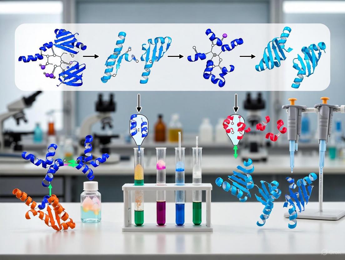
Workflow for Ubiquitin Remnant Profiling
The following diagram illustrates the core experimental workflow for the enrichment and identification of K-ε-GG-containing peptides, integrating sample preparation, affinity enrichment, and final analysis.
Key Protocol Steps and Methodologies
Cell Lysis and Trypsin Digestion
Effective sample preparation is critical for comprehensive ubiquitinome analysis. The process begins with cell lysis under denaturing conditions to preserve PTMs and halt enzymatic activity.
- Lysis Buffer: 8 M urea, 50 mM Tris-HCl (pH 7.5), 150 mM NaCl, 1 mM EDTA, supplemented with protease inhibitors (e.g., 2 μg/ml aprotinin, 10 μg/ml leupeptin), deubiquitinase inhibitors (e.g., 50 μM PR-619), and 1 mM chloroacetamide as an alkylating agent [10].
- Protein Processing: Reduce proteins with 5 mM dithiothreitol (DTT) for 45 minutes at room temperature, followed by alkylation with 10 mM iodoacetamide for 30 minutes in the dark [10] [12].
- Tryptic Digestion: Dilute lysates to 2 M urea with 50 mM Tris-HCl (pH 7.5) to create conditions favorable for trypsin. Digest overnight at 25°C with sequencing-grade trypsin at an enzyme-to-substrate ratio of 1:50 [10]. This step is what generates the K-ε-GG remnant on the target peptides.
Peptide Pre-Fractionation
To reduce sample complexity and increase depth of analysis, digested peptides are often fractionated prior to immunoenrichment.
- Method: Basic pH reversed-phase chromatography (RPLC) [10].
- Typical Setup: Use a Zorbax 300 Extend-C18 column (9.4 × 250 mm) on an HPLC system.
- Gradient: Employ a shallow gradient from 8% to 60% solvent B (90% acetonitrile, 5 mM ammonium formate, pH 10) over 64 minutes.
- Pooling Strategy: Collect 80 fractions and combine them in a non-contiguous manner into 8-12 pooled fractions to minimize variability and maximize peptide identification [10].
Immunoaffinity Enrichment of K-ε-GG Peptides
The core of the methodology is the specific enrichment of K-ε-GG-containing peptides using a high-fidelity antibody.
- Antibody Cross-Linking: To prevent antibody co-elution and improve performance, cross-link the anti-K-ε-GG antibody to protein A agarose beads. Wash beads with 100 mM sodium borate (pH 9.0), then incubate with 20 mM dimethyl pimelimidate (DMP) for 30 minutes at room temperature. Block cross-linking with 200 mM ethanolamine (pH 8.0) for 2 hours at 4°C [10] [12].
- Enrichment Procedure: Resuspend dried peptide fractions in 1.5 ml of Immunoaffinity Purification (IAP) buffer (50 mM MOPS, pH 7.2, 10 mM sodium phosphate, 50 mM NaCl). Incubate with cross-linked antibody beads for 1-2 hours at 4°C with rotation [10] [12].
- Washing and Elution: Wash beads extensively with ice-cold PBS to remove non-specifically bound peptides. Elute bound K-ε-GG peptides with two applications of 50-100 μl of 0.15% trifluoroacetic acid (TFA) [10] [12].
- Sample Cleanup: Desalt eluted peptides using C18 StageTips or similar micro-solid-phase extraction tips before LC-MS/MS analysis [10].
Quantitative Assessment of Protocol Optimization
Systematic optimization of the K-ε-GG enrichment workflow has dramatically improved the sensitivity and scale of ubiquitinome analyses. The following table summarizes key performance metrics achieved with different levels of protocol refinement.
Table 1: Impact of Protocol Optimization on Ubiquitination Site Identification
| Protocol Version | Key Optimizations | Protein Input | Number of Ubiquitination Sites Identified | Reference |
|---|---|---|---|---|
| Early Workflow | Basic anti-K-ε-GG enrichment | ~35 mg | < 5,000 sites | [10] |
| Refined Workflow | Antibody cross-linking, optimized peptide & antibody inputs, off-line fractionation | 5 mg per SILAC channel | ~20,000 sites in a single experiment | [10] [11] |
| SCASP-PTM | Desalting-free sequential PTM enrichment; uses SDS and cyclodextrins | Variable | Enables tandem ubiquitinome, phosphoproteome, and glycoproteome quantification | [13] [8] |
The Scientist's Toolkit: Essential Research Reagents
Successful ubiquitin remnant profiling requires specific, high-quality reagents. The table below details essential materials and their functions in the workflow.
Table 2: Key Research Reagent Solutions for K-ε-GG Remnant Enrichment
| Research Reagent | Function/Application | Example Specifications |
|---|---|---|
| Anti-K-ε-GG Antibody | Immunoaffinity enrichment of ubiquitinated peptides; core specificity | PTMScan Ubiquitin Remnant Motif Kit (CST #5562); highly specific for K-ε-GG remnant [9] [10] |
| Magnetic Beads | Solid support for antibody immobilization and immunoprecipitation | Protein A/G magnetic beads (e.g., Dynabeads); enable gentle washing and high recovery [14] |
| Sequencing-Grade Trypsin | Proteolytic digestion to generate K-ε-GG remnant peptides | High-purity, modified trypsin (e.g., Promega); ensures complete and specific digestion [15] [10] |
| Urea | Protein denaturation in lysis buffer; disrupts non-covalent interactions for efficient digestion | Ultra-pure grade (e.g., Urea Ultra from MP Biomedicals); minimizes carbamylation artifacts [15] [10] |
| Deubiquitinase Inhibitors | Preserve endogenous ubiquitination states during sample preparation | PR-619 (broad-spectrum DUB inhibitor); included in lysis buffer at 50 μM [10] |
| IAP Buffer | Optimal buffer for antibody-peptide binding during immunoenrichment | 50 mM MOPS, pH 7.2, 10 mM sodium phosphate, 50 mM NaCl; compatible with PTMScan Kit [10] [12] |
Advanced Applications and Integrated PTM Profiling
The K-ε-GG remnant motif technology has enabled sophisticated studies of ubiquitin biology. Researchers can now investigate dynamic changes in the ubiquitinome in response to cellular perturbations, such as proteasome inhibition with MG-132 or DUB inhibition with PR-619 [10]. Furthermore, the integration of Stable Isotope Labeling by Amino acids in Cell culture (SILAC) allows for precise quantitative comparisons of ubiquitination site occupancy across multiple experimental conditions [10] [11].
Recent methodological advances, such as the SCASP-PTM workflow, now enable the sequential enrichment of multiple PTMs, including ubiquitination, phosphorylation, and glycosylation, from a single sample [13] [8]. This integrated approach provides a more comprehensive view of the complex PTM networks that regulate cellular signaling, as demonstrated by the application of SCASP-PTM to uncover the role of ALDOA K330 ubiquitination and acetylation in tumor progression [13].
The tryptic digestion process is fundamental to generating the K-ε-GG remnant motif, which serves as the crucial handle for proteome-wide ubiquitination site mapping. Through rigorous optimization of the enrichment workflow—including antibody cross-linking, pre-fractionation, and optimized binding conditions—researchers can now routinely identify and quantify tens of thousands of endogenous ubiquitination sites from modest protein inputs. This powerful methodology, complemented by emerging multi-PTM profiling platforms, continues to drive discoveries in ubiquitin biology and its roles in health and disease.
Protein ubiquitination is a crucial post-translational modification (PTM) that regulates a vast array of cellular processes, including protein degradation by the 26S proteasome, cell cycle progression, signal transduction, and apoptosis [16] [17]. The process is mediated by a cascade of enzymes (E1, E2, and E3) that covalently attach the small protein ubiquitin to lysine (K) residues on substrate proteins [16] [17]. Often, polyubiquitin chains are formed through subsequent ubiquitin attachments to lysine residues on the previously conjugated ubiquitin molecule.
A transformative advancement in the proteomic study of ubiquitination was the development and commercialization of antibodies specific for the ubiquitin remnant motif (K-ε-GG). During standard proteomic sample preparation, proteins are digested with the protease trypsin. This digestion cleaves the ubiquitin molecule itself, but leaves a di-glycine ("GG") remnant attached via an isopeptide bond to the epsilon-amino group of the modified lysine residue on the substrate protein. This signature, known as the K-ε-GG motif, serves as a universal handle for identifying ubiquitination sites [18] [16]. Anti-K-ε-GG antibodies are therefore not specific to a single protein, but instead bind this conserved di-glycine remnant, enabling the systematic enrichment and mass spectrometry-based identification of thousands of endogenous ubiquitination sites from complex biological samples [18].
Molecular Basis of Anti-K-ε-GG Antibody Specificity
The anti-K-ε-GG antibody is a rabbit polyclonal antibody renowned for its high specificity towards the ubiquitin remnant motif. Its core function is to recognize and bind the di-glycine adduct that remains on a lysine side chain following tryptic digestion of ubiquitinated proteins [19] [18]. This binding is highly specific for the K-ε-GG structure, allowing the antibody to distinguish ubiquitinated peptides from a vast background of unmodified peptides in a protein digest.
The specificity of this antibody-antigen interaction is paramount for the sensitivity and accuracy of ubiquitin proteomics. Recent investigations into antibody-antigen binding affinity (ΔΔG) prediction highlight that achieving generalizable models requires immense data volume and diversity, underscoring the complexity of these molecular recognition events [20]. Furthermore, accurately predicting antibody-antigen interactions must account for atomic flexibility, particularly in the complementarity-determining regions (CDRs) of the antibody, to model the dynamic binding process effectively [21]. The refined application of anti-K-ε-GG antibodies, including optimized peptide input and antibody cross-linking, has been instrumental in pushing the boundaries of ubiquitin proteomics, enabling the routine quantification of over 10,000 distinct ubiquitination sites from a single experiment [18].
Table 1: Key Research Reagent Solutions for K-ε-GG Immunoaffinity Enrichment
| Reagent / Kit Name | Supplier | Primary Function | Key Features |
|---|---|---|---|
| Ubiquitin Remnant Motif (K-ε-GG) Antibody | Thermo Fisher | Immunoaffinity enrichment of K-ε-GG peptides | Rabbit polyclonal; validated for WB and ELISA [19] |
| PTMScan Ubiquitin Remnant Motif (K-ε-GG) Kit | Cell Signaling Technology | Ubiquitinated peptide enrichment for MS | Bead-conjugated antibody; includes proprietary protocol and buffer [16] |
| PTMScan HS Ubiquitin/SUMO Remnant Motif Kit | Cell Signaling Technology | High-sensitivity enrichment of ubiquitin/SUMO peptides | Magnetic bead version; also enriches SUMO remnant motifs (AGG, SGG, TGG, VGG) [22] |
Detailed Experimental Protocol for Ubiquitin Remnant Enrichment
The following protocol details the standard workflow for enriching ubiquitinated peptides using anti-K-ε-GG antibody beads, based on established PTMScan technology and recent high-efficiency methodologies [18] [16] [17].
Sample Preparation and Protein Digestion
- Cell Lysis: Lyse cells or tissue samples in a urea-based lysis buffer (e.g., 8 M urea, 10 mM EDTA, 10 mM DTT, 1% protease inhibitor cocktail). Perform sonication on ice to ensure complete disruption and homogenization. Remove insoluble debris by centrifugation at 12,000-20,000 × g for 10 minutes at 4°C [17].
- Protein Quantification: Determine the protein concentration of the supernatant using a compatible assay, such as the 2D Quant kit.
- Reduction and Alkylation: Reduce disulfide bonds with 10 mM dithiothreitol (DTT) for 1 hour at 56°C. Subsequently, alkylate cysteine residues with 30 mM iodoacetamide (IAA) for 45 minutes at room temperature in darkness.
- Trypsin Digestion: Dilute the protein sample with 100 mM ammonium bicarbonate (NH₄HCO₃) to reduce the urea concentration below 2 M. Digest the proteins first with trypsin at a 1:50 (w/w) enzyme-to-substrate ratio overnight, followed by a second digestion at a 1:100 ratio for 4 hours [17].
- Peptide Cleanup: Desalt the resulting peptide mixture using reversed-phase solid-phase extraction (e.g., C18 cartridges or pillars). Elute peptides with 0.1% formic acid (FA) in 80% acetonitrile (ACN) and dry using a vacuum concentrator.
Immunoaffinity Enrichment (Immunoprecipitation)
- Antibody Bead Preparation: Resuspend the anti-K-ε-GG antibody bead conjugate (supplied in kits like the PTMScan Ubiquitin Remnant Motif Kit) by gentle vortexing [16].
- Peptide Binding: Dissolve the dried tryptic peptides in NETN buffer (100 mM NaCl, 1 mM EDTA, 50 mM Tris-HCl, 0.5% NP-40, pH 8.0). Incubate the peptide solution with the antibody beads at 4°C overnight with gentle shaking to allow the anti-K-ε-GG antibody to bind its target peptides [16] [17].
- Washing: After incubation, wash the beads extensively to remove non-specifically bound peptides. A typical wash regimen involves four washes with NETN buffer, followed by two washes with pure water [17].
- Peptide Elution: Elute the captured K-ε-GG-modified peptides from the antibody beads using 0.1% trifluoroacetic acid (TFA). Combine the eluates and dry them in a vacuum concentrator.
Mass Spectrometric Analysis
- Sample Preparation for MS: Desalt the enriched peptides using C18 ZipTips or stage tips prior to LC-MS/MS analysis.
- LC-MS/MS Analysis: Resuspend the peptides in a loading buffer (e.g., 0.1% FA) and separate them using a nano-flow ultra-high-performance liquid chromatography (UHPLC) system with a C18 reversed-phase column. The peptides are typically eluted with a gradient of increasing acetonitrile.
- Data Acquisition: Analyze the eluting peptides using a tandem mass spectrometer (e.g., Q-Exactive HF-X) operating in data-dependent acquisition (DDA) mode. MS1 spectra are acquired at high resolution (e.g., 60,000), followed by fragmentation of the top N most intense ions for MS2 analysis [17].
- Database Searching: Process the raw MS/MS data using search engines like MaxQuant. Search parameters should include carbamidomethylation of cysteine as a fixed modification, and oxidation of methionine and GlyGly modification on lysine (K-ε-GG) as variable modifications [17].
Advanced Applications and Protocol Innovations
The foundational protocol has been refined and adapted for increasingly complex proteomic analyses.
Refinements for High-Throughput Quantification
Key improvements to the K-ε-GG enrichment workflow have enabled the routine identification and quantification of ~20,000 distinct endogenous ubiquitination sites. These include:
- Optimized Antibody and Peptide Input: Determining the ideal ratio of antibody beads to peptide input maximizes recovery and minimizes non-specific binding [18].
- Antibody Cross-linking: Covalently cross-linking the antibody to the beads reduces antibody leaching and prevents the co-elution of antibody-derived peptides, which can interfere with MS analysis [18].
- Improved Off-line Fractionation: Implementing high-pH reverse-phase HPLC fractionation of peptides prior to immunoenrichment reduces sample complexity and increases the depth of ubiquitinome coverage [18].
Table 2: Performance of Advanced K-ε-GG Enrichment Methodologies
| Method / Approach | Key Innovation | Reported Performance | Reference |
|---|---|---|---|
| Refined K-ε-GG Enrichment | Antibody cross-linking, optimized fractionation | ∼20,000 ubiquitination sites from a single SILAC experiment | [18] |
| SCASP-PTM Protocol | Tandem enrichment of multiple PTMs (Ubiquitination, Phosphorylation, Glycosylation) from one sample | Serial enrichment without intermediate desalting | [8] |
| PTMScan HS Technology | High-sensitivity magnetic bead-based enrichment | Also enriches for SUMO remnant motifs (AGG, SGG, TGG, VGG) | [22] |
Integrated and Tandem Enrichment Strategies
Recent protocol developments focus on maximizing information from limited samples. The SCASP-PTM (SDS-cyclodextrin-assisted sample preparation-post-translational modification) approach allows for the tandem enrichment of ubiquitinated, phosphorylated, and glycosylated peptides from a single sample digest in a serial manner, without the need for intermediate desalting steps [8]. This integrated workflow is particularly valuable for studying crosstalk between different PTM networks.
Application in Disease Research
Anti-K-ε-GG antibodies have been pivotal in uncovering the role of ubiquitination in disease mechanisms. For example, a proteomic analysis of human primary and metastatic colon adenocarcinoma tissues using anti-K-ε-GG antibody-based enrichment identified 375 differentially regulated ubiquitination sites. This study revealed ubiquitination events in pathways highly related to cancer metastasis, such as RNA transport and cell cycle, suggesting the altered ubiquitination of CDK1 may be a pro-metastatic factor [17]. In virology, this technology has been used to analyze the RTA-dependent ubiquitin-modified proteome in Kaposi's sarcoma herpesvirus (KSHV), identifying a novel mechanism of immune evasion involving inhibition of antigen presentation [23].
Distinguishing Ubiquitination from NEDD8 and ISG15 Modifications
The ubiquitin-proteasome system is a critical regulatory pathway in eukaryotic cells, controlling the stability, function, and localization of thousands of proteins. At the heart of this system lies ubiquitin, a 76-amino acid protein that can be covalently attached to substrate proteins via an enzymatic cascade involving E1 activating, E2 conjugating, and E3 ligase enzymes [24]. This modification, known as ubiquitination, primarily targets proteins for proteasomal degradation but also regulates diverse non-proteolytic functions including protein-protein interactions, endocytosis, and signal transduction [25]. The complexity of ubiquitin signaling stems from its ability to form different polyubiquitin chains through eight distinct linkage sites (M1, K6, K11, K27, K29, K33, K48, K63), with K48-linked chains being predominantly associated with proteasomal degradation [26] [24].
Ubiquitin belongs to a broader family of ubiquitin-like proteins (UBLs) that share structural similarities with ubiquitin but serve distinct cellular functions. Among these, NEDD8 and ISG15 have emerged as particularly important regulators of cellular physiology. NEDD8 shows the highest sequence similarity to ubiquitin (approximately 60%) and primarily regulates the activity of cullin-RING ligases (CRLs), the largest family of E3 ubiquitin ligases [27] [28]. ISG15, in contrast, is unique among UBLs as it consists of two ubiquitin-like domains connected by a short linker region and is strongly induced by interferon signaling as part of the innate immune response to infections [25] [29]. These UBLs utilize similar enzymatic cascades for conjugation but have distinct E1, E2, and E3 enzymes that provide specificity [24].
The ubiquitin remnant motif antibody technology has revolutionized the study of these modifications by enabling proteome-wide identification of modification sites. This approach exploits the fact that trypsin digestion of ubiquitinated or UBL-modified proteins leaves a characteristic di-glycine (diGly) remnant attached to the modified lysine residue. Antibodies specifically recognizing this K-ε-GG motif allow immunopurification and subsequent mass spectrometry analysis for system-wide mapping of modification sites [30] [31]. However, the shared diGly signature presents a significant challenge for distinguishing between ubiquitin and UBL modifications, necessitating specialized experimental approaches for accurate assignment.
Molecular Differentiation of Ubiquitin, NEDD8, and ISG15
Structural and Functional Characteristics
Despite shared structural features, ubiquitin, NEDD8, and ISG15 possess distinct molecular characteristics that underlie their specific biological functions. Ubiquitin serves as the master regulator of protein fate, with its diverse functions mediated through different chain linkages and recognition by ubiquitin-binding domains in target proteins. The human genome encodes approximately 40 E2 enzymes and over 600 E3 ligases that provide specificity for thousands of potential substrates [24].
NEDD8 (Neural precursor cell-expressed developmentally down-regulated 8) is the most ubiquitin-like UBL, sharing approximately 60% sequence identity. Its primary function is the neddylation of cullin proteins, which activates CRL complexes and enhances their E3 ligase activity toward substrates involved in cell cycle regulation, signaling, and development. Unlike ubiquitin, NEDD8 primarily modifies a limited set of substrates, with cullins being the major physiological targets [27] [28]. Both canonical neddylation (through NEDD8-specific enzymes) and atypical neddylation (through ubiquitin system enzymes) have been described, with each modifying distinct protein subsets [28].
ISG15 (Interferon-stimulated gene 15) is structurally unique as a di-ubiquitin-like protein consisting of two ubiquitin-like domains. It is strongly induced by type I interferons (IFN-α and IFN-β) as part of the innate immune response to infections [25] [29]. Unlike ubiquitin and NEDD8, which are constitutively expressed, ISG15 expression is typically minimal under normal conditions but dramatically upregulated during viral, bacterial, and parasitic infections. The ISG15 conjugation system includes the E1 enzyme UBE1L/UBA7, E2 enzyme Ube2L6/UbcH8, and E3 ligases including HERC5, ARIH1, and TRIM25 [25] [32]. ISG15 functions as both a conjugated modifier (ISGylation) and a free molecule with cytokine-like properties, stimulating IFN-γ secretion when secreted extracellularly [32] [29].
Table 1: Key Characteristics of Ubiquitin, NEDD8, and ISG15
| Feature | Ubiquitin | NEDD8 | ISG15 |
|---|---|---|---|
| Size/Structure | 76 aa, single ubiquitin domain | 81 aa, single ubiquitin domain | 165 aa, two ubiquitin-like domains |
| Sequence Identity to Ubiquitin | 100% (reference) | ~60% | ~30% (per domain) |
| Induction Conditions | Constitutive | Constitutive | Interferon-induced, infection |
| Primary E1 Enzyme | UBA1 | NAE1 (APPBP1-UBA3) | UBE1L (UBA7) |
| Primary E2 Enzyme | ~40 different E2s | UBE2M (UBC12), UBE2F | UBE2L6 (UbcH8) |
| Major E3 Enzymes | >600 different E3s | DCN1-RBX1, MDM2 | HERC5, ARIH1, TRIM25 |
| Major Functions | Protein degradation, signaling, trafficking | CRL activation, regulation of E3 ligases | Antiviral defense, innate immunity |
| Deconjugating Enzymes | ~100 DUBs | DEN1, COP9 signalosome | USP18, USP16, USP21, USP24, viral proteases |
Biological Pathways and Physiological Roles
The distinct biological functions of ubiquitin, NEDD8, and ISG15 are reflected in their involvement in different cellular pathways and physiological processes. Ubiquitin represents the most versatile regulator, controlling virtually every cellular process through its ability to target proteins for proteasomal degradation or alter their function through non-proteolytic mechanisms. Key pathways regulated by ubiquitination include NF-κB signaling, DNA damage repair, cell cycle progression, and receptor endocytosis [26] [24]. Dysregulation of ubiquitination is implicated in numerous diseases, including cancer, neurodegenerative disorders, and inflammatory conditions.
NEDD8 functions as a master regulator of the ubiquitin-proteasome system itself through its control of CRL activity. Neddylation of cullin proteins induces conformational changes that promote ubiquitin transfer to CRL substrates. This positions neddylation as a critical upstream regulator of ubiquitin-dependent proteolysis, particularly for proteins involved in cell cycle control (e.g., p27, cyclins) and signal transduction [27] [28]. Pharmacological inhibition of neddylation has emerged as a promising therapeutic strategy in cancer, with the NEDD8-activating enzyme (NAE) inhibitor pevonedistat currently in clinical trials.
ISG15 plays a specialized role in innate immunity and host defense against pathogens. During viral infection, ISG15 is conjugated to both viral and host proteins to inhibit various stages of the viral life cycle, including replication, assembly, and release [25] [29]. The antiviral activity of ISG15 is demonstrated by the susceptibility of ISG15-deficient mice and humans to various viral infections, including influenza, herpesvirus, and SARS-CoV-2. Notably, several viral pathogens encode deISGylating enzymes that cleave ISG15 from modified proteins to counteract this host defense mechanism [25]. Beyond its antiviral functions, ISG15 has been implicated in antibacterial immunity, cancer progression, autophagy, and DNA damage response [32] [29].
Experimental Strategies for Discrimination
DiGly Proteomics and Specific Enrichment Techniques
The diGly remnant motif antibody technology has emerged as a powerful tool for system-wide identification of ubiquitin and UBL modification sites. This approach exploits the fact that trypsin digestion cleaves after arginine and lysine residues, but when these residues are modified by ubiquitin or UBLs, cleavage occurs after the diglycine motif, leaving a signature K-ε-GG remnant on the modified lysine [30] [31]. Antibodies specifically recognizing this diGly motif enable immunopurification of modified peptides followed by liquid chromatography-tandem mass spectrometry (LC-MS/MS) for comprehensive mapping of modification sites.
Despite its power, standard diGly proteomics faces a significant challenge: the shared diGly signature between ubiquitin, NEDD8, ISG15, and other UBLs. This necessitates additional strategies for definitive assignment of modification type. Several approaches have been developed to address this limitation:
Genetic manipulation: Expression of epitope-tagged versions of ubiquitin, NEDD8, or ISG15 (e.g., HA-, FLAG-, or His-tagged) allows specific enrichment using tag-specific antibodies [28] [29]. This approach provides unambiguous assignment but requires genetic modification of cellular systems.
Mutant constructs: Use of R74K NEDD8 mutant prevents tryptic cleavage between R74 and G75, resulting in a distinct 13-amino acid remnant (NEDD8({}_{72-85})) instead of the typical diGly signature, enabling discrimination from ubiquitin modifications [28].
Specific nanobodies and antibodies: Recently developed ISG15-specific nanobodies (VHHISG15-A and VHHISG15-B) enable highly specific immunoprecipitation of ISGylated proteins with minimal background [32]. These nanobodies recognize distinct epitopes on ISG15's C- and N-terminal domains, providing tools for specific enrichment.
Enzyme inhibition: Pharmacological inhibition of specific pathways (e.g., NAE1 for neddylation) combined with quantitative diGly proteomics allows identification of substrates dependent on particular modification pathways.
Table 2: Experimental Approaches for Discriminating Ubiquitin-like Modifications
| Method | Principle | Applications | Advantages | Limitations |
|---|---|---|---|---|
| Standard diGly Proteomics | Anti-K-ε-GG antibody enrichment | Global mapping of ubiquitin/UBL sites | Unbiased, proteome-wide coverage | Cannot distinguish between different UBLs |
| Tagged UBL Expression | Epitope-tagged ubiquitin/UBLs with specific antibodies | Specific substrate identification for particular UBL | Unambiguous assignment | Requires genetic manipulation, potential overexpression artifacts |
| NEDD8 R74K Mutant | Altered tryptic cleavage pattern creates unique remnant | Specific identification of neddylation sites | Distinguishes NEDD8 from ubiquitin | May affect conjugation efficiency |
| ISG15-specific Nanobodies | High-affinity binders to ISG15-specific epitopes | Selective enrichment of ISGylated proteins | Minimal background, work under denaturing conditions | Not applicable to other UBLs |
| Pathway Inhibition | Pharmacological or genetic inhibition of specific pathways | Identification of pathway-dependent substrates | Functional context, applicable to endogenous proteins | Indirect evidence, potential off-target effects |
| Quantitative Proteomics | SILAC or TMT labeling to monitor dynamics | Temporal regulation of modifications | Dynamic information, high precision | Requires specialized instrumentation and expertise |
Proteomic Workflows for Modification Discrimination
The most effective strategies for discriminating ubiquitin from NEDD8 and ISG15 modifications often combine multiple approaches in integrated workflows. A comprehensive proteomic workflow typically includes the following steps:
Sample preparation under denaturing conditions: Use of strong denaturants (e.g., urea, guanidine hydrochloride) ensures inactivation of endogenous deconjugating enzymes and provides a snapshot of the modification status at the time of lysis [30] [31].
Parallel enrichment strategies: Simultaneous processing of samples for (a) total diGly proteomics using anti-K-ε-GG antibodies, (b) UBL-specific enrichment using tagged constructs or specific antibodies/nanobodies, and (c) negative controls using inactive mutants or isotype control antibodies.
Advanced mass spectrometry analysis: High-resolution LC-MS/MS with fragmentation techniques (e.g., HCD, ETD) to sequence modified peptides and identify exact modification sites.
Bioinformatic analysis: Computational pipelines to filter, validate, and assign modifications, including discrimination based on unique tryptic peptides (e.g., NEDD8 R74K mutant signature) and cross-referencing against UBL-specific databases.
For NEDD8-specific identification, the combination of the R74K mutant with standard diGly proteomics has proven highly effective. This approach identified 1,101 unique neddylation sites on 620 proteins, revealing distinct proteomes for canonical and atypical neddylation [28]. Canonical neddylation primarily targets spliceosome, mRNA surveillance, and DNA replication factors, while atypical neddylation modifies ribosomal and proteasomal proteins.
For ISG15-specific profiling, the development of high-affinity nanobodies represents a significant advancement. These nanobodies enable efficient immunoprecipitation of ISGylated substrates under various conditions, including viral and bacterial infections [32] [29]. When combined with quantitative proteomics, this approach can monitor dynamic changes in the ISGylome during immune activation.
The following diagram illustrates a comprehensive workflow that integrates these strategies for discriminating ubiquitin, NEDD8, and ISG15 modifications:
The Scientist's Toolkit: Essential Research Reagents
Table 3: Key Research Reagents for Studying Ubiquitin-like Modifications
| Reagent Category | Specific Examples | Applications | Key Features |
|---|---|---|---|
| Anti-diGly Antibodies | PTMScan Ubiquitin Remnant Motif Kit [30], Pan-Ubiquitin Remnant Motif Antibody [33] | Global ubiquitin/UBL site identification | Recognizes K-ε-GG motif after trypsin digestion; compatible with LC-MS/MS |
| ISG15-specific Reagents | VHHISG15-A and VHHISG15-B nanobodies [32] | Specific ISG15 substrate identification | Nanomolar affinity; target distinct ISG15 epitopes; inhibit deISGylation (VHHISG15-A) |
| NEDD8-specific Tools | NEDD8 R74K mutant [28], NAE1 inhibitors (e.g., MLN4924/Pevonedistat) | Specific neddylation site identification, pathway inhibition | Altered tryptic signature distinguishes from ubiquitin; pharmacological neddylation blockade |
| Tagged UBL Constructs | HA-Ubiquitin, FLAG-NEDD8, His-ISG15 | UBL-specific substrate identification | Epitope tags enable specific immunopurification; available for various expression systems |
| Deconjugating Enzyme Inhibitors | USP/ULD inhibitors, viral protease inhibitors (e.g., SARS-CoV-2 PLpro inhibitors) | Stabilization of UBL conjugates | Prevent deconjugation; enhance detection of modified substrates |
| Activity-Based Probes | Ubiquitin vinyl sulfone, ISG15 suicide probes | Detection of active deubiquitinases/deISGylases | Covalently label active site cysteine of DUBs/DIGs; monitor enzyme activity |
Detailed Experimental Protocols
Protocol 1: Discrimination of NEDD8 Modifications Using R74K Mutant
Purpose: To specifically identify NEDD8 modification sites while discriminating from ubiquitination.
Background: The NEDD8 R74K mutation prevents tryptic cleavage between R74 and G75, resulting in a unique 13-amino acid remnant (NEDD8({}_{72-85}): TLGMLQGKEKSTG) instead of the typical diGly signature. This enables unambiguous identification of neddylation sites by mass spectrometry [28].
Materials:
- NEDD8 R74K expression plasmid
- Control vector (wild-type NEDD8)
- Transfection reagent
- Urea lysis buffer (8 M urea, 50 mM Tris-HCl pH 8.0, 75 mM NaCl, protease inhibitors)
- Anti-K-ε-GG antibody (e.g., PTMScan Ubiquitin Remnant Motif Kit [30])
- Protein A/G agarose beads
- Trypsin (sequencing grade)
- C18 desalting columns
- LC-MS/MS system
Procedure:
- Cell culture and transfection: Culture appropriate cells (e.g., HEK293T) and transfect with NEDD8 R74K plasmid or control vector using standard protocols.
- Stimulation: Treat cells with neddylation-inducing conditions if desired (e.g., serum stimulation, specific stressors) for appropriate duration.
- Cell lysis: Harvest cells and lyse in urea lysis buffer. Sonicate to reduce viscosity and clarify by centrifugation at 16,000 × g for 15 minutes.
- Protein digestion: Reduce with 5 mM DTT (30 minutes, 25°C), alkylate with 15 mM iodoacetamide (30 minutes, 25°C in dark), and quench with 10 mM DTT. Dilute urea to 2 M with 50 mM Tris-HCl pH 8.0 and digest with trypsin (1:50 w/w) overnight at 25°C.
- Peptide cleanup: Acidify with trifluoroacetic acid (TFA) to pH < 3 and desalt using C18 columns according to manufacturer's instructions.
- diGly peptide enrichment: Resuspend peptides in immunoaffinity purification (IAP) buffer and incubate with anti-K-ε-GG antibody conjugated to protein A agarose beads (2-4 hours, 4°C). Wash beads extensively with IAP buffer followed by water.
- Peptide elution: Elute bound peptides with 0.15% TFA.
- LC-MS/MS analysis: Analyze peptides by nanoflow LC-MS/MS using a 2-hour gradient on a C18 column coupled to a high-resolution mass spectrometer.
- Data analysis: Search data against appropriate database using search engines (e.g., MaxQuant, Proteome Discoverer) with the following parameters:
- Variable modifications: K-ε-GG (diglycine remnant), NEDD8 R74K signature peptide (TLGMLQGKEKSTG)
- Fixed modification: carbamidomethylation (C)
- Specificity: Require identification of the unique NEDD8 R74K remnant for neddylation site assignment
Troubleshooting:
- Low enrichment efficiency: Ensure proper antibody-to-peptide ratio; optimize incubation time and temperature
- High background: Include appropriate controls (vector-only transfection); increase wash stringency
- Poor MS identification: Check peptide loading amounts; optimize LC gradient and MS parameters
Protocol 2: Specific Identification of ISG15 Substrates Using Nanobodies
Purpose: To specifically enrich and identify ISG15-modified proteins using ISG15-specific nanobodies.
Background: The recently developed nanobodies VHHISG15-A and VHHISG15-B recognize distinct epitopes on ISG15 with nanomolar affinity, enabling highly specific immunoprecipitation of ISGylated substrates with minimal background [32].
Materials:
- VHHISG15-A and/or VHHISG15-B nanobodies (available through research collaborations or commercial sources)
- IFN-α or other ISG15-inducing stimuli
- Lysis buffer (6 M guanidine-HCl, 100 mM NaH₂PO₄, 10 mM Tris-HCl pH 8.0)
- Ni-NTA agarose beads (for His-tagged nanobody purification)
- Wash buffer 1 (8 M urea, 100 mM NaH₂PO₄, 10 mM Tris-HCl pH 8.0)
- Wash buffer 2 (8 M urea, 100 mM NaH₂PO₄, 10 mM Tris-HCl pH 6.3)
- Elution buffer (200 mM imidazole, 150 mM Tris-HCl pH 6.7, 30% glycerol, 0.72 M β-mercaptoethanol, 5% SDS)
- Trypsin (sequencing grade)
- C18 stage tips
Procedure:
- ISG15 induction: Treat cells with IFN-α (1000 U/mL, 24 hours) or other appropriate stimuli to induce ISG15 expression and ISGylation.
- Cell lysis: Harvest cells and lyse in guanidine-HCl lysis buffer. Sonicate and clarify by centrifugation.
- Nanobody immobilization: Couple VHHISG15-A or VHHISG15-B nanobodies to Ni-NTA agarose beads according to manufacturer's instructions.
- Immunoprecipitation: Incubate cell lysates with nanobody-conjugated beads (overnight, 4°C). Include control with non-specific nanobody or beads only.
- Washing: Wash beads sequentially with:
- Wash buffer 1 (3 × 10 minutes)
- Wash buffer 2 (3 × 10 minutes)
- Elution: Elute bound proteins with elution buffer (10 minutes, 25°C).
- Protein digestion: Reduce, alkylate, and digest eluted proteins with trypsin as described in Protocol 1.
- Peptide cleanup: Desalt peptides using C18 stage tips.
- LC-MS/MS analysis: Analyze by LC-MS/MS as described in Protocol 1.
- Data analysis: Search data against appropriate database with the following parameters:
- Variable modifications: K-ε-GG (diglycine remnant), oxidation (M), acetylation (protein N-term)
- Fixed modification: carbamidomethylation (C)
- Specificity: Compare to control samples to identify specifically enriched ISG15 substrates
Troubleshooting:
- Low ISG15 induction: Optimize IFN concentration and duration; verify induction by immunoblotting
- Non-specific binding: Include appropriate controls; optimize wash conditions; pre-clear lysates
- Incomplete elution: Ensure fresh elution buffer; consider alternative elution conditions
The following diagram illustrates the specific nanobody-based enrichment strategy for ISG15 substrates:
Data Interpretation and Validation
Analysis of Proteomic Data
Effective interpretation of ubiquitin and UBL proteomic data requires careful consideration of several factors. First, site localization confidence should be assessed using appropriate scoring algorithms (e.g., PTM score in MaxQuant) with a minimum threshold of 0.75 for reliable site assignment. Second, quantitative changes should be evaluated using appropriate normalization and statistical analysis, considering both fold-change and significance thresholds (typically ≥2-fold change with p-value < 0.05). Third, biological context is crucial for distinguishing functionally relevant modifications from bystander events.
For discriminating ubiquitin from NEDD8 modifications, the unique tryptic signature of the NEDD8 R74K mutant provides unambiguous assignment. In standard diGly proteomics, neddylation sites can be inferred through specific sequence contexts or through correlation with neddylation pathway manipulation, but this provides only indirect evidence [28].
For ISG15 substrate identification, the high specificity of ISG15 nanobodies enables confident assignment. Comparison to control samples (non-induced or non-specific nanobody) is essential to eliminate background binders. Additionally, ISG15 substrates should show increased abundance after interferon stimulation, providing orthogonal validation [32] [29].
Orthogonal Validation Methods
Proteomic findings require validation through orthogonal methods to ensure biological relevance:
- Immunoblotting: Confirm modification of specific candidates using target protein immunoprecipitation followed by ubiquitin/UBL immunoblotting, or vice versa.
- Mutagenesis: Validate specific modification sites by mutating identified lysine residues to arginine and assessing loss of modification.
- Microscopy: Visualize subcellular localization of modifications using immunofluorescence with specific antibodies.
- Functional assays: Assess the functional consequences of modifications through phenotypic readouts relevant to the biological context.
The integration of multiple proteomic strategies with orthogonal validation provides the most robust approach for distinguishing ubiquitination from NEDD8 and ISG15 modifications and understanding their functional implications in cellular regulation and disease pathogenesis.
The study of ubiquitin-like modifiers (UBLs) has been revolutionized by immunopurification strategies targeting the remnant motifs left on trypsin-digested peptides. Whereas ubiquitin and certain other UBLs leave a di-glycine (K-ε-GG) remnant on modified lysine residues [34] [35], the Ubiquitin Fold Modifier 1 (UFM1) presents a distinct C-terminal sequence. Following tryptic digestion, UFM1-conjugated substrate proteins yield a unique Val-Gly (VG-ε-K) isopeptide attached to the substrate lysine [36]. This VG remnant is a specific signature of UFMylation, enabling its selective enrichment and distinguishing it from other ubiquitin-like modifications. This application note details the development and implementation of a novel immunoaffinity approach to capture this motif, providing the scientific community with a powerful tool to decode the in vivo UFMylome.
Antibody Generation and Specificity Validation
To enable the site-specific study of UFMylation, researchers generated three monoclonal pan-anti-VG-ε-K antibody clones designed to immunoprecipitate the remnant VG UFMylated sites independently of the surrounding amino acid sequence [36].
The specificity of these antibody clones was rigorously validated using enzyme-linked immunosorbent assay (ELISA). The results demonstrated that the antibodies had a 6- to 17-fold enhanced specificity for VG-ε-K-containing peptides compared to GG-ε-GG-containing peptides, confirming their high selectivity for the UFM1 remnant motif [36].
A screening experiment was performed using tryptic peptides from mouse gastrocnemius skeletal muscle, immunoprecipitated with each clone individually and with a pooled cocktail. The captured peptides were analyzed via two-dimensional liquid chromatography-tandem mass spectrometry (2D-LC-MS/MS) and processed with two distinct search algorithms (Sequest+Percolator and MSFragger+PTM-Prophet) for orthogonal validation [36].
Table 1: Performance of Anti-VG-ε-K Antibody Clones in Identifying UFMylation Sites
| Antibody Format | Total Unique VG-Modified Peptides Identified | Peptides Identified by Both Search Algorithms (High Confidence) | Sequence Preference Noted |
|---|---|---|---|
| Clone 1 | Subset of 385 total | Part of 199 total | Slight upstream acidic residue preference |
| Clone 2 | Subset of 385 total | Part of 199 total | Most prominent upstream acidic residue preference |
| Clone 3 | Subset of 385 total | Part of 199 total | Slight upstream acidic residue preference |
| Pooled Cocktail | Greatest number | 199 unique peptides | Slight upstream acidic residue preference |
The data conclusively showed that while each clone identified unique subsets of peptides, the pooled antibody cocktail yielded the greatest number of identifications, consistent with previous findings for other post-translational modification enrichments [36]. This pooled approach was therefore recommended for comprehensive UFMylome profiling.
Detailed Protocol for UFMylome Enrichment and Analysis
Below is a standardized protocol for the enrichment and identification of UFMylation sites from tissue samples using the anti-VG-ε-K antibody.
Sample Preparation and Trypsin Digestion
- Protein Extraction: Homogenize tissue samples (e.g., mouse gastrocnemius muscle) in a denaturing lysis buffer to preserve PTMs and inactivate proteases.
- Protein Reduction and Alkylation: Reduce disulfide bonds with dithiothreitol (DTT) and alkylate cysteine residues with iodoacetamide (IAA).
- Proteolytic Digestion: Digest cellular proteins with sequencing-grade trypsin. Trypsin cleaves C-terminal to arginine and lysine residues. Critically, it cleaves after Arg81 on UFM1ΔSC-conjugated substrate proteins, liberating the remnant ValGly (VG) dipeptide attached via an isopeptide bond to the substrate lysine [36].
Immunoaffinity Enrichment of VG-Modified Peptides
- Peptide Pre-Clearing: Purify and desalt the resulting tryptic peptides using reversed-phase, solid-phase extraction (e.g., C18 desalting columns).
- Immunoprecipitation: Incubate the purified peptides with the pooled anti-VG-ε-K antibody clones conjugated to protein A agarose beads. Use PTMScan IAP Buffer or a similar optimized immunoaffinity purification buffer for this step [34].
- Washing and Elution: After incubation, extensively wash the beads to remove non-specifically bound peptides. Elute the captured VG-containing peptides using a dilute acid solution (e.g., 0.15% trifluoroacetic acid).
- Post-Enrichment Cleanup: Desalt the eluted peptides using reversed-phase purification microtips to remove antibodies and salts prior to MS analysis.
Mass Spectrometric Analysis and Data Processing
- Chromatographic Separation: Analyze the enriched peptides by two-dimensional liquid chromatography (2D-LC) to enhance peptide separation.
- Mass Spectrometry: Perform tandem mass spectrometry (LC-MS/MS) using a high-resolution instrument.
- Data Interpretation: Search the resulting MS/MS spectra against an appropriate protein sequence database. Include VG modification (+169.13 Da) on lysine as a variable modification. The use of multiple search algorithms (e.g., Sequest and MSFragger) with distinct statistical models is highly recommended for orthogonal validation and to increase confidence in identifications [36].
Diagram 1: VG-ε-K UFMylome Enrichment Workflow.
Key Applications and Biological Insights
The application of this methodology has yielded significant biological discoveries, particularly in the context of skeletal muscle biology and disease.
Characterization of the In Vivo UFMylome
Applying this protocol to various mouse tissues identified over 250 unique VG-containing peptides from 160 proteins, with extensive modification observed in skeletal muscle [36]. Bioinformatic analysis revealed that UFMylated proteins are significantly over-represented in several key cellular compartments and pathways.
Table 2: Functional Characterization of Identified UFMylation Targets
| Gene Ontology (GO) Category | Representative UFMylated Proteins | Biological Implication |
|---|---|---|
| Contractile Apparatus | Myosin heavy chain (MYH1, MYH2, MYH3, MYH4) | Regulation of muscle contraction and structural integrity |
| Endoplasmic/Sarcoplasmic Reticulum (ER/SR) | Proteins involved in calcium handling | Potential role in ER stress response and protein quality control |
| Mitochondria | Metabolic enzymes | Linking UFMylation to central carbon and amino acid metabolism |
| Ribosomes | RPL26 (validated site Lys134) | Regulation of protein translation |
Network analysis further revealed interconnected associations between the contractile apparatus, calcium-handling proteins, glucose metabolism enzymes, and translational regulators, suggesting a coordinated regulatory framework governed by UFMylation [36].
Functional Validation via UFC1 Knockdown
The specificity of the identified sites was confirmed through an in vivo functional validation experiment.
- In Vivo Knockdown: The UFM1-specific E2 ligase, UFC1, was knocked down in mouse extensor digitorum longus muscle using recombinant adeno-associated virus serotype 6 (rAAV6) delivering UFC1-targeting shRNA. The contralateral leg received a scramble shRNA control.
- Quantitative UFMylome Profiling: After 28 days, tryptic peptides from harvested muscles were immunoprecipitated with the anti-VG-ε-K antibody and labeled with tandem mass tags (TMT) for multiplexed quantification via 2D-LC-MS/MS.
- Results: A total of 22 unique VG-containing peptides were quantified. Knockdown of UFC1 resulted in a concomitant decrease in the abundance of these UFMylated peptides. Ten peptides were significantly down-regulated (q < 0.05), and manual annotation of MS/MS spectra confirmed myosin isoforms as major UFMylation targets, validating myosin UFMylation in vivo [36].
Discovery of UFMylation Alterations in Human Disease
This technology enabled the investigation of UFMylation in human pathology. Analysis of vastus lateralis skeletal muscle biopsies from people living with amyotrophic lateral sclerosis (plwALS) and age-matched controls revealed a conserved increase in UFMylation in the disease state [36]. Quantitative profiling with multiplexed isotopic labeling identified prominent increases in myosin UFMylation in plwALS biopsies, suggesting a potential role for dysregulated UFMylation in ALS pathogenesis.
The Scientist's Toolkit: Essential Research Reagents
Table 3: Key Reagents for VG-ε-K UFMylation Research
| Reagent / Kit | Provider Examples | Function in Workflow |
|---|---|---|
| Anti-VG-ε-K Antibody | Custom generation [36] | Immunoaffinity enrichment of UFMylated tryptic peptides. |
| PTMScan Ubiquitin Remnant Motif (K-ε-GG) Kit | Cell Signaling Technology [34] | For parallel ubiquitinome analysis; methodology is analogous to VG-ε-K approach. |
| HS Ubiquitin/SUMO Remnant Motif Kit | Cell Signaling Technology [37] | High-sensitivity enrichment for ubiquitin and SUMO remnants. |
| pan-Ubiquitin Remnant Motif (K-ε-GG) Antibody | Assay Genie, Thermo Fisher [35] [38] | Commercial antibodies for K-ε-GG enrichment, useful for comparative studies. |
| Sequencing-Grade Trypsin | Various | Proteolytic generation of remnant VG-ε-K and K-ε-GG peptides. |
Concluding Remarks
The development of antibodies targeting the VG-ε-K remnant motif has fundamentally expanded the toolbox for ubiquitin-like modification research, moving beyond the established K-ε-GG paradigm. This methodology provides a robust, site-specific approach for identifying and quantifying the in vivo UFMylome, revealing a more complex landscape of UFMylation than previously appreciated. Its successful application in mapping UFMylation across tissues and in human disease underscores its transformative potential for uncovering new biology and therapeutic targets associated with the UFM1 pathway.
Advanced Protocols and Applications in Ubiquitinome Profiling
The efficacy of mass spectrometry (MS)-based ubiquitin remnant motif immunopurification research is fundamentally dependent on the initial sample preparation, where the choice of lysis buffer dictates protein extraction efficiency, post-translational modification (PTM) preservation, and ultimately, data quality and depth. Within this framework, sodium deoxycholate (SDC) and urea have emerged as prominent lysis agents with distinct mechanisms and performance characteristics. SDC, an ionic detergent, provides robust solubilization of membrane proteins and rapid enzyme inactivation, while urea, a chaotrope, offers a milder denaturing environment traditionally used in proteomics. Recent advances have systematically compared these buffers, revealing that the optimized SDC-based protocol significantly enhances ubiquitinated peptide identification without sacrificing enrichment specificity [39]. This application note details the quantitative advantages and procedural protocols for both methods, contextualized within ubiquitinomics research, to guide researchers in selecting and implementing the optimal sample preparation strategy for their specific experimental goals in drug development and basic research.
Performance Comparison: SDC vs. Urea Lysis Buffers
The selection between SDC and urea lysis buffers involves trade-offs between identification depth, reproducibility, and compatibility with downstream steps. The table below summarizes key performance metrics from comparative studies.
Table 1: Quantitative Comparison of SDC and Urea Lysis Buffer Performance in Ubiquitinome Profiling
| Performance Metric | SDC-Based Lysis | Urea-Based Lysis | Experimental Context |
|---|---|---|---|
| K-ε-GG Peptide Identifications | 26,756 peptides (avg) [39] | 19,403 peptides (avg) [39] | HCT116 cells, 6h MG-132 treatment, n=4 |
| Relative Performance Gain | ~38% increase vs. urea [39] | Baseline | |
| Reproducibility | Higher; more peptides with CV < 20% [39] | Lower | |
| Key Additive | Chloroacetamide (CAA) for immediate DUB inhibition [39] | Iodoacetamide (risk of di-carbamidomethylation artifacts) [39] | |
| Typical Protein Input | Can be as low as 2 mg for deep coverage [39] | Often requires higher input (e.g., UbiSite used 20x more protein) [39] | Jurkat cell lysate |
| Compatibility with High-Throughput | Suitable for single-shot, high-throughput analyses [39] | Less suited due to lower identifications and reproducibility [39] | |
| Primary Advantage | Superior protein solubilization, rapid protease inactivation, higher yields [39] [40] | Widely adopted, traditional PTM proteomics buffer [39] |
Detailed Experimental Protocols
Optimized SDC Lysis Protocol for Ubiquitinome Profiling
The following step-by-step protocol is adapted from the method that demonstrated superior performance in head-to-head comparisons with urea-based lysis [39].
Reagents:
- SDC Lysis Buffer: 5% SDC in 50 mM Tris-HCl (pH 8.5)
- 500 mM Chloroacetamide (CAA) in water
- 500 mM Dithiothreitol (DTT) in water
- Protease Inhibitor Cocktail (EDTA-free)
- Deubiquitinase (DUB) Inhibitors (e.g., PR-619)
- Phosphatase Inhibitors (optional)
- Acetone or Ethanol for precipitation
Procedure:
- Cell Lysis and Protein Extraction:
- Aspirate culture medium from cell pellets and wash with ice-cold PBS.
- Lyse cells directly in SDC Lysis Buffer supplemented with 10 mM CAA, 5 mM DTT, and appropriate inhibitors. Use 1 mL buffer per 100 mg of cell pellet.
- Vortex the mixture vigorously and immediately boil at 95°C for 5-10 minutes to ensure complete lysis and rapid inactivation of endogenous enzymes, particularly DUBs.
- Sonicate the lysate on ice to shear DNA and reduce viscosity.
- Centrifuge at 16,000 × g for 10 minutes at room temperature to pellet insoluble debris. Transfer the clear supernatant to a new tube.
Protein Precipitation and Cleanup:
- Add 4-5 volumes of ice-cold acetone or ethanol to the supernatant to precipitate proteins. Incubate at -20°C for at least 4 hours or overnight.
- Centrifuge at 8,000 × g for 10 minutes to pellet the protein. A visible pellet should form.
- Carefully decant the supernatant and wash the pellet twice with cold 80% acetone to ensure complete SDC removal, which is critical for downstream enzymatic steps.
- Air-dry the pellet for 5-10 minutes to evaporate residual acetone. Do not over-dry, as this will make resuspension difficult.
Protein Digestion:
- Redissolve the protein pellet in a digestion-compatible buffer (e.g., 2 M urea, 50 mM Tris, pH 8.0) using gentle vortexing and sonication.
- Determine protein concentration using a standard assay (e.g., BCA assay).
- Digest proteins with Lys-C (1:100 w/w) for 3-4 hours at room temperature, followed by trypsin (1:50 w/w) overnight at 37°C.
Acidification and Peptide Cleanup:
- Acidify the digest to a final concentration of 1% formic acid (FA) or 0.5% trifluoroacetic acid (TFA). This precipitates any residual SDC.
- Centrifuge at 16,000 × g for 10 minutes and collect the supernatant.
- Desalt the peptides using C18 solid-phase extraction (SPE) cartridges or StageTips before proceeding to K-ε-GG immunoaffinity enrichment [39].
Standard Urea Lysis Protocol for Ubiquitinome Profiling
This protocol outlines the traditional urea-based approach, provided as a benchmark for comparison.
Reagents:
- Urea Lysis Buffer: 8 M Urea in 50 mM Tris-HCl (pH 8.0)
- 1 M DTT in water
- 500 mM Iodoacetamide (IAA) in water
- Protease and DUB Inhibitors
Procedure:
- Cell Lysis:
- Resuspend the cell pellet in ice-cold Urea Lysis Buffer supplemented with 10 mM DTT and inhibitors.
- Sonicate the suspension on ice to ensure complete lysis and reduce viscosity.
- Centrifuge at 16,000 × g for 10 minutes at room temperature. Collect the supernatant.
Protein Reduction and Alkylation:
- The reduction step is inherent with DTT in the lysis buffer. For alkylation, add IAA to a final concentration of 20-40 mM.
- Incubate in the dark at room temperature for 30 minutes.
- Quench excess IAA by adding DTT to a final concentration of 10 mM.
Protein Digestion:
- Dilute the urea concentration to below 2 M using 50 mM Tris-HCl (pH 8.0) to ensure trypsin activity.
- Digest with Lys-C (1:100 w/w) for 3-4 hours, followed by trypsin (1:50 w/w) overnight at 37°C.
Peptide Cleanup:
Workflow Integration and Visualization
The lysis and sample preparation process serves as the foundational step in a larger ubiquitinome profiling workflow. The following diagram illustrates the parallel paths for SDC and urea protocols and their integration with downstream mass spectrometry analysis.
Diagram 1: Ubiquitinome Profiling Workflow: SDC vs. Urea Paths
The Scientist's Toolkit: Essential Research Reagents
Successful implementation of the protocols depends on key reagents. The table below lists critical materials, their functions, and considerations for use.
Table 2: Key Research Reagent Solutions for Ubiquitinome Sample Preparation
| Reagent | Function/Role | Key Consideration |
|---|---|---|
| Sodium Deoxycholate (SDC) | Ionic detergent for efficient membrane protein solubilization and protein extraction [39] [42]. | Must be thoroughly removed via precipitation/desalting pre-digestion; tolerable by trypsin up to ~0.5% if not removed [42]. |
| Urea | Chaotropic agent for protein denaturation in traditional PTM workflows [39] [41]. | Concentration must be reduced to <2M for tryptic digestion; can cause lysine carbamylation at high temps or if impure [42]. |
| Chloroacetamide (CAA) | Cysteine alkylating agent; rapidly inactivates DUBs during lysis [39] [41]. | Preferred over IAA for SDC-boiling protocol as it avoids di-carbamidomethylation artifacts that mimic K-ε-GG mass [39]. |
| K-ε-GG Motif Antibody | Immunoaffinity enrichment of ubiquitin remnant peptides post-trypsin digestion [39] [41]. | Critical for specificity; enables purification of diglycine-modified peptides from complex protein digests. |
| Paramagnetic Beads (e.g., for SP3) | Solid-phase support for detergent removal, protein cleanup, and digestion [40]. | Enables processing in SDS-containing buffers; offers high efficiency and minimal sample loss [40]. |
| Data-Independent Acquisition (DIA-MS) | Mass spectrometry acquisition method for deep, reproducible ubiquitinome quantification [39] [43]. | Boosts coverage (>70,000 ubiquitinated peptides per run) and quantitative precision vs. DDA [39]. |
The comparative data and protocols presented herein establish that SDC-based lysis, when executed with a optimized protocol featuring immediate heating and CAA alkylation, provides a substantial advantage over traditional urea-based methods for deep-scale ubiquitinome profiling. The primary benefit is a significant increase in the number of identified ubiquitination sites without compromising quantitative reproducibility, making it particularly suitable for high-throughput applications and studies where material is limited. The urea-based protocol remains a viable and well-understood alternative, especially in workflows where detergent compatibility is a concern. The choice of lysis buffer should be a deliberate decision based on experimental goals, sample type, and downstream analytical plans. By adopting the optimized SDC protocol detailed in this application note, researchers in drug development and proteomics can achieve deeper and more robust insights into the ubiquitin-proteasome system, thereby accelerating target discovery and mechanistic studies.
UbiFast represents a significant advancement in ubiquitin remnant motif antibody immunopurification, enabling deep-scale enrichment and site-specific identification of ubiquitylation sites. This sensitive method employs anti-K-ε-GG antibodies for immunopurification of peptides containing the diglycine remnant left on protein substrates after trypsin digestion, followed by on-antibody isobaric labeling for sample multiplexing [44] [45]. The methodology was specifically developed to characterize the myriad roles of protein ubiquitylation in cell signaling, disease biology, and therapeutic mechanisms.
Recent technological innovations have led to the robotic automation of the UbiFast method using magnetic bead-conjugated K-ε-GG antibodies (mK-ε-GG) and magnetic particle processors [44]. This automated approach has demonstrated substantial improvements in throughput, reproducibility, and quantitative precision, making it particularly suitable for studying ubiquitylation in large sample sets such as drug discovery pipelines and clinical cohorts. The workflow enables processing of up to 96 samples in a single day, dramatically increasing capacity compared to manual processing methods [44] [45].
Experimental Design and Comparative Objectives
Core Experimental Framework
The comparative evaluation of manual versus automated UbiFast workflows was designed to assess multiple performance parameters across standardized sample types. The experimental setup utilized consistent input materials—typically 500 μg of peptide input per sample derived from either cell lines or patient-derived xenograft (PDX) tissue samples [44] [45]. Both workflows employed the PTMScan HS Ubiquitin/SUMO Remnant Motif (K-ε-GG) Kit (#59322) from Cell Signaling Technology, ensuring reagent consistency across methodological comparisons [46].
For the automated workflow, researchers implemented the method on a ThermoFisher KingFisher Apex system, a magnetic bead-handling platform specifically engineered for reproducible magnetic particle processing [46]. The manual protocol followed established UbiFast procedures with agarose bead-based immunopurification. Critical performance metrics including ubiquitylation site identification, quantitative reproducibility, processing time, and sample throughput were systematically evaluated across multiple experimental replicates to ensure statistical robustness [44] [46].
Platform Compatibility and Configuration
The compatibility of UbiFast methodology extends across multiple automation platforms, each offering distinct advantages for specific experimental requirements:
- Bead-handler platforms such as ThermoFisher's KingFisher line, which move only magnetic beads across wells, are compatible with PTMScan HS kits containing magnetic beads [46].
- Hybrid platforms including Agilent's AssayMap Bravo line utilize customized tips fitted with enrichment antibody reagents, moving liquid through the tips across wells. These systems require custom PTMScan reagent formulations without magnetic beads [46].
- Liquid handling platforms from manufacturers including Hamilton, Tecan, Beckman Coulter, and Revvity can accommodate PTMScan HS kits with appropriate method optimization [46].
This platform flexibility allows researchers to implement automated UbiFast workflows within existing laboratory infrastructure while maintaining methodological consistency across research groups and applications.
Detailed Experimental Protocols
Manual UbiFast Immunopurification Protocol
The manual UbiFast workflow requires meticulous attention to technique to minimize variability between samples and processing batches:
Sample Preparation: Cells or tissues are lysed in urea-containing buffer (6-8 M urea, 50 mM Tris-HCl, pH 8.0). Cellular proteins are reduced with 5 mM dithiothreitol (37°C, 30 min), alkylated with 10 mM iodoacetamide (room temperature, 30 min in darkness), and digested with trypsin (1:50 enzyme-to-substrate ratio) at 37°C overnight [47].
Peptide Cleanup: Resulting peptides are purified by reversed-phase, solid-phase extraction using C18 cartridges or plates. Peptides are equilibrated in immunoaffinity purification (IAP) buffer (50 mM MOPS/NaOH, pH 7.2, 10 mM Na2HPO4, 50 mM NaCl) [47].
Immunoaffinity Purification: The PTMScan Ubiquitin Remnant Motif Antibody Bead Conjugate is washed with IAP buffer, then incubated with peptide samples (500 μg input recommended) for 2 hours at 4°C with end-over-end mixing [47].
Wash Steps: Beads are collected by centrifugation and washed three times with IAP buffer and twice with HPLC-grade water to remove non-specifically bound peptides [47].
Peptide Elution: Captured ubiquitin remnant-containing peptides are eluted with two washes of 0.1% trifluoroacetic acid (50 μL each) with gentle agitation. Combined eluates are concentrated and desalted using C18 microtips prior to LC-MS/MS analysis [47].
The entire manual process typically requires 6-8 hours of hands-on time per batch of 8-12 samples, with additional time for incubation steps.
Automated UbiFast Immunopurification Protocol
The automated UbiFast protocol implemented on the KingFisher Apex system streamlines the enrichment process while enhancing reproducibility:
System Setup: Magnetic beads, peptide samples (500 μg in 500 μL IAP buffer), and wash/elution buffers are distributed across a 96-well plate, with each well assigned to a sample-enrichment combination [46].
Bead Conditioning: The protocol initiates with bead conditioning using 200 μL of TBS buffer (25 mM Tris, 150 mM NaCl, pH 7.2). Beads are mixed for 30 seconds and allowed to settle before supernatant removal [46].
Sample Incubation: Peptide samples are combined with magnetic bead-conjugated K-ε-GG antibody (mK-ε-GG) and mixed for 1 hour at room temperature to facilitate antibody-peptide binding [44].
Automated Wash Cycles: The system performs three automated wash cycles with IAP buffer (250 μL per wash) with 30-second mixing and 2-minute settling intervals between washes [46].
Peptide Elution: Bound peptides are eluted with 80 μL of 0.1% formic acid with mixing for 10 minutes at room temperature. The eluant is automatically transferred to a clean 96-well plate for immediate LC-MS/MS analysis or storage [46].
The automated processing completes in approximately 2 hours for up to 96 samples, with minimal hands-on time required primarily for initial plate setup [44].
Quantitative Performance Comparison
Efficiency and Throughput Metrics
Table 1: Workflow Efficiency Comparison
| Parameter | Manual Workflow | Automated Workflow |
|---|---|---|
| Processing Time | ~6-8 hours hands-on time for 12 samples | ~2 hours for 96 samples [44] |
| Daily Throughput | 12-24 samples | Up to 96 samples [44] |
| Hands-on Time | 30-40 minutes per sample | <5 minutes per sample [44] |
| Simultaneous Processing | Limited to 12 samples per batch | Up to 96 samples per run [44] |
The automated UbiFast workflow demonstrates substantial improvements in processing efficiency, reducing total processing time by approximately 70% while increasing sample throughput by 4-8 fold compared to manual methods [44]. This enhanced throughput enables researchers to process significantly larger sample cohorts within practical timeframes, making comprehensive ubiquitin profiling feasible for extensive drug discovery screens and clinical cohort analyses.
Analytical Performance Metrics
Table 2: Analytical Performance Comparison
| Performance Metric | Manual Workflow | Automated Workflow | Improvement |
|---|---|---|---|
| Ubiquitylation Sites Identified | ~15,000-18,000 sites from TMT10-plex [44] | ~20,000 sites from TMT10-plex [44] | ~11-33% increase |
| Quantitative Reproducibility | CV >15% between process replicates [44] | CV <10% between process replicates [44] | >30% improvement |
| Peptide Identification | Baseline (100%) | 30-135% higher than manual [46] | 30-135% increase |
| Inter-experimental Variability | Moderate to high | Significantly reduced [44] | Substantial improvement |
Automation substantially enhances analytical performance, with particularly notable improvements in quantitative reproducibility. The reduced variability across process replicates significantly enhances data quality for statistical comparisons between experimental conditions, a critical factor for robust biomarker discovery and validation studies [44] [46].
Visualization of Workflow Architecture
Diagram 1: UbiFast Immunopurification Workflow Architecture. The parallel paths illustrate the procedural differences between manual (red) and automated (green) methodologies, highlighting key divergence points in bead handling, incubation conditions, and processing mechanisms.
Integration in Multi-Omic Applications
The UbiFast methodology has been successfully integrated into sophisticated multi-omic workflows, demonstrating particular utility in comprehensive profiling of limited clinical samples. The MONTE (Multi-Omic Native Tissue Enrichment) workflow exemplifies this integration, enabling serial analysis of HLA-I and HLA-II immunopeptidomes, ubiquitylomes, proteomes, phosphoproteomes, and acetylomes from a single tissue sample as small as 50 mg wet weight [48].
In this advanced application, automated UbiFast processing occurs after immunopeptidome enrichment but before serial, multiplexed proteome, phosphoproteome, and acetylome collection. The peptide flow-throughs from the UbiFast enrichment step containing unlabeled, non-K-ε-GG peptides are subsequently processed for deep-scale measurement of additional omic data types [48]. This serial approach maximizes the informational yield from precious clinical specimens while maintaining the depth of coverage and quantitative precision for each omic modality.
The compatibility of automated UbiFast with high-throughput proteomic screening platforms has also been demonstrated in targeted protein degradation studies. Recent research employed automated ubiquitinomics profiling to map cereblon neosubstrate landscapes following treatment with molecular glue degraders, identifying novel ubiquitination events and protein targets in high-throughput screening formats [49]. This application highlights the utility of automated UbiFast in modern drug discovery paradigms, particularly for characterizing mechanisms of action for targeted protein degradation therapeutics.
Research Reagent Solutions
Table 3: Essential Research Reagents for UbiFast Workflows
| Reagent/Kit | Catalog Number | Application | Key Features |
|---|---|---|---|
| PTMScan HS Ubiquitin/SUMO Remnant Motif Kit | #59322 | High-sensitivity ubiquitin enrichment | Magnetic bead-conjugated antibody, optimized for automation [50] [46] |
| PTMScan Ubiquitin Remnant Motif Kit | #5562 | Standard ubiquitin enrichment | Agarose bead-conjugated antibody, manual workflows [50] [47] |
| PTMScan IAP Buffer | #9993 | Immunoaffinity purification | Optimized buffer chemistry for antibody-peptide interactions [47] |
| Magnetic Bead-Conjugated K-ε-GG Antibody | Custom format | Automated workflows | Specifically engineered for magnetic particle processors [44] |
| KingFisher Apex System | N/A | Automation platform | Magnetic bead-handling robot for high-throughput processing [46] |
The selection of appropriate reagents is critical for successful UbiFast implementation. The high-sensitivity (HS) kits provide enhanced detection capabilities and are specifically formulated for automated platforms, while the standard kits remain suitable for manual processing. Researchers should note that magnetic bead-conjugated versions are essential for bead-handling automation platforms like the KingFisher system, while non-bead-conjugated antibodies are required for hybrid platforms such as the AssayMAP Bravo system [46].
Technical Considerations and Optimization Strategies
Methodological Pitfalls and Solutions
Successful implementation of automated UbiFast workflows requires attention to several technical considerations:
Bead Handling Consistency: Although automated systems minimize variability, manually dispensing beads with thorough mixing between pipette steps yields more consistent, reproducible aliquots across wells when setting up automation plates [46].
Sample Clarification: For hybrid automation platforms like the AssayMAP Bravo system, samples should be sonicated in a water bath and centrifuged at 10,000×g for 5 minutes to remove insoluble microparticulates that could clog tips during liquid handling [46].
Input Requirements: While the automated workflow achieves robust results with 500 μg peptide input per sample, researchers can scale inputs based on availability, with corresponding adjustments to expected identification yields [44].
Cross-platform Compatibility: Method optimization is essential when transferring protocols between automation platforms. Bidirectional aspirate programs on the AssayMAP Bravo system significantly outperform unidirectional programs, with PTM peptide identifications 30-135% higher than manual preparation [46].
Quality Assessment Parameters
Rigorous quality control should be implemented throughout UbiFast processing:
Process Replicates: Include technical replicates to assess inter-experimental variability, with coefficient of variation (CV) values <10% indicating acceptable reproducibility in automated workflows [44].
Identification Consistency: Monitor overlap of identified ubiquitylation sites between replicates, with automated workflows typically demonstrating >85% consistency compared to 70-80% in manual processing [44] [46].
Quantitative Precision: Evaluate label-free quantification MS1 peak areas for shared PTM peptides between manual and automated workflows to ensure consistent quantitative performance [46].
The automated UbiFast workflow represents a substantial advancement over manual methods, delivering enhanced throughput, improved reproducibility, and increased depth of ubiquitylation site coverage. The implementation of robotic automation using magnetic bead-conjugated K-ε-GG antibodies and magnetic particle processors has transformed the scalability of ubiquitin profiling studies, making large-scale cohort analyses practically feasible.
For laboratories establishing UbiFast capabilities, the automated approach is strongly recommended for studies involving more than 24 samples or requiring high quantitative precision across experimental batches. The modest reduction in hands-on time and significant improvement in data quality justify the initial investment in automation platform access and method optimization. For smaller-scale studies or method development phases, manual processing remains a viable option, though researchers should implement rigorous standardization protocols to minimize technical variability.
The continued evolution of UbiFast methodology, particularly its integration into comprehensive multi-omic workflows, positions this technology as a cornerstone approach for advancing our understanding of ubiquitin biology in both basic research and translational applications.
In the field of proteomics, particularly for the analysis of post-translational modifications (PTMs) such as ubiquitination, the choice of liquid chromatography-tandem mass spectrometry (LC-MS/MS) acquisition method is critical. Ubiquitin remnant motif antibody immunopurification research enables the specific enrichment of peptides containing the di-glycine (K-ε-GG) remnant left after tryptic digestion of ubiquitinated proteins [51] [52]. The effectiveness of this enrichment, however, relies heavily on the subsequent mass spectrometry acquisition strategy. The two predominant approaches are Data-Dependent Acquisition (DDA) and Data-Independent Acquisition (DIA), each with distinct characteristics, advantages, and limitations that researchers must consider within experimental design [53]. This application note provides a detailed comparison of these methods, protocols for their implementation in ubiquitinomics, and performance data to guide researchers and drug development professionals in selecting the optimal approach for their specific research context.
Technical Comparison of DDA and DIA
Core Characteristics and Workflows
Data-Dependent Acquisition (DDA), also known as "shotgun" proteomics, operates by first performing a full MS1 scan to detect peptide precursor ions. The instrument then automatically selects the most abundant ions (typically the "top N" precursors, where N is often 10-15) from that scan for subsequent isolation and fragmentation, generating MS2 spectra [53]. This sequential process means the instrument decides in real-time which peptides to fragment based on signal intensity.
Data-Independent Acquisition (DIA) fundamentally changes this paradigm by systematically fragmenting all ions within predefined, sequential mass-to-charge (m/z) windows throughout the entire scanning range [51] [53]. Instead of selectively choosing precursors, DIA collects fragmentation spectra for all peptides within a given m/z window simultaneously, resulting in highly multiplexed MS2 spectra where fragment ions cannot be directly traced back to their specific precursor ions without sophisticated computational deconvolution [53].
The following diagram illustrates the fundamental operational differences between the two acquisition workflows:
Performance Comparison and Quantitative Data
The fundamental operational differences between DDA and DIA translate directly to distinct performance characteristics, particularly evident in ubiquitinomics applications. The table below summarizes key performance metrics from comparative studies:
Table 1: Performance Comparison of DDA and DIA in Ubiquitinomics Applications
| Performance Metric | Data-Dependent Acquisition (DDA) | Data-Independent Acquisition (DIA) |
|---|---|---|
| Identification Capacity | ~21,434 K-ε-GG peptides (single run) [51] | ~68,429 K-ε-GG peptides (single run, >3x DDA) [51] |
| Quantitative Precision | Lower precision, higher missing values [51] [53] | Excellent precision (median CV ~10%) [51] |
| Reproducibility | Moderate; ~50% IDs without missing values in replicates [51] | High; 68,057 peptides quantified in ≥3 replicates [51] |
| Dynamic Range Bias | Biased toward high-abundance peptides; under-representation of low-abundance species [53] | Less biased; broader dynamic range coverage [51] [53] |
| Throughput & Scalability | Suitable for smaller studies; gaps in large cohorts [53] | Superior for large sample series; highly robust [51] |
| Data Analysis Complexity | Simpler; direct database search [53] | Complex; requires specialized software (e.g., DIA-NN) [51] [53] |
Experimental Protocols for Ubiquitin Remnant Analysis
Sample Preparation for Ubiquitinomics
A. Cell Lysis and Protein Extraction
- SDC-Based Lysis Protocol: Prepare lysis buffer containing 1-2% Sodium Deoxycholate (SDC), 50-100 mM Tris-HCl (pH 8.5), supplemented with 10-40 mM Chloroacetamide (CAA) for immediate cysteine protease inhibition [51]. Note that CAA is preferred over iodoacetamide as it does not induce unspecific di-carbamidomethylation of lysine residues, which can mimic K-ε-GG peptides [51].
- Cell Processing: Wash cells with cold PBS, then lyse in SDC buffer with brief sonication (e.g., 3 × 20 seconds at 15W with 1-minute cooling intervals). Centrifuge at 20,000 × g for 15 minutes at 4°C to remove insoluble material [54].
- Protein Quantification: Determine protein concentration using a BCA assay. Typical inputs range from 500 µg to 4 mg protein per enrichment, with 2 mg providing optimal identification numbers [51] [10].
B. Protein Digestion and Peptide Cleanup
- Reduction and Alkylation: Reduce proteins with 5 mM dithiothreitol (45 minutes, room temperature) and alkylate with 10 mM CAA (30 minutes, room temperature in the dark) [10].
- Proteolytic Digestion: Dilute lysate to 1-2 M urea with 50 mM Tris-HCl (pH 7.5-8.5). Digest with sequencing-grade trypsin (1:50 enzyme-to-substrate ratio) overnight at 25-37°C [10].
- Peptide Desalting: Acidify peptides with formic acid or TFA to pH <3. Desalt using C18 solid-phase extraction cartridges. Condition cartridges with methanol, 50% acetonitrile/0.1% formic acid, then 0.1% TFA. Load sample, wash with 0.1% TFA, and elute with 30-50% acetonitrile/0.1% formic acid [10].
Ubiquitin Remnant Peptide Immunoaffinity Enrichment
A. Antibody Bead Preparation
- Use commercial PTMScan Ubiquitin Remnant Motif (K-ε-GG) Kit (#5562) or the higher sensitivity magnetic bead version (HS Ubiquitin/SUMO Remnant Motif Kit #59322) [52].
- For cross-linking (recommended for improved performance): Wash antibody beads three times with 100 mM sodium borate (pH 9.0). Resuspend in 20 mM dimethyl pimelimidate and incubate 30 minutes at room temperature with rotation. Block with 200 mM ethanolamine (pH 8.0) for 2 hours at 4°C [10].
B. Peptide Immunoprecipitation
- Resuspend dried peptides in 1-1.5 mL ice-cold IAP Buffer (50 mM MOPS, pH 7.2, 10 mM sodium phosphate, 50 mM NaCl) [52] [10].
- Incubate peptide solution with cross-linked anti-K-ε-GG antibody beads (typically 31-62 µg antibody per enrichment) for 1-2 hours at 4°C with rotation [10].
- Wash beads 4 times with 1.5 mL ice-cold PBS or IAP buffer [10].
- Elute K-ε-GG peptides with 2 × 50 µL of 0.15% TFA [10].
- Desalt eluted peptides using C18 StageTips or microcolumns prior to LC-MS/MS analysis [10].
LC-MS/MS Acquisition Methods
A. Data-Dependent Acquisition Parameters
- Chromatography: Use nanoflow LC systems with C18 columns (75µm × 15-25cm, 1.5-2µm particles). Employ gradients of 60-120 minutes with acetonitrile or methanol in 0.1% formic acid [51].
- Mass Spectrometry: Set MS1 resolution to 60,000-120,000 (at 200 m/z) with AGC target of 3e6. For MS2: select "Top N" (N=10-15) most abundant precursors for fragmentation; use higher-energy collisional dissociation (HCD) with normalized collision energy 28-32; set MS2 resolution to 15,000-30,000 [53].
- Dynamic Exclusion: Enable (exclusion duration 20-30 seconds) to improve identifications of lower abundance peptides [53].
B. Data-Independent Acquisition Parameters
- Chromatography: Similar LC conditions as DDA; 75-120 minute gradients provide optimal results [51].
- Mass Spectrometry: Set MS1 resolution to 60,000-120,000. For DIA: acquire MS2 spectra in sequential m/z windows (e.g., 4-20 m/z width) covering the entire precursor range (e.g., 400-1000 m/z). Use HCD fragmentation with normalized collision energy 25-32. Set MS2 resolution to 30,000-60,000 [51].
- Cycle Time: Optimize to achieve ~8-12 data points per chromatographic peak [51].
Table 2: Key Research Reagent Solutions for Ubiquitin Remnant Analysis
| Reagent/Kit | Manufacturer | Function & Application |
|---|---|---|
| PTMScan Ubiquitin Remnant Motif (K-ε-GG) Kit | Cell Signaling Technology (#5562) | Immunoaffinity enrichment of tryptic ubiquitinated peptides for LC-MS/MS analysis [52] |
| PTMScan HS Ubiquitin/SUMO Remnant Motif Kit | Cell Signaling Technology (#59322) | High-sensitivity magnetic bead version for improved enrichment efficiency [52] |
| Anti-di-glycine Remnant (K-ε-GG) Antibody | Cell Signaling Technology | Specific recognition and enrichment of K-ε-GG-containing peptides [10] |
| DIA-NN Software | Open Source | Deep neural network-based data processing for DIA ubiquitinomics [51] |
| SILAC Kits | Various | Metabolic labeling for quantitative ubiquitinomics studies [10] |
Data Analysis and Bioinformatics
Processing DDA Data
- Software Tools: Utilize established platforms like MaxQuant [51] or PEAKS Studio [54].
- Database Search: Search MS2 spectra against appropriate protein sequence databases (e.g., UniProt) with K-ε-GG (114.0429 Da) as variable modification on lysine residues [54].
- False Discovery Rate: Apply strict FDR control (typically ≤1%) at peptide and protein levels [51].
- Quantification: For label-free studies, use extracted ion chromatograms of precursor ions. For labeled experiments (SILAC, TMT), employ appropriate quantification algorithms [53].
Processing DIA Data
- Specialized Software: Use DIA-NN (recommended for ubiquitinomics), Spectronaut, or Skyline [51].
- Library Generation: Employ library-free mode (direct search against sequence database) or used project-specific spectral libraries generated from fractionated DDA runs [51].
- Deconvolution: Algorithms must deconvolute highly multiplexed MS2 spectra to trace fragment ions back to specific precursor peptides [51] [53].
- Quantification: Extract both precursor and fragment ion chromatograms from MS1 and MS2 data for more precise quantification [51].
The following diagram illustrates the complete experimental workflow from sample preparation to data analysis:
Application to Drug Development Research
The application of DIA-based ubiquitinomics in drug discovery is particularly powerful for profiling the mode-of-action of compounds targeting ubiquitin pathway enzymes. As demonstrated in a 2021 study, combining optimized sample preparation with DIA-MS enabled simultaneous monitoring of ubiquitination changes and protein abundance for over 8,000 proteins following inhibition of the deubiquitinase USP7, an oncology target [51]. This approach revealed that while ubiquitination of hundreds of proteins increased within minutes of USP7 inhibition, only a small fraction of those targets underwent degradation, thereby precisely delineating the scope of USP7 action and providing critical insights for drug development [51].
For drug development professionals, DIA ubiquitinomics offers:
- High-Content Screening: Simultaneous assessment of compound effects on thousands of ubiquitination events
- Mechanistic Insights: Distinction between degradative and non-degradative ubiquitination events
- Temporal Resolution: High-resolution tracking of ubiquitination dynamics following treatment
- Biomarker Discovery: Identification of pharmacodynamic biomarkers for target engagement monitoring
The choice between DDA and DIA for ubiquitin remnant motif analysis depends heavily on research objectives. DDA remains a robust, accessible approach for targeted studies or when computational resources are limited. However, for comprehensive ubiquitinome profiling in drug development contexts, DIA offers superior coverage, reproducibility, and quantitative precision. The implementation of optimized sample preparation with SDC-based lysis, combined with DIA-MS and neural network-based data processing, enables unprecedented depth and precision in ubiquitin signaling analysis, making it particularly valuable for mode-of-action studies of therapeutics targeting the ubiquitin-proteasome system.
Tandem Mass Tag (TMT) labeling has emerged as a powerful strategy for multiplexed proteomic analysis, enabling researchers to quantitatively profile post-translational modifications (PTMs) across multiple samples simultaneously. Within ubiquitin remnant motif antibody immunopurification research, this technology enables the systematic investigation of ubiquitination dynamics under various experimental conditions. TMT-based workflows utilize isobaric reagents that covalently label primary amines on peptide N-termini and lysine side chains, allowing for multiplexing of up to 18 samples in a single experiment [55] [56]. The isobaric nature of these tags means that labeled peptides from different samples appear as a single peak in MS1 spectra, while fragmentation in MS2 or MS3 generates reporter ions with distinct mass-to-charge ratios that enable relative quantification across all samples [55]. For ubiquitination studies specifically, this approach provides exceptional reproducibility in quantifying single peptides across conditions, which is particularly valuable for tracking site-specific ubiquitination changes in drug time course studies, patient cohorts, or cellular perturbation experiments [55].
The integration of TMT multiplexing with ubiquitin remnant immunopurification creates a powerful pipeline for comprehensive ubiquitylome profiling. After tryptic digestion of cellular proteins, the resulting peptides containing the characteristic K-ε-GG remnant of ubiquitination are enriched using motif-specific antibodies [57] [58]. These enriched ubiquitinated peptides are then labeled with TMT reagents before being pooled and analyzed by liquid chromatography-tandem mass spectrometry (LC-MS/MS) [55]. This combined approach allows researchers to simultaneously monitor hundreds to thousands of non-redundant ubiquitination sites across multiple biological conditions, providing unprecedented insights into the ubiquitin-modified proteome with high quantitative precision and reduced technical variation [57] [59].
Optimized TMT Labeling Protocol for Ubiquitin Remnant Analysis
Sample Preparation and Digestion
The success of TMT-based ubiquitin remnant profiling begins with proper sample preparation. Proteins should be extracted using denaturing lysis buffers containing protease inhibitors to preserve ubiquitination states. For cell cultures, lysis can be performed with 8M urea in 40mM Tris-HCl (pH 7.6) or similar urea-containing buffers, followed by centrifugation to remove insoluble debris [55]. Protein concentration should be determined using compatible assay kits such as Pierce Coomassie or BCA Protein Assay [55]. Disulfide bridges are reduced using 5-10mM DTT at 30-37°C for 30-60 minutes, followed by alkylation with 10-50mM chloroacetamide or iodoacetamide in the dark at room temperature for 30-45 minutes [55]. The lysate is then diluted to <2M urea with appropriate buffers before digestion. For comprehensive proteolysis, a double digestion with LysC (2 hours at 25°C) followed by trypsin (overnight at 25°C or 37°C) at 1:50 enzyme-to-substrate ratio is recommended [55]. Digests are acidified with formic acid to 1% final concentration, centrifuged to pellet insoluble material, and desalted using C18 solid-phase extraction cartridges before drying by vacuum centrifugation [55].
TMT Labeling Procedure
The optimized TMT labeling protocol significantly reduces reagent requirements while maintaining high efficiency. Dried peptide samples are reconstituted in 50mM HEPES (pH 8.5) for the labeling reaction [55]. Critical parameters for efficient labeling include:
- Maintain peptide concentrations of at least 2 g/L [55]
- Use TMT-to-peptide ratios as low as 1:1 (wt/wt) while ensuring complete labeling [55]
- Keep TMT concentration at least 10mM in the reaction mixture [55]
- Reduce reaction volumes to maintain adequate concentrations of both peptides and TMT reagents [55]
The TMT reagent is dissolved in 100% anhydrous acetonitrile and added to the peptide solution. The mixture is incubated for 1 hour at 25°C with agitation at 400 rpm [55]. The labeling reaction is quenched with 5% hydroxylamine (final concentration 0.4%) or 1M Tris (pH 8), followed by incubation for 15 minutes at 25°C [55]. Acidification is performed with 10% formic acid in 10% acetonitrile prior to pooling samples or further processing [55]. This optimized approach uses eight times less TMT reagent than vendor recommendations while achieving >99% labeling efficiency, substantially reducing experiment costs without compromising data quality [55].
Ubiquitin Remnant Peptide Immunopurification
For ubiquitinated peptide enrichment, the PTMScan Ubiquitin Remnant Motif (K-ε-GG) Kit or similar antibody-based reagents are employed [57] [58]. The tryptic peptides are subjected to immunoaffinity purification using a motif-specific antibody conjugated to protein A agarose beads [57]. After binding, unbound peptides are removed through extensive washing, and captured ubiquitin remnant-containing peptides are eluted with dilute acid [57]. Reversed-phase purification is then performed using microtips to desalt and separate peptides from antibodies before LC-MS/MS analysis [57]. This enrichment strategy specifically isolates peptides containing the di-glycine remnant left after trypsin cleavage of ubiquitinated proteins, enabling comprehensive profiling of ubiquitination sites [58].
Table 1: Key Reagents for TMT-based Ubiquitin Remnant Proteomics
| Research Reagent | Function | Application Notes |
|---|---|---|
| PTMScan Ubiquitin Remnant Motif (K-ε-GG) Kit [57] | Immunoaffinity enrichment of ubiquitinated peptides | Higher sensitivity magnetic bead version available (#59322, #19089) |
| TMT Reagents (TMT10-plex, TMT16-plex) [55] | Multiplexed peptide labeling for quantification | 8-fold reduction in reagent usage possible with optimized protocol |
| Anti-diglycyl-lysine Antibody (GX41) [58] | Enrichment of K-ε-GG containing peptides | Monoclonal antibody with high specificity for diglycine-modified lysines |
| Trypsin/Lys-C Mix [56] | Protein digestion | Enables efficient proteolysis while generating K-ε-GG remnants |
| PTMScan IAP Buffer [57] | Immunoaffinity purification | Optimized buffer for ubiquitin remnant peptide enrichment |
Quantitative Comparison of TMT Methodologies
The implementation of optimized TMT labeling protocols provides significant advantages in both cost-efficiency and data quality. Systematic evaluation of TMT-to-peptide ratios has demonstrated that ratios as low as 1:1 (wt/wt) can achieve labeling efficiencies exceeding 99% when reaction parameters are properly controlled [55]. This represents an eight-fold reduction in reagent usage compared to manufacturer recommendations while maintaining excellent intra- and inter-laboratory reproducibility [55]. The economic impact is substantial, as TMT reagent costs typically represent a major portion of overall experiment expenses, particularly for large-scale studies involving patient cohorts or time-course experiments [55].
When comparing automated versus manual sample preparation, automated platforms like the AccelerOme system demonstrate superior performance in protein identifications and labeling efficiency. In a direct comparison, automated workflow resulted in 23% more protein identifications compared to manual processing by an expert, along with nearly 100% TMT labeling efficiency across all replicates [56]. Automated systems also minimize variability, particularly when processing large sample sets, ensuring consistent results independent of operator experience level [56]. For ubiquitination studies where quantitative accuracy is critical, these improvements in reproducibility and efficiency are particularly valuable.
Table 2: Performance Metrics of TMT Labeling Approaches
| Parameter | Vendor Recommended | Optimized Protocol | Automated Workflow |
|---|---|---|---|
| TMT-to-peptide ratio | 8:1 (wt/wt) [55] | 1:1 (wt/wt) [55] | Similar to optimized [56] |
| Labeling efficiency | >99% [55] | >99% [55] | ~100% [56] |
| Peptide concentration | Variable | ≥2 g/L [55] | Controlled by platform |
| Protein identifications | Baseline | Comparable [55] | +23% vs. manual [56] |
| Cost per sample | High | 8-fold reduction [55] | Reduced labor costs |
Integrated Workflow for Ubiquitin Remnant Profiling with TMT Multiplexing
The complete integrated workflow for high-throughput ubiquitin remnant analysis combines sample multiplexing with specific immunoenrichment to enable comprehensive ubiquitylome profiling. The process begins with protein extraction from multiple biological conditions, followed by standard digestion procedures to generate peptides containing the K-ε-GG ubiquitin remnant motif [57] [55]. These peptides are then labeled with distinct TMT reagents, pooled, and subjected to immunoaffinity purification using ubiquitin remnant-specific antibodies [57] [58]. The enriched ubiquitinated peptides are analyzed by LC-MS/MS, where quantification occurs via TMT reporter ions in MS2 or MS3 scans [55]. This integrated approach leverages the strengths of both techniques: the quantitative precision and multiplexing capacity of TMT labeling combined with the specificity of antibody-based enrichment for ubiquitination sites.
Applications in Ubiquitination Research
The combination of TMT multiplexing with ubiquitin remnant profiling has enabled sophisticated experimental designs in ubiquitination research. One significant application is the analysis of patient-derived xenograft (PDX) models, where this approach has demonstrated high intra- and inter-laboratory reproducibility for deep-scale ubiquitome and phosphoproteome analyses [55]. The method's sensitivity allows researchers to quantify ubiquitination dynamics across large patient cohorts, enabling the identification of disease-specific ubiquitination signatures and potential therapeutic targets [55]. Additionally, the optimized TMT protocol has been successfully applied to profile ubiquitination in diverse biological contexts, from cell line models to complex tissues, demonstrating its broad utility in both basic and translational research [55].
This integrated methodology also facilitates the study of ubiquitination dynamics in response to pharmacological inhibitors, genetic manipulations, or environmental stressors. For example, researchers can track site-specific changes in ubiquitination across multiple time points or dose responses within a single multiplexed experiment, providing comprehensive insights into ubiquitination regulation [55] [58]. The approach has been particularly valuable for characterizing multi-ubiquitinated proteins like proliferating cell nuclear antigen (PCNA) and tubulin α-1A, revealing differential regulation of ubiquitination at specific sites in response to microtubule inhibitors [58]. These applications highlight how TMT multiplexing enhances the scale and precision of ubiquitination studies, moving beyond simple identification of modified sites to dynamic quantification of ubiquitination changes under various physiological and pathological conditions.
TMT multiplexing technology represents a transformative approach for high-throughput ubiquitin remnant proteomics, enabling quantitative analysis of ubiquitination dynamics across multiple experimental conditions with exceptional reproducibility and depth. The optimized protocols described herein demonstrate that significant reductions in reagent usage are achievable without compromising data quality, making large-scale ubiquitome profiling more accessible to the research community. When combined with automated sample processing platforms, these methods provide robust, standardized workflows that minimize technical variability and enhance quantitative accuracy. As mass spectrometry instrumentation continues to advance, further improvements in multiplexing capacity and sensitivity will undoubtedly expand the applications of this powerful methodology in both basic research and drug development contexts.
Ubiquitin remnant motif antibody immunopurification, specifically through antibodies targeting the tryptic di-glycine (K-ε-GG) remnant of ubiquitin, has emerged as a transformative technology for profiling ubiquitination events in disease contexts. This approach enables the systematic identification and quantification of ubiquitination sites from complex biological samples, providing critical insights into disease mechanisms. The application of this methodology is particularly valuable in oncology and neurodegeneration research, where ubiquitination plays fundamental roles in regulating protein degradation, signaling pathways, and homeostasis [60] [61].
In cancer research, ubiquitin profiling helps identify dysregulated pathways in tumorigenesis and uncover potential therapeutic targets. Simultaneously, in neurodegenerative diseases, which affect over 57 million people globally, understanding ubiquitination patterns offers window into the proteostasis breakdown that characterizes these conditions [62] [61]. The integration of this technology with advanced mass spectrometry platforms now enables researchers to discover novel biomarkers and therapeutic targets by capturing disease-relevant ubiquitination events with high specificity and sensitivity [61] [63].
Ubiquitin Remnant Enrichment Methodologies
Core Immunoaffinity Principles
The foundational principle underlying ubiquitin remnant profiling involves the specific immunoaffinity enrichment of peptides containing the K-ε-GG motif. This motif represents the signature tryptic remnant left on substrate proteins following ubiquitination and tryptic digestion. Antibodies specifically recognizing this di-glycine modification enable the selective isolation of ubiquitinated peptides from complex protein digests, significantly reducing sample complexity and enhancing detection of low-abundance ubiquitination events [60]. The enriched peptides are subsequently analyzed by liquid chromatography tandem mass spectrometry (LC-MS/MS) for identification and quantification [63].
Commercial kits such as the PTMScan Ubiquitin Remnant Motif Kit employ bead-conjugated antibodies for this enrichment process. The basic protocol involves cell lysis in urea-containing buffer, protein digestion, peptide purification by reversed-phase solid-phase extraction, immunoaffinity purification using the motif-specific antibody, washing to remove unbound peptides, elution of captured ubiquitinated peptides, and final desalting prior to LC-MS/MS analysis [60]. Recent advancements include magnetic bead-based versions that offer enhanced sensitivity and specificity, such as the PTMScan HS Ubiquitin/SUMO Remnant Motif Kit, which also captures SUMOylated peptides [63].
Advanced Methodological Adaptations
Tandem Enrichment Approaches
The SCASP-PTM (SDS-cyclodextrin-assisted sample preparation-post-translational modification) protocol represents a significant advancement by enabling tandem enrichment of ubiquitinated, phosphorylated, and glycosylated peptides from a single sample. This approach details steps for protein extraction and digestion, followed by serial enrichment of different PTM-containing peptides without intermediate desalting steps. The method allows researchers to maximize information obtained from precious clinical samples, particularly relevant when working with limited human tissue specimens [8].
Proximity-Dependent Ubiquitome Mapping
For deubiquitinase (DUB) substrate discovery, an integrative proximal-ubiquitome workflow combines APEX2-based proximity labeling with K-ε-GG ubiquitin remnant enrichment. This innovative approach allows for spatially resolved detection of site-specific deubiquitination events within the native microenvironment of a DUB. When applied to USP30, a mitochondrial DUB involved in mitophagy, this method successfully identified known substrates (TOMM20, FKBP8) and novel candidates (LETM1), providing a robust framework for mapping DUB-substrate relationships in disease-relevant pathways [64].
Table 1: Key Ubiquitin Remnant Enrichment Methodologies
| Method Name | Principle | Applications | Key Advantages |
|---|---|---|---|
| PTMScan Ubiquitin Remnant Motif Kit [60] | Immunoaffinity enrichment of K-ε-GG peptides | Global ubiquitome profiling | High specificity, commercially available |
| PTMScan HS Ubiquitin/SUMO Kit [63] | Enhanced immunoaffinity with magnetic beads | Ubiquitin and SUMO remnant co-enrichment | Improved sensitivity, dual PTM capture |
| SCASP-PTM Protocol [8] | Serial PTM enrichment without desalting | Tandem ubiquitin, phosphate, glycan peptide profiling | Multi-PTM data from single sample |
| Integrative Proximal-Ubiquitomics [64] | APEX2 proximity labeling + K-ε-GG enrichment | DUB substrate identification | Spatial resolution of ubiquitination events |
Applications in Cancer Research
Comprehensive Genomic Profiling and Biomarker Discovery
In oncology, ubiquitin remnant profiling contributes to comprehensive genomic profiling (CGP) approaches that enable tumor-agnostic precision medicine. A recent study of 1,166 Asian patient samples across 29 cancer types demonstrated that 62.3% contained actionable biomarkers, with 8.4% harboring established tumor-agnostic biomarkers including MSI-high and TMB-high statuses [65]. Ubiquitination profiling complements such genomic approaches by providing functional proteomic data on key cancer drivers.
The ROME trial highlighted the clinical utility of multi-modal profiling, showing that patients with concordant tissue and liquid biopsy findings receiving tailored therapy achieved significantly improved overall survival (11.05 months vs. 7.7 months with standard of care) and progression-free survival (4.93 months vs. 2.8 months) [66]. Ubiquitin remnant profiling adds another dimension to such approaches by capturing post-translational regulation of cancer pathways.
Table 2: Actionable Biomarkers in Cancer with Ubiquitination Links
| Biomarker Category | Prevalence in CGP Study | Example Cancer Types | Ubiquitination Connection |
|---|---|---|---|
| TMB-high | 6.6% of samples (77/1166) [65] | Lung (15.4%), Endometrial (11.8%) | DNA repair pathway regulation |
| MSI-high | 1.4% of samples (16/1166) [65] | Endometrial (5.9%), Gastric (4.7%) | Mismatch repair protein stability |
| HRD-positive | 34.9% of samples (407/1166) [65] | Breast (50%), Ovarian (42.2%) | BRCA1/2 and DNA repair regulation |
| ERBB2 amplification | 3.6% of samples (42/1166) [65] | Breast (15%), Endometrial (11.8%) | Receptor ubiquitination and degradation |
Therapeutic Target Identification
Ubiquitin profiling enables identification of dysregulated ubiquitination events in oncogenic pathways. For instance, CDK4/6 inhibitors were found to suppress a "T cell exclusion program" in melanoma through modulation of ubiquitination pathways, enhancing T-cell mediated killing and improving tumor control in models resistant to checkpoint inhibition [67]. Similarly, bispecific antibodies that target both checkpoint pathways and tumor stroma represent another application where understanding ubiquitination mechanisms can guide therapeutic development for "cold" tumors that lack immune infiltration [67].
Applications in Neurodegenerative Disease
Proteostasis Dysregulation in Neurodegeneration
In neurodegenerative diseases, ubiquitin remnant profiling provides critical insights into disrupted proteostasis, a hallmark of conditions like Alzheimer's disease (AD), Parkinson's disease (PD), frontotemporal dementia (FTD), and amyotrophic lateral sclerosis (ALS). The Global Neurodegeneration Proteomics Consortium (GNPC) has established one of the world's largest harmonized proteomic datasets, including approximately 250 million protein measurements from over 35,000 biofluid samples [62]. This resource enables the identification of disease-specific differential protein abundance and transdiagnostic proteomic signatures of clinical severity.
Mass spectrometry-based proteomics has proven particularly valuable for analyzing protein aggregates that characterize neurodegenerative diseases. Unlike epitope-based technologies, MS doesn't rely on predefined detection targets and can identify hundreds of thousands of peptides, including PTMs, even after dissolving resilient protein deposits under harsh denaturing conditions [61]. This capability is crucial for understanding composition of neurodegenerative aggregates and their role in disease pathogenesis.
Subcellular and Spatial Ubiquitomics
Advanced techniques like "zap-and-freeze" electron microscopy have enabled visualization of synaptic vesicle recycling in live brain tissue from both mice and humans, revealing conserved molecular mechanisms of ultrafast endocytosis [68]. This approach captures synaptic membrane dynamics critical for understanding neurodegenerative processes, with researchers planning to leverage it to study synaptic vesicle dynamics in brain tissue from Parkinson's disease patients.
Spatial proteomics approaches combining laser capture microdissection with high-sensitivity MS offer opportunities to study disease at subtissue resolution, particularly important given that many neurodegenerative disorders affect specific brain areas with prion-like spreading to neighboring regions [61]. Similarly, immunopeptidomics applications enable study of neuroinflammation by surveying cell surface decoration of immunopeptides and monitoring MHC molecules shed into circulation [61].
Table 3: Neurodegenerative Applications of Ubiquitin Remnant Profiling
| Application Area | Methodology | Key Findings | Reference |
|---|---|---|---|
| Protein Aggregate Analysis | MS of insoluble protein fractions | Identification of hundreds of proteins sequestered in aggregates | [61] |
| Synaptic Vesicle Recycling | Zap-and-freeze EM technique | Conservation of ultrafast endocytosis in mouse and human neurons | [68] |
| DUB Substrate Identification | Proximal-ubiquitome workflow | USP30 substrates TOMM20, FKBP8, LETM1 in mitophagy | [64] |
| Multi-cohort Biomarker Discovery | Harmonized proteomic profiling | APOE ε4 proteomic signature across AD, PD, FTD, ALS | [62] |
Experimental Protocols
SCASP-PTM Protocol for Tandem PTM Enrichment
Sample Preparation:
- Extract proteins using SDS-cyclodextrin containing buffer to maintain solubility
- Perform reduction and alkylation of cysteine residues
- Digest proteins using sequence-grade trypsin
- Acidify digests to terminate proteolysis
Ubiquitinated Peptide Enrichment:
- Adjust peptide digest pH to comply with immunoaffinity buffer requirements
- Incubate with ubiquitin remnant motif antibody conjugated to beads
- Wash extensively to remove non-specifically bound peptides
- Elute bound ubiquitinated peptides using dilute acid
Sequential PTM Enrichment:
- Use flowthrough from ubiquitin enrichment for subsequent phosphorylated peptide enrichment
- Similarly, use subsequent flowthrough for glycosylated peptide enrichment
- Perform all enrichment steps without intermediate desalting
- Desalt each PTM fraction separately prior to LC-MS/MS analysis
Mass Spectrometry Analysis:
- Reconstitute desalted peptides in LC-MS compatible solvent
- Analyze by nanoflow liquid chromatography coupled to tandem mass spectrometry
- Utilize data-independent acquisition (DIA) methods for comprehensive peptide detection
- Process data using specialized software for PTM identification and quantification [8]
Integrative Proximal-Ubiquitome Profiling for DUB Substrates
Proximity Labeling:
- Express APEX2-tagged DUB (e.g., USP30) in relevant cellular model
- Incubate with biotin-phenol substrate
- Stimulate labeling with hydrogen peroxide for precise time interval
- Quench reaction and harvest cells
Sample Processing:
- Lyse cells under denaturing conditions
- Digest proteins with trypsin to generate K-ε-GG remnants
- Enrich biotinylated peptides using streptavidin beads
Ubiquitin Remnant Enrichment:
- Following proximity labeling enrichment, subject peptides to standard K-ε-GG immunoaffinity purification
- Wash and elute bound peptides
- Desalt using StageTips or similar micro-purification methods
LC-MS/MS Analysis:
- Analyze peptides on high-resolution mass spectrometer
- Use data-dependent acquisition for comprehensive ubiquitination site mapping
- Compare samples with and without DUB inhibition to identify candidate substrates [64]
The Scientist's Toolkit
Table 4: Essential Research Reagent Solutions for Ubiquitin Remnant Profiling
| Reagent/Kit | Supplier | Function | Application Notes |
|---|---|---|---|
| PTMScan Ubiquitin Remnant Motif Kit | Cell Signaling Technology | Immunoaffinity enrichment of K-ε-GG peptides | Standard sensitivity, uses protein A agarose beads |
| PTMScan HS Ubiquitin/SUMO Remnant Motif Kit | Cell Signaling Technology | Enhanced sensitivity ubiquitin/SUMO remnant co-enrichment | Magnetic bead format, improved identifications |
| PTMScan IAP Buffer | Cell Signaling Technology | Optimized immunoaffinity purification buffer | Maintains antibody-peptide interaction specificity |
| SCASP-PTM Reagents | Protocol-specific | SDS-cyclodextrin assisted sample preparation for multiple PTMs | Enables serial PTM enrichment without desalting |
| APEX2 Proximity Labeling System | Multiple suppliers | Spatial proteomics through catalytic labeling | Enables proximal-ubiquitome mapping for DUB substrates |
Workflow Diagrams
Tandem PTM Enrichment Workflow
Proximal-Ubiquitome Profiling Strategy
Integrated Cancer Biomarker Discovery Pipeline
Troubleshooting and Enhancing Sensitivity, Specificity, and Reproducibility
Ubiquitin remnant motif antibody immunopurification has revolutionized the study of ubiquitination by enabling specific enrichment of diGly-modified peptides for mass spectrometry analysis. The sensitivity and depth of ubiquitinome coverage are critically dependent on optimizing starting material amounts, which presents a significant challenge for researchers working with limited samples such as primary tissues and clinical specimens. This application note synthesizes current methodologies and quantitative data to provide evidence-based guidance on input requirements and scaling considerations for ubiquitin remnant proteomics, facilitating experimental design across diverse biological systems from cell lines to tissue samples.
The development of antibodies specific to the di-glycine (K-ε-GG) remnant left on trypsinized ubiquitinated peptides has transformed our ability to study ubiquitination sites at a proteome-wide scale [69] [31]. Despite this technological advancement, achieving comprehensive ubiquitinome coverage requires careful consideration of starting material amounts, particularly when studying clinically relevant samples where material is often limited. The ubiquitin remnant immunopurification workflow involves multiple steps where peptide loss can occur, necessitating strategic planning of input amounts to balance practical constraints with scientific objectives.
Research indicates that ubiquitination is a low-stoichiometry modification, with substrate proteins generally present at low abundance relative to their unmodified counterparts [69]. This fundamental characteristic of the ubiquitinome necessitates both effective enrichment strategies and sufficient starting material to achieve meaningful coverage. Furthermore, the dynamic nature of ubiquitination, with constant addition by E3 ligases and removal by deubiquitinases, adds temporal considerations to the input amount equation.
Quantitative Guidelines for Input Amounts
Established Input Amount Ranges Across Sample Types
Systematic optimization studies have yielded specific guidelines for input amounts across different sample types. The table below summarizes evidence-based recommendations for achieving optimal ubiquitinome coverage:
Table 1: Recommended Input Amounts for Ubiquitin Remnant Enrichment
| Sample Type | Recommended Input | Expected Coverage | Method | Citation |
|---|---|---|---|---|
| Cultured Mammalian Cells | 5 mg protein | ~20,000 ubiquitination sites | SILAC with optimized workflow | [10] |
| Cultured Mammalian Cells | 0.5-1 mg protein | ~10,000 ubiquitination sites | UbiFast (on-antibody TMT labeling) | [69] |
| Mammalian Tissue | 7 mg protein | 5,000-9,000 ubiquitination sites | Pre-enrichment TMT labeling | [69] |
| Mouse Tissue (Various) | Not specified | Hundreds of UFMylation sites | Anti-VG-ε-K enrichment | [36] |
| Plant Tissue (Maize) | Not specified | Global ubiquitination changes | K-ε-GG antibody enrichment | [70] |
Impact of Input Reduction on Coverage
Progressive input reduction experiments demonstrate a non-linear relationship between starting material and site identification. The UbiFast method represents a significant advancement in sensitivity, enabling identification of approximately 10,000 ubiquitination sites from just 500 μg of peptide input per sample in a TMT 10-plex experiment [69]. This represents a substantial improvement over earlier methodologies that required 5-7 mg of input material to achieve similar coverage, particularly for tissue samples [69].
For specialized ubiquitin-like modifications such as UFMylation, recent research has successfully identified hundreds of modification sites from mouse tissue samples, though specific input amounts were not detailed in the available literature [36]. In plant research, ubiquitinome analyses have been successfully applied to study viral infection responses in maize, demonstrating the applicability of these methods across diverse biological systems [70].
Experimental Protocols for Optimal Yield
Standard Immunoaffinity Enrichment Protocol
The foundational protocol for ubiquitin remnant enrichment has been systematically optimized through rigorous testing of key parameters [10]:
Cell Lysis and Digestion: Lyse cells in 8 M urea buffer containing 50 mM Tris-HCl (pH 7.5), 150 mM NaCl, and protease inhibitors. Reduce proteins with 5 mM DTT for 45 minutes at room temperature, followed by alkylation with 10 mM iodoacetamide for 30 minutes in the dark. Dilute lysates to 2 M urea with 50 mM Tris-HCl (pH 7.5) and digest overnight at 25°C with trypsin at a 1:50 enzyme-to-substrate ratio.
Peptide Cleanup and Fractionation: Acidify digested samples with formic acid and desalt using C18 solid-phase extraction cartridges. For deep coverage, implement basic pH reversed-phase fractionation using a 64-minute gradient from 2% to 60% acetonitrile in 5 mM ammonium formate (pH 10). Pool fractions in a non-contiguous manner to create 8 fractions for parallel enrichment.
Antibody Cross-linking: Cross-link anti-K-ε-GG antibody to protein A agarose beads using 20 mM dimethyl pimelimidate in 100 mM sodium borate (pH 9.0) for 30 minutes at room temperature. Block excess cross-linker with 200 mM ethanolamine (pH 8.0) for 2 hours at 4°C. Cross-linking significantly reduces antibody leakage during elution, improving specificity and reducing background.
Immunoaffinity Enrichment: Incubate fractionated peptides with cross-linked antibody beads (31 μg antibody per fraction) for 1 hour at 4°C with rotation. Wash beads four times with 1.5 mL ice-cold PBS. Elute bound peptides with 2 × 50 μL of 0.15% trifluoroacetic acid.
Desalting and MS Analysis: Desalt eluted peptides using C18 StageTips and analyze by LC-MS/MS using appropriate instrument methods for ubiquitinated peptide identification.
Figure 1: Standard workflow for ubiquitin remnant enrichment showing key steps where yield optimization is critical.
Advanced Protocol: UbiFast for Limited Input Samples
For researchers working with limited sample amounts, the UbiFast protocol provides a specialized approach that significantly reduces input requirements while maintaining high coverage [69]:
On-Antibody TMT Labeling: Following ubiquitinated peptide enrichment but prior to elution, resuspend antibody beads with captured peptides in 100 mM HEPES (pH 8.5). Add 0.4 mg TMT reagent dissolved in anhydrous acetonitrile and incubate for 10 minutes at room temperature with agitation.
Reaction Quenching: Quench the labeling reaction by adding hydroxylamine to a final concentration of 5% and incubating for 15 minutes. This critical step prevents cross-labeling when samples are combined for multiplexed analysis.
Peptide Elution and Combination: Elute TMT-labeled peptides with 0.15% TFA and combine samples at this stage. This approach eliminates the need for pre-enrichment labeling that compromises antibody recognition.
FAIMS Fractionation: Implement High-Field Asymmetric Waveform Ion Mobility Spectrometry (FAIMS) separation to improve quantitative accuracy for TMT-labeled ubiquitinated peptides. This technology significantly reduces co-isolation interference common in PTM analysis.
LC-MS/MS Analysis: Analyze combined samples using single-shot LC-MS/MS with extended gradients (approximately 3 hours) to maximize identifications without the need for offline fractionation.
Critical Factors Influencing Yield and Scaling
Antibody Performance Optimization
The efficiency of the anti-K-ε-GG antibody is a primary determinant of overall yield. Systematic optimization reveals that:
Antibody Amount: For 1 mg of peptide input, 62 μg of antibody provides optimal balance between yield and efficiency, with diminished returns observed at higher amounts [10].
Cross-linking Benefits: Antibody cross-linking reduces non-specific background and enables more stringent washing conditions, improving overall specificity without compromising yield [10].
Bead-based Format: Magnetic bead-based formats offer improved handling characteristics and potentially higher recovery compared to traditional agarose resin [71].
Quantitative Comparison of Methodologies
Table 2: Performance Comparison of Ubiquitin Remnant Enrichment Methods
| Parameter | Standard Enrichment | UbiFast Method | Pre-enrichment TMT |
|---|---|---|---|
| Minimum Input | 5 mg (deep coverage) | 0.5 mg | 1-7 mg |
| Typical Coverage | ~20,000 sites | ~10,000 sites | 5,000-9,000 sites |
| Multiplexing Capacity | Limited (SILAC: 3-plex) | High (TMT 10-11 plex) | High (TMT 10-11 plex) |
| Throughput | Moderate | High (5 hours hands-on) | Moderate to Low |
| Quantitative Accuracy | High (SILAC) | Improved (FAIMS) | Standard (MS3) |
| Best Application | Deep coverage from abundant material | Large-scale studies with limited samples | Tissue samples with sufficient material |
Scaling Considerations for Different Sample Types
The relationship between input amount and ubiquitinome coverage varies across sample types:
Cell Lines: Provide the most consistent results due to homogeneous material. The highest coverage (>20,000 sites) typically requires 5 mg input when using standard protocols [10] [31].
Primary Cells and Tissues: Often yield fewer identifications per mg input due to cellular heterogeneity and potential protein degradation. The UbiFast method demonstrates particular value for these samples, achieving ~10,000 sites from 0.5 mg input [69].
Complex Tissues: Mammalian tissues, particularly those with high structural protein content (e.g., skeletal muscle), may require specialized processing to access ubiquitinated peptides from insoluble protein fractions [36].
The Scientist's Toolkit: Essential Research Reagents
Table 3: Key Reagents for Ubiquitin Remnant Enrichment Studies
| Reagent/Kit | Supplier | Function | Application Notes |
|---|---|---|---|
| PTMScan Ubiquitin Remnant Motif (K-ε-GG) Kit | Cell Signaling Technology | Immunoaffinity enrichment of diGly-modified peptides | Foundation for most protocols; available in standard and high-sensitivity formats [71] |
| Anti-K-ε-GG Antibody | Multiple suppliers | Specific recognition of ubiquitin remnant motif | Critical for enrichment; performance varies between lots [10] [31] |
| TMT Isobaric Labels | Thermo Scientific | Multiplexed quantification of ubiquitinated peptides | Enables comparison of up to 11 conditions; requires specialized labeling approaches [69] |
| Anti-VG-ε-K Antibody | Research-grade | Enrichment of UFMylation remnant peptides | Specialized reagent for UFMylation studies; not yet commercially available [36] |
| PTMScan IAP Buffer | Cell Signaling Technology | Optimized buffer for immunoaffinity procedures | Maintains antibody-antigen interaction during enrichment [71] |
Troubleshooting and Yield Optimization Strategies
Addressing Common Yield Limitations
Low Overall Recovery: Increase starting material if possible; verify antibody activity; extend incubation time to 2 hours; ensure proper pH control during enrichment (pH 7.2 optimal).
High Background: Implement antibody cross-linking; increase wash stringency (add 0.1% deoxycholate to wash buffers); optimize antibody-to-peptide ratio to avoid overloading.
Incomplete Digestion: Extend trypsin digestion to 16 hours; consider using Lys-C in combination with trypsin for more complete digestion; verify urea concentration does not inhibit enzymatic activity.
Poor MS Detection: Utilize desalting steps pre- and post-enrichment; implement advanced separation technologies (FAIMS, high-pH fractionation); optimize LC gradients for hydrophilic ubiquitinated peptides.
Strategic Approaches for Limited Samples
When sample amount is severely restricted (≤ 1 mg total protein), consider these evidence-based strategies:
Single-Shot Analysis: The UbiFast protocol demonstrates that offline fractionation can be omitted when using advanced MS instrumentation with long gradients (≥3 hours), preserving sample while still identifying ~10,000 sites [69].
Reduced Fractionation: Instead of 8 fractions, implement a 4-fraction scheme to maintain reasonable depth while reducing the amount of each fraction required for enrichment.
Carrier Proteome Approaches: Incorporate a labeled carrier proteome (e.g., Super-SILAC) to improve ionization efficiency without interfering with quantification of endogenous peptides.
Maximizing peptide yield in ubiquitin remnant immunopurification requires careful consideration of input amounts balanced with the specific research objectives. While traditional approaches requiring 5-10 mg of protein input continue to provide the deepest ubiquitinome coverage, recent methodological advances like the UbiFast protocol have dramatically reduced input requirements to 0.5 mg while maintaining robust quantification of approximately 10,000 ubiquitination sites. The optimal approach depends on sample availability, desired coverage depth, and quantitative precision requirements. By implementing the optimized protocols and scaling considerations outlined in this application note, researchers can design appropriate strategies for their specific biological questions within the context of ubiquitin remnant motif antibody research.
Within the field of ubiquitin remnant motif research, the specificity of antibody-based enrichment is paramount for the accurate identification and quantification of post-translational modifications (PTMs). The challenge of isolating low-abundance PTM peptides from complex protein digests demands a strategic approach to antibody selection and application. This application note details a framework for enhancing enrichment specificity through the careful selection of individual antibody clones and the rational formulation of antibody cocktails, with a specific focus on ubiquitin-like modifications, including the recently profiled UFMylome. The methodologies described herein are designed to support researchers and drug development professionals in maximizing the yield and reliability of their immunopurification workflows for mass spectrometric analysis.
Core Principles of Specific Antibody Selection
The foundation of a highly specific enrichment protocol lies in the characteristics of the antibody clones themselves. Selecting clones based on rigorous validation ensures optimal performance in challenging applications.
- High Specificity Validation: Antibody clones must be validated for enhanced specificity towards the target remnant motif over similar structures. For instance, in UFMylation studies, monoclonal anti-VG-ε-K antibody clones have been shown to possess a 6- to 17-fold enhanced specificity for remnant VG-ε-K peptides compared to diGly (GG-ε-K) peptides, which is crucial for distinguishing UFMylation from ubiquitination [36]. This level of specificity is typically confirmed via ELISA and surface plasmon resonance (SPR) binding kinetics assays [36].
- Clonal Diversity for Comprehensive Coverage: Different antibody clones, even when raised against the same epitope, can exhibit unique binding patterns due to variations in their paratopes and three-dimensional structures. Employing a pool of multiple, unique clones can significantly increase the number of identified modified peptides. Research on anti-VG-ε-K antibodies demonstrated that a pooled cocktail of three distinct clones identified a greater number of unique VG-modified peptides than any single clone alone [36]. This approach mitigates the sequence bias inherent to individual clones and provides a more holistic view of the modified proteome.
- Structural Insight for Informed Selection: The use of computational tools like AlphaFold 3 to generate molecular models of antibody Fv regions in complex with their target peptide provides critical insight into binding interactions [72]. Analyzing the structural conformation and electrostatic properties of different clones helps explain their distinct performance characteristics and guides the selection of clones with complementary binding profiles for cocktail formulation [72].
Strategic Formulation of Antibody Cocktails
The rational combination of selected antibody clones into a cocktail is a powerful strategy to augment enrichment specificity and breadth without compromising sensitivity.
- Synergistic Binding for Enhanced Recovery: A primary advantage of antibody cocktails is their ability to overcome the limited sequence context preferences of individual clones. When clones with slightly different amino acid residue preferences are combined, their synergistic action enables the immunopurification of a more diverse set of modified peptides, as evidenced by the increased identification of UFMylation sites [36].
- Streamlined Workflows and Protocol Efficiency: Beyond improving coverage, antibody cocktails can significantly reduce experimental time. A modified Western blot protocol demonstrates that combining primary antibodies into a cocktail for a single incubation step can cut protocol time in half while maintaining detection accuracy [73]. This principle of streamlined incubation is directly transferable to bead-based immunopurification protocols for mass spectrometry, reducing hands-on time and potential sample loss.
- Stability and Commercial Viability: Antibody cocktails formulated for specific applications, such as the anti-Whitlow linker antibodies used to detect CAR T-cells, have been shown to remain stable over time, suggesting potential for commercialization and standardized use across laboratories [73] [72]. This ensures reproducibility and harmonization of assays in both research and clinical settings.
Research Reagent Solutions
The following table catalogues essential reagents critical for executing high-specificity ubiquitin remnant enrichment protocols.
Table 1: Key Research Reagents for Antibody-Based Enrichment
| Reagent | Function/Description | Application Example |
|---|---|---|
| Anti-VG-ε-K Monoclonal Antibodies [36] | Immunoprecipitates the remnant ValGly dipeptide left after tryptic digestion of UFMylated proteins. | Enrichment of endogenous UFMylation sites from tissue lysates for LC-MS/MS identification and quantification [36]. |
| Anti-GG-ε-K (diGly) Antibody [36] | Classic reagent for immunoprecipitating the remnant GlyGly dipeptide left on ubiquitinated substrates. | Broad-scale profiling of the ubiquitinome. |
| Whitlow Linker-Targeting Antibodies [72] | Monoclonal antibodies recognizing a synthetic linker peptide common in many clinical CAR constructs. | Detection, phenotyping, and enrichment of CAR T-cells across different specificities in both fresh and FFPE tissues [72]. |
| SCASP (SDS-cyclodextrin-assisted sample preparation) Buffers [8] | A sample preparation method that facilitates direct enrichment from protein digests without intermediate desalting. | Tandem serial enrichment of ubiquitinated, phosphorylated, and glycosylated peptides from a single sample [8]. |
| Stable Isotope Tandem Mass Tags (TMT) [36] | Isobaric labels for multiplexed quantification of peptides across multiple samples in a single LC-MS/MS run. | Quantitative comparison of PTM site abundance (e.g., UFMylation) between experimental conditions and control groups [36]. |
Experimental Protocol: Tandem Enrichment of Ubiquitin Remnant Motifs
This protocol outlines a streamlined workflow for the specific enrichment of ubiquitinated and UFMylated peptides from complex samples, integrating the principles of clone selection and cocktail use.
Workflow Diagram
The following diagram illustrates the key stages of the tandem enrichment protocol, from sample preparation to mass spectrometry analysis.
Materials and Reagents
- Lysis Buffer: 1% SDS, 50 mM Tris-HCl (pH 8.0), 150 mM NaCl, supplemented with protease and phosphatase inhibitors.
- Anti-GG-ε-K Antibody: Commercial grade, validated for immunopurification-mass spectrometry (IP-MS).
- Anti-VG-ε-K Antibody Cocktail: Pool of at least three distinct monoclonal antibody clones [36].
- Protein A/G Magnetic Beads: Pre-washed and equilibrated.
- Wash Buffers: Multiple buffers of varying stringency (e.g., IAP buffer, ABC buffer, high-salt buffer).
- Elution Buffer: 0.1-0.5% trifluoroacetic acid (TFA) or 1-2% acetic acid.
- SCASP Buffers: As described in the SCASP-PTM protocol for direct digestion and enrichment without desalting [8].
Step-by-Step Procedure
Protein Extraction and Digestion:
- Homogenize tissue or lyse cells in a denaturing lysis buffer.
- Reduce and alkylate proteins using standard methods (e.g., DTT and iodoacetamide).
- Digest proteins to peptides using trypsin (typically 1:50 w/w enzyme-to-protein ratio) at 37°C overnight. The SCASP-PTM method is recommended for its compatibility with direct enrichment [8].
Enrichment of Ubiquitinated Peptides (Anti-GG-ε-K):
- Clarify the peptide digest by centrifugation.
- Incubate the supernatant with the anti-GG-ε-K antibody conjugated to Protein A/G magnetic beads for 1-2 hours at 4°C with gentle rotation.
- Pellet the beads and collect the flowthrough for subsequent enrichments.
- Wash the beads sequentially with IAP buffer, ABC buffer, and a high-salt buffer to remove non-specifically bound peptides.
- Elute the ubiquitinated peptides with 2 x 100 µL of 0.1% TFA.
Enrichment of UFMylated Peptides (Anti-VG-ε-K Cocktail):
- Take the flowthrough from Step 2 and incubate it with the pooled anti-VG-ε-K antibody cocktail conjugated to fresh Protein A/G beads.
- Repeat the incubation, washing, and elution steps as described in Step 2 to isolate the UFMylated peptides.
Sample Cleanup and MS Analysis:
- Desalt the eluted peptides from both enrichments using C18 StageTips or solid-phase extraction columns.
- Lyophilize the peptides and reconstitute in a suitable MS injection solvent (e.g., 0.1% formic acid).
- Analyze the samples by liquid chromatography-tandem mass spectrometry (LC-MS/MS). For quantification, employ multiplexed isotopic labeling with TMT tags as performed in UFMylome studies [36].
Data Analysis and Validation
Rigorous validation is required to confirm the specificity and quantitative accuracy of the enrichment.
- Mass Spectrometry Data Analysis: Process raw MS data using search algorithms like Sequest+Percolator or MSFragger+PTM-Prophet for peptide identification [36]. The orthogonal validation provided by using two distinct algorithms increases confidence in the identified PTM sites.
- Quantitative Validation via Genetic Perturbation: To validate specific PTM sites, such as UFMylation, perform a knockdown of a key enzyme in the pathway (e.g., the E2 ligase UFC1). A concomitant down-regulation of the identified sites, confirmed by western blot and normalized to total protein levels, provides strong functional validation [36].
- Specificity Profiling: For characterizing antibody-antigen interactions at a large scale, high-throughput methods like PolyMap, which combines ribosome display and microfluidics, can be employed to profile the binding specificity of antibodies against a library of antigen variants [74].
Table 2: Quantitative Performance of Anti-VG-ε-K Antibody Cocktail in Mouse Tissues
| Tissue Type | Unique VG-Modified Peptides Identified | Notable Biological Pathways Enriched |
|---|---|---|
| Skeletal Muscle | Most extensively modified (>199 sites) | Muscle contraction, cardiomyopathy, central carbon metabolism [36]. |
| Other Tissues | 250 unique peptides across 160 proteins (total) | Endoplasmic/sarcoplasmic reticulum function, calcium handling, translational regulation [36]. |
Reproducibility is a cornerstone of scientific research, yet manual laboratory techniques are often a significant source of variability. This is particularly critical in sensitive proteomics workflows, such as the immunopurification of proteins modified by ubiquitin-like modifiers (Ubls), where consistency is paramount for reliable quantification. The isolation of Ubl-modified peptides using remnant motif antibodies is a powerful strategy for profiling modifications like UFMylation. However, the manual execution of these immunoprecipitation (IP) steps is time-consuming and introduces contaminants and variability that can lead to potential sample loss and decreased sensitivity [75]. This application note explores how automation is transforming this field by standardizing protocols, thereby enhancing reproducibility, reducing hands-on time, and enabling more robust and high-throughput analysis of the UFMylome and other post-translational modifications (PTMs).
The Reproducibility Challenge in Manual PTM Analysis
Manual immunoprecipitation workflows are fraught with opportunities for inconsistency. Key challenges include:
- Specificity and Sensitivity Issues: Non-specific binding to beads or antibodies can cause false positives, while inefficient pull-down of low-abundance proteins can lead to false negatives [76].
- Technician-Induced Variability: Inconsistent pipetting, incubation timing, and washing efficiency between experiments and technicians directly impact results [77].
- Sample Degradation: Extended manual handling times can increase the risk of sample degradation or undesirable modifications, such as methionine oxidation [78].
These factors collectively introduce variability that can obscure true biological signals, a critical problem when quantifying dynamic PTM changes in disease states like amyotrophic lateral sclerosis (ALS), where UFMylation alterations have been observed [36].
Quantitative Evidence: Automation Significantly Improves Reproducibility
Empirical data from automated proteomics workflows demonstrate a clear advantage over manual methods. The following table summarizes key performance metrics from published studies and commercial platforms:
Table 1: Impact of Automation on Workflow Performance Metrics
| Performance Metric | Manual Method | Automated Method | Improvement & Citation |
|---|---|---|---|
| Sample-to-Sample Variation (CV) | 21.9% CV (plasma samples) [78] | 12.14% CV (plasma samples) [78] | 1.8-fold improvement in consistency [78] |
| Process Recovery | Variable, user-dependent | ~70% recovery, highly consistent [79] [80] | Robust recovery across a wide range of antibody loads [80] |
| Antibody Purification Throughput | Low, limited by user capacity | >2000 antibodies per day [80] | Enables large-scale discovery campaigns [80] |
| User Hands-on Time | High (hours) | As little as 5 minutes [78] | Frees scientist time for data analysis [78] |
| Methionine Oxidation | Higher rates in manual workflow [78] | Notable reduction using automated POPtips [78] | Improved sample quality and data integrity [78] |
Beyond these metrics, an automated acetyl peptide enrichment protocol demonstrated a direct benefit for PTM studies, achieving more comprehensive acetylome analysis by minimizing the variability and contamination associated with manual methods [75].
Automated Protocol for UFMylated Peptide Enrichment and Analysis
The protocol below is adapted from methods used for the site-specific quantification of the in vivo UFMylome, which employs immunopurification with anti-VG-ε-K antibodies followed by mass spectrometry [36].
The following diagram illustrates the key stages of the automated enrichment and analysis workflow for UFMylated peptides:
Materials and Reagents
Table 2: Essential Research Reagent Solutions for UFMylome Analysis
| Item | Function / Description | Example / Key Consideration |
|---|---|---|
| Anti-VG-ε-K Antibody | Immunoprecipitation of remnant VG-modified peptides following tryptic digest of UFMylated proteins [36]. | Pan-specific monoclonal antibody clones; a pooled cocktail can increase coverage of unique UFMylated peptides [36]. |
| Magnetic Beads | Solid support for antibody immobilization and target capture. | Protein A or G-coated magnetic beads enable automated separation using a magnetic stand, eliminating centrifugation [75] [76]. |
| Automated Liquid Handler | Precision pipetting for all reagent additions, mixing, and transfer steps. | Platforms such as Tecan EVO, Opentrons OT-2, or Andrew+ ensure consistent pipetting and protocol execution [76] [79] [80]. |
| Lysis & Wash Buffers | Cell disruption and removal of non-specifically bound material. | Use denaturing buffers for extraction; optimize wash buffer stringency to maximize specificity while retaining target peptides. |
| Elution Buffer | Release of captured peptides from the antibody-bead complex. | Acidic elution buffer (e.g., Glycine-HCl, pH 2.5). The protocol is robust between pH 2-3 [79]. |
| Stable Isotope Labels (TMT) | Multiplexed quantification of UFMylation sites across multiple samples [36]. | Allows direct comparison of UFMylation levels in experimental conditions (e.g., knockdown vs. control, disease vs. healthy) [36]. |
Step-by-Step Procedure
Protein Extraction and Digestion:
- Extract proteins from tissues or cells using a denaturing lysis buffer to preserve PTMs and inactivate enzymes.
- Perform reduction and alkylation of cysteine residues.
- Digest the protein mixture to peptides using trypsin. Trypsin cleaves after Arg81 on UFM1ΔSC-conjugated proteins, generating the characteristic "remnant ValGly (VG)" isopeptide attached to the substrate lysine [36].
Automated Immunoprecipitation:
- The following workflow is executed on an automated liquid handling platform equipped with a magnetic module and a shaker.
- Antibody-Bead Incubation: Combine the tryptic peptide mixture with anti-VG-ε-K antibody and magnetic beads. The platform incubates the mixture with gentle mixing for a predetermined time to allow for binding [79] [36].
- Bead Capture and Washing: Engage the magnetic module to separate beads from the solution. Using the liquid handler, remove the flow-through and perform a series of wash steps to eliminate non-specifically bound peptides.
- Peptide Elution: Add a low-pH elution buffer (e.g., 0.2 M Glycine-HCl, pH 2.5) to dissociate the VG-modified peptides from the antibody. The magnetic module is engaged again, and the eluate containing the enriched UFMylated peptides is transferred to a clean plate [79].
- The following workflow is executed on an automated liquid handling platform equipped with a magnetic module and a shaker.
Downstream LC-MS/MS Analysis and Data Processing:
- Desalt the enriched peptide samples using StageTips or an automated clean-up step [8].
- Analyze by Liquid Chromatography-Tandem Mass Spectrometry (LC-MS/MS). For quantification, incorporate multiplexed stable isotope labeling with Tandem Mass Tags (TMT) during the automated protocol [36].
- Process the raw MS data using search algorithms (e.g., Sequest+Percolator, MSFragger+PTM-Prophet) to identify and quantify VG-modified peptides with high confidence [36].
Discussion and Future Perspectives
Automation in ubiquitin remnant motif immunopurification directly addresses the "reproducibility crisis" in translational research. The standardized workflows and reduced variability are essential for detecting subtle but biologically significant changes in PTMs, as demonstrated by the quantification of increased myosin UFMylation in ALS muscle biopsies [36]. The integration of automated sample preparation instruments—from mid-throughput benchtop units to high-throughput 96-well processors—into proteomics pipelines ensures that results are driven by biology, not technical artifact [78].
Future developments will see deeper integration of automation, where software platforms like Green Button Go Orchestrator seamlessly coordinate liquid handlers, centrifuges, and analytical instruments into a single, traceable workflow [77]. This end-to-end integration will further enhance data integrity and accelerate the pace of discovery in PTM research and therapeutic antibody development [77] [80].
Addressing Challenges with Low-Abundance Substrates
The identification and quantification of protein ubiquitination, a crucial post-translational modification (PTM), is fundamental to understanding diverse cellular processes ranging from protein degradation to signal transduction. However, studying endogenous ubiquitination presents significant challenges, particularly when investigating low-abundance substrates. These challenges are compounded by the dynamic and sub-stoichiometric nature of ubiquitination, the technical limitations of enrichment reagents, and the competitive background of unmodified peptides in complex biological samples. This application note details innovative immunoaffinity enrichment strategies and quantitative methodologies designed to overcome these hurdles, enabling comprehensive ubiquitin remnant motif profiling even for scarce cellular targets. We present optimized protocols and reagent solutions that significantly enhance sensitivity and specificity for low-abundance substrate identification, supported by quantitative performance data from recent studies.
Technical Challenges and Innovative Solutions
Key Limitations in Low-Abundance PTM Analysis
The systematic analysis of low-abundance ubiquitinated substrates encounters several interconnected technical barriers. Traditional antibody-based enrichment methods often suffer from restricted specificity and limited availability, potentially leading to substantial false-negative results for rare substrates [81]. The lower abundance of specific ubiquitin-like modifiers (UBLs) compared to their more common counterparts further exacerbates this issue; for instance, endogenous SUMO-1 modified proteins are estimated to be about 1% as abundant as SUMO-2/3 proteins, creating a significant detection challenge [81]. Additionally, standard immunoprecipitation protocols exhibit varying efficiency across different targets, with antibody bias potentially obscuring accurate identification of modification sites, particularly for low-abundance targets or less well-characterized modifications [82]. Finally, the signal from low-abundance modified peptides is often overwhelmed by unmodified peptides in mass spectrometry analysis, necessitating highly selective enrichment strategies to achieve meaningful identification.
Advanced Enrichment Methodologies
Recent technological innovations have directly addressed these challenges through improved enrichment strategies. The development of a novel combinatorial peptide enrichment strategy utilizing phage-display-derived ligands specifically targets the C-terminal remnant motifs of UBLs like SUMO-1, achieving unprecedented depth in profiling the endogenous SUMO-1 landscape with identification of 1,312 SUMOylation sites in HeLa cells [81]. For ubiquitination studies, the SCASP-PTM (SDS-cyclodextrin-assisted sample preparation-post-translational modification) approach enables tandem enrichment of ubiquitinated, phosphorylated, and glycosylated peptides from a single sample without intermediate desalting steps, maximizing recovery of low-abundance modified peptides [8]. The engineered BioE3 system utilizes BirA-E3 ligase fusions combined with a bioGEF-tagged ubiquitin variant with lower affinity for BirA, enabling proximity-dependent biotinylation of ubiquitinated substrates specifically at the site of E3 ligase activity, dramatically reducing non-specific background [83]. Furthermore, peptide-level immunoaffinity enrichment consistently outperforms protein-level pull-downs, yielding greater than fourfold higher levels of ubiquitinated peptides and identifying additional modification sites on individual proteins [84].
Table 1: Performance Comparison of Enrichment Methods for Low-Abundance Substrates
| Method | Key Feature | Identified Sites | Advantage for Low-Abundance Targets |
|---|---|---|---|
| Combinatorial Peptide Enrichment [81] | Phage-display-derived peptide ligands | 1,312 SUMO-1 sites (HeLa) | High specificity for SUMO-1 remnants; avoids antibody limitations |
| SCASP-PTM [8] | Tandem enrichment without desalting | Not specified | Reduces sample loss; enables multi-PTM profiling from single sample |
| Anti-VG-ε-K Antibody Cocktail [36] | Pooled monoclonal antibodies | 250 unique UFMylated peptides | Increased coverage through antibody complementarity |
| Peptide-level Immunoaffinity [84] | K-ε-GG antibody enrichment | Not specified | 4x higher yield than protein-level AP-MS |
Experimental Protocols
Comprehensive Workflow for Ubiquitin Remnant Enrichment
Sample Preparation and Protein Digestion Begin with protein extraction using urea-containing or SDS-based denaturing buffers to preserve PTMs and eliminate enzymatic activity. For tissue samples, employ rigorous homogenization. For the SCASP-PTM approach, utilize SDS-cyclodextrin containing buffer for efficient extraction and digestion [8]. Reduce and alkylate proteins using standard protocols (e.g., DTT and iodoacetamide). Digest proteins using sequencing-grade trypsin (typically 1:50 w/w enzyme-to-protein ratio) at 37°C for 12-16 hours. Trypsin cleavage after ubiquitin's C-terminal arginine leaves a di-glycine (GG) remnant on modified lysine residues, which is essential for subsequent immunoaffinity recognition [85].
Peptide Cleanup and Desalting Desalt digested peptides using reversed-phase solid-phase extraction (C18 columns or tips) according to manufacturer protocols. Elute peptides in immunoaffinity purification (IAP) buffer compatible with subsequent enrichment steps. Note that the SCASP-PTM protocol eliminates desalting between extractions of different PTMs, reducing sample loss [8].
Immunoaffinity Enrichment Resuspend PTMScan Ubiquitin Remnant Motif (K-ε-GG) antibody beads in IAP buffer. Combine desalted peptides with antibody beads and incubate with agitation for 2 hours at 4°C [85]. For maximal coverage of UBL modifications, consider parallel enrichment using anti-VG-ε-K antibodies for UFMylation or combinatorial peptide ligands for SUMO-1 [36] [81]. Wash beads extensively with IAP buffer followed by water to remove non-specifically bound peptides.
Elution and MS Sample Preparation Elute bound peptides using 0.15% trifluoroacetic acid or dilute acid as specified in the kit protocol. Perform a final cleanup using C18 microtips to desalt and concentrate peptides prior to LC-MS/MS analysis [85]. Elute in MS-compatible buffer (e.g., 0.1% formic acid/50% acetonitrile).
LC-MS/MS Analysis and Data Processing Separate peptides using nano-flow liquid chromatography with a C18 column gradient. Analyze eluted peptides with a high-resolution tandem mass spectrometer operating in data-dependent acquisition (DDA) or data-independent acquisition (DIA) mode. For quantification, employ isobaric labeling strategies such as TMT [36]. Search MS/MS data against appropriate protein databases using search algorithms that incorporate the GG remnant (+114.04293 Da) on lysine as a variable modification. Utilize complementary search algorithms (e.g., Sequest+Percolator and MSFragger+PTM-Prophet) for orthogonal validation of modification site identifications [36].
Quantitative Ubiquitin Remnant Profiling Workflow
Stable Isotope Labeling and Sample Preparation For quantitative applications, metabolic (SILAC) or isobaric (TMT) labeling should be incorporated. For tissue samples, use TMT labeling post-digestion as described for UFMylome analysis [36]. Prepare samples in biological triplicates to ensure statistical robustness.
Parallel Enrichment and Fractionation Process labeled samples following the enrichment protocol in Section 3.1. For deep coverage, fractionate peptides by basic reversed-phase chromatography before or after enrichment. In the UFMylation study, researchers used two-dimensional LC-MS/MS for comprehensive analysis [36].
Mass Spectrometry and Quantitative Analysis Analyze fractions using high-resolution mass spectrometry with MS3-based quantification to reduce ratio compression in TMT experiments. For the BioE3 system, limit biotin labeling to 2 hours to maintain spatial specificity [83]. Process raw data using quantitative proteomics software, normalizing against total protein levels obtained via parallel proteomic analysis without enrichment [36].
Statistical Validation and IP Efficiency Correction Apply statistical tests (e.g., t-tests with Benjamini-Hochberg FDR correction) to identify significantly regulated modification sites. For epitranscriptome applications, implement computational tools like AEEIP to estimate and correct for immunoprecipitation efficiency variations, which is particularly crucial for accurate quantification of low-abundance targets [82].
Table 2: Key Research Reagent Solutions for Low-Abundance Substrate Enrichment
| Reagent/Catalog Number | Specificity | Primary Application | Key Feature/Benefit |
|---|---|---|---|
| PTMScan Ubiquitin Remnant Motif Kit #5562 [85] | K-ε-GG remnant | Ubiquitin remnant peptide enrichment | High specificity for di-glycine tag after trypsin digestion |
| Anti-VG-ε-K Antibody Cocktail [36] | UFM1 remnant (Val-Gly) | UFMylated peptide enrichment | Pan-specific to VG-ε-K isopeptide; 3 clones for complementarity |
| Combinatorial Peptide Ligands [81] | SUMO-1 C-terminal remnant | SUMO-1 modified peptide enrichment | Phage-display derived; avoids antibody limitations |
| bioGEFUb [83] | Proximity-dependent biotinylation | BioE3 substrate identification | Engineered low-affinity AviTag variant minimizes background |
Applications and Data Analysis
Case Study: UFMylome Profiling in Mouse Tissues
The power of optimized enrichment strategies is exemplified by a recent comprehensive analysis of the in vivo UFMylome, which identified over 200 endogenous UFMylation sites across multiple mouse tissues [36]. This study employed a pooled cocktail of three monoclonal anti-VG-ε-K antibody clones to immunoprecipitate remnant VG UFMylated peptides, with each clone identifying unique subsets of peptides and the pooled cocktail identifying the greatest number (385 unique VG-modified peptides) [36]. The approach demonstrated particularly high sensitivity in skeletal muscle, revealing extensive modification of contractile apparatus proteins. Bioinformatics analysis revealed that UFMylated proteins were significantly associated with central carbon metabolism, amino acid/glucose metabolism, and muscle contraction pathways [36].
Quantitative Assessment of Enrichment Efficiency
Direct comparison of enrichment methodologies provides crucial insights for optimizing low-abundance substrate identification. As shown in Table 1, peptide-level immunoaffinity enrichment consistently outperforms protein-level approaches, with SILAC-based quantification demonstrating greater than fourfold higher levels of ubiquitinated peptides recovered [84]. This enhanced efficiency directly translates to increased site identification, with the combinatorial peptide strategy for SUMO-1 identifying 1,312 modification sites in HeLa cells—the most comprehensive SUMO-1 landscape to date [81]. Furthermore, the implementation of computational correction for immunoprecipitation efficiency, as demonstrated by the AEEIP tool for epitranscriptomics, significantly reduces false negatives and improves the accuracy of modification site quantification [82].
Practical Implementation Guidance
Optimization Strategies for Challenging Samples
When working with limited starting material (≤100 μg protein), several protocol adjustments can enhance sensitivity. Incorporate carrier proteomes to minimize non-specific binding losses during enrichment steps. For tissue-specific modifications, verify extraction efficiency using western blotting for global UFMylation patterns prior to proteomic analysis [36]. Implement cross-validation using multiple search algorithms (e.g., Sequest+Percolator and MSFragger+PTM-Prophet) to increase confidence in identified modification sites, particularly for low-abundance targets [36]. For quantitative applications, ensure proper normalization against total protein levels obtained via parallel proteomic analysis without enrichment [36].
Troubleshooting Common Issues
High background signal in streptavidin detection often indicates non-specific biotinylation, which can be mitigated by using the bioGEF tag instead of conventional AviTags and strictly controlling biotin availability and timing [83]. Low enrichment efficiency may result from antibody lot variability; whenever possible, select recombinant monoclonal antibodies for superior lot-to-lot consistency [86]. For tissue samples, mouse-on-mouse cross-reactivity can be problematic when using mouse-derived antibodies; switching to rabbit host antibodies with anti-rabbit secondary detection effectively eliminates this background [86]. Incomplete tryptic digestion resulting in missed remnant motifs can be addressed by optimizing digestion time and enzyme-to-protein ratio, and verifying digestion efficiency by western blot.
Within the context of ubiquitin remnant motif antibody immunopurification research, the enrichment and identification of K-ε-GG modified peptides via mass spectrometry (MS) generates complex datasets. The primary challenge lies in accurately distinguishing true ubiquitination sites from false positives amidst a vast background of unmodified peptides. This Application Note details a robust framework for data analysis optimization, focusing on the selection of software tools and rigorous determination of False Discovery Rate (FDR). These protocols are essential for researchers, scientists, and drug development professionals aiming to derive biologically relevant and statistically sound conclusions from proximal-ubiquitome experiments, such as those used to identify deubiquitinase (DUB) substrates [64].
Essential Software Tools for Ubiquitinomics Data Analysis
The table below summarizes the core software tools and their specific functions in the analysis pipeline for K-ε-Gg enrichment data.
Table 1: Key Software Tools for Ubiquitin Remnant Motif Data Analysis
| Software Tool / Platform | Primary Function in Analysis | Key Application in Ubiquitinomics |
|---|---|---|
| Search Engines (e.g., MaxQuant, SEQUEST) | Peptide Spectrum Matching | Identifies peptides from MS/MS spectra, specifically searching for the K-ε-GG (Gly-Gly) remnant mass shift (+114.0426 Da) on lysine residues. |
| Quantification Platforms | Isotopic or Label-Free Quantification | Measures relative changes in ubiquitination levels between experimental conditions (e.g., DUB inhibition vs. control) [64]. |
| Custom Bioinformatics Scripts (R/Python) | Data Filtering & Statistical Analysis | Performs downstream analysis, including normalization, significance testing, and FDR calculation across thousands of putative ubiquitination sites. |
| Visualization Tools (e.g., Cytoscape) | Pathway and Interaction Mapping | Integrates identified ubiquitinated proteins and their dynamics into functional pathways and protein-protein interaction networks. |
Research Reagent Solutions for Ubiquitin Immunopurification
Successful data analysis begins with a high-quality experimental setup. The following reagents are critical for effective ubiquitin remnant immunopurification.
Table 2: Essential Research Reagents for K-ε-GG Immunopurification Workflows
| Research Reagent | Function & Importance |
|---|---|
| K-ε-GG Motif-Specific Antibody | The core reagent for immunoaffinity enrichment; selectively binds to the diglycine remnant left on lysine residues after tryptic digestion of ubiquitinated proteins, enabling purification from complex lysates. |
| Magnetic Beads (e.g., Protein A/G) | Solid support for antibody immobilization. Magnetic beads are preferred for their ease of use, reproducibility, and gentle separation using a magnet, which helps preserve weak antibody-antigen interactions and reduces non-specific binding [14]. |
| Cell Lysis Buffer (e.g., RIPA) | Solubilizes proteins while preserving ubiquitination states. Must include protease inhibitors (to prevent protein degradation) and deubiquitinase inhibitors (e.g., N-Ethylmaleimide) to maintain the native ubiquitome [87]. |
| Enrichment & Wash Buffers | Remove non-specifically bound proteins after enrichment. Stringent, MS-compatible buffers are essential for minimizing background and ensuring high-purity samples for mass spectrometry. |
Experimental Protocol: Data Processing and FDR Determination
This protocol outlines the steps for processing raw MS data from a K-ε-GG enrichment experiment to generate a validated list of ubiquitination sites.
Stage 1: Database Searching and Peptide Identification
- Raw Data Conversion: Use a tool like MSConvert (ProteoWizard) to transform raw mass spectrometer files into an open format (e.g., .mzML).
- Database Search Setup:
- Search Engine: Configure a search engine (e.g., MaxQuant's Andromeda or MSFragger) with the appropriate parameters.
- Database: Specify a canonical protein sequence database relevant to your organism (e.g., Human UniProt).
- Digestion Enzyme: Set to Trypsin/P with a maximum of two missed cleavages.
- Fixed Modifications: Typically Carbamidomethylation (C).
- Variable Modifications: Must include Oxidation (M) and the critical modification: GlyGly (K) (+114.0426 Da).
- Mass Tolerance: Set precursor and fragment mass tolerances according to your mass spectrometer's accuracy (e.g., 10-20 ppm for precursors, 0.02-0.05 Da for fragments).
- Execute Search: Run the search engine to generate a list of peptide-spectrum matches (PSMs).
Stage 2: False Discovery Rate (FDR) Control
- Target-Decoy Strategy: The search engine automatically employs this method by creating a decoy database (e.g., reversed sequences) to model false matches [64].
- FDR Calculation: The engine calculates a q-value for each PSM, representing the minimum FDR at which that PSM is called significant.
- Threshold Application: Apply a standard FDR cutoff of ≤ 1% at the PSM, peptide, and protein levels. This filter is applied within the search software to yield a list of high-confidence ubiquitinated peptides.
Stage 3: Post-Search Filtering and Validation
- Localization Probability: For each GlyGly-modified peptide, assess the confidence that the modification is assigned to the correct lysine. Filter sites based on a localization probability score (e.g., > 0.75 or > 0.95 for high-stringency studies).
- Manual Validation (Optional): For key findings or unexpected sites, manually inspect a subset of MS/MS spectra to verify the presence of diagnostic b- and y-ions that support the modified peptide's sequence and modification site.
Workflow and Pathway Visualization
The following diagrams, created using Graphviz with the specified color palette, illustrate the core experimental and data analysis workflows.
Diagram 1: K-ε-GG Ubiquitinomics Data Analysis Workflow
Diagram 2: FDR Determination Logic Pathway
Validation Strategies and Comparative Analysis with Related PTM Methods
Genetic validation through RNA interference, such as the knockdown of the E2 ligase UFC1, serves as a critical experimental strategy to confirm the specificity of ubiquitin-like modifications and the functional roles of their targets. This approach is central to studies of the UFMylome—the comprehensive set of proteins modified by UFM1 (Ubiquitin Fold Modifier 1). Research has demonstrated that in vivo knockdown of UFC1 in mouse muscle tissue leads to a concomitant downregulation of global UFMylation and a significant reduction in UFMylation sites on specific substrates like myosin heavy chain, thereby validating these sites as genuine UFMylation targets [36]. This application note details protocols for using UFC1 knockdown to validate UFMylation sites, framed within the broader context of ubiquitin remnant motif antibody immunopurification research. The methodologies described herein, particularly the use of anti-VG-ε-K antibodies for site-specific enrichment, provide a framework for rigorous genetic validation in the study of post-translational modifications.
Experimental Principles and Workflows
The core principle of this genetic validation protocol is that reducing the expression of a specific E2 ligase (UFC1) essential for the UFMylation cascade should lead to a specific decrease in the modification levels of its downstream substrates. This decrease, measurable through quantitative proteomics, provides functional validation of the substrate-modifier relationship. The process involves the in vivo knockdown of the target E2 ligase, followed by the enrichment and quantification of the modified peptides to confirm the reduction of the specific modification sites.
The following workflow outlines the key stages of the genetic validation process, from initial genetic manipulation to final data analysis:
The experimental workflow begins with the In Vivo Knockdown of UFC1, a crucial step for reducing the cellular machinery required for UFMylation [36]. Following genetic intervention, proteins are extracted from tissue samples under denaturing conditions. These proteins are then subjected to Trypsin Digestion, which cleaves UFM1-conjugated proteins after arginine 81, leaving a characteristic "remnant ValGly (VG)" motif attached via an isopeptide bond to the modified lysine residue on the substrate peptide [36]. This remnant VG motif is the key epitope for subsequent immunoaffinity enrichment.
The next stage involves Anti-VG-ε-K Antibody-based Peptide Immunoprecipitation, where specific monoclonal antibodies are used to isolate UFMylated peptides from the complex peptide mixture [36]. To enable precise, multiplexed quantification across multiple samples (e.g., control vs. knockdown), the immunoprecipitated peptides are labeled with Multiplexed Isotopic Labeling reagents, such as Tandem Mass Tags (TMT) [36]. The pooled, labeled peptides are then analyzed by LC-MS/MS Analysis, and the resulting spectra are processed with search algorithms like Sequest+Percolator or MSFragger+PTM-Prophet for confident identification and quantification [36]. The final output is the Quantification of UFMylation Sites, where a significant decrease in the abundance of a VG-modified peptide in the UFC1 knockdown sample versus the control provides strong evidence for its validation as a true UFMylation site [36].
Key Research Reagent Solutions
The following table catalogues the essential reagents and materials required for the genetic validation of UFMylation sites via UFC1 knockdown.
Table 1: Essential Research Reagents for UFC1 Knockdown and UFMylome Analysis
| Reagent/Material | Function and Application in Protocol |
|---|---|
| Anti-VG-ε-K Monoclonal Antibody | Immunoprecipitation of UFMylated peptides after tryptic digestion. Specifically binds the remnant ValGly (VG) motif left on the substrate lysine [36]. |
| UFC1-specific shRNA AAV | In vivo knockdown of the UFM1-specific E2 conjugating enzyme (UFC1) to reduce global UFMylation and validate substrate sites [36]. |
| Tandem Mass Tags (TMT) | Multiplexed isotopic labeling of peptides for simultaneous quantification of UFMylation sites from multiple conditions (e.g., control vs. knockdown) by LC-MS/MS [36]. |
| Cross-linked Antibody Beads | Beads with covalently cross-linked anti-VG-ε-K antibody used for immunoprecipitation to minimize antibody leaching and improve specificity [12]. |
| IAP Buffer | Immunoaffinity Purification Buffer (e.g., 50 mM MOPS, pH 7.2, 10 mM sodium phosphate, 50 mM NaCl) used for peptide incubation with antibody beads [12]. |
Detailed Experimental Protocols
Protocol 1: In Vivo UFC1 Knockdown in Mouse Tissue
This protocol describes the use of recombinant adeno-associated virus (rAAV) to deliver short hairpin RNA (shRNA) for tissue-specific knockdown of UFC1.
- Viral Preparation: Obtain recombinant AAV serotype 6 (rAAV6) containing UFC1-specific shRNA and a scramble negative control shRNA.
- Animal Injection: For paired analysis, inject the left leg muscle (e.g., extensor digitorum longus) of a mouse with the control shRNA rAAV6 and the right contralateral leg with the UFC1 shRNA rAAV6.
- Incubation: Allow 28 days for viral infection, shRNA expression, and UFC1 protein knockdown to occur.
- Validation of Knockdown: Harvest the muscle tissues. Homogenize the tissue and perform western blotting on the protein lysates using an anti-UFC1 antibody to confirm a reduction in UFC1 protein levels compared to the control-injected tissue. A concomitant decrease in global UFMylation can be visualized by anti-UFM1 western blot [36].
- Sample Preparation for MS: Proceed with protein extraction and digestion from the validated tissue samples.
Protocol 2: Immunopurification of UFMylated Peptides
This protocol is adapted from established methods for ubiquitin remnant motif enrichment and detailed in the search results [36] [12].
Protein Extraction and Digestion:
- Extract proteins from control and UFC1 knockdown tissues using a denaturing lysis buffer.
- Perform reduction and alkylation of cysteine residues.
- Digest the proteins to peptides with trypsin. Trypsin cleavage after Arg81 on UFM1-conjugated substrates generates the remnant VG-ε-K isopeptide, the target for immunopurification [36].
Antibody Bead Preparation (Cross-linking):
- Wash anti-VG-ε-K antibody beads twice with 1 ml of 100 mM sodium borate (pH 9.0) at 4°C.
- Cross-link the antibody by incubating the beads with 1 ml of 20 mM dimethyl pimelimidate (DMP) for 30 minutes at room temperature on a rotating wheel.
- Wash beads twice with 1 ml of 200 mM ethanolamine (pH 8.0) at 4°C.
- Block the cross-linking reaction by incubating the beads with 1 ml of 200 mM ethanolamine for 2 hours at 4°C on a rotating wheel.
- Wash the cross-linked beads twice with 1 ml of IAP buffer and resuspend in IAP buffer for immediate use [12].
Peptide Immunoprecipitation:
- Resuspend the concentrated tryptic peptide fractions in 1 ml of IAP buffer.
- Incubate the peptide solution with the prepared cross-linked anti-VG-ε-K antibody beads for 2 hours at 4°C on a rotating wheel.
- After immunoprecipitation, wash the beads twice with 1 ml of ice-cold PBS and once with 1 ml of ice-cold water.
- Elute the bound UFMylated (VG-modified) peptides twice with 100 µl of 0.15% trifluoroacetic acid (TFA) [12].
- Purify the eluted peptides using C18-based StageTips or similar micro-solid phase extraction tips before LC-MS/MS analysis.
Protocol 3: Quantitative LC-MS/MS Analysis with TMT Labeling
- Peptide Labeling: Reconstitute the purified peptides from the control and UFC1 knockdown samples in TMT labeling buffer. Label each sample with a different isobaric TMT reagent according to the manufacturer's instructions. Quench the reaction.
- Sample Pooling: Combine the TMT-labeled peptide samples into a single vial at a 1:1 ratio.
- LC-MS/MS Analysis:
- Analyze the pooled sample using a two-dimensional liquid chromatography (2D-LC) system coupled to a tandem mass spectrometer (MS/MS).
- The MS method should include a data-dependent acquisition (DDA) mode where the top N most intense precursor ions are selected for fragmentation (MS2) for peptide identification.
- For accurate TMT quantification, higher-energy collisional dissociation (HCD) should be used to generate reporter ions.
- Data Processing:
- Process the raw MS data using two different search algorithms (e.g., Sequest+Percolator and MSFragger+PTM-Prophet) for orthogonal validation of peptide identifications [36].
- Search parameters must include the variable modification of VG-ε-K ( remnant UFM1 motif) on lysine residues.
- For quantification, normalize the intensity of the TMT reporter ions for each VG-containing peptide to the total protein levels obtained from a parallel proteomic analysis of the same samples without anti-VG-ε-K enrichment [36].
- Apply statistical tests (e.g., t-test with Benjamini-Hochberg false discovery rate correction) to identify VG-modified peptides that are significantly down-regulated in the UFC1 knockdown samples compared to the control.
Data Presentation and Analysis
The application of the above protocols enables the site-specific quantification of the UFMylome. The following table summarizes example quantitative data from a hypothetical experiment in which 22 UFMylation sites were quantified across control and UFC1 knockdown samples.
Table 2: Quantification of UFMylation Sites Following UFC1 Knockdown
| Protein & Site | Modified Peptide Sequence | Control (TMT Intensity) | UFC1 KD (TMT Intensity) | Fold Change (KD/Control) | q-value |
|---|---|---|---|---|---|
| MYH1 (Lys1381) | VG-ε-K*[+TMT]ATGFGNAKT | 150,500 | 45,150 | 0.30 | < 0.001 |
| MYH2 (Lys1381) | VG-ε-K*[+TMT]ATGFGNAKT | 148,200 | 51,870 | 0.35 | < 0.001 |
| MYH4 (Lys1378) | VG-ε-K*[+TMT]ATGFGNAKT | 145,800 | 58,320 | 0.40 | 0.002 |
| RPL26 (Lys134) | VG-ε-K*[+TMT]ESLEAILK | 98,350 | 68,845 | 0.70 | 0.035 |
| UFM1 (Lys19) | VG-ε-K*[+TMT]QTALVELVK | 55,100 | 49,590 | 0.90 | 0.210 |
*VG-ε-K denotes the remnant ValGly motif attached via isopeptide bond to the substrate lysine.
The UFMylation pathway is a cascade of enzymatic reactions analogous to ubiquitination. The following diagram illustrates the key steps in this pathway and highlights the point of intervention for genetic validation using UFC1 knockdown:
Data Analysis and Interpretation
The data in Table 2 demonstrates the utility of UFC1 knockdown for validating UFMylation sites. Sites on myosin heavy chain isoforms (MYH1, MYH2, MYH4) show a strong (60-70%) and statistically significant (q < 0.05) reduction upon UFC1 knockdown, providing high-confidence validation that these are bona fide UFMylation targets [36]. In contrast, the modification on UFM1 itself (Lys19), which is indicative of poly-UFMylation, shows a minimal, non-significant change, suggesting this particular modification might be less dependent on UFC1 under the experimental conditions or could be an artifact. The parallel proteomic analysis ensures that observed changes are due to a loss of the modification and not a reduction in the substrate protein itself. The successful application of this workflow in human studies is highlighted by its use in identifying prominent increases in myosin UFMylation in skeletal muscle biopsies from people living with amyotrophic lateral sclerosis (plwALS) compared to healthy controls [36].
In ubiquitin remnant motif antibody immunopurification research, the definitive identification of post-translational modifications (PTMs) is complicated by the vast complexity of proteomic samples and the potential for false discoveries. Orthogonal confirmation, the practice of using multiple, independent methods to validate a finding, is therefore paramount for producing high-confidence results. This approach is particularly critical when characterizing the ubiquitin-like modifier UFMI and its modification process (UFMylation), where accurately defining the "UFMylome" has proven challenging. This document outlines application notes and detailed protocols for employing multiple search algorithms as a form of orthogonal confirmation, providing a robust framework for researchers in drug development and related fields to validate ubiquitin-related PTMs.
The Principle of Orthogonal Confirmation in PTM Analysis
Orthogonal confirmation in mass spectrometry (MS)-based proteomics involves using fundamentally different experimental or computational techniques to verify a single result. In the context of data analysis, this translates to applying multiple search algorithms with distinct scoring systems and underlying principles to the same dataset. A PTM identification that is consistently recovered across different algorithms gains substantial credibility.
This principle is powerfully illustrated in a 2025 study on the in vivo UFMylome. The researchers combined antibody-based immunopurification with isotopic labeling and LC-MS/MS, a standard workflow. The orthogonality in their analysis came from a two-pronged approach: first, the use of specific anti-VG-ε-K antibody clones to enrich for UFMylated peptides, and second, the biological validation through in vivo knockdown of the E2 ligase UFC1. The knockdown resulted in the concomitant down-regulation of a subset of identified UFMylation sites, providing orthogonal, biological confirmation of the mass spectrometry findings [88]. This methodology led to the identification of over 200 UFMylation sites in mouse tissues and revealed prominent UFMylation of myosin in the context of amyotrophic lateral sclerosis (ALS).
A diverse set of search algorithms is available for the identification of ubiquitylation sites from mass spectrometry data. The table below summarizes the key characteristics of several prominent search strategies, highlighting how their differences form the basis for orthogonal confirmation.
Table 1: Search Algorithms for Ubiquitin Remnant Analysis
| Algorithm Type | Core Principle | Key Application in Ubiquitin Research | Strengths | Limitations |
|---|---|---|---|---|
| Database Search (e.g., Sequest, Andromeda) | Matches experimental MS/MS spectra against theoretical spectra from a protein sequence database. | Primary workhorse for identifying diGly-modified peptides from tryptic digests. | High-throughput; well-established; sensitive. | Prone to false positives from noisy data; dependent on database quality. |
| Spectral Library Search | Compares experimental MS/MS spectra to a curated library of previously identified spectra. | Rapid identification of known ubiquitylation sites. | Very fast and sensitive for known motifs. | Requires existing spectral library; cannot discover novel sites. |
| De Novo Sequencing | Infers peptide sequence directly from the MS/MS spectrum without a database. | Identifying novel ubiquitylation sites or sites in non-model organisms. | Database-independent; ideal for discovery. | Computationally intensive; challenging for complex mixtures. |
| Ion Mobility-MS (IM-MS) Analysis | Separates ions based on their size and shape (collision cross section, CCS) in addition to mass. | Distinguishing and quantifying different ubiquitin chain linkage isomers. | Provides structural information on chain topology. | Requires specialized instrumentation and data analysis. |
| Network Propagation Analysis | Integrates OMICs data by "diffusing" signals through protein-protein interaction networks. | Identifying key pathway nodes and functional modules altered by ubiquitin-related processes. | Holistic view; integrates multiple data types. | Computational complexity; indirect validation of specific sites. |
The utility of IM-MS as an orthogonal search strategy was demonstrated in a study quantifying diubiquitin isomers. The research team used a multiple linear regression analysis on IM-MS data to deconvolute complex mixtures of di-Ub isomers, which adopt distinct conformations based on their linkage type. This method provided a quantitative profile of linkage abundance that was consistent with results from the bottom-up AQUA (Absolute QUAntification) method, thereby orthogonally validating the findings [89]. This approach is powerful because it relies on the physical properties of the ions (size and shape), which is independent of the spectral matching algorithms used in bottom-up or top-down approaches.
Integrated Experimental Protocols
This section provides a detailed workflow for the orthogonal identification and validation of ubiquitin remnant motifs, integrating immunopurification with multiple search algorithms.
Protocol: Antibody-based Immunopurification of UFMylated Peptides
This protocol is adapted from methods used for in vivo UFMylome analysis [88].
Research Reagent Solutions:
- Anti-VG-ε-K Antibody: Clone-specific antibodies immobilized on beads for immunopurification of UFMylated peptides containing the remnant diglycine motif.
- Lysis Buffer: 50 mM Tris-HCl (pH 8.0), 150 mM NaCl, 1% NP-40, 0.1% SDS, supplemented with protease inhibitors (e.g., 1 mM PMSF) and UFMylation-specific protease inhibitors (e.g., 10 mM N-Ethylmaleimide).
- Digestion Buffer: 50 mM Triethylammonium bicarbonate (TEAB), pH 8.5.
- Trypsin/Lys-C Mix: Mass spectrometry-grade enzymes for proteolytic digestion.
- Isotopic Labeling Reagents: Tandem Mass Tag (TMT) or Isobaric Tags for Relative and Absolute Quantitation (iTRAQ) reagents for multiplexed quantitative analysis.
- StageTip Desalting Columns: C18 material for sample clean-up prior to LC-MS/MS.
Procedure:
- Tissue Homogenization and Protein Extraction: Homogenize frozen tissue samples (e.g., mouse skeletal muscle) in ice-cold lysis buffer. Centrifuge at 20,000 x g for 15 minutes at 4°C to clear the lysate.
- Protein Digestion: Reduce and alkylate extracted proteins. Digest the proteins into peptides using a Trypsin/Lys-C mix (1:20 enzyme-to-protein ratio) overnight at 37°C.
- Peptide Immunopurification: Incubate the digested peptide mixture with anti-VG-ε-K antibody-conjugated beads for 2 hours at 4°C with gentle rotation.
- Wash and Elution: Wash the beads extensively with ice-cold PBS to remove non-specifically bound peptides. Elute the bound UFMylated peptides using a low-pH elution buffer (e.g., 0.1% TFA).
- Peptide Desalting: Desalt the eluted peptides using C18 StageTips according to the manufacturer's instructions. Lyophilize the peptides to dryness.
- Isotopic Labeling (Optional for Quantification): Resuspend the dried peptides in TEAB buffer and label with TMT or iTRAQ reagents as per the manufacturer's protocol. Pool the labeled samples.
Protocol: Orthogonal LC-MS/MS Analysis and Data Processing
Procedure:
- Liquid Chromatography (LC): Separate the enriched peptides using a nano-flow UHPLC system with a C18 reverse-phase column. Employ a linear gradient from 2% to 35% mobile phase B (0.1% Formic Acid in Acetonitrile) over 120 minutes.
- Tandem Mass Spectrometry (MS/MS): Analyze the eluting peptides using a high-resolution mass spectrometer (e.g., Orbitrap Fusion). Acquire full MS scans in the Orbitrap at a resolution of 120,000. Select the most intense ions for fragmentation by Higher-Energy Collisional Dissociation (HCD) and analyze the MS/MS spectra in the Orbitrap at a resolution of 30,000.
- Data Analysis with Primary Search Algorithm:
- Process the raw data using a database search algorithm such as Sequest or Andromeda, integrated into software like MaxQuant or Proteome Discoverer.
- Search the data against the appropriate species-specific protein database (e.g., Uniprot mouse proteome).
- Search Parameters:
- Enzyme: Trypsin/P (allowing for cleaved peptides with the diGly remnant on lysine).
- Fixed modification: Carbamidomethylation of cysteine.
- Variable modifications: Oxidation of methionine, Acetylation of the protein N-terminus, and GlyGly (K) for the ubiquitin/UFM1 remnant.
- Apply a strict false discovery rate (FDR) of ≤1% at the peptide-spectrum match level.
Protocol: Orthogonal Confirmation via Ion Mobility-MS
This protocol provides a separate physical-chemical method for validation, as demonstrated for diubiquitin isomers [89].
Procedure:
- Sample Preparation: Buffer exchange the immunopurified ubiquitin or UFM1 conjugates into 50 mM ammonium acetate, pH 7.0, using size exclusion chromatography.
- IM-MS Analysis:
- Acquire data on a drift tube or traveling wave IM-MS instrument (e.g., Waters Synapt G2).
- Use denaturing conditions: dilute the sample to 10 μM in 49/50/1% (v/v) water/methanol/formic acid.
- Instrument Settings: Capillary voltage: 1.0 kV; Source temperature: 20°C; IMS gas flow: 80 mL/min.
- Include an internal standard for CCS calibration (e.g., 5 μM apo-myoglobin).
- Data Deconvolution:
- Extract arrival time distributions (ATDs) for ions of interest.
- Use multiple linear regression analysis to deconvolute the ATDs and quantify the relative abundance of different conformational isomers (e.g., different ubiquitin chain linkages) based on their characteristic collision cross sections (CCS).
Diagram 1: Orthogonal Confirmation Workflow for UFMylation
Data Presentation and Analysis
The quantitative data derived from orthogonal search strategies must be synthesized for clear interpretation. The following table presents a hypothetical dataset demonstrating how results from different algorithms converge to validate specific UFMylation sites.
Table 2: Orthogonal Confirmation of Candidate UFMylation Sites in Mouse Muscle
| Protein & Site | Database Search\n(MaxQuant Score) | Spectral Library Match\n(Dot Product) | IM-MS CCS (Ų)\nvs. Expected | Biological Validation\n(Δ upon UFC1 KD) | Orthogonal Confidence Score |
|---|---|---|---|---|---|
| Myosin-7 (K1235) | 156.8 | 0.92 | 3450 (Exp: 3445) | -85% | High |
| ATP Synthase (K402) | 132.1 | 0.88 | 3320 (Exp: 3322) | -78% | High |
| Novel Protein X (K88) | 45.2 | N/A | 3280 (Exp: N/A) | -5% | Low |
The Scientist's Toolkit: Essential Research Reagents
Successful execution of these protocols requires specific, high-quality reagents. The table below details the essential materials for ubiquitin remnant immunopurification and orthogonal analysis.
Table 3: Research Reagent Solutions for Ubiquitin Remnant Research
| Reagent / Material | Function / Application | Example / Specification |
|---|---|---|
| Anti-diglycine (K-ε-GG) Antibody | Immunoaffinity enrichment of ubiquitylated peptides from tryptic digests. | Clone-specific monoclonal antibodies; high affinity and specificity. |
| Anti-VG-ε-K Antibody | Immunoaffinity enrichment of UFMylated peptides, specific to the VG-ε-K remnant motif. | Critical for UFMylome studies [88]. |
| Recombinant E1, E2 Enzymes | In vitro reconstitution of ubiquitin/UFM1 conjugation for generating control standards. | e.g., UBE1 (E1), Cdc34 (K48-specific E2), Ubc13-Mms2 (K63-specific E2) [89]. |
| Tandem Mass Tag (TMT) Reagents | Multiplexed quantitative proteomics; allows comparison of up to 18 samples in one MS run. | 11- or 16-plex TMT kits for relative quantification. |
| Stable Isotope-labeled AQUA Peptides | Absolute quantification of specific modified peptides; internal standard for targeted MS. | Synthetic peptides with heavy isotopes for precise quantification of ubiquitin linkages [89]. |
| Apo-myoglobin | Internal standard for Collision Cross Section (CCS) calibration in Ion Mobility-MS. | Ensures accuracy and reproducibility of CCS measurements [89]. |
The integration of multiple search algorithms and analytical techniques provides a powerful framework for achieving high-confidence identification of ubiquitin and ubiquitin-like modifications. By combining standard database searches with orthogonal methods like ion mobility-MS and biological validation, researchers can move beyond simple cataloging to generate robust, functionally validated data. This rigorous approach is essential for advancing our understanding of the UFMylome and its implications in diseases such as ALS, thereby providing a solid foundation for future drug development efforts targeting the ubiquitin system.
Post-translational modifications (PTMs) represent a crucial regulatory layer in cellular biology, controlling protein function, localization, and stability. Among the diverse PTMs, ubiquitination, UFMylation, and phosphorylation stand out for their profound biological impacts. The development of ubiquitin remnant motif antibody immunopurification has revolutionized the study of ubiquitin-like modifications, enabling proteome-wide profiling of modification sites. This application note provides a comparative framework for profiling the ubiquitinome, UFMylome, and phosphoproteome, detailing specific methodologies, analytical approaches, and practical considerations for researchers in proteomics and drug development.
Table 1: Key Characteristics of Profiled PTMs
| Parameter | Ubiquitinome | UFMylome | Phosphoproteome |
|---|---|---|---|
| Modification Type | Ubiquitin (76 AA protein) [90] | UFM1 (Ubiquitin Fold Modifier 1) [88] | Phosphate group (on Ser, Thr, Tyr) |
| Primary Function | Protein degradation, signaling, trafficking [90] | Protein quality control, cellular stress response [88] | Signaling transduction, enzyme activation |
| Enrichment Strategy | Remnant motif antibodies (e.g., anti-K-ε-GG) [91] | Anti-VG-ε-K antibody clones [88] | Immobilized metal affinity chromatography (IMAC), TiO₂ |
| Profiling Complexity | High (various chain types: K48, K63, M1, etc.) [90] | Emerging complexity [88] | Very High (>100,000 sites) |
| Notable Biological Context | Immune signaling, NF-κB pathway [90] [92] | Amyotrophic lateral sclerosis (ALS) [88] | Virtually all signaling pathways |
Table 2: Quantitative Profiling Data from Key Studies
| Profiling Aspect | Ubiquitinome | UFMylome | Phosphoproteome |
|---|---|---|---|
| Identified Sites (Example) | >10,000 sites possible (varies by study) | >200 sites in mouse tissues [88] | Typically tens of thousands per study |
| Quantification Precision | ±18% for relative quantification [93] | Multiplexed isotopic labeling [88] | High (varies by method) |
| Key Quantitative Finding | Coarse-grained sectors relate to growth rate [93] | Increased myosin UFMylation in ALS [88] | Dynamic response to cellular stimuli |
| Tissue-Specific Enrichment | Various | Skeletal muscle [88] | Tissue and cell-type specific |
Experimental Workflows and Signaling Pathways
Core Profiling Workflow for Ubiquitinome and UFMylome
The following diagram illustrates the generalized experimental workflow for ubiquitinome and UFMylome profiling using remnant motif antibodies:
Ubiquitin Signaling in Immune Responses
Ubiquitination plays a fundamental role in immune signal transduction, as illustrated in this simplified NF-κB pathway:
The Scientist's Toolkit: Essential Research Reagents
Table 3: Key Research Reagents for PTM Profiling
| Reagent Type | Specific Examples | Function & Application |
|---|---|---|
| Site-specific Antibodies | Anti-K-ε-GG (Ubiquitin) [91]; Anti-VG-ε-K (UFM1) [88] | Immunopurification of modified peptides for mass spectrometry |
| Enzymes for Sample Prep | Trypsin/Lys-C; Deubiquitinases (DUBs) [94]; λ-phosphatase [94] | Protein digestion; PTM validation and specificity controls |
| Activity-Based Probes | Ubiquitin-based probes [91]; Kinase inhibitors | Monitoring enzyme activity and specificity |
| Stabilizing Reagents | N-ethylmaleimide (NEM) [94]; Proteasome inhibitors (e.g., MG132) [92] | Preserving labile PTMs during sample preparation |
| Quantification Reagents | Isobaric tags (TMT, iTRAQ); Stable isotope labeling [88] [93] | Multiplexed quantitative comparison across samples |
Detailed Methodologies for PTM Profiling
Site-Specific UFMylome Profiling Protocol
Sample Preparation and Peptide Immunopurification
- Tissue Homogenization: Homogenize mouse or human tissue samples (e.g., skeletal muscle) in lysis buffer containing N-ethylmaleimide (NEM) to preserve UFMylation by inhibiting deconjugating enzymes [94].
- Protein Digestion: Digest proteins into peptides using trypsin, which cleaves after the UFM1 modification, leaving a VG-ε-K remnant dipeptide signature on modified lysines [88].
- Immunopurification: Incubate peptides with anti-VG-ε-K antibody clones to specifically enrich UFMylated peptides. Use control IgG to assess non-specific binding [88].
- Peptide Cleanup: Wash beads extensively and elute bound peptides for LC-MS/MS analysis.
LC-MS/MS Analysis and Data Interpretation
- Chromatographic Separation: Separate peptides using liquid chromatography (LC) with a C18 column, tracking separation via Total Ion Chromatogram (TIC) and Base Peak Intensity (BPI) [95].
- Mass Spectrometry Analysis: Analyze eluting peptides using data-dependent acquisition (DDA) on a high-resolution mass spectrometer, selecting intense precursor ions for MS2 fragmentation [95].
- Data Processing: Identify UFMylation sites by searching MS2 spectra against protein sequence databases, detecting the characteristic VG remnant mass shift on lysine residues [88].
- Quantitative Analysis: Incorporate multiplexed isotopic labeling to compare UFMylation sites across conditions, such as between healthy and ALS patient muscle biopsies [88].
Ubiquitin Remnant Motif Antibody Generation Protocol
Antigen Design and Synthesis
- Epitope Selection: Identify target ubiquitination site (e.g., H2B-K123, PCNA-K164) based on biological significance [91].
- Stable Antigen Synthesis: Generate proteolytically stable antigens using click chemistry to create triazole isostere linkages that mimic the native isopeptide bond while resisting cleavage by deubiquitinases [91].
- Native Conjugate Preparation: Synthesize extended native isopeptide-linked ubiquitin-peptide conjugates for hybridoma screening [91].
Antibody Production and Validation
- Animal Immunization: Immunize mice with stable antigen conjugates to generate immune response against site-specific ubiquitination [91].
- Hybridoma Screening: Screen resulting hybridomas using native conjugates to identify clones with specificity for the target ubiquitination site [91].
- Antibody Validation: Validate selected antibodies using techniques including immunoblotting and chromatin immunoprecipitation to confirm specificity in biological contexts [91].
Data Visualization and Interpretation for Proteomics
Chromatogram Interpretation
- Total Ion Chromatogram (TIC): Provides an overview of all peptides eluting over the separation, useful for assessing overall run quality [95].
- Base Peak Intensity (BPI): Highlights the most abundant ions at each time point, helping identify dominant species [95].
- Extracted Ion Chromatograms (XIC): Enables tracking of specific peptides of interest, crucial for targeted quantification [95].
Spectral Analysis
- MS2 Spectrum Interpretation: Identify fragment ions (b-ions from N-terminus, y-ions from C-terminus) to deduce peptide sequence [95].
- Mirrored Spectra: Compare experimental fragmentation patterns with theoretical predictions to validate peptide identifications [95].
- Ion Mobility Integration: Combine retention time with ion mobility measurements for improved separation and identification of complex samples [95].
Coverage and PTM Mapping
- Sequence Coverage Plots: Visualize which regions of a protein are detected by identified peptides, often showing differential coverage between conditions [95].
- PTM Localization: Precisely map modification sites to specific amino acid residues using fragmentation data [95].
Troubleshooting and Technical Considerations
Sample Preparation Challenges
- PTM Stability: Always include N-ethylmaleimide (NEM) or other deconjugating enzyme inhibitors in lysis buffers to preserve ubiquitin and UFM1 modifications during sample preparation [94].
- Antibody Specificity: Validate remnant motif antibodies using knockdown or knockout samples of relevant E2 or E3 enzymes to confirm signal reduction at specific modification sites [88].
- Chromatographic Performance: Monitor chromatograms for stable baselines and symmetric peaks; exclude early and late retention times where system stabilization and cleaning can affect data quality [95].
Data Quality Assessment
- Quantification Accuracy: Establish standard curves using labeled standards to determine the accurate dynamic range of quantification for your MS platform [93].
- Fragmentation Quality: Ensure high-quality MS2 spectra with continuous b-ion and y-ion series for confident peptide identification and PTM localization [95].
- Biological Replication: Perform sufficient experimental replicates to distinguish biological effects from technical variation, particularly for subtle quantitative changes.
Ubiquitination is a sophisticated post-translational modification that extends far beyond the well-characterized K48- and K63-linked chains. The term "atypical ubiquitin chains" encompasses polymers linked through K6, K11, K27, K29, and K33, as well as M1-linked linear chains and complex branched architectures in which ubiquitin monomers are simultaneously modified on at least two different acceptor sites [96] [97]. These atypical chains significantly expand the ubiquitin code's complexity, enabling precise regulation of diverse cellular processes. Historically, the research focus has centered on the ubiquitin remnant motif (K-ε-GG) for proteomic profiling, which identifies the diglycine signature left on trypsinized peptides from ubiquitinated substrates [98] [99]. However, this approach has limitations in differentiating atypical linkage types and revealing the architecture of branched chains. This application note details contemporary methodologies designed to move beyond K-ε-GG enrichment, enabling comprehensive characterization of atypical ubiquitin linkages and branched chains within the broader context of ubiquitin remnant motif antibody immunopurification research.
Biological Significance of Atypical and Branched Ubiquitin Chains
Atypical ubiquitin chains are now recognized as crucial regulators in specific biological pathways, particularly in innate immune signaling and tissue-specific functions. The table below summarizes key enzymes and functions of selected atypical ubiquitin linkages.
Table 1: Functions and Enzymes of Atypical Ubiquitin Chains in Innate Immune Signaling
| Ubiquitin Linkage | Modifying Enzyme | Substrate | Functional Outcome | References |
|---|---|---|---|---|
| Linear (M1) | LUBAC | NEMO | Potentiates NF-κB activation; disrupts MAVS-TRAF3 interaction, inhibiting IRF3 activation. | [96] |
| K11 | RNF26 | STING | Inhibits STING degradation, enhancing type I IFN and cytokine production. | [96] |
| K27 | TRIM23 | NEMO | Leads to NF-κB and IRF3 activation; also activates TBK1 to induce antiviral autophagy. | [96] |
| K27 & K29 | RNF34 | MAVS | Induces autophagy-mediated degradation of MAVS, restricting type I IFN response. | [96] |
| K29 | SKP1-Cullin-Fbx21 | ASK1 | Promotes IFNβ and IL-6 production. | [96] |
| K33 | USP38 (DUB) | TBK1 | Prevents TBK1 degradation, inducing IRF3 activation. | [96] |
Branched ubiquitin chains further increase signaling complexity. Forks in the ubiquitin polymer can combine linkages with different functions, such as a K63-linked chain (often involved in signaling) with K48-linked branches (often targeting for degradation). This architecture can act as a molecular switch, converting a non-proteolytic signal into a degradative one [97]. Synthesis of branched chains frequently involves collaboration between pairs of E3 ligases with distinct linkage specificities. For instance, during NF-κB signaling, TRAF6 first synthesizes K63-linked chains, which are then recognized by HUWE1, which attaches K48 linkages to create a branched K48/K63 chain [97].
The physiological relevance of these chains is underscored by tissue-specific enrichment patterns. Targeted proteomic analyses have revealed a significant enrichment of K33-linked ubiquitin chains in contractile murine tissues such as heart and muscle, suggesting a specialized role in these tissues [100].
Methodological Strategies for Characterization
Overcoming the challenge of low abundance and architectural complexity requires specialized enrichment and detection strategies. The following workflow outlines a streamlined, tandem mass spectrometry approach for analyzing multiple PTMs, including ubiquitination.
Enrichment Techniques for Ubiquitinated Species
A critical first step is isolating ubiquitinated proteins or peptides from complex lysates.
Ubiquitin Remnant Motif Immunoaffinity Purification: This method uses antibodies specific for the K-ε-GG remnant. High-sensitivity kits (e.g., PTMScan HS) are conjugated to magnetic beads for efficient enrichment of modified peptides from tryptic digests, which are then analyzed by LC-MS/MS [99]. This is the gold standard for identifying ubiquitination sites but provides limited direct information on linkage type within chains.
Linkage-Specific Antibodies: Antibodies have been developed that recognize specific chain topologies (e.g., M1-, K11-, K27-, K48-, K63-linkage specific antibodies). These are powerful for immunoblotting or enriching proteins modified with a particular chain type [101]. For example, a K48-linkage specific antibody revealed aberrant ubiquitination of tau in Alzheimer's disease [101].
Tandem Ubiquitin-Binding Entities (TUBEs): These engineered tools comprise multiple ubiquitin-binding domains in tandem, which dramatically increase affinity for ubiquitin chains over single domains. TUBEs can protect chains from deubiquitinases during extraction and are useful for enriching ubiquitinated proteins regardless of linkage type [101].
Tagged Ubiquitin Expression: Cells can be engineered to express ubiquitin with an N- or C-terminal tag (e.g., His, Strep, or HA). Following lysis, ubiquitinated proteins are purified using affinity resins matching the tag [101]. While useful, this method can introduce artifacts, as the tag may alter ubiquitin's native structure and function.
Analytical Techniques for Linkage and Architecture Determination
Following enrichment, determining the precise chain linkage and architecture is paramount.
Targeted Mass Spectrometry (Ub-AQUA-PRM): This quantitative method uses synthetic, stable isotope-labeled ubiquitin peptides as internal standards for Absolute QUantification By Parallel Reaction Monitoring [100]. It allows for precise, high-throughput quantification of the relative abundance of all ubiquitin chain types in a sample, revealing global linkage composition.
Advanced Proteomic Workflows: Novel protocols like SCASP-PTM enable the tandem enrichment of ubiquitinated, phosphorylated, and glycosylated peptides from a single sample without intermediate desalting, maximizing the information gained from precious samples [8].
Enzyme-Assisted Linkage Deconvolution: The use of linkage-specific deubiquitinases (DUBs) in controlled experiments can help confirm the presence of specific chains. The selective cleavage of a particular linkage, followed by immunoblotting or MS, can indicate its contribution to the signal.
Table 2: Summary of Key Methodologies for Atypical Ubiquitin Chain Analysis
| Methodology | Primary Application | Key Advantage | Inherent Limitation |
|---|---|---|---|
| K-ε-GG Immunopurification | Global ubiquitination site mapping. | High sensitivity and specificity for the ubiquitin remnant. | Limited information on chain linkage and architecture. |
| Linkage-Specific Antibodies | Enrichment/Western detection of specific chains. | Direct readout of a specific chain type. | Availability for all atypical linkages is limited; may not distinguish branched forms. |
| TUBEs | Pan-selective enrichment of ubiquitinated proteins. | Protects chains from DUBs; linkage-independent. | Does not provide linkage information without downstream analysis. |
| Ub-AQUA-PRM | Absolute quantification of all chain linkages. | High-throughput, quantitative, provides linkage landscape. | Requires specialized instrumentation and synthetic peptide standards. |
| Branched Chain Antibodies | Detection of branched ubiquitin motifs. | Specifically recognizes branched, not mixed, chains. | Emerging technology, not yet widely available or characterized for all branch types. |
Detailed Experimental Protocol: Tandem Enrichment of Ubiquitinated Peptides
This protocol is adapted from a published method for the tandem enrichment of ubiquitinated, phosphorylated, and glycosylated peptides with SCASP-PTM, allowing researchers to maximize data from a single sample [8].
Materials and Reagents
Table 3: Research Reagent Solutions for Ubiquitin Enrichment
| Item | Function/Description | Example/Catalog Number |
|---|---|---|
| PTMScan HS Ubiquitin/Sumo Remnant Motif Kit | Immunoaffinity purification of K-ε-GG-containing peptides. | Cell Signaling Technology #59322 [99] |
| Magnetic Beads with Conjugated Antibody | Solid support for immunoaffinity purification. | Dynabeads M-270 Epoxy [102] |
| Sodium Phosphate Buffer (0.1 M, pH 7.4) | Coupling and washing buffer for bead conjugation. | N/A |
| Ammonium Sulfate (3 M) | Used to promote antibody binding to magnetic beads. | N/A |
| Urea-containing Lysis Buffer | Efficient cell lysis and protein denaturation while preserving PTMs. | N/A |
| Sequencing Grade Trypsin | Proteolytic digestion of proteins into peptides for MS analysis. | N/A |
| C18 Solid-Phase Extraction Microcolumns | Desalting and concentration of peptides prior to LC-MS/MS. | N/A |
Step-by-Step Procedure
Protein Extraction and Digestion:
- Lyse cells or tissues in a urea-containing buffer to denature proteins and preserve PTMs.
- Reduce disulfide bonds with dithiothreitol (DTT) and alkylate with iodoacetamide (IAA).
- Digest the protein lysate into peptides using sequencing-grade trypsin. The SCASP method utilizes SDS-cyclodextrin to assist in sample preparation, improving recovery [8].
Immunoaffinity Purification of Ubiquitinated Peptides:
- Acidify the tryptic peptide digest to a pH suitable for immunoaffinity purification.
- Incubate the peptide mixture with the PTMScan HS Ubiquitin Remnant Motif Antibody conjugated to magnetic beads. The antibody specifically recognizes the K-ε-GG motif [99].
- Wash the beads extensively to remove non-specifically bound peptides.
- Elute the captured ubiquitinated peptides using dilute acid.
Sequential Enrichment of Other PTMs (Optional):
- The flowthrough from the ubiquitin enrichment step can be immediately used for the subsequent enrichment of phosphorylated peptides using immobilized metal affinity chromatography (IMAC) or metal oxide chromatography (MOAC).
- The flowthrough from the phosphopeptide enrichment can, in turn, be used for glycopeptide enrichment, all without intermediate desalting steps [8].
Sample Cleanup and MS Analysis:
- Desalt the enriched ubiquitinated peptides using C18 solid-phase extraction microcolumns.
- Concentrate and reconstitute the peptides in a suitable MS injection solvent.
- Analyze by Liquid Chromatography tandem Mass Spectrometry (LC-MS/MS). For targeted quantification of linkages, the Ub-AQUA-PRM method can be applied [100].
The functional landscape of ubiquitin signaling is vast and extends far beyond the canonical K48- and K63-linked chains. Atypical linkages and branched polymers represent a sophisticated regulatory layer controlling critical pathways in immunity, cellular stress, and tissue homeostasis. Moving beyond simple K-ε-GG profiling to a deeper architectural understanding requires a multifaceted methodological approach. As outlined in this application note, the combination of refined immunoaffinity techniques, quantitative mass spectrometry, and novel biochemical tools is empowering researchers to decode this complex language. These advanced protocols provide a roadmap for elucidating the specific roles of atypical and branched ubiquitin chains in health and disease, opening new avenues for therapeutic intervention in cancer, neurodegenerative disorders, and inflammatory conditions.
This application note provides a systematic benchmark of experimental workflows for ubiquitin and ubiquitin-like modifier (Ubl) proteomics, with a focused analysis on identification depth and quantitative precision. We present comparative data from recent studies employing antibody-based enrichment of remnant modification motifs, detailing performance across multiple acquisition methods and sample types. The protocols and benchmarking data serve as a practical guide for researchers designing proteomic studies of protein ubiquitination and related modifications, with particular emphasis on achieving comprehensive coverage and reproducible quantification for drug discovery applications.
Ubiquitin remnant motif antibody immunopurification has revolutionized the system-wide study of ubiquitination and Ubl modifications. This approach leverages antibodies specific to the diGly remnant (K-ε-GG) or other modification-specific signatures (e.g., VG-ε-K for UFMylation) left on trypsinized peptides to enrich and identify modification sites [58]. While initial studies identified hundreds of sites, technological advances in mass spectrometry, chromatography, and computational proteomics have dramatically improved the depth and precision of ubiquitin remnant profiling.
The scope of this methodology has expanded beyond canonical ubiquitination to include various Ubl modifications such as UFMylation, which regulates protein quality control and cellular stress responses [36]. For drug development professionals, precise quantification of these modifications is crucial for understanding mechanism of action of compounds targeting ubiquitin ligases, deubiquitinases (DUBs), and other components of the ubiquitin-proteasome system. This note provides a standardized framework for benchmarking performance metrics critical to these applications.
Performance Benchmarking: Comparative Analysis of Methodologies
Quantitative Comparison of Ubiquitin/Ubl Profiling Performance
Table 1: Performance benchmarking of ubiquitin and Ubl profiling methods
| Methodology | Identification Depth | Quantitative Precision (Median CV) | Sample Input | Key Applications | Reference |
|---|---|---|---|---|---|
| DDA-MS (urea lysis) | ~19,400 K-GG peptides | >20% | 2-4 mg | Target validation | [39] |
| DDA-MS (SDC lysis) | ~26,750 K-GG peptides | <20% | 2 mg | System-level profiling | [39] |
| DIA-MS (SDC lysis) | ~68,400 K-GG peptides | ~10% | 2 mg | High-resolution dynamics | [39] |
| Anti-VG-ε-K (mouse tissues) | 250 unique VG peptides | q < 0.05 (FDR) | 100 μg-2 mg | UFMylation profiling | [36] |
| USP7 inhibition profiling | >8,000 proteins monitored | High temporal resolution | Standard input | DUB target engagement | [39] |
Key Performance Insights
The benchmarking data reveals several critical trends. First, the choice of lysis buffer significantly impacts performance, with sodium deoxycholate (SDC)-based protocols yielding approximately 38% more identifications than conventional urea-based methods while improving reproducibility [39]. Second, data-independent acquisition (DIA) methods substantially outperform data-dependent acquisition (DDA), more than tripling identification depth while achieving excellent quantitative precision (median CV ~10%) [39]. This makes DIA particularly valuable for capturing modification dynamics in drug treatment studies.
For Ubl-specific profiling, the development of modification-specific antibodies such as anti-VG-ε-K for UFMylation has enabled system-wide mapping of these elusive modifications, identifying over 200 sites across tissues with skeletal muscle showing extensive modification [36]. The quantitative rigor of these approaches has been validated through knockdown studies of essential enzymes (e.g., UFC1), showing concomitant down-regulation of modification sites [36].
Experimental Protocols for Optimal Performance
Protocol 1: SDC-Based Sample Preparation for Deep Ubiquitinomics
Principle: SDC lysis with immediate boiling and chloroacetamide (CAA) alkylation rapidly inactivates DUBs, preserving the native ubiquitination landscape while maximizing protein extraction efficiency.
Reagents Needed:
- Lysis Buffer: 2% SDC, 100 mM Tris-HCl (pH 8.5), 10 mM TCEP, 40 mM CAA
- Precipitation Solution: 100% Ethanol
- Digestion Buffer: 100 mM TEAB (pH 8.0)
- Trypsin/Lys-C Mix (Mass Spec Grade)
Procedure:
- Cell Lysis: Add pre-heated (95°C) SDC lysis buffer directly to cell pellets or tissues. Vortex immediately and boil for 10 minutes.
- Protein Precipitation: Add 4 volumes of ethanol, incubate at -20°C for 2 hours, and centrifuge at 8,000 × g for 10 minutes.
- Digestion: Resuspend pellets in digestion buffer. Add trypsin/Lys-C mix (1:25 w/w) and incubate at 37°C for 12-16 hours.
- Acidification: Add TFA to 1% final concentration, centrifuge to remove precipitated SDC.
- Desalting: Desalt peptides using C18 solid-phase extraction cartridges.
Critical Step Notes: Immediate boiling with CAA alkylation is essential for DUB inhibition without causing di-carbamidomethylation artifacts that can mimic GG-modified peptides [39].
Protocol 2: Immunoaffinity Enrichment of Remnant Motifs
Principle: Modification-specific antibodies immobilized on beads selectively bind tryptic peptides containing the remnant motif (K-ε-GG or VG-ε-K).
Reagents Needed:
- Anti-K-ε-GG Antibody (Clone GX41) or Anti-VG-ε-K Antibody
- Protein A/G Magnetic Beads
- IAP Buffer: 50 mM MOPS (pH 7.2), 10 mM Na2HPO4, 50 mM NaCl
- Elution Buffer: 0.1% TFA
Procedure:
- Antibody Immobilization: Incubate antibody with magnetic beads for 1 hour at 4°C.
- Peptide Incubation: Mix desalted peptides with IAP buffer and add to antibody-bound beads. Incubate for 2 hours at 4°C with rotation.
- Washing: Wash beads twice with IAP buffer and twice with water.
- Elution: Elute bound peptides with 0.1% TFA.
- Concentration: Concentrate peptides using C18 StageTips or similar.
Critical Step Notes: For UFMylation studies, a pooled cocktail of anti-VG-ε-K antibody clones significantly increases identification numbers compared to individual clones [36].
Protocol 3: DIA-MS Acquisition for Comprehensive Ubiquitinome Profiling
Principle: DIA-MS eliminates stochastic sampling by fragmenting all ions within predetermined isolation windows, maximizing reproducibility across samples.
LC-MS Parameters:
- LC System: Nanoflow UHPLC
- Column: 25 cm C18 column (1.9 μm particles)
- Gradient: 75-120 minutes from 2-30% acetonitrile
- MS: TimeTOF Pro or Orbitrap Exploris 480
- DIA Method: 4 m/z precursor isolation windows covering 400-1000 m/z
- Collision Energy: Stepped (25-35 eV)
Data Processing:
- Software: DIA-NN in library-free mode
- Database: Swiss-Prot for organism of interest
- FDR: Set to 1% at both peptide and protein levels
Critical Step Notes: DIA-NN's specialized scoring module for modified peptides significantly improves identification confidence for ubiquitin remnant peptides [39].
Signaling Pathways and Regulatory Networks
The ubiquitin-proteasome system represents a sophisticated regulatory network controlling protein degradation and signaling. The following diagram illustrates the core ubiquitination machinery and its interconnection with Ubl modifications such as UFMylation:
Diagram 1: Ubiquitin/Ubl modification workflow from conjugation to MS identification. The core enzymatic cascade (E1-E2-E3) conjugates ubiquitin/UFM1 to substrate proteins, which can be reversed by DUBs or targeted for degradation. Trypsin digestion creates remnant peptides for immunoenrichment and MS detection.
Recent studies have revealed sophisticated regulatory patterns in ubiquitin and Ubl signaling. For example, USP30 and the E3 ligase March5 reciprocally regulate mitochondrial import substrates at the TOM complex, demonstrating how ubiquitination and deubiquitination dynamically control protein localization [103]. Similarly, UPL3 with UBP12 forms a regulatory module that controls metabolic-leaf senescence in plants through both proteolysis-dependent and independent mechanisms [104].
In disease contexts, ubiquitination plays crucial regulatory roles. In ischemic stroke, extensive ubiquitination occurs at the postsynaptic density (PSD), affecting key neuronal kinases and receptor subunits [105]. The following diagram illustrates the post-ischemic ubiquitination network identified in stroke models:
Diagram 2: Post-ischemic ubiquitination network affecting postsynaptic density proteins. Ischemic stress induces widespread ubiquitination of key PSD proteins, regulating their activity, localization, and stability, ultimately altering neuronal signaling and viability.
The Scientist's Toolkit: Essential Research Reagents
Table 2: Key research reagents for ubiquitin remnant profiling studies
| Reagent Category | Specific Examples | Function & Application | Performance Notes |
|---|---|---|---|
| Modification-specific Antibodies | Anti-K-ε-GG (Clone GX41) | Enrichment of ubiquitinated peptides | Specificity confirmed by ELISA; minimal cross-reactivity with N-terminal GG [58] |
| Anti-VG-ε-K antibody clones | UFMylation site identification | Pooled clones increase identifications; 6-17× specificity over GG-ε-K [36] | |
| Lysis Reagents | Sodium Deoxycholate (SDC) | Protein extraction with DUB inhibition | 38% improvement over urea; compatible with immediate CAA alkylation [39] |
| Chloroacetamide (CAA) | Cysteine alkylation | Prevents di-carbamidomethylation artifacts; superior to iodoacetamide for ubiquitinomics [39] | |
| Enzymes | Trypsin/Lys-C mix | Protein digestion | Generates remnant motifs (K-ε-GG from ubiquitin; VG-ε-K from UFM1) [36] [58] |
| MS Standards | Tandem Mass Tags (TMT) | Multiplexed quantification | Enable precise relative quantification across multiple conditions [36] |
| Chromatography Resins | C18 stationary phase | Peptide separation | Nanoflow UHPLC with 25cm columns for optimal resolution of complex mixtures [39] |
Application Notes for Drug Development
For researchers in pharmaceutical development, ubiquitin remnant profiling offers powerful approaches for target engagement and mechanism-of-action studies. The high-resolution DIA-MS workflow enables simultaneous monitoring of ubiquitination changes and corresponding protein abundance shifts, distinguishing degradative from regulatory ubiquitination events [39]. This is particularly valuable for profiling compounds targeting DUBs or ubiquitin ligases.
In practice, applying these methods to patient-derived samples has revealed disease-specific modification patterns. For example, analysis of skeletal muscle biopsies from people living with amyotrophic lateral sclerosis (plwALS) showed prominent increases in myosin UFMylation compared to healthy controls [36]. Such findings highlight the translational potential of these methods for identifying biomarkers and validating therapeutic targets.
When implementing these protocols for drug discovery applications, we recommend:
- Using the SDC-based lysis protocol with immediate heating to preserve in vivo ubiquitination states
- Employing DIA-MS with neural network-based processing for maximal reproducibility across large sample cohorts
- Incorporating TMT multiplexing when comparing multiple treatment conditions or time courses
- Validating key findings with orthogonal methods such as immunoprecipitation and western blotting
These standardized protocols enable robust quantification of modification dynamics essential for evaluating compound efficacy, selectivity, and phenotypic consequences in relevant model systems.
Conclusion
Ubiquitin remnant motif antibody immunopurification has matured into an indispensable tool for system-wide ubiquitinome analysis, with proven applications from basic signaling research to clinical investigations in diseases like amyotrophic lateral sclerosis and cancer. The integration of optimized lysis protocols, automated high-throughput processing, and advanced DIA-MS has dramatically increased coverage, reproducibility, and quantitative precision. Future directions will focus on further multiplexing capabilities, improved analysis of complex ubiquitin chain architectures, including branched chains, and the development of antibodies for increasingly diverse ubiquitin-like modifications. As these methodologies continue to evolve, they will undoubtedly uncover novel regulatory mechanisms and therapeutic targets, solidifying the role of ubiquitinome profiling in both biomedical research and drug development.
References
- 1. Ubiquitin-like modifiers - PubMed - NIH [https://pubmed.ncbi.nlm.nih.gov/22708563/]
- 2. PTMScan® Ubiquitin Remnant Motif (K-epsilon-GG) Kit [https://www.cellsignal.com/products/proteomic-analysis-products/ubiquitin-remnant-motif-k-epsilon-gg-kit/5562?srsltid=AfmBOoq8HnI1Zto_umYehPgDl4Ubi8MibG_o8vJTTLKWfRz2Y979FWg8]
- 3. Ubiquitin-like protein [https://en.wikipedia.org/wiki/Ubiquitin-like_protein]
- 4. exploring the ubiquitin system for drug development [https://www.nature.com/articles/cr201631]
- 5. Emerging drug development technologies targeting ... [https://pmc.ncbi.nlm.nih.gov/articles/PMC7112620/]
- 6. A Data Set of Human Endogenous Protein Ubiquitination ... [https://pmc.ncbi.nlm.nih.gov/articles/PMC3098580/]
- 7. Quantitative Proteomic Atlas of Ubiquitination and ... [https://www.sciencedirect.com/science/article/pii/S1097276515003421]
- 8. Protocol for tandem enrichment of ubiquitinated, ... [https://www.sciencedirect.com/science/article/pii/S2666166725003508]
- 9. PTMScan® Ubiquitin Remnant Motif (K-epsilon-GG) Kit [https://www.cellsignal.com/products/proteomic-analysis-products/ubiquitin-remnant-motif-k-epsilon-gg-kit/5562?srsltid=AfmBOopkh2-EIlzY4x3hlxH9BZbIa4i5qVj--uM8lo4hp2Gnv6qJ4p9p]
- 10. Refined Preparation and Use of Anti-diglycine Remnant (K- ... [https://pmc.ncbi.nlm.nih.gov/articles/PMC3591673/]
- 11. Refined preparation and use of anti-diglycine remnant ... [https://pubmed.ncbi.nlm.nih.gov/23266961/]
- 12. Enrichment of ubiquitinated (K-ε-GG) peptides [https://bio-protocol.org/exchange/minidetail?id=2242819&type=30]
- 13. Comprehensive PTM profiling with SCASP-PTM uncovers ... [https://www.sciencedirect.com/science/article/pii/S2211124725002712]
- 14. Overview of the Immunoprecipitation (IP) Technique [https://www.thermofisher.com/us/en/home/life-science/protein-biology/protein-biology-learning-center/protein-biology-resource-library/pierce-protein-methods/immunoprecipitation-ip.html]
- 15. Quantitative assessment of the effects of trypsin digestion ... [https://pmc.ncbi.nlm.nih.gov/articles/PMC5656008/]
- 16. PTMScan® Ubiquitin Remnant Motif (K-epsilon-GG) Kit [https://www.cellsignal.com/products/proteomic-analysis-products/ubiquitin-remnant-motif-k-epsilon-gg-kit/5562?srsltid=AfmBOoqE_XURPc1zS5vViyUbIIH65c0JXhEjzem0F-XMBpsbK8Erxw98]
- 17. Proteomic Analysis of Protein Ubiquitination Events in ... [https://www.frontiersin.org/journals/oncology/articles/10.3389/fonc.2020.01684/full]
- 18. Refined Preparation and Use of Anti-diglycine Remnant (K- ... [https://www.sciencedirect.com/science/article/pii/S1535947620330565]
- 19. Ubiquitin Remnant Motif (K-Epsilon-GG) Antibodies [https://www.thermofisher.com/antibody/primary/target/ubiquitin%20remnant%20motif%20(k-epsilon-gg)]
- 20. Investigating the volume and diversity of data needed for ... [https://www.nature.com/articles/s43588-025-00823-8]
- 21. Enhancing antibody-antigen interaction prediction with atomic ... [https://journals.plos.org/ploscompbiol/article?id=10.1371/journal.pcbi.1013576]
- 22. PTMScan ® HS Ubiquitin/SUMO Remnant Motif (K-epsilon- ... [https://www.cellsignal.com/products/proteomic-analysis-products/hs-ubiquitin-sumo-remnant-motif-k-epsilon-gg-kit/59322?srsltid=AfmBOorXuCrDn16yhBHSjrzqswSFTuN8apItkI89O5o7o1nfYMoe98SN]
- 23. Analysis of the ubiquitin-modified proteome identifies novel ... [https://pmc.ncbi.nlm.nih.gov/articles/PMC11784101/]
- 24. Quantitative proteomics to decipher ubiquitin signaling - PMC [https://pmc.ncbi.nlm.nih.gov/articles/PMC3498854/]
- 25. DIG-DUBs: mechanisms and functions of ISG15 ... [https://pmc.ncbi.nlm.nih.gov/articles/PMC12410001/]
- 26. The Role of Ubiquitination and Deubiquitination in ... [https://www.mdpi.com/2227-9059/13/12/2873]
- 27. , SUMO, and Ubiquitin : Key Targets of Bacterial Pathogens NEDD 8 [https://pubmed.ncbi.nlm.nih.gov/30107971/]
- 28. Proteome-wide identification of NEDD8 modification sites ... [https://www.sciencedirect.com/science/article/pii/S2211124720316247]
- 29. Proteomics Mapping of the ISGylation Landscape in Innate ... [https://www.frontiersin.org/journals/immunology/articles/10.3389/fimmu.2021.720765/full]
- 30. PTMScan® Ubiquitin Remnant Motif (K-epsilon-GG) Kit [https://www.cellsignal.com/products/proteomic-analysis-products/ubiquitin-remnant-motif-k-epsilon-gg-kit/5562?srsltid=AfmBOoq49DgGmdIMTWSCgtfABqlsD5DECvfonRztiX02Sol208qIpMES]
- 31. Systematic and Quantitative Assessment of the Ubiquitin- ... [https://www.sciencedirect.com/science/article/pii/S1097276511006757]
- 32. Identification and characterization of nanobodies specific ... [https://pmc.ncbi.nlm.nih.gov/articles/PMC12406269/]
- 33. pan-Ubiquitin Remnant Motif Antibody [https://www.novusbio.com/primary-antibodies/pan-ubiquitin-remnant-motif?srsltid=AfmBOooAjBnY__eXKEE87-dca3UK2EORgRgKzLM9LP-IiJHAmLZFiVls]
- 34. PTMScan® Ubiquitin Remnant Motif (K-epsilon-GG) Kit [https://www.cellsignal.com/products/proteomic-analysis-products/ubiquitin-remnant-motif-k-epsilon-gg-kit/5562?srsltid=AfmBOord2CqWTi9yNvDYY0neZTyOoLdXxKWK9zv6tVzTZ0hFk-549cWP]
- 35. pan-Ubiquitin Remnant Motif (K-eps... Antibody [https://www.assaygenie.com/anti-pan-ubiquitin-remnant-motif-k-gg-antibody-cab20303/?srsltid=AfmBOortULDJHwu1qk728j73staqRDZyH7frI709rWcx1g7fihaYNSvx]
- 36. Site-specific quantification of the in vivo UFMylome reveals ... [https://pmc.ncbi.nlm.nih.gov/articles/PMC12146631/]
- 37. PTMScan ® HS Ubiquitin/SUMO Remnant Motif (K-epsilon- ... [https://www.cellsignal.com/products/proteomic-analysis-products/hs-ubiquitin-sumo-remnant-motif-k-epsilon-gg-kit/59322?srsltid=AfmBOorMjJnnmLTuRpXNqkDjUPf8F99xZHV6j6gSW5Rm7MYrYXGuwxZ_]
- 38. Ubiquitin Remnant Motif (K-Epsilon-GG) Polyclonal Antibody [https://www.thermofisher.com/antibody/product/Ubiquitin-Remnant-Motif-K-Epsilon-GG-Antibody-Polyclonal/PA5-120707]
- 39. Time-resolved in vivo ubiquitinome profiling by DIA-MS ... [https://www.nature.com/articles/s41467-021-25454-1]
- 40. Comparing Efficiency of Lysis Buffer Solutions and Sample ... [https://pmc.ncbi.nlm.nih.gov/articles/PMC9181984/]
- 41. Global Proteome and Ubiquitinome Changes in the ... [https://pmc.ncbi.nlm.nih.gov/articles/PMC6731087/]
- 42. Development of a Novel Immunoprecipitation Method for ... [https://pmc.ncbi.nlm.nih.gov/articles/PMC12351365/]
- 43. Unbiased mapping of cereblon neosubstrate landscape by ... [https://pmc.ncbi.nlm.nih.gov/articles/PMC12368047/]
- 44. Automating UbiFast for High-throughput and Multiplexed ... [https://www.sciencedirect.com/science/article/pii/S1535947621001262]
- 45. Automating UbiFast for High-throughput and Multiplexed ... [https://www.broadinstitute.org/publications/broad1126561]
- 46. Automating PTM Profiling for Mass Spec: Scalable Proteomics ... [https://blog.cellsignal.com/high-throughput-proteomics-automating-ptm-enrichment-for-mass-spec]
- 47. PTMScan® Ubiquitin Remnant Motif (K-epsilon-GG) Kit [https://www.cellsignal.com/products/proteomic-analysis-products/ubiquitin-remnant-motif-k-epsilon-gg-kit/5562?srsltid=AfmBOorYKTU5MWMxaRXbF-9AusaaHToyt7MGqYW6Fh-S2LI3bNLQOdbt]
- 48. Workflow enabling deepscale immunopeptidome ... [https://www.nature.com/articles/s41467-023-37547-0]
- 49. Unbiased mapping of cereblon neosubstrate landscape by ... [https://www.nature.com/articles/s41467-025-62829-0]
- 50. PTMScan Motif Antibody Kits [https://www.cellsignal.com/applications/proteomics/ptmscan-motif-antibody-kits?srsltid=AfmBOoouALzQGRWdhRsLgL6Jyp7v6Yx7fyFp-2REuZugpvK7mFaMGLXW]
- 51. Time-resolved in vivo ubiquitinome profiling by DIA -MS reveals... [https://www.nature.com/articles/s41467-021-25454-1?error=cookies_not_supported&code=636437d9-8baa-4ed3-a190-62aa3bba229c]
- 52. PTMScan® Ubiquitin ... | Cell Signaling Technology Remnant Motif [https://www.cellsignal.com/products/proteomic-analysis-products/ubiquitin-remnant-motif-k-e-gg-kit/5562]
- 53. Data-dependent vs. Data-independent... | Technology Networks [https://www.technologynetworks.com/proteomics/lists/data-dependent-vs-data-independent-proteomic-analysis-331712]
- 54. Confident PTM site localization of immunoaffinity-enriched ... [https://sciex.com/tech-notes/life-science-research/proteomics/confident-ptm-site-localization-of-immunoaffinity-enriched-pepti]
- 55. TMT Labeling for the Masses: A Robust and Cost-efficient ... [https://pmc.ncbi.nlm.nih.gov/articles/PMC6601210/]
- 56. Automated Sample Preparation Workflow for Tandem Mass ... [https://pmc.ncbi.nlm.nih.gov/articles/PMC10557128/]
- 57. PTMScan® Ubiquitin Remnant Motif (K-epsilon-GG) Kit [https://www.cellsignal.com/products/proteomic-analysis-products/ubiquitin-remnant-motif-k-epsilon-gg-kit/5562?srsltid=AfmBOoqDaC-yi_5yYFDJ7_6Iz8I4ecEp2VnawlWPFtknMEfAZjRbBJkh]
- 58. Global analysis of lysine ubiquitination by ... [https://pmc.ncbi.nlm.nih.gov/articles/PMC2946519/]
- 59. UbiScan® - Ubiquitination Proteomics [https://www.cellsignal.com/services/proteomics-analytical-services/ubiscan-ubiquitination-proteomics?srsltid=AfmBOoq7NYBtBPtEgeKcMMKNEE9M_hOsCABXwMPQxQJhTtut50lLOZlG]
- 60. PTMScan® Ubiquitin Remnant Motif (K-epsilon-GG) Kit [https://www.cellsignal.com/products/proteomic-analysis-products/ubiquitin-remnant-motif-k-epsilon-gg-kit/5562?srsltid=AfmBOoqdCO7YKv6XfFVPul1Xo5b1qUk4ORtOxNnm0iFOoy4FIVYmvSmJ]
- 61. Deciphering proteome alterations in neurodegeneration ... [https://www.ddw-online.com/deciphering-proteome-alterations-in-neurodegeneration-with-mass-spectrometry-37493-202510/]
- 62. The Global Neurodegeneration Proteomics Consortium [https://www.nature.com/articles/s41591-025-03834-0]
- 63. PTMScan ® HS Ubiquitin/SUMO Remnant Motif (K-epsilon- ... [https://www.cellsignal.com/products/proteomic-analysis-products/hs-ubiquitin-sumo-remnant-motif-k-epsilon-gg-kit/59322?srsltid=AfmBOop0GsCIzbK5ixn9RlYR8nsa5Xo5fyYHAGFBHAP_jaF_YnI-LbQx]
- 64. Integrative proximal-ubiquitomics profiling for ... [https://www.sciencedirect.com/science/article/pii/S2451945625001278]
- 65. High clinical actionability of a pan-cancer tissue-based ... [https://www.nature.com/articles/s41698-025-01126-x]
- 66. Patient Outcomes May Improve With Tailored Treatment ... [https://www.aacr.org/about-the-aacr/newsroom/news-releases/patient-outcomes-may-improve-with-tailored-treatment-guided-by-tissue-plus-liquid-biopsies-vs-individually/]
- 67. CICON25 Reveals Bold New Strategies to Make Cancer ... [https://www.cancerresearch.org/blog/cicon25-highlights]
- 68. Researchers use "zap-and-freeze" technique to watch hard ... [https://www.news-medical.net/news/20251125/Researchers-use-zap-and-freeze-technique-to-watch-hard-to-see-brain-cell-communications.aspx]
- 69. Rapid and deep-scale ubiquitylation profiling for biology ... [https://www.nature.com/articles/s41467-019-14175-1]
- 70. Proteome and Ubiquitinome Analyses Reveal the ... [https://pmc.ncbi.nlm.nih.gov/articles/PMC12413316/]
- 71. PTMScan® Ubiquitin Remnant Motif (K-epsilon-GG) Kit [https://www.cellsignal.com/products/proteomic-analysis-products/ubiquitin-remnant-motif-k-epsilon-gg-kit/5562?srsltid=AfmBOor7s4GKzup3H8hz677ytkCX1mCoOovm-d6cDkhiO4t1xKdxwMRr]
- 72. Novel antibodies for identification, selection, and manipulation ... [https://pmc.ncbi.nlm.nih.gov/articles/PMC12636878/]
- 73. Streamlining antibody cocktails for reduced protocol time ... [https://pubmed.ncbi.nlm.nih.gov/39464852/]
- 74. High-throughput specificity profiling of antibody libraries ... [https://www.sciencedirect.com/science/article/pii/S2667237524003242]
- 75. Automated Immunoprecipitation Workflow for ... - PubMed - NIH [https://pubmed.ncbi.nlm.nih.gov/39052221/]
- 76. Immunoprecipitation: Techniques and Best Practices [https://opentrons.com/applications/immunoprecipitation?srsltid=AfmBOoqacKY2Fg6-IDM60II7_U2-eZ8UWfrVvMdOUsSTGcrCeZyFZlyk]
- 77. Transforming protein purification workflows through lab ... [https://biosero.com/blog/streamlining-protein-purification-with-lab-automation/]
- 78. Automated Sample Preparation | Proteomics Workflow [https://www.preomics.com/applications/automated-sample-preparation]
- 79. Automated Antibody Purification - Protocol - OneLab [https://onelab.andrewalliance.com/library/automated-antibody-purification-jlwKQ0aY]
- 80. Automated high throughput microscale antibody ... [https://pmc.ncbi.nlm.nih.gov/articles/PMC5973699/]
- 81. a novel combinatorial peptide enrichment strategy for global ... [https://pubs.rsc.org/en/content/articlehtml/2025/sc/d4sc07379g]
- 82. Statistical modeling of immunoprecipitation efficiency ... [https://www.sciencedirect.com/science/article/pii/S2001037025003526]
- 83. BioE3 identifies specific substrates of ubiquitin E3 ligases [https://www.nature.com/articles/s41467-023-43326-8]
- 84. Peptide Level Immunoaffinity Enrichment Enhances ... [https://www.sciencedirect.com/science/article/pii/S1535947620342663]
- 85. PTMScan® Ubiquitin Remnant Motif (K-epsilon-GG) Kit [https://www.cellsignal.com/products/proteomic-analysis-products/ubiquitin-remnant-motif-k-epsilon-gg-kit/5562?srsltid=AfmBOorU9WnAZyeedqW6z8gXnTS6wRRPDDp4y1R9-artXekCZNhlFwrL]
- 86. Important Considerations for Antibody Selection - CST Blog [https://blog.cellsignal.com/antibody-essentials-part-5-important-considerations-for-antibody-selection]
- 87. Immunoprecipitation and co-immunoprecipitation protocol [https://www.abcam.com/en-us/technical-resources/protocols/immunoprecipitation]
- 88. Site-specific quantification of the in vivo UFMylome reveals ... [https://www.sciencedirect.com/science/article/pii/S2667237525000840]
- 89. Quantitative Analysis of Diubiquitin Isomers Using Ion ... [https://pmc.ncbi.nlm.nih.gov/articles/PMC10712319/]
- 90. Ubiquitin signaling in immune responses | Cell Research [https://www.nature.com/articles/cr201640]
- 91. Strategy for Development of Site-Specific Ubiquitin Antibodies [https://pmc.ncbi.nlm.nih.gov/articles/PMC7047734/]
- 92. Structural Motifs Involved in Ubiquitin-Mediated Processing ... [https://pmc.ncbi.nlm.nih.gov/articles/PMC84174/]
- 93. Quantitative proteomic analysis reveals a simple strategy of ... [https://pmc.ncbi.nlm.nih.gov/articles/PMC4358657/]
- 94. Optimising methods for the preservation, capture and ... [https://www.sciencedirect.com/science/article/pii/S0006291X15305015]
- 95. A Practical Guide to Interpreting Proteomics Data Visualization [https://aiinbioinformatics.com/a-practical-guide-to-interpreting-proteomics-data-visualization/]
- 96. The Role of Atypical Ubiquitin Chains in the Regulation ... [https://pmc.ncbi.nlm.nih.gov/articles/PMC6987411/]
- 97. Emerging functions of branched ubiquitin chains [https://www.nature.com/articles/s41421-020-00237-y]
- 98. PTMScan® Ubiquitin Remnant Motif (K-epsilon-GG) Kit [https://www.cellsignal.com/products/proteomic-analysis-products/ubiquitin-remnant-motif-k-epsilon-gg-kit/5562?srsltid=AfmBOorQEsGGrRBWUTqqq81JvudxRl_ly7KvAtEOTyBd3KCs8mr_JQYD]
- 99. PTMScan ® HS Ubiquitin/SUMO Remnant Motif (K-epsilon- ... [https://www.cellsignal.com/products/proteomic-analysis-products/hs-ubiquitin-sumo-remnant-motif-k-epsilon-gg-kit/59322?srsltid=AfmBOormm2Dwo6WnjDDBhlF4gfOGirY2vze-hv3hSGWQXvWbuLkCfDoS]
- 100. Targeted proteomic analysis reveals enrichment of atypical ... [https://www.sciencedirect.com/science/article/pii/S1874391920303316]
- 101. Current methodologies in protein ubiquitination characterization [https://cellandbioscience.biomedcentral.com/articles/10.1186/s13578-022-00870-y]
- 102. of Magnetic Beads for Conjugation of Protein... Immunopurification [https://pmc.ncbi.nlm.nih.gov/articles/PMC6666400/]
- 103. Article Dynamic Regulation of Mitochondrial Import by the ... [https://www.sciencedirect.com/science/article/pii/S1097276520301052]
- 104. Ubiquitome profiling reveals a regulatory pattern of UPL3 ... [https://www.life-science-alliance.org/content/5/12/e202201492]
- 105. Post-ischemic ubiquitination at the postsynaptic density ... [https://pmc.ncbi.nlm.nih.gov/articles/PMC10937959/]