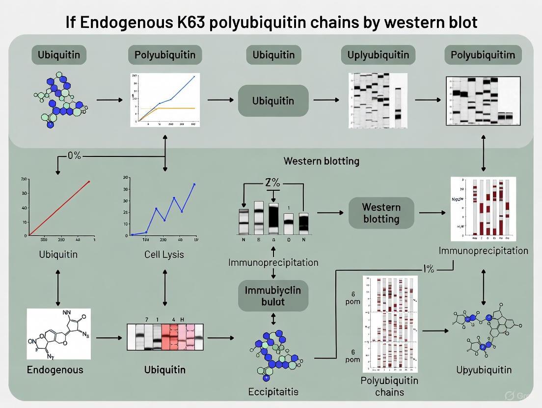
A Comprehensive Guide to Detecting Endogenous K63-Linked Polyubiquitin Chains by Western Blot
This article provides a detailed methodological framework for researchers and drug development professionals aiming to accurately detect endogenous K63-linked polyubiquitination.
A Comprehensive Guide to Detecting Endogenous K63-Linked Polyubiquitin Chains by Western Blot
Abstract
This article provides a detailed methodological framework for researchers and drug development professionals aiming to accurately detect endogenous K63-linked polyubiquitination. Covering foundational principles, step-by-step protocols, advanced troubleshooting, and validation techniques, the guide addresses the unique challenges of preserving and identifying these non-proteolytic ubiquitin signals. It emphasizes critical considerations such as the use of high-concentration deubiquitinase inhibitors, linkage-specific antibodies, and confirmatory assays to ensure data validity in studying inflammatory signaling, protein trafficking, and targeted protein degradation.
Understanding K63 Polyubiquitin: Biology, Function, and Detection Challenges
Ubiquitination is a crucial post-translational modification that regulates a vast array of cellular processes. Unlike the well-characterized K48-linked ubiquitin chains that typically target substrates for proteasomal degradation, K63-linked ubiquitin chains fulfill diverse non-proteolytic functions in eukaryotic cells [1] [2]. These functions include signal transduction, DNA damage repair, protein trafficking, and autophagy [1] [3]. The K63 ubiquitin code represents a complex signaling system where linkage-specific topology is recognized by ubiquitin-binding domains (UBDs) to direct distinct biological outcomes.
Recent research has revealed surprising complexity in the K63 code, including its role as a seed for branched ubiquitin chains that can indeed target proteins for degradation, blurring the traditional functional distinctions [4]. This application note details methodologies for the precise detection and analysis of endogenous K63 polyubiquitin chains via western blotting, providing critical technical insights for researchers investigating ubiquitin signaling pathways in health and disease.
Biological Significance of K63-Linked Ubiquitination
Key Signaling Pathways Regulated by K63 Ubiquitination
K63-linked ubiquitination serves as a fundamental regulatory mechanism across multiple immune and inflammatory signaling pathways. It acts as a scaffolding platform that facilitates the assembly and activation of signaling complexes [1]. The diagram below illustrates the major signaling pathways dependent on K63-linked ubiquitination.
The molecular machinery of K63 ubiquitination involves a specific E2 enzyme complex, Ubc13-Uev1a, which catalyzes K63-specific linkages [1]. This complex works in concert with various E3 ligases such as TRAF6, cIAP1/2, and RNF216 to create K63-linked chains on target substrates [1] [5]. These chains are then recognized by specific UBDs present in signaling proteins including NEMO (NF-κB Essential Modulator), which transduces signals to activate downstream transcription factors.
Emerging Role in Bridging Signaling and Degradation
Traditionally, K63-linked chains were considered exclusively non-proteolytic. However, recent evidence reveals a more nuanced picture where K63 ubiquitination can serve as a degradation signal under specific contexts. Research indicates that K63-linked chains can act as "seeds" for the formation of K48/K63-branched ubiquitin chains, which preferentially associate with proteasomes and direct substrates for degradation [4] [6]. This mechanism has been demonstrated for the proapoptotic regulator TXNIP, where ITCH-dependent K63 ubiquitination recruits additional ligases like UBR5 to assemble K48/K63-branched chains that trigger proteasomal degradation [4].
Table 1: Key Biological Functions of K63-Linked Ubiquitination
| Biological Process | Key Substrates/Proteins | Functional Outcome | Reference |
|---|---|---|---|
| Immune Signaling | TRAF6, RIPK1, NEMO | Activation of NF-κB and MAPK pathways | [1] |
| DNA Damage Repair | Various repair factors | Recruitment of repair complexes to DNA damage sites | [1] [2] |
| Protein Trafficking | Membrane receptors | Endosomal sorting and lysosomal targeting | [3] [6] |
| Selective Autophagy | Autophagy receptors | Cargo recognition and autophagosome formation | [3] [6] |
| Cell Death Regulation | RIPK1, RIPK3 | Regulation of apoptosis and necroptosis | [1] |
| Proteasomal Degradation* | TXNIP, various substrates | Formation of K48/K63-branched degradation signals | [4] |
*Note: The role in proteasomal degradation occurs through branched chains with K48 linkages.
Detection Methods for Endogenous K63 Polyubiquitin Chains
Detecting endogenous K63-linked ubiquitin chains presents significant technical challenges due to the presence of multiple ubiquitin chain types in cells, the dynamic nature of ubiquitination, and the susceptibility of ubiquitin chains to deubiquitinating enzymes (DUBs) during sample preparation [7]. The following table compares the primary methods used for K63 chain detection.
Table 2: Comparison of K63-Linked Ubiquitin Chain Detection Methods
| Method | Principle | Sensitivity | Specificity | Key Advantages | Key Limitations | |
|---|---|---|---|---|---|---|
| Linkage-Specific Antibodies | Immunodetection of K63 linkage-specific epitopes | High (endogenous detection) | High (when validated) | Direct, compatible with standard WB protocols | Epitope masking by denaturation; limited quantitative accuracy | [2] [7] |
| Tandem Ubiquitin Binding Entities (TUBEs) | High-affinity binding to K63 chain structure | High | ~1,000-10,000-fold preference for K63 | Preserves labile modifications; enriches endogenous chains | Requires non-denaturing conditions; specialized reagents | [8] [7] |
| Mass Spectrometry (SRM/MS) | Detection of linkage-specific signature peptides | Variable | High | Absolute quantification; comprehensive linkage profiling | Technically demanding; requires specialized equipment | [9] [6] |
| Linkage Determination Protocol | In vitro reconstitution with mutant ubiquitins | High for in vitro systems | Definitive | Provides definitive linkage assignment | Limited to in vitro applications | [10] |
Optimized Western Blot Protocol for K63 Chain Detection
The following section provides a detailed protocol for detecting endogenous K63 polyubiquitin chains by western blotting, incorporating critical optimization steps to ensure reliable results.
Sample Preparation and Preservation
Proper sample preparation is crucial for preserving endogenous K63 ubiquitin chains, which are highly dynamic and susceptible to DUB activity:
Lysis Buffer Composition: Use ice-cold lysis buffers containing:
Sample Handling:
- Process cells or tissues immediately after collection
- Maintain samples on ice throughout preparation
- Avoid multiple freeze-thaw cycles
- Pre-chill all centrifuges and equipment
Supplementation with DUB Inhibitors: In addition to NEM/IAA, include:
- 5-10 mM EDTA to chelate zinc and inhibit metalloprotease DUBs
- Protease inhibitor cocktails (without DUB-incompatible components)
- Consider specific DUB inhibitors like PR-619 for broad-spectrum coverage [7]
Electrophoresis and Transfer
Gel Electrophoresis:
- Use 4-12% Bis-Tris gradient gels for optimal separation of high molecular weight polyubiquitinated species
- Run gels at constant voltage (120-150V) with MES or MOPS buffer systems
- Avoid excessive heating during electrophoresis
Membrane Transfer:
- Transfer to PVDF membranes for superior protein binding
- Use semi-dry transfer systems for efficiency
- Confirm transfer efficiency with Ponceau S staining
Immunodetection
Blocking and Antibody Incubation:
- Block membranes with 5% non-fat dry milk in TBST for 1 hour at room temperature
- Incubate with primary antibodies diluted in blocking buffer overnight at 4°C
- Use anti-K63 linkage-specific antibodies (e.g., Cell Signaling #5621) at 1:1000 dilution [2]
Detection and Visualization:
- Use HRP-conjugated secondary antibodies with enhanced chemiluminescence
- Optimize exposure times to avoid saturation
- Include loading controls with antibodies against total ubiquitin or housekeeping proteins
The experimental workflow for proper detection and analysis of K63-linked ubiquitin chains is summarized below.
*Note: Non-denaturing conditions are specifically required when using TUBE-based detection methods.
Method Verification and Validation
To ensure specificity of K63 chain detection, employ these verification strategies:
DUB Sensitivity Assays: Treat samples with linkage-specific deubiquitinases such as:
- AMSH (K63-specific) for validation of K63 linkages
- OTUB1 (K48-specific) to demonstrate lack of cross-reactivity [6]
Competition Experiments: Pre-incubate antibodies with K63-linked diubiquitin to demonstrate competitive blocking of signal
Genetic Validation: Use RNAi-mediated knockdown of known K63-specific E2 (Ubc13) or E3 ligases (TRAF6, RNF216) to demonstrate reduction in specific signals [1] [5]
The Scientist's Toolkit: Essential Research Reagents
Table 3: Key Research Reagents for K63 Ubiquitin Research
| Reagent Category | Specific Examples | Function/Application | Supplier/Reference |
|---|---|---|---|
| K63 Linkage-Specific Antibodies | K63-linkage Specific Polyubiquitin (D7A11) Rabbit mAb #5621 | Western blot detection of endogenous K63 chains | Cell Signaling Technology [2] |
| Tandem Ubiquitin Binding Entities (TUBEs) | K63 TUBE (Biotin) UM304 | High-affinity capture and detection of K63 chains under non-denaturing conditions | LifeSensors [8] |
| Ubiquitin Mutants | Ubiquitin K63R Mutant; Ubiquitin K63 Only Mutant | Linkage determination and control experiments | Boston Biochem/R&D Systems [10] |
| Deubiquitinases (DUBs) | AMSH (K63-specific) | Verification of K63 linkage specificity | Multiple suppliers [6] |
| E2 Enzyme Complex | Ubc13-Uev1a complex | In vitro K63 chain assembly | Multiple suppliers [1] |
| DUB Inhibitors | N-Ethylmaleimide (NEM), Chloroacetamide (CAA) | Preservation of ubiquitin chains during sample preparation | Multiple suppliers [7] [6] |
Troubleshooting Common Experimental Issues
Problem: Weak or Absent K63 Signal
Potential causes and solutions:
- DUB activity during preparation: Ensure fresh NEM/IAA is added immediately to lysis buffer
- Antibody specificity issues: Validate antibody with positive and negative controls
- Over-denaturation of samples: For TUBE-based detection, avoid boiling samples and use non-denaturing conditions [8] [7]
Problem: High Background or Non-Specific Signals
Potential causes and solutions:
- Insufficient blocking: Optimize blocking conditions with different blockers (BSA, milk)
- Antibody cross-reactivity: Include linkage specificity controls with purified ubiquitin chains of different linkages
- Incomplete transfer: Verify transfer efficiency with Ponceau S staining
Problem: Inconsistent Results Between Experiments
Potential causes and solutions:
- Variability in DUB inhibition: Standardize inhibitor concentrations and preparation methods
- Sample degradation: Process all samples simultaneously and minimize freeze-thaw cycles
- Differences in cell state: Account for cell density, passage number, and treatment conditions
The detection of endogenous K63-linked polyubiquitin chains requires careful methodological consideration to preserve the labile nature of this modification while ensuring linkage specificity. While traditional views positioned K63 ubiquitination strictly as a non-proteolytic signal, emerging evidence reveals a more complex landscape where K63 chains can serve as seeds for branched chains that target substrates for degradation [4] [6]. The optimized protocols described here provide a framework for reliable detection of endogenous K63 chains, enabling researchers to better understand the nuanced roles of the K63 ubiquitin code in cellular signaling and disease pathogenesis, including cancer, neurodegenerative disorders, and immune dysregulation [1] [5] [3]. As the field advances, continued refinement of these methodologies will be essential for deciphering the complex language of the ubiquitin code.
Why Detect Endogenous Protein? Overcoming Challenges of Overexpression Artifacts
The study of post-translational modifications, particularly K63-linked polyubiquitination, presents significant challenges that are profoundly amplified when relying on overexpression systems. K63 polyubiquitin chains serve crucial non-proteolytic functions in cells, including regulation of signal transduction, protein trafficking, DNA repair, and the oxidative stress response [11] [12]. During oxidative stress, K63 ubiquitination rapidly accumulates in a highly regulated manner, impacting translation and cellular survival mechanisms [11]. However, overexpression of ubiquitin or target proteins can artificially inflate conjugation levels, disrupt native stoichiometry of ubiquitination enzymes, and promote non-physiological interactions that compromise data validity [13] [14].
Detecting endogenous proteins provides an authentic representation of cellular signaling events under physiological conditions, maintaining proper enzyme-substrate ratios and subcellular localization. This is particularly crucial for K63 ubiquitination studies, as this linkage type is specifically triggered by oxidative stress and inflammatory signaling in a tightly controlled spatiotemporal manner [11] [15] [14]. Advances in detection methodologies now enable researchers to capture these endogenous ubiquitination events with increasing sensitivity and specificity, bridging the gap between observational biology and mechanistic understanding.
K63 Polyubiquitination: Biological Significance and Detection Challenges
Functional Roles of K63 Ubiquitin Chains
K63-linked polyubiquitination differs structurally and functionally from the more well-known K48-linked chains that target proteins for proteasomal degradation. Unlike K48 chains, K63 linkages primarily serve regulatory functions in numerous cellular pathways, as detailed in Table 1 [12].
Table 1: Key Functional Roles of K63-Linked Polyubiquitination
| Cellular Function | Molecular Mechanism | Biological Outcome |
|---|---|---|
| Oxidative Stress Response | Inhibits Ubp2 deubiquitinase, leading to K63 chain accumulation [11] | Enhances cellular viability under peroxide-induced stress |
| NF-κB Inflammatory Signaling | Forms K63 chains on RIPK2, NEMO, and other signaling components [14] | Activates pro-inflammatory gene expression programs |
| Protein Trafficking & Endocytosis | Modifies cell surface receptors and sorting complexes [12] | Regulates membrane receptor internalization and degradation |
| DNA Damage Repair | Facilitates recruitment of repair complexes to damage sites [11] | Maintains genomic integrity |
Limitations of Overexpression Approaches
Overexpression systems, while initially valuable for discovering ubiquitination pathways, introduce several critical artifacts:
- Stoichiometric Imbalance: Exogenous expression of ubiquitin or target proteins disrupts the natural balance of E1, E2, and E3 enzymes, potentially overwhelming quality control mechanisms and creating non-physiological ubiquitination patterns [13].
- Non-specific Chain Formation: Overexpressed ubiquitin mutants (e.g., lysine-to-arginine mutants) may not accurately recapitulate wild-type ubiquitin behavior and can produce misleading results regarding chain topology [14].
- Subcellular Mislocalization: Artificially high protein concentrations often lead to improper subcellular localization, disrupting compartment-specific signaling events that are crucial for K63 ubiquitin function [15].
Table 2: Quantitative Comparison of Endogenous vs. Overexpression Detection Methods
| Parameter | Endogenous Detection | Overexpression System |
|---|---|---|
| Stoichiometry | Maintains natural enzyme-substrate ratios | Disrupts native stoichiometry |
| Subcellular Localization | Preserves physiological compartmentalization | Often causes mislocalization |
| K63 Chain Specificity | High specificity with proper controls | Prone to non-specific chain formation |
| Physiological Relevance | High | Variable to low |
| Technical Difficulty | High (requires enrichment) | Lower (easier detection) |
Advanced Methodologies for Endogenous K63 Ubiquitin Detection
Affinity Enrichment Strategies
Several powerful affinity enrichment strategies have been developed to isolate endogenous ubiquitinated proteins with linkage specificity:
Linkage-Specific Antibodies: Monoclonal antibodies specifically recognizing K63-linked ubiquitin chains enable immunoprecipitation of endogenous K63-ubiquitinated proteins without genetic manipulation [13]. These antibodies can distinguish K63 linkages from other chain types with high specificity, though cross-reactivity must be carefully evaluated.
Tandem Ubiquitin Binding Entities (TUBEs): TUBEs are engineered fusion proteins containing multiple ubiquitin-binding domains that exhibit high affinity for polyubiquitin chains. K63-specific TUBEs can selectively enrich endogenous K63-ubiquitinated proteins from native cell lysates, protecting them from deubiquitinases during extraction [14]. This approach has been successfully applied to study endogenous RIPK2 K63 ubiquitination in inflammatory signaling.
Ubiquitin Binding Domain (UBD)-Based Probes: Specific UBDs from various cellular proteins that recognize K63 linkages with high selectivity can be harnessed as affinity capture tools, though their generally lower affinity compared to TUBEs may limit effectiveness for low-abundance targets [13].
Mass Spectrometry-Based Approaches
Advanced proteomic methods allow system-wide mapping of endogenous ubiquitination sites:
DiGly Antibody Enrichment: Antibodies recognizing the diglycine remnant left on trypsinized ubiquitination sites enable proteome-wide identification of ubiquitination sites without genetic tags, providing an unbiased view of endogenous ubiquitination events [13].
Cross-Linking Mass Spectrometry: Emerging methodologies incorporating chemical cross-linking with mass spectrometry help preserve labile endogenous ubiquitin conjugates during sample preparation, enhancing detection of transient modification events [13].
Detailed Experimental Protocol: Endogenous K63 Ubiquitin Detection by Western Blot
Cell Lysis and Protein Extraction under Denaturing Conditions
Objective: To efficiently extract proteins while preserving endogenous K63 ubiquitination patterns by inactivating deubiquitinases.
Reagents Needed:
- Urea Lysis Buffer (6M urea, 50 mM Tris-HCl pH 7.5, 1% Triton X-100, 150 mM NaCl)
- Protease Inhibitor Cocktail (without EDTA)
- Deubiquitinase Inhibitors (N-ethylmaleimide or PR-619)
- Benzonase Nuclease (optional, for reducing viscosity)
- BCA Protein Assay Kit
Procedure:
- Prepare fresh lysis buffer supplemented with protease and deubiquitinase inhibitors immediately before use.
- Aspirate culture media from cells and wash once with ice-cold PBS.
- Add appropriate volume of lysis buffer directly to cells (typically 100-200 µL per 10⁶ cells).
- Scrape cells and transfer lysate to pre-cooled microcentrifuge tubes.
- Vortex vigorously for 10 seconds, then incubate on ice for 15 minutes with occasional vortexing.
- Clarify lysates by centrifugation at 16,000 × g for 15 minutes at 4°C.
- Transfer supernatant to new tubes and determine protein concentration using BCA assay.
- Adjust samples to equal protein concentrations with lysis buffer.
Critical Step: Maintain samples at 4°C or lower throughout the procedure to minimize deubiquitination. Avoid using SDS at this stage as it may interfere with subsequent immunoprecipitation steps.
K63-Ubiquitinated Protein Enrichment
Objective: To specifically isolate K63-ubiquitinated proteins from complex cell lysates.
Reagents Needed:
- K63-linkage Specific TUBE Magnetic Beads or K63 Ubiquitin Antibody-Conjugated Beads
- Wash Buffer 1 (50 mM Tris-HCl pH 7.5, 150 mM NaCl, 0.5% NP-40, 1 mM DTT)
- Wash Buffer 2 (50 mM Tris-HCl pH 7.5, 500 mM NaCl, 0.5% NP-40)
- Elution Buffer (1X Laemmli buffer with 100 mM DTT)
Procedure:
- Pre-clear 500-1000 µg of protein lysate with control beads for 30 minutes at 4°C.
- Incubate pre-cleared lysate with K63-TUBE magnetic beads or K63 ubiquitin antibody-conjugated beads for 2-4 hours at 4°C with gentle rotation.
- Collect beads using magnetic separation and wash sequentially:
- Three times with Wash Buffer 1 (5 minutes each wash)
- Two times with Wash Buffer 2 (5 minutes each wash)
- One time with 50 mM Tris-HCl pH 7.5 (quick rinse)
- Completely remove final wash buffer and elute bound proteins by adding 30-50 µL of Elution Buffer.
- Heat eluates at 95°C for 10 minutes, then briefly centrifuge to collect condensate.
Western Blot Optimization for K63 Ubiquitin Detection
Objective: To achieve high-sensitivity detection of endogenous K63 ubiquitin conjugates with minimal background.
Reagents Needed:
- Pre-cast SDS-PAGE gels (4-12% Bis-Tris gradient gels)
- PVDF membrane (0.2 µm pore size)
- Transfer Buffer: Towbin buffer (25 mM Tris, 192 mM glycine, 20% methanol)
- Blocking Buffer: 5% BSA in TBST (for phospho-specific or ubiquitin detection)
- Primary Antibodies: Anti-K63 ubiquitin linkage-specific antibody, loading control antibodies
- Secondary Antibodies: HRP-conjugated antibodies appropriate to host species
Procedure:
- Separate immunoprecipitated proteins by SDS-PAGE using MES or MOPS running buffer for optimal high molecular weight separation.
- Transfer to activated PVDF membrane using wet transfer system at 100V for 90 minutes at 4°C.
- Block membrane with 5% BSA in TBST for 1 hour at room temperature with gentle agitation.
- Incubate with primary K63 linkage-specific antibody diluted in blocking buffer overnight at 4°C.
- Wash membrane 3 times for 10 minutes each with TBST.
- Incubate with appropriate HRP-conjugated secondary antibody diluted in blocking buffer for 1 hour at room temperature.
- Wash membrane 3 times for 10 minutes each with TBST.
- Develop using enhanced chemiluminescence substrate with appropriate exposure time.
Troubleshooting Tips:
- High background: Increase wash stringency, try different blocking agents (casein or commercial blocking buffers), or titrate antibody concentrations [16] [17].
- Weak signal: Extend exposure time, try more sensitive ECL substrates, or increase protein input.
- Non-specific bands: Include ubiquitin mutant controls, validate with deubiquitinase treatment, or use secondary antibody controls.
The Scientist's Toolkit: Essential Research Reagents
Table 3: Key Research Reagents for Endogenous K63 Ubiquitin Detection
| Reagent Category | Specific Examples | Function & Application Notes |
|---|---|---|
| K63 Enrichment Tools | K63-linkage Specific TUBEs (e.g., LifeSensors) | High-affinity capture of endogenous K63-ubiquitinated proteins; protects from DUBs [14] |
| K63 Linkage-Specific Antibodies (e.g., Millipore 05-1308) | Immunoprecipitation and western blot detection of K63 chains; validate for specific applications | |
| Cell Lysis Reagents | Deubiquitinase Inhibitors (N-ethylmaleimide, PR-619) | Preserve endogenous ubiquitination during extraction |
| Benzonase Nuclease | Reduce sample viscosity by digesting nucleic acids | |
| Detection Antibodies | Anti-K63 Ubiquitin (Linkage-Specific) | Primary detection antibody for western blot; requires proper validation |
| HRP-conjugated Secondary Antibodies | Signal generation for chemiluminescent detection | |
| Positive Controls | L18-MDP (for RIPK2 ubiquitination) | Induces endogenous K63 ubiquitination of RIPK2 in immune signaling studies [14] |
| Sodium Arsenite/H₂O₂ | Induces oxidative stress-dependent K63 ubiquitination [11] [15] |
K63 Ubiquitin Signaling Pathways and Experimental Workflow
The following diagrams illustrate key K63 ubiquitin signaling pathways and the experimental workflow for endogenous detection:
K63 Ubiquitin Signaling in Oxidative Stress and Inflammation
Figure 1: K63 Ubiquitin Signaling in Cellular Stress Response Pathways
Experimental Workflow for Endogenous K63 Ubiquitin Detection
Figure 2: Experimental Workflow for Endogenous K63 Ubiquitin Detection
Detection of endogenous K63 polyubiquitination represents a critical advancement in ubiquitin research, moving beyond the artifacts and limitations of overexpression systems. The methodologies outlined in this application note—including linkage-specific TUBEs, advanced immunocapture techniques, and optimized western blot protocols—enable researchers to capture authentic K63 ubiquitination events under physiological conditions. These approaches have revealed crucial insights into oxidative stress response mechanisms, inflammatory signaling pathways, and protein quality control systems that were previously obscured by overexpression artifacts. As drug discovery increasingly targets the ubiquitin-proteasome system with PROTACs and other modality drugs, accurate assessment of endogenous ubiquitination events becomes paramount for validating target engagement and mechanism of action. The protocols and reagents described herein provide a robust foundation for investigating endogenous K63 ubiquitination, offering researchers the tools necessary to advance our understanding of this complex regulatory system in health and disease.
The study of K63-linked polyubiquitin (K63-Ub) chains is pivotal for understanding crucial cellular processes such as DNA damage repair, immune signaling, and chaperone-mediated autophagy [18] [19]. However, their accurate detection via western blot is fraught with two major technical hurdles: the inherent lability of these chains due to the activity of cellular deubiquitinases (DUBs), and the persistent challenge of antibody specificity. This application note provides a detailed framework of optimized protocols and critical controls designed to help researchers overcome these obstacles, enabling the reliable detection of endogenous K63-Ub chains.
Understanding the Technical Hurdles
The Structural Lability of K63-Ub Chains
K63-Ub chains exhibit a relaxed and extended conformational topology, which distinguishes them from the compact structures of K48-linked chains [18]. While this open structure is functionally important, facilitating direct DNA binding in repair processes, it also renders the chains more susceptible to disassembly by DUBs. This structural accessibility, combined with their transient signaling nature, makes K63-Ub chains particularly labile during sample preparation. Without proper stabilization, the signal of endogenous chains can be rapidly lost, leading to false-negative results.
The Critical Issue of Antibody Cross-Reactivity
A primary concern in the field is ensuring that antibodies used for detection are truly specific for the K63 linkage. Many commercially available linkage-specific antibodies have been reported to exhibit significant cross-reactivity with other ubiquitin chain types, including K11-, K27-, and K33-linked chains, or with heterotypic branched chains containing K63 linkages [20] [21]. This lack of absolute specificity can produce false-positive signals, fundamentally compromising data interpretation. Therefore, rigorous validation of all immunological reagents is not merely recommended but essential for credible research.
Table 1: Key Characteristics of K63-Linked Ubiquitin Chains
| Feature | Description | Impact on Detection |
|---|---|---|
| Structural Topology | Relaxed, extended, and labile conformation [18] | High susceptibility to DUBs; requires immediate stabilization. |
| Cellular Functions | DNA damage repair, CMA, immune signaling, protein trafficking [18] [20] [19] | Signals can be transient, necessitating precise experimental timing. |
| Commonly Reported Antibody Cross-Reactivity | K11, K27, K33 linkages, and heterotypic/branched chains [20] [21] | Mandates rigorous antibody validation with defined ubiquitin standards. |
Optimized Protocols for Reliable Detection
Sample Preparation and Preservation of Lability
The single most critical step for successful detection is the immediate inhibition of DUB activity at the moment of cell lysis. The following protocol is adapted from best practices in the field [20] [7].
Protocol 3.1: Sample Lysis with DUB Inhibition
Preparation of Lysis Buffer: Prepare a standard RIPA or NP-40 lysis buffer. Immediately before use, supplement it with a potent DUB inhibitor. Two common options are:
- N-Ethylmaleimide (NEM): 10-25 mM final concentration.
- Chloroacetamide (CAA): 20-50 mM final concentration.
- Note: NEM is a more potent and irreversible inhibitor but has a higher risk of off-target alkylation. CAA is more cysteine-specific but may allow partial chain disassembly [20].
Cell Lysis: Aspirate culture media and immediately add the ice-cold, supplemented lysis buffer directly to the cell culture dish. Scrape the cells quickly and transfer the lysate to a pre-cooled microcentrifuge tube.
Sample Processing: Sonicate the lysate briefly to shear DNA and reduce viscosity. Incubate on ice for 15-30 minutes with occasional vortexing.
Clarification: Centrifuge the lysate at >14,000 x g for 15 minutes at 4°C to pellet insoluble debris. Transfer the clarified supernatant to a new tube.
Protein Quantification and Denaturation: Perform a protein assay (e.g., BCA). Dilute the lysate in 2X or 4X Laemmli sample buffer and boil for 5-10 minutes to fully denature proteins and inactivate any residual enzyme activity.
Experimental Design and Antibody Validation
To confidently assign a signal to a K63-Ub chain, a multi-pronged validation strategy is required.
Protocol 3.2: Validating Antibody Specificity by Western Blot
This protocol uses defined ubiquitin standards to test antibody specificity [22] [10].
Acquire Defined Ubiquitin Standards: Obtain recombinant purified proteins, including:
- K63-linked Ub2-Ub7 chains
- Other homotypic chains (K48, K11, K33, etc.)
- Heterotypic/branched chains (if available)
- Mono-ubiquitin
Western Blot Analysis: Load 50-100 ng of each ubiquitin standard on an SDS-PAGE gel. Transfer to a PVDF or nitrocellulose membrane.
Probining: Probe the membrane with your anti-K63 linkage-specific antibody (e.g., ab179434, EPR8590-448 clone) [22].
Interpretation: A valid K63-specific antibody should produce a strong signal for the K63-Ub ladder and minimal to no signal for ladders of other linkages or for mono-ubiquitin. Any signal for other chain types indicates cross-reactivity and necessitates caution in interpreting results from complex lysates.
Protocol 3.3: The UbiCRest Assay for Linkage Verification
The UbiCRest assay uses linkage-specific DUBs to digest ubiquitin chains in your samples, providing orthogonal validation [21].
Immunoprecipitation: Immunoprecipitate ubiquitylated proteins or your protein of interest from the cell lysate under denaturing conditions to remove associated proteins.
Elution: Divide the IP material into several aliquots and elute using mild denaturation.
DUB Digestion: Incubate each aliquot with a different linkage-specific DUB in its appropriate reaction buffer for 2 hours at 37°C. Key examples include:
- AMSH/ OTUD4: K63-specific [20].
- OTUB1: K48-specific.
- VCIP: Pan-specific DUB control.
Analysis by Western Blot: Terminate the reactions with SDS sample buffer, boil, and analyze by western blot. A genuine K63-Ub signal should be significantly diminished or eliminated in the aliquot treated with the K63-specific DUB (e.g., AMSH) but remain largely intact in aliquots treated with DUBs specific for other linkages.
Diagram 1: UbiCRest Assay Workflow for K63 Linkage Verification
The Scientist's Toolkit: Essential Research Reagents
Success in detecting endogenous K63-Ub chains relies on a suite of well-characterized reagents. The following table details key tools for this application.
Table 2: Research Reagent Solutions for K63-Ub Research
| Reagent Category | Specific Example | Function and Application Note |
|---|---|---|
| Linkage-Specific Antibodies | Anti-Ubiquitin (K63-linkage specific) [22] | Clone EPR8590-448; used for western blot (1/1000 dilution), IHC-P, and Flow Cytometry. Must be validated with ubiquitin standards. |
| DUB Inhibitors | N-Ethylmaleimide (NEM), Chloroacetamide (CAA) [20] [7] | Preserve labile ubiquitin chains during lysis. NEM is more potent; CAA is more cysteine-specific. |
| Ubiquitin Traps | ChromoTek Ubiquitin-Trap [23] | VHH-based resin for pulldown of ubiquitin and ubiquitinylated proteins from cell extracts. Not linkage-specific. |
| Defined Ubiquitin Standards | K63-Ub2-7, K48-Ub2-7, other linkage-specific chains [22] [10] | Critical controls for validating antibody specificity in western blot assays. |
| Linkage-Specific DUBs | AMSH (K63-specific), OTUB1 (K48-specific) [20] [21] | Used in UbiCRest assays to enzymatically verify ubiquitin chain linkage in samples. |
| Ubiquitin Mutants | Ubiquitin K63R, K48R; "K-only" mutants [10] | Used in in vitro ubiquitination assays to determine chain linkage requirements for E3 ligases. |
Navigating the technical challenges of K63-Ub chain lability and antibody specificity demands a rigorous and multi-faceted approach. By implementing the detailed protocols outlined here—prioritizing rapid DUB inhibition, systematically validating antibodies with defined standards, and employing orthogonal methods like UbiCRest—researchers can significantly enhance the reliability and interpretability of their data. Mastering these techniques is fundamental to advancing our understanding of the critical biological pathways governed by K63-linked ubiquitination.
A Step-by-Step Protocol for Reliable K63 Ubiquitin Detection
The detection of endogenous K63-linked polyubiquitin (K63-Ub) chains by western blot is a critical methodology for researchers studying cellular stress responses, DNA damage repair, and signal transduction pathways. However, the labile nature of this specific ubiquitin linkage presents significant technical challenges. K63-Ub chains are inherently susceptible to rapid disassembly by deubiquitylating enzymes (DUBs) and are not typically targeted for proteasomal degradation, making their preservation in cell lysates particularly difficult. The foundational step for successful detection lies in the immediate and complete inactivation of these enzymatic activities upon cell lysis. This application note details optimized protocols for the preparation of lysis buffers containing high-dose N-ethylmaleimide (NEM) and proteasome inhibitors, framed within the context of a broader thesis on reliable K63-Ub chain detection. These methods are designed to provide researchers, scientists, and drug development professionals with robust tools to accurately capture the endogenous state of K63 ubiquitination, a modification increasingly recognized as a key regulator in oxidative stress and other pathophysiological conditions [15].
Essential Reagents and Rationale
The integrity of ubiquitin chains during sample preparation hinges on the use of specific inhibitors that halt enzymatic degradation. The table below summarizes the critical components of the lysis buffer and their functions.
Table 1: Key Research Reagent Solutions for Ubiquitin Preservation
| Reagent | Function | Recommended Working Concentration | Rationale |
|---|---|---|---|
| N-Ethylmaleimide (NEM) | DUB Inhibitor (Cysteine protease family) | 50-100 mM [24] | Alkylates active site cysteine residues of DUBs. Standard 5-10 mM concentrations are often insufficient; high doses are critical for preserving K63 and M1 chains. |
| Iodoacetamide (IAA) | Alternative DUB Inhibitor | 5-100 mM (context-dependent) [24] | Alternative alkylating agent. Less stable than NEM and can interfere with mass spectrometry due to a 114 Da adduct identical to the Gly-Gly ubiquitin remnant. |
| Mg132 (or other proteasome inhibitors e.g., Bortezomib, Epoxomicin) | Proteasome Inhibitor | Varies by inhibitor (e.g., 10-20 µM for MG132) [24] [25] | Blocks degradation of proteins modified with "degradative" ubiquitin chains (e.g., K48, K11), preventing loss of substrates and mitigating stress responses from prolonged inhibition. |
| EDTA/EGTA | DUB Inhibitor (Metalloprotease family) | 1-10 mM [24] | Chelates heavy metal ions, inactivating metal-dependent DUBs such as the JAMM/MPN+ family. |
The choice between NEM and IAA is critical. While both are effective, NEM is strongly recommended for experiments focusing on K63-Ub chains, as it demonstrates superior performance in preserving these linkages [24]. Furthermore, for any subsequent proteomic analysis, NEM is the preferred alkylating agent because it does not create an adduct that confounds the identification of ubiquitylation sites by mass spectrometry [24].
Optimized Protocols for Sample Preparation
Formulation of a High-Efficacy Denaturing Lysis Buffer
A lysis buffer that rapidly denatures proteins is essential to "freeze" the ubiquitination state at the moment of cell disruption. The following formulation is recommended for optimal preservation of K63-Ub chains.
Table 2: Recommended High-Dose NEM Lysis Buffer Composition
| Component | Final Concentration | Purpose |
|---|---|---|
| SDS | 1% | Rapid denaturation of proteins and enzymes |
| Tris-HCl | 50 mM, pH 7.5 | Buffering capacity |
| Sodium Chloride (NaCl) | 150 mM | Osmotic balance |
| N-Ethylmaleimide (NEM) | 50-100 mM [24] | Potent inhibition of cysteine-based DUBs |
| EDTA | 5-10 mM | Inhibition of metalloprotease DUBs |
| Proteasome Inhibitor Cocktail | As per manufacturer | e.g., MG132, Bortezomib, or Epoxomicin |
Preparation Notes:
- The buffer should be prepared fresh for maximum efficacy, particularly due to the sensitivity of some inhibitors.
- A 1M stock of NEM can be prepared in ethanol or water and added to the buffer immediately before use.
- Cell lysates should be heated at 95°C for 5-10 minutes immediately after resuspension in this buffer to ensure complete and irreversible denaturation [24].
Step-by-Step Cell Lysis and Sample Preparation Workflow
The following workflow is designed to minimize post-lysis degradation and ensure reliable results.
- Pre-chill Equipment and Pre-warm Denaturing Buffer. Have ice-cold PBS and a centrifuge ready. Pre-heat the denaturing lysis buffer (from Section 3.1) to 95°C.
- Wash and Harvest Cells. Rapidly wash cultured cells with ice-cold PBS. Scrape or lyse cells directly on the culture plate.
- Immediate Denaturation. For a 60 mm dish of cells, immediately add 100-200 µL of pre-heated 95°C denaturing lysis buffer to the cell pellet or monolayer. Vortex vigorously for 10-15 seconds to ensure complete and instantaneous lysis.
- Boil Samples. Transfer the lysate to a heat-resistant microcentrifuge tube and incubate at 95°C for 5-10 minutes.
- Shear DNA and Clarify. Cool samples to room temperature. Briefly sonicate (10-15 seconds) or pass the lysate through a small-gauge needle (e.g., 25G) several times to reduce viscosity from genomic DNA. Centrifuge at >16,000 x g for 10 minutes to pellet insoluble debris.
- Store or Proceed. Transfer the clarified supernatant to a new tube. Samples can be stored at -80°C or prepared for SDS-PAGE by adding sample buffer with reducing agent (if compatible with subsequent detection).
Essential Experimental Controls for Western Blot
Incorporating the correct controls is non-negotiable for validating the specificity of your K63-Ub chain detection [26].
- Positive Control: Use a lysate from cells treated with an agent known to induce K63 ubiquitination, such as sodium arsenite to induce oxidative stress [15], or TNFα. This verifies that your experimental and detection system is functioning correctly.
- Negative Control: The most robust negative control is a lysate from a validated knockout cell line (e.g., a key E3 ligase or adaptor protein involved in your pathway of interest). If this is unavailable, a non-stimulated cell lysate can serve as a baseline control.
- Knockdown/Knockout Validation: For genetic knockdown or knockout experiments, always run a wild-type control lysate alongside the modified cell lysate to confirm the loss of the ubiquitin signal and the specificity of your antibody.
- Loading Control: Use a constitutively expressed housekeeping protein like GAPDH or Actin to ensure equal protein loading across all lanes. Ensure the molecular weight of your loading control does not overlap with your target signal [26].
- No Primary Antibody Control: This control, incubated with secondary antibody only, identifies any non-specific binding from your secondary antibody [26].
Quantitative Data and Inhibitor Titration
The preservation of ubiquitin chains is highly dependent on inhibitor concentration. Empirical data demonstrates that standard doses of DUB inhibitors are often inadequate.
Table 3: Impact of DUB Inhibitor Concentration on Ubiquitin Chain Preservation
| DUB Inhibitor | Standard Concentration | High Concentration | Observed Effect with High Concentration |
|---|---|---|---|
| NEM | 5-10 mM | 50-100 mM [24] | Markedly improved preservation of K63-Ub and M1-Ub chains, as visualized by reduced degradation smearing and stronger high-molecular-weight signals on western blots. |
| IAA | 5-10 mM | 50-100 mM | Improved preservation of some ubiquitin linkages, though generally less effective than NEM for K63 chains. |
Visualizing the Workflow and Signaling Context
The following diagrams outline the core experimental workflow and the biological context of K63 ubiquitin signaling in oxidative stress.
Diagram 1: Sample Preparation Workflow for K63-Ub Detection
Diagram 2: K63-Ub Signaling in Oxidative Stress
The reliable detection of endogenous K63 polyubiquitin chains by western blot is fundamentally dependent on sample preparation. The use of a denaturing lysis buffer supplemented with high-dose NEM (50-100 mM) and potent proteasome inhibitors is critical to inactivate DUBs and preserve the native ubiquitination state. The protocols and controls detailed in this application note provide a robust framework for researchers to accurately study the dynamics of K63 ubiquitination, a vital post-translational modification in cellular stress and signaling pathways. By adhering to these optimized methods, scientists can minimize artifacts and generate high-quality, reproducible data for both basic research and drug discovery applications.
The detection of specific ubiquitin signals, such as K63-linked polyubiquitin chains, is crucial for understanding critical cellular processes including inflammation, immune responses, and the cellular response to mitochondrial damage [27] [28]. Western blotting remains the most commonly used technique for studying these ubiquitylation events due to its high specificity, sensitivity, and relatively low cost [24]. However, the successful detection of endogenous K63 chains, which are often present at low levels and can be masked by other ubiquitin linkages, requires meticulous optimization of the electrophoretic separation conditions. This application note provides detailed protocols for optimizing gel percentage and buffer systems to achieve high-resolution separation of K63-linked polyubiquitin chains, framed within the context of detecting endogenous ubiquitin signals for research and drug development applications.
The Critical Role of Electrophoresis in Ubiquitin Chain Resolution
Ubiquitination is a reversible post-translational modification where a small 8.6 kDa protein, ubiquitin, is covalently attached to substrate proteins [12] [28]. A protein can be modified by the addition of 20 or more ubiquitin molecules, adding over 200 kDa to its molecular mass and resulting in a characteristic smear on western blots that typically stretches upward toward the top of the gel [24]. The linkage type of the polyubiquitin chain, determined by which of the seven lysine residues or N-terminal methionine in ubiquitin is used to form chains, dictates the functional outcome for the modified protein [12] [10]. K63-linked chains, in particular, are known to play roles in non-proteasomal pathways such as immune signaling, DNA damage repair, and mitochondrial quality control [27].
The resolution of different ubiquitinated species is technically challenging due to the size heterogeneity of the conjugates and the structural similarities between different linkage types. Without proper electrophoretic separation, researchers risk misinterpreting ubiquitination patterns or failing to detect specific endogenous chains altogether, particularly the often less abundant K63-linked chains.
Optimizing Electrophoretic Conditions
Gel Percentage and Buffer Selection
The choice of gel percentage and running buffer significantly impacts the resolution of ubiquitin chains. The table below summarizes the optimal conditions for resolving different ubiquitinated species:
Table 1: Optimized Electrophoretic Conditions for Ubiquitin Separation
| Separation Goal | Gel Percentage | Running Buffer | Key Advantages |
|---|---|---|---|
| Broad Range Separation | 8% acrylamide | Tris-Glycine (TG) | Good separation of chains up to 20 ubiquitins [24] |
| Small Chains & Mono-ubiquitination | 12% acrylamide | Tris-Glycine (TG) | Enhanced resolution of smaller ubiquitin conjugates [24] [29] |
| Chains >8 Ubiquitins | Pre-cast gradient | MOPS | Superior resolution of longer polyubiquitin chains [24] [29] |
| Chains of 2-5 Ubiquitins | Pre-cast gradient | MES | Improved resolution of small ubiquitin oligomers [24] |
| Proteins 40-400 kDa | Pre-cast gradient | Tris-Acetate (TA) | Optimal for higher molecular weight ubiquitinated proteins [24] |
Sample Preparation for Preserving K63-Linked Chains
The preservation of ubiquitination states, particularly the more labile K63-linked chains, begins with proper sample preparation before electrophoresis.
Table 2: Essential Inhibitors for Sample Preparation
| Inhibitor | Working Concentration | Purpose | Special Considerations |
|---|---|---|---|
| N-Ethylmaleimide (NEM) or Iodoacetamide (IAA) | 10-100 mM (NEM) | Deubiquitinase (DUB) inhibition; alkylates active site cysteine residues of DUBs [24] [29] | K63 chains are particularly sensitive; may require 10x higher concentrations [29]. NEM is preferred for mass spectrometry workflows [24]. |
| EDTA or EGTA | 5-10 mM | Chelates metal ions; inhibits metalloproteinase family DUBs [24] [29] | Essential component of a complete DUB inhibition cocktail. |
| MG132 (Proteasome Inhibitor) | 5-25 µM (cell treatment) | Prevents degradation of ubiquitinated proteins; stabilizes K48/K11-linked and other proteasomal-targeted chains [24] [28] | Treatment longer than 12-24 hours can induce cytotoxic stress responses [24] [29]. |
Protocol: Sample Lysis for K63 Ubiquitin Chain Preservation
- Prepare fresh lysis buffer supplemented with 50-100 mM NEM and 10 mM EDTA.
- For cell cultures, treat with 10 µM MG132 for 4-6 hours prior to lysis to stabilize ubiquitinated proteins without inducing significant stress responses.
- Lyse cells directly in pre-heated SDS-sample buffer and immediately boil for 5-10 minutes to fully denature proteins and inactivate DUBs [24].
- Briefly sonicate samples to shear DNA and reduce viscosity for improved gel resolution.
Step-by-Step Electrophoresis Protocol
Gel Casting and System Setup
Materials Required:
- Appropriate acrylamide percentage gel (see Table 1)
- Selected running buffer (MOPS, MES, or Tris-Glycine)
- Pre-stained protein molecular weight marker
- Electrophoresis cell and power supply
Protocol:
- Gel Selection: Based on your target ubiquitin species, select the appropriate gel percentage and buffer system from Table 1. For initial experiments aiming to detect endogenous K63 chains, an 8% gel with Tris-Glycine buffer is recommended as a starting point.
- Gel Preparation: Cast gels according to standard protocols, ensuring complete polymerization. Pre-cast commercial gels often provide more consistent results.
- Buffer Preparation: Prepare fresh running buffer appropriate for your chosen system. For MOPS and MES buffers, use the formulations provided by the gel manufacturer.
- Sample Loading: Load 20-50 µg of protein per lane for endogenous ubiquitin detection. Include appropriate controls such as a non-ubiquitinated protein control and a positive control if available.
- Electrophoresis Conditions: Run gels at constant voltage (100-150V) until the dye front reaches the bottom of the gel. Lower voltages may improve resolution for high molecular weight species.
Transfer Optimization for Ubiquitinated Proteins
The transfer of high molecular weight ubiquitin conjugates to membranes requires special consideration:
Protocol: Optimized Western Blot Transfer
- Membrane Selection: Use PVDF membranes with 0.2 µm pore size for optimal signal strength with ubiquitin conjugates [29].
- Transfer Conditions: For high molecular weight ubiquitin conjugates, use a semi-dry or wet transfer system at 30V for 2.5 hours [29]. Faster transfers can cause ubiquitin chains to unfold, potentially interfering with linkage-specific antibody recognition.
- Transfer Efficiency Verification: After transfer, stain the gel with Coomassie blue to confirm complete transfer of high molecular weight species.
Downstream Detection and Verification
Immunoblotting for K63-Linked Chains
After optimal separation and transfer, specific detection of K63-linked chains requires appropriate antibody selection and detection methods:
Protocol: K63 Chain Immunodetection
- Blocking: Block membranes with 5% non-fat milk or BSA in TBST for 1 hour at room temperature.
- Primary Antibody Incubation: Incubate with K63-linkage specific antibody (e.g., anti-K63-Ub) according to manufacturer's recommendations. Typical dilutions range from 1:1000 to 1:5000 in blocking buffer overnight at 4°C.
- Secondary Antibody Incubation: Incubate with appropriate HRP-conjugated secondary antibody for 1 hour at room temperature.
- Detection: Use enhanced chemiluminescence (ECL) reagents with extended exposure times to detect potentially weak endogenous signals.
Verification of K63 Linkage
Given the challenges with antibody specificity, verification of K63 linkage is recommended using complementary approaches:
Protocol: Linkage Verification Using Ubiquitin Mutants Materials: Ubiquitin lysine to arginine (K-to-R) mutants and ubiquitin "K-only" mutants (where only one lysine remains) [10].
- Set up two parallel experiments: one with wild-type ubiquitin and one with ubiquitin K63R mutant.
- Transfer proteins to membrane and probe with K63-linkage specific antibody.
- Interpretation: Genuine K63-linked chains will show significantly reduced signal with the K63R mutant, while non-specific signals will remain similar between conditions [10].
Diagram 1: Complete workflow for detecting endogenous K63-linked polyubiquitin chains, highlighting critical optimization steps from sample preparation to result verification.
Research Reagent Solutions
Table 3: Essential Reagents for K63 Ubiquitin Research
| Reagent Category | Specific Examples | Function/Application |
|---|---|---|
| DUB Inhibitors | N-Ethylmaleimide (NEM), Iodoacetamide (IAA) | Preserve labile ubiquitin chains (especially K63) during sample preparation [24] [29] |
| Proteasome Inhibitors | MG132 | Stabilize ubiquitinated proteins by preventing proteasomal degradation [24] [28] |
| Linkage-Specific Antibodies | Anti-K63 ubiquitin antibodies | Specifically detect K63-linked polyubiquitin chains in western blotting [29] |
| Ubiquitin Mutants | Ubiquitin K63R, K63-only mutants | Critical controls for verifying antibody specificity and chain linkage [10] |
| Ubiquitin Traps | ChromoTek Ubiquitin-Trap | Enrich ubiquitinated proteins from complex lysates prior to analysis [28] |
The reliable detection of endogenous K63-linked polyubiquitin chains by western blotting requires a comprehensive optimization strategy spanning from sample preparation to final detection. The critical parameters include: (1) effective inhibition of deubiquitinases with high concentrations of NEM specifically during sample lysis; (2) selection of appropriate gel percentages and buffer systems matched to the target molecular weight range; (3) optimized transfer conditions to maintain the structural epitopes required for antibody recognition; and (4) rigorous verification using ubiquitin mutants to confirm linkage specificity. By implementing these detailed protocols, researchers can significantly improve the detection of these biologically important but often elusive ubiquitin signals, advancing our understanding of their roles in cellular regulation and disease pathogenesis.
The reliable detection of high-molecular-weight (HMW) protein complexes, such as those formed by K63-linked polyubiquitination, presents a significant challenge in western blotting. Standard protocols often lead to inefficient transfer and poor detection of proteins larger than 150 kDa. This application note details optimized methodologies for the successful transfer and preservation of HMW complexes, with a specific focus on conditions that maintain the integrity of endogenous K63 polyubiquitin chains for accurate analysis in drug discovery and basic research. By systematically addressing gel selection, transfer parameters, and buffer composition, researchers can achieve significantly improved results for these critical signaling complexes.
The detection of endogenous K63-linked polyubiquitin chains by western blotting is essential for understanding non-degradative ubiquitin signaling in processes such as inflammation, protein trafficking, and DNA repair [30]. Unlike the well-characterized K48-linked chains that target proteins for proteasomal degradation, K63-linked polyubiquitin serves as a regulatory scaffold for signal transduction complexes, including those in the NF-κB and MAPK pathways [30]. These HMW complexes often exceed 150 kDa and are notoriously difficult to transfer efficiently from polyacrylamide gels to membranes using standard western blotting conditions. Inefficient transfer results in weak or false-negative signals, compromising data reliability. This protocol addresses these challenges by providing optimized conditions specifically tailored for preserving and detecting HMW complexes, with particular emphasis on K63-linked polyubiquitinated proteins in their endogenous states.
Key Challenges in HMW Protein Transfer
High-molecular-weight proteins and complexes migrate more slowly through gel matrices due to their size, leading to several transfer challenges:
- Gel Entrapment: HMW proteins become physically trapped in the dense polyacrylamide matrix, preventing complete elution during standard transfer times [31] [32].
- Inefficient Migration: The large size of K63-polyubiquitinated complexes (which can exceed 300 kDa with multiple ubiquitin modifications) significantly reduces their electrophoretic mobility during transfer [31].
- Protein Aggregation: Membrane proteins and hydrophobic domains in ubiquitin complexes can aggregate during cell lysis, forming higher-order complexes that further impede transfer [33].
Without proper optimization, these factors collectively contribute to the poor transfer efficiency typically observed for HMW complexes, ultimately limiting detection sensitivity for critical targets like endogenous K63-ubiquitinated proteins.
Optimized Conditions for HMW Complex Transfer
Gel Selection and Electrophoresis Conditions
Choosing the appropriate gel chemistry is crucial for the initial separation of HMW complexes before transfer.
Table 1: Gel Selection Guidelines for HMW Complexes
| Gel Type | Optimal Percentage | Separation Range | Advantages for HMW Complexes |
|---|---|---|---|
| Tris-acetate | 3-8% | 50-300+ kDa | Open matrix structure allows better migration and transfer of HMW proteins [31] |
| Bis-Tris | 3-8% | 50-200 kDa | Improved separation over Tris-glycine for proteins >150 kDa [31] |
| Tris-glycine | 4-20% | 20-200 kDa | Not recommended for proteins >200 kDa; poor resolution of HMW complexes [31] |
Tris-acetate gels are particularly recommended for HMW complexes as their open matrix structure allows increased distance between protein bands, facilitating better transfer out of the gel and resulting in higher detection sensitivity [31]. As demonstrated in Figure 1B of the search results, 3-8% Tris-acetate gels provide superior separation of HMW proteins compared to 4-20% Tris-glycine gels [31].
Transfer Method Optimization
Both wet and semi-dry transfer systems can be optimized for efficient HMW complex transfer.
Table 2: Transfer Conditions for HMW Complexes (>150 kDa)
| Parameter | Standard Conditions | Optimized for HMW Complexes | Rationale |
|---|---|---|---|
| Transfer Time | 60-90 min | 3-4 hours (wet); 8-12 min (rapid) | HMW proteins migrate more slowly and require extended transfer times [31] [32] |
| Methanol Concentration | 20% | 5-10% | Reduced methanol improves elution of large proteins from gel [34] [32] |
| SDS Addition | 0% | 0.1% | Enhances protein elution from gel matrix [35] |
| Voltage/Current | 100V constant | 25-30V constant or 500mA (wet transfer) | Prevents overheating during extended transfers [32] |
| Temperature | Room temperature | 4°C | Maintains complex integrity during extended transfers [32] |
For rapid dry transfer systems like the iBlot 2, increasing transfer times to 8-10 minutes at 20-25V significantly improves detection of ~190 kDa proteins compared to standard 7-minute protocols [31]. When using semi-dot systems like the Power Blotter, extended run times of 10-12 minutes are recommended for proteins >150 kDa [31].
Diagram Title: HMW Protein Transfer Optimization Workflow
Special Considerations for K63-Ubiquitin Chain Preservation
When specifically studying endogenous K63-linked polyubiquitin chains, additional considerations are necessary:
- Lysis Buffer Optimization: Use lysis buffers specifically formulated to preserve polyubiquitination, including protease inhibitors and deubiquitinase (DUB) inhibitors to prevent chain degradation [30].
- Avoid Over-transfer: While extended times are needed, excessive transfer can cause loss of very large complexes; validate with time-course experiments.
- Membrane Selection: PVDF membranes with 0.2μm pore size are recommended for better retention of HMW complexes [32].
The alcohol equilibration step is particularly beneficial when not using ideal gel chemistries. Submerging the gel in 20% ethanol for 5-10 minutes before transfer removes contaminating electrophoresis buffer salts, reduces heat generation during transfer, and allows the gel to adjust to its final size, significantly improving transfer efficiency of HMW proteins [31].
Experimental Protocol for K63-Ubiquitinated HMW Complexes
Sample Preparation for Endogenous K63-Ubiquitin Detection
Materials:
- RIPA lysis buffer with protease inhibitors
- Protease Inhibitor Cocktail (100X) [34]
- 20% ethanol in deionized water
- Pre-chilled transfer buffer: 25mM Tris, 192mM Glycine, 5-10% methanol, 0.1% SDS [34] [32]
Procedure:
- Prepare cell lysates using RIPA buffer supplemented with protease inhibitors and DUB inhibitors to preserve endogenous ubiquitination states [30].
- Determine protein concentration using BCA assay [36].
- Prepare samples with 1X SDS sample buffer containing fresh DTT or β-mercaptoethanol [35].
- Heat samples at 60°C for 20 minutes rather than 95°C to prevent aggregation of membrane proteins and ubiquitin complexes [35].
Gel Electrophoresis and Transfer
Materials:
- 3-8% Tris-acetate gel [31]
- PVDF membrane, 0.2μm pore size [32]
- Pre-chilled 1X transfer buffer with 5-10% methanol and 0.1% SDS [34] [32]
Procedure:
- Load 20-50μg total protein per lane alongside HMW markers [32].
- Electrophorese at 150V for approximately 1.5 hours using pre-chilled running buffer [32].
- Post-electrophoresis, equilibrate gel in 20% ethanol for 10 minutes with gentle shaking [31].
- Activate PVDF membrane in 100% methanol for 15 seconds, then equilibrate in transfer buffer [32].
- Assemble transfer sandwich and transfer at 4°C for 3-4 hours at 500mA (wet transfer) or 8-10 minutes at 25V (rapid dry transfer) [31] [32].
- After transfer, rinse membrane in deionized water and proceed with immunodetection.
Immunodetection of K63-Linked Ubiquitin
Materials:
- Blocking buffer: 5% BSA in TBST [37]
- K63-linkage specific ubiquitin antibody
- HRP-conjugated secondary antibody
Procedure:
- Block membrane in 5% BSA/TBST for 1 hour at room temperature or overnight at 4°C [32].
- Incubate with primary antibody diluted in blocking buffer for 1 hour at room temperature or overnight at 4°C [32].
- Wash membrane 3 times with TBST, 10 minutes each wash [32].
- Incubate with HRP-conjugated secondary antibody for 1 hour at room temperature [32].
- Wash 3 times with TBST, 10 minutes each wash [32].
- Develop with enhanced chemiluminescence substrate and image [32].
Diagram Title: K63-Ubiquitin HMW Complex Detection Workflow
The Scientist's Toolkit: Essential Research Reagents
Table 3: Key Reagents for HMW K63-Ubiquitin Complex Detection
| Reagent/Category | Specific Examples | Function/Application |
|---|---|---|
| Specialized Gels | 3-8% Tris-acetate gels | Superior separation of HMW complexes (>150 kDa) [31] |
| Transfer Buffers | Tris-glycine with 5-10% methanol, 0.1% SDS | Enhanced elution of HMW proteins from gels [34] [32] |
| Membranes | PVDF, 0.2μm pore size | Better retention of HMW complexes [32] |
| Ubiquitin Enrichment Tools | Chain-specific TUBEs (Tandem Ubiquitin Binding Entities) | Selective capture of K63-linked ubiquitin chains [30] |
| Protease Inhibitors | Protease Inhibitor Cocktail (100X) | Prevent protein degradation during sample preparation [34] |
| Detection Antibodies | K63-linkage specific ubiquitin antibodies | Specific detection of K63-linked polyubiquitin chains [30] |
Troubleshooting HMW Complex Transfer
Common issues and solutions when working with HMW K63-ubiquitin complexes:
- Weak or No Signal: Increase transfer time, add 0.1% SDS to transfer buffer, verify antibody specificity with positive controls [35] [37].
- Smearing: Reduce methanol to 5% in transfer buffer, ensure constant cooling at 4°C during transfer [32].
- High Background: Switch from milk to BSA as blocking agent, especially for phospho-specific antibodies [37].
- Multiple Bands: Expected for polyubiquitinated proteins; include enzymatic treatments (e.g., DUBs) to verify specificity [33].
For critical applications, always validate transfer efficiency by staining the gel post-transfer with Coomassie blue to check for residual protein, and the membrane with Ponceau S to confirm successful protein transfer [37].
The successful detection of endogenous K63-linked polyubiquitin chains and other HMW complexes requires deliberate optimization of standard western blotting protocols. Key factors include appropriate gel selection, reduced methanol concentrations, extended transfer times, and specialized buffer formulations. By implementing the optimized conditions detailed in this application note, researchers can significantly improve the reliability of their data when studying HMW protein complexes, advancing our understanding of K63-linked ubiquitin signaling in health and disease.
Ubiquitination is a critical post-translational modification that regulates nearly every cellular process, from protein degradation to DNA repair and signaling pathways. The ubiquitin code's complexity arises from the ability of ubiquitin molecules to form chains of different lengths, linkage types, and architectures. Among the seven possible lysine linkage types, K48-linked polyubiquitin chains primarily target proteins for proteasomal degradation, while K63-linked chains are involved in non-proteolytic functions including inflammatory signaling, endocytic trafficking, and DNA repair processes [38] [39]. A significant challenge in ubiquitin research involves accurately distinguishing between these chain types when detecting endogenous polyubiquitin chains in western blot experiments. This application note provides detailed methodologies and validation strategies for ensuring antibody specificity when studying K63 polyubiquitin chains, with particular attention to avoiding cross-reactivity with the abundant K48-linked chains.
The critical importance of linkage-specific detection is underscored by the discovery of heterotypic branched chains, which contain both K48 and K63 linkages and perform unique regulatory functions. For instance, K48/K63 branched ubiquitin chains constitute approximately 20% of all K63 linkages in cells and have been shown to regulate NF-κB signaling by protecting K63 linkages from deubiquitination [20] [40]. This emerging complexity of the ubiquitin code necessitates rigorous validation of the detection tools used to decipher it.
Establishing Specificity: Validation Methodologies
Key Validation Experiments for Linkage Specificity
Recombinant Ubiquitin Chain Arrays: The most definitive method for establishing antibody specificity involves testing against panels of recombinant diubiquitin or polyubiquitin chains of defined linkage. A linkage-specific antibody should demonstrate strong reactivity only toward its target linkage and minimal to no cross-reactivity with other linkage types. As illustrated in validation data for several commercial antibodies, recommended experimental setups include western blotting with K6-, K11-, K27-, K29-, K33-, K48-, and K63-linked Ub2-7 recombinant proteins [22] [41]. This comprehensive approach ensures that the antibody recognizes only the intended linkage type without significant cross-reactivity with other common chain types.
Endogenous Protein Analysis: After establishing specificity with recombinant chains, researchers should validate antibody performance in complex biological samples. This involves western blot analysis of cell lysates (e.g., HEK-293, HeLa) or tissue lysates (e.g., brain tissue) where ubiquitinated proteins typically appear as smears across a wide molecular weight range (~16-300 kDa) rather than discrete bands [22] [42]. The observed staining pattern should align with the expected molecular weight distribution for polyubiquitinated proteins, and any discrete bands should be investigated to determine whether they represent specifically modified proteins or non-specific interactions.
Table 1: Recommended Control Experiments for Antibody Validation
| Validation Experiment | Purpose | Expected Outcome | Interpretation |
|---|---|---|---|
| Recombinant Chain Panel | Test linkage specificity | Strong signal only with target linkage | Antibody is linkage-specific if no cross-reactivity observed |
| Cell Lysate Western Blot | Assess performance in complex samples | Smear pattern across high molecular weights | Confirms detection of endogenous polyubiquitinated proteins |
| Peptide Competition | Verify epitope specificity | Loss of signal with immunogen peptide | Confirms antibody binding to intended epitope |
| Knockdown/Overexpression | Functional specificity | Corresponding decrease/increase in signal | Further validates target recognition in cellular context |
Alternative Affinity Reagents: Tandem Ubiquitin Binding Entities (TUBEs)
For applications requiring high-affinity ubiquitin chain recognition, Tandem Ubiquitin Binding Entities (TUBEs) offer a potential alternative to traditional antibodies. TUBEs are engineered tandem ubiquitin-associated domains (UBAs) with nanomolar affinity for tetra-ubiquitin and demonstrate notable linkage specificity [43]. The key advantages of TUBEs include their ability to protect polyubiquitin chains from deubiquitinating enzymes (DUBs) and proteasomal degradation, often eliminating the need for additional inhibitors during purification procedures. Furthermore, K48- and K63-specific TUBE variants have been developed and validated, showing minimal cross-reactivity with non-cognate linkage types [43].
TUBEs are particularly valuable for enrichment studies, as their high affinity allows efficient isolation of ubiquitinated proteins from cells, tissues, and organs. These reagents are available in various formats including biotin-, flag-, and fluorescently-labeled conjugates, enabling applications from pull-down experiments to cytochemical staining [43]. When compared to traditional antibodies, some TUBE preparations have demonstrated superior specificity compared to certain commercial K63-linkage specific antibodies that showed cross-reactivity with K11-linked chains [43].
Research Reagent Solutions
Table 2: Commercially Available Linkage-Specific Detection Reagents
| Product Name | Supplier | Reactivity | Applications | Key Validation Data |
|---|---|---|---|---|
| K63-linkage Specific Polyubiquitin (D7A11) Rabbit mAb #5621 | Cell Signaling Technology | All species expected | WB | Specific for K63 polyUb chains; no reaction with monoubiquitin or other linkages [38] |
| K48-linkage Specific Polyubiquitin Antibody #4289 | Cell Signaling Technology | All species expected | WB | Specific for K48 chains; slight cross-reactivity with linear chains only [39] |
| Anti-Ubiquitin (linkage-specific K63) [EPR8590-448] | Abcam | Human, Mouse, Rat | WB, IHC-P, Flow Cytometry (Intra) | Specific for K63 linkages across multiple applications [22] |
| Anti-Ubiquitin (linkage-specific K48) [EP8589] | Abcam | Human, Mouse, Rat | WB, IHC-P, ICC/IF, Flow Cytometry | Specific for K48 linkages; extensive validation [41] |
| Anti-Ubiquitin (K63-linkage Specific) UBB Antibody | Boster Bio | Human, Mouse, Rat | WB, ICC, IHC | Validation in multiple applications including WB and immunofluorescence [42] |
| Anti-K48/K63 TUBEs (Various Conjugates) | LifeSensors | Broad species reactivity | Pull-downs, Detection, Staining | High-affinity, linkage-specific ubiquitin binding with DUB protection [43] |
Experimental Protocol: Validating K63 Linkage Specificity by Western Blot
Sample Preparation and Electrophoresis
Begin by preparing cell lysates using RIPA buffer supplemented with comprehensive protease and deubiquitinase (DUB) inhibitors. Critical inhibitors include N-ethylmaleimide (NEM) or chloroacetamide (CAA) to prevent ubiquitin chain disassembly by cysteine protease DUBs during extraction [20]. Research indicates that while NEM provides more complete DUB inhibition, CAA is more cysteine-specific with fewer off-target effects; however, researchers should be aware that partial Ub3 to Ub2 disassembly may occur with CAA treatment [20]. Load 20-30 μg of total protein per lane alongside pre-stained protein molecular weight markers spanning 10-250 kDa. Separate proteins using 4-12% Bis-Tris gradient gels with MOPS or MES running buffer to optimize resolution across the molecular weight range expected for polyubiquitinated proteins (typically 25-300 kDa).
Membrane Transfer and Blocking
Following electrophoresis, transfer proteins to PVDF membranes using standard wet or semi-dry transfer systems. PVDF is preferred over nitrocellulose for its superior binding capacity for ubiquitinated proteins, which often display hydrophobic properties. After transfer, block membranes with 5% non-fat dry milk (NFDM) in TBST for 1 hour at room temperature with gentle agitation. This blocking condition has been specifically validated for multiple linkage-specific ubiquitin antibodies and effectively reduces non-specific background [22] [41].
Antibody Incubation and Detection
Prepare primary antibody dilutions in 5% NFDM/TBST according to manufacturer recommendations, typically ranging from 1:1,000 to 1:5,000 for most monoclonal antibodies [38] [22]. Incubate membranes with primary antibody solution overnight at 4°C with gentle agitation. The following day, wash membranes three times for 10 minutes each with TBST before incubating with appropriate HRP-conjugated secondary antibodies (typically at 1:2,000-1:10,000 dilution) for 1 hour at room temperature. Complete final washes (3 × 10 minutes with TBST) before developing with enhanced chemiluminescence (ECL) reagents. Optimize exposure times to capture the characteristic ubiquitin smear without signal saturation.
Specificity Verification and Troubleshooting
To confirm K63 linkage specificity, include controls consisting of recombinant ubiquitin chains of defined linkages (K48, K63, K11, etc.) whenever possible. If recombinant chains are unavailable, an alternative validation approach involves expressing linkage-specific ubiquitin mutants (e.g., K63R or K48R) in cell lines and demonstrating corresponding changes in antibody reactivity. If non-specific binding is observed, consider increasing the stringency of washes by adding 0.1% SDS to the TBST wash buffer or titrating the primary antibody concentration. For high background, alternative blocking buffers containing 5% BSA/TBST may improve signal-to-noise ratios, though this should be validated against manufacturer recommendations.
Technical Considerations for Endogenous K63 Chain Detection
Addressing Branch-Specific Ubiquitin Recognition
Recent research has revealed that the ubiquitin code includes not only homotypic chains but also branched ubiquitin chains containing multiple linkage types within a single chain. A 2024 ubiquitin interactome study identified the first K48/K63-linked branch-specific ubiquitin interactors, including PARP10, UBR4, and HIP1 [20] [6]. This discovery has important implications for antibody validation, as branched chains present structural epitopes that may be recognized differently by certain antibodies. When validating antibodies for K63 chain detection, researchers should be aware that some antibodies might preferentially recognize K63 linkages within specific architectural contexts, potentially including branched chains.
Molecular Weight Considerations and Analysis
When analyzing endogenous K63 polyubiquitin chains by western blot, the expected pattern is a characteristic smear rather than discrete bands, typically ranging from approximately 16 kDa to over 300 kDa [22]. This heterogeneous pattern reflects the diversity of polyubiquitinated proteins in the cell. The predicted molecular weight for a single ubiquitin moiety is approximately 8.6 kDa, with each additional ubiquitin adding a similar molecular weight increment; however, the apparent molecular weight on SDS-PAGE may vary slightly from theoretical calculations. Researchers should note that discrete bands observed within the smear may represent abundant specific ubiquitinated substrates, but verification through additional experiments such as immunoprecipitation or mass spectrometry is recommended before drawing firm conclusions about specific modified proteins.
Accurate detection of endogenous K63 polyubiquitin chains requires rigorous antibody validation and careful experimental design. By implementing the comprehensive validation strategies outlined in this application note—including testing against recombinant ubiquitin chain panels, optimizing detection conditions, and employing appropriate controls—researchers can confidently interpret their western blot results. The continuing discovery of complex ubiquitin chain architectures, including branched chains, underscores the importance of these validation approaches. As research in ubiquitin signaling advances, the precise discrimination between K48 and K63 linkages remains fundamental to understanding their distinct biological functions in health and disease.
The detection of endogenous K63-linked polyubiquitin chains by western blot presents significant challenges due to the low abundance of these chains, their rapid turnover by deubiquitinases (DUBs), and the presence of mixed chain populations within cells. This application note details the use of Tandem Ubiquitin Binding Entities (TUBEs) as a powerful tool to overcome these limitations, enabling the specific, sensitive, and reliable enrichment of K63-polyubiquitinated proteins from complex biological samples. We provide detailed protocols, key reagent specifications, and experimental workflows designed to support researchers in the study of K63 ubiquitination, a critical regulator of non-proteolytic cellular signaling in health and disease.
K63-linked polyubiquitin chains represent one of the most abundant non-degradative ubiquitin signals in eukaryotic cells, playing pivotal roles in DNA damage repair, kinase/phosphatase activation, protein trafficking, and immune signaling [44] [20]. Unlike the well-characterized K48-linked chains that target substrates for proteasomal degradation, K63 linkages function primarily as scaffolding elements in cellular signaling complexes. Their dysregulation has been implicated in various pathological conditions, including cancer, neurodegenerative disorders, and inflammatory diseases, making them a subject of intense research interest and a potential target for therapeutic intervention [44] [45].
Despite their biological significance, the accurate detection of endogenous K63-linked polyubiquitin by western blot remains technically challenging for several reasons. First, endogenous polyubiquitin chains exist at relatively low levels compared to bulk cellular proteins and are highly dynamic, with rapid conjugation and deconjugation cycles regulated by DUBs. Second, cells contain complex mixtures of different ubiquitin chain types (K6, K11, K27, K29, K33, K48, K63, and linear), and antibodies often exhibit cross-reactivity between these linkages. Third, traditional immunoprecipitation methods using linkage-specific antibodies frequently lack the affinity required to efficiently capture endogenous ubiquitinated proteins, leading to poor sensitivity and potential loss of signal during processing [46] [20].
What are TUBEs?
Tandem Ubiquitin Binding Entities (TUBEs) are engineered recombinant proteins comprising multiple ubiquitin-associated domains (UBA) connected by rigid, helical linkers that space the domains for optimal binding to extended polyubiquitin chains. This multivalent design confers several advantages over conventional ubiquitin detection reagents. The linkage-specific K63 TUBE is engineered with ubiquitin interaction motifs (UIMs) spaced to achieve selective binding to extended K63-linked polyubiquitin chains, resulting in a reagent with nanomolar dissociation constants (Kd) and demonstrated 1,000 to 10,000-fold preference for K63 chains over K48- or K11-linked chains [46].
Advantages of TUBEs for K63 Ubiquitin Research
The implementation of TUBE-based enrichment provides multiple benefits for studying endogenous K63 polyubiquitination:
- High Affinity Capture: The multivalent binding design enables TUBEs to interact simultaneously with multiple ubiquitin moieties within a chain, dramatically increasing affinity compared to single-domain reagents or antibodies.
- Protection from Deubiquitinases: By occupying ubiquitin chains along their entire length, TUBEs sterically hinder the access of DUBs, thereby preserving the endogenous ubiquitination status during cell lysis and processing.
- Linkage Specificity: K63 TUBEs exhibit exceptional selectivity for K63-linked chains, minimizing co-enrichment of other chain types that could complicate data interpretation.
- Elimination of Tagged Ubiquitin Overexpression: TUBEs enable the study of endogenous ubiquitination without the need for overexpression of tagged ubiquitin variants, which can artifactually alter cellular physiology and ubiquitination patterns [46].
Table 1: Comparison of K63 Ubiquitin Detection Methods
| Method | Sensitivity for Endogenous Chains | Linkage Specificity | DUB Protection | Technical Complexity |
|---|---|---|---|---|
| Direct Western Blot | Low | Variable (antibody-dependent) | No | Low |
| Traditional IP + Western | Moderate | Moderate | No | Moderate |
| K63 TUBE Enrichment + Western | High | High (1,000-10,000x specificity) | Yes | Moderate |
| Mass Spectrometry with TUBEs | Highest | Highest | Yes | High |
Essential Reagents and Materials
Research Reagent Solutions
Successful implementation of TUBE-based enrichment requires specific reagents optimized for ubiquitin research. The following table details essential materials:
Table 2: Key Research Reagents for TUBE-Based K63 Ubiquitin Enrichment
| Reagent | Function/Application | Specifications | Example Product |
|---|---|---|---|
| K63 TUBE | Specific enrichment of K63-polyubiquitinated proteins | FLAG-tagged, 22 kDa, liquid formulation, 50 µg quantity | LifeSensors UM604 (FLAG K63 TUBE) [46] |
| Anti-FLAG Resin | Immunoprecipitation of FLAG-tagged TUBE-protein complexes | Agarose or magnetic bead formats | M2 anti-FLAG resin [47] |
| DUB Inhibitors | Prevent ubiquitin chain disassembly during processing | Cysteine alkylators (e.g., CAA, NEM) | Chloroacetamide (CAA), N-Ethylmaleimide (NEM) [20] |
| Lysis Buffer | Cell/tissue disruption with ubiquitin preservation | 50 mM Tris-HCl pH 7.5, 100 mM NaCl, 5 mM EDTA, DUB inhibitors | Custom formulation [47] |
| K63 Linkage-Specific Antibody | Detection of enriched K63 chains by western blot | Rabbit monoclonal, reacts with K63 branches only | Cell Signaling Technology #5621 [44] |
| Protease Inhibitors | Prevent general protein degradation | Cocktail without DUB-inhibiting activity | Commercial protease inhibitor cocktails [47] |
Buffer Formulations
The following buffer compositions have been optimized for TUBE-based ubiquitin enrichment:
- Complete Lysis Buffer: 50 mM Tris-HCl (pH 7.5), 100 mM NaCl, 5 mM EDTA, 20 mM chloroacetamide (CAA), 50 nM FLAG K63-TUBE peptide, 1X protease inhibitor cocktail [47].
- Wash Buffer: 50 mM Tris-HCl (pH 7.5), 100 mM NaCl, 0.1% NP-40 alternative.
- Elution Buffer: 0.1 M glycine (pH 2.5), with immediate neutralization using NaOH and rebuffering with 100 mM Tris-HCl (pH 8.0).
Detailed Experimental Protocol
Sample Preparation with DUB Inhibition
Proper sample preparation is critical for preserving endogenous K63 ubiquitination states:
- Pre-chill all equipment and buffers to 4°C to minimize DUB activity during processing.
- Prepare fresh lysis buffer containing DUB inhibitors immediately before use. Both chloroacetamide (CAA, 20 mM) and N-ethylmaleimide (NEM) are effective cysteine protease inhibitors, though studies indicate NEM provides more complete DUB inhibition while CAA offers fewer off-target effects [20].
- Lyse cells or tissue using mechanical homogenization (e.g., Dounce homogenizer or polytron) in complete lysis buffer. For cultured cells, use approximately 1 mL lysis buffer per 10⁷ cells.
- Clear lysates by centrifugation at 16,000 × g for 15 minutes at 4°C to remove insoluble material.
- Quantify protein concentration using compatible assays (e.g., BCA assay), adjusting for potential interference from detergents or reducing agents in the lysis buffer [48].
TUBE-Based Enrichment of K63-Ubiquitinated Proteins
The following protocol is optimized for 1-2 mg of total protein from cell or tissue extracts:
- Incubate cleared lysate with 1-2 µg of FLAG-tagged K63 TUBE per mg of total protein for 2 hours at 4°C with gentle rotation [46].
- Add anti-FLAG resin (20 µL bead slurry per µg of TUBE) and continue incubating for an additional 1-2 hours at 4°C with rotation.
- Pellet resin by brief centrifugation at 5,000 × g for 1 minute and carefully remove supernatant.
- Wash resin three times with 1 mL of wash buffer, resuspending beads completely each time.
- Elute bound proteins with 2 × 50 µL aliquots of elution buffer (0.1 M glycine, pH 2.5), incubating for 5 minutes each with gentle agitation.
- Immediately neutralize combined eluates with 1 M Tris-HCl (pH 8.0) to a final concentration of approximately 100 mM.
- Prepare eluates for western blot by adding 4X Laemmli buffer and heating at 95°C for 5 minutes [47].
Western Blot Detection and Analysis
- Separate proteins by SDS-PAGE using appropriate gel percentages (e.g., 8-12% acrylamide) alongside pre-stained molecular weight markers.
- Transfer to PVDF membrane using standard wet or semi-dry transfer systems.
- Block membrane with 5% non-fat dry milk or BSA in TBST for 1 hour at room temperature.
- Incubate with primary antibody specific for K63 linkages (e.g., D7A11 Rabbit mAb #5621 from Cell Signaling Technology at 1:1000 dilution) overnight at 4°C [44].
- Wash membrane 3 × 10 minutes with TBST.
- Incubate with HRP-conjugated secondary antibody for 1 hour at room temperature.
- Detect signal using enhanced chemiluminescence substrate and appropriate imaging systems.
- Implement proper normalization using total protein normalization (TPN) rather than housekeeping proteins, as HKP expression can be variable and may saturate at different loads than target proteins [49].
Workflow for K63 Ubiquitin Enrichment Using TUBEs
Technical Considerations and Troubleshooting
Optimization Guidelines
- Protein Input Titration: Test different protein input amounts (0.5-3 mg) to determine the optimal signal-to-noise ratio for your specific system.
- TUBE Concentration: While 1-2 µg TUBE per mg protein works for most applications, certain samples with exceptionally high or low ubiquitination levels may require adjustment.
- DUB Inhibitor Selection: N-ethylmaleimide (NEM) provides more potent DUB inhibition but may have more off-target effects, while chloroacetamide (CAA) is more specific but may allow partial chain disassembly [20].
- Simultaneous Enrichment and Detection: For maximum specificity, combine TUBE-based enrichment with linkage-specific antibodies rather than pan-ubiquitin antibodies for detection.
Common Issues and Solutions
- High Background Signal: Increase wash stringency by adding 150-300 mM NaCl to wash buffer or include 0.1% SDS for more stringent washing.
- Low Signal Intensity: Verify DUB inhibitor activity, increase protein input, or extend TUBE incubation time to 4 hours.
- Non-Specific Bands: Include control samples with TUBE but no lysate to identify TUBE-associated bands, and confirm specificity using linkage-specific antibodies.
- Incomplete Elution: Consider competitive elution with free FLAG peptide (100-200 µg/mL) as an alternative to low-pH elution.
Applications in Drug Discovery and Development
The sensitivity and specificity of TUBE-based K63 ubiquitin detection make it particularly valuable in pharmaceutical research, especially in the developing field of Proteolysis-Targeting Chimeras (PROTACs) and other targeted protein degradation therapeutics. TUBE-based assays can monitor dynamic changes in ubiquitination status during treatment with candidate compounds, providing mechanistic insights into drug action and facilitating optimization of drug candidates [50]. Furthermore, the ability to detect endogenous ubiquitination without overexpression artifacts makes TUBE technology particularly valuable for translational research using patient-derived samples, where maintaining physiological signaling contexts is essential for predictive results.
Recent advances in ubiquitin detection technologies continue to emerge, with newer approaches like Tandem Hybrid Ubiquitin Binding Domain (ThUBD) coated plates demonstrating 16-fold wider linear range for capturing polyubiquitinated proteins compared to traditional TUBE-based methods [50]. However, TUBEs remain a well-validated, accessible technology that provides the sensitivity and specificity required for most research applications involving K63-linked polyubiquitin detection.
TUBE-based enrichment represents a robust methodology for overcoming the significant technical challenges associated with detecting endogenous K63-linked polyubiquitin chains. Through their high-affinity, linkage-specific binding and DUB-protective properties, TUBEs enable researchers to capture the native ubiquitination landscape with unprecedented sensitivity and specificity. The protocols and technical considerations outlined in this application note provide a foundation for implementing this powerful technology in diverse research contexts, from basic mechanism elucidation to drug discovery and development.
Solving Common Problems: Smears, No Signal, and High Background
In the study of post-translational modifications, detecting endogenous K63-linked polyubiquitin chains via western blot is a fundamental yet challenging technique. Unlike canonical K48-linked chains that target proteins for proteasomal degradation, K63-linked ubiquitination plays critical roles in non-degradative processes such as inflammatory signaling, protein trafficking, and DNA repair [51] [12]. When researching these pathways, scientists are frequently confronted with complex western blot patterns—smears and multiple bands—that complicate interpretation. This application note demystifies these patterns within the context of K63 ubiquitination research, providing structured protocols and analytical frameworks to enhance data accuracy and reliability.
The K63 Ubiquitin Code: Biological Significance and Detection Challenges
K63-linked polyubiquitin chains are distinguished by their non-proteolytic functions and are pivotal in regulating numerous cellular pathways. The table below summarizes key functional roles of K63 ubiquitination relevant to experimental contexts where it is commonly studied.
Table 1: Key Cellular Functions of K63-Linked Polyubiquitination
| Functional Domain | Specific Role | Experimental Context |
|---|---|---|
| Inflammatory Signaling | Activates NF-κB and MAPK pathways; K63 ubiquitination of RIPK2 and NEMO promotes kinase complex assembly [30]. | Studied in immune cells (e.g., THP-1) stimulated with ligands like L18-MDP [30]. |
| Mitochondrial Quality Control | Regulates mitophagy; Parkin-mediated K63 ubiquitination of mitochondrial proteins facilitates sequestration [27]. | Induced by mitochondrial uncouplers (e.g., FCCP); monitored via ubiquitin ligase recruitment [27] [52]. |
| Protein Trafficking & Endocytosis | Acts as a signal for endocytic sorting; Nedd4/Rsp5 family E3 ligases mediate K63-linked ubiquitination of membrane proteins [53]. | Examined in yeast and mammalian models using trafficking assays. |
| Cell Death Regulation | RIPK3 K63 ubiquitination by SMURF1 inhibits necrosome formation and necroptosis [51]. | Induced by TNF-α and caspase inhibition; detected via immunoprecipitation [51]. |
The structural complexity of ubiquitin chains themselves is a primary source of detection challenges. Beyond homotypic K63 chains, cells contain heterotypic branched chains (e.g., K48/K63 branched), where a single ubiquitin molecule connects to different chain types [20]. This diversity, combined with variation in chain length and the dynamic antagonism between E3 ligases and deubiquitinases (DUBs), generates a mixture of ubiquitinated species that manifest on western blots as smears or multiple discrete bands [51] [20].
Deciphering Western Blot Patterns in K63 Ubiquitination Studies
A typical western blot probing for K63-ubiquitinated proteins may show several distinct patterns, each with specific biological implications.
Table 2: Interpretation of Common Western Blot Patterns in K63 Ubiquitination Assays
| Pattern Observed | Primary Cause | Biological Interpretation | Recommended Action |
|---|---|---|---|
| Discrete High-Molecular-Weight Bands | Mono- or oligo-ubiquitination of a specific protein substrate. | Defined ubiquitination events, often at specific lysine residues (e.g., RIPK3 at K55/K363) [51]. | Verify with linkage-specific tools (TUBEs, ubiquitin mutants); perform immunoprecipitation for the target protein. |
| Continuous Smear | Heterogeneous population of polyubiquitinated species with varying chain lengths and/or multiple substrates. | Active ubiquitin conjugation; may indicate widespread signaling complex formation or preparation for autophagy [27]. | Optimize antibody specificity; use chain-length separation gels; confirm with mass spectrometry. |
| Ladder-like Pattern (Multiple Discrete Bands) | Distinct polyubiquitin chain lengths (e.g., Ub2, Ub3, Ub4) on a substrate. | Processive ubiquitin chain elongation; may reflect regulation by E2/E3 complexes or DUBs that prefer specific chain lengths [20]. | Compare with ubiquitin ladder standards; utilize DUB inhibitors (CAA, NEM) during lysis to preserve chains [20]. |
| High Background Smear | Non-specific antibody binding or poor membrane blocking. | Obscures specific signal, complicating analysis. | Optimize antibody concentration; use fresh buffers and low-fluorescence PVDF membranes; ensure sufficient wash steps [54]. |
Methodological Framework: Protocol for Determining Ubiquitin Chain Linkage
The following established protocol utilizes ubiquitin mutants to definitively determine the linkage type of polyubiquitin chains, a crucial step in verifying K63-linked ubiquitination [10].
Materials and Reagents
Table 3: Essential Research Reagents for Ubiquitin Linkage Determination
| Reagent/Solution | Function in Protocol | Typical Working Concentration |
|---|---|---|
| E1 Activating Enzyme | Initiates ubiquitin activation in an ATP-dependent manner. | 100 nM |
| E2 Conjugating Enzyme (e.g., Ubc13/Uev1a) | Determines linkage specificity; Ubc13/Uev1a specifically generates K63-linked chains [20]. | 1 µM |
| E3 Ligase (e.g., SMURF1, Parkin) | Provides substrate specificity and catalyzes ubiquitin transfer. | 1 µM |
| Wild-Type Ubiquitin | Positive control for ubiquitination reactions. | ~100 µM |
| Ubiquitin K63R Mutant | Single lysine-to-arginine mutant; prevents K63-linked chain formation, serving as a critical negative control [10]. | ~100 µM |
| Ubiquitin K63-Only Mutant | Contains only lysine 63, with other six lysines mutated to arginine; confirms K63 linkage capability [10]. | ~100 µM |
| MgATP Solution | Energy source for the enzymatic ubiquitination cascade. | 10 mM |
| 10X E3 Ligase Reaction Buffer | Provides optimal pH and ionic strength for E3 ligase activity (e.g., 50 mM HEPES, pH 8.0, 50 mM NaCl, 1 mM TCEP) [10]. | 1X |
| SDS-PAGE Sample Buffer | Denatures proteins and terminates reactions for western blot analysis. | 1X |
| Anti-Ubiquitin Antibody | Primary antibody for detecting ubiquitinated species on western blots. | Manufacturer's dilution |
Experimental Workflow
Step-by-Step Procedure
Reaction Setup: For a 25 µL in vitro ubiquitination reaction, combine the following components in order:
- dH₂O to volume
- 2.5 µL of 10X E3 Ligase Reaction Buffer
- 1 µL of Ubiquitin or specified Ubiquitin mutant (~100 µM final)
- 2.5 µL of MgATP Solution (10 mM final)
- Substrate protein (5-10 µM final)
- 0.5 µL of E1 Enzyme (100 nM final)
- 1 µL of E2 Enzyme (1 µM final)
- X µL of E3 Ligase (1 µM final) [10]
Incubation: Incubate reactions in a 37°C water bath for 30-60 minutes.
Reaction Termination:
- For direct western blot analysis: Add 25 µL of 2X SDS-PAGE sample buffer
- For downstream enzymatic applications: Add 0.5 µL of 500 mM EDTA (20 mM final) or 1 µL of 1 M DTT (100 mM final) [10]
Western Blot Analysis:
- Separate proteins by SDS-PAGE and transfer to PVDF or nitrocellulose membrane
- Perform western blot using anti-ubiquitin antibody
- Visualize using appropriate detection system
Data Interpretation
The expected outcomes for K63-linked ubiquitination are:
K-to-R Mutant Series: All ubiquitin K-to-R mutants except K63R will support polyubiquitin chain formation. The K63R mutant reaction will show only mono-ubiquitination or significantly reduced chain formation [10].
K-Only Mutant Series: Only wild-type ubiquitin and the K63-Only mutant will form polyubiquitin chains. Other K-Only mutants should not support chain formation [10].
Advanced Techniques and Troubleshooting
Addressing Technical Challenges in K63 Ubiquitin Detection
Optimizing Western Blot Quality:
- High Background: Use fresh buffers, optimize blocking agents, increase wash steps, and titrate antibody concentrations to reduce non-specific binding [54].
- Signal Preservation: Include deubiquitinase inhibitors (CAA or NEM) in lysis buffers to prevent chain disassembly during sample preparation [20].
Validating K63 Specificity:
- Tandem Ubiquitin-Binding Entities (TUBEs): Utilize K63-linkage specific TUBEs to preferentially enrich for K63-ubiquitinated proteins from cell lysates, providing greater specificity than antibody-based approaches alone [30].
- Mass Spectrometry Validation: For definitive linkage identification, combine western blot analysis with mass spectrometry-based ubiquitin interactor screens, which can distinguish between homotypic K63 chains and complex heterotypic branched chains [20].
Differentiating K63-Linked Ubiquitination in Cellular Pathways: When studying specific biological processes, employ pathway-specific inducers:
- Inflammatory Signaling: Stimulate with L18-MDP in THP-1 cells to induce K63 ubiquitination of RIPK2 [30].
- Necroptosis Regulation: Modulate SMURF1 and USP5 expression to observe opposing effects on K63 ubiquitination of RIPK3 [51].
- Mitochondrial Sequestration: Use engineered ligases like ProxE3 to specifically induce K63 ubiquitination on mitochondrial membranes without inducing mitochondrial damage [27].
Accurate interpretation of western blot patterns—particularly smears and multiple bands—is essential for valid conclusions in K63 polyubiquitination research. The combination of ubiquitin mutant-based linkage determination, appropriate control experiments, and careful attention to technical optimization provides a robust framework for distinguishing true K63-linked signaling from experimental artifacts. As research into the ubiquitin code continues to evolve, these foundational techniques enable researchers to explore the complex roles of K63 ubiquitination in health and disease with greater confidence and precision.
Ubiquitination is a crucial post-translational modification that regulates a vast array of cellular processes. Unlike K48-linked polyubiquitin chains that primarily target proteins for proteasomal degradation, K63-linked polyubiquitin (K63-polyUb) chains function as regulatory signals in diverse non-proteolytic pathways [55]. These include protein trafficking, endocytosis, selective autophagy, kinase activation, and DNA damage response [56] [55] [57]. The ability to accurately detect endogenous K63-polyUb chains via western blotting is therefore essential for researchers investigating these critical cellular mechanisms. However, this detection presents significant technical challenges, including antibody specificity, effective blockade of ubiquitin signaling, and interpretation of complex banding patterns. This application note provides detailed methodologies and troubleshooting guidance to overcome these obstacles, enabling reliable detection of endogenous K63-polyUb signaling in research and drug development contexts.
Biological Significance of K63 Polyubiquitin
Key Cellular Functions
K63-polyUb serves as a versatile signaling mechanism in multiple cellular compartments and pathways. Recent research has illuminated its specific roles in:
Membrane Protein Trafficking: K63-polyUb acts as a signal for directing cargo to lysosomes via endocytosis or autophagy. When K63-polyUb signaling is blocked, plasma membrane proteins accumulate in ATG9A-associated vesicular clusters, suggesting an unexpected role for ATG9A in trafficking K63-polyUb-modified membrane proteins [56].
Oxidative Stress Response: During oxidative stress induced by sodium arsenite, K63-polyUb chains accumulate primarily in non-cytosolic compartments. The ATPase valosin-containing protein (VCP) and its adaptor NPLOC4 regulate this localized K63 ubiquitin signaling, participating in a feedforward mechanism of K63-chain accumulation under stress conditions [15].
Synaptic Function: In neuronal cells, the cytosolic protein cypin regulates K63-polyUb levels at synapses, modifying both pre- and postsynaptic content. This regulation influences synaptic protein composition and function, revealing an important role for K63-polyUb in neuronal signaling and development [57].
Ion Channel Regulation: Distinct polyubiquitin chains control different aspects of KCNQ1 potassium channel abundance and subcellular localization. K63 linkages specifically enhance channel endocytosis and reduce recycling, demonstrating how the "ubiquitin code" regulates membrane protein function [58].
K63 Polyubiquitin Signaling Pathway
The diagram below illustrates the major cellular pathways regulated by K63-linked polyubiquitination and the key proteins involved in its regulation.
Figure 1: K63 Polyubiquitin Signaling Pathways. This diagram illustrates the major cellular processes regulated by K63-linked polyubiquitination and key proteins involved in its biological functions.
Research Reagent Solutions
The following table details essential reagents for studying K63-linked polyubiquitination, including specific inhibitors, detection antibodies, and functional tools:
Table 1: Key Research Reagents for K63 Polyubiquitin Studies
| Reagent | Type | Specific Function | Application Notes |
|---|---|---|---|
| Vx3 Sensor | High-affinity K63-polyUb inhibitor | Binds avidly to K63-polyUb chains with ≥3 ubiquitins, blocking downstream signaling [56] | Useful for functional studies; expression traps K63-ubiquitinated proteins in ATG9A-positive vesicles |
| K63-linkage Specific Antibody (D7A11) | Rabbit monoclonal antibody | Specifically detects K63-polyUb chains without cross-reacting with monoubiquitin or other linkage types [55] | Validated for Western Blot (1:1000 dilution); species cross-reactivity expected across all species |
| enDUBs (Engineered DUBs) | Linkage-selective deubiquitinases | Fusion of DUB catalytic domains to GFP-nanobody for substrate-specific K63-chain hydrolysis [58] | OTUD1-based enDUB selectively cleaves K63 linkages; enables precise dissection of ubiquitin code |
| Proteasome Inhibitors (MG132) | Peptide aldehyde | Inhibits proteasomal degradation of ubiquitinated proteins [58] | Increases overall ubiquitinated proteins; use to stabilize K63-ubiquitinated substrates for detection |
| VCP/p97 Inhibitors | ATPase inhibitors | Block VCP-mediated processing of K63-ubiquitinated substrates [15] | Leads to accumulation of non-cytosolic K63-polyUb chains during oxidative stress |
Experimental Protocols
Western Blot Protocol for K63 Polyubiquitin Detection
Sample Preparation
Materials Required:
- Lysis Buffer (e.g., RIPA buffer)
- Protease Inhibitor Cocktail
- Phosphatase Inhibitor Cocktail (for phosphoprotein analysis)
- PBS (Phosphate Buffered Saline)
- Dithiothreitol (DTT)
- BCA or Bradford Assay Kit
- Concentrated Loading Buffer
Procedure:
- Prepare ice-cold lysis buffer supplemented with protease inhibitors (add phosphatase inhibitors if studying phosphorylated proteins).
- For adherent cells: Wash cells twice with PBS, then scrape into lysis buffer (1 mL per 1×10⁷ cells).
- For tissue samples: Homogenize tissue in lysis buffer (1,200 µL per 200 mg tissue) using an automated homogenizer.
- Incubate lysates on ice for 10 minutes with gentle rocking.
- Centrifuge at 14,000–17,000 × g for 5-10 minutes at 4°C.
- Transfer supernatant to a fresh tube and determine protein concentration using BCA or Bradford assay.
- Dilute lysates with loading buffer containing DTT to a final concentration of 1-2 mg/mL.
- Denature samples by heating at 100°C for 10 minutes before loading [59].
Gel Electrophoresis and Transfer
Materials Required:
- SDS-PAGE Gel (appropriate percentage for target protein size)
- Molecular Weight Marker
- Gel Running Apparatus
- Running Buffer
- Transfer System
Procedure:
- Select an appropriate SDS-PAGE gel based on protein size:
- 10-30 kDa: 4-12% acrylamide gradient Bis-Tris gel with MES buffer
- 31-150 kDa: 4-12% acrylamide gradient Bis-Tris gel with MOPS buffer
- >150 kDa: 3-8% acrylamide gradient Tris-Acetate gel [59]
- Load 10-40 µg of protein lysate per well alongside molecular weight marker.
- Run gel at appropriate voltage until proper separation is achieved.
- Transfer proteins to nitrocellulose or PVDF membrane using standard transfer protocols.
Immunoblotting for K63 Polyubiquitin
Procedure:
- Block membrane with 5% non-fat dry milk or BSA in TBST for 1 hour at room temperature.
- Incubate with primary K63-linkage specific antibody (D7A11, #5621) at 1:1000 dilution in blocking buffer overnight at 4°C [55].
- Wash membrane 3×10 minutes with TBST.
- Incubate with appropriate HRP-conjugated secondary antibody for 1 hour at room temperature.
- Wash membrane 3×10 minutes with TBST.
- Develop using chemiluminescent substrate and image.
Experimental Workflow for K63 Polyubiquitin Analysis
The diagram below outlines the comprehensive experimental workflow for analyzing K63 polyubiquitination, from experimental treatment to data interpretation.
Figure 2: Experimental Workflow for K63 Polyubiquitin Analysis. This diagram outlines the key steps in detecting endogenous K63-linked polyubiquitin chains, highlighting critical considerations for experimental design.
Specific Experimental Applications
Assessing Inhibitor Efficacy with Vx3 Sensor
Background: The Vx3 sensor contains three ubiquitin-interacting motifs (UIMs) designed to promote avid interaction with three adjacent ubiquitin moieties in K63-polyUb chains, effectively sequestering them and blocking downstream signaling [56].
Protocol:
- Generate stable cell lines expressing Vx3-EGFP under inducible promoter control.
- Induce Vx3 expression with doxycycline for 24-48 hours.
- Apply experimental treatments relevant to K63 signaling pathways.
- Process cells for western blotting as described in section 4.1.
- Monitor accumulation of K63-ubiquitinated proteins in Vx3-expressing cells compared to controls.
Expected Results: Effective Vx3 inhibition results in:
- Formation of cytoplasmic foci containing K63-polyUb and ATG9A
- Trapping of K63-ubiquitinated membrane proteins in vesicular clusters
- Inhibition of lysosomal delivery of K63-ubiquitinated cargos
Validating Antibody Specificity with Engineered DUBs
Background: Engineered deubiquitinases (enDUBs) fuse linkage-specific DUB catalytic domains to target-specific nanobodies, enabling selective cleavage of particular polyubiquitin linkages from specific substrates [58].
Protocol:
- Express enDUB specific for K63 linkages (e.g., OTUD1 catalytic domain fused to GFP-nanobody) in cells expressing your protein of interest tagged with GFP/YFP.
- Process samples for western blotting as described in section 4.1.
- Probe blots with K63-linkage specific antibody (D7A11).
- Compare K63 signal in enDUB-expressing cells versus controls.
Expected Results: Valid antibody specificity shows:
- Significant decrease in K63 signal specifically for the target protein in enDUB-expressing cells
- Minimal effect on K48 or other ubiquitin linkages
- Substrate-specific cleavage of K63 chains without global ubiquitination changes
Data Interpretation and Troubleshooting
Quantitative Data from Key Studies
Table 2: Quantitative Effects of K63 Polyubiquitin Manipulation in Experimental Systems
| Experimental Manipulation | Biological System | Key Quantitative Outcome | Reference |
|---|---|---|---|
| Vx3 Expression | HeLa cells | Trapping of K63-ubiquitinated plasma membrane proteins in ATG9A-positive vesicles | [56] |
| Cypin Overexpression | Neuronal cultures | Increased K63-polyubiquitination of synaptic proteins; decreased proteasome activity (all three catalytic activities) | [57] |
| Oxidative Stress (NaAsO₂) | Mammalian cells | 2.5-fold expansion of ubiquitinated proteins (2,494) and sites (10,157) in non-cytosolic compartments | [15] |
| K63-enDUB Application | HEK293 cells | Significant decrease in K63-ubiquitination of target protein (KCNQ1); K48 (72%) and K63 (24%) dominant linkages identified by mass spectrometry | [58] |
| Proteasome Inhibition (MG132) | HEK293 cells expressing KCNQ1 | Moderate increase in total and surface KCNQ1; enhanced ER and Golgi localization; decreased EE and lysosomal association | [58] |
Troubleshooting Common Issues
No Signal Detection
Cause: Inefficient cell lysis or protein extraction. Solution: Ensure fresh protease inhibitors are used; optimize lysis buffer composition (RIPA recommended); confirm complete cell disruption by sonication or mechanical homogenization.
Cause: Insensitive detection method. Solution: Increase protein loading (up to 40 µg for lysates); use fresh enhanced chemiluminescent substrate; optimize antibody concentration (try 1:500-1:1000 for primary antibody).
Cause: Epitope masking. Solution: Try different denaturation conditions; test different gel percentages for better separation.
Non-Specific Bands
Cause: Antibody cross-reactivity. Solution: Include linkage specificity controls (e.g., enDUB-treated samples); ensure proper blocking conditions (5% milk or BSA); optimize antibody dilution.
Cause: Incomplete transfer. Solution: Verify transfer efficiency with reversible protein stains; optimize transfer time and conditions based on protein size.
Cause: Endogenous IgG interference. Solution: Use species-specific secondary antibodies with minimal cross-reactivity; include secondary-only controls.
High Background
Cause: Non-specific antibody binding. Solution: Increase blocking time; include additional washes with TBST; titrate primary and secondary antibodies to optimal concentrations.
Cause: Membrane contamination. Solution: Use clean forceps; ensure proper membrane handling throughout the procedure.
Accurate detection of endogenous K63-linked polyubiquitin chains requires careful attention to experimental design, reagent validation, and appropriate controls. The protocols outlined here provide a comprehensive framework for investigating K63-polyUb signaling in various biological contexts. By implementing these methodologies—including proper use of linkage-specific tools like the D7A11 antibody, Vx3 sensor, and engineered DUBs—researchers can overcome common challenges in K63-polyUb detection. The troubleshooting guidance offered addresses frequent technical obstacles, enabling more reliable interpretation of results. As research into the diverse functions of K63 polyubiquitination continues to expand, these refined detection approaches will support advancements in understanding cellular regulation and developing targeted therapeutic interventions.
In the specific context of researching endogenous K63-linked polyubiquitin chains, achieving a low-background western blot is not merely a technical preference but a fundamental necessity. The non-degradative signaling functions of K63-linked ubiquitination, which regulate crucial processes like signal transduction and protein trafficking [30], often involve proteins at low endogenous abundance. High background noise can easily obscure these critical signals, leading to false positives or a failure to detect genuine post-translational modifications. This application note details optimized protocols for blocking and washing, framed within a broader thesis on detecting endogenous K63 chains, to ensure the high signal-to-noise ratio required for reliable data.
The Critical Role of Blocking
Mechanism and Choice of Blocking Agent
Blocking works by saturating the unoccupied protein-binding sites on the membrane after transfer, preventing antibodies from binding to these sites non-specifically and causing a uniform haze or high background [16] [60]. The choice of blocking agent is paramount and depends on the specific application.
The table below compares the most common blocking agents to guide your selection:
Table 1: Comparison of Common Western Blot Blocking Agents
| Blocking Agent | Best For | Advantages | Disadvantages | Considerations for K63 Ubiquitin Research |
|---|---|---|---|---|
| Non-Fat Dry Milk | General purpose, high-abundance targets [16]. | Low cost, effective for many targets [16]. | Contains phosphoproteins and biotin, which can cause background in certain detections [16] [61]. | Can interfere with phospho-specific antibodies often used in signaling studies; use with caution. |
| Bovine Serum Albumin (BSA) | Phosphoprotein detection [16] [61]; recommended for fluorescent blotting [16]. | Lacks interfering phosphoproteins; defined composition [16]. | More expensive than milk. | Ideal for following upstream kinase activation in K63 ubiquitination pathways (e.g., NF-κB, MAPK) [30]. |
| Commercial Protein-Based Buffers | Sensitive applications, high specificity requirements [16]. | Often serum-free, optimized for low background, preserve protein conformation [16]. | Higher cost. | Useful for detecting low-abundance, endogenous ubiquitinated proteins. |
Optimized Blocking Protocol
The following detailed protocol is designed to minimize background effectively.
- Preparation of Blocking Buffer: Prepare a 5% (w/v) solution of your chosen blocking agent (e.g., BSA or non-fat dry milk) in Tris-buffered saline with Tween 20 (TBST). For fluorescent western blotting, TBS is preferred over PBS to minimize autofluorescence [16]. Filter the buffer through a 0.45 µm filter to remove any particulate matter that can cause spotting.
- Blocking Incubation: Immediately after transfer, incubate the membrane in sufficient volume of the blocking buffer to ensure complete coverage. The incubation should be performed with gentle agitation at room temperature for 1 hour. For particularly challenging backgrounds or low-abundance targets like endogenous K63-ubiquitinated proteins, extending the blocking time to 2 hours or even overnight at 4°C can enhance efficiency [60].
- Washing Post-Blocking: After blocking, wash the membrane three times with TBST or TBS for 5-10 minutes each under gentle agitation. This step removes excess blocking agent and prepares the membrane for antibody incubation [16].
Mastering the Wash Steps
The Importance of Washing
Adequate washing is critical for removing unbound and weakly non-specifically bound antibodies, thereby reducing background noise without diminishing the specific signal [60]. Insufficient washing is a primary culprit behind high background.
Optimized Washing Protocol
Implement this washing protocol after both primary and secondary antibody incubations.
Table 2: Optimized Wash Conditions for Low Background
| Condition | Standard Protocol | Optimized for High Background | Rationale |
|---|---|---|---|
| Wash Buffer | TBST or PBST (0.1% Tween-20) [16]. | TBST (0.1% Tween-20). | The mild detergent reduces hydrophobic interactions and disrupts weak, non-specific binding [16] [61]. |
| Number of Washes | 3 x 5-10 minutes [60]. | 4-5 x 10-15 minutes [60] [61]. | Increased number and duration more thoroughly remove loosely-bound antibodies. |
| Buffer Volume | Enough to cover the membrane. | Large volume with frequent changes. | Ensures efficient dilution and removal of contaminants. |
| Salvage for High Background | - | Extended washing (e.g., 1 hour or overnight) after detection can sometimes salvage a blot [60]. | A last-resort effort to wash away background after the fact. |
A Systematic Workflow for Troubleshooting High Background
The following diagram summarizes the decision-making process for diagnosing and resolving high background issues, from initial assessment to final optimization.
Special Considerations for K63-Ubiquitin Research
Research on endogenous K63-linked polyubiquitin chains presents unique challenges that necessitate stringent optimization. The endogenous levels of specific ubiquitinated proteins are often very low, and the dynamic nature of the modification requires high sensitivity and low background for clear detection. When using linkage-specific antibodies (e.g., K63-linkage specific antibodies) or tandem ubiquitin binding entities (TUBEs) that specifically capture K63 chains [30], any non-specific binding can be misinterpreted as a specific signal. For these critical reagents, rigorous titration and validation with appropriate controls (e.g., cells treated with DUBs that cleave K63 linkages) are essential. The protocols outlined above for blocking with BSA and implementing extended, vigorous washes are particularly recommended to achieve the requisite clarity for interpreting results from such specialized tools.
The Scientist's Toolkit: Essential Research Reagents
The table below lists key reagents and their specific functions in optimizing western blots for sensitive applications like K63 ubiquitin detection.
Table 3: Key Research Reagent Solutions for Low-Background Blotting
| Reagent | Function/Application | Specific Example/Note |
|---|---|---|
| BSA | Protein-based blocking agent; ideal for phospho-studies and minimizing interference. | Use a 5% solution in TBS/TBST for blocking [16]. |
| Tween-20 | Non-ionic detergent used in wash buffers to reduce non-specific binding. | Standard concentration is 0.1% (v/v) in TBS (TBST) [16]. |
| K63-TUBEs | Tandem Ubiquitin Binding Entities with high affinity for K63-linked polyubiquitin chains; used to enrich endogenous ubiquitinated proteins. | Enables specific capture of K63-ubiquitinated proteins from lysates, enhancing detection sensitivity [30]. |
| Linkage-Specific Ubiquitin Antibodies | Antibodies that specifically recognize K63-linked polyubiquitin chains. | Critical for direct detection; requires extensive validation and optimized blocking to ensure specificity [30]. |
| Commercial Blocking Buffers | Pre-formulated, optimized blocking solutions. | Abcam's protein block (ab64226) is a ready-to-use, serum-free option [16]. |
By systematically applying these optimized blocking and washing conditions, researchers can significantly improve the quality of their western blots, which is indispensable for the accurate detection and analysis of endogenous K63-linked polyubiquitination events.
Unexpected band sizes on western blots are a frequent challenge in protein research, often leading to misinterpretation of data and inconclusive results. These anomalies are particularly prevalent when studying post-translational modifications (PTMs) such as K63-linked polyubiquitination, where the addition of ubiquitin chains significantly alters a protein's molecular weight and mobility [62] [63]. For researchers detecting endogenous K63 polyubiquitin chains, distinguishing the specific laddering pattern of this modification from artifacts caused by protein degradation or other PTMs is crucial for data accuracy [64] [57]. This application note provides a structured framework to diagnose the source of unexpected bands and outlines validated protocols to ensure reliable detection of K63-linked polyubiquitin chains, a modification with critical non-proteolytic functions in signal transduction and synaptic regulation [64] [57].
Understanding K63-Linked Polyubiquitination
K63-linked polyubiquitination involves the formation of ubiquitin polymers through linkage at the lysine 63 residue of ubiquitin [64]. Unlike K48-linked chains that typically target proteins for proteasomal degradation, K63 linkages are primarily involved in non-proteolytic functions, including:
- Protein kinase activation [64] [57]
- DNA damage response [64]
- Protein trafficking and endocytosis [64] [57]
- Synaptic function and regulation [57]
When detecting endogenous K63 polyubiquitin chains using linkage-specific antibodies such as the K63-linkage Specific Polyubiquitin (D7A11) Rabbit Monoclonal Antibody, the expected pattern consists of a characteristic ladder corresponding to the progressive addition of ubiquitin moieties (each ~8 kDa) to substrate proteins [64]. This pattern differs from the smeared appearance often seen with protein degradation.
Table 1: Common Ubiquitin Linkages and Their Primary Functions
| Linkage Type | Primary Functions | Expected Western Blot Pattern |
|---|---|---|
| K63 | Signal transduction, DNA repair, endocytosis, synaptic regulation [64] [57] | Discrete ladder with ~8 kDa increments |
| K48 | Targeting to proteasome for degradation [57] | Can appear as ladder or smear |
| Mono-Ubiquitination | Endocytosis, histone regulation [63] | Single band shift of ~8 kDa |
Troubleshooting Unexpected Band Sizes
Systematic Analysis of Band Artifacts
Unexpected bands typically arise from two main sources: protein degradation during sample preparation or cross-reactivity from antibodies detecting similar epitopes. The flowchart below outlines a systematic diagnostic approach to identify the source of these artifacts.
Quantitative Assessment of Common Artifacts
Understanding the molecular characteristics of different banding patterns enables more accurate interpretation of western blot results. The following table summarizes key indicators for distinguishing true K63-polyubiquitination from common artifacts.
Table 2: Diagnostic Patterns for K63-Ubiquitination vs. Common Artifacts
| Band Pattern | Appearance on Blot | Probable Cause | Corrective Actions |
|---|---|---|---|
| True K63-PolyUb Signal | Discrete ladder with regular ~8 kDa increments [64] | K63-linked polyubiquitin chains on substrate proteins | Validate with linkage-specific antibody [64] and ubiquitin mutants [10] |
| Protein Degradation | Smearing from top to bottom of lane or discrete lower molecular weight bands [65] | Protease activity during sample preparation | Use fresh protease inhibitors [65], work on ice, freeze samples immediately |
| Alternative Linkages | Ladder pattern similar to K63 | Cross-reactivity with other ubiquitin linkages (K48, K11, etc.) | Verify antibody specificity [64] and use linkage-specific ubiquitin mutants [10] |
| Non-specific Binding | Bands at inconsistent molecular weights | Antibody cross-reactivity with unrelated proteins | Include appropriate controls, optimize antibody concentration [36] |
| Aggregated Protein | High molecular weight stacking at top of gel | Protein insolubility or improper denaturation | Use fresh reducing agents, ensure complete denaturation [65] |
Protocols for Detecting Endogenous K63-Linked Polyubiquitin
Sample Preparation to Minimize Degradation
Proper sample preparation is critical for preserving native ubiquitination states and preventing artifacts.
Materials:
- RIPA buffer or NP-40 buffer [65]
- Complete protease inhibitor cocktail (including 1 mM PMSF, 1-10 µg/mL leupeptin, 1 µg/mL pepstatin A) [65]
- Phosphatase inhibitors (1-2 mM β-glycerophosphate, 1 mM sodium orthovanadate) [65]
- N-ethylmaleimide (NEM) to inhibit deubiquitinases
- 2X Laemmli sample buffer with fresh 5% β-mercaptoethanol or 100 mM DTT [65]
Procedure:
- Cell Lysis: Harvest cells and lyse in pre-chilled appropriate buffer (RIPA for whole cell extracts or NP-40 for cytoplasmic extracts) [65] containing protease inhibitors, phosphatase inhibitors, and 10-20 mM NEM. Keep samples on ice throughout.
- Clarification: Centrifuge lysates at 12,000 × g for 15 minutes at 4°C to remove insoluble debris.
- Protein Quantification: Determine protein concentration using BCA assay [65] compatible with detergents.
- Sample Preparation: Mix lysate with 2X Laemmli buffer, boil for 5-10 minutes, and immediately load gel or store at -80°C.
Troubleshooting Tips:
- Compare fresh versus frozen samples to identify degradation patterns [65]
- Include a positive control (e.g., cells treated with proteasome inhibitor MG132) to enhance ubiquitination signals
- Avoid repeated freeze-thaw cycles of lysates
Linkage Specificity Validation Using Ubiquitin Mutants
Confirming that observed bands represent genuine K63-linked polyubiquitination requires rigorous specificity controls. The protocol below, adapted from R&D Systems, utilizes ubiquitin mutants to validate chain linkage [10].
Materials for Validation Protocol:
- E1 Enzyme (5 µM stock) [10]
- E2 Enzyme (25 µM stock) - select based on E3 ligase compatibility [10]
- E3 Ligase (10 µM stock) - specific to your protein of interest [10]
- 10X E3 Ligase Reaction Buffer (500 mM HEPES pH 8.0, 500 mM NaCl, 10 mM TCEP) [10]
- Wild-type Ubiquitin (1.17 mM stock)
- Ubiquitin Mutants: Seven K-to-R (lysine to arginine) and seven K-Only (single lysine) mutants (1.17 mM stocks) [10]
- MgATP Solution (100 mM)
- K63-linkage Specific Polyubiquitin Antibody (e.g., D7A11 from Cell Signaling Technology) [64]
Procedure Details:
- Initial Screening with K-to-R Mutants:
- Set up nine 25 µL reactions containing wild-type ubiquitin or individual K-to-R mutants
- Include components: 2.5 µL 10X E3 buffer, 1 µL ubiquitin (∼100 µM final), 2.5 µL MgATP (10 mM final), substrate (5-10 µM final), 0.5 µL E1 (100 nM final), 1 µL E2 (1 µM final), and E3 ligase (1 µM final) [10]
- Incubate at 37°C for 30-60 minutes, terminate with SDS-PAGE buffer
- Analyze by western blot - the reaction that fails to form polyubiquitin chains indicates the essential lysine for linkage
- Verification with K-Only Mutants:
- Repeat with wild-type ubiquitin and K-Only mutants
- Only the reaction containing the specific lysine residue should form polyubiquitin chains, confirming linkage type [10]
Western Blot Optimization for K63 Detection
Electrophoresis and Transfer:
- Use 8-12% gradient gels for optimal separation of ubiquitin ladders [65]
- Transfer to PVDF or nitrocellulose membrane using standard protocols [36]
- Confirm transfer efficiency with Ponceau S staining [36]
Antibody Incubation and Detection:
- Block membrane with 5% skim milk or BSA for 1 hour [36]
- Incubate with K63-linkage specific primary antibody (1:1000 dilution recommended for D7A11 antibody) [64] overnight at 4°C
- Use HRP-conjugated secondary antibody (1:2000-1:5000) for 1 hour at room temperature [36]
- Detect with enhanced chemiluminescent substrate [36]
Antibody Conservation Technique: For rare or expensive antibodies, use the sheet protector (SP) strategy:
- After blocking, blot membrane on paper towel to remove excess moisture
- Place membrane on sheet protector leaflet
- Apply minimal antibody volume (20-150 µL for mini-gels) directly to membrane [36]
- Cover with upper leaflet, ensuring even distribution of antibody solution
- Incubate without agitation for 2 hours at room temperature or sealed in zipper bag with wet paper towel to prevent evaporation for longer incubations [36]
Research Reagent Solutions
The following table outlines essential reagents for studying K63-linked polyubiquitination, along with their specific applications in experimental workflows.
Table 3: Essential Research Reagents for K63-Ubiquitination Studies
| Reagent | Specific Function | Application Example | Validation Parameters |
|---|---|---|---|
| K63-linkage Specific Antibody [64] | Detects polyubiquitin chains formed specifically through K63 linkage | Western blot, immunoprecipitation | Does not react with monoubiquitin or other polyubiquitin linkages [64] |
| Ubiquitin Mutant Panel [10] | Determines ubiquitin chain linkage specificity | In vitro ubiquitination assays | K-to-R mutants prevent chain formation; K-Only mutants verify specific linkage [10] |
| Protease Inhibitor Cocktail [65] | Prevents protein degradation during sample preparation | Cell lysis and protein extraction | Includes PMSF (serine proteases), leupeptin (lysosomal proteases), pepstatin A (aspartic proteases) [65] |
| Deubiquitinase Inhibitors | Preserves ubiquitination state | Sample preparation | N-ethylmaleimide (NEM) inhibits cysteine-based DUBs |
| E1/E2/E3 Enzyme Set [10] | Facilitates in vitro ubiquitination | Reconstitution of ubiquitination | E1 (5 µM), E2 (25 µM), E3 (10 µM) working concentrations [10] |
| Proteasome Inhibitors | Enhances ubiquitinated protein detection | Cell treatment before lysis | MG132, epoxomicin increase global ubiquitination levels |
Accurate interpretation of unexpected band sizes when detecting endogenous K63-linked polyubiquitin chains requires methodical troubleshooting and rigorous validation. By implementing the protocols outlined in this application note - including optimized sample preparation to prevent degradation, comprehensive linkage verification using ubiquitin mutants, and optimized western blot techniques - researchers can confidently distinguish true K63-polyubiquitination signals from common artifacts. These approaches are particularly valuable in neuroscience and cancer research where K63-linked polyubiquitination plays critical regulatory roles in synaptic function and signal transduction pathways [64] [57]. As journal guidelines increasingly require stringent validation of western blot data, particularly for PTM studies [49], these methods provide a framework for generating publication-quality results that accurately reflect the complex biology of ubiquitin signaling.
The detection of endogenous K63-linked polyubiquitin chains by western blot is a critical technique for studying non-proteolytic ubiquitin signaling in processes such as inflammation, protein trafficking, and DNA repair. However, the susceptibility of ubiquitin chains to deubiquitinase (DUB) activity and the cross-reactivity of antibodies pose significant challenges to assay specificity. Rigorous validation using appropriate positive and negative controls is therefore not merely recommended but essential for generating reliable, interpretable data. This application note details the core principles and protocols for establishing these controls, framed within the context of a broader thesis on authenticating K63-linked ubiquitin signaling.
The K63 Polyubiquitin Signal: Context and Detection Challenges
K63-linked polyubiquitin (K63-polyUb) chains are one of the most abundant non-degradative ubiquitin signals, playing pivotal roles in cellular signaling pathways. Unlike the well-characterized K48-linked chains that predominantly target substrates for proteasomal degradation, K63 linkages are involved in signal transduction, protein trafficking, endocytosis, and the DNA damage response [57] [20]. Recent research has expanded our understanding of K63 ubiquitination, revealing its regulation by specific E3 ligases like HUWE1 and deubiquitinases (DUBs) such as the newly characterized USP53 and USP54 [40] [45].
A significant advance in the field is the recognition of heterotypic branched chains, which contain both K48 and K63 linkages. These branched chains can exhibit unique properties; for instance, a K48-K63 branched chain can amplify NF-κB signaling by protecting the K63 linkage from deubiquitination by CYLD [40]. This complexity underscores the necessity of highly specific detection methods.
The primary challenges in detecting endogenous K63 chains include:
- DUB Activity: Ubiquitin chains are highly labile during cell lysis due to endogenous DUBs, necessitating the use of potent DUB inhibitors in lysis buffers.
- Antibody Specificity: Many commercial antibodies may cross-react with other ubiquitin linkages or non-ubiquitinated proteins, leading to false positives.
- Signal Verification: Without proper controls, it is impossible to distinguish true K63-polyUb signals from non-specific bands.
Experimental Protocols for K63 Ubiquitination Analysis
Protocol 1: Cell Lysis with DUB Inhibition for Endogenous K63-polyUb Detection
Principle: Preserve labile K63-polyUb chains during sample preparation by using denaturing lysis and potent DUB inhibitors.
Materials & Reagents:
- Lysis Buffer: 50 mM Tris-HCl (pH 7.5), 150 mM NaCl, 1% NP-40, 0.1% SDS, 1.0% Sodium Deoxycholate, 1 mM EDTA.
- DUB Inhibitors: N-Ethylmaleimide (NEM) at 5-10 mM or Chloroacetamide (CAA) at 5-10 mM. Note that NEM is a more potent cysteine alkylator but has a higher risk of off-target effects, while CAA is more cysteine-specific but may allow partial chain disassembly [20] [6].
- Protease Inhibitor Cocktail (without EDTA).
- Benchtop centrifuge, heating block, and BCA assay kit.
Procedure:
- Prepare Lysis Buffer: Add fresh DUB inhibitors (NEM or CAA) and protease inhibitors to the lysis buffer immediately before use.
- Harvest Cells: Aspirate media from cultured cells and wash once with ice-cold PBS.
- Lyse Cells: Add an appropriate volume of lysis buffer (e.g., 100-200 µL per 1x10⁶ cells) directly to the culture dish. Scrape and collect the lysate into a microcentrifuge tube.
- Incubate and Clarify: Incubate samples on a rotator at 4°C for 30 minutes. Centrifuge at 14,000 x g for 15 minutes at 4°C to pellet insoluble debris.
- Quantify and Denature: Transfer the supernatant to a new tube. Determine protein concentration using a BCA assay. Add 4X Laemmli sample buffer and heat at 95°C for 5-10 minutes before SDS-PAGE and western blotting.
Protocol 2: Determining Ubiquitin Chain Linkage Using Mutant Ubiquitins
Principle: This in vitro approach definitively identifies the lysine residue used for polyubiquitin chain formation by utilizing wild-type and mutant ubiquitin proteins [10].
Materials & Reagents:
- E1 Activating Enzyme (5 µM)
- E2 Conjugating Enzyme (e.g., Ubc13/Uev1a for K63; 25 µM)
- E3 Ligase (e.g., TRAF6; 10 µM)
- Wild-type Ubiquitin (1.17 mM)
- Ubiquitin K-to-R (Lysine-to-Arginine) Mutant Set (K6R, K11R, K27R, K29R, K33R, K48R, K63R; 1.17 mM each)
- Ubiquitin K-Only Mutant Set (Each mutant has only one lysine, e.g., K63-Only; 1.17 mM each)
- 10X E3 Ligase Reaction Buffer (500 mM HEPES, pH 8.0, 500 mM NaCl, 10 mM TCEP)
- MgATP Solution (100 mM)
- Substrate protein
Procedure: Part A: Identifying the Linkage
- Set up nine 25 µL in vitro ubiquitination reactions. Each reaction should contain:
- 2.5 µL 10X E3 Reaction Buffer
- 1 µL Ubiquitin (Wild-type or one of the seven K-to-R Mutants)
- 2.5 µL MgATP
- Substrate, E1, E2, and E3 enzymes at recommended concentrations [10]
- dH₂O to 25 µL
- Negative Control: Replace MgATP with dH₂O.
- Incubate reactions at 37°C for 30-60 minutes.
- Terminate reactions by adding SDS-PAGE sample buffer and heating at 95°C for 5 minutes.
- Analyze by western blot using an anti-ubiquitin antibody.
- Interpretation: The reaction containing the K-to-R mutant that is unable to form chains (showing only monoubiquitination) identifies the essential lysine for linkage. For example, if only the K63R mutant reaction fails to form chains, the linkage is K63.
Part B: Verifying the Linkage
- Repeat the above procedure using the set of seven Ubiquitin K-Only Mutants.
- Interpretation: Only the wild-type ubiquitin and the K-Only mutant corresponding to the correct linkage will form polyubiquitin chains. For a K63 linkage, only the wild-type and K63-Only mutant reactions will produce chains [10].
The following diagram illustrates the logical workflow and expected outcomes for this protocol.
The Control Framework: A Strategy for Assay Validation
A robust control strategy for K63-polyUb western blotting incorporates both genetic and pharmacological interventions to establish specificity. The framework below outlines the logical relationship between the type of control, its purpose, and the experimental outcome that validates the assay.
Quantitative Data for Control Strategies
The table below summarizes expected quantitative outcomes when using various controls, providing a benchmark for experimental validation.
Table 1: Expected Signal Changes with Different Control Strategies
| Control Type | Specific Example | Mechanism of Action | Expected Impact on K63-polyUb Signal |
|---|---|---|---|
| Positive Control | L18-MDP stimulation of RIPK2 [14] | Activates NOD2/RIPK2 signaling, recruiting E3 ligases (XIAP, cIAP1/2) for K63 ubiquitination of RIPK2. | >5-fold increase in specific high-molecular-weight smears. |
| Positive Control | Sodium Arsenite treatment [15] | Induces oxidative stress, leading to non-cytosolic accumulation of K63-polyUb chains. | ~2.5-fold expansion of ubiquitinated proteome; significant increase in K63 signal. |
| Negative Control | GPS2 Overexpression [52] | GPS2 directly inhibits the K63-specific E2 conjugating enzyme Ubc13. | Significant decrease (e.g., >60%) in global K63-polyUb levels. |
| Negative Control | Ponatinib treatment (for RIPK2) [14] | Inhibits RIPK2 kinase activity, preventing its L18-MDP-induced ubiquitination. | Abrogation of the L18-MDP-induced K63-polyUb signal. |
| Specificity Control | K63-linkage specific antibody (e.g., ab179434) [22] | Antibody specifically recognizes the epitope unique to K63-linked chains. | No cross-reactivity with K6, K11, K27, K29, K33, or K48-linked di-ubiquitin. |
Essential Research Reagent Solutions
The following table catalogs key reagents that are indispensable for implementing the controls and protocols described herein.
Table 2: Essential Research Reagents for K63-polyUb Studies
| Reagent / Tool | Specific Example / Catalog Number | Function in Assay Validation |
|---|---|---|
| K63-linkage Specific Antibody | Anti-Ubiquitin (linkage-specific K63) [EPR8590-448] (ab179434) [22] | Primary antibody for specific detection of K63-polyUb chains by western blot. |
| Tandem Ubiquitin Binding Entities (TUBEs) | K63-TUBE (e.g., LifeSensors) [14] | Affinity matrices to enrich and protect K63-polyUb chains from DUBs prior to western blot. |
| Deubiquitinase (DUB) Inhibitors | N-Ethylmaleimide (NEM), Chloroacetamide (CAA) [20] [6] | Added to lysis buffer to prevent the cleavage of polyubiquitin chains during sample preparation. |
| Ubiquitin Mutant Set | Ubiquitin K-to-R and K-Only Mutants (e.g., Boston Biochem) [10] | Used in in vitro assays to definitively identify the linkage type of synthesized polyubiquitin chains. |
| K63-specific DUB | Recombinant USP53/USP54 catalytic domain [45] | An enzymatic tool to specifically remove K63-polyUb chains as a specificity control (e.g., in in vitro reactions). |
Troubleshooting and Data Interpretation
Even with controls, data interpretation requires caution. A common issue is the appearance of multiple high-molecular-weight bands. A true K63-polyUb signal often manifests as a characteristic high-molecular-weight smear (e.g., from ~50 kDa to the top of the gel), rather than discrete bands. This smear represents the heterogeneous population of ubiquitinated proteins with varying chain lengths.
Critical Validation Steps:
- Confirm Antibody Specificity: Always include a panel of purified, linkage-defined polyubiquitin chains (e.g., K48-Ub2, K63-Ub2) on the same blot as your samples. A valid K63-specific antibody should only detect the K63-linked chains [22].
- Correlate with Functional Data: A genetic knockdown of a proposed regulator (e.g., cypin [57]) should correspondingly alter the K63-polyUb signal, providing functional validation.
- Use Multiple Controls: No single control is sufficient. The most compelling evidence comes from a combination of positive, negative, and specificity controls that collectively point to the same conclusion.
The reliable detection of endogenous K63-linked polyubiquitin chains is foundational to advancing our understanding of its diverse cellular roles. By integrating the outlined protocols—employing DUB-inhibited lysis, linkage-specific antibodies, and a rigorous framework of positive, negative, and specificity controls—researchers can significantly enhance the validity and reproducibility of their findings. This disciplined approach to assay validation is indispensable for producing high-quality data that can confidently inform models of ubiquitin-dependent signaling in health and disease.
Confirming Your Results: Validation Techniques and Technology Comparisons
Ubiquitination is a vital post-translational modification that regulates nearly every cellular process. Among the eight possible polyubiquitin chain linkages, K63-linked polyubiquitination stands out for its unique non-proteolytic functions. Unlike the well-characterized K48-linked chains that predominantly target proteins for proteasomal degradation, K63-linked chains serve as critical regulators of signal transduction, protein trafficking, DNA repair mechanisms, and inflammatory pathways [11] [30].
The detection of endogenous K63 polyubiquitin chains presents significant technical challenges. Western blot analysis, while widely accessible, is prone to artifacts and specificity issues without proper validation. This application note details two robust, complementary methodologies for validating K63 linkage specificity: ubiquitin mutant-based conjugation and linkage-selective deubiquitinase (DUB) digestion. When implemented within a comprehensive detection workflow, these techniques provide researchers with high-confidence validation of K63-linked polyubiquitination events in western blot research.
Fundamental Principles of K63 Ubiquitin Signaling
Functional Significance of K63 Linkages
K63-linked polyubiquitin chains function as multifunctional scaffolds in cellular signaling complexes. Research has established their necessity in activating the NF-κB pathway through RIPK2 ubiquitination during inflammatory signaling [30], regulating mitochondrial sequestration via Parkin-mediated ubiquitylation [27], and coordinating the cellular oxidative stress response through a specialized pathway involving Rad6 and Bre1 enzymes [11]. The ability to accurately detect these endogenous chains is therefore crucial for advancing our understanding of fundamental biological processes and developing targeted therapeutic interventions.
Technical Challenges in Specific Detection
Accurately detecting endogenous K63 polyubiquitination by western blot is complicated by several factors:
- Antibody cross-reactivity with other ubiquitin linkages and non-specific proteins
- Low abundance of endogenous ubiquitinated species relative to total cellular protein
- Epitope masking where the primary antibody's binding site becomes inaccessible in certain polyubiquitin chain conformations
- Dynamic regulation by DUBs that rapidly remove ubiquitin modifications during cell lysis
Without proper validation controls, these factors can lead to both false positive and false negative results, compromising experimental conclusions.
Methodological Approach 1: Ubiquitin Mutant Strategy
Theoretical Basis
The ubiquitin mutant strategy employs site-directed mutagenesis of ubiquitin genes to create linkage-specific ubiquitin variants. By systematically mutating specific lysine residues to arginine, researchers can prevent chain formation through particular linkages while preserving others. This approach leverages the fact that ubiquitin contains seven lysine residues (K6, K11, K27, K29, K33, K48, K63) that can each form distinct polyubiquitin chains with unique biological functions [10].
Experimental Design and Workflow
The protocol involves two parallel sets of in vitro ubiquitin conjugation reactions to first identify and then verify the ubiquitin chain linkage [10]:
Table 1: Ubiquitin Mutant Sets for Linkage Determination
| Mutant Set | Composition | Mechanism of Action | Interpretation |
|---|---|---|---|
| K-to-R Mutants | All lysines intact except one mutated to arginine | Prevents chain formation if mutated lysine is required for linkage | Absence of chains indicates targeted lysine is essential for linkage |
| K-Only Mutants | Only one lysine intact, all others mutated to arginine | Restricts chain formation to only the single available lysine | Chain formation confirms the available lysine can support linkage |
- Set 1: K-to-R Mutant Screen - Seven reactions containing ubiquitin mutants where each has a different single lysine mutated to arginine (K6R, K11R, K27R, K29R, K33R, K48R, K63R), plus wild-type ubiquitin and negative controls.
- Set 2: K-Only Mutant Verification - Seven reactions containing ubiquitin "K-Only" mutants where only one lysine remains intact and all others are mutated to arginine.
The reaction components follow a standardized formulation as detailed in Table 2.
Table 2: Standard In Vitro Ubiquitin Conjugation Reaction Setup
| Component | Volume | Working Concentration | Purpose |
|---|---|---|---|
| 10X E3 Ligase Reaction Buffer | 2.5 µL | 1X (50 mM HEPES, pH 8.0, 50 mM NaCl, 1 mM TCEP) | Maintain optimal enzymatic activity |
| Ubiquitin or Ubiquitin Mutant | 1 µL | ~100 µM | Ubiquitin source for conjugation |
| MgATP Solution | 2.5 µL | 10 mM | Energy source for E1 activation |
| Substrate | Variable | 5-10 µM | Target protein for ubiquitination |
| E1 Enzyme | 0.5 µL | 100 nM | Ubiquitin-activating enzyme |
| E2 Enzyme | 1 µL | 1 µM | Ubiquitin-conjugating enzyme |
| E3 Ligase | Variable | 1 µM | Ubiquitin ligase providing specificity |
| dH₂O | To 25 µL | N/A | Volume adjustment |
Protocol Execution
- Reaction Setup: Prepare nine parallel conjugation reactions for each mutant set in microcentrifuge tubes, adding components in the specified order to a final volume of 25 µL [10].
- Incubation: Transfer reactions to a 37°C water bath for 30-60 minutes to allow complete conjugation.
- Termination: Based on downstream applications:
- For direct western blot analysis: Add 25 µL of 2X SDS-PAGE sample buffer
- For downstream enzymatic applications: Add 0.5 µL of 500 mM EDTA (20 mM final) or 1 µL of 1 M DTT (100 mM final)
- Analysis: Separate proteins by SDS-PAGE, transfer to PVDF or nitrocellulose membranes, and perform western blotting with anti-ubiquitin antibodies.
Data Interpretation
A typical K63 linkage validation would show:
- K-to-R Panel: Ubiquitin chains form in all reactions EXCEPT the K63R mutant
- K-Only Panel: Ubiquitin chains form ONLY in the wild-type and K63-Only mutant reactions
This pattern provides compelling evidence for K63-linked chain formation. If all K-to-R mutants still produce chains, this suggests either M1 (linear) linkage or mixed/branched chains containing multiple linkages [10].
Methodological Approach 2: Linkage-Selective DUB Validation
Theoretical Basis
Deubiquitinases (DUBs) are specialized enzymes that cleave ubiquitin modifications with remarkable linkage specificity. Recent research has identified several DUB families with exceptional specificity for K63-linked chains, including:
- ZUFSP family DUBs that selectively cleave K63 linkages [66]
- USP53 and USP54, recently reclassified as active K63-specific DUBs rather than pseudoenzymes as previously thought [66] [45]
- JAMM family metalloproteases with K63 preference [66]
This inherent specificity makes DUBs ideal validation tools for K63-linked polyubiquitin detection.
TUBE-Based Enrichment and DUB Digestion Protocol
The Tandem Ubiquitin Binding Entity (TUBE) platform provides a powerful alternative to conventional immunoprecipitation by preserving labile ubiquitin modifications during cell lysis [30] [67]:
Cell Lysis and Ubiquitin Preservation:
- Lyse cells in RIPA buffer supplemented with UPS inhibitor cocktail (PR-619, MG-132, and 1,10-phenanthroline) to prevent DUB activity and proteasomal degradation [68]
- Maintain samples at 4°C throughout processing
K63-Selective Enrichment:
- Incubate clarified lysates with K63-linkage specific TUBE reagents (immobilized on agarose beads for pull-down assays or plate-bound for ELISA formats)
- Wash extensively with modified RIPA buffer to remove non-specifically bound proteins
Linkage-Specific DUB Digestion:
- Divide enriched ubiquitinated proteins into two aliquots
- Treat one aliquot with K63-linkage specific DUB (e.g., USP54 catalytic domain)
- The second aliquot serves as untreated control (incubated with storage buffer only)
- Incubate at 37°C for 1-2 hours with gentle agitation
Termination and Analysis:
- Add SDS-PAGE sample buffer to terminate DUB reactions
- Analyze by western blot using target protein-specific antibodies
Data Interpretation
A successful K63 linkage validation shows:
- Strong signal for the ubiquitinated target protein in the non-DUB-treated control
- Significant reduction in high-molecular-weight ubiquitinated species specifically in the K63-DUB-treated sample
- Minimal change in signal when using linkage-nonspecific DUBs or K63-DUB inactive mutants
This approach is particularly valuable for confirming endogenous K63 ubiquitination of specific target proteins like RIPK2 in inflammatory signaling [30].
Integrated Workflow for Endogenous K63 Chain Detection
Combining these validation methods within a comprehensive workflow significantly enhances detection reliability:
Sample Preparation with Ubiquitin Stabilization
- Rapidly harvest cells and lyse in DUB-inhibiting buffers
- Use TUBE reagents during lysis to protect ubiquitinated proteins from degradation
Two-Tier Specificity Validation
- Tier 1: Perform linkage-specific DUB digestion on endogenous samples
- Tier 2: Confirm with ubiquitin mutant conjugation in reconstituted systems
Orthodox Detection Methods
- Western blot with multiple ubiquitin antibodies
- Comparison to linkage-specific ubiquitin standards
Quantitative Assessment
- Densitometric analysis of ubiquitin ladder patterns
- Normalization to total protein load
Research Reagent Solutions
Table 3: Essential Reagents for K63 Ubiquitin Research
| Reagent Category | Specific Examples | Research Application | Commercial Sources |
|---|---|---|---|
| Linkage-Specific Ubiquitin Mutants | K63-Only Ubiquitin; K63R Ubiquitin mutants | Determine chain linkage specificity in in vitro conjugation assays | Boston Biochem (Cayman Chemical), R&D Systems [10] |
| K63-Selective DUBs | USP53 catalytic domain; USP54 catalytic domain; ZUFSP/ZUP1 | Validate K63 linkage specificity through selective chain cleavage | Recombinant expression; various suppliers |
| TUBE Reagents | K63-linkage specific TUBEs; Pan-selective TUBEs | Preserve and enrich polyubiquitinated proteins from cell lysates | LifeSensors, Enzo Life Sciences [30] [67] |
| DUB Inhibitors | PR-619; MG-132; 1,10-phenanthroline | Prevent deubiquitination during sample preparation | Multiple suppliers including LifeSensors [68] |
| Linkage-Specific Antibodies | Anti-K63 ubiquitin linkage antibodies | Detect K63 chains by western blot (requires validation) | Multiple suppliers |
Troubleshooting and Technical Considerations
Common Pitfalls and Solutions
- Incomplete DUB Digestion: Optimize enzyme-to-substrate ratio and incubation time; include positive control substrates
- Non-specific Antibody Binding: Validate antibodies with ubiquitin mutant panels; use multiple antibodies for confirmation
- Mixed Linkage Chains: Employ branched chain analysis techniques; consider that K48/K63 branched chains represent ~20% of all K63 linkages [20] [40]
- Sample Degradation: Implement rapid processing and potent DUB inhibitor cocktails
Emerging Technologies
Recent advances in K63 ubiquitin research include:
- K63-linkage directed deubiquitination discovery in USP53/USP54, revealing new mechanisms of K63 chain editing [66] [45]
- Branched ubiquitin chain analysis revealing functional cooperation between K48 and K63 linkages in NF-κB signaling [40]
- High-throughput TUBE-based assays enabling quantitative analysis of linkage-specific ubiquitination in drug discovery [30]
Accurate detection of endogenous K63 polyubiquitin chains requires rigorous specificity validation. The complementary approaches of ubiquitin mutant analysis and linkage-selective DUB digestion provide researchers with powerful tools to confidently verify K63-linked ubiquitination events. When implemented as part of a comprehensive detection workflow, these methods significantly enhance data reliability and advance our understanding of K63 ubiquitin biology in health and disease.
Protein ubiquitination is a crucial post-translational modification that regulates diverse cellular processes, including protein degradation, DNA repair, and signal transduction [11] [69]. Among the different polyubiquitin chain linkages, K63-linked chains are notably involved in non-degradative functions such as cell signaling, endocytosis, and the DNA damage response [11] [70]. Accurately detecting these endogenous K63 chains is therefore essential for understanding fundamental cellular mechanisms and developing targeted therapies. This application note provides a detailed comparison of three primary detection methods—western blot, TUBE assays, and mass spectrometry—evaluating their applications, advantages, and limitations for researchers studying endogenous K63 polyubiquitination.
Method Comparison and Selection Guide
The table below summarizes the key characteristics, advantages, and limitations of each major detection method to guide researchers in selecting the most appropriate technique for their specific experimental needs.
| Method | Key Principle | Best Applications | Key Advantages | Key Limitations |
|---|---|---|---|---|
| Western Blot [11] [9] | Immunodetection using linkage-specific antibodies. | - Initial, low-cost screening.- Assessing gross changes in K63-ubiquitin levels. | - Widely accessible and familiar.- Semi-quantitative.- Direct molecular weight estimation. | - Antibodies may have cross-reactivity [9].- Denaturing conditions can destroy conformational epitopes [9].- Limited throughput. |
| TUBE Assays [70] [71] | Affinity enrichment using Tandem Ubiquitin-Binding Entities. | - Isolating endogenous K63-ubiquitinated proteins for downstream analysis (e.g., WB, MS).- High-throughput screening. | - High affinity and specificity for K63 chains [71].- Protects ubiquitin chains from deubiquitinases (DUBs) and proteasomal degradation [71].- Can be adapted to plate-based formats for HTS [70]. | - Requires specialized reagents.- Enriched proteins require subsequent analysis (e.g., WB). |
| Mass Spectrometry (SRM/PRM) [9] [72] | Quantification of signature peptides from digested ubiquitin chains. | - Absolute, precise quantification of all ubiquitin linkage types.- Mapping ubiquitination sites. | - Highly sensitive and quantitative (e.g., can detect attomole levels) [72].- Unambiguous identification of linkage type via unique peptides [9].- Comprehensive profiling of multiple chain types simultaneously. | - Requires specialized instrumentation and expertise.- Complex sample preparation.- Can be costly. |
Detailed Experimental Protocols
Protocol 1: Detecting K63 Polyubiquitin by Western Blot
This protocol is adapted from standard methodologies used in ubiquitin research [11].
- 1. Sample Preparation (Under Denaturing Conditions)
- Lysis Buffer: Use a RIPA buffer or similar, supplemented with:
- Protease Inhibitors
- Deubiquitinase (DUB) Inhibitors: 5-10 mM N-ethylmaleimide (NEM) is crucial to prevent chain disassembly during processing [73].
- Lyse cells or tissue rapidly on ice. Clarify lysates by centrifugation at >12,000 × g for 15 minutes at 4°C.
- Lysis Buffer: Use a RIPA buffer or similar, supplemented with:
- 2. Protein Quantification and Denaturation
- Determine protein concentration using a BCA or Bradford assay.
- Mix protein lysate with 4× Laemmli SDS loading buffer containing 100 mM DTT [73].
- Denature samples by heating at 95°C for 5-10 minutes. Avoid prolonged heating to prevent excessive ubiquitin aggregation.
- 3. Gel Electrophoresis and Transfer
- Load 20-50 µg of total protein per lane on a 4-12% Bis-Tris gradient gel for optimal separation of high molecular weight ubiquitin smears.
- Run gel at constant voltage until adequate separation is achieved.
- Transfer proteins to a PVDF membrane using standard wet or semi-dry transfer systems.
- 4. Immunoblotting
- Blocking: Block membrane with 5% non-fat milk in TBST for 1 hour at room temperature.
- Primary Antibody Incubation: Incubate with a K63-linkage specific primary antibody (e.g., anti-Ubiquitin (Lys63-Specific) Rabbit mAb) diluted in blocking buffer, overnight at 4°C.
- Washing: Wash membrane 3 times for 5 minutes each with TBST.
- Secondary Antibody Incubation: Incubate with an HRP-conjugated anti-rabbit secondary antibody for 1 hour at room temperature.
- Washing: Repeat washing steps.
- Detection: Develop using enhanced chemiluminescence (ECL) substrate and image with a digital imager.
Protocol 2: Enriching K63-Ubiquitinated Proteins with TUBEs
This protocol describes the use of K63-selective TUBEs for affinity purification, compatible with downstream western blot analysis or mass spectrometry [70] [71].
- 1. Sample Preparation (Under Near-Native Conditions)
- Lysis Buffer: Use a non-denaturing lysis buffer (e.g., 50 mM Tris-HCl pH 7.5, 150 mM NaCl, 1% NP-40) supplemented with:
- Protease Inhibitor Cocktail.
- DUB Inhibitors (e.g., 10 mM NEM).
- Lyse cells on ice for 30 minutes. Clarify lysates by centrifugation at 12,000 × g for 15 minutes at 4°C.
- Lysis Buffer: Use a non-denaturing lysis buffer (e.g., 50 mM Tris-HCl pH 7.5, 150 mM NaCl, 1% NP-40) supplemented with:
- 2. Affinity Pulldown with K63-TUBEs
- K63-Selective TUBE Beads: Use agarose or magnetic beads conjugated with K63-selective TUBEs, which have a 1,000 to 10,000-fold preference for K63-linked chains [71].
- Pre-clear lysate with control beads for 30 minutes at 4°C.
- Incubate the pre-cleared lysate (200-500 µg total protein) with K63-TUBE beads for 2-4 hours at 4°C with end-over-end mixing.
- 3. Washing and Elution
- Pellet beads and carefully remove the supernatant.
- Wash beads 3-4 times with ice-cold lysis buffer to remove non-specifically bound proteins.
- Elution: For western blot analysis, elute bound proteins directly by resuspending beads in 2× Laemmli SDS sample buffer and heating at 95°C for 5 minutes.
Protocol 3: Quantifying Ubiquitin Linkages by Targeted Mass Spectrometry (PRM/SRM)
This protocol outlines the core steps for using Parallel Reaction Monitoring (PRM) or Selected Reaction Monitoring (SRM) to quantitatively profile K63 and other ubiquitin linkages [9] [72].
- 1. Ubiquitin Enrichment and Digestion
- Enrich ubiquitinated proteins from cell or tissue lysates (e.g., using Pan-Selective TUBEs [71] or biotin-ubiquitin based systems [73]).
- Denature and reduce enriched proteins. Alkylate cysteine residues with iodoacetamide.
- Digest proteins to peptides using sequencing-grade trypsin overnight at 37°C.
- 2. Mass Spectrometric Analysis (PRM)
- LC-MS Setup: Use a nanoflow LC system coupled to a high-resolution mass spectrometer (e.g., Q-Exactive orbitrap) capable of PRM.
- Signature Peptides: The tryptic peptide
TLSDYNIQK(derived from residues 54-62 of ubiquitin) is unique for K63-linkages, as the C-terminal Gly-Gly remnant of the adjacent ubiquitin is attached to the ε-amino group of this lysine (K63) [9]. - Heavy Isotope Standards: Spike in known quantities of synthetic, heavy isotope-labeled versions of the K63 signature peptide and other ubiquitin peptides for absolute quantification [9].
- Data Acquisition: Configure the mass spectrometer to specifically isolate and fragment the precursor ions of the target ubiquitin peptides, measuring the abundance of the resulting product ions.
- 3. Data Analysis
- Use software (e.g., Skyline) to integrate the chromatographic peaks of the fragment ions from the target peptides.
- Calculate the ratio of light (endogenous) to heavy (synthetic standard) peptides to determine the absolute abundance of K63-linked chains in the sample.
Research Reagent Solutions
The table below lists key reagents essential for studying K63 polyubiquitination.
| Reagent / Tool | Function / Utility | Key Features |
|---|---|---|
| K63-linkage Specific Antibodies [9] | Detection of K63 chains in western blot and immunofluorescence. | - Recognizes structural epitopes of K63 chains.- Performance can be affected by sample denaturation. |
| K63-Selective TUBEs [70] [71] | Affinity purification and protection of K63-ubiquitinated proteins. | - High nanomolar affinity.- 1,000-10,000 fold preference for K63-linkages.- Protects chains from DUBs and proteasomal degradation. |
| Ubiquiton System [74] | Induces specific polyubiquitylation on a protein of interest. | - Set of engineered E3 ligases for inducible K63-polyubiquitylation.- Useful as a positive control for detection methods. |
| Signature Ubiquitin Peptides [9] [72] | Absolute quantification of ubiquitin linkages by MS. | - Synthetic heavy isotope-labeled peptides (e.g., for TLSDYNIQK[ε-GG]).- Serve as internal standards for SRM/PRM assays. |
| Active USP53/USP54 DUBs [66] [45] | Enzymatic tools to validate K63-specific signals. | - Recently characterized K63-linkage specific deubiquitinases.- Can be used to confirm K63-dependent results. |
Visual Workflows and Pathway Diagrams
K63-Ubiquitin Research Workflow
This diagram illustrates a typical integrated workflow for detecting and validating endogenous K63 polyubiquitination, incorporating the three core methods.
K63 Signaling and Detection Pathways
This diagram outlines the biological context of K63 ubiquitination and how specific tools interact with this pathway for detection.
The choice of method for detecting endogenous K63 polyubiquitin chains depends heavily on the research question and available resources. Western blot remains a fundamental tool for initial screening. TUBE assays offer superior enrichment and protection for analyzing endogenous proteins, facilitating both identification and functional studies. Targeted mass spectrometry provides the highest level of specificity and absolute quantification for comprehensive ubiquitin profiling. An integrated approach, using TUBE enrichment followed by western blot or mass spectrometry analysis, often yields the most robust and informative results for deciphering the complex roles of K63 ubiquitination in health and disease.
Ubiquitination is a critical post-translational modification that regulates a vast array of cellular processes, with the functional outcome being largely dictated by the topology of the polyubiquitin chain formed. Among the eight distinct linkage types, K63-linked polyubiquitin chains are notably non-proteolytic and serve as specialized signaling scaffolds [12] [30]. Unlike K48-linked chains that target substrates for proteasomal degradation, K63 linkages are primarily involved in regulating signal transduction, protein trafficking, DNA damage repair, and inflammatory signaling [20] [12] [30]. The ability to accurately detect and validate endogenous K63 polyubiquitin chains is therefore fundamental to deciphering their role in both normal physiology and disease states, including cancer and neurodegenerative disorders [12] [30].
This application note provides a detailed framework for correlating K63 ubiquitin signals with specific biological pathways, focusing on robust western blot-based methodologies for detecting endogenous chains. We present optimized protocols for pathway manipulation, validation using linkage-specific tools, and data interpretation, equipping researchers with the techniques needed to translate K63 ubiquitin detection into meaningful biological insights.
Detection of Endogenous K63-Linked Ubiquitination
Sample Preparation for Western Blotting
The accurate preservation of ubiquitin chains, which is easily disrupted by deubiquitinating enzymes (DUBs) during cell lysis, is the most critical step in sample preparation.
- Lysis Buffer Composition: Use a RIPA or NP-40 based lysis buffer supplemented with DUB inhibitors. Common and effective inhibitors include:
- Considerations for Inhibitor Selection: NEM is a potent, broad-spectrum cysteine alkylator that effectively preserves K63 chains but may have off-target effects on other cysteine-containing proteins. CAA is more cysteine-specific but may offer less complete DUB inhibition, leading to partial chain disassembly [20]. The choice of inhibitor can influence subsequent pull-down experiments, as differential enrichment of ubiquitin-binding proteins has been observed with NEM versus CAA [20].
- Handling Procedure: Pre-chill all buffers on ice. Aspirate media from cell culture plates and immediately add cold lysis buffer containing inhibitors. Scrape cells swiftly and transfer the lysate to a microcentrifuge tube. Perform a brief sonication (3x 5-second pulses) to disrupt nucleic acids and reduce viscosity, then centrifuge at 14,000 x g for 15 minutes at 4°C to clarify the lysate [7].
Immunoblotting with Linkage-Specific Antibodies
Using a K63-linkage specific antibody is the most direct method for detecting these chains via western blot.
- Primary Antibody: The K63-linkage Specific Polyubiquitin (D7A11) Rabbit Monoclonal Antibody #5621 (Cell Signaling Technology) is a well-validated reagent. It detects polyubiquitin chains formed specifically through K63 residue linkage and does not react with monoubiquitin or other polyubiquitin chain types [75].
- Recommended Dilution: 1:1000 for western blotting [75].
- Blotting Protocol:
- Gel Electrophoresis: Separate 20-50 µg of total protein per lane on a 4-12% Bis-Tris gradient gel using MOPS or MES running buffer. This gel system is ideal for resolving high molecular weight polyubiquitin smears.
- Transfer: Use a semi-dry transfer system to move proteins onto a PVDF membrane. PVDF is preferred over nitrocellulose for its superior binding affinity for ubiquitin conjugates.
- Blocking: Block the membrane with 5% non-fat dry milk in TBST for 1 hour at room temperature.
- Antibody Incubation: Incubate with the primary antibody diluted in 5% BSA in TBST overnight at 4°C with gentle agitation.
- Detection: Use a species-appropriate HRP-conjugated secondary antibody and a chemiluminescent substrate for detection. Ensure exposure times are within the linear range to allow for semi-quantitative analysis [7].
Table 1: Key Reagents for K63 Ubiquitin Detection by Western Blot
| Reagent | Function/Purpose | Example/Specification |
|---|---|---|
| K63-linkage Specific Antibody | Primary antibody for immunoblotting | K63-linkage Specific (D7A11) Rabbit mAb #5621 [75] |
| DUB Inhibitors | Preserve ubiquitin chains during lysis | NEM (5-10 mM), IAA (5-10 mM) [20] [7] |
| Lysis Buffer | Extract proteins while maintaining integrity | RIPA or NP-40 based buffer with inhibitors [7] |
| PVDF Membrane | Solid support for immunoblotting | Superior protein binding for ubiquitin conjugates [7] |
Validation and Troubleshooting
- Specificity Controls: To confirm signal specificity, treat lysates with a K63-linkage specific deubiquitinase (DUB), such as AMSH, prior to loading the gel. The K63 signal should be abolished, while signals from other linkages (e.g., K48) should remain intact [20].
- Common Pitfalls:
- Smearing vs. Discrete Bands: A high-molecular-weight smear is characteristic of polyubiquitinated proteins. The absence of a smear may indicate inefficient DUB inhibition.
- High Background: Optimize blocking conditions and antibody concentrations. Using BSA instead of milk for primary antibody dilution can sometimes reduce background.
- Weak or No Signal: Verify antibody specificity and activity. Ensure DUB inhibitors are fresh and added to the lysis buffer immediately before use.
Experimental Protocols for Pathway Correlation
Protocol 1: Validating K63 Ubiquitination in NF-κB Signaling
The NF-κB pathway is a classic model system for studying K63-linked ubiquitin signaling, where K63 chains act as a platform for kinase assembly and activation.
Pathway Induction and Analysis
- Cell Stimulation: Use Interleukin-1β (IL-1β) at 10 ng/mL for 5-30 minutes to activate the canonical NF-κB pathway in amenable cell lines (e.g., HEK293, HeLa) [40]. This triggers the formation of K63-linked chains on signaling components like RIPK2 and TRAF6.
- Cell Lysis: Lyse cells in the aforementioned inhibitor-supplemented buffer.
- Immunoprecipitation (IP): To study the ubiquitination status of a specific protein (e.g., RIPK2), perform an IP. Incubate 500 µg of total protein lysate with 1-2 µg of an anti-RIPK2 antibody for 2 hours at 4°C, followed by incubation with Protein A/G beads for an additional hour.
- Western Blot Analysis:
- IP Eluate Blot: Probe the immunoprecipitated samples with the K63-linkage specific antibody to confirm that RIPK2 is modified with K63 chains.
- Total Lysate Blot: Probe the total cell lysate with K63-linkage specific antibody to observe the global increase in K63 ubiquitination in response to IL-1β stimulation.
Functional Validation with Tandem Ubiquitin Binding Entities (TUBEs)
TUBEs are engineered multivalent ubiquitin-binding domains with high affinity for polyubiquitin chains. Chain-selective TUBEs can be used to enrich specific linkage types from cell lysates.
- Enrichment: Incubate 500 µg of cell lysate with K63-TUBE-coated beads for 2 hours at 4°C [30].
- Washing: Wash the beads extensively with lysis buffer to remove non-specifically bound proteins.
- Elution and Analysis: Elute the bound proteins by boiling in SDS sample buffer. Perform western blotting to detect the presence of specific proteins known to be K63-ubiquitinated in the pathway, such as RIPK2 or NEMO [30]. As a control, parallel samples can be run using K48-TUBEs, which should not enrich these signaling proteins.
Table 2: Quantitative Analysis of K63-Ubiquitin in NF-κB Signaling
| Experimental Condition | Method of Detection | Key Observation/Outcome | Reference |
|---|---|---|---|
| IL-1β Stimulation (10 ng/mL, 5-30 min) | K63-linkage specific western blot | Increased global K63-ubiquitination and specific modification of RIPK2 | [40] [30] |
| L18-MDP Stimulation (200-500 ng/mL, 30 min) | K63-TUBE enrichment + western blot | Specific enrichment of ubiquitinated RIPK2 with K63-TUBEs, but not K48-TUBEs | [30] |
| Co-treatment with Ponatinib (100 nM) | K63-linkage specific western blot | Inhibition of L18-MDP-induced RIPK2 K63-ubiquitination | [30] |
Diagram 1: K63 Ubiquitin Signaling in NF-κB Pathway. Extracellular signals initiate a cascade leading to K63 ubiquitination of signaling kinases, which serve as platforms for complex assembly and downstream activation of pro-inflammatory gene transcription.
Protocol 2: Investigating K63 Ubiquitin in Mitochondrial Sequestration
K63 ubiquitination plays a critical role in marking dysfunctional mitochondria for sequestration and clearance, a process relevant to neurodegenerative diseases like Parkinson's.
Inducing Mitochondrial K63 Ubiquitination
- Chemical Induction: Treat cells (e.g., HeLa or SH-SY5Y) expressing the E3 ligase Parkin with 10-20 µM Carbonyl cyanide m-chlorophenyl hydrazone (CCCP) for 1-4 hours. This mitochondrial uncoupler induces depolarization, leading to Parkin recruitment and K63 ubiquitination of mitochondrial outer membrane proteins [27].
- Genetic Induction: For a more specific approach, use an inducible engineered ligase system like ProxE3. This system allows for inducible translocation of a K63-specific HECT ligase (derived from NEDD4) to mitochondria, where it conjugates K63 chains to a reference substrate without the need for mitochondrial damage [27].
Phenotypic and Biochemical Analysis
- Immunofluorescence and Microscopy:
- Fix and stain cells with antibodies against K63-ubiquitin and a mitochondrial marker (e.g., TOM20).
- Use confocal microscopy to visualize the co-localization of K63 signal with mitochondria and the redistribution of mitochondria to perinuclear clusters—a hallmark of sequestration [27].
- Biochemical Confirmation: Isolate mitochondrial fractions via differential centrifugation. Analyze the mitochondrial fractions by western blot with the K63-linkage specific antibody to confirm enrichment of K63 chains on mitochondria.
Table 3: K63 Ubiquitin in Mitochondrial Quality Control
| Experimental Approach | Key Readout | Functional Outcome | Reference |
|---|---|---|---|
| CCCP (10-20 µM) + Parkin | Western blot and IF showing K63 chains on mitochondria | Recruitment of p62, perinuclear clustering of mitochondria | [27] |
| Inducible ProxE3 System | Specific K63 ubiquitination on mitochondria without damage | Mitochondrial sequestration without progression to full mitophagy | [27] |
Diagram 2: K63 Ubiquitin in Mitochondrial Sequestration. Mitochondrial stress or targeted ligase systems trigger K63 ubiquitination of mitochondrial outer membrane (MOM) proteins, leading to cargo recognition, transport, and perinuclear clustering. Progression to full mitophagy requires additional signals.
The Scientist's Toolkit: Research Reagent Solutions
Successfully correlating K63 signals with biological function relies on a suite of specialized reagents. The table below details key tools for detection, validation, and functional manipulation.
Table 4: Essential Research Reagents for K63 Ubiquitin Studies
| Reagent Category | Specific Example | Function/Application in Research |
|---|---|---|
| Linkage-Specific Antibodies | K63-linkage Specific (D7A11) Rabbit mAb #5621 | Gold-standard for direct detection of K63 chains in western blot; does not cross-react with other linkages [75]. |
| Ubiquitin Mutants | Ubiquitin K63R Mutant; Ubiquitin K63 Only Mutant | Used in in vitro ubiquitination assays to confirm linkage specificity. K63R prevents chain formation, while K63 Only forces exclusive K63 linkage [10]. |
| Chain-Selective Affinity Reagents | K63-Tandem Ubiquitin Binding Entities (TUBEs) | High-affinity enrichment of K63-ubiquitinated proteins and/or K63 chains from complex cell lysates for downstream analysis [30]. |
| Linkage-Specific DUBs | AMSH (K63-specific) | Enzyme-based validation of K63 chains. Treatment cleaves K63 linkages, serving as a negative control to confirm antibody/TUBE specificity [20]. |
| Pathway Agonists | L18-MDP; Interleukin-1β (IL-1β) | Activate endogenous K63 ubiquitination in specific pathways (NOD2/NF-κB) for studying physiological signaling events [30]. |
| E3 Ligase Tools | Parkin (for mitochondria); Inducible ProxE3 System | Parkin mediates K63 ubiquitination on depolarized mitochondria. ProxE3 allows controlled, damage-independent K63 chain formation [27]. |
| DUB Inhibitors | N-Ethylmaleimide (NEM); Chloroacetamide (CAA) | Alkylating agents added to lysis buffers to preserve labile ubiquitin chains by inhibiting endogenous deubiquitinating enzymes [20] [7]. |
The functional validation of K63-linked ubiquitin signals requires a multifaceted approach that moves beyond simple detection. By integrating rigorous sample preparation, the use of highly specific detection tools like linkage-specific antibodies and TUBEs, and direct correlation with phenotypic outcomes, researchers can confidently assign biological meaning to the presence of K63 chains. The protocols and reagents detailed herein provide a robust foundation for investigating the diverse roles of K63 ubiquitination in health and disease, ultimately supporting the development of targeted therapeutic strategies that modulate this critical signaling pathway.
The detection of specific ubiquitin linkages on endogenous proteins represents a significant challenge in molecular biology. This application note details a validated protocol for the specific detection of endogenous K63-linked polyubiquitination of RIPK2 (Receptor-Interacting Serine/Threonine-Protein Kinase 2), a key regulator of inflammatory signaling pathways. The methodology employs chain-specific Tandem Ubiquitin Binding Entities (TUBEs) to capture linkage-specific ubiquitination events with high sensitivity and specificity, enabling researchers to investigate the intricate dynamics of ubiquitin-mediated signaling in physiological contexts [30].
RIPK2 functions as a critical signaling hub downstream of NOD-like receptors (NOD1 and NOD2), which are pattern recognition receptors involved in innate immune responses. Upon activation by bacterial components such as muramyl dipeptide (MDP), RIPK2 undergoes K63-linked polyubiquitination, creating a scaffold for the assembly of signaling complexes that activate NF-κB and MAPK pathways, ultimately leading to inflammatory gene expression [30] [76]. Understanding the regulation of this process requires specific tools to detect K63 ubiquitination without cross-reactivity with other linkage types, particularly K48-linked chains which target proteins for proteasomal degradation [30].
Key Experimental Findings
RIPK2 Ubiquitination Dynamics
Experimental data generated using the described protocol reveals crucial insights into RIPK2 ubiquitination dynamics. Treatment of human monocytic THP-1 cells with L18-MDP (Lysine 18-muramyldipeptide), a potent NOD2 agonist, induces robust time-dependent ubiquitination of endogenous RIPK2 [30]. This ubiquitination is readily detectable within 30 minutes of stimulation, with signal intensity diminishing after 60 minutes, indicating a transient modification event [30].
Pharmacological inhibition studies demonstrate that pre-treatment with the RIPK2 inhibitor Ponatinib (100 nM) effectively suppresses L18-MDP-induced RIPK2 polyubiquitination, confirming the specificity of the observed signal and the role of RIPK2 kinase activity in this process [30].
Specificity of Chain-Selective TUBEs
The critical advantage of this methodology lies in the linkage specificity afforded by chain-selective TUBEs. When applied to RIPK2 immunoprecipitates:
- K63-TUBEs and Pan-selective TUBEs efficiently capture L18-MDP-stimulated RIPK2 ubiquitination [30]
- K48-TUBEs show minimal capture of RIPK2 ubiquitination under inflammatory stimulation conditions [30]
- Conversely, PROTAC-induced RIPK2 ubiquitination is captured by K48-TUBEs and Pan-selective TUBEs, but not by K63-TUBEs [30]
This differential capture capability demonstrates the utility of TUBEs in discriminating between context-dependent ubiquitination events and provides a powerful tool for investigating the complex landscape of ubiquitin signaling.
Table 1: Summary of Key Experimental Conditions and Outcomes for Detecting Endogenous RIPK2 K63 Ubiquitination
| Experimental Parameter | Specific Conditions | Key Outcome/Measurement |
|---|---|---|
| Cell Line | Human monocytic THP-1 cells [30] | Suitable model for studying endogenous RIPK2 ubiquitination |
| Stimulation | L18-MDP (200-500 ng/mL) for 30-60 minutes [30] | Time-dependent induction of RIPK2 ubiquitination |
| Inhibition | Ponatinib (100 nM) pre-treatment for 30 minutes [30] | Suppression of L18-MDP-induced RIPK2 polyubiquitination |
| Capture Reagent | K63-TUBEs with nanomolar affinities [30] | Selective enrichment of K63-linked ubiquitinated RIPK2 |
| Specificity Control | Comparison with K48-TUBEs and Pan-TUBEs [30] | Differentiation between inflammatory (K63) and degradative (K48) ubiquitination |
| Detection Method | Immunoblotting with anti-RIPK2 antibody [30] | Direct visualization of ubiquitinated endogenous RIPK2 |
Table 2: Essential Research Reagents for Endogenous K63 Ubiquitination Detection
| Research Reagent | Function/Application | Key Features |
|---|---|---|
| Chain-specific TUBEs (K63-specific) | Affinity capture of K63-linked polyubiquitin chains [30] | Nanomolar affinity; linkage-specific; preserve labile ubiquitination |
| L18-MDP | NOD2 receptor agonist to induce RIPK2 K63 ubiquitination [30] | Potent inflammatory stimulus; activates endogenous NOD2-RIPK2 pathway |
| RIPK2 Inhibitor (Ponatinib) | Kinase inhibition control [30] | Confirms specificity of ubiquitination signal |
| Proteasome Inhibitors | Optional: prevent co-occurring protein degradation [30] | Stabilize proteins undergoing ubiquitin-mediated regulation |
| Lysis Buffer (Ubiquitination-Preserving) | Cell lysis while maintaining ubiquitin conjugates [30] | Contains deubiquitinase inhibitors; optimized ionic conditions |
| RIPK2 Antibodies | Detection of target protein [30] | Validated for immunoblotting of endogenous RIPK2 |
Experimental Protocols
Cell Culture and Stimulation
- Cell Maintenance: Culture THP-1 human monocytic cells in appropriate medium supplemented with 10% FBS at 37°C with 5% CO₂ [30]
- Stimulation: Treat cells at 70-80% confluence with L18-MDP (200-500 ng/mL) for 30 minutes to induce K63 ubiquitination of RIPK2 [30]
- Inhibition Control: Pre-treat cells with 100 nM Ponatinib for 30 minutes prior to L18-MDP stimulation to confirm RIPK2-dependent ubiquitination [30]
Cell Lysis and Protein Extraction
- Lysis Preparation: Use ice-cold lysis buffer specifically optimized to preserve polyubiquitination (containing deubiquitinase inhibitors, 50 mM Tris-HCl pH 7.5, 150 mM NaCl, 1% NP-40, and protease inhibitors) [30]
- Cell Harvesting: Wash cells with cold PBS and lyse in ubiquitination-preserving buffer (200 μL per 1×10⁶ cells) [30]
- Clarification: Centrifuge lysates at 14,000 × g for 15 minutes at 4°C and collect supernatant for analysis [30]
- Quantification: Determine protein concentration using a compatible assay (e.g., BCA); load 50 μg per Western blot lane [30]
TUBE-Based Affinity Capture
- TUBE Incubation: Incubate cell lysates with K63-TUBEs (pre-coated on 96-well plates or beads) for 2 hours at 4°C with gentle agitation [30]
- Washing: Wash captures three times with lysis buffer to remove non-specifically bound proteins [30]
- Elution: Elute bound proteins with 2× Laemmli buffer containing 100 mM DTT by heating at 95°C for 5 minutes [30]
Western Blotting and Detection
- Gel Electrophoresis: Separate proteins by SDS-PAGE (8-12% gradient gels recommended) and transfer to PVDF membranes [49]
- Blocking: Block membranes with 5% non-fat dry milk or BSA in TBST for 1 hour at room temperature [77]
- Antibody Incubation:
- Normalization: For quantitative analysis, use total protein normalization (TPN) rather than housekeeping proteins for more accurate quantification [49] [78]
- Detection: Visualize using enhanced chemiluminescence substrate and appropriate imaging system [77]
Signaling Pathways and Experimental Workflow
NOD2-RIPK2 Signaling Pathway
Experimental Workflow for K63 Detection
Technical Considerations
Optimization Guidelines
For successful detection of endogenous K63 ubiquitination, several technical aspects require careful optimization:
- Protein Loading: Consistent loading of 50 μg total protein per lane ensures detectable signal while remaining within the linear range of detection [30]
- Lysis Conditions: The composition of lysis buffer is critical; must include deubiquitinase inhibitors to prevent cleavage of ubiquitin chains during processing [30]
- TUBE Specificity: Validate chain specificity using appropriate controls including K48-TUBEs and stimulation with RIPK2 PROTACs which induce K48-linked ubiquitination [30]
- Normalization Method: Implement total protein normalization (TPN) rather than housekeeping protein normalization for more accurate quantification of Western blot data [49] [78]
Troubleshooting
Common challenges and solutions in detecting endogenous RIPK2 K63 ubiquitination include:
- Weak or No Signal: Ensure fresh protease and deubiquitinase inhibitors are used in lysis buffer; optimize L18-MDP concentration and stimulation time [30]
- High Background: Increase wash stringency during TUBE capture; titrate antibody concentrations to optimal levels [30]
- Non-Specific Bands: Validate antibody specificity using RIPK2 knockout cells or siRNA knockdown controls [77]
- Inconsistent Results: Maintain consistent cell culture conditions and passage numbers; use freshly prepared reagents [30]
This application note demonstrates a robust methodology for detecting endogenous K63-linked ubiquitination of RIPK2 using chain-specific TUBEs. The approach enables specific capture of inflammation-induced ubiquitination events while discriminating against other ubiquitin linkage types, providing researchers with a powerful tool to investigate the complex dynamics of ubiquitin signaling in physiological contexts. The protocol is readily adaptable to high-throughput screening formats, facilitating drug discovery efforts targeting the ubiquitin-proteasome system, including the characterization of PROTACs and molecular glues [30].
Conclusion
Mastering the detection of endogenous K63-linked polyubiquitin chains by western blot is achievable through meticulous sample preparation, understanding the unique lability of these chains, and rigorous validation of antibody specificity. This capability is paramount for advancing research in inflammatory diseases, cancer, and neurodegenerative disorders where K63 signaling is a key regulator. Future directions will likely involve the increased use of engineered deubiquitinases (enDUBs) and chain-specific binders like TUBEs for functional studies and high-throughput drug screening, particularly in the development of PROTACs and molecular glues that manipulate the ubiquitin-proteasome system.
References
- 1. K63 ubiquitination in immune signaling - PMC - PubMed Central [https://pmc.ncbi.nlm.nih.gov/articles/PMC8755460/]
- 2. K63-linkage Specific Polyubiquitin (D7A11) Rabbit ... [https://www.cellsignal.com/products/primary-antibodies/k63-linkage-specific-polyubiquitin-d7a11-rabbit-monoclonal-antibody/5621?srsltid=AfmBOophzYfOR-UxYhUORlt0wW2SRfg3mBy5pYos4mjWSNSlxDv3HErH]
- 3. Role of K63-linked ubiquitination in cancer [https://www.nature.com/articles/s41420-022-01204-0]
- 4. K63 ubiquitylation triggers proteasomal degradation by ... [https://pubmed.ncbi.nlm.nih.gov/29378950/]
- 5. Structural basis of K63-ubiquitin chain formation by ... - PubMed [https://pubmed.ncbi.nlm.nih.gov/34998453/]
- 6. K48- and K63-linked ubiquitin chain interactome reveals ... [https://www.life-science-alliance.org/content/7/8/e202402740]
- 7. Optimising methods for the preservation, capture and ... [https://www.sciencedirect.com/science/article/pii/S0006291X15305015]
- 8. Detection of Polyubiquitin Protein K63 Biotin TUBE [https://lifesensors.com/product/detection-of-polyubiquitin-protein-k63-biotin-tube/]
- 9. Characterizing the connectivity of poly-ubiquitin chains by ... [https://pmc.ncbi.nlm.nih.gov/articles/PMC3057100/]
- 10. Protocol for Determining Ubiquitin Chain Linkage [https://www.rndsystems.com/resources/protocols/determine-ubiquitin-chain-linkage]
- 11. K63 polyubiquitination is a new modulator of the oxidative ... [https://pmc.ncbi.nlm.nih.gov/articles/PMC4318705/]
- 12. Ubiquitination detection techniques - PMC - PubMed Central [https://pmc.ncbi.nlm.nih.gov/articles/PMC10625345/]
- 13. Current methodologies in protein ubiquitination characterization [https://cellandbioscience.biomedcentral.com/articles/10.1186/s13578-022-00870-y]
- 14. Unraveling chain specific ubiquitination in cells using ... [https://www.nature.com/articles/s41598-025-07242-9]
- 15. Localized K63 Ubiquitin Signaling Is Regulated by VCP/ ... [https://www.sciencedirect.com/science/article/pii/S1535947625000180]
- 16. Western blot blocking: Best practices [https://www.abcam.com/en-us/knowledge-center/western-blot/western-blot-blocking-methods]
- 17. Western Blot Troubleshooting [https://www.thermofisher.com/us/en/home/life-science/protein-biology/protein-biology-learning-center/protein-gel-electrophoresis-information/western-blot-troubleshooting.html]
- 18. K63-linked polyubiquitin chains bind to DNA to facilitate ... [https://pmc.ncbi.nlm.nih.gov/articles/PMC6434707/]
- 19. K63 linked ubiquitin chain formation is a signal for HIF1A ... [https://www.nature.com/articles/srep10210]
- 20. K48- and K63-linked ubiquitin chain interactome reveals ... [https://pmc.ncbi.nlm.nih.gov/articles/PMC11109483/]
- 21. Branched Ubiquitination: Detection Methods, Biological ... [https://www.mdpi.com/1420-3049/25/21/5200]
- 22. Anti-Ubiquitin (linkage-specific K63) antibody [EPR8590-448] [https://www.abcam.com/en-us/products/primary-antibodies/ubiquitin-linkage-specific-k63-antibody-epr8590-448-ab179434]
- 23. How to Better Study Ubiquitination [https://www.ptglab.com/news/blog/how-to-better-study-ubiquitination/?srsltid=AfmBOooVU9BKOHfF-ek896q4tsRyvlxxymsB63RFQqh3pn923Lip1mBK]
- 24. Optimising methods for the preservation, capture and ... [https://pmc.ncbi.nlm.nih.gov/articles/PMC4709362/]
- 25. Inhibition of the ubiquitin-proteasome system by an NQO1- ... [https://www.nature.com/articles/s41419-021-04191-9]
- 26. Recommended controls for western blot [https://www.abcam.com/en-us/technical-resources/guides/western-blot-guide/recommended-controls-for-western-blot]
- 27. K63-linked ubiquitylation induces global sequestration of mitochondria | Scientific Reports [https://www.nature.com/articles/s41598-020-78845-7]
- 28. How to Better Study Ubiquitination [https://www.ptglab.com/news/blog/how-to-better-study-ubiquitination/?srsltid=AfmBOopNOpscj6QBbr5BrEaWWFlWvH7W9n6jb71uoPz2oY11fX4kDCW3]
- 29. Extracting Better Ubiquitin Data from your Samples [https://bitesizebio.com/31971/extracting-better-ubiquitin-data-samples/]
- 30. Unraveling chain specific ubiquitination in cells using tandem ... [https://pmc.ncbi.nlm.nih.gov/articles/PMC12217038/]
- 31. Successful Transfer High Molecular Weight Proteins during ... [https://www.thermofisher.com/us/en/home/life-science/protein-biology/protein-biology-learning-center/protein-biology-resource-library/protein-biology-application-notes/successful-transfer-high-mw-proteins-western-blotting.html]
- 32. Western blot protocol for high molecular weight proteins [https://www.abcam.com/en-us/technical-resources/protocols/western-blot-for-high-molecular-weights]
- 33. Western Blot Troubleshooting: Why Does The Observed ... [https://www.ptglab.com/support/western-blot-protocol/western-blot-troubleshooting-why-does-the-observed-protein-molecular-weight-mw-differ-from-the-calculated-one/?srsltid=AfmBOorzkmC_oiqCIE0Z5tmIydynvuTq7QHrtmec9UB3vFo8MGY3zS4P]
- 34. Western Blotting Troubleshooting Guide [https://www.cellsignal.com/learn-and-support/troubleshooting/western-blot-troubleshooting-guide?srsltid=AfmBOopG4-xHeyEYlyVWAUyvdoo9BolLR1JoGHXOpgXSHMY2pmG2a3s1]
- 35. Western Blot Troubleshooting [https://www.antibodies.com/applications/western-blotting/western-blot-troubleshooting]
- 36. Sheet Protector Strategy for Western Blot to Reduce ... [https://pmc.ncbi.nlm.nih.gov/articles/PMC12462392/]
- 37. The 8 Western Blot Failures and How to Prevent Them (2025 ... [https://wildtypeone.substack.com/p/the-8-western-blot-failures-and-how?utm_campaign=post&triedRedirect=true]
- 38. K63-linkage Specific Polyubiquitin (D7A11) Rabbit ... [https://www.cellsignal.com/products/primary-antibodies/k63-linkage-specific-polyubiquitin-d7a11-rabbit-monoclonal-antibody/5621?srsltid=AfmBOoq6XQrWO3kV-YlMpBPBDxBhbeCeLILWt7qpTnnIwZqeLpscHfz8]
- 39. K48-linkage Specific Polyubiquitin Antibody #4289 [https://www.cellsignal.com/products/primary-antibodies/k48-linkage-specific-polyubiquitin-antibody/4289?srsltid=AfmBOooFjWdzmkP_kt45qlNN8KcE4Dl10EnLR-oT0PTRtbVPTZlUZxw_]
- 40. The K48-K63 Branched Ubiquitin Chain Regulates NF-κB ... [https://www.sciencedirect.com/science/article/pii/S1097276516305639]
- 41. Anti-Ubiquitin (linkage-specific K48) antibody [EP8589] [https://www.abcam.com/en-us/products/primary-antibodies/ubiquitin-linkage-specific-k48-antibody-ep8589-ab140601]
- 42. Anti-Ubiquitin (K63-linkage Specific) UBB Antibody [https://www.bosterbio.com/anti-ubiquitin-k63-linkage-specific-ubb-antibody-a02848-boster.html?srsltid=AfmBOoqM_TwDTnaEJYEIs01bBQuu3F7Fh5XdQ3eZ4YYkRbao2gWIJoLg]
- 43. Differentiate between K48- and K63-specific Ubiquitination [https://www.tebubio.com/en_fr_eur/content/post/how-to-differentiate-between-k48-and-k63-specific-ubiquitination]
- 44. K63-linkage Specific Polyubiquitin (D7A11) Rabbit ... [https://www.cellsignal.com/products/primary-antibodies/k63-linkage-specific-polyubiquitin-d7a11-rabbit-monoclonal-antibody/5621?srsltid=AfmBOoryVDCrFmCrQOsMXcdclBnUqECdnwlihkZHCB9ywk6D_zDcp_yW]
- 45. Discovery and mechanism of K63-linkage-directed ... [https://www.nature.com/articles/s41589-024-01777-0]
- 46. K63 TUBE (FLAG) - ubiquitin interaction motifs [https://lifesensors.com/product/um604-k63-tube-flag/]
- 47. Site-specific K63 ubiquitinomics provides insights into ... [https://pmc.ncbi.nlm.nih.gov/articles/PMC6465141/]
- 48. An overview of technical considerations for Western ... [https://pmc.ncbi.nlm.nih.gov/articles/PMC5138151/]
- 49. 2024 Guide to Quantitative Western Blot Publication [https://www.thermofisher.com/blog/life-in-the-lab/2023-guide-to-quantitative-western-blot-publication/]
- 50. A High-Throughput Method for Specific, Rapid, Precise and ... [https://www.sciencedirect.com/science/article/abs/pii/S0039914025015681]
- 51. Opposing regulation of the K63-linked polyubiquitination ... [https://www.nature.com/articles/s41467-025-62723-9]
- 52. Inhibition of K63 ubiquitination by G-Protein pathway ... [https://www.sciencedirect.com/science/article/pii/S1043661824002810]
- 53. Adaptor linked K63 di-ubiquitin activates Nedd4/Rsp5 E3 ... [https://elifesciences.org/articles/77424]
- 54. Help! Why do my Western blots look terrible? [https://azurebiosystems.com/blog/help-why-do-my-western-blots-look-terrible/]
- 55. -linkage Specific K ... | Cell Signaling Technology 63 Polyubiquitin [https://www.cellsignal.com/products/primary-antibodies/k63-linkage-specific-polyubiquitin-d7a11-rabbit-mab/5621]
- 56. Trafficking of K63-polyubiquitin–modified membrane proteins ... [https://pmc.ncbi.nlm.nih.gov/articles/PMC12005115/]
- 57. Cypin regulates K -linked polyubiquitination to shape synaptic content... 63 [https://pmc.ncbi.nlm.nih.gov/articles/PMC12248297/]
- 58. Decoding polyubiquitin regulation of KV7. 1 (KCNQ1) ... [https://www.nature.com/articles/s41467-025-60893-0]
- 59. | Abcam Western blot protocol [https://www.abcam.com/en-us/technical-resources/protocols/western-blot]
- 60. A Scientist's Guide to Conquering High Background in ... - clyte [https://www.clyte.tech/post/guide-to-conquering-high-background-in-western-blotting?srsltid=AfmBOorHmwSnjleOsxfBqhqr52k3idl22xLOeriQVo9eCpLHHkSJleHW]
- 61. How To Optimize Your Western Blot [https://www.ptglab.com/support/western-blot-protocol/how-to-optimize-your-western-blot/?srsltid=AfmBOorB8I1_G1uwUEKHAnA3asv4-lxuXVOr6TgzoN_HMiqj9JUmseAk]
- 62. Detecting Post-translational Modifications: 5 Easy Methods [https://bitesizebio.com/49322/detecting-post-translational-modifications/]
- 63. Overview of Post-Translational Modifications (PTMs) [https://www.thermofisher.com/us/en/home/life-science/protein-biology/protein-biology-learning-center/protein-biology-resource-library/pierce-protein-methods/overview-post-translational-modification.html]
- 64. K63-linkage Specific Polyubiquitin (D7A11) Rabbit ... [https://www.cellsignal.com/products/primary-antibodies/k63-linkage-specific-polyubiquitin-d7a11-rabbit-monoclonal-antibody/5621?srsltid=AfmBOoqZXtxh3erfDwA9gvmRgEJVcLdsZH18Zrsci1C5Rk2u7_HC0jH5]
- 65. Western Blot: The Complete Guide [https://www.antibodies.com/applications/western-blotting]
- 66. Discovery and mechanism of K63-linkage-directed ... [https://pmc.ncbi.nlm.nih.gov/articles/PMC12037411/]
- 67. K63 Linkage Specific UbiTest - Agarose TUBE Elution Kit [https://www.enzo.com/product/k63-linkage-specific-ubitest-agarose-tube-elution-kit/]
- 68. PA630: K63 Ubiquitin Linkage ELISA Kit (Chain Selective) [https://lifesensors.com/product/pa630-k63-ubiquitin-linkage-elisa-kit-chain-selective/]
- 69. Emerging drug development technologies targeting ... [https://pmc.ncbi.nlm.nih.gov/articles/PMC7112620/]
- 70. Unraveling Chain specific Ubiquitination in Cells Using ... [https://lifesensors.com/unraveling-chain-specific-ubiquitination-in-cells-using-tandem-ubiquitin-binding-entities-in-a-high-throughput-assay/]
- 71. Unveiling the Power of TUBEs: Revolutionizing Ubiquitin ... [https://lifesensors.com/unveiling-the-power-of-tubes-revolutionizing-ubiquitin-research/]
- 72. The parallel reaction monitoring method contributes to a ... [https://www.sciencedirect.com/science/article/pii/S0006291X13008875]
- 73. Deciphering Tissue-specific Ubiquitination by Mass ... [https://pmc.ncbi.nlm.nih.gov/articles/PMC3475722/]
- 74. Ubiquiton—An inducible, linkage-specific ... [https://www.sciencedirect.com/science/article/pii/S1097276523009619]
- 75. K63-linkage Specific Polyubiquitin (D7A11) Rabbit ... [https://www.cellsignal.com/products/primary-antibodies/k63-linkage-specific-polyubiquitin-d7a11-rabbit-monoclonal-antibody/5621?srsltid=AfmBOoryhrlfcryqEVYyaWor3Y-tQ_UXumSsUjSSxrX6jGO-gKthYUd2]
- 76. Current advances on RIPK2 and its inhibitors in ... [https://www.frontiersin.org/journals/molecular-neuroscience/articles/10.3389/fnmol.2025.1492807/full]
- 77. RIPK2 promotes colorectal cancer metastasis by protecting ... [https://www.nature.com/articles/s41419-025-07599-9]
- 78. How to Achieve Quantitative Western Blot Data: a Step-by- ... [https://www.licorbio.com/blog/step-by-step-guide-to-achieving-quantitative-western-blot-data]