Advanced Methods for Enriching Low-Abundance Ubiquitinated Proteins: From Fundamental Principles to Clinical Applications
This comprehensive review explores cutting-edge methodologies for enriching low-abundance ubiquitinated proteins, a critical challenge in proteomics and drug discovery.
Advanced Methods for Enriching Low-Abundance Ubiquitinated Proteins: From Fundamental Principles to Clinical Applications
Abstract
This comprehensive review explores cutting-edge methodologies for enriching low-abundance ubiquitinated proteins, a critical challenge in proteomics and drug discovery. Covering foundational principles to advanced applications, we examine antibody-based enrichment targeting the K-ε-GG remnant, tandem ubiquitin-binding domains (TUBEs), and engineered affinity tags. The article provides practical troubleshooting guidance for common pitfalls like non-specific binding and low yield, while comparing the sensitivity, specificity, and throughput of different platforms. With emerging technologies like data-independent acquisition mass spectrometry enabling identification of over 35,000 ubiquitination sites in single measurements, these methodologies are revolutionizing our understanding of ubiquitin signaling in cancer, neurodegeneration, and circadian biology, opening new avenues for therapeutic intervention in the ubiquitin-proteasome system.
Understanding Ubiquitination: Why Low Abundance Presents a Critical Challenge
The Ubiquitin Proteasome System (UPS) is a highly conserved, hierarchical enzymatic cascade responsible for the targeted degradation of the majority of intracellular proteins in eukaryotes [1] [2]. This system governs critical cellular processes, including the cell cycle, DNA repair, immune responses, and apoptosis, by controlling the stability of key regulatory proteins [3] [4]. The process begins with a three-step enzymatic cascade involving E1 (activating), E2 (conjugating), and E3 (ligase) enzymes, which collectively tag target proteins with a polyubiquitin chain, primarily linked through lysine 48 (K48), marking them for degradation by the 26S proteasome [1] [5] [2].
For researchers, a paramount challenge in studying this system is the low stoichiometry of protein ubiquitination under normal physiological conditions [6]. Ubiquitinated proteins are often transient and exist in very low abundance within the complex milieu of the cell, making them difficult to detect without prior enrichment [6] [7]. Successfully isolating these modified proteins is therefore a critical prerequisite for downstream analysis, whether the goal is to identify novel ubiquitination substrates, characterize ubiquitination sites via mass spectrometry (MS), or understand the dynamics of the ubiquitin code in disease [6] [8]. This guide addresses the specific experimental hurdles associated with enriching these low-abundance ubiquitinated proteins.
Troubleshooting Guides
Low Yield of Ubiquitinated Proteins during Enrichment
Problem: Inadequate recovery of ubiquitinated proteins from cell lysates for downstream detection or analysis.
| Possible Cause | Verification Method | Corrective Action |
|---|---|---|
| Insufficient Enrichment | Check protocol: Is an enrichment step (e.g., immuno-precipitation, TUBE pull-down) included? | Always use a specific enrichment method. Avoid analyzing whole cell lysate without enrichment [6] [7]. |
| Transient Nature of Ubiquitination | Treat a sample with a proteasome inhibitor (e.g., MG-132); check for increased ubiquitin signal via WB. | Incubate cells with 5-25 µM MG-132 for 1-2 hours before harvesting to stabilize ubiquitinated proteins [7]. |
| Inefficient Lysis or Ubiquitin Loss | Compare ubiquitin levels in pre- and post-enrichment flow-through fractions via WB. | Ensure lysis buffer is appropriate. Include protease and deubiquitinase (DUB) inhibitors in all buffers to prevent degradation [7]. |
| Weak Affinity of Enrichment Reagent | Test the binding capacity of reagents with a positive control. | Use high-affinity reagents like engineered Tandem Hybrid UBDs (ThUBDs) or high-quality affinity resins [8]. |
High Background or Non-Specific Binding
Problem: Co-purification of non-ubiquitinated proteins obscures the target ubiquitin signal.
| Possible Cause | Verification Method | Corrective Action |
|---|---|---|
| Non-Specific Antibody Binding | Perform the enrichment assay in the absence of the primary antibody or with an isotype control. | Use linkage-specific Ub antibodies or high-affinity nano-traps (e.g., Ubiquitin-Trap) for cleaner results [6] [7]. |
| Carryover of Endogenous Biotinylated or His-Rich Proteins | Perform a control enrichment from a non-transfected cell lysate. | For Strep-tag systems, use Strep-Tactin resin. For His-tag, use Ni-NTA and include imidazole in wash steps [6]. |
| Insufficient Washing | Analyze the final wash fraction by WB for the presence of your protein of interest. | Increase the number of washes and/or adjust the stringency of wash buffers (e.g., increase salt concentration) [7]. |
Inability to Detect Specific Ubiquitin Linkage Types
Problem: Successful enrichment of ubiquitinated proteins, but inability to determine the type of polyubiquitin chain linkage (e.g., K48 vs. K63).
| Possible Cause | Verification Method | Corrective Action | | :--- | :--- | : Corrective Action | | Using a Pan-Ubiquitin Enrichment Method | Check the specificity of the antibody or reagent used (e.g., it should be linkage-specific). | Follow a general enrichment with linkage-specific immunoblotting. Use reagents like K48-linkage specific antibodies for detection [6]. | | Lack of Specific Tools in Workflow | Review the experimental design; MS may be needed to identify linkage-specific sites. | Incorporate linkage-specific Ub Binding Domains (UBDs) or antibodies into the enrichment or detection steps [6] [8]. |
Frequently Asked Questions (FAQs)
Q1: Why do I see a smear instead of a discrete band when I blot for ubiquitin? A: A smear is the typical and expected pattern for ubiquitinated proteins. It represents a heterogeneous mixture of your protein of interest conjugated to ubiquitin chains of varying lengths (monoubiquitin, short chains, long polyubiquitin chains) [7]. A discrete band would suggest a single, uniform modification, which is uncommon.
Q2: My enrichment worked, but mass spectrometry failed to identify ubiquitination sites. What went wrong? A: This is a common challenge. The issue often lies in the tryptic digestion step, as the di-glycine remnant on the modified lysine is large and can hinder trypsin access. To overcome this, consider using alternative proteases like Glu-C or Asp-N, which may generate more suitable peptides for MS analysis [6]. Additionally, ensure you are using MS-compatible, harsh lysis buffers (e.g., containing SDS) and that your enrichment method is compatible with MS workflows [7].
Q3: Can I differentiate between different types of ubiquitin linkages (e.g., K48 vs. K63) in my experiment? A: Yes, but it requires specific tools. General ubiquitin traps and pan-ubiquitin antibodies will bind to most or all linkage types. To differentiate, you must use linkage-specific reagents. After a general enrichment, you can probe the blot with linkage-specific antibodies (e.g., anti-K48-Ub or anti-K63-Ub) [6] [7]. Alternatively, some engineered UBDs have inherent linkage preferences that can be exploited [8].
Q4: How can I prove that the observed ubiquitination is specific to my protein of interest and not a global cellular response? A: To demonstrate specificity, you should:
- Use a Proteasome Inhibitor: Treat cells with a inhibitor like MG-132. If the ubiquitination of your protein increases, it is likely a specific target of the UPS [2] [7].
- Perform a Co-immunoprecipitation (co-IP): Immunoprecipitate your protein of interest and then probe the blot for ubiquitin. This confirms the ubiquitin is physically attached to your specific protein [2].
- Employ Mutagenesis: Mutate the putative lysine residue(s) on your protein to arginine. A loss of the ubiquitin signal strongly suggests that specific site is ubiquitinated [6].
Essential Experimental Protocols
Protocol 1: Enrichment of Ubiquitinated Proteins Using Ubiquitin-Binding Domains (UBDs)
Principle: This protocol uses engineered Tandem Hybrid UBDs (ThUBDs) to affinity-purify ubiquitinated proteins from cell lysates with high affinity and reduced linkage bias [8].
Reagents:
- Lysis Buffer: (e.g., RIPA buffer supplemented with 1x protease inhibitor cocktail and 10 µM deubiquitinase (DUB) inhibitor such as PR-619).
- ThUBD Agarose/Magnetic Beads (e.g., ThUDQ2 or ThUDA20 constructs [8])
- Wash Buffer: (e.g., PBS or Tris-based buffer with 0.1% Triton X-100)
- Elution Buffer: (e.g., 2x SDS-PAGE sample buffer containing 100 mM DTT, or a low-pH buffer like 0.1 M glycine, pH 2.5-3.0)
Procedure:
- Cell Lysis and Preparation: Harvest and lyse cells (e.g., 5-10 x 10^6 cells) in ice-cold lysis buffer. Incubate on ice for 15-30 minutes, then clarify the lysate by centrifugation at 14,000-16,000 x g for 15 minutes at 4°C. Collect the supernatant.
- Pre-clearing (Optional): Incubate the lysate with control beads (e.g., bare agarose) for 30 minutes at 4°C to reduce non-specific binding.
- Enrichment: Incubate the pre-cleared lysate with ThUBD-conjugated beads for 2-4 hours at 4°C with gentle rotation.
- Washing: Pellet the beads and carefully remove the supernatant (flow-through). Wash the beads 3-4 times with 1 mL of wash buffer to remove unbound proteins.
- Elution: Elute the bound ubiquitinated proteins by adding 30-50 µL of 2x SDS-PAGE sample buffer and heating at 95°C for 5-10 minutes. The eluate is now ready for western blot analysis or MS sample preparation.
Protocol 2: Detection of a Specific Ubiquitinated Protein via Co-Immunoprecipitation
Principle: This method immunoprecipitates a specific protein of interest (POI) under denaturing conditions to preserve the ubiquitin modification, followed by immunoblotting for ubiquitin to confirm the modification.
Reagents:
- Lysis Buffer: (e.g., SDS-based lysis buffer: 1% SDS, 50 mM Tris-HCl, pH 7.5, supplemented with protease and DUB inhibitors).
- Dilution Buffer: (e.g., 1% Triton X-100 in PBS).
- Protein A/G Agarose/Magnetic Beads.
- Antibody against your Protein of Interest (POI).
- Anti-Ubiquitin Antibody.
Procedure:
- Denaturing Lysis: Lyse cells directly in 100-200 µL of hot SDS-lysis buffer and immediately boil for 5-10 minutes. This denatures proteins and inactivates DUBs.
- Dilution and Clarification: Dilute the lysate 10-fold with dilution buffer to reduce the SDS concentration. Clarify by centrifugation.
- Immunoprecipitation: Incubate the diluted lysate with an antibody against your POI for 2 hours at 4°C. Add Protein A/G beads and incubate for another 1-2 hours.
- Washing and Elution: Wash the beads 3-4 times with a mild wash buffer. Elute the proteins by heating the beads in 2x SDS-PAGE sample buffer.
- Detection: Separate the eluted proteins by SDS-PAGE. Transfer to a membrane and perform a western blot, first probing with an anti-ubiquitin antibody to detect the ubiquitinated species, and then re-probing for your POI to confirm successful IP.
The Scientist's Toolkit: Key Research Reagents
The following table summarizes essential reagents for studying the UPS and enriching ubiquitinated proteins.
Research Reagent Solutions
| Reagent / Tool | Function / Application | Key Considerations |
|---|---|---|
| Tandem Hybrid UBDs (ThUBDs) | High-affinity enrichment of ubiquitinated proteins for proteomics (IP-MS) [8]. | Engineered for high affinity and broad linkage recognition; superior to single UBDs [8]. |
| Ubiquitin-Trap (Nanobody) | Immunoprecipitation of mono- and polyubiquitinated proteins from various cell types [7]. | High specificity, low background; not linkage-specific. Can be used in IP-MS workflows [7]. |
| Linkage-Specific Antibodies | Detection of specific polyubiquitin chain types (e.g., K48, K63) via western blot [6]. | Essential for determining the functional consequence of ubiquitination; verify specificity for your application. |
| Tagged Ubiquitin (e.g., His, HA, Strep) | Overexpression to purify and identify ubiquitinated substrates [6]. | May create artifacts; use cell lines like StUbEx for more physiological relevance [6]. |
| Proteasome Inhibitors (e.g., MG-132, Bortezomib) | Stabilize ubiquitinated proteins by blocking their degradation [9] [2] [7]. | Critical for accumulating ubiquitinated species. Titrate for optimal effect and minimal cytotoxicity [7]. |
| Deubiquitinase (DUB) Inhibitors | Prevent deubiquitination during lysis and processing, preserving the ubiquitin signal [7]. | Should be included in all lysis and enrichment buffers to maintain modifications. |
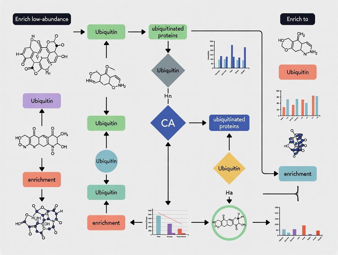
Visualizing the Process: Pathways and Workflows
The Ubiquitin-Proteasome System Cascade
This diagram illustrates the core three-step enzymatic cascade of the UPS, from ubiquitin activation to proteasomal degradation.
Strategic Workflow for Ubiquitinated Protein Enrichment & Analysis
This flowchart outlines a strategic decision-making process for selecting the appropriate enrichment method based on research goals.
Frequently Asked Questions (FAQs)
What are the primary challenges in studying the ubiquitinated proteome? The main challenges include the low natural abundance (stoichiometry) of ubiquitinated proteins within the cell, the highly transient and reversible nature of the modification, the complexity of ubiquitin chain types (e.g., K48, K63, K11, M1), and the limited specificity of some research reagents, which can lead to high background noise and co-purification of non-target proteins [6] [10].
How can I increase the yield of ubiquitinated proteins from my cell samples? A widely recommended strategy is to treat cells with proteasome inhibitors, such as MG-132, prior to harvesting. This prevents the degradation of polyubiquitinated proteins, thereby increasing their intracellular levels available for purification. A typical starting point is incubation with 5-25 µM MG-132 for 1–2 hours, though conditions should be optimized for specific cell types to avoid cytotoxicity [10].
My western blot for ubiquitin shows a smear. Is this expected? Yes, a smear is typical and often indicates a successful experiment. It represents the diverse population of proteins in your sample that have been modified by monomeric ubiquitin, polyubiquitin chains of varying lengths, and ubiquitin polymers, all of which have different molecular weights [10].
Can I differentiate between different types of ubiquitin linkages in my enriched samples? While general ubiquitin enrichment reagents (like Ubiquitin-Traps or broad-spectrum antibodies) are not linkage-specific, you can subsequently identify the linkage types in your enriched samples. This is typically done by using linkage-specific antibodies (e.g., for K48 or K63 chains) in a western blot analysis following enrichment [6] [10].
Troubleshooting Guides
Problem: Low Yield of Ubiquitinated Proteins
Potential Causes and Solutions:
- Cause 1: Rapid deubiquitination after cell lysis.
- Solution: Include deubiquitinase (DUB) inhibitors in your lysis buffer to preserve ubiquitin signals.
- Cause 2: Inefficient enrichment due to suboptimal reagent binding capacity.
- Solution: Consider using high-affinity engineered reagents. For example, Tandem Hybrid Ubiquitin-Binding Domains (ThUBDs) have been shown to have markedly higher affinity for ubiquitinated proteins compared to naturally occurring UBDs [8].
- Cause 3: Proteasomal degradation of substrates before lysis.
- Solution: As noted in the FAQs, pre-treat cells with a proteasome inhibitor like MG-132 [10].
Problem: High Background Noise in Mass Spectrometry Analysis
Potential Causes and Solutions:
- Cause 1: Co-purification of non-specifically bound proteins.
- Cause 2: Endogenous proteins interfering with purification.
- Solution: Be aware of the method's limitations. His-tag purifications can co-purify histidine-rich proteins, while Strep-tag purifications can bind endogenously biotinylated proteins. Using antibody-based or UBD-based enrichment from non-engineered samples can circumvent this [6].
Quantitative Comparison of Enrichment Methodologies
The following table summarizes the key characteristics of major methods used to enrich ubiquitinated proteins, aiding in the selection of the most appropriate technique for your research goals.
Table 1: Comparison of Ubiquitinated Protein Enrichment Methodologies
| Method | Principle | Key Advantages | Key Limitations | Typical Applications |
|---|---|---|---|---|
| Tagged Ubiquitin [6] | Ectopic expression of affinity-tagged Ub (e.g., His, Strep) in cells. | Relatively easy and low-cost; enables high-throughput screening. | Potential artifacts from overexpression; not feasible for clinical/animal tissues; lower identification efficiency. | Proteome-wide screening of ubiquitination sites in cultured cells. |
| Ubiquitin Antibodies [6] | Immunoaffinity purification using anti-ubiquitin antibodies (e.g., P4D1, FK2). | Works with endogenous ubiquitination; applicable to any sample, including tissues. | High cost of antibodies; potential for non-specific binding. | Enrichment from animal tissues or clinical samples; targeted studies. |
| Ubiquitin-Binding Domains (UBDs) [6] [8] | Affinity purification using proteins/domains that naturally bind ubiquitin. | Captures endogenous ubiquitination; can be engineered for high affinity and broad linkage recognition. | Single UBDs may have low affinity; requires careful selection of UBDs. | General and linkage-specific enrichment; used in tools like Ubiquitin-Trap [10]. |
| Engineered Tandem Hybrid UBDs (ThUBDs) [8] | Purification using artificially designed tandem UBDs with optimized affinity. | Very high affinity; low background; broad recognition of different ubiquitin chain linkages. | A relatively new technology that may require protocol optimization. | High-sensitivity profiling of the ubiquitinome from limited sample material. |
Detailed Experimental Protocols
Protocol 1: Enrichment Using Tandem Hybrid UBDs (ThUBDs)
This protocol is adapted from research demonstrating enhanced purification of ubiquitinated proteins using engineered ThUBDs [8].
1. Cell Lysis and Preparation:
- Lyse cells in a suitable non-denaturing lysis buffer (e.g., RIPA buffer) supplemented with DUB inhibitors and protease inhibitors.
- Clarify the lysate by centrifugation at high speed (e.g., 14,000 x g for 15 minutes at 4°C).
2. Affinity Purification with ThUBD Resin:
- Incubate the cleared cell lysate with ThUBD-coupled beads (e.g., Glutathione Sepharose for GST-tagged ThUBDs) for 1-2 hours at 4°C with gentle rotation.
- Pellet the beads by brief centrifugation and carefully remove the supernatant (flow-through fraction).
3. Washing:
- Wash the beads 3-4 times with a large volume (e.g., 1 mL per wash) of ice-cold lysis buffer to remove non-specifically bound proteins.
4. Elution:
- Elute the bound ubiquitinated proteins using a suitable elution buffer. This can be achieved by:
- Competitive Elution: Using a high concentration of free ubiquitin (e.g., 2 mg/mL).
- Denaturing Elution: Using SDS-PAGE sample buffer for direct western blot analysis.
5. Downstream Analysis:
- The eluted proteins can be identified and quantified using mass spectrometry (MS). For MS, on-bead digestion is often recommended [10].
Diagram 1: ThUBD Enrichment Workflow
Protocol 2: Immunoprecipitation Using Commercial Ubiquitin-Trap
This protocol outlines the use of a commercially available nanobody-based product for ubiquitin pulldowns [10].
1. Sample Preparation:
- Prepare cell extracts from the organism of choice (compatible with mammalian, insect, plant, and yeast cells) using the recommended lysis buffer.
2. Pulldown Procedure:
- Use the provided Ubiquitin-Trap Agarose or Magnetic Agarose beads.
- Incubate the cleared cell lysate with the beads for fast and easy pulldowns. The product is stable under harsh washing conditions.
3. Washing and Elution:
- Wash the beads extensively with the provided wash buffer to achieve low-background results.
- Elute using the provided elution buffer or SDS-PAGE sample buffer.
4. Detection:
- Analyze the input (I), flow-through (F), and bound (B) fractions by western blot using a recommended ubiquitin antibody.
The Scientist's Toolkit: Essential Research Reagents
Table 2: Key Reagents for Ubiquitination Enrichment Studies
| Reagent / Tool | Function | Example / Note |
|---|---|---|
| MG-132 (Proteasome Inhibitor) | Increases cellular levels of polyubiquitinated proteins by blocking their degradation. | Use at 5-25 µM for 1-2 hours pre-harvest [10]. |
| DUB Inhibitors | Prevents the removal of ubiquitin chains after lysis, preserving the ubiquitination signal. | Often used in combination with protease inhibitors in lysis buffer. |
| ChromoTek Ubiquitin-Trap | A ready-to-use nanobody-based reagent for immunoprecipitation of ubiquitin and ubiquitinated proteins. | Provides clean IPs from various species; available in agarose and magnetic formats [10]. |
| Tandem Hybrid UBDs (ThUBDs) | Engineered high-affinity binders for unbiased enrichment of ubiquitinated proteins with various linkages. | e.g., ThUDQ2 and ThUDA20; demonstrated high affinity for all seven lysine-linked chains [8]. |
| Linkage-Specific Ub Antibodies | Allows detection or enrichment of specific ubiquitin chain topologies (e.g., K48-, K63-linked). | Critical for determining the functional consequence of ubiquitination on your substrate [6]. |
| Epitope-Tagged Ubiquitin | (His)₆-, HA-, or FLAG-tagged ubiquitin for affinity-based purification in overexpression systems. | Enables pulldown via Ni-NTA (for His) or antibody-conjugated beads [6]. |
In the context of protein ubiquitination, the Stoichiometry Problem refers to the fundamental challenge that ubiquitinated forms of proteins exist at significantly lower abundance compared to their non-modified counterparts within the cell. This phenomenon arises from the combination of transient regulation, rapid turnover, and enzymatic constraints that collectively maintain ubiquitinated proteins at minute stoichiometric ratios.
Ubiquitination is a highly dynamic process where a 76-amino acid ubiquitin protein is covalently attached to substrate proteins, typically targeting them for proteasomal degradation or altering their function [11] [12]. The transient nature of this modification, coupled with the fact that ubiquitinated proteins are often rapidly degraded by the 26S proteasome, ensures their inherently low abundance under normal physiological conditions [11]. Additionally, only one or a few lysine residues are modified in a ubiquitinated protein, further reducing the detectable pool of ubiquitinated species [11].
Key Challenges & Frequently Asked Questions
Q1: Why are ubiquitinated proteins so difficult to detect in standard proteomic experiments?
A: The detection of ubiquitinated proteins faces multiple technical hurdles:
- Low Stoichiometry: The abundance of ubiquitinated proteins is very low in cells under normal physiological conditions because many are rapidly degraded by the proteasome or dynamically regulated in cell signaling pathways [11].
- Sensitivity Masking: High-abundance resident proteins like immunoglobulin and albumin create a billion-fold excess that masks the signal of low-abundance ubiquitinated proteins in MS experiments [13].
- Structural Complexity: Ubiquitin is larger than many other post-translational modifications, and ubiquitinated proteins can form complex chains with different linkages and architectures, complicating their analysis [11] [12].
Q2: What are the major biological factors contributing to the low abundance of ubiquitinated proteins?
A: Several intrinsic biological mechanisms maintain low levels of ubiquitinated proteins:
- Rapid Turnover: Proteins modified with K48-linked polyubiquitin chains are rapidly degraded by the 26S proteasome, significantly shortening their half-life [11] [12].
- Enzymatic Regulation: The coordinated action of E1 activating enzymes, E2 conjugating enzymes, and E3 ligases creates a tightly controlled system where ubiquitination occurs transiently and specifically [12].
- Deubiquitinating Enzymes (DUBs): Protein ubiquitination can be reversed by DUBs, creating a dynamic equilibrium that further reduces steady-state levels of ubiquitinated proteins [11].
Q3: How does the stoichiometry problem impact drug discovery and biomarker identification?
A: The low abundance of ubiquitinated proteins presents both challenges and opportunities:
- Therapeutic Targeting: E3 ubiquitin ligases and DUBs represent promising drug targets, but identifying their native substrates is complicated by low ubiquitination stoichiometry [12].
- Biomarker Discovery: Early disease biomarkers derived from small pre-metastatic lesions exist at concentrations below the detection limit of conventional mass spectrometry platforms [13].
- Pathway Analysis: Understanding disease mechanisms requires comprehensive ubiquitinome profiling, which is hindered by the low abundance of ubiquitinated signaling proteins [14].
Troubleshooting Guide: Common Experimental Issues
Table 1: Troubleshooting Low Yield in Ubiquitinated Protein Enrichment
| Problem | Potential Causes | Recommended Solutions |
|---|---|---|
| Low ubiquitinated peptide yield after enrichment | Insufficient starting material; inefficient antibody binding; sample degradation | Increase input protein to 5-10 mg; validate antibody specificity (e.g., FK2 for monoubiquitin and polyubiquitin); include protease inhibitors and DUB inhibitors [12] [15] |
| High background in MS analysis | Non-specific binding during enrichment; co-purification of abundant proteins | Optimize wash stringency; implement pre-clearing steps; combine depletion of high-abundance proteins with ubiquitin enrichment [16] [13] |
| Inconsistent results between replicates | Variable enrichment efficiency; incomplete tryptic digestion; instrument variability | Use internal standards (SILAC, TMT); standardize digestion protocols with quality control; implement replicate measurements [14] |
| Poor identification of ubiquitination sites | Low stoichiometry at specific lysines; missed cleavages; incomplete fragmentation | Utilize remnant motif antibodies (K-ε-GG); optimize MS fragmentation energy; employ complementary proteases [12] [14] |
Table 2: Quantitative Analysis of Ubiquitin Linkage Changes Following E3 Ligase Perturbation
Data derived from global ubiquitinome profiling in neural crest cells following NEDD4 knockdown [14]
| Ubiquitin Linkage Type | Primary Function | Relative Abundance Change (After NEDD4 knockdown) | Key Biological Implications |
|---|---|---|---|
| K48-linked chains | Proteasomal degradation [12] | Pronounced reduction [14] | Stabilization of proteasome substrates; disrupted protein turnover |
| K63-linked chains | Non-proteolytic signaling [12] | Pronounced reduction [14] | Altered cell signaling, DNA repair, endocytosis |
| K11-linked chains | Proteasomal degradation; cell cycle [12] | Not specified in results | Potential cell cycle dysregulation |
| M1-linked chains | NF-κB signaling; inflammation [12] | Not specified in results | Potential inflammatory signaling defects |
Methodologies & Experimental Protocols
Ubiquitinated Protein Enrichment Using Immunoaffinity Purification
The FK2 immunoaffinity purification method enables efficient isolation of endogenously ubiquitinated protein complexes without genetic manipulation [15].
Protocol Details:
- Cell Lysis: Harvest HeLa or XP2OS cells and lyse in modified RIPA buffer (50 mM Tris-HCl pH 7.5, 150 mM NaCl, 1% NP-40, 0.5% sodium deoxycholate, 0.1% SDS) supplemented with complete protease inhibitors and 10 mM N-ethylmaleimide (DUB inhibitor) [15].
- Antibody Coupling: Covalently couple monoclonal FK2 antibody to protein A/G beads using dimethyl pimelimidate crosslinker to prevent antibody leaching during purification.
- Immunoprecipitation: Incubate clarified cell lysate (1-2 mg total protein) with FK2-conjugated beads for 2-4 hours at 4°C with gentle rotation.
- Wash Steps: Perform sequential washes with lysis buffer (2x), high-salt buffer (1 M NaCl in lysis buffer, 1x), and low-salt buffer (50 mM NaCl in lysis buffer, 1x) to reduce non-specific binding.
- Elution: Elute ubiquitinated proteins with 0.1 M glycine pH 2.5-3.0 for 10 minutes at room temperature, followed by immediate neutralization with 1 M Tris-HCl pH 8.0.
- Analysis: Process eluates for SDS-PAGE and western blotting or tryptic digestion for LC-MS/MS analysis [15].
Ubiquitin Remnant Profiling (K-ε-GG) for Site-Specific Identification
This method uses antibodies specific for the di-glycine remnant left on ubiquitinated lysine residues after tryptic digestion, enabling systematic mapping of ubiquitination sites [14].
Protocol Details:
- Protein Extraction and Digestion: Extract proteins in 8 M urea buffer, reduce with DTT, alkylate with iodoacetamide, and digest with trypsin (1:50 w/w) overnight at 37°C [14].
- Peptide Desalting: Desalt digested peptides using C18 solid-phase extraction cartridges and lyophilize.
- K-ε-GG Enrichment: Resuspend peptides in immunoaffinity purification buffer (50 mM MOPS pH 7.2, 10 mM Na2HPO4, 50 mM NaCl) and incubate with anti-K-ε-GG antibody-conjugated beads for 2 hours at 4°C.
- Wash and Elution: Wash beads with PBS (3x) and elute with 0.1% TFA.
- LC-MS/MS Analysis: Analyze enriched peptides using nanoflow LC-MS/MS on a high-resolution instrument (e.g., Orbitrap series). Use data-dependent acquisition with higher-energy collisional dissociation for fragmentation.
- Data Analysis: Search data against appropriate protein databases using software (MaxQuant, Proteome Discoverer) with K-ε-GG (Gly-Gly, +114.042 Da) as a variable modification on lysine [14].
Tandem Enrichment of Ubiquitinated Peptides with SCASP-PTM
The SCASP-PTM approach enables serial enrichment of ubiquitinated, phosphorylated, and glycosylated peptides from a single sample without intermediate desalting steps [17].
Protocol Highlights:
- Protein Extraction and Digestion: Use SDS-cyclodextrin-assisted sample preparation for efficient protein extraction and digestion.
- Ubiquitinated Peptide Enrichment: First enrichment step targets ubiquitinated peptides using appropriate affinity matrices.
- Serial PTM Enrichment: Utilize flowthrough from previous enrichment steps for subsequent capture of phosphorylated or glycosylated peptides without desalting.
- Sample Cleanup: Desalt enriched peptides prior to MS analysis.
- MS Data Acquisition: Analyze using data-independent acquisition (DIA) MS for comprehensive PTM profiling [17].
Signaling Pathways & Experimental Workflows
Ubiquitination Cascade and Stoichiometry Problem
Ubiquitinated Protein Enrichment Workflow
Research Reagent Solutions
Table 3: Essential Research Reagents for Ubiquitinated Protein Enrichment
| Reagent | Function | Key Applications | Considerations |
|---|---|---|---|
| FK2 Antibody | Recognizes mono- and polyubiquitinated conjugates [15] | Immunoaffinity purification of endogenous ubiquitinated complexes [15] | Does not distinguish linkage types; optimal for native complex isolation |
| TUBEs (Tandem Ubiquitin Binding Entities) | High-affinity ubiquitin traps with multiple UBDs [12] | Protection of polyubiquitinated chains from DUBs and proteasomal degradation [12] | Preferentially binds polyubiquitin; reduces background degradation |
| K-ε-GG Motif Antibody | Specific for diglycine remnant on modified lysines after trypsin digestion [14] | Ubiquitination site mapping by MS; ubiquitin remnant profiling [14] | Requires complete tryptic digestion; may miss incomplete cleavages |
| Linkage-Specific Ub Antibodies | Recognize specific ubiquitin chain linkages (K48, K63, etc.) [12] | Analysis of chain topology and functional characterization [12] | Variable specificity and affinity between vendors; requires validation |
| N-Ethylmaleimide (NEM) | Irreversible DUB inhibitor [15] | Preservation of ubiquitinated proteins during extraction by inhibiting deubiquitination [15] | Must be added fresh to lysis buffers; can modify other cysteine residues |
| Ubiquitin Activating Enzyme (E1) Inhibitor | Inhibits ubiquitin activation [18] | Negative control for ubiquitination assays; studying dynamic ubiquitination [18] | PYR-41 and similar compounds; can affect global protein homeostasis |
The stoichiometry problem in ubiquitination research presents significant but not insurmountable challenges. Through the implementation of robust enrichment methodologies, careful experimental design, and appropriate controls, researchers can successfully overcome the limitations posed by the inherently low abundance of ubiquitinated proteins. The continuing development of more sensitive mass spectrometry platforms, improved affinity reagents, and novel chemical biology tools promises to further enhance our ability to study the ubiquitinome and unravel the complex regulatory networks controlled by this essential post-translational modification.
Ubiquitination is a critical post-translational modification (PTM) that regulates diverse cellular functions, including protein degradation, DNA repair, and immune responses, by covalently attaching a small protein (ubiquitin) to substrate proteins [6] [19]. The versatility of ubiquitination stems from its complexity—it can manifest as monoubiquitination, multiple mono-ubiquitination, or polyubiquitination chains with different linkage types (e.g., K48, K63, K11, K6, K27, K29, K33, M1), each potentially encoding distinct functional outcomes [6] [20]. However, studying ubiquitination presents significant technical hurdles. The stoichiometry of protein ubiquitination is typically very low under normal physiological conditions, and ubiquitinated proteins often represent a minute fraction within a complex proteomic background [6]. Furthermore, the dynamic range of protein abundance in biological samples can span 10 to 12 orders of magnitude, allowing highly abundant proteins to suppress the detection of scarce ubiquitination signals [21]. This guide addresses these key technical barriers—detection sensitivity, dynamic range, and sub-stoichiometric modification—by providing targeted troubleshooting advice and proven methodologies for enriching and analyzing low-abundance ubiquitinated proteins.
Key Technical Barriers and Troubleshooting FAQs
FAQ 1: How can I overcome the low stoichiometry and transient nature of ubiquitination in my samples?
Challenge: Ubiquitination is a highly transient and reversible modification. The percentage of ubiquitinated proteins in a cell lysate is often very small, making them difficult to detect without effective enrichment [20].
Solutions:
- Use Proteasome Inhibitors: Treat cells with proteasome inhibitors (e.g., MG-132) prior to harvesting. This prevents the degradation of ubiquitinated proteins, allowing their accumulation. A recommended starting point is incubating cells with 5–25 µM MG-132 for 1–2 hours, though conditions should be optimized for each cell type to avoid cytotoxicity [20].
- Employ Robust Enrichment Tools: Utilize high-affinity enrichment tools designed specifically for ubiquitin. Tandem Ubiquitin Binding Entities (TUBEs) can selectively bind ubiquitin chains and protect them from deubiquitinases (DUBs) during extraction. Similarly, ubiquitin traps (e.g., ChromoTek's Ubiquitin-Trap) use anti-ubiquitin nanobodies/VHH coupled to beads to immunoprecipitate monomeric ubiquitin, ubiquitin chains, and ubiquitinylated proteins from complex cell extracts [20] [22].
- Optimize Lysis Conditions: Avoid surfactant-based cell lysis methods that use detergents like Tween, Nonident P-40, and Triton X-100, as residual surfactants can cause severe ion suppression in mass spectrometry (MS), obscuring peptide signals. If surfactants are necessary, extreme care must be taken to remove them completely prior to analysis [23].
FAQ 2: What strategies can mitigate the immense dynamic range of protein abundance to detect low-abundance ubiquitinated proteins?
Challenge: The protein dynamic range in biological samples spans 10–12 orders of magnitude. Highly abundant structural proteins can suppress the ionization and detection of low-abundance regulatory proteins and their ubiquitinated forms [21].
Solutions:
- Deplete High-Abundance Proteins: Use affinity columns to remove highly abundant proteins like albumin and immunoglobulins from samples such as serum or plasma. This reduces dynamic range complexity and reduces ion suppression [21].
- Implement Multi-Dimensional Fractionation: Reduce sample complexity by fractionating peptides or proteins before MS analysis. Common techniques include strong cation exchange (SCX) or high-pH reverse-phase chromatography. These steps separate the peptide mixture into simpler fractions, increasing the depth of analysis and the likelihood of detecting low-abundance ubiquitinated peptides [21].
- Choose Advanced MS Acquisition Methods: Move from Data-Dependent Acquisition (DDA) to Data-Independent Acquisition (DIA). DIA reduces undersampling by systematically fragmenting all peptides within sequential isolation windows, providing more complete MS/MS data and significantly reducing missing values, which is a common issue with low-abundance species [21].
FAQ 3: How can I prevent the loss of ubiquitinated peptides during sample preparation?
Challenge: Peptides are prone to adsorption to the surfaces of sample preparation vessels (e.g., plastic vials and micropipette tips), leading to significant and selective losses, especially for low-abundant targets [23].
Solutions:
- Use "High-Recovery" Vials: Select LC vials and tubes specifically engineered to minimize analyte adsorption [23].
- Avoid Complete Drying: When using vacuum centrifugation to remove solvents, avoid drying the sample completely, as this promotes strong analyte adsorption onto surfaces. Leave a small amount of liquid in the vial to increase recovery [23].
- Limit Sample Transfers: Minimize the number of sample transfers between vessels to reduce contact with surfaces that can cause adsorption. Consider "one-pot" sample preparation methods (e.g., SP3, FASP) that perform digestion and cleanup in a single vessel [23].
- Prime Vessels: "Prime" vessels with a sacrificial protein like Bovine Serum Albumin (BSA) to saturate adsorption sites on the material before introducing your analytical sample [23].
Experimental Protocols for Ubiquitin Enrichment
Protocol 1: Ubiquitin-Trap Based Immunoprecipitation
This protocol uses a high-affinity nanobody to isolate ubiquitin and ubiquitinated proteins [20].
Detailed Methodology:
- Cell Lysis: Lyse cells in an appropriate, chilled lysis buffer. It is critical to avoid surfactants that interfere with MS. Consider mechanical lysis methods if possible.
- Pre-Clear Lysate (Optional): Centrifuge the lysate at high speed to remove insoluble debris.
- Incubation with Beads: Incubate the clarified cell lysate with Ubiquitin-Trap Agarose or Magnetic Agarose beads for 1–2 hours at 4°C with gentle agitation.
- Washing: Pellet the beads and carefully remove the supernatant. Wash the beads multiple times with a suitable wash buffer to remove non-specifically bound proteins. The nanobody-based trap is stable under harsh washing conditions, enabling low-background results.
- Elution: Elute the bound ubiquitinated proteins using a low-pH elution buffer or by directly adding SDS-PAGE loading buffer and heating.
- Downstream Analysis: The eluate can be analyzed by western blotting or prepared for mass spectrometry analysis. For MS, proteins can be digested on-bead following the manufacturer's optimized protocol [20].
Protocol 2: Antibody-Based Enrichment of Ubiquitinated Proteins
This method uses anti-ubiquitin antibodies to pull down ubiquitinated conjugates [6] [22].
Detailed Methodology:
- Antibody Selection: Choose an antibody based on your needs. Pan-ubiquitin antibodies (e.g., P4D1, FK1/FK2) recognize all ubiquitin linkages. Linkage-specific antibodies (e.g., K48-, K63-specific) are used to study specific chain types [6] [22].
- Antibody Immobilization: Covalently couple the chosen antibody to protein A/G agarose or sepharose beads to prevent antibody leaching and contamination of the eluate.
- Incubation and Binding: Incubate the pre-cleared cell lysate with the antibody-conjugated beads for several hours or overnight at 4°C.
- Stringent Washing: Wash the beads thoroughly with a series of buffers containing mild detergents and salts to eliminate non-specific interactions.
- Elution and Digestion: Elute ubiquitinated proteins. For MS analysis, this is often followed by tryptic digestion. A key signature of ubiquitination is the detection of a 114.04 Da mass shift (from the diGly remnant) on the modified lysine residues during MS analysis, which allows for the mapping of ubiquitination sites [6].
Protocol 3: Tandem Ubiquitin Binding Entity (TUBE) Enrichment
TUBEs are engineered high-affinity ubiquitin-binding domains used for affinity purification [22].
Detailed Methodology:
- Preparation of TUBE Resin: If not purchased pre-coupled, immobilize recombinant TUBE protein onto a solid-phase matrix like agarose beads.
- Sample Preparation and Binding: Prepare cell lysate and incubate with the TUBE resin. TUBEs protect ubiquitin chains from deubiquitinating enzymes (DUBs) and proteasomal degradation during the process.
- Washing and Elution: Perform wash steps to remove unbound proteins. Elute the enriched ubiquitinated proteins for downstream analysis.
Data Analysis and Validation Strategies
Managing Missing Values and Controlling False Discovery
A common issue in shotgun proteomics, particularly with low-abundance ubiquitinated peptides, is the presence of missing values (where a peptide is identified in some runs but not others) [21].
- Advanced Imputation: The strategy for handling missing data should depend on whether data is Missing At Random (MAR) or Missing Not At Random (MNAR). For MNAR data (missing due to abundance being below detection), imputation with small, low-intensity values from the bottom of the quantitative distribution is appropriate. For MAR data, more robust methods like
k-nearest neighbor (KNN) or singular value decomposition (SVD) should be used [21]. - False Discovery Rate (FDR) Control: Use stringent FDR controls, typically set at 1%, for peptide and protein identification to minimize false positives. This is especially important when searching for modified peptides [21].
Research Reagent Solutions
The table below summarizes key reagents essential for studying protein ubiquitination.
Table 1: Essential Research Reagents for Ubiquitination Studies
| Reagent Category | Specific Example | Function and Application |
|---|---|---|
| Affinity Enrichment Reagents | Ubiquitin-Trap (Agarose/Magnetic) | High-affinity nanobody-based beads for pulldown of mono/poly-ubiquitin and ubiquitinated proteins from various cell extracts [20]. |
| Tandem Ubiquitin Binding Entities (TUBEs) | Engineered high-affinity domains for enrichment of ubiquitinated proteins; offer protection from DUBs [22]. | |
| Linkage-Specific Antibodies (e.g., α-K48, α-K63) | Immunoprecipitation or western blot detection of specific polyubiquitin chain linkages (e.g., K48 for degradation, K63 for signaling) [6] [22]. | |
| Chemical Inhibitors | MG-132 | Proteasome inhibitor used to treat cells before lysis to increase the cellular pool of ubiquitinated proteins [20]. |
| Detection Antibodies | Pan-Ubiquitin Antibodies (e.g., P4D1, FK2) | Recognize ubiquitin regardless of linkage type; used for western blotting or immunofluorescence to detect total ubiquitinated proteins [6] [20]. |
| Enzymes for In Vivo Tagging | His-Tagged Ubiquitin, Strep-Tagged Ubiquitin | Genetically encoded tags allow purification of ubiquitinated proteins from cell lysates using Ni-NTA or Strep-Tactin affinity resins, respectively [6]. |
Visualizing the Workflow: From Ubiquitination to Analysis
The following diagram illustrates the core decision-making pathway for selecting the appropriate enrichment strategy based on research goals.
Ubiquitination is a versatile and reversible post-translational modification that regulates diverse fundamental features of protein substrates, including stability, activity, and localization [6]. This modification involves the covalent attachment of ubiquitin, a small 76-amino acid protein, to substrate proteins through a sequential enzymatic cascade involving E1 activating, E2 conjugating, and E3 ligase enzymes [24] [6]. The complexity of ubiquitin signaling arises from the ability of ubiquitin itself to become modified, forming polymers (polyubiquitin chains) through its seven lysine residues (K6, K11, K27, K29, K33, K48, K63) or N-terminal methionine (M1), with different chain linkages triggering distinct functional consequences [24] [6].
The dysregulation of the delicate balance between ubiquitination and deubiquitination is implicated in numerous pathologies, with particularly intriguing connections to cancer and neurodegenerative diseases [25] [6]. Epidemiologic evidence reveals an inverse comorbidity relationship between these disease families, where neurodegenerative diseases occur less frequently in cancer survivors and vice versa [25]. This relationship has biological plausibility, as neurons and cycling cells utilize the same proteins and pathways in different, and sometimes opposite, ways [25]. For instance, the tumor suppressor p53 is upregulated in Alzheimer's disease (AD), Parkinson's disease (PD), and Huntington's disease (HD) but downregulated in most cancers [25].
Understanding the molecular mechanisms of ubiquitination signaling requires sophisticated methodologies to characterize ubiquitination sites, linkage types, and ubiquitin chain architecture [6]. This technical support center provides comprehensive troubleshooting guidance for researchers studying low-abundance ubiquitinated proteins, with particular emphasis on methodologies relevant to cancer and neurodegenerative disease research.
Technical Support Center: Ubiquitinated Protein Enrichment
Frequently Asked Questions (FAQs)
Q1: Why is studying protein ubiquitination particularly challenging, especially in the context of disease research?
A1: Several technical challenges complicate ubiquitination studies:
- Low Abundance: The stoichiometry of protein ubiquitination is very low under normal physiological conditions, increasing the difficulty of identifying ubiquitinated substrates [6].
- Transient Nature: The ubiquitination process is highly transient and reversible, with ubiquitinated proteins representing a small percentage of total cellular proteins [24].
- Structural Complexity: Ubiquitin can modify substrates at one or several lysine residues simultaneously and can itself form polymers of varying length, linkage, and architecture [6].
- Immunogenic Limitations: Ubiquitin proteins are weakly immunogenic due to their small size, resulting in antibodies that may bind non-specifically and produce artifacts [24].
- Enzyme Diversity: With over 600 different E3 ligases, it's possible for multiple ligases to ubiquitinate one protein simultaneously, requiring reagents that can detect multiple specificities [24].
Q2: How can I preserve ubiquitination signals in my samples before enrichment?
A2: To protect and enhance ubiquitination signals:
- Proteasome Inhibition: Treat cells with proteasome inhibitors such as MG-132 prior to harvesting. A recommended starting point is incubating cells with 5-25 μM MG-132 for 1-2 hours, though conditions should be optimized for each cell type [24].
- Prevent Cytotoxicity: Note that overexposure to MG-132 can lead to cytotoxic effects, so optimization is crucial [24].
- Rapid Processing: Use rapid lysis methods with RIPA or SDS-based buffers immediately after sample collection to prevent deubiquitination [26].
- Cold-Chain Maintenance: Perform all enrichment procedures under cold-chain conditions (4°C) to minimize enzymatic activity [26].
Q3: What are the key considerations when choosing between different ubiquitinated protein enrichment strategies?
A3: The selection depends on several factors:
- Research Objective: Determine if you need protein-level or site-specific analysis [26].
- Chain-Type Specificity: Decide whether you require broad ubiquitin capture or specific linkage types [26].
- Sample Type and Amount: Consider your starting material (cell lines, animal tissues, clinical samples) and quantity [26].
- Downstream Applications: Match the enrichment method to your planned analysis (Western blot, MS, etc.) [26].
- Budget and Resources: Evaluate costs associated with antibodies, reagents, and specialized equipment [26].
Q4: Why do ubiquitinated proteins often appear as smears on Western blots, and how can I interpret these results?
A4: The smeared appearance is normal and expected because:
- Size Heterogeneity: enrichment reagents bind monomeric ubiquitin, ubiquitin polymers, and ubiquitinylated proteins, resulting in proteins of varying molecular weights [24].
- Polyubiquitin Chains: Proteins modified with polyubiquitin chains of different lengths migrate as diffuse bands rather than discrete bands [24].
- Interpretation Guidance: The smear pattern actually indicates successful capture of diverse ubiquitinated species. Using linkage-specific antibodies during Western blot analysis can help differentiate between chain types [24].
Q5: My ubiquitinated protein enrichment yields high background noise in mass spectrometry. How can I reduce this?
A5: To minimize background:
- Pre-Enrichment Cleanup: Use ion exchange (SCX/SAX) or high-pH reverse phase chromatography to remove interfering substances before enrichment [26].
- Optimized Wash Conditions: Increase stringency of wash conditions (e.g., higher salt concentrations, detergents) while ensuring ubiquitinated proteins are retained [24].
- Specific Enrichment Methods: Consider tandem hybrid UBDs (ThUBDs) which show higher affinity and lower background compared to some antibody-based methods [27].
- Control Experiments: Include appropriate controls (e.g., no antibody, isotype control, or bead-only) to identify non-specific binders [6].
Troubleshooting Guide: Common Experimental Issues
Problem: Low yield of ubiquitinated proteins after enrichment.
| Possible Cause | Solution |
|---|---|
| Insufficient starting material | Increase input protein (1-10 mg recommended); concentrate samples if needed [26] |
| Ineffective lysis | Use fresh lysis buffer with protease inhibitors; include 1% SDS for difficult samples [26] |
| Rapid deubiquitination | Add deubiquitinase (DUB) inhibitors to lysis buffer; process samples on ice [24] |
| Suboptimal binding conditions | Extend incubation time (≥1 hour); optimize buffer pH and salt concentrations [26] |
| Overly stringent washes | Reduce wash stringency; include a quick wash step before elution [24] |
Problem: Inability to distinguish specific ubiquitin linkage types.
| Possible Cause | Solution |
|---|---|
| Using non-linkage-specific reagents | Incorporate linkage-specific antibodies (e.g., K48-, K63-specific) in Western blot [24] [6] |
| Limited method specificity | Use TUBEs (tandem ubiquitin binding entities) with known linkage preferences [26] |
| Lack of appropriate controls | Include controls with known linkage types to validate detection methods [6] |
| MS limitations | Combine K-ε-GG enrichment with advanced mass spectrometry for site-specific identification [26] |
Problem: Inconsistent results between experimental replicates.
| Possible Cause | Solution |
|---|---|
| Variable inhibitor treatment | Standardize MG-132 concentration and treatment time across replicates [24] |
| Inconsistent cell states | Use cells at consistent passage numbers and confluence levels [6] |
| Protease/phosphatase activity | Use fresh inhibitors with each preparation; aliquot to avoid freeze-thaw cycles [26] |
| Binding capacity exceeded | Determine binding capacity of enrichment resin; do not overload [24] |
| Temperature fluctuations | Perform all steps at consistent temperatures; use pre-cooled equipment [26] |
Comprehensive Enrichment Strategy Comparison
Quantitative Analysis of Enrichment Methods
Table 1: Comparison of Ubiquitinated Protein Enrichment Strategies
| Method | Principle | Advantages | Limitations | Ideal Application | Typical Cost |
|---|---|---|---|---|---|
| General Anti-Ubiquitin Antibodies (e.g., FK2, P4D1) [26] | Monoclonal antibodies capture ubiquitin-modified proteins | Broad applicability; straightforward procedure; compatible with standard workflows | Lack of chain-type specificity; potential co-purification of non-target proteins; complex MS background | Global quantification; exploratory studies; preliminary screening | Moderate (antibody purchase main cost) |
| Tandem Ubiquitin Binding Entities (TUBEs) [26] | Tandemly arranged ubiquitin-binding domains with multivalency increase affinity | High binding affinity; improved capture efficiency; can selectively enrich specific chain types | Complex design; higher cost; requires a priori knowledge of target chain type | Studies of polyubiquitin chains; protein degradation pathways; signal transduction | Moderate to High |
| Site-Specific K-ε-GG Remnant Enrichment [26] | Antibodies recognize Gly-Gly remnants on lysine after trypsin digestion | Precise localization of ubiquitination sites; high signal-to-noise ratio in MS | Limited to trypsin-digested samples; limited peptide coverage; time-intensive | Ubiquitin site proteomics; quantitative comparisons; detailed site characterization | High (antibody costs) |
| Ubiquitin Tagging-Based Approaches (e.g., His-, Strep-tags) [6] | Expression of affinity-tagged ubiquitin in cells | Easy implementation; relatively low-cost; good for cellular studies | Not applicable to tissues; potential artifacts from tagged ubiquitin; low identification efficiency | High-throughput screening in cell culture; initial discovery studies | Low to Moderate |
| Tandem Hybrid UBDs (ThUBDs) [27] | Artificial tandem UBDs with high affinity and minimal linkage bias | Unbiased high affinity to multiple chain types; applicable to native conditions and tissues | Complex cloning and protein purification required | Global ubiquitome profiling; tissue samples; biomarker discovery | High (development costs) |
Table 2: Ubiquitin Linkage Types and Their Biological Significance
| Linkage Site | Ubiquitin Chain Length | Primary Biological Functions | Relevance to Disease |
|---|---|---|---|
| K48 [24] [6] | Polymeric | Targeted protein degradation via proteasome | Accumulation in Alzheimer's disease (tau) [6]; cancer progression |
| K63 [24] [6] | Polymeric | Immune responses, inflammation, lymphocyte activation, DNA repair | NF-κB pathway activation in cancer [6]; neurodegenerative inflammation |
| K6 [24] | Polymeric | Antiviral responses, autophagy, mitophagy, DNA repair | Potential role in cancer resistance; neurodegenerative mitochondrial dysfunction |
| K11 [24] | Polymeric | Cell cycle progression, proteasome-mediated degradation | Dysregulation in cancers; cell cycle defects in neurodegeneration |
| K27 [24] | Polymeric | DNA replication, cell proliferation | Associated with tumor proliferation; DNA damage response in neurodegeneration |
| K29 [24] | Polymeric | Neurodegenerative disorders, Wnt signaling downregulation, autophagy | Direct link to neurodegenerative pathways; Wnt signaling in cancer |
| M1 (Linear) [24] | Polymeric | Cell death and immune signaling | Inflammation pathways in both cancer and neurodegeneration |
| Substrate lysines [24] | Monomer | Endocytosis, histone modification, DNA damage responses | Receptor trafficking in cancer; DNA repair in neurodegeneration |
Research Reagent Solutions
Table 3: Essential Research Reagents for Ubiquitination Studies
| Reagent Category | Specific Examples | Function | Applications |
|---|---|---|---|
| General Ubiquitin Antibodies [24] | P4D1, FK1, FK2, VU-1 | Recognize all ubiquitin linkages; capture diverse ubiquitinated proteins | Immunoprecipitation, Western blot, immunofluorescence |
| Linkage-Specific Antibodies [6] | K48-specific, K63-specific, M1-linear specific | Identify specific ubiquitin chain linkages | Western blot validation, selective enrichment |
| Ubiquitin Affinity Traps [24] | ChromoTek Ubiquitin-Trap (Agarose/Magnetic) | High-affinity nanobodies for ubiquitin and ubiquitinylated protein isolation | Pulldown assays, IP-MS, clean low-background IPs |
| TUBE Reagents [26] | Commercial TUBEs (K48/K63 preferring) | High-affinity capture of polyubiquitinated proteins with linkage selectivity | Native purification, proteasome studies, signaling pathways |
| K-ε-GG Antibodies [26] | Commercial di-glycine remnant antibodies | Enrich ubiquitinated peptides after trypsin digestion | Ubiquitin site mapping by MS, quantitative ubiquitomics |
| Proteasome Inhibitors [24] | MG-132, Bortezomib | Prevent degradation of ubiquitinated proteins | Enhance ubiquitination signals before enrichment |
| Tandem Hybrid UBDs [27] | ThUDQ2, ThUDA20 | Artificial UBDs with high affinity and minimal linkage bias | Global ubiquitome profiling, tissue samples, biomarker discovery |
| Tagged Ubiquitin Plasmids [6] | His-Ub, HA-Ub, Strep-Ub | Expression of affinity-tagged ubiquitin in cells | Pull-down assays in cultured cells, interaction studies |
Experimental Protocols
K-ε-GG Enrichment Protocol for Ubiquitination Site Mapping
This protocol enables precise mapping of ubiquitination sites through enrichment of tryptic peptides containing the di-glycine remnant on modified lysines [26].
Sample Preparation:
- Rapid Lysis: Lyse cells or tissues using RIPA or SDS-based buffer with protease and phosphatase inhibitors.
- Protein Quantification: Determine protein concentration using BCA or Bradford assay.
- Reduction and Alkylation: Reduce disulfide bonds with DTT (5-10 mM, 30 min, 56°C) and alkylate with iodoacetamide (10-15 mM, 30 min, room temperature in darkness).
- Protein Digestion: Digest proteins with trypsin (1:50 enzyme-to-substrate ratio) for 4-16 hours at 37°C. Optional: Use secondary digestion (LysC + trypsin) to enhance coverage.
Pre-Enrichment Cleanup:
- Desalt peptides using C18 solid-phase extraction or perform ion exchange chromatography (SCX/SAX) to remove interfering substances.
- Lyophilize or vacuum concentrate peptides and reconstitute in immunoaffinity purification buffer.
K-ε-GG Antibody Enrichment:
- Incubation: Incubate peptides with K-ε-GG antibody conjugated to beads (e.g., protein A/G) for ≥1 hour at 4°C with gentle rotation.
- Washing: Wash beads extensively with cold PBS or Tris-buffered saline to remove non-specifically bound peptides.
- Elution: Elute bound peptides using 0.1-0.2% TFA or low-pH glycine solution.
- Cleanup: Desalt eluted peptides using C18 stage tips or columns before MS analysis.
Mass Spectrometry Analysis:
- Chromatography: Separate peptides using nano-scale LC with C18 column and acetonitrile gradient.
- MS Acquisition: Analyze peptides using high-resolution mass spectrometer (Orbitrap Exploris or Fusion Lumos recommended).
- Data Analysis: Search data with MaxQuant, Proteome Discoverer with PTMProfiler, or similar software, with false discovery rate (FDR) ≤1% for both protein and site identifications.
Tandem Hybrid UBD (ThUBD) Enrichment Protocol
This protocol utilizes artificial tandem UBDs for efficient and relatively unbiased enrichment of ubiquitinated proteins under native conditions [27].
ThUBD Preparation:
- Protein Expression: Express GST-ThUBD fusion proteins (e.g., ThUDQ2, ThUDA20) in E. coli BL21 (DE3) cells induced with 0.5 mM IPTG for 4 hours at 30°C.
- Protein Purification: Purify fusion proteins from cell lysates using glutathione-Sepharose 4B beads according to manufacturer's instructions.
- Immobilization: Couple purified ThUBDs to NHS-activated Sepharose following manufacturer's protocol. Store conjugated agarose at 4°C in PBS with 30% glycerol.
Sample Preparation and Enrichment:
- Cell Lysis: Lyse cells in native lysis buffer (e.g., 50 mM Na2HPO4, pH 8.0, 500 mM NaCl, 0.01% SDS, 5% glycerol) with protease inhibitors and DUB inhibitors.
- Clarification: Centrifuge lysates at 70,000 × g for 30 minutes to remove insoluble material.
- Incubation: Incubate clarified lysate with immobilized ThUBD beads at 4°C for 30 minutes with gentle rotation.
- Washing: Wash beads sequentially with:
- Lysis buffer
- Wash buffer B (50 mM NH4HCO3 with 5 mM iodoacetamide)
- 50 mM NH4HCO3 to remove iodoacetamide
- Elution: Elute bound ubiquitin conjugates by boiling in 1× SDS-PAGE loading buffer or using specific elution buffers for downstream applications.
Downstream Applications:
- Western Blotting: Analyze eluates by SDS-PAGE and immunoblotting with ubiquitin antibodies.
- Mass Spectrometry: Process eluted proteins for LC-MS/MS analysis to identify ubiquitinated proteins and sites.
- Functional Assays: Use enriched ubiquitinated proteins for enzymatic assays or interaction studies.
Workflow Visualization
Ubiquitinated Protein Enrichment and Analysis Workflow
Ubiquitin Cascade and Chain Linkage Diversity
Comprehensive Enrichment Platforms: From Antibodies to Engineered Binding Domains
Protein ubiquitination, the covalent attachment of ubiquitin to substrate proteins, represents one of the most versatile post-translational modifications in eukaryotic cells, regulating diverse fundamental processes including protein degradation, subcellular localization, and signal transduction [6]. The dysregulation of ubiquitination pathways has been implicated in numerous human diseases, particularly cancer and neurodegenerative disorders, making the comprehensive characterization of ubiquitination events a critical priority in biomedical research [28] [6]. However, the systematic analysis of ubiquitination presents substantial technical challenges due to the low stoichiometry of modified proteins, the dynamic nature of the modification, and the complexity of ubiquitin chain architectures [6] [29].
The development of antibodies specifically recognizing the di-glycine (K-ε-GG) remnant left on trypsinized ubiquitinated peptides has revolutionized the ubiquitination proteomics field [28] [30] [29]. This immunoaffinity enrichment technology has enabled researchers to transition from identifying merely hundreds of ubiquitination sites to routinely quantifying tens of thousands of distinct sites in single experiments [30] [31] [29]. This technical support center provides comprehensive guidance for researchers implementing Anti-K-ε-GG antibody platforms, addressing common experimental challenges and detailing optimized methodologies for enriching low-abundance ubiquitinated proteins.
Technical FAQs: Resolving Common Experimental Challenges
Low Ubiquitinated Peptide Recovery
Question: What are the primary factors affecting ubiquitinated peptide yield following immunoaffinity enrichment?
Inadequate recovery of K-ε-GG peptides can result from several methodological issues. First, insufficient antibody-to-peptide ratios significantly impact enrichment efficiency; studies demonstrate that using at least 62μg of anti-K-ε-GG antibody per milligram of peptide input maximizes recovery [30]. Second, improper tryptic digestion conditions may fail to efficiently generate the di-glycine remnant, while excessive digestion can promote sample degradation. Third, incomplete quenching of cross-linking reactions when using immobilized antibodies leads to antibody leakage during enrichment procedures [30]. Finally, sample overdilution during incubation reduces binding kinetics, while insufficient washing stringency introduces high background interference in downstream mass spectrometry analysis.
Solution: Implement a cross-linked antibody protocol with optimized input ratios. Systematic optimization has demonstrated that cross-linking the anti-K-ε-GG antibody to solid supports using dimethyl pimelimidate (DMP) dramatically improves enrichment performance by preventing antibody leakage during elution steps [30]. Additionally, maintain precise antibody-to-peptide ratios of 62-125μg antibody per milligram of peptide input, and employ fractionation strategies such as basic reversed-phase chromatography to reduce sample complexity prior to enrichment [30].
High Background and Non-Specific Binding
Question: How can researchers minimize non-specific binding during K-ε-GG immunoaffinity enrichment?
Excessive background signal typically originates from non-specific interactions between cellular peptides and solid support matrices or antibody frameworks. This problem becomes particularly pronounced when analyzing complex samples with wide dynamic ranges of protein abundance, such as tissue lysates or whole cell extracts. The presence of endogenous biotin or lectins in certain sample types can further exacerbate background issues [32]. Additionally, antibody overloading beyond optimal capacities can promote non-specific binding through charge-based interactions rather than specific antigen recognition.
Solution: Implement stringent wash protocols and optimize buffer composition. Following immunoaffinity enrichment, perform at least four washes with ice-cold phosphate-buffered saline (PBS) to remove non-specifically bound peptides [30]. Incorporate NaCl at concentrations between 0.15M and 0.6M in wash and antibody dilution buffers to reduce ionic interactions [32]. For tissue samples with high endogenous biotin content, employ avidin/biotin blocking steps prior to enrichment, and consider using non-glycosylated streptavidin alternatives to prevent lectin binding interactions [32].
Incomplete TMT Labeling for Multiplexed Experiments
Question: What strategies improve TMT labeling efficiency for multiplexed ubiquitination studies?
Traditional approaches involving TMT labeling following peptide elution from antibodies frequently result in suboptimal labeling efficiency due to the low quantities of enriched material and interference from elution buffers [29]. The standard method where K-ε-GG peptides are enriched, eluted, and then labeled in solution typically yields labeling efficiencies below 50%, severely compromising quantitative accuracy in multiplexed experimental designs [29]. Additionally, the amine groups on the di-glycine remnant itself can potentially react with TMT reagents, further complicating accurate quantification.
Solution: Implement on-antibody TMT labeling prior to peptide elution. The UbiFast method demonstrates that labeling peptides with TMT reagents while still bound to anti-K-ε-GG antibodies dramatically improves labeling efficiency to >92% while simultaneously increasing the relative yield of K-ε-GG peptides by nearly 10% [29]. Optimized protocols utilize 0.4mg of TMT reagent with a 10-minute labeling duration, followed by thorough quenching with 5% hydroxylamine to prevent cross-labeling when combining samples [29].
Quantitative Performance Optimization
Table 1: Key Performance Metrics for K-ε-GG Enrichment Methodologies
| Method Parameter | Standard Enrichment | Optimized Cross-linked Protocol | UbiFast (On-Antibody TMT) |
|---|---|---|---|
| Protein Input | 10-35mg [30] | 5mg per SILAC channel [30] | 0.5mg per TMT channel [29] |
| Antibody Amount | Not specified | 31-62μg per enrichment [30] | 31μg per enrichment [29] |
| Sites Identified | 1,000-5,000 [30] | ~20,000 [30] [31] | >10,000 [29] |
| Labeling Efficiency | Not applicable | >95% (SILAC) [30] | 92-98% (TMT) [29] |
| Relative Yield | 44.2% (in-solution TMT) [29] | 85.7% (label-free) [29] | 85.7% (on-antibody TMT) [29] |
| Key Innovation | Basic K-ε-GG enrichment | Antibody cross-linking + fractionation | On-antibody TMT labeling |
Table 2: Troubleshooting Guide for Common Experimental Issues
| Problem | Potential Causes | Recommended Solutions |
|---|---|---|
| Low ubiquitinated peptide recovery | Insufficient antibody; Antibody leakage; Inefficient digestion | Cross-link antibody with DMP; Optimize antibody:peptide ratio (62-125μg:1mg); Validate tryptic digestion efficiency [30] |
| High background interference | Non-specific binding; Endogenous enzymes; Inadequate washing | Add NaCl (0.15-0.6M) to buffers; Quench endogenous peroxidases with H₂O₂; Increase wash stringency (4× with cold PBS) [30] [32] |
| Poor quantitative reproducibility | Incomplete TMT labeling; Sample-to-sample variation; Instrument variability | Implement on-antibody TMT labeling; Use internal standard controls; Employ FAIMS separation for LC-MS/MS [29] |
| Inconsistent results across replicates | Variable antibody performance; Digestion inefficiency; Fractionation inconsistency | Cross-link antibody beads; Standardize digestion protocols with quality controls; Implement non-contiguous fraction pooling [30] |
Experimental Workflows and Signaling Pathways
Optimized K-ε-GG Enrichment Workflow
Figure 1: Optimized K-ε-GG Enrichment Workflow
This diagram illustrates the refined workflow for ubiquitination site identification, highlighting critical improvements including antibody cross-linking, basic reversed-phase fractionation, and on-antibody TMT labeling that collectively enable deep-scale ubiquitinome profiling [30] [29].
Ubiquitination Signaling Cascade
Figure 2: Ubiquitination Signaling Cascade
This diagram outlines the ubiquitination enzymatic cascade and subsequent cellular decision points, with the K-ε-GG remnant serving as the critical analytical handle for mass spectrometry-based detection and quantification [28] [6].
Research Reagent Solutions
Table 3: Essential Research Reagents for K-ε-GG Immunoaffinity Enrichment
| Reagent Category | Specific Examples | Function in Workflow | Performance Considerations |
|---|---|---|---|
| Anti-K-ε-GG Antibodies | PTMScan Ubiquitin Remnant Motif Kit [30] | Specific recognition and enrichment of K-ε-GG peptides | Cross-linking improves yield; 31-62μg per enrichment optimal [30] |
| Protein Digestion Enzymes | Sequencing grade trypsin [30] | Generates K-ε-GG remnant peptides from ubiquitinated proteins | Enzyme-to-substrate ratio of 1:50 with overnight digestion recommended [30] |
| Chromatography Media | Zorbax 300 Extend-C18 column [30] | Basic reversed-phase fractionation reduces sample complexity | Non-contiguous pooling of 80 fractions into 8 pools enhances depth [30] |
| Cross-linking Reagents | Dimethyl pimelimidate (DMP) [30] | Immobilizes antibody to solid support preventing leakage | 20mM DMP in 100mM sodium borate (pH 9.0) for 30 minutes [30] |
| Isobaric Labeling Reagents | Tandem Mass Tags (TMT) [29] | Enables multiplexed quantification of ubiquitination sites | On-antibody labeling with 0.4mg TMT for 10 minutes achieves >92% efficiency [29] |
| Enrichment Buffers | IAP Buffer (50mM MOPS, pH 7.2) [30] | Provides optimal binding conditions for antibody-antigen interaction | Contains 10mM sodium phosphate and 50mM NaCl for maintaining binding specificity [30] |
Advanced Methodologies: Pushing Sensitivity Boundaries
UbiFast: Multiplexed Ubiquitinome Profiling
The recently developed UbiFast methodology represents a significant advancement in ubiquitination proteomics by enabling highly multiplexed quantification from limited sample inputs [29]. This approach exploits the epitope protection phenomenon, where the di-glycine remnant becomes shielded from solvent exposure when bound to the anti-K-ε-GG antibody. By performing TMT labeling while peptides remain antibody-bound, the method prevents derivatization of the di-glycine primary amine while efficiently labeling peptide N-termini and lysine side chains. This innovation permits quantification of >10,000 ubiquitination sites from merely 500μg of peptide input per sample while reducing total processing time to approximately 5 hours [29]. The integration of High-field Asymmetric Waveform Ion Mobility Spectrometry (FAIMS) further enhances quantitative accuracy by reducing background interference during LC-MS/MS analysis.
Alternative Enrichment Strategies: Engineered Tandem Hybrid UBDs
While anti-K-ε-GG antibodies currently represent the gold standard for ubiquitination site identification, emerging technologies offer complementary approaches. Recently developed engineered tandem hybrid ubiquitin-binding domains (ThUBDs) combine multiple ubiquitin-binding domains with high affinity for different ubiquitin chain types, creating reagents with markedly improved binding capabilities compared to naturally occurring UBDs [8]. These ThUBDs demonstrate almost unbiased high affinity to all seven lysine-linked ubiquitin chains and have successfully identified thousands of ubiquitinated proteins from both yeast and mammalian cells [8]. Although this approach does not provide site-specific information like anti-K-ε-GG enrichment, it offers advantages for studying ubiquitin chain architecture and does not require epitope exposure through tryptic digestion.
Anti-K-ε-GG antibody platforms have fundamentally transformed our capacity to interrogate the ubiquitinome at unprecedented depth and precision. Through systematic optimization of enrichment conditions, implementation of antibody cross-linking strategies, and development of innovative labeling approaches such as UbiFast, researchers can now routinely quantify tens of thousands of ubiquitination sites across multiple experimental conditions. As these methodologies continue to evolve, particularly through integration with complementary enrichment technologies and advanced separation techniques, they promise to unlock new insights into the complex regulatory networks governed by protein ubiquitination in both physiological and disease contexts.
Frequently Asked Questions (FAQs)
Q1: What are the primary advantages of using ThUBDs over traditional TUBEs for enriching ubiquitinated proteins? ThUBDs offer two significant advantages: superior affinity and reduced linkage bias. They are engineered tandem hybrid ubiquitin-binding domains that provide markedly higher affinity for ubiquitinated proteins compared to naturally occurring UBDs or TUBEs [27]. Furthermore, they display almost unbiased high affinity to all seven lysine-linked ubiquitin chains, enabling a more comprehensive view of the ubiquitinome, unlike many TUBEs which may have preferences for specific chain types [27] [33].
Q2: My immunoblot shows weak or no ubiquitination signal after ThUBD pulldown. What could be wrong? Weak signals can often be traced to sample preparation or buffer conditions. First, ensure your lysis buffer contains fresh protease inhibitors (e.g., 1 mM PMSF) and 5-10 mM N-ethylmaleimide (NEM) to inhibit deubiquitinating enzymes (DUBs) that can rapidly remove ubiquitin signals [34] [35]. Second, verify that you are using a non-ionic detergent like 1% Triton X-100 in your lysis and wash buffers to maintain protein interactions while reducing background [27] [34]. Finally, confirm the binding capacity of your resin; for a 1 mL bed volume of ThUBD-conjugated NHS-activated Sepharose, do not exceed 2 mg of total protein input from cell lysate to avoid overloading [27].
Q3: How can I distinguish covalently ubiquitinated proteins from non-covalent binders in my ThUBD enrichment? This is a critical distinction. Use a denaturing workflow to isolate covalent ubiquitination. After lysing cells in your standard buffer, add SDS to a final concentration of 1% and boil the samples for 5-10 minutes [34]. Dilute the denatured lysate 10-fold with a neutral buffer (e.g., 50 mM Na₂HPO₄, pH 8.0, 500 mM NaCl) containing 0.01% SDS before incubating with the ThUBD resin. This denaturation step disrupts non-covalent protein-protein interactions, ensuring that only covalently ubiquitinated proteins and direct interactors are captured [34].
Q4: Can ThUBD-based methods be used for high-throughput drug screening, such as in PROTAC development? Yes, ThUBD-coated high-density 96-well plates have been developed specifically for this purpose. This platform allows for high-throughput, flexible analysis of both global and target-specific protein ubiquitination [33]. It exhibits a 16-fold wider linear range for capturing polyubiquitinated proteins compared to TUBE-coated plates, making it highly suitable for efficiently detecting and precisely quantifying ubiquitination signals in drug development pipelines like PROTAC discovery [33].
Q5: My mass spectrometry results show high background. How can I improve the specificity of my ThUBD enrichment for proteomics? High background in MS is often due to non-specific binding. Incorporate a high-stringency wash step with a buffer containing 500 mM to 1 M NaCl and 0.1% SDS before the final wash [27] [34]. Additionally, for proteomic applications, perform on-bead digestion. After the final wash with 50 mM NH₄HCO₃, add 5 mM iodoacetamide to alkylate cysteine residues, then wash again before adding trypsin directly to the beads for digestion [27]. This minimizes sample handling and loss.
Troubleshooting Guides
Problem: Low Yield of Ubiquitinated Proteins
Possible Causes and Solutions:
- Cause 1: Ineffective cell lysis or protein extraction.
- Cause 2: Depleted or inactive ThUBD resin.
- Solution: Always include a positive control (e.g., a cell line known to have high ubiquitination). The ThUBD-conjugated agarose should be stored at 4°C in PBS with 30% glycerol to maintain stability. Avoid more than 5-7 repeated uses of the same resin batch [27].
- Cause 3: Ubiquitinated proteins are degraded during preparation.
Problem: High Non-Specific Binding
Possible Causes and Solutions:
- Cause 1: Inadequate washing of the resin.
- Solution: Implement a stepwise washing protocol:
- Wash 3x with lysis buffer (e.g., 50 mM Na₂HPO₄, pH 8.0, 500 mM NaCl, 0.01% SDS).
- Wash 2x with a high-salt buffer (e.g., the same buffer with 1 M NaCl).
- Perform a final wash with a low-salt, neutral buffer (e.g., 50 mM NH₄HCO₃) [27].
- Solution: Implement a stepwise washing protocol:
- Cause 2: The protein concentration in the lysate is too high, leading to overloading.
- Solution: Determine the optimal protein-to-resin ratio. Do not exceed 2 mg of total protein per 1 mL of ThUBD resin bed volume. Pre-clear the lysate by centrifuging at 70,000 × g for 30 minutes before incubation with the resin [27].
Data Presentation: Comparison of UBD-Based Technologies
The following table summarizes the properties of TUBEs and the advanced ThUBDs.
| Feature | TUBEs (Tandem Ubiquitin-Binding Entities) | Engineered ThUBDs (Tandem Hybrid UBDs) |
|---|---|---|
| Basic Design | Tandem repeats of identical or different natural UBDs [12]. | Artificially constructed hybrid of four UBDs (e.g., DSK2p-UBA + UQ2-UBA) [27]. |
| Binding Affinity | Moderate affinity, improved over single UBDs [33]. | Markedly higher affinity than naturally occurring UBDs [27] [36]. |
| Linkage Bias | Often exhibit bias towards specific ubiquitin chain linkages (e.g., K48 or K63) [12]. | Almost unbiased high affinity to all seven lysine-linked chains [27] [33]. |
| Detection Sensitivity | Standard sensitivity, can miss low-abundance conjugates. | High sensitivity; can detect ubiquitinated proteins at levels as low as 0.625 μg, a 16-fold improvement over TUBEs in some formats [33]. |
| Monoubiquitination Detection | Often poor affinity for monoubiquitinated proteins [34]. | Effectively enriches both mono- and polyubiquitinated proteins [27]. |
| Primary Application | General enrichment of polyubiquitinated proteins under native conditions [12]. | Unbiased profiling of the global ubiquitinome, high-throughput screening, and detection of low-abundance conjugates [27] [33]. |
Experimental Protocols
Protocol 1: Enrichment of Ubiquitinated Proteins from Mammalian Cells using ThUBD Resin
This protocol details the steps for a native pulldown of ubiquitinated proteins from cultured mammalian cells, suitable for downstream immunoblotting or mass spectrometry [27] [34].
Key Reagent Solutions:
- Lysis Buffer (Native): 50 mM Na₂HPO₄ (pH 8.0), 500 mM NaCl, 0.01% SDS, 5% Glycerol. Add 1% Triton X-100, 1 mM DTT, cOmplete EDTA-free Protease Inhibitor Cocktail, and 10 mM NEM fresh before use [27] [34].
- ThUBD Resin: GST-tagged ThUBD (e.g., ThUDA20) coupled to NHS-activated Sepharose 4B beads and stored in PBS with 30% glycerol at 4°C [27].
- Wash Buffer A: Identical to lysis buffer without DTT and NEM.
- Wash Buffer B (for MS): 50 mM NH₄HCO₃, 5 mM iodoacetamide [27].
- Elution Buffer: 1X SDS-PAGE loading buffer (50 mM Tris-HCl, pH 6.8, 2% SDS, 10% glycerol, 0.1% bromophenol blue, 1% β-mercaptoethanol) [27].
Methodology:
- Cell Lysis: Harvest human hepatocellular carcinoma MHCC97-H cells (or your cell line of interest) and wash with PBS. Lyse the cell pellet in an appropriate volume of Lysis Buffer (Native) for 30 minutes on a rotator at 4°C [27].
- Clarification: Centrifuge the lysate at 70,000 × g for 30 minutes at 4°C. Transfer the supernatant to a new tube [27].
- Pulldown: Incubate the clarified lysate with pre-equilibrated ThUBD resin for 30 minutes at 4°C with constant mixing [27].
- Washing: Pellet the beads and carefully remove the supernatant.
- Wash the beads 3 times with Wash Buffer A.
- For mass spectrometry analysis, perform an additional wash with Wash Buffer B (to alkylate cysteine residues), followed by a final wash with 50 mM NH₄HCO₃ to remove iodoacetamide [27].
- Elution: Elute the bound ubiquitinated proteins by adding 1X SDS-PAGE loading buffer and boiling the beads at 95°C for 10 minutes. The eluate is now ready for analysis by immunoblotting or, after on-bead digestion, for mass spectrometry [27].
Protocol 2: High-Throughput Detection using ThUBD-Coated 96-Well Plates
This protocol is designed for rapid, sensitive, and quantitative analysis of ubiquitination in a high-throughput format, ideal for screening applications [33].
Key Reagent Solutions:
- Coating Solution: 1.03 μg ± 0.002 of purified ThUBD protein in carbonate-bicarbonate buffer (pH 9.6) per well of a Corning 3603-type 96-well plate [33].
- Blocking Buffer: 5% non-fat milk or 1% BSA in PBST (PBS with 0.05% Tween-20).
- Assay Diluent/Wash Buffer: PBS or TBS with 0.1% Tween-20.
- Detection Antibody: Primary antibody against your target protein or ubiquitin, and an appropriate HRP-conjugated secondary antibody.
- Detection Reagent: Chemiluminescent or colorimetric substrate suitable for HRP.
Methodology:
- Plate Coating: Coat each well of the high-binding 96-well plate with the ThUBD Coating Solution. Seal the plate and incubate overnight at 4°C [33].
- Blocking: Remove the coating solution and block the plates with 200-300 μL of Blocking Buffer for 2 hours at room temperature to prevent non-specific binding.
- Sample Incubation: Add your complex proteome samples (e.g., cell lysates) or purified proteins to the wells. Incubate for 1-2 hours at room temperature with gentle shaking to allow ubiquitinated proteins to bind to the immobilized ThUBD.
- Washing: Wash the plate 3-5 times with Wash Buffer to remove unbound proteins and contaminants.
- Detection: Add your primary antibody, wash, then add the HRP-conjugated secondary antibody. After final washes, develop the signal with your chosen detection reagent and read the plate using a microplate reader [33].
Visualization of Concepts and Workflows
Diagram 1: ThUBD Engineering and Ubiquitin Chain Recognition Concept
Diagram 2: Core Experimental Workflow for ThUBD-Based Enrichment
The Scientist's Toolkit: Essential Research Reagents
| Reagent / Material | Function / Explanation | Example Source / Reference |
|---|---|---|
| ThUBD Protein (GST-tagged) | Engineered recombinant protein used to create affinity resin; high affinity and low linkage bias are key. | Purified from E. coli BL21(DE3) using pGEX-4T-2 vector [27]. |
| NHS-activated Sepharose 4B | Chromatography resin for covalent coupling of ThUBD protein to create a reusable affinity matrix. | GE Healthcare [27]. |
| N-Ethylmaleimide (NEM) | Deubiquitinating enzyme (DUB) inhibitor. Critical to preserve ubiquitin signals in lysates by preventing deubiquitination. | Sigma-Aldrich [34] [35]. |
| cOmplete EDTA-free Protease Inhibitor Cocktail | Inhibits a broad spectrum of serine, cysteine, and metalloproteases to prevent protein degradation during sample preparation. | Roche [34]. |
| Ni-NTA Agarose | For purifying His-tagged ubiquitinated proteins in alternative or validation protocols. | Qiagen [35]. |
| Anti-Ubiquitin Antibodies (P4D1, FK2) | Used for immunodetection (Western Blot) of enriched ubiquitinated proteins after pulldown. | Enzo, Invitrogen, Cell Signaling [34]. |
| ThUBD-Coated 96-Well Plates | Pre-coated plates for high-throughput, quantitative analysis of ubiquitination in drug screening (e.g., PROTACs). | Corning 3603-type plates coated with 1.03 μg/well ThUBD [33]. |
Technical Support Center: Troubleshooting & FAQs
This guide provides targeted troubleshooting and FAQs for researchers using affinity tags to study protein ubiquitination, a key post-translational modification regulating protein stability, activity, and localization [6]. These protocols are essential for enriching low-abundance ubiquitinated proteins, which is critical for understanding disease mechanisms in cancer and neurodegeneration [13] [6].
His-Tagged Ubiquitin Systems
This system uses ubiquitin (Ub) genetically fused to a polyhistidine (His) tag for purification via Immobilized Metal Ion Affinity Chromatography (IMAC) [6] [37].
Frequently Asked Questions
Q: I get high background binding when purifying His-tagged ubiquitin from mammalian cell lysates. How can I reduce this?
- A: Background binding often comes from endogenous histidine-rich proteins or proteins with metal-binding centers [38]. To reduce this:
- Optimize Wash Buffers: Include 5-10 mM imidazole in your wash buffers to displace weakly bound proteins [38].
- Include Denaturants: Wash with low concentrations of a denaturant (e.g., 2-3 M urea) to disrupt non-specific interactions, provided your tagged ubiquitin is correctly folded [38].
- Consider Tag Position: If background persists, try purifying the tag from a different terminus (N- or C-) of ubiquitin, as accessibility can vary [38].
- A: Background binding often comes from endogenous histidine-rich proteins or proteins with metal-binding centers [38]. To reduce this:
Q: The yield of my His-tagged ubiquitin conjugates is low. What could be wrong?
- A: Low yield can have several causes:
- Incomplete Binding: Ensure the resin is not overloaded. Check the binding capacity of your IMAC resin and do not exceed it.
- Protease Degradation: Ubiquitinated proteins can be rapidly degraded. Treat cells with a proteasome inhibitor (e.g., MG-132 at 5-25 µM for 1-2 hours before harvesting) to preserve conjugates [39].
- Tag Inaccessibility: The His-tag might be sterically hindered. Testing a different tag location or adding a flexible linker between the tag and ubiquitin can improve accessibility [38].
- A: Low yield can have several causes:
Detailed Protocol: Enriching Ubiquitinated Proteins with His-Tagged Ubiquitin
- Construct Generation: Clone the His-tag (typically 6xHis) at the N-terminus of ubiquitin. The N-terminal location helps avoid interference with the C-terminal glycine (G76) required for conjugation [6].
- Cell Transfection & Treatment: Stably or transiently express the His-tagged Ub in your cell line (e.g., HEK293T, U2OS). To preserve polyubiquitin chains, treat cells with a proteasome inhibitor like MG-132 (e.g., 10 µM for 4 hours) before harvesting [39].
- Cell Lysis: Lyse cells using a non-denaturing lysis buffer (e.g., RIPA buffer) supplemented with protease inhibitors and 10 mM imidazole. The imidazole helps reduce non-specific binding during the lysate preparation step.
- IMAC Purification:
- Incubate the clarified cell lysate with Ni-NTA agarose resin for 1-2 hours at 4°C.
- Wash the resin extensively with a wash buffer containing 20-50 mM imidazole to remove contaminating proteins.
- Elution: Elute the bound His-tagged ubiquitin and its conjugates with elution buffer containing 250-500 mM imidazole.
- Analysis: Analyze the eluate by SDS-PAGE and Western blotting using an anti-ubiquitin or anti-His antibody. A characteristic smear is expected due to the heterogeneous molecular weights of ubiquitin-protein conjugates [39].
Strep-Tagged Ubiquitin Systems
This system utilizes ubiquitin fused to a short Strep-tag (WRHPQFGG), which binds reversibly to Strep-Tactin resin [6].
Frequently Asked Questions
Q: The binding capacity for my Strep-tagged ubiquitin seems low. Why?
- A: The binding capacity can be affected by the tag's accessibility and the resin itself.
- Check the Tag: Ensure the Strep-tag is correctly fused and expressed.
- Resin Reusability: Strep-Tactin resin has a limited number of reuses. Use fresh resin if the binding capacity drops significantly.
- Chain Length: If purifying polyubiquitin chains, note that a single long chain may occupy multiple binding sites on the resin, effectively reducing the capacity for distinct conjugates [39].
- A: The binding capacity can be affected by the tag's accessibility and the resin itself.
Q: I see co-elution of endogenous biotinylated proteins in my mammalian system prep. How do I avoid this?
- A: This is a known limitation, as streptavidin binds tightly to biotin [38]. To mitigate this:
- Pre-clear Lysate: Pre-clear your cell lysate with neutralvidin or streptavidin resin to remove endogenous biotinylated proteins before applying it to the Strep-Tactin column.
- Use a Control: Always include a negative control (e.g., cells expressing untagged ubiquitin) to identify which bands are non-specifically bound biotinylated proteins.
- A: This is a known limitation, as streptavidin binds tightly to biotin [38]. To mitigate this:
Detailed Protocol: Enriching Ubiquitinated Proteins with Strep-Tagged Ubiquitin
- Construct Generation: Clone the Strep-tag onto the N-terminus of ubiquitin [6].
- Cell Culture & Lysis: Generate a cell line stably expressing Strep-tagged Ub (e.g., U2OS or HEK293T) [6]. Lyse cells as described for the His-tag protocol, but without imidazole.
- Strep-Tactin Affinity Purification:
- Incubate the clarified lysate with Strep-Tactin resin (agarose or magnetic beads) for 1-2 hours at 4°C.
- Wash the resin with the recommended buffer (e.g., Tris-buffered saline) to remove non-specifically bound proteins.
- Elution: Elute the bound complexes under mild conditions using a buffer containing 10 mM biotin or a desthiobiotin-containing buffer for even gentler elution [38].
- Analysis: Analyze by Western blot or mass spectrometry (IP-MS). For MS, the trap has been optimized for on-bead digestion [39].
General Epitope-Tagged Ubiquitin Systems
Epitope tags (e.g., HA, Myc, FLAG) are small peptides recognized by specific monoclonal antibodies, allowing immunoprecipitation of ubiquitin conjugates [40] [6].
Frequently Asked Questions
Q: My immunoprecipitation of FLAG-tagged ubiquitin shows low yield. What are potential causes?
- A: Low yield in IP can stem from several issues:
- Antibody Capacity: The antibody binding capacity on the resin may be exceeded. Use more resin or concentrate your lysate less.
- Inefficient Elution: Elution with the FLAG peptide is generally efficient. If using low-pH elution, this can denature the antibody and reduce resin reusability, potentially affecting yield over time [37]. Neutralize low-pH eluates immediately.
- Transfection Efficiency: Ensure a high percentage of cells are expressing your epitope-tagged ubiquitin.
- A: Low yield in IP can stem from several issues:
Q: Does adding an epitope tag to ubiquitin affect its biological function?
- A: It can. While epitope-tagged ubiquitin can suppress phenotypic defects in yeast and be correctly conjugated [40], some studies show that tags can subtly alter function. For example, N-terminal epitope tags can inhibit the proteolysis of certain test proteins, suggesting the N-terminal region of ubiquitin is important for protease recognition [40]. Always validate your system with functional assays.
Detailed Protocol: Immunoprecipitation of Ubiquitin Conjugates with Epitope Tags
- Construct Generation: Clone your chosen epitope tag (e.g., HA, Myc, FLAG) at the N-terminus of ubiquitin [40] [6].
- Cell Transfection & Lysis: Express the tagged ubiquitin in your cell system and lyse cells with an appropriate IP lysis buffer.
- Immunoprecipitation:
- Pre-clear the lysate with protein A/G beads to reduce non-specific binding.
- Incubate the pre-cleared lysate with the antibody against your epitope tag (e.g., anti-FLAG M2 antibody) for several hours or overnight at 4°C.
- Add protein A/G beads to capture the antibody-antigen complex. Wash the beads thoroughly to remove non-specifically bound proteins.
- Elution: Elute the complexes. The best method depends on your downstream application.
- Competitive Elution: Use an excess of the epitope peptide (e.g., FLAG peptide) for mild, native elution.
- Low-pH Elution: Use a glycine buffer (pH 2.0-3.0), which is efficient but can denature proteins and damage the antibody on the beads [37].
- Analysis: Proceed with Western blotting or mass spectrometry analysis.
Comparative Data and Reagent Solutions
The table below summarizes key characteristics of the affinity tags discussed.
| Tag | Typical Size | Affinity Resin / Antibody | Elution Method | Key Advantages | Key Limitations / Background |
|---|---|---|---|---|---|
| His-Tag [38] [37] | 0.8 - 1.6 kDa (e.g., 6xHis is 0.8 kDa) | Ni-NTA (Ni2+) or other IMAC resins | Imidazole (250-500 mM) | Small size; low immunogenicity; stable binding | High background from endogenous His-rich proteins in mammalian/insect cells [38] [6]. |
| Strep-Tag [38] [6] | 1.0 kDa (8 aa) | Strep-Tactin | Biotin or Desthiobiotin (mild) | Mild, native elution; high specificity | Co-elution of endogenous biotinylated proteins in mammalian systems [38]. |
| Epitope Tags (e.g., FLAG, HA) [6] [37] | ~1 kDa (e.g., FLAG is 8 aa) | Monoclonal Antibody (e.g., anti-FLAG M2) | Low pH, peptide competition, or EDTA (FLAG) | High specificity; wide range of validated antibodies | Low pH elution can denature proteins; antibody-based purification can have lower yields [37]. |
Research Reagent Solutions
| Item Name | Function / Application | Example Product / Note |
|---|---|---|
| Ubiquitin-Trap [39] | Immunoprecipitates endogenous ubiquitin and ubiquitinylated proteins from various cell extracts using a anti-Ubiquitin nanobody. | ChromoTek Ubiquitin-Trap Agarose (uta) or Magnetic Agarose (utma). |
| Linkage-Specific Antibodies [6] | Enrich and detect ubiquitinated proteins with specific chain linkages (e.g., K48, K63). | Used for Western blot or IP to study specific ubiquitin signaling events. |
| Proteasome Inhibitor (MG-132) [39] | Preserves ubiquitination signals by inhibiting the proteasome, preventing degradation of polyubiquitinated proteins. | A typical treatment is 5-25 µM for 1-2 hours before cell harvesting. |
| StUbEx System [6] | A cellular system where endogenous ubiquitin is replaced with a His-tagged ubiquitin for proteomic profiling of ubiquitination. | "Stable Tagged Ubiquitin Exchange" system for identifying ubiquitination sites. |
Workflow and Pathway Diagrams
Diagram Title: His-Tagged Ubiquitin Experimental Workflow
Diagram Title: Ubiquitin Conjugation Simplified Pathway
Diagram Title: Tag Selection for Low-Abundance Protein Research
Ubiquitination is a crucial post-translational modification that regulates diverse cellular functions, including protein degradation, cellular trafficking, and kinase signaling [12]. The versatility of ubiquitin signaling stems from its ability to form chains of different architectures through eight distinct linkage types (M1, K6, K11, K27, K29, K33, K48, K63) [41]. Among these, K48-linked chains primarily target proteins for proteasomal degradation, while K63-linked chains regulate non-degradative functions such as protein-protein interactions and activation of kinase signaling pathways [42] [12]. The remaining "atypical" chain types (K6, K11, K27, K29, K33) are less abundant and more challenging to study, creating a significant knowledge gap in our understanding of the full ubiquitin signaling landscape [41].
The identification of ubiquitinated proteins is particularly challenging in the context of low-abundance proteins, where signal detection is often masked by more abundant cellular proteins [43] [44]. Linkage-specific antibodies provide a powerful solution to this problem by enabling targeted enrichment of ubiquitinated proteins with specific chain architectures, thereby significantly enhancing detection sensitivity for ubiquitination events that would otherwise go unnoticed in global proteomic analyses [12]. This technical resource document provides comprehensive guidance on the application, troubleshooting, and experimental protocols for linkage-specific ubiquitin enrichment, with particular emphasis on overcoming the challenges associated with low-abundance protein research.
Research Reagent Solutions: A Toolkit for Linkage-Specific Ubiquitin Research
The following table summarizes key reagents essential for conducting linkage-specific ubiquitin enrichment studies:
Table 1: Key Research Reagents for Linkage-Specific Ubiquitin Analysis
| Reagent Type | Specific Examples | Key Applications | Considerations for Low-Abundance Proteins |
|---|---|---|---|
| K63-linkage Specific Antibodies | Anti-Ubiquitin (K63-linkage) [45] [46] | Western Blot, IHC-P, Flow Cytometry (Intra) [45] | Use high-affinity monoclonal antibodies (e.g., EPR8590-448) for enhanced signal detection [45] |
| K48-linkage Specific Antibodies | K48-linkage Specific Polyubiquitin Antibody [42] | Western Blotting | Demonstrates slight cross-reactivity with linear chains; validate with appropriate controls [42] |
| Atypical Chain Affinity Reagents | K6- and K33-linkage specific affimers [41] | Western Blotting, Confocal Microscopy, Pull-downs | Non-antibody protein scaffolds offer high specificity for understudied linkage types [41] |
| Pan-Ubiquitin Antibodies | P4D1, FK1/FK2 [12] | Enrichment of total ubiquitinated proteins | Useful for initial surveys before linkage-specific analysis; may miss atypical linkages [12] |
| Ubiquitin-Binding Domains | Tandem-repeated Ub-binding entities (TUBEs) [12] | Enrichment of ubiquitinated proteins from cell lysates | Overcome low affinity of single UBDs; preserve labile ubiquitin signals during extraction [12] |
Technical FAQs: Addressing Common Challenges in Linkage-Specific Enrichment
Q1: What are the primary challenges when studying atypical ubiquitin linkages (K6, K11, K27, K29, K33), and what tools are available to address them?
A1: The study of atypical ubiquitin linkages presents several distinct challenges. These chain types are generally low-abundance in cells compared to K48 and K63 linkages, making their detection technically challenging [41]. Until recently, specific detection reagents were largely unavailable for most atypical linkages, creating a significant knowledge gap [41]. Additionally, some commercially available reagents may demonstrate cross-reactivity issues; for example, the K33 affimer originally showed cross-reactivity with K11-linked chains, requiring structure-guided improvements to enhance specificity [41].
Novel affinity reagents called affimers have been developed to address these challenges. These 12-kDa non-antibody scaffolds based on the cystatin fold can be selected for high specificity and affinity toward particular atypical linkages [41]. For instance, improved K6-specific affimers have proven effective in western blotting, confocal microscopy, and pull-down applications, enabling the identification of HUWE1 as a major E3 ligase for K6 chains and mitofusin-2 as a substrate modified with K6-linked ubiquitin [41].
Q2: What specific strategies can enhance the detection of low-abundance ubiquitinated proteins during immunoblotting?
A2: Detecting low-abundance ubiquitinated proteins requires specialized strategies to enhance signal-to-noise ratio:
Protein Depletion Methods: Prior to ubiquitination analysis, implement methods to deplete abundant proteins that may mask signals of interest. Techniques include centrifugation, organic solvent-based approaches (acetone, methanol-chloroform), and acid precipitation (e.g., perchloric acid) [43]. One systematic evaluation found perchloric acid precipitation particularly effective for enriching low-molecular-weight proteins [43].
Signal Amplification: Consider using fluorescently conjugated secondary antibodies or specialized amplification systems to enhance detection sensitivity for weak signals [45].
Protein Loading Optimization: Increase protein loading appropriately while ensuring the gel resolution is not compromised. For low-abundance targets, loading 20-30 µg of total protein per lane may be necessary [45].
Validation with Multiple Methods: Confirm findings using complementary techniques such as immunofluorescence, flow cytometry, or mass spectrometry to rule out antibody artifacts [45] [46].
Q3: How can researchers verify the linkage specificity of their ubiquitin antibodies and avoid misinterpretation due to cross-reactivity?
A3: Rigorous validation is essential for accurate interpretation of linkage-specific ubiquitin data:
Specificity Testing: Validate antibodies against a panel of different linkage types. High-quality linkage-specific antibodies should show minimal cross-reactivity with non-cognate ubiquitin chains [45] [42]. For example, the K48-linkage specific antibody demonstrates no cross-reactivity with polyubiquitin chains formed by linkage to different lysine residues, though it shows slight cross-reactivity with linear polyubiquitin chains [42].
Appropriate Controls: Always include cells or samples without primary antibody, with isotype control antibodies, and (if possible) genetic models lacking the specific ubiquitin linkage [45] [41].
Competition Assays: Perform competition experiments with recombinant diubiquitin of specific linkages to demonstrate that signal detection can be specifically blocked by the cognate antigen [41].
Correlative Approaches: Confirm findings using orthogonal methods such as mass spectrometry-based proteomics or alternative affinity reagents when possible [41] [12].
Troubleshooting Guides: Overcoming Common Experimental Challenges
Weak or No Signal in Western Blotting
Table 2: Troubleshooting Weak Signal in Linkage-Specific Ubiquitin Detection
| Problem | Potential Causes | Solutions | Considerations for Low-Abundance Targets |
|---|---|---|---|
| Weak signal | Insufficient protein loading | Increase amount of loaded protein (e.g., 20-30 µg) [45] | Combine with abundant protein depletion methods [43] [44] |
| Suboptimal antibody dilution | Perform antibody titration experiments | For low-abundance proteins, may need higher antibody concentrations than recommended | |
| Inefficient transfer | Use extended transfer times or validate transfer efficiency | Low-molecular-weight ubiquitin signals may transfer too efficiently | |
| No signal | Improper antigen retrieval | Optimize heat-mediated antigen retrieval conditions [45] | For IHC-P, use Tris-EDTA buffer pH 9.0 for antigen retrieval [45] |
| Antibody incompatibility with fixation | Test different fixation methods or antibody clones | ||
| True absence of target | Validate with positive control samples | Use known positive control cell lines (e.g., HeLa for K63 linkages) [45] [46] |
High Background or Non-Specific Signals
- Cause: Antibody cross-reactivity or non-specific binding
Solutions:
- Include linkage specificity controls using recombinant ubiquitin chains of defined linkages [45]
- Increase stringency of washes (increase salt concentration, add mild detergents)
- Use affinity-purified antibodies when possible [42]
- For K48-specific antibodies, be aware of slight cross-reactivity with linear chains and interpret results accordingly [42]
Cause: Non-specific antibody binding
- Solutions:
- Optimize blocking conditions (e.g., 5% non-fat dry milk in TBST) [45]
- Include secondary antibody-only controls
- Use cross-adsorbed secondary antibodies
Inconsistent Results Between Experimental Replicates
- Cause: Variation in ubiquitin chain stability during sample preparation
Solutions:
- Include deubiquitinase inhibitors in lysis buffers
- Maintain consistent and rapid sample processing
- Use fresh protein extracts rather than repeated freeze-thaw cycles
Cause: Inconsistent enrichment efficiency
- Solutions:
- Standardize incubation times and temperatures for antibody-based enrichment
- Use tandem-repeated Ub-binding entities (TUBEs) for more consistent ubiquitinated protein preservation and enrichment [12]
Detailed Experimental Protocols
Standard Protocol: Enrichment of K63-Linked Ubiquitinated Proteins for Western Blotting
This protocol is adapted from commercial linkage-specific antibody protocols and represents a robust method for detecting K63-linked ubiquitination [45].
Diagram: Workflow for K63-Linked Ubiquitin Detection
Materials:
- K63-linkage specific antibody (e.g., ab179434) [45]
- Lysis buffer with protease inhibitors and deubiquitinase (DUB) inhibitors
- Precast SDS-PAGE gels
- PVDF or nitrocellulose membrane
- Blocking buffer: 5% non-fat dry milk (NFDM) in TBST [45]
- Secondary antibody: HRP-conjugated goat anti-rabbit (H+L) [45]
Procedure:
- Prepare Cell Lysates: Lyse cells in appropriate buffer containing protease inhibitors and DUB inhibitors. Maintain samples on ice throughout to prevent protein degradation.
- Protein Quantification: Determine protein concentration using a compatible assay (e.g., BCA assay). Use 20-30 µg of total protein per lane for optimal detection [45].
- SDS-PAGE: Separate proteins by SDS-PAGE using appropriate percentage gels based on target protein size. Note that ubiquitinated proteins often appear as smears due to heterogeneity.
- Membrane Transfer: Transfer proteins to PVDF membrane using standard wet or semi-dry transfer methods.
- Blocking: Block membrane with 5% non-fat dry milk in TBST for 1 hour at room temperature with gentle agitation [45].
- Primary Antibody Incubation: Incubate with K63-linkage specific primary antibody at 1:1000 dilution in blocking buffer overnight at 4°C with gentle agitation [45].
- Washing: Wash membrane 3×10 minutes with TBST.
- Secondary Antibody Incubation: Incubate with HRP-conjugated goat anti-rabbit secondary antibody at 1:1000 dilution in blocking buffer for 1 hour at room temperature [45].
- Detection: Develop using enhanced chemiluminescence (ECL) substrate according to manufacturer's instructions.
Troubleshooting Notes:
- For low-abundance targets, consider increasing primary antibody incubation time to 48 hours at 4°C.
- If background is high, try increasing TBST wash stringency (0.5% Tween-20) or reducing primary antibody concentration.
- Always include positive controls (e.g., HeLa cell lysates) to validate antibody performance [45] [46].
Advanced Protocol: Enrichment of Low-Abundance Ubiquitinated Proteins Using Peptide-Level Immunoaffinity Enrichment
This advanced protocol leverages peptide-level enrichment to significantly enhance the identification of ubiquitination sites, particularly for low-abundance proteins [47] [48].
Diagram: Peptide-Level Immunoaffinity Enrichment Workflow
Materials:
- PTMScan Ubiquitin Remnant Motif Kit (Cell Signaling) or equivalent [48]
- Strong cation exchange (SCX) or basic reverse-phase (bRPLC) chromatography materials
- Mass spectrometry-compatible buffers
- Linkage-specific antibodies for validation [12]
Procedure:
- Sample Preparation:
- Lyse cells in 8M urea buffer containing protease and DUB inhibitors [48].
- Reduce proteins with 10 mM TCEP for 1 hour at 37°C [48].
- Alkylate with 12 mM iodoacetamide for 30 minutes at room temperature protected from light [48].
- Dilute urea concentration to 1M with 50 mM Tris-HCl (pH 8.0) and digest with trypsin overnight at 25°C using 1:50 enzyme-to-substrate ratio [48].
Peptide-level Immunoaffinity Enrichment:
- Acidify digested peptides to 1% formic acid and centrifuge to remove precipitates [48].
- Desalt peptides using reverse-phase C18 columns [48].
- Incubate peptides with anti-K-ε-GG antibody resin for 2 hours at 4°C with rotation [48].
- Wash beads extensively to remove non-specifically bound peptides.
- Elute ubiquitinated peptides using 0.15% trifluoroacetic acid [47].
LC-MS/MS Analysis:
- Fractionate enriched peptides using basic reversed-phase liquid chromatography (bRPLC) to reduce complexity [48].
- Analyze fractions by LC-MS/MS using appropriate instrumentation and settings.
Data Analysis:
- Search MS/MS spectra against appropriate protein databases.
- Filter results to identify high-confidence ubiquitination sites based on presence of GG remnant motif on lysine residues.
Key Advantages for Low-Abundance Proteins:
- Peptide-level enrichment provides >4-fold higher levels of modified peptides compared to protein-level affinity purification approaches [47].
- Enables identification of ubiquitination sites on individual proteins that would be difficult to detect in whole proteome analyses [47].
- Compatible with multiplexed quantitative approaches (e.g., TMT, SILAC) for comparative studies [44] [48].
Linkage-specific antibodies and affinity reagents represent indispensable tools for elucidating the complex landscape of ubiquitin signaling, particularly in the context of low-abundance proteins. The methodologies and troubleshooting guides presented here provide researchers with a comprehensive framework for implementing these techniques in their experimental workflows. As the field continues to advance, the development of increasingly specific reagents for atypical ubiquitin linkages—such as the affimers described for K6 and K33 linkages—will further enhance our ability to decipher the ubiquitin code in its entirety [41]. By applying these optimized protocols and addressing common technical challenges through systematic troubleshooting, researchers can significantly advance our understanding of ubiquitin-mediated regulatory mechanisms in both health and disease.
FAQs & Troubleshooting Guide
Q1: I am observing low yields of ubiquitinated peptides after the serial enrichment. What could be the cause? A: Low ubiquitin peptide recovery is often due to competition or steric hindrance from the more abundant phosphorylated and glycosylated peptides. Ensure the lysis buffer contains strong denaturants (e.g., 8M Urea) to disrupt protein complexes and expose ubiquitination sites. Additionally, verify the efficiency of the diGly remnant immunoprecipitation step by including a positive control lysate.
Q2: Why are my phosphorylated peptide signals weak following the serial workflow? A: This can occur if residual IMAC or TiO2 beads from the phosphorylation enrichment carry over into the subsequent glycosylation step. Implement stringent washing and a buffer exchange step (e.g., using C18 spin columns) between enrichment phases. Also, check that the sample pH is correctly adjusted (~2.5-2.7) for optimal binding to TiO2 or IMAC resins.
Q3: How can I minimize sample loss during the multiple clean-up steps? A: Sample loss is cumulative. Use high-recovery clean-up methods such as StageTips or single-use C18 cartridges instead of vacuum centrifugation when possible. Adding carrier proteins (e.g., 0.1 µg/µL BSA) to digestion and storage buffers can reduce non-specific adsorption to tube walls, but ensure they do not interfere with downstream LC-MS/MS.
Q4: My LC-MS/MS shows high background. Is this related to the serial enrichment? A: Yes, incomplete tryptic digestion can leave partially digested peptides that co-enrich and cause high background. Optimize digestion efficiency by using a protein-to-trypsin ratio of 20:1 to 50:1 and extending digestion time to 16-18 hours. Also, include a robust desalting step after the final enrichment.
Q5: The specificity for glycosylated peptides seems reduced. How can I improve it? A: Non-specific binding to the hydrazide resin is a common issue. Increase the stringency of washes after the coupling step. Use 8M Urea in 1.15M NaCl followed by 80% Acetonitrile/0.1% TFA. This effectively removes non-specifically bound peptides without eluting the conjugated O-GlcNAcylated peptides.
Quantitative Performance Data
Table 1: Comparison of Peptide Recovery Rates in Single vs. Serial Enrichment Workflows
| PTM Type | Enrichment Method | Average Peptides Identified (Single) | Average Peptides Identified (Serial) | % Recovery in Serial Workflow |
|---|---|---|---|---|
| Ubiquitination | Anti-diGly Immunoprecipitation | 1,250 | 980 | 78.4% |
| Phosphorylation | TiO2 Chromatography | 8,500 | 6,800 | 80.0% |
| O-GlcNAcylation | Hydrazide Chemistry | 450 | 320 | 71.1% |
Table 2: Common Contaminants and Mitigation Strategies
| Contaminant | Source | Impact | Mitigation Strategy |
|---|---|---|---|
| Keratin | Skin, hair, dust | High MS background signals | Perform pre-digest in a laminar flow hood, use clean lab coats and gloves. |
| Nucleic Acids | Cell Lysis | Viscosity, interferes with chromatography | Include Benzonase nuclease treatment during lysis. |
| Lipids | Cell Membranes | Ion suppression in MS | Pre-clean lysate with chloroform-methanol precipitation. |
Experimental Protocols
Protocol 1: Integrated Serial PTM Enrichment Workflow
- Lysis and Denaturation: Lyse cells in a buffer containing 8M Urea, 100mM Tris-HCl (pH 8.0), 1x Protease Inhibitor Cocktail, 10mM N-Ethylmaleimide (NEM), 5mM Sodium Pyrophosphate, and 1x O-GlcNAcase Inhibitor. Sonicate and clarify by centrifugation.
- Protein Digestion: Reduce with 5mM DTT (30 min, 25°C), alkylate with 15mM Iodoacetamide (30 min, 25°C in dark), and quench with 10mM DTT. Dilute urea to 2M with 100mM Tris-HCl. Digest with Lys-C (1:100 w/w, 2h, 25°C) followed by Trypsin (1:50 w/w, 16h, 25°C).
- Desalting: Acidify digest with 1% TFA. Desalt peptides using a C18 Solid Phase Extraction (SPE) cartridge. Elute with 50% Acetonitrile (ACN)/0.1% TFA. Dry completely.
- Ubiquitin Peptide Enrichment:
- Reconstitute peptides in Immunoaffinity Purification (IAP) Buffer (50mM MOPS-NaOH, 10mM Na2HPO4, 50mM NaCl, pH 7.2).
- Incubate with anti-K-ε-GG antibody-conjugated beads for 2h at 4°C.
- Wash beads 3x with IAP Buffer and 2x with HPLC-grade H2O.
- Elute ubiquitinated peptides with 0.15% TFA. Dry and store the eluate (Ubiquitin Fraction). The flow-through is retained for the next step.
- Phosphopeptide Enrichment:
- Adjust the flow-through from Step 4 to a final concentration of 80% ACN, 5% TFA, and 2M Glycolic Acid.
- Add TiO2 beads and incubate with rotation for 30 min.
- Wash sequentially with 80% ACN/1% TFA, then 10% ACN/0.1% TFA.
- Elute phosphopeptides with 1% NH4OH. Immediately acidify with 10% TFA. Dry and store (Phospho Fraction). The flow-through is retained for the final step.
- O-GlcNAc Peptide Enrichment:
- Oxidize the flow-through from Step 5 in 10mM NaIO4, 100mM Sodium Acetate (pH 5.5) for 1h in the dark.
- Quench with 1mM Sodium Sulfite.
- Couple to Hydrazide resin for 4h at room temperature.
- Wash stringently: 1.5M NaCl, 8M Urea, then 80% ACN/0.1% TFA.
- On-bead trypsinization (2h) to remove non-O-GlcNAc peptides.
- Release O-GlcNAc peptides by PNGase F treatment in H218O (16h, 37°C) to incorporate a 18O-label on the former glycosylation site.
- Collect supernatant containing enriched O-GlcNAc peptides.
Visualizations
Diagram 1: Serial PTM Enrichment Workflow
Diagram 2: Key PTM Cross-talk Signaling Pathway
The Scientist's Toolkit
Table 3: Research Reagent Solutions for Serial PTM Enrichment
| Reagent / Material | Function in the Workflow |
|---|---|
| Anti-K-ε-GG Antibody Beads | Immunoaffinity enrichment of peptides containing the diGly lysine remnant, specific for ubiquitin/subiquitin-like modifiers. |
| TiO2 Magnetic Beads | Metal oxide affinity chromatography for global enrichment of phosphorylated peptides. |
| Hydrazide Resin | Covalently captures glycopeptides after periodate oxidation of cis-diols in sugars for O-GlcNAc enrichment. |
| PNGase F (in H218O) | Enzymatically releases N-linked glycans. When performed in H218O, it labels the site with a +3 Da mass shift, allowing for confident site mapping. |
| Tandem Mass Tag (TMT) Reagents | Isobaric labels for multiplexing samples, enabling quantitative comparison of PTM levels across multiple conditions in a single MS run. |
| High-pH Reversed-Phase Fractionation Kit | Fractionates complex peptide mixtures after enrichment to reduce complexity and increase proteome depth prior to LC-MS/MS. |
Ubiquitinome analysis, the system-wide study of protein ubiquitination, presents significant challenges due to the low stoichiometry and dynamic nature of this crucial post-translational modification. The selection of an appropriate mass spectrometry acquisition method is paramount for achieving comprehensive coverage and reliable quantification. This technical support center provides detailed guidance on integrating Data-Independent Acquisition (DIA) and Data-Dependent Acquisition (DDA) methodologies to advance your research on low-abundance ubiquitinated proteins.
Fundamental Principles: DIA vs. DDA
What are the core technological differences between DDA and DIA?
Data-Dependent Acquisition (DDA) is a traditional discovery-mode technique where the mass spectrometer performs a full MS1 scan to detect all ions, then automatically selects the most abundant precursor ions for fragmentation and MS/MS analysis. This intensity-based selection provides clean, readily interpretable MS2 spectra but introduces inherent bias toward high-abundance peptides, often missing lower-abundance ubiquitinated peptides [49] [50].
Data-Independent Acquisition (DIA) systematically fragments all ions within predefined, sequential m/z windows across the full mass range. Unlike DDA, this process is unbiased by ion intensity, ensuring that all detectable peptides—including low-abundance ubiquitinated species—are fragmented and measured in every run. This results in highly complex MS2 spectra containing mixed fragment ions from all co-eluting precursors, requiring advanced computational deconvolution for interpretation [49] [51] [50].
Table 1: Fundamental Characteristics of DDA and DIA
| Characteristic | Data-Dependent Acquisition (DDA) | Data-Independent Acquisition (DIA) |
|---|---|---|
| Selection Method | Intensity-based precursor selection | Systematic, predetermined m/z windows |
| Coverage Bias | Favors high-abundance ions | Comprehensive across abundance range |
| MS2 Spectra Quality | Clean, precursor-specific spectra | Complex, multiplexed spectra |
| Quantitative Performance | Moderate reproducibility | Excellent reproducibility and precision |
| Data Completeness | Higher missing values across samples | Low missing values across samples |
| Best Application | Spectral library generation, exploratory identification | Large-scale quantitative studies, biomarker discovery |
Why is DIA particularly advantageous for ubiquitinome analysis?
DIA demonstrates particular superiority for ubiquitinome research due to its enhanced sensitivity, reproducibility, and quantitative accuracy. Research shows DIA can identify 35,000-70,000 distinct diGly (ubiquitin remnant) peptides in single measurements, dramatically outperforming DDA which typically identifies approximately 20,000-21,000 peptides under similar conditions [52] [53].
This technological advantage translates directly to research outcomes. In studies analyzing ubiquitination dynamics, DIA achieves significantly lower coefficients of variation (median CV ~10%) compared to DDA, ensuring more reliable detection of subtle ubiquitination changes in biological systems [53]. Furthermore, the method's comprehensive acquisition strategy ensures that data for low-abundance ubiquitination sites is captured and remains accessible for retrospective analysis as new research questions emerge [50].
Methodological Implementation
What optimized sample preparation protocols enhance ubiquitinome coverage?
Lysis Buffer Optimization The foundation of successful ubiquitinome analysis begins with effective protein extraction that preserves ubiquitin signatures. Recent innovations demonstrate that sodium deoxycholate (SDC)-based lysis buffers, supplemented with chloroacetamide (CAA) for immediate protease inhibition, increase ubiquitinated peptide identification by approximately 38% compared to conventional urea-based buffers [53]. SDC facilitates efficient protein extraction while maintaining compatibility with downstream MS analysis.
Denatured-Refolded Ubiquitinated Sample Preparation (DRUSP) For challenging samples or when studying specific ubiquitin chain topologies, the DRUSP method provides exceptional performance. This approach involves:
- Complete protein denaturation using strong denaturing buffers to inactivate deubiquitinating enzymes (DUBs) and extract ubiquitinated proteins efficiently
- Controlled refolding using filter-based approaches to restore ubiquitin structure
- Enrichment using ubiquitin-binding domains (UBDs) with high affinity and specificity
DRUSP enhances ubiquitin signal intensity by approximately 10-fold compared to conventional methods, significantly improving quantitative accuracy and reproducibility [54].
Digestion and Cleanup Considerations
- Dual-enzyme approaches (Trypsin + Lys-C) increase cleavage efficiency and reduce missed cleavages [55]
- Optimized enzyme-to-protein ratios (1:50-1:100) with extended digestion (12-16 hours) ensure complete digestion [55]
- Rigorous desalting using C18 solid-phase extraction or StageTips removes contaminants that cause ion suppression [55]
- Peptide input normalization (0.5-1 µg/µL) minimizes technical variation [55]
How should DIA acquisition parameters be optimized for ubiquitinome analysis?
DIA method optimization is crucial for maximizing ubiquitinated peptide identification. Research indicates that tailored parameter settings can improve diGly peptide identification by approximately 13-19% compared to standard proteomic methods [52].
Table 2: Optimized DIA Acquisition Parameters for Ubiquitinome Analysis
| Parameter | Standard Proteomics Setting | Optimized Ubiquitinome Setting | Impact |
|---|---|---|---|
| MS2 Resolution | 15,000-17,500 | 30,000 | Improved fragment ion detection and quantification |
| Number of Windows | 32-40 | 46-64 | Better precursor separation for complex mixtures |
| Window Size | 25-32 m/z | 10-25 m/z | Reduced chimeric spectra, improved deconvolution |
| Cycle Time | 3-5 seconds | ≤3 seconds | Sufficient peak sampling (~8-10 points/peak) |
| Collision Energy | DDA-optimized | DIA-optimized | Improved fragmentation efficiency |
Ubiquitinated peptides often exhibit unique characteristics due to impeded C-terminal cleavage at modified lysine residues, frequently generating longer peptides with higher charge states. Method optimization should account for these distinct properties through:
- Precursor-specific window schemes based on empirical ubiquitinated peptide distributions [52]
- Adaptive retention time scheduling aligned with chromatographic peak width [56]
- Inclusion of indexed retention time (iRT) peptides for consistent retention time calibration across runs [56]
Troubleshooting Guide
Why am I obtaining low ubiquitinated peptide identification rates?
Issue: Incomplete Digestion Efficiency
- Symptoms: Reduced peptide IDs by 20-30%, lower ion intensity, increased missed cleavages
- Root Causes: Insufficient enzyme activity, incorrect pH or temperature, shortened digestion time
- Solutions:
- Implement dual-enzyme approach (Trypsin + Lys-C)
- Maintain optimal enzyme:protein ratio (1:50-1:100), pH 8.0, 37°C for 12-16 hours
- QC Check: Monitor missed cleavage rate (<15%) using DIA-NN digestion stats or pilot LC-MS run [55]
Issue: Detergent Contamination
- Symptoms: Ion suppression up to 90%, poor chromatographic peak shape, column clogging
- Root Causes: Residual SDS or surfactants after lysis
- Solutions:
- Prefer MS-compatible detergents (SDC, RapiGest)
- Apply FASP or SPE cleanup for SDS removal
- QC Check: Use colorimetric SDS assay or monitor TIC baseline in test run [55]
Issue: Suboptimal Spectral Libraries
- Symptoms: Low match confidence, inflated false discovery rates, biologically meaningless results
- Root Causes: Tissue/species mismatch, poor DDA library quality, gradient incompatibility
- Solutions:
- Develop project-specific libraries using matching biological matrices
- Ensure ≥2 replicate DDA runs per sample type under matching LC conditions
- Implement hybrid library approaches (public + custom DDA) for optimal coverage [56]
How can I improve quantitative reproducibility in large-scale ubiquitinome studies?
Pre-Analytical Quality Control
- Protein quantification check via BCA/NanoDrop to normalize sample input [56] [55]
- Peptide yield assessment post-digestion to ensure sufficient material for MS injection [56]
- LC-MS scout run on subset digests to preview peptide complexity and distribution [56]
Batch Effect Mitigation
- Implement standardized SOPs for each processing step [55]
- Normalize peptide loading across all samples (0.5-1 µg/µL) [55]
- Include pooled QC samples analyzed every 5-10 runs to monitor instrument performance [55]
- Utilize sample randomization to distribute technical variance evenly across experimental groups
Advanced Normalization Strategies
- Internal standard incorporation (iRT peptides) for retention time alignment [56]
- Cross-run normalization algorithms in tools like DIA-NN and Spectronaut [52] [53]
- Statistical batch correction during data analysis phase
Experimental Workflows
DIA Ubiquitinome Analysis Workflow
Method Selection Decision Framework
Research Reagent Solutions
Table 3: Essential Reagents for Advanced Ubiquitinome Research
| Reagent/Category | Specific Examples | Function & Application Notes |
|---|---|---|
| Lysis Buffers | Sodium Deoxycholate (SDC), Urea, RapiGest | Protein extraction with maintained ubiquitin signals; SDC shows 38% improvement over urea [53] |
| Protease Inhibitors | Chloroacetamide (CAA), Proteasome inhibitors (MG132) | Prevent deubiquitination and protein degradation; CAA prevents di-carbamidomethylation artifacts [53] |
| Enzymes | Trypsin, Lys-C | Generate diGly remnant peptides; dual-enzyme approach reduces missed cleavages [55] |
| Enrichment Reagents | Anti-diGly antibodies, Ubiquitin-Binding Domains (UBDs), Tandem Hybrid UBD (ThUBD) | Immunoaffinity purification of ubiquitinated peptides; ThUBD with DRUSP enhances signals 10-fold [52] [54] |
| Spectral Libraries | Project-specific DDA libraries, Public repositories (SWATHAtlas) | Enable DIA data extraction; project-specific libraries essential for biological relevance [56] |
| QC Standards | indexed Retention Time (iRT) peptides, Pooled QC samples | Monitor LC-MS performance and enable cross-run normalization [56] [55] |
Frequently Asked Questions
Can DIA be used for analyzing ubiquitin chain topology?
Yes, DIA is highly suitable for ubiquitin chain topology analysis when combined with appropriate sample preparation and data analysis strategies. The DRUSP method coupled with chain-specific ubiquitin-binding domains enables the enrichment and quantification of all eight ubiquitin chain linkage types with minimal bias [54]. The comprehensive data acquisition of DIA ensures that information about specific chain linkages is captured and can be extracted during data analysis, particularly when using spectral libraries containing linkage-specific ubiquitinated peptides.
How much sample input is required for comprehensive DIA ubiquitinome analysis?
For in-depth ubiquitinome coverage using DIA, recommended protein input ranges from 500 μg to 2 mg of total protein. Research demonstrates that input amounts below 500 μg significantly reduce identification numbers (below 20,000 diGly peptides), while inputs of 2 mg support the identification of >30,000 diGly peptides [53]. For limited samples, strategies such as single-cell proteomics adaptations, carrier channel designs, or microflow LC configurations can be implemented with adjusted expectations for coverage depth.
What software tools are most effective for DIA ubiquitinome data analysis?
The most effective software tools for DIA ubiquitinome analysis include:
- DIA-NN: Particularly effective for library-free analysis, with specialized scoring for modified peptides and demonstrated 40% improvement in ubiquitinated peptide identification compared to other tools [53]
- Spectronaut: Offers advanced algorithms for deep proteome coverage and comprehensive measurements, ideal for large-scale experiments [51]
- Skyline: Provides robust targeted data extraction capabilities, especially useful for hypothesis-driven verification of specific ubiquitination events [56]
The optimal software selection depends on specific project requirements, with DIA-NN generally providing superior performance for discovery-phase ubiquitinome studies and Spectronaut excelling in large-scale quantitative applications.
How does DIA performance compare for ubiquitinome versus phosphoproteome analysis?
DIA demonstrates significant advantages for both ubiquitinome and phosphoproteome analyses, but with distinct considerations. For ubiquitinome analysis, DIA typically identifies 35,000-70,000 diGly peptides in single runs [52] [53], while for phosphoproteomics, DIA enables quantification of >50,000 phosphopeptides with high reproducibility [52]. The fundamental advantage of DIA—improved quantitative accuracy and data completeness—applies equally to both modifications, but optimal acquisition parameters differ due to distinct physicochemical properties of diGly versus phosphopeptides.
Optimizing Enrichment Efficiency: Practical Solutions for Common Challenges
The successful enrichment and detection of low-abundance ubiquitinated proteins are foundational to advancing our understanding of this crucial post-translational modification. Antibodies are indispensable tools in this endeavor, used in techniques like immunoblotting, immunofluorescence, and immunoaffinity enrichment for mass spectrometry. However, the performance of these antibodies is not inherent; it must be meticulously optimized. Two of the most critical optimization parameters are antibody titration, which determines the optimal reagent concentration for specific staining, and binding capacity optimization, which defines the limits of an affinity support's capability. Failure to optimize these parameters can lead to high background noise, nonspecific binding, insufficient signal, and ultimately, unreliable data. This guide provides detailed troubleshooting advice and protocols to ensure your antibodies perform at their best, thereby maximizing the sensitivity and specificity of your ubiquitination studies.
FAQs and Troubleshooting Guides
FAQ 1: Why is antibody titration necessary, and what are the consequences of skipping it?
Answer: Antibody titration is the process of identifying the concentration of an antibody that provides the best possible specific signal with the least amount of non-specific background [57]. It is a critical validation step for several reasons:
- Too Much Antibody: Using an excessive concentration of antibody increases the risk of non-specific binding and high background fluorescence, which can mask weak positive signals and reduce the sensitivity of your assay [57] [58]. It can also lead to reagent waste and detector overloading.
- Too Little Antibody: Using a sub-saturating concentration will result in a weak or false-negative signal because not all target antigens are bound, again reducing the assay's sensitivity and reliability [57] [58]. For ubiquitination research, where target proteins can be of very low abundance, proper titration is essential to distinguish a true ubiquitination signal from background noise.
FAQ 2: My antibody was previously titrated by another lab member. Can I use the same dilution?
Answer: While a previously established dilution is a good starting point, it may not be optimal for your specific experimental conditions. The optimal titer can be influenced by several factors, and it is required for each sample type, reagent clone and lot, as well as the methods used for cell collection, staining, and storage conditions [57]. We recommend verifying the titration whenever a key parameter changes, such as:
- A new lot of the same antibody is purchased.
- You switch to a different cell or tissue type.
- You alter the staining protocol (e.g., fixation method, buffer composition).
- You use a different instrument for detection.
FAQ 3: What is Dynamic Binding Capacity (DBC) and how does it differ from static capacity?
Answer:
- Dynamic Binding Capacity (DBC) is a measure of the amount of a specific biomolecule that can be bound by a chromatography medium under flow conditions before a significant amount (breakthrough) is detected in the flow-through. It is determined by loading a sample onto a packed column and measuring the target molecule concentration in the effluent over time to generate a breakthrough curve [59]. DBC is the most relevant metric for purifications as it accounts for real-world operational factors like flow rate and diffusion limitations.
- Static Binding Capacity is measured in a batch format (e.g., a beaker or tube) with no flow, where excess protein is used to ensure maximum binding to the chromatography media [59].
The DBC is always lower than the static binding capacity because flow reduces the time available for the target molecule to diffuse into the pores of the resin and access all binding sites [59]. Designing a purification process based on static capacity can lead to overloading and product loss during scale-up.
FAQ 4: During DBC determination for ubiquitinated protein enrichment, should I use a purified protein or a complex cell lysate?
Answer: For the most accurate and applicable results, you should use representative load material. For enriching ubiquitinated proteins from a cell lysate, this would mean using the clarified cell lysate itself [59]. Using a purified target (like free ubiquitin) may overestimate the capacity because it does not account for the competitive binding effects from the multitude of other biomolecules (impurities) present in the complex lysate. These impurities can occupy binding sites on the resin or membrane, thereby reducing the available capacity for your target ubiquitinated proteins [59].
Troubleshooting Guide: Common Problems and Solutions
| Problem | Potential Causes | Recommended Solutions |
|---|---|---|
| High background staining | Antibody concentration too high; insufficient blocking; non-specific Fc receptor binding. | Re-titrate antibody to find optimal dilution [58]; ensure blocking buffer is appropriate; use an Fc receptor blocking agent [57]. |
| Weak or no signal | Antibody concentration too low; antigen loss or inaccessibility; insufficient assay sensitivity. | Re-titrate antibody; validate protocol with a positive control; consider a more sensitive detection method (e.g., chemiluminescent vs. colorimetric ELISA) [60]. |
| Low yield during enrichment | Affinity support is saturated; binding conditions (pH, conductivity) are suboptimal. | Determine the DBC for your specific lysate and target [59]; screen binding conditions (salt, pH) to maximize target binding and minimize impurity binding [59]. |
| Inconsistent results between experiments | Lot-to-lot antibody variability; slight changes in staining protocol; column packing inconsistencies. | Re-titrate new antibody lots upon arrival [57]; standardize all protocols; ensure consistent column packing methods for DBC measurements [59]. |
Experimental Protocols
Protocol 1: Antibody Titration for Flow Cytometry
This protocol is adapted for flow cytometry but can be adapted for immunofluorescence or other applications [57] [58].
Principle: To find the antibody dilution that provides the highest staining index (best separation between positive and negative cell populations).
Materials:
- Antibody to be titrated
- Cell sample expressing the target antigen
- Flow staining buffer (e.g., PBS with 0.5-1% BSA)
- V-bottom 96-well plates
- Centrifuge with plate adapters
- Optional: Fc receptor blocking agent
Procedure:
- Preparation: Determine the antibody's stock concentration. Resuspend cells in staining buffer at a concentration of 2 × 10^6 cells/mL.
- Dilution Series: Prepare a series of 8-12 antibody dilutions in a 96-well plate using 2-fold serial dilutions in staining buffer. It is recommended to start dilutions at 1000 ng/test or at double the manufacturer's recommended concentration [57].
- Staining:
- Aliquot 100 μL of cell suspension (containing 2 × 10^5 cells) into each well of the titration plate.
- Pipette to mix, ensuring cells are incubated with the different antibody concentrations.
- Incubate for 20 minutes at room temperature (or according to your specific protocol) in the dark.
- Washing: Centrifuge the plate at 400 × g for 5 minutes. Decant the supernatant and blot the plate on a paper towel. Resuspend the cell pellets in 200 μL of staining buffer. Repeat this wash step twice.
- Acquisition: Resuspend the final cell pellet in an appropriate volume of buffer and acquire data on a flow cytometer.
- Analysis: For each dilution, measure the median fluorescence intensity (MFI) of the positive population and the negative population. Calculate the Staining Index (SI) as follows: SI = (MFIpositive - MFInegative) / (2 × SD_negative), where SD is the standard deviation of the negative population. The optimal titer is the concentration that yields the highest Staining Index [57].
Protocol 2: Determining Dynamic Binding Capacity (DBC)
This protocol outlines the general breakthrough approach for determining the DBC of a chromatography resin or membrane used in enrichment [59].
Principle: To measure the amount of target molecule bound to a chromatography medium under flow before it "breaks through" the column.
Materials:
- Packed chromatography column with affinity medium (e.g., anti-ubiquitin antibody resin)
- Bioprocess system or chromatography system (e.g., ÄKTA)
- Sample solution (clarified cell lysate in appropriate binding buffer)
- Buffer for equilibration and washing
- Method for analyzing target protein concentration (e.g., UV absorbance, ELISA)
Procedure:
- Equilibration: Equilibrate the packed column with at least 5 column volumes (CV) of binding buffer at the desired flow rate.
- Sample Loading: Continuously load the sample (e.g., cell lysate) onto the column at a constant flow rate. The residence time (column volume / flow rate) should be noted and kept consistent.
- Fraction Collection: Collect the flow-through from the column outlet in small, sequential fractions.
- Analysis: Measure the concentration of the target molecule (e.g., ubiquitinated protein) in each fraction. This can be done by UV absorbance at 280 nm or a more specific assay.
- Generate Breakthrough Curve: Plot the relative concentration of the target in the flow-through (C/Cf, where Cf is the feed concentration) against the volume of load applied or the mass of target loaded.
- Calculate DBC: The DBC is typically defined as the mass of target protein loaded per mL of chromatography media when the relative concentration (C/Cf) reaches 10% (or another predefined threshold) [59]. It is calculated using the volume at 10% breakthrough (V10%):
- DBC{10%} = (V10% × C_f) / Column Volume (in mg/mL)
Table 1: Antibody Titration Data and Staining Index Calculation
This table provides an example dataset for a hypothetical anti-ubiquitin antibody. The optimal concentration is highlighted.
| Antibody Concentration (ng/test) | MFI (Positive Population) | MFI (Negative Population) | SD (Negative) | Staining Index |
|---|---|---|---|---|
| 1000 | 14520 | 520 | 45 | 155.6 |
| 500 | 14250 | 480 | 42 | 163.9 |
| 250 | 13800 | 455 | 40 | 166.9 |
| 125 | 12500 | 435 | 38 | 158.8 |
| 62.5 | 9800 | 420 | 36 | 130.3 |
| 31.3 | 6500 | 410 | 35 | 87.0 |
Table 2: Key Parameters for DBC Determination in Ubiquitin Enrichment
This table summarizes critical factors to consider when optimizing the binding capacity for enriching ubiquitinated proteins.
| Parameter | Consideration & Impact | Recommended Approach |
|---|---|---|
| Sample Type | Competitive binding from impurities in complex lysates can reduce DBC for the target. | Use representative load material (e.g., clarified cell lysate) rather than purified protein for DBC studies [59]. |
| Residence Time | The time the sample is in contact with the media. Longer times generally increase DBC for diffusion-limited resins. | Characterize DBC at several residence times (e.g., 3-6 min for beads); consult manufacturer guidelines [59]. |
| Flow Rate | Inversely related to residence time. Higher flow rates can decrease DBC for resin beads. | Keep flow rate constant during DBC determination and scale-up. |
| Binding Buffer | pH and ionic strength dramatically impact binding affinity and capacity. | Perform pre-DBC screens of pH and salt concentration to identify optimal binding conditions [59]. |
Visualized Workflows and Pathways
Titration and DBC Optimization Workflow
Ubiquitin Enrichment Methods Comparison
The Scientist's Toolkit: Research Reagent Solutions
Table 3: Essential Reagents for Ubiquitin Enrichment and Analysis
This table lists key reagents and materials used in the experiments and methodologies discussed in this guide.
| Reagent / Material | Function & Application | Key Considerations |
|---|---|---|
| Anti-Ubiquitin Antibodies | Immunoaffinity enrichment (e.g., on beads/columns) and detection (e.g., immunoblotting) of ubiquitinated proteins. | Choose pan-specific (e.g., P4D1, FK1/FK2) or linkage-specific (K48, K63, etc.) based on need [6]. Validate for your specific application. |
| Tandem Hybrid UBDs (ThUBDs) | Engineered high-affinity ubiquitin-binding domains for enriching endogenous ubiquitinated proteins without tags [8]. | Offers an alternative to antibodies with high affinity and broad linkage recognition [8]. |
| Nickel-NTA Resin | Affinity purification of polyhistidine (His)-tagged ubiquitin or ubiquitinated proteins from cell lysates [6]. | Can co-purify histidine-rich proteins; requires imidazole for elution. |
| Strep-Tactin Resin | Affinity purification of Strep-tagged ubiquitin or ubiquitinated proteins [6]. | High specificity and gentle elution with desthiobiotin. |
| Flow Staining Buffer | A buffer (e.g., PBS with BSA) for diluting and washing antibodies in flow cytometry and other staining protocols [57]. | BSA helps block non-specific binding. Must be sterile-filtered for cell-based assays. |
| ELISA Plates | Solid surface for immobilizing antigens or capture antibodies in enzyme-linked immunosorbent assays [60]. | Choose high-protein-binding plates (e.g., polystyrene) with low well-to-well variation [60]. |
| Chromatography System | A system (e.g., ÄKTA) for precise control of buffers and flow rates during DBC determination and protein purification. | Allows for automated fraction collection and real-time UV monitoring. |
The precise analysis of low-abundance ubiquitinated proteins is pivotal for advancing research in cellular signaling, protein homeostasis, and targeted protein degradation therapeutics. A major technical challenge in this field is the rapid turnover of polyubiquitinated proteins by the proteasome and the lability of the ubiquitin signal itself. This technical support guide outlines key methodologies, centered on proteasome inhibition and optimized cell lysis, which are essential for capturing an accurate snapshot of the cellular ubiquitinome. Implementing these refinements is critical for successful downstream applications, including mass spectrometry-based proteomics and immunoblotting, enabling researchers to overcome significant hurdles in ubiquitin research.
Frequently Asked Questions (FAQs) & Troubleshooting
FAQ 1: Why is proteasome inhibition necessary when studying ubiquitination?
Answer: Proteasome inhibition is essential because the primary fate of many polyubiquitinated proteins is rapid degradation by the proteasome. Without inhibition, these substrates are quickly destroyed, making them nearly impossible to detect. Inhibition stabilizes the ubiquitinated proteome, allowing for the accumulation and subsequent analysis of these otherwise transient signals.
- Supporting Evidence: Research comparing the roles of deubiquitinating enzymes (DUBs) and the proteasome demonstrated that treatment with the proteasome inhibitor MG132 leads to a significant accumulation of ubiquitin conjugates. This accumulation is crucial for capturing a comprehensive view of the ubiquitinome for mass spectrometry analysis [61].
FAQ 2: My lysis buffer isn't working effectively. What are the most common issues?
Answer: Ineffective lysis is a common bottleneck. The table below summarizes frequent issues and their solutions.
| Common Issue | Potential Cause | Recommended Solution |
|---|---|---|
| Low Protein Yield [62] | Incorrect detergent concentration or type; incompatible buffer for cell type. | Ensure non-ionic detergents are ~1% (v/v). For salt-resistant proteins, consider adding an ionic detergent [62]. |
| Protein Degradation [63] | Inactive protease inhibitors. | Always add fresh protease inhibitors to the lysis buffer immediately before use. Do not store prepared buffer with inhibitors for more than 24 hours at 4°C [62]. |
| High Viscosity/ DNA Contamination [62] | Release of genomic DNA during lysis. | Use a cell scraper or briefly sonicate the lysate. For persistent issues, add Benzonase or DNase I to digest nucleic acids [62]. |
| Insoluble Protein Pellet [62] | Target protein is in inclusion bodies or is inherently insoluble. | Use denaturing agents like urea or guanidine-HCl in the lysis buffer to solubilize the proteins [62]. |
FAQ 3: How do I choose the right proteasome inhibitor for my experiment?
Answer: The choice of inhibitor depends on your experimental goals, as different inhibitors have distinct mechanisms and pharmacological properties. The table below compares common inhibitors.
| Inhibitor | Mechanism | Key Considerations |
|---|---|---|
| MG132 [61] | Reversible peptide aldehyde. | Broad-spectrum; commonly used for short-term treatments (a few hours). Can affect some cysteine proteases. |
| Bortezomib [61] | Reversible peptide boronate. | High specificity for the proteasome; used clinically and in research. |
| Carfilzomib [61] | Irreversible epoxyketone. | Highly specific; minimal off-target effects. Suitable for longer-term inhibition. |
| TAK243 [61] | Inhibits Ubiquitin-Activating Enzyme (E1). | Blocks the entire ubiquitination cascade, not just the proteasome. Useful for studying global ubiquitin dynamics. |
Experimental Protocols for Ubiquitinome Enrichment
Protocol 1: Cell Lysis with Proteasome Inhibition for Ubiquitination Studies
This protocol is optimized for the stabilization and extraction of ubiquitinated proteins from cultured mammalian cells.
Day 1: Inhibitor Treatment and Cell Harvesting
- Proteasome Inhibition: Treat cells with your selected inhibitor (e.g., 10-20 µM MG132) or a DMSO vehicle control for 3-6 hours prior to harvesting [61].
- Harvesting: Collect cells by centrifugation (2,000 rpm for 5 min at 4°C). Wash the cell pellet 2-3 times with ice-cold Phosphate-Buffered Saline (PBS) [63].
Day 2: Cell Lysis and Clarification
- Lysis Buffer Preparation: Prepare a modified RIPA lysis buffer (e.g., 50 mM Tris HCl, 150 mM NaCl, 1% NP-40, 0.5% sodium deoxycholate, 0.1% SDS) supplemented with fresh protease inhibitors [63]. Note: The SDS concentration can be adjusted based on protein solubility requirements.
- Cell Lysis: Resuspend the cell pellet in ice-cold lysis buffer (e.g., 100 µL per 10⁶ cells). Vortex to mix and incubate on ice for 30 minutes, vortexing occasionally [63].
- Sonication (Optional): To reduce viscosity and shear DNA, sonicate the lysate on ice (e.g., high power, 2-3 cycles of 10-15 seconds pulses with 30-second rests on ice) [63].
- Clarification: Centrifuge the lysate at 10,000-12,000 rpm for 20 minutes at 4°C to pellet insoluble debris [63].
- Storage: Transfer the clear supernatant to a new tube. Determine protein concentration via Bradford or BCA assay. Aliquots can be stored at -80°C for long-term storage [63].
Protocol 2: Enrichment of Ubiquitinated Proteins using diGly Antibody Pulldown
This protocol follows cell lysis and is used to specifically isolate ubiquitinated peptides for mass spectrometry analysis.
- Digestion: Digest the protein lysate (e.g., 1 mg) with trypsin or another suitable protease.
- diGly Peptide Enrichment: Use an antibody specific for the diGly (K-ε-GG) remnant left on trypsinized peptides after ubiquitin modification. Incubate the digested peptides with the anti-diGly antibody conjugated to beads [64] [61].
- Washing: Thoroughly wash the beads to remove non-specifically bound peptides.
- Elution: Elute the enriched ubiquitinated peptides using a low-pH buffer or a competing diGly peptide.
- LC-MS/MS Analysis: The eluted peptides are now ready for analysis by Liquid Chromatography-tandem Mass Spectrometry (LC-MS/MS) to identify the specific sites of ubiquitination [64].
Research Reagent Solutions
The following table details essential reagents for successful sample preparation in ubiquitination studies.
| Reagent | Function | Key Considerations |
|---|---|---|
| Proteasome Inhibitors (e.g., MG132) [61] | Stabilizes polyubiquitinated proteins by blocking their degradation. | Choose based on specificity, reversibility, and treatment duration. |
| Protease Inhibitor Cocktails [62] [63] | Prevents proteolytic degradation of proteins and ubiquitin marks during lysis. | Must be added fresh to lysis buffer for maximum efficacy. |
| Anti-diGly (K-ε-GG) Antibody [64] [61] | Immunoaffinity enrichment of ubiquitinated peptides for MS analysis. | Note: This antibody may also cross-react with NEDDylated and ISGylated peptides [64]. |
| UbiSite Antibody [61] | Enrichment of ubiquitinated peptides with higher specificity for ubiquitin over NEDD8. | Useful for distinguishing ubiquitination from other UBL modifications [61]. |
| RIPA Lysis Buffer [63] | Efficiently lyses cells and solubilizes proteins while maintaining protein integrity. | Detergent concentration and composition can be tuned for different sample types. |
Workflow and Pathway Visualization
The diagram below illustrates the logical workflow for sample preparation, from cellular stabilization to ubiquitinome analysis, and the role of key reagents within this pathway.
Frequently Asked Questions (FAQs)
Q1: Why do K48-linked ubiquitin chain peptides cause significant interference in mass spectrometry analysis?
K48-linked ubiquitin chains are the most abundant linkage type in cells and are strongly upregulated by proteasome inhibition, a common pretreatment to stabilize ubiquitinated proteins. During tryptic digestion, these chains generate a highly abundant signature peptide (derived from the K48-linkage site) that competes for ionization and detection resources in the mass spectrometer. This abundance suppresses the signal of lower-abundance ubiquitination peptides, reducing overall coverage and dynamic range [52].
Q2: What are the primary methodological strategies to mitigate K48-chain interference?
Three core strategies have proven effective:
- Pre-fractionation of Peptides: Separating the highly abundant K48-peptide from the broader peptide pool before enrichment.
- Enhanced Enrichment Affinity: Using high-affinity capture reagents like Tandem Hybrid UBDs (ThUBDs) to improve recovery of lower-abundance ubiquitinated peptides.
- Advanced Mass Spectrometry Acquisition: Employing Data-Independent Acquisition (DIA) methods, which are less susceptible to signal suppression and provide more comprehensive and reproducible quantification compared to traditional Data-Dependent Acquisition (DDA) [6] [8] [52].
Q3: How does the choice of deubiquitinase (DUB) inhibitor affect the analysis of specific ubiquitin chain types?
The common DUB inhibitors N-ethylmaleimide (NEM) and Chloroacetamide (CAA) have different efficacies and potential off-target effects. N-ethylmaleimide (NEM) is a more potent cysteine alkylator that almost completely prevents chain disassembly. In contrast, Chloroacetamide (CAA) may allow for partial disassembly of Ub3 chains to Ub2 during pulldown experiments. The choice of inhibitor can influence the observed interactome, as some ubiquitin-binding proteins may be sensitive to these chemicals. It is crucial to select the inhibitor based on the required balance between chain stability and minimizing perturbation of protein function [65] [66].
Troubleshooting Guides
Issue: Low Identification of Non-K48 Ubiquitination Sites
Potential Cause: Signal suppression from abundant K48-linked ubiquitin chain peptides during LC-MS/MS analysis. Solutions:
- Implement Pre-fractionation: Use high-pH or basic reversed-phase (bRP) chromatography to separate peptides before diGly antibody enrichment. Specifically pool fractions to isolate the highly abundant K48-peptide, preventing it from dominating the subsequent analysis [52].
- Optimize the Enrichment Workflow: Use higher amounts of starting material (≥1 mg peptide) and ensure optimal antibody-to-peptide ratios. For label-free experiments, injecting 25% of the enriched material can maintain sensitivity while reducing column fouling [26] [52].
- Switch to a DIA Method: Transition from Data-Dependent Acquisition (DDA) to Data-Independent Acquisition (DIA). DIA fragments all ions within predefined mass windows, reducing the bias toward the most intense precursors and significantly improving the identification and quantification of low-abundance diGly peptides [52].
Issue: Inefficient Enrichment of Ubiquitinated Proteins from Complex Lysates
Potential Cause: Low affinity of standard ubiquitin-binding domains (UBDs) or antibodies for ubiquitinated proteins, especially those with atypical chain linkages. Solutions:
- Use Tandem Hybrid UBDs (ThUBDs): Replace single UBDs with engineered tandem UBDs (e.g., ThUDQ2, ThUDA20). These artificial binders combine multiple UBDs, resulting in markedly higher affinity for ubiquitinated proteins and broad recognition of diverse lysine-linked chains [8].
- Select Appropriate Capture Reagents: For global profiling without genetic manipulation, use broad-spectrum anti-ubiquitin antibodies (e.g., FK2, P4D1) or TUBEs. If studying specific linkages, employ linkage-specific antibodies or UBDs, though this requires a priori knowledge of the target [6] [26].
Experimental Protocols
Protocol 1: Separation of K48-Peptides via Basic Reversed-Phase (bRP) Chromatography
This protocol is adapted from a study that identified 35,000+ distinct diGly peptides in a single measurement [52].
- Sample Preparation: Lyse cells in a suitable buffer (e.g., RIPA or SDS-based). Reduce, alkylate, and digest proteins to peptides using trypsin (4-16 hours).
- Initial Fractionation: Separate the resulting peptides using basic reversed-phase (bRP) chromatography into 96 fractions.
- Strategic Pooling: Concatenate the 96 fractions into 8-9 larger pools. Crucially, isolate fractions containing the highly abundant K48-linked ubiquitin-chain derived diGly peptide into a separate pool.
- diGly Enrichment: Enrich each pooled fraction for diGly peptides using an anti-K-ε-GG antibody.
- LC-MS/MS Analysis: Analyze the enriched peptides by LC-MS/MS. The separated pools will now yield a much higher number of unique ubiquitination site identifications.
The following workflow diagram illustrates this protocol:
Protocol 2: High-Sensitivity diGly Proteome Analysis Using DIA
This protocol leverages Data-Independent Acquisition (DIA) for superior coverage and quantification [52].
- Library Generation (Optional but Recommended): Generate a comprehensive spectral library by performing deep, fractionated DDA runs on diGly-enriched samples from your model system. This library is used to mine the DIA data.
- Sample Preparation & Enrichment: Prepare peptide samples from your experimental conditions. Enrich for diGly peptides from 1 mg of peptide material using an optimized amount of anti-diGly antibody (e.g., 31.25 µg).
- DIA Method Setup: Configure the mass spectrometer with a DIA method tailored for diGly peptides. An optimized method may use ~46 variable windows and a high MS2 resolution (e.g., 30,000).
- Data Acquisition and Analysis: Inject a portion (e.g., 25%) of the enriched material and run the DIA method. Process the acquired data using the pre-generated spectral library for peptide identification and quantification.
Research Reagent Solutions
Table: Essential Reagents for Managing K48-Chain Interference
| Reagent / Tool | Function / Principle | Key Application |
|---|---|---|
| Anti-K-ε-GG Antibody [6] [26] [52] | Specifically enriches peptides with a diGly remnant on lysines after trypsin digestion. | Precise mapping of ubiquitination sites; essential for both DDA and DIA workflows. |
| Tandem Hybrid UBDs (ThUBDs) [8] | Engineered tandem ubiquitin-binding domains with high, almost unbiased affinity for various ubiquitin chain types. | Enhanced purification of ubiquitinated proteins, improving capture efficiency for low-abundance targets. |
| Tandem Ubiquitin Binding Entities (TUBEs) [6] [26] | Naturally derived tandem UBDs that protect ubiquitinated proteins from deubiquitination and proteasomal degradation. | General enrichment of polyubiquitinated proteins, suitable for studies on protein degradation and signaling. |
| DUB Inhibitors (CAA/NEM) [65] [66] | Cysteine alkylators that inhibit deubiquitinating enzymes, preserving ubiquitin chains during cell lysis and pull-down. | Stabilization of the ubiquitome; choice between CAA (less disruptive) and NEM (more potent) is context-dependent. |
| Isobaric Labeling Tags (TMT/TMTpro) [26] | Allows multiplexing of several samples for simultaneous LC-MS/MS analysis. | High-throughput ubiquitinome profiling across multiple conditions (e.g., time courses, drug treatments). |
Data Presentation
Table: Comparative Performance of MS Acquisition Methods for diGly Peptide Analysis
| Parameter | Data-Dependent Acquisition (DDA) | Data-Independent Acquisition (DIA) |
|---|---|---|
| Identification Depth (single run) | ~20,000 diGly peptides [52] | ~35,000 diGly peptides [52] |
| Quantitative Reproducibility (CV < 20%) | 15% of diGly peptides [52] | 45% of diGly peptides [52] |
| Principle | Selects most intense precursors for fragmentation, prone to missing low-abundance ions. | Fragments all ions in pre-defined windows, providing a more complete data record. |
| Best Use Case | Preliminary studies, when a project-specific spectral library is not available. | Large-scale, high-throughput studies requiring maximum coverage and quantitative accuracy. |
The strategic relationship between the main methodological approaches for reducing K48 interference is summarized below:
Sample Requirements for Proteomics Analysis
How much starting material is required for different sample types in proteomics?
The optimal amount of starting material varies significantly depending on your sample type and the specific proteomics approach. The table below summarizes recommended quantities for common sample types.
Table 1: Sample Requirements for Proteomics Analysis
| Sample Type | Minimum Requirement | Optimal/Ideal Amount | Key Considerations |
|---|---|---|---|
| Cultured Cells | 0.5 - 1 million cells [67] | ≥1 million cells [67] | Cell count must be sufficient for protein yield [67]. |
| Tissue (General) | 50 - 200 µg total protein [68] | Varies by tissue and protein concentration [68] | Optimize based on specific tissue and assay sensitivity [67]. |
| Tissue (Mouse Brain) | ~100 µg total protein [69] | 200 mg for mitochondrial isolation [67] | Larger amounts needed for subcellular fractionation [67]. |
| Plasma/Serum | 100 µL [67] | 100 - 200 µL [67] | Volume required depends on method sensitivity and target proteins [67]. |
| Bacteria | 100 mg [67] | 200 mg [67] | Recommended amount for optimal results [67]. |
| Fungi | 150 mg [67] | 300 mg [67] | Recommended amount for optimal results [67]. |
| Peptides (for LC-MS) | 10 µg [67] | 20 µg [67] | Sample must be desalted and in a compatible buffer [67]. |
What is the minimum protein amount required per sample for different proteomic services?
For mass spectrometry-based analysis, the ideal minimum sample requirement is typically between 50 and 200 micrograms of total protein per sample [68]. The exact requirement depends on the type of analysis.
Table 2: Typical Protein Amounts for MS-Based Proteomics
| Analysis Type | Cost per Sample | Typical Protein Input | Best For |
|---|---|---|---|
| DIA (Data-Independent Acquisition) | $71 [68] | 50-200 µg [68] | Larger experiments with dozens to hundreds of samples [68]. |
| TMT (Tandem Mass Tag) | $196 [68] | 50-200 µg [68] | Maximizing proteomic depth or phospho-proteomic analysis [68]. |
| DDA (Data-Dependent Acquisition) | $68 [68] | 50-200 µg [68] | Standard protein identification. |
| Phosphopeptide Enrichment | +$100 [68] | 50-200 µg [68] | Analysis of protein phosphorylation. |
Troubleshooting Guide for Sample Preparation
Why might my protein quantification be inaccurate, and how can I fix it?
Inaccurate protein quantification is a common issue that can stem from various interfering substances in your sample buffer.
Table 3: Troubleshooting Protein Quantification Assays
| Problem | Possible Cause | Solution |
|---|---|---|
| Low Absorbance | Low molecular weight proteins/peptides (<3-5 kDa) [70] | Use an alternative assay (e.g., BCA) for smaller proteins [70]. |
| Interfering substances (e.g., detergents) [71] [70] | Dilute the sample, dialyze it, or desalt it into a compatible buffer [71]. | |
| High Absorbance | Protein concentration is too high [70] | Dilute your sample and repeat the assay [70]. |
| Inconsistent Standards | Old or improperly stored dye reagents [70] | Replace outdated Bradford reagent [70]. |
| Incorrect standard dilutions [70] | Follow the manufacturer's protocol precisely [70]. | |
| Sample Precipitates | Detergents in your protein buffer [70] | Dialyze or dilute the sample to reduce detergent concentration [70]. |
General Best Practices:
- Dilution is the simplest method to overcome interference. If the starting protein concentration is sufficient, dilution can reduce interfering substances to a non-critical level [71].
- Precipitation can be used to eliminate interfering substances. Precipitate the protein with acetone or TCA, remove the supernatant, and then re-dissolve the pellet in a compatible assay reagent [71].
- Be aware of assay-specific sensitivities [71]:
- BCA and Micro BCA Assays are disrupted by reducing agents and chelators.
- Bradford Assays are sensitive to detergents.
- 660 nm Assay is affected by ionic detergents.
My samples are limited. Can I still proceed with proteomics?
Yes, it is often possible to generate a good preliminary data set or a semi-quantitative comparison with limited sample numbers or protein amounts [68]. However, this must be discussed with your core facility, as it may affect the depth and quality of the results. A minimum of three biological replicates is essential for any quantitative analysis, with five or more being recommended in many cases [68].
Special Considerations for Ubiquitination Research
How do sample requirements change for ubiquitination studies?
Studying ubiquitination, a post-translational modification (PTM), introduces additional complexity. The stoichiometry of protein ubiquitination is very low under normal physiological conditions, making enrichment a critical step before analysis [6]. The required starting material is often 2-5 times greater than for standard whole-proteome analysis to ensure sufficient amounts of low-abundance ubiquitinated peptides can be captured and detected.
What are the key reagents for enriching ubiquitinated proteins?
Table 4: Research Reagent Solutions for Ubiquitination Enrichment
| Reagent / Tool | Function | Application in Ubiquitination Research |
|---|---|---|
| K-ε-GG Antibody | Immunoaffinity enrichment of peptides with di-glycine (K-ε-GG) remnant motif, a signature of ubiquitination [64]. | Most common method for enriching ubiquitinated peptides from complex samples for mass spectrometry [64]. |
| Linkage-Specific Ub Antibodies | Antibodies that recognize polyUb chains with specific linkages (e.g., K48, K63) [6]. | Used to enrich for proteins with a specific ubiquitin chain topology to study its functional consequences [6]. |
| Tandem Ub-Binding Domains (UBDs) | Protein domains with high affinity for ubiquitin, used as affinity reagents [6]. | An alternative to antibodies for enriching endogenously ubiquitinated proteins without genetic manipulation [6]. |
| His or Strep-Tagged Ubiquitin | Affinity tags (e.g., 6x-His, Strep) genetically fused to ubiquitin [6]. | Expressed in cells to allow purification of ubiquitinated proteins using Ni-NTA or Strep-Tactin resins [6]. |
| Phosphatase Inhibitors | Inhibit phosphatases in lysis buffer. | Essential for phosphoproteomics and often included in ubiquitination studies due to crosstalk between PTMs [67]. |
| Protease Inhibitors | Inhibit proteases in lysis buffer. | Crucial for all sample preparation to prevent protein degradation during processing [67] [72]. |
What is a typical workflow for a ubiquitination study?
The following diagram illustrates a generalized workflow for a mass spectrometry-based ubiquitination study, from sample collection to data analysis.
Frequently Asked Questions (FAQs)
General Sample Preparation
Q: Can I use the same protein collection protocol for different species or tissues? A: While tempting, this is not recommended. Different species and tissues have varying protein compositions, structures, and unique metabolites. For example, a protocol for mammalian tissues may not work for plant tissues due to cell wall differences. It is best to optimize extraction protocols for each specific tissue type or species [67].
Q: How should I preserve my samples to prevent protein degradation during collection? A: Use ice-cold conditions and incorporate protease inhibitors into collection tubes to slow enzymatic degradation. Process samples as quickly as possible. If immediate processing isn't feasible, snap-freeze samples in liquid nitrogen and store them at -80°C [67]. Avoid repeated freeze-thaw cycles, as this can denature proteins and cause aggregation [67].
Q: My protein of interest is a membrane protein. Are there special considerations? A: Yes. Membrane proteins often require specialized lysis buffers containing detergents to solubilize them effectively. In some cases, you may need to first isolate the organelle of interest (e.g., mitochondria) before protein extraction to increase the relative abundance of your target protein [67].
Ubiquitination-Specific Queries
Q: Why is more starting material needed for ubiquitination studies compared to standard proteomics? A: This is due to the low stoichiometry of this modification. Only a tiny fraction of any given protein is ubiquitinated at a specific site at any moment. Enrichment is required to isolate these rare modified peptides, and sufficient starting material is necessary to have a detectable amount post-enrichment [6].
Q: What is the critical reagent for enriching ubiquitinated peptides for mass spectrometry? A: The most common reagent is a specific antibody that recognizes the di-glycine (K-ε-GG) remnant left on lysine residues after tryptic digestion of ubiquitinated proteins [64]. This antibody is used to immunoaffinity-purify these peptides from a complex peptide mixture.
Q: My research focuses on a specific ubiquitin chain type (e.g., K48 vs K63). How can I study this? A: You can use linkage-specific ubiquitin antibodies. These antibodies are designed to recognize and enrich for proteins or peptides modified with a particular polyubiquitin chain linkage, allowing you to probe the specific topology you are interested in [6].
Troubleshooting Guides
Common Experimental Issues and Solutions
Table 1: Troubleshooting NEDD8/ISG15 Cross-Recognition in Proteomics
| Problem | Potential Cause | Solution | Verification Method |
|---|---|---|---|
| High background noise in MS after DiGly enrichment | Antibody cross-reactivity with NEDD8/ISG15 DiGly remnants | Pre-clear lysate with anti-NEDD8/ISG15 antibodies; Use linkage-specific Ub antibodies (e.g., K48-specific) [6] | Western blot to check remnant levels pre/post clearance |
| Incomplete specificity with tagged ubiquitin | Tagged Ub does not fully mimic endogenous Ub; artifacts from histidine-rich/biotinylated proteins [6] | Combine with antibody-based enrichment; Use orthogonal methods (e.g., UBD-based approaches) for validation [6] | Compare enrichment efficiency between tagged-Ub and antibody-based methods |
| Low identification efficiency of ubiquitination sites | Low stoichiometry of ubiquitination; interference from other Ubl modifiers [6] | Enrich ubiquitinated proteins using tandem-repeated UBDs for higher affinity [6] | Use DUB inhibitors during lysis; Optimize MS fragmentation parameters |
| Unable to distinguish Ub vs. Ubl conjugation | Shared β-grasp fold and C-terminal GlyGly motif between Ub and Ubls [73] [74] | Employ Fubi-VS chemoproteomic probes to identify cross-reactive DUBs [75] | Validate with catalytic cysteine mutants of identified DUBs |
Experimental Protocol: Enriching Endogenous Ubiquitinated Proteins
Objective: To profile endogenous ubiquitinated substrates while minimizing cross-reactivity with NEDD8 and ISG15.
Materials:
- Lysis Buffer: (50 mM Tris-HCl pH 7.5, 150 mM NaCl, 1% NP-40, 0.5% sodium deoxycholate, 1 mM EDTA) supplemented with protease inhibitors (e.g., 10 μM MG132) and DUB inhibitors (e.g., 10 mM N-ethylmaleimide) [6].
- Pre-clearing Resins: Anti-NEDD8 and Anti-ISG15 antibodies conjugated to agarose beads.
- Enrichment Resin: Linkage-specific Ub antibody (e.g., FK1, FK2) or tandem-repeated Ub-binding domains (UBDs) coupled to beads [6].
- Wash Buffer: (50 mM Tris-HCl pH 7.5, 500 mM NaCl, 0.5% NP-40).
- Elution Buffer: (0.15% TFA, 2% Acetonitrile) for mass spectrometry analysis.
Procedure:
- Cell Lysis: Harvest cells and lyse in cold lysis buffer. Centrifuge at 15,000 × g for 15 minutes at 4°C to collect the supernatant.
- Protein Quantification: Determine protein concentration using a BCA assay.
- Pre-clearing: Incubate the lysate with anti-NEDD8 and anti-ISG15 antibody-conjugated beads for 2 hours at 4°C with gentle rotation [6]. Remove the beads by centrifugation.
- Enrichment: Incubate the pre-cleared lysate with the primary enrichment resin (linkage-specific Ub antibody or tandem UBD beads) overnight at 4°C [6].
- Washing: Pellet the beads and wash 3-5 times with 10 column volumes of wash buffer.
- On-bead Digestion: For mass spectrometry, perform on-bead tryptic digestion.
- Mass Spectrometry Analysis: Analyze the digested peptides by LC-MS/MS. Identify ubiquitination sites by searching for the GlyGly (K) remnant mass shift (+114.04 Da) on lysine residues [6].
Frequently Asked Questions (FAQs)
Q1: Why is cross-reactivity between NEDD8, ISG15, and ubiquitin a significant problem in proteomics studies? The enzymes within each pathway (E1, E2, E3, and DUBs) are highly specific under normal physiological conditions to ensure signaling fidelity [73] [76]. However, the C-terminal di-glycine (DiGly) motif, a common feature of ubiquitin and Ubls, is exposed after tryptic digestion for mass spectrometry. Standard anti-DiGly antibodies used to enrich modified peptides can cross-react with these similar motifs from NEDD8 and ISG15, leading to false-positive identifications and data misinterpretation [6].
Q2: What are the key structural differences that can be exploited to mitigate cross-reactivity? Although they share the β-grasp fold, key structural differences exist. ISG15 is a two-domain ubiquitin-like protein connected by a short linker, and its C-terminal ubiquitin-like domain (CTD) contains a unique hydrophobic patch that is specifically recognized by USP18, its primary deconjugating enzyme [73] [77]. NEDD8 shares higher sequence identity with ubiquitin but has a distinct Ile44-centered hydrophobic patch that is critical for its interaction with the cullin-RING ligase (CRL) machinery during neddylation [78] [79]. Targeting these unique interaction interfaces with specific antibodies or engineered binding domains is a primary mitigation strategy.
Q3: Are there any deubiquitinases (DUBs) known to be cross-reactive, and how can this knowledge be used? Yes, some DUBs exhibit cross-reactivity. For example, USP16 and USP36 have been shown to possess dual activity, cleaving both ubiquitin and the ubiquitin-like protein Fubi [75]. This activity is mediated by evolutionarily conserved interfaces within the USP that can recognize both modifiers. Researchers can use activity-based probes like HA-Fubi-VS to chemoproteomically identify such cross-reactive enzymes in their experimental systems, which is crucial for understanding and controlling deconjugation events in lysates [75].
Q4: What are the best practices for validating the specificity of ubiquitin antibodies?
- Use Linkage-Specific Antibodies: Whenever possible, use antibodies that are well-characterized for specific ubiquitin chain linkages (e.g., K48 or K63-specific) to reduce the chance of Ubl cross-reactivity [6].
- Employ Negative Controls: Always include samples from cells where key ubiquitination enzymes (E1) are knocked down or inhibited.
- Orthogonal Validation: Confirm key findings using an independent method. For instance, if using tagged ubiquitin (e.g., His-Ub), validate results with an antibody-based enrichment of endogenous ubiquitin or vice-versa [6].
Signaling Pathways and Experimental Workflows
Distinct Conjugation Pathways of Ubiquitin, NEDD8, and ISG15
Workflow for Specific Enrichment of Ubiquitinated Proteins
The Scientist's Toolkit: Research Reagent Solutions
Table 2: Essential Reagents for Mitigating NEDD8/ISG15 Cross-Reactivity
| Reagent | Function/Principle | Key Characteristics | Application Example |
|---|---|---|---|
| Linkage-specific Ub Antibodies [6] | Enrich ubiquitinated proteins with specific chain linkages (K48, K63, etc.) | High specificity reduces cross-reactivity with NEDD8/ISG15; applicable to tissue samples | Profile K48-linked ubiquitome in patient-derived cancer tissues |
| Tandem-Repeated UBDs [6] | High-affinity enrichment of endogenous ubiquitinated proteins via multiple Ub-binding domains | Overcomes low affinity of single UBD; no genetic manipulation required | Global profiling of endogenous ubiquitination under physiological conditions |
| Activity-Based Probes (e.g., Fubi-VS) [75] | Chemoproteomic identification of cross-reactive DUBs by covalent trapping | Contains recognition element (Fubi/Ub) and covalent warhead (Vinyl Sulfone) | Identify DUBs with dual ubiquitin/Fubi activity like USP16 and USP36 |
| MLN4924 (NAE1 Inhibitor) [80] | Selective inhibitor of NEDD8-activating enzyme (NAE) | Blocks global neddylation; useful as a negative control | Confirm NEDD8-origin of suspected cross-reactive signals |
| StUbEx System [6] | Stable Tagged Ubiquitin Exchange for purifying ubiquitinated substrates | Replaces endogenous Ub with His/Strep-tagged Ub in cells | High-throughput screening of ubiquitinated substrates in cell lines |
FAQs: Addressing Key Experimental Challenges
1. How do I choose between SCX and high-pH Reversed-Phase for the first dimension of 2D-LC in proteomics?
The choice depends on your primary goal. Strong Cation Exchange (SCX) is highly effective for charge-based separation, exclusively sorting tryptic peptides into neutral (RH0), singly charged (RH1), and multi-charged (RH2) groups with 93-99% selectivity [81]. This makes it ideal for samples where charge state profiling can aid peptide validation. In contrast, high-pH Reversed-Phase (RP) chromatography offers superior orthogonality when paired with low-pH RP as the second dimension. It provides better resolution than SCX, is highly robust, uses low-salt buffers, and reduces sample complexity with less cross-contamination between fractions [82]. For broadest proteome coverage, high-pH RP is often preferable.
2. What is fraction concatenation and when should I use it?
Fraction concatenation is a strategy where non-adjacent fractions from a first-dimension separation are pooled before the second dimension analysis. For example, in a 60-fraction high-pH RP separation, you might combine fractions 1, 16, 31, and 46; then 2, 17, 32, 47; and so on [82]. This approach significantly improves orthogonality in 2D-LC systems by ensuring that peptides with similar hydrophobicity in the first dimension are distributed across different second-dimension runs. Use concatenation to increase proteome coverage, improve protein sequence coverage, and simplify sample processing while reducing analysis time.
3. Why is my LC baseline drifting and how can I fix it?
Baseline drift during a gradient run often originates from an imbalance in the UV-absorbing properties of your eluents. As the proportion of organic modifier (Eluent B) increases, the background absorbance can change progressively [83]. To compensate, balance the concentrations of UV-absorbing components (like ion-pairing agents) between Eluent A and B. For example, when using TFA, try a concentration of 0.065% in aqueous Eluent A and 0.05% in organic Eluent B [83]. The balanced concentrations should be determined empirically for your specific conditions.
4. What are "ghost peaks" in my chromatogram and how do I eliminate them?
"Ghost peaks" are unknown peaks that appear in your chromatogram, typically caused by two main factors [83]:
- Poor-quality eluent components: Trace organic impurities that bind to the chromatography medium and elute during the gradient.
- Incomplete elution: Molecules from a previous run that weren't fully eluted. To troubleshoot, run a blank gradient with no sample injected. If ghost peaks persist, use higher purity eluent components or incorporate more stringent cleaning steps between runs.
Troubleshooting Guides
SCX Separation Issues
| Problem | Possible Causes | Solutions |
|---|---|---|
| Poor Peptide Resolution | Incorrect pH or salt gradient | Optimize pH (typically 3.0) and use a shallow salt gradient (10-500 mM ammonium formate) [82] [81]. |
| Carryover Between Fractions | Incomplete elution between fractions | Incorporate cleaning steps with high-salt buffers between runs; increase wash volumes [81]. |
| Low Protein Identification | Poor orthogonality with 2nd dimension RP | Consider using high-pH RP instead or implement peptide concatenation strategies [82]. |
Reversed-Phase Separation Issues
| Problem | Possible Causes | Solutions |
|---|---|---|
| Baseline Drift | Unbalanced UV-absorbing eluents | Use different concentrations of UV-absorbing agents (e.g., TFA) in Eluents A and B [83]. |
| Ghost Peaks | Contaminated eluents or column | Run blank gradients; use HPLC-grade solvents; implement column cleaning protocols [83]. |
| Broad Peaks | Column degradation or suboptimal pH | Replace aging column; use stable pH buffers (e.g., 0.1% formic acid for low pH) [82]. |
Performance Comparison of Fractionation Methods
The table below summarizes quantitative data from studies comparing different fractionation approaches, highlighting their effectiveness in proteomic analysis.
| Method / Study | Peptides Identified | Proteins Identified | Key Advantages |
|---|---|---|---|
| SCX-RP [81] | 29,843 peptides | >5,000 proteins | Excellent charge-state separation (93-99% selectivity) |
| High-pH RP (Concatenated) [82] | - | 1.6× more than SCX | Better orthogonality; simplified processing; reduced sample losses |
| OFFGEL Protein [84] | - | Higher protein count | Superior for shotgun analysis; better focusing resolution |
| OFFGEL Peptide [84] | More peptide matches | - | Better recovery; improved protein coverage; ideal for iTRAQ/TMT |
Experimental Workflows
Detailed Protocol: Concatenated High-pH Reversed-Phase Fractionation
This protocol is adapted from a study demonstrating a 1.6-fold increase in protein identifications compared to SCX [82].
Materials:
- Column: XBridge C18, 250 × 4.6 mm, 5μm particles with guard column
- Mobile Phase A: 10 mM ammonium formate, pH 10
- Mobile Phase B: 10 mM ammonium formate, pH 10, in 90% ACN
- Equipment: HPLC system with fraction collector
Procedure:
- Sample Preparation: Digest 300μg of protein lysate with trypsin. Clean peptides using a C18 solid-phase extraction column.
- Fractionation Setup: Reconstitute peptides in Mobile Phase A. Set flow rate to 0.5 mL/min.
- Gradient Elution:
- 0-10 min: 0-5% B
- 10-70 min: 5-35% B
- 70-85 min: 35-70% B
- 85-95 min: 70% B (hold)
- Fraction Collection: Collect 60 fractions at regular intervals throughout the entire gradient.
- Concatenation: Pool fractions concatenatively into 15 final fractions (e.g., 1+16+31+46, 2+17+32+47, etc.).
- Analysis: Dry pooled fractions and analyze by LC-MS/MS.
Detailed Protocol: SCX Fractionation for Charge-Based Separation
This protocol achieves 93-99% selectivity in separating peptides by charge state [81].
Materials:
- Column: PolySulfoethyl A, 200 × 2.1 mm, 5μm particles with guard column
- Mobile Phase A: 10 mM ammonium formate (pH 3.0) with 25% ACN
- Mobile Phase B: 500 mM ammonium formate (pH 6.8) with 25% ACN
- Equipment: HPLC system with fraction collector
Procedure:
- Sample Preparation: Digest protein lysate with trypsin. Desalt peptides if necessary.
- SCX Separation:
- 0-10 min: 100% A
- 10-60 min: 0-50% B (linear gradient)
- 60-70 min: 50-100% B
- 70-85 min: 100% B (hold)
- Fraction Collection: Collect 40 fractions at a flow rate of 0.2 mL/min.
- Pooling: Pool fractions based on UV profile into 15 final fractions.
- Desalting: Desalt fractions if necessary before MS analysis.
- Analysis: Analyze by LC-MS/MS using low-pH RP separation.
Workflow Visualization
The Scientist's Toolkit: Essential Research Reagents
| Reagent / Material | Function in Fractionation | Application Notes |
|---|---|---|
| C18 Stationary Phase | Reversed-phase separation based on hydrophobicity | Use 3-5μm particles; compatible with high pH (up to 10) for extended column life [82]. |
| PolySulfoethyl A | Strong cation exchange matrix | Separates peptides by charge; ideal for fractionating tryptic peptides [81]. |
| Ammonium Formate | Volatile buffer component | Effective for both SCX (pH 3.0-6.8) and high-pH RP (pH 10) methods; MS-compatible [82] [81]. |
| Trifluoroacetic Acid (TFA) | Ion-pairing reagent for low-pH RP | Use balanced concentrations (e.g., 0.065% in A, 0.05% in B) to minimize baseline drift [83]. |
| Sequence-Grade Trypsin | Protein digestion | Ensures complete, specific cleavage at lysine and arginine residues; essential for reproducible results [82]. |
| Ubiquitin-Binding Domains (UBDs) | Enrichment of ubiquitinated proteins | Engineered tandem hybrid UBDs (ThUBDs) show high affinity to multiple ubiquitin chain types [8]. |
Method Validation and Performance Assessment: Quantitative Comparisons and Real-World Applications
Experimental Protocol for Deep Ubiquitinome Analysis
This section details the optimized protocol for identifying over 35,000 diGly sites in a single measurement, based on the DIA (Data-Independent Acquisition) workflow developed by researchers.
Sample Preparation and Lysis
Cell Culture and Treatment:
- Grow HEK293 or U2OS cells in appropriate media. For quantitative proteomics, use SILAC (Stable Isotope Labeling with Amino Acids in Cell Culture) media with light (normal Lys/Arg) or heavy (13C6,15N2-Lys and 13C6,15N4-Arg) labels for at least six cell doublings [85] [86].
- Treat cells with 10 µM MG132 (proteasome inhibitor) for 4 hours or 10 µM bortezomib for 8 hours to enhance ubiquitinated protein accumulation [86] [52].
- For tissue samples (e.g., mouse brain), use lysis buffer containing 100 mM Tris-HCl (pH 8.5), 12 mM sodium deoxycholate, and 12 mM sodium N-lauroylsarcosinate [86].
Lysis Buffer Composition:
- 8M Urea, 150 mM NaCl, 50 mM Tris-HCl (pH 8) [85]
- Add protease inhibitors (e.g., Complete Protease Inhibitor Cocktail)
- Phosphatase inhibitors (1 mM NaF, 1 mM β-glycerophosphate)
- Options vary for deubiquitinase inhibitors: 5 mM N-Ethylmaleimide (NEM) can be added [85], though some protocols omit them to avoid unwanted protein modifications [86].
Protein Processing:
- Quantify protein using BCA assay (need several milligrams for successful diGly peptide IP) [86].
- Reduce proteins with 5 mM DTT (30 min, 50°C), alkylate with 10 mM iodoacetamide (15 min, dark) [86].
- Digest sequentially with Lys-C (1:200 enzyme-to-substrate ratio, 4h) followed by trypsin (1:50 ratio, overnight, 30°C) [85] [86].
- Acidify with trifluoroacetic acid (TFA) to 0.5% final concentration, centrifuge to remove precipitates [86].
Peptide Fractionation and DiGly Enrichment
Offline High-pH Reverse-Phase Fractionation:
- Use C18 chromatography with polymeric stationary phase material (300 Å, 50 µM) [86].
- Load peptides onto column, wash with 0.1% TFA followed by water.
- Elute with 10 mM ammonium formate (pH 10) containing 7%, 13.5%, and 50% acetonitrile [86].
- Critical Note: Separate fractions containing highly abundant K48-linked ubiquitin-chain derived diGly peptide and process them separately to prevent competition for antibody binding sites [52].
DiGly Peptide Immunopurification:
- Use ubiquitin remnant motif (K-ε-GG) antibodies conjugated to protein A agarose beads [85] [86].
- Optimal binding conditions: 1 mg peptide material with 31.25 µg antibody [52].
- Wash beads extensively before elution.
- For single-shot DIA analysis, inject only 25% of total enriched material [52].
Mass Spectrometry Analysis
Data-Independent Acquisition (DIA) Method:
- Use Orbitrap mass analyzer with 46 precursor isolation windows [52].
- Set MS2 resolution to 30,000 [52].
- Employ optimized DIA window widths based on diGly precursor characteristics [52].
Spectral Library Generation:
- Create comprehensive library from multiple cell lines (HEK293, U2OS) under treated and untreated conditions [52].
- Combine DDA libraries with direct DIA searches to generate hybrid spectral libraries [52].
- Reported libraries contain >90,000 diGly peptides for comprehensive matching [52].
Troubleshooting Guides
Common Experimental Issues and Solutions
Table 1: Troubleshooting DiGly Enrichment and Identification
| Problem | Possible Causes | Solutions |
|---|---|---|
| Low diGly peptide recovery | Insufficient antibody, excessive sample input, K48 peptide competition | Titrate antibody (optimal: 31.25 µg per 1 mg peptides) [52]; Fractionate samples to separate abundant K48 peptides [52] |
| High non-specific binding | Incomplete lysis, improper bead washing, antibody quality | Optimize lysis conditions; Increase wash stringency; Use fresh protease inhibitors [85] |
| Poor reproducibility | Inconsistent enrichment, variable MS performance | Standardize peptide input across samples; Use internal standards; Implement DIA instead of DDA [52] |
| Incomplete protein digestion | Insufficient digestion time, enzyme quality issues | Extend digestion time; Use sequential Lys-C/trypsin digestion; Verify enzyme activity [85] [86] |
| Low MS identification | Suboptimal DIA parameters, inadequate library | Optimize DIA window widths and number; Use comprehensive spectral library (>90,000 diGly peptides) [52] |
Optimization Strategies for Enhanced Sensitivity
Sample Preparation Optimization:
- Ensure complete cell lysis with vigorous shaking in urea-based buffer [85].
- Maintain pH at 8.0-8.5 throughout sample preparation to preserve diGly epitope [85] [86].
- Avoid over-alkylation which can modify diGly remnants [86].
Fractionation Strategies:
- Implement high-pH reverse-phase fractionation to reduce sample complexity [86].
- Process K48-rich fractions separately to prevent signal suppression [52].
- For ultra-deep coverage, fractionate into 8-12 fractions despite increased processing time [52].
MS Parameter Optimization:
- Adjust DIA window widths for diGly peptides which are longer and have higher charge states [52].
- Balance cycle time with sufficient MS2 resolution (30,000 optimal) [52].
- Use hybrid spectral library approach combining DDA and direct DIA searches [52].
Frequently Asked Questions
Q1: What are the key advantages of DIA over DDA for diGly proteomics?
A: DIA provides significantly improved sensitivity and quantitative accuracy for diGly peptide analysis. Direct comparisons show DIA identifies approximately 35,000 diGly peptides in single measurements compared to 20,000 with DDA [52]. Additionally, DIA demonstrates superior reproducibility with 45% of diGly peptides having coefficients of variation (CVs) below 20% compared to only 15% with DDA [52]. The method also provides greater data completeness across samples with fewer missing values.
Q2: How specific is the diGly antibody for ubiquitination versus other modifications?
A: The diGly antibody primarily recognizes ubiquitin-derived modifications but can also detect identical remnants from ubiquitin-like proteins (NEDD8 and ISG15). Studies indicate that approximately 95% of diGly peptides identified using this approach originate from ubiquitination rather than neddylation or ISGylation [85]. For higher specificity, an antibody targeting a longer remnant generated by LysC digestion can be used to better exclude ubiquitin-like modifications [52].
Q3: What is the recommended starting material for deep ubiquitinome analysis?
A: For comprehensive analysis, begin with at least 10 mg of total protein digest [86]. However, with optimized DIA workflows, successful enrichments can be achieved with 1 mg of peptide material when using the appropriate antibody amount (31.25 µg) [52]. Larger amounts are required for fractionated approaches aiming for maximum coverage.
Q4: How does proteasome inhibition enhance diGly peptide detection?
A: Proteasome inhibitors (MG132, bortezomib) prevent degradation of ubiquitinated proteins, causing accumulation of polyubiquitinated substrates [86] [52]. This increases diGly peptide abundance approximately 2-3 fold, enabling identification of otherwise transient ubiquitination events. Treatment typically increases identification from ~10,000 sites (untreated) to over 35,000 sites [86] [52].
Q5: What are the critical steps for maximizing diGly peptide identifications?
A: Key steps include: (1) Efficient lysis under denaturing conditions to preserve ubiquitination; (2) Comprehensive digestion with quality-controlled enzymes; (3) Strategic fractionation to manage highly abundant K48 peptides; (4) Antibody titration to avoid under- or over-binding; (5) Optimized DIA parameters tailored to diGly peptide characteristics [52].
Quantitative Data and Performance Metrics
Table 2: Performance Comparison of DiGly Identification Methods
| Method | DiGly Peptides Identified | Quantitative Precision (CV <20%) | Sample Throughput | Key Applications |
|---|---|---|---|---|
| DDA (Standard) | ~20,000 in single runs [52] | 15% of peptides [52] | Moderate | Targeted studies, verification |
| DIA (Optimized) | ~35,000 in single runs [52] | 45% of peptides [52] | High | Systems-wide studies, time courses |
| Fractionated DDA | >67,000 with extensive fractionation [52] | 60-70% of peptides [52] | Low | Ultimate depth, library building |
| SILAC-based | Variable (depends on fractionation) | High with isotope labeling [85] | Low | Precise quantification, dynamics |
Table 3: Key Research Reagent Solutions for DiGly Proteomics
| Reagent | Function | Specification | Alternatives |
|---|---|---|---|
| K-ε-GG Antibody | DiGly peptide immunoenrichment | PTMScan Ubiquitin Remnant Motif Kit [85] | In-house conjugates |
| Lys-C Protease | Primary protein digestion | 2AU vial, 0.005AU/μL working concentration [85] | Other endoproteases |
| Trypsin | Secondary protein digestion | TPCK-treated, 0.1mg/mL in ammonium bicarbonate [85] | Modified trypsin |
| SepPak tC18 | Peptide desalting | 500mg for 30mg protein digest [85] | Other C18 materials |
| SILAC Media | Metabolic labeling | DMEM lacking Lys/Arg with dialyzed FBS [85] | Chemical labeling |
Workflow Visualization
Optimized DiGly Proteomics Workflow: This diagram illustrates the key steps in the optimized protocol for deep ubiquitinome analysis, highlighting critical optimization points that enable identification of 35,000+ diGly sites in single measurements [52].
Performance Comparison: This diagram compares the key performance metrics between DIA and DDA methods for diGly peptide analysis, illustrating why DIA has become the preferred method for comprehensive ubiquitinome studies [52].
Core Quantitative Comparison: DIA vs DDA Performance
For researchers investigating low-abundance ubiquitinated proteins, the choice of mass spectrometry acquisition method directly impacts data quality and reliability. The tables below summarize key performance metrics from comparative studies.
Table 1: Overall Performance Metrics for DIA vs DDA in Proteomic Studies
| Performance Metric | Data-Independent Acquisition (DIA) | Data-Dependent Acquisition (DDA) | Reference/Context |
|---|---|---|---|
| Protein Identification | 701 proteins [87] | 396 proteins [87] | Tear fluid proteomics [87] |
| Peptide Identification | 2,444 peptides [87] | 1,447 peptides [87] | Tear fluid proteomics [87] |
| Data Completeness (Protein) | 78.7% [87] | 42% [87] | Across 8 replicates [87] |
| Data Completeness (Peptide) | 78.5% [87] | 48% [87] | Across 8 replicates [87] |
| Technical Reproducibility (CV) | Median CV: 9.8% (proteins) [87] | Median CV: 17.3% (proteins) [87] | Lower CV indicates higher precision [87] |
| Quantification Accuracy | Superior consistency [87] | Lower consistency [87] | Serial dilution series [87] |
Table 2: Advanced Instrument Performance (Orbitrap Astral Platform)
| Performance Metric | DIA Method | DDA Method | Context |
|---|---|---|---|
| Protein Groups Quantified | Over 10,000 [88] | 2,500 - 3,600 [88] | Mouse liver tissue [88] |
| Data Matrix Completeness | 93% [88] | 69% [88] | Experimental replicates [88] |
| Quantified Peptides | ~45,000 [88] | ~20,000 [88] | Mouse liver study [88] |
| Low-Abundance Protein Coverage | Significantly enhanced [88] | Limited coverage [88] | Extended dynamic range [88] |
Technical Foundations: How DIA and DDA Work
Fundamental Acquisition Differences
Data-Dependent Acquisition (DDA) operates through a selective process. The mass spectrometer first performs a full scan (MS1) to identify the most intense precursor ions eluting at a given time. It then automatically selects these top-N ions for isolation and fragmentation, obtaining fragment ion spectra (MS2) for peptide identification. This method is inherently biased toward high-abundance ions, causing under-sampling of lower-abundance species—a critical limitation for detecting low-stoichiometry ubiquitinated peptides [49] [88].
Data-Independent Acquisition (DIA) takes a comprehensive approach. Instead of selecting specific ions, the instrument systematically fragments all ions within consecutive, pre-defined mass-to-charge (m/z) windows across the full scanning range. This generates highly complex MS2 spectra containing fragment ions from all analytes within each window, ensuring no ions are preferentially excluded based on intensity [87] [89].
Method Selection Guide
The choice between DIA and DDA depends on research goals, sample type, and data analysis capabilities [49]:
Choose DIA for:
- Large-scale protein quantification studies requiring high coverage and quantitative accuracy
- Analysis of complex samples (whole cells, tissues)
- Projects requiring high reproducibility across many samples
- Detection of low-abundance proteins and post-translational modifications
Choose DDA for:
- Small-scale studies requiring high sensitivity for specific targets
- Analysis of less complex samples (purified protein extracts)
- Projects with limited bioinformatics support for data analysis
- Building spectral libraries for subsequent DIA analyses
Experimental Protocols for Ubiquitination Research
Workflow for Ubiquitinated Protein Enrichment and Analysis
Studying ubiquitination presents specific challenges due to the low stoichiometry of modified proteins and complexity of ubiquitin chains. The following workflow has been successfully implemented for global ubiquitination site mapping [6] [90].
Detailed Methodologies
Sample Preparation and Protein Digestion:
- Extract proteins under denaturing conditions to preserve ubiquitination status and inhibit deubiquitinases [90]
- Process samples using filter-aided sample preparation (FASP) or in-solution digestion
- Digest with trypsin or multiple enzymes (trypsin/Lys-C) to increase coverage of ubiquitinated peptides [91]
- Trypsin cleavage leaves a di-glycine remnant (K-ε-GG) on modified lysines, producing a diagnostic 114.1 Da mass shift [90] [91]
Immunoaffinity Enrichment of Ubiquitinated Peptides:
- Use anti-K-ε-GG antibodies for immunoaffinity purification [90] [91]
- Incubate digested peptides with antibody-conjugated beads for 2 hours to overnight
- Wash beads extensively to remove non-specifically bound peptides
- Elute ubiquitinated peptides using acidic conditions or low-percentage acid [6]
LC-MS/MS Analysis:
- Separate peptides using nanoflow liquid chromatography with extended gradients (60-120 minutes)
- Analyze using DIA method with appropriate mass spectrometer settings [89]
- For DIA: Use 2-4 m/z precursor isolation windows with 1 m/z overlap for comprehensive coverage [92]
- For accurate quantification, incorporate retention time alignment markers
Research Reagent Solutions
Table 3: Essential Materials for Ubiquitination Proteomics
| Reagent/Material | Function | Examples/Specifications |
|---|---|---|
| K-ε-GG Antibody | Enrichment of ubiquitinated peptides | CST Ubiquitin Antibody; Cell Signaling Technology [91] |
| Protein Digestion Enzymes | Protein cleavage to peptides | Trypsin, Lys-C; use 2-3 different enzymes for full coverage [91] |
| Immunoaffinity Resins | Peptide capture | Anti-K-ε-GG conjugated agarose/ magnetic beads [6] |
| LC-MS/MS System | Peptide separation & analysis | Nano-LC coupled to Orbitrap Fusion Lumos, Q Exactive HF [91] |
| Spectral Libraries | DIA data interpretation | Sample-specific DDA libraries; public repositories (Pan-Human) [92] |
| Data Analysis Software | DIA data processing | DIA-NN, Spectronaut, PEAKS Studio [92] |
Frequently Asked Questions (FAQs)
Method Selection & Optimization
Q: Why should I choose DIA over DDA for ubiquitination studies?
A: DIA provides significant advantages for ubiquitination research due to its superior reproducibility and data completeness. Since ubiquitination is a low-stoichiometry modification, the increased sensitivity and consistency of DIA are crucial. Studies demonstrate DIA achieves 78.7% data completeness for proteins compared to 42% with DDA, and lower technical variation (median CV 9.8% vs 17.3%) [87]. This reliability is essential for confident quantification of ubiquitination changes in response to experimental treatments.
Q: What are the informatics requirements for DIA data analysis?
A: DIA data analysis requires specialized software solutions such as DIA-NN, Spectronaut, or PEAKS Studio [92]. These tools handle the complex multiplexed spectra through either spectral library-based or library-free approaches. While DIA data analysis is more computationally intensive than DDA, recent benchmarks show that tools like DIA-NN provide excellent quantitative accuracy, making them well-suited for ubiquitination studies [92].
Troubleshooting Experimental Issues
Q: How can I improve detection of low-abundance ubiquitinated proteins?
A: Implement a multi-faceted enrichment strategy:
- Use high-specificity anti-K-ε-GG antibodies for immunoaffinity purification [90] [91]
- Digest with multiple enzymes (trypsin plus Lys-C) to increase sequence coverage of ubiquitinated peptides [91]
- Employ pre-fractionation at both protein and peptide levels to reduce complexity [44]
- Use state-of-the-art DIA instrumentation like Orbitrap Astral platforms, which provide 3x greater protein detection compared to previous generation instruments [88]
Q: What controls should I include in my ubiquitination experiment?
A: Always include:
- Positive controls: Samples treated with proteasome inhibitors (MG-132) or deubiquitinase inhibitors (PR-619) which increase global ubiquitination levels [90]
- Negative controls: Immunoprecipitations with isotype control antibodies to identify non-specific binding
- Process replicates: At least 3-5 biological replicates to account for variability, with DIA providing more consistent results across replicates [87]
Data Analysis & Interpretation
Q: How do I handle missing values in my ubiquitination dataset?
A: DIA significantly reduces missing values compared to DDA (93% vs 69% data matrix completeness) [88]. For remaining missing values:
- Use data analysis pipelines specifically optimized for single-cell proteomics that include advanced imputation methods [92]
- Apply filtering to retain only ubiquitination sites detected in a defined percentage of replicates (e.g., 50%)
- Choose bioinformatics tools like DIA-NN that demonstrate better quantitative accuracy in benchmark studies [92]
Q: Can I use DIA for analyzing different ubiquitin chain linkages?
A: Yes, though this requires specialized approaches. While standard K-ε-GG enrichment detects all ubiquitination events, studying specific linkages (K48, K63, etc.) requires linkage-specific antibodies or ubiquitin binding domains (UBDs) for enrichment prior to DIA analysis [6]. The comprehensive data acquisition of DIA makes it ideal for these applications where sample amount may be limited after multiple enrichment steps.
Frequently Asked Questions
Q1: My immunoblot for ubiquitinated proteins shows high background. What could be the cause and how can I fix it? High background is often due to non-specific antibody binding or insufficient blocking.
- Solution: Ensure you are using the correct blocking agent. For phospho-specific or certain primary antibodies, avoid using milk; use normal serum from the host species of the primary antibody instead. Increase the number and stringency of washes with TBST, and consider including Tween-20 in the antibody dilution buffers. Always include a control without the primary antibody to check for secondary antibody cross-reactivity [93] [94].
Q2: I am not detecting any ubiquitinated proteins in my western blot. What are the most common issues to check? A lack of signal can stem from multiple factors.
- Solution: First, verify that your antibody is validated for applications like western blotting and that you are using a positive control. Check that your sample preparation is performed on ice with fresh protease inhibitors (including deubiquitinase inhibitors like N-ethylmaleimide) to prevent degradation. Ensure your transfer was efficient, especially for large proteins, by optimizing transfer buffer composition (e.g., reducing methanol) and conditions. Finally, confirm that the primary and secondary antibodies are compatible and active [93] [95] [94].
Q3: When should I choose a linkage-specific Ub antibody over a pan-specific one? Your choice depends on your research question.
- Solution: Use a pan-specific Ub antibody (e.g., P4D1, FK1/FK2) when you want a broad overview of total protein ubiquitination or when discovering new ubiquitinated substrates. Use a linkage-specific antibody (e.g., K48-, K63-specific) when you need to investigate the specific functional consequence of ubiquitination, such as targeting for proteasomal degradation (K48-linked) or roles in kinase activation and autophagy (K63-linked) [6].
Q4: What is the main advantage of using a UBD-based approach over antibodies? UBD-based tools like TUBEs (Tandem-repeated Ubiquitin-Binding Entities) offer superior affinity for ubiquitin chains and, crucially, protect ubiquitinated proteins from degradation by deubiquitinases (DUBs) and the proteasome during cell lysis and preparation. This makes them ideal for preserving labile ubiquitination signals [6] [95].
Q5: Can I use tag-based enrichment for tissue samples from patients or animal models? Generally, no. Tag-based enrichment (e.g., His-, Strep-tagged Ub) requires genetic engineering to express the tagged ubiquitin in the cells or organism. This approach is not feasible for most clinical or wild-type animal tissue samples. For these native samples, antibody- or UBD-based enrichment from cell lysates are the recommended methods [6].
Comparison of Ubiquitin Enrichment Platforms
The following table summarizes the core characteristics of the three primary platforms for enriching low-abundance ubiquitinated proteins.
| Feature | Antibody-Based | UBD-Based | Tag-Based |
|---|---|---|---|
| Basis of Enrichment | Immunoaffinity using anti-ubiquitin antibodies [6] | Affinity of Ubiquitin-Binding Domains (UBDs) like OtUBD or TUBEs [96] [6] | Affinity purification of epitope-tagged ubiquitin (e.g., His, Strep) [6] |
| Key Advantage | Works on endogenous proteins; linkage-specific antibodies available [6] | Protects ubiquitin chains from DUBs; high affinity; works on endogenous proteins [96] [6] [95] | Relatively low-cost; easy protocol [6] |
| Key Disadvantage | High cost of antibodies; potential for non-specific binding [6] | Requires expression/purification of recombinant UBD proteins [96] | Not suitable for clinical/animal tissues; tagged Ub may not fully mimic endogenous Ub [6] |
| Ideal Use Case | Profiling endogenous ubiquitination in tissues/clinical samples; studying specific chain linkages [6] | Studying labile ubiquitination events; general ubiquitome profiling from cell lysates [96] [6] | High-throughput ubiquitome screening in engineered cell lines [6] |
| Typical Enrichment Yield | Variable; depends on antibody affinity and abundance of target [6] | High yield due to strong affinity and DUB protection [96] | Good yield, but can be contaminated by non-specific binders (e.g., histidine-rich proteins) [6] |
The Scientist's Toolkit: Essential Research Reagents
Successful enrichment of ubiquitinated proteins requires a suite of specific reagents. Below is a table of essential materials and their functions.
| Research Reagent / Tool | Function / Explanation |
|---|---|
| Linkage-Specific Ub Antibodies | Immunoaffinity reagents that selectively enrich for polyUb chains with a specific linkage (e.g., K48, K63), allowing functional studies [6]. |
| Tandem UBDs (TUBEs) | Engineered recombinant proteins with multiple Ub-binding domains in tandem, offering high affinity for polyUb and protection from deubiquitinases (DUBs) during processing [6] [95]. |
| OtUBD Affinity Resin | A high-affinity ubiquitin-binding domain from Orientia tsutsugamushi immobilized on resin, used to strongly enrich both mono- and poly-ubiquitinated proteins from crude lysates [96]. |
| Epitope-Tagged Ubiquitin (His-, Strep-) | Genetically encoded tags (e.g., 6xHis, Strep-tag) fused to Ub, enabling purification of ubiquitinated conjugates using Ni-NTA or Strep-Tactin affinity chromatography [6]. |
| Deubiquitinase (DUB) Inhibitors (e.g., NEM) | Alkylating agents like N-ethylmaleimide (NEM) are added to lysis buffers to inhibit DUB activity, preventing the removal of Ub from substrates during sample preparation [95]. |
| K-ε-GG Antibody | A key MS-based proteomics reagent that recognizes the di-glycine (K-ε-GG) remnant left on trypsinized peptides from ubiquitinated proteins, enabling ubiquitinome profiling [97] [6]. |
| pLink-UBL Software | A dedicated mass spectrometry search engine for the precise identification of ubiquitin and ubiquitin-like protein (UBL) modification sites on substrate proteins [98]. |
Detailed Experimental Protocols
Protocol 1: Enrichment of Ubiquitinated Proteins Using OtUBD Affinity Resin
This protocol describes a UBD-based method for the native or denaturing enrichment of ubiquitinated proteins from cell lysates [96].
Lysate Preparation:
- Native Enrichment: Lyse cells in a non-denaturing lysis buffer (e.g., RIPA buffer) supplemented with fresh 1-10 mM N-ethylmaleimide (NEM) and protease inhibitors. Gently rotate the lysate for 30 minutes at 4°C, then clarify by centrifugation at >15,000× g for 15 minutes [96].
- Denaturing Enrichment (for Covalent Ubiquitome): Lyse cells directly in a denaturing buffer like 1% SDS. Boil samples for 5-10 minutes, then dilute the SDS concentration to 0.1-0.5% with a neutral buffer before proceeding. This step disrupts non-covalent interactions [96].
Affinity Pulldown:
- Equilibrate the OtUBD affinity resin with the appropriate lysis buffer.
- Incubate the clarified cell lysate with the resin for 1-2 hours at 4°C with gentle rotation.
Washing:
- Pellet the resin and carefully remove the supernatant.
- Wash the resin 3-4 times with 10-20 column volumes of wash buffer (e.g., TBST or a buffer with 150-300 mM NaCl) to remove non-specifically bound proteins.
Elution:
- Elute the bound ubiquitinated proteins using a competitive elution with free ubiquitin (e.g., 1 mg/mL), or by using an SDS-PAGE sample buffer for direct western blot analysis [96].
- The eluate can now be analyzed by immunoblotting or prepared for mass spectrometry.
Protocol 2: Identification of Ubiquitination Sites via K-ε-GG Antibody Enrichment and MS
This is the core workflow for ubiquitinome analysis, as used in modern studies [97] [6].
Protein Digestion:
- Reduce, alkylate, and digest the protein mixture (this can be a complex lysate or pre-enriched ubiquitinated proteins) with trypsin.
K-ε-GG Peptide Enrichment:
Mass Spectrometry Analysis:
- Wash and elute the enriched peptides from the beads.
- Analyze the eluate by liquid chromatography-tandem mass spectrometry (LC-MS/MS).
Data Analysis:
- Process the raw MS data using search engines specifically designed for ubiquitination site identification, such as pLink-UBL, which offers superior precision and sensitivity for this purpose [98].
Experimental Workflow Diagram
The following diagram illustrates the logical decision-making process and parallel workflows for selecting and applying the three enrichment platforms.
Diagram: Platform Selection Workflow
Troubleshooting Guide: FAQs for Ubiquitination Research
FAQ 1: Why is my detection of low-abundance ubiquitinated TNF pathway components inconsistent?
Inconsistent detection often stems from the dynamic and transient nature of ubiquitination events, particularly for signaling intermediates like RIPK1.
- Root Cause: The balance between ubiquitination and deubiquitination regulates TNF signaling outcomes. E3 ligases and deubiquitinating enzymes (DUBs) rapidly modify key proteins, creating short-lived ubiquitinated species that are difficult to capture [99].
- Solution:
- Use Proteasome Inhibitors: Add MG-132 (10-20 µM) or Bortezomib (100 nM) to cell cultures 4-6 hours before harvesting to prevent degradation of polyubiquitinated proteins.
- Enhance Lysis Conditions: Use freshly prepared lysis buffer containing 1% SDS and immediately boil samples to denature enzymes.
- Employ Tandem Ubiquitin Binding Entities (TUBEs): Utilize TUBE reagents in your pull-down assays to protect polyubiquitin chains from deubiquitinating enzymes and concentrate low-abundance targets.
FAQ 2: How can I improve the enrichment of rhythmically ubiquitinated circadian clock proteins?
Circadian proteins like PER and CRY undergo precise, time-dependent degradation, requiring synchronization and timing optimization.
- Root Cause: The ubiquitin-proteasome system (UPS) ensures clock proteins are cleared at precise times. E3 ligases such as β-TrCP mediate rhythmic degradation, making yield highly dependent on circadian phase [100] [101].
- Solution:
- Cell Synchronization: Synchronize cells using a double dexamethasone (100 nM) treatment or serum shock before harvesting.
- Time-Course Analysis: Perform immunoprecipitation every 4 hours over a 24-28 hour period to capture rhythmic ubiquitination. For PER proteins, the peak ubiquitination often occurs during the late subjective day.
- Combined IP: Perform a primary immunoprecipitation of the target protein (e.g., PER), followed by a denaturing step and a second IP for ubiquitin to reduce background.
FAQ 3: What are the major challenges in capturing ubiquitin-modified NF-κB subunits in cancer models?
The primary challenges are the diversity of ubiquitin linkages and the complex regulation by other post-translational modifications (PTMs).
- Root Cause: NF-κB activity is fine-tuned by a spectrum of PTMs. Ubiquitination of RelA, often mediated by E3 ligases like SOCS1 or PDLIM2, can be inhibitory and is context-dependent [102].
- Solution:
- Linkage-Specific Antibodies: Use antibodies specific for K48-linked or K63-linked polyubiquitin chains to distinguish proteasomal targeting from signaling roles.
- Inhibit Competing PTMs: Treat cells with HDAC inhibitors (e.g., TSA) or kinase inhibitors to reduce crosstalk from acetylation and phosphorylation.
- Ubiquitin Mutants: Co-express a ubiquitin mutant where all lysines except one (e.g., K48-only) are mutated to arginine to simplify the detection of specific chain types.
FAQ 4: Why does my ubiquitin pulldown from HCC cell lines show high non-specific background?
High background is common in aggressive cancer cell lines due to elevated global ubiquitination.
- Root Cause: In hepatocellular carcinoma (HCC), ubiquitination-related genes are significantly upregulated, leading to a hyperactive ubiquitin landscape that complicates specific target isolation [103].
- Solution:
- Stringent Washes: Include wash buffers with 300-500 mM NaCl and 0.1% SDS.
- Competitive Elution: Elute bound ubiquitinated proteins with synthetic ubiquitin peptides or a low-pH buffer instead of SDS loading buffer.
- Validation via Knockdown: Validate your findings by knocking down key ubiquitination enzymes identified in HCC studies, such as UBE2C. Reduced signal after knockdown confirms specificity [103].
Table 1: Key Ubiquitination-Related Genes and Their Clinical Correlations in HCC
| Gene / Protein | Function | Association with Cancer | Experimental Validation in HCC Models |
|---|---|---|---|
| UBE2C | E2 Ubiquitin-Conjugating Enzyme | Upregulated; correlates with poor prognosis [103] | Knockdown via shRNA reduced cell proliferation (CCK-8 assay), invasion (Transwell), and migration (Wound Healing) in Huh7 and Hep3B cells [103]. |
| MDM2 | E3 Ubiquitin Ligase | Regulates p53 degradation, affecting proliferation and apoptosis [103] | - |
| USP7 | Deubiquitinating Enzyme (DUB) | Influences HCC cell growth by modulating cell cycle and apoptosis proteins [103] | - |
| RIPK1 | Kinase regulated by ubiquitination | Critical switch in TNF-mediated cell survival (K63-Ub) vs. death [99] | - |
Table 2: Optimized Experimental Conditions for Enriching Low-Abundance Ubiquitinated Proteins
| Experimental Parameter | TNF Signaling Pathway | Circadian Clock Proteins | NF-κB Pathway |
|---|---|---|---|
| Recommended Inhibitor | Necrostatin-1 (RIPK1 inhibitor) | MG-132 / Bortezomib | MG-132 |
| Inhibitor Concentration | 30 µM (Nec-1) | 10-20 µM (MG-132) | 10 µM (MG-132) |
| Critical Timing | 5-15 min post-TNFα stimulation | Zeitgeber Time (ZT) 8-12 for PER/CRY | 15-30 min post-stimulation (e.g., LPS, TNFα) |
| Key Lysis Additive | 1% SDS, Iodoacetamide | 1% SDS, N-Ethylmaleimide | 1% SDS, NEM |
| Optimal Ubiquitin Affinity Resin | TUBE2 | Ubiquitin Ab (linkage-specific) | K48-linkage specific Ab |
Detailed Experimental Protocols
Protocol 1: Co-immunoprecipitation (Co-IP) for Ubiquitinated RIPK1 from TNFα-Stimulated Cells
Background: This protocol is designed to capture the rapidly changing ubiquitination status of RIPK1, a key node in TNF signaling that dictates cell survival or death [99] [104].
Reagents:
- Lysis Buffer: 50 mM Tris-HCl (pH 7.5), 150 mM NaCl, 1% NP-40, 1% Sodium Deoxycholate, 1 mM EDTA, supplemented with:
- 10 mM N-Ethylmaleimide (NEM)
- 5 mM Iodoacetamide
- Protease Inhibitor Cocktail
- Phosphatase Inhibitor Cocktail
- 10 µM MG-132
- Protein A/G Magnetic Beads
- Anti-RIPK1 Antibody [104]
- Recombinant Human TNFα Protein [104]
Procedure:
- Stimulation: Stimulate 5x10^6 cells with 20 ng/mL recombinant human TNFα for 10 minutes.
- Lysis: Aspirate media and immediately lyse cells in 500 µL of pre-chilled, supplemented lysis buffer. Scrape and vortex vigorously.
- Clarification: Centrifuge lysates at 16,000 x g for 15 minutes at 4°C. Transfer the supernatant to a new tube.
- Pre-clearing: Incubate lysate with 20 µL of Protein A/G beads for 30 minutes at 4°C. Pellet beads and keep the supernatant.
- Immunoprecipitation: Add 2-5 µg of anti-RIPK1 antibody to the pre-cleared lysate and incubate with rotation for 2 hours at 4°C.
- Capture: Add 40 µL of Protein A/G beads and incubate for an additional 1 hour.
- Washing: Wash beads 4 times with 1 mL of lysis buffer (without inhibitors).
- Elution: Elute proteins by boiling beads in 40 µL of 2X Laemmli sample buffer containing 100 mM DTT for 10 minutes.
- Analysis: Analyze by SDS-PAGE and Western blotting using an anti-Ubiquitin antibody (e.g., P4D1) and anti-RIPK1 for confirmation.
Protocol 2: Analyzing Rhythmic Ubiquitination of Core Circadian Protein PER2
Background: This protocol leverages cell synchronization to capture the precise timing of PER2 ubiquitination, which is mediated by E3 ligases like β-TrCP and is essential for clock resetting [100] [101].
Reagents:
- Dexamethasone
- Cycloheximide
- Anti-PER2 Antibody
- Anti-β-TrCP Antibody
Procedure:
- Synchronization: Culture and serum-starve cells for 12 hours. Then, treat with 100 nM dexamethasone for 2 hours to synchronize the circadian clock.
- Time-Course Harvesting: After synchronization, replace with fresh medium. Harvest cells at 4-hour intervals over 28 hours. For each time point, use two dishes:
- Dish 1 (for Ub-IP): Lyse directly in 1% SDS lysis buffer with inhibitors.
- Dish 2 (for Input): Lyse in standard RIPA buffer for total protein analysis.
- Inhibit New Protein Synthesis: At each time point, 1 hour before harvesting, add 50 µg/mL cycloheximide to a separate set of dishes to block new protein synthesis and better visualize degradation.
- Denaturing IP: For Dish 1 lysates, dilute the SDS concentration to 0.1% with a standard lysis buffer. Perform immunoprecipitation with anti-PER2 antibody as described in Protocol 1.
- Western Blotting: Probe for ubiquitin and PER2. The ubiquitinated PER2 will appear as a characteristic "smear" or ladder above the core protein band. The intensity should oscillate with a period of approximately 24 hours.
Signaling Pathway and Workflow Diagrams
TNF Signaling Ubiquitination Nodes
Circadian Ubiquitination Workflow
NF-κB Activation & Ubiquitination
The Scientist's Toolkit: Research Reagent Solutions
Table 3: Essential Reagents for Studying Ubiquitination in Signaling Pathways
| Reagent | Function / Target | Example Application | Key Feature |
|---|---|---|---|
| MG-132 | Reversible proteasome inhibitor | Enrichment of polyubiquitinated proteins in Co-IPs; used in TNF and circadian studies [99] [100]. | Broad-spectrum, cell-permeable |
| TUBE (Tandem Ubiquitin Binding Entity) | High-affinity ubiquitin chain binder | Pulldown of endogenous ubiquitinated proteins without overexpression; ideal for low-abundance targets. | Protects chains from DUBs |
| N-Ethylmaleimide (NEM) | Irreversible DUB inhibitor | Preserves ubiquitination state during cell lysis; critical for all ubiquitination workflows. | Alkylates cysteine residues |
| Anti-K48-Ubiquitin Antibody | Specific for K48-linked chains | Distinguish proteasomal targeting in NF-κB pathway (IκBα degradation) [102] [105]. | Linkage-specific detection |
| Anti-K63-Ubiquitin Antibody | Specific for K63-linked chains | Detect non-degradative signaling (e.g., in TNF pathway for RIP1) [99] [104]. | Linkage-specific detection |
| Anti-RIPK1 Antibody | Immunoprecipitation of RIPK1 | Study of the critical TNF signaling node regulated by ubiquitination [99] [104]. | Validated for IP |
| Anti-β-TrCP Antibody | E3 Ligase for IκBα & PER | Investigate shared degradation mechanism in NF-κB and Circadian pathways [100] [105]. | Key regulator |
| Recombinant TNFα Protein | TNF pathway agonist | Standardized stimulation of the TNF pathway to initiate signaling and ubiquitination events [104]. | Defined activity |
Troubleshooting SILAC and Ubiquitinomics Experiments
This section addresses common challenges in experiments designed to enrich and identify low-abundance ubiquitinated proteins, providing targeted solutions for researchers.
Q1: My SILAC experiment shows a compressed dynamic range and cannot accurately quantify large changes in protein ubiquitination. What could be the cause and solution?
A primary limitation of SILAC quantification is its accurate dynamic range. Benchmarking studies have demonstrated that most software tools reach a dynamic range limit of approximately 100-fold for accurate quantification of light/heavy ratios [106]. This compression can cause underestimation of significant changes in ubiquitination levels.
- Root Cause: This limitation is inherent to the quantification algorithms and the dynamic range of the mass spectrometer itself.
- Solutions:
- Software Cross-Validation: For critical ubiquitination targets showing large fold-changes, analyze your dataset with more than one software package (e.g., MaxQuant, FragPipe, DIA-NN) to cross-validate the results [106].
- Data Filtering: Improve quantification accuracy by implementing stringent data filters. Remove low-abundance peptide signals and outlier ratios that can skew the results [106].
- Complementary DIA: Consider using Data-Independent Acquisition (DIA) methods for SILAC quantification. Evidence shows that DIA can improve the quantitative accuracy and precision of SILAC by an order of magnitude compared to traditional Data-Dependent Acquisition (DDA) [107].
The low stoichiometry of ubiquitinated species is a major hurdle [108]. For specific, low-abundance proteins like those in the kynurenine pathway, traditional spectral libraries often lack the depth needed for reliable detection [109].
- Root Cause: Standard data-dependent acquisition (DDA) spectral libraries are limited by analytical depth, causing lower-abundance proteins to be underrepresented [109].
- Solutions:
- Recombinant Protein Spectral Library (rPSL): Generate or use a spectral library built from authentic human recombinant proteins. This library provides high-quality reference spectra for specific low-abundance targets, drastically improving their detection and quantification in DIA-MS analyses [109].
- Combined Biological-rPSL: For the most comprehensive coverage, create a hybrid library by integrating your sample-specific biological spectral library with the rPSL. This approach provides a robust background of the total proteome while ensuring sensitive detection of your proteins of interest [109].
Q3: How can I distinguish true ubiquitination sites from false positives after enrichment?
After enriching ubiquitinated peptides using diglycine (GG)-remnant immunoaffinity purification, the dataset can still contain non-specific binders.
- Root Cause: Biochemical enrichments are not 100% specific, leading to co-purification of non-ubiquitinated peptides [108].
- Solution - SILAC-Based False Positive Control:
- Incorporate a SILAC-based reverse labeling strategy into your experimental design [108].
- Grow your control group (e.g., wild-type cells) in "light" media and the experimental group (e.g., mutant ubiquitin cells or drug-treated cells) in "heavy" media.
- After mixing the samples in a 1:1 ratio and performing the ubiquitin enrichment, true ubiquitination sites will show a SILAC ratio corresponding to the experimental condition.
- Proteins/peptides with a 1:1 ratio are likely non-specifically bound contaminants and should be filtered out from the final list of ubiquitinated targets [108].
Q4: My ubiquitin enrichment yield is low, potentially due to deubiquitinase (DUB) activity. How can I mitigate this?
Rapid deubiquitination by persistent DUB activity during cell lysis and purification is a common reason for poor recovery of ubiquitinated conjugates [108] [110].
- Root Cause: DUBs remain active in cell lysates, cleaving ubiquitin from substrates before they can be isolated.
- Solutions:
- Use Denaturing Lysis Buffers: Lyse cells directly in a strong denaturant like 8 M urea to instantly inactivate DUBs [108].
- Include DUB Inhibitors: Add a broad-spectrum DUB inhibitor cocktail to your lysis and wash buffers.
- Act Quickly: Keep samples on ice and process them rapidly to minimize the time for DUB activity.
Frequently Asked Questions (FAQs)
Q: Can SILAC detect unconventional, non-lysine ubiquitination? A: Yes, with modifications to standard workflows. Conventional bioinformatics tools are often tuned to find lysine modifications. However, a novel peptide-based SILAC approach can help identify which specific peptides become modified, even when the exact nature of a non-lysine linkage (e.g., on serine, threonine, or cysteine) is unknown [111]. This allows researchers to pinpoint sites of unconventional ubiquitination for further validation.
Q: What is the recommended labeling time for a dynamic SILAC (dSILAC) experiment to study protein turnover? A: Selecting appropriate labeling time points is crucial for dynamic SILAC experiments [106]. The ideal duration depends on the expected half-lives of the proteins you are studying. A pilot experiment with multiple time points (e.g., 0, 1, 2, 4, 8, 24 hours) is recommended to establish a time course that effectively captures the turnover rates for your proteins of interest.
Q: My project involves patient tissues, not cell lines. Can I still use the SILAC strategy? A: Yes, using the Super-SILAC approach. For tissue or exosome analysis, a "super-SILAC" mix is created from multiple, heavy-labeled cell lines that represent the biological system. This heavy mix is then spiked into your individual tissue lysates, enabling accurate quantification across many non-labeled samples [107].
Q: Why is a specialized spectral library necessary for immune cell research? A: Because protein abundance cannot be directly predicted from mRNA levels [112]. A comprehensive, cell-type-specific spectral library provides direct proteomic information that is essential for understanding cellular function. Publicly available spectral libraries for immune cells (e.g., covering CD4 T, CD8 T, NK, and B cells) now exist, which can save resources, time, and sample material while improving the quality of your DIA analyses [112].
Experimental Protocols for Key Techniques
This protocol is adapted for comparing ubiquitinated proteomes between two yeast strains (e.g., wild-type vs. mutant ubiquitin).
Differential SILAC Labeling
- Use auxotrophic yeast strains (deleted for LYS2 and ARG4).
- Culture one strain in "light" SILAC media (regular L-lysine and L-arginine).
- Culture the other strain in "heavy" SILAC media ([13C6, 15N2] L-lysine and [13C6, 15N4] L-arginine).
- Grow for more than 5-6 generations to ensure >99% incorporation of heavy amino acids.
Cell Lysis and Ubiquitin Enrichment under Denaturing Conditions
- Harvest cells and lyse them in a denaturing Lysis Buffer (10 mM Tris pH 8.0, 0.1 M NaH2PO4, 8 M Urea, 10 mM β-mercaptoethanol) to inhibit DUBs.
- Reduce and alkylate cysteines with iodoacetamide (IAA).
- Enrich His-tagged ubiquitin conjugates using Ni-NTA agarose beads.
- Wash beads stringently with buffers at decreasing pH (e.g., pH 8.0, then pH 6.3) to remove non-specifically bound proteins.
- Elute ubiquitinated proteins with a low-pH Elution Buffer (pH 4.5).
Mass Spectrometric Analysis and Data Processing
- Digest the enriched protein mixture with trypsin.
- Analyze the resulting peptides by LC-MS/MS (either using a GeLC-MS/MS or multidimensional LC/LC-MS/MS approach).
- Search MS/MS data against a protein database using search algorithms (e.g., Sequest).
- Identify ubiquitination sites by searching for the di-glycine (GG) remnant (a +114.043 Da mass shift) on lysine residues.
- Quantify changes by comparing the heavy/light SILAC ratios for each identified ubiquitinated peptide.
This protocol enhances detection of low-abundance cancer-associated and ubiquitination pathway proteins.
Protein Selection and Pooling
- Select a set of human recombinant proteins relevant to your research focus (e.g., 42 proteins were chosen for a study on the kynurenine pathway).
- Stoichiometrically balance and pool all the recombinant proteins together.
Library Generation by Data-Dependent Acquisition (DDA)
- Digest the recombinant protein pool with trypsin.
- Analyze the peptide mixture using LC-MS/MS with a DDA method on a high-resolution mass spectrometer.
- Use a software platform (e.g., FragPipe) to process the DDA data and generate a high-confidence spectral library, filtering at a protein probability of ≥0.99.
Integration with Biological Libraries and DIA Analysis
- For the most powerful analysis, generate a combined biological-rPSL.
- Process the DDA data from your recombinant proteins and the DDA data from your fractionated biological samples together through the same computational pipeline.
- Use this combined library to interrogate DIA-MS data acquired from your complex biological samples (e.g., patient tissues, cell lines) for superior identification and quantification of low-abundance targets.
Research Reagent Solutions
The table below lists key reagents and materials essential for conducting robust SILAC and ubiquitinomics studies.
| Item | Function/Benefit | Key Example(s) / Notes |
|---|---|---|
| SILAC Amino Acids [108] [107] | Metabolic labeling for accurate quantification. | [13C6, 15N2] Lysine (+8.0142 Da) and [13C6, 15N4] Arginine (+10.0083 Da). Labeling efficiency >99%. |
| Epitope-Tagged Ubiquitin [108] | Enables high-specificity enrichment of ubiquitinated conjugates from complex lysates. | His-tag, HA-tag, FLAG-tag. Used with corresponding resin (e.g., Ni-NTA for His-tag) for pull-down. |
| Ubiquitin Binding Domains/Antibodies [108] | Alternative enrichment strategy for ubiquitinated proteins, can be used for endogenous ubiquitin. | |
| Recombinant Proteins [109] | Generation of custom spectral libraries (rPSL) to dramatically improve detection of low-abundance proteins in DIA-MS. | 42-cancer associated proteins used to build a specialized rPSL. |
| Denaturing Lysis Buffer [108] | Instant inactivation of deubiquitinases (DUBs) to preserve the native ubiquitome during sample preparation. | 8 M Urea, 10 mM Tris, 0.1 M NaH2PO4, 10 mM β-mercaptoethanol. |
| Data Analysis Software [106] | Critical for identification and quantification. Using multiple tools for cross-validation is recommended for high-confidence results. | MaxQuant, FragPipe, DIA-NN, Spectronaut, Proteome Discoverer. |
Technical Workflows and Pathways
Diagram Title: Integrated SILAC, Spectral Library, and Orthogonal Validation Workflow
The diagram illustrates the synergy between core techniques. The SILAC & Enrichment Workflow (green) enables quantitative profiling of the ubiquitinome. The Spectral Library Enhancement path (red) shows how recombinant proteins and deep biological libraries combine to create a superior resource for DIA-MS, boosting low-abundance protein detection. Finally, findings from both streams are confirmed using Orthogonal Assays (yellow), such as the OUT cascade [113], for rigorous substrate validation.
Technical Support Center: Ubiquitinated Protein Enrichment
This support center provides troubleshooting guides and FAQs to address common challenges in the enrichment of low-abundance ubiquitinated proteins, a critical step for downstream mass spectrometry analysis and clinical biomarker discovery.
Troubleshooting Guides
Problem: Low Yield of Ubiquitinated Proteins After Enrichment
- Symptoms: Few ubiquitinated peptides identified by MS; weak ubiquitin signal in immunoblots.
- Root Cause: Inefficient enrichment due to protein degradation, deubiquitinase (DUB) activity, or incomplete binding to enrichment reagents.
- Solution:
- Validate Lysis Buffer: Ensure fresh addition of DUB inhibitors (e.g., N-ethylmaleimide) and proteasome inhibitors (e.g., MG-132) to your lysis buffer [6].
- Consider Denaturing Conditions: If low yield persists, switch to a strongly denaturing lysis buffer (e.g., containing SDS or urea) to inactivate DUBs and extract proteins more efficiently. Use a refolding protocol, like the Denatured-Refolded Ubiquitinated Sample Preparation (DRUSP) method, before enrichment to restore ubiquitin structure for binding [54].
- Confirm Reagent Binding Capacity: Titrate the amount of enrichment resin (e.g., Ub-TUBE, linkage-specific antibody beads) against a constant protein input to determine the optimal ratio and avoid overloading [6].
Problem: High Background of Non-Ubiquitinated Proteins
- Symptoms: MS data shows a high proportion of non-modified peptides; high background smear in immunoblots.
- Root Cause: Non-specific binding of proteins to the solid support or the affinity reagent.
- Solution:
- Optimize Wash Stringency: Increase the salt concentration (e.g., 300-500 mM NaCl) or add mild detergents (e.g., 0.1% Triton X-100) to the wash buffers to reduce non-specific ionic and hydrophobic interactions [6].
- Include Specific Competitors: Add excess bovine serum albumin (BSA) or gelatin to the binding and wash buffers to block non-specific sites on the resin.
- Use a Tandem Purification Strategy: For cell lines, express a tandem affinity-tagged ubiquitin (e.g., His-Bio tag). Perform an initial enrichment with one tag (e.g., on streptavidin beads) followed by a second, more stringent purification using the other tag (e.g., Ni-NTA), which significantly reduces co-purifying contaminants [6].
Problem: Inability to Detect Specific Ubiquitin Chain Linkages
- Symptoms: Successful enrichment of ubiquitinated proteins but no signal with linkage-specific antibodies or inability to characterize chains by MS.
- Root Cause: The enrichment method is not suitable for the specific linkage, or the chain architecture is lost during sample preparation.
- Solution:
- Select Linkage-Specific Reagents: Use linkage-specific ubiquitin-binding domains (UBDs) or monoclonal antibodies validated for the specific chain type (e.g., K48, K63, M1) for enrichment [6].
- Verify Method Compatibility: Ensure your lysis and wash conditions are compatible with your chosen reagent. Some antibodies or UBDs may not function correctly under denaturing conditions.
- Utilize the DRUSP Method: The DRUSP protocol has been demonstrated to effectively refold a wide range of ubiquitin chain linkages (K6, K11, K27, K29, K33, K48, K63), making them accessible for enrichment by chain-specific UBDs, thereby improving detection [54].
Frequently Asked Questions (FAQs)
Q: What are the primary methods for enriching ubiquitinated proteins, and how do I choose? A: The three main methodologies are compared in the table below.
| Method | Principle | Advantages | Limitations |
|---|---|---|---|
| Ubiquitin Tagging [6] | Expression of affinity-tagged Ub (e.g., His, Strep) in cells. | Easy to use; relatively low-cost; good for screening. | Cannot be used on tissue samples; tagged Ub may not fully mimic endogenous Ub. |
| Antibody-Based [6] | Immunoaffinity purification using anti-ubiquitin antibodies. | Applicable to native tissues and clinical samples; can be linkage-specific. | High cost of antibodies; potential for non-specific binding. |
| UBD-Based [6] [54] | Enrichment using ubiquitin-binding domains (e.g., TUBEs, UBDs). | High affinity; can be linkage-specific; protects from DUBs. | Requires optimization of binding and wash conditions. |
Q: How can I improve the reproducibility of my ubiquitinome profiling? A: Key strategies include:
- Use of Denaturing Lysis: As implemented in the DRUSP method, denaturing conditions upon lysis deactivate DUBs and proteasomes, preserving the ubiquitinome and significantly improving quantitative accuracy and reproducibility across replicates [54].
- Internal Standards: Spike in a standardized, ubiquitinated protein lysate (if available) to monitor enrichment efficiency and normalize between runs.
- Consistent Workflow: Adhere strictly to a standardized protocol for lysis, enrichment, and digestion to minimize technical variation.
Q: My protein of interest is of low abundance. How can I enhance its ubiquitination signal? A: For low-abundance targets, sensitivity is paramount.
- Maximize Recovery: The DRUSP method reports a nearly threefold stronger ubiquitin signal and approximately tenfold improvement in enrichment efficiency compared to some native protocols, making it particularly suitable for low-abundance proteins [54].
- Targeted Enrichment: After a broad ubiquitin enrichment, perform a second, target-specific immunoprecipitation (IP) to isolate your protein of interest from the enriched pool.
- Signal Amplification: Use highly sensitive detection methods, such as fluorescently labeled secondary antibodies for western blot or tandem mass tags (TMT) for MS.
Experimental Protocols & Workflows
Detailed Protocol: Enrichment Using Tandem Hybrid UBD (ThUBD) with DRUSP [54]
This protocol is designed for high-efficiency, high-reproducibility enrichment of ubiquitinated proteins from cell cultures.
Key Research Reagent Solutions
| Reagent | Function in the Protocol |
|---|---|
| Denaturing Lysis Buffer | Contains strong denaturants (e.g., Guanidine HCl) to fully disrupt cellular structures, inactivate DUBs, and efficiently extract all proteins. |
| Refolding Buffer | A neutral-pH buffer without denaturants, used during filter-aided buffer exchange to allow ubiquitinated proteins to regain their native conformation for UBD binding. |
| Tandem Hybrid UBD (ThUBD) Resin | An artificial, high-affinity ubiquitin-binding domain immobilized on beads. It binds various ubiquitin chain linkages with low bias and protects chains from DUBs. |
| DUB/Proteasome Inhibitor Cocktail | Added fresh to the lysis buffer to prevent the removal of ubiquitin signals during the initial steps of sample preparation. |
Workflow Steps:
- Cell Lysis: Lyse cells in a strongly denaturing lysis buffer supplemented with DUB and proteasome inhibitors. Incubate to ensure complete denaturation.
- Protein Refolding: Transfer the denatured lysate to a centrifugal filter device with a suitable molecular weight cutoff. Perform multiple cycles of dilution with a refolding buffer and concentration to remove denaturants and facilitate protein refolding.
- ThUBD Enrichment: Incubate the refolded protein mixture with the ThUBD resin under native conditions with gentle mixing.
- Washing: Pellet the resin and wash several times with a stringent wash buffer (e.g., containing 300-500 mM NaCl) to remove non-specifically bound proteins.
- Elution: Elute the bound ubiquitinated proteins using an acidic elution buffer (e.g., low pH glycine) or by boiling in SDS-PAGE loading buffer.
- Downstream Analysis: The eluate is now ready for analysis by western blotting or for further processing (e.g., tryptic digestion) for mass spectrometry.
Workflow and Pathway Visualizations
Conclusion
The field of ubiquitinated protein enrichment has evolved from basic biochemical methods to sophisticated proteomic platforms capable of mapping tens of thousands of ubiquitination sites with unprecedented sensitivity. Peptide-level immunoaffinity enrichment consistently outperforms protein-level approaches, while emerging technologies like DIA mass spectrometry and engineered TUBEs offer dramatic improvements in quantitative accuracy and coverage. These advancements are cracking open previously intractable biological systems, revealing intricate ubiquitin signaling networks in circadian regulation, cancer pathogenesis, and neurodegenerative disease. Future directions will focus on improving in vivo application, developing more specific affinity reagents, and translating these powerful methodologies into clinical diagnostics and targeted therapies, particularly through PROTAC technology and E3 ligase modulation. As enrichment methodologies continue to mature, they will undoubtedly uncover novel therapeutic vulnerabilities in the ubiquitin system, enabling precision targeting of previously 'undruggable' pathways in human disease.
References
- 1. Ubiquitin-Proteasome System [https://lifesensors.com/ubiquitinproteasomesystem/]
- 2. Protein Degradation using the Ubiquitin-Proteasome ... [https://www.thermofisher.com/us/en/home/life-science/cell-analysis/cell-analysis-learning-center/protein-degradation-resource-center/protein-degradation-using-ubiquitin-proteasome-pathway.html]
- 3. Biochemistry, Ubiquitination - StatPearls - NCBI Bookshelf - NIH [https://www.ncbi.nlm.nih.gov/books/NBK556052/]
- 4. The ubiquitin system: from cell signalling to disease ... [https://www.nature.com/articles/s41418-020-00703-w]
- 5. E3 ubiquitin ligases: styles, structures and functions - PMC [https://pmc.ncbi.nlm.nih.gov/articles/PMC8607428/]
- 6. Current methodologies in protein ubiquitination ... [https://pmc.ncbi.nlm.nih.gov/articles/PMC9373315/]
- 7. How to Better Study Ubiquitination [https://www.ptglab.com/news/blog/how-to-better-study-ubiquitination/?srsltid=AfmBOor44JclEqG5_19kHqy0hCOx2D4ZrXFxV6LnY1cpTDu1cJn1gGB8]
- 8. Enhanced Purification of Ubiquitinated Proteins by ... [https://www.sciencedirect.com/science/article/pii/S1535947620336288]
- 9. Measuring activity in the ubiquitin-proteasome system [https://pmc.ncbi.nlm.nih.gov/articles/PMC3758771/]
- 10. How to Better Study Ubiquitination [https://www.ptglab.com/news/blog/how-to-better-study-ubiquitination/?srsltid=AfmBOor0YS-PhSJDQqhxigwsYVDEYZVy99t79Zsa0p2rcrnZozXc1_P9]
- 11. Proteomic Identification of Protein Ubiquitination Events - PMC [https://pmc.ncbi.nlm.nih.gov/articles/PMC3937853/]
- 12. Current methodologies in protein ubiquitination characterization [https://cellandbioscience.biomedcentral.com/articles/10.1186/s13578-022-00870-y]
- 13. Affinity Enrichment for MS: Improving the yield of low ... [https://pmc.ncbi.nlm.nih.gov/articles/PMC6081742/]
- 14. Global ubiquitinome profiling identifies NEDD4 as a ... [https://www.nature.com/articles/s41467-022-29660-3]
- 15. New Method to Isolate and Purify Ubiquitinated Protein ... [https://www.thermofisher.com/blog/proteomics/new-method-developed-to-isolate-and-purify-ubiquitinated-protein-complexes/]
- 16. Low-Abundance Protein Enrichment for Medical Applications [https://pmc.ncbi.nlm.nih.gov/articles/PMC10299117/]
- 17. Protocol for tandem enrichment of ubiquitinated, ... [https://www.sciencedirect.com/science/article/pii/S2666166725003508]
- 18. Activity-based protein profiling reveals both canonical and ... [https://journals.plos.org/plospathogens/article?id=10.1371/journal.ppat.1013032]
- 19. The Ubiquitin Tale: Current Strategies and Future Challenges [https://pmc.ncbi.nlm.nih.gov/articles/PMC11406696/]
- 20. How to Better Study Ubiquitination [https://www.ptglab.com/news/blog/how-to-better-study-ubiquitination/?srsltid=AfmBOopkFMWVe-KIWaSugp_u8nEevIOJ0_D1SefxiRNk6umZJD8oZ4Ko]
- 21. Overcoming Common Challenges in Proteomics Experiments [https://www.technologynetworks.com/proteomics/articles/addressing-proteomics-challenges-enhancing-sample-preparation-techniques-405806]
- 22. What Is Used to Enrich Ubiquitin Proteins? [https://www.mtoz-biolabs.com/what-is-used-to-enrich-ubiquitin-proteins.html]
- 23. Pitfalls in Proteomics: Avoiding Problems That Can Occur ... [https://www.chromatographyonline.com/view/pitfalls-in-proteomics-avoiding-problems-that-can-occur-before-data-acquisition-begins]
- 24. How to Better Study Ubiquitination [https://www.ptglab.com/news/blog/how-to-better-study-ubiquitination/?srsltid=AfmBOorBmHnyYDL9-xyh2Z48AhBeuQOQrlpwV82lg29W1NKia70NNtAY]
- 25. At the Crossroads Between Neurodegeneration and Cancer [https://pmc.ncbi.nlm.nih.gov/articles/PMC6519136/]
- 26. How to Choose the Appropriate Strategies for ... [https://www.mtoz-biolabs.com/how-to-choose-the-appropriate-strategies-for-ubiquitinated-protein-enrichment.html]
- 27. Enhanced Purification of Ubiquitinated Proteins by ... [https://pmc.ncbi.nlm.nih.gov/articles/PMC4824862/]
- 28. Peptide Level Immunoaffinity Enrichment Enhances ... [https://pmc.ncbi.nlm.nih.gov/articles/PMC3879610/]
- 29. Rapid and deep-scale ubiquitylation profiling for biology ... [https://www.nature.com/articles/s41467-019-14175-1]
- 30. Refined Preparation and Use of Anti-diglycine Remnant (K- ... [https://pmc.ncbi.nlm.nih.gov/articles/PMC3591673/]
- 31. Refined preparation and use of anti-diglycine remnant (K-ε ... [https://www.broadinstitute.org/publications/broad4541]
- 32. IHC Troubleshooting Guide [https://www.thermofisher.com/us/en/home/life-science/protein-biology/protein-biology-learning-center/protein-biology-resource-library/pierce-protein-methods/ihc-troubleshooting-guide.html]
- 33. A High-Throughput Method for Specific, Rapid, Precise and ... [https://www.sciencedirect.com/science/article/abs/pii/S0039914025015681]
- 34. Use of a High-Affinity Ubiquitin-Binding Domain to Detect and ... [https://pmc.ncbi.nlm.nih.gov/articles/PMC12423275/]
- 35. An optimized protocol to detect protein ubiquitination and ... [https://pmc.ncbi.nlm.nih.gov/articles/PMC10050764/]
- 36. Enhanced Purification of Ubiquitinated Proteins by ... - PubMed [https://pubmed.ncbi.nlm.nih.gov/27037361/]
- 37. Overview of Affinity Tags for Protein Purification - PMC [https://pmc.ncbi.nlm.nih.gov/articles/PMC4527311/]
- 38. Affinity tags [https://www.abcam.com/en-us/technical-resources/guides/fusion-tags-guide/affinity-tags]
- 39. How to Better Study Ubiquitination [https://www.ptglab.com/news/blog/how-to-better-study-ubiquitination/?srsltid=AfmBOoqKuKXfd4_LXNpM4dVmvSZz4ecuUGeZiew4Wo9JQu5Q0w5eiYsQ]
- 40. Epitope-tagged ubiquitin. A new probe for analyzing ... [https://www.sciencedirect.com/science/article/pii/S002192581854833X]
- 41. Ubiquitin Linkage - Specific Affimers Reveal Insights into K6-Linked... [https://pmc.ncbi.nlm.nih.gov/articles/PMC5640506/]
- 42. K48- linkage Polyubiquitin Specific Antibody [https://www.cellsignal.com/products/primary-antibodies/k48-linkage-specific-polyubiquitin-antibody/4289]
- 43. Enrichment of Low-Abundance and Low-Molecular-Weight ... [https://pmc.ncbi.nlm.nih.gov/articles/PMC12461359/]
- 44. In-Depth Investigation of Low-Abundance Proteins ... [https://www.mdpi.com/2073-4409/9/6/1517]
- 45. Anti-Ubiquitin ( linkage - specific K63) antibody ... | Abcam [https://www.abcam.com/en-us/products/primary-antibodies/ubiquitin-linkage-specific-k63-antibody-epr8590-448-ab179434]
- 46. Anti-Ubiquitin (K63- linkage ) UBB Specific Antibody [https://www.bosterbio.com/anti-ubiquitin-k63-linkage-specific-ubb-antibody-a02848-boster.html]
- 47. Peptide Level Immunoaffinity Enrichment Enhances ... [https://www.sciencedirect.com/science/article/pii/S1535947620342663]
- 48. Proteomic analysis of degradation ubiquitin signaling by ... [https://clinicalproteomicsjournal.biomedcentral.com/articles/10.1186/s12014-020-9265-x]
- 49. DIA vs DDA Mass Spectrometry [https://www.creative-proteomics.com/ngpro/resource-dia-vs-dda-mass-spectrometry-a-comprehensive-comparison.html]
- 50. DDA vs. DIA vs. MRM vs. PRM [https://www.metwarebio.com/mass-spectrometry-acquisition-modes-dda-vs-dia-vs-mrm-vs-prm/]
- 51. What is the difference between DDA and DIA? [https://biognosys.com/what-is-the-difference-between-dda-and-dia/]
- 52. Data-independent acquisition method for ubiquitinome ... [https://www.nature.com/articles/s41467-020-20509-1]
- 53. Time-resolved in vivo ubiquitinome profiling by DIA-MS ... [https://www.nature.com/articles/s41467-021-25454-1]
- 54. Super Enhanced Purification of Denatured-Refolded ... [https://www.sciencedirect.com/science/article/pii/S1535947624001427]
- 55. Optimizing Sample Preparation for DIA Proteomics [https://www.creative-proteomics.com/ngpro/resource-sample-preparation-for-dia-proteomics.html]
- 56. Avoiding Failure in DIA Proteomics: Common Pitfalls and ... [https://www.creative-proteomics.com/ngpro/resource-dia-proteomics-failure-pitfalls-fixes.html]
- 57. The Power of Reagent Titration in Flow Cytometry - PMC [https://pmc.ncbi.nlm.nih.gov/articles/PMC11506663/]
- 58. Titration of Antibodies – The Basic Flow Cytometry Staining ... [https://www.leinco.com/facs-titration/]
- 59. Determining and Optimizing Dynamic Binding Capacity [https://www.biopharminternational.com/view/determining-and-optimizing-dynamic-binding-capacity]
- 60. ELISA Assay Technique | Thermo Fisher Scientific - US [https://www.thermofisher.com/us/en/home/life-science/protein-biology/protein-biology-learning-center/protein-biology-resource-library/pierce-protein-methods/overview-elisa.html]
- 61. Deubiquitinating enzymes and the proteasome regulate ... [https://pmc.ncbi.nlm.nih.gov/articles/PMC9117253/]
- 62. Why is my lysis buffer not working? 9 Lysis Buffer Issues [https://www.goldbio.com/blogs/articles/why-is-my-lysis-buffer-not-working-how-to-resolve-9-lysis-buffer-issues-that-might-be-holding-you-back?srsltid=AfmBOooIwN_oZmtnLqaowUgoVPf5-iXxmFx5NMJd3vv58FgOUaATBXR4]
- 63. IP Sample Preparation [https://www.ptglab.com/support/immunoprecipitation-protocol/ip-sample-preparation/?srsltid=AfmBOooo68TkUFPe0seZhHg88RmuOaevz38JE5eLznV4bM4ujUY3LyNN]
- 64. Aging and diet alter the protein ubiquitylation landscape in ... [https://www.nature.com/articles/s41467-025-60542-6]
- 65. K48- and K63-linked ubiquitin chain interactome reveals ... [https://pmc.ncbi.nlm.nih.gov/articles/PMC11109483/]
- 66. K48- and K63-linked ubiquitin chain interactome reveals ... [https://www.life-science-alliance.org/content/7/8/e202402740]
- 67. Proteomics Sample Preparation FAQ [https://www.metwarebio.com/proteomic-sample-preparation-faq/]
- 68. FAQs [https://idearesourceproteomics.org/opportunities/faqs/]
- 69. A Comprehensive Guide for Performing Sample Preparation ... [https://pmc.ncbi.nlm.nih.gov/articles/PMC5489772/]
- 70. Bradford Assay Troubleshooting Guide Common Errors ... [https://go.zageno.com/blog/bradford-assay-troubleshooting]
- 71. Protein Assays and Quantitation Support—Troubleshooting [https://www.thermofisher.com/us/en/home/technical-resources/technical-reference-library/protein-assays-analysis-support-center/protein-assays-quantitation-support/protein-assays-quantitation-support-troubleshooting.html]
- 72. Proteomics Sample Preparation Information [https://www.thermofisher.com/us/en/home/clinical/clinical-translational-research/clinical-translational-research-learning-center/translational-research-information/proteomics-research-information/proteomics-sample-preparation-information.html]
- 73. DIG-DUBs: mechanisms and functions of ISG15 ... [https://pmc.ncbi.nlm.nih.gov/articles/PMC12410001/]
- 74. The Antiviral Activities of ISG15 - PMC - PubMed Central [https://pmc.ncbi.nlm.nih.gov/articles/PMC4090058/]
- 75. Molecular basis for ubiquitin/Fubi cross-reactivity in USP16 ... [https://www.nature.com/articles/s41589-023-01388-1]
- 76. Cryo-EM structures of Uba7 reveal the molecular basis for ... [https://www.nature.com/articles/s41467-023-39780-z]
- 77. Structural basis of the specificity of USP18 toward ISG15 [https://pmc.ncbi.nlm.nih.gov/articles/PMC5405867/]
- 78. NEDD8 and ubiquitin ligation by cullin-RING E3 ligases - PMC [https://pmc.ncbi.nlm.nih.gov/articles/PMC8096640/]
- 79. Structural Insights into NEDD8 Activation of Cullin-RING ... [https://www.sciencedirect.com/science/article/pii/S0092867408009422]
- 80. The covalent modifier Nedd8 is critical for the activation of ... [https://www.nature.com/articles/ncomms4733]
- 81. SCX charge state selective separation of tryptic peptides combined with 2D-RP-HPLC allows for detailed proteome mapping - ScienceDirect [https://www.sciencedirect.com/science/article/abs/pii/S1874391913003813]
- 82. Reversed-phase chromatography with multiple fraction concatenation strategy for proteome profiling of human MCF10A cells - PMC [https://pmc.ncbi.nlm.nih.gov/articles/PMC3120047/]
- 83. Troubleshooting Reversed Phase Chromatography [https://www.sigmaaldrich.com/US/en/technical-documents/technical-article/analytical-chemistry/purification/reversed-strategy-phase-chromatography?srsltid=AfmBOooI4JTx-JPW_RctU61mALQeBacuDZlSRkJX0FIUGiNQ8o_CgFwA]
- 84. (PDF) Complementary Protein and Peptide OFFGEL Fractionation for... [https://www.academia.edu/14728262/Complementary_Protein_and_Peptide_OFFGEL_Fractionation_for_High_Throughput_Proteomic_Analysis]
- 85. Ubiquitin diGLY proteomics as an approach to identify and ... [https://pmc.ncbi.nlm.nih.gov/articles/PMC6791129/]
- 86. Detection of Protein Ubiquitination Sites by Peptide ... [https://www.jove.com/t/59079/detection-protein-ubiquitination-sites-peptide-enrichment-mass]
- 87. a comparative study of DIA and DDA mass spectrometry [https://www.sciencedirect.com/science/article/pii/S2667145X25000240]
- 88. What's the difference between DIA & DDA proteomics? [https://blog.cellsignal.com/deep-proteome-profiling-with-dia]
- 89. Data-independent acquisition (DIA) [https://pmc.ncbi.nlm.nih.gov/articles/PMC8674493/]
- 90. Methods for Quantification of in vivo Changes in Protein ... [https://www.sciencedirect.com/science/article/pii/S1535947620304576]
- 91. Quantitative Ubiquitinomics Service [https://www.mtoz-biolabs.com/ubiquitin-proteomics.html]
- 92. Benchmarking informatics workflows for data-independent ... [https://www.nature.com/articles/s41467-025-65174-4]
- 93. Immunohistochemistry (IHC) Troubleshooting Guide & the ... [https://www.cellsignal.com/learn-and-support/troubleshooting/ihc-troubleshooting-guide?srsltid=AfmBOopQcDU5KMkeSjyIjjz1HY0KRNxycwlxTjZkLBiN_aRNlONGZ9Yl]
- 94. Western Blot Troubleshooting [https://www.antibodies.com/applications/western-blotting/western-blot-troubleshooting]
- 95. Optimising methods for the preservation, capture and ... [https://www.sciencedirect.com/science/article/pii/S0006291X15305015]
- 96. Use of a High-Affinity Ubiquitin-Binding Domain to Detect ... [https://pubmed.ncbi.nlm.nih.gov/40948893/]
- 97. Proteome and Ubiquitinome Analyses Reveal the ... [https://pmc.ncbi.nlm.nih.gov/articles/PMC12413316/]
- 98. Global Analysis of Protein and Small-Molecule Substrates ... [https://www.sciencedirect.com/science/article/pii/S1535947625000738]
- 99. The Balance of TNF Mediated Pathways Regulates Inflammatory Cell... [https://pubmed.ncbi.nlm.nih.gov/32671059/]
- 100. The ubiquitin-proteasome system in circadian regulation [https://www.frontiersin.org/journals/neuroscience/articles/10.3389/fnins.2025.1632905/full]
- 101. The ubiquitin-proteasome system in circadian regulation - PMC [https://pmc.ncbi.nlm.nih.gov/articles/PMC12417491/]
- 102. Post-translational governance of NF-κB in cancer immunity [https://pmc.ncbi.nlm.nih.gov/articles/PMC12479462/]
- 103. Frontiers | Integrating multi-omics and experimental techniques to... [https://www.frontiersin.org/journals/pharmacology/articles/10.3389/fphar.2025.1545472/full]
- 104. Antibodies | Rockland TNF Pathway [https://www.rockland.com/resources/tnf-pathway-antibodies/]
- 105. NF-κB in inflammation and cancer [https://www.nature.com/articles/s41423-025-01310-w]
- 106. Benchmarking SILAC Proteomics Workflows and Data ... [https://www.sciencedirect.com/science/article/pii/S1535947625000787]
- 107. SILAC - Based Proteomics Analysis [https://www.creative-proteomics.com/services/silac-based-proteomics-analysis-2.htm]
- 108. Analysis of Ubiquitinated Proteome by Quantitative Mass ... [https://pmc.ncbi.nlm.nih.gov/articles/PMC3467951/]
- 109. Recombinant Protein Spectral Library (rPSL) DIA-MS ... [https://www.nature.com/articles/s42004-025-01531-0]
- 110. Proteomic approaches to study ubiquitinomics - PMC [https://pmc.ncbi.nlm.nih.gov/articles/PMC10612121/]
- 111. A Novel Peptide-Based SILAC Method to Identify the ... [https://pmc.ncbi.nlm.nih.gov/articles/PMC3574754/]
- 112. Comprehensive immune cell spectral library for large-scale ... [https://pmc.ncbi.nlm.nih.gov/articles/PMC11316730/]
- 113. Engineering a cell-based orthogonal ubiquitin transfer ... [https://www.sciencedirect.com/science/article/pii/S2589004225011745]