Advanced Strategies for Sensitive Detection of K29 and K33-Linked Ubiquitin Chains
The identification of atypical K29 and K33-linked ubiquitin chains has been a significant challenge in ubiquitin research due to their low abundance and the historical lack of sensitive, specific tools.
Advanced Strategies for Sensitive Detection of K29 and K33-Linked Ubiquitin Chains
Abstract
The identification of atypical K29 and K33-linked ubiquitin chains has been a significant challenge in ubiquitin research due to their low abundance and the historical lack of sensitive, specific tools. This article provides a comprehensive guide for researchers and drug development professionals, covering the foundational biology of these chains, their associated enzymes, and receptors. It details cutting-edge methodological advances in mass spectrometry, affinity enrichment, and chemical biology that significantly enhance detection sensitivity. The content also includes crucial troubleshooting protocols for sample preparation and optimization, and concludes with a comparative analysis of validation techniques, offering a complete framework for advancing the study of these enigmatic post-translational modifications.
Understanding K29 and K33 Ubiquitin Chains: Biology, Enzymes, and Cellular Roles
Ubiquitination is a crucial post-translational modification that regulates virtually every cellular process, from protein degradation to signal transduction. While K48- and K63-linked ubiquitin chains are well-characterized, atypical ubiquitin chains linked through K29 and K33 remain poorly understood. These atypical linkages represent a frontier in ubiquitin research, presenting both challenges and opportunities for developing more sensitive detection methods. This technical support center provides targeted guidance for researchers grappling with the experimental complexities of K29 and K33 chain identification and characterization.
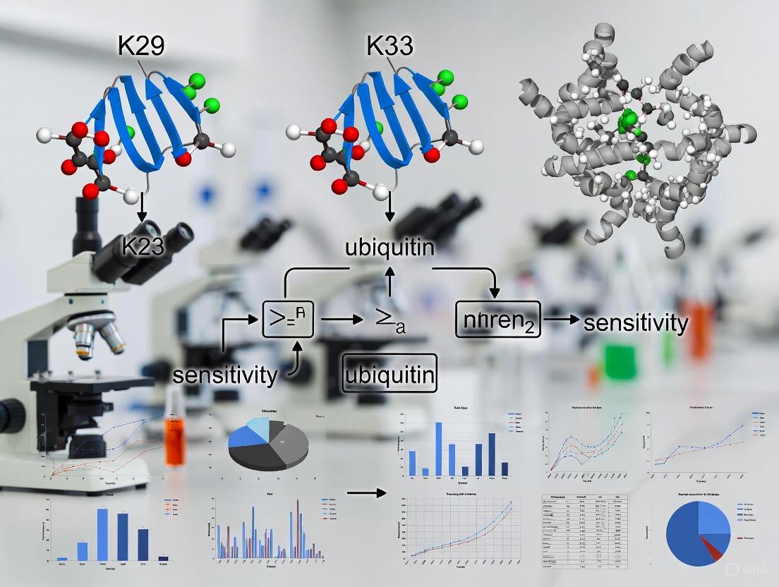
Fundamental Characteristics of K29 and K33 Linkages
Key Assembly Enzymes and Structural Features
K29- and K33-linked ubiquitin chains are assembled by specific HECT E3 ligases and exhibit unique structural properties that differentiate them from classical ubiquitin linkages.
Table 1: Enzymes Assembling Atypical Ubiquitin Chains
| Linkage Type | Primary E3 Ligase | Chain Architecture | Solution Conformation |
|---|---|---|---|
| K29-linked | UBE3C [1] | Homotypic or branched with K48 [1] [2] | Extended, open, and dynamic [1] [3] |
| K33-linked | AREL1 (KIAA0317) [1] | Homotypic or branched with K11 [1] [2] | Extended, open, and dynamic [1] |
The HECT E3 ligase UBE3C assembles chains containing both K29 and K48 linkages, with mass spectrometry analyses revealing approximately 23% K29, 63% K48, and 10% K11 linkages in its assembly products [1]. In contrast, the HECT E3 ligase AREL1 assembles chains containing K33 (36%), K11 (36%), and K48 (20%) linkages [1]. Both K29- and K33-linked diubiquitin adopt open and dynamic conformations in solution, similar to K63-linked chains, making them structurally distinct from the compact conformations of K48-linked chains [1] [3].
Biological Functions and Significance
Although less characterized than classical linkages, K29 and K33 chains play important roles in cellular regulation:
- K29-linked chains have been associated with neurodegenerative disorders, Wnt signaling downregulation, and autophagy [4]. They frequently exist within mixed or branched chains containing other linkages rather than as pure homotypic polymers [3].
- K33-linked chains are involved in immune signaling and kinase regulation, though their full physiological roles remain under investigation [1].
The presence of these atypical linkages within heterotypic branched chains significantly expands the complexity of the ubiquitin code and presents particular challenges for specific detection [5] [2].
Essential Experimental Protocols
Determining Ubiquitin Chain Linkage Using Mutational Analysis
This protocol uses ubiquitin lysine mutants to identify specific chain linkages in vitro [6].
Table 2: Reaction Components for Linkage Determination
| Component | Stock Concentration | Volume per 25µL Reaction | Final Concentration |
|---|---|---|---|
| E1 Enzyme | 5 µM | 0.5 µL | 100 nM |
| E2 Enzyme | 25 µM | 1 µL | 1 µM |
| E3 Ligase | 10 µM | Variable | 1 µM |
| 10X E3 Ligase Buffer | 10X | 2.5 µL | 1X |
| Wild-type or Mutant Ubiquitin | 1.17 mM (10 mg/mL) | 1 µL | ~100 µM |
| MgATP Solution | 100 mM | 2.5 µL | 10 mM |
| Substrate | Variable | Variable | 5-10 µM |
Procedure:
Set up two parallel experimental series:
- Series A (K-to-R mutants): Eight reactions containing wild-type ubiquitin and seven Ubiquitin K-to-R Mutants (K6R, K11R, K27R, K29R, K33R, K48R, K63R)
- Series B (K-only mutants): Eight reactions containing wild-type ubiquitin and seven Ubiquitin K-only Mutants (K6-only, K11-only, K27-only, K29-only, K33-only, K48-only, K63-only)
Assemble reactions in the order listed in Table 2, adding components to microcentrifuge tubes
Incubate at 37°C for 30-60 minutes
Terminate reactions with:
- SDS-PAGE sample buffer (for direct analysis)
- EDTA to 20 mM or DTT to 100 mM (for downstream applications)
Analyze by Western blot using anti-ubiquitin antibodies
Data Interpretation:
- In Series A, the reaction that fails to form polyubiquitin chains indicates the essential lysine for linkage
- In Series B, only the reaction with the K-only mutant corresponding to the linkage type will form chains
- If all K-to-R mutant reactions form chains, consider M1 (linear) linkage or mixed/branched chains [6]
UbiCRest Assay for Chain Architecture Analysis
The Ubiquitin Chain Restriction (UbiCRest) assay uses linkage-specific deubiquitinases (DUBs) to decipher chain composition [5] [2].
Table 3: Linkage-Specific DUBs for UbiCRest Analysis
| DUB Enzyme | Preferred Linkage Specificity |
|---|---|
| OTUD3 | K6, K11 |
| Cezanne | K11 |
| OTUD2 | K11, K27, K29, K33 |
| TRABID | K29, K33, K63 |
| OTUB1 | K48 |
| OTUD1/AMSH | K63 |
| OTULIN | M1 (linear) |
| USP21/vOTU | Non-specific (controls) |
Procedure:
- Prepare ubiquitinated samples of interest
- Aliquot samples into multiple tubes for parallel DUB digestions
- Incubate with individual linkage-specific DUBs (Table 3) under appropriate buffer conditions
- Terminate reactions with SDS-PAGE sample buffer
- Analyze by Western blot using anti-ubiquitin antibodies
Data Interpretation:
- DUBs cleave their preferred linkages, altering the ubiquitin ladder pattern on Western blots
- Residual signal after DUB treatment indicates presence of resistant linkages
- Comparison across multiple DUB treatments reveals chain composition
- K29/K33 chains show specific cleavage by TRABID and resistance to other DUBs [5]
Troubleshooting Guides & FAQs
Frequently Encountered Experimental Challenges
Q: My ubiquitin Western blots show smearing rather than discrete bands. Is this normal for K29/K33 chains? A: Yes, smearing is normal and expected when analyzing polyubiquitin chains. The smear represents proteins modified with ubiquitin chains of varying lengths. K29 and K33 chains often form heterogeneous populations, potentially contributing to smearing patterns [4].
Q: How can I distinguish between K29/K33 homotypic chains and branched chains containing these linkages? A: Use multiple complementary approaches:
- Perform UbiCRest with DUB panels - branched chains often show partial resistance to digestion [5]
- Use ubiquitin mutants (K-to-R) in combination assays
- Employ specialized MS approaches like UbiChEM-MS that can detect branch points directly [5]
- Use recently developed bispecific antibodies when available [5]
Q: Why can't I detect endogenous K29/K33 ubiquitination despite using linkage-specific antibodies? A: Endogenous atypical chains are often low in abundance and transient. Try these sensitivity enhancements:
- Treat cells with proteasome inhibitors (e.g., MG-132 at 5-25 µM for 1-2 hours) to stabilize modifications [4]
- Use ubiquitin enrichment tools like Ubiquitin-Trap prior to Western blotting [4]
- Optimize lysis conditions with fresh N-ethylmaleimide (NEM) to inhibit DUB activity
- Validate antibody specificity with positive controls using ubiquitin mutants
Q: How do I confirm that my observed signal is truly K29- or K33-linked and not mixed/branched chains? A: Implement a multi-step verification protocol:
- Use both K-to-R and K-only ubiquitin mutants in parallel assays [6]
- Express single-lysine ubiquitin mutants (K29-only or K33-only) in cells
- Perform reciprocal immunoprecipitation with linkage-specific binders like TRABID NZF1 domain [1]
- Use mass spectrometry for definitive linkage identification when possible
Optimization of Sensitivity for K29/K33 Detection
Increasing Signal Detection:
- Use high-affinity ubiquitin capture reagents like tandem-repeated ubiquitin-binding entities (TUBEs) to enhance enrichment [7]
- Combine multiple enrichment strategies sequentially (e.g., Ubiquitin-Trap followed by linkage-specific binder)
- Optimize electrophoresis conditions - use Tris-acetate gels for better separation of higher molecular weight ubiquitinated species [7]
Reducing Background:
- Include NEM (5-20 mM) in lysis buffers to prevent deubiquitination during sample preparation [7]
- Use high-stringency washes (e.g., with 0.1% SDS) during enrichment steps
- Include appropriate negative controls (catalytically dead E3 mutants, ubiquitin mutants)
The Scientist's Toolkit: Essential Research Reagents
Table 4: Key Reagents for K29/K33 Ubiquitin Chain Research
| Reagent Category | Specific Examples | Utility and Function |
|---|---|---|
| E3 Ligases | UBE3C (for K29) [1], AREL1 (for K33) [1] | Linkage-specific chain assembly in reconstituted systems |
| Linkage-Specific DUBs | TRABID (K29/K33-specific) [1] [5], OTUD2 (K29/K33 and others) [5] | Analytical tools for linkage verification via UbiCRest |
| Ubiquitin Binders | TRABID NZF1 domain [1] [3], Ubiquitin-Trap [4] | Enrichment and detection of specific chain types |
| Ubiquitin Mutants | K29-only, K33-only, K29R, K33R [6] | Essential controls and tools for linkage determination |
| Detection Tools | Linkage-specific antibodies, Anti-ubiquitin antibodies [4] | Western blot detection and quantification |
Advanced Methodologies for Enhanced Sensitivity
Structural Insights for Assay Design
The N-terminal NZF1 domain of the deubiquitinase TRABID specifically recognizes K29- and K33-linked diubiquitin [1] [3]. Structural studies reveal that TRABID NZF1 binds each Ub-Ub interface in K33-linked chains, exploiting their flexibility for selective recognition [1]. This structural information enables rational design of sensitive detection reagents:
- Engineered TRABID NZF domains can be used as affinity capture tools
- Structure-guided mutations can enhance linkage selectivity
- Fusion proteins with reporter tags facilitate sensitive detection
Mass Spectrometry Approaches
Advanced MS methods provide the highest specificity for identifying atypical ubiquitin linkages:
- UbiChEM-MS: Uses minimal trypsinolysis to preserve branch points, allowing identification of branched ubiquitin modifications [5]
- DiGly remnant enrichment: Standard proteomic approach modified with specialized sample preparation to preserve atypical linkages
- Absolute quantification (AQUA): Uses isotope-labeled standard peptides for precise quantification of specific linkage types [1]
Troubleshooting Guides & FAQs
General Experimental Setup
Q1: My ubiquitination assays show weak or no signal for K29/K33 linkages. What could be the cause? A: Weak signal for K29/K33 chains is common due to antibody sensitivity issues and linkage competition.
- Primary Cause: Commercial antibodies for K29/K33 have significantly lower affinity compared to K48/K63 antibodies.
- Solution: Increase input protein (200-400 µg for immunoprecipitation). Use chain-specific E3 ligases (TRIP12/UBE3C for K29; AREL1 for K33) in in vitro assays to enrich target linkages. Verify with linkage-specific deubiquitinases (DUBs) as negative controls.
Q2: I am observing high background noise in my Western blots when probing for atypical ubiquitin chains. How can I improve the signal-to-noise ratio? A: High background is often due to antibody cross-reactivity.
- Primary Cause: Antibodies may cross-react with more abundant K48/K63 chains or non-specific proteins.
- Solution:
- Pre-clear lysates with control agarose beads for 1 hour.
- Increase the number of washes in your IP protocol (5-7 washes with high-stringency buffer: 50 mM Tris-HCl, pH 7.5, 500 mM NaCl, 0.1% NP-40).
- Optimize antibody concentration; too much antibody increases background. Titrate to find the optimal dilution.
E3 Ligase-Specific Issues
Q3: The knockdown of TRIP12 or UBE3C in my cells does not show a clear reduction in K29-linked ubiquitination. Why? A: Functional redundancy is a key challenge.
- Primary Cause: TRIP12 and UBE3C can compensate for each other in K29 chain formation.
- Solution: Perform a double knockdown or knockout of both E3s. Use CRISPR/Cas9 to generate dual-knockout cell lines for a more definitive phenotype. Confirm knockdown/knockout efficiency at both mRNA (qPCR) and protein (Western blot) levels.
Q4: My in vitro reconstitution assay with AREL1 is not producing K33-linked chains. What components should I verify? A: An incomplete reaction mix is the most likely culprit.
- Primary Cause: Missing or inactive components in the ubiquitination cascade.
- Solution: Confirm the presence and activity of all essential factors as per the table below.
Table: Critical Components for In Vitro Ubiquitination Assay
| Component | Function | Recommended Concentration |
|---|---|---|
| E1 Activating Enzyme | Activates ubiquitin | 50-100 nM |
| E2 Conjugating Enzyme (e.g., UBE2K for K29) | Cooperates with E3 for linkage specificity | 1-5 µM |
| E3 Ligase (TRIP12, UBE3C, AREL1) | Substrate recognition and catalysis | 0.5-2 µM |
| Ubiquitin | Substrate for chain formation | 50-100 µM |
| ATP | Energy source for E1 | 2-5 mM |
| Mg²⁺ | Essential cofactor for E1/E2 activity | 5 mM |
Detection and Validation
Q5: How can I definitively confirm that the chains I'm detecting are genuinely K29 or K33-linked and not a mix? A: Use orthogonal validation methods beyond Western blotting.
- Primary Cause: Reliance on a single, potentially cross-reactive detection method.
- Solution:
- Tandem Ubiquitin Binding Entity (TUBE) Pulldown: Use linkage-specific TUBEs (e.g., K29-specific TUBEs) for enrichment, followed by mass spectrometry.
- Linkage-Specific DUB Treatment: Treat your samples with linkage-specific DUBs (e.g., vOTU for K11/K33/K48; Cezanne for K11) and observe the cleavage pattern.
- Mass Spectrometry: The gold standard. Digest samples with trypsin and analyze for di-glycine remnants on specific lysines (K29 or K33) of ubiquitin.
Detailed Experimental Protocols
Protocol 1: Tandem Immunoprecipitation for Enhanced K29/K33 Detection
This protocol minimizes background and enriches for atypical chains.
- Cell Lysis: Lyse cells in 1 mL of NP-40 lysis buffer (50 mM Tris-HCl pH 7.5, 150 mM NaCl, 1% NP-40, 1x protease inhibitor, 10 mM N-Ethylmaleimide (NEM to inhibit DUBs), 50 µM PR-619 (DUB inhibitor)) for 30 min on ice.
- Pre-clearing: Centrifuge at 16,000 x g for 15 min. Transfer supernatant to a new tube and incubate with 20 µL of control IgG-agarose beads for 1 hour at 4°C.
- First IP (Total Ubiquitin): Incubate pre-cleared lysate with 2 µg of pan-ubiquitin antibody (e.g., FK2) and 30 µL of Protein A/G beads overnight at 4°C.
- Washing: Pellet beads and wash 5 times with 1 mL of high-salt wash buffer (50 mM Tris-HCl pH 7.5, 500 mM NaCl, 0.1% NP-40).
- Elution: Elute ubiquitinated proteins by incubating beads with 50 µL of 0.1 M Glycine pH 2.5 for 10 min at room temperature. Immediately neutralize with 5 µL of 1 M Tris-HCl pH 8.0.
- Second IP (Linkage-Specific): Dilute the eluate with 500 µL of lysis buffer. Add 1 µg of linkage-specific antibody (e.g., anti-K29) and fresh beads. Incubate for 4 hours at 4°C.
- Final Wash and Elution: Wash 5 times with high-salt wash buffer. Elute with 2x Laemmli buffer for Western blot analysis.
Protocol 2:In VitroUbiquitination Assay with Recombinant E3s
A method to study E3 activity and linkage specificity directly.
- Reaction Setup: Assemble a 30 µL reaction on ice:
- 3 µL 10X Reaction Buffer (200 mM Tris-HCl pH 7.5, 50 mM MgCl₂, 10 mM DTT)
- 1 µL 100x Energy Regeneration Solution (40 mM ATP, 400 mM Creatine Phosphate, 2 mg/mL Creatine Kinase)
- 50 nM E1 (Human UBA1)
- 2 µM E2 (e.g., UBE2K for TRIP12/UBE3C)
- 1 µM E3 (Recombinant TRIP12, UBE3C, or AREL1)
- 50 µM Ubiquitin (Wild-type or mutant)
- Nuclease-free water to 30 µL
- Incubation: Incubate the reaction at 30°C for 90 minutes.
- Termination: Stop the reaction by adding 10 µL of 4x Laemmli buffer and boiling at 95°C for 5 minutes.
- Analysis: Resolve 15-20 µL of the reaction by SDS-PAGE (4-12% Bis-Tris gel). Perform Western blotting with anti-ubiquitin antibody to detect polyubiquitin chain formation.
Signaling Pathway and Workflow Visualizations
E3 Ligase Specificity in Chain Assembly
Tandem IP Workflow for K29/K33 Detection
The Scientist's Toolkit
Table: Essential Research Reagents for K29/K33 Ubiquitin Research
| Reagent | Function / Application | Key Note |
|---|---|---|
| Anti-K29 Linkage Antibody | Detection of K29-linked chains via WB/IF. | High batch-to-batch variability; requires extensive validation. |
| Anti-K33 Linkage Antibody | Detection of K33-linked chains via WB/IF. | Less characterized; prone to cross-reactivity. |
| Recombinant TRIP12/UBE3C/AREL1 | In vitro ubiquitination assays to study direct activity. | Crucial for confirming E3 specificity without cellular redundancy. |
| Linkage-Specific DUBs (e.g., vOTU) | Enzymatic validation of linkage identity. | Cleaves specific linkages; loss of signal upon treatment confirms presence. |
| TUBEs (Tandem Ubiquitin Binding Entities) | Affinity enrichment of polyubiquitinated proteins. | Protects chains from DUBs; some TUBEs show linkage preference. |
| N-Ethylmaleimide (NEM) | DUB inhibitor. | Alkylates cysteine residues; essential in lysis buffer to preserve chains. |
| UBE2K (E2) | Cooperates with TRIP12/UBE3C for K29 synthesis. | E2 choice is critical for reconstituting specific linkage formation. |
| PR-619 | Broad-spectrum DUB inhibitor. | Used in conjunction with NEM for maximum DUB inhibition. |
Technical Support Center
Welcome to the TRABID NZF1 Technical Support Center. This resource is designed to assist researchers in overcoming common experimental challenges in the study of K29- and K33-linked ubiquitin chains, with a focus on the role of TRABID's NZF1 domain as a critical linkage-specific reader.
Frequently Asked Questions (FAQs)
Q1: Our lab is struggling with the sensitivity of detecting endogenous K29/K33 linkages in cell lysates. What is the most critical factor for success? A1: The most critical factor is the preservation of the ubiquitin linkage during lysis. Standard RIPA buffers can contain high concentrations of SDS or be used at non-physiological pH, which can disrupt non-covalent interactions between readers like TRABID-NZF1 and ubiquitin chains. We recommend using a mild, non-denaturing lysis buffer (e.g., 50 mM Tris-HCl pH 7.5, 150 mM NaCl, 1% NP-40, 10% Glycerol, 1 mM EDTA) supplemented with 10 mM N-Ethylmaleimide (NEM) to inhibit deubiquitinases (DUBs) and 1x protease inhibitors. Pre-clearing lysates with control beads is also essential to reduce non-specific binding.
Q2: We are performing a pulldown with recombinant GST-TRABID-NZF1, but the background binding is high. How can we improve the signal-to-noise ratio? A2: High background is often due to non-specific ionic interactions. Ensure your wash buffer contains a sufficient salt concentration (e.g., 150-300 mM NaCl). Including a low concentration of a non-ionic detergent like 0.1% Tween-20 in the wash buffer can also help. Furthermore, titrating the amount of recombinant bait protein used in the assay can be beneficial; using more than necessary often increases background without improving specific binding.
Q3: What are the best negative and positive controls for a TRABID-NZF1 ubiquitin linkage binding assay? A3:
- Positive Control: Use di-ubiquitin chains of known linkage (K29-, K33-, and K63-linked) in a slot-blot or pull-down format to confirm your recombinant protein is functional.
- Negative Control 1: A mutant version of TRABID-NZF1 where the key residues for ubiquitin binding (e.g., T74, D35) are mutated to alanine.
- Negative Control 2: Include K48- and K11-linked di-ubiquitin chains, as TRABID-NZF1 shows significantly lower affinity for these linkages.
- Negative Control 3: Beads coupled to the tag-only (e.g., GST alone) in your cellular pulldown experiments.
Troubleshooting Guides
Issue: Inconsistent results between pulldown assays and immunohistochemistry (IHC) when using a TRABID-NZF1 specific antibody.
| Symptom | Possible Cause | Solution |
|---|---|---|
| Strong signal in pulldown, no signal in IHC. | Antibody cannot recognize TRABID in its native, fixed state due to epitope masking. | Try antigen retrieval methods (heat-induced or enzymatic). Test another antibody raised against a different epitope of TRABID. |
| High background staining in IHC. | Non-specific antibody binding or insufficient blocking. | Optimize antibody dilution. Increase blocking time (use 5% normal serum + 1% BSA). Include a no-primary-antibody control. |
| Discrepancy between linkage detection (K29/K33 high in pulldown, low in IHC). | IHC may reflect total TRABID localization, not its active, ubiquitin-bound state. | Perform proximity ligation assay (PLA) using anti-TRABID and anti-ubiquitin antibodies to visualize specific interaction sites in situ. |
Issue: Low yield of K29/K33-linked polypeptides after affinity purification with TRABID-NZF1 for mass spectrometry.
| Symptom | Possible Cause | Solution |
|---|---|---|
| Few unique peptides identified for K29/K33 linkages. | Sample loss during clean-up steps or ion suppression from highly abundant proteins. | Use StageTips (C18) for sample clean-up instead of column-based methods for higher recovery. Pre-fractionate your sample by strong cation exchange (SCX) chromatography before LC-MS/MS. |
| High identification of K48/K63 linkages. | Incomplete specificity of the NZF1 domain or carryover of abundant chains. | Use the NZF1 domain as a pre-clearance step to deplete non-specific chains before the main purification. Optimize wash stringency (see FAQ A2). |
| Poor MS/MS spectrum quality for ubiquitin remnants. | Inefficient digestion or missed cleavage sites. | Use a high-quality, sequencing-grade trypsin/Lys-C mix for digestion. Ensure denaturation and reduction/alkylation steps are performed thoroughly. |
Experimental Protocols
Protocol 1: Recombinant TRABID-NZF1 Ubiquitin Chain Binding Assay
Purpose: To qualitatively and quantitatively assess the binding specificity of the TRABID NZF1 domain to different ubiquitin linkages.
- Protein Immobilization: Incubate 10 µg of recombinant GST-TRABID-NZF1 (or GST alone as control) with Glutathione Sepharose 4B beads in 500 µL of Binding Buffer (50 mM Tris pH 7.5, 150 mM NaCl, 0.1% NP-40, 1 mM DTT, 0.5 mg/mL BSA) for 1 hour at 4°C on a rotator.
- Wash Beads: Wash beads 3 times with 1 mL of Binding Buffer (without BSA) to remove unbound protein.
- Binding Reaction: Resuspend the beads in 400 µL of Binding Buffer (with BSA). Add 1 µg of the desired di-ubiquitin chain (K29, K33, K48, K63, etc.). Incubate for 2 hours at 4°C on a rotator.
- Stringency Washes: Pellet beads and wash 3 times with 1 mL of Wash Buffer (Binding Buffer with 300 mM NaCl).
- Elution: Elute bound proteins by adding 40 µL of 2x Laemmli sample buffer and boiling at 95°C for 5 minutes.
- Analysis: Resolve eluates by SDS-PAGE and transfer to a PVDF membrane. Perform immunoblotting using a pan-ubiquitin antibody (e.g., P4D1) or linkage-specific antibodies if available.
Protocol 2: Affinity Purification of K29/K33-Linked Proteins from Cell Lysates
Purpose: To enrich and identify endogenous proteins modified with K29- and K33-linked ubiquitin chains.
- Cell Lysis: Harvest cells and lyse in 1 mL of non-denaturing Lysis Buffer per 10 cm dish (50 mM Tris-HCl pH 7.5, 150 mM NaCl, 1% NP-40, 10% Glycerol, 1 mM EDTA, 10 mM NEM, 1x protease/phosphatase inhibitors). Incubate on ice for 20 min, then centrifuge at 16,000 x g for 15 min at 4°C.
- Pre-clearance: Incubate the supernatant with 50 µL of control GST beads for 1 hour at 4°C to remove non-specific binders.
- Affinity Purification: Transfer the pre-cleared supernatant to a tube containing 50 µL of GST-TRABID-NZF1 beads. Incubate for 3 hours at 4°C on a rotator.
- Washing: Wash the beads 4 times with 1 mL of Lysis Buffer (without inhibitors) and a final wash with 50 mM Ammonium Bicarbonate (pH 8.0).
- On-bead Digestion: Reduce, alkylate, and digest the captured proteins on-bead using trypsin/Lys-C mix overnight at 37°C.
- Peptide Recovery: Acidify the supernatant with trifluoroacetic acid (TFA) to pH <3. Desalt peptides using C18 StageTips and proceed with LC-MS/MS analysis.
Pathway and Workflow Visualizations
Title: TRABID NZF1 Role in Ubiquitin Signaling
Title: K29/K33 Enrichment Workflow
The Scientist's Toolkit: Research Reagent Solutions
| Reagent / Material | Function & Application |
|---|---|
| Recombinant GST-TRABID-NZF1 | The key bait protein for affinity purification and binding assays to specifically capture K29/K33-linked ubiquitin chains. |
| Linkage-specific Di-ubiquitin (K29, K33, K48, K63) | Essential positive and negative controls for validating the binding specificity of your TRABID-NZF1 reagent in vitro. |
| N-Ethylmaleimide (NEM) | A deubiquitinase (DUB) inhibitor. Critical to add to lysis buffers to prevent the degradation of ubiquitin chains during sample preparation. |
| Non-denaturing Lysis Buffer | Preserves non-covalent protein-protein interactions, allowing for the co-purification of ubiquitin readers with their cognate chains. |
| Glutathione Sepharose 4B | The solid support for immobilizing GST-tagged TRABID-NZF1 for pull-down experiments. |
| Pan-Ubiquitin Antibody (P4D1) | A standard antibody for detecting total ubiquitin in western blots following affinity purification. |
| Trypsin/Lys-C Mix | A high-efficiency protease for on-bead digestion of captured proteins prior to mass spectrometric identification. |
| C18 StageTips | A low-cost, high-recovery method for desalting and concentrating peptide samples for LC-MS/MS, minimizing sample loss. |
Troubleshooting Guide: Key Challenges in K29/K33 Ubiquitin Chain Research
This guide addresses common experimental hurdles in the study of atypical K29- and K33-linked ubiquitin chains and provides targeted solutions to improve detection sensitivity and specificity.
Table 1: Troubleshooting Atypical Ubiquitin Chain Analysis
| Problem | Potential Cause | Recommended Solution | Key Research Reagents |
|---|---|---|---|
| Low sensitivity for endogenous K29/K33 chain detection | - Antibody low affinity or specificity- Masking by abundant chains (K48/K63)- Low endogenous abundance | - Use linkage-specific DUBs (e.g., TRABID) for validation [1]- Enrich chains using recombinant NZF1 domain of TRABID [1]- Optimize E3 ligases (UBE3C for K29; AREL1 for K33) for in-vitro assembly [1] | TRABID DUB, UBE3C E3 Ligase, AREL1 E3 Ligase |
| Difficulty distinguishing mixed/branched from homotypic chains | - Standard MS/MS may miss complex topology- Lack of tools for branched chain analysis | - Use Ubiquitin Activated Enzyme (E1) and UBE2D E2 in vitro [1]- Implement AQUA mass spectrometry with isotope-labeled GlyGly peptides for absolute quantification [1] | AQUA Mass Spectrometry Kits, Ubiquitin Chain Assembly Kit |
| High background in western blotting | - Non-specific antibody cross-reactivity | - Switch to high-sensitivity, quantitative methods like Simple Western [8]- Validate with genetic (DUB siRNA) and chemical (proteasome inhibitor) controls | Simple Western System, Proteasome Inhibitor (e.g., MG132) |
| Inability to monitor dynamics in cells | - Lack of live-cell reporters for atypical chains | - Develop cell lines expressing tagged ubiquitin (K29-only, K33-only mutants) [1]- Monitor TRABID localization to Ub-rich puncta as a sensor [1] | K29-only/K33-only Ubiquitin Mutants, TRABID Expression Plasmid |
Experimental Protocol: Using TRABID NZF1 Domain for K29/K33 Chain Enrichment
This protocol uses the specific binding of the TRABID NZF1 domain to isolate and enrich K29- and K33-linked ubiquitin chains from complex cell lysates, thereby improving downstream detection sensitivity [1].
- Recombinant NZF1 Production: Express and purify the N-terminal NZF1 domain (amino acids 1-70) of human TRABID in E. coli using a standard His-tag system [1].
- Immobilization: Couple the purified NZF1 domain to a solid chromatography resin (e.g., NHS-activated Sepharose) according to the manufacturer's instructions.
- Lysate Preparation: Lyse cells in a non-denaturing lysis buffer (e.g., RIPA buffer) supplemented with 10 mM N-Ethylmaleimide (NEM) to inhibit deubiquitinases and 1× protease inhibitor cocktail.
- Enrichment: Incubate the clarified cell lysate with the NZF1-coupled resin for 2 hours at 4°C with gentle rotation.
- Washing: Wash the resin extensively with cold lysis buffer to remove non-specifically bound proteins.
- Elution: Elute the bound ubiquitin chains using a low-pH elution buffer (e.g., 0.1 M glycine, pH 2.5) and immediately neutralize the eluate with 1 M Tris-HCl, pH 8.0.
- Analysis: Analyze the eluted material by western blotting or mass spectrometry.
Frequently Asked Questions (FAQs)
Q1: Why is the research on K29 and K33-linked ubiquitin chains important for drug discovery?
Understanding these atypical chains expands the "druggable" proteome. Since these linkages have distinct cellular roles, targeting their assembly or recognition offers new avenues for therapeutic intervention in cancer and neurodegenerative diseases where these chains are implicated [9] [2]. Furthermore, components of these pathways, like the E3 ligase UBE3C, can themselves be investigated as drug targets [1].
Q2: My mass spectrometry data suggests the presence of K29 linkages, but western blot confirmation is inconsistent. What is the best validation strategy?
Employ an orthogonal, activity-based validation method. The recommended approach is to treat your samples with the linkage-specific deubiquitinase (DUB) TRABID, which preferentially cleaves K29 and K33 linkages [1]. A significant reduction in your signal upon TRABID treatment, compared to a catalytically inactive mutant or a buffer control, provides strong functional evidence for the presence of these specific chains.
Q3: Beyond proteasomal degradation, what are the primary cellular functions of K29 and K33-linked ubiquitin chains?
While some K29 linkages (particularly when mixed with K48) can target substrates for proteasomal degradation, evidence suggests both K29 and K33 chains are primarily involved in non-proteolytic signaling [1]. They are implicated in critical processes such as:
- Intracellular trafficking: Regulating protein movement within the cell.
- Inflammatory signaling: Modulating pathways like NF-κB.
- Kinase signaling: Controlling the activity of various kinases.
- The DNA damage response: Facilitating the repair of DNA lesions [2].
Q4: What are the advantages of using recombinant E3 ligases like UBE3C and AREL1 to study atypical ubiquitin chains?
These HECT-family E3 ligases are essential tools because they provide a defined enzymatic source to generate homotypic K29- or K33-linked chains in vitro [1]. This allows researchers to:
- Produce reference standards for mass spectrometry.
- Generate substrates for DUB activity assays.
- Study the biochemistry of chain assembly without the complexity of a full cellular extract.
Pathway Visualization: Sensing Proteotoxic Stress and Activating the HSR
The following diagram illustrates the core cytoplasmic pathway through which cells sense proteotoxic stress, such as heat shock, and activate the Heat Shock Response (HSR) to restore proteostasis [10].
Cytoplasmic Heat Shock Response Pathway
The Scientist's Toolkit: Essential Research Reagents
This table details key reagents used in the study of atypical ubiquitin chains and proteotoxic stress, as featured in the troubleshooting guides and protocols.
Table 2: Key Research Reagent Solutions
| Reagent / Tool | Function / Application | Example Use in K29/K33 Research |
|---|---|---|
| TRABID (DUB) | Linkage-specific deubiquitinase for K29 and K33 chains [1]. | Functional validation of K29/K33 linkages via enzymatic cleavage assays [1]. |
| UBE3C (HECT E3 Ligase) | Assembles K29- and K48-linked ubiquitin chains [1]. | In-vitro generation of K29-linked chains for use as standards or substrates [1]. |
| AREL1 (HECT E3 Ligase) | Assembles K33- and K11-linked ubiquitin chains [1]. | In-vitro generation of K33-linked chains to study their biophysics and recognition [1]. |
| TRABID NZF1 Domain | High-affinity ubiquitin-binding domain specific for K29/K33 linkages [1]. | Affinity enrichment of K29/K33 chains from cell lysates to improve detection sensitivity [1]. |
| K29-only / K33-only Ub Mutants | Ubiquitin mutants where all lysines except K29 or K33 are mutated to arginine [1]. | Tools to force homotypic chain formation in cellular and in-vitro assays [1]. |
| Simple Western System | Automated, capillary-based western blot system [8]. | High-throughput, quantitative analysis of protein degradation and ubiquitin chain formation [8]. |
| AQUA Mass Spectrometry | Absolute quantification of ubiquitin chain linkage types using heavy isotope-labeled peptides [1]. | Precise measurement of the relative abundance of all ubiquitin chain types in a sample [1]. |
Troubleshooting Guide: FAQs on Neurodegeneration-Cancer Research
FAQ: What are the core molecular pathways that exhibit opposite regulation in neurodegeneration and cancer?
Several key oncogenic signaling pathways are dysregulated in opposite ways in cancer versus neurodegenerative diseases. While cancer promotes cell survival and proliferation, neurodegeneration drives cell death and apoptosis, often through the same molecular triggers [11].
- The Hippo Pathway & its Effector YAP: In many cancers, the Hippo pathway is inactivated, leading to YAP (Yes-associated protein) nuclear localization and the transcription of pro-survival and proliferative genes [11]. Conversely, in Alzheimer's disease (AD) and Huntington's disease (HD) models, YAP becomes sequestered in the cytoplasm, which is linked to neuronal death and endoplasmic reticulum stress. Restoring nuclear YAP in a mouse model of AD decreased Aβ plaques and improved behavioral outcomes [11].
- The p53 Pathway: The p53 tumor suppressor is commonly downregulated or mutated in cancer, allowing for unchecked cell growth [12]. In contrast, p53 expression is upregulated in the brains of patients with AD, PD, and HD, where it is associated with the activation of cell death pathways in post-mitotic neurons [12].
- The Pin1 Pathway: Pin1 is notably upregulated in many cancers but is downregulated in AD, illustrating another example of inverse molecular regulation [12].
FAQ: How can I specifically study K29-linked ubiquitination in the context of disease mechanisms?
A primary challenge in ubiquitin research is the specific detection and analysis of atypical ubiquitin chain linkages like K29. Standard methods like western blotting are low-throughput and may lack linkage specificity [13].
- Solution: Employ chain-specific Tandem Ubiquitin Binding Entities (TUBEs). These are high-affinity binding reagents engineered to selectively capture specific polyubiquitin chain topologies from cell lysates [13].
- Protocol Overview:
- Cell Stimulation & Lysis: Treat cells (e.g., THP-1 monocytic cells) with your experimental stimulus (e.g., a PROTAC to induce degradation/K48 linkages, or an inflammatory agent like L18-MDP to induce K63 linkages). Use a lysis buffer optimized to preserve polyubiquitination [13].
- Affinity Capture: Incubate the cell lysate with chain-specific TUBEs (e.g., K29-TUBE, K48-TUBE) immobilized in a 96-well plate [13].
- Wash and Elute: Remove non-specifically bound proteins.
- Detection: Detect your protein of interest (e.g., RIPK2) using standard immunoblotting. The presence of your target in the pull-down indicates its modification with the specific ubiquitin chain captured by the TUBE [13].
FAQ: My research suggests an inverse comorbidity between neurodegeneration and cancer. What biological principles explain this?
The observed inverse relationship stems from the fundamentally different priorities and evolutionary trade-offs of neurons and cycling cells [12].
- The Dividing Cell's Priority: The main goal is controlled proliferation and genomic integrity. The trade-off is a higher risk of cancer if cell-cycle control is lost [12].
- The Neuron's Priority: The main goal is long-term survival and maintaining complex synaptic connections for the life of the organism. The trade-off is a loss of proliferative capacity and a higher vulnerability to age-related stressors, leading to neurodegeneration [12].
The same proteins in these two cell types are often utilized in different, sometimes opposite, ways. For instance, the re-entry into the cell cycle is a desired outcome in many tissues but is a pro-apoptotic signal in neurons [12].
Experimental Protocols & Methodologies
Detailed Protocol: Using Chain-Specific TUBEs to Probe Ubiquitination
This protocol outlines a method for investigating linkage-specific ubiquitination of an endogenous target protein, adapted from research on RIPK2 [13].
1. Key Reagents and Materials
- Cell Line: THP-1 human monocytic cells (or other relevant cell line).
- Stimuli: L18-MDP (Lysine 18-muramyldipeptide) to induce K63-linked ubiquitination via inflammatory signaling. A relevant PROTAC (e.g., "RIPK2 PROTAC") to induce K48-linked ubiquitination and proteasomal degradation [13].
- Inhibitors: Ponatinib (RIPK2 inhibitor) for control experiments [13].
- Chain-Specific TUBEs: K29-, K48-, K63-, and Pan-selective TUBEs.
- Lysis Buffer: A specialized buffer (e.g., containing N-ethylmaleimide to inhibit deubiquitinases) to preserve labile polyubiquitin chains.
- Antibodies: Antibody against your protein of interest (e.g., anti-RIPK2).
2. Step-by-Step Procedure
| Step | Action | Details & Purpose |
|---|---|---|
| 1 | Cell Treatment | Pre-treat cells with inhibitor (e.g., Ponatinib) or vehicle control (DMSO) for 30 minutes. Then, stimulate with L18-MDP (200-500 ng/mL), PROTAC, or vehicle control for a defined time (e.g., 30 min) [13]. |
| 2 | Cell Lysis | Lyse cells in the specialized ubiquitin-preserving lysis buffer. Clear lysates by centrifugation [13]. |
| 3 | Affinity Capture | Incubate cell lysates with the different chain-specific TUBEs (immobilized on a plate or beads) to allow binding of ubiquitinated proteins [13]. |
| 4 | Washing | Wash the TUBE matrix thoroughly to remove non-specifically bound proteins. |
| 5 | Elution & Detection | Elute bound proteins and detect your protein of interest by immunoblotting. The signal intensity correlates with the level of specific ubiquitin linkage on the target [13]. |
3. Expected Results and Analysis
- L18-MDP stimulation should yield a strong signal for your target when captured with K63-TUBEs and Pan-TUBEs, but not with K48-TUBEs [13].
- PROTAC stimulation should yield a strong signal when captured with K48-TUBEs and Pan-TUBEs, but not with K63-TUBEs [13].
- Pre-treatment with a specific kinase inhibitor (e.g., Ponatinib) should suppress L18-MDP-induced ubiquitination, serving as a useful control [13].
The Scientist's Toolkit: Key Research Reagent Solutions
| Research Reagent | Function / Application in the Field |
|---|---|
| Chain-Specific TUBEs (K29, K48, K63, Pan) | High-affinity reagents for the selective capture and analysis of specific polyubiquitin chain linkages from cell lysates in a high-throughput format [13]. |
| PROTACs (Proteolysis Targeting Chimeras) | Heterobifunctional small molecules that recruit an E3 ligase to a target protein, inducing its K48-linked ubiquitination and degradation by the proteasome. Useful for studying degradation-dependent phenotypes [13]. |
| Planarian (flatworm) Model System | A powerful organism for studying stem cell regulation, tissue renewal, and cancer development due to its remarkable regenerative capacity and the ease of inducing cancer-like traits by disrupting genes like PTEN [14]. |
| Cryo-EM Structural Analysis | A key technique for determining the high-resolution structures of large complexes, such as E3 ligases like TRIP12 in complex with ubiquitin, revealing the molecular mechanics of linkage-specific chain formation [15]. |
Table 1. Inverse Dysregulation of Key Pathways in Cancer vs. Neurodegeneration [11] [12].
| Pathway / Molecule | Role in Cancer | Role in Neurodegeneration |
|---|---|---|
| Hippo / YAP | Inactivated; YAP nuclear, promotes proliferation & survival [11]. | Activated; YAP cytoplasmic, linked to neuronal death & ER stress [11]. |
| p53 | Frequently downregulated/mutated; allows unchecked growth [12]. | Upregulated; associated with neuronal apoptosis [12]. |
| Pin1 | Upregulated; drives proliferation [12]. | Downregulated; loss of function implicated in pathology [12]. |
Table 2. Ubiquitin Chain Linkages and Their Primary Functions [15] [13].
| Ubiquitin Linkage | Primary Known Functions |
|---|---|
| K48-linked | Targets proteins for proteasomal degradation [13]. |
| K63-linked | Regulates signal transduction, protein trafficking, NF-κB/MAPK pathways [13]. |
| K29-linked | Associated with proteotoxic stress responses; can form branched chains with K48 linkages [15]. |
Pathway and Workflow Visualizations
Hippo Pathway in Neurodegeneration vs Cancer
TUBE Assay for Linkage-Specific Ubiquitination
TRIP12 Forms K29-Linked Branched Ubiquitin Chains
Cutting-Edge Techniques for Sensitive K29 and K33 Chain Capture and Identification
Parallel Reaction Monitoring (PRM) is a targeted mass spectrometry technique that combines the high selectivity of traditional triple quadrupole methods with the high resolution and accurate mass capabilities of modern Orbitrap instrumentation [16]. Unlike discovery-mode proteomics approaches that attempt to characterize entire proteomes, PRM focuses on predefined sets of target peptides, enabling precise quantification and characterization of specific proteins with exceptional sensitivity and accuracy [16]. This makes PRM particularly valuable for applications requiring absolute protein quantification, including the study of challenging post-translational modifications such as K29- and K33-linked ubiquitin chains [17] [15].
The fundamental principle of PRM involves using the first quadrupole to selectively isolate precursor ions corresponding to target peptides, fragmenting these ions in a collision cell, and then performing high-resolution mass analysis of all fragment ions in parallel using an Orbitrap mass analyzer [16] [18]. This approach provides complete fragment ion spectra for each targeted precursor, offering both qualitative confirmation and quantitative data in a single analysis [19].
Figure 1: PRM Workflow for Absolute Protein Quantification
Technical FAQs: PRM Principles and Applications
What distinguishes PRM from other targeted mass spectrometry approaches like SRM/MRM? PRM differs fundamentally from Selected Reaction Monitoring (SRM) or Multiple Reaction Monitoring (MRM) in its detection mechanism. While SRM/MRM on triple quadrupole instruments monitors a few predefined fragment ions in the third quadrupole, PRM utilizes a high-resolution mass analyzer (typically an Orbitrap) to detect ALL fragment ions simultaneously in parallel [20]. This eliminates the need for prior transition selection and optimization, simplifies method development, and provides complete fragment ion spectra for enhanced specificity [18] [19]. The high resolution and mass accuracy (<5 ppm) of PRM significantly reduces chemical background interference, improving detection limits and quantitative accuracy, particularly in complex samples [20].
How does PRM enhance sensitivity for detecting challenging ubiquitin chain types? PRM significantly improves sensitivity for analyzing atypical ubiquitin linkages like K29 and K33 through several mechanisms. The high resolution (typically >30,000) and sub-5 ppm mass accuracy of Orbitrap-based PRM methods enable discrimination of target ions from background interferences that often obscure low-abundance ubiquitin peptides [20]. Additionally, the ability to monitor multiple fragment ions in parallel provides redundant quantitative measurements, improving statistical confidence for low-abundance species [16]. Recent advancements in sample preparation, including the use of specific E3 ligases like UBE3C and AREL1 to generate well-defined K29- and K33-linked ubiquitin chains, combined with PRM detection, have enabled previously unattainable sensitivity for these challenging post-translational modifications [17].
What are the key applications of PRM in biomedical research? PRM has diverse applications across multiple domains of biomedical research:
- Biomarker Verification: PRM enables high-throughput validation of candidate protein biomarkers discovered in untargeted proteomic screens [16] [21]. For example, PRM successfully verified mucin-5AC and mucin-2 as biomarkers for distinguishing malignant pancreatic cystic lesions with 97% accuracy [21].
- Absolute Quantification: Using stable isotope-labeled internal standards, PRM provides absolute quantification of target proteins across different biological conditions [16] [18].
- Post-Translational Modification Analysis: PRM precisely quantifies phosphorylation, ubiquitination (including K29/K33 linkages), and other modifications [17] [18].
- Pharmacokinetics and Drug Target Engagement: PRM monitors drug concentrations, metabolic products, and target protein modulation in biological matrices [16].
- Multi-omics Integration: PRM serves as a crucial bridge between discovery proteomics and validation, often following DIA or DDA experiments to verify putative biomarkers [21].
Troubleshooting Guide: Common PRM Experimental Challenges
Problem: Poor Sensitivity and Low Signal Intensity
Table 1: Troubleshooting Poor Sensitivity in PRM Experiments
| Possible Cause | Diagnostic Steps | Solutions |
|---|---|---|
| Ion Source Contamination | Check gradual signal degradation over time; performance tests with standards | Clean ion source components (cone, skimmer); replace capillary if necessary [22] |
| Suboptimal LC Conditions | Verify retention time stability; check peak shape and width | Optimize gradient parameters; use nano-LC for limited samples; ensure proper buffer preparation [22] |
| Sample Complexity/Loading | Assess total ion chromatogram quality; check column backpressure | Implement sample cleanup/fractionation; optimize loading capacity; consider carrier proteins [16] |
| Instrument Calibration | Perform mass accuracy tests with calibration standards | Recalibrate mass spectrometer; ensure proper tuning [22] |
| Precursor Selection | Evaluate peptide properties in silico | Choose proteotypic peptides with favorable ionization; avoid modified residues [18] |
Problem: High Background Noise and Interference
Table 2: Addressing Spectral Interferences in PRM
| Symptom | Potential Causes | Resolution Strategies |
|---|---|---|
| Consistent chemical noise across runs | Contaminated mobile phases or reagents | Use fresh, LC-MS grade solvents; prepare new buffers [22] |
| Specific retention time interference | Co-eluting isobaric species | Optimize chromatography; increase separation selectivity; use narrower isolation windows (±1-2 m/z) [20] |
| Elevated baseline in specific mass ranges | System contamination | Flush system with strong solvents; replace in-line filters; clean ESI source [22] |
| Unexpected fragment ions | Poor precursor isolation | Optimize quadrupole isolation width; verify collision energy settings [18] |
Problem: Inconsistent Quantification Results
Inconsistent quantification in PRM experiments often stems from variations in sample preparation, liquid chromatography performance, or instrument stability. To address these issues:
- Standardize Sample Preparation: Implement rigorous protein quantification assays before digestion, use standardized digestion protocols with quality-controlled trypsin, and minimize sample handling steps [16].
- Implement Internal Standards: Use stable isotope-labeled (SIL) peptide analogs as internal standards added at the beginning of sample preparation to normalize for recovery and ionization variability [23].
- Monitor LC Performance: Track retention time stability using iRT standards, ensure consistent mobile phase composition and degassing, and maintain LC systems regularly [21].
- Control Instrument Conditions: Perform regular mass calibration, document system performance with quality control samples, and establish scheduling windows based on observed retention times [22].
Advanced Applications: PRM for K29 and K33 Ubiquitin Chain Research
The study of atypical ubiquitin chains, particularly K29- and K33-linked polyubiquitin, presents unique challenges due to their low abundance, dynamic conformations, and complex cellular regulation [17] [15]. PRM has emerged as a powerful tool for elucidating the biology of these modifications by enabling specific detection and quantification.
Experimental Design Considerations:
When designing PRM experiments for K29/K33 ubiquitin chain analysis:
- Peptide Selection: Target signature tryptic peptides containing the specific linkage sites (e.g., peptides encompassing K29 or K33 residues)
- Sample Enrichment: Implement ubiquitin enrichment strategies using ubiquitin-binding domains (e.g., NZF domains) or linkage-specific antibodies when available [17]
- Enzymatic Specificity: Utilize recently identified E3 ligases with defined linkage specificity - UBE3C for K29/K48-branched chains and AREL1 for K11/K33-linked chains [17]
- Cross-validation: Combine PRM data with biochemical assays and structural insights to verify chain connectivity
Key Research Reagents for Ubiquitin Studies:
Table 3: Essential Research Reagents for K29/K33 Ubiquitin Chain Analysis
| Reagent/Category | Specific Examples | Research Application |
|---|---|---|
| Linkage-Specific E3 Ligases | UBE3C, AREL1, TRIP12 | In vitro assembly of defined ubiquitin chains [17] [15] |
| Ubiquitin-Binding Domains | TRABID NZF1 domain | Selective recognition and enrichment of K29/K33-linked chains [17] |
| Stable Isotope-Labeled Standards | Heavy lysine-labeled ubiquitin, AQUA peptides | Absolute quantification of ubiquitin chain abundance [20] |
| Linkage-Specific DUBs | TRABID | Analytical tools for linkage verification [17] |
| Chemical Biology Tools | Ubiquitin warhead complexes | Trapping transient ubiquitination intermediates for structural studies [15] |
Structural Insights Guiding PRM Method Development:
Recent cryo-EM structures of TRIP12, a HECT E3 ligase that generates K29 linkages and K29/K48-branched chains, reveal a pincer-like architecture that positions the acceptor ubiquitin to specifically direct K29 toward the active site [15]. This structural information informs PRM assay development by identifying:
- Key residues involved in linkage specificity
- Potential interference from structurally similar linkages
- Optimal peptide sequences for monitoring specific chain types
Figure 2: Integrated Workflow for K29/K33 Ubiquitin Chain Analysis Using PRM
Quantitative Data Analysis and Interpretation
Best Practices for PRM Data Processing:
Effective analysis of PRM data requires careful attention to several processing steps:
- Peak Integration: Manually verify automated peak detection, particularly for low-abundance targets, ensuring correct integration boundaries and baseline assignment [18]
- Fragment Ion Selection: Choose 3-5 high-intensity, interference-free fragment ions for quantification; prioritize y-ions for tryptic peptides [21]
- Retention Time Alignment: Correct for minor chromatographic shifts using internal standard peptides [21]
- Quality Control Metrics: Monitor peak symmetry, co-elution of heavy and light peptides, and consistency of fragment ion ratios
Performance Benchmarks for PRM Assays:
Table 4: Expected Performance Metrics for PRM Quantification
| Parameter | Typical Performance Range | Optimal Performance |
|---|---|---|
| Mass Accuracy | <5 ppm | <1 ppm with internal calibration [20] |
| Retention Time Stability | <0.5 min variation | <0.1 min with iRT standardization [21] |
| Linear Dynamic Range | 4-5 orders of magnitude | Up to 6 orders of magnitude [18] |
| Quantitative Precision | 10-15% RSD | <10% RSD with SIL internal standards [23] |
| Detection Sensitivity | Low attomole to femtomole range | Attomole-level with optimized样品 preparation [18] |
Troubleshooting Data Quality Issues:
When PRM data quality falls below expectations:
- Poor Chromatographic Peaks: Verify peptide stability during sample preparation, check for unexpected modifications, and optimize LC conditions
- Inconsistent Standard Curves: Prepare fresh dilution series, verify standard peptide concentrations, and check for adsorption issues
- Abnormal Fragment Ion Ratios: Investigate potential co-isolation interferences, optimize collision energies, and confirm peptide identity
- High Technical Variability: Implement more rigorous internal standardization, automate sample preparation steps, and increase replication
Emerging Innovations: 4D-PRM and Future Directions
The recent integration of ion mobility separation with PRM - creating "4D-PRM" - represents a significant advancement in targeted proteomics [21]. This approach adds a separation dimension based on ion mobility (collision cross-section) to the existing dimensions of retention time, m/z, and intensity.
Key Advantages of 4D-PRM:
- Enhanced Selectivity: Ion mobility separation distinguishes isobaric and isomeric species that co-elute in conventional LC-MS [21]
- Improved Sensitivity: Reduced chemical background through additional separation dimension lowers detection limits
- Increased Multiplexing Capacity: 4D-PRM can monitor up to 100 proteins simultaneously without sacrificing sensitivity [21]
- Superior Quantification Accuracy: The additional separation dimension improves quantitative accuracy by minimizing interference
Implementation Considerations for 4D-PRM:
- Method Development: 4D-PRM requires optimization of ion mobility parameters in addition to standard LC-MS conditions
- Data Processing: Specialized software capable of handling four-dimensional data structures is essential
- Instrument Requirements: TIMS (Trapped Ion Mobility Spectrometry) platforms like the timsTOF Pro enable 4D-PRM implementation [21]
Future Outlook: The continuing evolution of PRM methodologies, including 4D-PRM and integration with other emerging technologies, promises to further enhance sensitivity and specificity for challenging applications like K29 and K33 ubiquitin chain research. These advancements will enable more comprehensive profiling of ubiquitin signaling networks and their roles in health and disease.
Troubleshooting Guide: Linkage-Specific TUBE Experiments
FAQ: Common Challenges in K29/K33 Ubiquitin Chain Research
Q1: My linkage-specific TUBE experiment shows high background noise. What could be the cause?
- Potential Cause: Non-specific binding of cellular proteins to the affinity matrix or insufficient washing stringency.
- Solution: Increase salt concentration in wash buffers to 300-500 mM NaCl and include 0.1% Triton X-100. Include a negative control using a non-specific IgG-coated well/bead to establish background threshold. Pre-clear lysates with bare beads before TUBE incubation.
Q2: K29/K33 chain signals are weak despite known ubiquitination. How can I improve detection?
- Potential Cause: Low abundance of atypical chains compared to K48/K63 linkages, or epitope masking.
- Solution:
- Enrichment: Use a combination of tools. For example, perform an initial enrichment with Pan-TUBE to capture all ubiquitinated proteins, then use linkage-specific TUBEs for secondary analysis [24].
- Protection: Include 5-10 μM of the corresponding TUBE in your lysis buffer to protect labile K29/K33 chains from deubiquitinases (DUBs) during sample preparation [25].
- Sensitivity: Switch to a more sensitive detection method, such as fluorescently-labeled secondary antibodies for western blotting instead of chemiluminescence.
Q3: Can I use linkage-specific TUBEs to study monoubiquitination?
- Answer: Traditional TUBEs, designed with multiple ubiquitin-binding domains (UBDs), have low affinity for monoubiquitination due to their reliance on avidity effects [25] [24]. For monoubiquitination studies, consider the novel OtUBD reagent, a single, high-affinity UBD (Kd ≈ 5 nM) derived from Orientia tsutsugamushi, which efficiently enriches both mono- and polyubiquitinated proteins [25].
Q4: My mass spectrometry results do not match my TUBE enrichment data. How should I resolve this?
- Potential Cause: Technical differences; MS typically identifies ubiquitination sites (diGly remnants), while TUBEs enrich for specific chain linkage types on intact proteins. The signals represent different aspects of ubiquitination.
- Solution: Use complementary methods. Validate MS findings with linkage-specific TUBE western blots, and vice versa. Employ ubiquitin mutants (K-to-R or K-only) in validation experiments to confirm linkage specificity [6].
Experimental Protocols for K29/K33 Chain Identification
Protocol 1: Enrichment of Linkage-Specific Ubiquitinated Proteins using TUBE-Coated Plates
This protocol is adapted for high-throughput screening, as demonstrated in RIPK2 ubiquitination studies [24].
Materials:
- Chain-specific TUBE (e.g., K29-TUBE, K33-TUBE) or Pan-TUBE
- Coating Buffer (e.g., PBS or Carbonate-Bicarbonate buffer, pH 9.6)
- Blocking Buffer (e.g., 3-5% BSA in PBS-Tween)
- Cell Lysis Buffer (e.g., 50 mM Tris-HCl pH 7.5, 150 mM NaCl, 1% NP-40, plus protease and DUB inhibitors)
- Wash Buffer (PBS with 0.1% Tween-20)
- 96-well microplate
Methodology:
- Coating: Dilute chain-specific TUBE in coating buffer. Add 100 μL per well of a 96-well plate and incubate overnight at 4°C.
- Blocking: Aspirate coating solution. Add 200 μL Blocking Buffer per well and incubate for 2 hours at room temperature.
- Sample Preparation: Lyse cells in a DUB-inhibiting lysis buffer. Centrifuge at 15,000 x g for 15 minutes to clear the lysate. Determine protein concentration.
- Incubation: Add 50-100 μg of cleared cell lysate to each TUBE-coated well. Incubate for 2-3 hours at 4°C with gentle shaking.
- Washing: Aspirate lysate and wash wells 3-5 times with Wash Buffer.
- Detection/Elution: Elute bound proteins with SDS-PAGE sample buffer for western blot analysis, or with a mild elution buffer (e.g., low pH buffer) for downstream applications.
Protocol 2: Determining Ubiquitin Chain Linkage using Ubiquitin Mutants
This classic biochemical method is crucial for validating chain linkage [6].
Materials:
- E1 Activating Enzyme
- E2 Conjugating Enzyme (choose based on E3 specificity)
- E3 Ligase (e.g., UBE3C for K29-linkages [1])
- Wild-type Ubiquitin
- Ubiquitin Mutants: K-to-R (e.g., K29R, K33R) and K-Only (e.g., K29-only, K33-only) series.
- 10X Reaction Buffer (500 mM HEPES pH 8.0, 500 mM NaCl, 10 mM TCEP)
- MgATP Solution (100 mM)
Methodology:
- Set up two parallel sets of nine 25 μL reactions in microcentrifuge tubes.
- Set 1 (K-to-R): Reactions with wild-type Ubiquitin and each of the seven K-to-R mutants.
- Set 2 (K-Only): Reactions with wild-type Ubiquitin and each of the seven K-Only mutants.
- For each reaction, combine: 2.5 μL 10X Buffer, 1 μL Ubiquitin (~100 μM), 2.5 μL MgATP (10 mM), substrate, 0.5 μL E1 (100 nM), 1 μL E2 (1 μM), and E3 ligase (1 μM). Bring to 25 μL with dH₂O.
- Incubate at 37°C for 30-60 minutes.
- Terminate reactions with SDS-PAGE sample buffer.
- Analyze by western blot using an anti-ubiquitin antibody.
- Interpretation: If chains form with all K-to-R mutants except K29R, it indicates K29-linkage. This is verified if chains form only with the K29-only mutant in the second set.
Table 1: Comparison of Ubiquitin Affinity Reagents
| Reagent | Affinity / Kd | Primary Application | Strengths | Limitations |
|---|---|---|---|---|
| K29/K33-TUBE | Nanomolar range (high avidity) [24] | Selective enrichment of K29/K33 polyubiquitinated proteins [24] | High specificity; protects chains from DUBs; suitable for HTS [24] | Low affinity for monoubiquitination [25] |
| OtUBD | ~5 nM (monomeric) [25] | Broad enrichment of mono- and polyubiquitinated proteins [25] | Very high monomeric affinity; detects non-lysine ubiquitination [25] | May not distinguish between linkage types |
| Anti-diGly Antibody | N/A | MS-based identification of ubiquitination sites [25] | High-throughput site mapping; well-established | Cannot assess chain linkage or intact proteins |
| Linkage-Specific Antibodies | Varies by product | Detection of specific chains in western blot/IF [26] | Direct and easy detection | Quality and specificity vary greatly between vendors |
Table 2: E3 Ligases and DUBs for Atypical Chains
| Enzyme | Type | Primary Linkage Specificity | Key Function / Note |
|---|---|---|---|
| UBE3C | HECT E3 Ligase | K48, K29 [1] | Assembles K29-linked chains on substrates [1] |
| AREL1 | HECT E3 Ligase | K33, K11 [1] | Assembles K33-linked chains on substrates and as free chains [1] |
| TRABID | OTU DUB | K29, K33 [1] | Contains NZF1 domain for specific recognition of K29/K33 linkages [1] |
Signaling Pathways and Experimental Workflows
Diagram: K29-Linked Ubiquitination in the Unfolded Protein Response
Diagram: Workflow for Studying Atypical Ubiquitin Chains
The Scientist's Toolkit: Research Reagent Solutions
Table 3: Essential Reagents for K29/K33 Ubiquitin Research
| Item | Function / Application | Example / Note |
|---|---|---|
| Chain-Specific TUBEs | High-affinity enrichment and protection of K29- or K33-linked polyubiquitin chains from cells. | K29-TUBE, K33-TUBE; used in HTS assays [24]. |
| OtUBD Affinity Resin | Enrichment of a broad range of ubiquitinated proteins, including monoubiquitylation and non-canonical linkages [25]. | MBP-OtUBD or MBP-3xOtUBD fusions bound to amylose resin [25]. |
| Ubiquitin Mutants | Determining chain linkage in in vitro ubiquitination assays [6]. | K-to-R (e.g., K29R, K33R) and K-Only (e.g., K29-only) mutant series. |
| Specific E3 Ligases | In vitro assembly of atypical ubiquitin chains. | UBE3C for K29-linkages [1]; AREL1 for K33-linkages [1]. |
| Linkage-Specific DUBs | Validating chain linkage by selective cleavage. | TRABID for K29/K33 linkages [1]. |
| DUB Inhibitors (NEM) | Preserving endogenous ubiquitination levels during cell lysis and purification. | Added to lysis buffer to inhibit deubiquitinating enzymes [25]. |
| UbiQuant ELISA Kit | Quantitative measurement of total ubiquitin (mono + poly) in cell and tissue lysates [27]. | Useful for monitoring global changes in ubiquitination. |
Ubiquitination is a critical post-translational modification that regulates diverse cellular functions, ranging from protein degradation to cell signaling. The study of specific ubiquitin chain linkages, particularly the less-characterized K29 and K33 types, presents significant technical challenges due to their low abundance and the lack of highly specific research tools. Affinity tagging strategies, including His/Strep-tagged ubiquitin and the Stable Tagged Ubiquitin Exchange (StUbEx) system, have become fundamental approaches for investigating these atypical ubiquitin chains. This technical support center provides troubleshooting guidance and detailed methodologies to help researchers optimize these systems for improved sensitivity in K29 and K33 chain identification.
Technical Troubleshooting Guide
Common Issues and Solutions for Ubiquitin Tagging Experiments
| Problem Category | Specific Issue | Potential Cause | Recommended Solution |
|---|---|---|---|
| Sample Preparation | Low ubiquitination signal | DUB activity degrading chains during lysis [28] | Add higher concentrations (up to 50-100 mM) of DUB inhibitors like NEM to lysis buffer, especially for K63 and atypical chains [28]. |
| Unexpected ubiquitin bands | Proteasomal degradation of substrates [28] | Include proteasome inhibitors (e.g., MG132) in cell culture media before lysis. Limit treatment to 1-2 hours to avoid stress-induced artifacts [28]. | |
| Western Blotting | Poor separation of ubiquitin chains | Incorrect gel/buffer system [28] | Use 8% Tris-glycine gels for full range (up to 20 Ub units), 12% gels for smaller chains, MOPS buffer for >8 units, MES buffer for 2-5 units [28]. |
| Weak western blot signal | Transfer issues or antibody recognition [28] | Use PVDF (0.2µm) membranes, transfer at 30V for 2.5 hours. Pre-treat blot with denaturing steps (boiling water, guanidine-HCl) for denatured Ub antibodies [28]. | |
| Linkage Specificity | Inability to detect K29/K33 chains | Lack of specific antibodies; low abundance | Use linkage-specific tools: UBE3C E3 ligase (K29), AREL1 E3 ligase (K33), TRABID NZF1 domain (binds K29/K33) [1]. |
| Method Specificity | High background in StUbEx | Non-specific binding during purification [29] | Include stringent washes; use appropriate controls (parental cell line without tag); consider alternative tags (Strep vs. His) to reduce background [29]. |
Frequently Asked Questions (FAQs)
Q1: What are the key advantages of the StUbEx system over traditional tagged ubiquitin overexpression? The StUbEx system replaces endogenous ubiquitin with tagged versions at near-physiological levels, avoiding the ubiquitination artifacts commonly associated with overexpression that can disrupt cellular homeostasis. This approach maintains the natural stoichiometry of ubiquitin and provides a more accurate representation of the cellular ubiquitinome [30].
Q2: How can I confirm that my ubiquitinated protein carries K29 or K33 linkages specifically? Since specific antibodies for K29 and K33 linkages are not commercially available, a combination of biochemical tools is recommended. Utilize linkage-specific deubiquitinases (DUBs) in control experiments, employ E3 ligases known to generate these chains (UBE3C for K29, AREL1 for K33), or use ubiquitin binding domains like the NZF1 domain of TRABID, which specifically recognizes K29- and K33-linked diubiquitin [1].
Q3: Why do I see a smear instead of discrete bands when blotting for ubiquitinated proteins? Ubiquitin smears are normal and expected because ubiquitinated proteins exist as heterogeneous populations with varying numbers of ubiquitin molecules (each adding ~8 kDa) attached at different positions. This creates a ladder or smear pattern on western blots rather than discrete bands [31] [28].
Q4: What critical controls should I include when using the StUbEx system? Essential controls include: (1) Parental cell line without tagged ubiquitin to identify non-specific binding, (2) Proteasome and DUB inhibitors in lysis buffer to preserve ubiquitination states, and (3) Linkage-specific DUB treatments to verify chain topology when investigating specific linkages like K29/K33 [30] [28].
Detailed Experimental Protocols
Protocol 1: StUbEx System Implementation for Global Ubiquitinome Analysis
Principle: The StUbEx system enables the replacement of endogenous ubiquitin with epitope-tagged ubiquitin (His or Strep tags) at physiological levels, allowing efficient affinity purification of ubiquitinated proteins without the artifacts of overexpression [30] [29].
Procedure:
- Cell Line Development: Generate cell lines stably expressing tagged ubiquitin using the StUbEx methodology where endogenous ubiquitin is replaced with His- or Strep-tagged ubiquitin [30].
- Cell Lysis and Inhibition: Lyse cells in buffer containing 50-100 mM N-ethylmaleimide (NEM) to inhibit deubiquitinases and 10-25 µM MG132 to inhibit proteasomal degradation. Note that K63-linked chains require higher NEM concentrations for preservation [28].
- Affinity Purification:
- Elution and Analysis: Elute ubiquitinated proteins using low-pH buffer or competitive elution (imidazole for His-tag, desthiobiotin for Strep-tag). Analyze by western blotting or mass spectrometry [29].
Visualization of StUbEx Workflow:
Protocol 2: Enrichment of K29- and K33-linked Ubiquitin Chains
Principle: This protocol utilizes specific E3 ligases and ubiquitin-binding domains to selectively enrich for K29- and K33-linked ubiquitin chains, which are particularly challenging to study due to their low abundance and lack of specific antibodies [1].
Procedure:
- Generation of K29/K33 Chains:
- Linkage Purification: Treat assembly reactions with linkage-specific DUBs to cleave and purify specific chain types away from mixed linkage chains [1].
- Binding Assays: Utilize the N-terminal NZF1 domain of TRABID, which specifically binds K29- and K33-linked diubiquitin, for pull-down experiments [1].
- Conformational Analysis: K29- and K33-linked chains adopt open conformations in solution, similar to K63-linked polyubiquitin, which can be confirmed through structural analysis techniques [1].
Visualization of K29/K33 Enrichment Strategy:
The Scientist's Toolkit: Essential Research Reagents
| Reagent | Function in K29/K33 Research | Specific Application Notes |
|---|---|---|
| StUbEx Cell Lines | Replacement of endogenous Ub with tagged versions for physiological ubiquitination studies [30]. | Prefer Strep-tag for reduced background vs. His-tag; applicable to various cell lines [29]. |
| UBE3C E3 Ligase | Assembles K29-linked polyubiquitin chains (also produces K48 linkages) [1]. | In vitro generation of K29 chains; used with DUBs to purify specific linkages [1]. |
| AREL1 E3 Ligase | Assembles K33-linked polyubiquitin chains (also produces K11 linkages) [1]. | Primary enzyme for K33 chain generation; apoptosis-resistant E3 ligase [1]. |
| TRABID NZF1 Domain | Specifically binds K29- and K33-linked diubiquitin for pull-down experiments [1]. | Zinc finger domain that recognizes atypical chains; crystal structure available [1]. |
| N-ethylmaleimide (NEM) | DUB inhibitor that prevents ubiquitin chain degradation during sample preparation [28]. | Use 50-100 mM for K63 and atypical chains; standard concentrations (5-10 mM) may be insufficient [28]. |
| MG132 | Proteasome inhibitor that prevents degradation of ubiquitinated substrates [28]. | Use 5-25 µM for 1-2 hours; avoid prolonged exposure to prevent stress-induced artifacts [28]. |
| Linkage-specific DUBs | Enzymes that cleave specific ubiquitin linkages for chain validation [1]. | Essential controls for verifying K29/K33 chain identity; used after chain assembly [1]. |
| Tris-Glycine Gels | SDS-PAGE separation of high molecular weight ubiquitinated proteins [28]. | 8% gels ideal for separation up to 20 ubiquitin units; 12% for better resolution of smaller chains [28]. |
Frequently Asked Questions (FAQs)
FAQ 1: What are the primary considerations when choosing between Cryo-EM and X-ray Crystallography for a new project?
The choice depends on your sample's properties and research goals. The table below summarizes the key selection criteria [32]:
| Factor | Cryo-EM | X-ray Crystallography |
|---|---|---|
| Molecular Size | Optimal for large complexes (>100-150 kDa) [33] [32] | Effective for smaller molecules (<100 kDa) [32] |
| Sample Purity & Homogeneity | Tolerates moderate heterogeneity; >95% purity recommended [34] [32] | Requires high homogeneity and monodispersity [35] [32] |
| Sample Amount | Relatively low (0.1-0.2 mg) [36] [32] | Typically larger amounts required (>2 mg) [32] |
| Structural Flexibility | Can capture multiple conformational states [37] [32] | Requires rigid, stable structures for crystal packing [35] [38] |
| Typical Timeline | Weeks [32] | Weeks to months (due to crystallization) [32] |
| Best for | Membrane proteins, large dynamic complexes, native-state analysis [38] [34] [32] | Small proteins, achieving ultra-high (atomic) resolution [39] [32] |
FAQ 2: My protein cannot form high-quality crystals for X-ray crystallography. What are my options?
This is a common challenge. You can pursue several strategies:
- Optimize Crystallization: Use advanced screening methods like Microseed Matrix Screening (MMS) or the counter-diffusion method to improve crystal quality [35]. Surface entropy reduction (SER) mutagenesis, which involves replacing flexible surface residues like Lys and Glu with Ala or Thr, can promote crystal contacts [35].
- Switch to Cryo-EM: If your protein is large enough (>100 kDa), Cryo-EM bypasses the need for crystals entirely by imaging individual particles flash-frozen in vitreous ice [38] [33].
- Alternative Methods: For microcrystals, consider Microcrystal Electron Diffraction (MicroED), which can provide atomic-resolution structures [35].
FAQ 3: How can I stabilize a flexible protein complex for high-resolution Cryo-EM analysis?
Sample heterogeneity is a major hurdle in Cryo-EM. The following table lists common stabilization methods with examples [34]:
| Stabilization Method | Function | Example Use Case |
|---|---|---|
| Small Molecule Inhibitors/Substrates | Locks the protein in a specific conformational state [34] | β-galactosidase stabilized with PETG inhibitor (EMD-7770) [34] |
| Non-hydrolyzable Nucleotide Analogs | Traps nucleotide-binding proteins in a specific state [34] | Vps4 stabilized with ADP·BeFx (EMD-8887) [34] |
| Fab Antibody Fragments | Binds to and stabilizes specific domains, often increasing particle size [34] | Insulin degrading enzyme stabilized with a Fab fragment (EMD-7062) [34] |
| Catalytic Inactive Mutants | Prevents conformational changes associated with the catalytic cycle [34] | Ribosome Quality Control Complex with a catalytic mutant (EMD-6170) [34] |
FAQ 4: What is the "phase problem" in X-ray crystallography and how is it solved?
The phase problem refers to the loss of phase information when measuring diffracted X-rays, which is essential for calculating an electron density map [36] [35]. Key phasing methods include:
- Molecular Replacement (MR): Uses a known homologous structure as a search model to estimate initial phases. This is the most common method and can be enhanced with AlphaFold2 predicted models [35] [37].
- Experimental Phasing: Requires introducing heavy atoms (e.g., selenium via selenomethionine) into the crystal. Techniques include Single-wavelength Anomalous Diffraction (SAD/MAD) [35].
- Cryo-EM as a Phasing Aid: A low-resolution Cryo-EM map can serve as an initial model for molecular replacement, helping to solve the phase problem [36] [38].
Troubleshooting Guides
Issue 1: Failure to Grow High-Quality Protein Crystals
Potential Causes and Solutions:
| Observed Problem | Potential Cause | Recommended Solution |
|---|---|---|
| No crystals form | Insufficient sample purity or monodispersity [35] | Optimize purification (e.g., multi-step chromatography). Use dynamic light scattering (DLS) to check for aggregation [35]. |
| Oily drops or precipitate | Protein instability or surface properties [35] | Implement surface entropy reduction (SER). Use fusion protein strategies (e.g., T4 lysozyme fusions) [35]. |
| Microcrystals form, but do not grow | Unoptimized nucleation conditions [35] | Use heterogeneous nucleants (e.g., SDB microspheres). Employ microseeding techniques (MMS) [35]. |
| Crystals form but diffract poorly | Internal disorder or crystal packing defects [35] | Perform post-crystallization treatments like controlled dehydration. Soak crystals in ligands to stabilize the structure [35]. |
Issue 2: Cryo-EM Samples Show Excessive Heterogeneity or Preferred Orientation
Potential Causes and Solutions:
| Observed Problem | Potential Cause | Recommended Solution |
|---|---|---|
| High structural heterogeneity in 2D classes | Sample contains multiple conformations or is compositionally impure [33] | Biochemically stabilize the complex (see FAQ 3). Use Size-exclusion chromatography immediately before grid preparation to ensure monodispersity [34]. |
| Preferred particle orientation on grid | The particle's surface chemistry favors binding to the air-water interface in a specific orientation [33] | Screen different grid types (e.g., different hydrophobicity). Add detergents (e.g., 0.01% Triton X-100) or amphipols to alter surface properties [34] [33]. |
| Particles aggregate or adsorb to carbon film | Harsh conditions during vitrification or unsuitable buffer [33] | Screen a range of pH and salt concentrations. Use additives like CHAPSO or glycerol. Check sample behavior using negative stain TEM first [34] [33]. |
Key Experimental Protocols
Protocol 1: Negative Stain TEM for Initial Sample Quality Assessment
This quick protocol is used to evaluate particle monodispersity and concentration before committing to Cryo-EM [34] [33].
- Grid Preparation: Glow discharge continuous carbon-coated grids to make them hydrophilic.
- Sample Application: Apply 3-4 µL of sample to the grid and incubate for ~60 seconds.
- Blotting: Remove excess liquid by gently touching a filter paper to the edge of the grid.
- Staining: Wash the grid on a droplet of water, then blot. Apply 3.5 µL of a heavy metal stain (e.g., 1-2% uranyl acetate) and incubate for 20-30 seconds.
- Drying: Blot away excess stain and allow the grid to air-dry completely.
- Imaging: The grid is now ready for imaging in the TEM and can be stored for long periods [34].
Protocol 2: Standard Workflow for Single Particle Cryo-EM Analysis
This outlines the main steps for a Cryo-EM project after sample purification [33].
- Vitrification: A small volume (~3-4 µL) of purified sample is applied to a holey carbon grid, blotted with filter paper to create a thin film, and rapidly plunged into liquid ethane to form vitreous ice [33] [40].
- Data Collection: Grids are imaged in a cryo-electron microscope at 200-300 keV under low-dose conditions (~50 e⁻/Ų). Movie stacks are collected on a direct electron detector to correct for beam-induced motion [33] [41].
- Pre-processing: Frames from the movie stacks are aligned and dose-weighted using software like MotionCor2 to produce a single, sharp image. The contrast transfer function (CTF) is estimated for each micrograph [33].
- Particle Picking: Hundreds of thousands to millions of particle images are automatically selected from the micrographs.
- 2D Classification: Particles are aligned and averaged into 2D classes to remove non-particle images and assess structural homogeneity and view diversity [33].
- 3D Reconstruction: Good particles are used to generate an initial 3D model, which is then refined iteratively to produce a final 3D density map [36] [33].
Research Reagent Solutions
Essential materials and reagents used in Cryo-EM and X-ray crystallography experiments.
| Reagent / Material | Function / Application |
|---|---|
| Lipidic Cubic Phase (LCP) | A lipid-based matrix used to crystallize membrane proteins in a more native environment [35]. |
| Amphipols | Amphipathic polymers that replace detergents to stabilize membrane proteins for Cryo-EM analysis [34]. |
| Detergents (e.g., DDM, LMNG) | Solubilize membrane proteins for purification and crystallization trials [35] [34]. |
| Selenomethionine | Used for experimental phasing in X-ray crystallography; incorporated into proteins to provide anomalous scattering atoms [35] [39]. |
| Size Exclusion Chromatography Resins | For final polishing step of protein purification to isolate monodisperse sample [34]. |
| Fab Fragments | Antibody fragments used to stabilize specific protein conformations and increase particle size for Cryo-EM [34]. |
| Uranyl Acetate | A common heavy metal salt used for negative stain TEM to provide high contrast [34]. |
| Cryo-Protectants (e.g., glycerol) | Used in X-ray crystallography to prevent ice formation when cryo-cooling crystals [39]. |
Workflow and Relationship Diagrams
Technique Selection Workflow
Cryo-EM Single Particle Workflow
Frequently Asked Questions (FAQs)
Q1: What are the main advantages of using Activity-Based Protein Profiling (ABPP) over conventional binding assays for ligand discovery? ABPP generates global maps of small molecule-protein interactions by measuring the binding of small molecules to endogenously expressed proteins in their native biological settings, unlike many conventional assays that require purified or engineered proteins. This approach bypasses the challenge of reconstituting a protein's native state in vitro and accounts for cellular mechanisms that regulate protein structure and function. It provides a uniform target engagement assay for diverse proteins, including poorly characterized ones, and offers deep insights into ligand selectivity by surveying hundreds to thousands of protein sites simultaneously [42].
Q2: My ABPP experiment shows weak or no signal for my target protein. What could be wrong? Weak or no signal can result from several issues [43] [44]:
- Delivery or Uptake Issue: If using non-covalent probes or in a short time frame, ensure sufficient time has been allowed for probe uptake and binding. For cell-based studies, verify cell permeability of the probe.
- Reagent Functionality: Check if your activity-based probe (ABP) or other reagents (e.g., luciferase assay reagents) are functional and have not degraded. Prepare fresh reagents where necessary.
- Target Expression/Activity: Confirm that your target protein is expressed and in an active state in your biological system. Check baseline activity levels.
- Probe Design: It is possible the ABP design is not effective for your specific target due to accessibility issues. Consulting literature on successful probe designs for your protein class may help.
Q3: I am observing high background or non-specific labeling in my ABPP experiment. How can I reduce this? High background can be mitigated by [43] [44]:
- Optimizing Assay Conditions: Use freshly prepared reagents and ensure proper washing steps to reduce non-specific binding.
- Validating Probe Specificity: Include appropriate controls, such as samples pre-treated with a broad-spectrum inhibitor or using a non-reactive probe, to identify non-specific signals.
- Using Competitive ABPP: Perform competition experiments with a well-characterized inhibitor to confirm that the signal reduction is specific to your target [42].
Q4: How can I improve the sensitivity for detecting less common ubiquitin chains like K29 and K33 linkages? Improving sensitivity for understudied chains like K29 and K33 involves:
- Using High-Affinity, Chain-Selective Reagents: Employ tools like linkage-specific antibodies (sABs) or Tandem Ubiquitin Binding Entities (TUBEs) with validated high specificity. For example, the sAB-K29 has been shown to have high specificity for K29-linked chains over seven other linkage types [26].
- Leveraging Advanced Mass Spectrometry: Combine chemical biology tools with sensitive, quantitative MS-based proteomics to detect low-abundance ubiquitination events [42].
- Cellular Context: Investigate these linkages under specific physiological conditions where they may be upregulated, such as during the Unfolded Protein Response (UPR) for K29 chains [26].
Q5: What are some common sources of high variability in quantitative profiling data, and how can I address them? High variability can arise from [44]:
- Pipetting Errors: Use calibrated multichannel pipettes and prepare master mixes for working solutions to ensure consistency.
- Reagent Instability: Avoid multiple freeze-thaw cycles of samples and reagents. Use freshly prepared bioluminescent or detection reagents.
- Data Normalization: Implement a robust normalization strategy. In dual-reporter assays, this involves using the ratio of firefly to Renilla luciferase activity. In MS-based proteomics, use isobaric tags for relative quantification [44].
Troubleshooting Guides
Troubleshooting Activity-Based Protein Profiling (ABPP) Experiments
This guide addresses common challenges in ABPP workflows, from probe design to data analysis.
| Problem | Possible Cause | Suggested Solution |
|---|---|---|
| Weak or No Target Signal | Non-functional probe or degraded reagents [44] | Validate probe activity; use fresh reagents. |
| Low target expression or activity [43] | Confirm target presence and activity state in your model system. | |
| Poor probe cell permeability or uptake [43] | Optimize delivery time/concentration; use cell-permeable probes. | |
| High Background Signal | Non-specific probe binding [43] | Optimize wash stringency; include control with non-reactive probe. |
| Incomplete blocking or non-specific antibody binding | Use appropriate blocking buffers; validate antibody specificity. | |
| High Variability Between Replicates | Pipetting inaccuracies [44] | Use calibrated pipettes and create master mixes. |
| Inconsistent sample processing or lysis | Standardize all steps from cell lysis to data acquisition. | |
| Failure in Target Engagement | Incorrect assay conditions (pH, temp, co-factors) | Re-configure assay buffer to match target protein's native environment. |
| Compound instability or poor solubility | Check compound integrity and solubility in the assay buffer. |
Troubleshooting Linkage-Specific Ubiquitin Chain Detection
This guide focuses on issues specific to identifying and studying K29 and K33-linked polyubiquitin chains.
| Problem | Possible Cause | Suggested Solution |
|---|---|---|
| Low Sensitivity for K29/K33 | Low abundance of these chains under standard conditions [26] | Induce relevant cellular pathways (e.g., UPR for K29) [26]. |
| Lack of specific high-affinity tools | Use validated, high-specificity reagents like sAB-K29 [26] or chain-specific TUBEs [24]. | |
| Inconsistent CUT&Tag Results | Inefficient antibody binding or tagmentation | Titrate linkage-specific antibodies; optimize tagmentation time. |
| Cross-Reactivity with Other Linkages | Low specificity of detection reagent [24] | Characterize reagent linkage selectivity thoroughly before use. Use multiple orthogonal tools for validation. |
| Difficulty in MS Identification | Low stoichiometry and signal suppression | Use affinity enrichment (e.g., with TUBEs) prior to MS analysis and dia-PASEF methods for improved sensitivity [24]. |
Detailed Experimental Protocols
Protocol 1: Competitive ABPP for Ligand Discovery
This protocol describes a method to discover ligands for proteins by competing with a broad-reactive ABPP probe [42].
1. Principle The binding of a small molecule to a protein target can be determined indirectly by its ability to compete with and reduce the binding of an activity- or residue-directed ABPP reagent to that protein [42].
2. Reagents
- Cell lysate or live cells
- ABPP probe (e.g., a cysteine-reactive iodoacetamide-alkyne or a serine hydrolase-directed fluorophosphonate-alkyne) [42]
- Small molecule library or compounds of interest
- Lysis buffer (e.g., PBS with 1% Triton X-100)
- Click chemistry reagents: CuSO₄, TBTA, sodium ascorbate, and an azide-functionalized reporter tag (e.g., azide-biotin for enrichment or azide-fluorophore for visualization)
- Streptavidin beads (if using biotin tag)
- Mass spectrometry-compatible buffers
3. Step-by-Step Procedure
- Sample Preparation: Incubate proteomes (cell lysates or live cells) with test compounds or DMSO vehicle for a predetermined time (e.g., 30 min) at a physiological temperature (e.g., 25°C or 37°C) [42].
- Probe Labeling: Add the ABPP probe to the samples and incubate to allow labeling of non-blocked target proteins.
- Cell Lysis: If using live cells, lyse cells with a suitable lysis buffer.
- Click Chemistry: Conjugate the azide-functionalized reporter tag (biotin or fluorophore) to the alkyne-bearing probe-labeled proteins using Cu(I)-catalyzed azide-alkyne cycloaddition (CuAAC) [42].
- Analysis:
- For Fluorescent Detection: Resolve proteins by SDS-PAGE and visualize with an in-gel fluorescence scanner.
- For MS-Based Proteomic Analysis: Enrich biotinylated proteins using streptavidin beads, trypsinize on-bead, and analyze the resulting peptides by liquid chromatography-tandem mass spectrometry (LC-MS/MS) [42].
- Data Analysis: Identify proteins showing reduced probe labeling in compound-treated samples compared to the DMSO control, indicating potential ligand binding.
Protocol 2: Assessing Linkage-Specific Ubiquitination Using TUBEs
This protocol uses Tandem Ubiquitin Binding Entities (TUBEs) to specifically capture and study K29- or K33-linked ubiquitination in cells [24].
1. Principle TUBEs are engineered ubiquitin-binding domains with high affinity for specific polyubiquitin chain linkages. Coating plates or beads with chain-specific TUBEs (e.g., K48-TUBE, K63-TUBE) allows for the selective enrichment of proteins modified with those specific chains from cell lysates [24].
2. Reagents
- Cells under treatment conditions (e.g., with UPR inducers like tunicamycin for K29 studies) [26]
- Lysis buffer optimized to preserve polyubiquitination (e.g., containing N-ethylmaleimide to inhibit DUBs)
- Chain-specific TUBEs (e.g., K48-, K63-, K29-selective) and Pan-TUBEs (LifeSensors)
- Appropriate affinity beads (e.g., magnetic beads for TUBE conjugation)
- Wash buffers
- Elution buffer (e.g., SDS-PAGE sample buffer)
- Antibodies for immunoblotting against your protein of interest
3. Step-by-Step Procedure
- Cell Treatment and Lysis: Treat cells under desired conditions (e.g., UPR induction). Lyse cells using a gentle, DUB-inhibiting lysis buffer to preserve ubiquitin chains [24].
- Enrichment: Incubate the clarified cell lysate with chain-specific TUBE-coated magnetic beads for several hours at 4°C.
- Washing: Wash the beads extensively with lysis buffer to remove non-specifically bound proteins.
- Elution: Elute the bound polyubiquitinated proteins by boiling the beads in SDS-PAGE sample buffer.
- Detection: Analyze the eluates by immunoblotting using an antibody against your target protein to determine if it is modified with the specific ubiquitin chain [24].
Experimental Workflow Visualization
ABPP Competitive Screening Workflow
K29-Linked Ubiquitin Research Workflow
The Scientist's Toolkit: Key Research Reagent Solutions
| Reagent / Tool | Function / Application | Key Characteristics |
|---|---|---|
| Activity-Based Probes (ABPs) [42] [45] | Covalently label active enzymes or specific amino acid residues (Cys, Lys, etc.) in native systems. | Contains a reactive group (warhead), a linker, and a reporter tag (e.g., biotin, fluorophore, or alkyne for bioorthogonal tagging). |
| Broad-Reactivity ABPs (Scout Fragments) [42] | Gather initial insights on ligandability across the proteome. | Simple electrophilic fragments (e.g., iodoacetamide-alkyne for cysteines) that survey many sites. |
| Tandem Ubiquitin Binding Entities (TUBEs) [24] | High-affinity enrichment of polyubiquitinated proteins; available in linkage-specific (K48, K63, K29) and pan-selective formats. | Protect ubiquitin chains from deubiquitinases (DUBs) and proteasomal degradation during lysis; enable study of endogenous proteins. |
| Linkage-Specific Antibodies (e.g., sAB-K29) [26] | Detect and localize specific ubiquitin chain linkages in techniques like immunofluorescence and CUT&Tag. | High specificity is critical; must be rigorously validated against other linkage types. |
| Bioorthogonal Chemistry Tags (e.g., Azide-Alkyne) [42] | Enable attachment of diverse reporters (fluorescent, affinity) to ABP-labeled proteins after the labeling event in complex systems. | Allows for flexible detection and enrichment without perturbing the initial protein-probe interaction. |
Optimizing Workflows and Overcoming Pitfalls in K29/K33 Chain Analysis
Frequently Asked Questions
What is the primary function of NEM and IAA in ubiquitylation experiments? N-Ethylmaleimide (NEM) and Iodoacetamide (IAA) are cysteine protease inhibitors. Their primary role is to inhibit Deubiquitylating Enzymes (DUBs), which would otherwise remove ubiquitin chains from substrates. This preservation is crucial for the reliable detection of labile ubiquitin chains, such as K29 and K33 linkages [46] [7].
Why are K29 and K33-linked ubiquitin chains considered challenging to study? K29 and K33-linked ubiquitin chains are often less abundant and more labile than canonical chains like K48 or K63. Their study has been hampered by a lack of specific tools; however, recent research has identified specific assembly enzymes (e.g., UBE3C for K29, AREL1 for K33) and recognition domains (e.g., TRABID's NZF1 domain) that are advancing the field [1].
My ubiquitylated proteins are degrading/deconjugating rapidly in lysates. What can I do? This is a classic sign of insufficient DUB inhibition. Ensure your lysis buffer contains fresh NEM (often at 1-10 mM) or IAA (often at 5-20 mM). For enhanced protection, consider using Tandem-repeated Ubiquitin-Binding Entities (TUBEs), which not only enrich poly-ubiquitylated proteins but also physically shield them from proteasomal degradation and DUB activity, even in the presence of standard inhibitors [46].
Are there alternatives to NEM and IAA for DUB inhibition? While NEM and IAA are the most common, the choice of inhibitor can depend on your downstream application. Note that IAA can sometimes form adducts with the same mass as a diglycine remnant, which could interfere with mass spectrometry interpretation [46]. Always validate your inhibitor choice for your specific experimental setup.
What are TUBEs and how do they help with K29/K33 chain preservation? Tandem-repeated Ubiquitin-Binding Entities (TUBEs) are engineered tools containing multiple ubiquitin-associated (UBA) domains in tandem. They bind to poly-ubiquitin chains with very high affinity, which physically protects the chains from DUBs and the proteasome. This makes them exceptionally useful for preserving dynamic and labile chains like K29 and K33 during isolation from cell extracts [46].
Which mass spectrometry method is highly sensitive for quantifying atypical ubiquitin chains? The Parallel Reaction Monitoring (PRM) method has been shown to be a highly sensitive technique for the quantification of all possible ubiquitin chains, including low-abundance ones like K29-linked chains, even in complex biological samples [47].
Troubleshooting Guide: DUB Inhibition & Chain Preservation
| Problem Area | Specific Issue | Potential Cause | Recommended Solution |
|---|---|---|---|
| Sample Preparation & Lysis | Rapid deubiquitylation during cell lysis. | Inactive or insufficient DUB inhibitors in lysis buffer. | - Use fresh NEM (e.g., 5-10 mM) or IAA (e.g., 10-20 mM) in lysis buffer.- Pre-chill buffers and perform lysis on ice [46] [7]. |
| High background or non-specific bands in blot. | Incomplete inhibition of proteases and DUBs. | - Combine NEM/IAA with other protease inhibitors (e.g., PMSF, cocktail tablets).- Use TUBEs to outcompete endogenous ubiquitin receptors and shield chains [46]. | |
| Detection of Specific Chains | Cannot detect K29 or K33-linked chains. | Low abundance and lability of chains; lack of specific tools. | - Use TUBEs for enrichment and preservation [46].- Employ linkage-specific tools (e.g., TRABID's NZF1 domain for binding, linkage-specific DUBs for validation) [1].- Utilize highly sensitive MS methods like PRM for quantification [47]. |
| Method Selection | Inconsistent results with ubiquitin pull-downs. | Low affinity of single UBA domains for poly-ubiquitin chains. | - Replace single UBA domains or UIMs with TUBEs, which have 100-1000x higher affinity for tetra-ubiquitin chains [46]. |
| Reagent Compatibility | Artifacts in mass spectrometry analysis. | IAA adducts can mimic the mass of GlyGly modifications on lysines. | - For MS-based studies, consider using NEM as the primary cysteine inhibitor instead of IAA to avoid this potential misinterpretation [46]. |
Research Reagent Solutions
The following table details key reagents essential for experiments focused on preserving and studying labile ubiquitin chains.
| Reagent Name | Function & Role in Research |
|---|---|
| N-Ethylmaleimide (NEM) | A cysteine protease inhibitor that irreversibly alkylates cysteine thiols, effectively inhibiting many DUBs and preventing the deconjugation of ubiquitin chains during sample preparation [46] [7]. |
| Iodoacetamide (IAA) | Another cysteine-reactive alkylating agent used to inhibit DUB activity. Note: Can form adducts that may interfere with mass spectrometry data interpretation [46]. |
| TUBEs (Tandem-repeated Ubiquitin-Binding Entities) | Engineered high-affinity ubiquitin binders (e.g., 4xUBA domains) that purify and protect poly-ubiquitylated proteins from DUBs and proteasomal degradation in native conditions, far outperforming single domains [46]. |
| Linkage-Specific DUBs (e.g., TRABID) | Deubiquitylases like TRABID, which cleave K29 and K33 linkages specifically, are used as enzymatic tools to validate chain topology in immunoblotting or MS experiments [1]. |
| UBE3C & AREL1 (HECT E3 Ligases) | Identification of these E3 ligases as assembly enzymes for K29/K48- and K11/K33-linked chains, respectively, provides tools to generate these atypical chains for study [1]. |
| Parallel Reaction Monitoring (PRM) | A highly sensitive and targeted mass spectrometry method that enables the absolute quantification of low-abundance ubiquitin chain linkages, such as K29, from complex mixtures [47]. |
Experimental Protocol: Preserving Ubiquitin Chains with NEM/IAA and TUBEs
Objective: To isolate and preserve labile poly-ubiquitin chains (e.g., K29, K33) from cell extracts for downstream analysis (e.g., immunoblotting, mass spectrometry).
Materials:
- Lysis Buffer (e.g., 50 mM Tris-HCl pH 7.5, 150 mM NaCl, 1% NP-40)
- Freshly prepared 500 mM NEM stock solution in ethanol or DMSO
- Freshly prepared 500 mM IAA stock solution in water (prepare protected from light)
- Complete protease inhibitor cocktail (without EDTA)
- TUBEs (commercially available with GST, His, or other tags)
- Appropriate beads for TUBE pull-down (e.g., Glutathione beads for GST-TUBEs)
Method:
- Preparation of Inhibitor-Enriched Lysis Buffer: Add NEM to a final concentration of 5-10 mM and IAA to 10-20 mM to the ice-cold lysis buffer immediately before use. Note: The choice between NEM and IAA may depend on downstream applications; IAA may be preferred for some assays, but NEM is often more effective and avoids MS artifacts [46].
- Cell Lysis: Lyse cells directly in the prepared lysis buffer. Maintain samples on ice and perform lysis rapidly to minimize DUB activity.
- Clarification: Centrifuge the lysates at high speed (e.g., 14,000 x g for 15 min at 4°C) to remove insoluble debris. Transfer the supernatant to a new tube.
- TUBE Enrichment (Optional but Recommended): Incubate the clarified lysate with the appropriate TUBEs (e.g., 1-5 µg) for 1-2 hours at 4°C with gentle agitation. TUBEs will bind with high affinity to poly-ubiquitin chains, offering physical protection [46].
- Capture: Add the corresponding beads to the lysate-TUBE mixture and incubate for an additional hour.
- Washing: Wash the beads thoroughly with lysis buffer (containing inhibitors) to remove non-specifically bound proteins.
- Elution: Elute the bound ubiquitylated proteins using standard methods, such as SDS-PAGE sample buffer for immunoblotting or a competitive elution with free ubiquitin for other applications.
Workflow for Identification of Atypical Ubiquitin Chains
The diagram below outlines a logical workflow for the preservation, capture, and identification of K29 and K33-linked ubiquitin chains.
Quantitative Data on Inhibitor and Tool Efficacy
The following table summarizes key quantitative findings from the literature that support the recommended protocols.
| Experimental Context | Key Comparative Metric | Result with Standard Methods | Result with Optimized Methods (TUBEs/PRM) |
|---|---|---|---|
| Affinity for Tetra-Ubiquitin [46] | Equilibrium Dissociation Constant (KD) for Lys63-linked chain | Ubiquilin1 single UBA: 800 nMHR23A single UBA: 5,120 nM | Ubiquilin1 TUBE: 0.66 nM (~1200x increase)HR23A TUBE: 5.79 nM (~884x increase) |
| Sensitivity in Mass Spectrometry [47] | Quantification Limit for Ubiquitin Chains | Varies by method; inefficient for low-abundance chains. | Parallel Reaction Monitoring (PRM): 100 attomole amounts of all chains. |
| Ubiquitin Chain Linkage on Ub-P-βgal [47] | Proportion of K29-linked Chains (in wild-type yeast) | Not specified | Identified via PRM: K29 (21%) and K48 (78%) linkages. |
| E3 Ligase Specificity [1] | Linkage Types Assembled by AREL1 | Not previously characterized. | AQUA-MS revealed: K33 (36%), K11 (36%), and K48 (20%) linkages. |
Why is it critical to include deubiquitylase (DUB) inhibitors in my lysis buffer, and what concentrations are effective for preserving K29- and K33-linked ubiquitin chains?
The reversible nature of ubiquitination means that deubiquitylases (DUBs) present in cell extracts can rapidly remove ubiquitin signals after cell lysis, making their inhibition fundamental to preserving the native ubiquitylation state of proteins, including K29 and K33 linkages [48].
Recommendations and Quantitative Data:
- Essential Inhibitors: Your lysis buffer must contain both:
- Optimal Concentrations: While 5-10 mM is common, research indicates that much higher concentrations (up to 50-100 mM) of NEM or IAA may be required to fully preserve the ubiquitylation status of some substrates, such as IRAK1, and specific chains like K63- and M1-linked ubiquitin. NEM is often more effective than IAA at preserving these chains [48].
- Considerations for Downstream Applications:
- For immunoblotting, NEM and IAA are equally compatible [48].
- For mass spectrometry to identify ubiquitylation sites, NEM is recommended over IAA. The adduct formed by IAA with cysteine has an identical mass to the Gly-Gly dipeptide remnant from trypsinized ubiquitin, which can interfere with data interpretation [48].
How do I choose between denaturing and native lysis conditions for studying K29/K33 ubiquitination?
The choice between denaturing and native lysis conditions involves a trade-off between preserving the native state of protein complexes and maximizing the preservation of the ubiquitin signal.
Comparison of Lysis Conditions:
| Feature | Native Lysis Conditions | Denaturing Lysis Conditions |
|---|---|---|
| Definition | Lysis with non-denaturing detergents (e.g., Triton X-100, NP-40) in isotonic buffers. | Lysis with strong denaturants (e.g., 1% SDS, high urea). |
| Key Advantage | Preserves native protein-protein interactions and complexes. | Instantly inactivates DUBs and proteasomes; maximizes protein solubility and extraction. |
| Key Disadvantage | DUBs and proteasomes remain active during lysis, requiring robust inhibition. | Disrupts native interactions; requires a refolding step for some enrichment techniques. |
| Ideal for K29/K33 Research | Co-immunoprecipitation of ubiquitinated protein complexes; studies requiring native conformation. | Deep ubiquitinome profiling; when working with insoluble proteins; maximizing sensitivity for low-abundance modifications. |
Innovative Protocol: Denatured-Refolded Ubiquitinated Sample Preparation (DRUSP) To overcome the limitations of native conditions, a method called DRUSP has been developed. This protocol involves [49]:
- Lysis: Using strongly denaturing buffers to fully extract ubiquitinated proteins and instantly inactivate DUBs and proteasomes.
- Refolding: The denatured lysate is subsequently refolded using filters before ubiquitin affinity enrichment. This method has been shown to yield a significantly stronger ubiquitin signal (approximately 10-fold improvement in enrichment efficiency) and greatly enhances reproducibility for ubiquitinomics, including the study of all eight ubiquitin chain types [49].
What specific E3 ligases and tools are available for studying K29- and K33-linked chains?
A critical step in studying specific ubiquitin linkages is having tools to generate and detect them. Recent research has identified specific enzymes and binders for these atypical chains.
Research Reagent Solutions for K29 and K33 Ubiquitin Chains
| Reagent / Tool | Function | Specificity / Key Finding | Citation |
|---|---|---|---|
| UBE3C (E3 Ligase) | Assembles ubiquitin chains. | Primarily assembles K29- and K48-linked chains in autoubiquitination reactions. [1] | [1] |
| AREL1 (E3 Ligase) | Assembles ubiquitin chains. | Assembles K11- and K33-linked chains; predominantly K33-linkages on free chains and substrates. [1] | [1] |
| TRIP12 (E3 Ligase) | Assembles ubiquitin chains and branched chains. | Forges K29-linked homotypic chains and K29/K48-branched ubiquitin chains. [15] | [15] |
| TRABID NZF1 domain | Ubiquitin Binding Domain (UBD). | Specifically binds K29- and K33-linked diubiquitin; useful for pull-down experiments. [1] | [1] |
| sAB-K29 | Synthetic Antigen-Binding Fragment. | Specifically recognizes K29-linked polyubiquitin at nanomolar concentrations; used for immunofluorescence and pull-downs. [50] | [50] |
My ubiquitin signal is weak or absent in western blots. What are the key troubleshooting steps?
A weak signal can stem from issues at multiple stages, from sample preparation to detection.
Troubleshooting Guide:
- Verify DUB Inhibition: Ensure your DUB inhibitors (NEM/IAA) are fresh and used at sufficiently high concentrations (try titrating up to 50-100 mM). Include a positive control protein known to be modified with the chain type of interest [48].
- Consider Proteasome Inhibition: If you are studying degradative ubiquitin signals, treat cells with a proteasome inhibitor (e.g., MG132) prior to lysis to prevent the rapid turnover of ubiquitylated proteins [48].
- Optimize Gel Electrophoresis:
- Gel Type: Use gradient gels for the best resolution over a broad molecular weight range.
- Running Buffer: For resolving ubiquitin oligomers of 2-5 ubiquitins, a MES buffer is superior. For longer chains (8+ ubiquitins), MOPS buffer provides better resolution. A Tris-acetate buffer is excellent for proteins in the 40-400 kDa range [48].
- Check Antibody Specificity: This is crucial for K29/K33 research. Ensure your linkage-specific antibodies (e.g., sAB-K29) are validated and used under appropriate conditions. Cross-reactivity with other abundant linkages like K48 or K63 can lead to misinterpretation [50].
Workflow for Sample Preparation Method Selection
This diagram illustrates the decision process for choosing a sample preparation method based on your experimental goals:
Experimental Protocol: DRUSP Method for Enhanced Ubiquitinome Profiling
This protocol is designed for deep ubiquitinome analysis and can be coupled with chain-specific UBDs for K29/K33 research [49].
Cell Lysis and Denaturation:
- Lyse cells in a strongly denaturing buffer (e.g., containing 1-2% SDS).
- Immediately heat the samples at 95°C for 5-10 minutes to fully denature proteins and instantaneously inactivate DUBs and proteasomes.
Protein Refolding:
- Dilute the denatured lysate with a neutral buffer to reduce the concentration of the denaturant.
- Use filter-based devices to buffer-exchange and refold the proteins. This step is critical to re-establish the native spatial structure of ubiquitin and ubiquitin chains so they can be recognized by Ubiquitin-Binding Domains (UBDs) in subsequent steps.
Enrichment of Ubiquitinated Proteins:
- Incubate the refolded lysate with your chosen affinity matrix.
- For pan-ubiquitin enrichment, use Tandem Hybrid UBD (ThUBD) or TUBEs (Tandem-repeated Ubiquitin-Binding Entities).
- For K29- or K33-specific enrichment, use linkage-specific binders such as the TRABID NZF1 domain (binds both K29 and K33) or the sAB-K29 synthetic antibody [1] [50].
Washing and Elution:
- Wash the beads extensively with a physiological buffer to remove non-specifically bound proteins.
- Elute the enriched ubiquitinated proteins using a denaturing buffer (e.g., Laemmli buffer with SDS) for western blotting, or with a low-pH buffer or competitive elution (e.g., with free ubiquitin) for mass spectrometry analysis.
The study of atypical ubiquitin chains, such as those linked via K29 and K33, presents unique challenges in protein biochemistry. Unlike the well-characterized K48 and K63 linkages, these atypical chains are often present in lower cellular abundance and require exceptionally high resolution for accurate identification and characterization. Electrophoresis and transfer techniques form the foundational steps in this process, where suboptimal conditions can obscure critical results. This technical support center provides targeted troubleshooting guidance and detailed protocols to overcome the specific challenges researchers face when working with ubiquitin oligomers, with a particular emphasis on improving sensitivity for K29 and K33 chain identification.
Troubleshooting Guide: Common Issues and Solutions
Poor Band Separation and Resolution
Problem: Ubiquitinated proteins appear as smeared, indistinct bands rather than sharp, well-separated bands, making molecular weight interpretation difficult.
Solutions:
- Optimize Protein Load: Overloading is a common cause of poor resolution. For mini-gels with 10-17 wells, the maximum recommended load for optimal resolution is approximately 0.5 μg per band or about 10-15 μg of cell lysate per lane [51]. Reduce sample load if bands appear smeared or show poor resolution [51].
- Ensure Complete Denaturation: Improper denaturation causes proteins to migrate unpredictably. Heat samples at 98°C for 5 minutes in denaturing loading buffer, then place immediately on ice to prevent renaturation [52]. Increase SDS concentration if necessary to ensure complete protein coating.
- Adjust Gel Percentage: Use appropriate polyacrylamide concentrations based on protein size. For high molecular weight ubiquitin oligomers, use lower percentage gels (e.g., 8-10%) to improve separation [52]. For smaller ubiquitin fragments, higher percentages (12-15%) provide better resolution [52].
- Verify Gel Polymerization: Incomplete polymerization creates inconsistent pore sizes. Ensure TEMED and ammonium persulfate are fresh and added in correct concentrations [52]. Pre-made gels can eliminate this variable.
- Control Electrophoresis Conditions: High voltage generates heat that distorts bands. Run gels at lower voltages for longer durations [52]. Use cooling packs or run in a cold room to maintain consistent temperature [52].
Sample Leakage and Distortion
Problem: Samples leak out of wells during or after loading, causing distorted bands and cross-contamination between lanes.
Solutions:
- Check Loading Buffer: Ensure loading buffer contains sufficient glycerol (10-15%) to help samples sink properly into wells [53].
- Eliminate Air Bubbles: Rinse wells with running buffer before loading to displace air bubbles that can cause sample spillage [53].
- Avoid Overfilling: Do not load wells more than 3/4 of their capacity [53]. Keep loading volumes consistent across all wells.
- Use Fresh Buffers: Prepare fresh running buffers before each run or as frequently as possible [52]. Overused buffers change conductivity and pH, affecting migration.
Protein Aggregation and Improper Migration
Problem: Proteins aggregate in wells or form clumps that don't migrate properly, creating distorted band patterns.
Solutions:
- Reduce Protein Aggregation: Add reducing agents like DTT (final concentration <50 mM) or β-mercaptoethanol (<2.5%) to lysis solutions to break protein aggregates [53] [51]. For hydrophobic proteins, consider adding 4-8M urea to improve solubility [53].
- Manage Salt Concentrations: High salt concentrations (e.g., from lysis buffers) increase conductivity and cause band widening and distortion. Ensure salt concentration does not exceed 100 mM [51]. Dialyze samples or use desalting columns if necessary.
- Address DNA Contamination: Genomic DNA can cause viscosity leading to aggregation. Shear DNA by sonication or brief nuclease treatment before loading [51].
- Control Detergent Concentrations: High nonionic detergent concentrations (Triton X-100, NP-40) interfere with SDS binding. Maintain SDS-to-nonionic detergent ratio at 10:1 or greater [51].
Table 1: Troubleshooting Common Electrophoresis Problems with Ubiquitin Oligomers
| Problem | Possible Cause | Solution | Prevention Tip |
|---|---|---|---|
| Smeared bands | Too much protein loaded | Reduce load to 10-15 μg cell lysate per lane [51] | Validate each protein-antibody pair to determine optimal load [52] |
| Vertical streaking | Incomplete denaturation | Increase boiling time to 5 min at 98°C, place on ice immediately after [52] | Ensure sample buffer contains fresh DTT or BME |
| Bands clustered near top | Protein aggregation | Add DTT/BME to lysis buffer; sonicate samples [53] | For hydrophobic proteins, add 4-8M urea [53] |
| Lane distortion | High salt concentration | Ensure salt concentration ≤100 mM; dialyze if needed [51] | Avoid using high-salt lysis buffers directly in samples |
| Sample leakage | Air bubbles in wells | Rinse wells with running buffer before loading [53] | Load samples slowly and steadily without introducing bubbles |
Frequently Asked Questions (FAQs)
Q1: Why do my high molecular weight ubiquitin oligomers not separate properly on 12% gels? High percentage gels have smaller pores that restrict migration of large proteins. For high molecular weight ubiquitin oligomers, use lower percentage gels (8-10%) to improve separation and resolution [52]. The higher cross-linking in high-percentage gels can trap large complexes near the well.
Q2: How can I prevent the loss of low molecular weight ubiquitin fragments during transfer? Small proteins (<10 kDa) can pass through standard 0.45 μm pore membranes. Use 0.2 μm pore size membranes to better retain low molecular weight fragments [54]. Also, reduce transfer time for small proteins and add 20% methanol to transfer buffer to enhance binding [51].
Q3: What causes "smiley gel" effects where bands curve upward at the edges? This effect is typically caused by excessive heat during electrophoresis. The center of the gel becomes warmer than the edges, causing faster migration. Run gels at lower voltage, use cooling apparatus, or perform electrophoresis in a cold room to maintain even temperature [52].
Q4: Why do I see multiple non-specific bands when probing for ubiquitinated proteins? This can result from antibody concentration that is too high, excessive protein loading, or insufficient blocking. Reduce primary antibody concentration, decrease protein load, and ensure proper blocking with 5% skim milk or BSA for 1 hour at room temperature or overnight at 4°C [51] [55].
Q5: How can I improve transfer efficiency for high molecular weight ubiquitin complexes? Large proteins transfer more slowly from gels. Add 0.01-0.05% SDS to transfer buffer to help elute large proteins from the gel [51]. Pre-equilibrate the gel in transfer buffer with 0.02-0.04% SDS for 10 minutes before assembling the transfer sandwich [54]. Increase transfer time and use wet transfer systems rather than semi-dry for better results with high molecular weight proteins [55].
Experimental Protocols for K29/K33 Ubiquitin Chain Analysis
Detecting Protein Ubiquitination by Immunoprecipitation and Western Blot
This protocol adapts the ubiquitination detection method for identifying K29 and K33-linked ubiquitin chains [56]:
Cell Preparation and Transfection:
- Culture HEK293T, HepG2, or HCCLM3 cells in complete DMEM (10% FBS, penicillin/streptomycin) at 37°C in 5% CO₂.
- At 80-90% confluency, transfect with plasmids encoding His-Ubiquitin, Flag-FBXO45 (for K29 chains) or other E3 ligases, and HA-tagged target proteins using Lipofectamine 2000 [56].
- Include mutants of target proteins (e.g., HA-IGF2BP1 (K190A), HA-IGF2BP1 (K450A)) as controls [56].
Treatment and Harvest:
- 24 hours post-transfection, treat cells with proteasome inhibitor MG-132 (10-20 μM) for 4-6 hours before harvesting to accumulate ubiquitinated proteins [56].
- Wash cells with cold PBS and lyse in lysis buffer (e.g., RIPA) containing protease inhibitor cocktail and 10 mM N-ethylmaleimide to inhibit deubiquitinases [56].
Immunoprecipitation:
- Clarify lysates by centrifugation at 12,000 × g for 10 minutes at 4°C.
- Incubate supernatant with Ni-NTA beads (for His-Ub pull-down) or anti-target protein antibodies for 2-4 hours at 4°C with gentle rotation [56].
- Wash beads 3-4 times with wash buffer containing 20 mM imidazole (for Ni-NTA) or standard wash buffer.
- Elute proteins with 2× Laemmli buffer containing 250 mM imidazole (for Ni-NTA) or by boiling in SDS sample buffer.
Electrophoresis and Immunoblotting:
- Separate proteins by SDS-PAGE using 8-10% gels for optimal resolution of high molecular weight ubiquitin oligomers.
- Transfer to PVDF membranes using wet transfer system at 100V for 90 minutes (for 1.0 mm gels) with cooling [55].
- Block membrane with 5% skim milk or BSA in TBST for 1 hour at room temperature.
- Probe with primary antibodies (e.g., anti-HA for target protein, anti-Flag for E3 ligase) diluted in blocking buffer overnight at 4°C [56].
- After washing, incubate with HRP-conjugated secondary antibodies for 1 hour at room temperature.
- Detect using enhanced chemiluminescence and optimize exposure time to avoid over-saturation.
Table 2: Key Reagents for Ubiquitination Assays [56]
| Reagent | Function | Application Notes |
|---|---|---|
| MG-132 | Proteasome inhibitor | Prevents degradation of ubiquitinated proteins; use 10-20 μM for 4-6 hours before harvesting [56] |
| Ni-NTA Agarose | His-tag purification | Pulls down His-tagged ubiquitin conjugates; wash with 20 mM imidazole [56] |
| Anti-HA Antibody | Target detection | Detects HA-tagged substrate proteins; use at 1:1000 dilution for Western blot [56] |
| Lipofectamine 2000 | Transfection reagent | For plasmid delivery; optimize ratio for different cell lines [56] |
| Protease Inhibitor Cocktail | Protein protection | Prevents protein degradation during cell lysis and processing [56] |
Linkage-Specific Ubiquitin Chain Detection Using TUBEs
Tandem Ubiquitin Binding Entities (TUBEs) provide a powerful method for detecting specific ubiquitin chain linkages, particularly valuable for low-abundance K29 and K33 chains [24] [29]:
Sample Preparation:
- Stimulate cells with appropriate agonists (e.g., L18-MDP for K63 chains in RIPK2 studies) or inhibitors to modulate ubiquitination [24].
- Lyse cells in TUBE-compatible buffer (e.g., 50 mM Tris-HCl pH 7.5, 150 mM NaCl, 1% NP-40, 1 mM DTT, protease inhibitors) to preserve ubiquitin chains.
TUBE-Based Enrichment:
- Incubate cell lysates (200-500 μg total protein) with linkage-specific TUBE-coated magnetic beads (e.g., K63-TUBE, K48-TUBE, or Pan-TUBE) for 2-4 hours at 4°C with gentle rotation [24].
- Wash beads 3-4 times with TBS containing 0.1% Tween-20.
- Elute bound proteins with 2× Laemmli buffer by boiling for 5 minutes at 95°C.
Detection and Analysis:
- Separate eluted proteins by SDS-PAGE (8-10% gels) alongside molecular weight standards.
- Transfer to PVDF membrane using optimized conditions.
- Probe with target-specific antibodies to detect ubiquitinated proteins.
- Use linkage-specific antibodies to confirm the presence of particular ubiquitin chain types.
Research Reagent Solutions for Ubiquitin Studies
Table 3: Essential Research Tools for K29/K33 Ubiquitin Chain Analysis
| Tool/Reagent | Specific Function | Application in K29/K33 Research |
|---|---|---|
| Chain-Specific TUBEs | High-affinity enrichment of specific ubiquitin linkages | Critical for pulling down low-abundance K29 and K33 chains; K63-TUBEs do not appreciably capture K48 linkages and vice versa, demonstrating specificity [24] |
| Linkage-Specific Antibodies | Immunodetection of specific ubiquitin chains | Limited availability for atypical linkages; K48-specific antibodies exist, but K29/K33-specific antibodies are less common [29] |
| E3 Ligase Expression Plasmids | Enzymes that determine linkage specificity | UBE3C generates K29 linkages; AREL1 generates K33 linkages [1] |
| Ubiquitin Mutants (K-only, R mutants) | Controlling permissible ubiquitin linkages | K29-only and K33-only ubiquitin mutants help study specific chain types in cells [1] |
| HECT E3 Ligases (UBE3C, AREL1) | Atypical chain assembly | UBE3C assembles K29- and K48-linked chains; AREL1 assembles K33- and K11-linked chains [1] |
| Deubiquitinases (DUBs) | Chain editing and analysis | TRABID specifically hydrolyzes K29 and K33 linkages; useful for validation [1] |
Visualization of Ubiquitin Signaling Pathways and Experimental Workflows
Ubiquitin Cascade and Atypical Chain Formation
Ubiquitin Cascade for Atypical Chain Formation
Experimental Workflow for K29/K33 Chain Analysis
K29/K33 Ubiquitin Chain Analysis Workflow
For researchers investigating the complex roles of atypical ubiquitin chains, particularly K29 and K33 linkages, proper experimental design is paramount. These chains play crucial roles in cellular regulation but remain poorly characterized compared to their canonical counterparts. Proteasome inhibitors like MG132 are indispensable tools in ubiquitin research, preventing the degradation of ubiquitylated proteins. However, their use introduces significant artifacts and misinterpretation risks if not properly controlled. This technical support guide provides troubleshooting and best practices to enhance sensitivity and reliability in K29/K33 chain identification studies.
FAQs: Fundamental Concepts
1. What are K29 and K33 ubiquitin chains and why are they difficult to study? K29- and K33-linked ubiquitin chains are classified as "atypical" ubiquitin linkages that adopt open, dynamic conformations in solution, similar to K63-linked chains [1]. They are challenging to study due to: (1) Limited specific enzymatic tools for their assembly and disassembly; (2) Low abundance in cellular contexts; (3) Lack of well-characterized receptors with specific binding properties; and (4) Potential for mixed chain populations that complicate analysis [1].
2. How does MG132 treatment potentially introduce artifacts in ubiquitin chain analysis? MG132, as a proteasome inhibitor, causes accumulation of polyubiquitinated proteins by blocking their degradation [57]. This can introduce artifacts through: (1) Non-specific accumulation of all ubiquitin chain types, masking linkage-specific effects; (2) Induction of cellular stress responses that alter ubiquitin signaling pathways; (3) Potential disruption of protein homeostasis leading to aggregation; and (4) Altered dynamics of chain assembly and disassembly that don't reflect physiological conditions [57] [58].
3. What specific controls should I include when using MG132 to study K29/K33 chains? Always implement these controls: (1) Dose-response curves with multiple MG132 concentrations; (2) Time-course experiments to identify optimal treatment duration; (3) Comparison with alternative proteasome inhibitors (e.g., bortezomib, carfilzomib); (4) Inclusion of linkage-specific deubiquitinases (DUBs) like TRABID for validation; and (5) Use of ubiquitin binding domains (UBDs) such as NZF1 from TRABID that specifically recognize K29/K33 linkages [1] [7].
Troubleshooting Guide
Problem 1: Non-specific ubiquitin accumulation obscuring K29/K33 signals
Issue: Western blot shows smeared ubiquitin signals after MG132 treatment, making specific chain identification impossible.
Solutions:
- Optimize MG132 concentration: Use the lowest effective dose. For A375 cells, the IC50 is approximately 1.26µM [59]. Start with 0.5-2µM range and titrate downward.
- Shorten treatment duration: Limit MG132 exposure to 4-8 hours instead of 24 hours to reduce non-specific accumulation [59].
- Combine with linkage-specific tools: Use TRABID NZF1 domain (binds K29/K33 specifically) alongside MG132 treatment to pull down chains of interest [1].
- Implement tandem ubiquitin binding entities (TUBEs): Use TUBEs with preference for atypical chains to enrich specific linkages before detection [7].
Problem 2: Inconsistent results across experimental replicates
Issue: High variability in K29/K33 chain detection between experiments using the same MG132 protocol.
Solutions:
- Standardize quenching procedures: Add N-ethylmaleimide (NEM) to samples immediately after collection to inhibit deubiquitinating enzymes [7].
- Control for cell density effects: Plate cells at consistent densities, as confluence affects MG132 sensitivity [59].
- Verify inhibitor activity: Prepare fresh MG132 stock solutions and confirm proteasome inhibition by monitoring accumulation of K48-linked chains, which should consistently increase.
- Include positive controls: Use known substrates modified with K29/K33 chains, such as those ubiquitylated by UBE3C (K29) or AREL1 (K33) [1].
Problem 3: Inability to distinguish branched versus homotypic chains
Issue: Current methods cannot determine if observed K29/K33 signals represent homotypic chains or branched chains containing these linkages.
Solutions:
- Apply sequential DUB digestion: Treat samples with linkage-specific DUBs in controlled order (e.g., TRABID for K29/K33 followed by OTUB1 for K48) [7].
- Utilize ubiquitin mutants: Express ubiquitin mutants (K29R, K33R) in combination with MG132 treatment to identify dependent signals.
- Implement 2D gel electrophoresis: Separate complex chain populations by two dimensions to resolve heterotypic versus homotypic chains [2].
- Employ specialized mass spectrometry: Use middle-down proteomics approaches that preserve chain architecture [60].
Quantitative Data for Experimental Planning
MG132 Cytotoxicity Across Cell Lines
Table 1: MG132 sensitivity profiles in various experimental cell lines
| Cell Line | Tissue Origin | MG132 IC50 (48h treatment) | Recommended Working Concentration |
|---|---|---|---|
| A375 | Melanoma | 1.258 ± 0.06 µM [59] | 0.5-2 µM |
| A549 | Lung carcinoma | Data not shown [59] | 1-5 µM* |
| MCF-7 | Breast cancer | Data not shown [59] | 1-5 µM* |
| HeLa | Cervical cancer | Data not shown [59] | 1-5 µM* |
*Based on general experimental practice when precise IC50 not provided
Apoptotic Response to MG132 in A375 Cells
Table 2: Time-dependent apoptotic effects of MG132 treatment
| MG132 Concentration | Early Apoptosis (24h) | Total Apoptosis (24h) | Recommended Max Exposure |
|---|---|---|---|
| 0.5 µM | Not specified | Not specified | 12-16 hours |
| 1 µM | Not specified | Not specified | 8-12 hours |
| 2 µM | 46.5% | 85.5% [59] | 4-8 hours |
Essential Research Reagents and Tools
Table 3: Key reagents for studying K29/K33 ubiquitin chains
| Reagent Category | Specific Examples | Function/Application | Key Features |
|---|---|---|---|
| E3 Ligases | UBE3C, AREL1 [1] | Assembly of K29- and K33-linked chains | UBE3C assembles K29/K48-branched chains; AREL1 primarily forms K33 linkages |
| DUBs | TRABID [1] | Linkage-specific hydrolysis | Cleaves K29 and K33 linkages specifically |
| Ubiquitin Binders | NZF1 domain of TRABID [1] | Specific recognition of K29/K33 linkages | Crystal structure reveals binding mechanism for K33 chains |
| Proteasome Inhibitors | MG132, Bortezomib [58] [59] | Stabilize ubiquitylated proteins | MG132 reversibly inhibits chymotrypsin-like activity |
| Ubiquitin Mutants | K29-only, K33-only Ub [1] | Linkage specificity controls | Enable specific chain formation in reconstituted systems |
Experimental Workflows
Optimized Protocol for K29/K33 Chain Enrichment
MG132-Induced Signaling Pathways in Ubiquitin Studies
Advanced Methodologies
Mass Spectrometry Artifact Prevention
When moving from western-based analyses to mass spectrometry for K29/K33 chain verification, implement rigorous controls to prevent misidentification [60]:
- Chromatographic Validation: Compare retention times of putative ubiquitin peptides with synthetic standards
- High-Resolution MS: Use HRMS to distinguish isobaric modifications (e.g., 2 Da mass differences)
- Metabolic Labeling: Employ stable isotope labeling (¹⁵N, ¹³C, ³⁴S) to confirm elemental composition
- Enzymatic Specificity: Use multiple proteases with different cleavage specificities to confirm identifications
- Cross-Validation: Confirm MS findings with orthogonal methods (DUB sensitivity, UBD binding)
Branched Chain Consideration
Recognize that K29 and K33 linkages frequently occur in branched chains with other linkages (particularly K48) [2]. When MG132 stabilizes these structures, use specialized approaches:
- Sequential Immunoprecipitation: Perform IP with linkage-specific antibodies in series
- Limited Proteolysis: Use subtilisin or other proteases with limited cleavage to preserve branch points
- Middle-Down MS: Implement proteolysis that generates larger fragments maintaining branch architecture
- E3 Collaboration Studies: Co-express E3 pairs known to generate specific branched chains (e.g., UBE3C for K29/K48 branches)
The study of atypical ubiquitin chains, particularly K29 and K33 linkages, presents significant challenges due to their low abundance in the cellular environment. Unlike the well-characterized K48 and K63 chains, these less common linkages require specialized enrichment strategies and highly sensitive detection methods. This technical support center provides troubleshooting guides, experimental protocols, and FAQs to assist researchers in overcoming the sensitivity limitations in K29 and K33 ubiquitin chain research, enabling more reliable identification and characterization of these biologically important post-translational modifications.
K29 and K33 Ubiquitin Chain Fundamentals
K29- and K33-linked ubiquitin chains are classified as "atypical" linkages due to their relatively low abundance in mammalian cells, typically representing less than 0.5% of total cellular ubiquitin chains under normal cycling conditions [61]. Despite their low abundance, these linkages play crucial regulatory roles in specific cellular processes.
K29-linked chains have been implicated in:
- Proteotoxic stress responses and colocalization with stress granule components [61]
- Transcriptional regulation during the unfolded protein response (UPR) [26]
- Epigenome integrity through regulation of SUV39H1 turnover and H3K9me3 homeostasis [61]
- Chromosome biology and associated processes [61]
K33-linked chains remain less characterized but have been associated with:
- Immune regulation and inflammatory signaling pathways
- Intracellular trafficking and protein sorting
Table 1: Key Characteristics of K29 and K33 Ubiquitin Chains
| Characteristic | K29-Linked Chains | K33-Linked Chains |
|---|---|---|
| Relative Abundance | <0.5% of total cellular ubiquitin [61] | <0.5% of total cellular ubiquitin [61] |
| Structural Conformation | Open and dynamic in solution [62] | Open and dynamic in solution [62] |
| Known E3 Ligases | TRIP12, UBE3C [15] [62] | AREL1 [62] |
| Known DUBs | TRABID [62] | TRABID [62] |
| Primary Functions | Proteotoxic stress response, transcriptional regulation, epigenome maintenance [26] [61] | Immune regulation, protein trafficking [62] |
Enrichment Strategies for Low-Abundance Chains
Chain-Specific Tandem Ubiquitin Binding Entities (TUBEs)
TUBEs represent a powerful tool for the specific enrichment of linkage-specific ubiquitin chains from complex cellular mixtures. These specialized affinity matrices consist of tandem ubiquitin-binding domains engineered for high-affinity interaction with specific polyubiquitin chain topologies.
Experimental Protocol: TUBE-Based Enrichment
- Cell Lysis: Use optimized lysis buffer (e.g., 50 mM Tris-HCl pH 7.5, 150 mM NaCl, 1% NP-40, 1 mM EDTA) with protease inhibitors and 10 mM N-ethylmaleimide to preserve polyubiquitination [24].
- Sample Preparation: Clarify lysates by centrifugation at 16,000 × g for 15 minutes at 4°C.
- TUBE Incubation: Incubate 500 μg of protein lysate with chain-specific TUBE-conjugated magnetic beads (5-10 μg TUBE) for 2 hours at 4°C with gentle rotation.
- Washing: Wash beads 3-5 times with ice-cold lysis buffer containing 300 mM NaCl to reduce non-specific binding.
- Elution: Elute bound proteins with 2× Laemmli buffer containing 8 M urea at 95°C for 10 minutes.
- Downstream Analysis: Process eluates for immunoblotting or mass spectrometry analysis.
Troubleshooting Guide: TUBE Enrichment
- Low Signal: Increase TUBE concentration; extend incubation time; verify buffer compatibility
- High Background: Increase salt concentration in wash buffer; reduce input protein; include additional washes
- Linkage Cross-Reactivity: Validate TUBE specificity using linkage-defined ubiquitin standards
- Sample Degradation: Ensure adequate protease inhibition and maintain samples at 4°C
Table 2: Performance Comparison of Enrichment Methods for K29 and K33 Chains
| Enrichment Method | Specificity | Sensitivity | Compatibility with Downstream Analysis | Typical Yield Improvement |
|---|---|---|---|---|
| K29/K33-TUBEs | High for designated linkage [24] | High (compatible with endogenous levels) [24] | Immunoblotting, MS, functional assays | 50-100x [24] |
| Linkage-Specific Antibodies | Variable (vendor-dependent) | Moderate to High [26] | Immunoblotting, immunofluorescence, CUT&Tag | 20-50x [26] |
| Ubiquitin Replacement | High (genetic disruption) [61] | N/A (cellular system) | Proteomics, functional assays | N/A (enables causal studies) |
| TRABID NZF1 Domain | Specific for K29/K33 hybrids [62] | Moderate | Structural studies, in vitro assays | 10-20x [62] |
Ubiquitin Replacement Strategy
The ubiquitin replacement strategy represents a genetic approach to study linkage-specific functions by conditionally abrogating specific ubiquitin linkages in human cells.
Experimental Protocol: Ubiquitin Replacement
- Cell Line Generation: Establish U2OS/shUb base cell line with inducible shRNAs targeting four human ubiquitin loci [61].
- Exogenous Ubiquitin Expression: Stably transfect with vectors encoding wild-type or K-to-R mutant ubiquitin (e.g., K29R or K33R) [61].
- Induction: Treat cells with doxycycline (1 μg/mL) for 72 hours to induce endogenous ubiquitin knockdown and mutant ubiquitin expression.
- Validation: Confirm ubiquitin replacement by immunoblotting and functional assays.
- Phenotypic Analysis: Assess specific cellular processes affected by linkage ablation.
This approach enables researchers to directly attribute cellular functions to specific ubiquitin linkages and identify substrates dependent on these modifications for regulation.
Amplification and Detection Methods
Signal Amplification Technologies
CUT&Tag for Chromatin-Associated Ubiquitin The Cleavage Under Targets and Tagmentation (CUT&Tag) method provides a sensitive approach for mapping the chromatin landscape of ubiquitin chains, including K29 linkages [26].
Workflow Diagram: K29 Ubiquitin CUT&Tag
Experimental Protocol: K29 CUT&Tag
- Cell Preparation: Harvest and permeabilize 500,000 cells with 0.05% digitonin.
- Antibody Incubation: Incubate with K29-linkage specific antibody (sAB-K29) at 1:100 dilution overnight at 4°C [26].
- Transposase Binding: Add protein A-Tn5 transposase (1:100) and incubate for 1 hour at room temperature.
- Tagmentation: Activate tagmentation with 10 mM MgCl₂ for 1 hour at 37°C.
- DNA Extraction: Purify DNA using spin columns or SPRI beads.
- Library Preparation and Sequencing: Prepare sequencing library using standard Illumina protocols.
Troubleshooting Guide: CUT&Tag
- Low Library Complexity: Optimize cell number; verify antibody specificity; increase tagmentation time
- High Background: Include no-antibody control; optimize wash stringency; verify Tn5 activity
- Poor Peak Resolution: Titrate antibody concentration; optimize cell permeabilization
Mass Spectrometry Approaches
Advanced LC-MS/MS methods enable detection and quantification of low-abundance ubiquitin linkages through several enhancement strategies:
DiGly Antibody Enrichment
- Utilize K29- and K33-specific diGly remnant antibodies to enrich ubiquitinated peptides
- Combine with stable isotope labeling for quantitative measurements
- Implement data-independent acquisition (DIA) for improved reproducibility
Native vs. Denaturing Digestion Considerations The choice between native and denaturing digestion significantly impacts ubiquitin detection sensitivity:
- Native Digestion: Better for soluble, unmodified HCPs but may underestimate covalently bound ubiquitin [63]
- Denaturing Digestion: Essential for comprehensive capture of ubiquitin modifications, particularly covalently linked species [63]
Experimental Protocol: Denaturing Digestion for Ubiquitin Detection
- Denaturation: Heat samples at 95°C for 10 minutes in 1% SDS.
- Alkylation: Treat with 10 mM iodoacetamide for 30 minutes in the dark.
- Precipitation: Acetone-precipitate proteins at -20°C overnight.
- Trypsin Digestion: Resuspend pellet in 2 M urea, 50 mM Tris pH 8.0, and digest with trypsin (1:50) overnight at 37°C.
- diGly Enrichment: Incubate with anti-diGly antibody beads for 2 hours.
- LC-MS/MS Analysis: Analyze using high-resolution mass spectrometry.
Research Reagent Solutions
Table 3: Essential Research Reagents for K29 and K33 Ubiquitin Research
| Reagent | Function | Specific Example | Application Notes |
|---|---|---|---|
| Linkage-Specific TUBEs | High-affinity enrichment of specific ubiquitin linkages [24] | K29-TUBE, K33-TUBE | Magnetic bead conjugation for pull-down assays; compatible with multiple detection methods |
| Linkage-Specific Antibodies | Immunodetection of specific ubiquitin linkages [26] | sAB-K29 (high specificity for K29 linkages) [26] | Validate specificity using linkage-defined ubiquitin polymers; applications: Western blot, immunofluorescence, CUT&Tag |
| Ubiquitin Mutants | Genetic disruption of specific linkages [61] | Ubiquitin K29R, K33R mutants | Use in ubiquitin replacement systems to study linkage-specific functions |
| E3 Ligase Tools | Enzymes for specific chain assembly [15] [62] | TRIP12 (K29 linkages) [15], UBE3C (K29/K33 linkages) [62] | Recombinant proteins for in vitro ubiquitination assays; CRISPR tools for cellular studies |
| DUB Tools | Enzymes for specific chain disassembly [62] | TRABID (specific for K29/K33 linkages) [62] | Recombinant proteins for validation; activity-based probes for profiling |
| Defined Ubiquitin Chains | Reference standards for method validation | K29-linked diUb, K33-linked diUb | Critical controls for specificity validation of antibodies and TUBEs |
Frequently Asked Questions
Q1: Why is native digestion insufficient for comprehensive ubiquitin detection? A: Native digestion preserves antibody structure but severely compromises detection of covalently bound ubiquitin. Under native conditions, trypsin has reduced accessibility to ubiquitin modification sites, particularly when ubiquitin is covalently linked to large protein substrates. This leads to impaired digestion and subsequent removal of undigested ubiquitin along with the antibody during the precipitation step, resulting in significant underestimation of ubiquitin levels [63]. For comprehensive ubiquitin analysis, denaturing digestion protocols are essential.
Q2: How can I validate the specificity of K29/K33 enrichment reagents? A: Employ a multi-pronged validation approach:
- Test reagents against a panel of defined ubiquitin linkages (K11, K48, K63, K29, K33)
- Use ubiquitin replacement cell lines expressing specific K-to-R mutants as negative controls [61]
- Perform competition assays with excess free linkage-specific chains
- Validate findings with multiple orthogonal methods (e.g., TUBE enrichment followed by linkage-specific immunoblotting)
Q3: What are the major limitations of current K29/K33 research tools? A: Key limitations include:
- Limited availability of highly specific antibodies for certain applications
- Potential cross-reactivity with other atypical linkages due to structural similarities
- Incomplete understanding of branched chains containing K29/K33 linkages
- Technical challenges in detecting endogenous levels without significant amplification
- Difficulty in distinguishing chain topology (homotypic vs. heterotypic vs. branched)
Q4: How does the ubiquitin replacement strategy overcome detection limitations? A: The ubiquitin replacement system enables conditional ablation of specific ubiquitin linkages (e.g., K29 or K33) by replacing endogenous ubiquitin with K-to-R mutants, allowing researchers to:
- Study the functional consequences of specific linkage loss
- Identify processes and substrates dependent on specific linkages
- Overcome sensitivity limitations by creating a null background for the linkage of interest
- Establish causal relationships between specific ubiquitin linkages and cellular phenotypes [61]
Q5: What controls are essential for K29/K33 linkage experiments? A: Implement a comprehensive control strategy:
- Include linkage-defined ubiquitin standards in enrichment and detection experiments
- Use ubiquitin mutants (K29R, K33R) as negative controls in cellular studies
- Include no-antibody controls in CUT&Tag experiments
- Perform competition experiments with excess free linkage-specific chains
- Validate proteomic findings with orthogonal methods (e.g., immunoblotting)
- Include biological replicates to account for experimental variability
Emerging Technologies and Future Directions
The field of atypical ubiquitin chain research is rapidly evolving with several promising technological developments:
Branched Chain Analysis New methods are emerging to address the complexity of branched ubiquitin chains that incorporate K29 and K33 linkages. These include:
- Tandem ubiquitin binding entities with specificity for branched topologies
- Advanced mass spectrometry workflows with improved fragmentation techniques
- Bioinformatics tools for deciphering heterogeneous chain architectures
Single-Cell Ubiquitomics Emerging single-cell proteomics approaches may eventually enable analysis of ubiquitin chain dynamics at single-cell resolution, revealing cell-to-cell heterogeneity in ubiquitin signaling.
Super-Resolution Imaging of Ubiquitin Advanced microscopy techniques are being adapted to visualize the spatial organization of specific ubiquitin linkages within cellular compartments, providing insights into the compartmentalization of ubiquitin-dependent signaling.
Advancing the study of low-abundance K29 and K33 ubiquitin chains requires a multifaceted approach combining specific enrichment strategies, sensitive detection methods, and appropriate experimental controls. The tools and methodologies outlined in this technical support center provide a foundation for overcoming the current sensitivity limitations in this challenging field. As these technologies continue to evolve, we anticipate significant advances in our understanding of the biological functions of these atypical ubiquitin linkages and their relevance to human health and disease.
Validating and Cross-Referencing K29/K33 Findings with Orthogonal Methods
Troubleshooting Guide: K29/K33 Ubiquitin Chain Research
Issue 1: Weak or No Signal in K29/K33 Chain Detection
- Problem Statement: Expected K29 or K33 ubiquitin chain signal is weak or absent in western blotting or pull-down assays.
- Possible Causes:
- Inefficient chain assembly in vitro.
- Low abundance of chains in cellular systems.
- Non-optimal binding conditions for linkage-specific reagents.
- Masking by more abundant ubiquitin chains (e.g., K48, K63).
- Step-by-Step Resolution:
- Verify Chain Assembly: Use the HECT E3 ligases UBE3C (for K29) and AREL1 (for K33) in in vitro assembly reactions. Confirm linkage specificity using linkage-specific deubiquitinases (DUBs) [1].
- Validate Reagent Specificity: Pre-incubate your linkage-specific binding domain (e.g., TRABID NZF1) with excess K29- or K33-linked diUb to confirm it competes away the signal.
- Enrich for Atypical Chains: Use the TRABID NZF1 domain as an affinity tool to selectively isolate K29 and K33 chains from complex cellular lysates before detection [64].
- Check for Heterotypic Chains: Be aware that K29 chains may exist in heterotypic chains with other linkages (e.g., K48). Using a combination of linkage-specific reagents may be necessary for complete detection [64].
Issue 2: Non-Specific Binding in Affinity Purification
- Problem Statement: Affinity purification using domains like NZF1 co-isolates non-target ubiquitin chains or unmodified proteins.
- Possible Causes:
- Insufficient washing stringency.
- Overexpression of the binding domain, leading to promiscuity.
- Endogenous binding proteins in lysates interacting with the affinity matrix.
- Step-by-Step Resolution:
- Optimize Wash Conditions: Increase salt concentration (e.g., 300-500 mM NaCl) in wash buffers to reduce non-specific ionic interactions. Include non-ionic detergents (e.g., 0.1% Triton X-100).
- Titrate the Bait: Use the minimal amount of the NZF1 domain required for efficient pull-down to minimize off-target binding.
- Use a Counter-Screen: Pre-clear lysates with an affinity matrix loaded with a mutant NZF1 domain that lacks linkage-specific binding.
Issue 3: Low Yield of K29/K33 Chains from Cellular Extracts
- Problem Statement: It is difficult to obtain sufficient quantities of endogenous K29 or K33 chains from cells for biochemical analysis.
- Possible Causes:
- Inherently low steady-state levels compared to canonical chains.
- Rapid turnover by DUBs with specificity for these linkages.
- Inefficient extraction from protein complexes.
- Step-by-Step Resolution:
- Inhibit DUBs: Include potent, broad-spectrum DUB inhibitors (e.g., N-ethylmaleimide) in all lysis and purification buffers.
- Enrich with Tandem Domains: Use a tandem repeat of the TRABID NZF1 domain on your affinity matrix to increase avidity and yield.
- Stimulate Pathway Activity: If the biological context is known, stimulate the cellular pathway that utilizes these chains (e.g., specific stress signals) to increase their abundance prior to lysis.
Frequently Asked Questions (FAQs)
Q1: What are the primary enzymatic tools for generating homotypic K29 and K33 chains in vitro? A1: The human HECT E3 ligases UBE3C and AREL1 are key enzymatic tools. UBE3C primarily assembles K29- and K48-linked chains, while AREL1 assembles K33- and K11-linked chains. These can be used in combination with linkage-specific DUBs to generate pure homotypic chains for research [1].
Q2: Which ubiquitin-binding domain shows specific recognition for K29 and K33 linkages, and what is the structural basis for this specificity? A2: The N-terminal NZF1 (Npl4-like zinc finger 1) domain of the deubiquitinase TRABID specifically binds K29- and K33-linked diUb. The crystal structure reveals that NZF1 binds the hydrophobic patch on the distal Ub and achieves linkage selectivity through additional interactions with a unique surface on the proximal Ub moiety, a conformation specific to K29 and K33 linkages [1] [64].
Q3: How can I confirm that the signal I'm detecting is from a homotypic K29/K33 chain and not a heterotypic chain containing these linkages? A3: Definitive confirmation requires mass spectrometric analysis of the purified chains. However, a strong experimental indicator is the susceptibility of the signal to cleavage by the linkage-specific DUB TRABID. Furthermore, using the NZF1 domain for isolation in conjunction with antibodies against other linkages can help identify heterotypic chains [64].
Q4: What are the known conformational properties of K29- and K33-linked ubiquitin chains? A4: Solution studies indicate that both K29- and K33-linked ubiquitin chains adopt open and dynamic conformations, similar to K63-linked chains. This open structure is distinct from the compact conformations of K48-linked chains and likely facilitates their non-proteolytic signaling roles [1].
Detailed Protocol: In Vitro Assembly and Purification of K33-Linked Ubiquitin Chains
Reaction Setup:
- Combine the following in a reaction buffer (e.g., 50 mM Tris-HCl pH 7.5, 50 mM NaCl, 10 mM MgCl2, 0.5 mM DTT):
- Ubiquitin (5-10 mg/mL)
- E1 activating enzyme
- Appropriate E2 conjugating enzyme (e.g., UBE2L3 for AREL1)
- Recombinant AREL1 HECT E3 ligase (436-823 aa) [1]
- ATP regeneration system
- Incubate at 30°C for 2-4 hours.
- Combine the following in a reaction buffer (e.g., 50 mM Tris-HCl pH 7.5, 50 mM NaCl, 10 mM MgCl2, 0.5 mM DTT):
Chain Termination and Digestion:
- Stop the reaction with 10 mM DTT or EDTA.
- To generate defined-length chains, treat the assembly reaction with a linkage-non-specific DUB that can cleave at the base of a chain (consult recent literature for optimal enzymes).
Purification:
- Purify chains by ion-exchange chromatography and/or size-exclusion chromatography.
- For isolation of K33 linkages, use the TRABID NZF1 domain immobilized on an affinity resin (e.g., glutathione-sepharose for GST-tagged NZF1) [64].
- Wash the resin extensively with high-salt buffer (e.g., 300 mM NaCl) to remove non-specifically bound proteins.
- Elute with a low-pH buffer or by competition with free K33-linked diUb.
Quantitative Data on Linkage Specificity of E3 Ligases
Table 1: Linkage Distribution in PolyUb Chains Assembled by HECT E3 Ligases (AQUA Mass Spectrometry Data) [1]
| E3 Ligase | K11 | K29 | K33 | K48 | K63 |
|---|---|---|---|---|---|
| UBE3C | 10% | 23% | - | 63% | - |
| AREL1 | 36% | - | 36% | 20% | - |
| NEDD4L | - | - | - | - | 96% |
The Scientist's Toolkit: Key Research Reagents
Table 2: Essential Reagents for K29/K33 Ubiquitin Chain Research
| Reagent / Tool | Function / Role in Research | Key Feature / Specificity |
|---|---|---|
| UBE3C (HECT E3) | Assembly of K29-linked polyUb chains [1] | Also assembles K48 linkages; requires DUB purification for homotypic K29 chains. |
| AREL1 (HECT E3) | Assembly of K33-linked polyUb chains [1] | Also assembles K11 linkages; primary tool for K33 chain generation. |
| TRABID NZF1 Domain | Linkage-specific recognition and affinity purification of K29/K33 chains [1] [64] | First identified specific binder; enables cellular isolation and biochemical study. |
| TRABID (Full-length DUB) | Linkage-specific hydrolysis of K29 and K33 chains [1] | Used to validate chain identity and to trim chains in cellular contexts. |
| FAM63A tMIU Domain | Selective binder of K48-linked chains (Useful as a counter-screen) [64] | Helps distinguish K29/K33 signals from abundant K48 chains. |
Experimental Workflow and Pathway Diagrams
Diagram 1: Integrated workflow for K29/K33 ubiquitin chain research, from *in vitro reconstitution to cellular analysis.*
Diagram 2: Molecular mechanism of K33-linked diubiquitin recognition by the TRABID NZF1 domain.
Troubleshooting Guides & FAQs
FAQ: General DUB and Linkage Concepts
Q1: What makes K29 and K33 ubiquitin linkages particularly difficult to study compared to more common linkages like K48 or K63?
A1: K29 and K33 linkages present several technical challenges:
- Low Abundance: They are less abundant in cells, requiring highly sensitive detection methods.
- Antibody Specificity: High-quality, linkage-specific antibodies are scarce and often cross-reactive.
- DUB Specificity: Few DUBs are known to be highly specific for these linkages, making enzymatic validation difficult.
- Structural Similarity: The proximity of K29 and K33 residues on the ubiquitin molecule can make distinguishing them challenging for probes and enzymes.
Q2: Why are linkage-specific DUBs like TRABID crucial for K29/K33 research?
A2: TRABID (and other specific DUBs) act as biological validation tools. Their high specificity provides orthogonal confirmation to antibody-based detection (e.g., in western blotting or proteomics). If a ubiquitin signal is cleaved by TRABID, it strongly indicates the presence of K29/K33 linkages, thereby improving the confidence of your identification.
Troubleshooting Guide: TRABID Cleavage Assay
Q3: I am not observing efficient cleavage of my substrate by recombinant TRABID in an in vitro deubiquitination assay. What could be wrong?
A3: Inefficient cleavage can result from several factors. Please consult the table below.
| Problem Area | Possible Cause | Solution & Verification Steps |
|---|---|---|
| Enzyme Activity | Inactive/denatured TRABID | • Run a positive control with a known K29/K33-linked substrate (e.g., a validated di-ubiquitin).• Check enzyme storage conditions; avoid repeated freeze-thaw cycles. |
| Reaction Conditions | Suboptimal buffer or incubation | • Ensure the use of an appropriate reducing agent (e.g., DTT) to keep the catalytic cysteine reduced.• Verify pH (typically 7.5-8.0) and incubation temperature/time (often 37°C for 1-2 hours). |
| Substrate | Inaccessible linkage in substrate | • The K29/K33 linkage in your protein of interest might be sterically hindered.• Confirm that your substrate is indeed ubiquitinated with K29/K33 chains (e.g., via mass spectrometry). |
| Inhibition | Contaminants in preparation | • Check for high salt concentrations or EDTA in your substrate or enzyme buffers, which can inhibit DUB activity.• Use a clean, concentrated protein preparation. |
FAQ: Validation and Controls
Q4: What are the essential controls for a USP-based validation experiment to rule out non-specific effects?
A4: A robust experimental design must include the following controls:
- Catalytic Mutant Control: Use a catalytically inactive TRABID mutant (e.g., C443A). This controls for any non-specific binding or effects of the enzyme preparation itself.
- Broad-Specificity DUB Control: Include a pan-DUB like USP2 or USP21. This confirms the signal is ubiquitin-dependent and provides a benchmark for total deubiquitination.
- Linkage-Specific Negative Control: Test the DUB on a lysate or substrate known to be modified with a different linkage type (e.g., K48 or K63). This confirms the linkage specificity of your DUB preparation.
- No Enzyme Control: A reaction without any DUB to show the baseline, unmodified state of your substrate.
Troubleshooting Guide: Specificity and Signal Interpretation
Q5: My TRABID treatment only partially reduces the ubiquitin signal in my western blot. Does this mean my chains are not pure K29/K33?
A5: Not necessarily. Partial cleavage is common and can be interpreted using the data below.
| Observation | Interpretation | Recommended Action |
|---|---|---|
| Complete Cleavage | High confidence of specific K29/K33 linkage presence. | Proceed with downstream analysis. |
| Partial Cleavage | • Mixed linkage types on the same substrate.• Steric hindrance preventing full DUB access.• Sub-optimal reaction conditions. | • Perform mass spectrometry to characterize the mixed linkages.• Optimize incubation time/temperature.• Compare to the "total ubiquitin" cleavage by a pan-DUB. |
| No Cleavage | • Linkages are not K29/K33.• TRABID is inactive.• Substrate is not ubiquitinated. | • Verify TRABID activity with a positive control substrate.• Confirm ubiquitination with a pan-specific ubiquitin antibody. |
Experimental Protocols
Detailed Protocol:In VitroDeubiquitination Assay with TRABID
Purpose: To validate the presence of K29 or K33 ubiquitin linkages on a protein of interest by cleavage with the linkage-specific DUB TRABID.
Key Research Reagent Solutions:
| Reagent | Function & Importance |
|---|---|
| Recombinant TRABID (Active) | The key linkage-specific enzyme for cleaving K29/K33 chains. The catalytic domain (e.g., ZRANB1) is often used. |
| Recombinant TRABID (C443A Mutant) | Catalytically dead mutant; essential negative control to rule out non-specific effects. |
| Broad-Specificity DUB (e.g., USP2) | Positive control to demonstrate total deubiquitination potential of the substrate. |
| K29- or K33-linked Di-Ubiquitin | Critical positive control substrate to verify TRABID activity and specificity in each experiment. |
| Dithiothreitol (DTT) | Reducing agent essential for maintaining the active site cysteine of DUBs in a reduced, active state. |
| HEPES or Tris-HCl Buffer (pH 7.5-8.0) | Provides optimal pH for TRABID enzymatic activity. |
Methodology:
- Prepare Reaction Mixtures: Set up the following 50 µL reactions in low-protein-binding tubes.
- Experimental: 1-2 µg of your ubiquitinated substrate + 100-500 nM active TRABID.
- Negative Control 1: 1-2 µg substrate + 100-500 nM catalytically dead TRABID (C443A).
- Negative Control 2: 1-2 µg substrate + DUB storage buffer only.
- Positive Control: 1 µg K29-linked di-ubiquitin + 100-500 nM active TRABID.
- Use DUB Reaction Buffer: The final reaction buffer should contain 50 mM HEPES (pH 7.5), 100 mM NaCl, 1 mM DTT, and 0.1 mg/mL BSA. Add DTT fresh.
- Incubate: Incubate reactions at 37°C for 1-2 hours.
- Terminate Reaction: Stop the reaction by adding 4x SDS-PAGE loading buffer containing 50 mM DTT and heating at 95°C for 5-10 minutes.
- Analyze: Resolve the proteins by SDS-PAGE and perform western blotting using a ubiquitin-specific antibody and an antibody for your protein of interest to assess the shift in molecular weight.
Detailed Protocol: Cell-Based Validation using DUB Overexpression
Purpose: To cleave and thus identify endogenous K29/K33-linked proteins in a cellular context by overexpressing active TRABID.
Methodology:
- Transfection: Transfect cells with plasmids encoding:
- Active TRABID (e.g., full-length or catalytic domain with a tag like FLAG or HA).
- Catalytically Inactive TRABID (C443A) as a negative control.
- Empty Vector as an additional control.
- Incubation: Allow 24-48 hours for gene expression and DUB activity to occur.
- Cell Lysis: Lyse cells in a mild, non-denaturing lysis buffer (e.g., RIPA buffer) supplemented with 10 mM N-Ethylmaleimide (NEM) to inhibit endogenous DUBs during lysis. Do not use DTT or β-mercaptoethanol in the lysis buffer at this stage, as it will inactivate NEM.
- Analysis: Proceed with SDS-PAGE and western blotting. Probe for suspected K29/K33-linked substrates or for a general ubiquitin signal. A reduction in high-molecular-weight smearing or specific bands in the active TRABID sample, but not the mutant control, suggests the presence of K29/K33 linkages.
Visualizations
TRABID K29/K33 Cleavage Workflow
DUB Validation Logic for K29/K33 Research
Experimental Workflow for Linkage Identification
The characterization of atypical ubiquitin (Ub) chains, particularly K29 and K33 linkages, presents significant challenges due to their low abundance and the historical scarcity of specific research tools. These chains are increasingly recognized for their roles in diverse cellular processes, yet their identification and verification require specialized, cross-platform approaches to ensure sensitivity and specificity. This technical support center provides detailed troubleshooting guides and FAQs to assist researchers in implementing robust methodologies that correlate data from mass spectrometry (MS), immunoblotting, and Tandem-repeated Ub-binding Entity (TUBE) assays. By providing standardized protocols and addressing common pitfalls, this resource aims to advance the study of K29 and K33 chain biology, from basic research to drug development.
Core Methodologies & Workflows
Key Experimental Protocols for K29/K33 Chain Analysis
Protocol 1: Enrichment of Ubiquitinated Proteins Using TUBEs
- Function: Tandem-repeated Ub-binding entities (TUBEs) are engineered proteins containing multiple Ub-binding domains (UBDs) that exhibit high affinity for polyUb chains. They are crucial for enriching endogenous ubiquitinated proteins without genetic manipulation, preserving native chain architecture.
- Application: Use TUBEs to pull down endogenously ubiquitinated proteins from cell lysates or tissue samples. This is particularly valuable for capturing labile ubiquitination signals and for subsequent analysis by immunoblot or MS.
- Procedure:
- Prepare cell lysate in a non-denaturing lysis buffer supplemented with protease inhibitors and deubiquitinase (DUB) inhibitors (e.g., N-ethylmaleimide).
- Incubate the lysate with TUBE-bound beads (e.g., agarose or magnetic beads) for 2-4 hours at 4°C with gentle rotation.
- Wash the beads thoroughly with lysis buffer to remove non-specifically bound proteins.
- Elute bound ubiquitinated proteins using a denaturing buffer (e.g., SDS-PAGE sample buffer) for immunoblotting, or a mild acidic buffer (e.g., 0.1 M glycine-HCl, pH 2.5) for downstream MS sample preparation.
Protocol 2: Immunoblotting for Atypical Ubiquitin Chains
- Function: This conventional approach validates the presence and relative abundance of ubiquitinated substrates and specific chain linkages.
- Application: Confirm TUBE enrichment and probe for specific linkages using available linkage-specific antibodies. Note that highly specific commercial antibodies for K29 and K33 are limited.
- Procedure:
- Separate TUBE-enriched proteins or whole-cell lysates by SDS-PAGE.
- Transfer proteins to a PVDF membrane.
- Block the membrane with 5% BSA or non-fat milk in TBST.
- Probe with primary antibodies. A sequential probing approach is recommended:
- Pan-Ubiquitin Antibodies (e.g., P4D1, FK1, FK2): Confirm total ubiquitin enrichment.
- Linkage-specific Antibodies: Probe for specific linkages (e.g., K48, K63) as controls. For K29/K33, rely on enzymatic tools (see below) or validated, newly developed antibodies.
- Substrate-specific Antibody: To verify ubiquitination of a protein of interest.
- Incubate with HRP-conjugated secondary antibodies and detect using chemiluminescence.
Protocol 3: LC-MS/MS for Ubiquitin Linkage Identification
- Function: Liquid chromatography with tandem mass spectrometry (LC-MS/MS) is the primary method for identifying ubiquitination sites and linkage types within polyUb chains.
- Application: Identify K29 and K33 linkages in enriched samples. This often requires AQUA (Absolute Quantification) strategies using isotope-labeled internal standard peptides for precise quantification.
- Procedure:
- Digest enriched proteins: Denature the sample, reduce with DTT, alkylate with iodoacetamide, and digest with trypsin/Lys-C. Trypsin cleaves after arginine and lysine, but the Gly-Gly remnant left on a ubiquitinated lysine (K-ε-GG) prevents cleavage, allowing for its identification.
- Peptide cleanup: Desalt the peptides using C18 stage tips or columns.
- LC-MS/MS analysis: Separate peptides on a nano-flow LC system and analyze with a high-resolution mass spectrometer.
- Data analysis: Search MS/MS spectra against a protein database, specifying Gly-Gly modification (di-glycine remnant, +114.042 Da) on lysine as a variable modification. For linkage type determination in unanchored chains, signature peptides spanning the linkage site can be identified [1] [29].
Experimental Workflow for Cross-Platform Validation
The following diagram outlines the integrated workflow for verifying K29/K33 ubiquitin chains, correlating data from multiple platforms to ensure robust results.
Troubleshooting FAQs
FAQ 1: Low Signal for Atypical Ubiquitin Chains in Immunoblots
- Problem: Despite successful TUBE enrichment (high total Ub signal), the signal for K29/K33 chains is weak or undetectable in immunoblots.
- Solution:
- Use Enzymatic Validation: Since highly specific antibodies for K29/K33 are scarce, use linkage-specific deubiquitinases (DUBs) for validation. The DUB TRABID is highly specific for K29 and K33 linkages [1]. Treat a portion of your TUBE-enriched sample with recombinant TRABID and run a control without DUB. The disappearance of a high-molecular-weight smear in the TRABID-treated sample indicates the presence of K29/K33 chains.
- Optimize Enrichment: Ensure your lysis buffer contains DUB inhibitors to prevent chain degradation during sample preparation.
- Positive Control: Use an in vitro assembly system. Co-express the E3 ligases UBE3C (for K29-linked chains) or AREL1 (for K33-linked chains) with Ub in cells. Use this lysate as a positive control in your TUBE enrichment and immunoblot [1].
FAQ 2: Inconsistent Identification of K29/K33 Linkages by MS
- Problem: LC-MS/MS analysis fails to consistently identify K29- and K33-linked chains, or the results are not reproducible.
- Solution:
- Implement AQUA Mass Spectrometry: Use Absolute Quantification (AQUA) with synthetic, isotope-labeled peptides that are specific for K29- and K33-linked diUb segments. Spiking these into your samples provides internal standards for highly sensitive and quantitative detection [1].
- Cross-Check with TUBE/DUB Data: Correlate your MS findings with your immunoblot and DUB treatment results. If TRABID treatment removes a signal but MS does not detect the linkage, it may indicate a sensitivity issue with your MS protocol.
- Verify Sample Purity: Non-ubiquitinated peptides can overwhelm the MS signal. Ensure efficient enrichment with TUBEs and consider further fractionation of peptides before LC-MS/MS to reduce sample complexity [29].
FAQ 3: High Background or Non-Specific Binding in TUBE Assays
- Problem: The TUBE pulldown shows many non-specific bands, making it difficult to distinguish specific ubiquitinated proteins.
- Solution:
- Increase Wash Stringency: Increase the salt concentration (e.g., 300-500 mM NaCl) or add mild detergents (e.g., 0.1% Triton X-100) to the wash buffer.
- Include Competitor Ub: Add free, non-tagged ubiquitin (e.g., 1-10 µM) to the binding reaction. This can compete for non-specific binding sites on the TUBEs without disrupting the high-affinity interaction with polyubiquitinated proteins.
- Use a Control Bead: Always perform a parallel pulldown with empty beads or beads coupled to an irrelevant protein to identify and subtract non-specific binders.
The Scientist's Toolkit: Research Reagent Solutions
Table 1: Essential Reagents for K29/K33 Ubiquitin Chain Research
| Reagent / Tool | Function / Application | Key Details / Specific Examples |
|---|---|---|
| Linkage-Specific E3 Ligases | In vitro generation of specific chains for use as positive controls. | UBE3C: Assembles K29- and K48-linked chains [1]. AREL1: Assembles K33- and K11-linked chains [1]. |
| Linkage-Specific DUBs | Enzymatic validation of chain linkage; used as a specificity control. | TRABID: Specifically cleaves K29 and K33 linkages [1]. |
| TUBEs (Tandem UBA Domains) | High-affinity enrichment of endogenous polyubiquitinated proteins from native conditions. | Prevents DUB activity and proteasomal degradation during lysis; available with various linkage preferences [29]. |
| AQUA Peptides | Absolute quantification of specific Ub linkages by LC-MS/MS. | Synthetic, isotope-labeled peptides with K29-GlyGly or K33-GlyGly modifications; spiked into samples as internal standards [1]. |
| Linkage-Specific Antibodies | Detection of specific chain types by immunoblotting. | Commercial availability is better for K48 and K63; K29/K33-specific antibodies are less common and require rigorous validation with E3/DUB tools [29]. |
| DUB Inhibitors | Preserves ubiquitin chains during sample preparation by inhibiting endogenous deubiquitinases. | N-ethylmaleimide (NEM), PR-619: Added to lysis buffers to prevent chain disassembly [65]. |
Ubiquitin Chain Assembly and Recognition Pathway
The following diagram illustrates the specific enzymes involved in the assembly and recognition of K29 and K33 ubiquitin chains, providing a visual summary of the key biological components.
Frequently Asked Questions (FAQs) and Troubleshooting Guides
Core Concepts: Atypical Ubiquitin Chains
Q1: What are K29 and K33-linked ubiquitin chains, and why are they difficult to study?
K29 and K33 are considered "atypical" ubiquitin chain linkages, meaning they are less common and well-characterized than canonical chains like K48 or K63. Their primary challenges include:
- Low Abundance: They are present in lower stoichiometry under normal physiological conditions, making them harder to detect [66].
- Lack of Specific Tools: For years, specific enzymes for their assembly and receptors for their detection were elusive [1].
- Dynamic Conformations: Solution studies indicate that both K29- and K33-linked chains adopt open and dynamic conformations, which can complicate structural analysis [1].
Q2: What are the known biological functions of K29 and K33 linkages?
While research is ongoing, known and emerging functions include:
- K29-linked chains are associated with proteotoxic stress responses and the formation of branched chains with K48 linkages, which have roles in regulating diverse substrates in response to oxidative, lipid, and pH stresses [15].
- K33-linked chains are assembled by the HECT E3 ligase AREL1, but their full biological outcomes are still being defined [1].
Experimental Design and Setup
Q3: Which E3 ligases should I use to generate K29 or K33-linked chains in vitro?
Specific HECT-family E3 ligases are crucial for generating these atypical chains. The table below summarizes key enzymes and their linkage specificity based on peer-reviewed studies.
Table 1: E3 Ligases for Atypical Ubiquitin Chain Formation
| E3 Ligase | Primary Linkage Synthesized | Key Features and Considerations |
|---|---|---|
| UBE3C | K29 and K48-linked chains [1] | In assembly reactions with wild-type Ub, it produced 63% K48, 23% K29, and 10% K11 linkages [1]. |
| AREL1 (KIAA0317) | K33 and K11-linked chains [1] | In assembly reactions, it produced 36% K33, 36% K11, and 20% K48 linkages on reported substrates [1]. |
| TRIP12 | K29 linkages and K29/K48-branched chains [15] | Preferentially modifies the K29 residue on the proximal Ub of a K48-linked diUb acceptor [15]. |
Q4: What is the basic protocol for determining ubiquitin chain linkage in my assay?
A standard method involves using ubiquitin mutants in in vitro conjugation reactions, followed by western blot analysis [6]. The workflow consists of two main steps:
- Identification with Lysine-to-Arginine (K-to-R) Mutants: Set up conjugation reactions with wild-type ubiquitin and a panel of K-to-R mutants (e.g., K6R, K11R, K27R, K29R, K33R, K48R, K63R). The reaction that fails to form polyubiquitin chains (showing only mono-ubiquitination) indicates the linkage type. For example, if chains form with all mutants except K29R, the chains are primarily K29-linked [6].
- Verification with "Lysine-Only" (K-Only) Mutants: Set up reactions with ubiquitin mutants where only a single lysine is present (e.g., K6-only, K29-only, K33-only). Chain formation should only occur with the wild-type ubiquitin and the K-Only mutant corresponding to the linkage type [6].
Table 2: Required Components for a 25 µL Linkage Determination Reaction [6]
| Reagent | Working Concentration | Notes |
|---|---|---|
| 10X E3 Ligase Reaction Buffer | 1X (50 mM HEPES, pH 8.0, 50 mM NaCl, 1 mM TCEP) | |
| Ubiquitin (WT or Mutant) | ~100 µM | 1 µL of a 1.17 mM stock. |
| MgATP Solution | 10 mM | |
| Substrate | 5-10 µM | Volume depends on stock concentration. |
| E1 Enzyme | 100 nM | |
| E2 Enzyme | 1 µM | E2 choice must be compatible with your E3. |
| E3 Ligase | 1 µM | Volume depends on stock concentration. |
Troubleshooting Common Problems
Q5: I am getting no or weak chain formation in my in vitro ubiquitination assay. What could be wrong?
- Check Enzyme Viability: Ensure your E1, E2, and E3 enzymes are active and have been stored properly (recommended at -80°C for recombinant proteins) [67].
- Verify Reaction Conditions: Confirm the pH of the reaction buffer (typically HEPES, pH 8.0), the presence of a reducing agent (TCEP), and the concentration of MgATP (10 mM) [6].
- Confirm Linkage Specificity: If you are using a specific E3 like UBE3C or AREL1, remember they can produce mixed linkages. Use the linkage determination protocol to confirm your primary product [1] [6].
Q6: How can I specifically detect or enrich for K29 and K33-linked chains from a complex mixture like cell lysate?
Traditional pan-ubiquitin antibodies may not distinguish these atypical chains. The following tools offer higher specificity:
- Linkage-Specific Binding Domains: The N-terminal NZF1 domain of the deubiquitinase TRABID has been shown to specifically bind K29- and K33-linked diUb [1]. Tandem-repeated versions of such domains can be used for enrichment.
- Linkage-Specific DUBs: TRABID itself is a K29/K33-linkage specific deubiquitinase [1]. Treatment of samples with TRABID can be used to confirm the presence of these chains by observing cleavage.
- Tandem Ubiquitin Binding Entities (TUBEs): LifeSensors offers pan-selective TUBEs that bind all linkage types, and also sells linkage-selective TUBEs for specific enrichments. Their pan-TUBEs will detect K29 and K33 linkages [67].
The Scientist's Toolkit: Key Research Reagent Solutions
Table 3: Essential Reagents for K29/K33 Ubiquitin Chain Research
| Reagent / Tool | Function | Example Use Case |
|---|---|---|
| Ubiquitin K-to-R & K-Only Mutants | Determining chain linkage specificity in in vitro assays [6]. | Identifying the primary linkage type synthesized by a novel E3 ligase. |
| HECT E3 Ligases (UBE3C, AREL1, TRIP12) | Enzymatic assembly of K29 and K33-linked chains [1] [15]. | Generating homotypic atypical chains for biophysical or functional studies. |
| TRABID NZF1 Domain / Full-length TRABID | Specific recognition and hydrolysis of K29/K33 linkages [1]. | Enriching atypical chains from lysates or verifying chain identity via cleavage. |
| Linkage-Specific TUBEs | High-affinity enrichment of poly-ubiquitinated proteins with linkage selectivity [67]. | Pulling down K29 or K33-ubiquitinated proteins for proteomic analysis. |
| Mass Spectrometry with AQUA | Absolute quantification of ubiquitin chain linkage types in a sample [1]. | Precisely measuring the relative abundance of K29 and K33 chains in cells under stress. |
Experimental Workflow and Pathway Visualization
Diagram: Experimental Workflow for K29/K33 Chain Analysis
The diagram below outlines a core experimental strategy for generating and validating atypical ubiquitin chains.
Diagram: Mechanism of K29-linked Chain Formation by TRIP12
Recent structural studies reveal how E3 ligases achieve linkage specificity. The diagram below summarizes the mechanism for K29-chain formation by the HECT E3 TRIP12.
What are the primary challenges in specifically detecting K29- and K33-linked ubiquitin chains?
The main challenges in detecting K29 and K33 ubiquitin linkages stem from their low abundance, the lack of specific commercial antibodies, and the dynamic nature of ubiquitination which is counteracted by deubiquitinase (DUB) activity [50] [1] [29]. Unlike the well-characterized K48 and K63 linkages, these "atypical" chains have been historically difficult to study due to a scarcity of high-affinity, linkage-specific detection tools [50]. Furthermore, K29-linked chains are among the most abundant atypical linkages in cells, close to K63 levels, making their study critical yet challenging [50].
Key Technical Hurdles:
- DUB Activity: The reversible nature of ubiquitination means DUBs can rapidly remove ubiquitin chains during cell lysis if not properly inhibited [48].
- Linkage Complexity: Ubiquitin chains can be homotypic (single linkage type), heterotypic (mixed linkages), or branched, complicating specific identification [29].
- Sensitivity Limitations: Traditional immunoblotting often lacks the sensitivity to detect endogenous levels of these chains without enrichment strategies [29].
What specific reagents and tools are available for K29 and K33 chain research?
Table 1: Research Reagent Solutions for K29/K33 Ubiquitin Chain Analysis
| Reagent Type | Specific Name/Example | Key Function | Considerations and Specificity |
|---|---|---|---|
| E3 Ligases | UBE3C [1] | Assembles K29- and K48-linked chains in vitro [1] | Used with DUBs to generate pure K29-linked chains for study [1]. |
| AREL1 (KIAA0317) [1] | Assembles K33- and K11-linked chains [1] | Predominantly generates K33 linkages on free chains and substrates [1]. | |
| Linkage-Specific Binders | sAB-K29 (synthetic antibody) [50] | Binds K29-linked diUb with nanomolar affinity; used for pull-down, MS, and IF [50] | Selected via phage display; crystal structure confirms binding to proximal Ub, distal Ub, and linker [50]. |
| TRABID NZF1 domain [1] | Specifically binds K29- and K33-linked diUb [1] | UBD used for enrichment; crystal structure with K33-diUb reveals binding mechanism [1]. | |
| DUB Inhibitors | N-Ethylmaleimide (NEM) [48] | Alkylates active-site cysteine residues of DUBs [48] | Critical for preservation; concentrations up to 50-100 mM may be needed for some substrates [48]. |
| Iodoacetamide (IAA) [48] | Alkylates active-site cysteine residues of DUBs [48] | Use NEM instead if subsequent MS analysis is planned, as IAA adducts interfere with GG-signature detection [48]. | |
| Chain-Selective TUBEs | K48- and K63-TUBEs [13] | Tandem ubiquitin-binding entities for high-affinity, linkage-specific capture in HTS [13] | K29/K33-specific TUBEs are an area of active development; Pan-TUBEs capture all linkages [13]. |
How can I optimize my sample preparation to preserve K29 and K33 ubiquitination?
Preserving the native ubiquitination state of proteins is the most critical step for reliable detection.
Essential Protocol: Lysis Buffer with DUB Inhibition
- Use High-Concentration DUB Inhibitors:
- Include 20-50 mM NEM or IAA in your lysis buffer. Standard concentrations (5-10 mM) may be insufficient, as some substrates (e.g., IRAK1) require up to 10-fold higher concentrations for preservation [48].
- Add EDTA or EGTA (e.g., 5-10 mM) to chelate metal ions and inhibit metalloproteinase-family DUBs [48].
- Employ Strong Denaturation:
- For analyses focused solely on ubiquitination status, lyse cells directly in boiling SDS buffer (1-2%) to instantly denature and inactivate all DUBs [48].
- Consider Proteasome Inhibition (Context-Dependent):
- If studying proteins targeted for degradation, use proteasome inhibitors like MG132 (e.g., 10-20 µM) to prevent the loss of ubiquitylated species. Note that K29-linked chains have been associated with proteotoxic stress and K33 with non-degradative roles, so this may not always be necessary [48] [50] [1].
What methodologies can I use to identify proteins modified by K29 or K33 chains?
Table 2: Methodologies for Ubiquitinated Protein and Linkage Analysis
| Methodology | Core Principle | Application to K29/K33 | Key Strengths | Key Limitations |
|---|---|---|---|---|
| Linkage-Specific sABs (e.g., sAB-K29) [50] | Synthetic antibody fragments from phage display bind linkage with high specificity. | Enrichment of K29-linked proteins for MS; immunofluorescence to visualize cellular localization [50]. | High specificity and affinity (nM); applicable to endogenous proteins in tissues. | Currently, a specific sAB is only available for K29; development for K33 is needed. |
| UBD-Based Enrichment (e.g., TRABID NZF1) [1] | Uses immobilized linkage-specific Ub-binding domains (e.g., NZF1) to pull down modified proteins. | Specifically enriches proteins modified with K29- or K33-linked chains [1]. | Targets endogenous ubiquitination; does not require genetic tagging. | Lower affinity than TUBEs; requires careful validation of linkage specificity. |
| Tandem-Repeated Ubiquitin-Binding Entities (TUBEs) [13] | Engineered tandem UBDs with nano-molar affinity for polyUb chains; can be pan- or linkage-specific. | Pan-TUBEs capture all ubiquitinated proteins; future K29/K33-TUBEs would be ideal. | Powerful for HTS; protects chains from DUBs during purification. | Commercial K29/K33-chain-specific TUBEs are not yet widely available. |
| Ubiquitin Tagging (StUbEx) [29] | Endogenous Ub is replaced with a tagged version (e.g., His-, Strep-II-) in the cellular system. | Allows affinity-based purification (Ni-NTA, Strep-Tactin) of all ubiquitinated proteins [29]. | Easy, friendly, and relatively low-cost for global profiling. | May not mimic endogenous Ub perfectly; not feasible for patient tissue samples. |
| Mass Spectrometry (AQUA) [1] | Absolute quantification using isotope-labeled standard peptides with a GG-lysine remnant. | Can absolutely quantify the percentage of different linkages in an E3 ligase reaction [1]. | Provides absolute, unambiguous quantification of all linkage types. | Labor-intensive; requires specialized instrumentation and expertise. |
Can you provide a workflow for analyzing K29-linked ubiquitination in cellular stress?
The following workflow is adapted from studies that successfully identified roles for K29-linked chains in proteotoxic stress response [50].
Experimental Workflow Diagram:
Detailed Protocol:
- Stimulation & Lysis: Treat cells (e.g., HEK293, HeLa) with the desired stressor (e.g., sodium arsenite for oxidative stress). Harvest and lyse cells in a buffer containing 50 mM NEM and other inhibitors [48] [50].
- Enrichment: Incubate clarified lysates with sAB-K29 conjugated to beads. Use a control sAB or beads alone for comparison. Wash stringently [50].
- Elution and Analysis:
- For Immunoblotting: Elute proteins with Laemmli buffer and probe for your protein of interest to confirm it is modified by K29 chains [50].
- For Proteomics: Perform on-bead tryptic digestion and analyze by LC-MS/MS. The identified proteins represent the K29-linked ubiquitin landscape under stress [50].
- Validation: Validate key hits by co-immunoprecipitation or siRNA knockdown of the responsible E3 ligases (e.g., UBE3C, TRIP12) [50] [1].
How do I choose the right methodology for my specific research question?
The choice of methodology depends on your experimental goals, resources, and the specific biological question.
Decision-Making Guide:
Summary:
- For discovery-driven "ubiquitinome" studies, linkage-specific sABs offer the highest specificity. If these are unavailable, Pan-TUBEs provide a broader capture that can be followed by MS to infer linkages, though with less specificity [50] [29] [13].
- For targeted studies on a specific protein, an immunoprecipitation of the protein followed by immunoblotting with a linkage-specific tool (like sAB-K29) is most direct [50].
- For quantifying linkage proportions in enzymatic assays or purified samples, AQUA mass spectrometry is the gold standard [1].
Conclusion
The field of K29 and K33 ubiquitin chain research is rapidly advancing, moving from obscurity to mechanistic clarity thanks to innovative tools that boost detection sensitivity. The synergy of biochemical, proteomic, and structural methods now allows researchers to precisely identify these chains, define their architectures, and link them to specific cellular functions and diseases. Future progress hinges on developing even more sensitive and accessible reagents, particularly highly specific antibodies and small-molecule probes. Ultimately, mastering the detection of these atypical chains will not only illuminate fundamental biology but also unlock new therapeutic avenues, especially in targeted protein degradation and the treatment of neurodegenerative disorders, by harnessing the unique properties of these ubiquitin signals.
References
- 1. Assembly and Specific Recognition of K29- and K33- ... [https://pmc.ncbi.nlm.nih.gov/articles/PMC4386031/]
- 2. Emerging functions of branched ubiquitin chains [https://www.nature.com/articles/s41421-020-00237-y]
- 3. Article K29-Selective Ubiquitin Binding Domain Reveals ... [https://www.sciencedirect.com/science/article/pii/S1097276515000908]
- 4. How to Better Study Ubiquitination [https://www.ptglab.com/news/blog/how-to-better-study-ubiquitination/?srsltid=AfmBOorEMsGxZhAKdZp-iUrawpef7ibg8lDJd8Clsthvj6Ks7dzb7wR4]
- 5. Branched Ubiquitination: Detection Methods, Biological ... [https://pmc.ncbi.nlm.nih.gov/articles/PMC7664869/]
- 6. Protocol for Determining Ubiquitin Chain Linkage [https://www.rndsystems.com/resources/protocols/determine-ubiquitin-chain-linkage]
- 7. Optimising methods for the preservation, capture and ... [https://www.sciencedirect.com/science/article/pii/S0006291X15305015]
- 8. Assays for Targeted Protein Degradation [https://www.bio-techne.com/research-areas/targeted-protein-degradation/assays]
- 9. Beyond canonical PROTAC: biological targeted ... protein degradation [https://biomaterialsres.biomedcentral.com/articles/10.1186/s40824-023-00385-8]
- 10. Mechanisms of sensing and response to proteotoxic stress [https://pmc.ncbi.nlm.nih.gov/articles/PMC7541750/]
- 11. Oncogenic Pathways in Neurodegenerative Diseases - PMC [https://pmc.ncbi.nlm.nih.gov/articles/PMC8952192/]
- 12. At the Crossroads Between Neurodegeneration and Cancer [https://pmc.ncbi.nlm.nih.gov/articles/PMC6519136/]
- 13. Unraveling chain specific ubiquitination in cells using tandem ... [https://pmc.ncbi.nlm.nih.gov/articles/PMC12217038/]
- 14. A study investigates how stem cell regulation influences ... [https://ecancer.org/en/news/27217-unravelling-cancers-neural-connections-a-study-investigates-how-stem-cell-regulation-influences-tissue-renewal-and-cancer-development]
- 15. TRIP12 structures reveal HECT E3 formation of K29 ... [https://www.nature.com/articles/s41594-025-01561-1]
- 16. Principles of Parallel Reaction Monitoring (PRM) [https://www.creative-proteomics.com/resource/parallel-reaction-monitoring-prm-principles-techniques-applications.htm]
- 17. Assembly and specific recognition of k - and 29 -linked polyubiquitin k 33 [https://pubmed.ncbi.nlm.nih.gov/25752577/]
- 18. Parallel Reaction Monitoring (PRM) Service [https://www.creative-proteomics.com/services/parallel-reaction-monitoring-prm.htm]
- 19. PRM靶向定量蛋白质组学 [https://cwmda.com/technology-services/456/519.html]
- 20. Parallel Reaction Monitoring for High Resolution and High ... [https://pmc.ncbi.nlm.nih.gov/articles/PMC3494192/]
- 21. 4D-PRM靶向蛋白质组学-多组学服务 [https://www.genechem.com.cn/proinfo/44.html]
- 22. 干货| 液质联用(LC-MS)经验宝典,常见问题/故障与排除! [https://www.shiyanjia.com/knowledge/articleinfo-3819.html]
- 23. Discovery Proteomics and Absolute Protein Quantification ... [https://pmc.ncbi.nlm.nih.gov/articles/PMC10077438/]
- 24. Unraveling chain specific ubiquitination in cells using ... [https://www.nature.com/articles/s41598-025-07242-9]
- 25. A versatile new tool derived from a bacterial deubiquitylase to ... [https://pmc.ncbi.nlm.nih.gov/articles/PMC9278747/]
- 26. K29‐Linked Ubiquitination of Transcription Regulators ... [https://pmc.ncbi.nlm.nih.gov/articles/PMC12622412/]
- 27. Lifesensors:UbiQuant 定量泛素ELISA Assay试剂盒 [https://www.bio-review.com/lifesensors%EF%BC%9Aubiquant-%E5%AE%9A%E9%87%8F%E6%B3%9B%E7%B4%A0-elisa-assay%E8%AF%95%E5%89%82%E7%9B%92/]
- 28. Extracting Better Ubiquitin Data from your Samples [https://bitesizebio.com/31971/extracting-better-ubiquitin-data-samples/]
- 29. Current methodologies in protein ubiquitination characterization [https://cellandbioscience.biomedcentral.com/articles/10.1186/s13578-022-00870-y]
- 30. StUbEx: Stable tagged ubiquitin exchange system for the ... [https://pubmed.ncbi.nlm.nih.gov/25093938/]
- 31. How to Better Study Ubiquitination [https://www.ptglab.com/news/blog/how-to-better-study-ubiquitination/?srsltid=AfmBOorSXJmIJvnQQMNM2W-XrzE3teQLXcIxRdu7JLgKIHSbshg7NfbO]
- 32. X-ray Crystallography vs Cryo-EM: Which is Best for Your ... [https://www.creative-biostructure.com/resource-cryo-em-vs-xray-crystallography-comparison.htm?srsltid=AfmBOorA9Ba7ClpBlcYtuZJ_xbJS0qwGpgx2F27RfdwTk3N-UDgCRV4z]
- 33. CryoEM SPR Overview - Biological Electron Microscopy Facility [https://bioem.ucdavis.edu/cryoem-spr-overview]
- 34. Chapter 1 – Cryo EM 101 [https://cryoem101.org/chapter-1/]
- 35. Common Problems in Protein X-ray Crystallography and ... [https://www.creative-biostructure.com/resource-common-problems-protein-xray-crystallography.htm?srsltid=AfmBOorKviWzJ7kJl5WsOG3bGDBGABHDVrwUJeym2bZcAJ821qgQvbEu]
- 36. How cryo‐electron microscopy and X‐ray crystallography ... [https://pmc.ncbi.nlm.nih.gov/articles/PMC5192981/]
- 37. Advancing time-resolved structural biology [https://www.nature.com/articles/s41592-025-02659-6]
- 38. JEOL USA blog | Cryo-EM vs. X-ray Crystallography [https://www.jeolusa.com/NEWS-EVENTS/Blog/cryo-em-vs-x-ray-crystallography]
- 39. X-rays in the Cryo-EM Era: Structural Biology's Dynamic Future [https://pmc.ncbi.nlm.nih.gov/articles/PMC5999524/]
- 40. How Cryo-EM Revolutionized Structural Biology [https://magazine.hms.harvard.edu/articles/how-cryo-em-revolutionized-structural-biology]
- 41. Review How cryo-EM is revolutionizing structural biology [https://www.sciencedirect.com/science/article/abs/pii/S096800041400187X]
- 42. Ligand Discovery by Activity-Based Protein Profiling - PMC [https://pmc.ncbi.nlm.nih.gov/articles/PMC11662599/]
- 43. Resources - AUM Biotech [https://www.aumbiotech.com/resources]
- 44. Luciferase-based Reporter Assay Troubleshooting Guide [https://www.goldbio.com/blogs/articles/luciferase-based-reporter-assay-troubleshooting-guide?srsltid=AfmBOopfwvCkDc8QFUjEof1CeLwa0XX7n_k0mWsORcAMJXvPj1Oy9Pvc]
- 45. Activity-based protein profiling: an enabling technology in ... [https://www.sciencedirect.com/science/article/abs/pii/S1367593112000038]
- 46. Efficient protection and isolation of ubiquitylated proteins ... [https://pmc.ncbi.nlm.nih.gov/articles/PMC2775171/]
- 47. The parallel reaction monitoring method contributes to a ... [https://www.sciencedirect.com/science/article/pii/S0006291X13008875]
- 48. Optimising methods for the preservation, capture and ... [https://pmc.ncbi.nlm.nih.gov/articles/PMC4709362/]
- 49. Super Enhanced Purification of Denatured-Refolded ... [https://www.sciencedirect.com/science/article/pii/S1535947624001427]
- 50. K29-Linked Ubiquitin Signaling Regulates Proteotoxic Stress ... [https://pmc.ncbi.nlm.nih.gov/articles/PMC8717942/]
- 51. Western Blot Troubleshooting [https://www.thermofisher.com/us/en/home/life-science/protein-biology/protein-biology-learning-center/protein-gel-electrophoresis-information/western-blot-troubleshooting.html]
- 52. 6 Best Tips for Troubleshooting Poor Band Separation on ... [https://azurebiosystems.com/blog/6-tips-for-troubleshooting-poor-band-separation-on-sds-page-gels/]
- 53. Troubleshooting SDS-PAGE Sample Preparation Issues [https://www.goldbio.com/blogs/articles/troubleshooting-sds-page-sample-preparation-issues?srsltid=AfmBOoqA27QnwOSB_3za3w6j2Cfc9d8zdUTh2RSh5FrjY4ik-2e9E6Vy]
- 54. Protein Standards and Ladders Support — Troubleshooting [https://www.thermofisher.com/us/en/home/technical-resources/technical-reference-library/protein-electrophoresis-western-blotting-support-center/protein-standards-ladders-support/protein-standards-ladders-support-troubleshooting.html]
- 55. Western Blot: Technique, Theory, and Trouble Shooting [https://pmc.ncbi.nlm.nih.gov/articles/PMC3456489/]
- 56. An optimized protocol to detect protein ubiquitination ... - PMC [https://pmc.ncbi.nlm.nih.gov/articles/PMC10050764/]
- 57. Proteostasis Disruption by Proteasome Inhibitor MG132 and ... [https://pmc.ncbi.nlm.nih.gov/articles/PMC12256276/]
- 58. Proteasome inhibitors as experimental therapeutics of ... [https://pmc.ncbi.nlm.nih.gov/articles/PMC4308859/]
- 59. Mechanistic insights into proteasome inhibitor MG132 ... [https://www.nature.com/articles/s41598-025-99151-0]
- 60. Strategies to Avoid Artifacts in Mass Spectrometry‐Based ... [https://pmc.ncbi.nlm.nih.gov/articles/PMC8597057/]
- 61. Functional landscape of ubiquitin linkages couples K29- ... [https://link.springer.com/article/10.1038/s44318-025-00599-7]
- 62. Assembly and Specific Recognition of K29- and K33- ... [https://www.sciencedirect.com/science/article/pii/S109727651500091X]
- 63. Ubiquitin: Characterization of a Host Cell Protein ... [https://www.sciencedirect.com/science/article/abs/pii/S0022354923005452]
- 64. Mechanism underlying linkage-selective polyubiquitin ... [https://discovery.dundee.ac.uk/en/studentTheses/mechanism-underlying-linkage-selective-polyubiquitin-recognition/]
- 65. Protocol for cross-platform characterization of human and ... [https://pmc.ncbi.nlm.nih.gov/articles/PMC10762520/]
- 66. Current methodologies in protein ubiquitination ... [https://pmc.ncbi.nlm.nih.gov/articles/PMC9373315/]
- 67. FAQs [https://lifesensors.com/faqs/]