Advanced Strategies to Resolve Ubiquitin Smears in Western Blotting: A Guide for Protein Researchers
This article provides a comprehensive guide for researchers and drug development professionals seeking to resolve the challenging ubiquitin smears in Western blots.
Advanced Strategies to Resolve Ubiquitin Smears in Western Blotting: A Guide for Protein Researchers
Abstract
This article provides a comprehensive guide for researchers and drug development professionals seeking to resolve the challenging ubiquitin smears in Western blots. It covers the foundational understanding of the ubiquitin code, detailing how specific chain linkages like K48 and K63 contribute to smear patterns. The guide presents optimized methodological protocols for sample preparation, gel electrophoresis, and transfer, alongside advanced application tools such as chain-specific TUBEs and engineered deubiquitinases (enDUBs) for precise detection. A dedicated troubleshooting section addresses common issues like weak signal and high background, while validation techniques including mass spectrometry and functional degradation assays are explored to confirm results. By integrating these strategies, scientists can significantly enhance the resolution and interpretability of ubiquitin Western blots, accelerating research in targeted protein degradation and the ubiquitin-proteasome system.
Decoding the Ubiquitin Smear: Understanding Linkages, Chains, and Complexity
Why does my ubiquitin western blot show a smear instead of sharp bands?
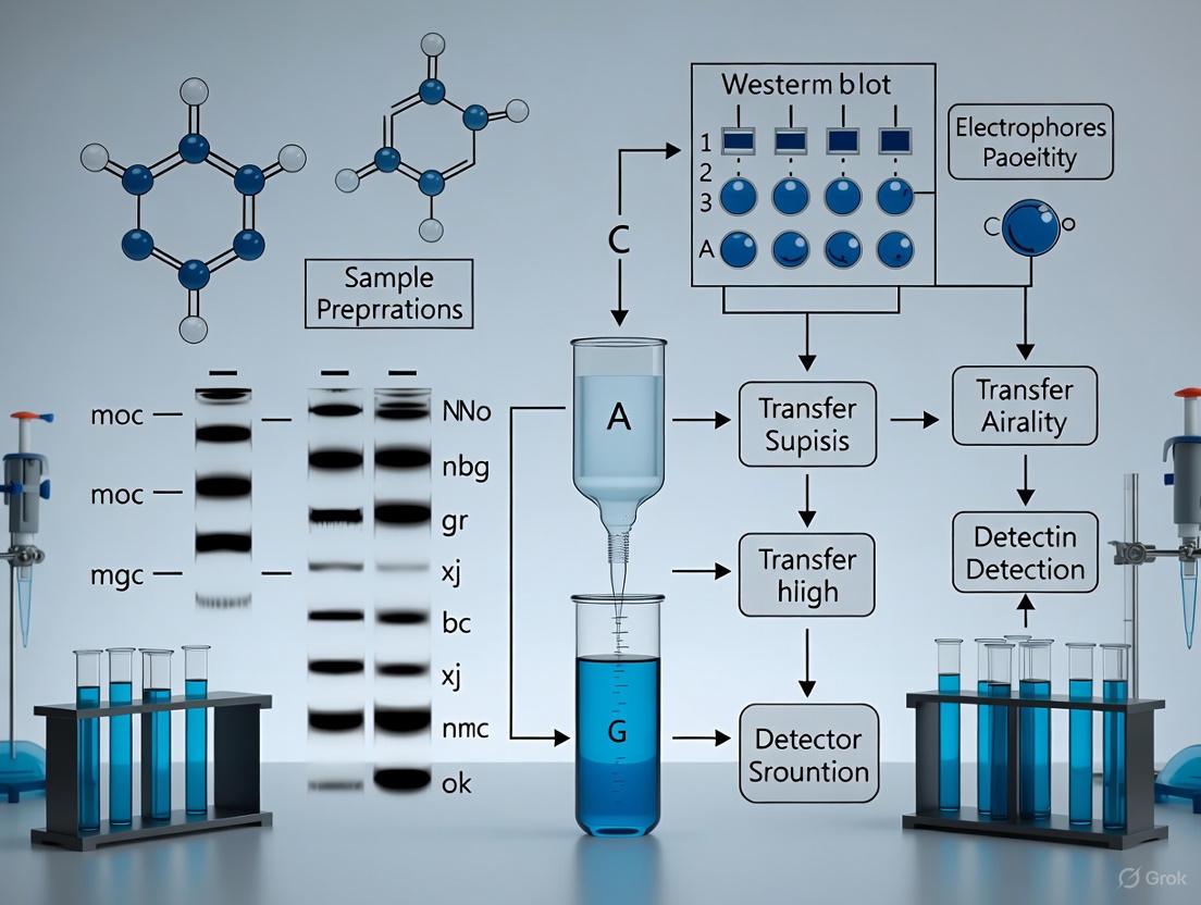
In ubiquitin western blotting, a smear is not a sign of failure but a characteristic and expected result that provides important biological information. The smear represents the heterogeneous population of ubiquitinated proteins in your sample [1].
- Polymerization: Ubiquitination involves attaching ubiquitin molecules to protein substrates. A poly-ubiquitin chain is produced when a ubiquitin molecule on a substrate is modified by additional ubiquitin monomers conjugating onto any of the seven lysine residues (K6, K11, K27, K29, K33, K48, K63) or the N-terminal methionine (M1) of ubiquitin [1].
- Heterogeneous Populations: Your sample contains the same target protein with varying numbers of ubiquitin molecules attached. Each addition of an 8.6 kDa ubiquitin molecule increases the molecular weight, creating a ladder of different molecular species [2] [1].
- Continuous Distribution: Since the number of attached ubiquitin molecules varies continuously across the protein population, this creates a smear pattern rather than discrete bands on your western blot [1].
Table: Ubiquitin Linkages and Their Functional Consequences
| Linkage Site | Chain Type | Primary Biological Function |
|---|---|---|
| K48 | Polymeric | Targeted protein degradation by the proteasome [1] |
| K63 | Polymeric | Immune responses, inflammation, lymphocyte activation [1] |
| K6 | Polymeric | Antiviral responses, autophagy, DNA repair [1] |
| K11 | Polymeric | Cell cycle progression, proteasome-mediated degradation [1] |
| K27 | Polymeric | DNA replication, cell proliferation [1] |
| K29 | Polymeric | Neurodegenerative disorders, autophagy [1] |
| M1 | Polymeric | Cell death and immune signaling [1] |
| Substrate lysines | Monomer | Endocytosis, histone modification, DNA damage responses [1] |
How can I distinguish meaningful ubiquitin smears from technical artifacts?
While smears are expected in ubiquitin western blots, it's crucial to distinguish biologically relevant smearing from artifacts caused by technical issues. The table below compares characteristic features of true ubiquitination patterns versus common artifacts.
Table: Differentiating Ubiquitination Smears from Technical Artifacts
| Feature | True Ubiquitination Signal | Technical Artifact |
|---|---|---|
| Pattern Appearance | Continuous smear extending upward from expected molecular weight [1] | Irregular, blotchy, or localized speckling [3] |
| Reproducibility | Consistent across experimental replicates | Variable between replicates |
| Response to Proteasome Inhibition | Enhanced with MG-132 treatment (5-25 µM for 1-2 hours) [1] | Unchanged |
| Band Pattern | No distinct bands within smear (unless studying specific chain types) | Multiple discrete non-specific bands [3] |
| Background | Clean background with specific smear pattern | High uniform background or speckling [3] |
What are the most effective methods to preserve ubiquitination signals before detection?
Ubiquitination is a transient and reversible modification, making preservation of these signals challenging. Proper sample handling is critical for accurate detection [1].
Sample Preparation Protocol for Ubiquitination Studies
Cell Treatment:
Lysis:
Protein Quantification:
Storage:
- Process samples immediately or store at -70°C to prevent degradation [5].
- Avoid repeated freeze-thaw cycles.
How can I reduce high background in my ubiquitin western blots?
High background is a common issue that can obscure ubiquitination smears. These troubleshooting strategies can improve signal-to-noise ratio [3] [6].
Optimization Strategies for Clean Background
Blocking Optimization:
Antibody Incubation:
Washing Enhancement:
Detection Optimization:
What controls should I include to validate my ubiquitination results?
Proper controls are essential for interpreting ubiquitination smears correctly and ensuring experimental validity [3] [7].
Essential Control Experiments
Secondary Antibody-Only Control: Incubate membrane with secondary antibody alone to identify non-specific binding [3].
Proteasome Inhibition Control: Treat cells with MG-132 to stabilize ubiquitinated proteins and enhance smear intensity [1].
Positive Control: Use a known ubiquitinated protein or lysate from cells treated with proteasome inhibitor [3].
Loading Control: Include housekeeping proteins (GAPDH, actin, tubulin) to normalize protein loading [7].
Genetic Controls: Express wild-type and ubiquitination-deficient mutants (e.g., lysine-to-arginine mutations) of your target protein [4].
Research Reagent Solutions for Ubiquitination Studies
Table: Essential Reagents for Ubiquitin Research
| Reagent | Function | Example Products |
|---|---|---|
| Proteasome Inhibitors | Stabilizes ubiquitinated proteins by blocking degradation | MG-132 (5-25 µM for 1-2 hours) [1] |
| Ubiquitin-Trap | Immunoprecipitates ubiquitin and ubiquitinated proteins | ChromoTek Ubiquitin-Trap Agarose/Magnetic Beads [1] |
| Ubiquitin Antibodies | Detects ubiquitin and ubiquitinated proteins | Ubiquitin Recombinant Antibody (Proteintech 80992-1-RR) [1] |
| Linkage-Specific Antibodies | Identifies specific ubiquitin chain linkages | K48-, K63-, M1-specific ubiquitin antibodies [1] |
| Plasmid DNA | Expresses ubiquitin and target proteins in cells | pCDNA3.1+ vectors with His-Ub or Flag-tagged constructs [4] |
| Enrichment Resins | Purifies ubiquitinated proteins for detection | Ni-NTA Agarose for His-Ub pulldowns [4] |
Can I use the sheet protector method to conserve antibodies in ubiquitin western blots?
Yes, the sheet protector (SP) strategy is an effective method to reduce antibody consumption while maintaining detection sensitivity for ubiquitin smears [5].
Sheet Protector Protocol for Ubiquitin Blots
Membrane Preparation:
- After transfer, block membrane conventionally with 5% skim milk for 1 hour.
- Briefly immerse in TBST to wash away excess milk.
- Blot membrane thoroughly with paper towel to absorb residual moisture.
Antibody Application:
- Place semi-dried membrane on a cropped sheet protector leaflet.
- Apply minimal antibody volume (20-150 µL for mini-gels) directly to membrane.
- Gently place upper leaflet over membrane, allowing solution to disperse as a thin layer.
Incubation:
- Incubate SP unit at room temperature for 15 minutes to several hours.
- For extended incubations, place SP unit on wet paper towel in sealed bag to prevent evaporation.
Detection:
- Proceed with standard washing and secondary antibody incubation.
- This method can reduce antibody consumption by up to 95% while maintaining comparable sensitivity to conventional methods [5].
What advanced techniques can I use to study specific ubiquitin chain linkages?
To move beyond simple smear detection and characterize specific ubiquitin linkages, consider these advanced methodologies [1].
Linkage-Specific Ubiquitination Analysis
Ubiquitin Traps with Linkage-Specific Antibodies: Use non-linkage specific Ubiquitin-Trap for pulldown followed by western blot with linkage-specific antibodies for differentiation [1].
In Vitro Ubiquitination Assays: Reconstitute ubiquitination using purified E1, E2, and E3 enzymes with recombinant target proteins to study specific enzymatic pathways [8].
Mass Spectrometry Analysis: Combine Ubiquitin-Trap immunoprecipitation with mass spectrometry (IP-MS) to identify specific ubiquitination sites and chain linkages [1].
Mutagenesis Studies: Create lysine-to-arginine mutations in target proteins (e.g., K190A, K450A) to identify specific ubiquitination sites [4].
FAQs: Core Concepts and Biological Significance
Q1: What are the primary cellular functions of K48-linked and K63-linked ubiquitin chains?
K48 and K63 linkages are the two most abundant ubiquitin chain types in cells and serve fundamentally different roles [9] [10].
- K48-linked chains are the principal signal for proteasomal degradation. Chains of four or more ubiquitins (Ub4) are particularly efficient, triggering degradation with half-lives as short as one minute [11].
- K63-linked chains are primarily non-proteolytic signals regulating diverse processes including DNA damage repair, inflammatory signaling (e.g., NF-κB and MAPK pathways), endocytosis, protein trafficking, and lysosomal degradation of membrane proteins [9] [12] [10].
Q2: Can K63 linkages ever signal for degradation?
Yes, the functional roles are not absolute. While K48 is the canonical proteasomal signal, K63 linkages can also target proteins for lysosomal degradation. For example, the LDL receptor (LDLR) can be targeted to the lysosome by either K48 or K63 linkages [12] [10]. Furthermore, when K63 chains are incorporated into certain branched structures, they can be converted into proteasomal degradation signals [13].
Q3: What are branched ubiquitin chains, and what is their functional significance?
Branched ubiquitin chains form when a single ubiquitin moiety is modified with two or more other ubiquitins via different linkages [9] [13]. A prominent example is the K48/K63-branched chain.
- Function: These chains add a layer of complexity to the ubiquitin code. They can act as superior degradation signals or regulate signaling pathways. For instance, K48/K63-branched chains can protect K63 linkages from deubiquitinating enzymes, thereby amplifying NF-κB signaling [14]. In other contexts, they trigger rapid proteasomal degradation [11] [13].
- Mechanism: Branched chains can increase ubiquitin density to enhance proteasome recruitment, and specific linkages within the branch can protect the chain from substrate-specific deubiquitinases (DUBs), ensuring the signal is not erased [14] [13].
Table 1: Primary Functions and Characteristics of K48 and K63 Ubiquitin Linkages
| Feature | K48-Linked Chains | K63-Linked Chains |
|---|---|---|
| Primary Function | Proteasomal degradation [11] [15] | Non-proteolytic signaling (DNA repair, inflammation, endocytosis) [9] [10] [15] |
| Secondary Degradation Role | - | Lysosomal degradation [12] [10] |
| Minimal Efficient Degradation Signal | K48-Ub3/Ub4 [11] | Not typically a direct proteasomal signal |
| Abundance in Cells | ~52% of all linkages [10] | ~38% of all linkages [10] |
| Key Regulatory Role in Branched Chains | Can serve as the proteasome-targeting branch in K48/K63 chains [14] [13] | Can be protected from DUBs (e.g., CYLD) by K48 branching, amplifying signaling [14] |
Experimental Guide: Methodologies and Workflows
Q4: What are the best practices for studying linkage-specific ubiquitination in pull-down assays?
Using ubiquitin interactor pulldown coupled with mass spectrometry is a powerful method, but the choice of deubiquitinase (DUB) inhibitor is critical [9].
- Inhibitor Choice: The commonly used DUB inhibitors N-ethylmaleimide (NEM) and chloroacetamide (CAA) have different efficacies and potential off-target effects.
- Recommendation: The choice of inhibitor can influence the identified interactors. It is advisable to perform experiments with both inhibitors separately and compare the datasets to distinguish overlapping interactors from inhibitor-specific ones [9].
Q5: How can I systematically compare the degradation capacity of different ubiquitin chains inside cells?
The UbiREAD (ubiquitinated reporter evaluation after intracellular delivery) technology is designed for this purpose [11].
- Workflow:
- Synthesis: Prepare GFP conjugated to ubiquitin chains of defined linkage, length, and composition in vitro.
- Delivery: Electroporate the ubiquitinated GFP protein directly into mammalian cells (e.g., RPE-1, HeLa).
- Quantification: Monitor degradation kinetics at high temporal resolution using flow cytometry or in-gel fluorescence to track the loss of the ubiquitinated GFP signal [11].
The following diagram illustrates the UbiREAD workflow for analyzing ubiquitin-dependent degradation.
Q6: What techniques are available for mapping the connectivity of ubiquitin chains?
Targeted mass spectrometry using Selected Reaction Monitoring (SRM) is a highly sensitive and quantitative method.
- Principle: This approach uses chemically synthesized, heavy isotope-labeled reference peptides that correspond to tryptic fragments of different ubiquitin linkages (K6, K11, K27, K29, K33, K48, K63) [16].
- Advantage: It allows for the unambiguous detection and quantification of linkage frequency in complex biological samples, overcoming the limitations of linkage-specific antibodies, which may not recognize denatured epitopes or exist for all linkage types [16].
Troubleshooting Guide: Common Experimental Challenges
Q7: My western blots show smearing, and I cannot distinguish specific linkages. What are my options?
Ubiquitin smearing is common due to heterogeneous chain lengths and mixed linkages.
- Use Linkage-Specific Tools:
- TUBEs (Tandem Ubiquitin Binding Entities): These are engineered reagents with high affinity for polyubiquitin chains. Using lysine-specific TUBEs (e.g., K48- or K63-specific) in a pull-down assay, often in a 96-well plate format, allows for the high-throughput separation and study of specific linkages before western blotting [15].
- Linkage-Specific DUBs: Employ enzymes like OTUB1 (K48-specific) and AMSH (K63-specific) in UbiCRest assays to selectively disassemble chains and confirm linkage composition [9].
- Validate with Mass Spectrometry: For definitive linkage identification, combine biochemical methods with targeted MS (SRM) or Ub-AQUA/PRM techniques [13] [16].
Q8: My ubiquitinated protein is not being degraded, despite having ubiquitin chains. Why?
This can occur if the chains are not an efficient proteasomal signal or if they are actively counteracted.
- Check Chain Length and Linkage: Ensure the chains are of sufficient length (≥Ub3 for K48) and of the correct linkage. K63 chains will not typically signal for proteasomal degradation unless they are part of a branched chain [11].
- Consider Branched Chains and DUB Protection: Your substrate might be modified with a K63 chain that is being protected by a DUB. In such cases, degradation requires the formation of a branched chain. For example, a K29 linkage (resistant to the DUB OTUD5) can act as a foundation for a K48 branch, overcoming DUB protection and targeting the substrate for degradation [13].
The diagram below illustrates how branched ubiquitin chains can integrate signals for proteasomal targeting.
Table 2: Essential Research Reagents for Studying K48 and K63 Ubiquitination
| Reagent / Tool | Function / Specificity | Key Application |
|---|---|---|
| TUBEs (Tandem Ubiquitin Binding Entities) [15] | High-affinity, linkage-specific binders (K48, K63, pan) | Enrichment and detection of specific ubiquitinated proteins from lysates; reduces DUB activity. |
| Linkage-Specific DUBs (OTUB1, AMSH) [9] | Cleave K48 or K63 linkages, respectively. | Validation of chain linkage composition in UbiCRest assays. |
| DUB Inhibitors (NEM, CAA) [9] | Cysteine alkylators that inhibit the largest family of DUBs. | Preserving ubiquitin chain integrity during pulldown and purification experiments. |
| SRM Mass Spectrometry Assays [16] | Quantifies all possible ubiquitin linkages using heavy isotope-labeled peptides. | Comprehensive and quantitative profiling of ubiquitin chain connectivity in complex samples. |
| UbiREAD System [11] | Delivers bespoke ubiquitinated reporters into cells. | Directly measuring intracellular degradation kinetics of defined ubiquitin chains. |
Advanced Topics: Signaling Pathways and Complex Regulation
Q9: How do K48/K63-branched ubiquitin chains regulate the NF-κB pathway?
In the NF-κB pathway, branched chains play a critical role in signal amplification.
- Mechanism: In response to IL-1β, the E3 ligase HUWE1 generates K48 branches onto K63-linked chains assembled by TRAF6.
- Functional Outcome: The resulting K48/K63-branched chain performs two functions:
- It is still recognized by the K63-binding domain of TAB2, maintaining signaling.
- The K48 branch protects the underlying K63 chain from the deubiquitinating enzyme CYLD. This prevents signal termination and leads to amplified NF-κB activation [14].
Fundamental Concepts: Why Atypical Linkages Cause Smears
Why do I get heterogeneous smears instead of clean bands when probing for ubiquitinated proteins? Heterogeneous smears on western blots are a common characteristic of polyubiquitinated proteins, not necessarily an indication of a failed experiment. Atypical ubiquitin linkages (K11, K29, K33) contribute significantly to this smear pattern for several key reasons [17] [18]:
- Mixed Chain Topologies: Your protein sample may be modified with ubiquitin chains of different linkages (homotypic), or even mixed/branched chains, simultaneously. Each topology has a slightly different molecular weight and migration pattern through the gel.
- Open Conformations: Biophysical studies show that K29- and K33-linked chains adopt open, dynamic conformations in solution, similar to K63-linked chains [17]. This flexible structure can lead to less uniform migration in SDS-PAGE compared to the compact structure of K48 chains.
- Variable Chain Length: Proteins can be modified with ubiquitin chains of varying lengths (e.g., from 2 to 10+ ubiquitins). This inherent heterogeneity in the number of ubiquitin moieties per substrate results in a continuum of molecular weights, visualized as a smear.
How do the properties of atypical linkages compare to canonical ones? The table below summarizes key characteristics of different ubiquitin chain linkages that influence their appearance on western blots and their cellular functions.
Table 1: Characteristics of Ubiquitin Chain Linkages
| Linkage Type | Common Structural Conformation | Primary Associated Functions | Key E3 Ligase Examples | Impact on Western Blot Appearance |
|---|---|---|---|---|
| K48 | Compact, closed | Proteasomal degradation [17] | UBE3C (also makes K29) [17] | Can produce discrete bands or tight smears |
| K63 | Open, extended | Non-degradative signaling (DNA repair, inflammation) [17] | NEDD4 family [17] | Often produces a diffuse smear |
| K11 | Mixed | Cell cycle regulation, ER-associated degradation [17] | AREL1 [17] | Contributes to heterogeneous smearing |
| K29 | Open, dynamic [17] | Proteotoxic stress, autophagy [17] | UBE3C, TRIP12 [17] [18] | Contributes to heterogeneous smearing |
| K33 | Open, dynamic [17] | Kinase signaling, endosomal trafficking [17] | AREL1 [17] | Contributes to heterogeneous smearing |
Troubleshooting Guide: FAQs for Smear Resolution
The smear on my ubiquitin western blot is too diffuse to draw any conclusions. What are the first steps I should take? A diffuse smear can be challenging to interpret. Follow this systematic troubleshooting approach:
- Verify Antibody Specificity: Ensure your primary antibody is validated for the specific linkage you are studying. Many "pan-ubiquitin" antibodies have varying affinities for different linkages.
- Optimize Gel Electrophoresis: Use an appropriate gel percentage. For large ubiquitin conjugates, a lower percentage acrylamide gel (e.g., 4-12% gradient Bis-Tris gel with MOPS buffer) improves separation of high molecular weight species [19]. Ensure you do not overload the gel with too much protein; 10–40 µg of lysate is typically recommended [19].
- Optimize Transfer Conditions: Use PVDF membranes for better retention of high molecular weight proteins. Confirm your transfer buffer and method (semi-dry vs. wet) are appropriate for your gel system [20].
I need to confirm the presence of a specific atypical linkage (e.g., K29) in my smear. What is the best methodological approach? Confirming a specific linkage requires a combination of enzymatic and genetic tools.
- Linkage-Specific Deubiquitinases (DUBs): Treat your purified protein or immunoprecipitated sample with linkage-specific DUBs in vitro before western blotting. For example, the DUB TRABID is highly specific for cleaving K29 and K33 linkages [17]. A collapse of the smear after treatment strongly indicates the presence of these chains.
- Ubiquitin Mutants: Co-express your protein of interest with ubiquitin mutants where all lysines except one are mutated to arginine (e.g., K29-only Ub). If the smear is preserved only with the K29-only mutant, it confirms the formation of K29-linked chains on your substrate [17].
- Mass Spectrometry (AQUA): Absolute quantification (AQUA) mass spectrometry using isotope-labeled internal standards can precisely quantify all linkage types present in a sample [17]. This is the gold standard for confirmation.
My negative control shows a background smear. What could be the cause? Background smear in controls is often due to non-specific antibody binding or incomplete blocking.
- Troubleshoot Blocking and Washing:
- Blocking Buffer: Use a high-quality blocking buffer (e.g., 5% non-fat milk or commercial blocking buffers) for 30-60 minutes at room temperature [20].
- Wash Stringency: Increase the number and duration of washes after primary and secondary antibody incubation. A standard protocol is 3 washes for 10 minutes each after primary antibody, and 6 washes for 5 minutes each after secondary antibody, using TBST or PBST [20].
- Antibody Concentration: Titrate your primary and secondary antibodies. High concentrations can increase background. Refer to the table below for general guidance on antibody dilution.
Table 2: Western Blot Antibody Dilution Guidelines with Chemiluminescent Detection
| Chemiluminescent Substrate Sensitivity | Recommended Primary Antibody Dilution | Recommended Secondary Antibody Dilution |
|---|---|---|
| Standard / Moderate (e.g., Pierce ECL) | 1:1,000 (0.2–10 µg/mL) [20] | 1:1,000 to 1:15,000 (0.07–1.0 µg/mL) [20] |
| High (e.g., SuperSignal West Pico Plus) | 1:1,000 (0.2–1.0 µg/mL) [20] | 1:20,000 to 1:100,000 (10–50 ng/mL) [20] |
| Very High / Ultra (e.g., SuperSignal West Femto) | 1:5,000 (0.01–0.2 µg/mL) [20] | 1:100,000 to 1:500,000 (2–10 ng/mL) [20] |
Advanced Experimental Protocols
Protocol 1: In Vitro Ubiquitin Chain Assembly Analysis for K29/K33 Linkages This protocol uses recombinant E3 ligases to generate atypical chains for in vitro assays or as standards [17] [18].
- Key Reagents:
Protocol 2: Linkage-Specific Deconvolution of Cellular Smears This workflow outlines how to confirm the presence of specific atypical linkages in a heterogeneous smear from cell lysates.
- Workflow Overview:
- Treat Cells under desired experimental conditions.
- Lysate Preparation: Lyse cells using RIPA or non-denaturing lysis buffer supplemented with protease inhibitors (e.g., 1 mL per 1x10^7 cells). Centrifuge at 14,000–17,000 g for 5 min at 4°C to clear lysate [19].
- Immunoprecipitation (IP): Immunoprecipitate your protein of interest or ubiquitinated proteins under denaturing conditions to preserve complexes.
- Elute and Split: Split the eluted protein into aliquots.
- DUB Treatment: Treat aliquots with buffer-only (control), non-specific DUB (e.g., USP2), or linkage-specific DUBs (e.g., TRABID for K29/K33).
- Analyze by Western Blot: A specific collapse of the smear in the TRABID-treated sample indicates the presence of K29/K33 linkages.
The Scientist's Toolkit: Essential Research Reagents
Table 3: Key Reagents for Studying Atypical Ubiquitin Linkages
| Reagent / Tool | Function / Application | Example Use Case |
|---|---|---|
| Linkage-Specific E3 Ligases (e.g., UBE3C, AREL1, TRIP12) | Enzymes that catalyze the formation of specific Ub chain linkages [17] [18]. | In vitro reconstitution of K29 or K33 chains to use as standards or for functional assays. |
| Linkage-Specific DUBs (e.g., TRABID) | Enzymes that selectively cleave a specific Ub linkage [17]. | Validation of a specific linkage type in a heterogeneous smear from cell lysates or in vitro reactions. |
| Ubiquitin Mutants (K-only, R-mutants) | Ub variants where only one lysine is available for chain formation (K-only) or all lysines are mutated to arginine (K0) [17]. | Identifying which lysines are used for chain formation on your substrate in cellular overexpression studies. |
| Linkage-Specific Antibodies | Antibodies raised against specific diUb linkages. | Detecting the presence and relative levels of a specific chain type directly on western blots. Requires careful validation for specificity. |
| Tandem Ubiquitin Binding Entities (TUBEs) | Engineered proteins with high affinity for polyUb chains, used to enrich ubiquitinated proteins from lysates [17]. | Pulling down ubiquitinated proteins while protecting them from DUBs during extraction, enriching signal on blots. |
| Mass Spectrometry (AQUA) | Gold-standard method for absolute quantification of all Ub linkage types in a sample [17]. | Unambiguous identification and quantification of the complex linkage composition within a smear. |
The ubiquitin code is a sophisticated post-translational language that regulates nearly all cellular processes. Its complexity stems from the ability of ubiquitin to form diverse polymeric chains through its seven internal lysine residues (K6, K11, K27, K29, K33, K48, K63) or N-terminal methionine (M1) [21] [22]. These chains exist in three principal architectures that create a spectrum of biological signals:
- Homotypic Chains: Uniform chains where all ubiquitin molecules are connected through the same linkage type (e.g., all K48 or all K63 linkages) [23] [24].
- Heterotypic Mixed Chains: Linear chains containing more than one linkage type, but each ubiquitin is modified by only one other ubiquitin molecule [24].
- Branched Chains: Complex structures where at least one ubiquitin molecule is connected to two or more other ubiquitins, forming branch points [24].
The following diagram illustrates the structural relationships and functional implications of these different chain architectures:
Critical Sample Preparation for Preserving Ubiquitin Chains
FAQ: Why do my ubiquitinated proteins disappear during sample preparation?
Answer: The loss typically occurs due to active deubiquitinases (DUBs) and proteasomes in your lysate. Ubiquitination is a reversible modification, and DUBs can rapidly remove ubiquitin chains during cell lysis and subsequent processing [21].
Troubleshooting Guide: Sample Degradation Issues
| Problem | Cause | Solution |
|---|---|---|
| Disappearing ubiquitin signal | DUB activity during lysis | Increase N-ethylmaleimide (NEM) to 50-100 mM or IAA to 20-50 mM [21] |
| Incomplete K63 chain preservation | Insufficient cysteine alkylation | Use higher concentrations of NEM (up to 100 mM) rather than IAA [25] [21] |
| Loss of ubiquitinated substrates | Proteasomal degradation | Include MG132 (10-20 µM) during cell treatment and lysis [25] [21] |
| Erroneous ubiquitination after MG132 | Cellular stress response | Limit MG132 treatment to <12 hours to avoid stress-induced artifacts [21] |
Experimental Protocol: Optimized Lysis for Ubiquitin Preservation
Prepare fresh lysis buffer containing:
- 50-100 mM NEM (freshly prepared)
- 5-10 mM EDTA or EGTA
- 10-20 µM MG132 or other proteasome inhibitor
- Standard protease inhibitors [21]
Lys cells directly in pre-heated SDS buffer (1% SDS) for complete DUB inactivation when subsequent immunoprecipitation isn't required [21].
Clarify lysates by centrifugation at 12,000 × g for 10 minutes at 4°C.
Transfer supernatant to fresh tubes and store at -80°C if not used immediately.
Gel Electrophoresis and Transfer Optimization for Ubiquitin Smears
FAQ: Why do I get poor resolution of ubiquitin smears on my western blots?
Answer: Poor resolution often results from using suboptimal gel and buffer systems for your target chain length. Different ubiquitin chain lengths require specific electrophoretic conditions for optimal separation [25] [21].
Quantitative Data: Buffer and Gel Selection Guide
| Target Analysis | Gel Type | Running Buffer | Benefits | Limitations |
|---|---|---|---|---|
| Short chains (2-5 ubiquitins) | 12% acrylamide | MES | Excellent separation of small oligomers | Poor resolution of long chains |
| Long chains (>8 ubiquitins) | 8% acrylamide | MOPS | Optimal for large ubiquitin polymers | Reduced small chain separation |
| Broad range analysis | 8-12% gradient | Tris-Glycine | Good separation up to 20 ubiquitins | Less optimal for extremes |
| High molecular weight | 3-8% gradient Tris-Acetate | Tris-Acetate | Superior 40-400 kDa separation | Specialized equipment needed |
Experimental Protocol: Western Blot Transfer for Ubiquitinated Proteins
Membrane selection: Use PVDF membranes (0.2 µm pore size) for higher signal strength compared to nitrocellulose [25].
Activation: Wet PVDF membrane in 100% methanol for 30 seconds, then equilibrate in transfer buffer.
Transfer conditions: Use wet transfer system at 30V for 2.5-3 hours [25].
Validation: After transfer, stain the gel with Coomassie or reversible protein stain to confirm transfer efficiency [26].
Advanced Detection Methods for Specific Chain Types
FAQ: How can I distinguish between different ubiquitin linkage types?
Answer: Use a combination of linkage-specific antibodies, ubiquitin-binding domains (UBDs), and deubiquitinase (DUB) digestion assays to characterize specific ubiquitin linkages [27] [24].
Research Reagent Solutions for Ubiquitin Analysis
| Reagent | Function | Applications |
|---|---|---|
| Tandem Ubiquitin Binding Entities (TUBEs) | High-affinity ubiquitin chain capture | Pull-down assays, protection from DUBs [27] |
| Linkage-specific DUBs (UbiCRest assay) | Selective cleavage of specific linkages | Chain linkage mapping [24] |
| K48/K63-chain specific TUBEs | Preferential binding to specific chains | Differentiation of degradation vs. signaling chains [27] |
| K11/K48 bispecific antibodies | Detection of branched chains | Identification of hybrid ubiquitin chains [24] |
| Denatured-Refolded Ubiquitinated Sample Prep (DRUSP) | Improved ubiquitinome analysis | Mass spectrometry sample preparation [28] |
Experimental Protocol: UbiCRest Linkage Analysis
Prepare ubiquitinated samples using standard immunoprecipitation or TUBE pull-down [27].
Aliquot samples into multiple tubes for parallel DUB digestion.
Incubate with linkage-specific DUBs:
- OTUB1 (K48-specific)
- AMSH/OTUD1 (K63-specific)
- OTULIN (M1-specific)
- Cezanne (K11-specific)
- vOTU/USP21 (pan-specific controls) [24]
Analyze cleavage patterns by western blotting with linkage-specific antibodies.
Interpret results: Resistance to specific DUBs may indicate branched chains or atypical structures [24].
The workflow for this comprehensive linkage analysis is illustrated below:
Functional Consequences of Chain Diversity in Biological Systems
The structural diversity of ubiquitin chains translates to specific functional outcomes in cellular regulation:
Heterotypic K11/K48 Branched Chains in Cell Cycle Regulation
- Structure: Branched ubiquitin chains containing both K11 and K48 linkages [23] [24]
- Function: Efficient targeting of cyclin B1 and other cell cycle regulators to proteasomal degradation [23]
- Mechanism: Heterotypic K11/K48 chains bind proteasomes more effectively than homotypic K11 chains [23]
- Biological context: Mitotic exit regulated by APC/C with Ube2S E2 enzyme [23]
K63-Linked Chains in Inflammatory Signaling
- Structure: Typically homotypic K63 chains, but can form heterotypic structures [27] [22]
- Function: Activation of NF-κB and MAPK pathways through signalosome assembly [27]
- Specific example: RIPK2 ubiquitination upon NOD2 activation by bacterial MDP [27]
- Detection: K63-specific TUBEs can capture this signaling event [27]
Comprehensive Troubleshooting Guide for Ubiquitin Western Blots
| Problem | Possible Causes | Solutions |
|---|---|---|
| High background | Antibody concentration too high | Decrease primary and/or secondary antibody concentration [26] [29] |
| Insufficient blocking | Increase blocking time to ≥1 hour; use 5% BSA for phosphoproteins [26] | |
| Membrane drying | Ensure membrane remains wet throughout processing [26] | |
| Weak or no signal | Incomplete transfer | Verify transfer efficiency with reversible protein stain [26] |
| Antigen masked by blocking buffer | Try different blocking agents (BSA vs. milk) [26] | |
| Low abundance targets | Use high-sensitivity chemiluminescent substrates [26] | |
| Non-specific bands | Antibody cross-reactivity | Include proper controls; validate antibodies [29] |
| Protein degradation | Use fresh protease inhibitors; avoid sample overheating [30] | |
| Too much protein loaded | Reduce total protein load (10-15 μg/lane recommended) [26] | |
| Diffuse bands/smears | Transfer too fast | Increase transfer time; ensure proper cooling [29] |
| Improper gel/buffer system | Match gel percentage and buffer to target size range [21] |
Experimental Protocol: Enhanced Denaturation for Better Antibody Recognition
After transfer, incubate PVDF membrane in boiling water for 15-30 minutes [25]
Treat membrane with denaturing solution:
- 20 mM Tris-HCl, pH 7.5
- 5 mM β-mercaptoethanol
- 6 M guanidine-HCl
- Incubate 30 minutes at 4°C [25]
Autoclave the membrane for additional denaturation (optional) [25]
Proceed with standard blocking and antibody incubation steps
This technical support resource provides comprehensive guidance for researchers investigating the complex landscape of polyubiquitin chain diversity. By implementing these optimized protocols and troubleshooting strategies, scientists can significantly improve their ability to resolve and interpret the spectrum of ubiquitin modifications in their experimental systems.
Optimized Protocols and Cutting-Edge Tools for Sharper Ubiquitin Detection
Proper sample preparation is the most critical step in western blotting, profoundly influencing the detection, resolution, and accurate interpretation of protein data. During cell lysis, the carefully controlled cellular environment is disrupted, releasing endogenous proteases and phosphatases that can rapidly degrade proteins and modify their activation states, leading to irreproducible results, loss of signal, or misleading bands [31] [32]. For researchers studying complex post-translational modifications like ubiquitination—which often appears as characteristic high-molecular-weight smears on western blots—meticulous sample preparation is not merely optional but essential for meaningful data [25]. This guide provides detailed troubleshooting and methodologies to preserve protein integrity from the moment of lysis, with particular emphasis on resolving ubiquitin-related signals.
Frequently Asked Questions (FAQs)
Q1: Why is adding protease inhibitors to my lysis buffer so critical? Cell lysis disrupts cellular compartmentalization, releasing sequestered proteases that become unregulated and can digest your proteins of interest [31] [32]. Protease inhibitors are chemical or biological compounds that prevent this protein degradation by binding to and inactivating these enzymes, thereby preserving the protein's native state, yield, and post-translational modifications for accurate analysis [32] [33].
Q2: What is a "protease inhibitor cocktail" and why should I use one? No single chemical compound can inhibit all protease types effectively [31] [33]. A protease inhibitor cocktail is a pre-formulated mixture of several inhibitors that broadly targets the major protease classes (serine, cysteine, aspartic, metallo-, and aminopeptidases) [32] [33]. Using a cocktail ensures comprehensive protection, saves time, and provides consistency compared to individually optimizing and mixing separate inhibitors [33].
Q3: My western blot shows a high background smear instead of clean bands. Could this be a sample preparation issue? Yes. Smearing, particularly in high molecular weight regions, can indicate protein degradation during sample preparation due to insufficient protease inhibition [34] [35]. It can also result from overloading too much protein, incomplete sample reduction, or, in the specific case of ubiquitination, the natural appearance of poly-ubiquitin chains of different lengths [34] [25] [35]. Using fresh, optimized protease inhibitor cocktails and appropriate protein loads can resolve this.
Q4: I am specifically studying ubiquitination. Are there special considerations for my lysis buffer? Yes. Ubiquitination is a dynamic and reversible modification. Standard protease inhibitors are insufficient, as you must also inhibit deubiquitinase (DUB) enzymes and the proteasome [25].
- DUB Inhibitors: Add 5-100 mM N-Ethylmaleimide (NEM) to your lysis buffer (K63-linked chains require higher concentrations, up to 50-100 mM) [25].
- Proteasome Inhibitors: Use MG132 to prevent the destruction of ubiquitinated proteins. However, avoid prolonged treatments (>12-24 hours) to prevent stress-induced ubiquitination [25].
- Chelators: Include EDTA or EGTA, as many DUBs are metalloproteases [25].
Troubleshooting Guide: Sample Preparation Issues
Table 1: Common Problems and Solutions Related to Sample Preparation
| Problem | Possible Cause | Solutions |
|---|---|---|
| No or Very Weak Signal | Protein degraded during/after lysis due to inactive or missing protease inhibitors [34] [36]. | Use fresh, functional protease inhibitor cocktails. Aliquot inhibitors to avoid freeze-thaw cycles. Keep samples on ice [34] [32] [35]. |
| Multiple Non-specific Bands or Smearing | Partial proteolysis by endogenous proteases creates protein fragments [34] [35]. | Optimize inhibitor cocktail concentration, especially for problematic tissues. Ensure lysis is performed quickly at cold temperatures [34] [35]. |
| High Molecular Weight Smear (Ubiquitin) | Inadequate preservation of ubiquitin chains during lysis [25]. | Incorporate specific DUB (NEM) and proteasome (MG132) inhibitors into the lysis buffer [25]. |
| Bands at Unexpected Molecular Weights | Post-translational modifications (e.g., glycosylation, phosphorylation) or alternative splicing [34] [35]. | Use phosphatase inhibitors when studying phosphorylation. Consult databases for known PTMs. Run appropriate positive and negative controls [34] [31] [35]. |
| Poor Gel Resolution / Smiling Bands | Sample viscosity from DNA contamination or excess salt [26]. | Shear genomic DNA by sonication or needle passage. Reduce salt concentration via dialysis or dilution [26] [35]. |
Table 2: Commonly Used Protease and Phosphatase Inhibitors
| Inhibitor | Target Enzyme Class | Mechanism | Typical Working Concentration |
|---|---|---|---|
| AEBSF | Serine Proteases | Irreversible | 0.2 - 1.0 mM [31] [32] |
| Aprotinin | Serine Proteases | Reversible | 100 - 200 nM [31] [32] |
| E-64 | Cysteine Proteases | Irreversible | 1 - 20 µM [31] [32] |
| EDTA | Metalloproteases | Reversible (Chelator) | 2 - 10 mM [31] [32] |
| Leupeptin | Serine & Cysteine Proteases | Reversible | 10 - 100 µM [31] [32] |
| Pepstatin A | Aspartic Proteases | Reversible | 1 - 20 µM [31] [32] |
| PMSF | Serine Proteases | Reversible | 0.1 - 1.0 mM [31] [32] |
| Sodium Orthovanadate | Tyrosine Phosphatases | Irreversible | 1 - 100 mM [31] |
| Sodium Fluoride | Ser/Thr & Acidic Phosphatases | Irreversible | 1 - 20 mM [31] |
Detailed Experimental Protocols
Protocol 1: Preparation of Complete Lysis Buffer for General Use
This protocol is designed for routine protein extraction while maximizing protein stability.
Materials Needed:
- RIPA Buffer or your preferred lysis buffer (e.g., Tris-HCl based)
- Broad-spectrum protease inhibitor cocktail (e.g., 100X stock)
- Phosphatase inhibitor cocktail (optional, for phospho-protein studies) [31]
- EDTA (0.5 M stock, pH 8.0) [32]
Procedure:
- Calculate Volumes: Determine the total volume of lysis buffer required.
- Add Inhibitors: To the lysis buffer, add:
- Protease inhibitor cocktail to a 1X final concentration (e.g., 10 µL of 100X stock per 1 mL of buffer) [33].
- Phosphatase inhibitor cocktail to a 1X final concentration, if needed [31].
- EDTA to a 2-10 mM final concentration to inhibit metalloproteases (if compatible with downstream applications) [31] [32].
- Mix and Store: Prepare the complete lysis buffer fresh immediately before use. Keep it on ice.
Protocol 2: Specialized Lysis Buffer for Ubiquitin Studies
This protocol is optimized for preserving labile ubiquitin conjugates.
Materials Needed:
- Standard Lysis Buffer (e.g., RIPA)
- N-Ethylmaleimide (NEM): 1 M stock in ethanol or water [25]
- Proteasome Inhibitor (e.g., MG132): 10 mM stock in DMSO
- EDTA: 0.5 M stock, pH 8.0
- Standard Protease Inhibitor Cocktail (100X)
Procedure:
- Prepare a standard complete lysis buffer as in Protocol 1.
- Add NEM to a final concentration of 10-100 mM. For K63-linked ubiquitin chains, use higher concentrations (50-100 mM) [25].
- Add MG132 to a final concentration of 10-50 µM.
- Vortex thoroughly and keep on ice. Use immediately.
Protocol 3: Step-by-Step Cell Lysate Preparation
Procedure:
- Harvest Cells: Culture cells and quickly aspirate media. Wash cells once with ice-cold PBS.
- Lysis: Add an appropriate volume of freshly prepared, ice-cold complete lysis buffer directly to the cell culture dish (e.g., 100-200 µL for a 6-well plate).
- Scrape and Collect: Using a cold cell scraper, gently but swiftly dislodge the lysed cells. Transfer the lysate to a pre-chilled microcentrifuge tube.
- Incubate: Keep the lysate on ice for 10-30 minutes with occasional gentle vortexing to ensure complete lysis.
- Clarify: Centrifuge the lysate at >12,000 x g for 15-20 minutes at 4°C to pellet insoluble debris.
- Collect Supernatant: Carefully transfer the clarified supernatant (which contains your soluble proteins) to a new pre-chilled tube.
- Quantify and Denature: Determine protein concentration using an assay like Bradford. Mix the lysate with an equal volume of 2X SDS-PAGE sample buffer, boil for 5-10 minutes, and then store at -80°C or load onto a gel.
Essential Workflow and Visual Guides
Sample Preparation Workflow for Optimal Western Blots
Protease Inhibitor Mechanism of Action
The Scientist's Toolkit: Essential Reagents
Table 3: Key Research Reagent Solutions for Sample Preparation
| Reagent | Function | Key Considerations |
|---|---|---|
| Broad-Spectrum Protease Inhibitor Cocktail | Inhibits serine, cysteine, aspartic proteases, and aminopeptidases [32] [33]. | Buy pre-made for consistency and convenience. Available with or without EDTA [33]. |
| Phosphatase Inhibitor Cocktail | Preserves protein phosphorylation status by inhibiting serine/threonine and tyrosine phosphatases [31]. | Essential for studying signaling pathways. Often sold as a combined protease/phosphatase cocktail [31] [35]. |
| N-Ethylmaleimide (NEM) | Irreversibly inhibits deubiquitinase (DUB) enzymes by covalently modifying active site cysteines [25]. | Critical for ubiquitination studies. Use at high concentrations (10-100 mM) [25]. |
| MG132 (Proteasome Inhibitor) | Prevents degradation of poly-ubiquitinated proteins by the proteasome, enriching them for detection [25]. | Avoid prolonged treatment of live cells to prevent stress artifacts [25]. |
| EDTA / EGTA | Chelates metal ions (Zn²⁺, Ca²⁺), inhibiting metalloproteases and many DUBs [31] [25] [32]. | Incompatible with downstream IMAC protein purification (strips Nickel ions) [32]. |
| PMSF (Phenylmethylsulfonyl fluoride) | Irreversibly inhibits serine proteases. | Highly unstable in aqueous solutions; must be added fresh. Toxic—handle with care [32]. |
Gel Electrophoresis and Transfer Optimization for High-Molecular-Weight Complexes
Troubleshooting Guides
FAQ: Resolving Ubiquitin Smears and High-Molecular-Weight Complexes
Why does my western blot for a ubiquitinated protein show a high molecular weight smear?
A high molecular weight smear is a classic indicator of polyubiquitinated proteins [25]. Each ubiquitin molecule adds approximately 8.5 kDa to your protein's apparent molecular weight [37]. As proteins can be modified with chains of varying lengths (from one ubiquitin to over twenty), this heterogeneity results in a smear rather than a discrete band [25]. This can be a valid biological signal, but the smear's appearance can be optimized for clearer interpretation.
How can I improve the transfer of very large proteins (>150 kDa) to the membrane?
Transferring high-molecular-weight (HMW) proteins is challenging because they migrate slowly out of the gel. Key optimizations include [38] [39] [40]:
- Adding SDS: Include 0.01-0.1% SDS in your transfer buffer to help elute large proteins from the gel [38] [39].
- Reducing Methanol: Lower the methanol concentration in your transfer buffer to 10-15%. Methanol can cause gel shrinkage and precipitate large proteins, trapping them in the gel [38] [40].
- Extending Transfer Time: Use a low voltage (25-30 V) and transfer overnight at 4°C to facilitate complete movement of large proteins [40].
- Pre-equilibration: Pre-equilibrate the gel in transfer buffer containing 0.02-0.04% SDS for 10 minutes before assembling the transfer sandwich [38].
My high molecular weight bands are faint or absent after transfer. What should I check?
This is a common sign of inefficient transfer. First, verify that the protein was present in the gel by post-transfer Coomassie blue staining [41]. If the protein was not transferred, implement the HMW transfer optimizations listed above. Also, ensure your membrane pore size is appropriate (0.45 µm is standard, but 0.2 µm may provide better retention) [38] and carefully roll out all air bubbles during sandwich assembly, as they create barriers to transfer [38] [40].
The bands for my ubiquitinated protein are diffuse and swirled. What caused this?
Swirling or diffuse banding patterns are typically caused by poor contact between the gel and the membrane or issues with sandwich compression [38].
- Poor Contact: Ensure all air bubbles are removed by thoroughly rolling a glass pipette or tube over each layer during assembly [38] [40].
- Over-compression: If the gel appears excessively flattened, the sandwich may be over-compressed. Remove enough pads or sponges so the blotter closes without excess pressure [38].
- Under-compression: The gel/membrane assembly should be held securely. If it's too loose, try adding fresh, resilient pads or sponges to ensure good contact [38].
Troubleshooting Table: Common Issues and Solutions
| Problem | Possible Cause | Recommended Solution |
|---|---|---|
| Faint/No HMW Bands | Inefficient transfer out of gel | Add 0.01-0.1% SDS to transfer buffer; Reduce methanol to 10-15%; Extend transfer time (overnight at low voltage) [38] [39] [40]. |
| High Background | Non-specific antibody binding | Optimize antibody concentrations; Use an alternate blocking agent (e.g., serum vs. BSA/milk); Increase number and stringency of washes [39]. |
| Ubiquitin Smear Lost | Deubiquitinase (DUB) activity in lysate | Include DUB inhibitors (e.g., 5-50 mM N-ethylmaleimide (NEM)) and proteasome inhibitors (e.g., MG132) in lysis buffer [25]. |
| Protein Aggregation | Improper sample preparation for hydrophobic proteins | For membrane proteins, avoid heating above 60°C during denaturation. Heat at 50°C for 20 minutes and optimize [39]. |
| Smearing During Electrophoresis | Gel running too hot or overloading | Run gel at a lower voltage; Ensure buffer composition is correct; Reduce the amount of protein loaded [42]. |
| Poor Gel-Membrane Contact | Air bubbles or over/under-compression of transfer sandwich | Roll out bubbles meticulously with a glass pipette; Adjust the number of pads/sponges for secure but not excessive compression [38] [40]. |
Experimental Protocols
Detailed Protocol: Optimized Western Blot for HMW Ubiquitinated Proteins
Sample Preparation (Lysate Collection)
- Lysis: Lyse cells or tissue in an appropriate lysis buffer supplemented with:
- Protease Inhibitors: To prevent general protein degradation.
- Deubiquitinase (DUB) Inhibitors: 10-50 mM N-ethylmaleimide (NEM) is critical to prevent the cleavage of ubiquitin chains, especially K63-linked chains which are particularly sensitive [25].
- Proteasome Inhibitors (e.g., MG132): To prevent the degradation of ubiquitinated proteins by the proteasome. Note that prolonged use can induce cellular stress [25].
- Denaturation: Denature protein samples in Laemmli buffer. For membrane proteins or HMW complexes prone to aggregation, heat at 50-60°C for 20 minutes instead of boiling at 95°C [39].
Gel Electrophoresis for Ubiquitin Chain Separation
- Gel Percentage: For resolving large ubiquitin chains (over 8 ubiquitin units), use 8% Tris-Glycine gels. For better separation of smaller chains (2-5 units), use 12% gels [25].
- Buffer System:
- Running Conditions: Run the gel at a constant voltage. To prevent smearing and overheating, use a lower voltage for a longer duration and/or run the gel in a cold room or with a cooling unit [42].
Transfer of High-Molecular-Weight Complexes
- Method Selection: Wet (tank) transfer is generally recommended for HMW proteins due to its higher efficiency and better heat dissipation [40].
- Gel Equilibration: Pre-equilibrate the gel in transfer buffer for 10 minutes [40].
- Membrane Preparation:
- Transfer Buffer Composition: Modify the standard Tris-Glycine buffer to enhance HMW protein transfer:
- Assembly and Transfer:
Immunodetection
- Blocking: Block the membrane with 5% BSA or non-fat milk in TBST for 1 hour at room temperature. For phospho-specific antibodies, BSA is preferred.
- Antibody Incubation: Incubate with primary antibody diluted in blocking buffer or TBST. Optimize concentration and incubation time (1 hour at RT or overnight at 4°C). Follow with appropriate secondary antibody.
- Validation: To confirm a smear is due to ubiquitination, treat samples with DUB enzymes or use ubiquitin linkage-specific antibodies [25].
Workflow Diagram: HMW Ubiquitin Detection
Transfer Method Comparison Table
| Transfer Method | Best For Protein Size | Typical Conditions | Advantages | Disadvantages for HMW Complexes |
|---|---|---|---|---|
| Wet Transfer | All sizes, especially >100 kDa | 25-30 V, Overnight, 4°C [40] | High efficiency, good cooling, versatile [40] | Time-consuming, high buffer consumption [40] |
| Semi-Dry Transfer | Low to Mid MW (up to ~120 kDa) | 10-25 V, 15-60 min, RT [40] | Fast, low buffer consumption [40] | Risk of incomplete transfer for HMW proteins, can overheat [40] |
| Dry Transfer | Varies with system | 7-10 min, RT [40] | Very fast, no buffer preparation [40] | Costly, less flexibility for optimization [40] |
The Scientist's Toolkit: Essential Research Reagents
| Reagent / Material | Function in HMW Ubiquitin Research |
|---|---|
| N-Ethylmaleimide (NEM) | A deubiquitinase (DUB) inhibitor critical for preserving labile ubiquitin chains (e.g., K63-linked) during cell lysis and sample preparation [25]. |
| MG132 / Proteasome Inhibitors | Prevents the degradation of polyubiquitinated proteins by the proteasome, allowing for their accumulation and detection [25]. |
| PVDF Membrane (0.2 µm) | Provides higher protein binding capacity and signal strength for ubiquitinated proteins compared to nitrocellulose. The smaller pore size helps retain smaller proteins and complexes [38] [25]. |
| TUBEs (Tandem Ubiquitin Binding Entities) | High-affinity tools used to enrich and stabilize polyubiquitinated proteins from cell lysates, improving detection and reducing DUB activity [27]. |
| Ubiquitin Linkage-Specific Antibodies | Antibodies that specifically recognize distinct polyubiquitin chain linkages (e.g., K48 vs. K63), allowing for the functional interpretation of ubiquitin signals [25]. |
| SDS (Electrophoresis Grade) | Added in small quantities (0.01-0.1%) to the transfer buffer to facilitate the elution of large, hydrophobic protein complexes from the gel matrix [38] [39]. |
| Pre-cast Low-% Gels | Gels with low acrylamide percentage (e.g., 8%) or gradient gels are essential for resolving high-molecular-weight complexes effectively [39] [25]. |
Ubiquitination is a critical post-translational modification that regulates diverse cellular processes, including protein degradation, signal transduction, and immune responses. The specific biological outcome is often determined by the topology of the polyubiquitin chain, dictated by the linkage between ubiquitin molecules. Tandem Ubiquitin Binding Entities (TUBEs) are engineered affinity tools designed to overcome historical challenges in capturing this complex ubiquitin code. They are constructed by linking multiple ubiquitin-binding domains (UBDs) in a single polypeptide, resulting in remarkably high affinity for polyubiquitin chains [43].
The ability to specifically enrich for proteins modified by particular chain types is paramount for understanding precise regulatory mechanisms. For instance, K48-linked polyubiquitin chains are primarily associated with targeting proteins for proteasomal degradation, while K63-linked chains are largely involved in non-proteolytic signaling pathways, such as NF-κB activation and DNA repair [27] [43]. Traditional methods, such as the use of antibodies against ubiquitin or epitope-tagged ubiquitin, often lack the sensitivity and linkage specificity required for detailed analysis and can be hampered by the activity of deubiquitinating enzymes (DUBs) that rapidly remove ubiquitin signals during sample preparation [44] [43]. TUBEs address these limitations by not only providing high-affinity capture but also protecting ubiquitin chains from DUB activity and proteasomal degradation during the enrichment process [43]. This technical guide outlines strategies for employing chain-specific TUBEs to improve the resolution and reliability of ubiquitin western blot research.
Technical Specifications and Quantitative Performance of TUBEs
The utility of TUBEs in biochemical assays is defined by their affinity and specificity. The quantitative data below summarizes the performance of different ubiquitin enrichment tools, highlighting the advantages of TUBEs.
Table 1: Comparative Analysis of Ubiquitin Enrichment Tools
| Method | Affinity/Sensitivity | Key Advantages | Key Limitations | Best Suited For |
|---|---|---|---|---|
| Chain-Specific TUBEs | Nanomolar affinity (Kd) [27] | High linkage specificity; protects from DUBs; works with endogenous proteins. | Commercial availability may be linkage-dependent. | Investigating specific signaling pathways (e.g., K63 in inflammation). |
| Pan-Selective TUBEs | Nanomolar affinity (Kd) [27] | Broad recognition of all chain types; strong DUB protection. | Does not differentiate chain linkages. | Global ubiquitinome analysis and co-interactome studies. |
| Anti-Ubiquitin Antibodies | Variable; can lack sensitivity [44] | Widely available; can be linkage-specific. | Variable linkage recognition; sensitive to protein denaturation [25]. | General detection after enrichment; immunoblotting. |
| UBD-based Resins (e.g., OtUBD) | Low nanomolar range (Kd) [44] | High affinity; enriches both mono- and poly-ubiquitinated proteins. | Requires preparation of affinity resin. | Proteomic studies where monoubiquitination is significant. |
Table 2: Application-Based Selection Guide for TUBEs
| Research Goal | Recommended TUBE Type | Experimental Evidence |
|---|---|---|
| Study PROTAC-induced Degradation | K48-TUBEs | Faithfully captures PROTAC RIPK2-2 induced K48-ubiquitination [27]. |
| Investigate Inflammatory Signaling | K63-TUBEs | Specifically captures L18-MDP-induced K63-ubiquitination of RIPK2 [27]. |
| Global Ubiquitinome Profiling | Pan-Selective TUBEs | Identified 643 ubiquitinated proteins from MCF7 cells after Adriamycin treatment [43]. |
| Enrichment of Monoubiquitination | OtUBD Resin | Strongly enriches both mono- and poly-ubiquitinated proteins from crude lysates [44]. |
The Scientist's Toolkit: Essential Reagents for TUBE-Based Experiments
Table 3: Key Research Reagent Solutions for TUBE Experiments
| Reagent / Material | Function / Explanation | Example Products / Components |
|---|---|---|
| Chain-Specific TUBEs | Recombinant proteins used as affinity baits to pulldown specific ubiquitin linkages (e.g., K48 or K63). | K48-TUBE, K63-TUBE (e.g., from LifeSensors) [27]. |
| Lysis Buffer with Inhibitors | Preserves the ubiquitin signal during cell disruption by inhibiting DUBs and the proteasome. | N-ethylmaleimide (NEM, 5-50 mM), EDTA, MG132 [25] [27]. |
| Magnetic or Beaded Agarose | Solid support for immobilizing TUBEs and performing pulldown assays. | Glutathione beads (for GST-TUBEs), magnetic beads [27] [43]. |
| Elution Buffer | Releases captured ubiquitinated proteins from TUBEs for downstream analysis. | Glycine buffers (low pH) or competition with free ubiquitin [43]. |
| Linkage-Specific Antibodies | Validate TUBE enrichment specificity via western blot. | Anti-K48 Ubiquitin, Anti-K63 Ubiquitin antibodies [25]. |
Detailed Experimental Protocol for TUBE-Mediated Enrichment
This protocol is adapted from methodologies successfully used to investigate endogenous RIPK2 ubiquitination [27] and global ubiquitinome analysis [43].
The following diagram illustrates the key stages of a TUBE-based enrichment experiment:
Step-by-Step Methodology
Step 1: Cell Lysis and Sample Preparation
- Culture and treat cells according to your experimental design (e.g., treat THP-1 cells with 200 ng/mL L18-MDP for 30 minutes to induce K63 ubiquitination of RIPK2) [27].
- Lyse cells on ice using a non-denaturing lysis buffer that preserves protein interactions. A recommended buffer consists of 40 mM Tris-HCl (pH 7.5), 150 mM NaCl, 0.5% NP-40, 1 mM EDTA, and 10% glycerol.
- CRITICAL: Add fresh inhibitors to the lysis buffer immediately before use:
- 5-50 mM N-Ethylmaleimide (NEM) to inhibit deubiquitinases (DUBs). Note that K63 linkages are particularly sensitive and may require higher concentrations (~50 mM) [25].
- 10-20 µM MG132 (or other proteasome inhibitors) to prevent degradation of ubiquitinated proteins.
- 1x concentration of a broad-spectrum protease inhibitor cocktail [25] [27] [45].
- Clear the lysate by centrifugation at 15,000 x g for 15 minutes at 4°C. Quantify the protein concentration of the supernatant using a BCA or Bradford assay. Typically, 500 µg to 1 mg of total protein is used as input for a TUBE pulldown.
Step 2: Immobilization of TUBEs
- If using GST-tagged TUBEs, incubate 10-20 µg of recombinant TUBE protein with 50 µL of glutathione-sepharose bead slurry for 1-2 hours at 4°C with gentle rotation [43].
- Wash the beads twice with ice-cold lysis buffer to remove unbound TUBE.
Step 3: Affinity Pulldown
- Incubate the cleared cell lysate (from Step 1) with the TUBE-immobilized beads for 2-4 hours at 4°C with constant rotation.
- Tip: Reserve a small aliquot of the input lysate (e.g., 20 µg) for later comparison by western blot.
Step 4: Washing
- Pellet the beads by brief centrifugation and carefully remove the supernatant.
- Wash the beads 3-4 times with 1 mL of ice-cold lysis buffer (without inhibitors) to remove non-specifically bound proteins. Each wash should involve 5-10 minutes of rotation followed by centrifugation.
Step 5: Elution
- Elute the bound ubiquitinated proteins using one of two methods:
- Low-pH Elution: Incubate beads with 50-100 µL of 0.2 M glycine (pH 2.5) for 10 minutes at room temperature. Neutralize the eluate immediately with 1/10 volume of 1 M Tris-HCl (pH 8.0) [43].
- Competition Elution: Incubate beads with a buffer containing 1-2 mg/mL of free ubiquitin to compete for binding to the TUBEs.
Step 6: Downstream Analysis
- The eluted proteins can now be analyzed by:
- Western Blotting: Resuspend the eluate in Laemmli sample buffer, boil for 5-10 minutes, and load onto an SDS-PAGE gel for immunoblotting with your target protein antibody.
- Mass Spectrometry (Ubiquitinomics): Process the eluted proteins for LC-MS/MS analysis to identify ubiquitination sites and endogenous ubiquitin-modified proteins [43].
Data Interpretation and Pathway Visualization
Signaling Context of Ubiquitin Linkages
Understanding the biological context of different ubiquitin linkages is crucial for interpreting TUBE enrichment data. The diagram below illustrates the distinct fates of proteins modified by K48 vs. K63 chains in a relevant signaling pathway:
Troubleshooting Guides and FAQs
Frequently Asked Questions
Q1: My TUBE pulldown shows a high background of non-specifically bound proteins. How can I improve specificity?
- A: Increase the stringency of your wash buffers. This can be done by:
- Increasing the salt concentration (e.g., to 300-500 mM NaCl).
- Adding a mild detergent (e.g., 0.1% SDS or Tween-20) to the wash buffer.
- Increase the number of washes from 3 to 4-5 times.
- Ensure your TUBEs are cross-linked to the beads to prevent leakage of the TUBE protein itself, which can co-elute and appear as a background band [43].
Q2: I am not able to detect my target protein after TUBE enrichment, even though the input lysate shows a clear signal. What could be wrong?
- A: Consider the following:
- Verify Ubiquitination Status: Confirm that your target is indeed ubiquitinated under your experimental conditions. Check the literature or use a pan-selective TUBE first.
- Insufficient Inhibitors: The ubiquitin signal may have been lost to DUBs or proteasomes. Ensure NEM and MG132 are fresh and used at appropriate concentrations.
- Antibody Compatibility: The epitope recognized by your detection antibody might be masked after ubiquitination or multiprotein complex formation. Try a different antibody targeting another region of the protein.
- Low Abundance: The ubiquitinated fraction of your target might be low. Scale up the amount of input protein (e.g., to 2 mg) [27].
Q3: Can TUBEs be used to enrich for monoubiquitinated proteins?
- A: While traditional TUBEs are highly efficient for polyubiquitin chains, they may work poorly for monoubiquitinated proteins [44]. For studies where monoubiquitination is significant, consider alternative high-affinity UBDs such as OtUBD, which has been shown to strongly enrich both mono- and poly-ubiquitinated proteins from crude lysates [44].
Q4: How do I validate that my K48-TUBE or K63-TUBE is working in a linkage-specific manner?
- A: Include control stimulations that are known to induce specific ubiquitin linkages. For example:
- For K63-TUBEs, treat cells with L18-MDP (an inflammatory agent known to induce K63-linked ubiquitination of RIPK2) and check for enrichment [27].
- For K48-TUBEs, use a validated PROTAC molecule that induces K48-linked ubiquitination and degradation of its target protein [27].
- The positive signal in the expected TUBE pulldown and the absence of signal in the other linkage-specific TUBE confirms specificity.
Troubleshooting Table
Table 4: Common Experimental Issues and Solutions
| Problem | Potential Causes | Recommended Solutions |
|---|---|---|
| Weak or No Signal | 1. DUB/proteasome activity.2. Inefficient binding or elution.3. Target is not ubiquitinated. | 1. Verify inhibitor freshness (NEM, MG132).2. Test different elution methods (low pH, free ubiquitin).3. Use a positive control stimulus. |
| High Background | 1. Non-specific binding to beads.2. Incomplete washing. | 1. Include an empty bead control.2. Increase wash stringency (salt, detergent).3. Pre-clear lysate with bare beads. |
| Poor Western Blot Resolution (Smears) | 1. Heterogeneous ubiquitination.2. Gel/transfer issues. | 1. This may be expected; optimize gel percentage (e.g., 8% for long chains) [25].2. Use PVDF membrane and optimize transfer conditions [25]. |
| Inconsistent Results Between Replicates | 1. Variable lysis or incubation times.2. Improper bead handling. | 1. Standardize all protocol steps and timings.2. Always use a consistent and precise bead slurry volume. |
Core Challenges in Linkage-Specific Ubiquitination Research
The study of the ubiquitin code is complicated by inherent technical challenges that often manifest as unresolved data, such as the classic "ubiquitin smear" on a Western blot.
- The Ubiquitin Smear: A common issue when probing for ubiquitin is a smeared appearance on the blot rather than distinct bands. This smear represents a heterogeneous mixture of proteins with varying numbers and linkages of ubiquitin chains [46]. While it confirms the presence of ubiquitination, it obscures critical information about the chain type and the molecular weight of the modified protein.
- Limitations of Standard Tools: Broad-spectrum ubiquitin enrichment tools (e.g., TUBEs, certain Ubiquitin-Traps) are excellent for confirming global ubiquitination but cannot differentiate between linkage types [46] [47]. Similarly, many commercial ubiquitin antibodies are non-specific and struggle to detect the small proportion of a specific protein that is ubiquitinated without prior enrichment [46].
- The Central Problem: Without linkage-specific insight, it is impossible to assign a functional consequence to a ubiquitination event, as different chain topologies signal for distinct cellular outcomes, such as proteasomal degradation (K48-linked) or activation of immune signaling (K63, M1-linked) [46].
Troubleshooting Guide: From Smears to Resolution
This guide addresses common experimental hurdles when working with linkage-selective tools to study ubiquitination.
Problem 1: Persistent Smear and High Background on Western Blot
- Potential Cause: Non-specific antibody binding or overloading of total protein.
- Solution:
- Optimize Blocking: Ensure sufficient blocking time (at least 1 hour) and use the recommended blocking agent. For some antibodies, BSA is superior to non-fat milk [48] [49].
- Titrate Antibodies: Run a reagent gradient to determine the optimal primary and secondary antibody concentrations that maximize signal-to-noise ratio [48] [34].
- Validate Transfer Efficiency: Use a reversible stain like Ponceau S after transfer to confirm proteins have moved evenly from the gel to the membrane and to check for air bubbles that cause blank spots [34] [48].
- Increase Wash Stringency: Ensure wash buffers contain a detergent like 0.05% Tween 20 and that washes are performed with sufficient volume and frequency [34] [49].
Problem 2: Weak or No Signal for the Protein of Interest
- Potential Cause: The target protein is of low abundance, or the epitope is masked by the ubiquitin chain.
- Solution:
- Enrich the Target: Use immunoprecipitation (IP) or Ubiquitin-Traps to concentrate the polyubiquitylated proteins from your lysate prior to Western blotting [45] [46] [47].
- Preserve Ubiquitination: Treat cells with a proteasome inhibitor (e.g., MG-132 at 5-25 µM for 1-2 hours) prior to harvesting to prevent the degradation of ubiquitylated proteins [46].
- Epitope Unmasking: If your primary antibody fails to detect the target because the epitope is buried, the UbiTest platform uses pan-selective deubiquitylating enzymes (DUBs) to cleave the chains, converting the smear into a single, quantifiable band corresponding to the unmodified protein [47].
Problem 3: Differentiating Specific Linkages in a Complex Sample
- Potential Cause: Standard tools cannot discriminate between the biologically distinct ubiquitin chain types.
- Solution:
- Linkage-Specific DUBs: Following a ubiquitin enrichment step (e.g., with TUBEs), treat your sample with linkage-specific DUBs. The cleavage of the smear into a discrete band indicates the presence of that specific linkage [47].
- Linkage-Specific Antibodies: Use antibodies validated for specific ubiquitin linkages (e.g., K48-only, K63-only) for detection after enrichment [46].
Research Reagent Solutions
The following tools are essential for advancing from detecting general ubiquitination to performing linkage-specific functional analysis.
| Tool / Reagent | Core Function | Key Application in Ubiquitin Research |
|---|---|---|
| TUBE (Tandem Ubiquitin Binding Entity) | High-affinity enrichment of polyubiquitinated proteins from cell lysates [47]. | Pulls down diverse ubiquitinated proteins; used upstream of linkage-specific DUB digestion or Western blotting to reduce background [47]. |
| Ubiquitin-Trap (Nanobody-based) | Immunoprecipitation of ubiquitin and ubiquitinated proteins using a VHH nanobody [46]. | Clean, low-background pulldowns compatible with harsh wash conditions; ideal for IP-MS workflows [46]. |
| Linkage-Specific DUBs (enDUBs) | Enzymes that selectively cleave one type of ubiquitin linkage (e.g., K48, K63, M1) [47]. | Identifies linkage type on a protein of interest in assays like UbiTest by digesting a ubiquitin smear into a clear band [47]. |
| Ubiquiton System | A synthetic biology tool for inducing specific polyubiquitination on a target protein [50]. | Causally explores the function of a defined ubiquitin chain type (e.g., K48 for degradation, K63 for endocytosis) on a protein of interest [50]. |
| Linkage-Specific Antibodies | Antibodies that recognize a single topology of ubiquitin chain (e.g., anti-K48, anti-K63). | Detects the presence of a specific chain linkage by Western blot after general ubiquitin enrichment [46]. |
Experimental Protocols for Functional Insight
Protocol 1: Using the UbiTest Platform for Linkage Identification
This protocol leverages the UbiTest service to identify the types of polyubiquitin chains on your protein of interest (POI) [47].
- Sample Preparation & Enrichment:
- Generate cell lysates from treated and control conditions.
- Enrich the polyubiquitylated protein fraction using TUBE technology.
- Linkage-Specific Digestion:
- Divide the enriched sample into aliquots.
- Treat each aliquot with a different linkage-specific deubiquitylase (enDUB) (e.g., one for K48, one for K63) or a pan-selective DUB. Include a no-DUB control.
- Analysis by Immunoblotting:
- Analyze all samples by Western blot, probing for your POI.
- Interpretation: The appearance or increased intensity of a lower molecular weight band (the unmodified POI) in a DUB-treated sample indicates that the POI was modified with that specific ubiquitin linkage. The pan-DUB treatment shows the total pool of ubiquitinated POI [47].
The workflow for this protocol is illustrated below.
Protocol 2: Employing the Ubiquiton System for Functional Testing
This protocol outlines how to use the Ubiquiton system to induce specific ubiquitination and study its functional consequences [50].
- System Setup:
- Engineer your target protein to fuse with the CUbo tag.
- Co-express the tagged protein with the rapamycin-inducible, linkage-specific E3 ligase (e.g., K48-Ubo or K63-Ubo) in your cellular system.
- Induction and Validation:
- Induce polyubiquitylation by adding rapamycin.
- Harvest cells and validate successful ubiquitination by running a Western blot probed for your protein of interest. Expect a characteristic upward smear or shift.
- Functional Assay:
- In parallel, after induction, perform an assay relevant to the expected function of the linkage.
The logic of applying the Ubiquiton system to a research question is shown below.
Frequently Asked Questions (FAQs)
Q1: My ubiquitin blot is still a smear even after using a linkage-specific antibody. What is wrong? This often indicates that the protein of interest is modified with multiple different chain linkages simultaneously or that the antibody lacks sufficient specificity. To resolve this, first enrich for your protein via immunoprecipitation, then probe the blot with the linkage-specific antibody. Alternatively, use the UbiTest approach with linkage-specific DUBs for clearer results [46] [47].
Q2: Can the Ubiquiton system be used for endogenous proteins? The current Ubiquiton technology requires genetic engineering to tag your protein of interest with the CUbo acceptor tag. Therefore, it is best suited for the study of transfected or transgenically expressed proteins in model systems [50].
Q3: How can I increase the ubiquitination signal in my samples? Treating cells with a proteasome inhibitor like MG-132 (typically 5-25 µM for 1-2 hours before harvesting) is highly effective. This prevents the rapid turnover of polyubiquitinated proteins, allowing them to accumulate and be more easily detected [46].
Q4: What is the advantage of using TUBEs or Ubiquitin-Traps over traditional immunoprecipitation? These reagents have a much higher affinity for polyubiquitin chains and can protect them from deubiquitylases (DUBs) during the purification process. This results in a more efficient and comprehensive capture of the ubiquitinated proteome, leading to stronger signals and fewer false negatives [46] [47].
Solving Ubiquitin Western Blot Problems: From Weak Signal to High Background
Troubleshooting Guide: Weak or No Ubiquitin Signal
| Problem Area | Possible Cause | Recommended Solution |
|---|---|---|
| Sample Preparation | Degradation of ubiquitin chains by Deubiquitinases (DUBs) | Add deubiquitinase inhibitors (e.g., 5-100 mM N-ethylmaleimide (NEM)) to lysis buffer [25]. |
| Degradation of target protein by proteasome | Use proteasome inhibitors (e.g., MG-132) in cell culture prior to lysis [25] [51]. | |
| Gel Electrophoresis | Poor resolution of ubiquitin smears | Use 8% gels for resolving large chains (>8 ubiquitin units); use 12% gels for smaller chains [25]. |
| Suboptimal separation | Use MOPS buffer for large chains; use MES buffer for smaller chains (2-5 units) [25]. | |
| Protein Transfer | Inefficient transfer of high molecular weight (MW) ubiquitinated proteins | Optimize transfer conditions; for long chains, use 30V for 2.5 hours instead of faster transfers [25]. |
| Poor retention of low MW proteins on membrane | For low MW antigens, add 20% methanol to transfer buffer; for high MW antigens, add 0.01–0.05% SDS [26]. | |
| Membrane & Blocking | Low signal strength | Use PVDF membranes instead of nitrocellulose for higher signal [25]. |
| High background | Use a different blocking buffer (e.g., BSA in TBS for phosphoproteins); ensure sufficient blocking time (≥1 hour at RT) [26]. | |
| Antibody Incubation | Low antibody affinity or concentration | Titrate primary antibody to find optimal concentration; increase antibody concentration if signal is weak [26]. |
| High background from antibody | Decrease concentration of primary and/or secondary antibody [29] [26]. |
Frequently Asked Questions (FAQs)
Q1: Why does my ubiquitin blot show a smear instead of distinct bands? A: A smear is a typical and often expected pattern for ubiquitinated proteins. Ubiquitination is a heterogeneous modification where target proteins can be modified by a varying number of ubiquitin molecules (from one to dozens), and at different lysine residues. This heterogeneity results in a distribution of molecular weights that appears as a smear or ladder on a Western blot [25] [52] [51].
Q2: What is the most critical step in preserving the ubiquitin signal in my samples? A: The most critical step is adding the appropriate inhibitors to your lysis buffer during sample preparation. Without them, deubiquitinase enzymes (DUBs) will rapidly remove ubiquitin chains, and the proteasome will degrade the ubiquitinated proteins. Always use a combination of deubiquitinase inhibitors (like NEM) and proteasome inhibitors (like MG-132) [25] [51].
Q3: My ubiquitin signal is still weak after optimizing my protocol. Are there any post-transfer membrane treatments that can help? A: Yes. A documented method to increase the ubiquitin signal is to subject the membrane to a heat treatment after transfer. You can either autoclave the membrane for 30 minutes on a wet cycle or boil the membrane in water for 30 minutes. This is thought to help denature the ubiquitin protein and expose hidden epitopes, leading to better antibody binding [53] [25].
Q4: How can I confirm that the smears on my blot are specifically due to ubiquitination? A: To confirm specificity, you can:
- Use a ubiquitin enrichment method, such as immunoprecipitation with a Ubiquitin-Trap, before running your Western blot [51].
- Perform an in vitro ubiquitination assay with your purified protein of interest, E1, E2, and E3 enzymes, and ubiquitin [52].
- Use linkage-specific ubiquitin antibodies to characterize the types of chains present [25] [51].
Q5: How can I save valuable antibody during my Western blot? A: The "Sheet Protector (SP) Strategy" is an effective method to drastically reduce antibody consumption. Instead of incubating the membrane in a large volume of antibody solution (e.g., 10 mL) in a container, you can blot the membrane, place it on a sheet protector, apply a small volume of antibody (20–150 µL), and then overlay with the top leaf of the sheet protector. This creates a thin, evenly distributed layer of antibody and can work without agitation in as little as 15 minutes [5].
Detailed Experimental Protocols
Protocol 1: Membrane Autoclaving for Signal Enhancement
This protocol is used after protein transfer to increase the signal intensity for ubiquitin detection.
Materials:
- Nitrocellulose or PVDF membrane with transferred proteins
- Autoclave or water bath
- Whatman paper or container for wet autoclaving
Procedure:
- Following standard Western blot transfer, carefully remove the membrane from the transfer stack.
- Place the moist membrane between two layers of wet Whatman paper or submerge it in a container with distilled water to keep it hydrated.
- Autoclave the membrane for 30 minutes on a wet cycle [53].
- Alternatively, you can submerge the membrane in water and boil it for 30 minutes on a hot plate [53] [25].
- After the heat treatment, proceed with your standard blocking and antibody incubation steps.
Protocol 2: In-Vivo Ubiquitination Assay
This protocol outlines the steps to detect the ubiquitination status of a specific protein within cells.
Materials:
- Plasmids: His- or HA-tagged Ubiquitin, Flag-tagged E3 ligase, your protein of interest (e.g., HA-IGF2BP1) [4].
- Cell lines (e.g., HEK293T, HepG2)
- Proteasome inhibitor (MG-132)
- Lysis Buffer (with protease inhibitors and NEM)
- Ni-NTA Agarose beads (for His-Ub pull-down)
- Antibodies for Western blot (e.g., anti-HA, anti-Flag)
Procedure:
- Cell Preparation and Transfection: Culture your cells and transfect them with the necessary plasmids, including His-Ubiquitin and your protein of interest [4].
- Inhibit Degradation: ~4-6 hours before harvesting, treat cells with 5-25 µM MG-132 to prevent the degradation of ubiquitinated proteins [4] [51].
- Cell Lysis: Harvest and lyse the cells in a buffer containing proteasome inhibitors, protease inhibitors, and 5-100 mM NEM to preserve ubiquitin chains [25] [4].
- Immunoprecipitation: Incubate the cell lysate with Ni-NTA beads to pull down His-tagged ubiquitin and any conjugated proteins [4].
- Wash and Elute: Wash the beads thoroughly to remove non-specifically bound proteins.
- Western Blot Analysis: Elute the bound proteins and analyze by Western blot. Use an antibody against your protein of interest (e.g., anti-HA) to detect its ubiquitinated forms, which will appear as higher molecular weight smears or ladders [4] [52].
Protocol 3: In-Vitro Ubiquitination Assay
This cell-free system allows you to test if a specific protein can be ubiquitinated by a particular set of enzymes.
Materials:
- E1 Activating Enzyme
- E2 Conjugating Enzyme
- E3 Ligase
- Ubiquitin
- Substrate (Your protein of interest)
- 10X Reaction Buffer (500 mM HEPES, pH 8.0, 500 mM NaCl, 10 mM TCEP)
- MgATP Solution (100 mM)
Procedure for a 25 µL Reaction:
- Set Up Reaction: On ice, combine in a microcentrifuge tube [52]:
- X µL dH₂O (to 25 µL total volume)
- 2.5 µL 10X Reaction Buffer
- 1 µL Ubiquitin (~100 µM final)
- 2.5 µL MgATP Solution (10 mM final)
- X µL Substrate (5-10 µM final)
- 0.5 µL E1 Enzyme (100 nM final)
- 1 µL E2 Enzyme (1 µM final)
- X µL E3 Ligase (1 µM final)
- Incubate: Incubate the reaction mix in a 37°C water bath for 30-60 minutes [52].
- Terminate: Stop the reaction by adding SDS-PAGE sample buffer and heating at 95°C for 5 minutes [52].
- Analyze: Run the products on an SDS-PAGE gel and perform Western blotting using an antibody against your substrate or ubiquitin. A successful reaction will show a smear or ladder of bands above the unmodified substrate band [52].
Research Reagent Solutions
| Reagent / Tool | Function in Ubiquitin Research | Key Considerations |
|---|---|---|
| Deubiquitinase (DUB) Inhibitors (e.g., NEM) | Preserves ubiquitin chains during sample preparation by inhibiting enzymes that remove ubiquitin [25]. | Concentration is critical; K63 chains may require up to 50-100 mM NEM for preservation [25]. |
| Proteasome Inhibitors (e.g., MG-132) | Prevents degradation of ubiquitinated proteins by the proteasome, allowing for accumulation and detection [25] [51]. | Overexposure (12-24 hrs) can induce cellular stress and non-specific ubiquitination [25]. |
| Linkage-Specific Ubiquitin Antibodies | Detects poly-ubiquitin chains linked through specific lysine residues (e.g., K48, K63) to determine chain topology and function [25] [51]. | Not all antibodies recognize all linkages equally; validation for your specific linkage is important [25]. |
| Ubiquitin Traps (e.g., Ubiquitin-Trap Agarose) | Immunoprecipitates ubiquitin and ubiquitinated proteins from complex lysates for enrichment and downstream analysis [51]. | Not linkage-specific; will pull down all ubiquitinated proteins. Ideal for IP-MS workflows [51]. |
| PVDF Membrane (0.2 µm pore) | The blotting membrane for ubiquitin detection. | Provides higher signal strength than nitrocellulose for ubiquitin detection [25]. The smaller pore size helps retain smaller ubiquitinated proteins [25]. |
| Sheet Protector (SP) Strategy | A method to drastically reduce antibody consumption during Western blot incubation [5]. | Can use as little as 20-150 µL of antibody solution per mini-blot and can reduce incubation time to minutes [5]. |
In the specific context of ubiquitin western blot research, achieving a clean signal-to-noise ratio is not merely a technical convenience—it is a fundamental requirement for accurate data interpretation. The detection of ubiquitylated proteins is particularly prone to challenges like smearing, high background, and non-specific bands due to the complex nature of the ubiquitin-proteasome system, which involves proteins modified with various polyubiquitin chain linkages (e.g., K48, K63) [27] [54]. These artifacts can obscure the true signal of a ubiquitylation event, leading to misinterpretation of crucial data on protein degradation, signaling, and trafficking. This guide provides targeted troubleshooting strategies to eliminate non-specific binding and high background, thereby enhancing the reliability of your ubiquitin western blot data.
Troubleshooting Guide: High Background and Non-Specific Bands
The tables below summarize the common causes and solutions for high background and non-specific bands, drawing from collective troubleshooting knowledge [55] [26] [3].
High Background (Uniform Haze)
| Possible Cause | Recommended Solution | Underlying Principle |
|---|---|---|
| Insufficient Blocking | Increase blocking time (e.g., 2 hours or overnight at 4°C) and/or concentration (e.g., 5% blocker) [55] [26]. Use fresh blocking buffer always. | Blocking agents occupy non-specific protein-binding sites on the membrane, preventing antibodies from sticking everywhere [56] [57]. |
| Antibody Concentration Too High | Titrate both primary and secondary antibodies. Systematically test lower concentrations (e.g., 2X and 5X dilutions) [55] [26] [3]. | An excess of antibody leads to widespread non-specific binding. The optimal dilution provides a strong specific signal with minimal noise [58]. |
| Incompatible Blocking Buffer | For phosphoprotein or avidin-biotin detection, switch from milk to BSA [26] [3]. Milk contains phosphoproteins and biotin that can cause interference. | BSA lacks common interfering substances found in milk, providing a cleaner background for specific detection systems [26] [58]. |
| Inadequate Washing | Increase wash number, duration, and vigor. Try 5-6 washes for 5-10 minutes each with TBST (0.1% Tween-20) [55] [26] [57]. | Thorough washing with a mild detergent removes unbound and weakly bound antibodies, clearing background noise [56]. |
| Membrane Handled Improperly | Never let the membrane dry out during the procedure. Always wear gloves and use clean equipment [26]. | A dried membrane causes irreversible, non-specific antibody binding, leading to a blotchy, high background. |
Non-Specific Bands (Extra, Unexpected Bands)
| Possible Cause | Recommended Solution | Underlying Principle |
|---|---|---|
| Poor Antibody Specificity | Use antibodies validated for western blotting. If using a polyclonal antibody, consider switching to a monoclonal one [26] [58]. | Polyclonal antisera contain a mix of antibodies that can recognize multiple epitopes on different proteins, causing extra bands. |
| Protein Degradation | Always use fresh, complete protease and phosphatase inhibitor cocktails during sample preparation [58]. | Proteases in the lysate cleave the target protein into smaller fragments, which the antibody still recognizes, creating a smear or ladder of bands below the expected size. |
| Post-Translational Modifications (PTMs) | Consult databases like PhosphoSitePlus for known PTMs. Treatments like PNGase F (for glycosylation) can confirm the identity of shifts or smears [58]. | Modifications like phosphorylation, glycosylation, or ubiquitination itself can alter a protein's molecular weight, causing multiple bands or smears [54] [58]. |
| Too Much Protein Loaded | Reduce the amount of total protein loaded per lane. A common starting range is 20–50 µg [26] [3] [58]. | Overloading the lane can saturate the membrane and cause non-specific signal, including high background and multiple bands. |
| Sub-Optimal Blocking Buffer | If using BSA, try switching to non-fat dry milk for non-phospho targets, as it can be more stringent [58]. | Different blocking agents have varying capacities to suppress non-specific interactions for a given antibody-antigen pair. |
The following diagram illustrates the logical troubleshooting workflow for these two common issues.
Detailed Experimental Protocols for Optimization
Protocol: Optimizing Blocking and Antibody Conditions
This protocol is designed to systematically identify the source of high background.
- Prepare the Membrane: After transfer, cut your membrane into vertical strips, each containing a molecular weight marker and a known positive sample lane [3].
- Blocking Variation: Block each strip with one of the following for 1 hour at room temperature with agitation:
- Strip A: 5% Non-fat dry milk in TBST.
- Strip B: 5% BSA in TBST.
- Strip C: A commercial protein-free blocking buffer.
- Antibody Incubation: Incubate each strip with your primary antibody. Test different concentrations (e.g., 1:500, 1:1000, 1:5000) diluted in their respective blocking buffers. Incubate overnight at 4°C with agitation [58].
- Washing: Wash all strips 5 times for 6 minutes each with abundant TBST (0.1% Tween-20) [57].
- Secondary Antibody: Incubate with HRP-conjugated secondary antibody at a dilution of 1:5000 to 1:10000 in the corresponding blocking buffer for 1 hour at room temperature [3] [57].
- Final Washing and Detection: Repeat the washing as in step 4. Perform detection, comparing the signal-to-noise ratio across the different strips and antibody concentrations.
Protocol: Linkage-Specific Detection of Ubiquitin Using TUBEs
This advanced protocol leverages Tandem Ubiquitin Binding Entities (TUBEs) to specifically capture and study endogenous protein ubiquitylation, which is crucial for avoiding artifacts from overexpression systems [27] [54].
- Cell Treatment and Lysis:
- Treat cells (e.g., THP-1) with your stimulus (e.g., L18-MDP for K63-linked chains, or a PROTAC for K48-linked chains) [27].
- Lyse cells using a buffer optimized to preserve polyubiquitination. The buffer must include 1-10 mM N-Ethylmaleimide (NEM) to inhibit deubiquitinases (DUBs) and prevent the loss of ubiquitin chains. Protease inhibitors are also essential [54].
- Enrichment with TUBEs:
- Incubate the clarified cell lysate with chain-specific TUBEs (e.g., K48-TUBEs, K63-TUBEs, or Pan-TUBEs) conjugated to magnetic beads. This can be done in a 96-well plate format for higher throughput [27].
- Incubate for 2 hours at 4°C with gentle agitation to allow the TUBEs to bind polyubiquitin chains on your target protein.
- Washing and Elution:
- Wash the beads thoroughly with your lysis buffer to remove non-specifically bound proteins.
- Elute the bound proteins by boiling in SDS-PAGE sample buffer.
- Immunoblotting:
- Separate the eluted proteins by SDS-PAGE and transfer to a membrane.
- Probe for your protein of interest (e.g., RIPK2). The enriched, linkage-specific ubiquitin signal should be clear with minimal background, allowing you to distinguish between different functional ubiquitin modifications [27].
The Scientist's Toolkit: Key Reagents for Ubiquitin Blotting
The following table details essential reagents for successful and specific detection of ubiquitylated proteins.
| Research Reagent | Function & Importance in Ubiquitin Research |
|---|---|
| TUBEs (Tandem Ubiquitin-Binding Entities) | High-affinity reagents that protect polyubiquitin chains from DUBs and enable the enrichment of specific chain linkages (K48, K63) from endogenous proteins, revolutionizing the study of ubiquitin signaling [27] [54]. |
| N-Ethylmaleimide (NEM) | A deubiquitinase (DUB) inhibitor. Its inclusion in the lysis buffer is non-negotiable for ubiquitin studies, as it prevents the rapid erasure of ubiquitin signals by endogenous DUBs during sample preparation [54]. |
| Protease Inhibitor Cocktail | Prevents general protein degradation, which is a major source of smearing and non-specific bands on a western blot. Essential for maintaining sample integrity [58]. |
| BSA (Bovine Serum Albumin) | A preferred blocking agent for detecting post-translationally modified proteins like phosphoproteins. It lacks phosphoproteins and casein found in milk, reducing the chance of background from cross-reactivity [55] [26] [58]. |
| Linkage-Specific Ubiquitin Antibodies | Antibodies that specifically recognize a particular ubiquitin chain linkage (e.g., K48-only, K63-only). They are critical for directly determining the type and function of a ubiquitin modification [27] [54]. |
Frequently Asked Questions (FAQs)
Q1: I'm still getting high background after switching to BSA and lowering antibody concentration. What else can I try?
A: The issue might be your secondary antibody. Perform a "secondary-only" control by incubating a membrane strip with only the secondary antibody (no primary). If you see background, your secondary antibody is cross-reacting with your sample or blocker. Use a highly cross-adsorbed secondary antibody to minimize this. Additionally, ensure all your buffers are fresh and filtered to remove any particulate contamination that can cause a speckled background [26] [3].
Q2: My ubiquitin blot shows a huge smear. Is this a problem, or is it expected?
A: For ubiquitylated proteins, a smear is often expected and biologically meaningful. A heterogeneous smear indicates that your target protein exists in multiple ubiquitylated states, with different numbers of ubiquitin molecules attached. This is a classic signature of a polyubiquitylated protein targeted for degradation. To confirm the smear is due to ubiquitin, you can enrich the signal using TUBEs [27] or perform an immunoprecipitation of your target and probe for ubiquitin. If the smear has a ladder-like pattern, it is a strong indicator of polyubiquitin chains. However, a non-specific smear could also be caused by protein degradation, so always include fresh protease inhibitors in your lysis buffer [54] [58].
Q3: Why is my positive control working, but my experimental samples show no signal or high background?
A: This is a classic sign of sub-optimal sample preparation for your specific experimental conditions. The positive control lysate is prepared in a way that preserves the antigen. For your experimental samples, especially tissues, ensure you are using:
- Sufficient lysis buffer volume to fully homogenize the sample.
- Sonication or repeated passage through a fine-gauge needle to shear genomic DNA, which reduces viscosity and prevents uneven transfer and smearing [58].
- Freshly added inhibitors (protease, phosphatase, and for ubiquitin work, NEM) to maintain protein stability and modification during the longer extraction process.
FAQs: Addressing Common Issues in Protein Analysis
Why do I see a smear or multiple bands in my western blot when studying ubiquitin?
Smearing or multiple bands in ubiquitin research are frequently caused by sample degradation due to protease activity, loss of ubiquitin chains by deubiquitinases (DUBs), or the natural presence of various ubiquitinated species (mono-ubiquitination, poly-ubiquitin chains of different lengths) on your target protein [25] [59]. Inefficient blocking or non-specific antibody binding can also contribute to a high background that obscures results [3] [60].
What are the most critical steps to prevent sample degradation before lysis?
Working quickly on ice is paramount [61]. Always flash-freeze tissue samples in liquid nitrogen immediately after dissection and store them at -80°C [62]. For cell cultures, wash cells with ice-cold PBS and keep them on ice throughout processing [62].
My ubiquitin signal is weak, even though I use inhibitors. What could be wrong?
Standard concentrations of some inhibitors may be insufficient. For example, while many protocols recommend 5-10 mM N-ethylmaleimide (NEM) to inhibit deubiquitinases, K63-linked ubiquitin chains are particularly sensitive and may require concentrations up to 10 times higher for proper preservation [25]. Also, ensure your proteasome inhibitor (e.g., MG132) is fresh and active, as prolonged use can induce cellular stress and alter the ubiquitin landscape [25].
How can I optimize my western blot transfer for better resolution of high molecular weight ubiquitinated proteins?
High molecular weight proteins can be difficult to transfer. For proteins over 400 kDa (which can occur with long ubiquitin chains), consider adding 0.1% SDS to your transfer buffer and performing the transfer at 30 V for 2.5 hours or even overnight at 4°C [25] [39]. A faster transfer can cause ubiquitin chains to unfold, potentially hindering antibody recognition [25].
Troubleshooting Guide: Sample Degradation and Smearing
The following tables outline common problems, their causes, and proven solutions to improve your data quality.
Prevention and Detection of Sample Degradation
| Problem & Symptoms | Primary Cause | Recommended Solution |
|---|---|---|
| Protein Degradation• Multiple lower molecular weight bands [59] [60]• Smearing down the gel [60] | • Protease activity in the sample [61] [62].• Inadequate or missing protease inhibitors [62] [60].• Samples left on ice or at room temperature for too long [61]. | • Always add a broad-spectrum protease inhibitor cocktail to your lysis buffer [61] [62].• Keep samples on ice and work quickly [62].• Flash-freeze samples in liquid nitrogen and store at -80°C [61] [62]. |
| Loss of Ubiquitin Signal• Faint or absent high molecular weight smear [25].• Inconsistent ubiquitination data. | • Deubiquitinase (DUB) activity after lysis, which removes ubiquitin chains [25].• Proteasomal degradation of ubiquitinated proteins [25]. | • Include DUB inhibitors (e.g., 5-100 mM NEM) in your lysis buffer [25].• Use proteasome inhibitors (e.g., MG132) [25]. Note that prolonged use (12-24 hours) can trigger stress responses [25]. |
| General Sample Integrity• Inconsistent protein concentrations.• Poor data reproducibility. | • Uneven sample handling and preparation [61].• Repeated freeze-thaw cycles of lysates [60]. | • Handle all samples identically to prevent unequal degradation [61].• Aliquot lysates to avoid multiple freeze-thaw cycles [60].• Measure protein concentration reliably after lysis (e.g., Bradford assay) [62]. |
Optimizing Western Blot Conditions for Ubiquitin
| Problem & Symptoms | Primary Cause | Recommended Solution |
|---|---|---|
| High Background• Uniform dark haze across the membrane [3] [60].• Blotchy, uneven staining [39]. | • Insufficient blocking or washing [3] [60].• Antibody concentration too high [3] [39].• Contaminated buffers or equipment [3]. | • Re-optimize blocking conditions; switch from milk to BSA, especially for phospho-proteins [3] [39].• Increase wash number and duration [3] [39].• Titrate antibody concentrations and use fresh, filtered buffers [3] [59]. |
| Non-Specific Bands• Bands at unexpected molecular weights [59] [60].• Too many bands. | • Antibody cross-reactivity [3] [60].• Protein isoforms or post-translational modifications (PTMs) like phosphorylation or glycosylation [39] [59]. | • Confirm antibody specificity for denatured epitopes of your target species [3].• Review literature for known isoforms or PTMs of your protein [39] [60].• Reduce the amount of lysate loaded [59]. |
| Weak or No Signal• Faint or invisible target bands. | • Target protein degraded or in low abundance [60].• Inefficient transfer, especially for high MW proteins [25] [39].• Inactive antibodies [3]. | • Enrich your target via immunoprecipitation if it is low abundance [60].• Optimize transfer for large proteins (add SDS, longer time) [25] [39].• Test antibodies on a positive control sample [3]. |
Experimental Protocol: Preserving the Ubiquitinome
This protocol is specifically designed for the preparation of cell culture samples where the preservation of ubiquitin modifications is the primary goal.
Materials Needed:
- Ice-cold PBS
- Pre-chilled lysis buffer (see recipe below)
- Cell scraper for adherent cells
- Pre-cooled microcentrifuge tubes
- Liquid nitrogen
Lysis Buffer Recipe (make fresh):
- Base buffer (e.g., RIPA or NP-40)
- Protease Inhibitor Cocktail (according to manufacturer's instructions) [61] [62]
- 50-100 mM N-ethylmaleimide (NEM) (Deubiquitinase inhibitor) [25]
- 10-20 µM MG132 (Proteasome inhibitor) [25]
- 5-10 mM EDTA or EGTA (Chelator, enhances DUB inhibitor efficacy) [25]
Step-by-Step Method:
- Grow and treat cells as required by your experimental design.
- Pre-chill equipment: Place culture dish on ice and use pre-cooled buffers [62].
- Wash cells: Aspirate media and wash cells gently with ice-cold PBS [62].
- Lyse cells: Aspirate PBS and add ice-cold lysis buffer containing all inhibitors (typically 1 mL per 10^7 cells) [62].
- Harvest lysate: For adherent cells, scrape them off the dish with a cold plastic cell scraper and transfer the suspension to a pre-cooled microcentrifuge tube [62].
- Agitate: Incubate the tubes with constant agitation for 30 minutes at 4°C [62].
- Clarify lysate: Centrifuge at ≥12,000 rpm for 20 minutes at 4°C [62].
- Collect supernatant: Gently transfer the supernatant (which contains your solubilized proteins) to a new pre-cooled tube. Discard the pellet.
- Determine protein concentration immediately using a compatible assay (e.g., BCA or Bradford assay) [62].
- Prepare samples for SDS-PAGE by adding Laemmli buffer. For membrane proteins or to prevent aggregation, heat at 70°C for 5-10 minutes instead of boiling [39] [62].
- Flash-freeze aliquots of the remaining lysate in liquid nitrogen and store at -80°C. Avoid repeated freeze-thaw cycles [61] [60].
Research Reagent Solutions: Essential Inhibitors for Ubiquitin Work
The following table lists key reagents critical for successful ubiquitin western blotting.
| Reagent | Function in Ubiquitin Research | Key Consideration |
|---|---|---|
| N-Ethylmaleimide (NEM) | Irreversibly inhibits deubiquitinases (DUBs), preventing the cleavage of ubiquitin chains from your protein target after lysis [25]. | Concentration is critical. Use 5-10 mM for general use, but up to 100 mM may be needed for sensitive linkages like K63 [25]. |
| Proteasome Inhibitor (MG132) | Blocks the proteasome from degrading polyubiquitinated proteins, thereby preserving the signal you want to detect [25]. | Use with caution in long-term cell treatments (>12h) as it can induce a cellular stress response that alters ubiquitination [25]. |
| EDTA/EGTA | Chelates metal ions (Ca2+, Mg2+), which is required for the activity of many DUBs, thereby enhancing the effect of DUB inhibitors [25]. | A standard component of many lysis buffers, but its importance is heightened in ubiquitin studies. |
| Protease Inhibitor Cocktail | Inhibits a wide range of cellular proteases, preventing general protein degradation that can create artifactual bands and smears [61] [62]. | Use a broad-spectrum cocktail tablet or solution. Essential for all sample preparation, not just ubiquitin studies. |
| PVDF Membrane (0.2 µm) | The solid support for western blotting. PVDF generally provides higher signal strength than nitrocellulose [25]. | A smaller pore size (0.2 µm) can help with the retention of smaller proteins and ubiquitin chains [25]. |
Workflow Diagram: From Cell Culture to Clear Data
The following diagram illustrates the critical steps for handling samples to prevent degradation and smearing, highlighting where specific inhibitors are most effective.
Frequently Asked Questions
Q1: Why does my ubiquitin blot show a high background or smeared signal? High background in ubiquitin blots is commonly caused by insufficient blocking, over-transfer of small ubiquitin chains, or suboptimal antibody conditions. Ensure thorough blocking (1-2 hours), use PVDF membranes with 0.2µm pore size for better small protein retention, and titrate your primary antibody to find the optimal concentration. For smearing, this is often characteristic of heterogeneous ubiquitination but can be minimized with deubiquitinase inhibitors (10-50mM NEM) in lysis buffer [25].
Q2: I get no signal on my ubiquitin western blot. What should I check first? First, confirm protein transfer efficiency using Ponceau S staining. Then verify your antibody compatibility—ensure secondary antibody matches the primary host species. Check that buffers don't contain sodium azide if using HRP detection systems, as it inhibits peroxidase activity. For ubiquitin specifically, try membrane denaturation treatments such as autoclaving or boiling the membrane for 30 minutes after transfer to enhance signal [63] [34].
Q3: Why do I see multiple bands or bands at unexpected molecular weights? Ubiquitinated proteins often appear as smears or multiple bands due to the addition of ubiquitin chains (each ubiquitin adds ~8kDa). This can range from mono-ubiquitination (+8kDa) to poly-ubiquitin chains that can extend beyond 400kDa [25]. Other causes include protein degradation (add protease inhibitors), alternative splicing, or other post-translational modifications. Run a positive control to distinguish specific signal from non-specific binding.
Q4: How can I improve resolution of different ubiquitin chain lengths? Optimize your gel system based on your target size range. Use 8% gels with tris-glycine buffer for good separation of large chains (>8 ubiquitin units), or 12% gels for better resolution of smaller chains (2-5 ubiquitin units). Consider switching buffer systems—MOPS buffer is ideal for large chains (>8 units), while MES buffer provides better separation for small chains (2-5 units) [25].
Experimental Protocol: Optimized Ubiquitin Western Blotting
Sample Preparation
- Lysis: Use RIPA buffer supplemented with:
- Protease inhibitors (e.g., PMSF, leupeptin)
- Deubiquitinase inhibitors (5-50mM NEM, 1-5mM EDTA/EGTA)
- Proteasome inhibitors (e.g., MG132) for 12-24 hour treatments [25]
- Quantification: Use BCA assay to measure protein concentration
- Loading: Load 20-50µg total protein per lane for whole cell extracts; up to 100µg for modified targets [64]
Electrophoresis and Transfer
- Gel Selection: Choose appropriate percentage gel based on target size (see Table 1)
- Electrophoresis: Run at appropriate voltage (150V for mini-gels); reduce voltage if smiling bands occur
- Transfer:
Detection and Imaging
- Blocking: Block with 5% BSA or non-fat dry milk in TBST for 1 hour at room temperature
- Antibody Incubation:
- Primary antibody: Incubate overnight at 4°C in recommended diluent
- Secondary antibody: Incubate 1-2 hours at room temperature
- Signal Development: Use fresh ECL substrate; avoid reused antibodies [64]
Troubleshooting Tables
Table 1: Ubiquitin Blotting Problem-Solution Guide
| Problem | Possible Causes | Solutions |
|---|---|---|
| Weak or No Signal | Failed transferDead antibodiesLow antigen levelsOver-blocking | Confirm transfer with Ponceau S [34]Use fresh antibodies; test secondary activity [3]Load 20-50µg protein; use positive control [65]Switch from milk to BSA as blocker [3] |
| High Background | Insufficient blockingToo much antibodyContaminated buffers | Extend blocking time to 1-2 hours [65]Titrate antibody concentrations [3]Prepare fresh, filtered TBST; clean equipment [3] |
| Ubiquitin Smears | Heterogeneous ubiquitinationProtease degradationTransfer issues | Expected for poly-ubiquitination [25]Add protease/deubiquitinase inhibitors [25]Optimize transfer time and buffer composition [48] |
| Bands at Wrong MW | PTMs (glycosylation, etc.)Protein isoformsAlternative splicing | Check databases for known modifications [66]Verify isoforms via literature/databases [65]Run BLAST for protein variants [3] |
Table 2: Research Reagent Solutions for Ubiquitin Blotting
| Reagent | Function | Optimization Tips |
|---|---|---|
| Lysis Buffer | Protein extraction | Include 5-50mM NEM, 1-5mM EDTA/EGTA as deubiquitinase inhibitors; add proteasome inhibitors (MG132) [25] |
| Protease Inhibitors | Prevent protein degradation | Use cocktails with PMSF, leupeptin; add phosphatase inhibitors for phosphoproteins [64] |
| Gel System | Protein separation | 8% gels: large ubiquitin chains; 12% gels: small chains (2-5 ubiquitin units) [25] |
| Transfer Buffer | Protein migration | 20% methanol standard; reduce to 5-10% for high MW proteins; add 0.1% SDS for large proteins [3] [64] |
| Membrane Type | Protein immobilization | PVDF preferred for ubiquitin (higher signal); 0.2µm pore for small proteins/chains [25] |
| Blocking Agent | Reduce background | BSA (5%) for phospho-proteins; milk (5%) for general use; avoid milk with anti-goat/sheep antibodies [34] |
Workflow Diagrams
Ubiquitin Blotting Workflow
Ubiquitin Blot Troubleshooting Guide
Confirming Your Results: Techniques for Specificity and Functional Validation
Within the broader thesis on improving ubiquitin smear resolution in Western blot research, this guide addresses a core experimental challenge: definitively identifying the types of ubiquitin chains present in these smears. A ubiquitinated protein often appears as a smear on a Western blot due to heterogeneous modification at multiple sites, chains of different lengths, and distinct linkage types, each contributing to differential running behavior [67]. This technical support center provides detailed methodologies for using Deubiquitinase (DUB)-based assays (UbiCRest) alongside Mass Spectrometry (MS) to deconvolute this complexity and validate linkage specificity.
Experimental Workflow Integration
The following diagram illustrates the complementary workflow for ubiquitin chain analysis, showing how UbiCRest and Mass Spectrometry can be integrated.
Key Research Reagent Solutions
Core Reagents for Ubiquitin Chain Analysis
| Reagent Type | Specific Examples | Function in Experiment |
|---|---|---|
| Linkage-specific DUBs | OTUB1 (K48-specific), AMSH (K63-specific), Cezanne (K11-specific), OTUD3 (K6/K11-specific), OTUD2 (K27-specific), TRABID (K29/K33-specific), OTUD1 (K63-specific), vOTU (pan-linkage except M1) [67] | Cleave specific ubiquitin linkages in the UbiCRest assay to create a signature cleavage pattern for linkage identification [67]. |
| Positive Control DUBs | USP21, USP2 (pan-linkage), CCHFV vOTU (all except Met1) [67] | Verify overall enzymatic activity in the UbiCRest assay and serve as a control for complete deubiquitination [67]. |
| Ubiquitin Chain Binders | Tandem Ubiquitin-Binding Entities (TUBEs), trypsin-resistant TUBE (TR-TUBE) [68] | High-affinity probes used to enrich ubiquitinated proteins from lysates or protect ubiquitin chains from trypsin digestion in the Ub-ProT method for chain length analysis [69] [68]. |
| Linkage-specific Antibodies | Antibodies for K11, K48, K63, Met1-linked chains [67] [69] | Enrich ubiquitinated proteins with specific chain types or validate the presence of particular linkages via Western blot [67] [69]. |
| Affinity-tagged Ubiquitin | His-tagged Ub, Strep-tagged Ub [69] | Enable purification of ubiquitinated proteins from cell lysates for downstream analysis via MS or other methods [69]. |
Detailed Experimental Protocols
UbiCRest (DUB-based Assay) Protocol
The UbiCRest assay is a qualitative method to gain insights into ubiquitin chain linkage types and architecture within hours and can be performed on Western blotting quantities of endogenously ubiquitinated proteins [67].
Step-by-Step Workflow:
- Sample Preparation: Generate your ubiquitinated substrate. This can be an immunoprecipitated protein of interest, in vitro ubiquitylation reaction products, or purified ubiquitin chains [67].
- DUB Panel Selection: Prepare parallel reactions with a panel of purified, linkage-specific DUBs. A suggested panel is detailed in Table 3.2.
- Reaction Setup: Incubate your substrate with each DUB in the appropriate reaction buffer. Critical controls include:
- A "no DUB" sample (substrate alone).
- A positive control (e.g., with USP2/USP21) to show complete deubiquitination.
- Incubation: Conduct reactions at 37°C for 1-2 hours. The duration and enzyme concentration may require optimization.
- Termination & Analysis: Stop reactions by adding SDS-PAGE loading buffer. Analyze the products by Western blotting using an anti-ubiquitin antibody or an antibody against your protein of interest.
Interpretation of Results:
- Compare the banding pattern of the "no DUB" control to each DUB-treated sample.
- The disappearance of high-molecular-weight smears or bands after treatment with a specific DUB indicates the presence of that particular linkage type in the sample.
- Sequential digestion with DUBs of different specificities can be used to probe the architecture of heterotypic (mixed or branched) chains [67].
Mass Spectrometry-Based Protocol
MS-based methods can identify ubiquitination sites and linkage types, with some approaches providing quantitative data on relative abundance [69].
Step-by-Step Workflow (Bottom-up LC-MS/MS):
- Enrichment: Enrich ubiquitinated proteins from complex cell lysates. This can be achieved using:
- Affinity-based Purification: Use cells expressing His- or Strep-tagged ubiquitin, and purify on the appropriate resin (Ni-NTA or Strep-Tactin) [69].
- Antibody-based Enrichment: Use anti-ubiquitin antibodies (e.g., P4D1, FK2) or linkage-specific antibodies [69].
- UBD-based Enrichment: Use TUBEs to pull down ubiquitinated proteins [69].
- Digestion: Digest the enriched proteins into peptides using trypsin.
- Peptide Enrichment (optional): Further enrich for ubiquitinated peptides using an antibody that recognizes the di-glycine (Gly-Gly) remnant left on the modified lysine after trypsin digestion [69].
- LC-MS/MS Analysis: Subject the peptides to liquid chromatography followed by tandem mass spectrometry.
- Data Analysis: Search MS/MS data against a protein database. Ubiquitination sites are identified by the diagnostic mass shift (+114.043 Da) of the Gly-Gly remnant on modified lysines. Linkage types are identified by mapping the Gly-Gly modification to specific lysine residues within ubiquitin itself [67] [69].
Troubleshooting Guides & FAQs
Troubleshooting UbiCRest Assays
| Problem | Possible Causes | Potential Solutions |
|---|---|---|
| No cleavage with any DUB | DUB enzyme inactivity; insufficient substrate; incompatible reaction buffer. | Include a positive control DUB (e.g., USP2) and a known ubiquitinated substrate to verify activity. Titrate DUB concentration and optimize buffer conditions [67]. |
| Non-specific cleavage by a linkage-specific DUB | DUB concentration is too high; reaction time is too long. | Titrate the DUB to the lowest effective concentration. Perform a time-course experiment to find the optimal incubation time where specificity is maintained [67]. |
| Incomplete cleavage with positive control DUB | Inaccessible chains (e.g., modified ubiquitin, steric hindrance); insufficient DUB. | Increase the concentration of the positive control DUB. Ensure the substrate is denatured if chains are buried. Check for other PTMs on ubiquitin that might impede cleavage [67]. |
| Uninterpretable smear persists after DUB treatment | Heterogeneous sample (multiple modification sites, chain lengths); presence of branched chains. | The smear may represent multiple mono-ubiquitination events resistant to DUBs. Use UbiCRest to identify the dominant linkage type(s) within the smear. Combine with MS for site-specific information [67]. |
Frequently Asked Questions (FAQs)
Q1: My Western blot shows a classic ubiquitin smear. Which method should I use first to characterize the linkages? For an initial, qualitative assessment that is relatively quick and requires standard lab equipment (gel electrophoresis, Western blot), begin with the UbiCRest assay. It can provide insights into linkage types within hours using the same blotting quantities of protein [67]. MS is more powerful for identifying specific modification sites and quantifying linkage abundance but requires specialized instrumentation and expertise [69].
Q2: Can UbiCRest and MS be used to study heterotypic or branched ubiquitin chains? Yes, both methods can provide evidence for complex chain architectures. UbiCRest can probe architecture by performing sequential digestions with DUBs of different specificities [67]. Middle-down MS approaches and methods like Ub-ProT, which protects chains for length analysis, are also being developed to better characterize branched and mixed chains [67] [68].
Q3: Why might my mass spectrometry data and UbiCRest results seem contradictory? These methods provide different, complementary information. MS often analyzes a global profile of ubiquitination across a population of molecules, while UbiCRest gives a snapshot of the entire substrate. Contradictions can arise if the substrate has multiple pools with different ubiquitination states. The apparent co-occurrence of chain types may reflect different stages of the protein's life cycle, which can be temporally resolved [67]. Integration and orthogonal validation are key.
Q4: How can I determine the length of the ubiquitin chains attached to my substrate? Traditional gel mobility is an unreliable indicator of chain length due to differential running behaviors of various linkage types [67]. The Ub-ProT (Ubiquitin chain Protection from Trypsinization) method is specifically designed for this purpose. It uses a trypsin-resistant TUBE (TR-TUBE) to protect substrate-attached polyubiquitin chains from trypsin digestion, allowing the protected chains to be resolved by gel electrophoresis and their length estimated by comparison to free ubiquitin chain markers [68].
Linkage Specificity of Common UbiCRest DUBs
The table below summarizes key deubiquitinases (DUBs) used in UbiCRest, their linkage preferences, and effective working concentrations. This data is critical for experimental design and interpreting cleavage patterns [67].
| Linkage Type | Recommended DUB | Useful Final Concentration | Notes on Specificity |
|---|---|---|---|
| Lys48 | OTUB1 | 1-20 µM | Highly specific for K48 linkages. Not very active, so can be used at higher concentrations [67]. |
| Lys63 | OTUD1 | 0.1-2 µM | Very active and specific for K63 at lower concentrations; can become non-specific at high concentrations [67]. |
| Lys11 | Cezanne | 0.1-2 µM | Very active for K11 chains; may cleave K63 and K48 at very high concentrations [67]. |
| Lys6 | OTUD3 | 1-20 µM | Cleaves K6 and K11 chains with similar efficiency. Can target other linkages (K63 > others) at high concentrations [67]. |
| Lys27 | OTUD2 | 1-20 µM | Cleaves K27, K11, K29, and K33. Prefers longer K11 chains. Non-specific at high concentrations [67]. |
| Lys29/Lys33 | TRABID | 0.5-10 µM | Cleaves K29 and K33 equally well, with lower activity on K63. Low yields from bacterial expression can be a limitation [67]. |
| All Linkages (Positive Control) | USP21 / USP2 | 1-5 µM (USP21) | Cleaves all eight linkage types, including the proximal ubiquitin-substrate bond [67]. |
| All except Met1 (Control) | CCHFV vOTU | 0.5-3 µM | Cleaves all isopeptide linkages but not linear/Met1-linked chains, useful as a specificity control [67]. |
Troubleshooting Guides
Resolving Ubiquitin Smears and Poor Resolution in Western Blotting
Problem: My western blots for ubiquitinated proteins show smeared bands instead of clear signals, making interpretation difficult.
| Problem Cause | Solution | Key Experimental Parameters |
|---|---|---|
| Incomplete Denaturation | - Boil samples for 5-10 minutes in SDS sample buffer [34].- Ensure fresh reducing agents (DTT, BME) are used [34]. | - 5-10 minute boil [34].- Fresh 50 mM DTT or 2.5% β-ME [34]. |
| Inefficient Transfer of High-MW Complexes | - Optimize transfer duration and voltage [25].- For large complexes (>8 ubiquitin units), use longer transfer times (e.g., 2.5 hours at 30V) [25].- Confirm transfer with reversible membrane stains [26]. | - 2.5 hours at 30V for long chains [25].- Use Ponceau S or reversible protein stain [34] [26]. |
| Inappropriate Gel/Buffer System | - Use 8% Tris-glycine gels for full-range separation (mono-ubiquitin to >20 units) [25].- Use 12% gels for better resolution of smaller chains (2-5 units) [25].- Choose MES buffer for small chains (2-5 units); MOPS buffer for large chains (>8 units) [25]. | - 8% gel: full range [25].- 12% gel: small chains [25].- MES (small), MOPS (large) buffers [25]. |
| Sample Degradation During Preparation | - Add proteasome inhibitors (e.g., MG132) to lysis buffer [25].- Include deubiquitinase (DUB) inhibitors (e.g., 5-50 mM N-ethylmaleimide/NEM, EDTA/EGTA) [25]. | - MG132 (avoid >12-24hr treatment) [25].- 5-50 mM NEM, 5-10 mM EDTA/EGTA [25]. |
| Antibody Specificity Issues | - Validate antibodies for specific ubiquitin linkages [25].- Use PVDF membrane (0.2µm pore) for stronger signal [25].- For denatured ubiquitin detection, pre-treat membrane with 6M guanidine-HCl [25]. | - PVDF membrane, 0.2µm pore [25].- 6M guanidine-HCl, 30min, 4°C [25]. |
Optimizing PROTAC-Mediated Degradation Studies
Problem: My PROTAC treatment does not yield expected degradation, or I cannot correlate ubiquitination with degradation.
| Problem Cause | Solution | Key Experimental Parameters |
|---|---|---|
| Inefficient Ternary Complex Formation | - Confirm ternary complex formation using live-cell assays (e.g., NanoBRET) [70].- Test PROTACs with different E3 ligase recruiters (e.g., VHL, CRBN, IAP) [71].- Optimize linker length and composition [71]. | - NanoBRET with HaloTag-VHL fusion [70].- Test ≥3 E3 ligase ligands [71].- PEG vs. hydrocarbon linkers [71]. |
| Poor Quantification of Degradation Kinetics | - Perform full time-course experiments, not single time points [72].- Calculate multiple parameters: DC50, Dmax, degradation rate (kdeg), and recovery time [72].- Use high-throughput methods (e.g., TUBE-based assays) for better sensitivity [73]. | - Kinetic measurements every 1-4 hours over ≥24 hours [72].- Monitor until target recovery [72].- TUBE assay for endogenous ubiquitination [73]. |
| Insufficient Ubiquitination Monitoring | - Directly monitor ubiquitination kinetics, not just degradation [70].- Use live-cell ubiquitination assays (e.g., HaloTag-Ubiquitin fusions) [70].- Employ TUBE (Tandem Ubiquitin Binding Entity) technology to monitor poly-ubiquitination of native targets [73]. | - HaloTag-Ubiquitin NanoBRET [70].- TUBE-based ELISA/HTS [73].- "UbMax" readout for potency ranking [73]. |
| Lack of Mechanistic Validation | - Confirm proteasome-dependence with MG132 [25].- Use negative control PROTACs (e.g., inactive epimers, E3-binding deficient) [71].- Check for compensatory protein synthesis via transcriptional inhibitors [72]. | - MG132 co-treatment [25].- Inactive linker/PROTAC controls [71].- Transcriptional inhibition time course [72]. |
Frequently Asked Questions (FAQs)
FAQ 1: Why is it crucial to monitor degradation kinetics rather than just DC50 values in PROTAC studies?
The DC50 (half-degradation concentration) at a single time point provides an incomplete picture of PROTAC efficacy [72]. Full kinetic profiling reveals critical parameters including:
- Degradation Rate (kdeg): How quickly the target is degraded [72].
- Maximal Degradation (Dmax): The maximum level of degradation achievable [72].
- Duration of Effect: How long the target remains degraded [72].
- Recovery Kinetics: The rate of target re-accumulation, which reflects the balance between degradation and new protein synthesis [72].
These parameters are independently variable and optimizing all of them is essential for developing highly efficacious degraders [72]. For example, a PROTAC might have an excellent DC50 but slow degradation rate or short duration of action, limiting its therapeutic utility.
FAQ 2: What are the key steps to improve the detection of ubiquitinated proteins in western blots, particularly for poly-ubiquitinated species?
- Sample Preparation is Critical: Use fresh DUB inhibitors (NEM at 5-50 mM, particularly for K63 chains) and proteasome inhibitors (MG132) in lysis buffers to preserve the native ubiquitination state [25].
- Gel and Buffer Optimization: Match your gel percentage and running buffer to your target ubiquitin chain size. Use 8% Tris-glycine for full-range separation, 12% for smaller chains, MOPS buffer for large chains (>8 ubiquitin units), and MES for smaller chains (2-5 units) [25].
- Transfer and Membrane Selection: Use prolonged transfer (e.g., 2.5 hours at 30V) for high molecular weight complexes and prefer PVDF membranes (0.2µm pore) over nitrocellulose for stronger signal retention [25].
FAQ 3: My PROTAC induces target ubiquitination but not efficient degradation. What could be the reason?
This disconnect suggests a failure in the pathway after ubiquitination. Key checkpoints include:
- Proteasome Engagement: Ensure the ubiquitin chain topology is appropriate for proteasomal recognition (typically K48-linked) [74]. Confirm using proteasome inhibitors (MG132) which should block degradation and cause accumulation of ubiquitinated species [25].
- Ubiquitin Chain Specificity: Not all ubiquitin linkages (K48, K63, K11, etc.) signal for proteasomal degradation [74]. Use linkage-specific ubiquitin antibodies to characterize the chains formed [25].
- Ternary Complex Stability: The PROTAC-induced complex must position the target for efficient ubiquitin chain elongation. Assess if different linker lengths or E3 ligases improve degradation without affecting ubiquitination [71].
FAQ 4: What advanced methods exist beyond western blotting to monitor PROTAC mechanism of action in live cells?
- NanoBRET Assays: These live-cell assays can quantitatively monitor both Ternary Complex Formation (using HaloTag-E3 ligase fusions) and Target Ubiquitination (using HaloTag-Ubiquitin fusions) in real-time with high sensitivity [70]. This allows for kinetic profiling of these key early events [70].
- TUBE-Based HTS (Tandem Ubiquitin Binding Entities): This technology uses high-affinity ubiquitin-binding domains in a plate-based format to monitor PROTAC-mediated poly-ubiquitination of endogenous, native target proteins with superior sensitivity and throughput compared to western blotting [73]. The "UbMax" readout correlates well with degradation potency [73].
Experimental Protocols
Protocol 1: Live-Cell Kinetic Monitoring of PROTAC-Induced Ternary Complex Formation and Ubiquitination Using NanoBRET
Purpose: To quantitatively monitor the early key events in PROTAC mechanism of action: (1) formation of the ternary complex (Target-PROTAC-E3 Ligase) and (2) subsequent ubiquitination of the target protein in live cells over time [70].
Materials:
- CLARIOstar plate reader with ACU or equivalent capable of BRET measurements [70].
- HEK293 cells with endogenous target gene (e.g., BRD4) tagged with HiBiT via CRISPR/Cas9 [70].
- Expression vectors for HaloTag-VHL (for ternary complex) or HaloTag-Ubiquitin (for ubiquitination) [70].
- Nano-Glo Vivazine substrate (Promega) [70].
- PROTAC compounds and controls (e.g., ARV-771 for BET proteins) [70].
Procedure:
- Cell Preparation: Seed the engineered HEK293 cells (HiBiT-BRD4, LgBiT stable) in appropriate multi-well plates and transfect with either HaloTag-VHL or HaloTag-Ubiquitin construct [70].
- Substrate Addition: On the day of assay, replace media with fresh media supplemented with 20 µM Nano-Glo Vivazine substrate. Incubate for 1 hour at 37°C, 5% CO2 [70].
- PROTAC Treatment: Treat cells with a concentration series of PROTAC (e.g., 4 nM - 1 µM) or DMSO vehicle control [70].
- Kinetic Measurement: Immediately place plate in pre-warmed plate reader and collect kinetic NanoBRET measurements using the following instrument settings [70]:
- Optics: Multichromatic luminescence, plate mode kinetic.
- Filters: 460-80 nm (Donor) / 610 LP (Acceptor).
- Kinetic Cycles: 120 cycles.
- Cycle Time: 180 seconds (3 minutes).
- Incubation: Maintained at 37°C with 5% CO2.
- Data Analysis: The BRET ratio (acceptor emission / donor emission) is calculated over time. An increase in BRET signal indicates proximity between the HiBiT-tagged target and the HaloTag-fusion, corresponding to ternary complex formation or ubiquitination [70].
Protocol 2: High-Throughput Assessment of Endogenous Target Ubiquitination Using TUBE Technology
Purpose: To sensitively monitor PROTAC-mediated poly-ubiquitination of native target proteins at physiological expression levels in a high-throughput format, enabling rank-ordering of PROTAC potency based on ubiquitination ("UbMax") [73].
Materials:
- TUBE (Tandem Ubiquitin Binding Entity) reagents (e.g., from LifeSensors) [73].
- Cell lysates from PROTAC-treated cells.
- Coated plates (e.g., PA950 PROTAC assay plates) [73].
- Target protein-specific antibody.
- HRP-conjugated secondary antibody and compatible chemiluminescent substrate.
- Plate reader capable of luminescence detection.
Procedure:
- PROTAC Treatment and Lysis: Treat cells with a concentration series of PROTACs for a predetermined time (e.g., 1-4 hours). Include DUB inhibitors (NEM, EDTA) and proteasome inhibitors (MG132) in the lysis buffer to preserve ubiquitination states [73] [25].
- Lysate Incubation with TUBE: Incubate cell lysates with TUBE reagents, which have high affinity for poly-ubiquitin chains, to capture ubiquitinated proteins [73].
- Target Detection: Detect the specific ubiquitinated target protein using an antibody against your protein of interest (e.g., anti-BRD3, anti-Aurora A, anti-KRAS), followed by an HRP-conjugated secondary antibody and chemiluminescent substrate [73].
- Quantification and Analysis: Measure luminescence. The level of signal corresponds to the amount of ubiquitinated target protein. The "UbMax" value (highest ubiquitination level achieved) and the concentration at which it occurs can be used to establish a rank order of potency for different PROTACs and correlate this with DC50 values from degradation assays [73].
Signaling Pathways and Experimental Workflows
The PROTAC-Induced Ubiquitination and Degradation Pathway
Experimental Workflow for Comprehensive PROTAC Profiling
The Scientist's Toolkit: Research Reagent Solutions
| Reagent / Tool | Function in Ubiquitin/PROTAC Research | Key Considerations |
|---|---|---|
| TUBE (Tandem Ubiquitin Binding Entity) | High-affinity capture and detection of poly-ubiquitinated proteins from native cell lysates; enables HTS of PROTAC-mediated ubiquitination [73]. | Superior sensitivity vs. western blot; allows ranking of PROTACs by "UbMax" ubiquitination level [73]. |
| NanoBRET Live-Cell Assay System | Real-time, live-cell kinetic monitoring of ternary complex formation and target protein ubiquitination [70]. | Requires engineered cell lines (HiBiT-tagged target, HaloTag-E3/Ub fusions); provides high-resolution kinetics [70]. |
| DUB Inhibitors (NEM, EDTA) | Preserve ubiquitin signatures in samples by inhibiting deubiquitinating enzymes during cell lysis and preparation [25]. | Concentration critical: 5-50 mM NEM (K63 chains need higher doses); always include in lysis buffer [25]. |
| Proteasome Inhibitors (MG132) | Block degradation of ubiquitinated proteins, allowing accumulation for analysis; validate proteasome-dependence [25]. | Avoid prolonged treatment (>12-24h) to prevent stress-induced ubiquitination [25]. |
| Linkage-Specific Ubiquitin Antibodies | Detect specific poly-ubiquitin chain topologies (e.g., K48-degradation, K63-signaling) on target proteins [25]. | Commercial antibodies vary in linkage recognition (e.g., poor M1 detection by some); validate carefully [25]. |
Ubiquitination is a dynamic post-translational modification that regulates protein abundance, function, and localization in eukaryotes. Traditional Western blotting has been a cornerstone technique for detecting ubiquitination, but researchers frequently encounter a characteristic "smear" pattern on their blots. This smear represents a heterogeneous mixture of proteins with varying numbers of ubiquitin molecules attached, creating a challenging analytical landscape for precise interpretation.
The limitations of Western blotting are particularly pronounced when studying linkage-specific ubiquitination, where the type of ubiquitin chain connection (K48, K63, K11, etc.) dictates distinct biological fates. While Western blotting can indicate overall ubiquitination levels through smear patterns, it struggles to differentiate between these specific linkage types or provide high-temporal-resolution kinetic data. This technical constraint has driven the development of innovative approaches that offer greater specificity, temporal control, and quantitative power for deciphering the complex ubiquitin code.
Technology Comparison at a Glance
The following table provides a systematic comparison of Western blotting with the newer UbiREAD and light-activatable ubiquitin technologies.
Table 1: Comparative analysis of ubiquitin detection methodologies
| Feature | Western Blot | UbiREAD | Light-Activatable Ubiquitin |
|---|---|---|---|
| Primary Application | Endpoint detection of total ubiquitination | Deciphering degradation code of defined ubiquitin chains [11] | Studying linkage-specific ubiquitin chain formation kinetics [75] |
| Temporal Resolution | Low (minutes to hours) | High (seconds to minutes) [11] | High (minute-scale) [75] |
| Linkage Specificity | Low (requires linkage-specific antibodies) | High (uses bespoke chains of defined linkage) [11] | High (precise lysine caging) [75] |
| Key Innovation | Immunodetection of transferred proteins | Intracellular delivery of pre-assembled ubiquitinated reporters [11] | Photocaged lysines for light-controlled activation [75] |
| Typical Output | Smear pattern on membrane | Degradation and deubiquitination kinetics [11] | Light-initiated polyubiquitin chain formation [75] |
| Quantitative Capability | Semi-quantitative | Highly quantitative (flow cytometry, in-gel fluorescence) [11] | Quantitative kinetic measurements [75] |
| Perturbation Strategy | Chemical inhibitors, overexpression | Electroporation of defined substrates [11] | Optical control with high temporal precision [75] |
Technology Deep Dive: Methodologies and Applications
Western Blotting: Traditional Workhorse with Persistent Smear Challenges
Standard Protocol for Ubiquitin Detection:
- Sample Preparation: Lyse cells in RIPA buffer containing protease inhibitors (e.g., PMSF, leupeptin) and ubiquitin-protecting reagents (e.g., N-ethylmaleimide to inhibit deubiquitinases) [76].
- Gel Electrophoresis: Load 20-50 μg of total protein per lane on SDS-PAGE gels. High molecular weight regions should be carefully resolved.
- Transfer: Use wet transfer systems; for high MW proteins, reduce methanol to 5-10% and extend transfer time to 3-4 hours [76].
- Blocking: Block membrane with 5% non-fat milk or BSA in TBST for 1 hour at room temperature.
- Antibody Incubation: Incubate with primary anti-ubiquitin antibody (often overnight at 4°C), followed by species-appropriate HRP-conjugated secondary antibody.
- Detection: Develop with enhanced chemiluminescence (ECL) substrate and image.
Troubleshooting Ubiquitin Smears:
- Problem: Excessive smearing obscuring specific bands.
- Problem: No signal or weak smearing.
- Solution: Check antibody specificity for ubiquitinated proteins; increase protein load; verify transfer efficiency for high MW proteins [26].
- Problem: High background interfering with smear detection.
- Solution: Titrate antibody concentrations; increase washing stringency; try alternative blocking buffers (BSA instead of milk) [3].
UbiREAD: Deciphering the Degradation Code
Experimental Workflow [11]:
- Substrate Preparation: Synthesize ubiquitin chains of defined length and linkage composition (K48, K63, or K48/K63-branched) conjugated to a GFP-based degradation reporter.
- Intracellular Delivery: Electroporate purified ubiquitinated GFP constructs into human cells (RPE-1, THP-1, U2OS, A549, HeLa, or 293T).
- Kinetic Monitoring: Track substrate fate using flow cytometry and in-gel fluorescence to distinguish between degradation and deubiquitination.
- Inhibitor Studies: Employ specific inhibitors (MG132 for proteasome, TAK243 for E1 enzyme, CB5083/NMS873 for p97) to dissect pathway contributions.
Key Findings [11]:
- K48 chains with ≥3 ubiquitins trigger rapid degradation (half-life ~1 minute)
- K63-ubiquitinated substrates are preferentially deubiquitinated rather than degraded
- Branched K48/K63 chains exhibit a functional hierarchy where the substrate-anchored chain determines fate
Figure 1: UbiREAD workflow for deciphering the ubiquitin degradation code
Light-Activatable Ubiquitin: Optical Control of Ubiquitination
Methodology [75]:
- Genetic Code Expansion: Incorporate photocaged lysine (pcK) at specific positions (K11, K48, K63) in ubiquitin using amber codon suppression with engineered pyrrolysyl-tRNA-synthetase/tRNA pair.
- Cellular Expression: Express photocaged ubiquitin variants in HEK293T cells cultivated with 0.32 mM pcK.
- Photoactivation: Irradiate cells with 365 nm light for 4 minutes to remove photocaging groups.
- Kinetic Monitoring: Track linkage-specific polyubiquitin chain formation over time (minutes to hours) in presence of proteasomal inhibitor MG132.
Key Insights [75]:
- Rapid, minute-scale ubiquitination kinetics observed for K11, K48, and K63 linkages
- K48-linked chains showed highest ubiquitination levels under experimental conditions
- Technology enables precise dissection of UPS component roles in early ubiquitination events
Figure 2: Light-activatable ubiquitin system for temporal control of ubiquitination
The Scientist's Toolkit: Essential Research Reagents
Table 2: Key reagents for advanced ubiquitin research
| Reagent / Tool | Function | Application Examples |
|---|---|---|
| Photocaged Lysine (pcK) | Light-activatable amino acid for precise temporal control | Incorporation at specific ubiquitin lysines for photo-controlled chain extension [75] |
| Methanosarcina mazei pyrrolysyl-tRNA-synthetase/tRNAPyl pair | Genetic code expansion system | Incorporation of non-canonical amino acids like pcK into ubiquitin [75] |
| Tandem Ubiquitin Binding Entities (TUBEs) | High-affinity ubiquitin chain binding domains | Enrichment of specific ubiquitin linkage types (K48, K63) from cellular lysates [77] |
| Ubiquitin Chain Restriction (UbiCRest) | Linkage-specific deubiquitinase assay | Characterization of ubiquitin chain linkage types in experimental samples [75] |
| Proteasome Inhibitors (MG132) | Reversible proteasomal inhibition | Stabilization of ubiquitinated proteins for analysis [75] [11] |
| E1 Inhibitor (TAK243) | Ubiquitin activation enzyme inhibition | Blocking de novo ubiquitination to study pre-formed ubiquitin chains [11] |
Frequently Asked Questions (FAQs)
Q1: How can I improve resolution of ubiquitin smears in Western blots? A: To enhance smear resolution: (1) Optimize protein loading (start with 20-30 μg for total lysates); (2) Use longer gels for better high molecular weight separation; (3) Extend transfer time (3-4 hours) with reduced methanol (5-10%) for large ubiquitin conjugates; (4) Include ubiquitin enrichment steps prior to blotting [76].
Q2: What controls are essential for interpreting ubiquitin smears? A: Always include: (1) Proteasome inhibitor treatment (MG132) to stabilize ubiquitinated proteins; (2) Deubiquitinase-treated samples to confirm ubiquitin-dependent signals; (3) Linkage-specific ubiquitin mutants (K48R, K63R) when available; (4) Positive controls using known ubiquitinated proteins [75] [11].
Q3: When should I consider using UbiREAD versus light-activatable ubiquitin? A: Choose UbiREAD when studying degradation kinetics of pre-defined ubiquitin chains, particularly for comparing different linkage types. Opt for light-activatable ubiquitin when you need precise temporal control over endogenous ubiquitination initiation in living cells [75] [11].
Q4: How do I validate linkage specificity in ubiquitin experiments? A: Employ multiple orthogonal approaches: (1) Linkage-specific deubiquitinases (OTUB1* for K48, AMSH* for K63); (2) Chain-specific TUBEs for enrichment; (3) Mass spectrometry analysis of ubiquitin chain topology; (4) Mutational analysis of specific ubiquitin lysines [75] [77].
Q5: What are the limitations of these new technologies? A: Light-activatable ubiquitin requires genetic manipulation and specialized equipment. UbiREAD involves electroporation which can stress cells, and studies pre-formed chains rather than endogenous ubiquitination. Both require significant optimization compared to standard Western blotting [75] [11].
FAQs and Troubleshooting Guides
FAQ 1: Why does my protein of interest, like RIPK2, appear at a different molecular weight than predicted?
Several biological and technical factors can cause this discrepancy. The most common reasons are summarized in the table below.
| Cause of Discrepancy | Description | Example |
|---|---|---|
| Post-Translational Modifications (PTMs) [78] | Covalent addition of functional groups (e.g., sugars, phosphate, ubiquitin) to the protein, altering its mass. | Glycosylation: Heavily glycosylated proteins like PD-L1 can run 10-40 kDa higher than calculated. Ubiquitination: Addition of ubiquitin (+8.6 kDa per unit) creates higher MW species or smears [78]. |
| Protein Cleavage [78] | Removal of signal peptides or pro-domains during protein maturation results in a lower MW mature protein. | Mitochondrial protein PINK1 is processed from a 65 kDa precursor to a 52 kDa mature form [78]. |
| Protein Complexes [78] | Proteins may run as stable dimers or higher-order oligomers even under denaturing conditions. | The transcription factor MLXIP forms a heterodimer with MLX, appearing at 130 kDa [78]. RIPK2 forms dimers and oligomers (RIPosomes) critical for its function [79]. |
| Alternative Splicing [78] | A single gene can produce multiple protein isoforms (splice variants) of different lengths and molecular weights. | MLXIP has three distinct isoforms of 110, 57, and 69 kDa [78]. |
FAQ 2: How can I resolve and interpret ubiquitin smears on my western blot?
Ubiquitin smears represent a heterogeneous mixture of proteins with varying numbers of ubiquitin chains. The table below outlines the causes and solutions.
| Problem | Possible Cause | Solutions |
|---|---|---|
| High Background [26] | Antibody concentration too high; incompatible blocking buffer; insufficient washing. | Titrate antibody concentrations. Use BSA in TBS instead of milk for blocking, especially for phosphoproteins. Increase wash number/volume with 0.05% Tween 20 [26]. |
| Weak or No Signal [26] | Inefficient transfer; insufficient antigen; low antibody affinity. | Validate transfer efficiency with reversible protein stains. Increase protein load. Use maximum sensitivity substrates (e.g., chemiluminescent or fluorescent) [26]. |
| Nonspecific or Diffuse Bands [26] | Poor antibody specificity; sample degradation; antibody cross-reactivity. | Use validated antibodies. Avoid sample overheating; heat samples at 70°C instead of boiling. Use highly cross-adsorbed secondary antibodies [26]. |
FAQ 3: What are the current best practices for publishing western blot data, particularly for quantitative analysis?
Journals now enforce strict guidelines to ensure data integrity and reproducibility.
- Normalization: Move away from Housekeeping Proteins (HKPs) like GAPDH and β-actin, as their expression can be variable. Total Protein Normalization (TPN) is now the gold standard, as it accounts for total protein loaded in each lane and provides a larger dynamic range [80].
- Image Integrity: Always save the original, unprocessed image. Any adjustments (brightness, contrast) must be applied evenly across the entire image and disclosed in the figure legend. It is never acceptable to remove or obscure background data [81] [80].
- Presentation: Avoid excessive cropping. Include molecular weight markers and relevant controls on the same blot. Many journals, including those from the Nature portfolio, now require full, uncropped images to be submitted as supplementary information [81] [80].
Experimental Protocols
Protocol 1: Verifying Protein Glycosylation via Enzymatic Deglycosylation
This protocol is used to confirm if a higher-than-expected molecular weight is due to glycosylation [78].
- Prepare Protein Samples: Divide your protein lysate into two aliquots.
- Denature: Add 1X denaturing buffer to each sample and denature by heating at 100°C for 10 minutes.
- Digest: To the experimental sample, add reaction buffers and the enzyme PNGase F, which cleaves N-linked glycans. The control sample receives buffer only.
- Incubate: Incubate samples at 37°C for 1-3 hours.
- Analyze: Run both digested and control samples on an SDS-PAGE gel side-by-side and perform western blotting. A downward shift in the MW of the digested sample confirms glycosylation [78].
Protocol 2: Total Protein Normalization for Quantitative Western Blotting
This protocol replaces HKP normalization for more accurate quantitation [80].
- Perform Western Blot: Run and transfer your protein samples as usual.
- Total Protein Labeling: Incubate the membrane with a total protein stain or fluorogenic labeling reagent (e.g., No-Stain Protein Labeling Reagent). This labels all proteins on the membrane.
- Image Total Protein: Image the membrane to capture the signal from the total protein in every lane.
- Detect Target Protein: Proceed with standard immunodetection (antibody incubation) for your protein of interest.
- Quantitate:
- Measure the band intensity of your target protein.
- Measure the total protein signal for the entire lane or a relevant section of the lane containing your protein.
- Calculate the normalized value: (Target Protein Intensity) / (Total Protein Signal).
Signaling Pathway and Experimental Workflow
RIPK2 Signaling Pathway
This diagram illustrates the key role of RIPK2 in NOD-like receptor signaling, a pathway relevant to inflammation and immune response [79].
Western Blot Troubleshooting Workflow
This workflow provides a logical sequence for diagnosing and resolving common western blot issues, especially smears.
Research Reagent Solutions
Essential materials and reagents for studying modified proteins and achieving publication-quality western blots.
| Item | Function |
|---|---|
| PNGase F [78] | Enzyme that cleves N-linked glycans from glycoproteins; used to confirm protein glycosylation. |
| Protease & Phosphatase Inhibitors [26] | Added to lysis buffers to prevent sample degradation and maintain post-translational modification states during preparation. |
| Total Protein Normalization Reagents [80] | Fluorescent stains or labels (e.g., No-Stain Protein Labeling Reagent) used to quantify total protein in each lane for superior normalization. |
| High-Sensitivity Substrates [26] | Chemiluminescent or fluorescent substrates that enable detection of low-abundance proteins. |
| Validated Primary Antibodies [26] | Antibodies specifically verified for use in western blotting to ensure specificity and reduce off-target signals. |
| Highly Cross-Adsorbed Secondary Antibodies [26] | Secondary antibodies that minimize cross-reactivity, which is crucial for multiplex experiments and reducing background. |
Conclusion
Resolving ubiquitin smears in Western blotting is no longer an insurmountable challenge but a manageable process that bridges fundamental understanding with technical refinement. By appreciating the complexity of the ubiquitin code, researchers can rationally select and apply advanced tools like chain-specific TUBEs and engineered deubiquitinases to dissect specific modifications. Meticulous optimization of traditional protocols, combined with robust troubleshooting, forms the foundation for clear and reproducible data. Finally, validating findings with orthogonal techniques such as mass spectrometry and functional assays is crucial for drawing biologically relevant conclusions. These integrated strategies empower scientists to accurately profile ubiquitination events, providing critical insights for drug discovery, particularly in the development of targeted protein degradation therapies like PROTACs, and advancing our knowledge of cellular regulation in health and disease.
References
- 1. How to Better Study Ubiquitination | Proteintech Group [https://www.ptglab.com/news/blog/how-to-better-study-ubiquitination/]
- 2. Ubiquitin 2D Western Blot [https://www.appliedbiomics.com/2d-western-blot/ubiquitination/]
- 3. The 8 Western Blot Failures and How to Prevent Them (2025 ... [https://wildtypeone.substack.com/p/the-8-western-blot-failures-and-how?utm_campaign=post&triedRedirect=true]
- 4. An optimized protocol to detect protein ubiquitination ... - PMC [https://pmc.ncbi.nlm.nih.gov/articles/PMC10050764/]
- 5. Sheet Protector Strategy for Western Blot to Reduce ... [https://pmc.ncbi.nlm.nih.gov/articles/PMC12462392/]
- 6. | Thermo Fisher Scientific - RU Western Blot Troubleshooting [https://www.thermofisher.com/ru/ru/home/life-science/protein-biology/protein-biology-learning-center/protein-gel-electrophoresis-information/western-blot-troubleshooting.html]
- 7. 10 Tips to Improve Your Western Blot Results (From a Core ... [https://synapse.patsnap.com/article/10-tips-to-improve-your-western-blot-results-from-a-core-facility-manager]
- 8. Protocol to investigate the biochemical details of immune ... [https://www.sciencedirect.com/science/article/pii/S2666166725005568]
- 9. K48- and K63-linked ubiquitin chain interactome reveals ... [https://pmc.ncbi.nlm.nih.gov/articles/PMC11109483/]
- 10. Both K63 and K48 ubiquitin linkages signal lysosomal ... [https://pmc.ncbi.nlm.nih.gov/articles/PMC3653405/]
- 11. UbiREAD deciphers proteasomal degradation code of ... [https://pmc.ncbi.nlm.nih.gov/articles/PMC7617769/]
- 12. Both K63 and K48 ubiquitin linkages signal lysosomal ... [https://www.sciencedirect.com/science/article/pii/S0022227520421777]
- 13. Combinatorial ubiquitin code degrades deubiquitylation- ... [https://www.nature.com/articles/s41467-025-57873-9]
- 14. The K48-K63 Branched Ubiquitin Chain Regulates NF-κB ... [https://www.sciencedirect.com/science/article/pii/S1097276516305639]
- 15. Unraveling Chain specific Ubiquitination in Cells Using ... [https://lifesensors.com/unraveling-chain-specific-ubiquitination-in-cells-using-tandem-ubiquitin-binding-entities-in-a-high-throughput-assay/]
- 16. Characterizing the connectivity of poly-ubiquitin chains by ... [https://pmc.ncbi.nlm.nih.gov/articles/PMC3057100/]
- 17. Assembly and Specific Recognition of K29- and K33- ... [https://pmc.ncbi.nlm.nih.gov/articles/PMC4386031/]
- 18. TRIP12 structures reveal HECT E3 formation of K29 ... [https://www.nature.com/articles/s41594-025-01561-1]
- 19. | Abcam Western blot protocol [https://www.abcam.com/en-us/technical-resources/protocols/western-blot]
- 20. and Recipes | Thermo Fisher Scientific - RU Western Blot Protocols [https://www.thermofisher.com/ru/ru/home/life-science/protein-biology/protein-biology-learning-center/protein-gel-electrophoresis-information/western-blot-protocols.html]
- 21. Optimising methods for the preservation, capture and ... [https://pmc.ncbi.nlm.nih.gov/articles/PMC4709362/]
- 22. Resolving the Complexity of Ubiquitin Networks [https://www.frontiersin.org/journals/molecular-biosciences/articles/10.3389/fmolb.2020.00021/full]
- 23. The Proteasome Distinguishes between Heterotypic and ... [https://pmc.ncbi.nlm.nih.gov/articles/PMC4533228/]
- 24. Branched Ubiquitination: Detection Methods, Biological ... [https://pmc.ncbi.nlm.nih.gov/articles/PMC7664869/]
- 25. Extracting Better Ubiquitin Data from your Samples [https://bitesizebio.com/31971/extracting-better-ubiquitin-data-samples/]
- 26. Western Blot Troubleshooting [https://www.thermofisher.com/us/en/home/life-science/protein-biology/protein-biology-learning-center/protein-gel-electrophoresis-information/western-blot-troubleshooting.html]
- 27. Unraveling chain specific ubiquitination in cells using ... [https://www.nature.com/articles/s41598-025-07242-9]
- 28. Super Enhanced Purification of Denatured-Refolded ... [https://www.sciencedirect.com/science/article/pii/S1535947624001427]
- 29. What went wrong? A Western Blot Troubleshooting Guide [https://precisionbiosystems.com/western-blot-troubleshooting-guide/]
- 30. Western Blot: Technique, Theory, and Trouble Shooting [https://pmc.ncbi.nlm.nih.gov/articles/PMC3456489/]
- 31. Overview of Protease and Phosphatase Inhibition for ... [https://www.thermofisher.com/us/en/home/life-science/protein-biology/protein-biology-learning-center/protein-biology-resource-library/pierce-protein-methods/protease-phosphatase-inhibitors.html]
- 32. Protease Inhibitors 101: Best Practices for Use in the Lab [https://bitesizebio.com/58195/protease-inhibitors-101/]
- 33. Protease Inhibitor Cocktails: How They Prevent Protein ... [https://www.bosterbio.com/blog/post/what-are-protease-inhibitor-cocktails?srsltid=AfmBOoqQ1GQh6sbu3x2hn999LzzVDR-sPbA7PTHXp-_zx2IiN_hVfs4o]
- 34. Western blot troubleshooting guide! [https://www.jacksonimmuno.com/secondary-antibody-resource/technical-tips/western-blot-trouble-shooting/]
- 35. Western Blotting Troubleshooting Guide [https://www.cellsignal.com/learn-and-support/troubleshooting/western-blot-troubleshooting-guide?srsltid=AfmBOooW-tvyYXXADwsjQEEyu8bCgYzkOc0IN_OF_LBpepW4iDyc43qc]
- 36. Western Blot Troubleshooting Guide | Boster Bio [https://www.bosterbio.com/protocol-and-troubleshooting/western-blot-troubleshooting?srsltid=AfmBOopv99KUJJ4JrWyqQFCshPQhFXXdyn3ZI7qOsVKX6KOhIQQkoLM9]
- 37. Western Blot Troubleshooting: Why Does The Observed ... [https://www.ptglab.com/support/western-blot-protocol/western-blot-troubleshooting-why-does-the-observed-protein-molecular-weight-mw-differ-from-the-calculated-one/?srsltid=AfmBOorg7r4BD50ZsZH2bt47Zqo9R4GQbMuEhbQXnu6xtqf9Wq0jV7N7]
- 38. Western Blotting Support—Troubleshooting [https://www.thermofisher.com/us/en/home/technical-resources/technical-reference-library/protein-electrophoresis-western-blotting-support-center/western-blotting-support/western-blotting-support-troubleshooting.html]
- 39. Western Blot Troubleshooting [https://www.antibodies.com/applications/western-blotting/western-blot-troubleshooting]
- 40. Western blot transfer techniques [https://www.abcam.com/en-us/knowledge-center/western-blot/western-blot-transfer-methods]
- 41. 3 Hot Tips to Optimize Your Western Blot Transfers [https://bitesizebio.com/7356/optimize-your-western-blot/]
- 42. 9 Tips to Troubleshoot Challenges in Gel Electrophoresis [https://www.igenels.com/9-tips-to-troubleshoot-the-challenges-you-encounter-in-gel-electrophoresis/]
- 43. Integrative analysis of the ubiquitin proteome isolated ... [https://www.sciencedirect.com/science/article/abs/pii/S1874391911006683]
- 44. Use of a High-Affinity Ubiquitin-Binding Domain to Detect and ... [https://pmc.ncbi.nlm.nih.gov/articles/PMC12423275/]
- 45. Western Blot: The Complete Guide [https://www.antibodies.com/applications/western-blotting]
- 46. How to Better Study Ubiquitination [https://www.ptglab.com/news/blog/how-to-better-study-ubiquitination/?srsltid=AfmBOornj-fDL64LgFtAZzC4rerMyTHf_3gi98Cjep52DjgAN_l3vipU]
- 47. UbiTest Substrate Validation [https://lifesensors.com/ubitest-substrate-validation/]
- 48. Troubleshooting and Optimizing a Western Blot [https://blog.addgene.org/troubleshooting-and-optimizing-a-western-blot]
- 49. Western Blotting: Products, Protocols, & Applications [https://www.thermofisher.com/us/en/home/life-science/protein-biology/protein-biology-learning-center/protein-biology-resource-library/pierce-protein-methods/overview-western-blotting.html]
- 50. Ubiquiton—An inducible, linkage-specific ... [https://pmc.ncbi.nlm.nih.gov/articles/PMC10804999/]
- 51. How to Better Study Ubiquitination [https://www.ptglab.com/news/blog/how-to-better-study-ubiquitination/?srsltid=AfmBOopWAWIt7EbBGOC1d67gVMv1u9Wp6ZcohUebffDSIWRkTmdm8MMG]
- 52. Protocol for In Vitro Ubiquitin Conjugation Reaction [https://www.rndsystems.com/resources/protocols/vitro-ubiquitin-conjugation-reaction]
- 53. How can I increase the western blot signal for ubiquitin? [https://www.cellsignal.com/learn-and-support/technical-support/how-can-i-increase-the-western-blot-signal-for-ubiquitin/000001153?srsltid=AfmBOoq0_I7RuyEX9e2v0ObJHMyih65RDP5Z5coF_u_QqIzDuY8lCmE8]
- 54. Optimising methods for the preservation, capture and ... [https://www.sciencedirect.com/science/article/pii/S0006291X15305015]
- 55. A Scientist's Guide to Conquering High Background in ... - clyte [https://www.clyte.tech/post/guide-to-conquering-high-background-in-western-blotting?srsltid=AfmBOorlSyJezOaljU961yPeumrdABrrTGjx1X1v5CUaOhPHqUrs28fF]
- 56. Optimizing Protein Transfer and Detection in Western Blotting [https://www.labx.com/resources/optimizing-protein-transfer-and-detection-in-western-blotting/5568]
- 57. Remove Non-specific Binding For Great Western Blot images [https://bitesizebio.com/26042/non-specific-binding-tips-to-sharpen-up-your-western-blot/]
- 58. Western Blotting Troubleshooting Guide [https://www.cellsignal.com/learn-and-support/troubleshooting/western-blot-troubleshooting-guide?srsltid=AfmBOoqT_iQv1jR_Yz5W43dku-uyFg1PLUPUPYgkGROU6LTGt_A8ixtW]
- 59. Multiple Bands Troubleshooting in Western Blots [https://www.sinobiological.com/category/wb-troubleshooting-multiple-bands]
- 60. Western Blotting Troubleshooting [https://www.southernbiotech.com/troubleshooting-tips-for-western-blotting/]
- 61. How To Preserve Your Samples In Western Blotting [https://bitesizebio.com/8422/how-to-preserve-your-samples-in-western-blotting-2/]
- 62. Sample preparation for western blot [https://www.abcam.com/en-us/technical-resources/guides/western-blot-guide/sample-preparation-for-western-blot]
- 63. How can I increase the western blot signal for ubiquitin? [https://www.cellsignal.com/learn-and-support/technical-support/how-can-i-increase-the-western-blot-signal-for-ubiquitin/000001153?srsltid=AfmBOoqLesKQCwEEKb43EpTyrTgrmRfWY6hlhrQqc7_etqxcvvEFJuxl]
- 64. Western Blotting Troubleshooting Guide [https://www.cellsignal.com/learn-and-support/troubleshooting/western-blot-troubleshooting-guide?srsltid=AfmBOoo1m3u6hDa0y-EpAWIYvMHYTp9CgLCJPYK8pGBMu7XunRYVN-It]
- 65. Avoiding common mistakes in western blot workflows - Yeasen [https://www.yeasenbio.com/blogs/protein/avoiding-common-mistakes-in-western-blot-workflows-nbsp]
- 66. Western Blot Troubleshooting: Why Does The Observed ... [https://www.ptglab.com/support/western-blot-protocol/western-blot-troubleshooting-why-does-the-observed-protein-molecular-weight-mw-differ-from-the-calculated-one/?srsltid=AfmBOorBz-D11bssNOPb1bLuOur-YgCBigIivrKOuMRqt4eLYjeUAW6M]
- 67. Deubiquitinase–based analysis of ubiquitin chain ... [https://pmc.ncbi.nlm.nih.gov/articles/PMC5011418/]
- 68. Ub-ProT reveals global length and composition of protein ... [https://www.nature.com/articles/s41467-018-02869-x]
- 69. Current methodologies in protein ubiquitination characterization [https://cellandbioscience.biomedcentral.com/articles/10.1186/s13578-022-00870-y]
- 70. assay for targeted protein PROTAC | BMG LABTECH degradation [https://www.bmglabtech.com/en/application-notes/elucidating-protac-moa-with-live-cell-kinetic-monitoring-of-ternary-complex-formation-and-target-protein-ubiquitination/]
- 71. Targeted Protein Degradation: Design Considerations for ... [https://pmc.ncbi.nlm.nih.gov/articles/PMC9729011/]
- 72. The importance of cellular degradation for understanding... kinetics [https://pubs.rsc.org/en/content/articlehtml/2022/cs/d2cs00339b]
- 73. Accelerating PROTAC drug discovery: Establishing a relationship... [https://pubmed.ncbi.nlm.nih.gov/36084553/]
- 74. Ubiquitin and Ubiquitin-Like Protein Resources [https://www.cellsignal.com/pathways/by-research-area/ubiquitin-protein-resources-pathways?srsltid=AfmBOorSXNgeIlKBkdH554CoQbT-6uTkxTBAbgMxB-O-GbCWyKZ62-0W]
- 75. Light‐Activatable Ubiquitin for Studying Linkage‐Specific ... [https://pmc.ncbi.nlm.nih.gov/articles/PMC11809417/]
- 76. Western Blotting Troubleshooting Guide [https://www.cellsignal.com/learn-and-support/troubleshooting/western-blot-troubleshooting-guide?srsltid=AfmBOorTpCTukQ5SuMUhiLVgCMdCNofUIqf8QSVZcHo4bZOZcOG-C7tb]
- 77. Unraveling chain specific ubiquitination in cells using tandem ... [https://pmc.ncbi.nlm.nih.gov/articles/PMC12217038/]
- 78. Western Blot Troubleshooting: Why Does The Observed Protein Molecular Weight (MW) Differ From The Calculated One? | Proteintech Group [https://www.ptglab.com/support/western-blot-protocol/western-blot-troubleshooting-why-does-the-observed-protein-molecular-weight-mw-differ-from-the-calculated-one/]
- 79. Current advances on RIPK2 and its inhibitors in ... [https://www.frontiersin.org/journals/molecular-neuroscience/articles/10.3389/fnmol.2025.1492807/full]
- 80. 2024 Guide to Quantitative Western Blot Publication [https://www.thermofisher.com/blog/life-in-the-lab/2023-guide-to-quantitative-western-blot-publication/]
- 81. 2024 Scientific Publication Requirements for Western Blots ... [https://azurebiosystems.com/blog/publication-requirements-for-western-blots-and-gels/]