Antibody vs. Affinity Tag Methods for Ubiquitination Enrichment: A Comprehensive Guide for Proteomics Research
This article provides a systematic comparison of antibody-based and affinity tag ubiquitination enrichment methods, crucial for mass spectrometry-based proteomics.
Antibody vs. Affinity Tag Methods for Ubiquitination Enrichment: A Comprehensive Guide for Proteomics Research
Abstract
This article provides a systematic comparison of antibody-based and affinity tag ubiquitination enrichment methods, crucial for mass spectrometry-based proteomics. It covers foundational principles, detailed methodologies, troubleshooting for common challenges, and a direct comparative analysis of specificity, throughput, and applicability. Tailored for researchers and drug development professionals, this guide synthesizes current evidence to inform method selection for studying protein ubiquitination in both basic research and therapeutic development contexts, including emerging techniques and AI-driven optimization.
Understanding Ubiquitination Complexity and Enrichment Principles
The ubiquitin code represents one of the most complex and versatile signaling systems in eukaryotic cells, governing virtually all cellular processes through the precise modification of protein substrates. This post-translational modification involves the covalent attachment of the 76-amino acid protein ubiquitin to target substrates, creating a code that influences protein stability, activity, localization, and interactions [1] [2]. The complexity of this code arises from the ability of ubiquitin to form diverse polymer chains through eight different linkage types (M1, K6, K11, K27, K29, K33, K48, and K63), each capable of encoding distinct functional outcomes [1] [2]. The deciphering of this complex language has become a central focus in molecular biology, with implications for understanding cancer, neurodegenerative diseases, and developing targeted therapies.
Current research efforts to crack the ubiquitin code rely primarily on two complementary methodological approaches: antibody-based enrichment and affinity tag-based purification. Antibody-based methods utilize antibodies that recognize ubiquitin-derived signatures or specific linkage types to isolate ubiquitinated proteins from complex biological samples under physiological conditions [1] [2]. In contrast, affinity tag approaches involve genetic fusion of tags like 6xHis, Strep, or HaloTag to ubiquitin, enabling purification of ubiquitinated substrates from engineered cellular systems [2] [3]. This review provides a comprehensive comparison of these foundational techniques, examining their performance characteristics, applications, and limitations through experimental data and methodological analysis.
Foundations of the Ubiquitin Code
Ubiquitination occurs through a well-defined enzymatic cascade involving E1 (activating), E2 (conjugating), and E3 (ligating) enzymes that work in concert to attach ubiquitin to substrate proteins [1] [2]. The human genome encodes approximately 2 E1 enzymes, over 35 E2 enzymes, and more than 600 E3 ligases, providing tremendous specificity in substrate selection [1] [2]. This process is reversible through the action of deubiquitinating enzymes (DUBs), which remove ubiquitin modifications, creating a dynamic regulatory system [1] [2].
The functional diversity of ubiquitin signaling is encoded through different chain architectures. Table 1 summarizes the major ubiquitin linkage types and their primary cellular functions.
Table 1: Ubiquitin Linkage Types and Their Cellular Functions
| Linkage Type | Primary Cellular Functions |
|---|---|
| K48-linked | Targets substrates for proteasomal degradation [1] |
| K63-linked | Regulates protein-protein interactions, signal transduction, DNA repair, and endocytosis [1] |
| K11-linked | Cell cycle regulation and proteasomal degradation [1] |
| K6-linked | DNA damage repair [1] |
| K27-linked | Controls mitochondrial autophagy [1] |
| K29-linked | Regulation of the cell cycle and stress response [1] |
| K33-linked | T-cell receptor-mediated signaling [1] |
| M1-linked | Regulates NF-κB inflammatory signaling [1] |
The following diagram illustrates the fundamental ubiquitination process and the key enzymes involved in writing, reading, and erasing the ubiquitin code:
Figure 1: The Ubiquitin Modification Cycle. This diagram illustrates the sequential action of E1, E2, and E3 enzymes in attaching ubiquitin (Ub) to substrate proteins, and the reversal of this process by deubiquitinating enzymes (DUBs). Reader proteins containing ubiquitin-binding domains (UBDs) interpret the ubiquitin code to initiate specific cellular responses.
Methodological Approaches for Deciphering the Ubiquitin Code
Antibody-Based Enrichment Methods
Antibody-based approaches leverage immunorecognition to isolate ubiquitinated proteins or peptides from complex biological samples. The most widely used antibodies target the di-glycine (diGly) remnant left on trypsinized peptides after ubiquitinated proteins are digested, enabling site-specific identification of ubiquitination events [4] [5]. Other antibodies recognize specific linkage types (K48, K63, etc.) or overall ubiquitin signatures regardless of linkage [2].
A key advantage of antibody-based methods is their ability to study endogenous ubiquitination under physiological conditions without genetic manipulation of the target cells or organisms [2]. This makes them particularly valuable for clinical samples and animal tissues where genetic tagging is infeasible. However, limitations include the high cost of high-quality antibodies, potential non-specific binding, and variable affinity for different ubiquitin chain architectures [2].
Recent technological advances have significantly improved the performance of antibody-based methods. The development of sensitive workflows combining diGly antibody-based enrichment with optimized data-independent acquisition (DIA) mass spectrometry has enabled identification of approximately 35,000 distinct diGly peptides in single measurements—doubling the identification rate of previous methods [4]. Furthermore, linkage-specific antibodies have been instrumental in characterizing the roles of less common ubiquitin linkages in diseases such as Alzheimer's, where K48-linked polyubiquitination of tau proteins was found to be abnormally accumulated [2].
Affinity Tag-Based Enrichment Methods
Affinity tag approaches involve genetically engineering cells to express ubiquitin with fused affinity tags such as polyhistidine (6xHis), Strep, HaloTag, or other epitopes [2] [3]. These tags enable purification of ubiquitinated substrates using complementary affinity resins—Ni-NTA for His-tags, Strep-Tactin for Strep-tags, or specific ligands for HaloTag [2] [3].
The primary advantage of tag-based systems is their ease of use and relatively low cost compared to antibody-based approaches [2]. The small size of tags like the 6xHis tag (6 amino acids) minimizes potential interference with protein structure and function [3]. Additionally, tags like HaloTag enable not only purification but also imaging applications through fluorescent ligands, providing spatial information about ubiquitinated proteins [3].
However, significant limitations exist. Tagged ubiquitin may not perfectly mimic endogenous ubiquitin, potentially introducing artifacts [2]. The method requires genetic manipulation, making it unsuitable for clinical samples or animal tissues [2]. Furthermore, purification can be complicated by co-purification of endogenous proteins—histidine-rich proteins with His-tags or biotinylated proteins with Strep-tags—reducing specificity and sensitivity [2].
Emerging and Specialized Technologies
Beyond these core approaches, several specialized technologies have emerged to address specific challenges in ubiquitin research. The UbiREAD (Ubiquitinated Reporter Evaluation After intracellular Delivery) technology enables studying cellular degradation of proteins carrying defined ubiquitin codes by delivering fluorescent-tagged ubiquitinated proteins into cells and tracking their fate through fluorescence intensity [6]. This approach has revealed that intracellular degradation is faster than in vitro degradation and that just three ubiquitin molecules are sufficient for effective protein recycling [6].
Tandem Ubiquitin Binding Entities (TUBEs) represent another innovative approach, using engineered ubiquitin-binding domains with nanomolar affinities for polyubiquitin chains [7]. Chain-specific TUBEs can differentiate between ubiquitin linkage types in high-throughput formats, enabling investigation of context-dependent ubiquitination as demonstrated in studies of RIPK2, where K63-linked ubiquitination was triggered by inflammatory stimuli while K48-linked ubiquitination was induced by PROTAC molecules [7].
Comparative Performance Analysis
Quantitative Comparison of Enrichment Methods
Table 2 presents experimental data comparing the performance of different ubiquitin enrichment methods based on identification sensitivity, specificity, and practical considerations.
Table 2: Performance Comparison of Ubiquitin Enrichment Methods
| Method | Typical Identification Yield | Key Advantages | Major Limitations |
|---|---|---|---|
| diGly Antibody (DIA MS) | ~35,000 diGly sites in single measurements [4] | High sensitivity and specificity for endogenous sites; suitable for clinical samples [4] [2] | High antibody cost; limited to trypsin-compatible sites [4] [2] |
| Linkage-Specific Antibodies | Varies by linkage abundance; ~96 sites for K48-tau in Alzheimer's study [2] | Linkage-specific information; physiological relevance [2] | Limited availability for rare linkages; potential cross-reactivity [2] |
| 6xHis-Tagged Ubiquitin | 110 ubiquitination sites on 72 proteins in initial study [2] | Low cost; small tag size; works well in E. coli [2] [3] | High background in mammalian cells; not suitable for tissues [2] |
| Strep-Tagged Ubiquitin | 753 ubiquitination sites on 471 proteins [2] | Strong binding to Strep-Tactin; low background [2] | Endogenous biotinylated proteins may co-purify [2] |
| TUBE Technology | Enables detection of endogenous RIPK2 ubiquitination in high-throughput format [7] | High affinity; linkage specificity; preserves labile modifications [7] | Requires specialized reagents; limited quantification standards [7] |
Application-Specific Method Selection
The optimal method selection depends heavily on the research question and experimental context. For discovery-level profiling of ubiquitination sites in cell lines, diGly antibody enrichment with DIA mass spectrometry currently provides the deepest coverage and highest quantitative accuracy [4]. For studies requiring physiological relevance in clinical samples or animal tissues, antibody-based approaches without genetic manipulation are essential [2].
When investigating specific biological questions involving particular ubiquitin linkages, linkage-specific antibodies or TUBEs offer targeted insights. For example, K48-TUBEs successfully captured PROTAC-induced RIPK2 ubiquitination, while K63-TUBEs specifically recognized inflammation-induced ubiquitination of the same protein [7]. This specificity enables precise dissection of ubiquitin signaling pathways in different biological contexts.
For functional studies requiring live-cell imaging or manipulation of ubiquitination, tag-based systems like HaloTag provide unique advantages through their compatibility with fluorescent ligands and covalent binding properties that enable stringent washing to reduce background [3].
Experimental Workflows and Protocols
Detailed Methodologies for Key Applications
Ubiquitin Site Identification Using diGly Antibody Enrichment
The most effective current protocol for comprehensive ubiquitin site identification involves diGly remnant immunoaffinity enrichment coupled with high-resolution mass spectrometry. The optimized workflow consists of the following steps [4]:
Sample Preparation: Cells are lysed under denaturing conditions to preserve ubiquitination and inhibit DUBs. Proteins are extracted, reduced, alkylated, and digested with trypsin.
Peptide-level Immunoaffinity Enrichment: Digested peptides are incubated with anti-diGly antibody beads (typically 31.25 μg antibody per 1 mg peptide input) for several hours to overnight. The beads are extensively washed to remove non-specifically bound peptides.
Mass Spectrometry Analysis: Enriched peptides are analyzed using data-independent acquisition (DIA) methods with optimized settings—46 precursor isolation windows with MS2 resolution of 30,000—which has been shown to improve diGly peptide identification by 13% compared to standard methods [4].
This peptide-level enrichment approach has been demonstrated to yield greater than fourfold higher levels of modified peptides compared to protein-level affinity purification methods, consistently identifying additional ubiquitination sites on proteins such as HER2, DVL2, and T-cell receptor subunits [5].
Targeted Ubiquitination Analysis Using TUBE-Based Assays
For targeted analysis of specific proteins of interest, TUBE-based assays provide a high-throughput compatible workflow [7]:
Cell Treatment and Lysis: Cells are treated with experimental conditions (e.g., L18-MDP for inflammatory signaling or PROTACs for targeted degradation) and lysed in specialized buffer optimized to preserve polyubiquitination.
TUBE-Mediated Enrichment: Cell lysates are incubated with chain-specific TUBEs (K48, K63, or pan-specific) coated on 96-well plates or conjugated to magnetic beads. After incubation, unbound proteins are removed through stringent washing.
Detection and Quantification: Enriched proteins are detected using target-specific antibodies. For example, RIPK2 ubiquitination can be quantified using anti-RIPK2 antibodies in a high-throughput format suitable for drug screening [7].
This approach has been successfully applied to demonstrate that the RIPK2 inhibitor Ponatinib completely abrogates L18-MDP induced RIPK2 ubiquitination, highlighting its utility for mechanistic studies and drug development [7].
The following diagram illustrates the core experimental workflows for these key methodologies:
Figure 2: Core Experimental Workflows. Key methodologies for ubiquitin research include (top) the diGly antibody-based mass spectrometry workflow for global site mapping, and (bottom) the TUBE-based assay for targeted analysis of specific proteins.
The Scientist's Toolkit: Essential Research Reagents
Table 3 catalogues key reagents and materials essential for implementing the ubiquitin enrichment methods discussed in this review.
Table 3: Essential Research Reagents for Ubiquitination Studies
| Reagent/Method | Primary Function | Example Applications |
|---|---|---|
| diGly Antibodies | Immunoaffinity enrichment of ubiquitinated peptides after trypsin digestion [4] [5] | Global ubiquitinome mapping; identification of ubiquitination sites on individual proteins [4] [5] |
| Linkage-Specific Antibodies | Selective enrichment of specific ubiquitin linkage types (K48, K63, etc.) [2] | Studying linkage-specific functions; disease biomarker identification [2] |
| Tandem Ubiquitin Binding Entities (TUBEs) | High-affinity capture of polyubiquitinated proteins with linkage selectivity [7] | High-throughput screening of endogenous target ubiquitination; PROTAC characterization [7] |
| HaloTag Ubiquitin System | Covalent capture of ubiquitinated proteins; compatible with imaging and purification [3] | Live-cell imaging of protein ubiquitination; purification under denaturing conditions [3] |
| 6xHis/Strep Tagged Ubiquitin | Affinity purification of ubiquitinated substrates from engineered cells [2] | Identification of ubiquitinated substrates in cell culture models [2] |
| UbiREAD Technology | Delivery of fluorescent reporters with defined ubiquitin codes to study degradation [6] | Analysis of ubiquitin code sufficiency for degradation; kinetics of protein recycling [6] |
The continuing evolution of methodologies for deciphering the ubiquitin code reflects the growing appreciation of this versatile post-translational modification in health and disease. Antibody-based and affinity tag-based enrichment methods each offer distinct advantages that make them suitable for different research contexts. Antibody approaches provide superior capability for studying endogenous ubiquitination in physiological and clinical samples, while tag-based systems offer flexibility for engineered cell models and imaging applications.
The emerging trend toward hybridization of these approaches—combining genetic tagging with immunological detection, or supplementing mass spectrometry with chemical biology tools—promises to further accelerate our understanding of ubiquitin signaling. As these technologies mature, they will undoubtedly uncover new dimensions of the ubiquitin code, revealing novel therapeutic opportunities for manipulating ubiquitin signaling in disease contexts. The ongoing challenge for researchers remains the thoughtful selection and application of these powerful tools to address specific biological questions within the constraints of their experimental systems.
Ubiquitination is a critical post-translational modification that regulates diverse cellular functions, from protein degradation to signal transduction. However, the comprehensive profiling of the ubiquitinome presents significant challenges due to the low stoichiometry of modifications, the immense diversity of ubiquitin chain architectures, and the dynamic nature of the ubiquitin code. To overcome these hurdles, researchers have developed primarily two enrichment strategies: antibody-based methods and affinity tag-based approaches. This guide provides an objective comparison of these methodologies, examining their performance characteristics, experimental requirements, and suitability for different research contexts to inform selection for proteomic studies.
Methodological Foundations: Core Principles and Workflows
The two primary methods for ubiquitin enrichment employ fundamentally different starting principles, which dictate their subsequent workflows and applications.
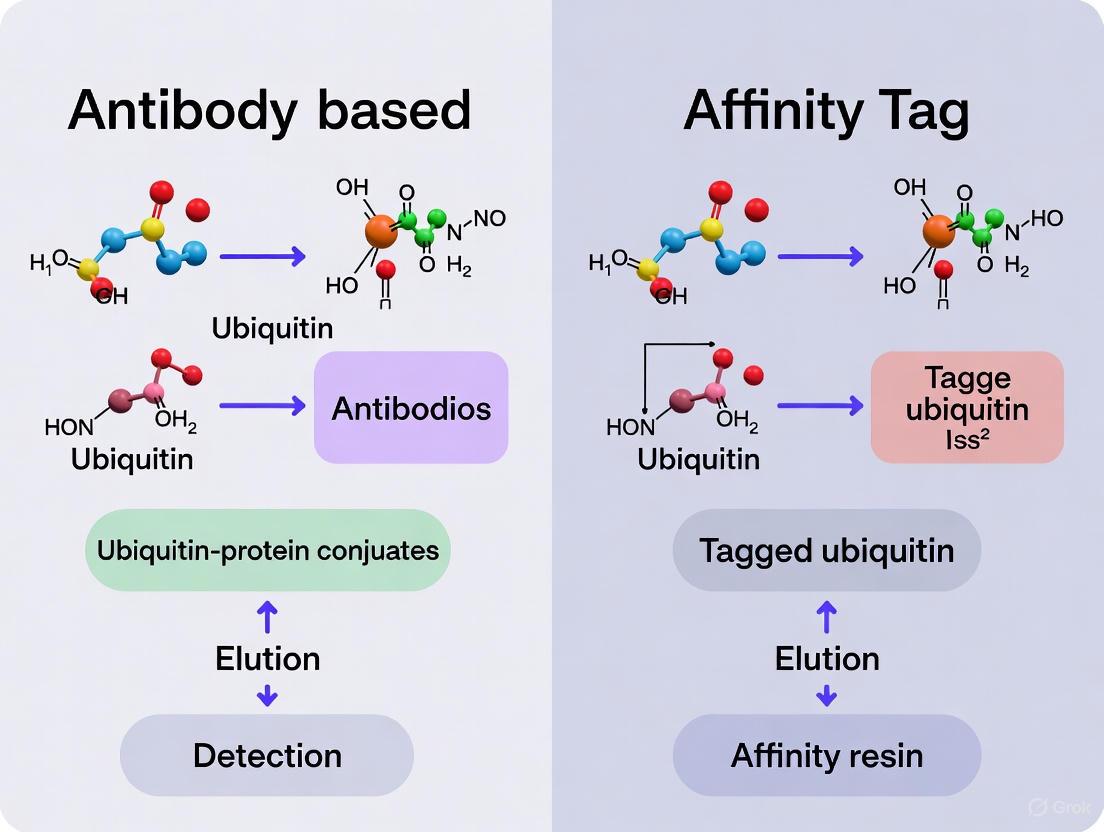
Antibody-based enrichment utilizes immunoprecipitation with antibodies that recognize endogenous ubiquitin or specific ubiquitin linkages. This approach captures ubiquitinated proteins or peptides directly from native biological systems without genetic manipulation. The workflow involves cell lysis, proteolytic digestion (for site identification), incubation with ubiquitin-specific antibodies immobilized on beads, washing to remove non-specifically bound proteins, and elution of enriched ubiquitinated species for mass spectrometry analysis. Key antibodies include pan-ubiquitin recognition antibodies (P4D1, FK1/FK2) and linkage-specific antibodies targeting M1-, K11-, K27-, K48-, or K63-linked chains [2].
Affinity tag-based enrichment relies on genetic engineering to express ubiquitin fused to an affinity tag (e.g., His, Strep, or HA) in cellular systems. The tagged ubiquitin incorporates into the cellular ubiquitination machinery, allowing purification of ubiquitinated proteins through affinity chromatography. The typical workflow involves creating cell lines stably expressing tagged ubiquitin, cell lysis, affinity purification using tag-specific resins (Ni-NTA for His-tag, Strep-Tactin for Strep-tag), and subsequent processing for proteomic analysis [2].
The conceptual relationship between these methods and their position in the experimental workflow can be visualized as follows:
Performance Comparison: Quantitative Metrics and Experimental Data
Direct comparison of antibody-based versus affinity tag methods reveals distinct performance characteristics across multiple parameters that influence method selection for specific research goals.
Table 1: Method Performance Comparison for Ubiquitin Profiling
| Performance Parameter | Antibody-Based Methods | Affinity Tag-Based Methods |
|---|---|---|
| Identification Efficiency | ~100 ubiquitination sites (MCF-7 breast cancer cells) [2] | 110-753 ubiquitination sites (various studies) [2] |
| Stoichiometric Sensitivity | Suitable for moderate to high abundance ubiquitination events | Enhanced detection of low-stoichiometry modifications |
| Linkage Specificity | Available through linkage-specific antibodies (K48, K63, etc.) [2] | Limited to expressed tag type; requires multiple constructs |
| Genetic Manipulation | Not required; works with native tissues and clinical samples [2] | Required; challenging for animal tissues and clinical samples [2] |
| Artifact Potential | Lower risk of artifacts from endogenous profiling | Potential artifacts from tagged ubiquitin expression [2] |
| Sample Compatibility | Broad (cell lines, tissues, clinical samples) [2] | Restricted to genetically modifiable systems [2] |
| Specificity Challenges | Non-specific binding with some antibody batches [2] | Co-purification of histidine-rich/biotinylated proteins [2] |
Table 2: Applications and Limitations in Research Contexts
| Research Context | Recommended Method | Rationale | Key Considerations |
|---|---|---|---|
| Clinical/Tissue Samples | Antibody-based | No genetic manipulation required; preserves native ubiquitinome [2] | Limited by antibody quality and specificity |
| High-Throughput Screening | Affinity tag (Strep/His) | Higher identification efficiency; streamlined purification [2] | Potential artifacts from tag expression |
| Linkage-Specific Studies | Antibody-based (linkage-specific antibodies) | Direct targeting of specific ubiquitin chain types [2] | Limited to characterized linkage types |
| Dynamic Stoichiometry Studies | Computational (occupancy-based) | Quantifies relative ubiquitin occupancy changes [8] | Requires proteasome inhibition and SILAC labeling |
| Low Abundance Targets | Affinity tag with optimized enrichment | Enhanced sensitivity for low-stoichiometry modifications [2] | May miss endogenous regulation patterns |
Experimental Protocols: Detailed Methodologies
Antibody-Based Enrichment Protocol
The FK2 antibody enrichment protocol exemplifies the antibody-based approach. Cells are lysed in modified RIPA buffer (50 mM Tris-HCl pH 7.5, 150 mM NaCl, 1% NP-40, 0.5% sodium deoxycholate, 1 mM EDTA) supplemented with protease inhibitors and 20 mM N-ethylmaleimide to preserve ubiquitination. Lysates are clarified by centrifugation, and protein concentration is normalized. For each enrichment, 1-2 mg of protein lysate is incubated with 5-10 μg of FK2 antibody preconjugated to protein A/G beads for 4 hours at 4°C with rotation. Beads are washed three times with lysis buffer, and bound proteins are eluted with 2× Laemmli buffer at 95°C for 10 minutes. For ubiquitination site mapping, proteins are digested in-solution with trypsin before or after enrichment, and GG-remnant peptides are analyzed by LC-MS/MS [2].
His-Tag Ubiquitin Enrichment Protocol
For His-tag ubiquitin enrichment, cells stably expressing 6×His-tagged ubiquitin are lysed in denaturing buffer (6 M guanidine-HCl, 0.1 M Na2HPO4/NaH2PO4, 10 mM Tris-HCl, pH 8.0) to dissociate non-covalent interactions. Ni-NTA agarose beads are added to the lysate and incubated for 4-16 hours at room temperature. Beads are washed sequentially with denaturing buffer (pH 8.0), denaturing buffer (pH 6.3), and finally with PBS containing 0.1% Triton X-100. Ubiquitinated proteins are eluted with 200 mM imidazole or by competition with 250 mM EDTA. For proteomic analysis, enriched proteins are digested with trypsin, and peptides containing the GG remnant (114.04292 Da mass shift) are identified by LC-MS/MS [2].
Stoichiometry Analysis Using SILAC and Proteasome Inhibition
A sophisticated approach to distinguish degradation versus non-degradation ubiquitin signaling involves stoichiometric analysis through SILAC labeling. SKOV3 ovarian cancer cells are cultured in heavy (13C6 15N4-l-arginine and 13C6-l-lysine) or light media. Light cells are treated with 20 μM MG132 proteasome inhibitor for 6 hours, while heavy cells serve as controls. Cells are mixed 1:1 based on protein concentration, digested with trypsin, and analyzed by LC-MS/MS. Relative ubiquitin occupancy is calculated by comparing heavy-to-light ratios of modified versus unmodified peptides, with increased occupancy at degradation sites upon proteasome inhibition [8].
The Scientist's Toolkit: Essential Research Reagents
Table 3: Key Research Reagents for Ubiquitin Profiling
| Reagent/Category | Specific Examples | Function/Application |
|---|---|---|
| Affinity Tags | 6×His-tag, Strep-tag, HA-tag | Purification of ubiquitinated proteins from engineered systems [2] |
| Pan-Ubiquitin Antibodies | P4D1, FK1, FK2 | Enrichment of total ubiquitinated proteins from native sources [2] |
| Linkage-Specific Antibodies | K48-linkage specific, K63-linkage specific | Selective enrichment of specific ubiquitin chain architectures [2] |
| Activity-Based Probes | Ub-Dha (biotin-Ub-Dha) | Capture active ubiquitin-conjugating machinery; identification of novel enzymes [9] |
| Ubiquitination Enzymes | E1 activating, E2 conjugating, E3 ligating enzymes | In vitro ubiquitination assays; ubi-tagging conjugation platforms [10] |
| Proteasome Inhibitors | MG132, Bortezomib | Stabilization of ubiquitinated proteins destined for degradation [8] |
| Computational Tools | Stoichiometry analysis algorithms | Quantification of ubiquitin occupancy from proteomic data [8] |
Visualization of Ubiquitin Signaling and Profiling Challenges
The complexity of ubiquitin signaling and the corresponding profiling challenges can be visualized through their interconnected relationships:
The selection between antibody-based and affinity tag methods for ubiquitin profiling depends critically on research objectives, sample availability, and desired outcomes. Antibody-based approaches offer superior compatibility with clinical samples and linkage-specific investigations without genetic manipulation requirements. Affinity tag methods provide enhanced identification efficiency and sensitivity for low-stoichiometry modifications in genetically tractable systems. For comprehensive ubiquitinome characterization, researchers may employ sequential or orthogonal strategies, leveraging the complementary strengths of both approaches. Emerging methodologies including activity-based profiling, stoichiometry quantification, and engineered conjugation platforms continue to expand our capacity to decipher the complex ubiquitin code, offering new avenues for therapeutic intervention in ubiquitination-related diseases.
Protein ubiquitination, the covalent attachment of a 76-amino acid ubiquitin protein to substrate proteins, is a fundamental post-translational modification (PTM) that regulates diverse cellular functions including protein stability, activity, and localization [2]. The central challenge in studying this modification lies in its typically low stoichiometry within the complex milieu of cellular lysates, where the signal from ubiquitinated peptides is vastly overshadowed by unmodified peptides [2]. Affinity enrichment strategies have therefore become indispensable for isolating this faint ubiquitinated signal. These methodologies primarily fall into two categories: those that enrich for ubiquitinated proteins prior to digestion (protein-level enrichment) and those that enrich for modified peptides after digestion (peptide-level enrichment) [2] [11]. The core principle uniting all these methods is the specific capture of ubiquitin conjugates—or their diagnostic remnants—from a background of non-ubiquitinated components, thereby enabling detection and analysis by mass spectrometry (MS). This guide objectively compares the performance of the predominant antibody-based and affinity-tag-based enrichment methods, providing the experimental data and protocols necessary for informed methodological selection.
Methodological Comparison: Antibody-Based vs. Affinity Tag Enrichment
The two dominant strategies for ubiquitin enrichment leverage distinct recognition principles, each with characteristic performance profiles in sensitivity, specificity, and practical application.
Core Principles and Workflows
Antibody-Based Enrichment (Peptide-Level): This method uses antibodies raised against the tryptic diglycine (K-ε-GG) remnant that remains attached to a modified lysine residue after trypsin digestion of a ubiquitinated protein. The workflow involves digesting the total cellular protein lysate into peptides, followed by immunoaffinity purification of the K-GG-modified peptides for subsequent LC-MS/MS analysis [12] [11]. This approach directly targets the mass spectrometry-diagnostic signature of ubiquitination.
Affinity Tag-Based Enrichment (Protein-Level): This method relies on the genetic engineering of cells to express epitope-tagged ubiquitin (e.g., His, HA, or Strep tags). After lysis, ubiquitinated proteins are purified en masse using resins that bind the affinity tag (e.g., Ni-NTA for His tags). The enriched proteins are then digested, and peptides are analyzed by MS, with ubiquitination sites identified by the characteristic K-GG mass shift [2].
The following diagram illustrates the logical and procedural relationships between these two primary workflows.
Performance and Experimental Data Comparison
Direct comparative studies reveal significant differences in the performance of these two strategies. As summarized in the table below, the choice of method involves a trade-off between sensitivity, specificity, and practical considerations for the experimental system.
Table 1: Performance Comparison of Ubiquitin Enrichment Methods
| Feature | Antibody-Based (K-GG) | Affinity Tag-Based (e.g., His-Ub) |
|---|---|---|
| Enrichment Target | K-GG-modified peptides (post-digestion) [11] | Ubiquitinated proteins (pre-digestion) [2] |
| Sensitivity | High; >4-fold higher levels of modified peptides than protein-level AP-MS [11] [5] | Moderate; limited by efficiency of protein-level pull-down [11] |
| Specificity | High for the K-GG motif; potential cross-reactivity with other UBLs [12] | Moderate; co-purification of histidine-rich/biotinylated proteins [2] |
| Physiological Context | Captures endogenous ubiquitination from any source, including tissues [2] [13] | Requires genetic manipulation; may not perfectly mimic endogenous Ub [2] |
| Key Advantage | Superior for mapping sites on individual proteins; identifies more sites per substrate [11] [5] | Cost-effective and easy to implement for cellular systems [2] |
| Key Limitation | High cost of high-quality antibodies [2] | Infeasible for clinical/animal tissue samples [2] |
A quantitative study using SILAC-labeled lysates directly compared the identification of K-GG peptides from the same starting material. The peptide-level immunoaffinity enrichment consistently yielded greater than fourfold higher levels of modified peptides for substrates like HER2 and TCRα compared to protein-level affinity purification mass spectrometry (AP-MS) methods [11]. This demonstrates a clear sensitivity advantage for the antibody-based approach in focused ubiquitination site mapping.
Detailed Experimental Protocols
To ensure reproducibility, below are detailed protocols for the two core enrichment methods.
Protocol for Peptide-Level Immunoaffinity Enrichment
This protocol is adapted from studies that successfully mapped ubiquitination sites on HER2, DVL2, and TCRα [11].
Cell Lysis and Digestion:
- Lyse cells in a suitable buffer (e.g., RIPA buffer: 50 mM Tris-HCl pH 8, 150 mM NaCl, 1% NP-40, 0.5% sodium deoxycholate, 0.1% SDS) supplemented with protease inhibitors.
- Determine protein concentration using a BCA assay.
- Reduce, alkylate, and digest the total protein lysate (e.g., 1-10 mg) with trypsin.
Peptide Immunoaffinity Enrichment:
- Incubate the digested peptide mixture with an anti-K-GG antibody (monoclonal are preferred for specificity) conjugated to agarose or magnetic beads.
- Perform the incubation for several hours to overnight at 4°C with gentle mixing.
Wash and Elution:
- Wash the beads extensively with cold lysis buffer followed by water or a volatile buffer (e.g., 25 mM ammonium bicarbonate) to remove non-specifically bound peptides.
- Elute the bound K-GG peptides with a low-pH solution (e.g., 0.1-0.5% TFA or formic acid). Alternative elution methods may involve competition with a Gly-Gly-lysine peptide.
Mass Spectrometry Analysis:
Protocol for Affinity Tag-Based Enrichment (His-Tag Example)
This protocol is based on high-throughput studies in yeast and mammalian cells [2] [14].
Cell Engineering and Lysis:
- Generate a cell line stably expressing N- or C-terminally 6xHis-tagged ubiquitin.
- Lyse cells under denaturing conditions (e.g., 6 M Guanidine-HCl, 100 mM NaH₂PO₄, 10 mM Tris-Cl, pH 8.0) to disrupt non-covalent interactions and inactivate deubiquitinases.
Enrichment of Ubiquitinated Proteins:
- Incubate the clarified lysate with Ni-NTA agarose beads for several hours at room temperature.
- Wash beads sequentially with:
- Denaturing wash buffer (same as lysis buffer, pH 8.0).
- Denaturing wash buffer at pH 6.3.
- A buffer with non-ionic detergent (e.g., 1% Triton X-100 in 25 mM Tris, pH 7.2) to remove residual denaturant.
On-Bead Digestion and Analysis:
- Either elute the bound proteins with imidazole or proceed directly with on-bead tryptic digestion.
- After digestion, the peptides are desalted and analyzed by LC-MS/MS. The ubiquitination sites are identified by searching for the +114.0429 Da mass shift on lysine residues [2].
The Scientist's Toolkit: Essential Research Reagents
Successful ubiquitination studies depend on a suite of specific reagents. The table below details key solutions for setting up these experiments.
Table 2: Essential Reagents for Ubiquitin Affinity Enrichment Studies
| Reagent / Tool | Function / Role | Examples & Notes |
|---|---|---|
| K-ε-GG Antibodies | Immunoaffinity enrichment of ubiquitinated peptides from digests [11]. | Monoclonal antibodies offer superior specificity. Critical for peptide-level enrichment. |
| Tag-Specific Resins | Capture of epitope-tagged ubiquitin or ubiquitinated proteins. | Ni-NTA (for His-tag), Strep-Tactin (for Strep-tag), Anti-FLAG/HA beads [2]. |
| Linkage-Specific Antibodies | Enrich for ubiquitin chains with a specific linkage (e.g., K48, K63) [2]. | Antibodies like FK2 (pan-ubiquitin) or K48-/K63-linkage specific ones for protein-level study. |
| Ubiquitin Binding Domains (UBDs) | Protein domains used as tools to enrich endogenously ubiquitinated proteins [2]. | Tandem UBDs (e.g., from certain DUBs or E3 ligases) increase binding affinity and specificity. |
| Proteasome Inhibitors | Stabilize labile ubiquitinated proteins by blocking their degradation. | MG132, Lactacystin. Often added to cells prior to lysis to increase yield [11]. |
| Deubiquitinase (DUB) Inhibitors | Prevent the cleavage of ubiquitin conjugates during sample preparation. | PR-619, N-Ethylmaleimide (NEM). Added to lysis buffers to preserve ubiquitination signals. |
Advanced Techniques and Emerging Tools
The field continues to evolve with new reagents that further refine the principle of affinity enrichment.
Distinguishing Ubiquitin-like Modifiers: While K-GG antibodies are powerful, they can sometimes cross-react with diglycine remnants from other ubiquitin-like modifiers (UBLs) like NEDD8. More specific reagents, such as antibodies targeting extended motifs, are being developed to address this [15].
Enrichment of Non-Canonical Ubiquitination: Novel antibody toolkits have been developed to detect N-terminal ubiquitination, a non-canonical form of the modification. These antibodies selectively recognize linear N-terminal diglycine motifs (GGX) without cross-reacting with the isopeptide-linked K-ε-GG, enabling the discovery of new substrates for enzymes like UBE2W [15].
The Affinity Enrichment Mindset: A key conceptual advance in the field is the shift from attempting complete "affinity purification" to embracing "affinity enrichment." Modern, sensitive mass spectrometers, coupled with quantitative proteomics, can distinguish true ubiquitination signals from a background of non-specifically bound contaminants. This allows for the use of milder, single-step enrichment protocols that preserve weak or transient interactions that might be lost in stringent multi-step purifications [14] [16].
Protein ubiquitination is a fundamental post-translational modification that regulates diverse cellular functions including protein degradation, DNA repair, and cell signaling pathways. The versatility of ubiquitination stems from its complexity—ranging from single ubiquitin monomers to polymers with different lengths and linkage types—which creates significant challenges for researchers studying this modification. To address these challenges, scientists have developed multiple classes of affinity ligands that enable the detection, purification, and characterization of ubiquitinated proteins. These tools have become indispensable in molecular biology labs worldwide, particularly for understanding disease mechanisms in cancer, neurodegenerative disorders, and immune responses [2] [17].
The current landscape of affinity ligands is broadly divided into two methodological approaches: antibody-based enrichment, which detects endogenous ubiquitination, and affinity tag-based methods, which utilize genetic tagging systems. Antibody-based tools include conventional ubiquitin antibodies, linkage-specific antibodies, ubiquitin-binding domains (UBDs), and tandem ubiquitin-binding entities (TUBEs). Meanwhile, affinity tag approaches involve engineering cells to express epitope-tagged ubiquitin, such as FLAG, HA, V5, Strep, and His tags [2]. Each methodology presents distinct advantages and limitations in specificity, sensitivity, applicability to endogenous proteins, and cost. This guide provides an objective comparison of these affinity ligands, focusing on their performance characteristics and experimental applications to inform researchers selecting tools for ubiquitination studies.
Comparative Performance Analysis of Affinity Ligands
Quantitative Comparison of Epitope Tags and Recognition Antibodies
The efficiency with which antibodies recognize their cognate epitope tags significantly impacts experimental outcomes in ubiquitination studies. A recent systematic comparison evaluated epitope tag/antibody pairs in immunofluorescence applications using recombinant antibodies fused to a standardized mouse Fc domain, enabling direct quantitative comparisons [18].
Table 1: Performance Classification of Epitope Tag/Antibody Pairs in Immunofluorescence
| Performance Category | Epitope Tag | Tested Antibodies | Signal Intensity at High Concentration (5 μg·mL⁻¹) | Signal Intensity at Low Concentration (50 ng·mL⁻¹) |
|---|---|---|---|---|
| Good | EPEA | AI215 | High (>50) | High |
| Good | HA | AF291 | High (>50) | High |
| Good | SPOT | AI196 | High (>50) | High |
| Fair | FLAG (DYKDDDDK) | TA001, AX047 | High (>50) | Moderate |
| Fair | 6xHis | AD946, AV248 | High (>50) | Moderate |
| Mediocre | FLAG (DYKDDDDK) | AI177 | Low (<25) | Not detected |
| Mediocre | 6xHis | AF371 | Low (<25) | Not detected |
| Mediocre | Myc | AI179 | Low (<25) | Not detected |
The study revealed that fixation methods impacted performance for some tags. Anti-myc antibodies generated much lower signals in methanol-fixed cells compared to paraformaldehyde-fixed cells, while the anti-SPOT antibody signal increased with methanol fixation [18]. This demonstrates how experimental conditions must be optimized for specific tag/antibody pairs.
Comparison of Ubiquitin Enrichment Methodologies
Beyond epitope tags, researchers have multiple options for enriching ubiquitinated proteins, each with distinct advantages and limitations.
Table 2: Performance Comparison of Ubiquitin Enrichment Methods
| Enrichment Method | Principle | Advantages | Limitations | Typical Applications |
|---|---|---|---|---|
| Epitope-Tagged Ubiquitin (His, Strep, FLAG, HA) | Expression of tagged ubiquitin in cells; purification with tag-specific antibodies or resins | - Easy, user-friendly workflow - Relatively low cost - High purity achievable | - Cannot mimic endogenous ubiquitin - Potential artifacts from overexpression - Infeasible for patient tissues | - Screening ubiquitinated substrates in cell lines - Proteomic identification of ubiquitination sites |
| Ubiquitin Antibodies (P4D1, FK1/FK2) | Immunoaffinity enrichment using antibodies recognizing ubiquitin | - Works with endogenous proteins - Applicable to clinical samples - No genetic manipulation needed | - High cost - Potential non-specific binding - May lack sensitivity for low-abundance targets | - Ubiquitin profiling from animal tissues - Analysis of patient samples |
| Linkage-Specific Antibodies | Antibodies specific to particular ubiquitin chain linkages (M1, K48, K63) | - Provides linkage information - Works under physiological conditions - High specificity | - Very high cost - Each antibody detects only one linkage type - Limited availability for atypical linkages | - Studying specific ubiquitin signaling pathways - Disease mechanism studies |
| UBD-Based Approaches (OtUBD) | Enrichment using ubiquitin-binding domains | - High affinity for endogenous ubiquitin - Works with all linkage types - Versatile application formats | - Variable affinity of different UBDs - May require optimization - Potential preference for certain chain types | - Both mono- and polyubiquitinated protein enrichment - Proteomics studies |
| TUBEs (Tandem Ubiquitin-Binding Entities) | Multiple UBDs linked in a single polypeptide | - High affinity for polyubiquitin chains - Protects ubiquitin chains from deubiquitinases - Efficient purification | - Poor efficiency for monoubiquitinated proteins - May alter natural ubiquitin topology - Limited commercial availability | - Studying polyubiquitinated substrates - Degradation pathway analysis |
The selection of an appropriate enrichment method depends heavily on research goals. For example, while TUBEs excel at purifying polyubiquitinated proteins, they work poorly for monoubiquitinated targets, which constitute a large fraction of ubiquitinated proteins in mammalian cells [19]. Similarly, linkage-specific antibodies provide exquisite precision but at high cost and with limited coverage of diverse linkage types.
Experimental Protocols for Key Methodologies
OtUBD-Based Enrichment of Ubiquitinated Proteins
The OtUBD protocol utilizes a high-affinity ubiquitin-binding domain from Orientia tsutsugamushi for enriching ubiquitinated proteins. This method offers both native and denaturing workflows to distinguish directly ubiquitinated proteins from interacting partners [19].
Key Reagents:
- pRT498-OtUBD or pET21a-cys-His6-OtUBD plasmids
- SulfoLink coupling resin for immobilization
- Lysis buffers: Native (50 mM Tris-HCl pH 7.5, 150 mM NaCl, 1% NP-40, 10% glycerol) or Denaturing (6 M guanidine-HCl, 100 mM NaH₂PO₄, 10 mM Tris-HCl pH 8.0)
- Protease inhibitors (EDTA-free cocktail, PMSF, N-ethylmaleimide)
- Elution buffer: 10-20 mM reduced glutathione in appropriate buffer
Procedure:
- Resin Preparation: Couple recombinant OtUBD to SulfoLink resin according to manufacturer's instructions
- Cell Lysis: Prepare lysates from yeast or mammalian cells using either native or denaturing conditions
- Enrichment: Incubate clarified lysates with OtUBD resin for 2-4 hours at 4°C
- Washing: Wash resin with appropriate buffer (native or denaturing)
- Elution: Elute bound proteins with glutathione-containing buffer
- Analysis: Process eluates for Western blotting or mass spectrometry analysis
The denaturing workflow specifically enriches covalently ubiquitinated proteins, while the native approach also captures proteins that interact with ubiquitin or ubiquitinated proteins [19]. This protocol has been successfully applied to both budding yeast and mammalian cell lysates, and can be adapted for other biological samples.
Side-by-Side Comparison of Epitope Tag Antibodies
The quantitative evaluation of epitope tag antibodies employed a standardized immunofluorescence protocol enabling direct comparison of recognition efficiency [18].
Key Reagents:
- HEK cells expressing IL2Ra fusion proteins with C-terminal epitope tags
- Recombinant anti-tag antibodies fused to mouse Fc domain
- Anti-IL2Ra antibody (AJ519) with rabbit Fc domain as reference
- Fixation solutions: 4% paraformaldehyde or cold methanol
- Permeabilization solution: 0.1% saponin
- Fluorescently-labeled secondary antibodies
Procedure:
- Cell Preparation: Culture HEK cells expressing IL2Ra-epitope tag fusion proteins
- Fixation and Permeabilization: Fix cells with either paraformaldehyde or methanol, then permeabilize with saponin
- Immunostaining: Incubate with anti-tag antibody (50 ng·mL⁻¹ to 5 μg·mL⁻¹) and reference anti-IL2Ra antibody
- Image Acquisition: Capture fluorescence images using standardized settings
- Signal Quantification: Measure fluorescence intensity in multiple regions; calculate ratio of anti-tag to anti-IL2Ra signal
- Data Normalization: Normalize signals to positive control (anti-HA AF291) included in each experiment
This methodology allowed researchers to classify antibodies into performance categories based on their signal intensity at different concentrations and under various fixation conditions [18].
Research Reagent Solutions for Ubiquitination Studies
Table 3: Essential Research Reagents for Ubiquitination Studies
| Reagent Category | Specific Examples | Function/Application | Key Characteristics |
|---|---|---|---|
| Epitope Tags | FLAG (DYKDDDDK), HA (YPYDVPDYA), 6xHis (HHHHHH), Myc (EQKLISEEDL) [20] | Protein detection and purification via antibody recognition | - Small size minimizes impact on protein function - High hydrophilicity promotes surface accessibility |
| Ubiquitin Antibodies | P4D1, FK1/FK2, E412J [19] [2] | Detection and enrichment of ubiquitinated proteins | - Recognize all ubiquitin linkages - Varying specificity for different ubiquitin forms |
| Linkage-Specific Antibodies | K48-linkage specific, K63-linkage specific [2] | Detection of specific ubiquitin chain types | - Enable study of chain-type specific functions - Essential for understanding signaling specificity |
| UBD-Based Tools | OtUBD [19], TUBEs [2] | High-affinity enrichment of ubiquitinated proteins | - OtUBD: high affinity for both mono- and polyubiquitin - TUBEs: preferred for polyubiquitin chains |
| Specialized Antibodies | Anti-GGX antibodies (1C7, 2B12, 2E9, 2H2) [21] | Specific detection of N-terminal ubiquitination | - Selective for linear diglycine peptides - No cross-reactivity with lysine-linked diglycine |
| Plasmid Systems | pRT498-OtUBD, pET21a-cys-His6-OtUBD [19] | Recombinant expression of affinity tools | - Enable production of custom enrichment reagents - Modular design for adaptability |
Visualization of Experimental Workflows
Ubiquitin Enrichment Method Selection Pathway
Figure 1: Ubiquitin Enrichment Method Selection Pathway. This decision tree guides researchers in selecting appropriate affinity ligands based on their experimental requirements, including whether they work with endogenous or tagged ubiquitin systems, need specific linkage information, or focus on particular ubiquitin chain types.
Protocol Deep Dive: Implementing Antibody and Tag-Based Enrichment
Protein ubiquitination is a crucial post-translational modification (PTM) that regulates diverse cellular functions, including protein degradation, activity, and localization [22]. The characterization of this modification is vital for understanding fundamental biology and the pathogenesis of numerous diseases, such as cancer and neurodegenerative disorders [22]. The versatility of ubiquitination, which can range from a single ubiquitin (Ub) monomer to complex polymers of different lengths and linkage types, presents significant analytical challenges [22]. Among the methods developed to address these challenges, antibody-based immunoaffinity enrichment has emerged as a powerful approach. This guide objectively compares two principal antibody-based workflows: those utilizing anti-ubiquitin antibodies and those employing anti-K-ε-GG antibodies, framing this comparison within the broader context of ubiquitination enrichment methodologies, including affinity-tag approaches.
Fundamental Principles of Ubiquitination and Detection
Ubiquitin is a small, 76-amino acid protein that is covalently attached to substrate proteins via a three-enzyme cascade (E1, E2, E3) [22]. The C-terminal glycine (G76) of Ub forms an isopeptide bond primarily with the epsilon-amino group of lysine residues on target proteins. Ub itself contains seven lysine residues (K6, K11, K27, K29, K33, K48, K63) and an N-terminal methionine (M1), which can serve as attachment points for additional Ub molecules, leading to the formation of polyubiquitin chains. These chains can be homotypic (same linkage), heterotypic (mixed linkages), or branched, with different linkage types often determining the functional outcome for the modified substrate [22].
For mass spectrometry (MS)-based detection, tryptic digestion of ubiquitinated proteins generates a unique signature. Trypsin cleaves after arginine and lysine residues, but the modified lysine on the substrate protein is no longer a cleavage site because it is conjugated to Ub. Furthermore, trypsin cleaves within the Ub moiety itself, leaving a di-glycine remnant (K-ε-GG) attached to the modified lysine of the substrate peptide [23]. This characteristic mass shift (+114.04 Da) on the modified lysine is the key identifier for ubiquitination sites in MS data and forms the basis for one of the primary antibody strategies discussed herein.
Table 1: Key Definitions in Ubiquitination Research
| Term | Description |
|---|---|
| Ubiquitin (Ub) | A 76-amino acid protein covalently attached to substrate proteins to regulate their function [22]. |
| K-ε-GG Remnant | A di-glycine peptide derived from the C-terminus of Ub that remains attached to a modified lysine on substrate peptides after tryptic digestion [24] [23]. |
| Anti-Ubiquitin Antibodies | Antibodies (e.g., P4D1, FK1/FK2) that recognize the ubiquitin protein itself, often regardless of linkage type [22]. |
| Anti-K-ε-GG Antibodies | Antibodies that specifically recognize the tryptic di-glycine remnant covalently linked to a lysine side chain [24] [23]. |
| Linkage-Specific Antibodies | Antibodies that recognize polyubiquitin chains of a specific linkage type (e.g., K48, K63) [22]. |
The low stoichiometry of ubiquitination and complexity of biological samples necessitate enrichment prior to MS analysis. The major methodological strands can be broadly categorized as follows:
- Antibody-Based Immunoaffinity Enrichment: This involves using antibodies to selectively isolate ubiquitinated proteins or peptides from complex mixtures. The two main sub-categories, which form the core of this comparison, are:
- Anti-Ubiquitin Workflow: Enriches intact ubiquitinated proteins prior to digestion.
- Anti-K-ε-GG Workflow: Enriches ubiquitinated peptides (carrying the K-ε-GG modification) after digestion.
- Affinity Tag-Based Enrichment: This requires genetic engineering to express ubiquitin (or the protein of interest) fused to an affinity tag, such as His, Flag, or Strep. Cells are engineered to express the tagged Ub, which is incorporated into the ubiquitination machinery. The ubiquitinated proteins can then be purified using resins that bind the tag (e.g., Ni-NTA for His-tags) [22].
- Ub-Binding Domain (UBD)-Based Enrichment: This approach uses proteins or engineered domains (e.g., Tandem-repeated Ub-binding Entities - TUBEs) that naturally interact with Ub to pull down ubiquitinated proteins [22].
The following diagram illustrates the logical relationship between these primary enrichment methodologies.
Detailed Comparison: Anti-Ubiquitin vs. Anti-K-ε-GG Workflows
Anti-Ubiquitin Immunoaffinity Workflow
This workflow is designed to capture the full complement of ubiquitinated proteins. Antibodies such as P4D1 or FK1/FK2, which recognize epitopes on the ubiquitin protein itself, are used for enrichment [22]. These are often described as "pan-ubiquitin" antibodies because they can potentially recognize mono- and polyubiquitinated proteins regardless of the chain linkage type.
Experimental Protocol:
- Cell Lysis & Preparation: Cells or tissues are lysed under denaturing conditions (e.g., using 8 M urea or SDS) to preserve ubiquitination states and disrupt non-covalent interactions, thereby inactivating deubiquitinases (DUBs) [24].
- Protein-Level Enrichment: The clarified lysate is incubated with anti-ubiquitin antibodies conjugated to solid-support beads (e.g., protein A/G agarose or magnetic beads). This step captures proteins that are ubiquitinated.
- Washing: Beads are extensively washed with lysis and wash buffers to remove non-specifically bound proteins.
- Elution: Bound ubiquitinated proteins are eluted, typically using low-pH buffers (e.g., glycine-HCl) or SDS-PAGE loading buffer.
- Downstream Processing: The eluted proteins are digested with trypsin (either in-solution or in-gel) and the resulting peptides are analyzed by LC-MS/MS. Ubiquitination sites are identified based on the discovery of peptides containing the K-ε-GG mass shift [22] [25].
Anti-K-ε-GG Immunoaffinity Workflow
This method targets the specific tryptic signature of ubiquitination. The commercialization of highly specific anti-K-ε-GG antibodies was a breakthrough that dramatically improved the depth and sensitivity of ubiquitin site identification by MS [24] [23].
Experimental Protocol:
- Total Proteome Digestion: The entire protein extract from cells or tissues is digested into peptides using trypsin. This exposes the K-ε-GG remnant.
- Peptide-Level Enrichment: The complex peptide mixture is incubated with anti-K-ε-GG antibodies conjugated to beads. The antibodies specifically bind to peptides containing the K-ε-GG modification.
- Washing & Elution: After washing to remove unmodified peptides, the enriched K-ε-GG peptides are eluted under acidic conditions (e.g., 0.15% TFA) [24].
- MS Analysis: The eluted peptides are directly analyzed by LC-MS/MS. The identification of a peptide with a di-glycine modification on a lysine is direct evidence of a ubiquitination site [24] [23].
Refinements: The workflow has been significantly optimized. Key improvements include:
- Antibody Cross-linking: Covalently cross-linking the antibody to the beads prevents co-elution of antibody fragments, reducing MS contamination and improving quantitative accuracy [24].
- Off-line Fractionation: Basic reversed-phase chromatography fractionation of peptides prior to enrichment reduces sample complexity and increases the number of identifiable ubiquitination sites [24].
- On-Antibody TMT Labeling (UbiFast): A highly sensitive, multiplexed method (UbiFast) allows for Tandem Mass Tag (TMT) labeling of K-ε-GG peptides while they are bound to the antibody. This protects the di-glycine remnant from being derivatized and enables high-throughput quantification from small sample amounts (e.g., ~10,000 sites from 500 μg of peptide) [23].
The following workflow diagram contrasts the two main antibody-based approaches.
Objective Performance Comparison
The choice between anti-ubiquitin and anti-K-ε-GG workflows depends heavily on the research goals, as they offer distinct advantages and suffer from different limitations.
Table 2: Performance Comparison of Anti-Ubiquitin and Anti-K-ε-GG Workflows
| Feature | Anti-Ubiquitin Workflow | Anti-K-ε-GG Workflow |
|---|---|---|
| Enrichment Target | Ubiquitinated proteins [22] [25] | K-ε-GG-modified peptides [24] [23] |
| Stoichiometric Sensitivity | Can be limited by the low abundance of specific ubiquitinated protein forms [22]. | Highly sensitive; optimized protocols can identify ~20,000 sites from 5 mg of protein input [24]. |
| Site Identification | Indirect; relies on post-enrichment digestion and K-ε-GG discovery by MS [22]. | Direct; the enrichment target is the modified site itself [24] [23]. |
| Linkage Information | Can be combined with linkage-specific antibodies to study chain topology [22]. | Primarily identifies modification sites; linkage information on the same peptide is limited. |
| Specificity | Can be less specific, yielding higher background noise due to co-purifying proteins [25]. | High specificity for the ubiquitin remnant, resulting in lower background [25]. |
| Sample Compatibility | Suitable for tissue and clinical samples without genetic manipulation [22]. | Suitable for tissue and clinical samples; UbiFast enables multiplexing of limited samples [23]. |
| Key Limitation | Non-specific binding, inability to distinguish ubiquitination from other Ub-Like modifiers [22]. | Requires high-quality, specific antibodies; the K-ε-GG motif can be labile [24]. |
| Typical Applications | Studying specific ubiquitinated protein complexes, linkage-type analysis via specific antibodies [22]. | Global, site-specific ubiquitinome mapping and quantitative profiling under different conditions [24] [23]. |
The Scientist's Toolkit: Key Research Reagent Solutions
Successful implementation of these workflows relies on specific, high-quality reagents. The table below details essential materials and their functions.
Table 3: Essential Reagents for Ubiquitin Immunoaffinity Workflows
| Reagent / Material | Function in the Workflow |
|---|---|
| Anti-K-ε-GG Antibody | Core reagent for peptide-level enrichment; specifically recognizes the tryptic di-glycine remnant on lysine [24] [23]. |
| Pan-Ubiquitin Antibodies (e.g., FK2, P4D1) | Core reagent for protein-level enrichment; recognizes epitopes on the ubiquitin protein [22]. |
| Linkage-Specific Ub Antibodies (e.g., α-K48, α-K63) | Used to enrich for proteins modified with specific polyubiquitin chain linkages [22]. |
| Protein A/G Agarose/Magnetic Beads | Solid support for immobilizing antibodies during immunoaffinity precipitation [24] [26]. |
| Cross-linking Reagents (e.g., DMP) | For covalently cross-linking antibodies to beads, preventing antibody leakage and improving MS data quality [24]. |
| TMT/Isobaric Tags | For multiplexed quantitative MS; used in the UbiFast protocol for on-antibody labeling [23]. |
| Denaturing Lysis Buffer (Urea, SDS) | To efficiently lyse cells, inactivate DUBs, and preserve the native ubiquitination state [24]. |
| Protease & DUB Inhibitors | Critical for preventing protein degradation and the removal of ubiquitin modifications during sample preparation [24]. |
| High-pH Reversed-Phase Chromatography | For off-line fractionation of peptides to reduce sample complexity prior to K-ε-GG enrichment [24]. |
Both anti-ubiquitin and anti-K-ε-GG immunoaffinity workflows are indispensable tools in the ubiquitin researcher's arsenal. The anti-K-ε-GG workflow is the unequivocal leader for deep, site-specific mapping of ubiquitinomes. Its superior sensitivity and specificity, especially when coupled with refined protocols like antibody cross-linking and the UbiFast method, make it ideal for large-scale quantitative studies aiming to understand global ubiquitination dynamics from limited biological samples, such as clinical tissues [24] [23].
In contrast, anti-ubiquitin workflows retain critical utility for applications focused on studying specific ubiquitinated proteins or ubiquitin chain architecture. Their primary strength lies in the ability to use linkage-specific antibodies to probe the topology of polyubiquitin chains, which is often obscured in standard K-ε-GG approaches [22]. Furthermore, they are applicable in situations where protein-level information or the study of intact complexes is required.
When positioned within the broader thesis of ubiquitination enrichment methods, antibody-based strategies offer a powerful, direct approach for analyzing endogenous ubiquitination without genetic manipulation. They complement affinity-tag methods, which, while powerful for controlled systems, are often infeasible for clinical and tissue samples. The choice between anti-ubiquitin and anti-K-ε-GG antibodies, therefore, is not a matter of which is universally better, but which is optimally suited to answer the specific biological question at hand.
The post-translational modification of proteins with ubiquitin (Ub) is a fundamental regulatory mechanism that controls diverse cellular processes, including protein degradation, DNA repair, and cell signaling [27] [2] [28]. The versatility of ubiquitin signaling stems from the ability of ubiquitin to form polymers (polyUb) of different topologies, which are specialized for distinct cellular functions [27] [28]. These polymers can be homotypic (linked through the same lysine residue), heterotypic (containing mixed linkages), or branched (containing ubiquitin subunits modified at multiple sites) [28]. The fate of a ubiquitinated protein is largely determined by the topology of the polyubiquitin chain attached to it [27].
To decipher the complex language of ubiquitin signaling, researchers have developed various enrichment strategies to isolate and characterize ubiquitinated proteins. Two principal methodologies have emerged: linkage-specific antibodies and affinity tag-based approaches. This guide provides an objective comparison of these methods, focusing on their performance characteristics, applications, and limitations within the context of ubiquitin chain topology studies.
Methodological Comparison: Antibody-Based vs. Affinity Tag Enrichment
Fundamental Principles and Workflows
Linkage-Specific Antibody Approach: This method utilizes antibodies engineered to recognize specific ubiquitin chain linkages (e.g., K48, K63, M1) [29] [2]. These antibodies can immunoprecipitate endogenously ubiquitinated proteins from complex biological samples without genetic manipulation, allowing for the study of ubiquitination under physiological conditions [2].
Affinity Tag-Based Approach: This method involves the expression of epitope-tagged ubiquitin (e.g., His, HA, Flag, Strep) in cells [2]. The ubiquitinated substrates are then purified using resins that bind the affinity tag, such as Ni-NTA for His-tagged ubiquitin or Strep-Tactin for Strep-tagged ubiquitin [2].
The fundamental workflows for these two primary methods in the study of the ubiquitinome are distinct, as illustrated below.
Performance Data and Technical Comparison
The table below summarizes key performance characteristics and experimental data for both enrichment methodologies.
| Parameter | Linkage-Specific Antibodies | Affinity Tag-Based Methods |
|---|---|---|
| Specificity | High for targeted linkages (e.g., K48, K63) [29] [2] | Broad, non-selective enrichment of all ubiquitinated proteins [2] |
| Sample Requirements | Native cell lysates or tissues; no genetic manipulation needed [2] | Requires genetic engineering to express tagged ubiquitin [2] |
| Physiological Relevance | Preserves endogenous ubiquitination states [2] | Potential artifacts from tagged ubiquitin expression [2] |
| Identification Yield | Denis et al.: 96 ubiquitination sites from MCF-7 cells [2] | Peng et al.: 110 ubiquitination sites (72 proteins) in yeast [2]Danielsen et al.: 753 ubiquitination sites (471 proteins) in human cells [2] |
| Key Limitations | High cost; potential non-specific binding; limited to characterized linkages [2] | Co-purification of non-ubiquitinated proteins (e.g., histidine-rich proteins); may not mimic endogenous ubiquitin structure [2] |
| Typical Applications | Studying specific ubiquitin-dependent pathways; analysis of clinical samples [29] [2] | Global ubiquitinome profiling; discovery-based studies [2] |
Experimental Protocol for Ubiquitin Enrichment and Analysis
Linkage-Specific Immunoprecipitation Protocol:
- Cell Lysis: Prepare cell lysates using non-denaturing lysis buffer to preserve protein complexes and ubiquitination states [2].
- Antibody Incubation: Incubate lysates with linkage-specific antibody (e.g., anti-K48 or anti-K63) [29] [2].
- Immunoprecipitation: Use protein A/G beads to capture antibody-antigen complexes.
- Washing: Wash beads extensively with lysis buffer to remove non-specifically bound proteins.
- Elution: Elute ubiquitinated proteins using low pH buffer or competitive elution.
- Analysis: Analyze by western blotting or mass spectrometry [2].
Tandem Mass Spectrometry Analysis:
Liquid chromatography tandem mass spectrometry (LC-MS/MS) provides detailed characterization of ubiquitin chain topology:
Liquid Chromatography:
- Use reverse-phase columns (e.g., ProSwift RP-4H monolith, 200μm × 25cm) [27].
- Mobile phases: A) water-acetonitrile (97.5:2.5) with 0.1% formic acid; B) water-acetonitrile (25:75) with 0.1% formic acid [27].
- Apply linear gradient from 5% to 55% mobile phase B over 20 minutes at 1.5 μL/min flow rate [27].
Tandem Mass Spectrometry:
- Use high-resolution instruments (e.g., Orbitrap Fusion Lumos) [27].
- Set mass resolution to 120,000 at 200 m/z [27].
- Employ fragmentation techniques: ETciD (electron transfer dissociation with collision-induced dissociation) or EThcD (ETD with higher-energy CID) [27].
- Optimize ion routing multipole pressure to 0.01-0.03 mTorr for maximum fragment ion density [27].
Research Reagent Solutions
The table below details essential research reagents for studying ubiquitin chain topology, along with their specific functions in experimental workflows.
| Reagent / Material | Function / Application |
|---|---|
| Linkage-Specific Antibodies (e.g., anti-K48, anti-K63) [29] [2] | Immunoprecipitation and detection of specific ubiquitin chain linkages |
| Epitope-Tagged Ubiquitin (His, HA, Flag, Strep) [2] | Affinity purification of ubiquitinated proteins from engineered cells |
| UBE2C (E2 Enzyme) [28] | Initiates mixed linkage chains for APC/C-mediated branched chain formation |
| UBE2S (E2 Enzyme) [28] | Extends K11 linkages on initial chains to form branched K11/K48 polymers |
| HECT E3 Ligases (e.g., HUWE1, ITCH, UBR5) [28] | Collaborate to synthesize branched ubiquitin chains (e.g., K48/K63) |
| Tandem Ubiquitin-Binding Entities (TUBEs) [2] | High-affinity enrichment of polyubiquitinated proteins without linkage specificity |
| Deubiquitinases (DUBs) [27] | Control ubiquitin chain editing and serve as analytical tools for chain validation |
| CRISPR-Cas9 Knockout Cell Lines [30] | Validation of antibody specificity through isogenic control cells |
Functional Roles of Ubiquitin Chain Topologies
Different ubiquitin chain topologies create specialized signals recognized by specific effector proteins, directing diverse cellular outcomes as shown in the pathway below.
The expanding toolbox of linkage-specific antibodies represents a significant advancement in ubiquitin research, enabling precise interrogation of specific ubiquitin chain topologies. When compared to affinity tag-based methods, linkage-specific antibodies offer the distinct advantage of studying endogenous ubiquitination without genetic manipulation, making them particularly valuable for investigating clinical samples and specific ubiquitin-dependent signaling pathways [2].
However, challenges remain. Antibody specificity and reliability are persistent concerns, with studies indicating that a significant percentage of commercial antibodies fail validation tests [30]. Additionally, the current repertoire of linkage-specific antibodies is limited to the most well-characterized linkages, leaving gaps in our ability to study atypical ubiquitin chains [2] [28].
For comprehensive ubiquitin chain topology studies, a combined approach often yields the most robust results. Linkage-specific antibodies can validate findings from global ubiquitinome profiling using affinity tags, while emerging techniques like top-down mass spectrometry [27] and specialized ubiquitin-binding domains [2] provide complementary information about chain architecture and complexity. As our understanding of branched and atypical ubiquitin chains grows [28], the continued development and rigorous validation of linkage-specific reagents will remain crucial for deciphering the complex language of ubiquitin signaling in health and disease.
The isolation of specific proteins or protein modifications is a cornerstone of biochemical research and therapeutic development. Within the study of ubiquitination—a critical post-translational modification regulating nearly all cellular processes—the choice of enrichment strategy profoundly impacts the outcomes and interpretations of experiments. Researchers are often faced with a decision: use an affinity tag (like His or Strep) fused to the protein of interest or ubiquitin itself, or employ ubiquitin-binding entities (like TUBEs) that recognize the native modification. This guide provides a objective, data-driven comparison of these predominant affinity tag strategies—His-tag, Strep-tag, and TUBE-based purification—focusing on their application in ubiquitination research. We will evaluate their performance based on purity, yield, specificity, and compatibility with downstream analytical techniques, providing a clear framework for selecting the optimal tool for specific research goals.
Understanding the Purification Technologies
Affinity Tag Purification: His-tag and Strep-tag
Affinity purification relies on a specific binding interaction between a tag fused to a target protein and an immobilized ligand [31]. The general process involves incubating a crude sample with the affinity support, washing away non-specifically bound contaminants, and then eluting the pure target protein [31].
- His-tag Purification: This system uses a polyhistidine tag (typically 6xHis) that binds to immobilized transition metal ions like Ni²⁺ or Co²⁺, a method known as immobilized metal affinity chromatography (IMAC) [32]. The small size of the tag is a key advantage, minimizing potential impact on the structure and function of the fused protein [33] [32].
- Strep-tag Purification: The Strep-tag (Strep-tagII and Twin-Strep-tag) is a peptide that binds with high specificity and affinity to a stabilized form of streptavidin called Strep-Tactin [32]. The tags are engineered for this specific interaction, making them less likely to occur naturally in biological samples [32].
Ubiquitin-Binding Domain Purification: TUBE and ThUBD
Instead of tagging the protein, an alternative strategy is to use proteins or domains that naturally recognize and bind ubiquitin. This is particularly valuable for studying endogenous ubiquitination without genetic manipulation.
- TUBE (Tandem Ubiquitin Binding Entities): TUBEs are recombinant proteins containing multiple ubiquitin-binding domains (UBDs) in tandem. This configuration confers high affinity for ubiquitin chains and protects them from cleavage by deubiquitinating enzymes (DUBs) during purification [2] [34].
- ThUBD (Tandem Hybrid Ubiquitin Binding Domain): A recent advancement on the TUBE technology, ThUBD combines different types of UBDs to achieve not only high affinity but also unbiased recognition of all ubiquitin chain linkage types, overcoming a significant limitation of earlier reagents [34].
Table 1: Core Characteristics of the Purification Technologies
| Technology | Core Principle | Typical Application in Ubiquitination Research | Genetic Manipulation Required? |
|---|---|---|---|
| His-tag | IMAC: Binding of His-tag to Ni²⁺/Co²⁺ [32] | Purification of recombinant ubiquitin or ubiquitinated substrates; requires expression of His-tagged ubiquitin [2] | Yes |
| Strep-tag | Affinity: Binding of Strep-tag to Strep-Tactin [32] | Similar to His-tag; used for purifying recombinant ubiquitinated proteins [2] | Yes |
| TUBE/ThUBD | Protein-Protein Interaction: Binding of UBDs to ubiquitin moieties [2] [34] | Enrichment of endogenously ubiquitinated proteins from native systems (cells, tissues); no tag needed [2] [34] | No |
Performance Comparison: Key Operational Metrics
Direct comparisons reveal significant differences in the performance of these tags, influencing their suitability for different experimental needs.
Purity, Specificity, and Yield
- His-tag: While providing good yields, His-tag purification often results in moderate purity due to the unspecific binding of host proteins that contain consecutive histidine residues or have an intrinsic affinity for the metal ions [35] [32]. This necessitates careful optimization of imidazole concentration in wash buffers and may require additional purification steps like size-exclusion chromatography, which can reduce final yield [32].
- Strep-tag: The Strep-tag system is characterized by high purity and high yield in a single step. Its engineered specificity means there is minimal co-purification of host proteins, leading to pure protein preparations without the need for further processing [35] [32].
- TUBE/ThUBD: The primary goal of TUBE/ThUBD is the specific enrichment of ubiquitinated proteins from complex mixtures. The recently developed ThUBD-coated plates demonstrate a 16-fold wider linear range for capturing polyubiquitinated proteins compared to TUBE-based plates, indicating superior sensitivity and dynamic range [34].
Binding Affinity and Applicability
The affinity of the tag-ligand interaction dictates which applications are feasible.
- His-tag has a relatively low affinity (µM-nM range), which can lead to rapid dissociation and poor immobilization, making it less suitable for techniques requiring stable binding like surface plasmon resonance (SPR) [32].
- Strep-tag technology offers a range of affinities from µM to pM, allowing researchers to select the appropriate tag (Strep-tagII or Twin-Strep-tag) for applications from routine purification to analytical techniques like SPR and bio-layer interferometry (BLI) [32].
- ThUBD exhibits unbiased, high-affinity binding for all ubiquitin chain types, making it ideal for comprehensive profiling of the ubiquitinome without linkage-based bias [34].
Table 2: Direct Comparison of His-tag and Strep-tag Performance
| Performance Metric | His-tag | Strep-tag |
|---|---|---|
| Purity from E. coli lysates | Moderate [35] [32] | Excellent (High) [35] [32] |
| Typical Yield | Good to High [35] [32] | Good to High [35] [32] |
| Affinity Range | µM - nM [32] | µM - pM [32] |
| Resin Cost | Low (Inexpensive, high-capacity resins) [35] | Moderate [35] |
| Common Elution Method | Imidazole (or low pH) [33] [32] | Desthiobiotin (gentle, specific) [32] |
Compatibility and Practical Considerations
Buffer and Reagent Compatibility
The chemical environment required for purification can be a deciding factor.
- His-tag has several limitations. Reducing agents like DTT are not recommended, and chelating agents like EDTA will strip the metal ions from the resin, rendering it useless. Furthermore, common buffer components such as Tris and MOPS are also not recommended [32].
- Strep-tag is highly flexible, tolerating a wide range of conditions including reducing agents (e.g., 50 mM DTT), chelating agents (e.g., 100 mM EDTA), high salt (up to 5 M NaCl), and various buffer substances [32]. This makes it suitable for challenging proteins like metalloproteases or membrane proteins.
- TUBE/ThUBD methods are typically optimized for physiological buffers but can be adapted to various conditions used in protein extraction and immunoassays [34].
Expression Host Compatibility
- His-tag works well in E. coli but faces challenges in other systems. The acidic pH of yeast and insect cell media can cause elution of the tagged protein, and media components (like amino acids) can compete with the tag for binding sites or cause leakage of nickel ions from the resin [32].
- Strep-tag and TUBE/ThUBD technologies do not face these limitations and can be used reliably across all common expression hosts, including mammalian cells [32] [34].
Experimental Protocols for Ubiquitination Research
Protocol 1: Enriching Ubiquitinated Proteins with TUBE/ThUBD
This protocol is for high-throughput, plate-based enrichment of ubiquitinated proteins using ThUBD, ideal for profiling or target-specific analysis [34].
- Plate Coating: Coat high-binding 96-well plates (e.g., Corning 3603) with 1.03 µg/well of purified ThUBD fusion protein in PBS overnight at 4°C [34].
- Blocking: Block the plates with a protein-based blocking agent (e.g., 5% BSA in PBST) for 2 hours at room temperature to prevent non-specific binding [34].
- Sample Application and Incubation: Add complex proteome samples (as low as 0.625 µg) to the wells and incubate for 2 hours at room temperature with gentle shaking. This allows ubiquitinated proteins to bind to the immobilized ThUBD [34].
- Washing: Wash the wells 3-5 times with a optimized wash buffer (e.g., PBST with 300-500 mM NaCl) to remove non-specifically bound material [34].
- Detection/Analysis: Detect captured ubiquitinated proteins using anti-ubiquitin antibodies conjugated to HRP for chemiluminescent readout. Alternatively, proteins can be eluted for downstream analysis by Western blot or mass spectrometry [34].
Protocol 2: Purifying Ubiquitinated Proteins via His-Tagged Ubiquitin
This is a common method for profiling ubiquitinated substrates from cultured cells [2].
- Genetic Engineering: Stably express 6x-His-tagged ubiquitin in your cell line of interest (e.g., HEK293T or HeLa cells) [2].
- Cell Lysis and Clarification: Lyse the cells under denaturing conditions (e.g., 6 M guanidine-HCl) to inactivate DUBs and proteases, preserving the ubiquitination state. Clarify the lysate by centrifugation [2].
- Affinity Chromatography: Incubate the clarified lysate with Ni-NTA agarose resin for several hours to allow binding of His-tagged ubiquitin and its conjugated proteins to the resin [2].
- Washing: Wash the resin stringently with a buffer containing low concentrations of imidazole (e.g., 20-30 mM) and potentially some denaturant (e.g., 8 M Urea, pH 6.0) to remove non-specifically bound proteins without eluting the target [2].
- Elution: Elute the enriched ubiquitinated proteins with an elution buffer containing high imidazole (250-500 mM) or low pH (e.g., 0.1 M glycine-HCl, pH 2.5) [2] [31].
- Analysis: The eluate can be analyzed by Western blot or, for proteomic studies, digested with trypsin. The resulting peptides, which contain a characteristic di-glycine (K-ɛ-GG) remnant on modified lysines, can be identified by mass spectrometry [2] [23].
Essential Research Reagent Solutions
Successful implementation of the discussed protocols relies on access to specific, high-quality reagents. The following table lists key materials and their functions.
Table 3: Research Reagent Solutions for Affinity and Ubiquitin Purification
| Reagent / Material | Function / Application | Example Product Types |
|---|---|---|
| Ni-NTA Resin | IMAC resin for purifying His-tagged proteins; high capacity, cost-effective [33] [32]. | Agarose resins, magnetic beads, prepacked columns [33]. |
| Strep-Tactin Resin | Affinity resin for purifying Strep-tagged proteins; high specificity, gentle elution [32]. | Strep-TactinXT 4Flow high capacity resin, magnetic beads [32]. |
| TUBE/ThUBD Protein | Recombinant ubiquitin-binding proteins for enriching endogenous ubiquitinated proteins [34]. | Purified proteins for plate coating, assay reagents [34]. |
| Anti-K-ɛ-GG Antibody | Immunoaffinity reagent for enriching tryptic peptides derived from ubiquitinated proteins for MS analysis [23]. | Agarose-conjugated antibodies for peptide enrichment [23]. |
| Desthiobiotin | Competitive ligand for eluting Strep-tagged proteins from Strep-Tactin resin under mild, native conditions [32]. | Crystalline powder, prepared elution buffer. |
| Imidazole | Competitive ligand for eluting His-tagged proteins from Ni-NTA resin; also used in wash buffers to reduce background [32]. | Component of lysis, wash, and elution buffers. |
The selection of an affinity strategy for ubiquitination research is not one-size-fits-all and must be aligned with the experimental objectives. His-tag purification offers a cost-effective, high-yield solution for recombinant protein work where ultimate purity is not the primary concern and the experimental conditions are compatible. In contrast, the Strep-tag system provides a superior alternative when high purity and specificity are required from the outset, offering greater flexibility with buffer conditions and direct compatibility with sensitive downstream applications. For researchers focused on the endogenous ubiquitinome without introducing genetic tags, TUBE-based technologies, particularly the advanced ThUBD platforms, enable unbiased, high-affinity capture of ubiquitinated proteins from native systems like patient tissues, making them invaluable for translational research and drug discovery. By understanding the strengths and limitations of each system, researchers can make an informed choice that ensures the efficiency, specificity, and biological relevance of their protein ubiquitination studies.
Protein ubiquitination is a fundamental post-translational modification that regulates diverse cellular processes, including protein degradation, signal transduction, DNA repair, and cell cycle progression [2]. The versatility of ubiquitin signaling arises from the complexity of ubiquitin conjugates, which range from a single ubiquitin monomer (monoubiquitination) to polymers of various lengths and linkage types (polyubiquitination) [2]. However, the low stoichiometry of ubiquitylated species in biological samples and the diversity of ubiquitin chain architectures present significant challenges for their comprehensive analysis [36]. Traditional methods for enriching ubiquitinated proteins, including antibody-based immunoprecipitation and tandem ubiquitin-binding entities (TUBEs), have limitations in affinity, specificity, and ability to capture the full spectrum of ubiquitin modifications [37] [2]. This comparison guide examines the performance of advanced high-affinity ubiquitin-binding domains (UBDs), particularly OtUBD, against other established and emerging alternatives, providing researchers with experimental data and protocols to inform their methodological selections.
Technology Comparison: Performance Metrics of Ubiquitin Enrichment Tools
The evolution of ubiquitin enrichment methodologies has yielded various tools with distinct strengths and limitations. The table below provides a systematic comparison of major technologies based on key performance metrics.
Table 1: Comprehensive Comparison of Ubiquitin Enrichment Methodologies
| Method | Affinity/Sensitivity | Ubiquitin Chain Coverage | Key Advantages | Major Limitations |
|---|---|---|---|---|
| OtUBD | Low nanomolar Kd [36] | Both mono- and poly-ubiquitinated proteins [37] | High affinity; native/denaturing workflows; economical [37] | Newer technology with less established track record |
| Traditional TUBEs | Micromolar Kd for single UBDs [36] | Primarily polyubiquitin [37] | Protects polyubiquitin from DUBs; some linkage specificity [2] | Poor efficiency for monoubiquitinated proteins [37] |
| Anti-Ubiquitin Antibodies | Variable | All types, but may have linkage bias [38] | Works with endogenous ubiquitin; well-established [2] | Potential linkage bias; high cost; limited sensitivity [37] |
| K-ε-GG Antibodies | High for site identification | Identifies modification sites | Precise site mapping; high signal-to-noise for MS [38] | Only works on digested peptides; misses non-lysine/modified ends [37] |
| ThUBD-coated Plates | 16-fold wider linear range than TUBEs [34] | Unbiased capture of all chain types [34] | High-throughput; excellent for dynamic monitoring [34] | Platform-specific (96-well plate); requires specialized equipment |
Quantitative data reveals significant performance differences. ThUBD-coated plates demonstrate a 16-fold wider linear range for capturing polyubiquitinated proteins compared to TUBE-based platforms [34]. OtUBD achieves low nanomolar binding affinity (Kd), a substantial improvement over single UBDs which typically bind ubiquitin with micromolar affinity [36]. This high affinity directly translates to enhanced sensitivity, enabling the detection of ubiquitinated proteins from smaller sample inputs.
Table 2: Throughput and Sample Requirements Comparison
| Method | Sample Throughput | Minimum Sample Input | Multiplexing Capacity | Best Applications |
|---|---|---|---|---|
| OtUBD | Medium (individual purifications) | Not specified | Low | Exploratory ubiquitinome/interactome studies [37] |
| ThUBD Plates | High (96-well format) | As low as 0.625 μg [34] | Medium | PROTAC development; high-throughput screening [34] |
| K-ε-GG + TMT | High (MS multiplexing) | 500 μg peptide [23] | High (up to 16-18 samples) | Deep ubiquitinome profiling; temporal studies [38] |
| Tagged Ubiquitin | Low to Medium | Requires genetic manipulation | Low | Controlled systems with engineered cells [2] |
Tool-Specific Workflows and Mechanisms
OtUBD: Protocol and Applications
The OtUBD technology utilizes a high-affinity ubiquitin-binding domain derived from Orientia tsutsugamushi deubiquitylase. The experimental protocol involves several key stages [37]:
Recombinant OtUBD Purification
- Expression: Transform E. coli with pRT498-OtUBD or pET21a-cys-His6-OtUBD plasmids (available from Addgene)
- Induction: Grow culture to OD600 ~0.6-0.8, induce with 0.2-0.5 mM IPTG at 16-18°C for 16-20 hours
- Purification: Lyse cells in binding buffer (20 mM Tris-HCl pH 8.0, 500 mM NaCl, 5-20 mM imidazole) and purify using Ni-NTA agarose chromatography
- Cleavage: Treat with His-tagged TEV protease to remove affinity tags
- Characterization: Verify purity by SDS-PAGE and concentration by Bradford/BCA assay
Affinity Resin Preparation
- Coupling: Immobilize purified Cys-containing OtUBD on SulfoLink coupling resin via cysteine sulfhydryl groups
- Blocking: Quench remaining reactive groups with L-cysteine
- Storage: Wash resin and store in buffer (e.g., PBS) with 0.02% sodium azide at 4°C
Ubiquitinated Protein Enrichment
- Cell Lysis: Prepare lysates from yeast or mammalian cells in appropriate lysis buffer (e.g., RIPA or SDS-based) supplemented with protease inhibitors (e.g., PMSF, Complete EDTA-free cocktail) and deubiquitinase inhibitors (e.g., N-ethylmaleimide)
- Binding: Incubate clarified lysate with OtUBD resin for 1-2 hours at 4°C with gentle agitation
- Washing: Wash resin with appropriate buffer (native or denaturing based on workflow)
- Elution: Elute bound proteins with SDS-PAGE sample buffer or competitive elution with free ubiquitin
- Analysis: Proceed to immunoblotting or mass spectrometry analysis
The OtUBD tool enables two distinct workflows: a native protocol for enriching covalently ubiquitinated proteins along with their interacting partners, and a denaturing protocol that specifically isolates directly ubiquitinated proteins without associated interactors [37]. This flexibility allows researchers to distinguish between the ubiquitinome (covalently modified proteins) and ubiquitin interactome (proteins that bind ubiquitin or ubiquitinated proteins non-covalently) [37] [36].
ThUBD-Coated Plates: A High-Throughput Alternative
ThUBD (Tandem Hybrid Ubiquitin Binding Domain)-coated plates represent a recent advancement for high-throughput ubiquitination detection. The methodology involves [34]:
- Plate Coating: Immobilize approximately 1.03 μg of ThUBD fusion protein on Corning 3603-type 96-well plates
- Sample Application: Apply complex proteome samples to coated wells
- Binding Incubation: Incubate for specific duration to allow ubiquitinated protein capture
- Washing: Remove non-specifically bound proteins with optimized washing buffers
- Detection: Detect captured ubiquitinated proteins using target-specific primary antibodies and ThUBD-HRP conjugates
This platform demonstrates particular utility in PROTAC drug development, enabling dynamic monitoring of ubiquitination status and target protein degradation kinetics [34].
UbiFast: Deep-Scale Ubiquitinome Profiling
For comprehensive ubiquitination site mapping, the UbiFast method combines K-ε-GG antibody enrichment with innovative TMT labeling [23]:
- Sample Preparation: Digest proteomes to peptides
- On-Antibody TMT Labeling: Label K-ε-GG peptides while bound to anti-K-ε-GG antibody (0.4 mg TMT reagent, 10 minutes)
- Peptide Combination: Combine TMT-labeled samples after quenching with hydroxylamine
- Elution and Analysis: Elute labeled peptides and analyze by LC-MS/MS
This approach identifies approximately 10,000 ubiquitylation sites from just 500 μg of peptide input per sample, significantly advancing sensitivity and throughput for ubiquitinome profiling [23].
Research Reagent Solutions: Essential Materials for Ubiquitin Enrichment
Successful implementation of high-affinity UBD methodologies requires specific reagents and materials. The following table details key solutions for establishing these protocols.
Table 3: Essential Research Reagents for High-Affinity UBD Experiments
| Reagent/Category | Specific Examples | Function/Purpose | Key Considerations |
|---|---|---|---|
| Expression Plasmids | pRT498-OtUBD, pET21a-cys-His6-OtUBD (Addgene) [37] | Recombinant OtUBD production | Verify sequence; choose appropriate antibiotic resistance |
| Affinity Resins | Ni-NTA agarose, SulfoLink coupling resin [37] | Protein purification and immobilization | Consider binding capacity and leakage |
| Protease Inhibitors | cOmplete EDTA-free cocktail, PMSF [37] | Prevent protein degradation during lysis | Include DUB inhibitors (NEM) to preserve ubiquitin chains |
| Chromatography Materials | Ni-NTA agarose (Qiagen) [37] | Protein purification | Follow manufacturer's binding capacity guidelines |
| Detection Antibodies | Anti-ubiquitin (P4D1, E412J) [37] | Immunoblotting verification | Validate specificity for intended ubiquitin linkages |
| Cell Lysis Reagents | RIPA buffer, SDS-based buffers [38] | Sample preparation | Denaturing vs. native conditions based on workflow |
| Mass Spectrometry | Trypsin, TMT reagents, LC-MS/MS systems [23] | Ubiquitin site identification | High-resolution instruments (Orbitrap) recommended |
The expanding toolkit for ubiquitin research enables increasingly sophisticated experimental designs. High-affinity UBDs like OtUBD provide versatile platforms for comprehensive ubiquitinome and interactome studies, particularly when both mono- and polyubiquitinated proteins are of interest [37] [36]. For high-throughput applications such as drug screening, ThUBD-coated plates offer superior sensitivity and throughput [34]. When precise site mapping is the primary objective, K-ε-GG remnant approaches like UbiFast deliver unprecedented depth and quantification accuracy [23].
Selection criteria should prioritize research objectives (protein-level vs. site-specific analysis), sample availability, throughput requirements, and available resources. As ubiquitin research continues to evolve, these advanced tools provide researchers with powerful capabilities to decipher the complex ubiquitin code and its implications in health and disease.
Ubiquitination is a crucial post-translational modification that regulates diverse cellular functions, including protein stability, activity, and localization [2]. Traditional methods for studying ubiquitination have primarily relied on antibody-based enrichment or affinity tags, which present significant limitations including sequence recognition bias, high cost, and inability to study endogenous ubiquitination in clinical tissues [2] [39]. In response to these challenges, researchers have developed innovative antibody-free approaches that leverage chemical labeling and click chemistry principles.
The Antibody-Free approach for Ubiquitination Profiling (AFUP) represents a cutting-edge methodology that enables researchers to selectively enrich and profile endogenous ubiquitinated peptides without antibodies or genetic tags [39]. This approach addresses critical gaps in conventional methods while providing enhanced reproducibility and coverage of the ubiquitinome.
Method Comparison: Performance and Technical Specifications
The following table summarizes the key differences between AFUP and other established ubiquitination profiling methods:
| Method | Key Principle | Throughput | Cost | Special Equipment | Typical Ub Sites Identified | Primary Applications |
|---|---|---|---|---|---|---|
| AFUP [39] | Chemical labeling of ε-amines after deubiquitination | High | Medium | Standard LC-MS/MS | 349 ± 7 (single run); ~4,000 with fractionation | Endogenous ubiquitination profiling in tissues and cell lines |
| Antibody-based [2] [39] | Anti-K-ε-GG antibody enrichment | High | High | Standard LC-MS/MS | 63,000+ (cumulative) | General ubiquitome screening |
| Ubiquitin Tagging [2] | Expression of tagged ubiquitin (e.g., His, Strep) | Medium | Low-medium | Standard LC-MS/MS | 110-750 (per study) | Controlled cell culture systems |
| Ubiquitin COFRADIC [39] | Reverse-phase chromatography | Low | Medium | Multiple HPLC systems | Not specified | Specialized ubiquitination studies |
Table 1: Performance comparison of major ubiquitination profiling methods. The number of ubiquitination sites identified varies significantly based on sample amount, fractionation, and specific methodology.
Experimental Protocol: AFUP Workflow
The AFUP method employs a sophisticated four-step process that enables specific capture of ubiquitination sites:
Step 1: Free Amine Blocking
All free amino groups at the protein level, including lysine ε-NH₂ and protein N-terminal α-NH₂, are blocked with formaldehyde through dimethylation. This critical step prevents off-target labeling in subsequent stages [39].
Step 2: Ubiquitin Removal
Ubiquitin chains are hydrolyzed from ubiquitinated proteins using non-specific deubiquitinases USP2 and USP21. This treatment generates free lysine ε-NH₂ groups specifically at ubiquitination sites while maintaining the integrity of other protein modifications [39].
Step 3: Selective Click Enrichment
The newly exposed free lysine ε-NH₂ groups are specifically labeled using NHS-SS-Biotin reagents. This selective biotinylation enables affinity purification of previously ubiquitinated peptides through streptavidin capture [39].
Step 4: Analysis and Identification
The enriched peptides are eluted using DTT, which cleaves the disulfide bond in the NHS-SS-Biotin reagent, and analyzed by LC-MS/MS for ubiquitination site identification [39].
Figure 1: AFUP workflow diagram showing the four key steps from sample preparation to mass spectrometry analysis.
Quantitative Performance Assessment
AFUP demonstrates exceptional performance characteristics, as shown in the following experimental data:
| Performance Metric | AFUP Result | Comparative Context |
|---|---|---|
| Reproducibility | CV = 2% [39] | Superior to many antibody-based approaches |
| Quantitative Stability | Pearson r ≥ 0.91 [39] | Excellent for differential analysis |
| Sensitivity | 349 ± 7 ubiquitination sites from 0.8 mg HeLa lysates [39] | Competitive with standard methods |
| Coverage | ~4,000 ubiquitination sites with basic C18 pre-fractionation [39] | Suitable for global ubiquitome analysis |
| Novel Site Discovery | ~40% of identified sites were novel [39] | Excellent complement to existing methods |
Table 2: Quantitative performance metrics of the AFUP method demonstrating its robustness for ubiquitination profiling.
Research Reagent Solutions for AFUP Implementation
Successful implementation of AFUP requires specific reagents and materials with defined functions:
- USP2 and USP21 Deubiquitinases: Catalytic core domains that remove ubiquitin chains to expose free ε-amines specifically at ubiquitination sites [39].
- NHS-SS-Biotin: Cleavable biotinylation reagent that enables specific labeling of exposed lysine ε-amines and subsequent release after enrichment [39].
- Formaldehyde: Primary amine blocking agent that prevents off-target labeling of non-ubiquitinated lysines [39].
- Streptavidin Sepharose Beads: Affinity resin for capturing biotinylated peptides after selective labeling [39].
- DTT (Dithiothreitol): Reducing agent that cleaves the disulfide bond in NHS-SS-Biotin to release enriched peptides without damaging samples [39].
Applications in Biological Research
The AFUP method has demonstrated particular utility in studying dynamic ubiquitination regulation. When applied to investigate UBE2O-regulated ubiquitination events, AFUP identified 209 ubiquitination sites that were significantly regulated in UBE2O knockdown cells after normalization to protein abundance [39]. This application highlights the method's robustness for identifying biologically relevant ubiquitination changes in complex cellular systems.
AFUP also provides unique advantages for studying endogenous ubiquitination in clinical samples and animal tissues, where genetic manipulation approaches such as tagged ubiquitin expression are infeasible [2] [39]. The method's compatibility with limited sample amounts makes it particularly valuable for translational research applications.
Method Selection Guidelines
Figure 2: Decision framework for selecting appropriate ubiquitination profiling methods based on research requirements and sample constraints.
The development of AFUP represents a significant advancement in ubiquitination profiling methodology, offering researchers a robust antibody-free alternative that complements existing approaches. Its ability to profile endogenous ubiquitination without genetic manipulation, combined with excellent reproducibility and quantitative stability, makes it particularly valuable for studying ubiquitination dynamics in physiological and pathological contexts. As the field continues to evolve, the integration of chemical labeling and click chemistry principles with mass spectrometry-based proteomics will undoubtedly yield further innovations in our ability to decipher the complex ubiquitin code.
Ubiquitination is a critical post-translational modification (PTM) that regulates diverse cellular functions, including protein degradation, signal transduction, and DNA repair [2] [40]. This modification involves the covalent attachment of the 76-amino acid protein ubiquitin to substrate proteins, a process catalyzed by a cascade of E1 (activating), E2 (conjugating), and E3 (ligating) enzymes [2]. The versatility of ubiquitin signaling arises from the complexity of ubiquitin conjugates, which can range from a single ubiquitin monomer to polymers of different lengths and linkage types [2].
A major challenge in ubiquitin research involves the selective enrichment and identification of ubiquitinated substrates from complex biological samples. The low stoichiometry of protein ubiquitination under physiological conditions, combined with the dynamic nature of this modification and the diversity of ubiquitin chain architectures, necessitates highly specific enrichment strategies [2]. This guide objectively compares antibody-based and affinity tag ubiquitination enrichment methods, providing experimental data and protocols to help researchers select the optimal approach for their specific sample types—from controlled cell cultures to clinically derived tissues.
Methodological Comparison: Antibody-Based vs. Affinity Tag Enrichment
The two predominant strategies for enriching ubiquitinated proteins or peptides are antibody-based methods, which use immunoreagents to recognize ubiquitin or its remnants, and affinity tag approaches, which rely on genetically engineered tags fused to ubiquitin.
Table 1: Comparison of Ubiquitin Enrichment Methods for Different Sample Types
| Method | Principle | Ideal Sample Types | Key Advantages | Key Limitations |
|---|---|---|---|---|
| Antibody-Based (Protein Level) | Antibodies (e.g., FK1, FK2, P4D1) recognize ubiquitin or specific ubiquitin linkages [2]. | Clinical tissues, animal models, any sample where genetic manipulation is infeasible [2]. | Profiles endogenous ubiquitination under physiological conditions; linkage-specific antibodies available [2]. | High cost of antibodies; potential for non-specific binding; may not recognize all linkages equally [2]. |
| Antibody-Based (Peptide Level, K-ε-GG) | Antibodies recognize the diglycine (K-ε-GG) remnant left on lysines after tryptic digestion [5] [15]. | Digested peptides from cell cultures, tissues, or clinical samples; ideal for global ubiquitin site mapping [5]. | Unbiased mapping of ubiquitination sites; high specificity and sensitivity (e.g., >4x higher levels of modified peptides vs. protein-level AP-MS) [5]. | Requires specific, high-quality antibodies; cannot distinguish linkage types from the GG remnant alone [5]. |
| Affinity Tag (e.g., His, Strep) | Cells are engineered to express ubiquitin with an affinity tag (e.g., 6xHis, Strep-tag) [2]. | Engineered cell cultures (e.g., yeast, HEK293T, U2OS) [2]. | Relatively low-cost and easy to use; effective for screening ubiquitinated substrates in cells [2]. | Cannot be used on clinical or animal tissues; tagged ubiquitin may not perfectly mimic endogenous ubiquitin, potentially creating artifacts [2]. |
Table 2: Performance Metrics of Ubiquitin Enrichment Methods
| Method | Specificity / Signal Enhancement | Identified Sites/Proteins (Example) | Throughput | Functional Context |
|---|---|---|---|---|
| Antibody-Based (Protein Level) | Specificity depends on antibody quality (e.g., linkage-specific). | 96 ubiquitination sites from MCF-7 breast cancer cells using FK2 antibody [2]. | Moderate | Preserves protein-level context and potential protein complexes. |
| Antibody-Based (Peptide Level, K-ε-GG) | Ion signal enhancements >1,000-fold; high precision (CVs <10%) [41] [5]. | Revolutionized global profiling; enabled mapping of 73 putative UBE2W substrates [5] [15]. | High (amenable to multiplexing) | Provides precise site-specific information but loses protein-level context. |
| Affinity Tag | Can co-purify non-ubiquitinated proteins (e.g., histidine-rich or biotinylated proteins) [2]. | 110 ubiquitination sites on 72 proteins from S. cerevisiae with 6xHis-Ub [2]. | High for applicable samples | Ideal for discovery in manipulable cell systems. |
Experimental Protocols for Key Methodologies
Protocol: Peptide-Level Immunoaffinity Enrichment with K-ε-GG Antibodies
This protocol is optimized for the identification of ubiquitination sites from complex protein digests and is widely used for samples including cell cultures, tissues, and clinical fluids [41] [5].
Sample Preparation and Digestion:
- Lysate Preparation: Lyse cells or homogenize tissue samples in a suitable buffer. For serum or plasma, consider using an affinity column to deplete abundant proteins like albumin and IgG [41].
- Denaturation and Reduction: Denature proteins with methanol (to a final concentration of 60%) and reduce with dithiothreitol (DTT, final concentration 10 mM) at 60°C for 1 hour [41].
- Alkylation: Alkylate cysteine residues by adding iodoacetamide to a final concentration of 50 mM and incubating at room temperature for 30 minutes in the dark [41].
- Trypsin Digestion: Dilute the mixture with 50 mM ammonium bicarbonate to reduce methanol concentration. Add trypsin at an initial enzyme-to-protein ratio of 1:100. After 2 hours, add a second aliquot for a final ratio of 1:50. Digest for a total of 6 hours at 37°C [41].
- Inhibition: Add a trypsin inhibitor (e.g., Lima bean inhibitor) at a 1:1.3 ratio (inhibitor:trypsin by weight) to stop digestion [41].
Peptide Immunoaffinity Enrichment:
- Antibody Immobilization: Immobilize anti-K-ε-GG monoclonal antibodies on magnetic beads or resin. For Protein G beads, incubate 100 μg of antibody with 7.5 mg of beads for 1 hour at room temperature. Cross-linking with dimethyl pimelimidate (DMP) is recommended to prevent antibody leaching [41].
- Antigen Capture: Incubate the tryptic peptide digest with the antibody-coupled beads at 4°C overnight with gentle agitation [41] [5].
- Washing: Wash the beads stringently to remove non-specifically bound peptides. A common wash buffer is 10 mM Tris, 1 mM EDTA, 0.5 M NaCl [41].
- Elution: Elute the enriched K-ε-GG peptides by incubating the beads with a low-pH eluent, such as 5% acetic acid, for 3 minutes at room temperature [41].
Mass Spectrometry Analysis:
Protocol: Affinity Tag-Based Enrichment from Cell Cultures
This method is suitable for engineered cell lines and is not applicable to tissues or clinical samples [2].
Cell Line Engineering:
- Stably transduce cells to express ubiquitin tagged with an affinity tag, such as 6xHistidine (6xHis) or Strep-tag, under a constitutive promoter. The StUbEx (Stable Tagged Ubiquitin Exchange) system can be used to replace endogenous ubiquitin with the tagged version [2].
Protein Extraction and Enrichment:
- Lysate Preparation: Lyse cells in a denaturing buffer, such as 6 M guanidine hydrochloride, to inactivate deubiquitinases (DUBs) and preserve the ubiquitination state.
- Enrichment: Incubate the clarified lysate with the appropriate resin—Ni-NTA for His-tag or Strep-Tactin for Strep-tag—for several hours to bind the ubiquitinated proteins [2].
- Washing: Wash the resin extensively with denaturing buffer, followed by a wash with a buffer containing a mild detergent to reduce non-specific binding.
- Elution: Elute the enriched ubiquitinated proteins using a competitive agent. For His-tag, elute with 250-300 mM imidazole. For Strep-tag, elute with desthiobiotin [2].
Downstream Analysis:
- The eluted proteins can be identified by bottom-up MS. For this, the enriched protein mixture is subjected to in-solution tryptic digestion, followed by LC-MS/MS analysis as described in section 3.1 [2].
Protocol: Ubi-Tagging for Defined Antibody Conjugates
A novel "ubi-tagging" technique leverages the ubiquitination enzymatic cascade for site-directed conjugation. While primarily used for generating therapeutic antibody conjugates, it exemplifies the power of ubiquitin enzymology [10].
Reaction Setup:
- In a conjugation reaction, combine the following components:
- Donor Ubi-tag (Ubdon): A protein (e.g., Fab' fragment, nanobody) fused to ubiquitin with a free C-terminal glycine but with the conjugating lysine mutated to arginine (e.g., K48R) to prevent homodimer formation. Use at 10 µM [10].
- Acceptor Ubi-tag (Ubacc): A ubiquitin tag carrying the corresponding conjugation lysine residue but with an unreactive C-terminus, and fused to the payload (e.g., a fluorescent dye, peptide, or drug). Use in fivefold excess (50 µM) [10].
- Enzymes: Recombinant E1 enzyme (0.25 µM) and a linkage-specific E2-E3 fusion protein (e.g., gp78RING-Ube2g2 for K48-linkage, 20 µM) [10].
- In a conjugation reaction, combine the following components:
Conjugation and Purification:
- Incubate the reaction mixture at room temperature for 30 minutes. The reaction is highly efficient, with conversion efficiencies of 93-96% [10].
- Purify the conjugate using standard protein purification techniques, such as Protein G affinity chromatography, to remove enzymes and unreacted components [10].
Visualizing Experimental Workflows
The following diagrams illustrate the logical flow of the key enrichment strategies discussed in this guide.
The Scientist's Toolkit: Essential Reagents and Materials
Table 3: Key Research Reagent Solutions for Ubiquitination Studies
| Reagent / Material | Function | Example Application / Note |
|---|---|---|
| K-ε-GG Motif Antibodies | Immunoaffinity enrichment of peptides from trypsin-digested ubiquitinated proteins for mass spectrometry [5] [15]. | The cornerstone of modern ubiquitin site mapping; critical for profiling endogenous ubiquitination in any biological sample [5]. |
| Linkage-Specific Ubiquitin Antibodies | Detect or enrich for ubiquitin chains with specific linkages (e.g., K48, K63, M1) [2]. | Essential for studying the functional consequences of specific chain types, such as K48-linked degradation signals [2]. |
| GGX Antibodies | Selectively recognize N-terminally ubiquitinated tryptic peptides (linear GGX motif), distinct from K-ε-GG [15]. | Specialized toolkit for studying non-canonical N-terminal ubiquitination, a lower-abundance modification [15]. |
| Recombinant Ubiquitin (His-tag, Strep-tag) | Genetically encoded affinity tag for purifying ubiquitinated substrates from engineered cell lines [2]. | A high-throughput, cost-effective discovery tool for use in manipulable cell systems [2]. |
| Ubiquitin-Activating Enzyme (E1) | Essential component for in vitro ubiquitination/conjugation reactions [10]. | Required for enzymatic conjugation methods like ubi-tagging [10]. |
| Ubiquitin-Conjugating Enzyme (E2) | Linkage-specific E2 or E2-E3 fusion enzymes determine the type of ubiquitin chain formed [10] [2]. | Critical for techniques requiring defined ubiquitin linkages, such as generating homotypic chains in ubi-tagging [10]. |
| Magnetic Protein G Beads | A versatile solid support for immobilizing antibodies for immunoaffinity purifications [41]. | Platform amenable to high-throughput peptide capture; used with both K-ε-GG and GGX antibodies [41] [15]. |
Solving Common Pitfalls and Enhancing Enrichment Performance
The study of protein ubiquitination is essential for understanding diverse cellular functions, from protein degradation to signal transduction. To profile this post-translational modification, researchers primarily employ two enrichment strategies: antibody-based methods and affinity tag-based systems. Each approach offers distinct advantages and introduces specific artifacts that can compromise experimental outcomes. Antibody-based methods utilize immunoreagents to capture endogenously ubiquitinated proteins, while tag-based systems require genetic manipulation to express tagged ubiquitin for affinity purification. Understanding the technical limitations and artifact potential of each methodology is crucial for experimental design and data interpretation in ubiquitination research.
The selection between these approaches involves careful consideration of multiple factors, including research objectives, biological context, and the specific ubiquitination events under investigation. This guide provides a systematic comparison of antibody-based versus affinity tag ubiquitination enrichment methods, focusing on their respective artifacts and mitigation strategies, to empower researchers in selecting the most appropriate methodology for their specific research needs.
Comparative Analysis of Enrichment Methods
Table 1: Direct comparison of antibody-based and tag-based ubiquitination enrichment methods
| Parameter | Antibody-Based Methods | Affinity Tag-Based Methods |
|---|---|---|
| Basis of Enrichment | Immunoaffinity using ubiquitin-specific antibodies [2] | Affinity purification using tagged ubiquitin (e.g., His, Strep) [2] |
| Physiological Relevance | Captures endogenous ubiquitination under physiological conditions [2] | Requires genetic manipulation; may not mimic endogenous ubiquitination [2] |
| Key Artifacts | Antibody cross-reactivity with similar epitopes [15] [42] | Forced overexpression disrupting cellular homeostasis [2] [43] |
| Primary Applications | Tissue samples, clinical specimens, physiological studies [2] | Cell culture systems, high-throughput screening [2] |
| Linkage Specificity | Available through linkage-specific antibodies (K48, K63, etc.) [2] [44] | Limited to general ubiquitin enrichment [2] |
| Typical Enrichment Efficiency | Moderate to high, depending on antibody quality [44] | Generally high due to strong affinity resins [2] |
| Sample Compatibility | Compatible with any biological sample [2] | Restricted to genetically manipulable systems [2] |
Table 2: Artifact profiles and mitigation strategies for each enrichment method
| Artifact Type | Manifestation | Impact on Data | Mitigation Strategies |
|---|---|---|---|
| Antibody Cross-Reactivity | Non-specific binding to non-ubiquitin epitopes [15] [42] | False-positive identifications; reduced specificity [15] | Use validated, specific antibodies; include appropriate controls; counter-selection during discovery [15] [42] |
| Tag-Induced Structural Alterations | Modified ubiquitin structure affecting enzyme recognition [2] [43] | Non-physiological ubiquitination patterns [2] | Use smaller tags; validate with endogenous approaches [43] |
| Lysine Artifacts in Tags | Artificial ubiquitination on tag lysine residues [43] | Misattribution of ubiquitination sites [43] | Use lysine-free tags (e.g., HiBiT-RR, nLucK0) [43] |
| Co-purification of Non-targeted Proteins | His-rich or biotinylated proteins contaminating preparations [2] | Reduced sensitivity and specificity [2] | Improved washing protocols; alternative tag systems [2] |
Antibody-Based Enrichment: Cross-Reactivity Challenges
Fundamental Principles and Applications
Antibody-based ubiquitination enrichment employs immunoreagents specifically developed to recognize ubiquitin-related epitopes. The most widely used antibodies target the diglycine (K-ε-GG) remnant left on tryptic peptides after mass spectrometry preparation, enabling identification of ubiquitination sites [45]. These approaches have been successfully adapted for specific applications, including the development of antibodies that selectively recognize N-terminally ubiquitinated proteins by targeting tryptic peptides with N-terminal diglycine remnants while excluding isopeptide-linked diglycine modifications on lysine [15]. Additionally, linkage-specific antibodies have been generated for various ubiquitin chain types, including M1-, K11-, K27-, K48-, and K63-linkages, allowing researchers to investigate the functional consequences of specific ubiquitin chain architectures [2] [44].
A significant advantage of antibody-based methods is their compatibility with diverse sample types, including animal tissues and clinical specimens, without requiring genetic manipulation [2]. This preservation of physiological conditions makes antibody-based approaches particularly valuable for translational research and investigations into disease-associated ubiquitination signatures. Furthermore, these methods can capture endogenous ubiquitination dynamics without artificially altering the cellular ubiquitination machinery.
Cross-Reactivity Artifacts and Experimental Evidence
Antibody cross-reactivity represents a fundamental challenge in immunoaffinity enrichment, occurring when antibodies bind to non-target epitopes that share structural similarity with the intended ubiquitin signature. This phenomenon was systematically investigated in phage display campaigns where cross-reactive antibodies were selected against snake toxins with varying similarity levels [42]. The study demonstrated that cross-reactivity is more likely to occur between antigens sharing high sequence, structural, or surface similarity, though predictability remains challenging based on these parameters alone [42].
The structural basis for selective antibody recognition was elucidated through x-ray crystallography of antibody 1C7 bound to a Gly-Gly-Met peptide, revealing the molecular mechanism underlying its selective recognition of N-terminal diglycine motifs without cross-reactivity to isopeptide-linked diglycine modifications on lysine [15]. This specificity is crucial for accurate ubiquitination site mapping, as cross-reactivity can lead to false-positive identifications and compromised data interpretation.
Cross-reactivity artifacts can be introduced at multiple stages: during antibody discovery through the selection process, and during application through non-specific binding to unrelated epitopes. These artifacts are particularly problematic in complex proteomic samples where antibody affinity may be influenced by competing epitopes or sample-specific factors.
Mitigation Strategies for Cross-Reactivity
Several strategic approaches can minimize cross-reactivity artifacts in antibody-based ubiquitination studies:
Counter-Selection During Antibody Discovery: Implementing negative selection steps against non-target epitopes during phage display campaigns can enrich for antibodies with superior specificity. This approach was successfully employed to generate antibodies selective for N-terminal diglycine motifs without cross-reactivity to isopeptide-linked diglycine modifications [15] [42].
Comprehensive Antibody Validation: Rigorous validation using knockout controls or synthetic peptide arrays can assess antibody specificity before implementation. This includes testing against a panel of potential cross-reactive epitopes to identify non-specific binding.
Multi-Antibody Approaches: Utilizing multiple antibodies with distinct recognition profiles can help control for individual antibody artifacts. The combination of four different anti-GGX monoclonal antibodies enabled broader coverage of N-terminal ubiquitination sites while mitigating individual cross-reactivity limitations [15].
Affinity Optimization: Balancing antibody affinity to ensure sufficient sensitivity while maintaining specificity can reduce non-specific binding. Overly high affinity antibodies may increase the likelihood of cross-reactivity with structurally similar epitopes.
Antibody Enrichment Workflow with Cross-Reactivity Risk
Tag-Based Systems: Overexpression Artifacts
System Designs and Implementation
Affinity tag-based ubiquitination enrichment involves engineering cells to express ubiquitin fused to affinity tags such as 6×His, FLAG, HA, or Strep. The most common implementations include stable expression of tagged ubiquitin or complete replacement of endogenous ubiquitin with tagged variants using systems like the stable tagged Ub exchange (StUbEx) [2]. These approaches enable efficient purification of ubiquitinated proteins using corresponding affinity resins such as Ni-NTA for His-tags or Strep-Tactin for Strep-tags, followed by identification of ubiquitination sites through mass spectrometry detection of the characteristic 114.04 Da mass shift on modified lysine residues [2].
Recent advancements have addressed specific artifact concerns through engineered tagging systems. Lysine-free tags such as HiBiT-RR and nLucK0, where all lysine residues are replaced with arginine, prevent artificial ubiquitination on the tag itself [43]. Similarly, the ubi-tagging system utilizes ubiquitin biochemistry itself as a conjugation strategy, enabling site-directed multivalent conjugation without introducing foreign lysine residues [10]. CRISPR-based endogenous protein tagging protocols further enhance physiological relevance by tagging endogenous proteins rather than relying on overexpression systems [46].
Overexpression Artifacts and Experimental Evidence
Forced overexpression of tagged ubiquitin introduces multiple artifacts that can compromise data quality and biological relevance:
Structural Perturbations: Tagged ubiquitin may not perfectly mimic endogenous ubiquitin structure and function, potentially altering interactions with E1, E2, and E3 enzymes and resulting in non-physiological ubiquitination patterns [2].
Cellular Homeostasis Disruption: Artificial elevation of ubiquitin levels can saturate the ubiquitination machinery, disrupting normal substrate selection and turnover. This is particularly problematic for studying dynamically regulated ubiquitination events.
Tag-Specific Artifacts: Different affinity tags introduce distinct artifacts. His-tags often co-purify histidine-rich proteins, while Strep-tags may isolate endogenously biotinylated proteins, reducing enrichment specificity [2].
Lysine-Dependent Artifacts: Conventional tags containing lysine residues can serve as artificial ubiquitination acceptors. A compelling demonstration compared wild-type nanoluciferase (nLucWT) with a lysine-free variant (nLucK0), revealing that certain PROTAC molecules induced stronger degradation of nLucWT fusion proteins, indicating that tag lysines were being ubiquitinated, potentially leading to misinterpretation of degradation efficiency [43].
Mitigation Strategies for Tag-Induced Artifacts
Several engineering and methodological approaches can minimize artifacts in tag-based systems:
Lysine-Free Tag Design: Replacing all lysine residues with arginine in tags eliminates artificial ubiquitination sites while maintaining structural integrity. HiBiT-RR maintains comparable binding affinity and luminescence intensity to the original HiBiT while preventing lysine-mediated artifacts [43].
Endogenous Tagging Approaches: CRISPR-based endogenous tagging strategies preserve physiological expression levels and regulation, avoiding overexpression artifacts [46]. These methods tag endogenous genes at their native chromosomal locations, maintaining authentic expression contexts.
Inducible Expression Systems: Using inducible promoters to control tagged ubiquitin expression can limit exposure time and reduce homeostasis disruption, allowing researchers to capture ubiquitination events before significant artifacts accumulate.
Validation with Endogenous Methods: Correlating findings from tag-based approaches with antibody-based methods on wild-type cells provides critical validation and helps identify tag-specific artifacts.
Tag-Based System Artifacts and Mitigation Pathways
Experimental Protocols for Critical Experiments
Protocol 1: Assessing Antibody Cross-Reactivity
Purpose: To evaluate antibody specificity and cross-reactivity potential for ubiquitination site mapping.
Materials:
- Anti-K-ε-GG antibody or anti-N-terminal GGX antibody [15] [45]
- Target and non-target peptide arrays (include structurally similar epitopes)
- ELISA plates and detection reagents
- Mass spectrometry sample preparation reagents
Procedure:
- Peptide Array Preparation: Synthesize or obtain peptides representing target ubiquitin remnants (K-ε-GG or GGX) and potential cross-reactive epitopes, including those from other ubiquitin-like modifiers (NEDD8, ISG15) [15].
- ELISA-Based Screening: Immobilize peptides and perform ELISA with the antibody of interest across a concentration range to determine binding affinity and specificity [15] [42].
- Competition Assays: Pre-incubate antibodies with soluble competitor peptides (target and non-target) before adding to immobilized target peptides to assess competitive binding [15].
- Structural Analysis (Optional): For comprehensive characterization, determine antibody-peptide complex structures using x-ray crystallography to understand molecular recognition mechanisms [15].
- Proteomic Validation: Apply antibodies to complex proteomic samples with known ubiquitination profiles and assess identification specificity through mass spectrometry analysis.
Validation Criteria: Antibodies should demonstrate ≥10-fold higher affinity for target epitopes compared to cross-reactive peptides and should not enrich non-target peptides in proteomic applications [15].
Protocol 2: Validating Tag-Based System Artifacts
Purpose: To identify and quantify artifacts introduced by tagged ubiquitin expression systems.
Materials:
- Cell lines expressing tagged ubiquitin (His-Ub, Strep-Ub, etc.)
- Isogenic wild-type control cells
- Lysine-free tag variants (HiBiT-RR, nLucK0) [43]
- Affinity purification resins (Ni-NTA, Strep-Tactin)
- DUB inhibitors (EDTA/EGTA, 2-chloroacetamide) [44]
- Mass spectrometry instrumentation
Procedure:
- System Comparison: Process parallel samples from tagged ubiquitin cells and wild-type controls using identical enrichment and analysis protocols [2] [43].
- Lysine-Free Control: Implement lysine-free tags (HiBiT-RR, nLucK0) to identify ubiquitination events occurring specifically on tag lysine residues [43].
- Expression Level Titration: Systematically vary tagged ubiquitin expression levels to assess dose-dependent artifacts and identify optimal expression conditions.
- Time-Course Analysis: Monitor ubiquitination patterns at multiple time points after tag induction to identify temporal changes indicative of homeostasis disruption.
- Comprehensive Enrichment: Include DUB inhibitors in lysis buffers to preserve ubiquitination signatures and prevent artifactual deubiquitination during sample preparation [44].
Analysis: Compare ubiquitination sites identified in tagged systems with those from wild-type cells using antibody-based methods. Sites exclusively identified in tagged systems, particularly those on the tag itself or showing expression-level dependence, likely represent artifacts [2] [43].
The Scientist's Toolkit: Essential Research Reagents
Table 3: Key research reagents for ubiquitination enrichment studies
| Reagent Category | Specific Examples | Primary Function | Considerations |
|---|---|---|---|
| Ubiquitin Antibodies | Anti-K-ε-GG, Anti-GGX (1C7, 2B12, 2E9, 2H2) [15] [45] | Immunoaffinity enrichment of ubiquitinated peptides | Validate for cross-reactivity; check linkage specificity |
| Linkage-Specific Antibodies | K48-specific, K63-specific, M1-linear specific [2] [44] | Enrichment of specific ubiquitin chain types | Availability varies for atypical linkages; confirm specificity |
| Affinity Tags | 6×His, Strep-tag, HA, FLAG [2] | Purification of ubiquitinated proteins | Co-purification of non-specific proteins; lysine content |
| Lysine-Free Tags | HiBiT-RR, nLucK0 [43] | Prevent artificial ubiquitination on tags | Verify functionality compared to wild-type tags |
| Ubiquitin Binding Domains | TUBEs, OtUBD, MultiDsk [44] [2] | Enrichment of polyubiquitinated proteins | Varying affinity for different chain types |
| DUB Inhibitors | EDTA/EGTA, 2-chloroacetamide, PR-619 [44] | Preserve ubiquitination signatures during processing | Essential for maintaining endogenous ubiquitination states |
| CRISPR Tagging Systems | Endogenous protein tagging protocols [46] | Physiological tagging without overexpression | Technical complexity; variable efficiency across cell types |
| Ubiquitination Enzymes | E1, E2-E3 fusion proteins (gp78RING-Ube2g2) [10] | Ubi-tagging conjugation methodology | Enable specific ubiquitin linkage formation |
The selection between antibody-based and tag-based ubiquitination enrichment methods requires careful consideration of research goals, sample types, and artifact tolerance. Antibody-based methods offer superior physiological relevance and compatibility with diverse sample types but face challenges with cross-reactivity and limited linkage coverage. Tag-based systems provide robust enrichment efficiency and experimental control but introduce artifacts through overexpression and genetic manipulation.
For studies prioritizing physiological relevance, particularly in clinical samples or animal tissues, antibody-based approaches represent the preferred option. For mechanistic studies in cell culture systems or high-throughput applications, tag-based systems offer practical advantages, especially when implementing lysine-free tags and endogenous tagging strategies. The most rigorous ubiquitination studies increasingly employ orthogonal validation using both methodologies to mitigate the limitations inherent in each approach.
As the ubiquitination field continues to evolve, emerging technologies such as improved cross-reactivity-resistant antibodies, enhanced lysine-free tagging systems, and more efficient endogenous tagging protocols will further enhance our ability to capture authentic ubiquitination signatures while minimizing methodological artifacts.
In the study of ubiquitination—a crucial post-translational modification regulating protein stability, activity, and localization—the specific enrichment of target ubiquitinated proteins from complex cellular lysates remains a significant technical challenge. Co-purification of non-target proteins is a pervasive issue that can compromise data integrity, lead to erroneous conclusions, and hinder drug discovery efforts. This problem originates from the complex nature of cellular lysates, which contain thousands of proteins, many of which may interact non-specifically with purification resins or reagents. For researchers and drug development professionals, selecting the optimal enrichment strategy is paramount for generating reliable, interpretable data. The scientific community has primarily developed two powerful approaches to address this: antibody-based enrichment and affinity tag-based purification. Each methodology offers distinct advantages and suffers from characteristic limitations concerning specificity, yield, and applicability. This guide provides an objective, data-driven comparison of these core techniques, framing them within a broader research strategy aimed at minimizing co-purification and optimizing experimental specificity in ubiquitination studies.
Methodological Comparison: Antibody-Based vs. Affinity Tag Enrichment
The choice between antibody-based and affinity tag-based enrichment is fundamental to experimental design. The table below summarizes the core characteristics, advantages, and limitations of each approach, providing a foundation for a detailed comparison.
Table 1: Core Characteristics of Ubiquitin Enrichment Methods
| Feature | Antibody-Based Enrichment | Affinity Tag-Based Enrichment |
|---|---|---|
| Principle | Utilizes antibodies specific to ubiquitin or specific ubiquitin linkages to immuno-precipitate conjugates [2]. | Relies on genetically fused affinity tags (e.g., His, Strep) on ubiquitin for resin-based purification [2]. |
| Key Reagents | Anti-ubiquitin antibodies (e.g., P4D1, FK2); linkage-specific antibodies (e.g., K48-, K63-specific) [37] [2]. | Tagged ubiquitin (e.g., His-Ub, Strep-Ub); affinity resins (e.g., Ni-NTA, Strep-Tactin) [2]. |
| Specificity | High specificity for ubiquitinated proteins; linkage-specific antibodies further refine enrichment [2]. | Moderate; susceptible to co-purification of endogenous histidine-rich or biotinylated proteins [2] [47]. |
| Throughput | Lower throughput due to high antibody cost and multi-step protocols. | Higher throughput; amenable to automated purification of multiple samples [2]. |
| Physiological Relevance | Can be used with endogenous ubiquitin under physiological conditions, including clinical samples [2]. | Requires genetic manipulation; tagged Ub may not perfectly mimic endogenous ubiquitin, potentially creating artifacts [2]. |
| Typical Applications | - Validation of single protein ubiquitination- Proteomic profiling of endogenous ubiquitinomes- Analysis of specific ubiquitin chain linkages [2]. | - High-throughput screening of ubiquitinated substrates- Proteomic identification of ubiquitination sites [2]. |
Co-purification Challenges by Method
A deeper understanding of the sources of co-purification in each method is key to optimizing specificity.
- Antibody-Based Enrichment: The primary risk for non-specific binding lies in antibody cross-reactivity. While high-quality, well-validated antibodies are highly specific, some may exhibit off-target binding to non-ubiquitinated proteins. The use of linkage-specific antibodies significantly mitigates this risk by narrowing the target pool [2].
- Affinity Tag-Based Enrichment: This method is inherently more susceptible to co-purification. His-tag purifications, for instance, are prone to background binding from endogenous histidine-rich proteins in mammalian cell lysates [48] [2]. Similarly, Strep-tag purification can co-purify endogenously biotinylated proteins, such as carboxylases [2]. These contaminants can dominate mass spectrometry analyses and obscure lower-abundance true targets.
Quantitative Data Comparison
To make an informed decision, researchers must consider the practical performance metrics of each method. The following table synthesizes key quantitative and qualitative data relevant to specificity and efficiency.
Table 2: Performance Comparison of Ubiquitin Enrichment Methods
| Performance Metric | Antibody-Based Enrichment | Affinity Tag-Based Enrichment |
|---|---|---|
| Enrichment Specificity | High (can be refined using linkage-specific antibodies) [2] | Moderate (confounded by endogenous protein binding) [2] |
| Identification Efficiency | Lower in proteomic studies due to high antibody cost and sample requirements [2] | Relatively high and cost-effective for proteomic screens [2] |
| Typical Purity | High, with low background when optimized [37] | Variable; requires stringent washing to minimize contaminants [48] |
| Proteomic Yield (Example) | 96 ubiquitination sites from MCF-7 cells using FK2 antibody [2] | 110 ubiquitination sites from S. cerevisiae using 6xHis-Ub [2] |
| Compatibility with Tissues/Clinical Samples | High; no genetic manipulation needed [2] | Infeasible without genetic engineering [2] |
Recent advancements have introduced hybrid approaches to overcome the limitations of traditional methods. For example, the Ubi-Tagging technique uses engineered ubiquitin fusions and the enzymatic ubiquitination cascade for site-directed conjugation. This method reports high conversion efficiencies of 93–96% and allows for the generation of homogeneous conjugates within 30 minutes, offering a promising alternative that combines aspects of both enzymatic specificity and tag-based utility [10]. Furthermore, leveraging high-affinity ubiquitin-binding domains (UBDs) like OtUBD presents another powerful tool. The OtUBD affinity resin can strongly enrich both mono- and poly-ubiquitinated proteins from crude lysates and can be used under native or denaturing conditions to distinguish covalent ubiquitination from non-covalent interactions [37].
Detailed Experimental Protocols
To ensure reproducibility, below are detailed protocols for two high-specificity methods: a denaturing workflow using OtUBD and the Ubi-Tagging conjugation protocol.
Protocol 1: OtUBD-Based Enrichment Under Denaturing Conditions
This protocol is designed to maximize specificity by denaturing proteins to disrupt non-covalent interactions, ensuring only covalently ubiquitinated proteins are isolated [37].
Step 1: Cell Lysis and Denaturation
- Lyse cells in a denaturing lysis buffer (e.g., containing 1% SDS and 50-100 mM Tris–HCl, pH 7.5).
- Immediately boil the lysate for 5-10 minutes to fully denature proteins.
- Dilute the lysate 10-fold with a non-denaturing buffer (e.g., containing 1% Triton X-100) to reduce SDS concentration for compatibility with the affinity resin.
Step 2: OtUBD Affinity Purification
- Incubate the diluted lysate with pre-equilibrated OtUBD affinity resin for 2-4 hours at 4°C with gentle agitation.
- Pellet the resin and wash sequentially with 3-5 column volumes of wash buffers. Start with low-stringency buffers (e.g., physiological salt) and progress to high-stringency buffers (e.g., containing 500 mM NaCl and 0.5% NP-40) to remove non-specifically bound proteins.
Step 3: Elution and Analysis
- Elute bound ubiquitinated proteins using 2-3 column volumes of a denaturing elution buffer (e.g., Laemmli sample buffer containing 1% SDS and 50 mM DTT, followed by boiling for 5 minutes).
- Analyze the eluate by SDS-PAGE and immunoblotting with anti-ubiquitin antibodies, or by mass spectrometry for proteomic analysis.
Protocol 2: Site-Specific Conjugation via Ubi-Tagging
This protocol describes a method to generate defined antibody conjugates using ubiquitin enzymes, highlighting a different application to control protein interactions [10].
Step 1: Reaction Setup
- Combine the following components in a reaction buffer:
- Donor Ubi-tag (Ubdon): 10 µM of Fab-Ub(K48R)don or similar.
- Acceptor Ubi-tag (Ubacc): 50 µM of cargo-Ubacc-ΔGG (e.g., Rhodamine-labeled).
- Enzymes: 0.25 µM E1 enzyme and 20 µM of a linkage-specific E2–E3 fusion protein (e.g., gp78RING-Ube2g2 for K48-linkage).
- Incubate the reaction mixture at a defined temperature (e.g., 30°C) for 30 minutes.
- Combine the following components in a reaction buffer:
Step 2: Product Purification and Validation
- Stop the reaction by placing it on ice.
- Purify the conjugated product (e.g., Rho-Ub2-Fab) from enzymes and excess reagents using affinity chromatography (e.g., Protein G beads for Fab fragments).
- Validate the conjugation efficiency and product homogeneity using techniques such as SDS-PAGE, ESI-TOF mass spectrometry, and functional binding assays (e.g., flow cytometry).
Visual Workflow Comparison
The following diagram illustrates the logical decision-making pathway and core workflows for selecting and implementing these enrichment strategies to maximize specificity.
Decision and Workflow for Ubiquitin Enrichment Methods
The Scientist's Toolkit: Essential Research Reagents
Successful and specific enrichment of ubiquitinated proteins requires a carefully selected set of reagents. The table below catalogs key solutions used in the protocols discussed in this guide.
Table 3: Essential Reagents for Ubiquitination Enrichment Studies
| Reagent / Solution | Function / Description | Example Use Case |
|---|---|---|
| OtUBD Affinity Resin | High-affinity ubiquitin-binding domain from O. tsutsugamushi for enriching mono- and poly-ubiquitinated proteins [37]. | Core matrix for specific ubiquitinated protein pulldown under native or denaturing conditions. |
| Linkage-Specific Antibodies | Antibodies recognizing specific ubiquitin chain linkages (e.g., K48, K63) [2]. | Immunoblot validation or enrichment of proteins modified with a specific chain type. |
| DUB Inhibitors (NEM) | Alkylating agent that inhibits deubiquitinases (DUBs) by modifying active site cysteines [37]. | Preserves the endogenous ubiquitinome by preventing conjugate degradation during lysis. |
| Protease Inhibitor Cocktails | EDTA-free mixtures to prevent proteolytic degradation of proteins during extraction [37]. | Maintains protein integrity in lysates prior to enrichment. |
| Denaturing Lysis Buffer | Buffer containing SDS (1-2%) to fully denature proteins and disrupt non-covalent interactions [37]. | Distinguishes covalently ubiquitinated proteins from non-covalent interactors. |
| E1, E2, E3 Enzymes | Recombinant ubiquitin-activating (E1) and conjugating (E2, E3) enzymes [10]. | Required for in vitro ubi-tagging conjugation reactions. |
| Strep-Tactin Resin | High-affinity resin for purifying proteins tagged with Strep-tag II [2]. | Affinity purification of Strep-tagged ubiquitin conjugates. |
| Ni-NTA Agarose | Nickel-charged resin for immobilised metal affinity chromatography (IMAC) of His-tagged proteins [37] [2]. | Affinity purification of His-tagged ubiquitin conjugates. |
The pursuit of optimal specificity in ubiquitinated protein enrichment requires a strategic balance between methodological rigor and practical experimental constraints. For studies demanding the highest level of specificity under physiological conditions, particularly with clinical samples, antibody-based approaches, especially those employing linkage-specific antibodies or novel tools like OtUBD resins, are the gold standard. When genetic manipulation is possible and high-throughput screening is the goal, affinity tag-based methods offer a powerful and cost-effective alternative, provided stringent washing protocols are implemented to mitigate co-purification.
The field continues to evolve with promising hybrid technologies like Ubi-Tagging, which demonstrates how engineered enzymatic cascades can achieve highly specific and efficient conjugations [10]. Furthermore, the integration of artificial intelligence and machine learning in antibody and protein design is poised to further refine these tools, enabling the prediction of optimal scaffolds and purification conditions to minimize non-specific binding [49] [50]. For the modern researcher, a critical understanding of the co-purification pitfalls associated with each method, combined with the strategic application of the protocols and reagents outlined in this guide, is essential for generating robust and reliable data in the complex landscape of ubiquitin research.
In ubiquitination research, the low stoichiometry of this essential post-translational modification poses a significant challenge for its comprehensive analysis. Efficient enrichment of ubiquitinated substrates is a critical first step, with antibody-based methods and affinity tag-based approaches representing two fundamental strategies. This guide provides an objective comparison of these methodologies, supporting researchers in selecting the optimal technique to enhance sensitivity and yield for their specific applications.
Fundamental Principles and Comparative Workflow
The core difference between these strategies lies in how ubiquitinated proteins are captured. Antibody-based methods use immunorecognition to isolate endogenous ubiquitinated proteins, while affinity tag approaches require genetic engineering to express a tagged ubiquitin, which is then purified using the tag's binding partner.
The diagram below illustrates the fundamental workflows for each method, highlighting key decision points and procedural steps.
Direct Performance Comparison of Enrichment Methods
The choice between antibody-based and affinity tag-based enrichment involves trade-offs between specificity, applicability to endogenous systems, and practical experimental factors. The following table summarizes the core characteristics of each method.
| Feature | Antibody-Based Methods | Affinity Tag-Based Methods |
|---|---|---|
| Basic Principle | Immunorecognition of ubiquitin or diglycine remnant [51] [2] | Affinity purification via tag (e.g., His, Strep) fused to ubiquitin [2] |
| Key Reagents | Anti-ubiquitin (e.g., P4D1, FK1/FK2) or anti-K-ε-GG antibodies [51] [2] | Tag-specific resins (e.g., Ni-NTA for His, Strep-Tactin for Strep) [2] |
| Specificity | High, but non-specific binding can occur [2] | High for the tag, but histidine-rich/biotinylated proteins may co-purify [2] |
| Physiological Relevance | Captures endogenous ubiquitination under native conditions [2] | May introduce artifacts; tagged ubiquitin may not fully mimic endogenous ubiquitin [2] |
| Best Application Context | Profiling ubiquitination in animal tissues, clinical samples, and other systems where genetic manipulation is infeasible [2] | High-throughput screening in genetically tractable cell systems (e.g., cell lines) [2] |
Detailed Experimental Protocols
To ensure reproducible results, follow these standardized protocols for each enrichment method.
Protocol 1: Antibody-Based Enrichment for Ubiquitome Analysis
This protocol is adapted from ubiquitome studies in plant and mammalian systems [51] [2].
- Protein Extraction and Digestion: Lyse cells or tissue in a denaturing buffer (e.g., 8 M urea, 50 mM Tris-HCl, pH 8.0) to inactivate deubiquitinases (DUBs). Reduce, alkylate, and digest the protein lysate with trypsin.
- Peptide Immunoprecipitation: Dilute the tryptic peptides to reduce urea concentration. Incubate the peptide mixture with anti-K-ε-GG antibody beads (specific for the diglycine remnant left on ubiquitinated lysines after trypsin digestion) for several hours at 4°C [51].
- Washing: Pellet the beads and wash sequentially with ice-cold immunoprecipitation buffer, high-salt buffer (e.g., 1 M KCl), and low-salt buffer to remove non-specifically bound peptides [51].
- Elution: Elute the enriched ubiquitinated peptides from the beads using a low-pH elution buffer (e.g., 0.1% trifluoroacetic acid).
- MS Analysis: Desalt and analyze the eluted peptides by liquid chromatography-tandem mass spectrometry (LC-MS/MS).
Protocol 2: Affinity Tag-Based Enrichment Using His-Tagged Ubiquitin
This protocol is based on the widely used His-tagged ubiquitin system [2].
- Cell Line Generation: Stably transfect cells with a plasmid encoding 6xHis-tagged ubiquitin. A system like Stable Tagged Ub exchange (StUbEx) can be used to replace endogenous ubiquitin with the tagged version [2].
- Cell Lysis and Denaturation: Lyse cells in a denaturing buffer, such as 6 M guanidinium-HCl, to disrupt non-covalent protein interactions and inactivate DUBs.
- Enrichment: Incubate the denatured lysate with Ni-NTA (Nickel-Nitrilotriacetic Acid) agarose resin for several hours. The Ni-NTA resin chelates the His-tag on ubiquitinated proteins [2].
- Stringent Washing: Wash the resin extensively with denaturing lysis buffer, followed by wash buffers with a high imidazole concentration (e.g., 20 mM) to reduce non-specific binding of endogenous histidine-rich proteins.
- Elution and Digestion: Elute the bound ubiquitinated proteins with elution buffer containing high imidazole (e.g., 200-500 mM) or by lowering the pH. The eluate can then be prepared for MS analysis by trichloroacetic acid (TCA) precipitation and in-solution tryptic digestion.
The Scientist's Toolkit: Key Research Reagents
Successful enrichment of ubiquitinated proteins relies on a core set of reagents. This table outlines essential tools for both major strategies.
| Reagent Category | Specific Examples | Function in Enrichment |
|---|---|---|
| General Antibodies | P4D1, FK1/FK2 [2] | Recognize ubiquitin protein; enrich a broad range of ubiquitinated substrates. |
| Linkage-Specific Antibodies | K48-, K63-, M1-linkage specific antibodies [2] | Isolate ubiquitinated proteins with specific polyubiquitin chain linkages. |
| Diglycine (K-ε-GG) Antibody | Anti-K-ε-GG [51] | The gold-standard antibody for MS-based ubiquitinomics; specifically immunoprecipitates tryptic peptides from ubiquitinated proteins. |
| Affinity Tags | 6xHis, Strep-tag II [2] | Genetically fused to ubiquitin for purification via affinity resins. |
| Tag-Binding Resins | Ni-NTA Agarose, Strep-Tactin Beads [2] | Bind to His and Strep tags, respectively, to pull down tagged ubiquitin and its conjugates. |
| Ubiquitination Enzymes (for Ubi-Tagging) | E1, linkage-specific E2-E3 fusion proteins (e.g., gp78RING-Ube2g2) [10] | Enable the ubi-tagging conjugation platform for site-directed antibody conjugation, not direct enrichment [10]. |
The experimental context dictates the optimal choice. Antibody-based methods are indispensable for clinical samples, animal tissues, and any scenario where studying endogenous ubiquitination without genetic manipulation is required. In contrast, affinity tag-based systems are powerful tools for high-throughput screening in engineered cell lines, offering a more straightforward and often lower-background enrichment workflow.
Emerging techniques like the ubi-tagging platform demonstrate how the ubiquitination machinery itself can be repurposed beyond enrichment, enabling the site-specific conjugation of antibodies with various payloads. This method, which uses engineered ubiquitin fusions and specific E1/E2-E3 enzymes, achieves high-efficiency conjugation in 30 minutes and facilitates the creation of homogeneous conjugates like bispecific T-cell engagers, showcasing the expanding toolbox for protein engineering in therapeutic development [10] [52].
Ultimately, combating the challenges of low abundance requires a strategic selection of enrichment technology. By understanding the capabilities and limitations of both antibody and affinity tag approaches, researchers can significantly enhance the sensitivity and yield of their ubiquitination studies.
The selection of lysis buffer formulations is a critical first step in proteomics research, directly influencing the success of downstream analyses. This is particularly true for the study of complex post-translational modifications like ubiquitination, where the integrity of protein complexes and modification states must be carefully preserved or deliberately disrupted based on research objectives. Denaturing buffers utilize strong detergents and chaotropic agents to fully disrupt cellular structures and inactivate enzymes, providing complete protein solubilization but destroying native protein interactions. In contrast, native buffers employ milder, non-ionic detergents that maintain protein-protein interactions and enzymatic activity while still effectively releasing cellular contents. For researchers investigating antibody-based versus affinity tag ubiquitination enrichment methods, this fundamental choice in lysis conditions dictates which molecular interactions remain intact for subsequent analysis and ultimately shapes experimental outcomes and conclusions.
Comparative Performance Analysis
Quantitative Comparison of Lysis Efficiency and Proteomic Depth
Systematic evaluations of protein extraction methodologies reveal significant performance differences between denaturing and native approaches. Research comparing four protein extraction protocols on bacterial models demonstrated that methods incorporating SDS-based denaturing conditions significantly enhanced protein identification rates and reproducibility in mass spectrometry analyses [53].
Table 1: Performance Comparison of Lysis Methods in Proteomic Studies
| Performance Metric | Denaturing Lysis (SDS-based) | Native Lysis (Non-ionic detergent) |
|---|---|---|
| Protein Identification (E. coli) | 16,560 peptides (SDT-B-U/S method) | Significantly lower peptide yield [53] |
| Technical Replicate Correlation (R²) | 0.92 (superior reproducibility) | Lower reproducibility observed [53] |
| Membrane Protein Recovery | Enhanced recovery (e.g., OmpC) | Limited efficiency for membrane proteins [53] |
| Protein-Protein Interactions | Disrupted | Preserved |
| Enzyme Activity | Destroyed | Maintained |
| Compatibility with Downstream Ubiquitination Analysis | Excellent for MS-based ubiquitination site mapping | Suitable for co-immunoprecipitation studies |
Impact on Ubiquitination Enrichment Methodologies
The choice between denaturing and native lysis conditions directly impacts the efficacy of subsequent ubiquitination enrichment strategies, with significant implications for both antibody-based and affinity tag approaches.
Table 2: Lysis Buffer Compatibility with Ubiquitination Enrichment Methods
| Ubiquitination Enrichment Method | Optimal Lysis Conditions | Key Advantages | Methodological Limitations |
|---|---|---|---|
| Antibody-based Enrichment | Denaturing conditions recommended | Prevents co-purification of non-specific binding proteins; preserves ubiquitination signatures [2] | Linkage-specific antibodies are expensive; non-specific binding may still occur [2] |
| Affinity Tag-based Enrichment (e.g., His, Strep) | Native conditions often employed | Maintains protein interactions for functional studies; can be used in living cells [2] | Tagged Ub may not perfectly mimic endogenous Ub; potential for artifacts [2] |
| Ubiquitin Binding Domain (UBD) Approach | Native or mild denaturing conditions | Can enrich endogenously ubiquitinated proteins without genetic manipulation [2] | Low affinity of single UBDs limits purification efficiency [2] |
Experimental Protocols and Methodologies
Denaturing Lysis Buffer Formulation and Application
The most effective denaturing protocols often combine thermal denaturation with mechanical disruption. The SDT lysis buffer (4% SDS, 100 mM DTT, 100 mM Tris-HCl, pH 7.6) has demonstrated superior performance when coupled with boiling and ultrasonication (SDT-B-U/S method) [53].
Protocol for SDT-B-U/S Denaturing Lysis:
- Resuspend cell pellet in SDT lysis buffer and vortex thoroughly
- Incubate in a 98°C water bath for 10 minutes for thermal denaturation
- Cool sample on ice
- Subject to ultrasonication on ice at 70% amplitude for 5 minutes total (5 seconds on, 8 seconds off per cycle)
- Centrifuge at 10,000 × g for 10 minutes at 4°C
- Collect supernatant for protein precipitation or direct analysis [53]
This combination approach has proven particularly effective for challenging applications, including membrane protein recovery and achieving high reproducibility in quantitative proteomics (R² = 0.92) [53].
Native Lysis Buffer Formulation and Application
Native lysis buffers utilize non-ionic detergents like NP-40 or Triton X-100 to maintain protein complexes in their functional states while effectively solubilizing cellular components.
Protocol for NP-40 Native Lysis:
- Buffer Composition: 50 mM Tris-HCl (pH 8.5), 150 mM NaCl, 1% NP-40 [54]
- Preparation: Add PMSF to a final concentration of 1 mM and other protease inhibitors immediately before use [54]
- Procedure: Resuspend cell pellet in chilled NP-40 buffer, vortex to mix, and keep on ice for 30 minutes with occasional vortexing [54]
- Clarification: Centrifuge at 10,000 × g for 20 minutes at 4°C and transfer supernatant to a fresh tube [54]
This approach is particularly valuable for co-immunoprecipitation experiments and studies requiring preservation of enzymatic activities or protein complexes.
Specialized Lysis Considerations for Ubiquitination Studies
For ubiquitination research, lysis conditions must be optimized based on the specific enrichment strategy employed. When using linkage-specific antibodies, denaturing conditions are preferred to prevent deubiquitinase activity and preserve ubiquitin chain architecture. For UBD-based approaches or studies of ubiquitin-protein complexes, milder conditions may be necessary to maintain these non-covalent interactions [2].
Experimental Workflows and Method Selection
Decision Pathway for Lysis Method Selection
The choice between denaturing and native lysis conditions depends on multiple factors, including research objectives, sample characteristics, and downstream applications. The following workflow outlines a systematic approach to this decision-making process:
Integrated Workflow for Ubiquitination Analysis
The following diagram illustrates a comprehensive experimental workflow for ubiquitination analysis, highlighting how lysis method selection integrates with downstream enrichment and detection approaches:
The Scientist's Toolkit: Essential Research Reagents
Successful implementation of lysis protocols requires specific reagents optimized for different sample types and research objectives. The following table outlines key solutions for handling challenging samples in ubiquitination studies:
Table 3: Essential Research Reagent Solutions for Protein Extraction and Ubiquitination Studies
| Reagent Solution | Composition | Primary Applications | Performance Considerations |
|---|---|---|---|
| RIPA Buffer | 50 mM Tris•HCl (pH 7.4), 150 mM NaCl, 1% Triton X-100 or NP-40, 0.5% Sodium deoxycholate, 0.1% SDS, 1 mM EDTA [54] | Whole-cell lysates, membrane-bound proteins, nuclear and mitochondrial proteins [54] | Harsh properties ideal for hard-to-solubilize proteins; may disrupt some protein complexes [54] |
| NP-40 Buffer | 50 mM Tris-HCl (pH 8.5), 150 mM NaCl, 1% NP-40 [54] | Cytoplasmic proteins, protein-protein interaction studies | Milder detergent preserves protein complexes; less effective for membrane proteins [54] |
| SDT Lysis Buffer | 4% SDS, 100 mM DTT, 100 mM Tris-HCl (pH 7.6) [53] | Mass spectrometry proteomics, membrane protein studies, ubiquitination site mapping | Superior for proteomic depth and reproducibility; incompatible with native applications [53] |
| Protease Inhibitor Cocktails | PMSF, aprotinin, EDTA, sodium orthovanadate (for phosphoproteins) [54] | All lysis protocols to prevent protein degradation | Essential addition to all lysis buffers; must be added freshly before use [54] |
| Ubiquitin Enrichment Resins | Ni-NTA (His tag), Strep-Tactin (Strep tag), linkage-specific antibody resins [2] | Affinity purification of ubiquitinated proteins | Choice depends on tagging strategy; consider specificity, yield, and cost [2] [35] |
The strategic selection between denaturing and native lysis buffer formulations represents a fundamental methodological decision that directly shapes the outcomes of ubiquitination studies. Denaturing approaches, particularly SDS-based systems like the SDT-B-U/S protocol, offer superior protein recovery, enhanced proteomic depth, and excellent reproducibility—attributes essential for comprehensive ubiquitination site mapping via mass spectrometry. Conversely, native lysis conditions preserve protein complexes and enzymatic activities necessary for functional studies and certain enrichment methodologies. For researchers comparing antibody-based versus affinity tag ubiquitination enrichment methods, the optimal lysis strategy must align with both the specific biological questions and the technical requirements of downstream applications. As ubiquitination research continues to evolve, the thoughtful integration of lysis methodologies with enrichment approaches will remain crucial for generating accurate, biologically relevant insights into this complex post-translational modification system.
The specific enrichment of ubiquitinated substrates from complex biological samples is a fundamental challenge in proteomics and cell signaling research. The field is primarily dominated by two parallel strategies: antibody-based enrichment, which uses antibodies to directly recognize ubiquitin or ubiquitin remnants, and affinity tag-based approaches, which require genetic engineering to express tagged ubiquitin (e.g., His, Strep, or FLAG tags) in cells [2]. While affinity tag methods are straightforward and cost-effective, their major limitation is the inability to study endogenous ubiquitination in native tissues or clinical samples, as they require genetic manipulation of the biological system [2]. Antibody-based methods overcome this limitation by working with endogenous proteins, but they have historically faced challenges with specificity, cost, and lot-to-lot variability [2] [55]. This comparison guide examines the emerging multi-ligand approach that synergistically combines antibodies with Ubiquitin-Binding Domains (UBDs) to achieve superior coverage, specificity, and applicability across diverse research scenarios.
Methodological Comparison: Core Enrichment Technologies
Antibody-Based Enrichment Methods
Antibody-based methods utilize immunoglobulins raised against ubiquitin or specific ubiquitin chain linkages. The most common implementations use:
- Pan-ubiquitin antibodies (e.g., P4D1, FK1/FK2) that recognize all ubiquitinated substrates regardless of linkage type [2].
- Linkage-specific antibodies that target particular ubiquitin chain architectures (e.g., K48-, K63-, M1-linked chains) [2].
- Protocol: Cell lysates are incubated with antibody-coated beads, followed by extensive washing to remove non-specifically bound proteins. Bound ubiquitinated substrates are then eluted and prepared for downstream analysis by mass spectrometry or immunoblotting [2].
The primary advantage of this approach is its applicability to endogenous ubiquitination in any biological sample, including clinical specimens. However, limitations include the high cost of high-quality antibodies, potential for non-specific binding, and the challenge of generating antibodies that simultaneously recognize both the ubiquitin modification and the surrounding peptide sequence with high specificity [2] [55].
Affinity Tag-Based Enrichment Methods
Affinity tag methods rely on genetic engineering to express ubiquitin fused to an affinity tag (e.g., His, Strep, HA, FLAG) in cells:
- Histidine tagging: 6× His-tagged Ub allows enrichment using Ni-NTA affinity chromatography [2].
- Strep-tagging: Strep-tagged Ub binds to Strep-Tactin resins for purification [2].
- Protocol: Cells are engineered to express tagged ubiquitin, often replacing endogenous ubiquitin pools. Lysates are prepared and incubated with the appropriate affinity resin, washed, and eluted with competitive ligands or imidazole (for His-tags) [2].
This approach benefits from well-characterized, renewable affinity resins and relatively low cost. However, it introduces artificial tags that may alter ubiquitin structure/function and is restricted to genetically tractable systems, making it unsuitable for clinical samples or studies of endogenous ubiquitination in unmodified tissues [2].
UBD-Based Enrichment Methods
UBDs are natural protein modules that recognize and bind to ubiquitin:
- Single UBDs: Initially used but limited by low affinity [2].
- Tandem-repeated UBDs: Engineered constructs with multiple UBDs show significantly improved affinity and specificity for ubiquitinated substrates [2].
- Specialized UBDs: Certain E3 ubiquitin ligases, deubiquitinases (DUBs), and ubiquitin receptors contain UBDs with inherent linkage specificity [2].
The strength of UBDs lies in their ability to recognize specific ubiquitin chain architectures and their renewable nature as recombinant proteins. However, achieving sufficient affinity often requires engineering tandem domains, and the specificity profiles of many UBDs remain incompletely characterized [2].
Table 1: Core Characteristics of Ubiquitin Enrichment Methods
| Method | Key Reagents | Sample Compatibility | Genetic Manipulation Required | Primary Limitation |
|---|---|---|---|---|
| Antibody-Based | Anti-ubiquitin antibodies (pan or linkage-specific) | Endogenous samples, clinical tissues | No | Antibody cost, specificity, and lot-to-lot variability |
| Affinity Tag-Based | Tagged ubiquitin (His, Strep, FLAG), appropriate resins | Genetically engineered cell lines | Yes | Not suitable for endogenous samples or clinical tissues |
| UBD-Based | Recombinant UBD proteins (single or tandem) | Endogenous samples | No | Often requires engineering to achieve sufficient affinity |
The Integrated Multi-Ligand Approach: Combining Antibodies with UBDs
The multi-ligand approach represents a strategic integration of antibodies and UBDs that leverages the complementary strengths of both technologies. This hybrid methodology typically employs:
- Primary capture using UBDs with broad specificity for ubiquitin.
- Secondary refinement using linkage-specific antibodies to isolate particular ubiquitin chain architectures.
- Alternatively, UBDs can be used to enhance the specificity of antibody-based enrichment by pre-enriching for ubiquitinated substrates before antibody application.
This combination effectively addresses the entropic challenge of recognizing flexible ubiquitin modification sites by creating a larger, more specific antigen-binding surface. Recent advances in "antigen clasping" technology demonstrate the power of this approach, where two distinct binding units (e.g., a PTM-recognition unit and an enhancer unit) cooperatively sandwich a single antigen, dramatically improving both affinity and specificity [55].
The experimental workflow for a typical multi-ligand enrichment involves:
- Sample Preparation: Cell lysis under non-denaturing conditions with protease and deubiquitinase inhibitors.
- Pre-Clearance: Incubation with control beads to reduce non-specific binding.
- Primary Enrichment: Incubation with UBD-coated beads (e.g., tandem UBD constructs).
- Wash Steps: Stringent washing to remove non-specifically bound proteins.
- Secondary Enrichment: Elution and re-capture with linkage-specific antibodies.
- Final Elution: Preparation for downstream analysis by mass spectrometry or immunoblotting.
Table 2: Performance Comparison of Ubiquitin Enrichment Methods
| Method | Specificity | Coverage | Sensitivity | Endogenous Compatibility | Linkage-Type Resolution |
|---|---|---|---|---|---|
| Antibody-Only | Moderate | High | Moderate | High | High (with linkage-specific antibodies) |
| Affinity Tag-Only | High | Moderate | High | None | Low (requires linkage-specific tags) |
| UBD-Only | Variable (low with single domains, high with engineered tandems) | Moderate | Moderate | High | Moderate (depends on UBD specificity) |
| Multi-Ligand (Antibody+UBD) | High | High | High | High | High |
Experimental Data and Performance Metrics
Recent studies directly comparing enrichment methodologies demonstrate the advantages of integrated approaches. In head-to-head comparisons:
- Tag-based methods identified 110-753 ubiquitination sites from engineered cell lines [2].
- Antibody-based approaches using pan-ubiquitin antibodies identified approximately 96 ubiquitination sites from endogenous MCF-7 breast cancer cells [2].
- UBD-based methods show variable performance depending on the affinity of the UBD, with engineered tandem UBDs consistently outperforming single domains [2].
The multi-ligand approach consistently demonstrates superior coverage in identifying low-abundance ubiquitination events and enhanced specificity for particular ubiquitin chain architectures. For example, the "ubi-tagging" technology, which combines ubiquitin biochemistry with antibody engineering, achieves conjugation efficiencies of 93-96% for antibody-ubiquitin fusions, far exceeding traditional methods [10].
Critical to the success of these methods is the preservation of protein function during enrichment. Studies comparing thermal unfolding profiles of proteins modified via ubi-tagging versus unmodified controls showed nearly identical inflection temperatures (~75°C), indicating that the modification and enrichment process does not compromise protein stability [10].
Research Reagent Solutions Toolkit
Table 3: Essential Reagents for Multi-Ligand Ubiquitin Enrichment Studies
| Reagent | Function | Examples/Specifications |
|---|---|---|
| Pan-Ubiquitin Antibodies | Broad capture of ubiquitinated substrates | P4D1, FK1, FK2; recognize all ubiquitin linkages |
| Linkage-Specific Antibodies | Isolation of specific ubiquitin chain types | K48-specific, K63-specific, M1-linear specific |
| Recombinant UBDs | Ubiquitin recognition with inherent linkage preferences | Tandem UBDs, UBDs from specific DUBs or E3 ligases |
| Tagged Ubiquitin Constructs | For affinity-based purification in engineered systems | His-Ub, Strep-Ub, FLAG-Ub for mammalian expression |
| Deubiquitinase Inhibitors | Preserve ubiquitination during sample preparation | PR-619, N-ethylmaleimide; prevent artifactual deubiquitination |
| Affinity Resins | Solid support for immobilizing capture reagents | Protein A/G, Strep-Tactin, Ni-NTA beads |
| E1/E2/E3 Enzyme Systems | For in vitro ubiquitination or conjugation assays | Recombinant E1, E2, E3 for ubi-tagging approaches [10] |
Technical Workflows and Visualization
The integrated multi-ligand approach employs sophisticated experimental workflows that combine multiple enrichment strategies. The following diagram illustrates a representative workflow for combining UBD and antibody-based enrichment:
The molecular recognition mechanism underlying high-performance multi-ligand approaches often involves cooperative binding, as illustrated in the antigen clasping mechanism:
The integration of antibodies with UBDs in multi-ligand approaches represents a significant advancement in ubiquitination enrichment technology. By combining the endogenous compatibility of antibodies with the renewable, engineerable nature of UBDs, this hybrid methodology achieves superior coverage and specificity compared to either method alone. The experimental data demonstrate clear performance advantages in identification of ubiquitination sites, preservation of protein function, and applicability to diverse biological questions.
For researchers designing ubiquitination studies, the multi-ligand approach offers particular value for:
- Clinical samples where genetic manipulation is impossible
- Low-abundance ubiquitination events requiring high sensitivity
- Specific ubiquitin chain architecture analysis needing high specificity
- Long-term studies benefiting from renewable reagents
As the field advances, further refinement of these integrated approaches—including the development of more specific UBDs, improved antibody clonality, and standardized protocols—will continue to enhance our ability to comprehensively profile the ubiquitinome in health and disease.
Head-to-Head Comparison: Validation Strategies and Method Selection Guide
Protein ubiquitination, the covalent attachment of a small 76-amino acid protein to substrate proteins, represents a crucial post-translational modification regulating diverse cellular functions including protein degradation, signal transduction, and DNA repair [2]. The analysis of ubiquitination events presents substantial technical challenges due to low stoichiometry, complexity of ubiquitin chain linkages, and dynamic nature of the modification [2]. To address these challenges, researchers have developed two principal enrichment strategies: antibody-based methods utilizing ubiquitin-specific antibodies, and affinity tag approaches employing genetically encoded tags. This guide provides a direct performance comparison between these methodologies, focusing on the critical metrics of specificity, sensitivity, reproducibility, and cost that dictate experimental success in ubiquitination studies.
The biological complexity of ubiquitination is immense, with eight distinct linkage types (M1, K6, K11, K27, K29, K33, K48, and K63) governing different functional outcomes [2] [56]. K48-linked chains primarily target substrates for proteasomal degradation, while K63-linked chains regulate signal transduction and protein trafficking [56]. This linkage specificity adds complexity to enrichment method selection, as different techniques offer varying capabilities for distinguishing between these structurally and functionally distinct ubiquitin modifications.
Performance Metrics Comparison
The selection between antibody-based and affinity tag methods requires careful consideration of multiple performance parameters. The table below provides a systematic comparison of these critical metrics based on current methodologies.
Table 1: Direct Performance Comparison of Ubiquitination Enrichment Methods
| Performance Metric | Antibody-Based Methods | Affinity Tag Methods |
|---|---|---|
| Specificity | High with linkage-specific antibodies; moderate with pan-specific antibodies due to non-specific binding [2] | Variable: Moderate with His-tags due to co-purification of histidine-rich proteins; higher with Strep-tag [2] [35] |
| Sensitivity | Capable of detecting endogenous ubiquitination from tissues and clinical samples without genetic manipulation [2] | Requires genetic manipulation; limited for endogenous studies; excellent sensitivity in engineered systems [2] |
| Reproducibility | High with commercial antibody lots; potential batch-to-batch variation with custom preparations [2] | Excellent reproducibility due to standardized binding resins and protocols [2] [35] |
| Cost | High (antibodies are expensive, low-capacity resins) [2] [35] | Moderate to low (Strep-Tactin resin offers good value); His-tag most cost-effective [2] [35] |
| Typical Applications | Physiological and pathological samples including clinical tissues; linkage-specific studies [2] [56] | Engineered cell systems; high-throughput screening; recombinant protein studies [2] [56] |
| Technical Limitations | Non-specific binding; inability to distinguish specific ubiquitination sites without MS [2] | Artifacts from tagged ubiquitin expression; infeasible for patient tissues [2] |
Beyond these core metrics, researchers must consider methodological constraints. Affinity tag approaches require genetic manipulation, making them unsuitable for clinical samples or animal tissues without sophisticated engineering [2]. Conversely, antibody-based methods face challenges with non-specific binding, particularly when using pan-specific ubiquitin antibodies, though linkage-specific antibodies have improved precision for studying particular ubiquitin signaling pathways [2] [56].
Experimental Protocols and Methodologies
Antibody-Based Enrichment Protocols
Immunoaffinity Purification of Ubiquitinated Proteins: For standard ubiquitination enrichment, cells or tissues are lysed in modified RIPA buffer containing protease inhibitors and deubiquitinase inhibitors (such as N-ethylmaleimide) to preserve ubiquitin conjugates [56]. Cell lysates are incubated with anti-ubiquitin antibodies (e.g., FK1, FK2, or P4D1 for pan-specific detection, or linkage-specific antibodies for precise applications) pre-conjugated to protein A/G beads [2]. After extensive washing with lysis buffer, bound proteins are eluted with SDS-PAGE sample buffer for immunoblotting analysis, or with competitive elution buffers (such as glycine pH 2.5) for mass spectrometry applications [2].
Tandem Ubiquitin Binding Entity (TUBE) Assay: For high-throughput analysis of linkage-specific ubiquitination, chain-specific TUBEs with nanomolar affinities for polyubiquitin chains can be employed in 96-well plate format [56]. Plates are coated with K48-TUBEs or K63-TUBEs depending on the application. Cell lysates are prepared in specialized lysis buffer optimized to preserve polyubiquitination and added to coated wells [56]. After incubation and washing, captured ubiquitinated proteins are detected using target-specific antibodies in an ELISA-like format, enabling quantitative assessment of endogenous protein ubiquitination in response to stimuli or treatments [56].
Affinity Tag Enrichment Protocols
Strep-Tag II Ubiquitin Enrichment: Cells expressing Strep-tagged ubiquitin are lysed in appropriate buffer systems. Lysates are incubated with Strep-Tactin resin, which binds with high specificity to the Strep-tag II [2] [35]. After binding, the resin is washed extensively, and ubiquitinated proteins are eluted using desthiobiotin-containing buffer, which competes with the tag-resin interaction while maintaining protein integrity for downstream applications [2]. This approach offers superior purity compared to His-tag methods, particularly from mammalian cell extracts [35].
His-Tag Ubiquitin Purification: For 6xHis-tagged ubiquitin studies, cell lysates are incubated with Ni-NTA (nickel-nitrilotriacetic acid) agarose resin, which chelates nickel ions that coordinate with the histidine residues [2] [35]. After binding, the resin is washed with buffer containing low concentrations of imidazole (10-20 mM) to reduce non-specific binding. His-tagged ubiquitin conjugates are eluted with higher concentrations of imidazole (250-500 mM) or low-pH buffer [2]. While cost-effective, this method demonstrates only moderate purity from E. coli extracts and relatively poor purification from yeast, Drosophila, and HeLa extracts [35].
Ubi-Tagging Conjugation Protocol: A novel affinity approach called "ubi-tagging" uses ubiquitin itself as a fusion tag for site-directed conjugation [10] [52]. In this 30-minute reaction, ubi-tagged proteins (10 µM) are combined with a fivefold excess of acceptor ubiquitin (50 µM) in the presence of ubiquitination enzymes (0.25 µM E1, 20 µM E2-E3 fusion protein) [10]. The reaction proceeds at room temperature with high efficiency (93-96%) and results in homogeneous conjugated products that can be purified using standard affinity methods [10] [52].
Signaling Pathways and Experimental Workflows
The following diagrams illustrate key ubiquitination pathways and methodological workflows central to enrichment experiments.
Diagram 1: Ubiquitination Signaling Pathways
Diagram 2: Ubiquitination Enrichment Workflow
Research Reagent Solutions
Successful ubiquitination studies require specialized reagents designed to address the unique challenges of working with this modification. The following table outlines essential materials and their functions.
Table 2: Essential Research Reagents for Ubiquitination Studies
| Reagent Category | Specific Examples | Function & Application |
|---|---|---|
| Ubiquitin Antibodies | P4D1, FK1/FK2 (pan-specific); K48-linkage specific; K63-linkage specific [2] | Immunoprecipitation and detection of ubiquitinated proteins; linkage-specific analysis |
| Affinity Tags | Strep-tag II, 6xHis-tag, FLAG-tag, HaloTag [2] [35] [57] | Genetic fusion tags for purification of ubiquitinated proteins from engineered systems |
| Enrichment Resins | Ni-NTA agarose (His-tag), Strep-Tactin (Strep-tag), Antibody-conjugated beads [2] [35] | Solid supports for capturing tagged or ubiquitinated proteins from complex mixtures |
| Enzymatic Tools | E1 activating enzymes, E2-E3 fusion proteins (e.g., gp78RING-Ube2g2) [10] | In vitro ubiquitination and novel conjugation approaches like ubi-tagging |
| Deubiquitinase Inhibitors | N-ethylmaleimide, PR-619, Ubiquitin-aldehyde [2] | Preservation of ubiquitin conjugates during cell lysis and processing |
| Mass Spec Standards | K-GG signature peptides, Tandem Ubiquitin Binding Entities (TUBEs) [56] [12] | Standardization and enrichment for mass spectrometry-based ubiquitinomics |
Methodological Selection Guidelines
Choosing between antibody-based and affinity tag methods requires strategic consideration of research objectives and experimental constraints. For physiological and pathological studies involving clinical samples, animal tissues, or investigation of endogenous ubiquitination dynamics without genetic manipulation, antibody-based methods represent the only viable option [2]. When studying specific ubiquitin linkage types, linkage-specific antibodies offer targeted insights into specialized signaling pathways [2] [56].
For engineered cell systems, high-throughput screening applications, recombinant protein studies, and investigations requiring precisely controlled ubiquitin expression, affinity tag methods provide superior reproducibility and cost-effectiveness [2] [35]. The recent development of TUBE-based technologies enables quantitative assessment of linkage-specific ubiquitination in high-throughput formats, bridging methodological gaps for PROTAC and molecular glue characterization [56]. For specialized applications requiring homogeneous multimeric conjugates or site-specific modifications, emerging approaches like ubi-tagging offer efficient alternatives with conjugation efficiencies of 93-96% in 30-minute reactions [10] [52].
The optimal methodological selection ultimately depends on the specific research question, available resources, and required throughput. As ubiquitination research continues to evolve, methodological innovations will further enhance our ability to decipher the complex ubiquitin code governing cellular homeostasis and disease pathogenesis.
Comparative Analysis of Ubiquitinome Coverage and Novel Site Discovery
Ubiquitination, the covalent attachment of ubiquitin to substrate proteins, is a pivotal post-translational modification regulating diverse cellular functions including protein degradation, signal transduction, and cell cycle progression. The comprehensive profiling of ubiquitination sites—known as the ubiquitinome—has become essential for understanding cellular physiology and disease mechanisms. Current research primarily utilizes two strategic approaches for ubiquitinome enrichment: antibody-based methods targeting ubiquitin-derived remnants and affinity tag-based methods utilizing genetically engineered tags. Understanding the relative performance of these methodologies in terms of site coverage, quantitative accuracy, and novel site discovery is crucial for experimental design in proteomics research. This guide provides an objective comparison of these platforms, supported by experimental data, to inform researchers and drug development professionals in selecting appropriate methodologies for their specific research contexts.
Antibody-Based Enrichment Methods
Antibody-based methods employ monoclonal antibodies specifically designed to recognize and isolate ubiquitin-derived motifs from complex protein digests. The most widely used approach targets the diGly (K-ε-GG) remnant left on modified lysine residues after tryptic digestion [4] [23]. Specialized antibodies have also been developed to distinguish between different ubiquitin-like modifications and to target less common ubiquitination forms, such as N-terminal diglycine motifs (GGX) for identifying N-terminally ubiquitinated substrates [21].
Key platforms in this category include:
- Anti-K-ε-GG Antibodies: Commercial kits (e.g., PTMScan Ubiquitin Remnant Motif Kit) enable enrichment of canonical lysine ubiquitination sites.
- Linkage-Specific Antibodies: Antibodies targeting specific ubiquitin chain linkages (M1-, K48-, K63-linked) allow focused investigation of particular signaling pathways [2].
- Anti-GGX Antibodies: A recently developed toolkit of four monoclonal antibodies (1C7, 2B12, 2E9, 2H2) specifically recognizes N-terminal diglycine motifs with minimal cross-reactivity to isopeptide-linked diGly peptides [21].
Affinity Tag-Based Enrichment Methods
Affinity tag methods utilize genetic engineering to express ubiquitin fused with purification tags in cellular systems. The most common implementations include:
- * epitope tags* (FLAG, HA, Myc)
- Purification tags (His, Strep)
The tagged ubiquitin is incorporated into cellular ubiquitination pathways, allowing subsequent affinity purification of ubiquitinated substrates under denaturing conditions [2] [58]. The Stable Tagged Ubiquitin Exchange (StUbEx) system represents an advanced implementation where endogenous ubiquitin is replaced with His-tagged ubiquitin, enabling profiling of endogenous ubiquitination sites without antibody-based enrichment [2].
Ubiquitin-Binding Domain (UBD) Based Approaches
As an alternative to antibodies and affinity tags, tandem ubiquitin-binding entities (TUBEs) engineered from natural ubiquitin-binding domains offer moderate to high affinity (Kd in nanomolar range) for ubiquitin or ubiquitin chains. TUBEs can be applied to isolate and identify ubiquitylated targets in various biological systems and show potential as powerful tools for ubiquitination analysis [58].
Table 1: Key Characteristics of Ubiquitin Enrichment Methods
| Method Category | Specific Approach | Principle | Compatibility with Endogenous Systems | Typical Sample Input |
|---|---|---|---|---|
| Antibody-Based | Anti-K-ε-GG | Immunoaffinity enrichment of tryptic peptides with diGly remnant | Yes (direct application) | 0.5-2 mg peptides [4] [23] |
| Anti-GGX | Recognition of N-terminal diglycine motifs | Yes | 1-2 mg peptides [21] | |
| Affinity Tag-Based | His/Strep-tagged Ub | Expression of tagged ubiquitin in cells; purification under denaturing conditions | No (requires genetic manipulation) | Culture cells expressing tagged Ub [2] |
| UBD-Based | TUBEs | Engineered tandem ubiquitin-binding domains with high affinity | Yes | Cell lysates [58] |
Comparative Performance Analysis
Ubiquitinome Coverage and Identification Depth
Recent technological advances, particularly in mass spectrometry and enrichment protocols, have dramatically improved ubiquitinome coverage. The table below summarizes the performance metrics of leading methodologies based on recent studies.
Table 2: Performance Comparison of Ubiquitin Enrichment Methods
| Methodology | Ubiquitin Sites Identified | Quantitative Accuracy (CV) | Key Technological Features | Reference |
|---|---|---|---|---|
| Anti-K-ε-GG with DIA MS | 35,111 ± 682 sites (single measurement) | 45% of sites with CV < 20% | Data-independent acquisition, optimized spectral libraries | [4] |
| Anti-K-ε-GG with DDA MS | ~20,000 sites (single measurement) | 15% of sites with CV < 20% | Data-dependent acquisition, standard workflow | [4] |
| UbiFast (on-antibody TMT) | ~10,000 sites from 500 μg peptide | High multiplexing capability (TMT10plex) | On-antibody TMT labeling, FAIMS separation | [23] |
| StUbEx (His-tagged Ub) | 277 ubiquitination sites (HeLa cells) | N/A | Stable tagged ubiquitin exchange in cells | [2] |
| Anti-GGX Antibodies | 73 putative UBE2W substrates | N/A | Specific for N-terminal ubiquitination | [21] |
The data demonstrates that antibody-based methods coupled with advanced mass spectrometry currently provide the deepest ubiquitinome coverage, with the DIA-based workflow identifying over 35,000 distinct diGly sites in single measurements of proteasome inhibitor-treated cells [4]. This represents approximately double the identification capacity of conventional DDA methods. Affinity tag methods, while powerful for controlled experimental systems, face limitations in studying endogenous ubiquitination in primary tissues and clinical samples.
Novel Site Discovery Potential
The capability to discover previously uncharacterized ubiquitination sites varies significantly between methods:
Antibody-based approaches have dramatically expanded the known ubiquitinome, with one study reporting that 57% of identified diGly sites were not previously documented in databases [4]. The development of specialized antibodies like the anti-GGX toolkit has enabled discovery of novel ubiquitination types, identifying UBE2W substrates including UCHL1 and UCHL5 where N-terminal ubiquitination modulates deubiquitinase activity [21].
Affinity tag methods have proven valuable for substrate identification under specific perturbations but may introduce artifacts due to overexpression of tagged ubiquitin. The StUbEx system identified 277 unique ubiquitination sites on 189 proteins in HeLa cells, contributing to novel site discovery in controlled systems [2].
Peptide-level immunoaffinity enrichment significantly enhances ubiquitination site identification on individual proteins compared to protein-level affinity purification. Quantitative comparisons show greater than fourfold higher levels of modified peptides with K-GG peptide immunoaffinity enrichment than with AP-MS approaches [5].
Quantitative Accuracy and Reproducibility
Quantitative performance is crucial for comparative studies of ubiquitination dynamics:
DIA-based antibody methods demonstrate superior quantitative accuracy, with 45% of diGly peptides showing coefficients of variation (CVs) below 20% across replicates, compared to only 15% with DDA methods [4].
Multiplexed antibody methods like UbiFast enable high-precision quantification across multiple samples simultaneously through on-antibody TMT labeling, achieving >92% labeling efficiency for K-ε-GG peptides bound to anti-K-ε-GG antibody [23].
Affinity tag methods using SILAC labeling provide good quantification but are limited to fewer comparison groups and require metabolic labeling of cells in culture, restricting application to primary tissues [2].
Experimental Workflows and Protocols
Antibody-Based Ubiquitinome Profiling Workflow
The following diagram illustrates the optimized workflow for deep ubiquitinome profiling using antibody-based enrichment:
Figure 1: Antibody-based ubiquitinome profiling workflow. This optimized protocol enables identification of over 35,000 ubiquitination sites in single measurements.
Detailed Protocol: DIA-based Ubiquitinome Profiling
Sample Preparation:
- Lyse cells or tissue in denaturing buffer (e.g., 8 M urea, 50 mM Tris-HCl, pH 8.0)
- Reduce disulfide bonds with 5 mM dithiothreitol (37°C, 30 min)
- Alkylate with 15 mM iodoacetamide (room temperature, 30 min in dark)
- Digest with trypsin (1:50 w/w) at 37°C overnight
- Desalt peptides using C18 solid-phase extraction
diGly Peptide Enrichment:
- Incubate 1-2 mg peptides with anti-K-ε-GG antibody (31.25 μg) for 1.5 hours at 4°C with rotation
- Use protein A/G beads for antibody capture
- Wash beads extensively with ice-cold IAP buffer (50 mM MOPS/NaOH, pH 7.2, 10 mM Na2HPO4, 50 mM NaCl)
- Elute peptides with 0.15% TFA
Mass Spectrometry Analysis:
- Utilize DIA method with 46 precursor isolation windows
- Set MS2 resolution to 30,000
- Employ 2-hour LC gradients for single-shot analysis
- For maximum depth, incorporate basic reversed-phase fractionation (8 fractions)
Data Analysis:
- Use comprehensive spectral libraries (>90,000 diGly peptides)
- Implement hybrid library approach combining DDA and direct DIA searches
- Process with specialized software (e.g., Spectronaut, DIA-NN, or Skyline)
This protocol achieves identification of >35,000 ubiquitination sites with high quantitative accuracy (45% of sites with CV < 20%) [4].
UbiFast Multiplexed Ubiquitinome Profiling
The UbiFast method enables highly multiplexed ubiquitinome analysis through innovative on-antibody labeling:
Figure 2: UbiFast multiplexed ubiquitinome workflow. This approach enables quantification of ~10,000 ubiquitination sites from 500 μg peptide input per sample.
Detailed Protocol: UbiFast Method
Sample Processing:
- Prepare peptides from up to 10 samples (500 μg each)
- Independently enrich K-ε-GG peptides from each sample using anti-diGly antibody
On-Antibody TMT Labeling:
- While peptides are bound to antibody beads, resuspend in 50 mM HEPES, pH 8.5
- Add TMT reagent (0.4 mg in anhydrous ACN) to each sample
- Incubate for 10 minutes with vigorous shaking
- Quench reaction with 5% hydroxylamine for 15 minutes
Sample Pooling and Analysis:
- Combine TMT-labeled samples
- Elute peptides from antibody beads with 0.15% TFA
- Analyze by LC-SPS-MS3 with FAIMS separation
- Use 2-hour gradient for single-shot analysis
This protocol identifies ~10,000 ubiquitination sites from minimal sample input (500 μg/sample), making it suitable for large-scale studies in primary tissue samples [23].
The Scientist's Toolkit: Essential Research Reagents
Table 3: Key Research Reagents for Ubiquitinome Analysis
| Reagent/Category | Specific Examples | Function and Application | Key Features/Benefits |
|---|---|---|---|
| Anti-K-ε-GG Antibodies | PTMScan Ubiquitin Remnant Motif Kit (CST) | Enrichment of canonical lysine ubiquitination sites | Well-validated, high specificity, compatible with various sample types |
| Anti-GGX Antibodies | 1C7, 2B12, 2E9, 2H2 clones | Specific detection of N-terminal ubiquitination | Minimal cross-reactivity with K-ε-GG peptides; crystal structure available for 1C7 [21] |
| Linkage-Specific Antibodies | K48-linkage specific, K63-linkage specific | Investigation of specific ubiquitin chain types | Enables functional studies of specific ubiquitin signaling pathways [2] |
| Affinity Tags | His-tag, Strep-tag | Purification of ubiquitinated proteins from engineered systems | Controlled expression; StUbEx system for endogenous replacement [2] |
| UBD-Based Reagents | Tandem Ubiquitin-Binding Entities (TUBEs) | Enrichment of ubiquitinated proteins | High affinity (Kd in nanomolar range); recognizes various chain types [58] |
| Enzymes for Specific Linkages | gp78RING-Ube2g2 (K48-specific) | Controlled ubiquitin conjugation in ubi-tagging | Enables specific ubiquitin chain formation for protein engineering [10] |
This comparative analysis demonstrates that antibody-based methods currently provide superior performance for comprehensive ubiquitinome mapping, particularly when coupled with advanced mass spectrometry techniques like DIA. The key advantages include compatibility with endogenous systems, unprecedented coverage (>35,000 sites), robust quantification (45% of sites with CV < 20%), and capabilities for novel site discovery (57% previously unrecorded sites).
Affinity tag methods offer utility in controlled experimental systems but face limitations for clinical and primary tissue applications due to requirements for genetic manipulation. The emerging UBD-based approaches show promise as complementary tools with potentially broader recognition of ubiquitin chain architectures.
For researchers designing ubiquitinome studies, the selection between methodologies should consider:
- Sample type (cell lines vs. primary tissues)
- Required depth of coverage
- Quantification precision needs
- Specific interest in novel vs. known ubiquitination sites
Future directions in the field will likely focus on improving antibody specificity for rare ubiquitination types, enhancing multiplexing capabilities for large cohort studies, and developing integrated approaches that combine strengths of multiple enrichment strategies.
In the field of proteomics, the study of protein ubiquitination is crucial for understanding critical cellular processes, from protein degradation to signal transduction [2]. The low stoichiometry and dynamic nature of this post-translational modification (PTM) necessitate highly efficient enrichment strategies prior to mass spectrometry (MS) analysis [2]. Two primary methodologies dominate this enrichment landscape: antibody-based approaches, which utilize antibodies specific to the di-glycine (K-ε-GG) remnant left on tryptic peptides, and affinity tag-based approaches, which rely on the genetic incorporation of an epitope-tagged ubiquitin (e.g., His, Strep, or HA) for purification [2]. The choice between these methods significantly impacts key downstream MS parameters, including peptide recovery, spectral quality, and the depth of ubiquitinome coverage. This guide provides an objective comparison of these methodologies, supported by experimental data, to inform researchers and drug development professionals in their experimental design.
Methodological Principles and Workflows
Antibody-Based Enrichment Workflow
The antibody-based method, often called the di-glycine (K-ε-GG) remnant capture technique, involves several key stages. The process begins with protein extraction and denaturation, often from cells or tissues, followed by tryptic digestion. This digestion cleaves proteins after lysine and arginine residues, and in the case of ubiquitinated proteins, it leaves a characteristic di-glycine remnant (+114.0429 Da) on the modified lysine [11]. The resulting peptide mixture is then subjected to immunoaffinity enrichment using antibodies immobilized on magnetic beads or resin that are specific for the K-ε-GG motif [11] [59] [41]. After extensive washing to remove non-specifically bound peptides, the enriched ubiquitinated peptides are eluted, desalted, and analyzed by LC-MS/MS.
The following diagram illustrates this workflow:
Affinity Tag-Based Enrichment Workflow
Affinity tag methods require genetic engineering to express epitope-tagged ubiquitin (e.g., 6xHis, Strep, or FLAG) in a cellular system. The tag can be attached to the N-terminus of ubiquitin. Cells are then lysed, and the ubiquitinated proteins are purified en masse under denaturing conditions using a resin specific to the tag (e.g., Ni-NTA for His-tags, Strep-Tactin for Strep-tags) [2]. The purified proteins are subsequently separated by SDS-PAGE, and the entire high molecular weight region (indicative of ubiquitinated proteins) is excised, subjected to in-gel tryptic digestion, and prepared for LC-MS/MS analysis. A more recent and sophisticated development is the "ubi-tagging" technique, which uses the enzymatic ubiquitination machinery for site-directed conjugation. This method employs engineered donor (Ubdon) and acceptor (Ubacc) ubiquitin tags, along with specific E1 and E2-E3 fusion enzymes, to create defined ubiquitin conjugates in vitro [10].
The following diagram illustrates the core affinity tag workflow:
Comparative Performance Data
The following tables summarize key performance metrics for the two enrichment strategies, based on data from published studies.
Table 1: Quantitative Comparison of Enrichment Performance
| Performance Metric | Antibody-Based Enrichment | Affinity Tag-Based Enrichment |
|---|---|---|
| Enrichment Efficiency | >4-fold higher levels of modified peptides than protein-level AP-MS [11] | Ubi-tagging conjugation efficiency: 93-96% for antibodies [10] |
| Typical Input Material | 1-10 mg of protein digest [11] [59] | Requires genetic engineering; not feasible for patient tissues [2] |
| Identification Depth | >23,000 diGly peptides from HeLa cells [59]; >5,000 sites from 1 mg input [11] | His-tag in yeast: 110 sites on 72 proteins [2]; Strep-tag: 753 sites on 471 proteins [2] |
| Key Advantage | Applicable to any biological sample, including clinical specimens [2] | Can be easy and low-cost for screening in engineered cells [2] |
| Key Limitation | High-cost antibodies and potential for non-specific binding [2] | Cannot mimic endogenous Ub perfectly; may generate artifacts [2] |
Table 2: Impact on Spectral Quality and Downstream MS Analysis
| Analysis Aspect | Antibody-Based Enrichment | Affinity Tag-Based Enrichment |
|---|---|---|
| Sample Purity | High specificity for K-ε-GG motif; co-enrichment of endogenous biotinylated peptides can occur with Strep-tag [2] | |
| Method Robustness | Highly reproducible; >9,000 diGly peptides identified across 3 biological replicates [59] | Ubi-tagging produces homogeneous conjugates without compromising protein stability (Tm ~75°C) [10] |
| Functional Compatibility | Maintains antigen binding capability after labeling [10] | Allows generation of defined conjugates (e.g., bispecific T-cell engagers) [10] |
| Typical Application | Global ubiquitinome profiling from diverse sample types [59] | Controlled, site-specific ubiquitination studies and engineered therapeutics [10] |
Detailed Experimental Protocols
Protocol for Antibody-Based K-ε-GG Peptide Enrichment
This protocol is adapted from established methodologies [11] [59].
- Protein Extraction and Denaturation: Lyse cells or tissue in an appropriate buffer (e.g., RIPA or 50 mM Tris HCl with 0.5% sodium deoxycholate). Boil the lysate at 95°C for 5 minutes and sonicate to ensure complete disruption.
- Protein Quantification and Digestion: Quantify the total protein amount using a BCA assay. Several milligrams of starting material are recommended. Reduce proteins with 5 mM DTT (30 min, 50°C) and alkylate with 10 mM iodoacetamide (15 min, in the dark). Digest proteins first with Lys-C for 4 hours, followed by trypsin overnight at 30°C.
- Peptide Fractionation (Optional but Recommended): To increase depth of coverage, fractionate the tryptic peptides using high-pH reverse-phase C18 chromatography. Elute peptides in a step gradient (e.g., 7%, 13.5%, and 50% acetonitrile in 10 mM ammonium formate) to create 3 fractions [59].
- Immunoaffinity Enrichment: Incubate each peptide fraction with anti-K-ε-GG antibody conjugated to protein A agarose or magnetic beads for 2 hours at 4°C with rotation. A sequential incubation with a fresh batch of beads can increase yield.
- Washing and Elution: Transfer the beads to a filter tip and wash extensively with ice-cold IAP buffer followed by water. Elute the bound peptides with 0.15% trifluoroacetic acid (TFA).
- MS Sample Preparation: Desalt the eluted peptides using C18 stage tips and dry by vacuum centrifugation. The peptides are now ready for LC-MS/MS analysis.
Protocol for Ubi-Tagging Conjugation
This protocol outlines the innovative ubi-tagging approach for generating defined ubiquitin conjugates [10].
- Preparation of Components:
- Donor Ubi-tag (Ubdon): A recombinant ubi-tagged protein (e.g., Fab-Ub(K48R) with a free C-terminal glycine.
- Acceptor Ubi-tag (Ubacc): A synthetic or recombinant ubiquitin (e.g., Rho-Ubacc-ΔGG) carrying the molecular cargo (e.g., a fluorophore) and a mutated C-terminus (ΔGG) to prevent elongation.
- Enzymes: Purify recombinant E1 and a linkage-specific E2-E3 fusion enzyme (e.g., gp78RING-Ube2g2 for K48-linkage).
- Conjugation Reaction: Combine the donor ubi-tag (e.g., 10 µM), a molar excess of the acceptor ubi-tag (e.g., 50 µM), E1 (0.25 µM), and E2-E3 (20 µM) in an appropriate reaction buffer.
- Incubation and Monitoring: Allow the reaction to proceed for 30 minutes at room temperature. Monitor the consumption of the donor tag and the formation of the conjugated product by SDS-PAGE and ESI-TOF mass spectrometry. The reaction is typically complete within this time frame.
- Purification: Purify the conjugate from the reaction mixture using standard protein purification techniques, such as protein G affinity chromatography for antibody-based fusions.
- Validation: Validate the functionality and biophysical properties of the conjugate through binding assays (e.g., flow cytometry) and stability assays (e.g., thermal unfolding).
The Scientist's Toolkit
Table 3: Essential Research Reagent Solutions for Ubiquitin Enrichment
| Reagent / Solution | Function / Application |
|---|---|
| Anti-K-ε-GG Motif Antibody | Core reagent for immunoaffinity enrichment of ubiquitinated peptides from tryptic digests [11] [59]. |
| Epitope-Tagged Ubiquitin (His, Strep, HA) | Genetically encoded tag for affinity-based purification of ubiquitinated proteins from engineered cell lines [2]. |
| Ubi-tagging System (Ubdon, Ubacc, E1, E2-E3) | Enzymatic system for rapid (30 min), site-specific, and homogeneous conjugation of antibodies, nanobodies, or payloads in vitro [10]. |
| Linkage-Specific Ubiquitin Antibodies | Antibodies specific to a particular ubiquitin chain linkage (e.g., K48, K63) for probing chain topology in blotting or enrichment [2]. |
| Tandem Ubiquitin-Binding Entities (TUBEs) | Engineered proteins with high affinity for polyubiquitin chains, used to protect ubiquitinated proteins from deubiquitinases and for enrichment [2]. |
| Proteasome Inhibitors (e.g., MG132) | Used to stabilize ubiquitinated proteins in cell culture by blocking their degradation, thereby increasing yield for analysis [11]. |
The choice between antibody-based and affinity tag-based enrichment methods is not a matter of one being universally superior, but rather hinges on the specific research question and sample type. Antibody-based K-ε-GG enrichment is the method of choice for deep, global ubiquitinome profiling from unmodified biological systems, including clinical samples. It offers superior depth of coverage and is the only viable option for patient-derived tissues. In contrast, affinity tag-based methods, particularly the novel ubi-tagging approach, excel in reductionist studies requiring precise control over the ubiquitination event. They are ideal for engineering defined protein conjugates for therapeutic applications and for fundamental studies of ubiquitin signaling in genetically tractable systems. As MS technologies continue to advance, coupling them with the appropriate, well-executed enrichment strategy remains the key to unlocking the complex and biologically critical code of the ubiquitinome.
Within proteomics and protein biochemistry research, the validation of protein interactions, modifications, and functions is paramount. This guide objectively compares key validation methodologies—immunoblotting, mutagenesis, and orthogonal enrichment—focusing on their application in evaluating antibody-based versus affinity tag methods for ubiquitination enrichment. Ubiquitination, a crucial post-translational modification (PTM), regulates protein degradation, signaling, and cellular localization, yet its study is hampered by low stoichiometry and transient nature, necessitating highly efficient and specific enrichment techniques [60]. Researchers must navigate a complex landscape of methods, each with distinct performance characteristics, experimental requirements, and limitations. This article provides a comparative analysis grounded in experimental data, detailing protocols and offering a framework for selecting the optimal strategy for specific research contexts within drug development and basic science.
Comparative Analysis of Enrichment and Validation Techniques
The choice of enrichment and validation strategy significantly impacts the reliability, specificity, and depth of ubiquitination studies. The following sections and tables provide a detailed, data-driven comparison of the primary techniques available.
Core Enrichment Techniques for Ubiquitination Studies
Table 1: Comparison of Core Ubiquitination Enrichment Methods
| Method | Principle | Key Advantage | Key Limitation | Typical Application |
|---|---|---|---|---|
| Antibody-Based IP (Ub-Ab) | High-affinity antibodies specific to ubiquitin or ubiquitin remnants (diGly) bind target proteins or peptides [26]. | High specificity for endogenous ubiquitin modifications [26]. | Antibody availability/cost; potential non-specific binding; may miss certain ubiquitin chain types [26] [61]. | Global ubiquitinome profiling; studying endogenous protein ubiquitination. |
| Affinity Tag Enrichment (e.g., His, Strep) | Recombinant proteins are fused with tags (e.g., His, Twin-Strep). Tags bind immobilized metal ions (Ni-NTA) or Strep-Tactin resin [26] [62]. | Unaffected by epitope inaccessibility; highly consistent purification [26]. | Requires genetic manipulation (not for endogenous proteins); tag may interfere with protein function or folding [62]. | Validation of candidate protein ubiquitination; controlled pull-down assays. |
| Tandem Affinity Purification (TAP) | Sequential use of two different affinity tags (e.g., Strep-His) for purification [26] [60]. | Extremely high purity, significantly reduced non-specific binding [26]. | Complex and time-consuming protocol; low yield due to two-step purification; requires genetic engineering [26]. | Isolation of very rare ubiquitinated proteins or complexes from dense interactomes. |
Orthogonal Validation Techniques
Following enrichment, orthogonal validation—using a method independent of the initial technique—is crucial for confirming results.
Table 2: Comparison of Orthogonal Validation Techniques
| Technique | Principle | Quantitative Data | Key Strength | Throughput |
|---|---|---|---|---|
| Immunoblotting (WB) | Protein separation by SDS-PAGE, transfer to membrane, and detection with specific antibodies [63]. | Semi-quantitative (band intensity) | Accessible; allows size verification of modified proteins; widely accepted [63]. | Low |
| Mass Spectrometry (MS) | Identification and quantification of proteins/peptides based on mass-to-charge ratio [62] [61]. | Yes (label-free or multiplexed) | Unbiased identification; maps exact ubiquitination sites (diGly remnant) [62]. | Medium to High |
| Mutagenesis (Base Editor) | Introduction of point mutations to alter ubiquitination sites (e.g., lysine to arginine) and assess functional impact [64]. | Functional readouts (e.g., viability, activity) | Establishes causal relationships; identifies resistance mechanisms [64]. | Low |
Performance Metrics of Advanced Workflows
Emerging methodologies combine techniques to overcome individual limitations. For instance, Affinity Purification coupled Proximity Labeling-Mass Spectrometry (APPLE-MS) integrates the high specificity of a Twin-Strep tag with PafA-mediated proximity labeling to capture weak, transient, and membrane-associated interactions that are difficult to isolate with standard AP-MS [62]. This combined workflow demonstrated a 4.07-fold improvement in specificity over traditional AP-MS and successfully revealed the dynamic mitochondrial interactome of SARS-CoV-2 ORF9B during antiviral responses [62].
Table 3: Performance Metrics of Key Techniques in Ubiquitination Research
| Methodology | Sensitivity | Specificity | Ability to Detect Weak/Transient PPIs | Suitability for Membrane Proteins |
|---|---|---|---|---|
| Traditional AP-MS | Moderate | Moderate | Limited | Poor |
| APPLE-MS | High [62] | High (4.07-fold over AP-MS) [62] | Enhanced [62] | Excellent (in situ mapping demonstrated) [62] |
| Immunoblotting | Moderate (ng-pg level) [63] | High (with validated antibodies) [61] | Not Applicable | Good (with optimized protocols) |
| Automated WB (JESS) | High (saves time, valuable sensitivity) [63] | High (greater reproducibility through automation) [63] | Not Applicable | Not Reported |
Experimental Protocols for Key Methodologies
Protocol: Traditional Immunoprecipitation (IP) for Antibody-Based Enrichment
This protocol is used to isolate ubiquitinated proteins using anti-ubiquitin or diGly remnant-specific antibodies [26].
- Cell Lysis: Lyse cells or tissue in a non-denaturing RIPA buffer (or a denaturing buffer like SDS if needed for tight interactions) supplemented with protease inhibitors and deubiquitinase (DUB) inhibitors (e.g., N-Ethylmaleimide) to preserve ubiquitination.
- Antibody Incubation: Pre-clear the lysate with Protein A/G beads to reduce non-specific binding. Incubate the pre-cleared lysate with the primary antibody against your target (for Co-IP) or ubiquitin (for Ub-IP) for 2-4 hours at 4°C with gentle agitation [26].
- Bead Capture: Add Protein A/G or anti-species IgG agarose/sepharose beads and incubate for an additional 1-2 hours to capture the antibody-antigen complex [26].
- Washing: Pellet beads by gentle centrifugation and wash 3-4 times with ice-cold lysis buffer to remove non-specifically bound proteins.
- Elution: Elute the bound proteins by boiling the beads in 2X Laemmli SDS-PAGE sample buffer for 5-10 minutes [26].
- Analysis: Analyze the eluate by immunoblotting or mass spectrometry.
Protocol: Affinity-Tag Pull-Down Assay
This protocol is for isolating recombinant ubiquitinated proteins using tags like Polyhistidine (His) or GST [26].
- Cell Transfection and Lysis: Transfect cells with a plasmid encoding the protein of interest fused to an affinity tag. After expression, lyse cells in a compatible binding buffer.
- Column/Bead Preparation: Equilibrate the affinity resin (e.g., Ni-NTA for His-tag, Glutathione-sepharose for GST-tag) in the binding buffer.
- Binding: Incubate the cleared cell lysate with the equilibrated resin for 1-2 hours at 4°C with gentle mixing to allow the tagged protein and its interactors to bind.
- Washing: Wash the resin extensively with wash buffer (typically binding buffer with 10-50 mM imidazole for His-tag) to remove non-specifically bound proteins.
- Elution: Elute the bound proteins using a competitive agent (e.g., 250-500 mM imidazole for His-tag, reduced glutathione for GST-tag) or low-pH buffer [26].
- Analysis: Analyze the eluted fraction by SDS-PAGE and immunoblotting or mass spectrometry.
Protocol: Automated Western Blotting (JESS Simple Western)
This capillary-based system automates size separation, immunoprobing, imaging, and analysis, offering enhanced reproducibility [63].
- Sample Preparation: Dilute protein lysates to a total protein concentration of 0.1-0.5 µg/µL in a fluorescent master mix as per the manufacturer's instructions [63].
- Plate Loading: Pipette 3 µL of each sample, primary antibody (e.g., 1:50 dilution), secondary antibody, and other reagents (chemiluminescent or fluorescent detection reagents) into designated wells of a pre-filled assay plate [63].
- Instrument Run: Place the assay plate and a capillary cartridge into the JESS instrument and start the automated run. The system performs:
- Size-based electrophoresis in individual capillaries.
- Photocapture of proteins on the capillary wall.
- Blocking, antibody incubations, and washes in the capillaries.
- Imaging and automated digital image analysis [63].
- Data Analysis: Review the generated digital data (peaks instead of bands) for target protein quantification and size determination using the manufacturer's software.
Workflow and Relationship Visualization
The following diagram illustrates the logical decision pathway for selecting and combining these validation techniques within a research workflow.
Diagram 1: A workflow for selecting enrichment and orthogonal validation techniques to confirm protein ubiquitination.
The Scientist's Toolkit: Essential Research Reagents
Successful execution of these techniques relies on high-quality, specific reagents. The following table lists essential materials and their functions.
Table 4: Essential Research Reagents for Ubiquitination and Validation Studies
| Reagent / Solution | Function / Application | Key Considerations |
|---|---|---|
| Anti-ubiquitin Antibodies | Immunoprecipitation and immunoblotting detection of ubiquitin [26] [61]. | Specificity (mono/polyubiquitin, chain linkage); validation for application (WB, IP) is critical [61]. |
| diGly Remnant Antibodies | MS-based enrichment and detection of ubiquitinated peptides; specifically recognizes diglycine lysine remnant after trypsin digest [60]. | Crucial for mass spectrometry-based ubiquitinome profiling. |
| Affinity Resins (Ni-NTA, Strep-Tactin) | Solid support for immobilizing metal ions (Ni²⁺) or Strep-Tactin for purifying His- or Strep-tagged proteins, respectively [26] [62]. | Binding capacity and purity vary; Strep-Tactin offers very high specificity. |
| Deubiquitinase (DUB) Inhibitors | Added to lysis buffers to prevent the cleavage of ubiquitin chains by endogenous DUBs during sample preparation [60]. | Essential for preserving the native ubiquitination state. |
| Protein A/G Agarose Beads | Binds the Fc region of antibodies, forming the solid support for immunoprecipitation [26]. | Protein A vs. G have different binding affinities for antibody species and subclasses. |
| Phosphatase & Protease Inhibitors | Added to lysis buffers to preserve protein phosphorylation status and prevent protein degradation [63]. | Critical for maintaining post-translational modification integrity and protein stability. |
| Validated Antibodies for WB (e.g., GAPDH) | Serve as loading controls to normalize for total protein amount across lanes in Western blot [63]. | Must show consistent expression across experimental conditions. |
| Base Editor Plasmids (CBE, ABE) | For introducing targeted point mutations (C→T or A→G) in cells to study functional consequences of ubiquitination site loss [64]. | Specificity and editing efficiency are key performance parameters. |
The orthogonal integration of immunoblotting, mutagenesis, and advanced enrichment methods provides a powerful framework for robustly validating protein ubiquitination. While antibody-based methods offer direct access to endogenous systems, affinity-tag strategies provide consistency and are enhanced by proximity labeling for challenging interactomes. The choice of technique is not a binary one; the most compelling evidence often comes from a convergent strategy employing multiple orthogonal methods. As exemplified by the APPLE-MS workflow, the future of validation lies in hybrid approaches that combine the strengths of different techniques to achieve unprecedented sensitivity and specificity, thereby accelerating discovery in basic research and drug development.
Ubiquitination is a versatile post-translational modification (PTM) that regulates diverse cellular functions, including protein stability, activity, localization, and signal transduction [2]. The modification involves the covalent attachment of ubiquitin (Ub), a 76-residue protein, to substrate proteins via a cascade of E1 (activating), E2 (conjugating), and E3 (ligase) enzymes [2] [65]. This process is reversible through the action of deubiquitinases (DUBs) [2]. The complexity of ubiquitination arises from the ability of ubiquitin itself to become modified, forming various chain architectures through one of eight possible linkage sites (M1, K6, K11, K27, K29, K33, K48, K63) [2] [7] [65]. These diverse ubiquitin modifications, often referred to as the "ubiquitin code," can range from a single ubiquitin monomer (monoubiquitination) to homotypic or heterotypic polymers (polyubiquitination) with different lengths and linkage types, each capable of eliciting distinct functional outcomes [2] [65]. For instance, K48-linked polyubiquitin chains primarily target substrates for proteasomal degradation, while K63-linked chains regulate protein-protein interactions in signaling pathways such as NF-κB activation [2] [7]. Characterizing these modifications presents significant challenges due to their low stoichiometry under physiological conditions, the transient nature of the modification, and the complexity of chain architectures [2]. This guide provides a structured framework for selecting appropriate ubiquitin enrichment methods based on specific biological questions and sample types, comparing antibody-based and affinity tag approaches with supporting experimental data.
Key Methodologies for Ubiquitin Enrichment
Antibody-Based Enrichment Methods
Antibody-based methods utilize ubiquitin-binding reagents to isolate ubiquitinated proteins or peptides from complex mixtures. These approaches can be further categorized based on the specific binding reagents employed:
- Pan-Specific Ubiquitin Antibodies: Reagents such as P4D1 and FK1/FK2 antibodies recognize ubiquitin epitopes regardless of linkage type, enabling enrichment of various ubiquitin modifications [2]. These antibodies are particularly valuable for initial discovery experiments where the linkage types are unknown.
- Linkage-Specific Antibodies: Specialized antibodies have been developed that selectively recognize specific ubiquitin chain linkages (e.g., M1, K11, K27, K48, K63) [2]. For example, Nakayama et al. generated a novel antibody specifically recognizing K48-linked polyUb chains and demonstrated abnormal accumulation of K48-linked polyubiquitination of tau proteins in Alzheimer's disease [2].
- Tandem Ubiquitin-Binding Entities (TUBEs): TUBEs are engineered fusion proteins containing multiple ubiquitin-binding domains (UBDs) in tandem, resulting in nanomolar affinities for polyubiquitin chains [7]. Recent advances include chain-specific TUBEs that can differentiate between distinct ubiquitin linkages. For instance, K63-TUBEs effectively capture inflammatory stimulus-induced K63 ubiquitination of RIPK2, while K48-TUBEs specifically enrich PROTAC-induced K48 ubiquitination of the same protein [7].
- Ubiquitin-Binding Domain (UBD)-Based Affinity Reagents: Proteins containing UBDs from various E3 ubiquitin ligases, DUBs, and ubiquitin receptors can be utilized to bind and enrich endogenously ubiquitinated proteins [2] [66]. Commercial products like UBA01 beads employ a proprietary formulation of UBDs and have demonstrated superior performance in capturing both mono- and polyubiquitinated species compared to other commercially available reagents [66].
Table 1: Comparison of Antibody-Based Ubiquitin Enrichment Reagents
| Reagent Type | Specificity | Advantages | Limitations | Example Applications |
|---|---|---|---|---|
| Pan-Specific Antibodies | Broad ubiquitin recognition | Compatible with native samples; no genetic manipulation required | Potential co-purification of antibody heavy/light chains; high cost | Initial discovery profiling; immunohistochemistry [2] |
| Linkage-Specific Antibodies | Specific ubiquitin linkages (K48, K63, etc.) | Enables study of linkage-specific functions | Limited to known linkages; may miss unconventional modifications | Studying proteasomal degradation (K48) or signaling pathways (K63) [2] |
| TUBEs | Polyubiquitin chains (pan or linkage-specific) | High affinity; protects ubiquitin chains from DUBs; compatible with native samples | May sterically hinder access to certain epitopes | High-throughput screening of PROTAC-induced ubiquitination [7] |
| UBD-Based Reagents | Varied specificities based on UBD used | High specificity; minimal background | Limited commercial availability; may require optimization | Comprehensive mono- and polyubiquitination detection [2] [66] |
Affinity Tag-Based Enrichment Methods
Affinity tag methodologies involve genetic engineering to express ubiquitin fused to an affinity tag in living cells, enabling purification of ubiquitinated substrates:
- Epitope Tags: Small peptide tags including Flag, HA, V5, Myc, Strep, and His can be fused to ubiquitin [2]. The 6× His-tagged ubiquitin system, first reported by Peng et al., enables purification of ubiquitinated proteins using Ni-NTA affinity resin under denaturing conditions, which helps preserve labile ubiquitin modifications [2].
- Protein/Domain Tags: Larger tags such as GST, MBP, SUMO, CBP, Halo, and FATT provide alternative affinity handles for purification [2]. Strep-tag systems have gained popularity due to the strong interaction with Strep-Tactin resin, offering high specificity and mild elution conditions.
- Cellular Systems for Tagged Ubiquitin Expression: The Stable Tagged Ubiquitin Exchange (StUbEx) system enables replacement of endogenous ubiquitin with His-tagged ubiquitin in cells, allowing comprehensive profiling of ubiquitination events under near-physiological conditions [2].
Table 2: Comparison of Affinity Tag-Based Ubiquitin Enrichment Methods
| Tag Type | Affinity Resin | Advantages | Limitations | Representative Performance |
|---|---|---|---|---|
| 6× His | Ni-NTA | Low cost; works under denaturing conditions | Co-purification of histidine-rich proteins; metal ion leakage | Identified 110 ubiquitination sites on 72 proteins in yeast [2] |
| Strep-tag | Strep-Tactin | High specificity and affinity; gentle elution | Endogenously biotinylated proteins may co-purify | Identified 753 ubiquitination sites on 471 proteins in human cells [2] |
| StUbEx System | Various | Near-complete replacement of endogenous ubiquitin | Requires genetic manipulation; may not work in all cell types | Identified 277 unique ubiquitination sites on 189 proteins in HeLa cells [2] |
Diagram 1: Ubiquitin Enrichment Methodologies Workflow. This diagram illustrates the two main approaches to ubiquitin enrichment and their path to biological insight through mass spectrometry analysis.
Experimental Protocols and Workflows
Detailed Protocol: TUBE-Based Enrichment for Linkage-Specific Ubiquitination Analysis
The following protocol has been optimized for studying linkage-specific ubiquitination dynamics, such as those induced by inflammatory stimuli or PROTAC treatments [7]:
Cell Treatment and Lysis:
- Culture THP-1 cells in appropriate medium and treat with either L18-MDP (200-500 ng/mL) to induce K63-linked ubiquitination or PROTAC compounds to induce K48-linked ubiquitination.
- Pre-treat cells with inhibitors (e.g., Ponatinib for RIPK2 studies) if investigating pathway modulation.
- Lyse cells using a specialized lysis buffer optimized to preserve polyubiquitination (e.g., 50 mM Tris-HCl pH 7.5, 150 mM NaCl, 1% NP-40, 1 mM EDTA) supplemented with 10 mM N-ethylmaleimide (NEM) to inhibit DUBs and complete protease inhibitors.
- Clear lysates by centrifugation at 16,000 × g for 15 minutes at 4°C.
TUBE-Mediated Affinity Enrichment:
- Incubate 500 μg to 1 mg of cell lysate with 20 μL of chain-specific TUBE-conjugated magnetic beads (e.g., K48-TUBE, K63-TUBE, or Pan-TUBE) for 2 hours at 4°C with gentle rotation.
- For high-throughput applications, perform enrichment in 96-well plates coated with appropriate TUBEs.
Washing and Elution:
- Wash beads three times with ice-cold lysis buffer without detergents.
- Elute bound ubiquitinated proteins using 2× Laemmli buffer containing 100 mM DTT at 95°C for 10 minutes.
Downstream Analysis:
- Analyze eluates by SDS-PAGE and western blotting with target-specific antibodies.
- For mass spectrometry analysis, digest proteins on-bead with trypsin after washing, then analyze peptides by LC-MS/MS.
This protocol has been successfully applied to demonstrate that L18-MDP stimulation induces K63 ubiquitination of RIPK2 captured by K63-TUBEs, while PROTAC-mediated ubiquitination is specifically captured by K48-TUBEs [7].
Detailed Protocol: Peptide-Level Immunoaffinity Enrichment for Ubiquitination Site Mapping
Peptide-level immunoaffinity enrichment specifically isolates ubiquitinated peptides following tryptic digestion, enabling precise localization of modification sites [5]:
Protein Extraction and Digestion:
- Lyse cells or tissues in urea-based buffer (8 M urea, 50 mM Tris-HCl pH 8.0) supplemented with protease and phosphatase inhibitors.
- Reduce proteins with 5 mM DTT for 30 minutes at 37°C, then alkylate with 15 mM iodoacetamide for 30 minutes at room temperature in the dark.
- Dilute samples with 50 mM ammonium bicarbonate to reduce urea concentration to 1.5 M.
- Digest proteins with trypsin (1:50 enzyme-to-substrate ratio) overnight at 37°C.
Peptide-Level Immunoaffinity Enrichment:
- Acidify digested peptides with trifluoroacetic acid (TFA) to pH < 3.
- Incubate peptides with anti-K-ε-GG antibody-conjugated beads for 2 hours at room temperature.
- Wash beads three times with ice-cold PBS and once with water.
- Elute ubiquitinated peptides with 0.1% TFA.
LC-MS/MS Analysis:
- Desalt eluted peptides using C18 StageTips.
- Analyze by LC-MS/MS using a 2-hour gradient on a C18 column coupled to a high-resolution mass spectrometer.
- Search data against appropriate protein databases using search engines that account for the di-glycine remnant (K-ε-GG, +114.042 Da mass shift) on lysine residues as a variable modification.
This approach has been shown to yield greater than fourfold higher levels of modified peptides compared to protein-level affinity purification methods, significantly enhancing ubiquitination site identification on individual proteins such as HER2, DVL2, and T-cell receptor subunits [5].
Diagram 2: Decision Framework for Ubiquitin Enrichment Method Selection. This flowchart guides researchers in selecting appropriate ubiquitin enrichment methods based on their specific experimental requirements and sample characteristics.
Performance Comparison and Experimental Data
Quantitative Comparison of Enrichment Reagents
Direct comparison of commercially available ubiquitin enrichment reagents reveals significant differences in performance characteristics:
Table 3: Experimental Performance Comparison of Ubiquitin Enrichment Reagents
| Reagent | Mono-Ub Detection | Poly-Ub Detection | K48 Affinity | K63 Affinity | Background Interference | Best Application |
|---|---|---|---|---|---|---|
| UBA01 Beads | Excellent | Excellent | High affinity at low concentrations | High affinity at low concentrations | Minimal | Comprehensive mono- and polyubiquitination studies [66] |
| FK2 Agarose | Good | Excellent | Moderate | Moderate | Antibody heavy/light chain contamination | General ubiquitination detection where genetic manipulation not possible [66] |
| TUBE1 | Good | Excellent | High (K48-TUBE) | High (K63-TUBE) | Low | Linkage-specific studies; PROTAC screening [7] |
| UBIQAPTURE-Q | Poor | Moderate | Moderate | Moderate | Moderate | Limited to polyubiquitination studies [66] |
| His-Tag System | Good | Good | Variable | Variable | Co-purification of histidine-rich proteins | Global ubiquitin profiling in engineered systems [2] |
Experimental data demonstrates that UBA01 beads consistently outperform FK2 agarose for recognition of K48 and K63 ubiquitin chains at low concentrations, making them particularly suitable for detecting endogenous ubiquitinated species which typically occur at low abundance [66]. In contrast, TUBE technology shows exceptional utility in high-throughput formats for investigating PROTAC-mediated ubiquitination, enabling quantitative assessment of endogenous target protein ubiquitination in a linkage-specific manner [7].
Method Comparison for Ubiquitination Site Identification
The choice between protein-level and peptide-level enrichment significantly impacts the number and quality of identified ubiquitination sites:
Table 4: Comparison of Ubiquitination Site Identification Methods
| Method | Number of Identified Sites | Sensitivity | Specificity | Required Sample Input | Compatibility with Native Samples |
|---|---|---|---|---|---|
| Protein-Level AP-MS | Moderate (~50-100 sites) | Lower | High | 1-5 mg | Yes |
| Peptide-Level Immunoaffinity | High (>100-1000 sites) | Higher (4-fold increase in modified peptide recovery) | Moderate | 0.5-2 mg | No (requires digestion) |
| His-Tag Ubiquitin + MS | Variable (100-500 sites) | Moderate | High | 2-5 mg | No (requires genetic manipulation) |
Comparative studies using SILAC-labeled lysates have demonstrated that peptide-level immunoaffinity enrichment yields greater than fourfold higher levels of modified peptides compared to protein-level affinity purification methods [5]. This enhanced sensitivity has enabled the identification of additional ubiquitination sites on various substrates including HER2, DVL2, and T-cell receptor α that were missed by conventional AP-MS approaches [5].
Applications in Drug Discovery and Development
The selection of appropriate ubiquitin enrichment methods has become particularly crucial in pharmaceutical development, especially with the emergence of targeted protein degradation platforms such as PROTACs (Proteolysis Targeting Chimeras) and molecular glues [7]. These therapeutic modalities hijack the ubiquitin-proteasome system to induce degradation of disease-relevant proteins, making the assessment of target protein ubiquitination essential for their development and optimization.
Chain-specific TUBEs have demonstrated significant utility in high-throughput screening assays for PROTAC characterization [7]. For example, research on RIPK2 degraders has shown that K48-TUBEs specifically capture PROTAC-induced ubiquitination, while K63-TUBEs capture inflammation-induced ubiquitination of the same target protein, enabling precise mechanistic studies of compound activity [7]. This linkage-specific assessment provides critical information beyond simple degradation readouts, allowing researchers to optimize PROTAC efficiency and specificity.
In therapeutic antibody development, affinity-based enrichment approaches are being employed to characterize critical post-translational modifications that may impact drug quality [67]. Semi-preparative affinity chromatography using immobilized target proteins enables isolation of antibody variants with differential binding affinity, facilitating identification of modifications that could serve as critical quality attributes [67].
The Scientist's Toolkit: Essential Research Reagents
Table 5: Essential Reagents for Ubiquitination Studies
| Reagent/Category | Function | Example Products | Key Considerations |
|---|---|---|---|
| Linkage-Specific Antibodies | Immuno-enrichment of specific ubiquitin chain types | K48-specific, K63-specific, M1-linear specific | Specificity validation essential; potential cross-reactivity |
| Pan-Ubiquitin Antibodies | Broad detection of ubiquitinated proteins | P4D1, FK1/FK2 | Recognition patterns may vary between antibodies |
| TUBE Reagents | High-affinity capture of polyubiquitinated proteins | LifeSensors TUBE1, K48-TUBE, K63-TUBE | Protects ubiquitin chains from DUBs during processing |
| UBD-Based Affinity Beads | Ubiquitin enrichment using natural ubiquitin-binding domains | UBA01 beads, Dsk2 UBA domain | Performance varies between products; UBA01 shows superior mono-Ub detection [66] |
| Tagged Ubiquitin Plasmids | Expression of affinity-tagged ubiquitin in cells | His-Ub, HA-Ub, Strep-Ub, GFP-Ub | Consider tag size and potential functional interference |
| DUB Inhibitors | Preservation of ubiquitin signals during processing | N-ethylmaleimide (NEM), PR619 | Include in all lysis buffers to prevent deubiquitination |
| K-ε-GG Antibody | Peptide-level enrichment for ubiquitination site mapping | Cell Signaling Technology #5562 | Critical for comprehensive site identification [5] |
| Ubiquitin Activating Enzyme Inhibitors | interrogation of ubiquitination dynamics | PYR-41, TAK-243 | Enables study of ubiquitination turnover |
Integrated Decision Framework
Selecting the optimal ubiquitin enrichment method requires careful consideration of multiple experimental parameters. The following decision framework integrates findings from comparative studies to guide method selection:
For Native Samples Without Genetic Manipulation: Antibody-based approaches (pan-specific or linkage-specific) or TUBE-based enrichment represent the only viable options. UBA01 beads show superior performance for comprehensive mono- and polyubiquitination detection with minimal background [66]. For high-throughput applications such as PROTAC screening, TUBE-based platforms in 96-well format provide robust solutions [7].
When Comprehensive Ubiquitination Site Mapping is Required: Peptide-level immunoaffinity enrichment consistently outperforms protein-level approaches, providing ≥4-fold enhancement in ubiquitinated peptide recovery [5]. This method is particularly valuable for mapping modifications on individual proteins of interest, though it requires protein digestion and cannot distinguish between different chain topologies.
For Dynamic Studies of Ubiquitination in Living Cells: Affinity tag systems (His, Strep, or StUbEx) enable pulse-chase experiments and assessment of ubiquitination kinetics. The StUbEx system, which replaces endogenous ubiquitin with tagged variants, offers near-physiological conditions for studying ubiquitination dynamics [2].
When Sample Amount is Limiting: TUBE-based enrichment and peptide-level immunoaffinity approaches offer superior sensitivity for limited samples. UBA01 beads demonstrate enhanced performance for low-abundance endogenous ubiquitinated species [66].
For Linkage-Specific Functional Studies: Chain-specific TUBEs or linkage-specific antibodies provide the highest specificity for studying the functional consequences of particular ubiquitin chain types. These reagents have been successfully employed to differentiate between K48-linked degradative ubiquitination and K63-linked signaling ubiquitination on the same target protein [7].
The rapid advancement of ubiquitin research tools continues to empower researchers to address increasingly complex biological questions. By carefully matching method capabilities to experimental goals, researchers can optimize their approach to deciphering the complex ubiquitin code in physiological and pathological contexts.
The analysis of the ubiquitin-proteasome system is fundamental to understanding cellular regulation, protein degradation, and disease mechanisms. For researchers, selecting the optimal method to enrich ubiquitinated proteins is a critical decision that balances specificity, efficiency, and practicality. Traditional approaches have largely bifurcated into two paradigms: antibody-based methods, which leverage immunoprecipitation with ubiquitin-specific antibodies, and affinity tag-based methods, which utilize genetically engineered tags for purification. The convergence of artificial intelligence (AI) and synthetic biology is now generating a third paradigm: de novo designed systems that transcend natural biological constraints. This guide provides an objective comparison of these methodologies, focusing on their performance characteristics, supported by experimental data and detailed protocols to inform research and drug development workflows.
Methodological Comparison: Performance and Experimental Data
The following tables summarize the key performance metrics of major ubiquitination enrichment methodologies, providing a basis for objective comparison.
Table 1: Overall Performance Comparison of Ubiquitination Enrichment Methods
| Method | Principle | Throughput | Specificity | Key Advantage | Primary Limitation |
|---|---|---|---|---|---|
| Antibody-based | Immunoprecipitation with anti-ubiquitin antibodies [2] | Medium | High (especially linkage-specific antibodies) | Works on endogenous proteins and clinical samples [2] | High cost; potential non-specific binding [2] |
| Affinity Tag-based | Purification of His- or Strep-tagged ubiquitin conjugates [2] | High | Medium | Low-cost, easy-to-use workflow [2] | Requires genetic manipulation; artifacts from tagged ubiquitin [2] |
| UBD-based (e.g., TUBEs, OtUBD) | Enrichment via Ubiquitin-Binding Domains [37] [2] | High | High for polyUb (TUBEs); High for mono/polyUb (OtUBD) | High affinity; native/denaturing options; works on mono-ubiquitination [37] | TUBEs perform poorly on mono-ubiquitinated proteins [37] |
| Ubi-Tagging (SynBio) | Enzymatic conjugation via ubiquitination machinery [10] | High | Very High (site-directed) | Rapid (<30 min), defined multimeric conjugates [10] | Requires recombinant ubi-tagged proteins [10] |
Table 2: Quantitative Experimental Data from Key Studies
| Method | Reported Efficiency/ Yield | Identified Ubiquitination Sites/Proteins | Time Required | Key Experimental Finding |
|---|---|---|---|---|
| His-Tag Affinity | N/A | 110 sites on 72 proteins (Yeast) [2] | N/A | Pioneering method for proteomic profiling of ubiquitination [2] |
| Strep-Tag Affinity | N/A | 753 sites on 471 proteins (Human Cells) [2] | N/A | Demonstrated scalability in human cell systems [2] |
| OtUBD-based | Strong enrichment of mono- and polyUb proteins [37] | Compatible with LC-MS/MS for proteomics [37] | ~1 day (including purification) | Effectively distinguishes ubiquitinome from ubiquitin interactome [37] |
| Ubi-Tagging | 93-96% conjugation efficiency [10] | N/A (Used for engineering, not discovery) | 30 minutes [10] | Enabled generation of bispecific T-cell engagers with potent activity [10] |
Experimental Protocols in Practice
Protocol 1: OtUBD-Based Affinity Enrichment
This protocol describes a high-affinity ubiquitin-binding domain (UBD) method for enriching ubiquitinated proteins from cell lysates [37].
Key Reagents:
- pET21a-cys-His6-OtUBD plasmid (Addgene #190091)
- SulfoLink coupling resin
- Lysis Buffer: 50 mM Tris-HCl (pH 8.0), 1% NP-40, 150 mM NaCl, 1 mM EDTA, supplemented with protease inhibitors (e.g., cOmplete EDTA-free) and 10 mM N-ethylmaleimide (NEM)
- Elution Buffer: 50 mM Tris-HCl (pH 8.0), 2% SDS, 10 mM DTT
Detailed Workflow:
- OtUBD Resin Preparation: Express and purify recombinant His6-OtUBD from E. coli. Couple the purified OtUBD to SulfoLink resin via cysteine chemistry according to the manufacturer's instructions.
- Cell Lysis and Lysate Preparation: Lyse yeast or mammalian cells in Lysis Buffer. Clarify the lysate by centrifugation at 20,000 × g for 15 minutes at 4°C. Determine the protein concentration of the supernatant.
- Affinity Pulldown: Incubate the clarified lysate with the OtUBD resin for 2 hours at 4°C with gentle agitation.
- Washing: Pellet the resin and wash thoroughly with Lysis Buffer to remove non-specifically bound proteins.
- Elution: Elute the bound ubiquitinated proteins by incubating the resin with Elution Buffer at 95°C for 10 minutes.
- Downstream Analysis: The eluate can be analyzed by immunoblotting with anti-ubiquitin antibodies or prepared for liquid chromatography–tandem mass spectrometry (LC–MS/MS) [37].
Protocol 2: Ubi-Tagging Conjugation
This synthetic biology protocol for site-specific antibody conjugation is based on the ubiquitination enzymatic cascade [10].
Key Reagents:
- Ubi-tagged Fab' fragment (e.g., Fab-Ub(K48R)don)
- Acceptor ubi-tag with payload (e.g., Rho-Ubacc-ΔGG)
- Recombinant ubiquitination enzymes: E1 (0.25 µM), and the K48-specific E2–E3 fusion protein gp78RING-Ube2g2 (20 µM)
- Reaction Buffer: 50 mM Tris-HCl (pH 7.5), 50 mM NaCl, 5 mM MgCl₂, 2 mM ATP
Detailed Workflow:
- Reaction Setup: Combine the donor Fab-Ub(K48R)don (10 µM) with a fivefold molar excess of the acceptor Rho-Ubacc-ΔGG (50 µM) in Reaction Buffer.
- Enzyme Addition: Initiate the conjugation by adding the E1 and E2-E3 enzymes to the reaction mixture.
- Incubation: Allow the reaction to proceed for 30 minutes at 30°C.
- Reaction Termination & Purification: Stop the reaction by placing it on ice. Purify the conjugated product (e.g., Rho-Ub2-Fab) using protein G affinity chromatography.
- Validation: Analyze the product by SDS-PAGE and ESI-TOF mass spectrometry to confirm conjugation efficiency and product homogeneity [10].
Visualizing the Core Workflows
The following diagrams illustrate the logical and procedural relationships in the key methodologies discussed.
OtUBD Affinity Enrichment Workflow
Ubi-Tagging Conjugation Mechanism
The Scientist's Toolkit: Key Research Reagents
The following table details essential reagents and tools for implementing the discussed ubiquitination enrichment techniques.
Table 3: Essential Research Reagents for Ubiquitination Studies
| Reagent / Tool | Function / Role | Example & Notes |
|---|---|---|
| Linkage-Specific Antibodies | Immunoprecipitation of ubiquitinated proteins with specific chain linkages (e.g., K48, K63) [2]. | Commercial antibodies (e.g., from Cell Signaling, Enzo); high cost but enable specific analysis [2]. |
| Epitope Tags | Genetic fusion to ubiquitin for affinity-based purification of conjugates under denaturing or native conditions [2]. | His-tag (purified with Ni-NTA) and Strep-tag (purified with Strep-Tactin) are most common [2]. |
| High-Affinity UBDs | Serve as core component of resin for enriching endogenous ubiquitinated proteins without genetic tags [37] [2]. | OtUBD from O. tsutsugamushi offers nanomolar affinity and works on mono- and polyUb conjugates [37]. |
| Ubiquitination Enzymes | Enable in vitro ubiquitination and novel conjugation strategies like ubi-tagging [10]. | Recombinant E1, E2, and E3 enzymes (commercially available); specific E2-E3 fusions (e.g., gp78RING-Ube2g2) enhance efficiency [10]. |
| Ubi-Tagged Proteins | Act as defined building blocks for creating homogeneous antibody-drug conjugates (ADCs) or bispecific engagers [10]. | Produced via CRISPR/HDR in hybridomas or transient expression (e.g., Fab-Ub(K48R)don) [10]. |
| Deubiquitinase (DUB) Inhibitors | Preserve labile ubiquitin signals in cell lysates during preparation by preventing cleavage by endogenous DUBs [37]. | N-ethylmaleimide (NEM) is commonly added to lysis buffers to inhibit cysteine-based DUBs [37]. |
The AI and Synthetic Biology Revolution
The integration of AI and synthetic biology is fundamentally reshaping the toolset available for ubiquitination research and therapeutic development.
- AI-Driven Protein Design: De novo protein design, powered by AI, enables the creation of functional protein modules with atom-level precision, unbound by evolutionary constraints [68]. Tools like RFdiffusion and protein Language Models (pLLMs) are being used to generate novel proteins, including de novo binders for hard-to-drug targets [69]. This capability could lead to the development of entirely new classes of affinity reagents with superior properties compared to traditional antibodies or UBDs.
- Predictive Modeling and Optimization: Machine learning and deep learning models are accelerating antibody and protein optimization. These models can predict properties like stability and binding affinity from sequence or structural data, drastically reducing the need for costly and time-consuming experimental screening [70] [69].
- Automated Workflows: The convergence of AI with laboratory automation is giving rise to autonomous design-build-test-learn cycles. These systems can plan and execute experiments, analyze results, and iteratively refine hypotheses with minimal human intervention, dramatically accelerating the pace of biological engineering [71]. This is particularly relevant for optimizing complex processes like the production of ubi-tagged proteins or the efficiency of enzymatic conjugation.
The methodological landscape for ubiquitination analysis is rich and diversifying. Antibody-based methods remain the gold standard for studying endogenous proteins in clinical samples, while affinity tag-based approaches offer a cost-effective and user-friendly route for proteomic profiling in engineered systems. The emergence of high-affinity UBDs like OtUBD provides a powerful hybrid approach, combining the specificity of antibodies with the robustness of affinity tags for comprehensive interactome studies.
The most significant shift, however, is being driven by synthetic biology and AI. Techniques like ubi-tagging demonstrate that the ubiquitination machinery itself can be repurposed as a precision engineering tool, moving beyond analytical applications to the creation of novel therapeutic biologics. As AI continues to enhance our ability to predict protein structures, design novel molecules, and automate complex workflows, the distinction between analyzing nature and engineering it will continue to blur, opening new frontiers in research and drug development.
Conclusion
The choice between antibody-based and affinity tag enrichment is not one-size-fits-all but must be guided by the specific research question. Antibody methods excel in studying endogenous ubiquitination in diverse sample types, including clinical tissues, despite potential cost and sequence bias. Affinity tag approaches offer high purity and are powerful in controlled systems, though they risk artifacts from genetic manipulation. The field is moving towards hybrid methods that leverage the strengths of both, alongside emerging antibody-free chemical techniques. Future progress will be driven by integrating high-throughput data with AI and machine learning for predictive modeling, ultimately accelerating the discovery of ubiquitination-related biomarkers and therapeutic targets in cancer and neurodegenerative diseases.
References
- 1. Ubiquitination detection techniques - PMC - PubMed Central [https://pmc.ncbi.nlm.nih.gov/articles/PMC10625345/]
- 2. Current methodologies in protein ubiquitination ... [https://pmc.ncbi.nlm.nih.gov/articles/PMC9373315/]
- 3. How to Choose a Tag for Your Protein [https://www.promega.com/resources/pubhub/2019/choosing-a-tag-for-your-protein/]
- 4. Data-independent acquisition method for ubiquitinome ... [https://www.nature.com/articles/s41467-020-20509-1]
- 5. Peptide Level Immunoaffinity Enrichment Enhances ... [https://www.sciencedirect.com/science/article/pii/S1535947620342663]
- 6. Cracking the Code of Protein Degradation [https://www.mpg.de/24380519/cracking-the-code-of-protein-degradation]
- 7. Unraveling chain specific ubiquitination in cells using ... [https://www.nature.com/articles/s41598-025-07242-9]
- 8. Proteomic analysis of degradation ubiquitin signaling by ubiquitin ... [https://clinicalproteomicsjournal.biomedcentral.com/articles/10.1186/s12014-020-9265-x]
- 9. Activity-based protein profiling reveals both canonical and ... [https://journals.plos.org/plospathogens/article?id=10.1371/journal.ppat.1013032]
- 10. Site-directed multivalent conjugation of antibodies to ... [https://www.nature.com/articles/s41551-024-01342-z]
- 11. Peptide Level Immunoaffinity Enrichment Enhances ... [https://pmc.ncbi.nlm.nih.gov/articles/PMC3879610/]
- 12. Characterizing Ubiquitination Sites by Peptide-based ... [https://pmc.ncbi.nlm.nih.gov/articles/PMC3518114/]
- 13. An immunoaffinity purification method for the proteomic ... [https://www.sciencedirect.com/science/article/pii/S0003269713002510]
- 14. Accurate Protein Complex Retrieval by Affinity Enrichment ... [https://pmc.ncbi.nlm.nih.gov/articles/PMC4288248/]
- 15. Antibody toolkit reveals N-terminally ubiquitinated ... [https://www.nature.com/articles/s41467-021-24669-6]
- 16. Affinity Enrichment is nearly always better than (attempts at ... [https://proteomicsnews.blogspot.com/2021/11/affinity-enrichment-is-nearly-always.html]
- 17. What is Ubiquitin ? Uses, How It Works & Top Companies... Antibody [https://www.linkedin.com/pulse/what-ubiquitin-antibody-uses-how-works-top-companies-isoef]
- 18. A quantitative comparison of antibodies against epitope ... [https://pmc.ncbi.nlm.nih.gov/articles/PMC10699100/]
- 19. Use of a High-Affinity Ubiquitin -Binding Domain to Detect and Purify... [https://bio-protocol.org/en/bpdetail?id=5426&type=0]
- 20. Affinity Tags | Protein Research | [Life Science] [https://labchem-wako.fujifilm.com/us/category/lifescience/proteomics/affinity_tag/index.html]
- 21. toolkit reveals N-terminally... | Nature Communications Antibody [https://www.nature.com/articles/s41467-021-24669-6?error=cookies_not_supported]
- 22. Current methodologies in protein ubiquitination characterization: from... [https://cellandbioscience.biomedcentral.com/articles/10.1186/s13578-022-00870-y]
- 23. Rapid and deep-scale ubiquitylation profiling for biology ... [https://www.nature.com/articles/s41467-019-14175-1]
- 24. Refined Preparation and Use of Anti-diglycine Remnant (K- ... [https://pmc.ncbi.nlm.nih.gov/articles/PMC3591673/]
- 25. Comparison of anti-peptide and anti-protein antibody- ... [https://www.sciencedirect.com/science/article/pii/S2772582022000158]
- 26. Protein Enrichment : IP vs ... - Creative Proteomics Affinity [https://www.creative-proteomics.com/resource/comparative-analysis-protein-enrichment-techniques.htm]
- 27. Analysis of the topology of ubiquitin chains - PMC [https://pmc.ncbi.nlm.nih.gov/articles/PMC6989142/]
- 28. Emerging functions of branched ubiquitin chains [https://www.nature.com/articles/s41421-020-00237-y]
- 29. Ubiquitin Chain Editing Revealed by Polyubiquitin Linkage ... [https://www.sciencedirect.com/science/article/pii/S0092867408009598]
- 30. Scaling of an antibody validation procedure enables ... [https://pmc.ncbi.nlm.nih.gov/articles/PMC10312534/]
- 31. Overview of Affinity Purification [https://www.thermofisher.com/us/en/home/life-science/protein-biology/protein-biology-learning-center/protein-biology-resource-library/pierce-protein-methods/overview-affinity-purification.html]
- 32. Strep-tag vs. His-tag [https://www.stratech.co.uk/iba/strep-tag-vs-his-tag-2/]
- 33. Free Guide: Optimizing Protein Purification Workflows - Yeasen [https://www.yeasenbio.com/blogs/protein/free-guide-optimizing-protein-purification-workflows]
- 34. A High-Throughput Method for Specific, Rapid, Precise and ... [https://www.sciencedirect.com/science/article/abs/pii/S0039914025015681]
- 35. Comparison of affinity tags for protein purification [https://pure.psu.edu/en/publications/comparison-of-affinity-tags-for-protein-purification/]
- 36. A versatile new tool derived from a bacterial deubiquitylase to ... [https://journals.plos.org/plosbiology/article?id=10.1371/journal.pbio.3001501]
- 37. Use of a High-Affinity Ubiquitin-Binding Domain to Detect and ... [https://pmc.ncbi.nlm.nih.gov/articles/PMC12423275/]
- 38. How to Choose the Appropriate Strategies for ... [https://www.mtoz-biolabs.com/how-to-choose-the-appropriate-strategies-for-ubiquitinated-protein-enrichment.html]
- 39. Antibody-free approach for ubiquitination profiling by ... [https://www.sciencedirect.com/science/article/abs/pii/S0003267023000983]
- 40. Proteomic approaches to study ubiquitinomics - PMC [https://pmc.ncbi.nlm.nih.gov/articles/PMC10612121/]
- 41. Antibody-based enrichment of peptides on magnetic beads ... [https://pmc.ncbi.nlm.nih.gov/articles/PMC1852426/]
- 42. Cross-reactivity trends when selecting scFv antibodies ... [https://www.nature.com/articles/s41598-023-37056-6]
- 43. Lysineless HiBiT and NanoLuc Tagging Systems as ... [https://pmc.ncbi.nlm.nih.gov/articles/PMC11318018/]
- 44. Ubiquitylomics: An Emerging Approach for Profiling Protein ... [https://pmc.ncbi.nlm.nih.gov/articles/PMC11634490/]
- 45. Characterizing Ubiquitination Sites by Peptide-based ... [https://www.sciencedirect.com/science/article/pii/S1535947620334502]
- 46. Protocol for CRISPR-based endogenous protein tagging in ... [https://www.sciencedirect.com/science/article/pii/S2666166724005690]
- 47. Protein tags, why and why not? [https://qkine.com/2025/01/14/protein-tags-why-and-why-not/]
- 48. How to choose between affinity tags for protein purification [https://www.biotage.com/blog/how-to-choose-between-affinity-tags-for-protein-purification]
- 49. Antibody Discovery Market worth US$3.54 billion by 2030 ... [https://finance.yahoo.com/news/antibody-discovery-market-worth-us-141500835.html]
- 50. Next Generation Antibody Therapeutics Market Size will Hit ... [https://www.prnewswire.com/news-releases/next-generation-antibody-therapeutics-market-size-will-hit-22-88-billion-by-2033--fueled-by-breakthroughs-in-oncology-and-ai-driven-drug-design--datam-intelligence-302550962.html]
- 51. Proteome-wide identification and functional analysis of ... [https://www.nature.com/articles/s41598-020-59342-3]
- 52. Advancing Antibody Conjugates with Ubi-Tagging [https://communities.springernature.com/posts/behind-the-paper-advancing-antibody-conjugates-with-ubi-tagging]
- 53. Evaluation of protein extraction methodologies on bacterial ... [https://pmc.ncbi.nlm.nih.gov/articles/PMC12312612/]
- 54. Choosing The Right Lysis Buffer [https://www.ptglab.com/support/western-blot-protocol/choosing-the-right-lysis-buffer/?srsltid=AfmBOoo8MAC4Nl6Et5439xHXIyiFcFD9eH9IQnyzh4jLjSmeJ5U2lLLN]
- 55. Binding mode–guided development of high-performance ... [https://pmc.ncbi.nlm.nih.gov/articles/PMC11725865/]
- 56. Unraveling chain specific ubiquitination in cells using tandem ... [https://pmc.ncbi.nlm.nih.gov/articles/PMC12217038/]
- 57. HiBiT-Tagged Knock-In Cells: Choosing the Right Protein ... [https://www.editco.bio/blog/hibit-tagged-knock-in-cells-choosing-the-right-protein-tag-for-your-experiment]
- 58. [Advances in the application of affinity separation for analyzing protein...] [https://pubmed.ncbi.nlm.nih.gov/34227356/]
- 59. Video: Detection of Protein Ubiquitination Sites by Peptide ... [https://www.jove.com/v/59079/detection-protein-ubiquitination-sites-peptide-enrichment-mass]
- 60. techniques for modified proteins and peptides | Fiveable Enrichment [https://fiveable.me/proteomics/unit-7/enrichment-techniques-modified-proteins-peptides/study-guide/gRdCkJqac0QXnmIF]
- 61. Antibody Validation Essentials: Orthogonal Strategy - CST Blog [https://blog.cellsignal.com/hallmarks-of-validation-orthogonal-strategy]
- 62. Sensitive and specific affinity purification-mass ... [https://www.sciencedirect.com/science/article/pii/S2667237525002024]
- 63. Comparison of Automated and Traditional Western Blotting ... [https://pmc.ncbi.nlm.nih.gov/articles/PMC10142486/]
- 64. Orthogonal resistance mechanisms of classical [https://pmc.ncbi.nlm.nih.gov/articles/PMC12132341/]
- 65. Frontiers | Resolving the Complexity of Ubiquitin Networks [https://www.frontiersin.org/journals/molecular-biosciences/articles/10.3389/fmolb.2020.00021/full]
- 66. Comparison of UBA01 (Ubiquitination Affinity Beads) and CUB02 (Ubiquitination Control Beads) with Tubes, Ubiqapture-Q, Dsk2 (ENZO), and FK2 Agarose Beads [https://www.cytoskeleton.com/comparison-of-uba01-ubiquitination-affinity-beads-and-cub02-ubiquitination-control-beads-with-tubes-]
- 67. A target affinity enrichment workflow to characterize critical ... [https://www.sciencedirect.com/science/article/pii/S0022354925001467]
- 68. Beyond evolution: De novo designed protein toolkit ... [https://www.sciencedirect.com/science/article/pii/S2590053625001314]
- 69. AI in Synthetic Biology: From Gene Editing to Pathway ... [https://www.biopharmatrend.com/artificial-intelligence/from-gene-editing-to-pathway-design-how-ai-is-transforming-synthetic-biology-642/]
- 70. Recent advances in antibody optimization based on deep ... [https://pmc.ncbi.nlm.nih.gov/articles/PMC12119181/]
- 71. The convergence of AI and synthetic biology: the looming ... [https://www.nature.com/articles/s44385-025-00021-1]