Beyond K48: Exploring Atypical Ubiquitin Chain Structures, Conformations, and Their Therapeutic Implications
This article provides a comprehensive exploration of atypical ubiquitin chain structures and their dynamic conformations, a rapidly advancing field with significant implications for understanding cellular regulation and drug discovery.
Beyond K48: Exploring Atypical Ubiquitin Chain Structures, Conformations, and Their Therapeutic Implications
Abstract
This article provides a comprehensive exploration of atypical ubiquitin chain structures and their dynamic conformations, a rapidly advancing field with significant implications for understanding cellular regulation and drug discovery. We first establish the foundational knowledge of non-canonical ubiquitin linkages and branched chains, detailing their unique structural properties and biological roles beyond proteasomal degradation. The article then delves into cutting-edge methodological approaches, including cryo-EM, single-molecule FRET, and native mass spectrometry, that are revolutionizing our ability to characterize these complex systems. We further address key challenges in the field and offer troubleshooting strategies for studying ubiquitin chain dynamics and interactions. Finally, we present a comparative analysis of how different ubiquitin chain topologies are recognized and processed by cellular machinery, validating their distinct functional outcomes. This resource is tailored for researchers, scientists, and drug development professionals seeking to leverage the ubiquitin code for therapeutic interventions.
Decoding the Ubiquitin Code: An Introduction to Atypical Chains and Conformational Dynamics
The post-translational modification of proteins by ubiquitin is a fundamental regulatory mechanism in eukaryotic cells, controlling processes ranging from proteasomal degradation to immune signaling and DNA repair. For decades, research primarily focused on two canonical ubiquitin chain linkages: K48-linked chains, which predominantly target substrates for proteasomal degradation, and K63-linked chains, which regulate non-proteolytic processes including signaling, trafficking, and DNA repair [1] [2]. However, advancing methodologies have revealed a far more complex ubiquitin code, encompassing chains linked through six other acceptor sites on ubiquitin itself: K6, K11, K27, K29, K33, and the N-terminal methionine (M1) [3] [1]. These atypical ubiquitin chains exhibit distinct structures and functions, expanding the signaling capacity of the ubiquitin system.
This technical guide provides a comprehensive overview of atypical ubiquitin chains, framing their study within the broader context of structural and conformational research. The dynamic conformations of these chains—existing in equilibrium between "open" and "closed" states—create a layer of regulation that is only beginning to be understood [4] [5]. For researchers and drug development professionals, deciphering this code is paramount, as dysregulation of atypical ubiquitination is implicated in cancer, neurodegenerative diseases, and immune disorders [6] [7]. The following sections detail the structures, functions, regulatory mechanisms, and experimental approaches defining this rapidly evolving field.
Structural and Functional Landscape of Atypical Ubiquitin Chains
Atypical ubiquitin chains are defined by their unique linkage sites and the distinct three-dimensional architectures they adopt. Unlike the well-characterized compact conformation of K48-linked chains or the extended conformation of K63-linked chains, atypical chains display diverse and dynamic structural states.
Structural Conformations and Dynamics
Ubiquitin chains are dynamic systems that sample multiple conformational states in solution. Single-molecule FRET studies have revealed that M1-linked linear chains and K63-linked chains exist in an equilibrium between extended "open" and more compact "closed" conformations [4]. This inherent flexibility is crucial for their biological function, as ubiquitin-interacting proteins (UbIPs), including deubiquitinases (DUBs) and ubiquitin-binding domains (UBDs), often select and stabilize pre-existing conformational states rather than inducing structural changes [4] [5]. This mechanism, known as conformational selection, allows a single chain type to be recognized by diverse effectors. For example, the UBAN domain of NEMO specifically binds to a compact conformation of M1-linked diubiquitin, while DUBs like AMSH-LP select open conformations of K63-linked chains [4].
Table 1: Structural Conformations and Dynamics of Ubiquitin Chains
| Linkage Type | Predominant Conformational States | Structural Characteristics | Implications for Recognition |
|---|---|---|---|
| K48 | Predominantly compact (90% high-FRET) [4] | Closed states with buried hydrophobic patches [4] | Remodeling may be required for DUB activity [4] |
| K63 | Multiple states (~70-75% low-FRET, ~25-30% non-FRET) [4] [5] | Equilibrium of open and closed quaternary states [5] | Conformational selection by readers (e.g., NZF domains) [5] |
| M1 (Linear) | Multiple states (~70-75% low-FRET, ~25-30% non-FRET) [4] | Equilibrium of open and closed states [4] | UBAN domain of NEMO enriches compact states [4] |
Functions and Biological Roles of Atypical Linkages
Each atypical ubiquitin linkage is associated with specific biological functions, often mediated by dedicated E3 ligases and deubiquitinating enzymes (DUBs). The functional roles of these linkages are far more diverse than initially presumed.
Table 2: Functions, Enzymes, and Substrates of Atypical Ubiquitin Chains
| Linkage Type | E3 Ligases (Examples) | Deubiquitinases (DUBs) | Key Biological Functions | Notable Substrates |
|---|---|---|---|---|
| K6 | Parkin, HUWE1, RNF144A/B [1] | USP8, USP30 [1] | Mitophagy, DNA Damage Response [1] | Mitochondrial proteins (e.g., TOM20) [1] |
| K11 | RNF26, APC/C (with UBE2S) [3] [1] | USP19 [3] | Cell Cycle Regulation, Innate Immunity [3] [1] | STING, Beclin-1 [3] |
| K27 | TRIM23, TRIM26, RNF185 [3] | USP13, USP21, USP19 [3] | Antiviral Innate Immune Signaling [3] | NEMO, MAVS, STING, cGAS [3] |
| K29 | SKP1-Cullin-Fbx21, UBR5 [3] [8] | Not specified in results | Innate Immune Response, Proteasomal Degradation [3] [8] | ASK1, TXNIP (in branched chains) [3] [8] |
| K33 | RNF2 [3] | USP38 [3] | Suppression of ISG Transcription [3] | STAT1, TBK1 [3] |
| M1 (Linear) | LUBAC [3] | Not specified in results | NF-κB Signaling, Inflammatory Response [3] [1] | NEMO, MAVS [3] |
K6-linked chains are prominently involved in quality control pathways. In mitophagy, the E3 ligase Parkin decorates damaged outer mitochondrial membrane proteins with K6-, K11-, K48-, and K63-linked chains, with K6 and K63 linkages primarily promoting the process [1]. This is counteracted by the DUB USP30, which shows a preference for K6-linked chains and acts as a key negative regulator [1]. K6-linked chains also play a role in the DNA damage response, with the E3 HUWE1 generating a significant portion of cellular K6-linked species [1].
K11-linked chains are established regulators of the cell cycle. The anaphase-promoting complex/cyclosome (APC/C) cooperates with the E2 enzymes UBE2C and UBE2S to build branched K11/K48 chains on substrates, targeting them for efficient proteasomal degradation [8] [1]. In innate immunity, RNF26 conjugates K11-linked chains to STING, preventing its degradation and enhancing type I interferon production [3].
K27-linked chains are critical mediators of antiviral innate immune signaling pathways. Multiple E3 ligases, including TRIM23 and RNF185, use K27-linkages to regulate key signaling molecules like NEMO, MAVS, and cGAS, leading to the activation of transcription factors NF-κB and IRF3 [3]. Conversely, other E3s like TRIM40 and MARCH8 use K27-ubiquitination to negatively regulate the immune response by targeting RIG-I and MDA5 for degradation [3].
Branched ubiquitin chains represent a sophisticated layer of signal encoding. They are formed when a single ubiquitin moiety is simultaneously modified on at least two different acceptor sites [8]. K11/K48-branched chains are among the best-characterized; they function as a priority degradation signal, fast-tracking substrates like mitotic regulators and misfolded proteins to the proteasome [9]. The formation of branched chains often involves the collaboration of multiple E3 ligases with distinct linkage specificities, such as TRAF6 (K63-specific) working with HUWE1 (K48-specific) to build branched K48/K63 chains during NF-κB signaling [8].
Methodologies for Studying Atypical Ubiquitination
Characterizing the diverse and complex landscape of atypical ubiquitin chains requires a suite of sophisticated and complementary methodologies. Key approaches focus on enriching ubiquitinated substrates, identifying linkage types, and defining chain architecture.
Enrichment and Proteomic Strategies
The low stoichiometry of endogenous ubiquitination necessitates robust enrichment strategies prior to mass spectrometric analysis.
Ubiquitin Tagging-Based Approaches: These methods involve expressing affinity-tagged ubiquitin (e.g., His, Strep, or HA tags) in cells. Ubiquitinated substrates are then purified under denaturing conditions using affinity resins like Ni-NTA for His tags or Strep-Tactin for Strep tags [7]. After tryptic digestion, ubiquitination sites are identified by a characteristic ~114.04 Da mass shift on modified lysine residues [7]. While cost-effective, a key limitation is that tagged ubiquitin may not perfectly mimic endogenous ubiquitin, potentially introducing artifacts.
Antibody-Based Enrichment: This approach leverages antibodies like P4D1 or FK1/FK2, which recognize all ubiquitin linkages, to precipitate endogenously ubiquitinated proteins from complex mixtures [7]. A major advancement has been the development of linkage-specific antibodies (e.g., for K11, K27, K48, K63, M1), which allow for the direct isolation and study of specific chain types from tissues or patient samples without genetic manipulation [7].
Ubiquitin-Binding Domain (UBD)-Based Approaches: Proteins containing UBDs can be engineered as tools for enrichment. Tandem-repeated Ub-binding entities (TUBEs) overcome the low affinity of single UBDs and show high affinity for multiple chain types. TUBEs protect ubiquitin chains from DUBs during purification and can be fused to tags like GST or MBP for purification [7].
Quantitative and Structural Techniques
Understanding the dynamics and structure of atypical chains is essential for deciphering their function.
Quantitative Mass Spectrometry: Techniques like Tandem Mass Tagging (TMT) and SILAC allow for relative and absolute quantification of ubiquitination events across different cellular conditions [10]. The development of MultiNotch MS3 has significantly improved quantification accuracy by reducing signal compression, enabling the precise measurement of changes in ubiquitination stoichiometry [10].
Activity-Based Probes (ABPs): These are covalent chemical tools designed to monitor the activity of enzymes in the ubiquitin system. An ABP typically consists of: (1) a reactive warhead (e.g., a vinyl sulfone) that covalently binds to the catalytic cysteine of E1s, E2s, HECT/RBR E3s, or DUBs; (2) a recognition element (ubiquitin itself); and (3) a reporter tag (e.g., biotin or a fluorophore) for detection and enrichment [6]. Probes like UbFluor-SH have enabled high-throughput screening for inhibitors of HECT E3 ligases [6].
Structural Techniques:
- Single-Molecule FRET: This technique has been instrumental in visualizing the dynamic equilibrium between different conformational states of ubiquitin chains in solution, revealing populations of open, closed, and extended states [4].
- Paramagnetic NMR Spectroscopy: NMR methods, particularly paramagnetic relaxation enhancement (PRE), can characterize transient, low-population conformational states of chains like K63-Ub2 in solution, providing atomic-level insights into their structural flexibility [5].
- Cryo-Electron Microscopy (cryo-EM): Recent cryo-EM structures have revealed how the 26S proteasome directly recognizes K11/K48-branched ubiquitin chains through a multivalent mechanism involving RPN2, RPN10, and RPT4/5, explaining why this topology serves as a potent degradation signal [9].
Research Reagent Solutions
Table 3: Key Reagents and Tools for Studying Atypical Ubiquitin Chains
| Reagent/Tool | Function/Principle | Key Applications |
|---|---|---|
| Linkage-Specific Antibodies [7] | Immuno-enrichment and detection of specific Ub chain types (e.g., K11, K27, K48). | Immunoprecipitation, Western blotting, immunohistochemistry. |
| Tandem-Repeated UBDs (TUBEs) [7] | High-affinity enrichment of polyubiquitinated proteins; protects chains from DUBs. | Proteomic analysis of endogenous ubiquitination. |
| Activity-Based Probes (ABPs) [6] | Covalently label active-site cysteines of E1, E2, E3, or DUB enzymes. | Activity profiling, enzyme discovery, inhibitor screening. |
| Di-Ubiquitin Probes (for FRET/NMR) [4] [5] | Site-specifically labeled chains for biophysical studies. | Analyzing chain conformation and dynamics in solution. |
| Ubiquitin Variants (K63R, etc.) [9] | Mutants that block specific linkages, simplifying chain topology analysis. | In vitro reconstitution of specific chain types (e.g., K11/K48 branches). |
| Ub-MES / UbFluor [6] | Chemically activated ubiquitin that forms E3~Ub thioester intermediates without E1/E2. | Direct measurement of HECT/RBR E3 ligase activity and inhibitor screening. |
The study of atypical ubiquitin chains has moved from the periphery to the forefront of ubiquitin research, revealing a complex language of cellular regulation that extends far beyond the canonical K48 and K63 linkages. The dynamic structural nature of these chains, their specialized functions in critical pathways like innate immunity and cell cycle control, and the existence of complex branched architectures all underscore the sophistication of the ubiquitin code.
Future research will focus on several key frontiers. First, there is a pressing need to further elucidate the structural and functional consequences of branched chain heterogeneity. Second, the intricate crosstalk between ubiquitination and other post-translational modifications on ubiquitin itself adds another layer of complexity that is poorly understood [2]. Finally, the development of new chemical and proteomic tools will be essential to map ubiquitination networks with spatial and temporal resolution in living cells [6] [7]. As these methodologies mature, so too will our understanding of how dysregulation of atypical ubiquitination drives disease, opening new avenues for therapeutic intervention in cancer, neurodegeneration, and inflammatory disorders.
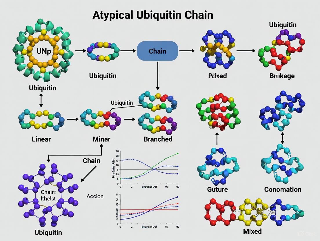
Ubiquitination is a fundamental post-translational modification that regulates virtually all aspects of eukaryotic cell biology. The versatility of ubiquitin signaling stems from the capacity of ubiquitin to form diverse polymeric chains through conjugation via one of its seven lysine residues (K6, K11, K27, K29, K33, K48, K63) or its N-terminal methionine (M1) [11]. While homotypic chains, connected through a single linkage type, have been extensively studied, recent research has revealed the abundance and functional importance of heterotypic chains. Among these, branched ubiquitin chains—in which at least one ubiquitin molecule is modified at two different lysine residues—represent a sophisticated layer of regulation within the ubiquitin code [12]. This review focuses on two biologically significant branched topologies: K11/K48 and K29/K48-linked chains, which function as potent proteasomal degradation signals under specific cellular conditions [9] [13].
The exploration of these atypical ubiquitin chain structures is reshaping our understanding of how cells encode degradation signals. Beyond the canonical K48-linked homotypic chains, branched ubiquitin chains represent enhanced, priority signals that facilitate the rapid elimination of critical regulators during cell cycle progression and the clearance of misfolded proteins during proteotoxic stress [9] [13]. This article synthesizes recent structural and mechanistic insights into the assembly, recognition, and disassembly of K11/K48 and K29/K48-branched chains, providing a technical resource for researchers investigating complex ubiquitin signaling pathways.
Structural Architectures of Branched Ubiquitin Chains
K11/K48-Branched Ubiquitin Chains
K11/K48-branched ubiquitin chains exhibit a unique structural organization that underlies their function as priority degradation signals. Structural analyses using X-ray crystallography and NMR have revealed that branched K11/K48-linked tri-ubiquitin adopts a unique interdomain interface between the distal ubiquitin molecules not observed in homotypic chains [14]. This interface is characterized by hydrophobic interactions between the distal ubiquitins and contributes to the enhanced proteasomal recognition of these chains.
Cryo-EM studies of human 26S proteasome in complex with K11/K48-branched ubiquitin chains have elucidated a multivalent substrate recognition mechanism [9]. The structures reveal:
- A novel K11-linked Ub binding site at the groove formed by proteasomal subunits RPN2 and RPN10
- Engagement of the canonical K48-linkage binding site formed by RPN10 and RPT4/5 coiled-coil
- Recognition of an alternating K11-K48-linkage by RPN2 through a conserved motif similar to the K48-specific T1 binding site of RPN1
This tripartite binding interface enables synergistic recognition of the branched chain architecture, explaining the molecular mechanism underlying priority processing of substrates modified with K11/K48-branched ubiquitin chains [9].
K29/K48-Branched Ubiquitin Chains
Recent structural studies of HECT-type E3 ligases TRIP12 and Ufd4 have visualized the formation of K29/K48-branched chains. TRIP12 resembles a pincer-like architecture in which one side comprises tandem ubiquitin-binding domains that engage the proximal ubiquitin to direct its K29 toward the active site, while selectively capturing a distal ubiquitin from a K48-linked chain [15]. The opposite pincer side—the HECT domain—precisely juxtaposes the ubiquitins to be joined, ensuring K29 linkage specificity [15].
Structural analysis of Ufd4 complexed with K29/K48-branched chains reveals a closed ring shape, where Ufd4 forms a clamp sandwiching the donor ubiquitin and the K48-linked diUb acceptor [16] [17]. The N-terminal ARM region and HECT domain C-lobe of Ufd4 collaboratively recruit K48-linked diUb and orient Lys29 of its proximal ubiquitin toward the catalytic cysteine for K29-linked branched ubiquitination [16] [17].
Table 1: Structural Features of K11/K48 and K29/K48-Branched Ubiquitin Chains
| Structural Feature | K11/K48-Branched Chains | K29/K48-Branched Chains |
|---|---|---|
| Branch Point Geometry | Unique hydrophobic interface between distal Ubs [14] | Acceptor Ub K29 positioned toward catalytic site [15] |
| Proteasome Recognition | Multivalent: RPN2-RPN10 groove (K11) + RPN10-RPT4/5 (K48) [9] | Not explicitly detailed in results |
| E3 Ligase Architecture | Not applicable | Pincer-like structure (TRIP12) [15] or closed ring shape (Ufd4) [16] |
| Key Binding Interfaces | RPN2 recognizes alternating K11-K48 linkage [9] | ARM region and HECT C-lobe orient K48-linked diUb [16] |
| Structural Methods | Cryo-EM, X-ray crystallography, NMR [9] [14] | Cryo-EM, biochemical assays [15] [16] |
Biological Functions and Physiological Significance
K11/K48-Branched Chains in Cell Cycle and Protein Quality Control
K11/K48-branched chains serve as potent degradation signals in two critical biological contexts: cell cycle progression and protein quality control. During mitosis, these chains modify key regulators such as cyclins and NEK2A, ensuring their timely elimination to facilitate proper cell division [13] [14]. In protein quality control pathways, K11/K48-branched chains target misfolded nascent polypeptides and pathological Huntingtin variants for rapid proteasomal clearance, thereby preventing protein aggregation [13].
The biological importance of these chains is underscored by the finding that mutations in K11/K48-specific enzymes are associated with neurodegenerative diseases, highlighting their essential role in maintaining proteostasis [13]. The enhanced degradation capacity of K11/K48-branched chains compared to homotypic K48-linked chains makes them particularly crucial under conditions of proteotoxic stress where efficient clearance of misfolded proteins is paramount [13].
K29/K48-Branched Chains in Stress Responses and Degradation
K29/K48-branched chains function as enhanced degradation signals in multiple physiological contexts. These chains are associated with proteotoxic stress responses and play important roles in regulating diverse substrates in processes ranging from responses to oxidative, lipid, and pH stresses to targeted protein degradation [15]. In the N-end rule pathway, K29/K48-heterotypic chains accelerate the degradation of N-end substrates [16].
More recently, K29/K48-branched chains have been implicated in small-molecule-induced targeted protein degradation, revealing their potential therapeutic relevance [15] [16]. TRIP12, a major E3 ligase responsible for generating K29 linkages and branched chains, has been associated with neurodegenerative disorders and autism spectrum disorders, suggesting physiological significance beyond protein turnover [15].
Quantitative Analysis of Branched Chain Properties
Table 2: Quantitative Functional Properties of Branched Ubiquitin Chains
| Functional Property | K11/K48-Branched Chains | K29/K48-Branched Chains |
|---|---|---|
| Proteasomal Affinity | Enhanced binding to Rpn1 [14] | Not quantitatively specified |
| Cellular Abundance | ~3-4% of total ubiquitin in mitotic arrest [12] | Not specified |
| Degradation Efficiency | Rapid elimination of mitotic regulators and misfolded proteins [13] | Accelerated degradation of N-end substrates [16] |
| E3 Ligase Efficiency | Not applicable | ~5.2-fold higher efficiency at proximal K29 vs distal K29 site (kcat/Km: 0.11 vs 0.021 μM⁻¹min⁻¹) [16] |
| DUB Specificity | UCH37 prefers K6/K48 > K11/K48 > K48/K63 branched chains [18] | UCH37 cleaves K48 linkages in branched chains [18] |
Assembly Mechanisms: Enzymes and Pathways
Assembly of K11/K48-Branched Chains
The assembly of K11/K48-branched chains involves coordinated enzymatic activities. The anaphase-promoting complex (APC/C), a multisubunit RING E3, cooperates with two different E2s (UBE2C and UBE2S) in a sequential fashion to produce branched K11/K48 polymers [11]. Additionally, other E3 ligases including cIAP1 can synthesize branched chains containing K11/K48 linkages through collaboration between different E2 enzymes (UBE2D and UBE2N-UBE2V1) [11].
Assembly of K29/K48-Branched Chains
The formation of K29/K48-branched chains typically requires collaboration between E3 ligases. The HECT E3s TRIP12 and Ufd4 preferentially catalyze K29-linked ubiquitination on preassembled K48-linked ubiquitin chains to form K29/K48-branched ubiquitin chains [15] [16] [17]. Biochemical studies demonstrate that Ufd4 shows markedly higher ubiquitination efficiency (~5.2-fold) at the proximal K29 site compared to the distal K29 site in K48-linked diUb substrates [16].
The geometric constraints for K29/K48-branched chain formation are precise, as demonstrated by the use of semi-synthetic K48-linked diUb substrates with lysine analogs of different side chain lengths. Formation of branched chains was undetectable for acceptor side chains shorter than lysine and impaired with longer side chains, indicating that the epsilon amino group of the acceptor lysine must be positioned precisely relative to the E3~Ub active site [15].
Experimental Approaches and Methodologies
Detection and Characterization Methods
Advancements in detecting and characterizing branched ubiquitin chains have been crucial for understanding their biological roles. Several sophisticated methods have been developed:
- UbiCRest (Ubiquitin Chain Restriction): Uses a library of linkage-specific deubiquitinases (DUBs) to digest ubiquitin chains, revealing linkage composition through the remnant cleavage patterns [12].
- UbiChEM-MS (Ubiquitin Chain Enrichment Middle-Down Mass Spectrometry): Combines limited proteolysis with mass spectrometry to identify branched points by detecting Ub1−74, GG-Ub1−74, and 2xGG-Ub1−74 species representing end-capped mono-ubiquitin, non-branched ubiquitin, and branched ubiquitin, respectively [12].
- Bispecific Antibodies: Engineered K11/K48 bispecific antibodies enable detection of endogenous K11/K48-linked ubiquitin chains in cellular contexts [13] [12].
- Ubiquitin Variants: Incorporation of tobacco etch virus protease (TEV)-cleaved sequence and FLAG-epitope at G53 or E64 of ubiquitin, or R54A mutation, enables distinction between branched and mixed chains [12].
Structural Biology Techniques
The elucidation of branched ubiquitin chain structures has relied on cutting-edge structural biology approaches:
- Cryo-Electron Microscopy: Recent cryo-EM studies have visualized the 26S proteasome in complex with K11/K48-branched ubiquitin chains at near-atomic resolution, revealing the molecular details of multivalent recognition [9]. Similarly, cryo-EM has captured transition states during K29/K48-branched chain formation by HECT E3 ligases [15] [16].
- Chemical Biology Probes: Engineered ubiquitin probes with chemical crosslinkers have enabled trapping of enzymatic intermediates during branched chain formation, facilitating structural characterization of transient states [15] [16].
- NMR and X-ray Crystallography: Solution NMR and crystal structures have revealed unique interdomain interfaces in branched K11/K48-tri-ubiquitin that contribute to proteasomal recognition [14].
The Scientist's Toolkit: Essential Research Reagents
Table 3: Key Research Reagents for Studying Branched Ubiquitin Chains
| Research Tool | Function/Application | Examples/References |
|---|---|---|
| Linkage-Specific DUBs | UbiCRest assay to decipher chain architecture | OTUD3 (cleaves K6/K11), etc. [12] |
| Bispecific Antibodies | Detect endogenous heterotypic chains | K11/K48-bispecific antibody [13] |
| Ubiquitin Variants | Distinguish branched from mixed chains | Flag-TEV insertion at G53/E64; R54A mutant [12] |
| Chemical Crosslinkers | Trap enzymatic intermediates for structural studies | triUbprobe for Ufd4 transition state [16] |
| Semisynthetic Ubiquitins | Probe geometric constraints in branch formation | K48-linked diUb with lysine analogs [15] |
| Branched Chain Probes | Study receptor recognition and DUB specificity | Defined K11/K48 and K29/K48-branched chains [14] [18] |
Visualization of Branched Ubiquitin Chain Pathways
K11/K48-Branched Chain Recognition by the Proteasome
K29/K48-Branched Chain Assembly by HECT E3 Ligases
The structural and functional characterization of K11/K48 and K29/K48-branched ubiquitin chains has unveiled sophisticated mechanisms by which cells encode priority degradation signals. The unique structural features of these chains—including the distinctive interdomain interface in K11/K48-branched chains and the precise geometric constraints in K29/K48-branched chain formation—enable their specific recognition and processing by the proteasomal machinery.
Future research directions in this field include:
- Elucidating the full spectrum of E2-E3 combinations that generate specific branched chain topologies
- Developing more sensitive and comprehensive methods for detecting and quantifying branched chains in physiological contexts
- Exploring the therapeutic potential of modulating branched chain formation and recognition in diseases characterized by proteostasis dysfunction
- Investigating the crosstalk between different branched chain types and their integrated functions in cellular regulation
As research methodologies continue to advance, particularly in structural biology and proteomics, our understanding of these complex ubiquitin signals will undoubtedly expand, potentially revealing new avenues for therapeutic intervention in cancer, neurodegenerative diseases, and other disorders linked to ubiquitin pathway dysregulation.
Ubiquitin chains represent one of the most versatile post-translational modifications in eukaryotic cells, forming a complex "ubiquitin code" that regulates myriad cellular processes including protein degradation, DNA repair, immune signaling, and trafficking [19] [20]. This 76-amino acid protein can be conjugated to substrate proteins as a monomer or as polyubiquitin chains through isopeptide bonds linking the C-terminal glycine of one ubiquitin to a specific lysine residue (K6, K11, K27, K29, K33, K48, K63) or the N-terminal methionine (M1) of another ubiquitin [19] [21]. The biological functions of distinct ubiquitin linkages are intrinsically linked to their three-dimensional structures and dynamic behavior. Conformational landscapes—the ensembles of three-dimensional structures that ubiquitin chains sample—enable this modest-sized protein to interact with structurally diverse ubiquitin-interacting proteins (UbIPs) and drive specific functional outcomes [4] [5].
Historically, ubiquitin chains were categorized simply as "open" or "closed," but recent research reveals astonishing conformational heterogeneity that extends beyond these binary states. Single-molecule FRET, NMR, and computational studies demonstrate that ubiquitin chains exist as dynamic ensembles of multiple conformational states in equilibrium [4] [5]. This review explores the structural principles governing ubiquitin chain dynamics, quantitative measurements of their conformational landscapes, and the functional implications of this structural plasticity for cellular signaling and drug discovery.
Structural Principles of Ubiquitin Chain Dynamics
Fundamental Conformational States
Ubiquitin chains sample three primary classes of conformational states characterized by distinct spatial relationships between ubiquitin moieties:
Open conformations: Ubiquitin moieties display minimal inter-domain contacts with extended linkers, maximizing solvent exposure of key binding surfaces like the hydrophobic patch (Ile44) [4]. These states are typically recognized by deubiquitinases (DUBs) that require access to the isopeptide bond [4].
Closed conformations: Characterized by compact arrangements where ubiquitin moieties make extensive inter-domain contacts, particularly through interaction between the hydrophobic patches of adjacent ubiquitins [4] [5]. These states often shield the isopeptide bond from solvent exposure.
Dynamic equilibria: Multiple studies confirm that ubiquitin chains exist as dynamic ensembles rather than static structures, rapidly interconverting between open, closed, and intermediate states [4] [5]. The relative populations of these states are influenced by linkage type, chain length, and cellular environment.
Different ubiquitin linkage types favor distinct conformational equilibria, creating unique structural "fingerprints" that are specifically recognized by cellular machinery:
Table 1: Conformational Preferences of Major Ubiquitin Linkage Types
| Linkage Type | Predominant Conformations | Key Functional Roles | Experimental Evidence |
|---|---|---|---|
| Lys48-linked | ~90% high-FRET (compact), ~10% low-FRET (semi-open) [4] | Proteasomal degradation [19] [20] | smFRET, NMR, SAXS [4] |
| Lys63-linked | ~25-30% non-FRET (open), ~70-75% low-FRET (closed) [4] | DNA repair, signaling, endocytosis [4] [5] | smFRET, PRE-NMR [4] [5] |
| Met1-linked (Linear) | Extended open and compact closed conformations [4] | NF-κB signaling, immune response [4] [21] | smFRET, crystallography [4] |
| Atypical Chains (K6, K11, K27, K29, K33) | Limited structural information, predicted diverse landscapes | Immune signaling, trafficking [20] [21] | Mass spectrometry, functional studies [21] |
The structural basis for these linkage-dependent preferences lies in the geometric constraints imposed by the specific lysine residue used for linkage formation. K48-linked chains favor closed conformations due to optimal positioning of hydrophobic patches for inter-domain interactions, while K63-linked chains have greater linker flexibility that enables sampling of both open and closed states [4] [5]. Met1-linked chains possess unique conformational properties as the only linkage formed through the N-terminus rather than a lysine side chain.
Quantitative Analysis of Ubiquitin Conformational Landscapes
Single-Molecule FRET Measurements
Single-molecule Förster resonance energy transfer (smFRET) has revolutionized our understanding of ubiquitin chain dynamics by enabling direct observation of conformational distributions and dynamics in solution:
Experimental Design: Diubiquitin constructs containing FRET-compatible dye pairs (e.g., Alexa488/Alexa647) are assembled with specific linkages. Donor excitation and emission from both donor and acceptor fluorophores are monitored at single-molecule level [4].
Data Interpretation: FRET efficiency histograms are fitted to Gaussian functions representing distinct conformational populations. Two-color coincidence detection (TCCD) quantifies molecules with both fluorophores to estimate non-FRET populations [4].
Key Findings: Studies reveal that K48-diUb exists primarily in a high-FRET compact state (~90%, E≈0.69) with a minor low-FRET population (~10%, E≈0.41). In contrast, K63- and Met1-linked diUb show more balanced distributions between low-FRET (~70-75%) and non-FRET (~25-30%) populations [4].
The following diagram illustrates the experimental workflow and conceptual framework for smFRET analysis of ubiquitin conformations:
Nuclear Magnetic Resonance Approaches
Paramagnetic relaxation enhancement (PRE) NMR provides atomic-resolution insights into ubiquitin chain dynamics and transient states:
Experimental Design: A paramagnetic probe (MTSL) is conjugated to engineered cysteine residues (e.g., N25C or K48C) in one ubiquitin subunit while the other subunit is 15N-labeled. PRE measurements reveal long-distance interactions [5].
Data Interpretation: The paramagnetic effect on nuclear spin relaxation rates provides distance restraints (∝1/r6) that are ensemble-averaged over all sampled conformational states, including low-population transient states [5].
Key Findings: PRE-NMR demonstrated that Lys63-linked diubiquitin exists as a dynamic ensemble comprising multiple closed and open quaternary states, with closed states involving interactions between hydrophobic patches [5].
Computational Sampling Methods
Advanced computational approaches now complement experimental methods for mapping ubiquitin conformational landscapes:
Back-mapping Based Sampling (BMBS): This hybrid approach combines efficient coarse-grained (CG) sampling with atomistic molecular dynamics. CG simulations rapidly explore conformational space, followed by back-mapping of selected structures to atomistic resolution for short explorative simulations [22].
Application to K48-linked Tri-Ub: BMBS revealed that K48-linked tri-ubiquitin samples distinct conformational states characterized by specific inter-domain contacts. The landscape can be divided into regions representing conformations with different Ub moiety interactions [22].
Validation: Comparison of computational predictions with experimental smFRET and NMR data validates the biological relevance of computed conformational ensembles [22].
Functional Consequences of Conformational Dynamics
Conformational Selection in Molecular Recognition
Ubiquitin chains employ conformational selection rather than induced fit as their primary molecular recognition mechanism:
Pre-existing Equilibria: Ubiquitin-interacting proteins (UbIPs) selectively bind and stabilize pre-existing conformational states rather than inducing new conformations [4] [5]. A K63-linkage-specific antibody enriches the closed population, while DUBs like AMSH-LP and USP21 enrich open conformations [4].
Linkage Specificity: The conformational equilibrium of each linkage type creates a unique binding preference profile. For example, OTUB1 recognizes semi-open conformations of K48-diUb, while USP21 can bind both semi-open and open states [4].
Functional Regulation: Shifting the conformational equilibrium through mutations or post-translational modifications can modulate binding affinities and functional outcomes without altering the primary sequence [5].
Atypical Ubiquitin Chains in Cellular Signaling
Atypical ubiquitin chains (K6, K11, K27, K29, K33, M1) exhibit distinct conformational properties that define their specialized roles in cellular signaling:
Table 2: Functions and Conformational Features of Atypical Ubiquitin Chains
| Chain Type | Biological Functions | Conformational Features | Regulatory Enzymes |
|---|---|---|---|
| M1-linked (Linear) | NF-κB activation, inhibition of type I IFN signaling [21] | Extended open and compact closed states [4] | LUBAC (writer), OTULIN (eraser) [21] |
| K11-linked | Cell cycle regulation, proteasomal degradation [21] | Associated with degradation, structural details limited | RNF26 (writer), USP19 (eraser) [21] |
| K27-linked | Balancing activation and inhibition in innate immunity [21] | Serves as interaction platform, structural flexibility | TRIM23 (writer) [21] |
| K29/K33-linked | ER retention, degradation, innate immunity [20] [21] | Unknown structural features | TRABID (reader/eraser) [20] |
| Branched Chains | Proteasomal degradation, NF-κB signaling, p97 processing [23] | Increased structural complexity, unknown details | UBE3C, UBR5, cIAP1 (writers) [23] |
The following diagram illustrates how conformational dynamics enable functional diversity in ubiquitin-dependent signaling pathways:
Engineered Tools for Decoding Ubiquitin Signaling
Recent advances in engineered tools have revolutionized our ability to study and manipulate ubiquitin conformational landscapes:
Linkage-Selective Engineered DUBs (enDUBs): Fusion proteins combining GFP-targeted nanobodies with catalytic domains of linkage-specific DUBs enable selective cleavage of particular ubiquitin chain types from specific substrates in live cells [20].
Application to KCNQ1 Regulation: enDUBs revealed distinct functions for different ubiquitin linkages in regulating the potassium channel KCNQ1: K11 and K29/K33 promote ER retention/degradation; K63 enhances endocytosis and reduces recycling; K48 is necessary for forward trafficking [20].
Chemical Biology Tools: Activity-based probes, diGly proteomics, and branched chain synthesis methods enable comprehensive analysis of ubiquitin chain architecture and function [23].
Experimental Methodologies and Research Toolkit
Key Experimental Protocols
Single-Molecule FRET for Ubiquitin Conformational Analysis
Sample Preparation:
- Generate ubiquitin constructs with cysteine mutations at specific positions for dye labeling (e.g., A28C, A66C)
- Express and purify ubiquitin proteins using standard recombinant techniques
- Conjugate donor (Alexa488) and acceptor (Alexa647) dyes via cysteine-maleimide chemistry
- Assemble linkage-specific diubiquitin using enzymatic synthesis with specific E2 enzymes
- Purify dual-labeled diUb using size-exclusion and ion-exchange chromatography
- Validate sample quality through mass spectrometry and enzymatic analysis [4]
Data Collection:
- Dilute samples to pM concentrations in imaging buffer with oxygen scavengers
- Perform single-molecule measurements using total internal reflection fluorescence (TIRF) microscopy
- Excite donor fluorophore with a single laser and monitor emission from both donor and acceptor channels
- Perform two-color coincidence detection (TCCD) using alternating laser excitation
- Collect data from thousands of individual molecules to ensure statistical significance [4]
Data Analysis:
- Calculate FRET efficiency for each molecule: E = IA/(ID+IA) where ID and IA are donor and acceptor intensities
- Construct FRET efficiency histograms and fit to Gaussian functions representing distinct populations
- Use TCCD data to quantify proportion of molecules in non-FRET conformations
- For interaction studies, repeat measurements with UbIPs at concentrations exceeding KD values
- Monitor changes in conformational populations upon UbIP binding [4]
PRE-NMR for Studying Ubiquitin Dynamics
Sample Preparation:
- Introduce cysteine mutations at strategic positions (N25C, K48C) in the distal ubiquitin unit
- Express 15N-labeled proximal ubiquitin unit and unlabeled distal unit
- Purify subunits and enzymatically assemble diubiquitin
- Conjugate paramagnetic probe (MTSL) to cysteine residues
- Prepare control samples with reduced (diamagnetic) probe for comparison [5]
Data Collection and Analysis:
- Collect 2D 1H-15N HSQC spectra for paramagnetic and diamagnetic samples
- Calculate PRE from intensity ratios: I(para)/I(dia)
- Use PRE restraints for ensemble structure calculation
- Analyze chemical shift perturbations to map interaction surfaces
- Validate findings with binding studies using known interaction partners [5]
Essential Research Reagents and Tools
Table 3: Key Research Reagents for Studying Ubiquitin Conformational Landscapes
| Reagent/Tool | Function/Application | Examples/Specifications |
|---|---|---|
| Linkage-Specific E2/E3 Pairs | Enzymatic synthesis of defined ubiquitin chains | MMS2-UBC13 (K63), UBE2R1 (K48), UBE2L3-UBE2S (K11) [19] [23] |
| Fluorophore-Labeled Ubiquitin | smFRET studies of conformational dynamics | Alexa488/Alexa647 dye pairs at specific cysteine mutants [4] |
| NMR-Labeled Ubiquitin | High-resolution structural studies | 15N-, 13C-labeled ubiquitin for chemical shift analysis [5] |
| Paramagnetic Probes | PRE-NMR for studying transient states | MTSL conjugation to engineered cysteine residues [5] |
| Engineered DUBs (enDUBs) | Linkage-selective ubiquitin chain cleavage in live cells | Nanobody-DUB fusions (OTUD1-K63, OTUD4-K48, Cezanne-K11) [20] |
| Coarse-Grained Force Fields | Enhanced sampling of conformational landscapes | Modified MARTINI for ubiquitin chains [22] |
| Linkage-Specific Antibodies | Detection and purification of specific chain types | K63-linkage specific antibodies for conformational selection studies [4] |
| Chemical Biology Probes | Activity-based profiling and detection | Ubiquitin vinyl sulfone (UbVS) for DUB profiling, diGly antibody for proteomics [23] |
The conformational landscapes of ubiquitin chains represent a fundamental regulatory mechanism that expands the coding potential of this versatile post-translational modification. Rather than adopting static structures, ubiquitin chains exist as dynamic ensembles of interconverting conformations whose equilibria are determined by linkage type, chain length, and cellular context. The conformational selection model—where ubiquitin-interacting proteins selectively bind pre-existing states—provides a paradigm for understanding how limited structural diversity can generate exquisite functional specificity.
Future research directions will likely focus on several key areas: First, characterizing the conformational landscapes of longer ubiquitin chains and branched architectures that remain poorly understood despite their biological importance [23]. Second, developing methods to study ubiquitin conformational dynamics in living cells rather than in purified systems. Third, understanding how post-translational modifications of ubiquitin itself or environmental factors alter conformational equilibria. Finally, leveraging this structural knowledge for drug discovery, particularly targeting the ubiquitin system in cancer, neurodegenerative diseases, and immune disorders.
The ongoing development of innovative tools—from engineered DUBs [20] to advanced computational methods [22]—promises to accelerate our exploration of ubiquitin conformational landscapes. As these technologies mature, we will gain unprecedented insights into how structural dynamics enable a single small protein to coordinate such remarkable functional diversity throughout cell biology.
Ubiquitination, the covalent attachment of ubiquitin to substrate proteins, is a quintessential post-translational modification. The conventional paradigm views ubiquitination primarily through the lens of targeting proteins for degradation by the 26S proteasome. However, a more nuanced understanding has emerged, revealing a vast landscape of non-proteolytic functions mediated by atypical ubiquitin chain topologies and conformations. This whitepaper explores how these non-degradative ubiquitin signals, including monoubiquitination, and chains linked through Lys63 (K63), Lys11 (K11), Met1 (M1; linear), and others, exert precise control over critical cellular processes such as cell signaling, the DNA damage response, and protein quality control. This exploration is framed within the broader thesis that the structural diversity of ubiquitin chains constitutes a complex molecular "code" that expands the functional repertoire of ubiquitination far beyond mere proteolysis.
Non-Proteolytic Ubiquitin Signaling in Kinase Activation Pathways
Atypical ubiquitin chains are indispensable for the activation and regulation of several key kinase-driven signaling pathways. Rather than inducing degradation, these ubiquitin modifications facilitate protein-protein interactions and complex assembly.
NF-κB Signaling Pathway: The activation of the NF-κB pathway is a canonical example of non-proteolytic K63-linked and linear ubiquitin chain function. Upon receptor engagement (e.g., TNFR, IL-1R), a signaling complex forms, leading to the recruitment of the K63-specific E2/E3 complex Ubc13/Uev1A/TRAF6. This complex synthesizes K63-linked chains on various components, including TRAF6 itself and RIP1. These chains serve as platforms to recruit the TAK1 kinase complex (TAK1, TAB1, TAB2) via ubiquitin-binding domains (UBDs) in TAB2. Simultaneously, the Linear Ubiquitin Chain Assembly Complex (LUBAC), composed of HOIP, HOIL-1L, and SHARPIN, generates M1-linked linear chains on NEMO, a regulatory subunit of the IKK complex. This dual ubiquitination event promotes the proximity and activation of TAK1, which then phosphorylates and activates the IKK complex. IKK subsequently phosphorylates the inhibitor of κB (IκB), targeting it for K48-linked ubiquitination and degradation, thereby releasing the NF-κB transcription factor for nuclear translocation.
Diagram Title: NF-κB Activation via Atypical Ubiquitin
Table 1: Key Non-Proteolytic Ubiquitin Signals in Kinase Pathways
| Pathway | Ubiquitin Chain Type | Key E2/E3 Enzymes | Functional Outcome |
|---|---|---|---|
| NF-κB Activation | K63-linked, M1-linear | Ubc13/Uev1A-TRAF6, LUBAC | Scaffold for TAK1/IKK recruitment and activation; IκB degradation is a downstream consequence. |
| MTORC1 Activation | K63-linked | TRAF6, UBE2N/UBE2V1 | Promotes mTORC1 membrane localization and activation in response to growth factors. |
| Wnt/β-Catenin | K63-linked | SCFβ-TrCP (atypical use) | Regulates β-catenin nuclear import and transcriptional activity without degradation. |
Experimental Protocol: Assessing K63-Linked Ubiquitination in NF-κB Signaling
Objective: To detect and validate the formation of K63-linked ubiquitin chains on RIP1 upon TNF-α stimulation.
Cell Stimulation and Lysis:
- Culture HEK293T or HeLa cells.
- Stimulate with 20 ng/mL human TNF-α for 0, 5, 15, and 30 minutes.
- Lyse cells in RIPA buffer (150 mM NaCl, 1% NP-40, 0.5% sodium deoxycholate, 0.1% SDS, 50 mM Tris, pH 8.0) supplemented with 1x protease inhibitor cocktail, 10 mM N-ethylmaleimide (NEM; to inhibit deubiquitinases), and 1x PhosSTOP phosphatase inhibitors.
Immunoprecipitation (IP):
- Pre-clear 1 mg of total protein lysate with Protein A/G Sepharose beads for 1 hour at 4°C.
- Incubate the pre-cleared lysate with 2 µg of anti-RIP1 antibody overnight at 4°C with gentle rotation.
- Add Protein A/G Sepharose beads and incubate for an additional 2 hours.
- Wash beads 3-4 times with ice-cold lysis buffer.
Western Blot Analysis:
- Elute immunoprecipitated proteins by boiling in 2x Laemmli sample buffer.
- Resolve proteins by SDS-PAGE (4-12% gradient gel).
- Transfer to a PVDF membrane.
- Probe the membrane with the following antibodies:
- Primary Antibodies: Anti-K63-linkage specific ubiquitin (e.g., clone Apu3, MilliporeSigma), Anti-RIP1 (loading control for IP).
- Use HRP-conjugated secondary antibodies and chemiluminescent detection.
Validation (Optional but Critical):
- siRNA Knockdown: Knockdown UBC13 (the E2 for K63 chains) and repeat the experiment. Loss of K63 signal confirms specificity.
- DUB Specificity: Treat immunoprecipitates with the K63-linkage specific deubiquitinase (DUB) AMSH or the general DUB USP2 as a control. AMSH should cleave the K63 signal.
Atypical Ubiquitin in Stress Response and Quality Control
Cellular stress, particularly proteotoxic and genotoxic stress, leverages non-proteolytic ubiquitination for sensing, signaling, and resolution.
DNA Damage Repair: The response to DNA double-strand breaks (DSBs) is orchestrated by the assembly of repair proteins at the break site, a process dependent on K63-linked ubiquitin chains. The E3 ligases RNF8 and RNF168 are sequentially recruited to DSB sites. RNF8, in conjunction with Ubc13, initiates the deposition of K63-linked chains on histones H2A and H2AX. This initial ubiquitination signal is amplified by RNF168, which also deposits K63 chains. This extensive K63-ubiquitin "cloud" serves as a binding platform to recruit downstream repair factors like 53BP1 and BRCA1, which contain UBDs (e.g., the UDR domain in 53BP1), thereby promoting repair through non-homologous end joining (NHEJ) or homologous recombination (HR).
Aggresome Formation and Autophagic Clearance: Under proteasomal impairment, misfolded proteins are tagged with K63-linked ubiquitin chains by E3 ligases like PARKIN. These K63-tagged proteins are recognized by the histone deacetylase HDAC6, which binds ubiquitin via its zinc-finger domain. HDAC6 then couples these ubiquitinated cargoes to the dynein motor complex, facilitating their transport along microtubules to the microtubule-organizing center (MTOC) to form an aggresome. The aggresome is subsequently enveloped by a membranous structure and targeted for degradation via selective autophagy (aggrephagy).
Diagram Title: Aggresome Formation via K63-Ubiquitin
Table 2: Quantitative Impact of Atypical Ubiquitin on Stress Responses
| Stress Type | Ubiquitin Chain | Key Regulatory Protein | Quantitative Effect on Repair/Clearance | Measurement Method |
|---|---|---|---|---|
| DNA DSB | K63-linked | RNF8 / RNF168 | ~10-15 fold increase in 53BP1 foci formation at DSB sites. | Immunofluorescence (foci counting) |
| Proteotoxic Stress | K63-linked | HDAC6 | ~60-70% reduction in aggregated protein load upon proteasome inhibition in HDAC6-/- cells. | Filter trap assay / Solubility fractionation |
| Mitophagy | K63-linked, Phospho-Ub (S65) | PARKIN, PINK1 | >80% of depolarized mitochondria are ubiquitinated and cleared within 4 hours. | Flow cytometry (MitoTimer), confocal microscopy |
Experimental Protocol: Monitoring Aggresome Formation via Immunofluorescence
Objective: To visualize the formation of K63-ubiquitin-positive aggressomes upon proteasomal inhibition.
Cell Culture and Treatment:
- Plate HeLa cells on glass coverslips in a 12-well plate.
- Treat cells with 5 µM MG132 (proteasome inhibitor) or DMSO (vehicle control) for 12-16 hours.
Immunofluorescence Staining:
- Fixation: Wash cells with PBS and fix with 4% paraformaldehyde in PBS for 15 minutes at room temperature (RT).
- Permeabilization: Permeabilize cells with 0.2% Triton X-100 in PBS for 10 minutes at RT.
- Blocking: Block with 5% normal goat serum in PBS for 1 hour at RT.
- Primary Antibody Incubation: Incubate with primary antibodies diluted in blocking buffer overnight at 4°C.
- Mouse anti-K63-linkage specific ubiquitin (1:200)
- Rabbit anti-HDAC6 (1:500)
- (Optional) Mouse anti-vimentin (1:1000) to label the vimentin cage surrounding the aggresome.
- Secondary Antibody Incubation: Wash 3x with PBS and incubate with fluorescently-labeled secondary antibodies (e.g., Alexa Fluor 488 goat anti-mouse, Alexa Fluor 568 goat anti-rabbit) for 1 hour at RT in the dark.
- Nuclear Staining: Incubate with DAPI (0.5 µg/mL) for 5 minutes.
- Mounting: Mount coverslips onto glass slides using an anti-fade mounting medium.
Imaging and Analysis:
- Image cells using a confocal or high-resolution fluorescence microscope.
- Expected Result: In MG132-treated cells, large, perinuclear aggressomes will be visible, which are positive for both K63-ubiquitin and HDAC6. DMSO-treated cells will show a diffuse, pan-cytoplasmic staining pattern.
Experimental Approaches for Studying Atypical Ubiquitination
Deciphering the non-proteolytic ubiquitin code requires specialized tools and techniques that go beyond standard ubiquitin pulldowns.
Linkage-Specific Reagents: The development of linkage-specific antibodies (e.g., for K63, K48, M1) and ubiquitin-binding domains (UBDs) engineered into tandem-repeated motifs (e.g., K63-TUBEs) has been revolutionary. These reagents allow for the selective enrichment and detection of specific chain types from complex lysates.
Di-Glycine Remnant Proteomics (Lys-N Digestion): Standard trypsin-based ubiquitin proteomics is confounded by the tryptic peptide's large size. Using Lys-N protease instead generates a di-glycine (Gly-Gly) remnant attached to the modified lysine on a short, hydrophilic peptide, improving mass spectrometry identification efficiency and coverage, allowing for system-wide mapping of ubiquitination sites and inference of chain types based on context.
In vitro Reconstitution Assays: Purified systems containing E1, a specific E2 (e.g., Ubc13/Uev1a for K63), an E3 ligase (e.g., TRAF6), and ubiquitin allow for the synthesis of a specific, homogeneous ubiquitin chain type. These defined chains can then be used in downstream assays to study their effect on protein complex assembly or kinase activation without the complexity of a cellular environment.
Diagram Title: Workflow for Atypical Ubiquitin Enrichment
The Scientist's Toolkit: Key Reagents for Non-Proteolytic Ubiquitin Research
| Research Reagent / Tool | Function & Application |
|---|---|
| K63-linkage Specific Antibody (e.g., clone Apu3) | Selective immunoprecipitation and Western blot detection of endogenous K63-linked ubiquitin chains. Critical for validating pathway-specific K63 ubiquitination. |
| Tandem Ubiquitin Binding Entities (TUBEs) | High-affinity probes based on engineered UBDs (e.g., from UBQLN1). Linkage-specific TUBEs (K63, M1) protect ubiquitinated proteins from DUBs during lysis and enable enrichment of specific chain types. |
| Active Ubc13/Uev1a Heterodimer (E2 Enzyme) | Recombinant E2 complex specifically for synthesizing K63-linked ubiquitin chains in in vitro ubiquitination assays. Essential for mechanistic studies. |
| Linkage-Specific Deubiquitinases (DUBs) | Recombinant DUBs like AMSH (K63-specific) and OTULIN (M1-specific). Used as enzymatic tools to validate the chain topology in samples, confirming antibody/TUBE specificity. |
| Ubiquitin Mutants (K63R, K48R, K0) | Ubiquitin plasmids where all lysines except one are mutated to arginine (e.g., K63-only) or all lysines are mutated (K0). Used in cellular transfection/transduction experiments to force the formation of or probe for specific chain types. |
| PINK1/PARKIN Inducers (e.g., CCCP/Antimycin A) | Mitochondrial uncouplers used to induce mitophagy, a process heavily reliant on K63 and phospho-ubiquitin signaling, providing a robust cellular model for studying these modifications. |
The functional diversity of non-proteolytic ubiquitination represents a paradigm shift with profound implications for therapeutic intervention. The traditional drug discovery focus on inhibiting E3 ligases to block degradation is now expanded to include modulating the activity of E2/E3 pairs that generate atypical chains, or targeting the reader proteins (UBDs) that interpret these signals. For instance, inhibiting the Ubc13/TRAF6 interaction could dampen aberrant NF-κB signaling in inflammatory diseases without globally disrupting protein degradation. Similarly, enhancing K63-linked signaling in quality control pathways could be a strategy for neurodegenerative diseases characterized by protein aggregation. The future of this field lies in moving from a "degradation-centric" view to a "signal-centric" one, leveraging structural insights into atypical chain conformations to develop a new class of highly specific "ubiquitin signaling" therapeutics. This aligns with the overarching thesis that a deep understanding of ubiquitin chain structural diversity is the key to unlocking its full therapeutic potential.
Ubiquitination is a fundamental post-translational modification where the small protein ubiquitin is covalently attached to substrate proteins, regulating myriad cellular processes from protein degradation to cell signaling [2]. The diversity of the "ubiquitin code" arises from the ability of ubiquitin itself to form polymers (polyubiquitin) through eight distinct linkage types, connecting the C-terminus of one ubiquitin to a specific lysine residue (K6, K11, K27, K29, K33, K48, K63) or the N-terminal methionine (M1) of another [2] [23]. For decades, the prevailing model suggested that differently linked ubiquitin chains adopted distinct, relatively static architectures—K48-linked chains forming compact conformations that target proteins for proteasomal degradation, and K63-linked/M1-linked chains adopting extended "open" conformations for non-degradative signaling roles [4] [5].
The conformational selection paradigm challenges this static view. Advanced biophysical techniques have revealed that ubiquitin chains exist as dynamic ensembles of multiple conformational states in solution [4] [5]. This review synthesizes evidence establishing that ubiquitin-binding proteins (UbIPs), including ubiquitin-binding domains (UBDs) and deubiquitinases (DUBs), recognize their specific ubiquitin chain substrates not by inducing structural changes, but by selecting and stabilizing pre-existing conformational states from this dynamic equilibrium. This molecular recognition mechanism adds a crucial regulatory layer to the ubiquitin system and provides novel insights for therapeutic intervention.
Quantitative Evidence for Pre-existing Conformational States
Single-molecule FRET (Förster Resonance Energy Transfer) studies have directly visualized the conformational heterogeneity of unbound ubiquitin chains, providing quantitative evidence for the coexistence of multiple states in solution.
Table 1: Conformational Populations of Diubiquitin Linkages Measured by smFRET
| Linkage Type | High-FRET Population | Low-FRET Population | Non-FRET Population | Dominant Conformation |
|---|---|---|---|---|
| K48-linked | ~90% (E ≈ 0.69) | ~10% (E ≈ 0.41) | Not detected | Compact conformations |
| K63-linked | Not reported | ~70-75% | ~25-30% | Extended and compact conformations |
| M1-linked | Not reported | ~70-75% | ~25-30% | Extended and compact conformations |
Table 2: Ubiquitin-Interacting Protein Binding Preferences
| Ubiquitin-Interacting Protein | Linkage Specificity | Conformation Selected | Biological Role |
|---|---|---|---|
| Lys63-linkage-specific antibody | K63-specific | Enriches FRET population (compact) | Experimental tool |
| NEMO UBAN domain | M1-specific | Enriches low-FRET population (compact) | NF-κB signaling activation |
| AMSH-LP (DUB) | K63-specific | Depletes FRET, enriches non-FRET (open) | Lys63-chain hydrolysis |
| USP21 (DUB) | Linkage-promiscuous | Binds semi-open and open conformations | Broad-spectrum deubiquitination |
| OTUB1 (DUB) | K48-specific | Enriches low-FRET (semi-open) | Lys48-chain hydrolysis |
These quantitative measurements demonstrate that differently linked diubiquitin samples distinct but overlapping conformational spaces before encountering binding partners, forming the structural basis for conformational selection.
Experimental Methodologies for Studying Conformational Dynamics
Single-Molecule FRET (smFRET) with Two-Color Coincidence Detection (TCCD)
Protocol Objective: To quantify distinct conformational populations and inter-domain distances within single diubiquitin molecules in solution.
Key Reagents:
- Diubiquitin Constructs: Pure K48-, K63-, or M1-linked diubiquitin with single cysteine mutations at specific positions for site-specific labeling [4].
- Fluorophores: FRET-compatible dye pairs (e.g., Alexa488 donor and Alexa647 acceptor with Förster radius R₀ = 5.6 nm) [4].
- Control Samples: Unlabeled diubiquitin and singly-labeled controls for photophysical characterization.
Experimental Workflow:
- Site-specific Labeling: Conjugate donor and acceptor fluorophores to engineered cysteine residues using thiol-reactive chemistry (e.g., maleimide derivatives).
- Sample Validation: Verify labeling efficiency and protein integrity using mass spectrometry, enzymatic cleavage assays, and ensemble fluorescence measurements (lifetime, anisotropy) [4].
- smFRET Data Acquisition:
- Dilute labeled diubiquitin to pM concentrations in observation buffer to ensure single-molecule detection.
- Excite donor fluorophore with a single laser and monitor emission of both donor and acceptor channels.
- Calculate FRET efficiency (E) for each molecule as E = Iₐ / (Iḍ + Iₐ), where Iₐ and Iḍ are acceptor and donor intensities, respectively.
- Two-Color Coincidence Detection (TCCD):
- Use two alternating lasers to independently excite donor and acceptor fluorophores.
- Quantify the proportion of molecules containing both fluorophores to estimate populations in "non-FRET" conformations where distances exceed the FRET range.
- Data Analysis:
- Construct FRET efficiency histograms from thousands of single-molecule events.
- Fit populations to Gaussian functions representing distinct conformational states.
- Calculate relative populations of high-FRET, low-FRET, and non-FRET species.
Diagram 1: smFRET-TCCD Workflow for Conformational Analysis (82 characters)
Paramagnetic Relaxation Enhancement (PRE) NMR Spectroscopy
Protocol Objective: To detect transient, low-population conformational states and characterize inter-domain interactions in ubiquitin chains at atomic resolution.
Key Reagents:
- Paramagnetically-Labeled Diubiquitin: K63-Ub2 with proximal unit ¹⁵N-labeled and distal unit unlabeled, containing a single cysteine mutation (e.g., N25C or K48C) in the distal unit [5].
- Paramagnetic Probe: Methanethiosulfonate (MTSL) or similar paramagnetic spin label conjugated to the engineered cysteine.
- Control Sample: Diamagnetic reference (e.g., reduced with ascorbate).
Experimental Workflow:
- Sample Preparation:
- Introduce single cysteine mutations at sites away from known binding interfaces to avoid perturbation of native interactions.
- Conjugate paramagnetic probe to cysteine thiol via MTSL chemistry.
- Confirm conjugation efficiency and protein folding integrity.
- NMR Data Collection:
- Acquire ²D ¹H-¹⁵N HSQC spectra of paramagnetic and diamagnetic samples.
- Measure paramagnetic relaxation enhancement (PRE) as the ratio of peak intensities (Iₚₐᵣₐₘₐg/Iḍᵢₐₘₐg).
- Data Analysis:
- Identify residues with significant PRE effects indicating transient close approaches (< ~25 Å).
- Calculate ensemble structures consistent with PRE-derived distance restraints.
- Model multiple conformational states weighted by their population contributions.
Diagram 2: PRE-NMR for Transient State Detection (55 characters)
Conformational Selection in Biological Contexts
Recognition by Ubiquitin-Binding Domains (UBDs)
The NEMO UBAN domain, essential for NF-κB signaling pathway activation, provides a compelling example of conformational selection. Single-molecule studies demonstrate that UBAN selectively enriches the pre-existing low-FRET (compact) population of M1-linked diubiquitin without remodeling the chain architecture [4]. Similarly, Lys63-linkage specific antibodies selectively stabilize compact conformational states that pre-exist in the K63-diubiquitin ensemble [4]. This direct selection mechanism enables rapid cellular response to ubiquitin signals without the kinetic barrier associated with induced-fit remodeling.
Processing by Deubiquitinases (DUBs)
DUB interactions with ubiquitin chains reveal more complex aspects of conformational selection. The Lys63-specific DUB AMSH-LP selectively binds open, non-FRET conformations of K63-diubiquitin, consistent with structural data showing AMSH-LP bound to an extended K63-Ub2 conformation [4]. Similarly, USP21 enriches open conformations of K48-linked chains necessary for isopeptide bond access [4].
However, the Lys48-specific DUB OTUB1 exhibits a hybrid mechanism—it preferentially binds semi-open K48-diubiquitin conformations (low-FRET species) but does not require fully open architectures for efficient cleavage [4] [24]. This suggests that some DUBs can utilize multiple pre-existing states from the conformational ensemble, with potential remodeling occurring after initial selection.
Diagram 3: Conformational Selection by Ubiquitin Binders (72 characters)
The Scientist's Toolkit: Essential Research Reagents
Table 3: Essential Research Reagents for Studying Ubiquitin Conformations
| Reagent Category | Specific Examples | Function & Application |
|---|---|---|
| Defined Linkage Diubiquitin | K48NC, K63NC, M1NC (with single cysteines for labeling) | Substrate for smFRET, NMR, and binding studies; enables site-specific labeling [4] |
| FRET Dye Pairs | Alexa488/Alexa647 (R₀ = 5.6 nm) | smFRET measurements of inter-domain distances and conformational distributions [4] |
| Paramagnetic Labels | MTSL (methanethiosulfonate spin label) | PRE-NMR studies to detect transient interactions and low-population states [5] |
| Inactive DUB Mutants | AMSH-LP (active site mutants), OTUB1i, USP21i | Trap ubiquitin chain complexes for structural studies without catalytic turnover [4] |
| Linkage-Specific UBDs | NEMO UBAN domain, TAB2 NZF domain, Rap80 tUIM | Investigate conformational selection mechanisms in signaling pathways [4] [5] |
| Chain Assembly Enzymes | UBE2N/UBE2V1 (K63-specific), UBE2R1 (K48-specific) | Synthesis of defined linkage ubiquitin chains for experimental studies [23] |
| Genetic Code Expansion Systems | Amber stop codon suppression with noncanonical amino acids | Incorporation of unique chemical handles for selective chain assembly and labeling [23] |
Implications for Drug Discovery and Therapeutic Intervention
The conformational selection paradigm opens new avenues for therapeutic intervention in ubiquitin-related pathologies. Small molecules that modulate the conformational equilibrium of ubiquitin chains could potentially upregulate or downregulate specific signaling pathways without complete inhibition. For instance, compounds that stabilize closed conformations of K63-linked chains might attenuate NF-κB signaling in inflammatory diseases, while molecules that promote open states of K48-linked chains could enhance proteasomal degradation of pathological proteins in neurodegenerative disorders.
Understanding that many ubiquitin-binding proteins recognize pre-existing states rather than inducing structural changes suggests that drug screening approaches should prioritize maintaining the native conformational dynamics of ubiquitin chains. Stabilizing specific conformational states offers a more nuanced therapeutic approach than traditional enzyme inhibition, potentially resulting in fewer off-target effects and greater pathway specificity.
The conformational selection paradigm represents a fundamental shift in our understanding of ubiquitin chain recognition. Through the integration of single-molecule FRET, paramagnetic NMR, and other biophysical approaches, we now appreciate that ubiquitin chains exist as dynamic conformational ensembles, with specific states being selected by readers and erasers of the ubiquitin code. This mechanism adds a crucial layer of regulation to ubiquitin signaling, enabling precise cellular responses to ubiquitination events. Future research exploring how branched ubiquitin chains and post-translational modifications of ubiquitin itself influence these conformational landscapes will further illuminate the sophisticated language of the ubiquitin code.
Cutting-Edge Tools: Structural and Biophysical Methods for Probing Ubiquitin Architecture
The ubiquitin-proteasome system (UPS) represents a fundamental regulatory mechanism in eukaryotic cells, controlling protein degradation and thereby influencing a vast array of cellular processes. For decades, the K48-linked homotypic polyubiquitin chain was regarded as the canonical proteasomal degradation signal. However, recent technological advances have revealed a more complex ubiquitin code, with branched ubiquitin chains emerging as potent targeting signals that can enhance degradation efficiency for specific cellular substrates. Among these, K11/K48-branched ubiquitin chains have been identified as priority degradation signals during critical processes such as cell cycle progression and proteotoxic stress response [9] [25].
The structural basis for how the 26S proteasome recognizes these complex branched chains remained elusive until recent breakthroughs in cryo-electron microscopy (cryo-EM). This technical guide explores how cryo-EM structural studies have illuminated the molecular mechanism underlying branched ubiquitin chain recognition by the human 26S proteasome, providing unprecedented insights into the sophistication of the cellular degradation machinery.
Structural Biology of Branched Ubiquitin Chain Recognition
Cryo-EM Reveals a Multivalent Binding Mechanism
A landmark 2025 study by Draczkowski et al. provided the first high-resolution cryo-EM structures of the human 26S proteasome in complex with a K11/K48-branched ubiquitin chain [9] [26]. The research team successfully reconstituted a functional complex containing the 26S proteasome, a polyubiquitinated substrate, and auxiliary proteins RPN13 and UCHL5 (a deubiquitinase). Through extensive cryo-EM classification and focused refinements, they determined four distinct structures resembling previously reported conformational states (EA, EB, and ED states) of the proteasome during substrate processing [9].
The structures revealed a multivalent substrate recognition mechanism involving multiple proteasomal subunits collaborating to engage the branched chain. Key findings include:
- Novel K11-linked Ub binding site: A previously unknown binding site for K11-linked ubiquitin was identified at a groove formed by subunits RPN2 and RPN10 [9] [26].
- Canonical K48-linkage binding: The canonical K48-linkage binding site formed by RPN10 and the RPT4/5 coiled-coil was simultaneously engaged [9].
- RPN2 as a key recognition component: RPN2 recognizes an alternating K11-K48-linkage through a conserved motif similar to the K48-specific T1 binding site of RPN1 [9].
This tripartite binding interface enables the proteasome to simultaneously engage both linkage types within the branched chain, explaining the enhanced binding affinity and degradation efficiency observed for substrates tagged with K11/K48-branched ubiquitin chains.
Structural Basis for Priority Recognition of Branched Chains
The structural insights gleaned from these cryo-EM studies explain why K11/K48-branched ubiquitin chains serve as priority degradation signals. The multivalent engagement creates a high-avidity interaction that effectively competes with other proteasomal substrates. This is particularly important during cell cycle progression where timely degradation of mitotic regulators is critical [9] [25].
Additionally, the positioning of the branched chain within the recognition complex appears to optimally present the substrate for subsequent processing steps. The proximity to deubiquitinating enzymes like UCHL5, which shows preference for K11/K48-branched chains, ensures efficient ubiquitin recycling while maintaining the degradation signal until substrate engagement is complete [9].
Experimental Methodologies for Studying Branched Ubiquitin Recognition
Sample Preparation and Complex Reconstitution
The successful structural determination of the proteasome-branched ubiquitin complex required meticulous sample preparation and validation:
Experimental Workflow for Complex Formation
- Substrate Design: The substrate consisted of residues 1-48 of S. cerevisiae Sic1 protein (Sic1PY), an intrinsically disordered region containing a single lysine residue (K40) as the ubiquitination site [9].
- Ubiquitination System: An engineered Rsp5 E3 ligase (Rsp5-HECTGML) was used to generate polyubiquitinated Sic1PY. While wild-type Rsp5 produces K63-linked chains, the engineered variant generates K48-linked chains, confirmed by Western blotting with linkage-specific antibodies [9].
- Branching Control: To prevent K63-linkage formation, a ubiquitin K63R variant was used in the ubiquitination reactions. Despite this control, subsequent analysis revealed the unexpected formation of branched chains [9].
- Chain Length Selection: The crude polyubiquitinated Sic1PY product was fractionated by size-exclusion chromatography (SEC) to enrich medium-length chains (Ub₄-Ub₈) for optimal processing by the 26S proteasome [9].
- Complex Assembly: The functional complex included the 26S proteasome, Sic1PY-Ubₙ, and preformed RPN13:UCHL5 complex with catalytically inactive UCHL5(C88A) to minimize disassembly of the ubiquitin chains during structural analysis [9].
Analytical Validation of Branched Chains
Critical to the study was verifying the presence and linkage types of branched ubiquitin chains in the prepared samples:
- Lbpro* Ub Clipping: This method revealed the presence of doubly ubiquitinated (12.6%) and triply ubiquitinated (3.6%) ubiquitin in addition to singly ubiquitinated ubiquitin (41.8%), providing clear evidence of branched chain formation [9].
- Mass Spectrometry Analysis: Intact mass spectrometry and Ub absolute quantification (Ub-AQUA) demonstrated that the SEC-enriched polyubiquitin chains contained almost equal amounts of K11- and K48-linked ubiquitin with a minor population of K33-linked ubiquitin [9].
- Native Gel Electrophoresis with Western Blotting: Confirmed the presence of UCHL5, RPN13, and Sic1PY-Ubₙ in the reconstituted complex [9].
- Negative Staining Electron Microscopy: Initial validation showed additional EM densities on the 19S regulatory particle of the reconstituted complex compared to apo 26S proteasome, indicating successful complex formation [9].
Cryo-EM Data Collection and Processing
The cryo-EM structural determination followed rigorous protocols:
- Grid Preparation: Vitrified samples were prepared using standard cryo-EM protocols.
- Data Collection: High-resolution data were collected on modern cryo-EM instruments equipped with direct electron detectors.
- Image Processing: Extensive classification and focused refinements were performed to resolve four distinct structural states of the complex [9].
- Resolution Determination: The final reconstructions achieved resolutions sufficient to discern molecular details of the ubiquitin-proteasome interactions.
Quantitative Analysis of Branched Ubiquitin Recognition
Proteasomal Ubiquitin Receptor Specificities
Table 1: Ubiquitin Binding Sites on the 26S Proteasome
| Proteasomal Subunit | Ubiquitin Linkage Specificity | Structural Features | Functional Role |
|---|---|---|---|
| RPN2 | K11 and alternating K11-K48 | Novel binding groove, conserved motif similar to RPN1 T1 site | Primary recognition site for K11 branch in branched chains |
| RPN10 | K11 and K48 | Ubiquitin-interacting motifs (UIMs), groove with RPN2 | Simultaneous engagement of both linkage types in branched chains |
| RPT4/5 coiled-coil | K48 | Canonical K48-linkage binding site | Part of the tripartite recognition interface |
| RPN1 | K48 (T1 site) | Three-helix bundle within PC domain | Reference site for K48 linkage recognition |
| RPN13 | Various linkages | PRU domain, flexible linker | Subsidiary role in branched chain recognition |
Branched Ubiquitin Chain Distribution and Properties
Table 2: Characteristics of Branched Ubiquitin Chains in Cellular Signaling
| Chain Type | Cellular Context | Biological Function | Recognition Features |
|---|---|---|---|
| K11/K48 | Cell cycle progression, proteotoxic stress | Rapid degradation of mitotic regulators, misfolded proteins | Multivalent proteasome binding, enhanced affinity for RPN1 and RPN10 |
| K29/K48 | Oxidative, lipid, and pH stress responses | Targeted protein degradation | Preferential modification of proximal Ub in K48-linked di-Ub by TRIP12 |
| K48/K63 | NF-κB signaling, p97/VCP processing | Diverse functions including proteasomal degradation | Recognized by specific UBDs, proteasome, and p97 |
| K11/K33 | Potentially synthetic | Under characterization | Assembly via genetic code expansion approaches |
The Scientist's Toolkit: Essential Research Reagents and Methods
Table 3: Key Research Reagents for Studying Branched Ubiquitin Chains
| Reagent / Method | Function / Application | Key Features / Considerations |
|---|---|---|
| Engineered E3 Ligases (e.g., Rsp5-HECTGML) | Generate specific ubiquitin linkages | Altered linkage specificity from wild-type enzymes |
| Linkage-Specific Ubiquitin Mutants (e.g., UbK63R) | Control linkage formation during chain assembly | Prevents formation of specific linkages |
| Ubiquitin Binding Domain (UBD) Probes | Detect specific ubiquitin linkages | Varying specificity for different chain types |
| Lbpro* Ub Clipping | Identify branched ubiquitin chains | Cleaves ubiquitin chains, revealing branching patterns |
| Ubiquitin AQUA (Absolute Quantification) | Precisely quantify linkage types in mixed chains | Mass spectrometry-based quantitative approach |
| C-terminally Blocked Ubiquitin (Ub¹⁻⁷², UbD77) | Controlled chain assembly for defined architectures | Prevents chain elongation beyond desired point |
| Chemical Biology Tools (e.g., warhead-equipped Ub) | Capture transient ubiquitylation states | Stabilizes intermediates for structural studies |
| Genetic Code Expansion | Site-specific incorporation of noncanonical amino acids | Enables precise chemical modification for chain assembly |
Specialized Methodologies for Branched Chain Research
Several specialized approaches have been developed specifically for studying branched ubiquitin chains:
- Sequential Ligation Strategy: This method uses C-terminally blocked proximal ubiquitin with mutant distal ubiquitins ligated sequentially using specific enzymes for each linkage type [23].
- Ub-Capping Approach: Incorporates yeast DUB Yuh1 to trim the C-terminus of D77-blocked ubiquitin, enabling assembly of more complex tetrameric branched structures [23].
- Photo-controlled Enzymatic Assembly: Utilizes ubiquitin moieties with target lysine residues protected by photolabile NVOC groups, allowing controlled assembly through alternating deprotection and elongation steps [23].
- Chemical Synthesis of Branched Ubiquitin: Enables production of precisely defined branched chains with incorporated modifications that would be challenging biosynthetically [23].
Integrated Recognition Mechanism and Cellular Implications
The cryo-EM structures of the proteasome-branched ubiquitin complex reveal an elegant recognition system that integrates multiple binding sites to achieve specificity and avidity:
Branched Ubiquitin Chain Recognition Mechanism
This integrated recognition system has profound implications for cellular physiology:
- Cell Cycle Regulation: The priority recognition of K11/K48-branched chains ensures rapid degradation of mitotic regulators, facilitating timely progression through cell division [9] [25].
- Proteostasis Maintenance: Under proteotoxic stress, the enhanced degradation efficiency for substrates tagged with branched chains helps clear misfolded proteins and maintain protein homeostasis [9].
- Disease Connections: Dysregulation of branched chain formation or recognition may contribute to various pathologies, including neurodegenerative diseases associated with TRIP12 dysfunction [27].
The recent cryo-EM revelations of branched ubiquitin chain recognition by the 26S proteasome represent a significant advance in our understanding of the complexity of the ubiquitin-proteasome system. The structures demonstrate how the proteasome integrates multiple binding sites to achieve specificity for complex ubiquitin signals, providing a molecular basis for the observed enhancement of degradation efficiency for substrates tagged with K11/K48-branched chains.
These findings open several promising research directions:
- Structural Diversity of Branched Chains: Further structural studies are needed to understand how the proteasome recognizes other types of branched chains, such as K29/K48 and K48/K63.
- Dynamic Processing Mechanisms: Time-resolved cryo-EM studies could reveal how branched chains are processed during the complete substrate degradation cycle.
- Therapeutic Targeting: The unique structural features of branched chain recognition interfaces may offer opportunities for developing specific modulators of proteasome function with potential therapeutic applications.
As cryo-EM methodologies continue to advance, along with complementary biochemical and biophysical approaches, our understanding of the intricate recognition mechanisms underlying ubiquitin-mediated proteasomal degradation will continue to deepen, potentially revealing new principles of cellular regulation and opportunities for therapeutic intervention.
Single-molecule Förster Resonance Energy Transfer (smFRET) has emerged as a powerful biophysical technique for investigating the structure and dynamics of biomolecules at the nanometer scale. By enabling the observation of individual molecules in real-time, smFRET provides previously unattainable access to elementary biological processes, revealing molecular heterogeneity, transient intermediates, and conformational changes that are obscured in ensemble-averaged measurements [28]. This technique serves as a "molecular ruler" that is exceptionally sensitive to distance changes in the 3-10 nm range, making it ideal for studying biomolecular conformations, protein-nucleic acid interactions, and molecular machines in action [29] [30].
The unique value of smFRET lies in its ability to monitor dynamic processes as they occur, matching the length and timescales of biological processes under native or near-native conditions [28]. When applied to the study of ubiquitin, a crucial regulatory protein in cellular homeostasis, smFRET offers the potential to visualize the conformational heterogeneity that underpins its diverse functions. Understanding ubiquitin's molecular motions is essential, as they facilitate its interactions with numerous enzymes and substrates throughout the ubiquitination cascade [31]. This technical guide explores the core principles, methodologies, and applications of smFRET, with particular emphasis on its role in elucidating the conformational dynamics of atypical ubiquitin chains.
Fundamental Principles of smFRET
Photophysical Mechanisms
smFRET is based on non-radiative energy transfer between two fluorophores—a donor and an acceptor—via dipole-dipole coupling. When the donor fluorophore is excited by a laser, it can transfer energy to an acceptor fluorophore if certain conditions are met: the donor emission spectrum must overlap with the acceptor absorption spectrum, the transition dipoles must be favorably oriented, and the two fluorophores must be in close proximity (typically 1-10 nm) [29] [30]. This energy transfer results in a decrease in donor fluorescence intensity and lifetime, with a corresponding increase in acceptor fluorescence intensity.
The efficiency of this energy transfer (E) exhibits a strong inverse dependence on the sixth power of the distance (R) between the two fluorophores:
[E{FRET} = \frac{1}{1 + (R/R0)^6}]
where (R_0) is the Förster radius—the distance at which the energy transfer efficiency is 50% for a specific donor-acceptor pair [30]. This steep distance dependence makes FRET exquisitely sensitive to molecular-scale distance changes, providing a powerful tool for monitoring conformational dynamics in biomolecular systems.
Key Experimental Modalities
Several implementation modalities have been developed for smFRET measurements, each with distinct advantages for specific applications:
Total Internal Reflection Fluorescence (TIRF) Microscopy: This approach uses an evanescent wave that excites only molecules within ~200 nm of the slide surface, significantly reducing background fluorescence [28] [29]. It allows simultaneous imaging of hundreds of individual molecules immobilized on a surface.
Confocal Microscopy with Pulsed Interleaved Excitation (PIE): In this solution-based approach, molecules diffuse through a confocal volume, and donor and acceptor fluorophores are excited by rapidly interleaving laser pulses on the nanosecond timescale [30] [32]. This enables the determination of stoichiometry in addition to FRET efficiency.
Anti-Brownian Electrokinetic (ABEL) Trap: This method cancels out Brownian motion through electrophoretic drift via feedback, enabling extremely long observation windows limited only by photobleaching [28].
Alternating Laser Excitation (ALEX): By rapidly alternating between donor and acceptor excitation lasers, this method allows sorting of molecules based on donor and acceptor stoichiometry and enables more accurate FRET efficiency measurements [33].
Table 1: Comparison of Major smFRET Experimental Modalities
| Method | Observation Window | Throughput | Key Advantages | Best Suited For |
|---|---|---|---|---|
| TIRF | Minutes to hours | High (100s of molecules simultaneously) | Stable observation, compatibility with multi-color FRET | Surface-immobilized molecules, kinetics studies |
| Confocal/PIE | Milliseconds (diffusing) | Medium | Solution conditions, stoichiometry information | Hydrodynamic properties, solution-state dynamics |
| ABEL Trap | Seconds to minutes | Low (single molecule) | Extended observation without surface tethering | Detailed single-molecule trajectories |
| FLIM/PIE-FRET | Milliseconds to minutes | Medium | Distance measurements via lifetime, environmental insensitivity | Live-cell applications, complex biological systems |
Technical Implementation and Methodologies
Instrumentation and Setup
A typical smFRET setup for TIRF microscopy includes several key components [29]:
- Lasers for donor and acceptor excitation (e.g., 532 nm and 638 nm lasers)
- High numerical aperture objective (e.g., 60× water immersion, 1.2 NA)
- Precision optical filters and dichroic mirrors to separate emission spectra
- High-sensitivity detector (e.g., emCCD camera)
- Total internal reflection optics to create evanescent field excitation
For confocal-based smFRET with PIE, the system incorporates [30] [32]:
- Pulsed diode lasers (e.g., 485, 531, and 636 nm) with interleaved excitation
- Time-correlated single photon counting (TCSPC) electronics
- Single-photon avalanche diodes (SPADs) for detection
- Autofocus system to maintain consistent detection volume position
- Temperature control for environmental stability
Recent advancements include automated multiwell plate systems that enable high-content screening of biomolecular conformations and dynamics. These systems integrate motorized x-y stages, automated liquid dispensers, and custom software to perform continuous, automated smFRET measurements across dozens to hundreds of conditions in a single experiment [32].
Sample Preparation and Labeling Strategies
Proper sample preparation is critical for successful smFRET experiments. Key considerations include:
Site-Specific Labeling: Fluorophores must be attached to specific positions on the biomolecule to report on meaningful conformational changes. Common strategies include:
- Cysteine-maleimide chemistry for thiol-reactive dyes
- Amine-reactive dyes for lysine residues
- Unnatural amino acid incorporation for expanded labeling options
- Self-labeling tags (SNAP, CLIP, Halo) for specific covalent labeling [28]
Fluorophore Selection: Optimal donor-acceptor pairs should have:
- High quantum yield and photostability
- Good spectral overlap for efficient energy transfer
- Minimal spectral cross-talk between channels
- Minimal direct excitation of the acceptor at donor excitation wavelengths Common pairs include Cy3-Cy5, Alexa Fluor 555-647, and ATTO 550-647.
Surface Immobilization: For TIRF measurements, molecules must be immobilized without affecting their function. Common approaches include:
- Biotin-streptavidin binding with biotinylated molecules
- Polyethylene glycol (PEG)-passivated surfaces to minimize nonspecific binding
- Antibody-based capture for specific protein complexes [28] [33]
Data Analysis and Interpretation
smFRET data analysis involves several steps to extract quantitative information about molecular conformations and dynamics:
Data Correction: Raw intensity data must be corrected for several factors including:
- Spectral leakage (donor emission detected in acceptor channel)
- Direct acceptor excitation by donor laser
- Differences in quantum yields and detection efficiencies between channels (γ factor) [33]
FRET Efficiency Calculation: The apparent FRET efficiency (E) can be calculated from corrected intensities: [E = \frac{IA}{IA + ID}] where (IA) and (I_D) are the corrected acceptor and donor intensities, respectively [29].
For PIE-FRET, additional information from fluorescence lifetimes can provide more accurate distance measurements that are independent of fluorophore concentration and excitation intensity [30].
Advanced Analysis Methods:
- Hidden Markov Models (HMMs) can identify discrete states and transition probabilities from noisy single-molecule trajectories [33]
- Maximum likelihood estimation approaches without binning photon sequences enable analysis of dynamics on multiple timescales [34]
- Burst analysis identifies individual molecules in diffusing experiments based on fluorescence intensity thresholds [32]
Table 2: Essential Research Reagents and Materials for smFRET Studies
| Reagent/Material | Function | Examples/Alternatives | Key Considerations |
|---|---|---|---|
| Fluorophores | Donor and acceptor for energy transfer | Cy3/Cy5, Alexa Fluor 555/647, ATTO 550/647 | Photostability, brightness, spectral separation |
| Labeling Chemistry | Site-specific attachment of fluorophores | Maleimide-thiol, NHS-amine, SNAP/CLIP tags | Specificity, labeling efficiency, bioorthogonality |
| Surface Passivation | Reduce nonspecific binding | PEG-biotin, BSA-biotin, lipid bilayers | Low background, functional group availability |
| Immobilization Strategy | Anchor molecules for observation | Streptavidin-biotin, His-tag-NTA, antibody-antigen | Minimal perturbation to native function |
| Oxygen Scavenging System | Reduce photobleaching | PCA/PCD, Trolox, cyclooctatetraene | Compatibility with biological system, longevity |
smFRET for Studying Ubiquitin Conformational Dynamics
Ubiquitin Conformational Heterogeneity
Ubiquitin is a 76-amino acid protein that plays a central role in cellular regulation through the ubiquitination pathway. Despite its small size and apparently stable β-grasp fold, ubiquitin exhibits significant conformational heterogeneity that is crucial for its function [31]. NMR studies have revealed that ubiquitin samples multiple conformational states on timescales ranging from picoseconds to milliseconds, with these dynamics being essential for its interactions with various enzymes (E1, E2, E3) and substrates [31].
The biological importance of ubiquitin's conformational plasticity becomes particularly evident when considering the diverse topologies of ubiquitin chains. Atypical ubiquitin chains—those linked through lysine residues other than K48 (K6, K11, K27, K29, K33) or the N-terminal methionine (M1)—adopt distinct conformations and create unique molecular signals recognized by specific ubiquitin-binding domains [19] [21]. For example:
- K63-linked chains are involved in non-proteasomal events like DNA damage response
- Linear (M1-linked) chains are crucial for NFκB signaling
- K11-linked chains regulate proteasome-mediated degradation and innate immune responses [21]
Applying smFRET to Ubiquitin Research
smFRET is uniquely positioned to address key questions about ubiquitin conformational dynamics and chain recognition. Specific applications include:
Monitoring Conformational Transitions: By site-specifically labeling ubiquitin with donor and acceptor fluorophores, smFRET can directly visualize the transitions between different conformational states in real-time. This enables characterization of the kinetics and thermodynamics of these transitions under various conditions [31].
Studying Chain Recognition: smFRET can investigate how ubiquitin-binding domains (UBDs) recognize specific chain topologies. By labeling both the ubiquitin chain and the UBD, researchers can monitor binding events and conformational changes associated with recognition [19].
Characterizing Enzyme Mechanisms: The mechanism of E1, E2, and E3 enzymes in assembling atypical ubiquitin chains can be elucidated by smFRET. For example, three-color smFRET has been used to study the cooperative action of nucleotide binding and conformational changes in molecular machines like Hsp90 [33], and similar approaches could be applied to ubiquitination enzymes.
Investigating Deubiquitinase (DUB) Specificity: smFRET can monitor DUB activity in real-time, revealing how these enzymes recognize and cleave specific ubiquitin chain linkages, including atypical chains [21].
Diagram 1: Relationship between ubiquitin conformational heterogeneity, atypical chains, and biological function. smFRET serves as a key tool to probe these relationships.
Advanced Applications and Integrative Approaches
Multi-Color smFRET
While conventional smFRET uses two fluorophores (donor and acceptor), multi-color smFRET extends this approach to three or more colors, enabling simultaneous monitoring of multiple distances and interactions within a complex biomolecular system [33]. This capability is particularly valuable for studying ubiquitin chains and their recognition, as it allows researchers to:
- Monitor multiple conformational coordinates simultaneously
- Directly observe correlated interactions and allosteric effects
- Determine the sequence of events in complex enzymatic mechanisms
- Study higher-order ubiquitin chain architectures
For example, in a study of the heat shock protein Hsp90, three-color smFRET enabled researchers to directly observe the cooperativity between the two nucleotide binding pockets of the Hsp90 dimer [33]. Similar approaches could be applied to understand how E2 and E3 enzymes cooperate to assemble specific atypical ubiquitin chains.
Integration with Other Biophysical Techniques
smFRET provides the most powerful insights when integrated with other structural and biophysical techniques:
smFRET and NMR: NMR provides atomic-resolution information about protein dynamics across multiple timescales, while smFRET adds single-molecule sensitivity and the ability to observe heterogeneous populations. Together, they offer complementary views of ubiquitin conformational dynamics [31].
smFRET and Cryo-EM: Recent advances have made it possible to perform correlative smFRET and cryo-electron microscopy (cryo-EM) analysis of protein complexes isolated from native tissues [28]. This approach could be used to link ubiquitin conformational states observed by smFRET with high-resolution structural information from cryo-EM.
smFRET and Single-Molecule Pull-Down (SiMPull): SiMPull combines principles of conventional pull-down assays with TIRF microscopy to visualize cellular protein complexes at the single-molecule level [28]. When combined with smFRET, this technique enables analysis of the composition, stoichiometry, and conformational states of native ubiquitin-protein complexes captured directly from cell extracts.
smFRET and Optical Tweezers: The integration of smFRET with mechanical manipulation using optical tweezers provides a powerful approach to study the coupling between conformational dynamics and force generation in ubiquitin-dependent processes [28].
Live-Cell Applications
The application of smFRET to live cells represents an important frontier, enabling the study of biomolecular dynamics in their native environment. Recent advances have made it possible to apply smFRET to image and track transmembrane receptors, such as G-protein coupled receptor (GPCR) dimers, in live mammalian cells [28]. Similarly, FLIM/PIE-FRET has been used to capture transient interactions of ribosome biogenesis factors in live Saccharomyces cerevisiae cells [30].
For ubiquitin research, live-cell smFRET offers the potential to:
- Monitor ubiquitin conformational dynamics in response to cellular signals
- Visualize the assembly and disassembly of ubiquitin chains in real-time
- Characterize the spatial and temporal regulation of ubiquitination events
- Investigate the effects of cellular environment on ubiquitin structure and function
Diagram 2: Generalized workflow for smFRET experiments, from sample preparation to biological interpretation.
Single-molecule FRET has transformed our ability to monitor real-time conformational dynamics in solution, providing unprecedented insights into biomolecular structure, function, and mechanism. As a technique that bridges structural and dynamic views of biomolecules, smFRET is ideally suited to investigate the conformational heterogeneity of ubiquitin and the molecular basis of atypical ubiquitin chain recognition and function.
The ongoing development of smFRET technology—including automated multiwell plate platforms [32], improved fluorophores, advanced analysis algorithms [34], and integration with other structural biology techniques—promises to further expand our understanding of ubiquitin biology. These advances will be particularly valuable for elucidating how the ubiquitin code is written, read, and erased through conformational selection and induced fit mechanisms.
As we continue to explore the complex landscape of atypical ubiquitin chains and their roles in cellular regulation, smFRET will undoubtedly play a central role in deciphering the relationship between ubiquitin conformational dynamics and biological function. The application of these sophisticated single-molecule approaches to ubiquitin research will not only advance our fundamental understanding of ubiquitin biology but may also facilitate the development of novel therapeutic strategies targeting the ubiquitin-proteasome system.
Native ion mobility-mass spectrometry (native IM-MS) has emerged as a powerful biophysical technique for probing the conformational landscapes of ubiquitinated proteins. By preserving non-covalent interactions during electrospray ionization and separating ions based on their size, shape, and charge, IM-MS provides unique insights into the structural ensembles of these biologically critical proteoforms. This technical guide explores how native IM-MS, particularly when integrated with complementary techniques like NMR spectroscopy and computational modeling, enables researchers to decipher the conformational signatures induced by ubiquitination—a key regulatory mechanism in cellular homeostasis and disease. With a focus on atypical ubiquitin chain architectures, we detail experimental methodologies, data interpretation frameworks, and applications for drug development professionals seeking to understand the structure-function relationships of ubiquitin signaling.
Ubiquitination represents one of the most versatile post-translational modifications (PTMs) in eukaryotic cells, involving the covalent attachment of the small protein ubiquitin (Ub) to substrate proteins. Ubiquitin itself can be modified at any of its seven lysine residues (K6, K11, K27, K29, K33, K48, K63) or its N-terminal methionine (M1), creating an extensive array of possible polyubiquitin chains with distinct architectures and functions [2] [7]. These configurations include homotypic chains (same linkage type), mixed chains (multiple linkages in linear order), and branched chains (multiple linkages at a single Ub molecule) [23]. The structural diversity encoded within these different ubiquitin modifications creates a sophisticated "ubiquitin code" that determines specific biological outcomes, ranging from proteasomal degradation to DNA repair and kinase activation [2].
The intrinsic structural properties of ubiquitinated proteins are fundamental to their biological activity. Ubiquitin adopts a compact β-grasp fold characterized by a five-stranded β sheet cradling a central α helix, which confers remarkable stability [2]. However, when conjugated to substrate proteins—particularly intrinsically disordered proteins (IDPs) like tau—ubiquitin can induce significant conformational rearrangements that modulate function and aggregation propensity [35]. Understanding these structural perturbations requires techniques capable of capturing heterogeneous conformational ensembles rather than static snapshots.
Native IM-MS has emerged as a particularly suitable method for studying such conformational dynamics. By measuring the collision cross-section (CCS) of ions in the gas phase under native-like conditions, IM-MS provides information about protein size and shape that reflects solution-phase structures [36] [37]. When applied to ubiquitinated proteins, this technique reveals how different ubiquitin modifications alter the global architecture and conformational distribution of target proteins—information crucial for deciphering the structural basis of ubiquitin signaling in health and disease [35].
Fundamentals of Native IM-MS for Structural Biology
Principles and Instrumentation
Native IM-MS combines two powerful analytical dimensions: mass spectrometry, which separates ions by their mass-to-charge ratio (m/z), and ion mobility, which separates ions based on their size and shape as they drift through an inert buffer gas under the influence of an electric field. The key parameters obtained from these measurements are:
- Mass-to-charge ratio (m/z): Identifies protein complexes and their stoichiometry
- Collision cross-section (CCS): Provides information about the three-dimensional size and shape of ions
- Charge state distribution: Serves as an indicator of protein folding state
The collision cross-section (CCS) represents the effective area for interaction between an ion and buffer gas molecules, serving as a proxy for the ion's three-dimensional structure. Compact, folded conformations exhibit smaller CCS values, while extended, unfolded states display larger CCS values for the same m/z [37]. The relationship between charge state and conformation is particularly informative; lower charge states typically correspond to more compact, native-like folds, while higher charge states indicate expanded, denatured structures [37].
Modern IM-MS platforms commonly employ Trapped Ion Mobility Spectrometry (TIMS), which offers several advantages for studying ubiquitinated proteins. TIMS traps ions using an electric field gradient against a flowing gas, enabling high-resolution separation in a compact design [36]. When coupled with top-down fragmentation techniques like electron capture dissociation (ECD), TIMS allows correlation of conformational states with site-specific structural information—a powerful combination for characterizing heterogeneous ubiquitinated species [36].
Integration with Complementary Techniques
While native IM-MS provides exceptional information about global conformations, its true power emerges when integrated with complementary structural biology techniques:
- Solution-phase hydrogen/deuterium exchange (HDX): When coupled with IM-MS, HDX provides insights into solvent accessibility and hydrogen bonding patterns, helping identify protected regions indicative of stable secondary structure [36].
- Nuclear Magnetic Resonance (NMR) spectroscopy: Offers atomic-resolution data on local structural propensities and dynamics, particularly valuable for intrinsically disordered regions affected by ubiquitination [35].
- Small-angle X-ray scattering (SAXS): Provides solution-state structural parameters that validate IM-MS measurements and help bridge the gap between gas-phase and solution structures [35].
- Computational modeling and molecular dynamics (MD): Atomistic simulations can generate conformational ensembles consistent with experimental CCS values, offering mechanistic insights into structural dynamics [35].
This multidisciplinary approach was exemplified in a recent study of ubiquitinated tau, where IM-MS patterns pinpointed similarities and differences between distinct tau proteoforms, while scaled MD calculations provided atomistic representations of the conformational ensembles consistent with the experimental data [35].
Experimental Workflows for Ubiquitinated Protein Analysis
Sample Preparation Strategies
The study of ubiquitinated proteins by native IM-MS requires specialized sample preparation to preserve native conformations and modification states:
Table 1: Key Considerations for Sample Preparation of Ubiquitinated Proteins
| Aspect | Recommendation | Rationale |
|---|---|---|
| Buffer Conditions | Volatile ammonium acetate/format e (150-200 mM), pH 6-8 | Maintains native folds while compatible with MS analysis |
| Ubiquitinated Species Generation | Enzymatic conjugation using E1/E2/E3 cascade; Chemical biology approaches (intein chemistry, disulfide-directed conjugation) | Ensures homogenous, site-specifically modified proteins [35] [23] |
| Purification | Size exclusion chromatography; Native gel electrophoresis | Removes aggregates, salts, and non-covalent adducts |
| Concentration | 5-20 μM in final MS buffer | Optimizes signal while minimizing non-specific aggregation |
For generating defined ubiquitinated proteins, several innovative strategies have been developed. Selective conjugation reactions using intein chemistry and disulfide-directed conjugation enable production of single- and double-monoubiquitinated protein samples with modification at specific sites [35]. For more complex branched ubiquitin chains, enzymatic assembly methods utilizing ubiquitin mutants with blocked C-termini or specific lysine-to-arginine mutations allow systematic construction of defined architectures [23]. Recently, photo-controlled enzymatic assembly using ubiquitin moieties with photolabile NVOC-protected lysine residues has enabled assembly of branched chains with wild-type ubiquitin sequences [23].
Native IM-MS Measurement and Data Acquisition
The core experimental workflow for native IM-MS analysis of ubiquitinated proteins involves multiple stages of instrumental analysis:
Figure 1: Native IM-MS experimental workflow for conformational analysis of ubiquitinated proteins.
Critical steps in the acquisition process include:
- Electrospray Ionization: Nano-electrospray ionization (nESI) from pulled quartz capillaries under soft ionization conditions (source temperature <150°C, low nebulizing gas pressure) helps preserve non-covalent interactions and maintain native-like conformations [36].
- Ion Mobility Separation: TIMS provides high-resolution separation with customizable trapping times (~70 to ~795 ms), enabling study of conformational dynamics and gas-phase H/D exchange processes [36].
- Mass Analysis: High-resolution mass analyzers (e.g., ToF) accurately determine mass and charge states, allowing identification of ubiquitination stoichiometry and heterogeneity.
- CCS Calibration: Experimental CCS values are calibrated using standard proteins of known cross-sections, enabling quantitative comparison across experiments and laboratories.
For structural characterization, TIMS-ECD workflows are particularly valuable. As demonstrated in ubiquitin studies, ECD fragmentation following mobility separation allows localization of modified residues and deuterium incorporation sites with ~90% sequence coverage, connecting conformational information with site-specific details [36].
Advanced Integration: HDX and Cross-linking Modalities
Beyond standard IM-MS measurements, advanced hybrid approaches provide enhanced structural information:
Gas-phase hydrogen/deuterium exchange (HDX) can be performed in the TIMS tunnel by introducing D₂O into the drift gas, revealing solvent-accessible regions through increased mass shifts. Varying TIMS trapping times enables measurement of exchange kinetics, identifying flexible versus protected regions [36]. Studies on ubiquitin have shown that the C-terminal tail and regions around Lys6, Lys11, Lys33, Lys48, and Lys63 undergo rapid H/D exchange, indicating high solvent accessibility, while the core β-grasp fold exhibits protected hydrogens [36].
Gas-phase cross-linking MS using ion/ion reactions with sulfo-EGS cross-linkers provides constraints on local tertiary structures. This approach has revealed charge-state dependent conformational changes in ubiquitin, with 6+ through 8+ charge states adopting folded conformations while 9+ through 11+ states exhibit unfolded structures [37]. The integration of sodiated cross-linkers ([sulfo-EGS – 2H + Na]⁻) enhances modification of both neutral and protonated basic residues, improving sensitivity to subtle conformational differences [37].
Research Reagent Solutions for Ubiquitinated Protein Studies
Table 2: Essential Research Reagents for Native IM-MS Studies of Ubiquitinated Proteins
| Reagent Category | Specific Examples | Function and Application |
|---|---|---|
| Ubiquitin Variants | Ub¹⁻⁷² (C-terminal truncation); UbK48R, K63R (Lys-to-Arg mutants) | Enables controlled assembly of specific ubiquitin chain architectures [23] |
| Enzymatic Assembly Tools | E2 enzymes (UBE2N/UBE2V1 for K63; UBE2R1/UBE2K for K48); OTULIN (M1-linkage specific DUB) | Facilitates linkage-specific ubiquitin chain assembly and editing [23] |
| Chemical Biology Probes | Photo-labile NVOC-protected ubiquitin; Propargyl acrylate-modified Ub | Enables light-controlled chain assembly and click chemistry approaches [23] |
| Cross-linking Reagents | Sulfo-EGS; BS³ | Captures transient conformations and protein interactions in gas-phase studies [37] |
| IM-MS Standards | Tuning mix; Denatured and native protein standards (cytochrome c, ubiquitin) | Enables instrument calibration and CCS value normalization |
The genetic code expansion approach has emerged as a particularly powerful method for generating functionalized ubiquitin chains. This technique incorporates noncanonical amino acids via amber stop codon suppression in E. coli, enabling site-specific introduction of chemical handles for precise chain assembly [23]. For example, incorporating butoxycarbonyl (BOC) lysine at positions K11 and K33 through amber suppression allowed synthesis of K11-K33 branched trimers, while incorporation of azidohomoalanine enables click chemistry-based assembly of non-hydrolysable chains [23].
For conformational studies, linkage-specific ubiquitin antibodies (e.g., FK1/FK2 for pan-ubiquitin, or specific antibodies for K48/K63 linkages) remain invaluable reagents for enriching endogenous ubiquitinated proteins from complex biological samples without genetic manipulation [7]. Similarly, tandem-repeated Ub-binding entities (TUBEs) exhibit enhanced affinity for polyubiquitin chains and protect them from deubiquitinase activity during purification [7].
Case Study: Conformational Impact of Tau Ubiquitination
A recent groundbreaking study exemplifies the application of native IM-MS to characterize the conformational impact of tau protein ubiquitination [35] [38]. Tau, an intrinsically disordered protein associated with Alzheimer's disease and other tauopathies, undergoes ubiquitination primarily in its repeat domain (4RD, residues 244-369). Researchers prepared site-specifically ubiquitinated tau variants using selective conjugation chemistry and employed a multi-technique approach including NMR, SAXS, and native IM-MS to elucidate structural changes.
Table 3: Structural Parameters of Ubiquitinated Tau Variants from Multi-technique Analysis
| Tau Proteoform | NMR Secondary Structure | SAXS Compactness | Native IM-MS CCS | Biological Implication |
|---|---|---|---|---|
| Unmodified tau4RD | Minimal transient β-strand propensity | Extended conformational ensemble | Reference CCS values | Baseline aggregation-prone state |
| tau4RD(317Ub) | Little change in local dynamics | Moderate compaction | Reduced CCS | Altered aggregation kinetics |
| tau4RD(353Ub) | Minimal distal effects | Significant compaction | Further reduced CCS | Strongly inhibited aggregation |
| tau4RD(311Ub,317Ub) | Persistent local disorder | Maximal compaction | Smallest CCS values | Synergistic suppression of phase separation |
The IM-MS analysis revealed that ubiquitination enhances compaction of tau's conformational ensemble, with the extent modulated by both the site and number of modifications [35]. This compaction occurred despite minimal changes in secondary structure propensities or local mobility of distal regions, as determined by NMR. The position-specific influence on conformational distribution correlated with functional outcomes—specifically, the inhibition of heparin-induced aggregation and phase separation propensity [35] [38].
This case study demonstrates how native IM-MS complements atomistic techniques by providing information about global architectural changes that might be inaccessible to other methods. The combination with computational ensemble modeling generated testable hypotheses about how ubiquitination-induced compaction might shield aggregation-prone motifs or alter chain dimensions to inhibit nucleation events [35].
Structural Biology of Branched Ubiquitin Chains
Beyond monoubiquitination, branched ubiquitin chains represent a particularly complex and biologically significant class of ubiquitin modifications. Recent structural studies have revealed specialized recognition mechanisms for these architectures, particularly for the well-characterized K11/K48-branched chains that serve as priority degradation signals [9].
Cryo-EM structures of the human 26S proteasome in complex with K11/K48-branched ubiquitin chains revealed a multivalent recognition mechanism involving previously unknown binding sites [9]. Specifically, RPN2 was identified as a crucial ubiquitin receptor that recognizes the K48-linkage extending from a K11-linked ubiquitin, while the K11-linked branch engages a groove formed by RPN2 and RPN10 [9]. This tripartite binding interface explains the accelerated degradation of substrates modified with K11/K48-branched chains compared to homotypic K48 chains.
The conformational properties of branched chains create unique interaction surfaces that enable specific recognition by ubiquitin-binding domains. Single-molecule studies and molecular dynamics simulations have shown that branched chains sample distinct conformational states compared to their homotypic counterparts, adopting more constrained configurations that pre-organize them for receptor binding [2]. This structural specialization illustrates how the ubiquitin system encodes specificity through three-dimensional structure rather than just linear sequence.
For native IM-MS studies of branched ubiquitin chains, linkage-specific deuterium exchange has proven particularly informative. TIMS-ECD analyses reveal that branch point regions often exhibit altered protection patterns compared to equivalent positions in homotypic chains, suggesting that branching induces long-range structural effects that modulate interaction interfaces [36] [2].
Future Perspectives and Therapeutic Applications
The integration of native IM-MS with emerging structural proteomics approaches promises to accelerate our understanding of ubiquitin signaling in health and disease. Several technological and methodological advances are particularly promising:
- In vivo footprinting methods: Recent developments in whole-animal protein footprinting using perfusion of labeling reagents enable mapping of solvent-accessible lysine residues in intact systems, preserving in vivo conformations [39]. When combined with IM-MS, this approach could reveal disease-associated structural alterations in ubiquitination pathways within physiological contexts.
- Artificial intelligence in structural proteomics: AI-based tools are increasingly supporting IM-MS data interpretation, from predicting CCS values to identifying structural models consistent with experimental data [40]. These approaches will be crucial for handling the conformational heterogeneity inherent to ubiquitinated proteins.
- Advanced activation methods: Techniques like collision-induced unfolding (CIU) add a kinetic dimension to IM-MS analyses, probing the energy landscapes of ubiquitinated proteins and their stability under physiological and stress conditions.
From a therapeutic perspective, understanding the conformational signatures of ubiquitinated proteins opens several promising avenues:
- Targeted protein degradation: The structural principles governing proteasomal recognition of K11/K48-branched ubiquitin chains [9] could inform the design of next-generation PROTACs and molecular glues that optimize ubiquitin chain architecture for enhanced degradation efficiency.
- Misfolding diseases: The observation that ubiquitination can modulate tau compaction and aggregation [35] suggests potential strategies for leveraging endogenous ubiquitination pathways to counteract protein misfolding in neurodegenerative disorders.
- Ubiquitin-specific therapeutics: Small molecules targeting specific ubiquitin chain architectures or the interfaces between ubiquitin and disease-relevant receptors represent an emerging class of therapeutic interventions with applications in cancer, neurodegeneration, and inflammation.
As these technologies mature, native IM-MS is poised to become an increasingly central tool in the biopharmaceutical development pipeline, particularly for quality control of therapeutic proteins and characterization of host cell protein impurities that may affect drug safety and efficacy [40].
The exploration of atypical ubiquitin chain structures and conformations presents a significant challenge in structural biology. Ubiquitin chains, particularly those linked through Lys48 (K48), are not static entities but exist in a dynamic equilibrium of conformational states, which is crucial for their function in designating proteins for proteasomal degradation [41] [2]. Traditional high-resolution techniques often struggle to fully characterize these flexible systems. Integrative modeling has emerged as a powerful paradigm that combines computational and experimental data to overcome these limitations. By systematically uniting information from Small-Angle X-Ray Scattering (SAXS), Nuclear Magnetic Resonance (NMR) spectroscopy, and Molecular Dynamics (MD) simulations, researchers can construct atomistically detailed models that capture both the structure and dynamics of complex biomolecular assemblies [42] [43]. This guide details the core methodologies and protocols for applying integrative modeling, with a specific focus on its transformative potential for elucidating the conformational landscape of ubiquitin chains.
Table: Core Techniques in Integrative Modeling of Ubiquitin Systems
| Technique | Key Information Provided | Role in Integrative Modeling |
|---|---|---|
| SAXS | Low-resolution overall shape, size, and flexibility in solution [44] | Provides global structural restraints and validates ensemble models [43]. |
| NMR | Atomic-level detail on local structure, dynamics on multiple timescales, and conformational heterogeneity [41] [42] | Offers structural restraints and unique insights into dynamics and transient states [45]. |
| MD Simulations | Atomistic models with femtosecond temporal resolution; explores conformational space [42] [45] | Provides a structural ensemble for interpretation or is biased by experimental data [41] [43]. |
Methodological Framework: Protocols for Integration
Small-Angle X-Ray Scouting (SAXS) – Global Shape and Size Analysis
SAXS provides low-resolution information about the overall structure and flexibility of a macromolecule in solution, making it ideal for studying conformations of ubiquitin chains [44].
Detailed Experimental Protocol:
- Sample Preparation: Purify the ubiquitin chain (e.g., K48-linked diubiquitin) to homogeneity. For SEC-SAXS, use a size-exclusion column (e.g., Superdex 75) pre-equilibrated with a buffer matching the desired experimental conditions (e.g., 25 mM sodium acetate, pH 5.0, 25 mM NaCl) [42] [43]. This step separates monodisperse particles from aggregates.
- Data Collection: Direct the eluent from the HPLC system through an in-line SAXS flow cell. Collect scattering data at a synchrotron beamline. Measurements are typically performed at low temperatures (e.g., 10°C) to enhance stability [43]. Record data over a q-range of approximately 0.01 to 0.3 or 0.5 Å⁻¹.
- Primary Data Analysis: Process the scattering data to obtain the forward scattering intensity I(0) and the radius of gyration (Rg) using the Guinier approximation. Compute the pair-distance distribution function, p(r), to assess the particle's shape and maximum dimension (Dmax) [43].
Nuclear Magnetic Resonance (NMR) Spectroscopy – Atomic-Level Detail and Dynamics
NMR yields high-resolution data on local structure and dynamics, which is crucial for probing inter-domain motions and transient interactions in ubiquitin chains.
Detailed Experimental Protocol:
- Isotopic Labeling: Produce ubiquitin chains in E. coli grown in minimal media containing
15NH₄Cl as the sole nitrogen source and/or13C-glucose as the sole carbon source to generate15N-/13C-labeled protein for multidimensional NMR experiments [42]. - Data Collection for Structural Restraints: Acquire a standard set of NMR experiments for backbone and side-chain assignment (e.g.,
15N-1H HSQC, HNCA, HNCOCA, HNCACB, CBCACONH). For structural restraints, collect1H-1H NOESY spectra to derive distance constraints. Paramagnetic Relaxation Enhancement (PRE) experiments can be performed by introducing a paramagnetic spin label (e.g., MTSL) at a specific cysteine residue to probe long-range distances and conformational dynamics [42]. - Dynamics Measurements: Perform
15N relaxation experiments (R₁, R₂, and1H-15N NOE) to probe backbone dynamics on the ps-ns timescale. Use Carr-Purcell-Meiboom-Gill (CPMG) relaxation dispersion orR1ρexperiments to detect and characterize conformational exchange processes on the μs-ms timescale, which are often critical for ubiquitin function [41] [45].
Molecular Dynamics (MD) Simulations – Sampling Conformational Space
MD simulations provide an atomistic view of molecular motion, filling the gaps between experimental snapshots.
Detailed Simulation Protocol:
- System Setup: Use a high-resolution structure (e.g., from crystallography or NMR) as a starting point. Place the protein in a simulation box (e.g., dodecahedron) filled with explicit water molecules (e.g., TIP3P model) and add ions to neutralize the system and achieve a physiological salt concentration (e.g., 150 mM NaCl) [42].
- Simulation Run: Energy-minimize the system and equilibrate it with position restraints on the protein heavy atoms, followed by unrestrained equilibration. Production simulations are then run for timescales ranging from hundreds of nanoseconds to microseconds, depending on the system size and process of interest. For larger systems or longer timescales, coarse-grained (CG) MD simulations can be employed, where groups of atoms are represented by a single "bead" [41].
- Enhanced Sampling (Optional): For processes involving high energy barriers (e.g., large-scale domain rearrangements), enhanced sampling methods like metadynamics or replica exchange can be used to improve conformational sampling [45].
The Integrative Modeling Workflow: From Data to Atomistic Ensemble
The true power of this approach lies in the rigorous combination of the data streams generated by SAXS, NMR, and MD. The following workflow and diagram illustrate this integrative process.
Diagram 1: The core integrative modeling workflow shows the cyclic process of combining experimental data with simulations to produce a validated structural ensemble.
The process often follows these specific computational strategies:
- A Posteriori Integration (Ensemble Reweighting): A broad conformational ensemble is first generated from extensive MD simulations. This ensemble is then reweighted so that the averaged calculated observables (e.g., SAXS profile, NMR PRE rates) match the experimental data. This is commonly achieved using the Bayesian/Maximum Entropy (BME) method, which minimizes the discrepancy with experiments while maximizing the similarity to the original simulation ensemble [43] [45].
- On-the-Fly Integration (Biased Simulations): Experimental data are incorporated as restraints or biases during the simulation to guide the conformational sampling. Methods like Metainference allow for the simultaneous fitting of multiple, potentially noisy and ambiguous, data sets [45]. Molecular dynamics flexible fitting (MDFF) can also be used to flexibly fit high-resolution structures into low-resolution cryo-EM maps [46].
- Hybrid Modeling with Rigid-Body Domains: For multi-domain proteins like polyubiquitin, high-resolution NMR structures of individual domains can be used as rigid bodies. SAXS data and NMR-derived restraints (e.g., PREs) are then used to model the relative domain orientations and the conformational space sampled by the linkers [42] [44].
Application to Ubiquitin Chain Conformations: A Case Study
The integrative approach has been successfully applied to reveal the structural consequences of cyclization on K48-linked diubiquitin (Ub2). Cyclization, which occurs naturally in human cells, introduces a covalent bond between the N- and C-termini of the polyubiquitin chain, profoundly affecting its function [41].
Table: Quantitative Effects of Cyclization on K48-linked Diubiquitin (Ub2)
| Stability Metric | Non-Cyclic Ub2 | Cyclic Ub2 | Experimental Method |
|---|---|---|---|
| Transition Temperature (Tm) | 359 K | 366 K (≈ Ub1 stability) | Differential Scanning Calorimetry (DSC) [41] |
| Denaturation Midpoint [GuHCl] | 3.78 ± 0.13 M | 4.23 ± 0.12 M | Chemical Denaturation [41] |
| Proteolytic Resistance | Readily cleaved | Highly resistant | Digestion with OTUB1 & Chymotrypsin [41] |
| Interdomain Motion | Microsecond exchange (observed) | Repressed | NMR relaxation dispersion [41] |
Integrative modeling explained this phenomenon: while cyclization represses the large-scale, nanosecond "open-closed" domain motion, it introduces novel microsecond-order dynamics. This was visualized through long coarse-grained MD simulations, which showed how cyclization slows down the intrinsic domain motion, leading to the observed stabilization and altered interaction properties with ubiquitin-binding proteins [41]. This case highlights how integrative modeling can reconcile seemingly conflicting data from different techniques to provide a unified mechanistic understanding.
Successful integrative modeling relies on a suite of specialized reagents, software, and computational resources.
Table: Essential Research Reagent Solutions for Integrative Modeling
| Item | Function/Description | Example in Ubiquitin Research |
|---|---|---|
| Isotopically Labeled Proteins | 15N, 13C labeling enables NMR assignment and dynamics studies. |
Production of 15N/13C-labeled K48-linked diubiquitin in E. coli for NMR characterization [41] [42]. |
| Site-Specific Spin Labels | Paramagnetic tags (e.g., MTSL) for NMR PRE measurements of long-range distances. | Introducing a single cysteine mutation in ubiquitin for MTSL labeling to probe inter-domain proximity and dynamics [42]. |
| Stable E2~Ub / E3~Ub Intermediates | Chemically trapped intermediates for structural studies of ubiquitination. | Used in cryo-EM studies of HECT E3s (e.g., UBR5) to visualize catalytic steps in K48-chain formation [47]. |
| Integrative Modeling Platform (IMP) | Software framework for combining diverse data into structural models. | Can be used to integrate SAXS, NMR, and EM data to model the architecture of ubiquitin complexes [46]. |
| Bayesian/Maximum Entropy (BME) Reweighting | Computational method to reweight MD ensembles against experimental data. | Used to derive a conformational ensemble of a nanodisc that agrees with SAXS, SANS, and NMR data [43]. |
| Molecular Dynamics Software | Packages for running all-atom and coarse-grained MD simulations (e.g., GROMACS, NAMD). | Used to simulate the dynamics of cyclic Ub2 and visualize the slowing of interdomain motion [41]. |
Integrative modeling, which synergistically combines SAXS, NMR, and MD simulations, represents a paradigm shift in structural biology. It moves beyond static snapshots to deliver dynamic, atomistically detailed ensembles that reflect the true nature of biomolecules in solution. For the field of ubiquitin research, this approach is indispensable. It provides the tools to decode the structural heterogeneity and dynamics of atypical ubiquitin chains, directly linking their conformational landscape to biological function and dysfunction. As methods in each domain continue to advance—with higher-resolution cryo-EM, more sensitive NMR experiments, and longer, more accurate simulations—the power of integrative modeling will only grow, offering unprecedented insights for fundamental discovery and drug development.
The study of ubiquitin signaling has been revolutionized by chemical biology tools that enable the generation of homogeneously modified ubiquitin conjugates. Traditional enzymatic methods often fail to provide the homogeneity and site-specific modifications required for detailed mechanistic and structural studies, particularly for atypical ubiquitin chains. Semisynthetic strategies have emerged as powerful alternatives, allowing for atomic-level control through the incorporation of non-native linkages, site-specific labels, and defined chain architectures. This whitepaper details the key chemical methodologies—including native chemical ligation (NCL), expressed protein ligation (EPL), and genetic code expansion—for producing these crucial biological tools. Furthermore, it provides explicit experimental protocols for their application and a curated list of essential research reagents. The development and application of these chemically defined ubiquitin conjugates are fundamental to exploring the conformational diversity and distinct biological functions of atypical ubiquitin chain structures [48] [49].
Ubiquitin is a 76-amino acid protein that is post-translationally attached to substrate proteins or to itself, forming polyubiquitin chains that regulate a vast array of cellular processes, from proteasomal degradation to DNA repair and immune signaling [48]. The complexity of ubiquitin signaling, often termed the "ubiquitin code," arises from the ability of ubiquitin to form chains via its N-terminus (M1) or any of its seven lysine residues (K6, K11, K27, K29, K33, K48, K63). These linkages can form homotypic chains, mixed chains, or even complex branched structures, each capable of eliciting a distinct functional outcome [9]. While K48- and K63-linked chains are well-characterized, the so-called "atypical" linkages (e.g., K6, K11, K27, K29, K33) are less understood due to the historical lack of specific enzymatic machinery to produce them for study [48].
The E1-E2-E3 enzymatic cascade, while highly specific in vivo, is often inadequate for in vitro biochemical and structural studies that require homogeneous, precisely defined conjugates. This limitation has driven the development of chemical and semisynthetic approaches that provide unparalleled precision in constructing ubiquitin conjugates. These methods allow for the incorporation of stable isopeptide linkages, fluorophores, cross-linking agents, and post-translational modifications, thereby opening new frontiers in ubiquitin research [48] [49]. This whitepaper focuses on these chemical strategies, framing them as essential tools for deconvoluting the structure and function of atypical ubiquitin chains.
Semisynthetic and Chemical Strategies for Ubiquitin Conjugate Synthesis
Chemical synthesis provides the ultimate control over the atomic composition of ubiquitin conjugates. The following table summarizes the primary strategies employed for generating semisynthetic ubiquitin and ubiquitin-like protein (Ubl) conjugates.
Table 1: Key Chemical and Semisynthetic Strategies for Ubiquitin/Ubl Conjugate Synthesis
| Strategy | Key Principle | Key Features | Example Applications |
|---|---|---|---|
| Native Chemical Ligation (NCL) & Desulfurization [48] [49] | Chemoselective reaction between a peptide C-terminal thioester and an N-terminal cysteine. Followed by desulfurization to convert Cys to native Ala. | Yields a native peptide backbone. Requires Cys residue(s) at ligation site, which can be removed. | Synthesis of ubiquitin monomers, diubiquitin, ubiquitinated peptides (e.g., ubiquitinated α-synuclein and histones) [48]. |
| Expressed Protein Ligation (EPL) [48] | A semisynthetic method using an intein-fused recombinant protein to generate a C-terminal thioester, which is ligated to a synthetic peptide. | Combines the ease of recombinant protein production with the flexibility of synthetic peptide chemistry. | Generation of protein thioesters for ubiquitin conjugate synthesis; site-specific incorporation of PTMs and probes [48]. |
| Genetic Code Expansion (GOPAL) [48] | Incorporation of unnatural amino acids (e.g., δ-thio-L-lysine) into recombinant ubiquitin using an orthogonal tRNA/synthetase pair. | Allows site-specific placement of a unique chemical handle for ligation within a full-length, recombinantly expressed protein. | Production of ubiquitin with a reactive thiol handle at a specific lysine residue for diubiquitin synthesis [48]. |
| E1-Mediated Functionalization [48] | Uses the E1 enzyme to equip the ubiquitin C-terminus with a reactive group (e.g., allylamine or an alkyne) via an amidation reaction. | Does not require specialized peptide chemistry expertise; utilizes native enzymatic activity to install a reactive handle. | Formation of non-hydrolysable ubiquitin dimers via cross-linking (e.g., with UV light or click chemistry) [48]. |
| KAHA Ligation [49] | α-Ketoacid-Hydroxylamine ligation; a complementary method to NCL that does not require a cysteine residue. | Expands the scope of ligation sites beyond cysteine. | Synthesis of SUMO-2 and SUMO-3 conjugates [49]. |
| Thioether/Oxime Ligation [48] | Formation of a non-hydrolysable isopeptide linkage mimic using thioether or oxime chemistry. | Generates stable, non-cleavable ubiquitin conjugates for structural and binding studies. | Preparation of diubiquitin, branched tri-ubiquitin, and polyubiquitin modules for DUB probing [48]. |
These strategies have been instrumental in revealing the structural variability of different diubiquitin linkages. For instance, solution NMR studies of semi-synthetic diubiquitin molecules have shown that each linkage type adopts a unique conformation, which directly influences how the chain is recognized by proteins containing ubiquitin-binding domains (UBDs) [48]. This conformational plasticity is a fundamental property of the ubiquitin code, which can only be systematically deciphered using chemically defined tools.
Experimental Protocols for Key Applications
Protocol: Synthesis of Linkage-Defined Diubiquitin via NCL and Desulfurization
This protocol is a cornerstone for generating all eight homotypic diubiquitin chains for biochemical and structural studies [48].
Synthesis of Ubiquitin Building Blocks:
- C-terminal Ubiquitin Thioester: Generate the C-terminal ubiquitin (1-76) thioester using EPL with an intein tag or via total solid-phase peptide synthesis (SPPS) [48].
- δ-thiolysine-containing Ubiquitin: Produce the proximal ubiquitin module containing a δ-thiolysine (a lysine analog with a sulfhydryl group on its side chain) at the desired lysine position (e.g., K11, K33). This can be achieved via total linear Fmoc-based SPPS or the GOPAL method using genetic code expansion [48].
Native Chemical Ligation:
- Combine the ubiquitin thioester (0.5-1 mM) with the δ-thiolysine-containing ubiquitin (1.2 equiv) in a ligation buffer (e.g., 6 M guanidinium HCl, 0.1 M sodium phosphate, 20 mM TCEP, pH 7.0-7.5).
- Add a catalytic amount of a thiol catalyst (e.g., 4-mercaptophenylacetic acid, MPAA; 50 mM).
- Allow the reaction to proceed at 25-37°C for 4-16 hours. Monitor completion by analytical HPLC and LC-MS.
Desulfurization:
- Once ligation is complete, dilute the reaction mixture into a desulfurization buffer (e.g., 0.1 M sodium phosphate, pH 6.5-7.0).
- Add a radical initiator (e.g., VA-044, 20 mM) and an excess of a sulfur scavenger (e.g., TCEP, 100 mM, and glutathione, 50 mM).
- Incubate at 37°C for 2-4 hours to convert the δ-thiolysine to a native lysine residue.
- Purify the final, natively-linked diubiquitin product using reverse-phase HPLC or size-exclusion chromatography.
Protocol: Generation of Activity-Based DUB Probes
Semisynthetic diubiquitins are critical tools for profiling the activity and specificity of deubiquitinases (DUBs) [48].
Probe Design: Synthesize diubiquitin where the C-terminal carboxylate of the distal ubiquitin is functionalized with a warhead that covalently traps the DUB's active site cysteine. Common warheads include propargylamide (for trapping and subsequent click chemistry with a reporter tag) or vinyl sulfone derivatives [48].
Synthesis: Incorporate the warhead during the SPPS of the C-terminal glycine of the distal ubiquitin unit. Alternatively, use E1-mediated functionalization to install an allylamine, which can be further modified to contain the desired electrophilic trap [48].
DUB Profiling:
- Incubate the activity-based probe (0.1-1 µM) with the DUB of interest in an appropriate reaction buffer (e.g., 50 mM Tris-HCl, pH 7.5, 150 mM NaCl, 1 mM DTT) for 15-30 minutes at 25°C.
- Quench the reaction with non-reducing SDS-PAGE loading buffer.
- Analyze the formation of a covalent DUB-diubiquitin complex by SDS-PAGE and Coomassie staining or western blotting. The linkage specificity of the DUB can be determined by using a panel of probes with different ubiquitin chain linkages.
Protocol: Structural Studies of Branched Ubiquitin Chains
Branched ubiquitin chains, such as K11/K48-branched chains, are priority signals for proteasomal degradation. Their structural analysis requires homogeneous material [9].
Chain Synthesis: Employ a sequential NCL strategy to synthesize a tetra-ubiquitin chain with a defined branching point. For example, a K11/K48-branched chain can be constructed by first synthesizing a K11-linked diubiquitin module and then using a specific lysine on the proximal ubiquitin as a handle to attach a K48-linked diubiquitin module [9].
Complex Formation with the 26S Proteasome:
- Reconstitute a stable complex by incubating the synthetic branched ubiquitin chain (covalently attached to a model substrate like Sic1PY) with the human 26S proteasome and an excess of catalytically inactive RPN13:UCHL5(C88A) complex in a suitable buffer (e.g., 25 mM HEPES, pH 7.5, 50 mM KCl, 5 mM MgCl2, 1 mM ATP) for 30 minutes on ice [9].
- Purify the complex using size-exclusion chromatography or glycerol gradient centrifugation.
Cryo-EM Structure Determination:
- Apply the purified complex to a cryo-EM grid, vitrify, and collect a large dataset of micrographs.
- Use extensive 2D and 3D classification to isolate homogeneous complexes with the branched chain bound.
- Determine the cryo-EM structure, which can reveal multivalent recognition mechanisms. For example, structures have shown that the K11 branch binds a groove formed by RPN2 and RPN10, while the K48 branch engages the canonical RPN10 and RPT4/5 binding site [9].
Essential Research Reagent Solutions
The following table catalogs key reagents and their functions critical for research in this field.
Table 2: Key Research Reagents for Semisynthetic Ubiquitin Research
| Research Reagent / Tool | Function & Application |
|---|---|
| Ubiquitin Lys-to-Cys Mutants [48] | Recombinant ubiquitin mutants (e.g., K11C, K48C) that provide a unique cysteine handle for site-specific chemical conjugation and ligation. |
| Intein Fusion Vectors [48] | Plasmid systems for the recombinant expression of ubiquitin as an intein fusion, enabling the production of C-terminal ubiquitin thioesters for EPL. |
| Orthogonal tRNA/Synthetase Pairs (e.g., MbPylRS/MbPylRNA_CUA) [48] | For genetic code expansion, allowing the incorporation of unnatural amino acids like δ-thio-L-lysine or Boc-protected lysine into ubiquitin. |
| E1 Activating Enzyme [48] [50] | Used in E1-mediated functionalization to install reactive groups (allylamine, alkyne) on the ubiquitin C-terminus. Also essential for enzymatic activity assays. |
| Mechanism-Based E1 Inhibitor (e.g., Compound 1) [50] | Forms a covalent adduct with ubiquitin or FAT10, used to inhibit E1 activity and study E1 mechanism and ternary complex formation. |
| Linkage-Specific Ubiquitin Antibodies [9] | Western blot verification of ubiquitin chain linkage types (e.g., K11, K48) produced by enzymatic or semisynthetic methods. |
| Activity-Based DUB Probes (e.g., Ub-PA, Ub-VS) [48] | Pan-DUB inhibitors that covalently label active site cysteines, useful for profiling DUB activity in cell lysates. |
| UCHL5 (Catalytic Mutant C88A) [9] | A DUB with specificity for K11/K48-branched chains; its catalytically dead form is used to trap and stabilize branched chains on the proteasome for structural studies. |
| Lbpro* Protease [9] | A viral protease that cleaves ubiquitin chains at specific sites ("Ub clipping"), used in mass spectrometry-based analysis to identify ubiquitin chain linkage types and branching. |
Visualizing Signaling Pathways and Workflows
Ubiquitin Conjugation and Recognition Pathway
Diagram 1: The Ubiquitin Conjugation and Recognition Cascade. This diagram illustrates the canonical E1-E2-E3 enzymatic cascade that conjugates ubiquitin to a substrate protein, and the subsequent recognition of the ubiquitin signal by downstream effectors like the proteasome. Semisynthetic conjugates are used to study each of these steps with defined linkage types [48] [51].
Semisynthetic Ubiquitin Conjugate Workflow
Diagram 2: Semisynthetic Ubiquitin Conjugate Workflow. This flowchart outlines the general process for generating semisynthetic ubiquitin conjugates, from choosing the synthesis strategy to the final application. Key steps involve producing peptide fragments via SPPS or recombinant methods, followed by chemoselective ligation and purification to obtain a homogenously modified product [48] [49].
Chemical biology approaches have become indispensable for dissecting the mechanistic intricacies of the ubiquitin system. The semisynthetic strategies detailed herein provide the means to generate ubiquitin conjugates with atomic-level precision, enabling researchers to move beyond the limitations of enzymatic synthesis. These tools have been pivotal in revealing the structural basis of linkage-specific recognition, such as how the proteasome multivalently engages K11/K48-branched chains for efficient substrate degradation [9]. As the complexity of the ubiquitin code continues to unfold, with the discovery of hybrid chains, phosphorylation events on ubiquitin itself, and non-proteinaceous ubiquitination, the role of chemical synthesis will only grow in importance. The continued development and application of these methods will undoubtedly yield deeper insights and open new therapeutic avenues for diseases linked to ubiquitin pathway dysregulation.
Navigating Complexity: Challenges and Solutions in Atypical Ubiquitin Research
Overcoming Linkage Specificity Hurdles in Enzymatic Synthesis of Defined Chains
The enzymatic synthesis of ubiquitin chains of defined length and linkage is a fundamental challenge in deciphering the ubiquitin code. The inherent linkage specificity of E2 conjugating enzymes and E3 ligases often results in heterogeneous chain mixtures, hindering biochemical and structural studies of atypical ubiquitin chains. This technical guide explores innovative strategies to overcome these hurdles, providing a framework for the production of well-defined ubiquitin chains essential for exploring their atypical structures and conformations.
The Fundamental Challenge: Linkage Specificity in Enzymatic Assembly
The ubiquitin system employs a sophisticated enzymatic cascade to conjugate ubiquitin to substrate proteins or growing ubiquitin chains. Central to this process are the E2 conjugating enzymes and E3 ligases, which largely dictate linkage specificity. While some E2s, such as Ubc13/Mms2 for K63-linkages and E2-25K for K48-linkages, exhibit inherent linkage specificity, many others produce heterogeneous chain mixtures [52] [19]. For example, the E2 enzyme UBCH5, in conjunction with E3 ligases like CHIP and MDM2, can assemble homotypic chains using all seven possible lysine linkages as well as mixed-linkage chains [19].
This inherent promiscuity presents a significant challenge for researchers seeking to produce homogeneous chains for functional studies. Traditional enzymatic methods using natural E2/E3 combinations are often limited by the inability to halt enzymatic assembly at defined chain lengths. The use of chain terminators, such as ubiquitin mutants (e.g., K48R or K48C), provides only a partial solution, as these approaches may produce surrogate linkages with altered geometrical and electronic properties that fail to fully replicate native ubiquitin signaling [52].
Methodological Framework: Strategies for Controlled Synthesis
Genetically Encoded Unnatural Amino Acids
The incorporation of genetically encoded unnatural amino acids with removable protecting groups represents a powerful strategy for controlling enzymatic ubiquitin chain assembly. This approach enables precise control over chain elongation by temporarily blocking specific lysine residues, then deprotecting them to continue chain extension.
Experimental Protocol:
- Site-Specific Incorporation: Engineer Ubiquitin (Ub) to contain Nε-(tert-butyloxycarbonyl)-l-lysine (Lys(Boc)) at a specific lysine position (e.g., K48) using the pyrrolysyl-tRNA-synthetase/tRNAPyl pair in E. coli [52].
- Enzymatic Assembly: Incubate UbK48Lys(Boc) with a proximal Ub variant (e.g., Ub1–74 lacking C-terminal glycines) in the presence of E1 and the linkage-specific E2-25K enzyme to generate diubiquitin. The Boc group halts further chain elongation.
- Deprotection: Treat the synthesized chain with 2% trifluoroacetic acid (TFA) to remove the Boc protecting group, regenerating a native lysine residue.
- Chain Elongation: Use the deprotected chain as a substrate for additional rounds of enzymatic assembly with UbK48Lys(Boc) to generate longer chains [52].
This method produces polyubiquitin chains with natural isopeptide linkages, allows precise control of chain length and composition, and enables incorporation of isotopically labeled or mutated Ub at desired positions in the chain [52].
Linkage-Specific Enzymes for Atypical Chains
While many E2/E3 combinations exhibit promiscuity, several enzymes demonstrate remarkable specificity for atypical linkages, providing valuable tools for synthesizing defined chains:
NleL from EHEC E. coli O157:H7: This bacterial effector E3 ligase assembles both K48-linked and K6-linked ubiquitin polymers. Structural analyses reveal that K6-linked chains propagate an asymmetric interface between Ile44 and Ile36 hydrophobic patches of neighboring Ub moieties, leading to marked structural perturbations [53].
E6AP and KIAA10: These HECT-type E3 ligases exhibit distinct linkage specificities. E6AP forms K48-linked chains, while KIAA10 catalyzes both K48- and K29-linked chains [19].
BRCA1-BARD1 Complex: This mammalian E3 ligase generates K6-linked polyubiquitin chains through an unconventional linkage mechanism, particularly in DNA damage response pathways [53].
Enzymatic Synthesis Monitoring and Validation
Rigorous validation of synthesized chains is essential for ensuring linkage fidelity and structural integrity:
NMR Spectroscopy: Compare chemical shift perturbations of synthesized chains with reference standards. For K48-linked diubiquitin, characteristic spectral perturbations around hydrophobic-interface residues L8, I44, and V70 at pH 6.8 indicate proper closed conformation, while the absence of these perturbations at pH 4.5 confirms conformational dynamics [52].
Mass Spectrometry: Verify chain length and deprotection efficiency through molecular weight analysis. Monitor the completeness of Boc removal and the absence of side reactions [52].
Linkage-Specific Deubiquitinases (DUBs): Employ DUBs with known linkage preferences to verify linkage specificity. For example, USP family DUBs cleave K6-linked polymers exclusively from the distal end, while specialized DUBs with preference for K6 linkages can cleave at any position in the chain [53].
Structural and Conformational Insights into Atypical Chains
Understanding the structural properties of synthesized atypical ubiquitin chains is crucial for interpreting their biological functions. Unlike the well-characterized K48- and K63-linked chains, atypical linkages often adopt unique conformations that define their signaling properties.
Table 1: Structural Properties of Atypical Ubiquitin Linkages
| Linkage Type | Predominant Conformation | Key Structural Features | Biological Functions |
|---|---|---|---|
| K6-linked | Compact, asymmetric | Interface between Ile44 and Ile36 patches; displaces Leu8 from Ile44 patch [53] | DNA damage response, mitochondrial homeostasis [53] |
| K11-linked | Compact | Closed conformations preferentially hydrolyzed by Cezanne DUB [53] | Cell cycle regulation, ER-associated degradation [19] |
| K27-linked | Not well characterized | – | Protein localization, kinase activation [19] |
| K29-linked | Not well characterized | – | Wnt signaling, nonsense-mediated decay [19] |
| K33-linked | Not well characterized | – | T-cell receptor signaling, kinase regulation [19] |
| Mixed/Linked | Heterogeneous | Bifurcated chains (e.g., K6/11, K27/29, K29/48) [19] | Hox gene expression, chromosome inactivation [54] |
Conformational Dynamics and Recognition
Single-molecule FRET studies have revealed that ubiquitin chains exist as dynamic ensembles of multiple conformational states, with important implications for their recognition by ubiquitin-binding proteins:
K63-linked diubiquitin exists in equilibrium between ~70-75% low-FRET (compact) and ~25-30% non-FRET (extended) populations, challenging the historical view that it exclusively adopts extended conformations [4] [5].
K48-linked diubiquitin predominantly samples compact conformations (~90% high-FRET population), with a minor population (~10%) adopting more extended configurations [4].
Met1-linked (linear) diubiquitin also exhibits conformational heterogeneity, existing in both extended and compact states [4].
The recognition of these chains by ubiquitin-binding domains and deubiquitinases occurs through conformational selection, where pre-existing conformational states are selectively stabilized by binding partners rather than induced fit mechanisms [4] [5]. This has been demonstrated for DUBs such as AMSH-LP and USP21, which selectively enrich open conformations of K63-linked and K48-linked chains, respectively [4].
Diagram 1: Conformational Selection in Ubiquitin Chain Recognition
Quantitative Analysis of Synthesis Approaches
Table 2: Quantitative Comparison of Ubiquitin Chain Synthesis Methods
| Synthesis Method | Maximum Length Achieved | Linkage Fidelity | Yield | Key Advantages | Key Limitations |
|---|---|---|---|---|---|
| Unnatural Amino Acid | Tri-Ub (demonstrated) [52] | High (natural linkage) | Moderate to High | Natural isopeptide linkages; Full control over chain composition; Compatible with isotopic labeling [52] | Multi-step process; Requires protein engineering |
| Traditional Enzymatic | High molecular weight oligomers [52] | Variable (enzyme-dependent) | High | Simple setup; No specialized reagents | Heterogeneous products; Limited length control |
| Chemical Synthesis | Di-Ub (typically) [52] | High | Low to Moderate | Complete control over linkage chemistry | Surrogate linkages; Not conducive to isotopic labeling; Limited to short chains [52] |
| Chain Terminator | Defined lengths | Moderate | High | Simple implementation; No complex chemistry | Non-native linkages (e.g., K48C); Altered geometrical properties [52] |
The Scientist's Toolkit: Essential Research Reagents
Table 3: Key Research Reagents for Controlled Ubiquitin Synthesis
| Reagent / Tool | Function | Specific Application Example |
|---|---|---|
| UbK48Lys(Boc) | Chain elongation control | Genetically encoded Ub with protected K48 for controlled enzymatic assembly [52] |
| E2-25K | K48-specific chain elongation | Polymerizes Ub to form K48-linked chains [52] |
| Ubc13/Mms2 | K63-specific chain elongation | Forms K63-linked chains with high specificity [52] |
| NleL | K6- and K48-chain assembly | Bacterial E3 ligase for synthesizing atypical K6-linked chains [53] |
| Linkage-specific DUBs | Chain validation and analysis | Verify linkage specificity (e.g., Cezanne for K11-linkages) [53] |
| Ub1-74 | Chain terminator | Proximal Ub unit lacking C-terminal glycines to block further elongation [52] |
| Pyrrolysyl-tRNA-synthetase/tRNAPyl | Unnatural amino acid incorporation | Genetically encodes Lys(Boc) into Ub at amber stop codons [52] |
Integrated Experimental Workflow
A typical integrated workflow for synthesizing defined ubiquitin chains combines multiple approaches to achieve high-fidelity products:
Diagram 2: Integrated Workflow for Defined Chain Synthesis
This comprehensive approach enables researchers to overcome the inherent linkage specificity hurdles in enzymatic ubiquitin chain synthesis. The methods described provide the foundation for producing well-defined atypical ubiquitin chains essential for elucidating their unique structures, conformational dynamics, and specific biological functions in cellular signaling pathways. As research in atypical ubiquitin chains progresses, these synthesis strategies will continue to evolve, offering increasingly sophisticated tools for deciphering the complex language of the ubiquitin code.
This technical guide addresses the significant challenges and common pitfalls in interpreting data from flexible and heterogeneous conformational ensembles, with a specific focus on atypical ubiquitin chains. These chains, encompassing all polyubiquitin structures beyond the canonical K48-linkage, exhibit remarkable structural diversity that dictates their distinct cellular functions. This whitepaper synthesizes current methodologies for studying these dynamic systems, highlights critical interpretation errors through detailed case studies, and provides a standardized framework for data analysis to enhance reproducibility and accuracy in ubiquitin research. The insights presented are particularly relevant for researchers investigating ubiquitin signaling and professionals engaged in targeted drug discovery against ubiquitin system components.
Ubiquitin chains formed through non-K48 linkages represent a sophisticated signaling system in eukaryotic cells, where structural plasticity enables functional specificity. Atypical ubiquitin chains include all variations of multimeric ubiquitin structure with the exception of classical K48 polyubiquitination, which was originally discovered as a destruction tag for proteasomal degradation [19]. These chains can be homotypic (using the same lysine residue sequentially), mixed-linkage (utilizing several distinct lysines), or heterologous (connecting ubiquitin with other ubiquitin-like modifiers) [19]. The conformational flexibility of these chains presents unique challenges for structural biologists, as they often exist as dynamic ensembles rather than fixed structures.
The biological significance of atypical ubiquitin chains extends across numerous cellular processes, including DNA damage repair (K6-linked chains), cell cycle regulation (K11-linked chains), mitophagy (K27-linked chains), and immune signaling (K63-linked and M1-linked chains) [55] [56]. Understanding the conformational ensembles of these chains is not merely an academic exercise but a fundamental requirement for deciphering their mechanism of action and developing therapeutic interventions for cancer, neurodegenerative disorders, and other diseases linked to ubiquitin system dysregulation [55] [56].
Structural Classification and Biological Significance of Atypical Ubiquitin Chains
Classification Framework
Atypical ubiquitin chains are categorized based on their linkage chemistry and overall architecture. Ikeda and Dikic proposed a classification system that divides these chains into several distinct classes [19]:
- Homotypic chains: Formed by conjugation through the same lysine residue in sequential ubiquitin molecules
- Mixed-linkage chains: Assembled through several distinct lysines in ubiquitin monomers, forming bifurcations
- Heterologous chains: Integration of other ubiquitin-like modifiers (SUMO, NEDD8) into ubiquitin chains
- Multivalent monoUb: Multiple monoubiquitin attachments packed spatially in close proximity
Linkage-Specific Structural and Functional Properties
Table 1: Structural and Functional Characteristics of Atypical Ubiquitin Chains
| Linkage Type | Chain Conformation | Primary Biological Functions | Associated E3 Ligases |
|---|---|---|---|
| K6 | Compact | DNA damage repair, mitochondrial homeostasis | BRCA1/BARD1, NleL |
| K11 | Compact | Cell cycle regulation, proteasomal degradation (branched chains) | APC/C, HUWE1 |
| K27 | Variable | Mitophagy, innate immunity, protein secretion | Parkin, HOIP/LUBAC |
| K29 | Variable | Ubiquitin fusion degradation pathway, proteasomal signal | UBE3C, AREL1 |
| K33 | Extended | TCR signaling inhibition, post-Golgi trafficking | - |
| K63 | Extended | DNA damage response, signal transduction, inflammation | MMS2-UBC13, parkin |
| M1 (Linear) | Extended | NF-κB signaling, immune responses, inflammation | HOIP/RNF31/LUBAC |
The structural properties of these chains directly influence their function. For example, K48-linked and K11-linked chains predominantly adopt compact conformations with hydrophobic patches sequestered at interfaces between adjacent ubiquitin moieties, while K63-linked and M1-linked chains assume extended conformations devoid of extensive non-covalent contacts between ubiquitin monomers [57]. This structural difference explains their divergent functional roles, with compact chains typically associated with proteasomal targeting and extended chains involved in scaffolding signaling complexes.
Methodological Approaches for Conformational Analysis
Biochemical and Enzymatic Tools
Linkage-specific deubiquitinases (DUBs) serve as critical tools for deciphering ubiquitin chain architecture. These enzymes act as "ubiquitin chain restriction enzymes" that can distinguish between different linkage types [58]. For example:
- OTUB1: Specifically cleaves K48-linked chains with minimal activity against K6-linkages
- OTUD3: Displays strong preference for K6-linked chains over K48-linkages
- vOTU: Non-specific DUB that hydrolyzes multiple linkage types with similar efficiency
The application of these DUBs in "ubiquitin chain restriction analysis" enables researchers to map the topology of heterotypic chains by generating distinctive cleavage patterns that can be resolved by SDS-PAGE [58]. When analyzing NleL-assembled heterotypic chains containing both K6 and K48 linkages, OTUB1 treatment disassembled chains to mono-, di-, tri-, and tetraUb, while OTUD3 treatment produced mainly mono- and diUb with faint triUb signals, indicating different accessibility to linkage types within the heterotypic polymer [58].
Genetic Interaction Mapping
Genetic approaches provide powerful complementary data for understanding the functional significance of atypical ubiquitin chains. A synthetic genetic array (SGA) analysis in yeast systematically combined lysine-to-arginine ubiquitin mutants with gene deletions to identify pathways regulated by specific polyubiquitin chain types [57]. This approach revealed that K11R mutants exhibited strong genetic interactions with threonine biosynthetic genes and impaired threonine import, uncovering a previously unknown role for K11-linkages in amino acid transport [57].
Structural Biology Techniques
X-ray crystallography and NMR spectroscopy have provided crucial insights into the three-dimensional architecture of atypical ubiquitin chains. Structural analysis of Lys6-linked chains revealed an asymmetric interface between Ile44 and Ile36 hydrophobic patches of neighboring ubiquitin moieties [58]. This interaction can displace Leu8 from the Ile44 patch, leading to marked structural perturbations of ubiquitin that distinguish Lys6-linked chains from other linkage types [58].
Table 2: Experimental Techniques for Studying Atypical Ubiquitin Conformations
| Methodology | Key Applications | Resolution | Limitations |
|---|---|---|---|
| X-ray Crystallography | High-resolution structure determination of stable conformers | Atomic | Requires crystallizable species, may trap non-physiological states |
| NMR Spectroscopy | Solution-state dynamics, transient interactions | Atomic to near-atomic | Limited for large, heterogeneous ensembles |
| Cryo-EM | Visualization of large complexes, heterogeneous samples | Near-atomic to low | Resolution limits for small, dynamic chains |
| Cross-linking Mass Spectrometry | Proximal residue mapping, validation of computational models | Residue-level | Distance constraints dependent on cross-linker properties |
| FRET/Single-Molecule | Conformational dynamics, real-time monitoring | Molecular | Requires labeling, potential perturbation of native structure |
| Genetic Interaction Mapping | Functional relationships, pathway identification | Pathway-level | Indirect structural information |
Common Data Interpretation Pitfalls and Mitigation Strategies
Overinterpretation of Dominant Conformers
A frequent pitfall in conformational analysis is the overinterpretation of dominant states observed under experimental conditions that may not reflect physiological environments. For example, crystallographic studies of Lys6-linked diubiquitin suggested a compact conformation, while solution studies revealed significant flexibility and context-dependent structural features [58].
Mitigation Strategy: Employ orthogonal techniques that capture dynamic states, such as NMR relaxation measurements and small-angle X-ray scattering (SAXS), to complement high-resolution structural data. Always consider the experimental context (pH, ionic strength, crowding agents) when interpreting structural models.
Misassignment of Linkage Specificity
The assumption that E3 ligases produce homogeneous chain types represents a significant oversimplification. Many E3 ligases, including NleL and members of the HECT "Other" subfamily, assemble heterotypic chains with mixed linkage compositions [59] [58]. For instance, NleL generates Ub polymers containing both K6 and K48 linkages, with the apparent predominance of K6-linkages in longer stretches [58].
Mitigation Strategy: Implement rigorous linkage validation using multiple complementary approaches, including:
- Linkage-specific DUB cleavage assays
- Mass spectrometric analysis of chain composition
- Immunoblotting with linkage-specific antibodies
- Functional validation through mutagenesis of acceptor lysines
Neglecting Cellular Context and Stoichiometry
In vitro reconstitution studies often fail to replicate the complex cellular environment, including factors that influence chain assembly and recognition. The combinatorial complexity of heterotypic ubiquitin chains presents significant analytical challenges—for tetraUb comprising two linkage types, 14 different species can theoretically be produced [58].
Mitigation Strategy:
- Develop quantitative models that account for chain branching and mixed linkages
- Employ cross-validation with cellular studies using genetic approaches
- Consider subcellular compartmentalization and local concentration effects
- Utilize physiological enzyme:substrate ratios in reconstitution experiments
Technical Artifacts in Ensemble Measurements
Biophysical techniques often introduce artifacts through sample preparation, labeling, or non-physiological experimental conditions. For example, the electrophoretic mobility of ubiquitin chains with three or more ubiquitin molecules varies with linkage type, which can lead to misinterpretation of chain length and composition if not properly calibrated [58].
Mitigation Strategy:
- Establish standardized controls for each methodology
- Validate findings using multiple independent approaches
- Consider potential perturbations from tags and labels through tag-less or cleavable tag strategies
- Account for buffer effects on chain conformation and stability
Experimental Workflows for Robust Conformational Analysis
Integrated Workflow for Ubiquitin Chain Characterization
The following diagram illustrates a comprehensive workflow for characterizing atypical ubiquitin chain conformational ensembles, incorporating multiple orthogonal techniques to minimize interpretation errors:
Specialized Workflow for Heterotypic Chain Analysis
For the particularly challenging case of heterotypic chain analysis, the following specialized workflow provides a systematic approach to decipher complex chain architectures:
Research Reagent Solutions for Atypical Ubiquitin Studies
Table 3: Essential Research Reagents for Atypical Ubiquitin Chain Studies
| Reagent Category | Specific Examples | Function/Application | Key Considerations |
|---|---|---|---|
| Linkage-Specific DUBs | OTUB1 (K48-specific), OTUD3 (K6-preferential), vOTU (non-specific) | Ubiquitin chain restriction analysis, linkage validation | Specificity must be validated under experimental conditions |
| Bacterial E3 Ligases | NleL (EHEC O157:H7) | Large-scale production of K6- and K48-linked chains | Generates heterotypic chains; requires purification from bacterial culture |
| Defined Ubiquitin Mutants | K-to-R mutants, lysine-less ubiquitin | Controlled chain assembly, linkage specificity studies | Comprehensive mutation of all ubiquitin loci necessary for full effect |
| E2-E3 Enzyme Pairs | UBC13-MMS2 (K63-specific), BRCA1-BARD1 (K6-linked) | Defined chain synthesis in reconstitution assays | Specificity can be context-dependent; validate linkage output |
| Linkage-Specific Antibodies | K11-linkage, K63-linkage specific antibodies | Immunodetection, Western blotting | Cross-reactivity with similar linkages must be tested |
| Activity-Based Probes | Ubiquitin-based electrophilic probes | DUB specificity profiling, enzyme activity monitoring | Warhead chemistry influences target recognition |
| Fragment Libraries | DSi-Poised library, covalent fragment libraries | E3 ligase inhibitor discovery, allosteric modulator identification | Rule of 3 compliance (MW <300, logP ≤3, HBD/HBA ≤3) enhances success |
Resolving flexible and heterogeneous conformational ensembles of atypical ubiquitin chains requires sophisticated multi-pronged approaches that acknowledge the inherent limitations of individual methodologies. The field is rapidly advancing with new technologies that promise to overcome current challenges. Fragment-based drug discovery (FBDD) platforms are being applied to develop chemical probes targeting E3 ligases and DUBs that recognize specific atypical chain conformations [60]. These approaches leverage smaller molecular fragments that achieve better coverage of chemical space and higher ligand efficiency compared to traditional high-throughput screening [60].
Emerging methods in cryo-electron microscopy and integrated structural biology are progressively enabling the visualization of dynamic ubiquitin chain conformations in complex with their binding partners. Additionally, the development of linkage-specific probes and sensors will facilitate monitoring of atypical chain dynamics in living cells, providing crucial contextual information missing from in vitro reconstitution studies.
As these technical advances mature, researchers must maintain rigorous standards for data interpretation, acknowledging the limitations of each methodological approach and cross-validating findings through orthogonal techniques. By adopting the comprehensive framework outlined in this guide, the scientific community can accelerate progress in understanding the structural and functional complexity of atypical ubiquitin chains, ultimately enabling targeted therapeutic intervention in ubiquitin-related diseases.
Optimizing Strategies for Capturing Transient E3-Substrate Complexes
The ubiquitin-proteasome system represents a crucial regulatory mechanism in eukaryotic cells, controlling protein stability, localization, and function through the covalent attachment of ubiquitin molecules. At the heart of this system lie E3 ubiquitin ligases, which confer substrate specificity by catalyzing the final transfer of ubiquitin to target proteins. With over 600 E3 ligases encoded in the human genome and the vast majority lacking known substrates, the development of robust strategies to identify E3-substrate relationships represents a fundamental challenge in cell biology [61] [62]. This challenge is compounded by the characteristically transient nature of E3-substrate interactions, weak binding affinities, rapid dissociation post-ubiquitination, and the subsequent rapid degradation of modified substrates by the proteasome [63]. Furthermore, the expanding recognition of atypical ubiquitin chains linked through non-canonical lysine residues (K6, K11, K27, K29, K33) or methionine (M1) adds additional complexity, as these chains exhibit diverse structural conformations and mediate non-degradative signaling functions [19] [57].
This technical guide synthesizes recent methodological advances that overcome these historical limitations by employing strategic trapping, proximity-based labeling, and high-throughput screening approaches. By framing these methodologies within the context of atypical ubiquitin chain research, we provide researchers with a comprehensive toolkit for illuminating the elusive interactome of E3 ubiquitin ligases.
The following table summarizes the core technical approaches currently employed for capturing transient E3-substrate complexes, highlighting their key applications and outputs.
Table 1: Comparative Overview of Strategies for Capturing E3-Substrate Complexes
| Strategy | Core Principle | Key Applications | Primary Output | Temporal Resolution |
|---|---|---|---|---|
| Substrate Trapping [63] | Fusion of E3 to tandem ubiquitin-binding entities (TUBE) to stabilize the E3~Ub-substrate complex. | Identification of physiological substrates for specific E3s (e.g., Parkin, TRIM28). | Catalog of ubiquitinated substrates and modification sites. | End-point analysis of stabilized complexes. |
| Ubiquitin Proximity Labeling (Ub-POD) [64] | Proximity-dependent biotinylation of substrates using E3-BirA and AP-tagged Ub, followed by streptavidin capture. | Mapping substrates of RING and U-box E3 ligases (e.g., RAD18, TRAF6, CHIP). | List of biotin-labeled, and thus ubiquitinated, candidate substrates. | Snapshot of ubiquitination events during labeling window. |
| High-Throughput Screening (COMET) [61] | Combinatorial pooled screening of many E3s against many candidate substrates in a single experiment. | Systematic mapping of E3-substrate pairs at scale; probing E3 specificity and redundancy. | Matrix of E3-substrate interaction pairs. | End-point measurement of degradation. |
| Covalent Ligand Trapping [65] | Use of small-molecule covalent ligands to trap and stabilize the E3 in a complex with a neosubstrate. | Targeted ubiquitination; probing E3 mechanisms; drug discovery. | Validation of direct substrate ubiquitination by a specific E3. | Real-time or end-point analysis of induced complexes. |
Detailed Experimental Protocols
Substrate-Trapping Strategy
The substrate-trapping method synergistically combines the principles of the ligase-trapping and TR-TUBE methods to overcome the low abundance and stability of endogenous E3-substrate complexes [63].
Protocol Workflow:
- Probe Construction: A DNA construct is engineered to encode an N-terminal FLAG tag, followed by four tandem UBA domains from human RAD23A connected by flexible polyglycine linkers, and a C-terminal fusion with the E3 ligase of interest.
- Stable Cell Line Generation: The FLAG-TUBE-E3 construct is stably introduced into HEK293T cells. Stable expression is superior to transient transfection for efficient substrate identification. For control experiments, establish cell lines expressing a catalytically inactive mutant of the same E3.
- Complex Immunoprecipitation: Cells are lysed under native conditions. The E3-substrate complex bound to the probe is immunoprecipitated using anti-FLAG antibody conjugated to beads.
- On-Bead Digestion and Ubiquitin Remnant Peptide Enrichment: The immunoprecipitated complex is digested with trypsin. The resulting peptides are subjected to enrichment using a ubiquitin remnant motif (K-ε-GG) antibody.
- LC-MS/MS Analysis and Candidate Validation: The enriched ubiquitinated peptides are identified by Liquid Chromatography with Tandem Mass Spectrometry (LC-MS/MS). Proteins are considered high-confidence substrate candidates if they meet the following criteria: Peptide-Spectrum Matches (PSMs) >3 in at least one experiment and >1 in at least two independent experiments. Candidates must be significantly enriched over negative controls (FLAG-TUBE alone or fused to inactive E3) as determined by label-free quantification (LFQ) abundance.
Ubiquitin Proximity Labeling (Ub-POD)
Ub-POD is a proximity-dependent labeling strategy that directly exploits the catalytic intermediate of RING E3 ligases to biotinylate substrates as they are being ubiquitinated, enabling their one-step purification under denaturing conditions [64].
Protocol Workflow:
- Molecular Engineering:
- E3-BirA Fusion: The E3 ligase of interest is tagged at its N-terminus (for RING E3s) or C-terminus (for HECT E3s) with the E. coli biotin ligase, BirA.
- AP-Ubiquitin Construct: The N-terminus of ubiquitin is fused to an Avi-tag variant (Acceptor Peptide, AP), which serves as the substrate for BirA.
- Transient Transfection and Labeling: HEK293 cells are co-transfected with the HA-BirA-E3 and AP-Ub constructs. During the E3-catalyzed ubiquitination reaction, the proximity and correct orientation between BirA (on the E3) and the AP tag (on ubiquitin) enable site-specific biotinylation of the AP-Ub.
- Denaturing Streptavidin Pulldown: Cells are lysed under denaturing conditions (e.g., using strong detergents or urea) to disrupt all non-covalent interactions. The biotinylated proteins (representing the ubiquitinated substrates) are isolated using streptavidin-coated beads.
- Proteomic Identification and Validation: The captured proteins are digested on-bead with trypsin and the resulting peptides are analyzed by LC-MS/MS. Identified candidates must be validated by orthogonal methods, such as immunoblotting to confirm E3-dependent ubiquitination and degradation.
High-Throughput COMET Screening
The COMET (Combinatorial Mapping of E3 Targets) framework is designed for testing the role of hundreds of E3s in degrading thousands of candidate substrates in a single, pooled experiment [61].
Protocol Workflow:
- Library Design: A pooled library is constructed containing open reading frames (ORFs) for candidate substrates (e.g., transcription factors) and E3 ligases (e.g., F-box proteins). The library is designed with unique barcodes for each E3 and substrate.
- Combinatorial Transduction and Screening: The library is introduced into a suitable cell line via viral transduction at a low Multiplicity of Infection (MOI) to ensure each cell receives a limited number of constructs. Cells are cultured, and the relative abundance of each substrate is monitored over time, typically by quantifying barcodes via next-generation sequencing.
- Data Analysis and Hit Calling: Substrates that are depleted in the presence of a specific E3 are identified as potential degradation targets. The primary data output is a matrix of E3-substrate pairs. The complex relationships (e.g., one-to-many, many-to-one) can be deciphered from this matrix.
- Computational Integration: COMET data can be integrated with in silico models, such as those generated by deep learning, to predict the structural basis of E3-substrate interactions and identify putative degron motifs in the substrates.
The Scientist's Toolkit: Key Research Reagents
Table 2: Essential Reagents for E3-Substrate Complex Capture Experiments
| Reagent / Tool | Function / Application | Key Features & Considerations |
|---|---|---|
| Tandem Ubiquitin-Binding Entity (TUBE) [63] | Stabilizes polyubiquitinated substrates by protecting them from deubiquitinases and proteasomal degradation during lysis and IP. | Often uses 4xUBA domains from RAD23A; binds various chain types; can be toxic upon prolonged high expression. |
| Ubiquitin Remnant Antibody (K-ε-GG) [63] | Immunoaffinity enrichment of tryptic peptides derived from ubiquitinated lysine residues. | Critical for MS-based substrate identification; requires high-quality, specific antibody. |
| BirA Biotin Ligase & Avi-tag [64] | Proximity-dependent labeling system. BirA catalyzes biotin transfer to the specific Avi-tag peptide. | Core engine of Ub-POD; enables stringent denaturing purification of biotinylated substrates. |
| Covalent Fragment Libraries [65] | Identify small molecule ligands for E3 substrate-binding domains (e.g., PRYSPRY of TRIM25). | Contains electrophilic warheads (e.g., chloroacetamide) for irreversible binding; useful for "undruggable" surfaces. |
| Stable Isopeptide-linked E2~Ub [66] | Mimics the native thioester-linked conjugate for structural and biophysical studies (e.g., smFRET). | Non-hydrolyzable; allows for detailed mechanistic studies of the E2~Ub conformational state. |
Mechanistic and Structural Insights
Understanding the conformational dynamics of the E2~Ub conjugate and the E3 ligase itself is critical for developing and optimizing capture strategies. Single-molecule FRET (smFRET) studies have demonstrated that RING E3 ligases facilitate ubiquitin transfer by stabilizing the E2~Ub conjugate in a closed conformation, where ubiquitin is folded back onto the E2, exposing the restrained thioester bond for nucleophilic attack by the substrate [66]. This closed state is a key active conformation during catalysis. Furthermore, E3 ligases themselves can exist in a dynamic equilibrium. For instance, the bacterial E3 SspH1 samples both open and closed states, with substrate binding modulating this equilibrium rather than inducing a single rigid conformation [67]. These insights justify strategies that trap the entire complex or exploit the proximity during this critical catalytic step.
The diagram below illustrates the conformational dynamics of the E2~Ub conjugate and the core principles of the Ub-POD and Substrate-Trapping methods.
Connecting Methods to Atypical Ubiquitin Chain Research
The strategies outlined above are particularly powerful for investigating the biology of atypical ubiquitin chains. The substrate-trapping method, employing TUBEs with broad chain-type specificity (e.g., from RAD23A), can identify substrates modified with diverse atypical linkages, as it is not restricted to K48 chains [63]. Furthermore, the Ub-POD method can, in principle, be adapted by fusing the AP tag to ubiquitin mutants that can only form specific atypical chains (e.g., K11-only, K63-only), thereby enabling the direct identification of substrates modified by a particular chain type. Genetic approaches, such as Synthetic Genetic Array (SGA) analysis in yeast with lysine-to-arginine ubiquitin mutants, have successfully uncovered specific physiological roles for atypical chains. For instance, K11-linked chains have been genetically linked to threonine import and anaphase-promoting complex (APC) function in cell cycle regulation [57]. Integrating these genetic findings with physical interaction data from the capture methods described provides a more complete picture of the E3s, substrates, and biological pathways governed by atypical ubiquitin signaling.
The ongoing development of innovative methods like Ub-POD, advanced substrate trapping, and high-throughput COMET screens is rapidly accelerating the mapping of the human E3-substrate interactome. When combined with a deeper mechanistic understanding of E3 and E2~Ub dynamics, as well as tools for probing atypical ubiquitin chains, these strategies provide a powerful, integrated framework for deciphering the complex logic of the ubiquitin system. This knowledge is foundational for exploring the therapeutic potential of targeting E3 ligases and their substrates in human disease.
Addressing Technical Limitations in Studying Ubiquitinated Intrinsically Disordered Proteins
The convergence of atypical ubiquitination and intrinsically disordered proteins (IDPs) represents a critical frontier in cell signaling and disease mechanisms. This technical guide examines the profound challenges in characterizing these complex systems, where the structural heterogeneity of IDPs intersects with the diverse signaling functions of non-canonical ubiquitin chains. We detail cutting-edge methodologies—from advanced NMR spectroscopy and live-cell imaging to linkage-specific proteomics—that are beginning to overcome these limitations. By providing a structured analysis of experimental workflows and reagent solutions, this review serves as an essential resource for researchers investigating the conformational dynamics and functional roles of ubiquitinated IDP complexes in health and disease, with particular relevance for drug development targeting these previously "undruggable" systems.
Intrinsically disordered proteins (IDPs) and intrinsically disordered regions (IDRs) represent a substantial fraction of eukaryotic proteomes, characterized by their lack of a stable three-dimensional structure and high conformational flexibility. These proteins challenge traditional structure-function paradigms, playing vital roles in cellular processes such as signal transduction, transcriptional control, and DNA repair [68] [69]. Simultaneously, the ubiquitin system exemplifies one of the most versatile post-translational modifications, with chain linkage diversity extending far beyond the canonical K48-linked degradation signal. Atypical ubiquitin chains—including those linked through K6, K11, K27, K29, K33, K63, and M1 (linear)—regulate numerous non-proteolytic functions in innate immunity, DNA repair, and signal transduction [19] [21].
The intersection of these two systems creates extraordinary technical challenges for structural and functional characterization. IDPs typically exist as dynamic ensembles of interconverting conformations, defying conventional structural biology approaches. When modified by atypical ubiquitin chains, which themselves adopt diverse architectures and interact with specialized ubiquitin-binding domains (UBDs), the combinatorial complexity becomes formidable [19] [70]. This guide addresses the methodological limitations in studying these hybrid systems and provides a technical framework for advancing research in this evolving field, with particular emphasis on their implications for therapeutic development.
Fundamental Challenges in Structural and Functional Analysis
Conformational Dynamics and Structural Heterogeneity
The inherent flexibility of IDPs results in fast conformational changes and structural heterogeneity that complicate traditional biophysical analyses. Unlike structured proteins, IDPs sample multiple conformations on timescales from picoseconds to microseconds, creating a dynamic structural ensemble rather than a single defined state [68]. This flexibility is central to their biological function but presents substantial obstacles for structural characterization. When IDPs undergo ubiquitination, particularly with atypical chains that form non-uniform interfaces, the structural complexity increases exponentially. For instance, Lys6-linked ubiquitin chains propagate an asymmetric interface between Ile44 and Ile36 hydrophobic patches of neighboring ubiquitin moieties, leading to marked structural perturbations that differ substantially from canonical ubiquitin chains [70].
Limitations of Conventional Structural Biology Techniques
X-ray crystallography encounters fundamental limitations with ubiquitinated IDPs due to the inability of these dynamic systems to form well-ordered crystals. Similarly, cryo-electron microscopy (cryo-EM) struggles with the conformational heterogeneity and relatively small sizes of many ubiquitinated IDP complexes. Standard biochemical approaches, including cross-linking and co-immunoprecipitation, may capture interactions but provide limited information about the structural consequences of ubiquitination or the dynamics of the modification process [68]. Even sophisticated techniques like molecular dynamics simulations face challenges in accurately modeling the energy landscapes of disordered regions and their interactions with ubiquitin conjugates, particularly over biologically relevant timescales [71].
Challenges in Detecting and Characterizing Atypical Ubiquitination
The study of atypical ubiquitination on IDPs is hampered by several technical issues. Low abundance of specific ubiquitin linkages, transient nature of modifications, and lability during sample preparation all contribute to detection difficulties. Furthermore, the dynamic folding of some IDPs upon binding can obscure ubiquitination sites or alter accessibility to detection reagents. Current antibodies often lack sufficient specificity to distinguish between different atypical ubiquitin linkages, particularly when they occur in mixed or branched chains [21] [72]. Mass spectrometry-based approaches must overcome the challenge of preserving labile ubiquitin modifications throughout sample processing while dealing with the unusual peptide properties that result from modification of disordered regions [72].
Table 1: Key Technical Challenges in Studying Ubiquitinated IDPs
| Challenge Category | Specific Limitations | Impact on Research |
|---|---|---|
| Structural Characterization | Inability to crystallize for X-ray diffraction; heterogeneity for cryo-EM; spectral overlap in NMR | Limited understanding of Ub-IDP conformational ensembles |
| Dynamic Analysis | Fast timescale motions (ps-ns); slow conformational exchange (μs-ms); aggregation propensity | Difficulty correlating structural dynamics with biological function |
| Linkage-Specific Detection | Lack of specific antibodies for rare chain types; linkage lability during enrichment; masking by folded regions | Incomplete mapping of atypical ubiquitination sites on IDPs |
| Functional Assessment | Challenges in reconstituting native-like systems in vitro; transient complex formation; compensatory mechanisms in cells | Limited insight into physiological relevance of modifications |
Advanced Methodologies for Structural and Dynamic Analysis
NMR Spectroscopy for Atomic-Resolution Ensemble Characterization
Nuclear Magnetic Resonance (NMR) spectroscopy stands as the premier technique for studying IDPs and their ubiquitinated forms at atomic resolution. Recent methodological advances have significantly enhanced NMR capabilities for these challenging systems:
- 13C-detected experiments: Mitigate issues of spectral overcrowding in the amide proton region, which is particularly problematic for disordered proteins with minimal chemical shift dispersion [68].
- Non-uniform sampling (NUS): Accelerates data acquisition while maintaining resolution, crucial for studying unstable ubiquitinated IDP complexes that may degrade during lengthy experiments [68].
- Segmental isotope labeling: Allows specific enrichment of ubiquitin or IDP regions, simplifying NMR spectra and enabling precise interrogation of interaction interfaces [68].
- Advanced relaxation measurements: Provide insights into dynamics across multiple timescales, from fast backbone motions (ps-ns) to slower conformational exchange processes (μs-ms) that may be modulated by ubiquitination [68].
Key NMR parameters including chemical shifts, residual dipolar couplings, and paramagnetic relaxation enhancement (PRE) offer constraints for calculating structural ensembles of ubiquitinated IDPs, revealing transient secondary structures and interaction modes that might be invisible to other techniques.
Integrative Approaches with Complementary Biophysical Methods
No single technique can fully characterize ubiquitinated IDPs, necessitating integrative approaches that combine multiple methodologies:
- Small-Angle X-ray Scattering (SAXS): Provides low-resolution information about the overall dimensions and shape of ubiquitinated IDP complexes in solution, complementing high-resolution NMR data [68].
- Single-molecule FRET (smFRET): Probes inter-domain distances and dynamics in individual molecules, capturing heterogeneity that may be averaged in ensemble measurements [68].
- High-speed Atomic Force Microscopy (AFM): Visualizes surface topography and conformational changes of ubiquitinated IDPs under near-physiological conditions, though with limited resolution for highly dynamic regions [68].
- Circular Dichroism (CD) and Fourier-Transform Infrared Spectroscopy (FTIR): Monitor secondary structure content and changes upon ubiquitination or binding events [68].
The power of integrative approaches lies in their ability to cross-validate findings and build comprehensive models that account for both structural features and dynamic properties of these challenging systems.
Quantitative Live-Cell Imaging and Biosensor Technologies
Advanced imaging methodologies enable real-time monitoring of ubiquitination dynamics and protein behavior in living cells:
- NanoBRET (Bioluminescence Resonance Energy Transfer): Allows live-cell monitoring of ubiquitination events, facilitating assessment of substrate ubiquitination efficiency and E3 ligase interactions in physiological conditions [73].
- HiBiT tagging: Enables real-time quantification of protein abundance and degradation kinetics through a small 11-amino-acid peptide tag, ideal for tracking turnover of ubiquitinated IDPs without significantly perturbing their natural properties [73].
- Super-resolution microscopy (SRM): Techniques such as dSTORM (direct Stochastic Optical Reconstruction Microscopy) and STED (Stimulated Emission Depletion) overcome the diffraction limit to visualize the spatial organization of ubiquitination machinery and modified proteins at nanometer resolution [74].
- PolyUb-FC (Polyubiquitin-mediated Fluorescence Complementation): Assembles fluorescent proteins upon ubiquitin chain formation, allowing visualization of specific chain types in cells when combined with linkage-deficient ubiquitin mutants [74].
Table 2: Advanced Experimental Methods for Studying Ubiquitinated IDPs
| Method Category | Specific Techniques | Key Applications |
|---|---|---|
| Spectroscopy | Advanced NMR (13C-detection, NUS, segmental labeling); Time-resolved FTIR | Atomic-resolution dynamics; transient structure identification; binding interfaces |
| Single-Molecule Methods | smFRET; High-speed AFM; Optical Tweezers | Conformational heterogeneity; mechanical properties; folding-upon-binding events |
| Live-Cell Monitoring | NanoBRET; HiBiT; FUCCI; Degron masking | Real-time ubiquitination dynamics; protein turnover; degron accessibility studies |
| Mass Spectrometry | Anti-K-ε-GG enrichment; TUBE-based purification; Cross-linking MS | Ubiquitination site mapping; linkage type determination; interaction surfaces |
Specialized Experimental Protocols
Protocol: Linkage-Specific Analysis of Atypical Ubiquitination on IDPs
This protocol combines biochemical enrichment with mass spectrometry to identify and quantify atypical ubiquitination sites on IDPs:
Sample Preparation and Protein Extraction
- Lyse cells or tissues in denaturing buffer (6 M guanidine-HCl, 100 mM NaH₂PO₄, 10 mM Tris-Cl, pH 8.0) containing 5 mM N-ethylmaleimide to preserve ubiquitin conjugates
- Reduce disulfide bonds with 5 mM dithiothreitol (60°C, 30 min) and alkylate with 10 mM iodoacetamide (room temperature, 30 min in darkness)
- Precipitate proteins using cold acetone (-20°C, 4 hours) to remove interfering compounds
Trypsin Digestion and Ubiquitinated Peptide Enrichment
- Digest proteins with sequencing-grade trypsin (1:50 enzyme-to-substrate ratio) at 37°C for 16 hours
- Desalt peptides using C18 solid-phase extraction columns
- Enrich ubiquitinated peptides using anti-K-ε-GG antibody-conjugated beads (5-10 μg antibody per mg total protein) with rotation at 4°C for 4 hours [72]
- Wash beads sequentially with ice-cold IAP buffer (50 mM MOPS/NaOH, pH 7.5, 10 mM Na₂HPO₄, 50 mM NaCl), water, and 50 mM ammonium bicarbonate (pH 8.0)
Mass Spectrometric Analysis and Data Processing
- Elute ubiquitinated peptides with 0.15% trifluoroacetic acid and analyze by LC-MS/MS using a Q-Exactive HF or similar high-resolution mass spectrometer
- Perform database searching with MaxQuant, setting variable modifications for Gly-Gly remnant (+114.04292 Da) on lysine residues [72]
- Filter results for high-confidence ubiquitination sites using a false discovery rate threshold of 1%
- For linkage determination, use spectral libraries of synthetic ubiquitin peptides or linkage-specific deubiquitinases (DUBs) as analytical tools [70]
Protocol: Real-Time Monitoring of Ubiquitinated IDP Turnover
This protocol employs HiBiT technology to quantify degradation kinetics of ubiquitinated IDPs in live cells:
DNA Construct Preparation
- Fuse the 11-amino-acid HiBiT tag to the N- or C-terminus of the IDP of interest using Gibson assembly or traditional restriction cloning
- For degron accessibility studies, create terminal tag fusions to mask potential degrons and assess their contribution to turnover [73]
- Subclone the construct into an appropriate mammalian expression vector with a selectable marker
Cell Transfection and Sample Preparation
- Seed HEK293T or other relevant cell lines in white 96-well plates with clear bottoms at 50-60% confluence
- Transfect with the HiBiT-IDP construct using polyethylenimine (PEI) or similar transfection reagent
- After 24 hours, replace medium with CO₂-independent medium supplemented with the cell-permeable LgBiT peptide (1:100 dilution) and incubate for 2 hours at 37°C [73]
Degradation Kinetics Measurement
- Treat cells with cycloheximide (100 μg/mL) to inhibit new protein synthesis and initiate the degradation time course
- Measure luminescence every 15-30 minutes for 4-24 hours using a plate reader equipped with injectors and temperature control
- Calculate degradation half-lives by fitting the luminescence decay data to a one-phase exponential decay model using appropriate software (e.g., GraphPad Prism)
- Validate ubiquitin-dependence by co-treatment with proteasome inhibitors (MG132, 10 μM) or specific E1 inhibitors (TAK-243, 1 μM) [73]
Visualization and Data Integration Strategies
Experimental Workflow for Ubiquitinated IDP Analysis
The following diagram outlines an integrated workflow for comprehensive analysis of ubiquitinated intrinsically disordered proteins, combining multiple techniques to overcome individual methodological limitations:
Diagram 1: Integrated Workflow for Ubiquitinated IDP Analysis
Atypical Ubiquitin Chain Signaling in Innate Immunity
The following diagram illustrates how atypical ubiquitin chains regulate key signaling pathways in antiviral innate immunity, demonstrating the complex regulatory networks that involve ubiquitinated IDPs:
Diagram 2: Atypical Ubiquitin Chains in Innate Immune Signaling
Research Reagent Solutions
Table 3: Essential Research Reagents for Studying Ubiquitinated IDPs
| Reagent Category | Specific Examples | Applications and Functions |
|---|---|---|
| Linkage-Specific Antibodies | Anti-K-ε-GG (clone various); Anti-linear Ub; Anti-K11 Ub; Anti-K27 Ub | Enrichment and detection of specific ubiquitin linkages; Western blotting; immunofluorescence |
| Activity-Based Probes | Ubiquitin-dehydroalanine (Ub-Dha); HA-Ub-VS; TUBE (Tandem Ubiquitin Binding Entities) | Activity profiling of DUBs; ubiquitin interactor capture; stabilization of labile ubiquitin conjugates |
| Engineered Enzymes | Linkage-specific DUBs (OTULIN, Cezanne, etc.); OUT (Orthogonal Ub Transfer) enzymes; E1 inhibitor (TAK-243) | Selective cleavage of specific ubiquitin linkages; defined ubiquitination in reconstituted systems; validation of ubiquitin-dependent processes |
| Live-Cell Biosensors | HiBiT tag; NanoBRET system; Ubiquitin-FC (Fluorescence Complementation) constructs; FUCCI cell cycle indicators | Real-time monitoring of protein turnover; ubiquitination dynamics; degron function analysis; cell cycle-dependent regulation |
| Mass Spectrometry Standards | SILAC-labeled ubiquitin; DiGly remnant peptides with heavy labels; Cross-linkers with cleavable bonds | Quantitative ubiquitinomics; site localization accuracy; interaction surface mapping |
Therapeutic Implications and Future Perspectives
The study of ubiquitinated IDPs has significant implications for therapeutic development, particularly for conditions such as cancer, neurodegenerative diseases, and disorders of immune regulation. The structural flexibility of IDPs and their involvement in biomolecular condensates presents both challenges and opportunities for drug discovery [69]. Traditional drug design strategies have focused on well-structured proteins with defined binding pockets, leaving many IDPs and their modifications considered "undruggable." However, emerging approaches are targeting these systems through alternative mechanisms:
- Condensate-modifying drugs (c-mods): Small molecules that alter the formation, composition, or material properties of biomolecular condensates containing ubiquitinated IDPs. These include dissolvers that prevent or reverse condensate formation, inducers that promote condensate assembly, localizers that alter subcellular distribution, and morphers that modify condensate properties without complete dissolution [69].
- Targeted protein degradation: Strategies such as PROTACs (Proteolysis-Targeting Chimeras) that exploit the ubiquitin system to direct specific IDPs to the proteasome for degradation, effectively addressing the "undruggable" nature of many disordered proteins [73].
- Allosteric modulation: Compounds that bind to structured domains interacting with ubiquitinated IDPs, indirectly influencing the behavior of the disordered regions and their modifications.
Future methodological developments will likely focus on time-resolved structural techniques that can capture the dynamics of ubiquitin transfer onto IDPs, single-molecule approaches for observing individual ubiquitination events, and integrative computational models that can predict the conformational landscapes and functional consequences of atypical ubiquitination on disordered protein regions. As these tools mature, our understanding of this complex regulatory layer will expand, opening new avenues for therapeutic intervention in diseases characterized by dysregulation of ubiquitin signaling or protein disorder.
The study of ubiquitinated intrinsically disordered proteins represents a frontier in molecular biology that demands specialized methodological approaches. The technical limitations—from structural heterogeneity and dynamic complexity to difficulties in detecting atypical ubiquitin linkages—are substantial but not insurmountable. Through the integrated application of advanced NMR techniques, innovative mass spectrometry methods, live-cell biosensors, and emerging therapeutic strategies, researchers are gradually unraveling the complexities of these hybrid systems. This technical guide provides a framework for addressing the key challenges in this field, emphasizing the need for multidisciplinary approaches that can bridge the gap between in vitro characterization and physiological function. As methodologies continue to evolve, so too will our understanding of how atypical ubiquitination regulates disordered protein function in health and disease, potentially unlocking new therapeutic paradigms for conditions ranging from cancer to neurodegenerative disorders.
Best Practices for Functional Validation of Chain-Specific Cellular Roles
Ubiquitin research has progressed from simply identifying modified proteins to deciphering a complex ubiquitin code, where chains of different linkages direct distinct cellular outcomes. The central thesis of modern ubiquitin research posits that the diverse cellular roles of ubiquitin chains are dictated by their atypical structures and dynamic conformations. Unlike rigid signaling molecules, ubiquitin chains exist as dynamic ensembles of interconverting conformations, creating a structural landscape that regulates their chain-specific functions [4] [5]. This conformational plasticity enables the same covalent chain type to participate in multiple signaling pathways, with specific conformations being selected by different binding partners [2].
Validating the specific cellular roles of distinct ubiquitin chains requires methodologies that account for this structural complexity. This technical guide synthesizes current best practices for functionally validating chain-specific roles, with emphasis on approaches that resolve conformational diversity and its functional consequences. We provide a structured framework for researchers navigating the challenges of linking ubiquitin chain structure to biological function in cellular contexts, drug discovery, and mechanistic studies.
Structural Foundations of Ubiquitin Chain Diversity
The Ubiquitin Code and Conformational Dynamics
Ubiquitin chains are formed through covalent linkage between the C-terminus of one ubiquitin and specific lysine residues (K6, K11, K27, K29, K33, K48, K63) or the N-terminal methionine (M1) of another [2]. The resulting polymeric chains adopt distinct conformational states—commonly categorized as "open" (extended) or "closed" (compact)—that determine their biological functions [4]. Rather than existing in single, well-defined structures, ubiquitin chains sample multiple conformational states in dynamic equilibrium [5] [22].
Table 1: Characteristics of Major Ubiquitin Chain Linkages
| Linkage Type | Predominant Conformations | Primary Cellular Roles | Structural Methods Employed |
|---|---|---|---|
| K48-linked | Compact (90% high-FRET), semi-open (10% low-FRET) [4] | Proteasomal degradation [2] | smFRET, NMR, X-ray crystallography [4] [47] |
| K63-linked | Multiple closed (~70% low-FRET) and open (~25-30% non-FRET) [4] [5] | DNA repair, signaling, inflammation [5] | NMR, smFRET, MD simulations [4] [5] |
| M1-linked | Extended open and compact closed [4] | NF-κB signaling [4] | smFRET, X-ray crystallography [4] |
| K11-linked | Mixed populations, less characterized | ER-associated degradation, cell cycle regulation [2] | Cryo-EM, MD simulations [2] |
The conformational landscape is not static; it is influenced by cellular environment, binding partners, and potentially by post-translational modifications to ubiquitin itself [2]. This dynamic nature underpins the mechanism of conformational selection, where ubiquitin-binding domains (UBDs) and deubiquitinases (DUBs) recognize and stabilize pre-existing conformational states rather than inducing structural changes [4] [5].
Visualizing Ubiquitin Chain Conformational Selection
The following diagram illustrates the fundamental mechanism by which ubiquitin chain conformations are recognized by interacting proteins, a process critical to their chain-specific cellular functions:
Figure 1: Conformational Selection Mechanism in Ubiquitin Signaling. Free ubiquitin chains exist in equilibrium between multiple conformational states. Specific interacting proteins (DUBs, readers) selectively bind to and stabilize pre-existing conformations, driving the equilibrium toward the functional complex.
Single-molecule FRET (Förster resonance energy transfer) studies have quantitatively demonstrated this conformational selection mechanism. For example, K63-linked diubiquitin exists in approximately 70-75% low-FRET (compact) and 25-30% non-FRET (extended) populations, with specific interactors selectively enriching particular states [4]. Lys63-linkage-specific antibodies increase the FRET population, while DUBs like AMSH-LP and USP21 enrich non-FRET conformations [4]. Similarly, K48-linked diubiquitin predominantly occupies compact conformations (≈90% high-FRET), with DUBs exhibiting different selection preferences—OTUB1 recognizes semi-open conformations while USP21 can bind both semi-open and open states [4].
Methodological Approaches for Functional Validation
Structural and Biophysical Characterization
Resolving the structural dynamics of ubiquitin chains is foundational to understanding their cellular functions. Multiple complementary techniques provide insights into different aspects of conformational ensembles:
Nuclear Magnetic Resonance (NMR) Spectroscopy NMR, particularly paramagnetic relaxation enhancement (PRE), is exquisitely sensitive to transient interactions and dynamic fluctuations in ubiquitin chains [5]. PRE measurements on K63-linked diubiquitin revealed the coexistence of multiple closed and open quaternary states, with the technique's sensitivity to transient, low-population states making it ideal for characterizing conformational equilibria [5]. NMR chemical shift perturbation analysis can also map interaction surfaces by comparing subunit chemical shifts between chains and monomeric ubiquitin [5].
Single-Molecule FRET (smFRET) smFRET provides nanometer-scale distance measurements between specific sites in ubiquitin chains, allowing direct observation of multiple conformational populations within heterogeneous samples [4]. By site-specifically labeling ubiquitin chains with donor and acceptor fluorophores (e.g., Alexa488/Alexa647), researchers can resolve distinct FRET populations corresponding to different conformational states and quantify their relative abundances [4]. Combined with two-color coincidence detection (TCCD), smFRET can also estimate proportions of molecules in non-FRET conformations [4].
Computational Approaches and Molecular Dynamics Molecular dynamics simulations, particularly when combined with enhanced sampling techniques like back-mapping based sampling (BMBS), can generate atomistic-resolution conformational landscapes [22]. BMBS efficiently explores conformational space by initiating atomistic simulations from structures sampled in coarse-grained simulations, effectively bridging the gap between computational efficiency and structural detail [22]. These approaches have revealed how ubiquitin chain landscapes can be divided into distinct regions representing different inter-domain contact patterns [22].
Cryo-Electron Microscopy (cryo-EM) Recent advances in cryo-EM have enabled visualization of full-length E3 ligases in complex with ubiquitin chains during catalysis. Studies of UBR5, a HECT E3 that specifically generates K48-linked chains, have captured structural snapshots along the ubiquitin chain assembly pathway, revealing how intricate interactions position the acceptor ubiquitin's K48 for linkage formation [47].
Functional Assays for Cellular Role Validation
Linkage-Specific Binding Assays Validating physiological interactions requires demonstrating selective binding to specific ubiquitin chain types. Surface plasmon resonance, isothermal titration calorimetry, and pull-down assays with homogeneously linked ubiquitin chains can quantify affinity and specificity [4] [2]. For example, the UBAN domain of NEMO specifically enriches compact conformations of M1-linked diubiquitin, while linkage-specific antibodies selectively recognize particular conformational states [4].
Enzymatic Activity Assays Deubiquitinase assays using purified components test whether DUBs selectively cleave specific ubiquitin linkages [4]. These assays can incorporate conformational insights by monitoring how DUB binding shifts conformational equilibria—as observed with OTUB1 enriching low-FRET K48-diubiquitin and USP21 generating non-FRET populations [4]. Pulse-chase assays can also track ubiquitin transfer through E2-E3 cascades, as demonstrated in UBR5-mediated K48-chain formation [47].
Cell-Based Functional Assays Validating chain-specific roles in cellular contexts requires complementary approaches:
- Gene knockout/knockdown of specific E2 or E3 enzymes followed by phenotypic rescue with wild-type or mutant constructs [75] [76]
- Quantitative proteomics to identify changes in ubiquitinated proteins after perturbing specific pathway components
- Localization studies using immunofluorescence or live-cell imaging of ubiquitin chain sensors
- Pathway-specific reporters (e.g., NF-κB luciferase assays for M1- and K63-linked chain signaling) [4]
Table 2: Functional Assays for Validating Chain-Specific Roles
| Assay Category | Key Readouts | Technical Considerations | Applications |
|---|---|---|---|
| Binding Affinity Measurements | KD, kinetics, specificity | Requires pure, well-defined ubiquitin chains; correct buffer conditions | UBD specificity, conformational selection [4] [5] |
| DUB Activity Assays | Cleavage rate, linkage specificity | Active enzyme concentration, substrate quality | DUB specificity, mechanism (conformational selection vs. induction) [4] |
| Ligase Activity Assays | Chain formation rate, linkage specificity | E1/E2/E3 stoichiometry, time course | E3 mechanism, processivity [47] |
| Cellular Phenotyping | Viability, proliferation, signaling, localization | Appropriate controls, redundancy compensation | Pathway requirement, functional hierarchy [75] [76] |
Experimental Workflows and Protocols
Comprehensive Validation Workflow
The following diagram outlines an integrated experimental workflow for comprehensively validating chain-specific ubiquitin functions, from initial structural characterization to functional assessment in cellular systems:
Figure 2: Integrated Workflow for Validating Chain-Specific Ubiquitin Functions. This sequential approach integrates structural biology, biochemical assays, and cellular validation to establish comprehensive understanding of ubiquitin chain functions.
Detailed Methodological Protocols
Protocol 1: smFRET for Conformational Distribution Analysis
- Sample Preparation: Introduce cysteine mutations at specific surface positions (e.g., N25C, K48C) for dye labeling while avoiding known interaction surfaces [4]. Confirm mutations do not alter binding affinities toward relevant partners.
- Dye Conjugation: Label with FRET-compatible dye pairs (e.g., Alexa488/Alexa647 with R0=5.6 nm) using maleimide chemistry. Verify labeling efficiency and specificity via mass spectrometry and absorbance measurements [4].
- Data Collection: Perform single-molecule measurements using alternating laser excitation to directly quantify molecules with both donor and acceptor fluorophores. Acquire data at pM concentrations to ensure single-molecule detection [4].
- Population Analysis: Fit FRET efficiency histograms to Gaussian functions representing distinct conformational populations. Use two-color coincidence detection (TCCD) to estimate proportions of molecules in non-FRET conformations [4].
- Ligand Titration: Repeat measurements with interacting proteins (UBDs, DUBs) at concentrations exceeding KD to monitor shifts in conformational equilibria [4].
Protocol 2: Functional Validation of Genetic Variants in Ubiquitin System
- Variant Identification: Sequence candidate genes (whole exome/genome sequencing) and filter variants based on population frequency, evolutionary conservation, and predicted impact [76].
- Segregation Analysis: Confirm co-segregation of variant with phenotype in family members when available [76].
- Functional Complementation: Introduce variant into appropriate cellular models (e.g., CRISPR/Cas9 editing) and assess rescue of phenotype [75] [76].
- Biochemical Characterization: Express and purify variant proteins for in vitro assessment of folding, stability, and enzymatic activity compared to wild-type [76].
- Multi-level Validation: Combine approaches—transcriptomics (RNA-seq), proteomics, and metabolomics—to obtain convergent evidence of functional impact [76].
Protocol 3: Cross-linking for Structural Analysis of E3-Ubiquitin Complexes
- Complex Formation: Incubate E3 ligase (e.g., UBR5) with E2~Ub thioester intermediates under native conditions [47].
- Stabilization: Use mechanism-based inhibitors or catalytic site mutations (e.g., UBR5C2768A) to trap intermediates [47].
- Cryo-EM Grid Preparation: Apply cross-linked complexes to cryo-EM grids, vitrify using plunge-freezing [47].
- Data Collection & Processing: Collect cryo-EM micrographs, perform 3D classification to isolate homogeneous complexes, and reconstruct high-resolution structures [47].
- Model Building: Fit atomic models into cryo-EM density maps, refining against experimental data to elucidate mechanistic details [47].
The Scientist's Toolkit: Essential Research Reagents
Table 3: Key Research Reagents for Ubiquitin Chain Functional Validation
| Reagent Category | Specific Examples | Function/Application | Considerations |
|---|---|---|---|
| Defined Linkage Ubiquitin Chains | K48-diUb, K63-diUb, M1-diUb | Structural studies, in vitro assays | Linkage homogeneity, proper folding confirmation [4] |
| Site-Directed Mutants | Ubiquitin K48R, K63R, surface residues | Mechanistic studies, interface mapping | Potential structural perturbations [4] |
| Activity-Based Probes | Ubiquitin vinyl sulfone, HA-Ub-VS | DUB profiling, activity measurements | Specificity, membrane permeability |
| linkage-Specific Antibodies | K48-linkage specific, K63-linkage specific | Immunoblotting, immunofluorescence | Cross-reactivity validation, application-specific optimization |
| E2/E3 Enzyme Libraries | UBE2D family, HECT E3s (UBR5), RING E3s | Ligase activity assays, mechanistic studies | Catalytic competence, complex formation requirements [47] |
| DUB Inhibitors | OTUB1 inhibitors, USP7 inhibitors | Functional validation, pathway modulation | Selectivity, cellular activity [4] |
| Cell Models | CRISPR-edited lines, patient-derived cells | Cellular validation, pathophysiological relevance | Genetic background, phenotypic robustness [75] [76] |
Data Integration and Interpretation Framework
Establishing Conclusive Functional Validation
Robust validation of chain-specific ubiquitin roles requires convergent evidence from multiple methodological approaches. The American College of Medical Genetics and Genomics guidelines outline strong evidence for pathogenicity/functionality, including: (1) statistically enriched prevalence in affected populations, (2) amino acid changes at positions of established functional importance, (3) null variants in genes where loss-of-function is known to be pathogenic, (4) de novo occurrence, and (5) functional studies demonstrating deleterious effects [76].
In ubiquitin research, conclusive functional validation should demonstrate:
- Specific Molecular Recognition: Show that interactors selectively bind specific chain linkages and conformations.
- Structural Mechanism: Resolve how recognition occurs at structural level (conformational selection vs. induced fit).
- Enzymatic Specificity: Establish that writers (E3 ligases) and erasers (DUBs) show linkage preference.
- Cellular Phenotype: Link molecular function to cellular outcomes through genetic perturbation.
- Physiological Relevance: Connect chain-specific functions to pathway regulation in physiological contexts.
Troubleshooting Common Challenges
Handling Structural Dynamics: When conformational heterogeneity complicates structural analysis, combine complementary techniques—NMR for dynamics, smFRET for population distributions, and MD simulations for atomistic models [4] [5] [22].
Addressing Cellular Redundancy: When genetic perturbation fails to yield clear phenotypes due to pathway redundancy, employ complementary approaches—acute protein degradation, pharmacological inhibition, or simultaneous targeting of multiple pathway components.
Validating Specificity: Ensure that observed effects are linkage-specific by using appropriate controls including linkage-deficient mutants (e.g., K48R, K63R), mixed linkage chains, and competition experiments.
Future Directions and Emerging Methodologies
The field continues to evolve with several promising technological advances. Mass spectrometry-based approaches are improving capabilities for quantifying endogenous ubiquitin chain populations. Cryo-EM methodologies are expanding to visualize larger and more dynamic ubiquitin ligase complexes [47]. Single-cell multi-omics approaches enable linking ubiquitin pathway activity to transcriptional outputs [77] [78]. Computational methods like BMBS are bridging sampling gaps between coarse-grained and atomistic simulations [22].
Additionally, research is expanding beyond canonical ubiquitination to include atypical chain architectures (mixed/branched chains), ubiquitin modifications by other post-translational modifications, and non-proteolytic ubiquitination events [2]. These emerging areas will require adaptation and validation of the methodologies described in this guide.
As these technologies mature, they will further illuminate the intricate relationship between ubiquitin chain conformation and function, enabling more precise therapeutic targeting of ubiquitin signaling pathways in disease contexts.
From Structure to Function: Validating and Comparing Ubiquitin Chain Biology
Within the ubiquitin-proteasome system, the specific recognition of ubiquitin chain topologies by the proteasome constitutes a critical regulatory node in cellular protein degradation. While the proteasome's role in degrading K48-linked homotypic polyubiquitin chains has been extensively characterized, its capacity to recognize and process atypical chain architectures—particularly branched ubiquitin chains—has remained less understood. This whitepaper examines the molecular mechanisms employed by proteasomal receptors to discriminate between diverse ubiquitin chain topologies, with emphasis on structural insights revealing specialized recognition pathways for branched chains. The discrimination between homotypic and heterotypic/branched chains represents a sophisticated decoding mechanism that expands the functional repertoire of ubiquitin signaling and ensures appropriate cellular responses to proteotoxic stress, cell cycle progression, and other vital processes.
Structural Basis of Branched Ubiquitin Chain Recognition
Multivalent Binding Strategy for K11/K48-Branched Chains
Recent cryo-EM structures of the human 26S proteasome in complex with K11/K48-branched ubiquitin chains have illuminated a remarkable multivalent recognition mechanism. The proteasome employs three distinct binding sites to engage branched chains simultaneously, creating a synergistic interaction that explains the preferential degradation of substrates modified with these chains [9].
The structural analysis reveals that the K48-linked branch of the ubiquitin chain binds to the canonical K48-linkage binding site formed by RPN10 and the RPT4/5 coiled-coil region. Simultaneously, the K11-linked branch engages a previously unidentified binding groove formed between RPN2 and RPN10. Additionally, RPN2 recognizes the alternating K11-K48 linkage through a conserved motif structurally similar to the K48-specific T1 binding site of RPN1 [9]. This tripartite binding interface ensures high-affinity engagement specifically with K11/K48-branched topology rather than homotypic chains.
Table 1: Proteasomal Ubiquitin Receptors and Their Recognition Specificities
| Receptor | Binding Site | Recognized Linkage | Structural Features |
|---|---|---|---|
| RPN1 | T1 site (three-helix bundle in PC domain) | K48 (canonical) | Conserved three-helix bundle |
| RPN10 | Two α-helical UIMs tethered to VWA domain | Multiple linkages | UIM motifs with linkage plasticity |
| RPN13 | N-terminal PRU domain | K48 preference | Pleckstrin homology receptor for ubiquitin |
| RPN2 | Groove with RPN10; conserved RPN1-like motif | K11/K48-branched | Previously cryptic ubiquitin receptor |
RPN2 as a Cryptic Ubiquitin Receptor for Branched Chains
The identification of RPN2 as a functional ubiquitin receptor represents a significant advancement in understanding proteasomal discrimination mechanisms. RPN2 is a structural paralog of RPN1 with homology to its T1 ubiquitin binding site [9]. The structural data demonstrate that RPN2 recognizes the K48-linkage extending from the K11-linked ubiquitin, forming a unique alternating K11-K48 linkage recognition pattern that helps position the K11-linked branch into the specialized RPN2/RPN10 binding groove [9].
This discovery explains earlier biochemical observations of enhanced binding of K11/K48-branched ubiquitin chains to isolated RPN1 and RPN10, which could account for the accelerated proteasomal degradation of substrates marked with these branched chains [9]. The structural insights further clarify how the 26S proteasome achieves versatility in decoding complex ubiquitin chain signaling necessary for maintaining cellular proteostasis.
Functional Consequences of Topology Discrimination
Differential Binding and Degradation Efficiency
The structural specialization for branched chain recognition has direct functional consequences for proteasomal degradation efficiency. Biochemical studies demonstrate that while homotypic K11-linked chains do not bind strongly to the mammalian proteasome, heterotypic chains containing both K11 and K48 linkages not only bind effectively but also stimulate robust proteasomal degradation of cell-cycle regulators like cyclin B1 [79].
This discrimination capability indicates that homotypic K11 linkages adopt conformations that prevent productive association with the proteasome, whereas heterotypic K11/K48 chains assume configurations compatible with proteasomal binding and degradation initiation. The proteasome thus exhibits remarkable capacity to distinguish between ubiquitin chains of nearly identical composition but distinct topology, with significant implications for the diverse biological functions of mixed ubiquitin chains [79].
Table 2: Functional Outcomes of Different Ubiquitin Chain Topologies
| Chain Topology | Proteasome Binding | Degradation Efficiency | Biological Context |
|---|---|---|---|
| Homotypic K48 | Strong | High | General protein turnover |
| Homotypic K11 | Weak | Low | Non-degradative signaling |
| Heterotypic K11/K48 | Very Strong | Very High | Cell cycle progression, proteotoxic stress |
| Homotypic K63 | Variable | Context-dependent | DNA repair, signaling |
| Heterotypic K29/K48 | Strong (predicted) | High (predicted) | Proteotoxic stress responses |
Coordination with Deubiquitinating Enzymes
The discrimination of ubiquitin chain topologies extends beyond initial recognition to include coordinated processing by proteasome-associated deubiquitinating enzymes (DUBs). The proteasome-associated DUB UCHL5 (UCH37) exhibits preferential recognition and processing of K11/K48-branched ubiquitin chains, with its activity enhanced upon binding to RPN13 [9].
This linkage-specific DUB activity creates a regulatory checkpoint where branched chains are not only recognized with high affinity but also processed in a manner that potentially facilitates degradation. The presence of DUBs with distinct linkage specificities at the proteasome adds another layer of topological discrimination, ensuring that only appropriate substrates are degraded while preserving ubiquitin chains destined for non-proteolytic functions.
Experimental Approaches for Studying Topology Discrimination
Reconstituted Proteasome Complex for Structural Studies
The elucidation of proteasomal recognition mechanisms requires specialized experimental approaches. A key methodology involves reconstituting functional complexes of the human 26S proteasome with polyubiquitinated substrates and auxiliary proteins [9].
Protocol: Reconstitution of Proteasome-Branched Ubiquitin Chain Complexes
Substrate Design: Employ intrinsically disordered protein domains (e.g., residues 1-48 of S. cerevisiae Sic1 protein) with single lysine residues as ubiquitination anchors [9].
Ubiquitination: Utilize engineered Rsp5 E3 ligase variants (Rsp5-HECTGML) to generate specific linkage types. Western blotting with linkage-specific antibodies confirms linkage formation [9].
Chain Characterization: Employ Lbpro* ubiquitin clipping and intact mass spectrometry to identify branched chain formation. Ubiquitin absolute quantification (Ub-AQUA) mass spectrometry determines relative proportions of different linkage types [9].
Complex Stabilization: Add excess preformed RPN13:UCHL5 complex with catalytic cysteine mutation (UCHL5(C88A)) to minimize disassembly while facilitating complex formation [9].
Structural Analysis: Apply cryo-EM with extensive classification and focused refinements to determine structures of substrate-bound proteasomal complexes [9].
Specialized Methodologies for Branched Chain Synthesis
Studying topology-specific recognition requires precisely defined ubiquitin chain architectures. Several advanced methodologies have been developed for generating branched ubiquitin chains of defined linkages and lengths [23].
Enzymatic Assembly of Branched Trimers:
- Start with C-terminally truncated (Ub1-72) or blocked proximal ubiquitin
- Ligate mutant distal ubiquitins sequentially using specific enzymes for each linkage type
- Example: K48-K63 branched trimers can be formed by generating a K63 dimer from Ub1-72 and UbK48R,K63R using UBE2N and UBE2V1, followed by K48 linkage using UBE2R1 or UBE2K [23]
Ub-Capping Approach for Complex Structures:
- Initiate assembly with M1-linked dimer containing wildtype distal ubiquitin and proximal Ub1-72, K48R, K63R mutant
- Perform K48 and K63 ligation to the distal ubiquitin
- Use M1-specific DUB OTULIN to remove the proximal cap, exposing native C-terminus for further chain extension [23]
Chemical Synthesis Strategies:
- Utilize native chemical ligation of SPPS-generated fragments
- Implement 'isoUb' core strategy with pre-formed isopeptide bond of desired linkage
- Incorporate noncanonical amino acids via genetic code expansion for precise functionalization [23]
Diagram Title: Proteasomal Discrimination of Ubiquitin Chain Topologies
Research Reagent Solutions for Ubiquitin Topology Studies
Table 3: Essential Research Reagents for Studying Ubiquitin Topology Recognition
| Reagent Category | Specific Examples | Function/Application | Key Features |
|---|---|---|---|
| Engineered E3 Ligases | Rsp5-HECTGML | Generate specific ubiquitin linkages | Engineered to produce K48-linked chains; useful for branched chain formation [9] |
| Linkage-Specific DUBs | OTULIN (M1-specific), UCHL5 (K11/K48-branched preference) | Selective chain cleavage or processing | OTULIN requires K33 on proximal Ub for cleavage; UCHL5 activated by RPN13 binding [9] [23] |
| Ubiquitin Mutants | Ub1-72, UbK48R,K63R, Ub[-1UbKallR but K33] | Controlled chain assembly | Enable stepwise assembly of defined branched architectures [23] |
| Proteasome Components | Recombinant RPN13:UCHL5 complex (C88A mutant) | Complex stabilization for structural studies | Catalytically inactive UCHL5 facilitates complex capture without chain disassembly [9] |
| Chemical Biology Tools | Photolabile NVOC-protected ubiquitin, noncanonical amino acids | Controlled chain assembly and functionalization | Enable photo-controlled enzymatic assembly; incorporation of probes and handles [23] |
The proteasome's capacity to discriminate between ubiquitin chain topologies represents a sophisticated molecular decoding system that significantly expands the functional complexity of ubiquitin signaling. The recent structural insights into multivalent binding mechanisms for branched chains, particularly the identification of RPN2 as a specialized receptor for K11/K48-branched topologies, provide a molecular framework for understanding how the proteasome prioritizes substrates during critical cellular processes. The coordinated recognition and processing of atypical chain architectures by specialized proteasomal receptors and associated DUBs reveals a layered system of quality control and substrate triage essential for cellular homeostasis. These findings not only advance fundamental understanding of ubiquitin-proteasome system function but also open new avenues for therapeutic intervention in diseases characterized by proteostasis dysfunction.
Diagram Title: Experimental Workflow for Topology Recognition Studies
The ubiquitin system represents one of the most sophisticated and versatile post-translational modification networks in eukaryotic cells, governing virtually every cellular process through a complex language known as the "ubiquitin code" [2]. This code derives its complexity from the ability of ubiquitin to form diverse polymeric chains through its seven lysine residues (K6, K11, K27, K29, K33, K48, K63) and N-terminal methionine (M1) [80] [2]. For decades, research focused primarily on K48-linked chains as the principal signal for proteasomal degradation and K63-linked chains for non-degradative signaling. However, recent advances have revealed the profound biological significance of atypical ubiquitin chains (K6, K11, K27, K29, K33) and branched architectures that expand the functional repertoire of ubiquitin signaling [27] [81].
The remodeling and editing of these atypical chains by linkage-specific deubiquitinases (DUBs) represents a crucial regulatory layer in maintaining cellular homeostasis. DUBs constitute a diverse enzyme family that counterbalances the activity of E3 ubiquitin ligases by cleaving ubiquitin chains from modified substrates or editing chain architectures [80] [82]. The specificity of DUBs for particular linkage types enables precise editing of the ubiquitin code, allowing dynamic rewiring of signaling pathways in response to cellular cues. This review integrates structural, biochemical, and functional insights to elucidate how linkage-specific DUBs recognize, remodel, and edit atypical ubiquitin chains, with particular emphasis on emerging mechanisms and their implications for therapeutic development.
The Expanding Landscape of Atypical Ubiquitin Chains
Structural and Functional Diversity of Atypical Linkages
Atypical ubiquitin chains exhibit distinct structural properties and biological functions that differentiate them from canonical K48 and K63 linkages. The structural plasticity of ubiquitin enables each linkage type to adopt unique conformations that are specifically recognized by dedicated receptor proteins [2]. K11-linked chains play important roles in cell cycle regulation and endoplasmic reticulum-associated degradation (ERAD), while K29-linked chains have been implicated in proteotoxic stress responses and neurodegenerative disorders [27] [81]. K27-linked chains serve as recruitment signals in DNA damage repair pathways and innate immune signaling, and K33-linked chains regulate intracellular trafficking and kinase signaling [80]. The least characterized atypical chains, K6-linked chains, have been associated with mitochondrial quality control and mitophagy [80].
Beyond homotypic chains, mixed and branched ubiquitin architectures significantly expand the coding potential of ubiquitin signaling. Branched ubiquitin chains contain ubiquitin molecules modified at multiple lysines, creating complex tree-like structures that comprise 10-20% of cellular ubiquitin polymers [81]. Particularly noteworthy are K11/K48-branched chains that function as priority degradation signals during cell cycle progression and proteotoxic stress, and K29/K48-branched chains that integrate multiple stress response pathways [27] [81]. These branched chains are not merely more complex versions of homotypic chains but exhibit emergent properties that enable enhanced receptor binding and specialized biological functions.
Quantitative Profiling of Atypical Chains in Cellular Systems
Comprehensive understanding of atypical chain functions requires quantitative assessment of their cellular abundance and dynamics. Advanced proteomic approaches have enabled system-level profiling of ubiquitin chain landscapes, revealing surprising prevalence of atypical linkages under specific physiological conditions.
Table 1: Relative Abundance of Ubiquitin Linkage Types in Human Cells
| Linkage Type | Relative Abundance | Primary Cellular Functions | Regulating Enzymes |
|---|---|---|---|
| K48-linked | ~40-50% | Proteasomal degradation | Various E2s/E3s |
| K63-linked | ~20-30% | DNA repair, signaling | UBE2N/UBE2V1, E3s |
| K11-linked | ~10-15% | Cell cycle, ERAD | UBE2S, APC/C |
| K29-linked | ~5-10% | Proteotoxic stress | TRIP12, UBE2D/E3s |
| K27-linked | ~1-5% | Immune signaling, DNA repair | RNF168, LUBAC |
| K33-linked | ~1-5% | Trafficking, kinase signaling | E3s |
| K6-linked | <1% | Mitophagy, DNA repair | PARKIN, BRCA1-BARD1 |
| M1-linked | ~5-10% | NF-κB signaling, inflammation | LUBAC |
Recent studies utilizing selective reaction monitoring (SRM) mass spectrometry have revealed that atypical chains can accumulate significantly in specific DUB deletion strains, indicating dedicated regulation by linkage-specific DUBs [80]. For instance, deletion of Ubp14 (human USP5) in yeast resulted in a 30-fold accumulation of K29- and K48-linked free chains, demonstrating both the abundance and specific regulation of these linkages [80]. Quantitative proteomics comparing wild-type yeast and 20 DUB-deletion strains has provided a comprehensive view of DUB linkage specificity in vivo, revealing that most USP DUBs lack strong linkage preferences, while OTU family DUBs exhibit remarkable linkage selectivity [80].
Mechanisms of Linkage Specificity in Deubiquitinase Families
Structural Basis of Linkage Recognition
Linkage-specific DUBs employ sophisticated molecular strategies to distinguish between chemically identical isopeptide bonds in different ubiquitin chain types. Structural studies across DUB families have revealed four principal mechanisms governing linkage specificity:
Active Site Architecture: The geometric and chemical complementarity between the DUB active site and the isopeptide linkage determines basal linkage preference. The OTU family DUBs exemplify this mechanism, with distinct active site configurations that selectively accommodate specific linkage types [82].
Ancillary Ubiquitin-Binding Sites: Many DUBs contain secondary ubiquitin-binding surfaces (S2, S2', etc.) that engage ubiquitin moieties distal to the cleavage site. These sites enhance specificity by recognizing linkage-dependent structural features. For instance, USP54 contains a cryptic S2 ubiquitin-binding site that specifically engages K63-linked chains [83].
Substrate-Assisted Specificity: Some DUBs achieve linkage specificity through cooperative binding to both the ubiquitin chain and the modified substrate, creating a composite interface that selectively recognizes specific chain types in the proper context [82].
Domain Integration: Multi-domain DUBs integrate ubiquitin binding and catalytic activities across domains to achieve linkage selectivity. The recently characterized TRIP12 E3 ligase, which also exhibits DUB-like editing functions, adopts a pincer-like architecture with tandem ubiquitin-binding domains on one side and the HECT domain on the other to specifically position K29 for linkage formation or cleavage [27].
Table 2: Linkage Specificity Profiles of Major DUB Families
| DUB Family | Representative Members | Linkage Specificity | Mechanisms of Specificity |
|---|---|---|---|
| OTU | OTUB1, OTUD3, A20 | K48, K11, K63, K6 | S1' and S2 Ub-binding sites, substrate-assisted recognition |
| USP | USP53, USP54, CYLD | K63 (USP53/54), K63/M1 (CYLD) | S2 Ub-binding sites, zinc finger domains |
| JAMM/MPN+ | BRCC36, AMSH | K63 | Ins1 motif, S1' binding site |
| MINDY | FAM63A, FAM63B | K48 | Distinct S1' site preference for K48 linkages |
| ZUFSP | ZUFSP/ZUP1 | K63 | Specific ubiquitin-binding domain array |
Specialized Mechanisms for Atypical Chain Recognition
The recognition of atypical chains presents unique challenges due to their distinct structural properties compared to canonical chains. Several specialized mechanisms have evolved to address these challenges:
K63-Linkage Specific Decoding by USP53 and USP54: Recent studies have overturned the previous classification of USP53 and USP54 as catalytically inactive pseudoenzymes, revealing instead that they are highly specific K63-linkage-directed DUBs [83]. Structural analysis of USP54 in complex with K63-linked diubiquitin revealed a cryptic S2 ubiquitin-binding site within its catalytic domain that specifically recognizes the unique conformation of K63-linked chains [83]. Remarkably, USP53 employs a novel "en bloc" deubiquitination mechanism, removing entire K63-linked chains from substrate proteins in a linkage-directed manner, a previously unobserved DUB activity [83].
K29/K48-Branched Chain Recognition: The structural basis for branched chain recognition has been elucidated through cryo-EM studies of TRIP12, which revealed a specialized pincer architecture that positions the proximal ubiquitin's K29 toward the active site while selectively engaging a distal ubiquitin from a K48-linked chain [27]. This precise geometric arrangement ensures specific modification of K29 in the context of K48-linked acceptors, enabling the formation and potential editing of K29/K48-branched chains.
K11/K48-Branched Chain Processing by the Proteasome: The 26S proteasome employs multivalent recognition strategies to process K11/K48-branched chains, with cryo-EM structures revealing simultaneous engagement of the K11-linked branch by a groove formed by RPN2 and RPN10, while the K48-linked branch binds the canonical RPN10/RPT4/5 site [81]. This cooperative binding mechanism underlies the preferential degradation of substrates modified with K11/K48-branched chains during cell cycle progression and proteotoxic stress.
Experimental Approaches for Studying Linkage-Specific DUBs
Quantitative Proteomics for Profiling DUB Specificity In Vivo
Understanding linkage specificity in physiological contexts requires methodologies that capture DUB functions within native cellular environments. The DUB-mediated identification of linkage-specific ubiquitinated substrates (DILUS) approach combines yeast genetics with quantitative proteomics to characterize the accumulation of specific ubiquitin chains in DUB-deletion strains [80].
Experimental Protocol: DILUS Method for Mapping DUB Specificity
Strain Generation: Create a comprehensive set of DUB-deletion strains (e.g., 20 DUB-deletion strains in S. cerevisiae) covering major DUB families [80].
Ubiquitin Conjugate Purification: Isify ubiquitinated proteins from wild-type and DUB-deletion strains under denaturing conditions using ubiquitin-affinity matrices (e.g., tandem ubiquitin-binding entities).
Fractionation by Molecular Weight: Separate purified ubiquitin conjugates into three fractions (<10 kDa, 10-50 kDa, >50 kDa) using size-exclusion chromatography to distinguish free ubiquitin, unanchored chains, and substrate-conjugated ubiquitin.
Linkage-Specific Analysis: Digest fractions and analyze using selective reaction monitoring (SRM) mass spectrometry with heavy isotope-labeled ubiquitin peptides as internal standards for absolute quantification of all seven lysine-linked ubiquitin chains.
Substrate Identification: For specific DUBs of interest, identify ubiquitinated substrates and modification sites through quantitative comparison of ubiquitin remnant peptides between wild-type and DUB-deletion strains.
Functional Validation: Validate identified substrates and linkage types through targeted mutagenesis of ubiquitination sites and functional assays relevant to the DUB's physiological role.
This approach revealed 166 Ubp2-regulated substrates with 244 sites potentially modified with K63-linked chains, including cyclophilin A (Cpr1) modified with K63-linked chains at K151, which regulates nuclear translocation of Zpr1 [80].
Biochemical and Structural Characterization of DUB Mechanisms
Activity-Based Profiling and Linkage Specificity Assays
Comprehensive biochemical characterization provides essential complementary information to proteomic approaches, enabling precise determination of catalytic efficiency and linkage preference.
Experimental Protocol: Tetraubiquitin Cleavage Assay for Linkage Specificity
Reagent Preparation: Generate a panel of homotypic tetraubiquitin chains representing all eight linkage types (K6, K11, K27, K29, K33, K48, K63, M1) through enzymatic synthesis using linkage-specific E2 enzymes or chemical ubiquitination methods.
DUB Incubation: Incubate purified recombinant DUB (e.g., USP53 or USP54 catalytic domains) with each tetraubiquitin substrate under standardized conditions (e.g., 50 nM DUB, 1 μM tetraubiquitin, 25°C, in assay buffer: 50 mM Tris pH 7.5, 150 mM NaCl, 1 mM DTT) [83].
Time-Course Sampling: Remove aliquots at multiple time points (e.g., 0, 5, 15, 30, 60, 120 minutes) and quench reactions with SDS-PAGE loading buffer.
Product Analysis: Resolve reaction products by SDS-PAGE and visualize using Coomassie staining or immunoblotting with ubiquitin-specific antibodies.
Quantification: Determine cleavage rates by quantifying the disappearance of tetraubiquitin substrate and appearance of reaction products (tri-, di-, and monoubiquitin) using densitometric analysis.
Mechanistic Studies: For enzymes showing strong linkage preference, employ specialized substrates such as asymmetrically fluorescent-labeled triubiquitin chains to determine cleavage position preferences and identify S2/S2' ubiquitin-binding sites [83].
This approach demonstrated that USP53 and USP54 exhibit remarkable specificity for K63-linked chains, with minimal activity toward other linkage types even after extended incubation [83].
Visualization of DUB Mechanisms and Experimental Approaches
DUB Specificity Determination Workflow
Linkage-Specific DUB Mechanism
Research Reagent Solutions for DUB Studies
Table 3: Essential Research Reagents for Studying Linkage-Specific DUBs
| Reagent Category | Specific Examples | Key Applications | Technical Considerations |
|---|---|---|---|
| Linkage-Specific Ubiquitin Chains | Homotypic tetraubiquitin (all 8 linkages); K11/K48-branched tetraubiquitin; K29/K48-branched diubiquitin | DUB activity and specificity assays; structural studies | Verify linkage purity by MS and NMR; ensure minimal contamination with other linkages |
| Activity-Based Probes | Ubiquitin-propargylamide (Ub-PA); HA-Ub-PA; linkage-specific diUb-PA probes | DUB activity profiling; identification of active DUBs; structural studies | Use catalytic cysteine mutants as negative controls; optimize labeling conditions |
| DUB Expression Constructs | Catalytic domains of USP53, USP54, OTUB1, OTUD3; full-length DUBs with tags | Recombinant protein production; biochemical characterization; structural biology | Include catalytic mutants (Cys to Ala/Ser) for control experiments |
| Cell-Based Assay Systems | DUB-deletion yeast strains (20 DUB knockout collection); DUB knockout mammalian cells | Physiological validation of DUB specificity; substrate identification | Consider compensatory mechanisms in knockout systems |
| Mass Spectrometry Standards | Heavy isotope-labeled ubiquitin reference peptides (K-ε-GG remnant peptides) | Absolute quantification of ubiquitin linkages; SRM/MS analysis | Use stable isotope-labeled internal standards for accurate quantification |
| Specialized Substrates | Fluorescently labeled triubiquitin (asymmetric labeling); ubiquitin-RhoG | Mechanistic studies; real-time activity measurements; S2 site identification | Verify that fluorescent labeling does not impair DUB activity |
Therapeutic Implications and Future Perspectives
The exquisite linkage specificity of certain DUB families presents compelling opportunities for therapeutic intervention in human diseases. Several pathological conditions have been directly linked to mutations in linkage-specific DUBs, most notably the association between USP53 mutations and progressive familial intrahepatic cholestasis [83]. Disease-associated missense mutations (R99S, G31S, C303Y, H132Y) cluster within the USP53 catalytic domain and abrogate its K63-linked deubiquitinating activity without affecting protein folding, establishing loss of DUB function as the primary disease mechanism [83].
Beyond monogenic disorders, linkage-specific DUBs represent promising targets for cancer therapy, as evidenced by the role of TRIP12 in regulating cell division and DNA damage responses through K29-linked and K29/K48-branched chains [27]. The development of selective DUB inhibitors requires careful consideration of linkage specificity to minimize off-target effects on related DUB family members. Structural insights from DUB-ubiquitin complexes provide blueprints for rational drug design, highlighting potential allosteric sites that could be targeted to modulate linkage specificity rather than general catalytic activity.
Future research directions will need to address several outstanding challenges in the field, including the development of improved tools for monitoring atypical chain dynamics in live cells, understanding the crosstalk between different ubiquitin linkages within mixed and branched chains, and elucidating the role of DUBs in regulating non-proteasomal outcomes of atypical ubiquitin signaling. The integration of structural biology, quantitative proteomics, and chemical biology approaches will continue to drive discoveries in this rapidly evolving field, ultimately enabling precise manipulation of the ubiquitin code for therapeutic benefit.
Ubiquitination is a fundamental post-translational modification that regulates virtually all cellular processes in eukaryotes. The covalent attachment of the small protein ubiquitin to substrate proteins can generate a diverse array of signals known as the "ubiquitin code" [2]. This code derives its complexity from the ability of ubiquitin itself to become modified, forming polyubiquitin chains through its seven lysine residues (K6, K11, K27, K29, K33, K48, K63) or N-terminal methionine (M1) [84] [2]. Among these diverse ubiquitin architectures, monoubiquitination and branched ubiquitin chains represent two functionally distinct signals with profound implications for substrate fate. Monoubiquitination, the attachment of a single ubiquitin molecule to a substrate, primarily regulates non-proteolytic functions including protein activity, localization, and interactions [85] [86]. In contrast, branched ubiquitin chains, where a single ubiquitin moiety within a chain is modified at two or more positions, often serve as potent degradation signals, particularly when they incorporate K48 linkages [87] [23] [88]. Understanding the functional consequences of these different ubiquitin modifications is essential for unraveling their roles in cellular signaling, homeostasis, and disease pathogenesis.
Monoubiquitination: Beyond Protein Degradation
Molecular Mechanisms and Functional Outcomes
Monoubiquitination involves the attachment of a single ubiquitin moiety to a substrate protein, typically through an isopeptide bond between the C-terminus of ubiquitin and the ε-amino group of a lysine residue on the substrate [85]. Unlike polyubiquitin chains, monoubiquitination does not typically target proteins for proteasomal degradation but instead regulates a diverse array of non-proteolytic functions. This modification can alter protein conformation, create new interaction surfaces, or regulate protein activity and localization [86] [2].
A prominent example of monoubiquitination's regulatory role occurs in histone modification, where monoubiquitination of histones H2A and H2B (H2Aub and H2Bub) plays crucial roles in chromatin regulation and gene expression [85]. H2A ubiquitination primarily attenuates transcription either independently or together with PRC2-mediated H3K27 trimethylation, while H2B monoubiquitination facilitates nucleosome dynamics and RNA polymerase II progression during gene activation [85]. The functional outcomes depend on the specific substrate and cellular context, demonstrating the versatility of this modification.
A Case Study in Non-Proteolytic Regulation: Leaf Senescence
Recent research on Arabidopsis leaf senescence provides a compelling example of monoubiquitination's non-degradative function. The RING-type E3 ligase ATL72 monoubiquitinates the senescence-suppressed protein phosphatase (SSPP), a negative regulator of leaf senescence [86]. Interestingly, this monoubiquitination does not affect SSPP stability but significantly reduces its ability to dephosphorylate the senescence-promoting kinase AtSARK [86]. This mechanism represents a sophisticated form of crosstalk between ubiquitination and phosphorylation, where monoubiquitination allosterically regulates phosphatase activity without triggering protein degradation.
Table 1: Functional Consequences of Monoubiquitination
| Biological Process | Substrate | E3 Ligase | Functional Outcome | Reference |
|---|---|---|---|---|
| Leaf Senescence | SSPP (Phosphatase) | ATL72 | Inhibits phosphatase activity without degradation | [86] |
| Chromatin Regulation | Histone H2A | PRC1 Complex | Transcriptional repression | [85] |
| Chromatin Regulation | Histone H2B | Bre1/RAD6 | Facilitates transcription elongation | [85] |
| DNA Damage Response | Histone H2A/H2AX | RNF168 | Recruitment of repair factors | [85] |
Branched Ubiquitin Chains: Enhanced Degradation Signals
Structural and Functional Diversity of Branched Chains
Branched ubiquitin chains represent a more complex architecture where at least one ubiquitin moiety within a chain is modified at two or more positions simultaneously, creating a bifurcation point that gives rise to chain branches [23]. These structures significantly expand the signaling capacity of the ubiquitin system and often function as superior degradation signals compared to their homotypic counterparts [87] [88]. Among the various types of branched chains, K29/K48 and K48/K63 branched chains have been particularly well-studied for their roles in targeted protein degradation.
The formation of branched ubiquitin chains requires precise coordination between E3 ligases with different linkage specificities. For example, the HECT-type E3 ligases TRIP12 and UBR5 cooperatively modify substrates with K29/K48-branched ubiquitin chains [87]. TRIP12, which specifically assembles K29 linkages, initially modifies the substrate, creating a foundation that is subsequently recognized by UBR5, which adds K48-linked branches [87]. This cooperative mechanism ensures efficient substrate targeting to the proteasome.
Quantitative Analysis of Degradation Efficiency
Recent technological advances, including the UbiREAD (Ubiquitinated Reporter Evaluation After Intracellular Delivery) system, have enabled precise quantification of intracellular degradation kinetics for proteins modified with different ubiquitin chains [88]. This approach involves synthesizing bespoke ubiquitinated proteins and delivering them into cells to monitor degradation and deubiquitination at high temporal resolution.
Table 2: Degradation Efficiency of Different Ubiquitin Chain Types
| Ubiquitin Chain Type | Half-life (min) | Degradation Efficiency | Deubiquitination Rate | Primary E3 Ligases |
|---|---|---|---|---|
| K48-Ub4 | 1.0 | High | Moderate | Multiple |
| K63-Ub4 | >60 | Low | Rapid | TRAF6, cIAP1 |
| K48/K63-Branched | Varies by context | Intermediate | Moderate | UBR5, TRIP12 |
| K29/K48-Branched | <1.0 | Very High | Slow | TRIP12, Ufd4 |
Studies using UbiREAD have revealed that K48-Ub4 chains trigger degradation with a remarkably short half-life of approximately 1 minute, while K63-ubiquitinated substrates are rapidly deubiquitinated rather than degraded [88]. Surprisingly, in K48/K63-branched chains, the identity of the substrate-anchored chain determines the degradation and deubiquitination behavior, establishing that branched chains are not simply the sum of their parts [88].
Comparative Analysis: Key Functional Distinctions
Mechanistic Differences in Substrate Fate Determination
The fundamental distinction between monoubiquitination and branched chains lies in their divergent mechanisms of action. Monoubiquitination typically functions as a regulatory signal that modulates protein function through allosteric effects or by creating new binding interfaces [86]. In contrast, branched ubiquitin chains, particularly those containing K48 linkages, primarily function as potent degradation signals that enhance proteasomal recognition and processing [87] [88].
This functional divergence is exemplified in their different roles in cellular signaling pathways. Monoubiquitination often participates in signal transduction by modifying key signaling components, as seen in histone regulation and the SSPP-AtSARK pathway in plants [85] [86]. Branched ubiquitin chains, however, frequently serve as signal amplifiers in degradation pathways, ensuring robust elimination of specific substrates under precise physiological conditions [87] [17].
Structural Determinants of Functional Specificity
The structural basis for the different functional outcomes between monoubiquitination and branched chains lies in their distinct three-dimensional architectures and how they are recognized by cellular machinery. Monoubiquitination creates limited surface area for receptor binding, typically engaging proteins with single ubiquitin-binding domains [2]. In contrast, branched ubiquitin chains present complex three-dimensional surfaces that can be simultaneously recognized by multiple ubiquitin receptors on the proteasome, significantly enhancing binding affinity and degradation efficiency [23] [88].
Recent structural studies of HECT E3 ligases like TRIP12 and Ufd4 have revealed how these enzymes achieve linkage specificity during branched chain formation. TRIP12 resembles a pincer, with one side comprising tandem ubiquitin-binding domains that engage the proximal ubiquitin to direct its K29 toward the active site, while the opposite side—the HECT domain—precisely juxtaposes the ubiquitins to be joined, ensuring K29 linkage specificity [27]. Similarly, Ufd4 uses its N-terminal ARM region and HECT domain C-lobe to recruit K48-linked diUb and orient Lys29 of its proximal Ub toward the active site [17].
Experimental Approaches and Methodologies
Techniques for Studying Ubiquitin Chain Architecture
Deciphering the functional consequences of different ubiquitin modifications requires specialized experimental approaches capable of distinguishing between various chain architectures. Several key methodologies have been developed to address this challenge:
Tandem Ubiquitin Binding Entities (TUBEs) are engineered affinity reagents with multiple ubiquitin-binding domains that preserve labile ubiquitin modifications during cell lysis and enable enrichment of ubiquitinated proteins [89]. Chain-specific TUBEs with nanomolar affinities for particular polyubiquitin chains (e.g., K48 or K63-linked) can differentiate context-dependent linkage-specific ubiquitination of endogenous proteins [89].
UbiREAD (Ubiquitinated Reporter Evaluation After Intracellular Delivery) is a recently developed technology that monitors cellular degradation and deubiquitination at high temporal resolution after defined ubiquitinated proteins are delivered into human cells [88]. This approach enables systematic comparison of Ub-chain-encoded degradation by uncoupling ubiquitination from degradation and deubiquitination processes.
Middle-down Mass Spectrometry (Ub-clipping) involves limited proteolysis of ubiquitin chains followed by mass spectrometric analysis to determine linkage composition and branching points [17]. This method was used to confirm the formation of K29/K48-branched chains by Ufd4, detecting Ub fragments with double-glycine remnants on both K29 and K48 residues [17].
Essential Research Reagents and Tools
Table 3: Key Research Reagents for Studying Ubiquitin Modifications
| Reagent/Tool | Composition/Mechanism | Primary Application | Key Features |
|---|---|---|---|
| Chain-specific TUBEs | Tandem ubiquitin-binding entities | Enrichment and detection of specific ubiquitin linkages | Preserves labile ubiquitin modifications; enables study of endogenous proteins |
| UbiREAD System | Defined ubiquitinated GFP reporters | Quantitative analysis of intracellular degradation kinetics | Uncovers degradation half-lives as short as 1 minute |
| Ub-clipping | Limited proteolysis + MS | Mapping ubiquitin chain topology and branching points | Identifies specific linkage combinations in branched chains |
| TRIP12/UBR5 E3 Pair | HECT E3 ligases with K29 and K48 specificity | Studying branched chain assembly | Cooperatively assemble K29/K48-branched chains |
| Chemical Biology Probes | Semisynthetic ubiquitin chains with crosslinkers | Trapping enzymatic intermediates for structural studies | Enables cryo-EM visualization of E3 mechanisms |
Research Workflow and Signaling Pathways
The following diagram illustrates the experimental workflow for comparing monoubiquitination versus branched chain functions using contemporary methodologies:
The molecular mechanisms of branched ubiquitin chain recognition and processing can be visualized through the following pathway:
Implications for Therapeutic Development
The distinct functional consequences of monoubiquitination versus branched ubiquitin chains have significant implications for therapeutic development, particularly in the field of targeted protein degradation. PROTACs (Proteolysis Targeting Chimeras) and molecular glues hijack the ubiquitin system to induce degradation of disease-relevant proteins [89]. Understanding how to optimize ubiquitin chain topology to enhance degradation efficiency could lead to more effective therapeutics.
The discovery that K29/K48-branched chains can overcome the protective effects of deubiquitinases like OTUD5 suggests new strategies for targeting currently "undruggable" proteins stabilized by DUB activity [87]. Similarly, the development of chain-specific TUBEs enables high-throughput screening of compounds that modulate specific ubiquitination events, accelerating drug discovery for inflammation, cancer, and neurodegenerative diseases [89].
The functional consequences of monoubiquitination and branched ubiquitin chains represent two divergent outcomes of ubiquitin signaling. Monoubiquitination serves primarily as a regulatory modification that fine-tunes protein function without triggering degradation, while branched chains function as potent degradation signals that enhance proteasomal targeting and overcome cellular protective mechanisms. This distinction arises from their fundamental structural differences and how they are recognized by cellular machinery.
Future research will continue to elucidate the complex interplay between different ubiquitin modifications and their integrated functions in cellular regulation. Advances in structural biology, chemical biology, and quantitative proteomics will provide increasingly sophisticated tools for deciphering the nuanced language of the ubiquitin code. As our understanding grows, so too will our ability to manipulate this system for therapeutic benefit, particularly in diseases characterized by protein homeostasis dysfunction.
The specificity of ubiquitin signaling is largely dictated by E3 ligases that forge chains of defined topology. While typical homotypic chains are well-characterized, the mechanisms underlying the formation of atypical ubiquitin linkages, particularly branched chains, remain elusive. This whitepaper delineates the structural basis for K29-linked and K29/K48-branched ubiquitin chain formation by the human HECT E3 ligase TRIP12, drawing on recent cryo-EM and biochemical studies. We examine how TRIP12's distinctive pincer-like architecture spatially constrains the donor and acceptor ubiquitins to achieve linkage specificity, with implications for proteotoxic stress responses, targeted protein degradation, and chromatin regulation. The mechanistic insights presented herein expand our understanding of ubiquitin code complexity and open new avenues for therapeutic intervention in associated pathologies.
Ubiquitin modification represents a versatile post-translational regulatory mechanism that governs virtually every cellular process in eukaryotes. The covalent attachment of ubiquitin to substrate proteins occurs through a sequential enzymatic cascade involving E1 activating, E2 conjugating, and E3 ligase enzymes. The resulting ubiquitin code—defined by chain length, linkage type, and topology—determines the functional outcome for modified substrates [27]. While homotypic chains connected through a single lysine residue (e.g., K48-linked chains for proteasomal degradation and K63-linked chains for signaling) have been extensively characterized, recent evidence highlights the biological significance of atypical linkages and branched structures.
Among the atypical ubiquitin linkages, K29-linked chains have emerged as crucial regulators of proteotoxic stress responses, and when assembled as K29/K48-branched chains, they function as potent degradation signals [27] [90]. The human HECT-family E3 ligase TRIP12 (Thyroid Hormone Receptor Interacting Protein 12) has been identified as a primary architect of these chain topologies. TRIP12 dysfunction has been linked to neurological disorders including autism spectrum disorders and neurodegenerative conditions, underscoring its physiological importance [27] [15].
This technical guide synthesizes recent structural and biochemical advances that elucidate the molecular mechanism of TRIP12-mediated ubiquitin chain formation, with particular emphasis on K29 linkage specificity and K29/K48-branched chain synthesis.
TRIP12 Biological Context and Physiological Relevance
TRIP12 occupies critical nodes in cellular regulation, influencing diverse processes ranging from cell division and DNA damage responses to gene expression and differentiation [27]. More recently, TRIP12 has been implicated in small-molecule-induced targeted protein degradation, a burgeoning therapeutic paradigm. Specifically, TRIP12 promotes PROTAC (Proteolysis-Targeting Chimera)-induced degradation by cooperating with Cullin-RING ligases (CRLs) to assemble K29/K48-branched ubiquitin chains on neo-substrates [90]. This function is dispensable for the turnover of endogenous CRL substrates, highlighting its specialized role in mediating forced degradation.
Beyond protein turnover control, K29-linked ubiquitylation has been strongly associated with chromosome biology and epigenome integrity. Recent systems-level analyses identified the H3K9me3 methyltransferase SUV39H1 as a prominent cellular target of TRIP12-mediated K29-linked ubiquitylation, establishing an essential degradation signal that regulates heterochromatin formation [91]. This places TRIP12 at the nexus of transcriptional regulation and chromatin organization.
Table 1: Biological Functions Associated with TRIP12 and K29/K48-Branched Ubiquitin Chains
| Biological Process | TRIP12 Function | Ubiquitin Linkage | Functional Outcome |
|---|---|---|---|
| Targeted Protein Degradation | Cooperates with CRL2VHL | K29/K48-branched | Accelerates degradation of neo-substrates (e.g., BRD4) |
| Chromatin Regulation | Degrades SUV39H1 | K29-linked | Controls H3K9me3 homeostasis and heterochromatin formation |
| Proteotoxic Stress Response | Forms ubiquitin chains on stressed proteins | K29-linked | Facilitates p97/VCP-mediated extraction and degradation |
| Cell Cycle & DNA Damage | Modulates key regulators | Not fully characterized | Ensures proper cell division and genomic integrity |
Structural Architecture of TRIP12
Recent cryo-EM structures of TRIP12 in complex with ubiquitin substrates reveal that the full-length enzyme resembles a molecular pincer that clamps around the acceptor ubiquitin [27] [15]. This architectural motif comprises two principal sides connected by a central helical domain (HEL-UBL):
- Pincer Side A: The N-terminal portion consists of tandem ubiquitin-binding domains, including Armadillo (ARM) repeats, that engage the proximal ubiquitin and position its K29 residue toward the active site. This arm selectively captures a distal ubiquitin from K48-linked chains.
- Pincer Side B: The C-terminal HECT domain precisely juxtaposes the donor and acceptor ubiquitins, ensuring K29 linkage specificity through exact spatial arrangement.
The pincer configuration creates a specialized geometric environment that optimally orients the acceptor lysine while excluding non-cognate linkage sites through steric and electrostatic constraints.
HECT Domain Conformational States
Like all HECT-family E3s, TRIP12 employs a bilobal HECT domain that undergoes conformational rearrangements during the ubiquitylation cycle [27] [47]. The N-lobe facilitates E2~Ub binding, while the C-lobe contains the catalytic cysteine (C2007 in TRIP12) that forms a thioester intermediate with ubiquitin. Structural analyses reveal that TRIP12's HECT domain adopts the characteristic L-conformation during polyubiquitylation, positioning the E3-linked donor ubiquitin's C-terminus in the active site facing the acceptor ubiquitin [27]. This configuration is essential for K29 linkage specificity, as it establishes the spatial parameters for isopeptide bond formation.
Mechanism of K29 Linkage Specificity
Acceptor Ubiquitin Selection and Positioning
Biochemical analyses using pulse-chase assays demonstrate that TRIP12 exhibits a marked preference for K48-linked di-ubiquitin acceptors over mono-ubiquitin or di-ubiquitins with alternative linkages [27] [15]. This selectivity is retained even at elevated acceptor concentrations, suggesting genuine binding specificity rather than mere catalytic efficiency. Through systematic mutagenesis approaches, researchers established that TRIP12 preferentially modifies K29 on the proximal ubiquitin within K48-linked chains, with the distal ubiquitin contributing critically to acceptor binding affinity.
Table 2: TRIP12 Activity Toward Different Ubiquitin Acceptors
| Acceptor Type | Relative Activity | Dependence on K29 | Notes |
|---|---|---|---|
| K48-linked di-Ub | High | Complete | Strong preference for proximal Ub K29 |
| Mono-Ub | Low | Complete | Reduced efficiency compared to K48-diUb |
| K6-linked di-Ub | Moderate | Not tested | Activity detectable at high concentrations |
| K11-linked di-Ub | Moderate | Not tested | Activity detectable at high concentrations |
| K63-linked di-Ub | Moderate | Not tested | Activity detectable at high concentrations |
| K29-linked di-Ub | Undetectable | N/A | Not utilized as acceptor |
Geometric Constraints on Lysine Positioning
The epsilon amino group of the acceptor lysine must be positioned with exceptional precision for efficient isopeptide bond formation. TRIP12 displays exquisite sensitivity to the molecular dimensions of the acceptor lysine side chain, as evidenced by experiments with semisynthetic K48-linked di-ubiquitin substrates containing lysine analogs of varying methylene chain lengths [27] [15]. Branch formation was undetectable with side chains shorter than the natural lysine (fewer than four methylenes) and significantly impaired with longer chains (five methylenes). This molecular ruler effect underscores the tight geometric constraints within the TRIP12 active site that underlie its linkage specificity.
Formation of K29/K48-Branched Ubiquitin Chains
Structural Basis for Branch Formation
The cryo-EM structure of TRIP12 captured during K29/K48-branched chain formation (PDB: 9GKM) provides unprecedented insights into the branching mechanism [92]. The structure represents a transition state mimic stabilized through a chemical crosslinker between TRIP12's catalytic C2007 and K29C of the proximal ubiquitin within a K48-linked di-ubiquitin acceptor. This innovative approach maintains the native bond geometry while enabling structural characterization of an otherwise transient reaction intermediate.
In this trapped complex, the donor and acceptor ubiquitins adopt a distinctive splayed arrangement across the HECT domain, with the proximal ubiquitin's K29 residue positioned precisely in the active site [27] [92]. The ARM domains engage the proximal ubiquitin, while additional ubiquitin-binding elements selectively interact with the distal ubiquitin of the K48-linked chain. This dual recognition mechanism ensures that branching occurs preferentially on preexisting K48-linked chains, explaining TRIP12's observed acceptor specificity.
Comparison with Other HECT E3 Mechanisms
Strikingly, comparison with the recently determined structure of UBR5, another HECT E3 that forges K48-linked chains, reveals a conserved mechanistic theme [47]. Both enzymes utilize non-HECT domains to engage and position the acceptor ubiquitin, while the HECT domain orchestrates the ubiquitylation reaction proper. This suggests a general paradigm for linkage-specific chain formation among HECT E3s, wherein E3-specific auxiliary domains buttress the acceptor ubiquitin to present a specific lysine to the conserved catalytic core.
Experimental Approaches and Methodologies
Biochemical Assays for Linkage Specificity
The investigation of TRIP12's linkage specificity employed sophisticated biochemical pulse-chase assays that enable precise tracking of ubiquitin transfer [27] [15]. The experimental workflow involves:
- Pulse Phase: Generation of a fluorescently labeled E2~*Ub(K0) thioester intermediate, where *Ub(K0) denotes a lysine-free ubiquitin variant that cannot serve as an acceptor, facilitating clear product resolution.
- Chase Phase: Introduction of TRIP12 and specific acceptor ubiquitins (mono-Ub or linkage-defined di-Ub), monitoring transfer of the fluorescent ubiquitin from E2 to E3 to acceptor via SDS-PAGE.
This approach allows unambiguous determination of linkage preference through side-by-side comparison of different acceptor ubiquitins and systematic mutagenesis of potential target lysines.
Structural Biology Techniques
Cryo-electron microscopy was instrumental in elucidating the structural basis of TRIP12 function [27] [92]. Key methodological considerations included:
- Complex Stabilization: Employment of a chemical biology strategy to covalently trap the transition state, involving a warhead connecting TRIP12's catalytic C2007 to K29C of the proximal ubiquitin in a K48-linked di-ubiquitin chain while maintaining native bond geometry.
- Sample Optimization: Use of a truncated TRIP12 construct (TRIP12ΔN) lacking the disordered N-terminal region to improve complex stability and resolution.
- Data Processing: Application of advanced reconstruction algorithms to address anisotropy resulting from preferred particle orientation, particularly around the active site region.
These techniques yielded structures at resolutions sufficient to discern domain organization and ubiquitin positioning, with the TRIP12ΔN complex reaching 3.69 Å resolution (EMDB-51429, PDB 9GKM) [92].
Research Reagent Solutions
Table 3: Essential Research Reagents for TRIP12 and Branched Ubiquitin Chain Studies
| Reagent / Tool | Specifications | Experimental Application | Key Features |
|---|---|---|---|
| *Ub(K0) | Lysine-free ubiquitin with N-terminal fluorescent tag | Pulse-chase assays | Cannot serve as acceptor; enables clear tracking of donor ubiquitin |
| K48-linked di-Ub | Defined linkage di-ubiquitin | Acceptor specificity assays | Preferred TRIP12 substrate for branch formation |
| Semi-synthetic di-Ub | K48-linked di-Ub with lysine analogs at position 29 | Geometric constraint studies | Varied methylene chain lengths (1-5) to probe active site geometry |
| TRIP12ΔN | Truncated TRIP12 (residues 1-477 removed) | Structural studies | Improved complex stability for cryo-EM while retaining activity |
| Transition State Mimic | Chemical crosslink between C2007 and K29C of proximal Ub in K48-diUb | Cryo-EM sample preparation | Stabilizes otherwise transient reaction intermediate |
| U2OS/shUb Cell Line | Inducible shRNA targeting endogenous ubiquitin genes | Cellular function studies | Enables replacement with mutant ubiquitins (e.g., K29R) |
Implications for Therapeutic Development
The structural elucidation of TRIP12's mechanism opens compelling therapeutic opportunities. Specifically, the central role of TRIP12 in PROTAC-mediated targeted protein degradation suggests potential strategies for enhancing degrader efficacy [90] [93]. Small molecules that modulate TRIP12 activity or its interactions with CRL complexes could potentiate the degradation of pathological proteins, particularly in contexts where conventional PROTACs exhibit limited efficiency.
Furthermore, the identification of K29/K48-branched chains as potent degradation signals suggests that synthetic ubiquitin chains with this topology could serve as novel therapeutic modalities. Such engineered degrons might direct specific proteins to the proteasome independent of endogenous E3 machinery, offering an alternative approach for targeted protein degradation.
The recent structural and biochemical advances in understanding TRIP12-mediated K29 and K29/K48-branched ubiquitin chain formation represent a significant milestone in ubiquitin biology. The pincer-like architectural motif, with its specialized geometric constraints, provides an elegant structural solution to the challenge of linkage specificity. These findings not only illuminate the mechanism of one HECT E3 but also suggest conserved principles that may operate across related enzymes.
Future research directions should include:
- Structural characterization of additional intermediates in the TRIP12 catalytic cycle
- Investigation of TRIP12 regulation, including post-translational modifications and interacting partners
- Development of chemical probes that selectively modulate TRIP12 activity
- Exploration of branched ubiquitin chain recognition by proteasomal receptors and deubiquitinases
As our structural understanding of atypical ubiquitin chain formation continues to mature, so too will our ability to manipulate this system for fundamental research and therapeutic purposes.
Ubiquitination is a crucial post-translational modification that regulates nearly all aspects of eukaryotic biology, primarily through the covalent attachment of the small protein ubiquitin to substrate proteins. While the canonical K48-linked ubiquitin chains predominantly target proteins for proteasomal degradation, and K63-linked chains serve non-degradative signaling roles, the remaining chain types—linked through K6, K11, K27, K29, K33, and M1 (linear)—are collectively termed "atypical" ubiquitin chains [94]. These atypical chains represent a sophisticated layer of regulation in the ubiquitin code, creating a multitude of distinct signals with specific cellular outcomes [19] [94]. The structural diversity of these chains, resulting from variations in linkage topology and length, generates unique three-dimensional surfaces that are specifically recognized by proteins containing ubiquitin-binding domains (UBDs), thereby dictating diverse functional consequences [57].
Recent research has revealed that the dysregulation of atypical ubiquitination is intimately connected to the pathogenesis of complex human diseases, particularly neurodegeneration and cancer [95] [96]. In neurodegenerative contexts, atypical ubiquitin chains are involved in regulating critical processes such as axon integrity and mitochondrial quality control, while in cancer, they influence cell cycle progression, immune signaling, and the stability of key oncoproteins and tumor suppressors [21] [95] [57]. This whitepaper provides an in-depth analysis of the pathophysiological roles of atypical ubiquitination, details experimental methodologies for its study, and discusses emerging therapeutic strategies targeting this intricate system for the treatment of neurological disorders and cancer.
Atypical Ubiquitin Chains: Structures and Functions
Atypical ubiquitin chains are classified based on the specific lysine residue or the N-terminal methionine used for linkage formation. The seven lysine residues (K6, K11, K27, K29, K33, K48, K63) and the N-terminal methionine (M1) provide eight possible linkage types for polyubiquitin chain formation [94]. Beyond homotypic chains, where all ubiquitin units are connected through the same residue, cells also contain heterotypic chains, including mixed-linkage chains (using different lysines sequentially) and branched chains (where a single ubiquitin moiety is modified at multiple lysines) [19] [94]. This complexity creates an extensive repertoire of ubiquitin signals with distinct biological functions.
Table 1: Characteristics and Functions of Atypical Ubiquitin Chain Linkages
| Linkage Type | Abundance | Known Physiological Functions | Associated E3 Ligases | Associated DUBs |
|---|---|---|---|---|
| K6 | Low (~0.3-1%) | DNA damage response, mitophagy, mitochondrial regulation | BRCA1-BARD1, Parkin | Unknown |
| K11 | High (~20-30%) | Cell cycle regulation (APC/C), ERAD, innate immune signaling | APC/C, RNF26 | USP19, Cezanne |
| K27 | Low (~0.5-1%) | Innate immune signaling, inflammatory pathways | TRIM23, HOIP | A20, VCPIP1 |
| K29 | Low (~1-2%) | Kinase suppression, proteasomal degradation, mRNA stability | HUWE1, ITCH | TRABID |
| K33 | Low (~0.5-1%) | Kinase suppression, trafficking, metabolic regulation | Unknown | TRABID |
| M1 (Linear) | Variable | NF-κB signaling, inflammation, immunity | LUBAC (HOIP/HOIL-1) | OTULIN, CYLD |
Table 2: Pathophysiological Roles of Atypical Ubiquitin Chains in Human Disease
| Linkage Type | Role in Neurodegeneration | Role in Cancer | Key Molecular Targets/Pathways |
|---|---|---|---|
| K6 | Regulates mitophagy via Parkin; mutations linked to early-onset Parkinson's disease | DNA damage response in breast cancer via BRCA1-BARD1 | Parkin substrates (mitofusins), BRCA1 substrates |
| K11 | Axon integrity through proteasomal degradation of NMNAT2 | Cell cycle progression via APC/C; regulates STING in innate immunity | NMNAT2, cyclin B, securin, β-catenin, STING |
| K27 | Potential role in protein aggregation clearance | Balancing activation/inhibition of innate immune signaling | NEMO, TBK1, MAVS signalosome |
| K29 | Linked to proteotoxic stress response | Possible role in Wnt signaling regulation | GluA1, ITCH substrates, β-catenin (indirectly) |
| K33 | Potential role in receptor trafficking in neurons | T-cell receptor signaling modulation | TRAF2, GAB2, EPS15 |
| M1 (Linear) | Regulates microglial activation and neuroinflammation | Potentiates NF-κB signaling while inhibiting type I IFN in tumor microenvironment | NEMO, RIPK1, ASC |
Molecular Mechanisms in Neurodegeneration
MYCBP2 and Axonal Degeneration
The E3 ubiquitin ligase MYCBP2 (also known as PHR1) represents a critical regulator of axon integrity through its unconventional mechanism of ubiquitination. MYCBP2 functions as a RING-Cys-Relay (RCR) ubiquitin ligase, a unique catalytic mechanism that differs from conventional RING or HECT E3 ligases [95]. This atypical E3 contains two catalytic cysteine residues within a tandem cysteine domain, enabling an intramolecular relay of ubiquitin over approximately 24Å before substrate modification [95]. Strikingly, MYCBP2 diverges from the typical ubiquitination mechanism by conjugating ubiquitin to hydroxyl groups (serine/threonine) rather than lysine residues, with a strong preference for threonine [95].
In the context of Wallerian degeneration, MYCBP2 promotes axon fragmentation by targeting the NAD+ biosynthetic enzyme NMNAT2 for proteasomal degradation [95]. NMNAT2 is a critical axon survival factor, and its depletion triggers a degenerative cascade. Following axonal injury, MYCBP2-mediated ubiquitination and degradation of NMNAT2 leads to NAD+ depletion, activating the pro-degenerative protein SARM1, which further consumes NAD+ and executes axon destruction [95]. This pathway is particularly relevant to chemotherapy-induced peripheral neuropathy, where drugs such as vincristine and bortezomib trigger MYCBP2-mediated axon degeneration. Genetic or pharmacological inhibition of MYCBP2 provides significant protection against these neuropathic side effects, suggesting therapeutic potential for MYCBP2 inhibitors in cancer treatment [95].
Atypical Chains in Protein Aggregation and Clearance
Beyond axonal degeneration, atypical ubiquitin chains contribute to other neurodegenerative processes. K6-linked ubiquitination by Parkin plays a crucial role in mitophagy, the selective autophagy of damaged mitochondria [94] [57]. In Parkinson's disease, mutations in Parkin impair this quality control mechanism, leading to the accumulation of dysfunctional mitochondria and increased oxidative stress, ultimately contributing to dopaminergic neuron loss [57]. Additionally, K11- and K29-linked chains have been implicated in the clearance of aggregated proteins associated with Alzheimer's disease and other proteopathies, potentially through proteasomal and autophagy-lysosomal pathways [94].
Figure 1: Atypical Ubiquitination Pathways in Neurodegeneration. MYCBP2 regulates axon integrity through NMNAT2 degradation, while Parkin-mediated K6-linked ubiquitination controls mitophagy.
Molecular Mechanisms in Cancer
Regulation of Innate Immune Signaling
Atypical ubiquitin chains play multifaceted roles in cancer pathogenesis, with significant implications for immune surveillance and tumor progression. K27-linked chains demonstrate a particularly complex role in regulating innate immune signaling. TRIM23 conjugates K27-linked chains to NEMO (NF-κB Essential Modulator), creating a platform for the recruitment of regulatory proteins that can either activate or inhibit NF-κB and IRF3 signaling, depending on cellular context [21]. This dual functionality enables precise control of inflammatory responses in the tumor microenvironment. Additionally, the linear ubiquitin assembly complex (LUBAC) generates M1-linked linear chains that potentiate NF-κB signaling while simultaneously inhibiting type I interferon responses, creating an immunosuppressive environment favorable for tumor growth [21].
K11-linked chains participate in immune regulation through RNF26, which stabilizes STING to potentiate type I interferon production, while also promoting autophagy-mediated degradation of IRF3 to limit interferon responses [21]. This temporal regulation of innate immune signaling represents a delicate balancing act that tumors often exploit to evade immune detection. Furthermore, K11-linked ubiquitination of Beclin-1 induces its degradation, thereby inhibiting autophagy and promoting type I interferon production in response to viral infection, a pathway that may be co-opted in cancer cells to resist cellular stress [21].
Cell Cycle Control and Oncogenic Signaling
The anaphase-promoting complex/cyclosome (APC/C), a multi-subunit E3 ubiquitin ligase, utilizes both K11- and K48-linked chains to control mitotic progression by targeting key cell cycle regulators such as cyclin B and securin for degradation [57] [96]. In vertebrate cells, K11-linked chains constitute a substantial proportion of APC/C-generated ubiquitin chains and are essential for efficient substrate degradation and timely mitotic exit [57]. Dysregulation of this process contributes to chromosomal instability and aneuploidy, hallmarks of cancer cells.
Beyond cell cycle control, atypical ubiquitination influences various oncogenic signaling pathways. For instance, the E3 ligase RNF26 utilizes K11-linked chains to regulate the stability of STING, a critical adaptor protein in the DNA sensing pathway, thereby modulating anti-tumor immune responses [21]. In colorectal cancer, K11-linked chains have been reported to stabilize β-catenin, enhancing Wnt signaling and promoting tumor growth, contrary to the typical degradative role associated with K11 linkages [96]. This context-dependent functionality highlights the complexity of atypical ubiquitin signaling in cancer pathogenesis.
Figure 2: Atypical Ubiquitination in Cancer Signaling. Multiple E3 ligases utilize atypical chains to regulate immune signaling and cell cycle progression in cancer.
Experimental Methods and Research Tools
Genetic and Proteomic Approaches
The study of atypical ubiquitin chains requires specialized methodologies due to their low abundance and structural complexity. Genetic interaction analysis in model organisms like Saccharomyces cerevisiae has proven invaluable for uncovering physiological functions of specific ubiquitin linkages. By systematically combining lysine-to-arginine ubiquitin mutants with gene deletions, researchers have identified synthetic genetic interactions that reveal pathways regulated by specific chain types [57]. For example, this approach demonstrated that K11-linkages are important for threonine import and cell cycle progression in yeast, suggesting conserved functions in higher eukaryotes [57].
Advanced mass spectrometry-based proteomics has revolutionized the mapping of ubiquitination sites and linkage types. Techniques employing tryptic digestion followed by liquid chromatography-tandem mass spectrometry (LC-MS/MS) can identify and quantify specific ubiquitin linkages using signature peptides [94]. Absolute quantification (AQUA) utilizing stable isotope-labeled ubiquitin peptides as internal standards enables precise measurement of chain abundance [94]. However, a significant limitation of these methods is the inability to assess the hierarchical interplay between modifications, particularly in branched ubiquitin chains where multiple lysines on a single ubiquitin molecule are modified [94].
Table 3: Key Research Reagents for Studying Atypical Ubiquitination
| Reagent Type | Specific Examples | Applications | Key Features |
|---|---|---|---|
| Linkage-Specific Antibodies | Anti-K11, Anti-K27, Anti-K29, Anti-M1/linear | Immunoblotting, immunofluorescence, immunohistochemistry | Enable detection of specific chain types without mass spectrometry |
| Ubiquitin Mutants | Lysine-to-arginine (K-to-R) mutants | Genetic studies, in vitro biochemical assays | Eliminate specific linkage types while preserving ubiquitin structure and function |
| Activity-Based Probes | Ubiquitin-based covalent probes | Identification of active E2 and E3 enzymes | Covalently label catalytic cysteine residues in transthiolation E3s like MYCBP2 |
| DUB Substrates | Linkage-specific di-ubiquitin substrates | DUB specificity profiling, inhibitor screening | Reveal cleavage preferences of deubiquitinating enzymes |
| E1/E2 Inhibitors | PYR-41 (E1 inhibitor), NSC697923 (UBE2N inhibitor) | Pathway validation, functional studies | Block specific steps in ubiquitination cascade |
Biochemical and Chemical Biology Tools
Linkage-specific antibodies have been developed for several atypical chain types, including K11, K27, K29, and M1-linked chains, enabling their detection in various experimental contexts without specialized equipment [94]. These reagents have been instrumental in validating the existence and regulation of atypical chains in physiological and pathological processes. Additionally, activity-based probes that covalently label E3 ligases with catalytic cysteine residues have facilitated the identification of unconventional E3s like MYCBP2 and characterization of their enzymatic mechanisms [95].
For functional studies, di-ubiquitin molecules of defined linkage serve as essential tools for characterizing the specificity of deubiquitinating enzymes (DUBs) and ubiquitin-binding domains (UBDs) [21] [94]. The development of ubiquitin variant (UbV) libraries through phage display has enabled the generation of high-affinity binders that can selectively inhibit or modulate the activity of specific E3 ligases and DUBs [97]. These reagents have significant potential for both basic research and therapeutic development.
Therapeutic Targeting and Drug Development
The ubiquitin system presents numerous attractive targets for therapeutic intervention in cancer and neurodegenerative diseases. Several strategies have emerged for targeting atypical ubiquitination pathways:
Targeting the Ubiquitination Machinery
Proteasome inhibitors such as bortezomib and carfilzomib represent the first clinically successful drugs targeting the ubiquitin-proteasome system, approved for the treatment of multiple myeloma and other hematological malignancies [97] [96]. These compounds induce apoptosis in malignant cells by disrupting protein homeostasis and activating unfolded protein responses [96]. However, their broad mechanism of action leads to significant side effects, including peripheral neuropathy, highlighting the need for more specific targeted therapies.
The development of E1 enzyme inhibitors has shown promise in preclinical models. Compounds like PYR-41 and PYZD-4409 inhibit UBA1, the primary ubiquitin-activating enzyme, preferentially inducing cell death in malignant cells [62]. Similarly, MLN4924 inhibits NEDD8-activating enzyme (NAE), blocking the neddylation of cullin-RING ligases (CRLs) and resulting in the accumulation of CRL substrates [62]. This compound has shown efficacy in phase II clinical trials for various cancers, demonstrating the therapeutic potential of targeting ubiquitin-like protein modification pathways [62].
Emerging Therapeutic Strategies
PROTACs (Proteolysis-Targeting Chimeras) represent a revolutionary approach that hijacks the ubiquitin system to selectively degrade target proteins [97] [96]. These bifunctional molecules consist of a target-binding moiety linked to an E3 ligase recruiter, bringing a specific protein of interest into proximity with an E3 ligase for ubiquitination and degradation [97]. The modular nature of PROTACs enables targeting of proteins previously considered "undruggable," including transcription factors and scaffold proteins.
Ubiquitin variant (UbV) technology offers another innovative strategy for modulating ubiquitination. Using phage display, researchers have developed UbVs that function as highly specific inhibitors or activators of E3 ligases and DUBs [97]. For example, UbVs targeting the HECT E3 NEDD4L and the DUB USP8 have shown promise in preclinical models, demonstrating the potential of this platform for developing research tools and therapeutic candidates [97].
The study of atypical ubiquitin chains has revealed an extraordinary complexity in the ubiquitin code that extends far beyond the traditional degradative signaling paradigm. These unconventional modifications play critical roles in neuronal integrity, immune regulation, cell cycle control, and stress responses, with dysregulation contributing significantly to neurodegenerative diseases and cancer. The development of sophisticated research tools, including linkage-specific antibodies, ubiquitin mutants, and advanced mass spectrometry methods, has been instrumental in uncovering the functions of these elusive modifications.
Future research directions should focus on elucidating the complex interplay between different ubiquitin chain types, particularly in the context of branched and hybrid chains that incorporate multiple linkage types. The development of methods to study the hierarchy of ubiquitin modifications will be essential for understanding how cells integrate signals from diverse ubiquitin topologies. Additionally, expanding our knowledge of the writers, erasers, and readers of atypical ubiquitin chains will provide new insights into their physiological regulation and pathological dysregulation.
From a therapeutic perspective, the continued development of targeted agents, including specific E3 ligase modulators, DUB inhibitors, and novel modalities like PROTACs, holds tremendous promise for treating diseases associated with aberrant ubiquitination. The clinical success of proteasome inhibitors and NAE inhibitors has established proof-of-concept for targeting the ubiquitin system in cancer, while the identification of specific regulators of axon integrity offers hope for treating neurodegenerative conditions. As our understanding of the ubiquitin code continues to expand, so too will our ability to manipulate this system for therapeutic benefit, potentially leading to more effective and specific treatments for some of the most challenging human diseases.
Conclusion
The study of atypical ubiquitin chains has evolved from mere curiosity to a central pillar in understanding sophisticated cellular regulation. The integration of foundational knowledge with advanced structural biology techniques has unequivocally established that chain topology and conformation constitute a complex molecular code that dictates diverse biological outcomes. Key takeaways include the specialized role of branched chains like K11/K48 and K29/K48 as priority degradation signals, the profound influence of pre-existing conformational equilibria on receptor recognition, and the precise mechanistic basis for linkage specificity in E3 ligases like TRIP12. Moving forward, future research must focus on elucidating the full spectrum of branched chain types, developing technologies to monitor ubiquitin conformation in living cells, and exploiting these insights for targeted therapeutic intervention. The burgeoning ability to precisely manipulate the ubiquitin code promises novel therapeutic strategies for a wide range of diseases, from neurodegeneration to cancer, heralding a new era in targeted protein degradation and precision medicine.
References
- 1. Beyond K48 and K63: non-canonical protein ubiquitination [https://cmbl.biomedcentral.com/articles/10.1186/s11658-020-00245-6]
- 2. Review Ubiquitin—A structural perspective [https://www.sciencedirect.com/science/article/abs/pii/S1097276524010104]
- 3. The Role of Atypical Ubiquitin Chains in the Regulation ... [https://pmc.ncbi.nlm.nih.gov/articles/PMC6987411/]
- 4. Ubiquitin chain conformation regulates recognition and ... [https://pmc.ncbi.nlm.nih.gov/articles/PMC3605796/]
- 5. Lys63-linked ubiquitin chain adopts multiple ... [https://elifesciences.org/articles/05767]
- 6. Decoding the Messaging of the Ubiquitin System Using ... [https://pmc.ncbi.nlm.nih.gov/articles/PMC7611516/]
- 7. Current methodologies in protein ubiquitination characterization [https://cellandbioscience.biomedcentral.com/articles/10.1186/s13578-022-00870-y]
- 8. Emerging functions of branched ubiquitin chains [https://www.nature.com/articles/s41421-020-00237-y]
- 9. Structural basis of K11/K48-branched ubiquitin chain ... [https://www.nature.com/articles/s41467-025-64719-x]
- 10. Quantifying Ubiquitin Signaling - PMC - PubMed Central [https://pmc.ncbi.nlm.nih.gov/articles/PMC4441763/]
- 11. Assembly and disassembly of branched ubiquitin chains [https://pmc.ncbi.nlm.nih.gov/articles/PMC10267395/]
- 12. Branched Ubiquitination: Detection Methods, Biological ... [https://www.mdpi.com/1420-3049/25/21/5200]
- 13. Assembly and Function of Heterotypic Ubiquitin Chains in ... [https://www.sciencedirect.com/science/article/pii/S0092867417311340]
- 14. Branching via K11 and K48 Bestows Ubiquitin Chains with ... [https://www.sciencedirect.com/science/article/pii/S0969212619303491]
- 15. TRIP12 structures reveal HECT E3 formation of K29 linkages ... [https://pmc.ncbi.nlm.nih.gov/articles/PMC12440805/]
- 16. Structural visualization of HECT-type E3 ligase Ufd4 accepting ... [https://pmc.ncbi.nlm.nih.gov/articles/PMC12062229/]
- 17. Structural visualization of HECT-type E3 ligase Ufd4 ... [https://www.nature.com/articles/s41467-025-59569-6]
- 18. Branched ubiquitin chain binding and deubiquitination by ... [https://elifesciences.org/articles/72798]
- 19. Atypical ubiquitin chains: new molecular signals. 'Protein ... [https://pmc.ncbi.nlm.nih.gov/articles/PMC2427391/]
- 20. Decoding polyubiquitin regulation of KV7. 1 (KCNQ1) ... [https://www.nature.com/articles/s41467-025-60893-0]
- 21. The Role of Atypical Ubiquitin Chains in the Regulation ... [https://www.frontiersin.org/journals/cell-and-developmental-biology/articles/10.3389/fcell.2019.00392/full]
- 22. Generating a conformational landscape of ubiquitin chains ... [https://www.frontiersin.org/journals/chemistry/articles/10.3389/fchem.2022.1087963/full]
- 23. Emerging tools and methods to study cell signalling ... [https://pmc.ncbi.nlm.nih.gov/articles/PMC12224918/]
- 24. Chain Assembly and Disassembly Processes Differently ... [https://www.sciencedirect.com/science/article/pii/S0969212617304100]
- 25. Mechanisms of substrate recognition by the 26S proteasome [https://pmc.ncbi.nlm.nih.gov/articles/PMC8096638/]
- 26. Academia Sinica-A new mechanism of protein degradation ... [https://www.sinica.edu.tw/en/news_content/36/3330]
- 27. TRIP12 structures reveal HECT E3 formation of K29 ... [https://www.nature.com/articles/s41594-025-01561-1]
- 28. Next generation single-molecule techniques: Imaging ... [https://pmc.ncbi.nlm.nih.gov/articles/PMC12104962/]
- 29. Single Molecule FRET | Erie Lab [https://erielab.web.unc.edu/methods/single-molecule-fret/]
- 30. Quantitative single-molecule FLIM and PIE-FRET imaging of ... [https://bmcmethods.biomedcentral.com/articles/10.1186/s44330-025-00048-1]
- 31. Ubiquitin's Conformational Heterogeneity as Discerned by ... [https://pmc.ncbi.nlm.nih.gov/articles/PMC11664922/]
- 32. An automated single-molecule FRET platform for high ... [https://www.nature.com/articles/s41467-023-42232-3]
- 33. Using Three-color Single-molecule FRET to Study the ... - PMC [https://pmc.ncbi.nlm.nih.gov/articles/PMC5912321/]
- 34. Quantitative analysis methods for free diffusion single- ... [https://www.sciencedirect.com/science/article/abs/pii/S0959440X25000934]
- 35. Conformational signatures induced by ubiquitin ... [https://pmc.ncbi.nlm.nih.gov/articles/PMC12012461/]
- 36. Top-Down Structural Characterization of Native Ubiquitin ... [https://pmc.ncbi.nlm.nih.gov/articles/PMC11798544/]
- 37. Structural differentiation of protein charge state conformers ... [https://www.sciencedirect.com/science/article/abs/pii/S1387380625000144]
- 38. Conformational signatures induced by ubiquitin ... [https://pubmed.ncbi.nlm.nih.gov/40198698/]
- 39. Using in vivo intact structure for system-wide quantitative ... [https://www.nature.com/articles/s41467-024-53582-x]
- 40. Advanced mass spectrometry techniques for monitoring ... [https://www.sciencedirect.com/science/article/pii/S0167779925003117]
- 41. An integrated approach of NMR experiments and MD ... [https://pmc.ncbi.nlm.nih.gov/articles/PMC10503408/]
- 42. Integrating NMR, SAXS, and Atomistic Simulations: Structure ... [https://pmc.ncbi.nlm.nih.gov/articles/PMC5985006/]
- 43. Structure and dynamics of a nanodisc by integrating NMR ... [https://elifesciences.org/articles/56518]
- 44. Combining NMR and small angle X-ray scattering for the ... [https://www.sciencedirect.com/science/article/pii/S0003986117302941]
- 45. Integrative modelling of biomolecular dynamics [https://arxiv.org/html/2510.01108v1]
- 46. Integrative Modeling of Macromolecular Assemblies from ... [https://pmc.ncbi.nlm.nih.gov/articles/PMC4588362/]
- 47. Structural snapshots along K48-linked ubiquitin chain ... [https://www.nature.com/articles/s41589-023-01414-2]
- 48. Synthetic and semi-synthetic strategies to study ubiquitin ... [https://pmc.ncbi.nlm.nih.gov/articles/PMC7125694/]
- 49. Chemical approaches to explore ubiquitin-like proteins [https://pubs.rsc.org/en/content/articlehtml/2025/cb/d4cb00220b]
- 50. Mechanistic Studies on Activation of Ubiquitin and Di ... [https://pmc.ncbi.nlm.nih.gov/articles/PMC3346097/]
- 51. mechanistic insights from structural studies of ubiquitin ... [https://www.sciencedirect.com/science/article/abs/pii/S0959440X0700125X]
- 52. Controlled enzymatic synthesis of natural-linkage, defined ... [https://pmc.ncbi.nlm.nih.gov/articles/PMC3190230/]
- 53. Assembly, analysis and architecture of atypical ubiquitin ... [https://www.nature.com/articles/nsmb.2547]
- 54. What are Atypical Ubiquitin Chains? [https://www.news-medical.net/life-sciences/What-are-Atypical-Ubiquitin-Chains.aspx]
- 55. The role of ubiquitination in tumorigenesis and targeted ... [https://www.nature.com/articles/s41392-020-0107-0]
- 56. Atypical Ubiquitination and Parkinson's Disease [https://www.mdpi.com/1422-0067/23/7/3705]
- 57. Genetic analysis reveals functions of atypical polyubiquitin ... [https://elifesciences.org/articles/42955]
- 58. Assembly, analysis and architecture of atypical ubiquitin ... [https://pmc.ncbi.nlm.nih.gov/articles/PMC4176834/]
- 59. Exploring the “Other” subfamily of HECT E3-ligases for ... [https://www.sciencedirect.com/science/article/abs/pii/S0163725821000103]
- 60. Targeting the ubiquitin system by fragment-based drug ... [https://www.frontiersin.org/journals/molecular-biosciences/articles/10.3389/fmolb.2022.1019636/full]
- 61. Combinatorial mapping of E3 ubiquitin ligases to their ... [https://www.sciencedirect.com/science/article/pii/S1097276525000516]
- 62. exploring the ubiquitin system for drug development [https://www.nature.com/articles/cr201631]
- 63. A substrate-trapping strategy to find E3 ubiquitin ligase ... [https://www.nature.com/articles/s42003-020-01328-y]
- 64. A ubiquitin-specific, proximity-based labeling approach for the ... [https://pmc.ncbi.nlm.nih.gov/articles/PMC11313854/]
- 65. Discovery and optimisation of a covalent ligand for TRIM25 ... [https://pubs.rsc.org/en/content/articlehtml/2025/sc/d5sc01540e]
- 66. Ubiquitin transfer by a RING E3 ligase occurs from a closed ... [https://pmc.ncbi.nlm.nih.gov/articles/PMC7275055/]
- 67. Conformational dynamics of the bacterial E3 ligase SspH1 [https://www.sciencedirect.com/science/article/pii/S0021925825025232]
- 68. Experimental methods to study the structure and dynamics ... [https://www.sciencedirect.com/science/article/pii/S2665928X24000151]
- 69. Current perspectives in drug targeting intrinsically disordered ... [https://pmc.ncbi.nlm.nih.gov/articles/PMC12054275/]
- 70. Assembly, analysis and architecture of atypical ubiquitin ... [https://pubmed.ncbi.nlm.nih.gov/23563141/]
- 71. Current Challenges and Limitations in the Studies of ... [https://pubmed.ncbi.nlm.nih.gov/33167839/]
- 72. Quantitative Analysis of Ubiquitinated Proteins in Human ... [https://www.frontiersin.org/journals/endocrinology/articles/10.3389/fendo.2019.00328/full]
- 73. Quantitative insights into protein turnover and ... [https://pubmed.ncbi.nlm.nih.gov/40992850/]
- 74. Visualizing ubiquitination in mammalian cells - PMC [https://pmc.ncbi.nlm.nih.gov/articles/PMC6362358/]
- 75. How to Validate Gene Knockout Efficiency: Methods & Best ... [https://synapse.patsnap.com/article/how-to-validate-gene-knockout-efficiency-methods-best-practices]
- 76. functional validation of genetic variants - PMC - PubMed Central [https://pmc.ncbi.nlm.nih.gov/articles/PMC5959958/]
- 77. Best practices for single-cell analysis across modalities [https://pmc.ncbi.nlm.nih.gov/articles/PMC10066026/]
- 78. Evaluating methods for the prediction of cell-type-specific ... [https://www.sciencedirect.com/science/article/pii/S2666979X25001351]
- 79. The Proteasome Distinguishes between Heterotypic and ... [https://www.sciencedirect.com/science/article/pii/S2211124715006877]
- 80. Ubiquitin Linkage Specificity of Deubiquitinases Determines ... [https://pmc.ncbi.nlm.nih.gov/articles/PMC7115106/]
- 81. Structural basis of K11/K48-branched ubiquitin chain ... [https://pmc.ncbi.nlm.nih.gov/articles/PMC12528685/]
- 82. OTU Deubiquitinases Reveal Mechanisms of Linkage ... [https://www.sciencedirect.com/science/article/pii/S0092867413006521]
- 83. Discovery and mechanism of K63-linkage-directed ... [https://www.nature.com/articles/s41589-024-01777-0]
- 84. Emerging roles for E3 ubiquitin ligases in neural development ... [https://pmc.ncbi.nlm.nih.gov/articles/PMC12149146/]
- 85. Monoubiquitination of histone H2A and H2B [https://www.sciencedirect.com/science/article/abs/pii/S1369526625001384]
- 86. The RING-type E3 ligase ATL72 positively regulates leaf ... [https://pmc.ncbi.nlm.nih.gov/articles/PMC12365828/]
- 87. Combinatorial ubiquitin code degrades deubiquitylation- ... [https://www.nature.com/articles/s41467-025-57873-9]
- 88. UbiREAD deciphers proteasomal degradation code of ... [https://pmc.ncbi.nlm.nih.gov/articles/PMC7617769/]
- 89. Unraveling chain specific ubiquitination in cells using ... [https://www.nature.com/articles/s41598-025-07242-9]
- 90. TRIP12 promotes small-molecule-induced degradation ... [https://www.sciencedirect.com/science/article/pii/S1097276521000435]
- 91. Functional landscape of ubiquitin linkages couples K29- ... [https://link.springer.com/article/10.1038/s44318-025-00599-7]
- 92. 9gkm - Structure of HECT E3 TRIP12 forming K29/K48 ... [https://pdbj.org/mine/summary/9gkm]
- 93. K29/K48-branched ubiquitin chains TRIP the alarm fueling ... [https://pubmed.ncbi.nlm.nih.gov/33798411/]
- 94. Ubiquitin modifications | Cell Research [https://www.nature.com/articles/cr201639]
- 95. An atypical ubiquitin ligase at the heart of neural development ... [https://pmc.ncbi.nlm.nih.gov/articles/PMC9120709/]
- 96. Progress in Anticancer Drug Development Targeting ... [https://www.mdpi.com/1422-0067/23/23/15104]
- 97. Emerging drug development technologies targeting ... [https://pmc.ncbi.nlm.nih.gov/articles/PMC7112620/]