Breaking the Detection Barrier: Advanced Strategies for Sensitive Ubiquitination Site Profiling in Disease and Drug Development
The low stoichiometry of protein ubiquitination presents a major challenge for its comprehensive analysis, limiting our understanding of its roles in cellular regulation and disease.
Breaking the Detection Barrier: Advanced Strategies for Sensitive Ubiquitination Site Profiling in Disease and Drug Development
Abstract
The low stoichiometry of protein ubiquitination presents a major challenge for its comprehensive analysis, limiting our understanding of its roles in cellular regulation and disease. This article provides researchers, scientists, and drug development professionals with a current and practical guide to overcoming this hurdle. We explore the foundational complexity of the ubiquitin code, detail cutting-edge methodological advances in mass spectrometry and enrichment technologies, offer troubleshooting strategies for common experimental pitfalls, and present a comparative framework for validating ubiquitination events. By synthesizing the latest technological breakthroughs, this resource aims to empower the scientific community to achieve unprecedented depth and sensitivity in ubiquitinome studies, thereby accelerating discoveries in fundamental biology and therapeutic development.
Decoding the Complexity: Why Low Stoichiometry Makes Ubiquitination Site Detection So Challenging
The ubiquitin code is a sophisticated post-translational regulatory system where information is stored through different types of ubiquitin modifications, reminiscent of how the Incas used quipus to record information [1]. Ubiquitination involves the covalent attachment of ubiquitin—a highly stable 76-amino acid protein—to substrate proteins, regulating virtually all cellular processes including protein degradation, activity, localization, and interactions [1] [2]. This modification versatility stems from the complexity of ubiquitin conjugates, which can range from a single ubiquitin monomer to various polymeric chains with different lengths and linkage types [2]. The specificity of ubiquitin signaling is determined by writers (E1-E2-E3 enzyme cascade), erasers (deubiquitinating enzymes), and readers (proteins with ubiquitin-binding domains) that collectively interpret and execute the encoded instructions [1].
Core Components of the Ubiquitin System
The Enzymatic Cascade
The ubiquitination process is executed through a sequential, hierarchical enzymatic cascade [3]:
- E1 Ubiquitin-Activating Enzymes: Initiate the pathway by activating ubiquitin in an ATP-dependent manner, forming a high-energy thioester bond. The human genome encodes only 2 E1 enzymes [2] [3].
- E2 Ubiquitin-Conjugating Enzymes: Receive activated ubiquitin from E1 enzymes. Approximately 40 E2 enzymes are encoded in the human genome [2].
- E3 Ubiquitin Ligases: Facilitate the final transfer of ubiquitin from E2 enzymes to specific substrate proteins, determining substrate specificity. With over 600 members, E3 ligases constitute the largest and most diverse component of the system [3].
The reverse reaction is catalyzed by deubiquitinating enzymes (DUBs), with approximately 100 encoded in the human genome, which remove ubiquitin modifications to maintain cellular homeostasis [2].
Ubiquitin Modification Types
Table: Types of Ubiquitin Modifications and Their Key Characteristics
| Modification Type | Structural Basis | Primary Functions | Detection Challenges |
|---|---|---|---|
| Monoubiquitination | Single ubiquitin on substrate lysine | Endocytic trafficking, histone regulation, DNA repair | Low abundance, transient nature |
| Multi-monoubiquitination | Multiple single ubiquitins on different lysines of same substrate | DNA repair, gene expression | Distinguishing from polyubiquitination |
| Homotypic Polyubiquitin Chains | Chains using same linkage type throughout | Varies by linkage type | Linkage-specific antibody specificity |
| K48-linked chains | Lys48 linkage | Proteasomal degradation [2] | Most abundant, requires separation for analysis [2] |
| K63-linked chains | Lys63 linkage | NF-κB pathway, kinase activation, autophagy [2] | Differentiated from degradative signals |
| M1-linked linear chains | N-terminal methionine linkage | NF-κB signaling, inflammation | |
| Heterotypic/Branched Chains | Mixed linkage types within same chain | Fine-tuned signaling outcomes | Complex structural analysis |
| Atypical Linkages | K6, K11, K27, K29, K33 | Less defined, various regulatory roles | Low abundance, limited tools [2] |
Experimental Methodologies for Ubiquitin Code Analysis
Ubiquitin Enrichment Strategies
Ubiquitin Tagging-Based Approaches utilize affinity tags (His, Strep, FLAG) genetically fused to ubiquitin, enabling purification of ubiquitinated proteins using appropriate resins [2]. While cost-effective and relatively easy to implement, these methods may introduce artifacts as tagged ubiquitin may not completely mimic endogenous ubiquitin, and co-purification of naturally histidine-rich or biotinylated proteins can reduce identification sensitivity [2].
Ubiquitin Antibody-Based Approaches employ antibodies (e.g., P4D1, FK1/FK2) that recognize ubiquitin or diGly remnants left after tryptic digestion [2]. Linkage-specific antibodies (for M1, K11, K27, K48, K63 linkages) enable selective enrichment of particular chain types. This approach works with native tissues and clinical samples without genetic manipulation but suffers from high cost and potential non-specific binding [2].
Ubiquitin-Binding Domain (UBD)-Based Approaches leverage natural ubiquitin receptors containing UBDs to capture ubiquitinated proteins. While offering potential linkage selectivity, single UBDs typically have low affinity, necessitating tandem-repeated UBD constructs for efficient enrichment [2].
Mass Spectrometry-Based Detection
Advanced mass spectrometry techniques have revolutionized ubiquitinome analysis, particularly through:
Data-Independent Acquisition (DIA) methods that fragment all co-eluting peptide ions within predefined m/z windows, enabling greater data completeness across samples, higher quantitative accuracy, and increased sensitivity compared to traditional data-dependent acquisition [4]. Optimized DIA workflows can identify >35,000 distinct diGly peptides in single measurements [4].
diGly Remnant Enrichment capitalizes on the characteristic diglycine signature left on modified lysines after tryptic digestion, using specific antibodies to enrich these peptides prior to LC-MS/MS analysis [4]. This approach has been commercialized in kits like the PTMScan Ubiquitin Remnant Motif Kit [4].
Table: Key Research Reagents for Ubiquitination Studies
| Reagent Category | Specific Examples | Primary Applications | Considerations |
|---|---|---|---|
| Affinity Tags | 6× His-tagged Ub, Strep-tagged Ub | Purification of ubiquitinated proteins | May alter Ub structure; artifacts possible |
| General Ubiquitin Antibodies | P4D1, FK1/FK2 | Immunoblotting, enrichment of ubiquitinated proteins | Cross-reactivity concerns |
| Linkage-Specific Antibodies | K48-specific, K63-specific, M1-linear specific | Enrichment of specific chain types | High cost, limited availability for atypical linkages |
| diGly Remnant Antibodies | PTMScan Ubiquitin Remnant Motif Kit | Enrichment of ubiquitinated peptides for MS | May capture other ubiquitin-like modifications |
| Activity-Based Probes | Ub-based probes with warheads | DUB activity profiling, E3 ligase mechanism studies | Requires specialized expertise |
| Spectral Libraries | Custom libraries >90,000 diGly peptides | DIA-MS analysis of ubiquitinomes | Library generation is resource-intensive |
Troubleshooting Guide: FAQs for Ubiquitination Experiments
Low Signal Detection Issues
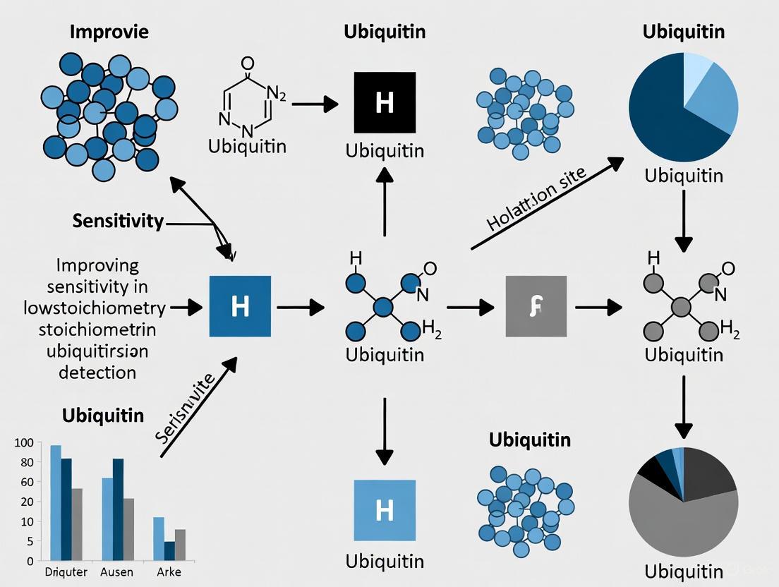
Q: What are the primary strategies to improve sensitivity for low stoichiometry ubiquitination site detection?
A: Implement pre-enrichment separation of highly abundant K48-linked chain-derived diGly peptides to reduce competition during antibody enrichment, as these can constitute a substantial portion of the ubiquitinome and impair detection of lower abundance sites [4]. Combine multiple enrichment strategies (e.g., antibody-based with UBD-based) to increase coverage. Use proteasome inhibitors (e.g., MG132) to increase ubiquitinated protein accumulation, but be aware this alters cellular physiology and primarily enhances proteasomal substrates [4].
Q: How can I optimize mass spectrometry parameters for diGly peptide detection?
A: Employ DIA methods with optimized window schemes tailored to diGly precursor characteristics, as these frequently generate longer peptides with higher charge states due to impeded C-terminal cleavage of modified lysines [4]. Use high MS2 resolution (30,000) with 46 precursor isolation windows, which has demonstrated 13% improvement over standard full proteome methods [4].
Specificity and Validation Concerns
Q: How can I distinguish true ubiquitination sites from other lysine modifications?
A: Utilize the longer ubiquitin remnant generated by LysC digestion instead of trypsin, as this excludes most ubiquitin-like modifications (NEDD8, ISG15) which contribute to <6% of diGly sites [4]. Always validate key findings using orthogonal methods such as mutagenesis of putative ubiquitination sites followed by immunoblotting [2].
Q: What controls are essential for ubiquitination experiments?
A: Include both positive controls (e.g., well-characterized ubiquitination substrates) and negative controls (substrates known not to be ubiquitinated). For MS experiments, use control samples without diGly enrichment to assess enrichment efficiency. When using linkage-specific reagents, verify specificity with defined ubiquitin chains of known linkage [2].
Technical Optimization
Q: How much starting material is required for comprehensive ubiquitinome analysis?
A: For DIA-based diGly proteomics, optimal results are achieved using 1 mg of peptide material and 31.25 μg of anti-diGly antibody, with only 25% of the total enriched material injected per run [4]. This represents a significant improvement over traditional DDA methods, which typically require more material for similar coverage.
Q: What are the key considerations for quantitative ubiquitination studies?
A: Account for the extremely low median ubiquitination site occupancy, which is three orders of magnitude lower than phosphorylation and spans over four orders of magnitude [5]. Understand that sites in structured protein regions exhibit longer half-lives and stronger upregulation by proteasome inhibitors than sites in unstructured regions [5].
Quantitative Properties of Ubiquitination
Recent systematic analyses have revealed fundamental quantitative properties of the ubiquitinome:
Ubiquitylation site occupancy spans over four orders of magnitude, with a median occupancy three orders of magnitude lower than phosphorylation, explaining the sensitivity challenges in detection [5]. The lowest 80% and highest 20% occupancy sites exhibit distinct properties, with high-occupancy sites concentrated in the cytoplasmic domains of solute carrier (SLC) proteins [5].
Turnover rates vary substantially across sites and are strongly interrelated with occupancy and regulation by proteasome inhibitors. The ubiquitin system employs a dedicated surveillance mechanism that rapidly and site-indiscriminately deubiquitylates all ubiquitin-specific E1 and E2 enzymes, protecting them against accumulation of bystander ubiquitylation [5].
Advanced Concepts and Emerging Research
Non-Traditional Ubiquitination Mechanisms
Beyond canonical lysine ubiquitination, several unconventional mechanisms expand the ubiquitin code:
Esterification with proteins, lipids, and sugars through ester linkages, as catalyzed by E3 ligases like MYCBP2 and HOIL-1 [6]. HOIL-1-mediated ubiquitination of unbranched glucosaccharides prevents toxic polyglucosan accumulation in human tissues [6].
Ubiquitylation through a phosphoribosyl bridge involving Arg42 of ubiquitin, discovered in the enzymatic pathways used by pathogens to rewrite the ubiquitin code during infection [6].
ADP-ribosylation of ubiquitin on Gly76 by the DELTEX family of E3 ligases, illustrating how post-translational modification of ubiquitin itself contributes to protein regulation during DNA repair [6].
Ubiquitin Code in Cellular Pathways and Disease
The ubiquitin code plays critical roles in numerous physiological and pathological processes:
Neurodevelopment and Disorders: Cullin-RING ubiquitin ligases (CRLs) regulate neuronal polarization, axonal outgrowth, synaptogenesis, and synaptic function [3]. Mutations in genes encoding CRL components and other E3 ligases are implicated in autism spectrum disorder, intellectual disability, and attention-deficit/hyperactivity disorder [3].
Circadian Biology: Systems-wide investigation of ubiquitination across the circadian cycle has uncovered hundreds of cycling ubiquitination sites and clusters within individual membrane protein receptors and transporters, revealing connections between ubiquitin-dependent regulation and circadian cycles [4].
NF-κB Signaling Pathway: Ubiquitin chain editing represents a sophisticated utilization of the code, where sequential actions of ubiquitin ligases and DUBs fine-tune pathway activation [1].
Visualizing Key Concepts
Ubiquitin Cascade and Code Interpretation
Ubiquitin Writing and Reading Cascade
Experimental Workflow for Sensitive Detection
High-Sensitivity Ubiquitinome Analysis Workflow
Ubiquitin Chain Diversity and Functions
Ubiquitin Modification Diversity and Functions
Ubiquitination is a critical post-translational modification that regulates diverse cellular functions, including protein degradation, cell signaling, and DNA damage repair [7] [8]. Despite its fundamental importance, researchers consistently face the challenge of detecting ubiquitination events that occur at very low stoichiometry under normal physiological conditions [9] [10]. This technical brief examines the inherent difficulties in studying low-stoichiometry ubiquitination and provides actionable troubleshooting guidance to improve detection sensitivity for research and drug development applications.
Key Challenges & Troubleshooting FAQs
FAQ 1: Why is native protein ubiquitination typically of low stoichiometry?
The low stoichiometry of ubiquitination stems from several inherent biological and technical factors that collectively make detection challenging.
Primary Contributing Factors:
- Dynamic and Reversible Nature: Ubiquitination is a highly transient process that is rapidly reversed by cellular deubiquitinase (DUB) enzymes. These DUBs can cleave ubiquitin tags from substrates before researchers can stabilize and analyze them [9] [10].
- Proteasomal Targeting: A primary function of certain ubiquitin chains (notably K48-linked) is to target the modified protein for immediate degradation by the 26S proteasome. This results in the simultaneous destruction of both the substrate and the ubiquitin signal [7] [11].
- Substrate Competition: A single E3 ligase may ubiquitinate numerous different substrate proteins, diluting the signal for any specific substrate [7].
- Low Abundance of Intermediates: The fractional stoichiometry of rate-limiting intermediates along the ubiquitination reaction trajectory is often inherently low, making these species difficult to capture [12] [13].
FAQ 2: How can I prevent the loss of ubiquitin signals during sample preparation?
Preserving labile ubiquitination during sample preparation is crucial. The key is using appropriate inhibitors to stabilize the ubiquitinated proteins.
Essential Inhibitors for Lysis Buffer:
| Inhibitor Type | Purpose | Recommended Compounds & Concentrations |
|---|---|---|
| Deubiquitinase (DUB) Inhibitors | Prevent deubiquitinating enzymes from removing Ub chains. | N-Ethylmaleimide (NEM): 5-100 mM (Note: K63 linkages may require higher concentrations ~50-100 mM) [11]. EDTA/EGTA: Include in buffer [11]. |
| Proteasome Inhibitors | Block degradation of proteasome-targeted ubiquitinated proteins. | MG-132: A common choice. Use caution with extended incubation (>12-24 hrs) to avoid stress-induced artifacts [7] [11]. |
FAQ 3: What are the best methods to enrich for low-abundance ubiquitinated proteins?
Due to low stoichiometry, enrichment is mandatory for detection. The choice of method depends on your experimental goals and model system.
Comparison of Ubiquitinated Protein Enrichment Methods:
| Method | Principle | Advantages | Limitations / Considerations |
|---|---|---|---|
| Antibody-based IP [9] | Uses anti-ubiquitin antibodies (e.g., P4D1, FK1/FK2) to immunoprecipitate ubiquitinated proteins. | Works on endogenous proteins; no genetic manipulation required; suitable for clinical samples. | High-quality antibodies can be costly; potential for non-specific binding. |
| Tandem Ubiquitin-Binding Entities (TUBEs) [9] | Uses engineered proteins with multiple ubiquitin-binding domains (UBDs) for high-affinity capture. | Protects chains from DUBs and proteasomal degradation during processing; high affinity. | Less common reagents; may not differentiate chain types. |
| Ubiquitin Traps [7] | Uses immobilized anti-ubiquitin nanobodies (VHH) for pulldown. | High affinity; low background; compatible with various cell and tissue lysates. | Not linkage-specific [7]. |
| Tagged Ubiquitin [9] | Cells express affinity-tagged Ub (e.g., His, Strep, HA). Tagged ubiquitinated proteins are purified with respective resins. | Relatively easy and low-cost; good for discovery proteomics. | May not mimic endogenous Ub perfectly; not feasible for animal/patient tissues; can co-purify non-specific proteins. |
FAQ 4: How can I optimize immunoblotting to detect smeared ubiquitin signals?
The variable molecular weights of polyubiquitinated proteins often appear as smears on western blots. Optimization is key to interpretation.
Western Blot Optimization Guide:
| Parameter | Recommendation for Ubiquitin Detection |
|---|---|
| Gel Percentage & Buffer | 8% gels with Tris-Glycine buffer: Good for large chains (>8 Ub units) [11]. 12% gels with MES buffer: Better for resolving smaller chains (2-5 Ub units) [11]. |
| Membrane | PVDF (over nitrocellulose) for higher signal strength; 0.2 µm pore size for smaller chains [11]. |
| Transfer | For long chains, use a slow transfer (e.g., 30V for 2.5 hours) to prevent unfolding and loss of antibody epitopes [11]. |
| Antibody Specificity | Most commercial anti-Ub antibodies recognize both mono- and poly-Ub. Be aware that recognition of different linkage types (e.g., M1 vs K48) can be unequal [11]. |
Advanced Methodologies for Sensitivity Improvement
For researchers requiring site-specific information or system-wide profiling, advanced methodologies are required.
Mass Spectrometry (MS)-Based Proteomics
MS is the primary tool for identifying ubiquitination sites and linkage types. Relative and absolute quantification strategies can be employed [12].
Quantitative MS Workflow for Ubiquitination: This diagram outlines the core steps for a typical MS-based experiment to identify and quantify ubiquitination, integrating key steps to handle low stoichiometry.
Computational Prediction of Ubiquitination Sites
To guide experiments, computational tools can predict potential ubiquitination sites, helping to overcome the "needle in a haystack" problem.
Comparison of Ubiquitination Site Prediction Tools:
| Tool | Key Algorithm / Approach | Features / Advantages |
|---|---|---|
| Ubigo-X [14] | Ensemble of XGBoost & ResNet34 | Integrates sequence-based, structure-based, and function-based features transformed into images. |
| EUP [15] | Conditional Variational Autoencoder (cVAE) based on ESM2 protein language model. | Cross-species prediction; uses pretrained model to capture evolutionary and structural information. |
| DeepUbi [14] | Convolutional Neural Network (CNN) | Uses one-hot encoding, physicochemical properties, and k-spaced amino acid pairs. |
The Scientist's Toolkit: Essential Research Reagents
A curated list of key reagents is vital for successful ubiquitination studies.
Table: Essential Reagents for Ubiquitination Detection
| Reagent / Tool | Function | Example & Notes |
|---|---|---|
| DUB Inhibitors | Stabilize ubiquitin conjugates during lysis. | N-Ethylmaleimide (NEM), EDTA [11]. |
| Proteasome Inhibitors | Prevent degradation of ubiquitinated proteins. | MG-132 [7] [11]. |
| Pan-Ubiquitin Antibodies | Detect/enrich total ubiquitinated proteins. | P4D1, FK1/FK2 [9]. Note variable affinity for different linkages [11]. |
| Linkage-Specific Antibodies | Detect specific Ub chain types (e.g., K48, K63). | Commercial antibodies available for K6, K11, K33, K48, K63 [9] [11]. |
| Ubiquitin Traps / TUBEs | High-affinity enrichment of ubiquitinated proteins. | ChromoTek Ubiquitin-Trap (nanobody-based) [7]; TUBEs (tandem UBDs) [9]. |
| Tagged Ubiquitin Plasmids | Enable affinity-based purification of ubiquitinated proteome. | His-, Strep-, or HA-tagged Ub for expression in cells [9]. |
| Ubiquitin Activating Enzyme (E1) Inhibitor | Tool for probing ubiquitination dynamics. | MLN4924 (inhibits NAE1, a NEDD8-activating enzyme) [8]. |
Troubleshooting Guide: Low Stoichiometry Ubiquitination Site Detection
This guide addresses common challenges in detecting low-abundance ubiquitination sites, a critical step for understanding cellular signaling and drug mechanisms.
Troubleshooting Scenarios and Solutions
| Problem Scenario | Possible Root Cause | Recommended Solution | Key Experimental Parameters to Check |
|---|---|---|---|
| Low yield of ubiquitinated peptides after enrichment. | Inefficient antibody binding or high sample complexity. | Use linkage-specific Ub antibodies or Ub-binding domains (UBDs) for enrichment [2]. Verify antibody specificity and optimize peptide-to-bead ratio. | Antibody lot, incubation time and temperature, sample-to-bead ratio. |
| High background noise in mass spectrometry data. | Non-specific binding during enrichment or co-enrichment of non-ubiquitinated peptides. | Incorporate more stringent wash steps (e.g., high-salt buffers). Use control samples (no enrichment) to identify non-specific binders [16]. | Wash buffer stringency, LC-MS/MS gradient conditions. |
| Inconsistent results with proteasome inhibitor treatment (e.g., MG-132). | Variable inhibitor efficacy or secondary effects on ubiquitination pathways. | Titrate inhibitor concentration and treatment duration. Use a combination of proteasome and deubiquitinase (DUB) inhibitors (e.g., PR-619) to stabilize ubiquitinated proteins [16]. | Cell viability post-treatment, confirmation of proteasome inhibition (e.g., accumulation of a known substrate). |
| Failure to detect ubiquitination on specific metabolites or drug-like molecules. | Technical limitations of metabolomics platforms or low abundance of modified metabolites. | Employ high-throughput, high-sensitivity metabolomics like FIA TOF-MS. Compare drug-treated metabolome profiles with those from protein overexpression strains to infer interactions [17]. | Metabolite extraction efficiency, instrument sensitivity, data normalization against biomass. |
Frequently Asked Questions (FAQs)
Q: What are the major advantages of using antibody-based enrichment for ubiquitination site mapping? A: Antibodies, particularly those specific for the K-ε-GG remnant, allow for the enrichment of endogenously ubiquitinated peptides without genetic manipulation of the target cells. This makes them suitable for use with clinical samples and animal tissues [2] [16].
Q: How can I improve the coverage of ubiquitinated proteins in my experiment? A: Beyond optimizing enrichment protocols, using minimal fractionation of digested lysates prior to immunoaffinity enrichment can significantly increase the yield of K-ε-GG peptides. One study demonstrated a three- to fourfold increase, leading to the detection of over 3,000 distinct ubiquitination sites [16].
Q: My research involves membrane proteins, which are notoriously difficult to study. Are there specific strategies for profiling their ubiquitination? A: Yes, novel strategies involve creating phenotypic signatures. One method is to profile the metabolome of yeast strains with inducible overexpression of the membrane protein and compare it to the metabolome of cells treated with a library of drug-like molecules. Matching the metabolic profiles can predict drug-target interactions for difficult-to-study membrane proteins [17].
Q: What does it mean if a ubiquitination site does not change after proteasome inhibition? A: Not all ubiquitination events target substrates for proteasomal degradation. Ubiquitination is also involved in signaling transduction, protein-protein interactions, and endocytosis. A site unaffected by proteasome inhibition is likely involved in a non-degradative function [16].
Detailed Experimental Protocols
Protocol 1: Large-Scale Enrichment and Identification of Ubiquitination Sites
This protocol is adapted from methods used to study global ubiquitination changes following inhibitor treatment [16].
1. Sample Preparation and Lysis
- Grow Jurkat cells to mid-log phase.
- Treat cells with DMSO (control), 10 μM MG-132 (proteasome inhibitor), or 10-20 μM PR-619 (DUB inhibitor) for 4-6 hours.
- Harvest cells by centrifugation and lyse in a denaturing lysis buffer (e.g., 6 M Guanidine-HCl, 100 mM Tris-HCl, pH 8.0) to instantly inactivate DUBs and proteases.
2. Protein Digestion
- Reduce disulfide bonds with 5 mM DTT (30 min, 60°C) and alkylate with 15 mM iodoacetamide (30 min, room temperature, in the dark).
- Dilute the guanidine-HCl concentration to less than 1 M to be compatible with trypsin digestion.
- Digest proteins with sequencing-grade trypsin (1:50 w/w enzyme-to-protein ratio) overnight at 37°C.
3. Peptide Desalting and Fractionation
- Acidify digested peptides with trifluoroacetic acid (TFA) to pH < 3.
- Desalt peptides using C18 solid-phase extraction cartridges or columns.
- For deep coverage, fractionate the desalted peptide mixture using basic pH reversed-phase chromatography into 8-12 fractions. This step increases the depth of analysis but is optional for simpler samples.
4. Immunoaffinity Enrichment (IAE) of K-ε-GG Peptides
- Reconstitute each peptide fraction in IAE buffer (e.g., 50 mM MOPS, pH 7.2, 10 mM Na2HPO4, 50 mM NaCl).
- Incubate the peptides with anti-K-ε-GG antibody conjugated to agarose beads for 90 minutes to 2 hours at 4°C with gentle agitation.
- Wash the beads extensively with IAE buffer followed by water to remove non-specifically bound peptides.
5. Mass Spectrometric Analysis
- Elute the K-ε-GG peptides from the beads with a low-ppH elution buffer (e.g., 0.15% TFA).
- Analyze the enriched peptides by LC-MS/MS using a high-performance instrument (e.g., Q-Exactive Orbitrap).
- Use data-dependent acquisition to fragment the top most abundant ions.
Data Analysis:
- Search the resulting MS/MS spectra against a protein sequence database using search engines like MaxQuant or Spectrum Mill.
- Set a variable modification of GlyGly (+114.0428 Da) on lysine residues to identify ubiquitination sites.
Protocol 2: Metabolomic Profiling for Drug-Target Prediction
This protocol, based on a proof-of-concept study in yeast, uses metabolomics to predict interactions between small molecules and their protein targets [17].
1. Culturing and Treatment
- Grow S. cerevisiae in synthetic defined media in a 96-deep well plate format. Start with an OD600 of 0.1.
- For drug treatment: Add compounds from a chemical library (e.g., 1,280 drugs) and incubate until the culture reaches an OD600 of ~1.0. Maintain consistent DMSO concentrations across all treatments.
- For overexpression: Use yeast strains with inducible (e.g., β-estradiol) overexpression of target genes (e.g., membrane proteins). Induce with a range of inducer concentrations for 1.5 and 3 hours.
2. Metabolite Extraction
- Harvest cells by rapid centrifugation.
- Immediately quench metabolism and extract intracellular metabolites using cold solvent (e.g., 80% methanol chilled to -40°C).
- Centrifuge to remove cell debris and collect the supernatant containing the metabolome.
3. High-Throughput Metabolome Analysis
- Analyze metabolite extracts using Flow-Injection Analysis Time-of-Flight Mass Spectrometry (FIA TOF-MS). This method provides high-throughput, chromatography-free profiling.
- The system is calibrated for mass accuracy, and ions are annotated against metabolite databases (e.g., KEGG).
4. Data Processing and Analysis
- Normalize raw ion intensities for temporal drift in the MS and for biomass at the time of sampling.
- Restrict analysis to a core set of annotated metabolites (e.g., 226 from the S. cerevisiae KEGG collection).
- Compare the metabolome profile of each drug treatment to the profiles of all overexpression strains. A high similarity between a drug's profile and an overexpression strain's profile suggests the drug targets that gene's product or its pathway.
Research Reagent Solutions
Essential materials and reagents for conducting experiments in ubiquitination site detection and metabolomic profiling.
| Reagent / Material | Function / Application |
|---|---|
| Anti-K-ε-GG Antibody | Immunoaffinity enrichment of tryptic peptides containing the di-glycine remnant of ubiquitination for mass spectrometry analysis [16]. |
| MG-132 | A cell-permeable proteasome inhibitor used to stabilize polyubiquitinated proteins targeted for degradation, increasing their abundance for detection [16]. |
| PR-619 | A broad-spectrum deubiquitinase (DUB) inhibitor. Used to prevent the removal of ubiquitin chains, thereby stabilizing the ubiquitinome [16]. |
| Strep- or His-Tagged Ubiquitin | Genetically encoded tags that allow for the purification of ubiquitinated proteins from living cells using Strep-Tactin or Ni-NTA resins, useful for substrate identification [2]. |
| Linkage-Specific Ub Antibodies | Antibodies that recognize specific polyUb chain linkages (e.g., K48 or K63). They are used to enrich for proteins modified with a particular chain type to study its function [2]. |
| FIA TOF-MS | A high-throughput mass spectrometry platform for rapid, comprehensive metabolome profiling. It sacrifices chromatographic separation for speed, enabling large-scale screens [17]. |
Experimental Workflow and Pathway Visualizations
Ubiquitination Site Analysis Workflow
Ubiquitin Conjugation Cascade
Metabolomic Drug-Target Prediction
Ubiquitination is a versatile post-translational modification (PTM) that regulates virtually all cellular processes, from protein degradation to immune signaling [18]. This 76-amino acid protein can be attached to substrates as a single moiety (monoubiquitination) or as polymers (polyubiquitin chains) with distinct biological functions [19] [18]. The complexity arises from eight possible linkage types (Met1, Lys6, Lys11, Lys27, Lys29, Lys33, Lys48, Lys63) that can form homotypic, heterotypic, or even branched chains, creating what is known as the "ubiquitin code" [18].
For researchers investigating ubiquitination, three interconnected technical hurdles persist: the dynamic nature of modifications governed by opposing E3 ligase and deubiquitinase (DUB) activities; the structural complexity of ubiquitin chain architectures; and the inherent lability of ubiquitinated substrates due to rapid degradation or regulatory turnover [2] [12]. This guide addresses these challenges through targeted troubleshooting advice and optimized methodologies to enhance detection sensitivity, particularly for low-stoichiometry ubiquitination events critical for understanding cellular signaling and disease mechanisms.
Troubleshooting Guide: FAQs for Ubiquitination Experiments
FAQ 1: How can I improve the recovery of low-abundance ubiquitinated substrates for mass spectrometry analysis?
- Problem: Low stoichiometry of endogenous ubiquitination and interference from abundant non-modified proteins impair detection.
- Solution: Implement tandem enrichment strategies.
- Step 1: Use ubiquitin affinity tags (e.g., His-, Strep-) for initial purification from cell lysates of engineered cell lines [2]. This provides a first level of isolation but may co-purify endogenous biotinylated or histidine-rich proteins.
- Step 2: Perform a secondary enrichment using antibodies that recognize the diglycine (diGly) remnant left on trypsinized peptides [20]. This signature GG-tag (a 114.04 Da mass shift on modified lysine) is a definitive marker for ubiquitination sites [2] [20].
- Tip: For clinical or tissue samples where genetic tagging is infeasible, use anti-ubiquitin antibodies (e.g., P4D1, FK1/FK2) for the initial protein-level enrichment [2].
FAQ 2: My western blot shows smearing, but I cannot identify specific ubiquitination sites. What is wrong?
- Problem: Smearing confirms ubiquitination but site identification requires proteomic techniques.
- Solution: Optimize sample preparation for mass spectrometry (MS).
- Use SILAC (Stable Isotope Labeling with Amino acids in Cell Culture) or TMT (Tandem Mass Tagging) for quantitative assessment of changes in diGly-site abundance upon perturbations like proteasomal inhibition [12].
- Employ an LC-MS3 workflow instead of standard LC-MS2 when using TMT tags. This significantly reduces signal compression (interference) from co-isolated peptides, providing more accurate quantification, which is crucial for detecting subtle changes in low-stoichiometry sites [12].
- For absolute quantification of modification stoichiometry, incorporate AQUA (Absolute QUAntification) peptides with labeled ubiquitin peptide standards into your MS workflow [18].
FAQ 3: How can I determine the linkage type of a polyubiquitin chain?
- Problem: Antibodies often only distinguish a few linkage types, and MS can be confounded by chain complexity.
- Solution: Use the UbiCRest (Ubiquitin Chain Restriction) assay [21].
- Protocol: Treat your immunopurified ubiquitinated protein with a panel of linkage-specific deubiquitinases (DUBs) in parallel reactions. Analyze the cleavage products by western blot.
- Interpretation: Each DUB will cleave only a specific subset of linkages. For example, OTUB1 is specific for Lys48-linked chains, while Cezanne prefers Lys11-linked chains [21] [18]. The pattern of band shifts across the reactions reveals the chain architecture present on your substrate.
- Reagent Source: Several linkage-specific DUBs are commercially available, or you can purify them as described in [21].
FAQ 4: My ubiquitinated substrate is too unstable for analysis. How can I stabilize it?
- Problem: The substrate is rapidly degraded by the proteasome or deubiquitinated.
- Solution: Use targeted inhibitors.
- For proteasomal degradation: Treat cells with MG132, Bortezomib, or MLN9704 prior to lysis to prevent the degradation of polyubiquitinated proteins, particularly those with Lys48-linked chains [2].
- For deubiquitination: Include broad-spectrum DUB inhibitors (e.g., PR-619, N-Ethylmaleimide) in your lysis buffer to preserve the ubiquitin signal during sample preparation [2].
- Critical Consideration: Inhibition time courses are essential, as prolonged proteasome inhibition alters global protein homeostasis and induces stress responses that may confound results [20].
Comparative Analysis of Key Methodologies
The table below summarizes the primary techniques used to study protein ubiquitination, their applications, and key limitations.
Table 1: Comparison of Primary Ubiquitin Analysis Methodologies
| Method | Principle | Application | Key Limitations |
|---|---|---|---|
| Ubiquitin Tagging [2] | Expression of affinity-tagged Ub (e.g., His, Strep) in cells for protein purification. | High-throughput screening of ubiquitinated substrates from engineered cell lines. | Cannot be used on patient tissues; tagged Ub may not fully mimic endogenous Ub; potential for co-purification of non-target proteins. |
| Antibody-Based Enrichment [2] | Use of anti-Ub or linkage-specific antibodies to pull down ubiquitinated proteins or peptides. | Analysis of endogenous ubiquitination from any biological source, including tissues. | High-cost of quality antibodies; potential for non-specific binding; limited availability for some atypical linkages. |
| diGly Antibody MS [20] | Enrichment of tryptic peptides containing the diGly lysine remnant followed by MS. | Global site-specific mapping of ubiquitination (the "ubiquitinome"). | Requires tryptic digestion, destroying information on chain architecture; low stoichiometry sites may be missed. |
| UbiCRest [21] | Digestion of purified ubiquitinated proteins with a panel of linkage-specific DUBs. | Determination of polyubiquitin chain linkage type and architecture. | A qualitative method; requires a relatively pure substrate of interest. |
| Middle-Down MS [18] | MS analysis of larger ubiquitin chain fragments without complete tryptic digestion. | Direct characterization of mixed/branched ubiquitin chain topologies. | Technically challenging; not yet a routine high-throughput application. |
The Scientist's Toolkit: Essential Research Reagents
Successful ubiquitination research relies on a suite of specific reagents and tools. The following table details key components for a functional ubiquitin toolkit.
Table 2: Essential Reagents for Ubiquitination Research
| Reagent / Tool | Function | Key Examples & Notes |
|---|---|---|
| Linkage-Specific Antibodies | Detect or enrich for specific ubiquitin chain types. | Anti-K48 (for degradation), Anti-K63 (for signaling), Anti-M1 (linear chains) [18]. Critical for western blot and immunofluorescence. |
| diGly Remnant Antibody | Immunoaffinity enrichment of ubiquitinated peptides for MS. | monoclonal antibody specific for tryptic peptides with Lys-ε-GG [20]. Foundation for ubiquitylome studies. |
| Deubiquitinase (DUB) Inhibitors | Preserve ubiquitin signal during cell lysis and protein purification. | PR-619 (broad-spectrum); specific inhibitors for USPs, UCHs, etc. Always use in lysis buffers [2]. |
| Proteasome Inhibitors | Stabilize proteasome-targeted, ubiquitinated substrates. | MG132 (reversible), Bortezomib (clinical). Use in time-course experiments to avoid compensatory effects [2]. |
| Linkage-Specific DUBs | Tool enzymes for UbiCRest assay to decipher chain linkage. | OTUB1 (K48-specific), Cezanne (K11-specific), OTULIN (M1-specific) [21]. Can be purified in-lab or purchased. |
| Tandem Affinity Tags | Purification of ubiquitinated proteins under denaturing conditions. | His-Biotin, His-Strep, or His-FLAG tags on ubiquitin. Denaturing lysis (e.g., with 6M Guanidine-HCl) helps avoid co-purifying non-covalent binders [2]. |
| Stable Isotope Labels (SILAC/TMT) | Enable quantitative MS to monitor dynamics of ubiquitination. | SILAC: metabolic labeling; TMT: isobaric tagging of peptides. TMT requires MS3 for accurate quantification of complex samples [12]. |
Experimental Protocol: A Workflow for Sensitive Ubiquitin Site Mapping
This detailed protocol outlines a robust strategy for identifying ubiquitination sites on a low-abundance protein of interest, integrating solutions to the key hurdles of lability and detection.
Goal: To identify and confirm ubiquitination sites on a low-stoichiometry substrate protein. Concept: The workflow combines genetic engineering, affinity purification, and highly sensitive mass spectrometry to overcome the challenges of dynamic modification and substrate lability.
Diagram 1: Ubiquitin Site Mapping Workflow
Step-by-Step Procedure:
Cell Line Preparation:
- Generate a cell line (e.g., HEK293T) stably expressing tandem-tagged ubiquitin (e.g., His-Strep tag) using the StUbEx (Stable Tagged Ub exchange) system [2]. This ensures all cellular ubiquitin is tagged for efficient purification.
Inhibition and Lysis:
- Treat cells with appropriate pathway activators/inhibitors. Crucially, before lysis, add 10 µM MG132 (or another proteasome inhibitor) for 4-6 hours and include 10 mM N-Ethylmaleimide (a DUB inhibitor) in the lysis buffer (e.g., a guanidine-based denaturing buffer) to stabilize ubiquitinated species [2].
Tandem Affinity Purification (TAP):
- Under denaturing conditions, perform immobilized metal affinity chromatography (IMAC, e.g., Ni-NTA) to capture His-tagged ubiquitinated proteins.
- Elute the bound proteins and subject them to a second purification step using Strep-Tactin resin. This two-step process drastically reduces non-specific binders [2].
On-bead Digestion and diGly Peptide Enrichment:
- Wash the final beads and digest the purified proteins with trypsin directly on the resin.
- Desalt the resulting peptide mixture and incubate it with anti-diGly remnant antibody beads overnight. This step specifically enriches for peptides derived from ubiquitinated lysines [20].
Quantitative Mass Spectrometry:
- Label the enriched peptides with Tandem Mass Tags (TMT). Analyze them using an LC-MS3 workflow on an Orbitrap Fusion mass spectrometer. The MS3 method minimizes co-isolation interference, providing superior quantification accuracy for low-abundance peptides across multiple samples [12].
Data Analysis and Validation:
- Search MS data against the appropriate protein database, filtering for the diGly modification (114.04 Da mass shift on lysine).
- Confirm identified sites by mutating the modified lysine(s) to arginine and repeating the initial ubiquitination assay (e.g., by western blot) to observe a loss of signal [2].
- For linkage information, use the UbiCRest protocol on the purified substrate [21].
Visualizing Ubiquitin Chain Complexity and Signaling
The following diagram illustrates the core concept of the ubiquitin code, showing how different chain architectures can lead to distinct cellular outcomes, which is fundamental to understanding the "why" behind the technical challenges.
Diagram 2: The Ubiquitin Code and Functional Outcomes
Cutting-Edge Tools: A Practical Guide to Enrichment and MS Methods for Maximum Sensitivity
The diGLY proteomics approach is a powerful method for systematically interrogating the ubiquitin-modified proteome with site-level resolution. This technique utilizes antibodies specifically developed to recognize the Lys-ϵ-Gly-Gly (diGLY) remnant left on trypsin-digested peptides that were previously modified by ubiquitin. The workflow involves specific cell culture preparation, protein digestion, affinity enrichment of diGLY-modified peptides, and their identification through mass spectrometry [22].
The following diagram illustrates the core workflow for a quantitative diGLY proteomics experiment, typically using Stable Isotope Labeling by Amino Acids in Cell Culture (SILAC) to compare different experimental conditions:
Key Research Reagent Solutions
Successful diGLY proteomics relies on a set of specific reagents designed to preserve, isolate, and identify the low-abundance ubiquitin remnants.
Table 1: Essential Reagents for diGLY Proteomics Workflow
| Reagent / Kit | Function / Application | Key Features / Notes |
|---|---|---|
| PTMScan Ubiquitin Remnant Motif (K-ε-GG) Kit [22] | Immunoaffinity enrichment of diGLY-modified peptides from complex digests. | Contains the core diGLY motif-specific antibody; critical for deep ubiquitinome coverage. |
| diGLY Lysis Buffer [22] | Cell/tissue lysis while preserving ubiquitin modifications and inhibiting deubiquitinases. | Contains 8M Urea, 50mM Tris-HCl (pH 8), 150mM NaCl, Protease Inhibitors, and 5mM N-Ethylmaleimide (NEM). |
| LysC & Trypsin Proteases [22] | Sequential digestion of proteins to generate peptides with the diGLY remnant. | LysC digestion is performed first in 8M urea, followed by trypsin digestion after urea dilution. |
| SILAC Media [22] | For metabolic labeling and quantitative comparisons between experimental conditions. | DMEM lacking Lysine and Arginine, supplemented with "light" (Lys0, Arg0) or "heavy" (Lys8, Arg10) isotopes and dialyzed FBS. |
Frequently Asked Questions (FAQs)
Q1: What is the specificity of the diGLY antibody? Does it cross-react with other modifications?
The diGLY antibody is highly specific for the lysine residue modified with a Gly-Gly moiety. However, it is important to note that identical diGLY remnants are generated not only from ubiquitin but also from the ubiquitin-like modifiers NEDD8 and ISG15 after trypsin digestion. Studies indicate that the vast majority (~95%) of diGLY peptides identified using this enrichment approach originate from bona fide ubiquitination, with a minor contribution from neddylation or ISGylation [22].
Q2: What is the recommended amount of starting material for a deep ubiquitinome analysis?
For a standard analysis, the minimum protein requirement is 1-2 mg of total protein per sample. This typically translates to approximately 10 million cells, depending on the cell type [22]. For single-shot DIA analyses that achieve deep coverage, enrichment from 1 mg of peptide material using 31.25 µg of anti-diGLY antibody has been determined to be optimal [4].
Q3: How does Data-Independent Acquisition (DIA) improve diGLY proteomics compared to traditional methods?
DIA mass spectrometry represents a significant advancement for diGLY proteomics. It markedly improves quantitative accuracy, sensitivity, and data completeness. A single DIA measurement can identify over 35,000 distinct diGLY sites—nearly double the number typically identified by Data-Dependent Acquisition (DDA) in a single run. Furthermore, DIA demonstrates superior reproducibility, with a much larger percentage of diGLY peptides showing low coefficients of variation (CVs) [4].
Q4: Can this technique be applied to tissues and in vivo models?
Yes. A major advantage of the diGLY antibody-based affinity approach is that it is not limited to cell lines. It can be successfully applied to identify ubiquitylated proteins from any human or murine primary tissue or other eukaryotic organisms [22].
Troubleshooting Guide
Table 2: Common Experimental Issues and Solutions
| Problem | Potential Cause | Recommended Solution |
|---|---|---|
| Low yield of enriched diGLY peptides | Inefficient cell lysis or incomplete digestion. | Verify lysis efficiency. Optimize protease-to-protein ratio and ensure sequential digestion with LysC followed by trypsin is performed correctly [22]. |
| Ubiquitin modifications degraded during sample prep. | Ensure fresh NEM (or other DUB inhibitors) is added to the lysis buffer to inhibit deubiquitinating enzymes [22]. | |
| High background in mass spectrometry | Incomplete removal of non-modified peptides. | Ensure proper washing of antibodies/beads after enrichment. Consider basic reversed-phase (bRP) fractionation prior to enrichment to reduce sample complexity, especially for very deep coverage [4]. |
| Poor quantitative reproducibility | High technical variation in enrichment or MS analysis. | Switch to a DIA-MS workflow, which provides significantly lower CVs and fewer missing values across samples [4]. |
| Inconsistent handling of samples. | Use SILAC or other isotope-labeling methods for internal quantitative control. Process control and experimental samples in parallel [22]. | |
| Low coverage of endogenous sites | Overwhelming abundance of a single diGLY peptide. | For inhibitor-treated cells where K48-linked ubiquitin-chain derived diGLY peptides are extremely abundant, pre-fractionate the digest and pool fractions to avoid this single peptide dominating the enrichment capacity [4]. |
Advanced Workflow: DIA for Enhanced Ubiquitinome Coverage
For researchers requiring the highest level of sensitivity and quantitative accuracy, a DIA-based workflow is recommended. This method relies on building or using comprehensive spectral libraries to match and quantify diGLY peptides from complex mixtures.
The following diagram outlines the key steps for implementing a sensitive DIA-based diGLY workflow:
Key methodological details for the DIA workflow [4]:
- Library Generation: Create a deep spectral library by fractionating a representative sample (e.g., proteasome inhibitor-treated cells) into 96 fractions, concatenating them into 8-9 pools, and performing diGLY enrichment and DDA analysis on each pool.
- DIA Method Optimization: Use ~46 precursor isolation windows with a fragment scan resolution of 30,000 for an optimal balance of sensitivity and sequencing speed.
- Data Analysis: Match the DIA data against a hybrid spectral library generated by merging the experimental DDA library with a direct DIA search of the samples themselves to maximize site identification.
Protein ubiquitination is one of the most common yet complex post-translational modifications in eukaryotes, playing pivotal roles in regulating protein stability, activity, localization, and virtually all cellular processes [23] [2]. The modification involves the covalent attachment of a 76-amino acid ubiquitin protein to substrate proteins via a cascade of E1 (activating), E2 (conjugating), and E3 (ligating) enzymes [2]. The versatility of ubiquitination stems from its ability to form diverse structures—from single ubiquitin molecules to complex polyubiquitin chains with different linkage types—each encoding distinct cellular functions [23] [24].
A central challenge in ubiquitination research lies in the low stoichiometry and dynamic nature of this modification under physiological conditions [23] [2]. Unmodified proteins vastly outnumber their ubiquitinated counterparts, creating significant detection hurdles. Furthermore, the rapid deubiquitination by deubiquitinating enzymes (DUBs) and proteasomal degradation of ubiquitinated targets make them transient and difficult to capture [2]. These technical barriers are particularly problematic for researchers investigating low-abundance ubiquitination events or working with limited clinical samples where genetic manipulation is infeasible.
Core Methodologies: Principles and Technical Specifications
Ubiquitin Tagging (Ub-Tagging) Approaches
Ub-tagging methodologies involve the genetic engineering of ubiquitin with affinity tags such as His, Flag, HA, or Strep tags [2]. When expressed in cells, these tagged ubiquitin molecules become incorporated into the ubiquitination machinery, allowing subsequent purification of ubiquitinated proteins under denaturing conditions that eliminate non-covalent interactors.
Experimental Protocol: Cells are transfected to express tagged ubiquitin, followed by lysis under denaturing conditions (e.g., with SDS or guanidine hydrochloride). The lysate is then incubated with appropriate affinity resins—Ni-NTA for His-tags or Strep-Tactin for Strep-tags. After extensive washing, bound proteins are eluted with competitive agents like imidazole (for His-tags) or desthiobiotin (for Strep-tags) for downstream analysis by immunoblotting or mass spectrometry [2].
Key Advantages: This approach provides a relatively straightforward and low-cost method for ubiquitinome profiling, with high specificity for covalently modified proteins when using denaturing conditions [2].
Inherent Limitations: The requirement for genetic manipulation makes it unsuitable for clinical specimens or animal tissues. Additionally, the tagged ubiquitin may not perfectly mimic endogenous ubiquitin, potentially creating artifacts, and the approach can co-purify histidine-rich or endogenously biotinylated proteins, increasing background noise [2].
Ubiquitin-Binding Domain (UBD) Approaches
UBDs are modular protein domains that naturally recognize and bind to ubiquitin or ubiquitin chains [23]. More than 20 different UBD families have been identified, with affinities ranging from 2 to 500 μM and varying preferences for different ubiquitin linkage types [23]. Single UBDs from proteins like DSK2p, ubiquilin, and RABGEF1 have been used for enrichment, but their moderate affinity limits comprehensive ubiquitinome coverage [23] [2].
Tandem Hybrid UBD (ThUBD) and TUBE Technologies
To overcome affinity limitations of single UBDs, engineered tandem constructs have been developed. Tandem Ubiquitin-Binding Entities (TUBEs) link multiple UBDs in a single polypeptide, significantly enhancing affinity for polyubiquitin chains [25]. More recently, Tandem Hybrid UBDs (ThUBDs) have been engineered by combining different types of UBDs with high natural affinity [23] [26].
ThUBD Engineering Protocol: Researchers systematically evaluated the affinity of various UBDs to different ubiquitin chains. Selected UBDs with high affinity were combined to create artificial tandem hybrids. For example, ThUDQ2 incorporates four UBDs from DSK2p-derived UBA and ubiquilin 2-derived UBA, while ThUDA20 combines DSK2p-derived UBA and RABGEF1-derived A20-ZnF domains [23] [26]. These constructs are cloned into expression vectors (e.g., pGEX-4T-2), expressed in E. coli, purified using glutathione-Sepharose beads, and coupled to NHS-activated Sepharose for sample enrichment [23].
Performance Advantages: ThUBDs exhibit markedly higher affinity than naturally occurring UBDs and display almost unbiased high affinity to all seven lysine-linked ubiquitin chains [23] [26]. This technology has enabled identification of thousands of ubiquitinated proteins from yeast and mammalian cells without requiring tagged ubiquitin overexpression [23].
The following workflow illustrates a typical ThUBD-based ubiquitinome profiling experiment:
Comparative Analysis of Affinity Purification Strategies
The table below provides a detailed comparison of the key affinity purification strategies for ubiquitination research:
| Method | Applications | Key Limitations | Affinity/Linkage Preference | Sample Requirements |
|---|---|---|---|---|
| Ub-Tagging | High-throughput screening in genetically modifiable systems [2] | Not applicable to tissues/clinical samples; potential artifacts from tag interference [2] | N/A (tags ubiquitin directly) | Requires genetic manipulation; unsuitable for human tissues [2] |
| Single UBD | Basic research on ubiquitinated proteins [23] | Low affinity; restricted to specific ubiquitin chain types [23] | Low affinity (μM range); strong linkage bias [23] | Compatible with various lysates; low sensitivity [23] |
| TUBE | Enrichment of polyubiquitinated proteins; proteomics [25] | Poor performance for monoubiquitinated proteins [25] | Moderate affinity; some linkage bias [25] | Compatible with native and denaturing conditions [25] |
| ThUBD | Unbiased ubiquitinome profiling; biomarker discovery [23] [26] | Requires protein expression and purification [23] | High affinity (nM range); minimal linkage bias [23] [26] | Works with yeast, mammalian cells, and tissues [23] |
| OtUBD | Enrichment of both mono- and polyubiquitinated proteins [25] | Bacterial origin requires recombinant production [25] | High affinity (low nM range) [25] | Compatible with baker's yeast and mammalian cells [25] |
The Scientist's Toolkit: Essential Research Reagents
| Reagent/Tool | Function | Example Applications |
|---|---|---|
| ThUBD Fusion Protein | High-affinity, unbiased capture of diverse ubiquitin chains [23] [26] | Ubiquitinome profiling in mammalian cells and tissues [23] |
| OtUBD Affinity Resin | Enrichment of mono- and polyubiquitinated proteins [25] | Proteomic analysis of ubiquitinated substrates [25] |
| diGly Remnant Antibodies | Immunoaffinity enrichment of tryptic peptides with K-ε-GG remnant [4] [27] | Ubiquitination site mapping by mass spectrometry [4] |
| Linkage-Specific Antibodies | Detection and enrichment of specific ubiquitin chain types [2] | Studying functions of particular ubiquitin linkages [2] |
| TUBE-Based Assay Plates | High-throughput screening of ubiquitination signals [28] | PROTAC development and drug discovery [28] |
| Tandem Mass Tag (TMT) Reagents | Multiplexed quantitative proteomics [12] | Relative quantification of ubiquitinated peptides across conditions [12] |
Troubleshooting Guide: Frequently Asked Questions
Q1: How can I improve the sensitivity of ubiquitinated protein detection from low-input clinical samples?
Challenge: Traditional Ub-tagging approaches are infeasible for clinical specimens, while antibody-based methods may lack sensitivity.
Solution: Implement ThUBD-based enrichment, which demonstrates 16-fold greater sensitivity compared to TUBE technology, detecting ubiquitinated proteins from as little as 0.625 μg of input material [28]. ThUBD's high affinity and minimal linkage bias enable more comprehensive capture of the endogenous ubiquitinome without genetic manipulation [23] [26].
Protocol Adjustment: For formalin-fixed paraffin-embedded (FFPE) tissue samples, optimize lysis conditions to balance protein extraction efficiency and ubiquitin preservation. Incorporate protease and DUB inhibitors throughout the process, and use ThUBD-coated 96-well plates for high-throughput screening when sample amount is limited [28].
Q2: What approach best addresses linkage bias in ubiquitin chain enrichment?
Challenge: Many naturally occurring UBDs and some antibodies preferentially recognize specific ubiquitin chain types (e.g., K48 or K63 linkages), creating biased representation of the ubiquitinome.
Solution: Utilize engineered ThUBDs that combine different UBD types to achieve nearly unbiased affinity across all seven lysine-linked ubiquitin chains [23] [26]. Validation experiments show ThUBDs maintain high affinity for K6-, K11-, K27-, K29-, K33-, K48-, and K63-linked chains without strong preference for any single type [23].
Verification Method: Confirm linkage independence using ubiquitin chain panels in surface plasmon resonance (SPR) or ELISA-style assays. Compare enrichment efficiency across different chain types to verify unbiased capture [23].
Q3: How can I distinguish covalently ubiquitinated proteins from mere interactors?
Challenge: UBD-based approaches under native conditions can co-purify proteins that non-covalently associate with ubiquitin or ubiquitinated proteins, confounding identification of genuine substrates.
Solution: Implement a dual-approach strategy using both native and denaturing conditions:
- Denaturing Workflow: Lyse cells in buffer containing 1% SDS or 8M urea and boil samples to disrupt non-covalent interactions. This isolates covalently ubiquitinated proteins [25].
- Native Workflow: Use mild detergents (e.g., 0.1-1% Triton X-100) in lysis and wash buffers to preserve protein complexes, enriching both ubiquitinated proteins and their interactors [25].
Comparative Analysis: Process parallel samples through both workflows, then analyze by immunoblotting or mass spectrometry. Proteins identified only under native conditions represent non-covalent interactors, while those under denaturing conditions are genuine ubiquitination targets [25].
Q4: What mass spectrometry advancements improve ubiquitination site identification?
Challenge: Traditional data-dependent acquisition (DDA) methods yield incomplete ubiquitinome coverage and high rates of missing values across samples.
Solution: Implement data-independent acquisition (DIA) methods specifically optimized for diGly peptide analysis. This approach can identify approximately 35,000 distinct diGly peptides in single measurements—nearly double the coverage of DDA methods—with significantly improved quantitative accuracy and reproducibility [4].
Workflow Optimization: Combine diGly antibody-based enrichment with DIA methods using optimized window schemes (e.g., 46 precursor isolation windows) and high MS2 resolution (30,000). Employ comprehensive spectral libraries containing >90,000 diGly peptides for optimal matching [4]. Pre-fractionate samples to separate highly abundant K48-linked ubiquitin chain-derived diGly peptides that might compete for antibody binding sites [4].
The evolving landscape of ubiquitination research tools has progressively addressed the central challenge of sensitivity in low stoichiometry detection. From the early days of Ub-tagging and single UBD applications to the current generation of ThUBDs and optimized mass spectrometry workflows, each methodological advancement has brought improved affinity, reduced bias, and expanded application scope. The integration of these affinity purification strategies with sophisticated proteomic platforms represents the cutting edge in ubiquitin research, enabling systems-level investigations of ubiquitin signaling in health and disease.
Future directions will likely focus on further enhancing the sensitivity for trace sample analysis, developing even more precise tools for mapping ubiquitin chain architecture, and creating integrated platforms that combine ubiquitin enrichment with other functional proteomic analyses. As these technologies mature, they will undoubtedly accelerate both fundamental discoveries in ubiquitin biology and the translation of this knowledge into novel therapeutic strategies for cancer, neurodegenerative disorders, and other diseases linked to ubiquitination dysregulation.
Data-Independent Acquisition (DIA) mass spectrometry represents a paradigm shift in proteomics, particularly for challenging applications like ubiquitination site detection. Unlike traditional Data-Dependent Acquisition (DDA) methods that selectively target the most abundant precursors, DIA systematically fragments all ions within predefined mass-to-charge (m/z) windows [29]. This approach eliminates stochastic sampling bias and provides more reproducible data across samples [30]. For ubiquitination research, where modification stoichiometry is typically low and dynamic range is vast, DIA offers transformative potential by nearly doubling identifications of diGly-modified peptides compared to DDA while significantly improving quantitative accuracy [31]. This technical support center provides comprehensive troubleshooting guidance and optimized protocols to help researchers implement DIA successfully in their ubiquitination studies, enabling deeper exploration of the ubiquitinome with enhanced sensitivity and reproducibility.
DIA Fundamentals: Comparison with DDA
How DIA Works
In DIA mass spectrometry, the entire m/z range of interest is divided into consecutive isolation windows (typically 5-25 Da wide). All precursor ions within each window are simultaneously fragmented, and all resulting fragment ions are recorded by a high-resolution mass analyzer [29]. This systematic and unbiased acquisition strategy ensures comprehensive recording of all detectable peptides, overcoming the under-sampling limitations inherent in DDA methods [30]. The resulting data contains complete fragment ion information for all eluting peptides, though this creates complex, multiplexed spectra that require specialized computational approaches for deconvolution and interpretation [32].
DIA Advantages for Ubiquitination Studies
Table 1: DIA vs. DDA Performance for Ubiquitination Site Analysis
| Performance Metric | Data-Dependent Acquisition (DDA) | Data-Independent Acquisition (DIA) |
|---|---|---|
| Typical diGly Peptide IDs (single run) | ~20,000 peptides [31] | ~35,000 peptides [31] |
| Quantitative Reproducibility (CV <20%) | 15% of peptides [31] | 45% of peptides [31] |
| Data Completeness | Higher missing values across samples | Minimal missing values [31] [30] |
| Dynamic Range Coverage | Limited by abundance-dependent sampling | Extended through unbiased fragmentation [29] |
| Stoichiometry Requirements | Challenging for low-stoichiometry modifications | Superior for low-abundance modifications [31] |
The implementation of DIA is particularly advantageous for ubiquitination research due to several inherent methodological strengths:
- Elimination of Under-sampling: By fragmenting all peptides regardless of abundance, DIA ensures detection of low-stoichiometry ubiquitination events that might be missed by DDA's intensity-based selection [29] [30].
- Enhanced Reproducibility: The systematic acquisition scheme minimizes run-to-run variability, critical for reliable quantification across multiple biological replicates [30].
- Improved Quantitative Accuracy: DIA provides more precise measurement of ubiquitination dynamics due to consistent fragment ion data across all samples [31].
Essential Research Reagent Solutions
Table 2: Key Reagents for DIA-based Ubiquitination Analysis
| Reagent/Category | Specific Examples | Function & Importance |
|---|---|---|
| Ubiquitin Remnant Antibodies | PTMScan Ubiquitin Remnant Motif (K-ε-GG) Kit [31] | Immunoaffinity enrichment of diGly-modified peptides; critical for reducing sample complexity |
| Abundant Protein Depletion Columns | High-Select Top14 Abundant Protein Depletion Mini Spin Columns [33] | Remove high-abundance proteins to improve detection of low-abundance ubiquitinated peptides |
| Proteasome Inhibitors | MG132 (10 µM, 4h treatment) [31] | Stabilize ubiquitinated proteins by blocking degradation; increases identifications |
| Digestion Enzymes | Sequencing-grade trypsin [33] | Generates characteristic diGly remnant on ubiquitinated lysines |
| Chromatography Standards | Indexed Retention Time (iRT) peptides [32] | Enable retention time alignment across runs; crucial for DIA data analysis |
| Reduction/Alkylation Reagents | DTT (100 mM) and IAA (500 mM) [33] | Standard protein denaturation and cysteine blocking for consistent digestion |
Critical Phases in DIA Experimental Workflow
Troubleshooting Common DIA Implementation Challenges
Sample Preparation Issues
Problem: Low peptide yield leading to poor identification rates
- Root Cause: Inefficient protein extraction, particularly from challenging samples like tissues or cell lines with high protease activity [34].
- Solutions:
Problem: Incomplete digestion resulting in missed cleavages
- Root Cause: Insufficient reduction/alkylation or suboptimal enzyme-to-substrate ratio [34].
- Solutions:
Problem: Chemical interference suppressing ionization
- Root Cause: Detergent residues (SDS), salts, or lipids retained post-extraction [34].
- Solutions:
Spectral Library Challenges
Problem: Low identification rates despite good signal intensity
- Root Cause: Spectral library mismatch due to species, tissue, or instrument differences [34].
- Solutions:
Problem: Inconsistent retention time alignment
- Root Cause: Chromatographic variability between library building and DIA runs [34].
- Solutions:
Acquisition Parameter Optimization
Problem: Chimeric spectra with poor deconvolution
- Root Cause: Overly wide isolation windows causing co-fragmentation of multiple peptides [34].
- Solutions:
Problem: Insufficient data points across chromatographic peaks
- Root Cause: Cycle time too long relative to peak width [34].
- Solutions:
Table 3: DIA Acquisition Optimization for Ubiquitination Analysis
| Parameter | Suboptimal Setting | Optimized Setting | Impact |
|---|---|---|---|
| Isolation Window Width | Fixed wide windows (e.g., 25-30 m/z) | Variable windows based on peptide density [31] | 6-13% improvement in diGly IDs [31] |
| MS2 Resolution | 15,000 | 30,000 [31] | Improved fragment ion accuracy and identification |
| Cycle Time | > 3 seconds | ≤ 3 seconds [34] | Better chromatographic sampling (8-10 points/peak) |
| LC Gradient Length | < 30 minutes | ≥ 45 minutes [34] | Improved separation of complex diGly peptide mixtures |
| Collision Energy | DDA-optimized settings | DIA-optimized stepped energy [34] | Improved fragmentation efficiency for diGly peptides |
Data Analysis Difficulties
Problem: High false discovery rates or questionable identifications
- Root Cause: Misconfigured software parameters or inappropriate tool selection [34].
- Solutions:
- Match software to experimental design (e.g., DIA-NN for library-free, Spectronaut for library-based) [34]
- Calibrate FDR thresholds using decoy databases specifically for modified peptides
- Validate critical ubiquitination sites with orthogonal methods when possible
Problem: Poor quantitative reproducibility between replicates
- Root Cause: Incorrect peak integration or alignment issues [34].
- Solutions:
- Manual inspection of peak boundaries for important targets
- Ensure consistent sample preparation and loading amounts
- Use hybrid library approaches combining DDA library with direct DIA search to increase quantitative accuracy [31]
Advanced Protocol for Deep Ubiquitinome Analysis
Sample Preparation and diGly Peptide Enrichment
Cell Treatment and Lysis:
- Treat cells with 10 µM MG132 proteasome inhibitor for 4 hours to stabilize ubiquitinated proteins [31]
- Lyse cells in urea-based buffer (e.g., 8M urea, 100 mM ammonium bicarbonate) with protease and phosphatase inhibitors
- Quantify protein concentration using BCA assay
Protein Processing:
diGly Peptide Enrichment:
Spectral Library Generation
Fractionation for Deep Library:
Library Construction:
Optimized DIA Acquisition Method
Chromatography Conditions:
- Column: C18, 1.9 µm beads, 25-30 cm length
- Gradient: 60-120 minutes from 2-30% acetonitrile in 0.1% formic acid
- Temperature: 50-60°C for improved separation efficiency
Mass Spectrometry Parameters:
Data Analysis Workflow
Library-Based Identification:
Quality Control Metrics:
FAQ: Addressing Common Researcher Questions
Q: How much sample input is required for deep ubiquitinome coverage using DIA? A: For comprehensive analysis, aim for 1-2 mg of peptide material pre-enrichment. However, with optimized methods, meaningful data can be obtained from as little as 100-500 µg input. The critical factor is maintaining the optimal antibody-to-peptide ratio during enrichment (31.25 µg antibody per 1 mg peptides) [31].
Q: What is the recommended number of biological replicates for reliable ubiquitination quantification? A: For robust statistical analysis, include at least 4-6 biological replicates per condition. The high reproducibility of DIA (45% of peptides with CV <20% vs. 15% in DDA) means fewer replicates may be needed compared to DDA approaches, but adequate replication remains essential for confident quantification of ubiquitination dynamics [31].
Q: How do we handle the highly abundant K48-linked ubiquitin chain-derived diGly peptide that can interfere with analysis? A: The K48-peptide can be separated during library generation by fractionation and processed separately to prevent competition during antibody enrichment [31]. For routine analysis, ensure adequate antibody capacity and consider slightly narrower isolation windows around the m/z of this peptide to reduce spectral complexity.
Q: Can DIA reliably quantify ubiquitination site occupancy rather than just abundance changes? A: Absolute quantification of site occupancy remains challenging with standard DIA workflows. However, combining DIA with spike-in standards or using targeted assays (PRM) for specific sites of interest can provide occupancy estimates. Most DIA ubiquitinome studies report relative changes in ubiquitination abundance rather than absolute occupancy.
Q: What software solutions are available for analyzing DIA ubiquitinome data? A: Multiple options exist, each with strengths:
- Spectronaut: Excellent for library-based analysis with sophisticated quantification [34]
- DIA-NN: High-performance library-free and library-based analysis [34] [30]
- OpenSWATH: Open-source option, integrates well with Galaxy platform [32]
- PEAKS DIA: Combined database search and de novo sequencing approach [30]
- CHIMERYS: AI-powered algorithm effective for DIA data deconvolution [35]
Q: How long does a complete DIA ubiquitinome analysis typically take from sample preparation to results? A: A reasonable timeline is:
- Sample preparation and digestion: 2 days
- diGly peptide enrichment: 1 day
- LC-MS/MS analysis: 1-3 days (depending on replicates and gradient length)
- Data processing and analysis: 1-2 days Total time: Approximately 1-2 weeks for a complete experiment with multiple replicates.
Implementing DIA mass spectrometry for ubiquitination site analysis represents a significant advancement in proteomics methodology, offering substantially improved sensitivity and reproducibility over traditional DDA approaches. By following the optimized protocols, troubleshooting guides, and best practices outlined in this technical support resource, researchers can overcome common implementation challenges and unlock the full potential of DIA for their ubiquitination studies. The systematic approach to sample preparation, acquisition optimization, and data analysis detailed here will enable more comprehensive characterization of the ubiquitinome, leading to deeper insights into ubiquitin signaling dynamics in health and disease.
The large-scale identification of endogenous protein ubiquitination sites by mass spectrometry (MS) is fundamental to understanding their roles in cellular regulation and disease. A principal challenge in this field is the low stoichiometry of ubiquitinated proteins amidst a high background of non-modified proteins. This technical support article, framed within a thesis on improving sensitivity, details a refined sample preparation workflow that addresses this challenge through semi-denaturing lysis, effective deubiquitinase (DUB) inhibition, and advanced peptide fractionation. The following FAQs and troubleshooting guides provide targeted solutions for researchers aiming to achieve maximal depth and reproducibility in their ubiquitinome analyses.
FAQs and Troubleshooting Guides
FAQ 1: What is the core principle behind enriching for ubiquitinated peptides?
After tryptic digestion of ubiquitinated proteins, the ubiquitin moiety is mostly cleaved off, leaving a signature di-glycine (Gly-Gly) remnant attached via an isopeptide bond to the epsilon-amino group of the modified lysine residue on the substrate-derived peptide [36] [37]. This "K-ε-GG" remnant serves as a specific handle for immunoenrichment using high-affinity antibodies. This method allows for the direct, site-specific enrichment of peptides that were formerly ubiquitinated, drastically simplifying the sample complexity compared to protein-level enrichment and enabling the identification of tens of thousands of distinct ubiquitination sites from a single sample [36] [4].
FAQ 2: Why is a semi-denaturing lysis buffer recommended, and what are its key components?
A semi-denaturing lysis buffer (e.g., containing 8 M Urea) is crucial for two main reasons:
- Effective Protein Solubilization: It efficiently solubilizes hydrophobic and membrane-associated proteins, expanding the coverage of the ubiquitinome.
- Inhibition of Enzyme Activity: It inactivates endogenous proteases and deubiquitinases (DUBs) at the point of lysis, preserving the native ubiquitination state of proteins.
The table below summarizes the essential components of a robust lysis buffer.
Table: Key Components of a Semi-Denaturing Lysis Buffer for Ubiquitinome Studies
| Component | Typical Concentration | Function | Critical Notes |
|---|---|---|---|
| Urea | 8 M | Denaturant for protein solubilization and enzyme inactivation. | CRITICAL: Prepare fresh to prevent protein carbamylation [36]. |
| Tris HCl | 50 mM (pH 8.0) | Buffering agent to maintain stable pH. | - |
| Sodium Chloride (NaCl) | 150 mM | Salt for maintaining ionic strength. | - |
| EDTA | 1 mM | Chelating agent to inhibit metalloproteases. | - |
| Deubiquitinase (DUB) Inhibitors | Prevents removal of Ub from substrates. | - | |
| ↳ PR-619 | 50 µM | Broad-spectrum DUB inhibitor [36]. | - |
| Protease Inhibitors | Prevents general protein degradation. | - | |
| ↳ PMSF | 1 mM | Serine protease inhibitor. | CRITICAL: Add immediately before use due to short half-life in aqueous solution (<35 min) [36]. |
| ↳ Aprotinin | 2 µg/mL | Serine protease inhibitor. | - |
| ↳ Leupeptin | 10 µg/mL | Cysteine and serine protease inhibitor. | - |
| Alkylating Agent | Blocks cysteine residues to prevent disulfide bond formation. | - | |
| ↳ Chloroacetamide (CAM) | 1 mM | Alkylating agent. Preferred over Iodoacetamide for its stability [36]. | - |
FAQ 3: How does basic pH Reversed-Phase (bRP) fractionation improve sensitivity, and how is it implemented?
Basic pH fractionation is a powerful offline separation step that reduces sample complexity before the antibody enrichment. By fractionating the complex peptide mixture into simpler pools, you reduce the dynamic range and minimize the competition for antibody binding sites during the immunoprecipitation step. This leads to a significant increase in the number of K-ε-GG peptides identified, often doubling the depth of coverage compared to non-fractionated samples [36] [4].
Table: Protocol for Offline Basic pH Reversed-Phase Fractionation
| Step | Details | Purpose |
|---|---|---|
| 1. Column Preparation | Use a C18 column with a polymeric stationary phase (300 Å, 50 µm). A bed size of 0.5 g of material is suitable for ~10 mg of protein digest [37]. | Ensures sufficient binding capacity for the peptide load. |
| 2. Sample Loading & Washing | Load the peptide digest. Wash with 0.1% TFA followed by water. | Desalts and equilibrates the sample on the column. |
| 3. Stepwise Elution | Elute peptides into distinct fractions using 10 mM ammonium formate (pH 10) with increasing Acetonitrile (AcN) concentrations: 7%, 13.5%, and 50% [37]. | Separates peptides based on hydrophobicity under basic conditions, which is orthogonal to subsequent acidic pH LC-MS/MS. |
| 4. Lyophilization | Completely dry all fractions. | Removes volatile solvents and prepares peptides for the enrichment step. |
Troubleshooting Guide: Common Pitfalls and Solutions
Table: Troubleshooting Common Issues in K-ε-GG Enrichment Workflows
| Problem | Potential Cause | Solution |
|---|---|---|
| Low yield of enriched K-ε-GG peptides | Inefficient antibody binding; insufficient peptide input. | • Cross-link the antibody to beads to prevent leaching and reduce contamination with antibody fragments [36].• Use the optimal antibody-to-peptide ratio (e.g., 31.25 µg antibody per 1 mg of total peptides) [4]. |
| High background of non-modified peptides | Incomplete tryptic digestion; insufficient washing during enrichment. | • Ensure thorough digestion using LysC followed by trypsin [37].• Optimize wash stringency after antibody incubation. |
| Poor reproducibility between replicates | Inconsistent sample handling; variable fractionation or enrichment efficiency. | • Use precise, pre-optimized protocols for each step.• Employ stable isotope labeling (SILAC) for internal quantitative control [36] [37].• Consider Data-Independent Acquisition (DIA) MS for improved quantitative accuracy and data completeness [4]. |
| Overwhelming signal from abundant K48-linked ubiquitin chain peptides | Proteasome inhibition greatly increases K48-chain abundance. | • Implement separate fractionation and handling of fractions rich in the K48-peptide to prevent it from dominating the MS analysis [4]. |
The Scientist's Toolkit: Essential Research Reagents
Table: Key Reagent Solutions for Ubiquitination Site Detection
| Reagent / Kit | Function / Application | Specific Example |
|---|---|---|
| Anti-K-ε-GG Antibody | Immunoaffinity enrichment of tryptic peptides containing the diglycine remnant. | PTMScan Ubiquitin Remnant Motif (K-ε-GG) Kit (Cell Signaling Technology, #5562) [36]. |
| Broad-Spectrum DUB Inhibitor | Preserves the cellular ubiquitinome upon lysis by inhibiting deubiquitinating enzymes. | PR-619 [36]. |
| SILAC Amino Acids | Enables accurate relative quantification of ubiquitination changes across different cellular states. | L-Lysine-⁸ (¹³C₆;¹⁵N₂) and L-Arginine-¹⁰ (¹³C₆;¹⁵N₄) for "heavy" labeling [37]. |
| Basic pH Fractionation Column | Offline high-pH reversed-phase chromatography to reduce sample complexity prior to enrichment. | C18 material with 300 Å pore size and 50 µm particle size [37]. |
Workflow Visualization
K-ε-GG Enrichment Workflow
Mechanism of K-ε-GG Peptide Generation
Protein ubiquitination is a crucial post-translational modification that regulates the degradation of misfolded and toxic proteins, a process fundamental to neuronal health. Neurodegenerative diseases, including Alzheimer's disease (AD), Parkinson's disease (PD), and Amyotrophic Lateral Sclerosis (ALS), are characterized by the accumulation of ubiquitin-positive protein aggregates, indicating severe impairment in cellular protein degradation pathways [38] [39]. The ubiquitin-proteasome system (UPS) and autophagy-lysosomal pathway (ALP) work in concert to maintain proteostasis, and both pathways depend on ubiquitin signaling to target proteins for destruction [38] [39]. With aging—the primary risk factor for neurodegeneration—there is a general decline in the activity of these degradative pathways, leading to the accumulation of neurotoxic aggregates of proteins such as β-amyloid, tau, α-synuclein, and TDP-43 [38]. This technical support center provides methodologies and troubleshooting guides for profiling ubiquitination, with a specific focus on overcoming the challenge of detecting low-stoichiometry ubiquitination events in disease models and drug screening applications.
Key Ubiquitin Signaling Pathways in Neurodegeneration
Understanding the core pathways is essential for contextualizing experimental outcomes. The following diagram illustrates the primary ubiquitin-dependent quality control pathways relevant to neurodegenerative disease.
Diagram: Ubiquitin-Dependent Quality Control in Neurodegeneration. This pathway illustrates how misfolded proteins are normally processed via the Ubiquitin-Proteasome System (UPS) or Autophagy-Lysosome Pathway (ALP). Impairment in these pathways, or in specialized ones like PINK1/Parkin-mediated mitophagy, leads to the accumulation of toxic protein aggregates, a hallmark of neurodegenerative diseases [38] [39].
Essential Research Reagent Solutions
The following table catalogs key reagents critical for designing robust ubiquitination assays, particularly in the context of neurodegenerative disease models.
Table 1: Key Research Reagents for Ubiquitination Studies
| Reagent Category | Specific Example(s) | Function & Application in Ubiquitination Profiling |
|---|---|---|
| Linkage-Specific Ub Antibodies | K48-linkage specific; K63-linkage specific; M1/K11/K27-linkage specific [2] | Immunoblotting or immunoprecipitation to determine the type of polyubiquitin chain, which dictates protein fate (e.g., K48 for proteasomal degradation) [8] [39]. |
| diGly Remnant Antibodies | Anti-K-ε-GlyGly (diGly) antibody [40] [37] | Enrichment and MS-based identification of ubiquitination sites after tryptic digestion of proteins. The workhorse for ubiquitinome profiling [2] [37]. |
| Tandem Ubiquitin-Binding Entities (TUBEs) | TR-TUBE (Trypsin-Resistant TUBE) [40] | Affinity tools to purify polyubiquitinated proteins from lysates. They protect ubiquitin chains from deubiquitinases (DUBs) and proteasomal degradation, stabilizing low-abundance ubiquitinated species [40]. |
| Tagged Ubiquitin | His-tagged Ub; Strep-tagged Ub; HA-tagged Ub [2] | Expression in cells enables affinity-based purification (e.g., with Ni-NTA for His-tags) of ubiquitinated proteins for downstream analysis [2]. |
| Proteasome Inhibitors | Bortezomib, MG132 [37] | Block the degradation of ubiquitinated proteins, leading to the accumulation of polyubiquitinated substrates and enhancing their detection [37]. |
| Deubiquitinase (DUB) Inhibitors | N-Ethylmaleimide (NEM) [40] | Added to cell lysis buffers to prevent the cleavage of ubiquitin chains by DUBs during sample preparation, preserving the native ubiquitination state. |
Core Experimental Protocols for Ubiquitination Profiling
Protocol: Deep Ubiquitinome Profiling using diGly Peptide Enrichment
This protocol is the gold standard for the system-wide identification of ubiquitination sites [37].
Cell Lysis and Protein Extraction:
- Action: Lyse cells or homogenize tissue (e.g., mouse brain) in a pre-heated lysis buffer containing 50 mM Tris-HCl (pH 8.2) and 0.5% Sodium Deoxycholate (DOC). Boil the lysate at 95°C for 5 minutes to denature proteins and inactivate DUBs, then sonicate.
- Troubleshooting Tip: Boiling in DOC is highly effective for denaturation and avoids the use of N-ethylmaleimide (NEM), which can introduce unwanted protein modifications that complicate MS analysis [37].
Protein Digestion:
- Action: Quantify protein concentration. Reduce proteins with DTT, alkylate with iodoacetamide, and digest sequentially with Lys-C (1:200 enzyme-to-substrate ratio) for 4 hours, followed by trypsin (1:50 ratio) overnight at 30°C.
- Troubleshooting Tip: The two-step digestion with Lys-C and trypsin increases digestion efficiency and yield of diGly peptides.
Peptide Clean-up and Fractionation:
- Action: Precipitate DOC by acidifying the digest with TFA to 0.5% and centrifuging. Subject the supernatant containing peptides to offline high-pH reverse-phase fractionation (e.g., using 7%, 13.5%, and 50% Acetonitrile steps at pH 10).
- Rationale: Fractionation dramatically reduces sample complexity, which is critical for achieving deep coverage of the ubiquitinome. This step can enable the identification of over 23,000 diGly peptides from a single HeLa cell sample [37].
diGly Peptide Immunoprecipitation (IP):
- Action: Reconstitute lyophilized peptide fractions. Incubate each fraction with anti-K-ε-GG antibody conjugated to protein A agarose beads. Use a filter-based plug system for all subsequent wash and elution steps to prevent bead loss.
- Troubleshooting Tip: The filter-based cleanup minimizes contamination and non-specific binding, resulting in greater specificity for diGly peptides [37].
Mass Spectrometry Analysis:
- Action: Analyze enriched peptides on an Orbitrap mass spectrometer. Use advanced fragmentation settings (e.g., HCD) and data-dependent acquisition (DDA) or data-independent acquisition (DIA) modes.
- Application: This workflow has been successfully applied to profile ubiquitination in diverse systems, including SILAC-labeled cell lines and in vivo brain tissue [37] [41].
Protocol: Validating Substrate Ubiquitination by Immunoprecipitation and Immunoblotting
This method is used to confirm ubiquitination of a specific protein substrate.
Stabilize Ubiquitinated Proteins:
Cell Lysis and Immunoprecipitation (IP):
- Action: Lyse cells in a suitable IP buffer supplemented with 1 mM N-Ethylmaleimide (NEM) and 10 µM MG132 to inhibit DUBs and the proteasome, respectively [40]. Perform IP using an antibody against your protein of interest.
Immunoblotting for Detection:
- Action: Resolve the immunoprecipitated proteins by SDS-PAGE and transfer to a membrane. Probe the membrane with an anti-ubiquitin antibody (e.g., P4D1, FK2) or a linkage-specific antibody (e.g., K48-specific).
- Troubleshooting Tip: A characteristic "smear" of higher molecular weight species above the substrate's expected size indicates polyubiquitination. For a more specific result, mutate putative lysine residues on the substrate to see if the ubiquitination signal is abolished [2].
The following diagram summarizes the core decision-making workflow for selecting the appropriate profiling methodology based on your research goals.
Diagram: Choosing a Ubiquitination Profiling Method. This workflow guides the selection of the most appropriate technical approach based on whether the aim is discovery of new sites, validation of a specific target, or analysis of ubiquitin chain topology.
Frequently Asked Questions (FAQs) & Troubleshooting
Q1: My ubiquitination signal is weak or undetectable by immunoblotting, even for a known substrate. What can I do? A: This is a classic problem in low-stoichiometry detection.
- Stabilize the Signal: Always use a proteasome inhibitor (e.g., Bortezomib) for several hours before lysis to allow ubiquitinated proteins to accumulate [37].
- Combat DUB Activity: Include DUB inhibitors like N-Ethylmaleimide (NEM) in your lysis buffer to prevent the erasure of ubiquitin chains during sample preparation [40].
- Use Advanced Tools: Express TR-TUBE in your cells. TR-TUBE binds polyubiquitin chains with high affinity, physically shielding them from DUBs and the proteasome, leading to a dramatic stabilization of the ubiquitination signal [40].
- Overexpress Components: In overexpression systems, ensure all components (Ubiquitin, E3 ligase, substrate) are present in sufficient quantities, and confirm the activity of your E3 ligase.
Q2: How can I determine the biological consequence of ubiquitination on my protein of interest? A: The functional outcome is largely dictated by the type of ubiquitin chain attached.
- Degradation vs. Signaling: Utilize linkage-specific ubiquitin antibodies (e.g., against K48 or K63) in your immunoblots to characterize the chain topology [8] [2]. K48-linked chains typically target proteins for proteasomal degradation, while K63-linked chains are often involved in signaling and autophagy [38] [39].
- Functional Assays: Couple ubiquitination detection with functional readouts. For a putative K48-ubiquitinated substrate, measure its protein half-life (e.g., via cycloheximide chase assay) or overall abundance upon proteasome inhibition.
Q3: I am studying a complex tissue like brain. How can I profile ubiquitination without genetic tagging? A: This is a key limitation of tagged-ubiquitin approaches.
- Leverage Endogenous Detection: The diGly peptide enrichment protocol is perfectly suited for this application. It relies on immunopurifying endogenous diGly-modified peptides from trypsinized tissue lysates, requiring no genetic manipulation [2] [37].
- Antibody-Based Enrichment: As an alternative to diGly IP, you can use anti-ubiquitin antibodies (e.g., FK2) or UBD-based tools like TUBEs to enrich for ubiquitinated proteins directly from brain tissue homogenates before MS analysis [2].
Q4: In a drug screen, how can I distinguish a true E3 ligase inhibitor from a substrate competitor? A: This requires careful mechanistic dissection.
- Check for Ubiquitination: As revealed by recent research, some reported "inhibitors" are, in fact, substrates for their target E3 ligase (e.g., HUWE1 inhibitors BI8622/BI8626) [42]. Run an in vitro ubiquitination assay with the drug candidate and check for the formation of ubiquitin-drug adducts using MS.
- Assay Design: Use single-turnover assays to pinpoint the step of inhibition. If the compound does not obstruct Ub transfer from E2 to the E3 (thioester formation) but blocks transfer to a protein substrate, it may be acting as a competitive substrate [42].
Table 2: Functions of Major Ubiquitin Linkage Types [8]
| Linkage Type | Primary Functions | Relevance to Neurodegeneration |
|---|---|---|
| K48 | Targets substrates for proteasomal degradation [8] [2]. | Failure leads to accumulation of toxic proteins like tau and α-synuclein [38] [39]. |
| K63 | Regulates protein-protein interactions, kinase signaling, autophagy, and lysosomal degradation [8] [2]. | Critical for clearing protein aggregates via selective autophagy (aggrephagy) [38] [39]. |
| K11 | Cell cycle regulation; can also target substrates for degradation [8]. | Less defined, but potential role in neuronal cell cycle re-entry pathologies. |
| K27 | Controls mitochondrial autophagy (mitophagy) [8]. | Implicated in quality control of damaged mitochondria, a key factor in PD [38]. |
| M1 (Linear) | Regulates NF-κB inflammatory signaling [8]. | Neuroinflammation is a common feature in neurodegenerative disease progression. |
Table 3: Comparison of Major Ubiquitinated Protein Enrichment Methods
| Method | Principle | Advantages | Limitations | Best For |
|---|---|---|---|---|
| diGly Antibody (MS) [2] [37] | Enrichment of tryptic peptides with K-ε-GG remnant. | High-throughput, maps exact modification sites, works on any tissue/sample. | Requires large protein input, loses information on chain topology. | Deep, system-wide ubiquitinome profiling. |
| Tagged Ubiquitin [2] | Affinity purification via tagged Ub (e.g., His, Strep). | Easy, relatively low-cost, good for cultured cells. | Cannot be used on human tissue; tagged Ub may not fully mimic endogenous Ub. | Substrate identification in cell culture models. |
| TUBEs/TR-TUBEs [40] | Affinity purification using high-affinity ubiquitin-binding domains. | Protects ubiquitination from DUBs, stabilizes low-abundance conjugates. | Can co-purify non-specifically bound proteins. | Studying unstable/transient ubiquitination and specific E3 substrates. |
| Linkage-Specific Antibodies [2] | Immunoprecipitation with linkage-selective antibodies. | Provides direct functional insight into chain type. | High cost, potential for non-specific binding. | Functional characterization of ubiquitin signaling. |
Optimizing Your Workflow: Proven Strategies to Overcome Specificity and Yield Challenges
The reversible nature of protein ubiquitination poses a significant challenge for researchers. Deubiquitylating enzymes (DUBs) remain active during cell lysis and can rapidly reverse this crucial post-translational modification, leading to loss of biological signal and erroneous conclusions. This technical guide provides optimized protocols and troubleshooting advice for using N-Ethylmaleimide (NEM) to effectively inhibit DUB activity and preserve the native ubiquitination state of proteins for downstream analysis.
Frequently Asked Questions (FAQs)
Why is NEM preferred over other DUB inhibitors in lysis buffers?
NEM is a cysteine protease inhibitor that alkylates the active site cysteine residues of DUBs, irreversibly inactivating them. Compared to iodoacetamide (IAA), another common alkylating agent, NEM demonstrates superior stability and is significantly more effective at preserving certain ubiquitin chain types, particularly K63-linked and M1-linked (linear) chains [43]. Furthermore, when studies involve subsequent mass spectrometry analysis, NEM is recommended because the adduct it forms with cysteine does not mimic the Gly-Gly dipeptide remnant left after tryptic digestion of ubiquitylated peptides, thus preventing potential misinterpretation of MS data [43].
What is the optimal concentration of NEM for my lysis buffer?
While many protocols suggest using NEM at 5-10 mM, research indicates this may be insufficient. For optimal preservation of the ubiquitination status, particularly for challenging targets like Interleukin receptor associated kinase-1 (IRAK1), concentrations up to 50-100 mM may be required [43]. The table below summarizes the key considerations for concentration.
Table 1: NEM Concentration Guidelines
| Concentration Range | Effectiveness | Use Case |
|---|---|---|
| 5-10 mM | May be insufficient for some proteins | Common starting point in older protocols |
| 50-100 mM | Superior for preserving K63/M1 chains and difficult targets | Recommended for sensitive detection or low-stoichiometry ubiquitination [43] |
Are other additives necessary besides NEM?
Yes, a comprehensive lysis buffer for ubiquitination studies should include a combination of inhibitors. DUBs include metalloproteinases, so the use of chelating agents like EDTA or EGTA is essential to remove heavy metal ions required for their activity [43]. Furthermore, because proteins modified with most ubiquitin linkage types can be rapidly degraded, including a proteasome inhibitor like MG132 in cell culture media prior to lysis is often necessary to stabilize ubiquitylated proteins [43].
My ubiquitination signal is still weak after using NEM. What could be wrong?
Weak signals can stem from several issues. First, verify that your protease inhibitor cocktail is fresh and has been added correctly; these inhibitors can degrade if stored in the lysis buffer, even at 4°C [44]. Second, consider the efficiency of your cell lysis itself. Incomplete lysis will lead to low protein yield. If the lysate is viscous, it indicates genomic DNA contamination, which can be mitigated by adding DNase I or Benzonase [44]. Finally, the intrinsic low stoichiometry of ubiquitination means enrichment strategies may be required for detection [5].
Troubleshooting Guide
Table 2: Common Problems and Solutions in Ubiquitination Preservation
| Problem | Potential Cause | Recommended Solution |
|---|---|---|
| No or low ubiquitination signal | 1. DUBs remain active due to insufficient NEM.2. Proteasome-mediated degradation of substrate.3. Inefficient cell lysis. | 1. Increase NEM concentration to 50-100 mM [43].2. Pre-treat cells with MG132 prior to lysis [43].3. Optimize detergent concentration (e.g., 1% for non-ionic) and confirm lysis under a microscope [44]. |
| High background or non-specific bands | 1. Incomplete inhibition of proteases.2. Antibody non-specificity. | 1. Use fresh protease inhibitors and include EDTA/EGTA. Avoid vortexing to prevent protein degradation [45].2. Include appropriate controls (e.g., DUB treatment of samples) to confirm signal specificity. |
| Viscous lysate, difficult to work with | Release of genomic DNA. | Add 200-2000 U/mL of Micrococcal Nuclease or 10-100 U/mL DNase I (with 1 mM CaCl₂) to the lysate and incubate for 5 minutes at room temperature [45]. |
| Ubiquitinated proteins degrading during pull-down | DUB activity during long incubation steps in native conditions. | Use high-affinity tools like Tandem-repeated Ubiquitin-Binding Entities (TUBEs), which protect ubiquitin chains from DUBs and the proteasome more effectively than single domains [46]. |
The Scientist's Toolkit: Key Research Reagent Solutions
Table 3: Essential Reagents for Ubiquitination Studies
| Reagent / Tool | Function | Key Feature |
|---|---|---|
| N-Ethylmaleimide (NEM) | Alkylating agent that irreversibly inhibits cysteine-dependent DUBs. | Critical for preserving K63 and M1-linked ubiquitin chains; preferred for MS samples [43]. |
| TUBEs (Tandem-repeated Ubiquitin-Binding Entities) | High-affinity ubiquitin binders used for enrichment and protection of poly-ubiquitylated proteins. | Protect ubiquitin conjugates from DUBs and proteasomal degradation during isolation, even in the absence of other inhibitors [46]. |
| MG132 (Proteasome Inhibitor) | Inhibits the 26S proteasome, preventing degradation of ubiquitylated proteins. | Stabilizes proteins modified with K48-linked and other proteasome-targeting ubiquitin chains [43]. |
| Linkage-Specific Ubiquitin Antibodies | Immunoblotting and immunoprecipitation of specific ubiquitin chain types. | Enable the study of the functional consequences of distinct ubiquitin linkages (e.g., K48 vs. K63) [2]. |
Optimized Experimental Protocol: A Step-by-Step Guide
Cell Lysis with High-Efficiency DUB Inhibition
This protocol is designed for the preservation of ubiquitinated proteins from cultured mammalian cells for subsequent immunoblotting analysis.
Reagents Needed:
- Lysis Buffer Base (e.g., 50 mM Tris-HCl pH 7.5, 150 mM NaCl, 1% NP-40 or other suitable detergent)
- 0.5 M NEM stock solution in ethanol or DMSO (prepare fresh)
- 0.5 M EDTA stock solution, pH 8.0
- Protease Inhibitor Cocktail (without EDTA, use tablets or solution)
- 10 mM MG132 stock solution in DMSO
Procedure:
- Pre-treatment: Treat cells with 10-20 µM MG132 for 4-6 hours before harvesting to inhibit the proteasome.
- Prepare Lysis Buffer: Prepare the complete lysis buffer immediately before use.
- Lysis Buffer Base
- 5 mM EDTA (add from 0.5 M stock)
- 50-100 mM NEM (add from 0.5 M stock)
- 1X Protease Inhibitor Cocktail
- Harvest and Lyse Cells:
- Place culture dish on ice and wash cells with ice-cold PBS.
- Aspirate PBS completely and add an appropriate volume of complete lysis buffer (e.g., 100-200 µL per 10-cm dish).
- Scrape cells and transfer the lysate to a pre-cooled microcentrifuge tube.
- Incubate on a rotator at 4°C for 30 minutes to ensure complete lysis.
- Clarify Lysate: Centrifuge at >12,000 x g for 15 minutes at 4°C to pellet insoluble material.
- Proceed with Analysis: Transfer the clear supernatant to a new tube. The sample is now ready for protein quantification, SDS-PAGE, immunoprecipitation, or other downstream applications.
Workflow and Pathway Diagrams
Experimental Workflow for Preserving Ubiquitination
Logical Relationship: DUB Inhibition Strategy
Frequently Asked Questions (FAQs)
Q1: Why do K48-linked ubiquitin chains cause specific interference in MG132-treated samples? K48-linked polyubiquitin chains represent the most abundant ubiquitin linkage in cells and serve as the primary signal for proteasomal degradation [2]. When proteasome activity is inhibited by MG132, the degradation of proteins tagged with K48-linked chains is blocked, causing these ubiquitinated substrates to accumulate dramatically within the cell [47]. During subsequent mass spectrometry analysis, tryptic digestion generates an intensely abundant K48-linked ubiquitin-chain derived diGly peptide (K48-peptide) that competes for antibody binding sites during enrichment and interferes with the detection of co-eluting peptides from lower-abundance ubiquitination events [48].
Q2: What are the primary methodological strategies to overcome this interference? The two most effective strategies are pre-enrichment fractionation and optimized lysis protocols. Pre-enrichment fractionation involves separating peptides by basic reversed-phase (bRP) chromatography before diGly enrichment to isolate and handle the highly abundant K48-peptide separately [48]. Alternatively, using sodium deoxycholate (SDC)-based lysis buffer supplemented with chloroacetamide (CAA) instead of conventional urea buffer improves ubiquitin site coverage and enrichment specificity by rapidly inactivating cysteine ubiquitin proteases, thereby preserving the ubiquitinome landscape [49].
Q3: How does mass spectrometry acquisition mode affect data quality when analyzing these complex samples? Data-Independent Acquisition (DIA) mass spectrometry significantly outperforms traditional Data-Dependent Acquisition (DDA) for analyzing enriched diGly peptides. DIA more than triples identification numbers, with one study quantifying over 68,000 K-GG peptides from single runs of proteasome inhibitor-treated cells compared to approximately 21,400 with DDA [49]. DIA also demonstrates superior quantitative precision, with median coefficients of variation (CVs) around 10% and dramatically fewer missing values across sample replicates, which is crucial for reliable quantification in complex samples [48] [49].
Troubleshooting Guides
Problem: Low Identification of Low-Stoichiometry Ubiquitination Sites Despite K48 Enrichment
Potential Cause: The overwhelming abundance of K48-linked ubiquitin-chain derived peptides is saturating the anti-diGly antibody binding capacity during enrichment, preventing efficient binding of less abundant ubiquitination sites.
Solutions:
- Implement Pre-Enrichment Fractionation: Separate tryptic peptides by basic reversed-phase (bRP) chromatography into 96 fractions, then concatenate into 8-9 pools. Crucially, isolate fractions containing the highly abundant K48-peptide and process them separately [48].
- Optimize Peptide-to-Antibody Ratio: For single DIA experiments without fractionation, use 1 mg of peptide material with 31.25 µg of anti-diGly antibody. This ratio maximizes peptide yield and depth of coverage [48].
- Reduce Sample Loading: Inject only 25% of the total enriched diGly material when using optimized DIA methods to maintain sensitivity while reducing potential signal suppression [48].
Problem: Poor Reproducibility of Ubiquitinome Quantification Across Replicates
Potential Cause: The semi-stochastic sampling inherent to Data-Dependent Acquisition (DDA) mass spectrometry leads to significant run-to-run variability, particularly problematic for low-abundance ubiquitination events.
Solutions:
- Transition to DIA-MS: Implement Data-Independent Acquisition methods which fragment all co-eluting peptide ions within predefined m/z windows, acquiring them simultaneously rather than relying on intensity-based precursor selection [48] [49].
- Use Specialized DIA Analysis Software: Process data with neural network-based software like DIA-NN, which includes specific scoring modules for confident identification of modified peptides and significantly increases proteomic depth and quantitative accuracy [49].
- Optimize DIA Parameters: Use 46 precursor isolation windows with high MS2 resolution (30,000) to strike an optimal balance between data quality and sufficient chromatographic peak sampling [48].
Problem: Inefficient Ubiquitinome Coverage Despite Adequate Protein Input
Potential Cause: Suboptimal protein extraction and digestion protocols may not effectively preserve the ubiquitinome or generate appropriate peptides for enrichment and detection.
Solutions:
- Implement SDC-Based Lysis Protocol: Extract proteins using sodium deoxycholate (SDC) buffer supplemented with 40mM chloroacetamide (CAA), followed by immediate boiling. This approach yields 38% more K-GG peptides than conventional urea buffer while improving enrichment specificity [49].
- Ensure Adequate Protein Input: Use 2 mg of protein input for ubiquitinome studies, as identification numbers drop below 20,000 K-GG peptides for inputs of 500 μg or less [49].
- Avoid Iodoacetamide: Use chloroacetamide instead of iodoacetamide for alkylation, as iodoacetamide can cause di-carbamidomethylation of lysine residues that mimics the diGly remnant mass tag (114.0249 Da) [49].
Experimental Protocols
Detailed Protocol: Pre-Enrichment Fractionation for K48 Interference Mitigation
Principle: Separate the highly abundant K48-linked ubiquitin-chain derived diGly peptide from the broader peptide pool before antibody enrichment to prevent competition for binding sites.
Procedure:
- Cell Culture and Treatment: Grow HEK293 or U2OS cells to 70-80% confluency. Treat with 10 μM MG132 for 4 hours to inhibit proteasomal activity and allow ubiquitinated substrates to accumulate [48].
- Protein Extraction: Lyse cells in SDC lysis buffer (4% SDC, 40 mM chloroacetamide, 100 mM Tris pH 8.5) followed by immediate heating at 95°C for 5 minutes [49].
- Digestion: Digest proteins with trypsin (1:50 enzyme-to-protein ratio) overnight at 37°C after diluting SDC to 1% [49].
- Basic Reversed-Phase Fractionation:
- Separate peptides using bRP chromatography on a C18 column with a gradient of increasing acetonitrile in pH 10 ammonium hydroxide.
- Collect 96 fractions, then concatenate into 8 primary pools.
- Crucially: Identify and isolate fractions containing the highly abundant K48-peptide into a separate pool (9th pool) [48].
- diGly Peptide Enrichment:
- Enrich each pool separately using anti-diGly antibody (31.25 μg per 1 mg peptide input).
- Use gentle rotation for 2 hours at 4°C [48].
- Mass Spectrometry Analysis:
Protocol: Optimized DIA-MS Analysis for Ubiquitinomics
Sample Preparation:
- Follow SDC-based lysis protocol as described above.
- Enrich diGly peptides without fractionation for single-shot analysis.
DIA-MS Parameters:
- Chromatography: 75-125 min nanoLC gradient [49].
- MS1: Resolution 120,000, mass range 350-1650 m/z [49].
- DIA Settings: 46 variable windows with higher density in lower m/z regions, MS2 resolution 30,000 [48].
- Collision Energy: Stepped normalized collision energy (25, 27.5, 30) [49].
Data Analysis:
- Process using DIA-NN software with deep neural network-based scoring.
- Use "library-free" mode searching against appropriate sequence database.
- Apply 1% FDR cutoff at both peptide and protein levels [49].
Data Presentation
Table 1: Quantitative Comparison of Methodological Strategies for K48 Interference Mitigation
| Strategy | K-GG Peptides Identified | Quantitative Precision (Median CV) | Key Advantages | Limitations |
|---|---|---|---|---|
| Pre-Enrichment Fractionation [48] | 35,000+ in single measurements | ~10% CV for 45% of peptides | Separates K48-peptide competition; Enables detection of low-stoichiometry sites | Increased processing time; Requires more starting material |
| SDC-Based Lysis [49] | 38% more than urea buffer | Significant improvement in CV <20% | Rapid protease inactivation; Improved enrichment specificity; Better ubiquitinome preservation | Requires protocol optimization from urea methods |
| DIA-MS with DIA-NN [49] | 68,429 vs 21,434 with DDA | ~10% median CV | Minimal missing values; Excellent dynamic range; High reproducibility | Requires specialized software and method development |
| Antibody Input Optimization [48] | 33,409±605 distinct diGly sites | 45% of peptides with CV <20% | Cost-effective; No specialized equipment needed | Limited improvement alone; Best combined with other strategies |
Table 2: Performance Metrics of Optimized Ubiquitinomics Workflow
| Parameter | Standard DDA | Optimized DIA | Improvement |
|---|---|---|---|
| Identification Depth (K-GG peptides) | 21,434 [49] | 68,429 [49] | 3.2x |
| Quantitative Precision (Median CV) | ~20-30% [49] | ~10% [49] | 2-3x |
| Data Completeness (Peptides in ≥3 replicates) | ~50% [49] | 68,057 peptides [49] | Dramatic improvement |
| Protein Input Requirement | 2 mg (similar) | 2 mg (similar) | Comparable |
| Enrichment Specificity | Moderate | High with SDC lysis [49] | Significant improvement |
Signaling Pathways and Workflows
Ubiquitin-Proteasome System and K48 Chain Accumulation
Optimized Experimental Workflow for K48 Interference Mitigation
Research Reagent Solutions
Essential Materials for K48 Interference Mitigation
| Reagent | Function | Specific Application Notes |
|---|---|---|
| Anti-diGly Antibody (CST #5562) | Immunoaffinity enrichment of ubiquitinated peptides | Use 31.25 μg per 1 mg peptide input; Competition from K48-peptide requires separation [48] |
| MG132 Proteasome Inhibitor | Blocks degradation of K48-tagged proteins | 10 μM for 4 hours treatment optimal for ubiquitinome accumulation [48] |
| Sodium Deoxycholate (SDC) | Protein extraction detergent | 4% SDC in lysis buffer with 40 mM CAA; Superior to urea for ubiquitinome preservation [49] |
| Chloroacetamide (CAA) | Cysteine alkylating agent | Prevents di-carbamidomethylation artifact that mimics diGly mass; Use instead of iodoacetamide [49] |
| DIA-NN Software | Deep neural network-based DIA data processing | Specialized scoring module for K-GG peptides; Library-free or library-based analysis [49] |
| Basic Reversed-Phase Chromatography | Pre-enrichment peptide separation | Enables separation of abundant K48-peptide before diGly enrichment [48] |
FAQs and Troubleshooting Guides
Q1: Why is antibody titration critical for improving sensitivity in ubiquitination site detection?
Antibody titration is essential because using an incorrect antibody concentration directly impacts the signal-to-noise ratio, which is paramount when detecting low stoichiometry ubiquitination sites. Too little antibody results in a weak specific signal that is susceptible to minor variations, potentially causing genuine ubiquitination events to be missed. Conversely, too much antibody increases the non-specific background signal, which can obscure the detection of true, low-abundance ubiquitinated peptides [50]. Proper titration ensures optimal resolution, maximizing the chance of identifying the ubiquitination sites while maintaining assay robustness against small pipetting errors or variations in cell numbers [50].
Q2: What are the common symptoms of suboptimal antibody or peptide input in an enrichment experiment?
The common symptoms that suggest suboptimal reagent input include:
- High Background Signal: This is often a direct result of antibody over-saturation [50].
- Poor Reproducibility: Significant variation between technical replicates can indicate that the assay is not robust, often due to an antibody concentration that is too low [50].
- Low Signal Intensity: A weak specific signal for known ubiquitinated proteins suggests insufficient antibody or peptide input for efficient enrichment.
- Failed Quality Control Metrics: In mass spectrometry analysis, this may manifest as a lower-than-expected number of identified K-ε-GG peptides.
Q3: How do I troubleshoot an experiment with low yield of enriched ubiquitinated peptides?
A systematic approach to troubleshooting is recommended. The flowchart below outlines the logical steps to diagnose and resolve issues related to low enrichment yield.
Q4: My enrichment seems efficient, but I am not identifying more ubiquitination sites. Where could the problem be?
The issue may lie downstream of the enrichment step. Consider the following:
- MS Instrument Calibration: Ensure your mass spectrometer is properly calibrated and has the required sensitivity.
- Chromatographic Performance: A poorly performing LC system can degrade separation and suppress ion signals.
- Sample Cleanup: Residual detergents or salts from the enrichment process can suppress ionization. Ensure your peptides are clean before MS injection.
- Data Analysis Parameters: Review your database search parameters to ensure they correctly account for the di-glycine (K-ε-GG) remnant (a 114.04 Da mass shift on lysine) and other potential modifications [51] [9].
Experimental Protocols
Protocol 1: Antibody Titration for Flow Cytometry and Enrichment Assays
The following protocol is adapted from established flow cytometry practices and is directly applicable to optimizing antibodies used in immunoaffinity enrichment steps [50].
1. Preparation:
- Create at least a 6-point, 1:2 serial dilution of the antibody in the buffer that will be used in your actual experiment. Critical: Perform the titration under the exact same conditions as your final assay (temperature, incubation time, volume, cell type, or peptide-bead mixture number) [50].
- Include an unstained control.
2. Staining and Enrichment Simulation:
- Incubate your samples (cells or peptide-bead complexes) with each antibody dilution.
- Process the samples through your standard workflow.
3. Data Collection and Analysis:
- For flow cytometry, collect data to include a minimum of 1,000 positive events for each concentration [50].
- Measure the Median Fluorescence Intensity (MFI) for both the negative and positive populations.
- Calculate the Separation Index (SI) or Staining Index for each dilution. The formula for the Separation Index is often derived from the MFI and the spread of the negative population (e.g., using percentiles). The concentration that yields the highest SI is optimal [50].
Table 1: Example Data from an Antibody Titration Experiment
| Antibody Dilution | MFI (Positive) | MFI (Negative) | Separation Index | Verdict |
|---|---|---|---|---|
| 1:50 | 45,200 | 1,150 | 38.3 | Too concentrated, high background |
| 1:200 | 32,500 | 550 | 58.2 | Optimal |
| 1:800 | 15,300 | 350 | 42.8 | Good, but signal is lower |
| 1:3200 | 4,100 | 250 | 15.6 | Too dilute |
Protocol 2: Optimizing Peptide Input for Ubiquitinome Enrichment
Optimizing the amount of peptide starting material is crucial for balancing depth of analysis with practical limitations. The workflow below outlines the key stages from sample preparation to mass spectrometry analysis, highlighting critical checkpoints.
Key Considerations for Peptide Input:
- Input Range: For global ubiquitinome studies using immunoaffinity enrichment, typical starting amounts range from 1 to 10 mg of total protein digest [51] [9]. Test a range within these limits to find the optimum for your specific sample.
- Purity is Critical: The initial purity of the peptide sample is a major factor. Higher crude purity dramatically reduces downstream processing costs and improves enrichment efficiency by minimizing interference [52].
- Minimize Non-Specific Binding: Use low-bind tips and plates during peptide handling to prevent loss. Adding bovine serum albumin (BSA) to receiver wells can help create a sink condition and minimize nonspecific binding during permeability or enrichment assays [53].
The Scientist's Toolkit: Essential Research Reagents
Table 2: Key Reagents for Ubiquitination Enrichment and Detection
| Reagent / Tool | Function / Description | Application in Ubiquitination Research |
|---|---|---|
| K-ε-GG Specific Antibody | Immunoaffinity reagent that recognizes the di-glycine remnant left on lysine after tryptic digestion of ubiquitinated proteins [51]. | The core reagent for enriching ubiquitinated peptides for mass spectrometry-based identification of ubiquitination sites [51] [9]. |
| Linkage-Specific Ub Antibodies | Antibodies that recognize polyUb chains with specific linkages (e.g., K48, K63) [9]. | Used to study the topology and functional consequences of specific ubiquitin chain types by immunoblotting or enrichment [9]. |
| Tandem Ub-Binding Entities (TUBEs) | Engineered proteins with multiple ubiquitin-binding domains that have high affinity for polyUb chains [9]. | Protect ubiquitinated proteins from deubiquitination and proteasomal degradation during lysis; used for enrichment and purification [9]. |
| Proteasome Inhibitors (e.g., MG-132) | Reversible inhibitor of the 26S proteasome. | Used in treatment to accumulate ubiquitinated proteins, thereby increasing their abundance for detection [51]. |
| Deubiquitinase (DUB) Inhibitors (e.g., PR-619) | Broad-spectrum inhibitor of deubiquitinating enzymes. | Added to cell lysis buffers to prevent the cleavage of Ub from substrates by endogenous DUBs, preserving the ubiquitination state [51]. |
| Strep- or His-Tagged Ub | Genetically encoded ubiquitin with an affinity tag. | Expressed in cells to allow purification of ubiquitinated proteins under denaturing conditions using Strep-Tactin or Ni-NTA resins [9]. |
This guide provides troubleshooting and best practices for reducing background noise, a common challenge in detecting low-abundance ubiquitination sites.
Frequently Asked Questions (FAQs) and Troubleshooting
1. My bead-based purification has high background. What should I check? High background in bead-based protocols is often due to nonspecific binding. To resolve this:
- Increase Wash Stringency: Add Tween 20 to your wash buffers to a final concentration of up to 0.1% to reduce electrostatic interactions [54].
- Optimize Buffer pH and Salt: Maintain a high pH and low salt concentration in wash buffers to enhance the negative charge on both beads and nonspecifically bound contaminants, promoting their removal [54].
- Ensure Complete Resuspension: During wash steps, agitate the plate thoroughly on an orbital shaker. The shaker should be set to the highest speed that does not cause splashing (approximately 500-800 rpm) to ensure complete bead resuspension and efficient washing [55].
- Change Blocking Agents: If using magnetic beads coated with carrier proteins like BSA, note that this can sometimes contribute to background. Switching to beads with a hydrophilic, carboxylic acid-based surface (e.g., Dynabeads M-270) can minimize nonspecific binding [54].
2. I am experiencing significant DNA loss during magnetic bead clean-up. How can I improve yield? DNA loss can occur at the binding, wash, or elution stages. Key optimization points include:
- Optimize Bead-to-Sample Ratio: The bead-to-DNA ratio is critical. Deviations from the optimal ratio can lead to 20-30% DNA loss. While a 1.8:1 ratio is common for ~500 bp fragments, you may need to fine-tune this for your specific application and fragment size [56].
- Avoid Bead Over-drying: Do not let the beads dry completely after the ethanol wash, as this can make the DNA difficult to rehydrate and elute. Ensure beads are only briefly air-dried to evaporate residual ethanol [56].
- Optimize Elution: Use a sufficient volume of pre-heated (65°C) elution buffer and incubate for more than 5 minutes with periodic mixing to ensure the DNA is fully released from the beads [54].
3. My immunoassay has high background signal. How can I fix this? For immunoassays like ELISA, high background is frequently caused by insufficient washing or non-optimized reagent concentrations.
- Perform Thorough Washes: Do not skimp on wash steps. Use ample wash buffer volume and ensure proper agitation during washes. For manual washing, ensure the plate is firmly seated on a magnetic separator and decant completely [57] [55].
- Optimize Antibody Concentrations: Using too much detection antibody can increase nonspecific binding. Titrate your antibodies to find the dilution that provides the strongest specific signal with the lowest background. Incubating antibodies at 4°C can also help reduce nonspecific binding [58] [57].
- Check Reagent Incubation Times: Do not exceed the recommended incubation times for detection antibodies or enzyme conjugates like Streptavidin-PE, as this can lead to increased background [55].
4. My Western blot has high, uneven background. What are the key steps to reduce this?
- Use Fresh Buffers: Prepare blotting and wash buffers fresh and consider filtering them to remove particulates that can bind antibodies [57].
- Select the Correct Blocking Agent: Ensure your blocking agent (e.g., BSA, non-fat milk) is compatible with your target and antibody. Some antibodies, especially anti-phosphoproteins, can interact poorly with certain blocking agents [57].
- Optimize Antibody Concentration: High antibody concentrations are a common cause of background. Start with the manufacturer's recommended dilution and perform a dilution series to find the optimal concentration. Incubating at 4°C can also help [57].
- Avoid Over-exposure: When detecting signals, especially with ECL, find the optimal exposure time. An over-exposed blot will always show high background [57].
Experimental Protocols for Enhanced Sensitivity
Advanced Bead-Based Washing Protocol for Low-Abundance Targets
This protocol is designed for stringent washing to minimize background in sensitive applications like ubiquitinated protein enrichment.
Key Reagent Solutions:
| Reagent | Function | Optimization Tip |
|---|---|---|
| Stringent Wash Buffer | Removes nonspecifically bound molecules; typically contains salt & detergent. | For magnetic beads, add Tween 20 to 0.05-0.1% final concentration [54]. |
| Magnetic Bead Rack | Immobilizes beads during washing. | Ensure plate is firmly seated. Increase separation time to 2-5 minutes for difficult samples [54]. |
| Wide-Bore Pipette Tips | Handles beads & nucleic acids without shearing. | Use for gentle resuspension of beads to prevent clumping [56]. |
| Ethanol Wash (for DNA/RNA) | Dehydrates bead surface, removes salts & contaminants. | Ensure ethanol is fresh, high-quality molecular grade to avoid yellowing & contaminants [54]. |
Methodology:
- Binding: After your target molecules (e.g., ubiquitinated proteins on antibody-conjugated beads) are captured, place the tube/plate on a magnetic separator until the solution clears.
- First Wash: Carefully aspirate and discard the supernatant. Resuspend the bead pellet thoroughly in a recommended volume of Stringent Wash Buffer. Use an orbital shaker at ~500-800 rpm for efficient mixing [55].
- Incubation (Optional): For sticky samples, incubate the beads in wash buffer containing 0.1% Tween 20 for 5-10 minutes at room temperature on a roller [54].
- Repeat Washes: Repeat the wash step 2-3 times. For manual washing, always ensure the bead pellet is fully resuspended between washes.
- Final Supernatant Removal: After the last wash, ensure all traces of wash buffer are removed. Use a fine pipette tip to aspirate any residual liquid without disturbing the bead pellet [54] [55].
Optimized ELISA Protocol for Low Signal-to-Noise Ratio
This protocol highlights critical washing and optimization steps to ensure a clean, sensitive ELISA.
Key Reagent Solutions:
| Reagent | Function | Optimization Tip |
|---|---|---|
| Coating Antibody | Captures target antigen onto microplate. | Use affinity-purified antibodies at 1-12 µg/mL for lowest background [58]. |
| Blocking Buffer | Covers unsaturated surface to prevent nonspecific antibody binding. | Test different agents (e.g., BSA, casein). Ensure it does not mask your epitope [57]. |
| Detection Antibody | Binds captured antigen; conjugated for detection. | Titrate affinity-purified antibody from 0.5-5 µg/mL [58]. |
| Enzyme Conjugate | Produces detectable signal (e.g., HRP, AP). | For colorimetric HRP, optimize concentration between 20-200 ng/mL [58]. |
| Wash Buffer | Removes unbound reagents; typically PBS with Tween 20. | Use fresh, filtered buffer. Perform multiple washes with sufficient volume [57] [55]. |
Methodology:
- Coating and Blocking: After coating with your capture antibody and blocking, ensure the plate is washed at least twice.
- Sample/Detection Incubation: Add your sample and detection antibody according to your protocol.
- Stringent Washes: This is the most critical step for background reduction.
- Use a plate washer or manual method to wash the plate 3-5 times after the detection antibody incubation.
- For manual washing, fill wells completely with wash buffer, agitate for 1 minute, then decard and blot firmly [55].
- Conjugate Incubation: Add the optimized concentration of enzyme conjugate. Do not exceed the recommended incubation time to prevent high background [55].
- Final Washes: Perform another 3-5 stringent wash cycles to remove any unbound conjugate.
- Signal Detection: Proceed with adding your substrate and reading the plate.
The Scientist's Toolkit: Research Reagent Solutions
Essential materials and tools for implementing low-background protocols.
| Item | Function in Background Reduction |
|---|---|
| Magnetic Beads (Carboxyl-modified) | Hydrophilic, negatively charged surface minimizes nonspecific binding of proteins and nucleic acids [54]. |
| Orbital Plate Shaker | Ensures complete resuspension of beads or coatings during wash steps for uniform cleaning [55]. |
| Magnetic Separation Rack | Allows for efficient supernatant removal without disturbing the pellet [54] [59]. |
| High-Quality Detergent (Tween 20) | Reduces hydrophobic and electrostatic interactions between molecules and surfaces [54] [55]. |
| Affinity-Purified Antibodies | Lower levels of contaminating antibodies lead to reduced nonspecific binding [58]. |
| Low-Fluorescence PVDF Membrane | For fluorescent Western blotting, reduces autofluorescence background inherent to nitrocellulose [57]. |
Workflow Visualization
Experimental Workflow for Low-Background Assays
Troubleshooting High Background
Frequently Asked Questions (FAQs)
Q1: What are the most common sources of artifact in tagged-ubiquitin experiments? Artifacts frequently arise from the tagged ubiquitin itself, which can alter the native structure and function of the ubiquitin protein, potentially changing its dynamics or interactions within the cell [2]. Furthermore, affinity purification resins (e.g., Ni-NTA for His-tags) can co-purify non-ubiquitinated, endogenous proteins that bind non-specifically to the resin, leading to high background and false positives in mass spectrometry analysis [2].
Q2: How can linkage-specific antibodies lead to misleading results? While powerful, linkage-specific antibodies can sometimes exhibit cross-reactivity with unintended ubiquitin chain types, especially when used under non-optimized conditions [2]. The quality and specificity of these antibodies can vary significantly between lots and commercial suppliers. Relying on a single antibody without independent validation can therefore lead to incorrect conclusions about the ubiquitin linkage present on a substrate.
Q3: What controls are essential for validating linkage-specific antibody signals? Essential controls include:
- Competition with purified ubiquitin chains: Pre-incubating the antibody with an excess of the purified ubiquitin linkage it is specific for should block the signal, while other chain types should not.
- Genetic validation: Using cells lacking a specific E3 ligase (e.g., LUBAC for linear chains) or expressing a linkage-disrupting mutant can confirm the loss of a specific signal [60].
- * orthogonal method:* Confirming key findings with an alternative method, such as using a different tagged-ubiquitin system (e.g., His-tag vs. Strep-tag) or mass spectrometry, is critical [2].
Q4: My tagged-ubiquitin pulldown yields many non-specific binders. How can I improve specificity?
- Increase wash stringency: Incorporate washes with higher salt concentrations (e.g., 300-500 mM NaCl), detergents (e.g., 0.1% Triton X-100), and competitive agents like imidazole (for His-tags) to disrupt weak, non-specific interactions.
- Use denaturing conditions: Lysis and purification under denaturing conditions (e.g., 1% SDS) can disrupt non-covalent interactions and is highly effective in reducing co-purifying proteins, though it may also disrupt some ubiquitin-binding complexes.
- Employ tandem tags: Using a ubiquitin with two different tags (e.g., His-Bio or His-Strep) allows for sequential, more stringent purification steps, dramatically reducing background [2].
Troubleshooting Guides
Problem: Low Yield of Ubiquitinated Proteins from Tagged-Ubiquitin Pulldown This issue makes subsequent detection or mass spectrometry analysis challenging.
| Possible Cause | Diagnostic Experiments | Recommended Solution |
|---|---|---|
| Inefficient cell lysis | Check for intact nuclei and cellular debris post-lysis. | Use a combination of mechanical disruption (sonication) and potent lysis buffers (with 1% SDS). Ensure complete lysis. |
| Sub-optimal binding to affinity resin | Test binding efficiency by comparing ubiquitin levels in flow-through vs. eluate via immunoblot. | Increase the amount of resin; extend incubation time; ensure correct pH and ionic strength of the binding buffer. |
| Competition with endogenous ubiquitin | Compare the yield from a system where tagged-Ub is the sole source of ubiquitin (e.g., StUbEx) [2]. | Stably express tagged ubiquitin in a cell line where endogenous ubiquitin genes have been knocked out or silenced to maximize incorporation. |
| Low stoichiometry of modification | This is inherent to many ubiquitination events. | Scale up the starting cell culture material. Use highly sensitive detection methods like Western blot with high-affinity anti-ubiquitin antibodies. |
Problem: High Background or Non-Specific Bands in Linkage-Specific Western Blots This can obscure the true signal and lead to misinterpretation.
| Possible Cause | Diagnostic Experiments | Recommended Solution |
|---|---|---|
| Antibody cross-reactivity | Test antibody on cell lysates deficient in (or enriched for) the specific linkage. | Titrate the antibody to find the lowest concentration that gives a clean specific signal. Validate with a genetic knockout of the E3 ligase responsible for that linkage [60]. |
| Non-optimal antibody concentration | Perform a dilution series of the primary antibody. | The antibody may be too concentrated. Dilute it further to reduce non-specific binding while retaining the specific signal. |
| Insufficient blocking or washing | N/A | Extend blocking time (overnight at 4°C if needed) with a protein-rich blocker (5% BSA or non-fat milk). Increase the number and duration of washes. |
Problem: Identification of Putative Substrates that are Known Contaminants in Mass Spectrometry This is a common issue in proteomic studies of ubiquitination.
| Possible Cause | Diagnostic Experiments | Recommended Solution |
|---|---|---|
| Co-purification of abundant proteins | Compare your pulldown list to databases of common mass spectrometry contaminants (e.g., the CRAPome). | Include a control sample from cells expressing no tagged ubiquitin or a control tag. Subtract proteins identified in the control from the experimental sample. |
| Inefficient elution of bound proteins | N/A | Use stringent elution conditions, such as low pH buffer or boiling in SDS-PAGE sample buffer, to ensure only tightly bound proteins are eluted. |
| Endogenous biotinylated or histidine-rich proteins | This is a known issue with Strep-tag/His-tag purifications [2]. | Use tandem purification strategies (e.g., His-Strep tag) to dramatically increase specificity. Perform the experiment in triplicate and only consider proteins enriched across all replicates. |
Research Reagent Solutions
This table details key reagents used in ubiquitination studies and their specific functions and considerations.
| Reagent | Function / Purpose | Key Considerations & Controls |
|---|---|---|
| His-Tagged Ubiquitin | Allows purification of ubiquitinated proteins under denaturing conditions using Ni-NTA resin [2]. | Co-purifies histidine-rich proteins. Control: Use cells without the tag and subtract background. |
| Strep-Tagged Ubiquitin | Enables purification under mild, non-denaturing conditions using Strep-Tactin resin [2]. | Can bind endogenously biotinylated proteins. Control: Compete elution with excess biotin. |
| Tandem Affinity Tags (e.g., His-Bio) | Two tags in series allow for sequential, highly stringent purification, drastically reducing non-specific binders [2]. | More complex cloning and expression. The larger tag may have a higher impact on Ub function. |
| Linkage-Specific Antibodies (e.g., K48, K63, M1) | Detect specific polyubiquitin chain topologies in Western blot or immunofluorescence [2]. | Prone to lot-to-lot variability and cross-reactivity. Control: Validate with competing free chains and genetic models. |
| TUBEs (Tandem Ubiquitin Binding Entities) | Recombinant proteins with high affinity for polyubiquitin, used to protect chains from deubiquitinases and enrich ubiquitinated proteins [2]. | Can bind all chain types non-specifically. They are tools for enrichment, not linkage identification. |
| Deubiquitinase (DUB) Inhibitors | Added to cell lysis buffers to prevent the cleavage of ubiquitin chains by endogenous DUBs during sample preparation [61]. | Use a broad-spectrum inhibitor cocktail. Some may have off-target effects. Control: Compare with and without inhibitor. |
Detailed Experimental Protocol: Validating a Linear Ubiquitination Event
This protocol provides a robust methodology to confirm that a protein of interest (POI) is modified by linear (M1-linked) ubiquitin.
1. Co-immunoprecipitation and Western Blot with Linkage-Specific Antibodies
- Method: Transfect cells with tagged versions of your POI and the LUBAC complex component HOIP (the linear ubiquitin E3 ligase). Treat cells with a relevant stimulus (e.g., TNFα for NF-κB pathway activation). Lyse cells in a buffer containing DUB inhibitors and 1% SDS, followed by dilution to 0.1% SDS for immunoprecipitation. Perform Western blot on the immunoprecipitated material first for the tag to see a ubiquitin smear, and then re-probe the membrane with a linear ubiquitin-specific antibody [60].
- Critical Control: Include a sample where cells are co-transfected with a catalytically inactive mutant of HOIP (e.g., C699S). The loss of the linear ubiquitin signal in this mutant confirms the specificity of the modification [60].
2. Genetic Validation using LUBAC-Deficient Cells
- Method: Use CRISPR/Cas9 to generate cells knockout (KO) for a key LUBAC component, such as HOIP or SHARPIN. Stimulate both wild-type and KO cells and check for the loss of linear ubiquitination on your POI via Western blot [60].
- Critical Control: Re-introduce wild-type HOIP into the KO cells via transfection. This "rescue" experiment should restore the linear ubiquitination signal, confirming the phenotype is due to the loss of LUBAC.
3. Mass Spectrometric Validation with Internally Tagged Ubiquitin (INT-Ub)
- Method: Express an internally tagged 7KR ubiquitin mutant (which cannot form Lys-linked chains but supports linear ubiquitination) in your cells. Enrich for ubiquitinated proteins and analyze by SILAC-based mass spectrometry. This method allows for the unambiguous assignment of linear ubiquitination sites on novel substrates [60].
The following diagram illustrates the logical workflow for validating a linear ubiquitination event, integrating the methods described above.
Benchmarking Performance: How to Validate and Compare Ubiquitination Data Across Platforms
This technical support center provides targeted guidance for researchers working to improve the sensitivity of detecting low stoichiometry ubiquitination sites. The content is structured as FAQs and troubleshooting guides to address specific experimental challenges, framed within the context of ubiquitination research. The protocols and metrics discussed are essential for scientists and drug development professionals requiring robust, quantifiable data.
Key Quantitative Metrics for Experimental Validation
The following tables summarize the essential metrics for evaluating sensitivity, reproducibility, and quantitative accuracy in ubiquitination site detection and related proteomics workflows.
Table 1: Key Metrics for Sensitivity and Quantitative Accuracy
| Metric | Definition | Typical Target or Range | Application Context |
|---|---|---|---|
| Limit of Detection (LOD) | The lowest amount of an analyte that can be consistently detected [62]. | Attogram (ag) level for Western blotting with high-sensitivity substrates [62]. | Detecting low-abundance ubiquitinated proteins or specific ubiquitin chain types. |
| Linear Range of Quantification | The concentration range over which the signal response is linearly proportional to the amount of analyte [63]. | 1 femtomol (fmol) to >60 fmol for mass spectrometry-based proteomics [63]. | Ensuring quantitative accuracy across expected protein concentrations in samples. |
| Isolation Interference | A measure of precursor signal contamination in MS-based proteomics that compromises quantitative accuracy [63]. | <30% for peptide spectrum matches (PSMs) used in quantification [63]. | Critical for TMT and iTRAQ isobaric labeling workflows to avoid signal compression. |
| Coefficient of Variation (CV) | The ratio of the standard deviation to the mean, expressing the precision of measurements [64]. | <8% for proteomic analyses of complex samples [64]. | Assessing technical variability in sample preparation and instrument performance. |
Table 2: Key Metrics for Reproducibility and Replicability
| Metric / Concept | Definition | Application Context |
|---|---|---|
| Replicability | The extent to which a study's design, implementation, and reporting enable a third party to repeat it and assess its findings [65]. | Ensuring that protocols for ubiquitination site enrichment (e.g., TUBEs, immunoaffinity) are described with sufficient detail. |
| Reproducibility | The extent to which the results of a study agree with those of replication studies [65]. | Comparing the results of independent replication studies, such as the identification of ubiquitination sites. |
| Reproducibility Standard Deviation (s_R) | A standard deviation calculated under reproducibility conditions (e.g., different operators, days, equipment) [66]. | Quantifying the long-term performance variability of a laboratory's Western blot or MS analysis for ubiquitinated proteins. |
| Significance Criterion | A replication is considered successful if it finds a statistically significant effect in the same direction as the original study [65]. | A binary metric sometimes used in large-scale replication projects to assess reproducibility. |
Essential Experimental Protocols
Protocol 1: Enriching and Identifying Ubiquitination Sites via Mass Spectrometry
This high-throughput protocol is used to profile ubiquitinated substrates and identify specific modification sites [9].
- Cell Line Preparation: Use an appropriate mammalian cell line (e.g., HEK293T, U2OS, HeLa).
- Introduction of Tagged Ubiquitin: Transfect cells with a plasmid encoding for an affinity-tagged Ubiquitin (e.g., 6x-His, Strep-tag) to replace or compete with endogenous Ub [9].
- Cell Lysis: Harvest and lyse cells using a denaturing lysis buffer (e.g., RIPA buffer) supplemented with a broad-spectrum protease inhibitor cocktail and 1-10 µM DUB inhibitors to preserve ubiquitination signatures.
- Enrichment of Ubiquitinated Proteins: Incubate the clarified lysate with a resin specific to the affinity tag (e.g., Ni-NTA for His-tag, Strep-Tactin for Strep-tag) for several hours at 4°C [9].
- Washing: Wash the resin extensively with high-stringency buffers (e.g., lysis buffer with 20 mM imidazole for His-tag purifications) to remove non-specifically bound proteins.
- On-Bead Digestion: Digest the captured proteins on the resin with trypsin to generate peptides for MS analysis.
- Mass Spectrometry Analysis: Analyze the peptides by LC-MS/MS. Ubiquitination sites are identified by searching for a characteristic 114.04 Da mass shift (Gly-Gly remnant) on modified lysine residues [9].
Protocol 2: Western Blotting for Low-Abundance Ubiquitinated Proteins
This protocol enhances the detection of low-stoichiometry ubiquitinated proteins, which are often challenging to visualize [62] [67].
- Sample Preparation:
- Use fresh lysate whenever possible to minimize protein degradation.
- Employ an optimized, specific lysis buffer (e.g., RIPA). For membrane or nuclear proteins, use ultrasonication to facilitate protein release [67].
- Add a 5x loading buffer instead of a 2x buffer to avoid excessive dilution of the protein sample [67].
- Gel Electrophoresis:
- Load 50-100 µg of total protein per lane to increase the target protein signal [67].
- Use a gel chemistry appropriate for your target's molecular weight: Bis-Tris (6-250 kDa) for general use and superior sensitivity, Tris-Acetate (40-500 kDa) for high molecular weight proteins, or Tricine (2.5-40 kDa) for low molecular weight proteins [62].
- Membrane Transfer:
- Transfer proteins to a PVDF membrane, which has a higher protein-binding capacity than nitrocellulose.
- Use a wet or dry transfer method that ensures complete transfer, especially for high molecular weight proteins.
- Immunodetection:
- Block the membrane for 1 hour at room temperature using 5% blocking buffer. Avoid over-blocking, which can mask signal [67].
- Incubate with a higher-than-standard concentration of a validated primary anti-ubiquitin antibody (e.g., P4D1, FK2) or linkage-specific antibody overnight at 4°C [67] [9].
- Incubate with a high-sensitivity HRP-conjugated secondary antibody for 1 hour at room temperature. Ensure no sodium azide is present in buffers, as it inhibits HRP [67].
- Detection:
- Use an ultra-sensitive chemiluminescent substrate (e.g., SuperSignal West Atto). These substrates can detect down to the attogram level, offering over 3x more sensitivity than conventional ECL [62].
- Image with a digital imager capable of detecting low-intensity signals.
Low Stoichiometry Ubiquitination Detection Workflow
Frequently Asked Questions (FAQs) & Troubleshooting
FAQ 1: My Western blot signal for a ubiquitinated protein is faint or non-existent, even though my total protein load looks good. What can I do to improve sensitivity?
- Answer: This is a common challenge with low-stoichiometry ubiquitination. Focus on enhancing signal at every step:
- Troubleshooting Guide:
- Sample Preparation: Increase protein load to 50-100 µg per lane. Use a 5x loading buffer to avoid dilution. For membrane proteins, avoid boiling samples to prevent aggregation [67].
- Separation: Switch to a Bis-Tris or Tris-Acetate gel (neutral pH) for better resolution and transfer efficiency, especially for high molecular weight ubiquitin chains [62].
- Transfer: Use a PVDF membrane for its superior binding capacity. Ensure the transfer method is optimized for your target's size [67].
- Antibodies: Use a higher concentration of a primary antibody that is validated for Western blotting. Incubate overnight at 4°C [62] [67].
- Detection: Use an ultra-sensitive chemiluminescent substrate. Standard ECL is often not sensitive enough for low-abundance targets [62].
- Troubleshooting Guide:
FAQ 2: In my mass spectrometry data for ubiquitin pull-downs, I get a lot of non-specific binders. How can I improve the specificity of my enrichment?
- Answer: High background noise can obscure true ubiquitination signals.
- Troubleshooting Guide:
- Lysis and Wash Stringency: Use denaturing lysis conditions (e.g., 1% SDS) to disrupt non-covalent interactions. Increase the stringency of wash buffers (e.g., add 20 mM imidazole to Ni-NTA washes or use higher salt concentrations) [9].
- Control Experiments: Always include a control from cells not expressing the tagged ubiquitin (or an empty vector control) to identify proteins that bind non-specifically to the resin.
- Alternative Enrichment Strategies: Consider using Tandem-repeated Ub-binding Entities (TUBEs), which have high affinity for ubiquitin and can be used under native or denaturing conditions, often with better specificity than single-step antibodies [9].
- Troubleshooting Guide:
FAQ 3: How do I know if my quantitative proteomics data (e.g., from TMT labeling) is accurate, and what is "signal compression"?
- Answer: Quantitative accuracy in isobaric labeling can be compromised by "precursor interference" or "signal compression," where co-isolation of contaminating peptides leads to distorted reporter ion ratios [63].
- Troubleshooting Guide:
- Evaluate Interference: Check the isolation interference metric in your MS data analysis. It is recommended to use only peptide spectrum matches (PSMs) with <30% isolation interference for reliable quantification [63].
- Mitigation Strategies: Use advanced instrumentation and methods like LC-MS3 or MultiNotch MS3, which significantly reduce signal compression by performing an additional stage of isolation before reporter ion quantification [12].
- Troubleshooting Guide:
FAQ 4: What is the most appropriate way to calculate and report the reproducibility of my experimental method?
- Answer: Reproducibility should be evaluated as a measure of precision under varied conditions.
- Troubleshooting Guide:
- Define a Condition: Select one factor to test at a time, such as different operators, different days, or different instruments [66].
- Experimental Design: Perform a balanced experiment where multiple operators (or days) each perform the same measurement multiple times.
- Calculation: The most statistically sound method is to calculate the reproducibility standard deviation (s_R) from the results of this experiment, as per ISO 5725 guidelines [66]. This provides a quantitative measure of your method's long-term variability.
- Troubleshooting Guide:
Ubiquitination & Phosphorylation Integration
Research Reagent Solutions
Table 3: Essential Reagents for Ubiquitination and Sensitive Detection Workflows
| Item | Function | Example Application |
|---|---|---|
| Affinity-Tagged Ubiquitin (His, Strep) | Enables high-affinity purification of ubiquitinated proteins from complex cell lysates for proteomic analysis [9]. | Identification of novel ubiquitination substrates and sites. |
| Linkage-Specific Ubiquitin Antibodies | Detect or immuno-enrich for polyUb chains with specific linkages (e.g., K48, K63, M1-linear) [9]. | Studying the role of specific chain types in signaling pathways. |
| Tandem-repeated Ub-binding Entities (TUBEs) | High-affinity tools to capture ubiquitinated proteins, protect them from deubiquitinases, and reduce background [9]. | General enrichment of ubiquitinated proteins under native or denaturing conditions. |
| High-Sensitivity Chemiluminescent Substrate | Ultra-sensitive ECL substrates for Western blotting that amplify the detection signal for low-abundance proteins [62]. | Detecting low-stoichiometry ubiquitination or proteins from limited samples. |
| Protease & Deubiquitinase (DUB) Inhibitors | Prevent the degradation of the protein of interest and the reversal of ubiquitination during sample preparation [67] [9]. | Essential in lysis buffers to preserve the native ubiquitination state. |
| Isobaric Label Tags (TMT, iTRAQ) | Enable multiplexed, relative quantification of proteins and post-translational modifications across multiple samples in a single MS run [12] [63]. | Quantifying changes in ubiquitination levels in response to cellular stimuli. |
Core Concept Comparison: DDA vs. DIA in Ubiquitinomics
FAQ: What are the fundamental differences between DDA and DDA in ubiquitinome analysis?
Answer: Data-Dependent Acquisition (DDA) and Data-Independent Acquisition (DIA) represent two fundamentally different approaches to mass spectrometry data acquisition that significantly impact ubiquitinome coverage, reproducibility, and quantitative accuracy.
DDA (Data-Dependent Acquisition): This method operates by first surveying all peptides entering the mass spectrometer and then selecting only the most abundant ions (typically the "top N" precursors) for fragmentation and further analysis. This selection introduces an inherent bias toward highly abundant peptides, which is particularly problematic for ubiquitinome studies where the modification stoichiometry is often very low. The process is sequential, with each selected peptide being fragmented one after another [68] [69].
DIA (Data-Independent Acquisition): In contrast, DIA fragments and analyzes all peptides within pre-defined mass-to-charge (m/z) windows, systematically covering the entire mass range of interest. This occurs in a parallel fashion, as all precursors within a given window are fragmented simultaneously. This unbiased approach ensures that low-abundance ubiquitinated peptides are not overlooked due to intensity-based triggering, thereby providing a more comprehensive view of the ubiquitinome [4] [68] [70].
Table: Fundamental Characteristics of DDA and DIA
| Characteristic | Data-Dependent Acquisition (DDA) | Data-Independent Acquisition (DIA) |
|---|---|---|
| Selection Principle | Intensity-based; selects "top N" abundant precursors [69] | Systematic; fragments all ions in pre-defined m/z windows [4] |
| Fragmentation | Sequential [69] | Parallel [68] |
| Bias | Bias towards high-abundance peptides [68] [69] | Less biased; more representative of the true sample composition [4] [70] |
| Data Complexity | Simpler, less multiplexed spectra [69] | Highly complex, multiplexed spectra requiring advanced software [68] [70] |
FAQ: Why is the choice of acquisition method particularly important for ubiquitinome studies?
Answer: Ubiquitination is a low-stoichiometry modification, meaning that at any given time, only a very small fraction of a specific protein's molecules will be ubiquitinated. This makes ubiquitinated peptides inherently low in abundance compared to their unmodified counterparts. Traditional DDA methods often fail to consistently select these low-abundance peptides for fragmentation, leading to significant "missing values" across sample replicates and an incomplete picture of the ubiquitinome. DIA's comprehensive acquisition strategy is therefore uniquely suited to overcome this central challenge, enabling more sensitive detection and more complete data matrices for reliable statistical analysis [4] [9].
Performance Benchmarking: Quantitative Data
FAQ: How much deeper is ubiquitinome coverage with DIA compared to DDA?
Answer: Direct comparative studies demonstrate that DIA consistently and significantly outperforms DDA in the number of ubiquitination sites identified. The improvement is substantial, often doubling or even tripling the number of identifications in a single run.
Table: Experimental Performance Comparison of DIA vs. DDA in Ubiquitinomics
| Study Context | DDA Identifications | DIA Identifications | Gain with DIA | Key Findings |
|---|---|---|---|---|
| MG132-treated HEK293 cells [4] | ~20,000 diGly peptides | ~35,000 diGly peptides | ~75% increase (doubled identification) | DIA showed superior quantitative accuracy (45% of peptides had CV<20% vs. 15% in DDA) [4] |
| Proteasome inhibitor-treated HCT116 cells [70] | ~21,434 K-GG peptides | ~68,429 K-GG peptides | ~219% increase (more than tripled) | DIA achieved excellent quantitative precision (median CV ~10%) and greatly improved reproducibility [70] |
| General Workflow [4] | Substantially fewer distinct diGly peptides | 33,000 - 36,000 distinct diGly sites in single measurements | Double the number compared to recent DDA reports [4] | DIA resulted in almost 48,000 distinct diGly peptides from six experiments, versus 24,000 from DDA [4] |
Optimized Experimental Protocols for DIA Ubiquitinomics
To achieve the performance benchmarks listed above, the following optimized methodologies were employed in the cited studies.
A comprehensive spectral library is critical for sensitive DIA analysis.
- Cell Treatment: Treat human cell lines (e.g., HEK293, U2OS) with a proteasome inhibitor (e.g., 10 µM MG132 for 4 hours) to stabilize ubiquitinated proteins.
- Protein Extraction and Digestion: Lyse cells and digest proteins into peptides.
- High-pH Fractionation: Separate peptides using basic reversed-phase (bRP) chromatography into 96 fractions.
- Pooling Strategy: Concatenate the 96 fractions into 8-9 pools. A key step is to isolate and process fractions containing the highly abundant K48-linked ubiquitin-chain derived diGly peptide separately to prevent it from dominating the enrichment and masking co-eluting peptides.
- diGly Peptide Enrichment: Enrich the diGly-containing peptides from each pool using an anti-diGly remnant motif antibody (e.g., PTMScan Ubiquitin Remnant Motif Kit).
- Library Acquisition: Analyze each enriched fraction using DDA to build an extensive spectral library. The study by B. Husnjak et al. generated a library containing over 90,000 diGly peptides [4].
This protocol optimizes sample preparation to maximize ubiquitin site recovery.
- Rapid Lysis and Alkylation: Lyse cells (e.g., HCT116) in a buffer containing Sodium Deoxycholate (SDC) supplemented with Chloroacetamide (CAA). Immediate boiling after lysis, combined with a high concentration of CAA, rapidly inactivates deubiquitinating enzymes (DUBs), preserving the native ubiquitinome.
- Digestion and Enrichment: Digest the proteins and enrich for K-GG remnant peptides using immunoaffinity purification.
- DIA-MS Analysis: Analyze the enriched peptides using an optimized DIA method. This SDC-based protocol was shown to yield ~38% more K-GG peptides compared to traditional urea-based lysis buffers [70].
For applications where fractionation is not feasible, a robust single-shot workflow has been established.
- Sample Input: Use 1 mg of peptide material from cell lysate as input for enrichment.
- Antibody Titration: Employ 31.25 µg (1/8th of a vial) of anti-diGly antibody for enrichment, determined as the optimal amount for maximum peptide yield.
- Injection Volume: With the improved sensitivity of DIA, only 25% of the total enriched material needs to be injected for analysis.
- Data Analysis: Use a hybrid spectral library approach (generated by merging a DDA library with a direct DIA search) to identify over 35,000 distinct diGly sites in a single measurement [4].
Troubleshooting Common Experimental Issues
FAQ: Our ubiquitinome study is plagued by inconsistent results and missing values across replicates. What is the likely cause and solution?
Answer: This is a classic symptom of DDA's stochastic precursor selection. The solution is to transition to a DIA-based workflow.
- Root Cause: In DDA, the instrument selects different subsets of peptides for fragmentation across runs, especially affecting low-abundance ubiquitinated peptides. This leads to poor reproducibility and data matrices with many missing values, hindering robust statistical analysis [4] [69].
- Solution with DIA: DIA fragments all detectable peptides in every run, ensuring consistent data acquisition across all samples. Studies confirm that DIA dramatically improves reproducibility, with a much higher percentage of ubiquitinated peptides being quantified in all replicates [4] [70]. For example, one study showed that while only about 50% of DDA identifications were without missing values in replicates, DIA quantified over 68,000 peptides in at least three out of four replicates [70].
FAQ: We are studying a dynamic biological process but cannot detect ubiquitination changes on many key low-abundance regulators. How can we improve sensitivity?
Answer: The combination of optimized sample preparation and DIA acquisition is key.
- Enrichment Optimization: Ensure you are using sufficient starting material (e.g., 1-2 mg of peptide input) and the correct antibody-to-peptide ratio during immunoaffinity enrichment to maximize the capture of low-stoichiometry diGly peptides [4].
- Lysis Buffer: Adopt the SDC/CAA lysis protocol, which has been proven to increase the yield of ubiquitinated peptides by rapidly inactivating DUBs, thereby preserving the native ubiquitinome [70].
- DIA's Dynamic Range: DIA's lack of bias towards the most abundant ions allows it to access a wider dynamic range. It can detect low-abundance peptides that DDA would typically overlook, making it possible to monitor ubiquitination changes on regulatory proteins even when their modified forms are scarce [68] [70].
Essential Research Reagent Solutions
The following reagents and tools are critical for implementing a successful DIA ubiquitinomics workflow.
Table: Key Reagents for DIA Ubiquitinome Analysis
| Reagent / Tool | Function | Application Note |
|---|---|---|
| Anti-diGly Remnant Motif Antibody [4] [9] | Immunoaffinity enrichment of tryptic peptides containing the Gly-Gly lysine remnant. | The core reagent for isolating ubiquitinated peptides from complex digests. Titration to 31.25 µg per 1 mg peptide input is recommended [4]. |
| Proteasome Inhibitor (e.g., MG132) [4] [70] | Stabilizes ubiquitinated proteins by blocking their degradation by the proteasome. | Used to enhance the signal for proteasome-targeted (e.g., K48-linked) ubiquitination, increasing identification depth [4]. |
| Sodium Deoxycholate (SDC) [70] | A powerful detergent for efficient protein extraction and solubilization. | Superior to urea for ubiquitinomics, providing higher yields of ubiquitinated peptides when used with immediate boiling and CAA [70]. |
| Chloroacetamide (CAA) [70] | Alkylating agent that rapidly cysteine alkylation and inactivates deubiquitinating enzymes (DUBs). | Preferred over iodoacetamide for ubiquitinomics as it does not cause di-carbamidomethylation of lysines, which can mimic diGly remnants [70]. |
| DIA Analysis Software (e.g., DIA-NN, Spectronaut) [70] | Computational tools to deconvolute highly multiplexed DIA spectra and perform quantification. | Essential for data processing. DIA-NN, for instance, has been optimized for ubiquitinomics and can be used in library-free or library-based modes [70]. |
Workflow and Signaling Visualization
DIA vs DDA Ubiquitinome Analysis Workflow
The following diagram illustrates the critical differences in the experimental workflows and data output for DDA and DIA in ubiquitinomics, highlighting why DIA achieves superior depth and data completeness.
Ubiquitin Signaling and Detection Principle
This diagram outlines the core biology of ubiquitination and the fundamental principle behind the most common mass spectrometry-based detection method.
Troubleshooting Guides and FAQs
Frequently Asked Questions
Q1: Our diGly proteomics experiments consistently show low signal for ubiquitinated substrates. What enrichment strategies can we use to improve sensitivity?
Low signal is a common challenge due to the low stoichiometry of ubiquitination. To improve sensitivity, we recommend:
- TUBE-based Enrichment: Use Tandem Ubiquitin-Binding Entities (TUBEs) for the pan-selective capture of polyubiquitinated proteins directly from cell lysates prior to digestion and diGly remnant enrichment [71]. TUBEs shield ubiquitin chains from deubiquitinating enzymes (DUBs) and proteasomal degradation, preserving the native ubiquitome for analysis [2] [71].
- Tagged Ubiquitin Systems: For cell culture models, consider stably expressing His- or Strep-tagged ubiquitin. This allows for affinity purification of ubiquitinated proteins using resins like Ni-NTA or Strep-Tactin, significantly enriching low-abundance targets before MS analysis [2].
- Antibody-based Enrichment: Linkage-specific anti-ubiquitin antibodies (e.g., for K48 or K63 chains) can be used to enrich for proteins with specific chain linkages, which is particularly useful for hypothesis-driven research on signaling versus degradation pathways [2].
Q2: How can we distinguish between changes in ubiquitination site occupancy versus protein abundance?
To decouple ubiquitination dynamics from changes in total protein levels, an integrated proteomic workflow is essential.
- Perform diGly Proteomics: This provides data on ubiquitination site abundance.
- Run Parallel Global Proteomics: Process a separate aliquot of the same sample using standard, non-enrichment proteomics to quantify total protein levels.
- Correlate the Datasets: Normalize the ubiquitination site intensities from the diGly data to the corresponding protein abundances from the global proteomics data. A change in this normalized value indicates a true change in ubiquitination occupancy independent of fluctuations in the substrate protein itself. Recent research shows ubiquitination site occupancy spans over four orders of magnitude, with a median occupancy three orders of magnitude lower than phosphorylation, making this normalization critical [5].
Q3: What are the best practices for validating proteomics-identified ubiquitination sites with functional assays?
Proteomic hits require functional validation to confirm biological significance.
- Site-Directed Mutagenesis: Mutate the identified lysine residue(s) to arginine (K-to-R) to prevent ubiquitination. Compare the stability, activity, localization, or interaction partners of the mutant protein to the wild-type in functional assays [2].
- Immunoblotting Validation: Use linkage-specific ubiquitin antibodies to confirm the presence and chain type of ubiquitin on your substrate of interest following immunoprecipitation [2].
- Cycloheximide Chase Assays: Combine K-to-R mutations with cycloheximide chase experiments to directly test if ubiquitination at a specific site regulates protein half-life and targets the protein for proteasomal degradation.
Q4: We are studying atypical, non-K48/K63 ubiquitin linkages. What methods are least biased for their detection?
Antibody-based methods can be biased towards more common linkages.
- Pan-Selective TUBEs: These reagents are engineered to bind all ubiquitin chain linkage types (K6, K11, K27, K29, K33, K48, K63, and M1) with high affinity, overcoming the inherent bias of antibody-based methods [71].
- Ub Tagging with Mutants: Express tagged ubiquitin in which all lysine residues except the one you wish to study are mutated to arginine. This forces chains to form only via your linkage of interest, allowing for its specific isolation and analysis [2].
Table 1: Key Quantitative Metrics in Ubiquitome Analysis
| Metric | Typical Range or Value | Experimental Context | Significance |
|---|---|---|---|
| Ubiquitination Site Occupancy [5] | Spans >4 orders of magnitude; Median is ~3 orders lower than phosphorylation | Global, site-resolved analysis in eukaryotic cells | Reveals the direct extent of modification on a protein; low occupancy is a key sensitivity challenge. |
| diGly Remnant Mass Shift [2] | +114.04 Da | Mass spectrometry analysis after tryptic digest | The diagnostic mass shift used to identify ubiquitination sites. |
| Contrast Ratio for Visualizations | At least 4.5:1 (large text) or 7.0:1 (other text) [72] | Data presentation in charts and diagrams | Ensures accessibility and readability for all audiences, including those with low vision. |
Table 2: Comparison of Ubiquitin Enrichment Methodologies
| Method | Mechanism | Advantages | Disadvantages | Best for |
|---|---|---|---|---|
| TUBE-based Enrichment [2] [71] | High-affinity UBA domains bind polyUb chains | Pan-selective; protects from DUBs; preserves native architecture | May not efficiently capture mono-ubiquitination | Untargeted discovery; studying dynamic ubiquitin remodeling; low stoichiometry sites |
| diGly Antibody Enrichment [2] | Antibody binds to diglycine remnant on lysine after tryptic digest | Directly identifies modification sites; can be combined with TUBEs | Requires protein digestion; loses information on chain topology | Site-specific identification; high-throughput screening of ubiquitination sites |
| Tagged Ubiquitin (e.g., His/Strep) [2] | Affinity purification of tagged ubiquitin conjugates | High purity; user-friendly | Cannot be used on tissue samples; may not fully mimic endogenous Ub | Cell culture models where genetic manipulation is possible |
Experimental Protocols
Protocol 1: Integrated TUBE and diGly Workflow for Sensitive Ubiquitinome Profiling
This protocol details a tandem enrichment strategy to maximize the depth of ubiquitination site detection.
- Cell Lysis: Lyse cells or tissue in a denaturing lysis buffer (e.g., containing SDS) supplemented with 10 mM N-Ethylmaleimide (NEM) to inhibit DUBs and 1x protease/phosphatase inhibitors.
- TUBE Pulldown: Dilute the lysate to reduce SDS concentration. Incubate with Pan-Selective TUBE beads (e.g., LifeSensors) for 2 hours at 4°C with gentle rotation [71].
- Wash and Elute: Wash beads extensively with a non-denaturing wash buffer to remove non-specifically bound proteins. Elute ubiquitinated proteins using an acidic elution buffer or by boiling in SDS-PAGE sample buffer.
- Protein Digestion: Concentrate the eluate and proceed with standard proteomic sample preparation: reduce with DTT, alkylate with IAA, and digest with trypsin overnight.
- diGly Remnant Enrichment: Desalt the digested peptides. Enrich for diGly-modified peptides using an anti-diGly remnant antibody (e.g., PTM Scan) following the manufacturer's instructions [2].
- Mass Spectrometry Analysis: Analyze the enriched peptides on a high-resolution LC-MS/MS system.
Protocol 2: Functional Validation via Cycloheximide Chase Assay
This protocol tests whether ubiquitination regulates protein stability.
- Transfection: Transfect cells with plasmids encoding either Wild-Type (WT) or K-to-R mutant versions of your protein of interest.
- Treatment: Treat cells with Cycloheximide (CHX, typically 100 µg/mL) to inhibit new protein synthesis. Harvest cell pellets at multiple time points (e.g., 0, 1, 2, 4, 8 hours) post-CHX addition.
- Immunoblotting: Lyse cells and perform SDS-PAGE and immunoblotting for your protein tag or the protein itself.
- Analysis: Quantify the band intensity at each time point. A faster decay rate for the WT protein compared to the K-to-R mutant indicates that ubiquitination at that site promotes protein degradation.
Integrated Workflow for Ubiquitination Analysis
The Scientist's Toolkit: Research Reagent Solutions
Table 3: Essential Reagents for Ubiquitination Research
| Reagent / Tool | Function | Key Feature |
|---|---|---|
| Pan-Selective TUBEs [71] | Enrichment of polyubiquitinated proteins from lysates. | Engineered high-affinity UBA domains protect chains from DUBs and capture all linkage types. |
| Linkage-Specific TUBEs (e.g., K48/K63) [71] | Targeted enrichment of proteins with specific ubiquitin chain linkages. | Enables deeper exploration of specific ubiquitination signals (e.g., proteasomal vs. signaling). |
| Anti-diGly Remnant Antibody [2] | Immunoaffinity enrichment of tryptic peptides containing the K-ε-GG remnant. | Enables site-specific identification of ubiquitination by mass spectrometry. |
| Linkage-Specific Ub Antibodies (K48, K63, M1, etc.) [2] | Detect specific chain types via immunoblotting or immunofluorescence. | Validates proteomics data and probes biological function of specific linkages. |
| Tagged Ubiquitin (His, Strep, HA) [2] | Affinity purification of ubiquitinated proteins in cell culture models. | Allows for high-yield purification of the ubiquitin-conjugated proteome. |
| Deubiquitinase (DUB) Inhibitors (e.g., NEM, PR-619) | Added to lysis buffers to prevent loss of ubiquitination during sample preparation. | Preserves the native ubiquitome by inhibiting ubiquitin-cleaving enzymes. |
PROteolysis TArgeting Chimeras (PROTACs) represent a revolutionary therapeutic modality in drug discovery, capable of targeting proteins previously considered 'undruggable' [73] [74]. These heterobifunctional molecules work by hijacking the cellular ubiquitin-proteasome system (UPS). A PROTAC molecule brings an E3 ubiquitin ligase into close proximity with a Protein of Interest (POI), facilitating the transfer of ubiquitin chains to the POI. This polyubiquitination, particularly through K48-linked chains, serves as a molecular tag marking the protein for destruction by the 26S proteasome [2] [75]. Consequently, the direct measurement of PROTAC-induced ubiquitination is not merely a supplementary assay but a crucial functional readout for rational PROTAC design and validation. Accurately evaluating this ubiquitination signature is essential for establishing a reliable relationship between ubiquitination kinetics and subsequent target protein degradation [73].
However, researchers often face significant challenges in detecting these ubiquitination events due to their low stoichiometry and transient nature. The ensuing technical support guide addresses these specific experimental hurdles, providing methodologies and troubleshooting advice to enhance detection sensitivity and reliability within the context of a broader thesis on improving low stoichiometry ubiquitination site detection.
Key Research Reagent Solutions
The following table summarizes essential reagents and tools critical for studying PROTAC-induced ubiquitination.
Table 1: Key Research Reagents for Ubiquitination Analysis
| Reagent/Tool | Primary Function | Key Features & Applications |
|---|---|---|
| TUBEs (Tandem Ubiquitin Binding Entities) [73] [2] | High-affinity enrichment of polyubiquitinated proteins. | Protects ubiquitin chains from deubiquitinases (DUBs); ideal for monitoring endogenous target protein ubiquitination; exceptional sensitivity in HTS. |
| Anti-K-ε-GG Antibody [4] [16] | Immunoaffinity enrichment of tryptic peptides with diGly remnant. | Gold standard for ubiquitinome studies via MS; enables system-wide site-specific quantification of ubiquitination. |
| Linkage-Specific Ub Antibodies (e.g., K48-specific) [2] | Detection and enrichment of ubiquitin chains with specific linkages. | Elucidates chain topology; confirms degradation-specific K48-linked ubiquitination. |
| Epitope-Tagged Ubiquitin (e.g., His-, Strep-, HA-Ub) [2] | Affinity-based purification of ubiquitinated substrates. | Allows purification under denaturing conditions; useful for validating putative substrates. |
| Proteasome Inhibitor (MG-132) [4] [16] | Blocks degradation of ubiquitinated proteins by the proteasome. | Increases abundance of ubiquitinated species for easier detection; essential for ubiquitination accumulation assays. |
| DUB Inhibitor (PR-619) [16] | Prevents removal of ubiquitin chains by deubiquitinating enzymes. | Stabilizes ubiquitination signals by reducing deubiquitination; often used in combination with MG-132. |
Core Methodologies & Experimental Protocols
This section details standard protocols for the primary methods used to evaluate PROTAC-induced ubiquitination.
Protocol A: Monitoring Global Ubiquitination Changes via TUBE-Based Immunoblotting
This method is optimal for initial validation and time-course studies to confirm that a PROTAC induces ubiquitination of the target protein.
- Principle: TUBEs are recombinant proteins with multiple ubiquitin-binding domains that selectively pull down polyubiquitinated proteins from cell lysates, protecting them from DUBs and enhancing detection sensitivity [73] [2].
- Procedure:
- Cell Treatment & Lysis: Treat cells with your PROTAC, a negative control (e.g., linker-less analog), and a positive control (e.g., known degrader). Use a lysis buffer containing protease inhibitors, DUB inhibitors (e.g., 10-20 µM PR-619), and 1-10 µM MG-132 to preserve ubiquitination.
- TUBE Pulldown: Incubate clarified cell lysates (500-1000 µg protein) with TUBE-coated beads (e.g., Streptavidin beads for biotinylated TUBEs) for 2-4 hours at 4°C.
- Wash & Elution: Wash beads thoroughly with lysis buffer to remove non-specifically bound proteins. Elute bound proteins with SDS-PAGE sample buffer.
- Detection: Analyze eluates by Western blotting. Probe for your POI to confirm its ubiquitination. The appearance of higher molecular weight smears indicates polyubiquitination.
- Troubleshooting FAQ:
- Q: I see a weak or no smear for my POI in the TUBE pulldown. What could be wrong?
- A1: Low Ubiquitination Stoichiometry. The fraction of the POI that is ubiquitinated at any given time may be very low. Increase the concentration of PROTAC and/or treatment time. Ensure you are using MG-132 and DUB inhibitors in the lysis buffer to stabilize the signal.
- A2: Insufficient Input Material. The POI might be lowly expressed. Scale up the amount of lysate used for the TUBE pulldown (≥1 mg).
- A3: Inefficient Pulldown. Verify the binding capacity and activity of your TUBE reagent.
- Q: I see a weak or no smear for my POI in the TUBE pulldown. What could be wrong?
Protocol B: Mapping Ubiquitination Sites via DiGly Antibody Enrichment and Mass Spectrometry
This is the definitive method for identifying the specific lysine residues on a target protein that are ubiquitinated in response to PROTAC treatment.
- Principle: Trypsin digestion of ubiquitinated proteins leaves a signature di-glycine (K-ε-GG) remnant on modified lysines. Specific antibodies against this remnant allow for the large-scale enrichment and mass spectrometric identification of ubiquitination sites [2] [4] [16].
- Procedure:
- Sample Preparation: Generate lysates from PROTAC-treated and control cells. It is critical to use robust denaturation (e.g., 8M Urea) to inactivate endogenous enzymes immediately.
- Protein Digestion: Reduce, alkylate, and digest proteins to peptides with trypsin or LysC.
- K-ε-GG Peptide Enrichment: Use anti-K-ε-GG antibody beads (e.g., from PTMScan kits) to enrich for ubiquitinated peptides. A starting input of 1-10 mg of total peptide is typical.
- Mass Spectrometry Analysis: Analyze enriched peptides on a high-resolution LC-MS/MS platform. For highest sensitivity and quantitative accuracy, Data-Independent Acquisition (DIA) is now recommended over traditional Data-Dependent Acquisition (DDA) [4].
- Troubleshooting FAQ:
- Q: My MS experiment identified very few K-ε-GG sites on my POI, even though degradation is observed.
- A1: Signal Suppression. The sheer abundance of non-modified peptides and highly abundant ubiquitin-chain-derived diGly peptides (e.g., from K48 linkages) can suppress the signal of low-stoichiometry POI ubiquitination. Solution: Pre-fractionate your peptide sample using basic reversed-phase chromatography before diGly enrichment to reduce complexity [4].
- A2: Low Stoichiometry. The ubiquitinated fraction of your POI is below the detection limit. Solution: Increase the scale of the experiment. Use longer treatment times with a proteasome inhibitor (MG-132) to allow ubiquitinated POI to accumulate.
- A3: Suboptimal MS Acquisition. DDA can stochastically miss low-abundance peptides. Solution: Transition to a DIA (Data-Independent Acquisition) method. As demonstrated in one study, DIA can double the number of diGly peptides identified in a single-run compared to DDA and significantly improve quantitative accuracy [4].
- Q: My MS experiment identified very few K-ε-GG sites on my POI, even though degradation is observed.
Diagram 1: DiGly MS workflow for site-specific identification.
Quantitative Data Comparison of MS Acquisition Methods
The choice of mass spectrometry acquisition method profoundly impacts the depth and quality of ubiquitinome data. The transition from DDA to DIA represents a major advancement for detecting low-stoichiometry modifications.
Table 2: Quantitative Comparison of DDA vs. DIA for DiGly Proteome Analysis [4]
| Performance Metric | Data-Dependent Acquisition (DDA) | Data-Independent Acquisition (DIA) |
|---|---|---|
| Typical DiGly Peptides ID'd (Single Run) | ~20,000 | ~35,000 |
| Quantitative Reproducibility (CV < 20%) | 15% of peptides | 45% of peptides |
| Quantitative Accuracy | Lower; prone to missing values | Higher; more complete data across samples |
| Principle | Selects top-N most intense precursors for fragmentation. | Fragments all ions in pre-defined m/z windows. |
| Key Advantage | Simpler data interpretation. | Superior sensitivity, reproducibility, and coverage for complex samples. |
| Main Challenge | Stochastic missing data; biased against low-abundance peptides. | Requires a comprehensive spectral library for data deconvolution. |
Advanced Troubleshooting Guide: Addressing Common Experimental Pitfalls
Problem: Inconsistent degradation efficiency despite confirmed POI binding and ubiquitination.
- Investigate E3 Ligase Availability: Low expression or mutation of the recruited E3 ligase (e.g., CRBN, VHL) can cause resistance [76]. Validate E3 ligase expression levels (Western blot) in your cell model.
- Check Ternary Complex Formation: Effective degradation requires a productive POI-PROTAC-E3 ternary complex. Use techniques like SPR or ITC to assess cooperative binding and complex stability [74].
- Verify Proteasome Function: Ensure the 26S proteasome is fully functional. Test sensitivity with a known proteasome substrate.
Problem: High background or non-specific ubiquitination in negative controls.
- Optimize PROTAC Specificity: The warhead (POI ligand) might have off-target binding. Use a more selective ligand or employ pro-PROTAC (latentiated) strategies for spatiotemporal control [76].
- Stringent Wash Conditions: Increase the stringency of wash buffers (e.g., add 500 mM NaCl, 0.1% SDS) during TUBE or immunoprecipitation steps to reduce non-specific binding.
- Employ CRISPR Controls: Use genetic knockout (CRISPR) of the POI as the ultimate control to distinguish specific from non-specific ubiquitination signals.
Problem: Difficulty detecting ubiquitination on membrane proteins or in specific cellular compartments.
- Alternative Degradation Modalities: Consider technologies like LYTAC (Lysosome-Targeting Chimeras), which are specifically designed to target extracellular and membrane proteins via the endosome-lysosome pathway [76].
- Optimized Lysis: Use specialized detergents compatible with membrane protein solubilization while preserving protein-protein interactions and ubiquitination.
Diagram 2: PROTAC mechanism inducing targeted protein degradation.
Troubleshooting Guides
Guide 1: Troubleshooting Weak or No Ubiquitination Signal
Problem: Inconsistent or absent detection of ubiquitinated proteins in western blot or mass spectrometry experiments, especially for low-abundance targets.
| Possible Cause | Diagnostic Steps | Solution |
|---|---|---|
| Low Stoichiometry of Modification | - Compare signal intensity with total protein levels.- Use proteasome inhibition (MG-132) to enrich for ubiquitinated species [77]. | - Enrich ubiquitinated peptides using K-ɛ-GG remnant antibodies prior to MS analysis [78].- Optimize immunoprecipitation with cross-linked antibodies to increase yield [78]. |
| Inefficient E3 Ligase Engagement | - Verify primary degron sequence conservation and location within an intrinsically disordered region (IDR) [79].- Check for inhibitory post-translational modifications masking the degron. | - Ensure the primary degron peptide motif is present in a structurally disordered region for proper E3 recognition [79].- Co-express the specific E3 ligase confirmed to target your protein of interest [77]. |
| Lack of Essential Degron Components | - Confirm the presence of a secondary ubiquitination lysine within a flexible region/IDR [79].- Check if the protein has a disordered initiation site for proteasomal engagement [79]. | - If using mutants, ensure critical lysine residues are present; consider lysine position variants (e.g., K190, K450) [77]. |
| Instability of Ubiquitinated Protein | - Treat cells with MG-132 proteasome inhibitor for 4-6 hours before lysis [77].- Include protease and deubiquitinase inhibitors in lysis buffer [78]. | - Perform lysis and immunoprecipitation procedures quickly on ice to preserve labile ubiquitination [78]. |
Guide 2: Troubleshooting Ambiguous Functional Degradation Data
Problem: Clear ubiquitination detection does not correlate with expected changes in protein half-life or degradation rate.
| Possible Cause | Diagnostic Steps | Solution |
|---|---|---|
| Non-Degradative Ubiquitination | - Determine ubiquitin chain linkage type (e.g., K48 vs K63).- Assess if ubiquitination alters protein activity or localization instead [77]. | - Use linkage-specific ubiquitin mutants or detection reagents.- Perform functional assays (e.g., CCK-8 for cell proliferation) alongside degradation assays [77]. |
| Inefficient Proteasomal Engagement | - Check if the protein lacks an unstructured initiation region near the ubiquitination site [79] [80]. | - Map the ubiquitination site; if within a structured domain, it may not induce unfolding [80].- Experimentally test if ubiquitination at a specific site creates a necessary disordered region [80]. |
| Insufficient Polyubiquitin Chain | - Use ubiquitin mutants (e.g., methylated ubiquitin) to test if mono-ubiquitination is sufficient for function [80]. | - Ensure the experimental system allows for polyubiquitin chain formation. |
| Off-Target Effects | - Use site-directed mutagenesis of specific lysines (e.g., K190A, K450A) to confirm functional ubiquitination sites [77]. | - Generate multiple lysine-to-arginine (K-to-R) mutants to identify the critical residue responsible for the functional outcome [77]. |
Frequently Asked Questions (FAQs)
Q1: What are the core components of a functional degron, and why is each important?
A1: Current research supports a tripartite degron model for regulated UPS-mediated degradation [79]:
- Primary Degron: A short, linear peptide motif that is specifically recognized by a cognate E3 ubiquitin ligase. Its location in intrinsically disordered regions (IDRs) is crucial for accessibility and induced folding upon binding [79].
- Secondary Degron: One or more specific substrate lysine residues that serve as the site for (poly)ubiquitin conjugation. These are often located within disordered or flexible segments [79].
- Tertiary Degron: A structurally disordered segment that initiates substrate unfolding and engagement with the 26S proteasome. This element can be exposed if ubiquitination itself destabilizes the protein's folded structure [79] [80].
Q2: How can I improve the sensitivity of ubiquitination site detection for low-stoichiometry events?
A2: Enhancing sensitivity requires a multi-faceted enrichment and analysis strategy:
- Immunoaffinity Enrichment: Use antibodies specific for the diglycine (K-ɛ-GG) remnant left on trypsinized peptides. Cross-linking these antibodies to beads improves enrichment efficiency and reduces background in mass spectrometry workflows [78].
- Pre-fractionation: Implement high-pH reversed-phase chromatography to fractionate peptides before enrichment, reducing sample complexity and increasing depth of coverage [78].
- Stabilization: Treat cells with proteasome inhibitors (e.g., MG-132) prior to lysis to prevent the degradation of ubiquitinated proteins, thereby increasing their abundance for detection [77] [78].
- Computational Prediction: Use tools like Ubigo-X or EUP to prioritize lysine residues for experimental validation, especially when working with novel proteins or multiple species [14] [15].
Q3: Why does mutating a known ubiquitination site sometimes not stabilize my protein?
A3: Several factors could explain this:
- Lysine Promiscuity: Many proteins contain multiple, redundant ubiquitination sites. Mutating a single lysine may not be sufficient if others can serve as secondary sites [79].
- Alternative Degradation Pathways: The protein might be targeted for degradation via a ubiquitin-independent pathway or a different E3 ligase recognizing a separate degron.
- Non-Proteolytic Function: The ubiquitination you detected might primarily regulate protein function, interaction, or localization rather than targeting it for degradation [77] [80].
- Incorrect Site: The mapped site might not be the functional site linked to degradation. Confirm using site-specific mutagenesis in functional assays [77].
Q4: How can I experimentally validate that a predicted ubiquitination site is functionally relevant for degradation?
A4: A robust validation pipeline includes:
- Site Identification: Use mass spectrometry to map the specific lysine residue(s) or start with a computationally predicted high-probability site from a tool like EUP [15].
- Mutagenesis: Create lysine-to-arginine (K→R) point mutants for the candidate site(s).
- Turnover Assay: Perform cycloheximide chase experiments to measure protein half-life, comparing wild-type and K→R mutants. Stabilization of the mutant suggests a functionally relevant site.
- Functional Rescue: If possible, demonstrate that the mutant phenotype (e.g., altered signaling) can be rescued by a fused degron that restores regulated degradation.
Q5: What is the biophysical relationship between the site of ubiquitination and proteasomal degradation?
A5: Beyond simply being a "degrade me" signal, ubiquitination can directly alter the energy landscape of a protein [80]. If ubiquitination occurs at a sensitive site within a structured domain, the attached ubiquitin can create steric clashes or alter conformational entropy. This destabilizes the native fold, populating partially unfolded states that expose the disordered initiation region required for the proteasome's AAA+ ATPase motor to engage and unfold the substrate [80]. This explains why the location of the ubiquitination site, not just its presence, is critical for degradation efficiency.
Experimental Protocols & Workflows
Protocol 1: In Vivo Ubiquitination Detection via Immunoprecipitation and Western Blot
This protocol is adapted for detecting ubiquitination of a specific protein of interest (POI) in cells [77].
Workflow Description: The process begins by transfecting cells with plasmids expressing the protein of interest (POI), relevant E3 ligase, and tagged ubiquitin (e.g., His-Ub). After treating cells with MG-132 to stabilize ubiquitinated proteins, cells are lysed under denaturing conditions. The lysate is then incubated with Ni-NTA beads to pull down His-tagged ubiquitin and its conjugates. After washing, the bound proteins are eluted and analyzed by western blotting using an antibody against the POI to detect slower-migrating ubiquitinated species.
Protocol 2: Large-Scale Ubiquitination Site Mapping by Mass Spectrometry
This protocol provides a high-level overview for the global identification of ubiquitination sites from cell or tissue samples [78].
Workflow Description: The process starts with preparing a protein lysate from samples, which can be metabolically labeled (e.g., with SILAC) for quantification. Proteins are digested with trypsin, which cleaves proteins and generates peptides with a K-ɛ-GG remnant from ubiquitinated lysines. Peptides are pre-fractionated using high-pH reversed-phase chromatography to reduce complexity. The K-ɛ-GG-containing peptides are then enriched using cross-linked anti-K-ɛ-GG antibodies. The enriched peptides are finally analyzed by LC-MS/MS, and the data is processed with search software (e.g., MaxQuant) to identify and quantify ubiquitination sites.
The Scientist's Toolkit: Research Reagent Solutions
| Reagent / Tool | Function / Application | Key Considerations |
|---|---|---|
| MG-132 (Proteasome Inhibitor) | Stabilizes ubiquitinated proteins by blocking their degradation by the proteasome, enabling detection [77]. | Use at typical working concentrations (e.g., 10-20 µM) for 4-6 hours before lysis. Can induce cellular stress. |
| Anti-K-ɛ-GG Antibody | Enriches for tryptic peptides containing the diglycine remnant of ubiquitination, crucial for MS-based site mapping [78]. | Cross-linking the antibody to beads reduces co-elution of antibodies and increases specificity and yield [78]. |
| His-Ubiquitin / Tagged Ubiquitin | Allows for affinity-based purification (e.g., with Ni-NTA beads) of ubiquitinated proteins from cell lysates [77]. | Ensures pull-down of all ubiquitinated conjugates, not just those bound to a specific protein. |
| Site-Directed Mutagenesis Kits | Generates point mutants (e.g., Lysine to Arginine) to confirm the functional role of specific ubiquitination sites [77]. | Critical for moving from correlation to causation in functional studies. |
| Ubigo-X Prediction Tool | Computationally predicts ubiquitination sites by integrating sequence-based, structure-based, and function-based features via machine learning [14]. | Uses an ensemble model; reported AUC of 0.85 on balanced test data. Useful for hypothesis generation and prioritization [14]. |
| EUP Prediction Tool | A cross-species webserver that uses a protein language model (ESM2) to predict ubiquitination sites, enhancing generalizability [15]. | Particularly valuable for non-model organisms or proteins with limited experimental data [15]. |
Conclusion
The field of ubiquitinome analysis is rapidly advancing, moving beyond mere cataloging to dynamic and functional investigations. The convergence of highly specific enrichment tools like TUBEs, ultra-sensitive DIA mass spectrometry, and robust protocols for preserving labile modifications has dramatically improved our ability to detect low-stoichiometry ubiquitination events. These technological leaps are not just academic exercises; they are fundamental to cracking the ubiquitin code in pathological contexts like cancer and neurodegeneration, and for rationally designing next-generation therapeutics such as PROTACs and DUB inhibitors. Future progress hinges on developing even more specific binders, refining single-cell ubiquitinomics, and creating integrated multi-omics workflows that can simultaneously capture ubiquitination alongside other post-translational modifications. By systematically applying the strategies outlined in this article, researchers can now illuminate the once-hidden landscape of low-abundance ubiquitination, opening new frontiers in biomedical research and drug discovery.
References
- 1. The Ubiquitin -Bohrium Code [https://www.bohrium.com/paper-details/the-ubiquitin-code/813309043647971329-4908]
- 2. Current methodologies in protein ubiquitination ... [https://pmc.ncbi.nlm.nih.gov/articles/PMC9373315/]
- 3. Cullin-RING Ubiquitin Ligases in Neurodevelopment and ... [https://pmc.ncbi.nlm.nih.gov/articles/PMC12024872/]
- 4. Data-independent acquisition method for ubiquitinome ... [https://www.nature.com/articles/s41467-020-20509-1]
- 5. Global, site-resolved analysis of ubiquitylation occupancy ... [https://www.sciencedirect.com/science/article/pii/S0092867424003155]
- 6. An expanded lexicon for the ubiquitin | Nature Reviews... code [https://www.nature.com/articles/s41580-022-00543-1?error=cookies_not_supported&code=dd082fa6-f1d5-4ff3-9831-d7ad1917db2e]
- 7. How to Better Study Ubiquitination [https://www.ptglab.com/news/blog/how-to-better-study-ubiquitination/?srsltid=AfmBOopZggVfmKUt_TscCpHQWYueaH8IhLgnOO2Uj-0VdpbbZZ11yLLD]
- 8. Ubiquitination detection techniques - PMC - PubMed Central [https://pmc.ncbi.nlm.nih.gov/articles/PMC10625345/]
- 9. Current methodologies in protein ubiquitination characterization [https://cellandbioscience.biomedcentral.com/articles/10.1186/s13578-022-00870-y]
- 10. Precautions and Challenges in Protein Ubiquitination ... [https://www.mtoz-biolabs.com/precautions-and-challenges-in-protein-ubiquitination-detection-experiments.html]
- 11. Extracting Better Ubiquitin Data from your Samples [https://bitesizebio.com/31971/extracting-better-ubiquitin-data-samples/]
- 12. Quantifying Ubiquitin Signaling - PMC - PubMed Central [https://pmc.ncbi.nlm.nih.gov/articles/PMC4441763/]
- 13. Review Quantifying Ubiquitin Signaling [https://www.sciencedirect.com/science/article/pii/S1097276515001343]
- 14. Ubigo-X: Protein ubiquitination site prediction using ... [https://pmc.ncbi.nlm.nih.gov/articles/PMC12303043/]
- 15. EUP: Enhanced cross-species prediction of ubiquitination ... [https://journals.plos.org/ploscompbiol/article?id=10.1371/journal.pcbi.1013268]
- 16. Methods for Quantification of in vivo Changes in Protein ... [https://www.sciencedirect.com/science/article/pii/S1535947620304576]
- 17. High‐throughput metabolomics predicts drug–target ... [https://pmc.ncbi.nlm.nih.gov/articles/PMC8864444/]
- 18. | Cell Research Ubiquitin modifications [https://www.nature.com/articles/cr201639?error=cookies_not_supported&code=3ce441dd-e8c0-4cfc-a285-45537e0ae844]
- 19. The Ubiquitin Tale: Current Strategies and Future Challenges [https://pmc.ncbi.nlm.nih.gov/articles/PMC11406696/]
- 20. Systematic and Quantitative Assessment of the Ubiquitin- ... [https://www.sciencedirect.com/science/article/pii/S1097276511006757]
- 21. Deubiquitinase-based analysis of ubiquitin using... chain architecture [https://www.nature.com/articles/nprot.2015.018?error=cookies_not_supported&code=423e42f6-8175-4c64-8cda-c6c525e97250]
- 22. Ubiquitin diGLY proteomics as an approach to identify and ... [https://pmc.ncbi.nlm.nih.gov/articles/PMC6791129/]
- 23. Enhanced Purification of Ubiquitinated Proteins by ... [https://pmc.ncbi.nlm.nih.gov/articles/PMC4824862/]
- 24. [Advances in the application of affinity separation for analyzing protein... [https://pubmed.ncbi.nlm.nih.gov/34227356/]
- 25. Use of a High- Affinity -Binding Domain to Detect and Ubiquitin ... Purify [https://bio-protocol.org/en/bpdetail?id=5426&type=0]
- 26. Enhanced Purification of Ubiquitinated Proteins by ... [https://www.sciencedirect.com/science/article/pii/S1535947620336288]
- 27. Characterizing Ubiquitination Sites by Peptide-based ... [https://www.sciencedirect.com/science/article/pii/S1535947620334502]
- 28. A High-Throughput Method for Specific, Rapid, Precise and ... [https://www.sciencedirect.com/science/article/abs/pii/S0039914025015681]
- 29. - Data ( independent ): an emerging proteomics... acquisition DIA [https://pmc.ncbi.nlm.nih.gov/articles/PMC8674493/]
- 30. PEAKS DIA - Bioinformatics Solutions Inc Workflow [https://www.bioinfor.com/peaks-dia-workflow/]
- 31. Data-independent acquisition method for... | Nature Communications [https://www.nature.com/articles/s41467-020-20509-1?error=cookies_not_supported&code=c6eff18a-abfd-4818-a08b-5d6c84224c25]
- 32. Hands-on: DIA Analysis using OpenSwathWorkflow / DIA Analysis... [https://training.galaxyproject.org/training-material/topics/proteomics/tutorials/DIA_Analysis_OSW/tutorial.html]
- 33. Data-Independent Acquisition and Label-Free ... [https://pmc.ncbi.nlm.nih.gov/articles/PMC11032743/]
- 34. Avoiding Failure in DIA Proteomics: Common Pitfalls and ... [https://www.creative-proteomics.com/ngpro/resource-dia-proteomics-failure-pitfalls-fixes.html]
- 35. FAQ about CHIMERYS [https://www.msaid.de/faq-about-chimerys]
- 36. Large-Scale Identification of Ubiquitination Sites by Mass ... [https://pmc.ncbi.nlm.nih.gov/articles/PMC4725055/]
- 37. Detection of Protein Ubiquitination Sites by Peptide ... [https://www.jove.com/t/59079/detection-protein-ubiquitination-sites-peptide-enrichment-mass]
- 38. Ubiquitin signalling in neurodegeneration - Nature [https://www.nature.com/articles/s41418-020-00706-7]
- 39. Ubiquitin signaling in neurodegenerative diseases [https://pmc.ncbi.nlm.nih.gov/articles/PMC7862246/]
- 40. Detection of ubiquitination activity and identification ... [https://www.sciencedirect.com/science/article/abs/pii/S0076687918305287]
- 41. Recent Advances in Mass Spectrometry-Based Studies of ... [https://www.sciencedirect.com/science/article/pii/S1535947625001021]
- 42. Selective ubiquitination of drug-like small molecules by the ... [https://www.nature.com/articles/s41467-025-63442-x]
- 43. Optimising methods for the preservation, capture and ... [https://pmc.ncbi.nlm.nih.gov/articles/PMC4709362/]
- 44. Why is my lysis buffer not working? 9 Lysis Buffer Issues [https://www.goldbio.com/blogs/articles/why-is-my-lysis-buffer-not-working-how-to-resolve-9-lysis-buffer-issues-that-might-be-holding-you-back?srsltid=AfmBOopS1YvkZXUXLzu7AsSolxfWTrAg0Lf0MDYmRD_8RxClGz5FlmKX]
- 45. Troubleshooting Guide for NEBExpress E. coli Lysis ... [https://www.neb.com/en-us/tools-and-resources/troubleshooting-guides/troubleshooting-guide-for-nebexpress-e-coli-lysis-reagent-neb-p811?srsltid=AfmBOoqhxAI65krKGFKkK8FAnR6Gr1RHaynMKXKfZQIn3KsayA4rIQZl]
- 46. Efficient protection and isolation of ubiquitylated proteins ... [https://pmc.ncbi.nlm.nih.gov/articles/PMC2775171/]
- 47. Deubiquitinating enzymes and the proteasome regulate ... [https://pmc.ncbi.nlm.nih.gov/articles/PMC9117253/]
- 48. Data-independent acquisition method for ubiquitinome ... [https://pmc.ncbi.nlm.nih.gov/articles/PMC7801436/]
- 49. Time-resolved in vivo ubiquitinome profiling by DIA-MS ... [https://www.nature.com/articles/s41467-021-25454-1]
- 50. Antibody Titration | McGovern Medical School [https://med.uth.edu/imm/flow-cytometry-services/protocols-and-guidelines/antibody-titration/]
- 51. Methods for quantification of in vivo changes in protein ubiquitination ... [https://pubmed.ncbi.nlm.nih.gov/22505724/]
- 52. Strategies for Optimizing Peptide Synthesis [https://www.genengnews.com/insights/strategies-for-optimizing-peptide-synthesis/]
- 53. Strategic Approaches to Optimizing Peptide ADME Properties [https://pmc.ncbi.nlm.nih.gov/articles/PMC4287298/]
- 54. Dynabeads Nucleic Acid Purification Support— Troubleshooting [https://www.thermofisher.com/ch/en/home/technical-resources/technical-reference-library/nucleic-acid-purification-analysis-support-center/dynabeads-nucleic-acid-purification-support/dynabeads-nucleic-acid-purification-support-troubleshooting.html]
- 55. Tips & Tricks | Immunoassay Protocols & Troubleshooting [https://www.sigmaaldrich.com/US/en/technical-documents/product-supporting/milliplex/tips-tricks-immunoassay-protocol-troubleshooting-advice]
- 56. Clean-up Blues: When Your DNA Goes Missing Bead [https://www.linkedin.com/pulse/bead-clean-up-blues-when-your-dna-goes-missing-d-harrington--onqfc]
- 57. Help! Why do my Western blots look terrible? [https://azurebiosystems.com/blog/help-why-do-my-western-blots-look-terrible/]
- 58. ELISA Development and Optimization [https://www.thermofisher.com/us/en/home/life-science/protein-biology/protein-biology-learning-center/protein-biology-resource-library/pierce-protein-methods/overview-elisa/elisa-development-optimization.html]
- 59. Bio-On-Magnetic- Beads (BOMB): Open platform for... | PLOS Biology [https://journals.plos.org/plosbiology/article?id=10.1371/journal.pbio.3000107]
- 60. Internally tagged : a tool to identify linear... | Nature Methods ubiquitin [https://www.nature.com/articles/nmeth.4228?error=cookies_not_supported]
- 61. Cdc48 regulates a deubiquitylase cascade critical for mitochondrial... [https://elifesciences.org/articles/30015]
- 62. Detecting Low Abundance Proteins in Western Blotting [https://www.thermofisher.com/us/en/home/life-science/protein-biology/protein-biology-learning-center/protein-biology-resource-library/pierce-protein-methods/detecting-low-abundance-proteins-in-western-blotting.html]
- 63. in mass spectrometry based Quantitative of... accuracy proteomics [https://pubmed.ncbi.nlm.nih.gov/24211767/]
- 64. is the cornerstone of meaningful Quantitative accuracy proteomics [https://www.evosep.com/applications/quantitative-accuracy/]
- 65. A scoping review on metrics to quantify reproducibility [https://pmc.ncbi.nlm.nih.gov/articles/PMC12395386/]
- 66. Reproducibility: What is it and How to Calculate it [https://www.isobudgets.com/reproducibility/]
- 67. Western blot protocol for low abundance proteins [https://www.abcam.com/en-us/technical-resources/protocols/western-blot-protocol-for-low-abundance-proteins]
- 68. vs DIA Mass Spectrometry: Key... - Creative Proteomics DDA [https://www.creative-proteomics.com/ngpro/resource-dia-vs-dda-mass-spectrometry-a-comprehensive-comparison.html]
- 69. Data-dependent vs. Data-independent... | Technology Networks [https://www.technologynetworks.com/proteomics/lists/data-dependent-vs-data-independent-proteomic-analysis-331712]
- 70. Time-resolved in vivo ubiquitinome profiling by DIA -MS reveals... [https://www.nature.com/articles/s41467-021-25454-1?error=cookies_not_supported&code=636437d9-8baa-4ed3-a190-62aa3bba229c]
- 71. Mass Spectrometry Proteomics using TUBEs [https://lifesensors.com/mass-spectromtry-proteomics-using-tubes/]
- 72. Text has enhanced contrast | ACT Rule | WAI [https://www.w3.org/WAI/standards-guidelines/act/rules/09o5cg/]
- 73. Accelerating PROTAC drug discovery: Establishing a ... [https://www.sciencedirect.com/science/article/pii/S0006291X22011792]
- 74. PROTAC targeted protein degraders: the past is prologue [https://www.nature.com/articles/s41573-021-00371-6]
- 75. PROTAC: An Effective Targeted Protein Degradation ... [https://www.frontiersin.org/journals/pharmacology/articles/10.3389/fphar.2021.692574/full]
- 76. Recent Advances in the Development of Pro-PROTAC for ... [https://pmc.ncbi.nlm.nih.gov/articles/PMC12473374/]
- 77. An optimized protocol to detect protein ubiquitination and activation... [https://pmc.ncbi.nlm.nih.gov/articles/PMC10050764/]
- 78. Large-scale identification of ubiquitination by mass spectrometry sites [https://experiments.springernature.com/articles/10.1038/nprot.2013.120?error=cookies_not_supported&code=4d6f15bf-8726-49f0-a8e0-d866504b7a39]
- 79. Tripartite degrons confer diversity and specificity on regulated protein ... [https://www.nature.com/articles/ncomms10239?error=cookies_not_supported]
- 80. -specific Site affects ubiquitination energetics and... protein [https://pmc.ncbi.nlm.nih.gov/articles/PMC8513624/]