Decoding Complexity: A Comprehensive Guide to Methodologies for Profiling Branched Ubiquitin Chains
Branched ubiquitin chains are complex polymeric structures that significantly expand the signaling capacity of the ubiquitin system, comprising 10-20% of cellular polyubiquitin and playing critical roles in protein degradation, cell...
Decoding Complexity: A Comprehensive Guide to Methodologies for Profiling Branched Ubiquitin Chains
Abstract
Branched ubiquitin chains are complex polymeric structures that significantly expand the signaling capacity of the ubiquitin system, comprising 10-20% of cellular polyubiquitin and playing critical roles in protein degradation, cell signaling, and disease pathogenesis. This article provides a systematic overview of current and emerging methodologies for profiling these intricate post-translational modifications, covering foundational concepts, synthesis techniques, detection platforms, and validation strategies. Designed for researchers, scientists, and drug development professionals, the content explores enzymatic and chemical assembly methods, advanced mass spectrometry approaches, specialized binders like bispecific antibodies and engineered nanobodies, and functional degradation assays. The guide also addresses critical troubleshooting considerations and comparative analyses of method performance to enable robust experimental design and implementation in both basic research and therapeutic development contexts.
Understanding Branched Ubiquitin Chains: Architecture, Synthesis, and Biological Significance
Ubiquitin chain topology is a fundamental determinant of functional outcomes in eukaryotic cell biology, acting as a complex post-translational signaling code [1] [2]. Branched ubiquitin chains represent a sophisticated architectural class within this coding system, defined as polyubiquitin structures where at least one ubiquitin monomer is simultaneously modified on two or more acceptor sites, creating a bifurcated or "forked" structure [1] [3]. This contrasts with homotypic chains (uniform linkage through the same acceptor site) and heterotypic mixed chains (multiple linkage types but each ubiquitin modified on only one site) [1] [2].
The biological significance of branched ubiquitin chains continues to expand, with demonstrated roles in proteasomal degradation [4], cell cycle progression [1] [4], NF-κB signaling [2] [5], and proteotoxic stress response [4]. Their structural complexity allows for an exponential increase in signaling capacity compared to homotypic chains, creating specific binding surfaces that recruit distinct effector proteins [3] [6]. This application note provides methodological frameworks for profiling these complex structures, enabling researchers to decipher their architectural principles and functional consequences.
Classification and Quantitative Profiling of Branched Ubiquitin Chains
Branched ubiquitin chains are classified based on their specific linkage combinations and architectural organization. The most rigorously characterized branched chains include K11/K48, K29/K48, and K48/K63 linkages, each demonstrating distinct functional specializations [2] [7]. The nomenclature for describing these chains follows an adapted version of the system proposed by Fushman and colleagues, where the linkage types and branching points are explicitly specified [3].
Table 1: Experimentally Validated Branched Ubiquitin Chain Types and Their Functions
| Linkage Type | Documented Functions | Key Enzymes in Assembly | Cellular Context |
|---|---|---|---|
| K11/K48 | Proteasomal degradation [4], Cell cycle progression [1] [4] | APC/C+UBE2C+UBE2S [1], UBR5 [1] | Mitosis [1], Proteotoxic stress [4] |
| K29/K48 | Proteasomal degradation [1] [2] | UBE3C [1], Ufd4+Ufd2 [1] | Ubiquitin Fusion Degradation (UFD) pathway [2] |
| K48/K63 | Proteasomal degradation [5], NF-κB signaling [2] [5], p97 processing signal [3] | ITCH+UBR5 [1] [2], TRAF6+HUWE1 [1] | NF-κB activation [2], Apoptotic response [2] |
| K6/K48 | Proposed regulatory functions [1] | Parkin [1], NleL [1] | In vitro characterized [1] |
Table 2: Relative Abundance and Detection Metrics for Branched Ubiquitin Chains
| Chain Type | Relative Cellular Abundance | Key Identification Methodologies | Branch-Specific Interactors |
|---|---|---|---|
| K48/K63 | ~20% of all K63 linkages [5] | Ubiquitin clipping [5], UbiCRest [5], Middle-down MS [7] | PARP10, UBR4, HIP1 [5] |
| K11/K48 | Prevalent during mitosis [4] | Cryo-EM structural analysis [4], Ub-AQUA [4] | Proteasomal receptors [4] |
| All Types | 10-20% of total Ub polymers [4] | Bispecific antibodies [7], Intact mass spectrometry [4] | Varies by linkage type |
Experimental Protocols for Branched Chain Analysis
Protocol: Enzymatic Assembly of Defined Branched Ubiquitin Trimers
This protocol describes the reliable synthesis of branched ubiquitin trimers with defined linkages using a sequential enzymatic ligation approach, ideal for generating substrates for binding assays or structural studies [3].
Key Materials:
- Proximal Ubiquitin Mutant: Ub₁₋₇₂ (C-terminally truncated) or Ubᴷ⁴⁸ᴿ,ᴷ⁶³ᴿ [3]
- Distal Ubiquitin Mutant: Ubiquitin with all lysines mutated to arginine except the specific linkage site required (e.g., Ubᴷ⁴⁸ᴿ,ᴷ⁶³ᴿ for K63 linkage formation) [3]
- E2 Enzymes: Linkage-specific E2s such as UBE2N/UBE2V1 (K63-specific) and UBE2R1 or UBE2K (K48-specific) [3] [5]
- Reaction Buffer: 50 mM Tris-HCl (pH 7.5), 50 mM NaCl, 10 mM MgCl₂, 5 mM ATP [3]
Procedure:
- First Ligation Step (Form Base Diubiquitin):
- Incubate 100 µM proximal Ub₁₋₇₂ with 150 µM distal Ubᴷ⁴⁸ᴿ,ᴷ⁶³ᴿ in reaction buffer.
- Add 500 nM E1 enzyme, 5 µM UBE2N/UBE2V1 complex to form K63-linked diubiquitin.
- Reaction: 2 hours at 37°C.
- Purify the K63-linked diubiquitin product via size-exclusion chromatography.
Second Ligation Step (Introduce Branch):
- Incubate 100 µM purified K63-linked diubiquitin with 150 µM distal Ubᴷ⁴⁸ᴿ,ᴷ⁶³ᴿ.
- Add 500 nM E1 enzyme, 5 µM UBE2R1 (or UBE2K) to form K48 linkage on the proximal Ub.
- Reaction: 2 hours at 37°C.
- Purify the resulting branched K48-K63 trimer via size-exclusion chromatography.
Validation:
Protocol: Interactome Profiling Using Branched Ubiquitin Chains
This methodology enables the identification of proteins that specifically bind to particular branched ubiquitin architectures, facilitating decoder discovery [5].
Key Materials:
- Branched Ubiquitin Baits: Enzymatically synthesized branched trimers (e.g., Br Ub3 K48/K63) [5]
- Immobilization System: Streptavidin resin, biotinylation linker with maleimide chemistry [5]
- Cell Lysate: Prepared from HeLa cells or yeast in appropriate lysis buffer [5]
- DUB Inhibitors: N-ethylmaleimide (NEM) or chloroacetamide (CAA) [5]
- LC-MS/MS System: For protein identification and quantification [5]
Procedure:
- Bait Preparation and Immobilization:
- Engineer a serine/glycine linker with a single cysteine residue at the C-terminus of the proximal ubiquitin in your branched chain.
- Conjugate biotin to the cysteine using maleimide chemistry.
- Confirm complete biotinylation via intact mass spectrometry.
- Immobilize biotinylated branched chains on streptavidin resin.
Pulldown Experiment:
- Pre-treat cell lysate with DUB inhibitors (5 mM NEM or 10 mM CAA) for 30 minutes on ice to preserve ubiquitin chain integrity.
- Incubate immobilized branched chains with lysate for 2 hours at 4°C with gentle rotation.
- Wash resin extensively with lysis buffer containing 150-300 mM NaCl to reduce non-specific binding.
Interactor Elution and Identification:
- Elute bound proteins using 2% SDS or low-pH buffer.
- Digest eluted proteins with trypsin.
- Analyze peptides by liquid chromatography-tandem mass spectrometry (LC-MS/MS).
- Identify significantly enriched proteins compared to control baits (e.g., monoubiquitin or homotypic chains) using statistical methods such as Significance Analysis of INTeractome (SAINT).
The Scientist's Toolkit: Essential Research Reagents
Table 3: Key Research Reagent Solutions for Branched Ubiquitin Chain Studies
| Reagent Category | Specific Examples | Function and Application |
|---|---|---|
| Branch-Capable E3 Ligases | UBE3C [1], UBR5 [1] [2], Parkin [1], cIAP1 [1] | Catalyze branched chain assembly on specific substrates in vitro and in cells. |
| Branching E2 Enzymes | UBE2K [1], Ubc1 (yeast) [1] [5] | Innate ability to assemble branched chains (e.g., K48/K63). |
| Linkage-Specific DUBs | OTUB1 (K48-specific) [5], AMSH (K63-specific) [5] | Linkage validation via UbiCRest assay; editing branched chains. |
| Branched Chain Reagents | Enzymatically synthesized Br Ub3 K48/K63 [5], Chemically synthesized "isoUb" cores [3] | Defined baits for interactome screens and structural studies. |
| Detection Reagents | Bispecific antibodies [7], Ubiquitin clipping reagents [4] [7] | Identification and quantification of endogenous branched chains. |
Visualization of Methodological Frameworks
Experimental Workflow for Branched Chain Analysis
Branched Ubiquitin Chain Assembly and Recognition
Ubiquitin chains can be classified into distinct architectural categories: homotypic chains (uniform linkage), mixed chains (multiple linkages in linear sequence), and branched (or heterotypic branched) chains, where at least one ubiquitin moiety is modified at two or more different sites, creating a bifurcated structure [3] [2]. It is this branched architecture that constitutes a significant, yet underappreciated, fraction of the cellular ubiquitin pool. A foundational study quantifying ubiquitin chain types revealed that branched chains account for approximately 10-20% of all polyubiquitin chains in unperturbed human cells [5]. Among these, branched chains containing both the degradative K48-linked and non-degradative K63-linked linkages (K48-K63 branched Ub) are particularly notable, making up a substantial portion of all K63 linkages [8] [5]. This prevalence underscores their potential importance in fundamental cellular processes, from protein degradation to DNA damage repair and immune signaling [2] [8].
The following table summarizes the key quantitative findings on the prevalence of branched ubiquitin chains in human cells.
Table 1: Cellular Prevalence of Branched Ubiquitin Chains
| Metric | Value | Context / Method of Determination |
|---|---|---|
| Overall Branched Chain Abundance | ~10-20% of all polyubiquitin chains | Quantification in unperturbed human cells [5] |
| K48-K63 Branched Ub Abundance | ~20% of all K63 linkages | Mass spectrometry-based studies [5] |
| Theoretical Branched Trimer Architectures | 28 possible distinct structures | Based on combinations of two different linkage types [3] |
Methodologies for Profiling Branched Ubiquitin Chains
Synthesis of Defined Branched Ubiquitin Chains
A critical prerequisite for biochemical studies is the production of well-defined branched ubiquitin chains. The following table outlines the primary methods employed.
Table 2: Methods for Assembling Branched Ubiquitin Chains
| Method | Description | Key Applications | Considerations |
|---|---|---|---|
| Enzymatic Assembly | Uses specific E2 enzymes and E3 ligases (e.g., Ubc1, UBE3C, UBR5) to build chains on a proximal ubiquitin with a truncated or blocked C-terminus (Ub1-72 or UbD77) [3] [8]. | Generation of chains for interactome screens, DUB specificity assays, and structural studies [8] [5]. | Yields native isopeptide bonds. The "Ub-capping" strategy allows assembly of longer, tetrameric chains [8]. |
| Chemical Synthesis | Uses solid-phase peptide synthesis (SPPS) and native chemical ligation (NCL) to generate chains with precise modifications [3]. | Incorporation of non-hydrolysable linkages, tags, and isotopic labels for structural and biophysical studies [3]. | Allows for absolute control over chain architecture and inclusion of non-natural elements. |
| Genetic Code Expansion | Incorporates non-canonical amino acids (e.g., with photolabile or "click chemistry" handles) into ubiquitin in E. coli [3]. | Assembly of chains via click chemistry; creation of photo-controlled branched architectures [3]. | Enables site-specific functionalization for controlled assembly and non-hydrolysable chain production. |
Experimental Workflow for Branch Analysis
The typical workflow for profiling branched ubiquitin chains involves chain synthesis, interaction or debranching analysis, and cellular detection, as summarized in the following diagram.
Protocol: Interactome Profiling Using Immobilized Branched Chains
This protocol outlines the procedure for identifying proteins that specifically bind to K48-K63 branched ubiquitin chains, adapted from Waltho et al. and Shi et al. [8] [5].
Key Reagent Solutions:
- Branched Ubiquitin Chains: Enzymatically synthesized K48-K63 branched Ub4 chains, immobilized via a defined C-terminal anchor on streptavidin resin.
- Control Chains: Homotypic K48-Ub4 and K63-Ub4, prepared similarly.
- Lysis Buffer: Containing DUB inhibitors (e.g., 20 mM Chloroacetamide, CAA) to preserve chain integrity.
- Mass Spectrometry Setup: Liquid chromatography-tandem mass spectrometry (LC-MS/MS) system with data-independent acquisition (DIA).
Procedure:
- Chain Immobilization: Covalently immobilize 10-20 µg of each chain type (Branched K48-K63-Ub4, K48-Ub4, K63-Ub4) on separate aliquots of streptavidin-agarose resin via a C-terminal biotin tag. A no-chain resin control is essential.
- Lysate Preparation: Harvest HEK293 or HeLa cells. Lyse cells in a buffer containing 20 mM CAA and protease inhibitors. Clarify the lysate by centrifugation at 20,000 x g for 15 minutes at 4°C.
- Pulldown: Incubate 1-2 mg of clarified cell lysate with the chain-bound resins for 2 hours at 4°C with gentle rotation.
- Washing: Pellet the resin and wash extensively with lysis buffer (without CAA) to remove non-specifically bound proteins.
- Elution and Digestion: Elute bound proteins using SDS-PAGE sample buffer. Resolve proteins by SDS-PAGE, perform in-gel tryptic digestion.
- LC-MS/MS Analysis: Analyze the resulting peptides by LC-MS/MS using DIA.
- Data Analysis: Process raw data to identify and quantify proteins. Use statistical analysis (e.g., normalized Z-scores) to classify proteins into clusters based on binding preferences for branched, K48-, or K63-chains.
Protocol: Detecting Branched Chains in Cells with Engineered Nanobodies
This protocol describes the use of a recombinant nanobody to detect endogenous K48-K63 branched chains, based on the work of Shi et al. [8].
Key Reagent Solutions:
- K48-K63 Branch-Specific Nanobody (Branchbody): A recombinant, high-affinity nanobody (e.g., Nb.bK48/K63) [8].
- Control IgG: Non-specific IgG for control immunoprecipitations.
- Cell Stimuli: DNA damaging agents (e.g., 0.5 µM Camptothecin for 4 hours) or VCP/p97 inhibitors (e.g., 5 µM CB-5083 for 6 hours) to induce branched chain accumulation.
Procedure:
- Cell Treatment and Lysis: Treat cells (e.g., U2OS) with the chosen stimulus or vehicle control. Lyse cells in a mild, non-denaturing lysis buffer containing DUB inhibitors.
- Immunoprecipitation: Pre-clear the lysate. Incubate 500 µg of lysate with 1-2 µg of branch-specific nanobody or control IgG conjugated to protein A/G beads for 4 hours at 4°C.
- Washing: Wash beads 3-4 times with lysis buffer.
- Immunoblotting:
- Elution: Elute proteins in SDS sample buffer and separate by SDS-PAGE.
- Detection: Transfer to PVDF membrane and probe with a pan-ubiquitin antibody (e.g., FK2) to visualize total enriched ubiquitinated proteins. To confirm the presence of K48-K63 branched chains, sequentially reprobe the blot with linkage-specific K48-Ub and K63-Ub antibodies.
- Immunofluorescence (Optional): For spatial detection, fix treated cells, permeabilize, and stain with the branch-specific nanobody (directly conjugated to a fluorophore or detected with a secondary antibody) for confocal microscopy.
The Scientist's Toolkit: Key Reagents for Branched Chain Research
Table 3: Essential Reagents for Branched Ubiquitin Chain Research
| Reagent / Tool | Function / Utility | Example(s) |
|---|---|---|
| Linkage-Specific DUBs | Analytical tools to confirm linkage composition of synthesized chains and identify "debranching" enzymes. | OTUB1 (K48-specific), AMSH (K63-specific); ATXN3, MINDY identified as debranching enzymes for K48-K63 chains [8] [5]. |
| Branched-Chain Specific Binders | Detection and pulldown of specific branched architectures from complex mixtures. | K48-K63 branch-specific nanobody (Nb.bK48/K63) with picomolar affinity [8]. |
| Tandem Ubiquitin Binding Entities (TUBEs) | High-affinity enrichment of ubiquitinated proteins from lysates while protecting against DUBs. | Chain-selective TUBEs (e.g., K63-TUBEs) to study linkage-specific ubiquitination of endogenous proteins like RIPK2 [9]. |
| Engineered Deubiquitinases (enDUBs) | Substrate-selective hydrolysis of specific polyubiquitin linkages in live cells to decipher function. | Fusion of linkage-specific DUB catalytic domains (e.g., OTUD1 for K63, OTUD4 for K48) to a GFP-targeted nanobody [10]. |
| E3 Ligase Pairs | For enzymatic synthesis of branched chains; study of physiological branching mechanisms. | TRAF6 (K63) & HUWE1 (K48); ITCH (K63) & UBR5 (K48); APC/C with UBE2C & UBE2S (K11/K48) [2]. |
Key Physiological Roles in Protein Degradation, Cell Cycle, and Signaling Pathways
Ubiquitination is a critical post-translational modification that controls diverse cellular processes, including protein degradation, cell cycle progression, and signal transduction. The complexity of ubiquitin signaling arises from the ability of ubiquitin to form various chain architectures. While homotypic chains are connected through a single type of linkage, heterotypic chains incorporate multiple linkage types and can be further classified as either mixed or branched. Branched ubiquitin chains contain at least one ubiquitin moiety modified at two or more distinct sites, creating a bifurcated structure that dramatically expands the signaling capacity of the ubiquitin system [2] [3].
Recent advances in detection methodologies have revealed that branched ubiquitin chains are not rare artifacts but rather abundant cellular signals with specialized functions. They constitute a significant fraction of the cellular ubiquitome and play pivotal roles in ensuring the fidelity of cell division, enhancing proteasomal degradation, and regulating key signaling pathways [11]. This application note details the physiological roles of branched ubiquitin chains and provides established methodologies for their study, framed within the broader context of profiling branched ubiquitin chain research.
Physiological Roles of Branched Ubiquitin Chains
Branched ubiquitin chains regulate several fundamental cellular processes. The table below summarizes the key physiological roles, molecular players, and functional outcomes of the most well-characterized branched chain types.
Table 1: Key Physiological Roles of Branched Ubiquitin Chains
| Branched Chain Type | Molecular Actors (E2/E3) | Physiological Role | Functional Outcome | Key References |
|---|---|---|---|---|
| K11/K48 | APC/C, UBE2C, UBE2S [12] [13] | Drives degradation of cell cycle regulators (e.g., Nek2A) during mitosis, especially when APC/C activity is limited [12] [14] | Enhanced proteasomal recognition and degradation efficiency [12] [14] | Cell, 2014 [12] |
| K48/K63 | TRAF6 & HUWE1; ITCH & UBR5 [2] [13] | NF-κB signaling; apoptotic regulation; substrate processing by p97/VCP [2] | Conversion from non-proteolytic to degradative signal; determines fate in protein quality control [2] [15] | Mol. Cell, 2016 |
| K29/K48 | Ufd4 & Ufd2 (Yeast) [2] [13] | Ubiquitin Fusion Degradation (UFD) pathway [2] | Proteasomal degradation of UFD pathway substrates [2] | Nature, 2016 |
| K6/K48 | Parkin [11] | Protein quality control; mitophagy [11] | Proteasomal degradation (proposed) [11] | Molecules, 2020 |
Detailed Experimental Protocols
Protocol: In Vitro Reconstitution of APC/C-Mediated Branched Ubiquitination
This protocol is adapted from Meyer & Rape, 2014 [12], and is used to study the synthesis of branched K11/K48 chains on APC/C substrates like Nek2A.
Principle: The Anaphase-Promoting Complex/Cyclosome (APC/C), working sequentially with the E2 enzymes UBE2C (initiator) and UBE2S (elongator/branching enzyme), assembles branched ubiquitin chains containing blocks of K11 linkages on a K48-linked primer [12] [13].
Reagents:
- Purified APC/C complex
- E1 activating enzyme
- E2 enzymes: UBE2C, UBE2S
- ATP-regenerating system
- Ubiquitin (wild-type and mutants, e.g., UbK48R, UbK11-only)
- APC/C substrate (e.g., Nek2A)
- Reaction buffer
Procedure:
- Reaction Setup: In a tube, combine reaction buffer, E1 enzyme (100 nM), UBE2C (250 nM), UBE2S (250 nM), ATP-regenerating system, and ubiquitin (50 µM).
- Initiation: Add purified APC/C and the substrate (e.g., Nek2A) to initiate the reaction.
- Incubation: Incubate at 30°C for 60-90 minutes.
- Termination: Stop the reaction by adding SDS-PAGE loading buffer.
- Analysis:
Protocol: UbiREAD Assay for Degradation Analysis
This protocol is based on the UbiREAD (Ubiquitinated Reporter Evaluation After intracellular Delivery) technology [15], which directly compares the degradation efficiency of substrates modified with defined ubiquitin chains.
Principle: Custom ubiquitinated substrates are delivered into human cells via electroporation. Subsequent monitoring of substrate stability and deubiquitination at high temporal resolution allows for a systematic comparison of the degradation capacity of different ubiquitin chain topographies [15].
Reagents:
- Purified model substrate (e.g., GFP) conjugated in vitro with defined ubiquitin chains (K48-Ub~3~, K63-Ub~3~, K48/K63-branched Ub~3~).
- Cell line of interest (e.g., HEK293T)
- Electroporation buffer
- Cycloheximide
- Lysis buffer and SDS-PAGE reagents
Procedure:
- Substrate Preparation: Generate a model substrate (e.g., GFP) modified with a specific, defined ubiquitin chain topology (homotypic or branched) using in vitro biochemical or chemical synthesis methods [3] [15].
- Intracellular Delivery: Electroporate the purified, ubiquitinated substrate into cells.
- Time-Course Sampling: Immediately after delivery, treat cells with cycloheximide to block new protein synthesis. Collect cell aliquots at short time intervals (e.g., 0, 5, 15, 30, 60 minutes).
- Analysis:
- Lyse cells and analyze lysates by SDS-PAGE and immunoblotting with an anti-GFP antibody.
- Quantify the remaining substrate levels over time to determine half-life.
- Parallel blots with anti-ubiquitin antibodies can monitor deubiquitination kinetics [15].
Key Insight: This method demonstrated that K48-Ub~3~ is a minimal efficient degradation signal (half-life ~1 min), while K63 chains are rapidly disassembled. For branched K48/K63 chains, the identity of the substrate-anchored chain dictates the fate, revealing a functional hierarchy rather than a simple additive effect [15].
Protocol: UbiCRest Assay for Linkage Analysis
The Ubiquitin Chain Restriction (UbiCRest) assay is used to characterize the linkage composition of ubiquitin chains [11].
Principle: A polyubiquitinated protein of interest is digested in vitro with a panel of linkage-specific deubiquitinases (DUBs). The resulting cleavage patterns provide insights into the types of linkages present in the chain.
Reagents:
- Polyubiquitinated protein (from immunoprecipitation or in vitro reaction)
- A panel of purified DUBs (e.g., OTUB1 (K48-specific), OTUD1 (K63-specific), Cezanne (K11-specific), OTULIN (M1-specific))
- DUB reaction buffer
Procedure:
- Prepare Substrate: Isolate the polyubiquitinated protein and divide it into equal aliquots.
- DUB Digestion: Incubate each aliquot with a different linkage-specific DUB or a control (non-specific DUB like USP21) for 1-2 hours at 37°C.
- Termination and Analysis: Stop the reactions with SDS-PAGE loading buffer. Analyze by immunoblotting with an anti-ubiquitin antibody.
- Interpretation: The disappearance of high-molecular-weight smears after treatment with a specific DUB indicates the presence of that linkage type in the sample. For example, cleavage by OTUB1 suggests the presence of K48 linkages [11].
Limitation Note: UbiCRest cannot definitively distinguish between mixed and branched chains, as both contain multiple linkages [11]. Confirmation of branching often requires orthogonal methods like middle-down mass spectrometry (UbiChEM-MS) [11].
The Scientist's Toolkit: Research Reagent Solutions
Table 2: Essential Research Reagents for Studying Branched Ubiquitin Chains
| Reagent / Tool | Function and Utility | Example Usage |
|---|---|---|
| Linkage-Specific DUBs (e.g., OTUB1, Cezanne) [11] | Enzymatic tools for digesting specific ubiquitin linkages in UbiCRest assay to determine chain composition. | Mapping linkage types in an unknown polyubiquitin sample. |
| K11/K48 Bispecific Antibody [11] | Immunoaffinity reagent that specifically enriches for heterotypic chains containing both K11 and K48 linkages. | Pull-down of endogenous K11/K48-branched chains from mitotic cell lysates. |
| Ubiquitin Mutants (e.g., K-O, K-R, K-only) [12] [11] | Used in in vitro reactions to restrict or permit specific linkages, allowing inference of chain topology. | Using UbK48R to test if K48 linkage is essential for high-molecular-weight chain formation by APC/C. |
| R54A Ubiquitin Mutant [11] [16] | A ubiquitin variant that facilitates MS-based detection of K48/K63 branched chains by preserving a diagnostic peptide. | Proteomic identification and quantification of cellular K48/K63 branched chains. |
| Tandem Ubiquitin-Binding Entities (TUBEs) [17] | High-affinity reagents for enriching ubiquitinated proteins from cell lysates while protecting chains from DUBs. | Isolation of endogenous branched ubiquitin conjugates for downstream analysis. |
| Defined Branched Chains (Chemical/Enzymatic) [3] | Synthesized branched ubiquitin chains of defined linkage and architecture, used as standards or in functional assays. | In vitro testing of proteasome degradation kinetics or DUB specificity using UbiREAD. |
Signaling Pathway Diagrams
Branched Ubiquitin Chain Enhances Degradation
Collaborative Assembly of Branched Chains
The enzymatic cascade comprising E1 (activating), E2 (conjugating), and E3 (ligating) enzymes orchestrates the precise assembly of ubiquitin chains, a fundamental process governing cellular signaling and protein degradation. This protocol details methodologies for investigating collaborative mechanisms between these enzymes, with emphasis on synthesizing complex chain architectures including branched ubiquitin polymers. We provide optimized procedures for in vitro reconstitution assays, linkage-specific ubiquitination analysis, and advanced mass spectrometry techniques to decode the enzymatic logic of chain assembly. Within the broader methodology for profiling branched ubiquitin chains, this application note serves as an essential technical resource for researchers elucidating the intricacies of ubiquitin signaling in health and disease.
The ubiquitination process involves a sequential enzymatic cascade that conjugates the small protein ubiquitin to substrate proteins, thereby modulating their stability, activity, and localization [18] [6]. The human genome encodes approximately 40 E2s and over 600 E3s, which collaborate to generate an astounding diversity of ubiquitin chain architectures [18] [19]. These architectures include homotypic chains (uniform linkage), mixed chains (multiple linkages in linear sequence), and branched chains (multiple linkages originating from a single ubiquitin molecule) [2] [11]. The specific topology of ubiquitin chains constitutes a sophisticated "ubiquitin code" that determines the functional outcome for modified substrates [20] [6].
Branched ubiquitin chains represent a particularly complex layer of regulation in ubiquitin signaling. These structures contain at least one ubiquitin monomer simultaneously modified at two different acceptor sites (e.g., K48/K63, K11/K48) [2] [7]. Emerging evidence indicates that branched chains are not rare artifacts but rather abundant cellular signals with specialized functions, including enhancing proteasomal degradation efficiency and organizing large signaling complexes [11] [7]. Understanding the enzymatic machinery responsible for assembling these complex structures is therefore crucial for deciphering the full complexity of ubiquitin-dependent signaling.
Enzymatic Logic of Chain Assembly
Fundamental Mechanisms
Ubiquitin chain assembly follows two primary mechanistic paradigms: sequential addition and en bloc transfer [18]. In the sequential addition model, individual ubiquitin molecules are added one at a time to a growing substrate-linked chain, with each ubiquitinated species serving as the substrate for subsequent elongation. This mechanism often exhibits a lag phase proportional to chain length as intermediates accumulate [18]. Conversely, the en bloc model involves transferring pre-assembled ubiquitin chains from the active-site cysteine of an E2 or HECT/RBR E3 directly to a substrate [18]. The mechanism employed depends on specific E2-E3 combinations and cellular context.
Table 1: Fundamental Mechanisms of Ubiquitin Chain Assembly
| Mechanism | Description | Key Characteristics | Representative Enzymes |
|---|---|---|---|
| Sequential Addition | Single ubiquitin molecules added consecutively to growing chain | Lag phase in kinetics; processive or distributive | SCFCdc4, APC/C |
| En Bloc Transfer | Pre-formed ubiquitin chains transferred to substrate | Requires E2 or E3 with chain-building capability | UBE2A, UBE2B, HECT E3s |
| Collaborative Assembly | Distinct E2-E3 pairs handle initiation vs. elongation | Specialization of enzymatic function | UBE2C/UBE2S with APC/C |
Collaborative E2-E3 Partnerships
Sophisticated collaboration between E2 and E3 enzymes enables the synthesis of complex ubiquitin chain architectures. Many systems employ division of labor between distinct E2 enzymes working with a single E3, where one E2 specializes in chain initiation while another handles chain elongation [18] [7]. For example, the anaphase-promoting complex/cyclosome (APC/C) collaborates with UBE2C for chain initiation and UBE2S for K11-linked chain elongation, potentially generating branched K11/K48 structures [2] [7].
Similarly, branched chain synthesis often involves collaboration between pairs of E3 ligases with distinct linkage specificities [2] [11]. In the ubiquitin fusion degradation (UFD) pathway in yeast, Ufd4 (K29-specific) and Ufd2 (K48-specific) collaborate to synthesize branched K29/K48 chains on substrates [2]. Likewise, during NF-κB signaling, TRAF6 (K63-specific) and HUWE1 (K48-specific) cooperate to assemble branched K48/K63 chains [2] [11]. These collaborative mechanisms allow spatial and temporal separation of ubiquitylation marks with different consequences, enabling precise regulation of signaling outcomes.
Diagram 1: E2-E3 Collaboration in Ubiquitin Chain Assembly. This workflow illustrates the sequential partnership between initiation and elongation E2 enzymes with an E3 complex, culminating in branched chain formation through collaboration with a secondary E3 ligase.
Experimental Protocols for Studying Enzymatic Collaboration
In Vitro Reconstitution of Branched Ubiquitin Chain Assembly
Purpose: To reconstitute and analyze the collaborative synthesis of branched ubiquitin chains by specific E2-E3 combinations in a controlled in vitro environment.
Reagents and Materials:
- Purified E1 enzyme (UBA1 or UBA6)
- E2 enzymes (e.g., UBE2C, UBE2S, UBE2L3)
- E3 ligases (e.g., APC/C, TRAF6, HUWE1)
- Wild-type ubiquitin and single-lysine ubiquitin mutants (e.g., K48-only, K63-only)
- ATP regeneration system (ATP, creatine phosphate, creatine kinase)
- Reaction buffer: 50 mM Tris-HCl (pH 7.5), 50 mM KCl, 5 mM MgCl₂, 0.5 mM DTT
Procedure:
- Reaction Setup: Prepare 50 μL reactions containing 100 nM E1, 1-5 μM E2, 50-100 nM E3, 50 μM ubiquitin, and substrate protein in reaction buffer.
- ATP Activation: Initiate the reaction by adding ATP to 2 mM along with an ATP regeneration system (10 mM creatine phosphate, 10 ng/μL creatine kinase).
- Time Course Sampling: Remove 10 μL aliquots at 0, 5, 15, 30, and 60 minutes and immediately quench with 2× SDS-PAGE loading buffer containing 50 mM DTT.
- Analysis: Resolve reaction products by SDS-PAGE and transfer to PVDF membrane for immunoblotting with linkage-specific ubiquitin antibodies (e.g., anti-K48, anti-K63, anti-K11).
- Validation: Confirm branched chain formation using UbiCRest assay (Section 3.2) or mass spectrometry (Section 3.3).
Troubleshooting Notes:
- If chain formation is inefficient, titrate E2 concentrations (some E2s require higher concentrations for processive chain assembly).
- For E3 pairs, pre-incubate the chain-initiating E3 with substrate before adding the branching E3 to visualize sequential assembly.
- Include control reactions omitting one E3 to confirm collaborative requirement for branched chain formation.
UbiCRest Assay for Linkage Determination
Purpose: To characterize ubiquitin chain linkage composition using linkage-specific deubiquitinases (DUBs).
Reagents and Materials:
- Purified ubiquitinated substrate (from in vitro reaction or immunopurified from cells)
- Panel of linkage-specific DUBs: OTUB1 (K48-specific), OTUD1/AMSH (K63-specific), Cezanne (K11-specific), OTULIN (M1-specific), TRABID (K29/K33-specific)
- DUB reaction buffer: 50 mM Tris-HCl (pH 7.5), 50 mM NaCl, 1 mM DTT
Procedure:
- Sample Preparation: Dilute ubiquitinated substrate to 0.1-0.5 μg/μL in DUB reaction buffer.
- DUB Digestion: Aliquot 20 μL of substrate into separate tubes and add 100-500 nM of each linkage-specific DUB.
- Incubation: Digest at 37°C for 30-60 minutes.
- Termination: Add SDS-PAGE loading buffer and heat at 95°C for 5 minutes.
- Analysis: Resolve by SDS-PAGE and immunoblot with pan-ubiquitin antibody or substrate-specific antibody.
Interpretation Guidelines:
- Complete digestion with a linkage-specific DUB indicates presence of that linkage type.
- Partial resistance to digestion may suggest branched architecture, as branched chains often show altered DUB sensitivity compared to homotypic chains [11].
- Use USP21 (non-specific) or vOTU (non-specific, except M1) as positive controls for complete digestion.
Table 2: Linkage-Specific DUBs for UbiCRest Analysis
| DUB Enzyme | Preferred Linkage Specificity | Incubation Conditions | Interpretation Notes |
|---|---|---|---|
| OTUB1 | K48 | 37°C, 30 min | K48-linkages cleaved |
| OTUD1/AMSH | K63 | 37°C, 30-60 min | K63-linkages cleaved |
| Cezanne | K11 | 37°C, 60 min | K11-linkages cleaved |
| OTULIN | M1 | 37°C, 30 min | M1-linear linkages cleaved |
| TRABID | K29, K33 | 37°C, 60 min | K29/K33-linkages cleaved |
| OTUD3 | K6, K11 | 37°C, 60 min | Cleaves both K6 and K11 |
Middle-Down Mass Spectrometry (UbiChEM-MS) for Branch Point Identification
Purpose: To directly identify and quantify branched ubiquitin chain architectures using specialized mass spectrometry approaches.
Reagents and Materials:
- Ubiquitinated substrates (≥10 μg for proteomic analysis)
- Sequencing-grade modified trypsin
- C18 solid-phase extraction columns
- LC-MS/MS system with high mass accuracy capability
- Buffer A: 0.1% formic acid in water
- Buffer B: 0.1% formic acid in acetonitrile
Procedure:
- Sample Preparation: Immunopurify ubiquitinated substrates from cells or in vitro reactions using ubiquitin affinity matrices (e.g., TUBEs).
- Minimal Trypsinolysis: Digest ubiquitinated samples with trypsin (1:50 enzyme:substrate) at 37°C for 2-4 hours to generate ubiquitin remnants (Ub1-74) with preserved GlyGly modifications.
- Peptide Cleanup: Desalt peptides using C18 columns according to manufacturer's instructions.
- LC-MS/MS Analysis: Separate peptides using a 60-90 minute gradient of 5-35% Buffer B at 300 nL/min on a C18 column.
- Data Acquisition: Operate mass spectrometer in data-dependent acquisition mode, selecting top 10-15 most intense precursors for MS/MS fragmentation.
- Data Analysis: Search data against ubiquitin database using software capable of identifying branched peptides. Identify branched chains by detecting Ub1-74 fragments with two GlyGly modifications (2xGG-Ub1-74) [11].
Data Interpretation:
- Ub1-74 with no GlyGly modifications represents unmodified ubiquitin or chain terminus.
- Ub1-74 with one GlyGly modification (GG-Ub1-74) indicates ubiquitin in non-branched position.
- Ub1-74 with two GlyGly modifications (2xGG-Ub1-74) identifies branch point ubiquitin [11].
The Scientist's Toolkit: Essential Research Reagents
Table 3: Key Reagent Solutions for Ubiquitin Chain Assembly Research
| Reagent Category | Specific Examples | Function/Application | Commercial Sources/References |
|---|---|---|---|
| Linkage-Specific Antibodies | Anti-K48, Anti-K63, Anti-K11, Anti-M1 | Detection and enrichment of specific ubiquitin linkages | Cell Signaling Technology, Merck |
| Tandem Ubiquitin Binding Entities (TUBEs) | K48-TUBE, K63-TUBE, Pan-TUBE | Affinity enrichment of polyubiquitinated proteins while protecting from DUBs | LifeSensors [19] |
| Ubiquitin Variants | Single-lysine ubiquitin (K48-only, K63-only), Ubiquitin R54A | Controlled chain assembly studies; MS-based linkage detection | Boston Biochem, UbiQ Bio |
| Activity-Based Probes | HA-Ub-VS, Ub-AMC | DUB activity profiling; ubiquitin conjugation assays | LifeSensors |
| Defined Ubiquitin Chains | K48-linked tetraUb, K63-linked tetraUb, Branched diUb standards | Analytical standards; in vitro activity assays | UbiQ Bio, Boston Biochem |
| Linkage-Specific DUBs | OTUB1, AMSH, OTULIN, Cezanne | UbiCRest analysis for linkage determination | Recombinantly expressed [11] |
Application Notes for Drug Discovery
The enzymatic machinery of ubiquitin chain assembly has become an attractive target for therapeutic intervention, particularly in the development of PROTACs (Proteolysis Targeting Chimeras) and molecular glues that redirect E3 ligase activity to degrade disease-causing proteins [19] [7]. Understanding E2-E3 collaboration is crucial for optimizing these degradation therapies, as efficient target removal often requires specific ubiquitin chain architectures.
Emerging evidence indicates that branched ubiquitin chains containing K48 linkages are particularly effective at promoting proteasomal degradation, and some E3 ligases employed in PROTAC design may naturally collaborate with branching enzymes to enhance degradation efficiency [7]. The protocols outlined here enable researchers to profile the ubiquitin chains assembled on therapeutic targets, facilitating the rational design of improved degraders. For example, chain-specific TUBEs can differentiate between K63-linked chains (often involved in inflammatory signaling) and K48-linked chains (associated with degradation), providing a means to monitor PROTAC efficacy and mechanism of action [19].
Diagram 2: PROTAC-Mediated Protein Degradation via E2-E3 Collaboration. Heterobifunctional PROTAC molecules bridge target proteins to E3 ligases, enabling ubiquitin chain assembly through recruited E2 enzymes, ultimately leading to proteasomal degradation of the target.
Concluding Remarks
The collaborative mechanisms between E1, E2, and E3 enzymes in ubiquitin chain assembly represent a sophisticated regulatory layer in cellular signaling. The experimental approaches outlined in this application note provide researchers with robust methodologies to investigate these complex enzymatic partnerships, particularly in the context of branched ubiquitin chain synthesis. As the ubiquitin field continues to evolve, the ability to precisely decode ubiquitin chain architecture and understand its enzymatic origins will be crucial for both basic research and therapeutic development, particularly in the expanding landscape of targeted protein degradation therapeutics.
Branched ubiquitin chains are complex polymers where a single ubiquitin molecule is modified at two or more distinct lysine residues, creating a forked topology. This branching significantly increases the complexity of ubiquitin signaling, enabling sophisticated regulation of cellular processes. Unlike homotypic chains, branched ubiquitin chains can be recognized by specific effector proteins in a unique manner, often resulting in functional outcomes that are distinct from their linear counterparts. These chains account for a significant portion (10–20%) of the total ubiquitin polymer population in cells and are increasingly recognized as critical signals in targeted protein degradation and cell signaling pathways [21] [2].
The study of branched ubiquitin chains presents distinct methodological challenges, particularly in detection, characterization, and functional validation. This application note focuses on three major branched chain types—K11/K48, K29/K48, and K48/K63—detailing their functions, synthesis mechanisms, and the experimental protocols essential for their investigation.
Table 1: Characteristics and Functions of Major Branched Ubiquitin Chains
| Branched Chain Type | Primary Biological Function | Key E3 Ligases Involved in Synthesis | Key Recognition/Effector Proteins | Cellular Abundance Notes |
|---|---|---|---|---|
| K11/K48 | Proteasomal priority degradation signal; rapid elimination of mitotic regulators and aggregation-prone proteins [22] | APC/C (with E2s UBE2C & UBE2S), UBR5 [2] [21] | RPN1, RPN10, RPN2 of the 26S proteasome; UCHL5 (DUB) [22] [21] | ~3-4% of total ubiquitin population in mitotic arrest [11] |
| K29/K48 | Targeted protein degradation (e.g., in PROTAC-induced degradation); ER-associated degradation [23] [24] | Ufd2, TRIP12, HECTD1 [24] [23] [25] | TRABID/ZRANB1 (DUB) [25] | Preferentially assembled by HECTD1 for full E3 activity [25] |
| K48/K63 | Regulation of NF-κB signaling; protection of K63 chains from deubiquitination [26] | HUWE1 (cooperates with TRAF6) [26] | TAB2 (NF-κB pathway); CYLD (DUB, counteracted) [26] | Abundant in mammalian cells; ~20% of all K63 linkages [5] [26] |
Table 2: Experimental Methodologies for Branched Ubiquitin Chain Analysis
| Methodology | Key Principle | Application to Branched Chains | Technical Considerations |
|---|---|---|---|
| UbiCRest [11] | Uses linkage-specific deubiquitinases (DUBs) to digest ubiquitin chains; remnant linkages analyzed by gel electrophoresis/Western blot | Can suggest heterotypic chain composition; cannot reliably distinguish branched from mixed chains [11] | Some DUBs have multi-linkage preference (e.g., OTUD3 cleaves K6/K11); branched chains may show DUB resistance [11] |
| UbiChEM-MS [11] | Middle-down mass spectrometry with minimal trypsinolysis to cleave C-terminal di-Gly residues, preserving branched ubiquitin peptides (2xGG-Ub1−74) | Directly identifies branched points; used to discover K11/K48 and K6/K48 branched chains [11] | Requires specialized MS expertise and data analysis; enables proteomic-scale quantification |
| Linkage-Specific Antibodies | Immunoprecipitation or Western blot with antibodies specific to ubiquitin linkages | K11/K48 bispecific antibody developed to capture heterotypic K11/K48 chains [11] | Cannot distinguish branched from mixed chains based on migration pattern alone |
| Ubiquitin Variants (e.g., R54A) [26] [11] | Mutation of trypsin cleavage sites (R54) in ubiquitin preserves two di-Gly modifications on the same peptide for MS analysis | Successfully used to characterize and quantify K48/K63 branched linkages [26] | Requires genetic engineering; must validate that mutation does not impair normal ubiquitin function |
K11/K48-Branched Ubiquitin Chains
Biological Functions and Recognition
K11/K48-branched ubiquitin chains function as a priority signal for the 26S proteasome, facilitating the rapid elimination of specific protein subsets including mitotic regulators and aggregation-prone proteins [22]. This branched architecture enhances affinity for proteasomal receptors compared to homotypic K48 chains, creating a fast-track degradation pathway essential for cell cycle progression and maintenance of proteostasis during proteotoxic stress [21].
Recent structural biology advances have illuminated the molecular mechanism underlying this priority recognition. Cryo-EM structures of the human 26S proteasome bound to K11/K48-branched ubiquitin chains reveal a multivalent substrate recognition mechanism involving:
- Engagement of the K48-linked branch with the canonical K48-linkage binding site formed by RPN10 and RPT4/5
- Simultaneous recognition of the K11-linked branch at a novel groove formed by RPN2 and RPN10
- Specific interaction of RPN2 with an alternating K11-K48-linkage through a conserved motif, enhancing binding affinity and specificity [21]
This tripartite binding interface explains the preferential degradation of substrates modified with K11/K48-branched chains and represents a sophisticated decoding mechanism for complex ubiquitin signals.
Experimental Protocol: Detection via UbiChEM-MS
The following protocol describes the Ubiquitin Chain Enrichment Middle-Down Mass Spectrometry (UbiChEM-MS) method for identifying K11/K48-branched ubiquitin chains.
Principle: Minimal trypsinolysis cleaves C-terminal di-Gly residues in the ubiquitin chain, generating diagnostic ubiquitin fragments (Ub1-74, GG-Ub1-74, and 2xGG-Ub1-74) that distinguish non-branched from branched ubiquitin species through mass spectrometry [11].
Procedure:
- Cell Lysis and Ubiquitin Enrichment: Lyse cells in denaturing buffer (e.g., 6M Guanidine-HCl) to preserve ubiquitination states. Enrich ubiquitinated proteins using Tandem Ubiquitin Binding Entities (TUBEs) or anti-ubiquitin antibodies.
- Limited Proteolysis: Digest enriched ubiquitin conjugates with sequencing-grade trypsin at an enzyme-to-substrate ratio of 1:50 for 2-4 hours at 25°C to achieve minimal proteolysis.
- LC-MS/MS Analysis: Separate ubiquitin peptides using reverse-phase liquid chromatography coupled to a high-resolution mass spectrometer. Perform data-dependent acquisition with MS/MS fragmentation.
- Data Analysis: Identify branched ubiquitin chains by searching for 2xGG-Ub1-74 peptides with a mass shift corresponding to two GlyGly modifications. Quantify the relative abundance of branched versus unbranched chains based on signal intensity [11].
Applications: This method has been successfully applied to demonstrate that ~3-4% of the total ubiquitin population consists of K11/K48-branched chains during mitotic arrest and to identify K6/K48-branched chains synthesized by Parkin [11].
Diagram: K11/K48-Branched Ubiquitin Chain Signaling Pathway
K29/K48-Branched Ubiquitin Chains
Biological Functions and Synthesis Mechanisms
K29/K48-branched ubiquitin chains have emerged as critical regulators in targeted protein degradation pathways, particularly in PROTAC-induced degradation and ER-associated degradation. The E4 enzyme Ufd2 preferentially catalyzes K29/K48-branched ubiquitin chain formation, with structural studies revealing that Ufd2's core region functions as an unprecedented K29 diubiquitin binding domain that orients the K48 site of proximal ubiquitin toward the active site of Ubc4 [24].
In targeted protein degradation, TRIP12 promotes small-molecule-induced degradation through K29/K48-branched ubiquitin chains. When BRD4 is targeted by PROTACs, TRIP12 cooperates with CRL2VHL to assemble K29/K48-branched ubiquitin chains, which accelerate the degradation process. This mechanism is dispensable for endogenous CRL2VHL substrates like HIF-1α, indicating a specialized role for this branched chain type in neo-substrate degradation [23].
Additionally, the E3 ligase HECTD1 preferentially assembles K29- and K48-linked ubiquitin chains, requiring branching at K29/K48 to achieve full ubiquitin ligase activity. The deubiquitinase TRABID stabilizes HECTD1 by processing these chains, establishing a functional DUB/E3 pair that regulates K29 linkages [25].
Experimental Protocol: UbiCREST Analysis
The Ubiquitin Chain Restriction (UbiCREST) assay is a versatile method for characterizing ubiquitin chain linkage composition using linkage-specific deubiquitinases (DUBs).
Principle: Selected chain-specific DUBs are used to digest a particular ubiquitin chain linkage in parallel reactions. The differential digestion patterns revealed by gel electrophoresis provide insights into chain composition [11].
Procedure:
- Sample Preparation: Incubate the ubiquitinated substrate of interest (approximately 100-500 ng) with individual DUBs in appropriate reaction buffers. Recommended DUB panel includes:
- OTUB1 (K48-specific)
- AMSH or OTUD1 (K63-specific)
- Cezanne (K11-specific)
- TRABID (K29/K33-specific)
- OTUD3 (K6/K11-specific)
- vOTU or USP21 (non-specific controls)
Digestion Conditions: Perform reactions in parallel at 37°C for 1-2 hours using 0.5-2 μM of each DUB.
Analysis: Terminate reactions with SDS-PAGE loading buffer and analyze by Western blotting using anti-ubiquitin antibodies. Compare digestion patterns across different DUB treatments.
Interpretation: Resistance to specific DUBs can suggest the presence of branched chains. For example, K48/K63-branched chains show resistance to CYLD-mediated deubiquitination [26] [11].
Applications: UbiCREST has been used to confirm the composition of K6/K48 polyubiquitination produced by bacterial E3 ligase NleL and to characterize the linkage specificity of TRABID and HECTD1 [11] [25].
Diagram: K29/K48-Branched Ubiquitin Chain Synthesis Pathways
K48/K63-Branched Ubiquitin Chains
Biological Functions in Cell Signaling
K48/K63-branched ubiquitin chains serve as critical regulators of NF-κB signaling, representing a fascinating cooperation between typically degradative (K48) and non-degradative (K63) ubiquitin linkages. These branched chains are abundant in mammalian cells, comprising approximately 20% of all K63 linkages [5].
During IL-1β signaling, the E3 ubiquitin ligase HUWE1 generates K48 branches on K63 chains previously assembled by TRAF6. The resulting K48/K63-branched chains exhibit unique properties:
- Recognition by TAB2: The branched chain is recognized by TAB2, a subunit of the TAK1 complex essential for NF-κB activation
- Protection from Deubiquitination: The K48 branch protects K63 linkages from CYLD-mediated deubiquitination, thereby amplifying and sustaining NF-κB signals [26]
This mechanism demonstrates how branching can create a ubiquitin code with unique properties not present in either homotypic chain, enabling precise control over inflammatory signaling pathways.
Experimental Protocol: Branch Detection with Ubiquitin R54A Mutant
The ubiquitin R54A mutant strategy enables specific detection and quantification of K48/K63-branched chains through mass spectrometry.
Principle: Mutation of arginine 54 to alanine in ubiquitin removes a trypsin cleavage site, preserving two GlyGly modifications on the same peptide (L43-R72) during MS analysis, allowing direct identification of the branched linkage [26] [11].
Procedure:
- Cell Line Engineering: Generate cell lines stably expressing ubiquitin with the R54A mutation. Validate that the mutation does not significantly affect ubiquitin chain elongation or cell growth.
Sample Preparation:
- Lyse cells under denaturing conditions to preserve ubiquitination states
- Digest proteins with trypsin overnight at 37°C
- Enrich ubiquitinated peptides using anti-diGly antibody beads
LC-MS/MS Analysis:
- Separate peptides using reverse-phase liquid chromatography
- Analyze by high-resolution tandem mass spectrometry
- Use data-dependent acquisition with MS/MS fragmentation
Data Analysis:
- Search for peptides corresponding to L43-R72 with two GlyGly modifications
- Quantify branched chain abundance relative to total ubiquitin
- Confirm identification using synthetic reference peptides [26]
Applications: This approach demonstrated the high abundance of K48/K63-branched chains in mammalian cells and identified their role in NF-κB signaling through cooperation between TRAF6 and HUWE1 [26].
Diagram: K48/K63-Branched Ubiquitin Chain in NF-κB Signaling
The Scientist's Toolkit: Essential Research Reagents
Table 3: Key Research Reagents for Branched Ubiquitin Chain Studies
| Reagent Category | Specific Examples | Function/Application | Key Characteristics |
|---|---|---|---|
| Linkage-Specific DUBs | OTUB1 (K48-specific), AMSH (K63-specific), Cezanne (K11-specific), TRABID (K29/K33-specific) [11] | UbiCREST analysis to determine ubiquitin chain linkage composition; validation of chain type | Recombinantly expressed and purified; specific for particular linkage types; used at 0.5-2 μM in digestion assays |
| Ubiquitin Variants | Ubiquitin R54A mutant, Flag-TEV-ubiquitin (insertion at G53/E64) [26] [11] | MS-based detection of specific branched chains (R54A for K48/K63); TEV-based pattern analysis for K11/K48 | Engineered to enable diagnostic MS peptide detection or alternative cleavage patterns; must validate functionality |
| Branched Chain Antibodies | K11/K48 bispecific antibody [11] | Immunoprecipitation of specific branched ubiquitin chains from cell lysates | Can capture heterotypic chains but cannot distinguish branched from mixed chains based on migration alone |
| Chemical Inhibitors | Chloroacetamide (CAA), N-ethylmaleimide (NEM) [5] | DUB inhibitors used during ubiquitin chain pulldown to prevent chain disassembly | CAA is relatively cysteine-specific; NEM may have more off-target effects; choice affects identified interactors |
| E2/E3 Enzyme Pairs | Ubc13/Uev1a (K63-specific), CDC34 (K48-specific), Ubc1 (K48-branching activity) [5] | Enzymatic synthesis of defined ubiquitin chains for in vitro studies | Enable production of homotypic chains (Ub2, Ub3) and branched Ub3 for interactor screens |
Branched ubiquitin chains represent a sophisticated layer of regulation in the ubiquitin code, with K11/K48, K29/K48, and K48/K63 linkages serving distinct and specialized functions in protein degradation and cell signaling. The methodological approaches detailed in this application note—including UbiChEM-MS, UbiCREST, and specialized ubiquitin variants—provide researchers with powerful tools to decipher the complex biology of these branched polymers.
As research in this field advances, the development of additional branch-specific reagents and more sensitive detection methodologies will further illuminate the functional significance of branched ubiquitin chains in health and disease. These advances hold particular promise for drug development, especially in the realm of targeted protein degradation where branched chains appear to play an important role in degradation efficiency.
Advanced Tools and Techniques for Branched Chain Synthesis and Detection
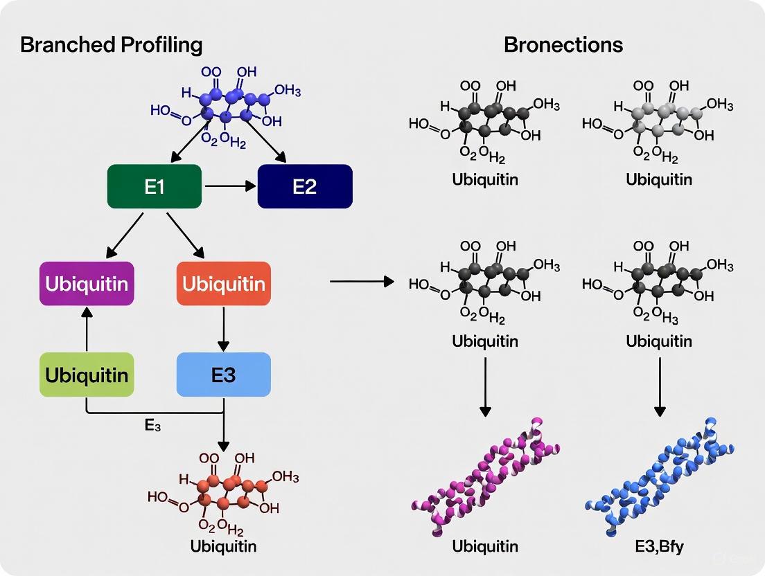
Branched ubiquitin chains are complex molecular structures in which two or more ubiquitin moieties are attached to distinct lysine residues of a single ubiquitin molecule within a polyubiquitin chain. These bifurcated architectures significantly expand the signaling capacity of the ubiquitin system and constitute a substantial fraction of cellular polyubiquitin [3]. The enzymatic assembly of defined branched ubiquitin chains is essential for understanding their distinct signaling functions, identifying interacting proteins, exploring deubiquitinase (DUB) specificity, and investigating processing by molecular machines such as the proteasome and p97 [3] [8]. This protocol focuses on two primary enzymatic strategies—sequential ligation and Ub-capping—that enable researchers to produce well-defined branched ubiquitin architectures for functional studies.
Key Methodological Approaches
Sequential Ligation Strategy
The sequential ligation approach represents a fundamental method for generating branched ubiquitin trimers of defined linkage composition. This strategy relies on systematic ligation of mutant ubiquitin moieties using linkage-specific enzymes [3].
Core Principle: The method begins with a C-terminally truncated (Ub1-72) or blocked proximal ubiquitin (e.g., UbD77 or Ub6his). Mutant distal ubiquitins are then ligated sequentially using specific enzymes for each individual linkage [3].
Protocol for K48-K63 Branched Trimer Assembly:
- Generate K63 dimer: Combine Ub1-72 and UbK48R,K63R using the E2 enzyme complex UBE2N and UBE2V1 (specific for K63-linkage formation).
- Attach K48 branch: Ligate UbK48R,K63R to the proximal Ub1-72 from step 1 using a K48-specific E2 enzyme such as UBE2R1 or UBE2K [3].
- Purify and validate: Isolve the branched trimer product and confirm linkage composition using linkage-specific deubiquitinases (DUBs) and mass spectrometry.
Table 1: Key Reagents for Sequential Ligation
| Reagent | Specification | Function in Protocol |
|---|---|---|
| Proximal Ubiquitin | Ub1-72 (truncated) or UbD77 | Prevents chain extension beyond branched trimer |
| Distal Ubiquitin | UbK48R,K63R mutant | Prevents unwanted secondary linkage formation |
| K63-specific E2 | UBE2N/UBE2V1 complex | Catalyzes formation of K63-linked dimer |
| K48-specific E2 | UBE2R1 or UBE2K | Catalyzes formation of K48-linked branch |
Ub-Capping Strategy
The Ub-capping approach enables assembly of more complex, extended branched ubiquitin structures beyond trimers by incorporating a reversible blocking group that can be enzymatically removed [3] [8].
Core Principle: This method utilizes a "capped" M1-linked ubiquitin dimer where the proximal ubiquitin contains a truncated C-terminus and lysine-to-arginine substitutions. After branch formation, the cap is removed by the M1-specific DUB OTULIN, exposing a native C-terminus for further chain extension [8].
Protocol for K48-K63 Branched Tetramer Assembly:
- Initiate with capped dimer: Start with an M1-linked dimer comprising a wild-type distal ubiquitin and a proximal Ub1-72, K48R, K63R mutant.
- Ligate branch units: Attach Ub moieties via K48 and K63 linkages to the distal ubiquitin of the capped dimer using linkage-specific E2 enzymes.
- Decap with OTULIN: Treat the structure with the M1-specific DUB OTULIN to remove the proximal cap, exposing the native C-terminus of the branch point ubiquitin [3] [8].
- Extend chain: Utilize the exposed C-terminus for further chain elongation using appropriate E2 enzymes to generate extended branched architectures.
Table 2: Key Reagents for Ub-Capping Approach
| Reagent | Specification | Function in Protocol |
|---|---|---|
| Capped M1-Dimer | Ub1-72, K48R, K63R as proximal ubiquitin | Provides defined starting point with one available distal ubiquitin |
| Linkage-specific E2s | e.g., UBE2N/UBE2V1 (K63), UBE2R1 (K48) | Catalyze formation of specific linkage branches |
| Decapping Enzyme | OTULIN (M1-specific DUB) | Removes M1-linked cap to expose native C-terminus |
| Chain Extension E2 | Dependent on desired linkage | Extends the de-capped chain to form tetramers or longer |
Visualizing Enzymatic Assembly Workflows
The following diagrams illustrate the key experimental workflows for both sequential ligation and Ub-capping strategies.
Diagram 1: Workflow comparison of the two main enzymatic assembly strategies.
Diagram 2: Structural organization and nomenclature of branched ubiquitin chains.
Research Reagent Solutions
The following table compiles essential materials and reagents required for implementing the described enzymatic assembly strategies.
Table 3: Essential Research Reagents for Branched Ubiquitin Chain Assembly
| Category | Specific Examples | Function & Application Notes |
|---|---|---|
| Ubiquitin Mutants | Ub1-72, UbK48R,K63R, UbKallR | Serve as chain termination points or prevent unwanted linkages during assembly [3]. |
| E2 Enzymes | UBE2N/UBE2V1 (K63), UBE2R1 (K48), UBE2K (K48) | Catalyze formation of specific ubiquitin linkages. Critical for linkage fidelity [3] [27]. |
| Deubiquitinases | OTULIN (M1-specific), other linkage-specific DUBs | OTULIN removes M1-caps in Ub-capping strategy; DUBs validate linkage composition [3] [8]. |
| Affinity Tags | 6xHis, Strep-tag | Facilitate purification of assembled chains when incorporated into ubiquitin building blocks [17]. |
| Binding Domains | Tandem Ubiquitin Binding Entities (TUBEs) | Aid in purification and detection of assembled branched chains without disrupting native architecture [17]. |
Technical Considerations and Applications
Validation and Quality Control
Rigorous validation of assembled branched ubiquitin chains is essential for experimental reliability. The following approaches are recommended:
- Linkage-specific DUB profiling: Treat assembled chains with panels of linkage-specific deubiquitinases (e.g., OTULIN for M1, etc.) and analyze cleavage patterns by immunoblotting to confirm linkage composition [3] [8].
- Mass spectrometry analysis: Employ advanced MS techniques to verify chain molecular weight and linkage architecture [8] [17].
- Functional validation: Test assembled chains in functional assays such as binding to known receptors (e.g., p97) or processing by the proteasome to confirm biological activity [8].
Applications in Ubiquitin Research
Well-defined branched ubiquitin chains produced through these enzymatic methods enable diverse research applications:
- Identification of branched chain receptors: Immobilized defined branched chains can be used in pulldown assays coupled with mass spectrometry to identify specific cellular binding proteins [8].
- Debranching enzyme discovery: Assembled branched chains serve as substrates to identify and characterize DUBs with debranching activity, such as ATXN3 and MINDY [8].
- Structural studies: Defined chains facilitate structural characterization of branched ubiquitin architectures and their complexes with receptors using X-ray crystallography and NMR [3] [8].
- Cellular function investigation: Engineered binders like K48-K63 branch-specific nanobodies can be used to detect endogenous branched chains during cellular processes like DNA damage response and p97 inhibition [8].
These enzymatic assembly strategies provide robust methodologies for generating the complex branched ubiquitin architectures essential for advancing our understanding of this expanding area of ubiquitin signaling.
The functional characterization of branched ubiquitin chains requires access to well-defined, homogeneous samples of these complex polymers. Branched ubiquitin chains, where a single ubiquitin moiety is modified at two or more distinct lysine residues, significantly expand the signaling capacity of the ubiquitin system beyond their homotypic counterparts [3] [2]. Their structural complexity enables specialized biological functions in processes ranging from cell cycle regulation to protein degradation [11]. However, studying these chains presents substantial technical challenges due to the inability of conventional biological systems to produce them in pure, defined architectures. This application note details two powerful chemical synthesis methods—native chemical ligation (NCL) and thiol-ene coupling (TEC)—that provide researchers with precise tools to overcome these limitations and advance branched ubiquitin chain research.
Table 1: Comparison of Branched Ubiquitin Chain Synthesis Methods
| Method | Key Principle | Key Advantages | Ideal Applications |
|---|---|---|---|
| Native Chemical Ligation | Chemoselective reaction between peptide-thioester and N-terminal cysteine | Incorporation of non-native modifications; full control over chain architecture | Synthesis of chains with specific labels, isotopes, or mutations |
| Thiol-Ene Coupling | Radical-mediated addition of thiol to alkene | Rapid reaction kinetics; oxygen tolerance in protein-protein systems | Activity-based probe development; protein profiling applications |
Native Chemical Ligation for Defined Chain Architectures
Fundamental Principles and Strategic Advantages
Native chemical ligation represents a powerful strategy for the total chemical synthesis of branched ubiquitin chains. This approach involves the chemoselective reaction between a peptide-thioester and another peptide containing an N-terminal cysteine, resulting in a native peptide bond at the ligation site [3]. The key advantage of NCL lies in its ability to generate ubiquitin chains with precisely defined architectures and incorporate diverse non-native modifications that are challenging or impossible to introduce through biological methods. These modifications include specific mutations, isotopic labels, chemical tags, and warheads that facilitate subsequent biochemical and structural studies [3]. For branched ubiquitin synthesis, researchers have employed an innovative 'isoUb' core strategy where residues 46-76 of the distal ubiquitin are linked via a pre-formed isopeptide bond to residues 1-45 of the proximal ubiquitin [3]. This core contains both an N-terminal cysteine and C-terminal hydrazide, enabling efficient native chemical ligation of additional ubiquitin building blocks to construct longer branched polymers.
Protocol: NCL for Branched K11-K48 Ubiquitin Trimer
Table 2: Key Reagents for Native Chemical Ligation
| Reagent | Specifications | Function |
|---|---|---|
| Ubiquitin Building Blocks | Synthesized via SPPS; C-terminal thioester and N-terminal cysteine | Ligation substrates for chain assembly |
| IsoUb Core | Residues 46-76 (distal) linked to 1-45 (proximal) via isopeptide bond | Pre-formed branch point for chain elongation |
| Ligation Buffer | 6 M guanidine HCl, 0.1 M sodium phosphate, 0.1% TCEP, pH 7.0 | Denaturing conditions with reducing agent |
| Thiol Catalysts | 20-50 mM 4-mercaptophenylacetic acid (MPAA) | Acceleration of ligation kinetics |
| Refolding Buffer | 25 mM Tris, 150 mM NaCl, 1 mM DTT, pH 8.0 | Restoration of native ubiquitin fold |
Step 1: Preparation of Ubiquitin Building Blocks
- Synthesize ubiquitin fragments (residues 1-45 and 46-76) using Fmoc-based solid-phase peptide synthesis (SPPS)
- Incorporate necessary mutations at branch point lysines (e.g., K11C, K48C) using appropriate side-chain protection
- Generate C-terminal thioester using established protocols such as N-acyl-benzimidazolinone (Nbz) chemistry
- Purify all building blocks to >95% purity using reverse-phase HPLC and confirm identity by mass spectrometry
Step 2: Ligation Reaction Assembly
- Dissolve isoUb core (50 μM) and ubiquitin-thioester (75 μM) in ligation buffer
- Add MPAA to final concentration of 50 mM and TCEP to 0.1% (w/v)
- Incubate reaction at 37°C with gentle agitation for 12-16 hours
- Monitor reaction progress by analytical HPLC and MALDI-TOF mass spectrometry
Step 3: Purification and Refolding
- Quench reaction by acidification with 0.1% trifluoroacetic acid (TFA)
- Purify ligated product by semi-preparative HPLC using a C18 column with water-acetonitrile gradient
- Lyophilize pure fractions and refold by rapid dilution into refolding buffer at 4°C
- Concentrate using centrifugal filters (10 kDa MWCO) and characterize by LC-MS and circular dichroism
Figure 1: Native chemical ligation workflow for branched ubiquitin chain synthesis
Thiol-Ene Coupling for Rapid and Selective Modification
Mechanism and Applications in Protein Profiling
Thiol-ene coupling is a radical-mediated 'click' reaction between a thiol and an alkene that produces a stable thioether linkage [28] [29]. This reaction proceeds through a radical chain mechanism initiated by homolytic cleavage of the thiol S-H bond (bond dissociation energy ~87 kcal mol⁻¹), followed by anti-Markovnikov addition of the resulting thiyl radical to the alkene, generating a carbon-centered radical that propagates the chain by abstracting a hydrogen from another thiol molecule [29]. In the context of branched ubiquitin research, TEC has been exploited for selective labeling of cysteine residues in deubiquitinating enzymes (DUBs) using alkene-functionalized ubiquitin activity-based probes (ABPs) [28]. The exceptional orthogonality, high yields, and lack of required metal catalysts make TEC particularly suitable for modifying proteins and peptides under physiological conditions without damaging sensitive functional groups [29]. Recent applications demonstrate that TEC can be successfully initiated through multiple methods—UV light, visible wavelengths, and redox activation—providing flexibility for different experimental setups [28].
Protocol: Thiol-Ene Based Activity-Based Profiling of DUBs
Table 3: Quantitative Comparison of Thiol-Ene Activation Methods
| Activation Method | Initiator | Optimal Conditions | Reaction Time | Relative Efficiency |
|---|---|---|---|---|
| UV Light | Irgacure 2959 (0.1 mM) | 365 nm, degassed | 2 minutes | 84% yield (chemical model) |
| Visible Light | Mes-Acr+ (0.5 mM) | Blue LED, aerobic | 2 minutes (high intensity) | 80% conversion (chemical model) |
| Redox Activation | Mn(OAc)₃ (1 mM) | Aerobic, 37°C | 5 minutes - 1 hour | Lower than light-mediated |
Step 1: Preparation of Ubiquitin Alkene Probe
- Express and purify HA-tagged ubiquitin (Ub-1-75) with C-terminal alkene functionality (probe 1) [28]
- Confirm probe identity and purity by SDS-PAGE and mass spectrometry
- Store aliquots at -80°C in 25 mM Tris, 150 mM NaCl, pH 7.5
Step 2: Thiol-Ene Reaction with DUB Active Site Cysteine
- Prepare reaction mixture containing:
- Ubiquitin alkene probe (10 μM)
- Recombinant DUB or cell lysate (1 mg/mL total protein)
- Selected initiator (Irgacure 2959 for UV, Mes-Acr+ for visible light, or Mn(OAc)₃ for redox)
- For UV activation: Degas solution if necessary (improves efficiency but not essential), irradiate at 365 nm for 2 minutes
- For visible light activation: Use high-intensity blue LED (450-495 nm) for 2 minutes without degassing
- For redox activation: Incubate at 37°C for 5 minutes to 1 hour (longer times increase labeling)
Step 3: Analysis of Labeling Efficiency
- Terminate reaction by adding SDS-PAGE loading buffer with 50 mM DTT
- Separate proteins by SDS-PAGE (4-20% gradient gel)
- Transfer to PVDF membrane and detect HA-tagged DUBs using anti-HA antibody (1:5000)
- Quantify band intensity using densitometry software
- Confirm specificity using catalytically inactive DUB mutants (Cys to Ala)
Figure 2: Thiol-ene coupling mechanism and activation pathways for DUB profiling
Research Reagent Solutions for Branched Ubiquitin Synthesis
Table 4: Essential Research Reagents for Branched Ubiquitin Studies
| Reagent Category | Specific Examples | Function in Research | Key Characteristics |
|---|---|---|---|
| Chemical Initiators | Irgacure 2959, Mes-Acr+, Mn(OAc)₃, DPAP | Radical generation for thiol-ene coupling | Varying activation requirements and oxygen sensitivity |
| Ubiquitin Mutants | UbK48R,K63R, Ub1-72, UbKallR | Controlled enzymatic assembly of defined chains | Strategic lysine mutations to direct specific linkages |
| Thiol-Ene Substrates | N-Boc-L-cysteine methyl ester, allyl alcohol | Chemical model system development and optimization | Simple system for reaction parameter screening |
| Enzymatic Tools | UBE2N/UBE2V1 (K63), UBE2R1 (K48), OTULIN (M1-specific DUB) | Linkage-specific chain assembly and processing | Enable controlled synthesis of homotypic chain precursors |
| Noncanonical Amino Acids | Azidohomoalanine (Aha), BOC-lysine, propargyl acrylate | Chemical handle incorporation for orthogonal conjugation | Enable bioorthogonal modification strategies |
Concluding Applications in Branched Ubiquitin Research
The integration of native chemical ligation and thiol-ene coupling provides a comprehensive toolkit for addressing the synthetic challenges in branched ubiquitin research. NCL offers unparalleled precision for generating structurally defined chains with customized modifications, enabling detailed structure-function studies and the development of specific detection reagents [3]. Meanwhile, TEC provides a rapid, efficient platform for activity-based protein profiling that leverages the chemoselectivity of radical reactions to capture enzyme-substrate interactions in complex biological systems [28] [29]. The complementary strengths of these methods—NCL for structural biology applications requiring homogeneous materials and TEC for functional proteomics in complex milieus—create a powerful synergistic relationship. As research continues to uncover the biological significance of branched ubiquitination in cellular regulation and disease pathogenesis, these chemical synthesis methods will remain indispensable for deciphering the complex signaling functions of these remarkable polymeric signals.
Genetic Code Expansion for Incorporation of Noncanonical Amino Acids
Genetic Code Expansion (GCE) technology represents a revolutionary approach in chemical and synthetic biology that enables the site-specific incorporation of noncanonical amino acids (ncAAs) into proteins, thereby expanding the chemical and functional diversity of polypeptides beyond the constraints of the 20 canonical amino acids [30] [31]. This methodology relies on the introduction of an engineered orthogonal aminoacyl-tRNA synthetase (aaRS)/tRNA pair into a host organism, along with a repurposed codon (typically the amber stop codon, UAG), to direct the incorporation of a desired ncAA during translation [31]. The orthogonality of this system—meaning the engineered tRNA/RS pair does not cross-react with the host's natural amino acids, tRNAs, or RSs—is essential for maintaining the fidelity of protein synthesis while expanding the genetic code [31].
In the specialized context of profiling branched ubiquitin chains, GCE offers unique and powerful capabilities for deciphering the complex "ubiquitin code." Branched ubiquitin chains, which contain at least one ubiquitin moiety modified concurrently on more than one lysine residue, constitute 10-20% of cellular polyubiquitin and function as potent degradation signals that ensure the timely removal of regulatory and misfolded proteins [4] [13] [32]. The molecular machinery responsible for assembling, recognizing, and disassembling these branched chains—including E3 ligases, proteasomal receptors, and deubiquitinases (DUBs)—represents a fertile area of research with significant implications for understanding cell cycle regulation, proteotoxic stress responses, and developing targeted therapeutic strategies [4] [13]. GCE provides the methodological foundation to probe these processes with unprecedented precision by enabling the site-specific installation of photo-crosslinkers for capturing transient enzyme-substrate interactions, biophysical probes for monitoring conformational dynamics, and chemically-defined ubiquitin chain architectures for functional studies.
Key Platform: A Robust Biosynthetic Pathway for Aromatic ncAAs
A significant advancement in GCE technology addresses the "Achilles' heel" of ncAA supply: the high cost and poor membrane permeability of many ncAAs that hinder large-scale protein production [33]. A promising solution couples the in situ biosynthesis of aromatic ncAAs directly with GCE within E. coli, creating a semi-autonomous production platform.
Pathway Design and Validation
The developed pathway utilizes aryl aldehydes as abundant, low-cost starting materials and consists of three enzymatic steps [33]:
- Aldol Reaction: Catalyzed by L-threonine aldolase (LTA) from Pseudomonas putida (PpLTA), condensing glycine with an aryl aldehyde to produce aryl serines.
- Deamination: Catalyzed by L-threonine deaminase (LTD) from Rahnella pickettii (RpTD), converting aryl serines to aryl pyruvates.
- Transamination: Catalyzed by the endogenous aromatic amino acid aminotransferase (TyrB) in E. coli, using L-glutamate as an amino donor to produce the final ncAAs.
This pathway demonstrated remarkable versatility, successfully synthesizing 40 different aromatic ncAAs from their corresponding aldehydes. From this library, 19 ncAAs were site-specifically incorporated into superfolder GFP using three different orthogonal translation systems in E. coli, confirming the platform's compatibility with genetic code expansion [33]. The utility of this integrated system was further demonstrated through the production of macrocyclic peptides and antibody fragments containing ncAAs [33].
Table 1: Key Research Reagent Solutions for Integrated ncAA Biosynthesis and GCE
| Research Reagent | Function in Protocol | Key Characteristics |
|---|---|---|
| PpLTA-RpTD Engineered E. coli | Host for coupled ncAA biosynthesis and protein expression. | Expresses L-threonine aldolase and deaminase; enables in vivo ncAA production from aryl aldehydes [33]. |
| Aryl Aldehyde Precursors | Starting material for ncAA biosynthesis. | Abundant, commercially available, low-cost; diverse functional groups enable a wide ncAA scope (40 demonstrated) [33]. |
| Orthogonal Translation System (OTS) | Incorporates biosynthesized ncAA into protein. | Requires aaRS/tRNA pair orthogonal to host and active with the target ncAA (3 systems demonstrated) [33]. |
| Lyophilized Whole-Cell Catalyst | In vitro ncAA production format. | Prepared from PpLTA-RpTD E. coli; converts 1 mM aldehyde to ~0.96 mM ncAA (e.g., pIF) within 6 hours [33]. |
Experimental Protocol: Coupled Biosynthesis and ncAA Incorporation
Objective: To produce a target protein containing a site-specifically incorporated aromatic ncAA using the integrated biosynthesis and GCE platform in E. coli.
Materials:
- E. coli BL21(DE3) strain harboring pACYCDuet-1-PpLTA-RpTD plasmid [33]
- Expression vector for target protein, with an amber mutation at the desired position, and the corresponding orthogonal aaRS/tRNA pair
- Aryl aldehyde precursor (e.g., para-iodobenzaldehyde)
- Luria-Bertani (LB) broth and agar plates with appropriate antibiotics
- Isopropyl β-d-1-thiogalactopyranoside (IPTG)
Procedure:
- Transformation: Co-transform the chemically competent PpLTA-RpTD E. coli strain with the target protein expression vector and the orthogonal aaRS/tRNA plasmid. Plate on LB agar with appropriate antibiotics and incubate overnight at 37°C.
- Culture Inoculation: Pick a single colony to inoculate a small LB starter culture with antibiotics. Grow overnight at 37°C with shaking.
- Main Culture and Induction: Dilute the starter culture 1:100 into fresh, pre-warmed LB medium with antibiotics. Grow at 37°C with shaking until the OD600 reaches 0.6-0.8.
- Supplementation and Induction:
- Supplement the culture with a filter-sterilized solution of the aryl aldehyde precursor (e.g., 1-2 mM final concentration from a stock solution in DMSO or ethanol).
- Simultaneously, add IPTG to a final concentration of 0.1-1.0 mM to induce expression of the target protein and the orthogonal aaRS/tRNA pair.
- Incubate the induced culture for 16-24 hours at a lower temperature (e.g., 18-25°C) to facilitate proper protein folding.
- Harvest and Purification: Pellet the cells by centrifugation. Resuspend the cell pellet in an appropriate lysis buffer and lyse by sonication or homogenization. Clarify the lysate by centrifugation and purify the target protein using a suitable chromatographic method (e.g., affinity chromatography).
- Verification: Confirm the identity and incorporation fidelity of the ncAA-containing protein using mass spectrometry.
Application in Branched Ubiquitin Chain Research: Targeted Deubiquitinase Engineering
GCE methodologies empower the development of highly specific molecular tools to dissect the function of branched ubiquitin chains. A prime example is the creation of linkage-selective engineered deubiquitinases (enDUBs) to manipulate the ubiquitin code on specific proteins in live cells [10].
enDUB Design and Functional Validation
This application involves fusing the catalytic domain of a DUB with known linkage specificity to a protein-targeting module, such as a GFP-targeted nanobody [10]. The resulting enDUB can be recruited to a specific substrate, where it selectively hydrolyzes a particular polyubiquitin linkage, allowing researchers to infer that linkage's functional role.
Key enDUBs developed and their specificities include [10]:
- OTUD1 (O1) enDUB: Selective for K63-linked chains.
- OTUD4 (O4) enDUB: Selective for K48-linked chains.
- Cezanne (Cz) enDUB: Selective for K11-linked chains.
- TRABID (Tr) enDUB: Selective for K29/K33-linked chains.
- USP21 (U21) enDUB: A non-specific control.
When applied to the ion channel KCNQ1-YFP, these enDUBs revealed distinct biological functions for different ubiquitin linkages. Mass spectrometry had shown that KCNQ1 is predominantly modified with K48-linked (72%) and K63-linked (24%) chains, with minor contributions from other linkages [10]. The enDUB experiments demonstrated that these linkages control different aspects of the channel's lifecycle: K48 linkages were necessary for forward trafficking, while K63 linkages enhanced endocytosis and reduced recycling [10].
Table 2: Quantitative Analysis of enDUB Effects on KCNQ1 Ion Channel Trafficking
| Polyubiquitin Linkage Targeted | Corresponding enDUB | Observed Effect on KCNQ1 | Proposed Biological Role of Linkage |
|---|---|---|---|
| K48 | OTUD4 (O4) | Reduced surface expression | Necessary for forward trafficking from the ER [10] |
| K63 | OTUD1 (O1) | Increased surface expression | Enhances endocytosis and reduces recycling [10] |
| K11 | Cezanne (Cz) | Increased surface expression | Promotes ER retention/degradation [10] |
| K29/K33 | TRABID (Tr) | Increased surface expression | Promotes ER retention/degradation [10] |
| Non-specific | USP21 (U21) | Increased surface expression | Confirms ubiquitination generally negatively regulates surface density [10] |
Experimental Protocol: Probing Linkage-Specific Function with enDUBs
Objective: To determine the functional role of specific polyubiquitin linkages on a GFP-tagged protein of interest (POI-GFP) in live cells using linkage-selective enDUBs.
Materials:
- Mammalian expression vectors for POI-GFP and the suite of linkage-selective enDUBs (e.g., O1, O4, Cz, Tr, U21) [10]
- Appropriate mammalian cell line (e.g., HEK293)
- Transfection reagent
- Flow cytometry buffers or equipment for confocal microscopy
Procedure:
- Cell Seeding and Transfection: Seed cells into multiple wells of a culture plate. Transfert separate wells with:
- a) POI-GFP alone (control).
- b) POI-GFP + each enDUB construct (experimental).
- c) POI-GFP + nanobody-only construct (negative control).
- Incubation: Allow 24-48 hours for gene expression and enDUB activity.
- Phenotypic Analysis:
- For Surface Expression (Flow Cytometry): For a plasma membrane protein, harvest cells and analyze by flow cytometry. Use GFP fluorescence to gate on transfected cells and a surface marker (e.g., an extracellular tag like BBS stained with BTX-647) to measure surface density [10]. Compare the surface fluorescence intensity of enDUB-expressing cells to controls.
- For Subcellular Localization (Confocal Microscopy): Fix cells and perform immunofluorescence staining for organelle markers (ER, Golgi, endosomes, lysosomes). Use confocal microscopy to analyze co-localization of POI-GFP with each organelle marker across the different enDUB conditions [10].
- Biochemical Validation (Immunoprecipitation & Immunoblotting): Lyse a portion of the transfected cells. Immunoprecipitate POI-GFP and probe with anti-ubiquitin antibodies to confirm the reduction in global ubiquitination. Stripping and re-probing the membrane with linkage-specific ubiquitin antibodies can further verify the selective removal of the targeted linkage [10].
- Data Interpretation: Correlate the specific ubiquitin linkage removed by each enDUB with the observed phenotypic changes (e.g., increased surface expression upon K63 removal suggests that linkage normally promotes endocytosis).
Visualizing Experimental Workflows
The following diagrams illustrate the core logical and experimental relationships in the methodologies discussed.
Integrated ncAA Biosynthesis and Incorporation Workflow
Branched Ubiquitin Chain Analysis with enDUBs
The Scientist's Toolkit: Essential Research Reagents
Table 3: Key Research Reagent Solutions for Branched Ubiquitin Chain Profiling
| Research Reagent / Tool | Function in Ubiquitin Research | Application Context |
|---|---|---|
| Linkage-Selective enDUBs | To hydrolyze a specific polyubiquitin linkage on a target protein in live cells. | Deciphering the functional role of a single linkage type on a substrate (e.g., K63 vs K48 on KCNQ1 trafficking) [10]. |
| Chain-Selective TUBEs (Tandem Ubiquitin Binding Entities) | High-affinity affinity matrices to capture and enrich linkage-specific polyubiquitinated proteins from cell lysates. | High-throughput screening to investigate context-dependent ubiquitination (e.g., L18-MDP-induced K63 vs PROTAC-induced K48 ubiquitination of RIPK2) [9]. |
| UCH37/RPN13 Complex | Proteasome-associated deubiquitinase complex with unique debranching activity, preferentially cleaving K48 linkages in branched chains. | Facilitating proteasomal clearance of substrates modified with branched ubiquitin chains (e.g., K6/K48, K11/K48); studied in vitro and in cells [32]. |
| Engineered E3 Ligases (e.g., Rsp5-HECTGML) | Generate specific ubiquitin chain linkages (e.g., K48-linked chains) for biochemical reconstitution experiments. | Producing defined ubiquitin chain architectures, including branched chains, for structural and mechanistic studies (e.g., proteasome binding via cryo-EM) [4]. |
Ubiquitylation is a fundamental post-translational modification that controls diverse cellular processes through the assembly of complex ubiquitin architectures. Among these, branched ubiquitin chains represent a sophisticated signaling mechanism where at least one ubiquitin moiety is simultaneously modified at two or more distinct acceptor sites, creating a bifurcated structure that dramatically expands the ubiquitin code's informational capacity [3] [2]. These complex polymers differ from homotypic chains (uniform linkage throughout) and mixed chains (multiple linkages but no branching points) through their unique topological arrangements [2]. The biological functions of branched ubiquitin chains are rapidly emerging, with research implicating them in essential processes including proteasomal degradation, NF-κB signaling, cell cycle progression, and apoptosis regulation [3] [2].
The profiling of branched ubiquitin chains presents substantial technical challenges due to their structural complexity and the limitations of conventional proteomic approaches. Middle-down mass spectrometry approaches, which analyze larger peptide fragments than typical bottom-up methods, offer a promising solution for preserving and identifying branching information. This application note details specialized methodologies and protocols for the comprehensive analysis of branched ubiquitin chains, with emphasis on middle-down mass spectrometry techniques that enable researchers to decode these complex post-translational modifications.
Branched Ubiquitin Chain Architectures and Functions
Structural Classification and Nomenclature
Branched ubiquitin chains exhibit remarkable diversity in their chemical linkages and three-dimensional architectures. The proposed standardized nomenclature adapts the system originally proposed by Fushman and colleagues to accurately describe these complex structures [3]. Theoretically, 28 different trimeric branched ubiquitin chain types containing two different linkages can be formed, though only a subset has been identified and functionally characterized in cellular contexts [3].
Table 1: Experimentally Confirmed Branched Ubiquitin Chain Types and Their Functions
| Linkage Type | Key E3 Ligases | Cellular Functions | Identification Methods |
|---|---|---|---|
| K11-K48 | APC/C (UBE2C/UBE2S), UBR5 | Cell cycle progression, proteasomal degradation [2] | Middle-down MS, linkage-specific antibodies [2] |
| K29-K48 | Ufd4/Ufd2 collaboration | Proteasomal degradation [2] | Enzymatic profiling, MS analysis [2] |
| K48-K63 | TRAF6/HUWE1, ITCH/UBR5 | NF-κB signaling, p97 processing, apoptosis [3] [2] | Chemical biology tools, DUB profiling [3] |
| K6-K48 | Parkin, NleL | Protein quality control, bacterial infection [2] | Genetic code expansion, MS detection [2] |
Biosynthesis Mechanisms
The synthesis of branched ubiquitin chains involves specialized enzymatic mechanisms that often deviate from standard ubiquitination pathways. Two predominant biosynthesis mechanisms have been identified:
Collaborative E3 Mechanisms: Pairs of E3 ligases with distinct linkage specificities work sequentially to build branched architectures. For example, in the synthesis of branched K48-K63 chains during NF-κB signaling, TRAF6 first assembles K63-linked chains which are then recognized by HUWE1 through its UIM and UBA domains, enabling the addition of K48 linkages to create branch points [2]. Similarly, Ufd2 recognizes K29-linked chains assembled by Ufd4 through specific loops in its N-terminal domain before adding K48 linkages to form branched K29-K48 chains [2].
Single E3 Mechanisms: Certain E3 ligases possess intrinsic capability to synthesize branched chains, either by recruiting multiple E2s with different linkage specificities or through inherent catalytic flexibility. The APC/C, a multisubunit RING E3, coordinates with UBE2C (which builds initial chains with mixed linkages) and UBE2S (which specifically extends K11 linkages) to form branched K11-K48 chains during mitosis [2]. Some HECT E3s, including WWP1 and UBE3C, can assemble branched chains with single E2s, potentially through non-covalent ubiquitin-binding sites that facilitate branching [2].
Experimental Models and Research Reagents
Essential Reagents for Branched Ubiquitin Research
Table 2: Key Research Reagent Solutions for Branched Ubiquitin Chain Studies
| Reagent Category | Specific Examples | Function and Application |
|---|---|---|
| Recombinant Ubiquitin Variants | Ub1-72, UbK48R,K63R, UbKallR mutants | Enable controlled enzymatic assembly of defined branched chains by blocking specific linkage sites [3] |
| E2 Enzymes | UBE2N/UBE2V1 (K63-specific), UBE2R1/UBE2K (K48-specific) | Catalyze formation of specific linkage types in branched chain assembly [3] |
| E3 Ligases | TRAF6, HUWE1, UBR5, APC/C, Parkin | Synthesize branched chains through collaborative or single-E3 mechanisms [2] |
| Deubiquitinases (DUBs) | OTULIN (M1-specific), Yuh1 | Trim specific linkages to enable chain editing or expose native C-termini for further extension [3] |
| Chemical Biology Tools | IsoUb cores, photolabile NVOC groups, noncanonical amino acids | Facilitate chemical synthesis and controlled assembly of branched architectures [3] |
| Mass Spectrometry Standards | Synthetic branched ubiquitin standards, isotopically labeled ubiquitin | Enable method development and quantification in proteomic experiments |
Branch Chain Assembly Methodologies
Enzymatic Assembly with Capped Proximal Ubiquitin
This established method utilizes C-terminally blocked ubiquitin mutants (Ub1-72 or UbD77) to control the stepwise assembly of branched trimers:
- Begin with a proximal ubiquitin containing a truncated or blocked C-terminus
- Ligate the first distal ubiquitin using linkage-specific enzymes (e.g., UBE2N/UBE2V1 for K63 linkages)
- Attach the second distal ubiquitin using different linkage-specific enzymes (e.g., UBE2R1 or UBE2K for K48 linkages) to the same proximal ubiquitin, creating a branch point [3]
A limitation of this approach is the inability to extend chains beyond the trimer due to the modified proximal ubiquitin. To overcome this, our lab adapted a Ub-capping approach using the yeast DUB Yuh1 to trim the C-terminus of a D77-blocked ubiquitin, thereby exposing the native C-terminus for further chain extension [3].
Chemical Synthesis Approaches
Chemical synthesis provides unparalleled flexibility for generating defined branched ubiquitin architectures:
Full Chemical Synthesis: Utilizes solid phase peptide synthesis (SPPS) or native chemical ligation (NCL) of SPPS-generated fragments to incorporate diverse modifications including mutations, tags, and warheads at specific positions [3].
IsoUb Core Strategy: Employs a synthesized core consisting of residues 46-76 of the distal ubiquitin linked via a pre-formed isopeptide bond to residues 1-45 of the proximal ubiquitin, containing an N-terminal cysteine and C-terminal hydrazide for efficient NCL of additional ubiquitin building blocks [3].
Genetic Code Expansion: Incorporates noncanonical amino acids through amber stop codon suppression in E. coli, enabling precise chemical functionalization for controlled branch chain assembly [3].
Mass Spectrometry-Based Profiling Workflows
Sample Preparation for Middle-Down Approaches
Middle-down proteomics analyzes larger peptide fragments (typically 3-9 kDa) than conventional bottom-up approaches, preserving valuable information about coexisting post-translational modifications within a single molecule. For ubiquitin clipping approaches, specific enzymatic cleavages are employed to generate ideal-sized fragments for middle-down analysis:
- Ubiquitin Enrichment: Immunopurification using ubiquitin affinity matrices or tandem ubiquitin-binding entities
- Controlled Proteolysis: Limited proteolysis with specific proteases (e.g., GluC, AspN) that generate larger ubiquitin-containing fragments
- Peptide Separation: High-resolution liquid chromatography separation optimized for larger peptides
- Mass Spectrometry Analysis: High-resolution tandem MS with electron-transfer dissociation (ETD) or hybrid fragmentation methods
Data Acquisition and Analysis
Mass spectrometry data visualization and interpretation play crucial roles in analyzing complex proteomes [34]. For branched ubiquitin chain analysis, several specialized spectral interpretation approaches are required:
- MS2 Spectrum Analysis: Interpretation of fragmentation spectra to identify diagnostic ions indicating branching points
- Chromatographic Alignment: Correlation of precursor and fragment ion elution profiles across retention time dimensions
- Ion Mobility Separation: Additional separation of ions based on shape and charge for improved specificity
- Spectral Matching: Comparison of experimental spectra with theoretical fragmentation patterns of branched ubiquitin structures
Workflow for branched ubiquitin chain profiling using middle-down MS approaches.
Data Interpretation and Visualization
Mass spectrometry data visualization bridges the gap between raw data and biological interpretation [34]. For ubiquitin clipping and middle-down approaches, several specialized visualization methods facilitate data interpretation:
MS2 Spectrum Visualization: Vertical bar graphs displaying mass-to-charge ratio (m/z) on the x-axis and relative abundance on the y-axis, with color-coded fragmentation ions (b-ions and y-ions) indicating peptide sequence and modification sites [34] [35].
Mirrored Spectra Plots: Comparison of experimental and predicted fragmentation spectra to validate peptide identities and fragmentation patterns [34].
Elution Profile Analysis: Visualization of peptide precursor and fragment ion elution across retention time, with intensity profiles indicating relative abundance [34].
Ion Mobility Heatmaps: Two-dimensional representations combining retention time and ion mobility separation, with color intensity indicating ion abundance [34].
Ubiquitin clipping workflow for middle-down MS analysis.
Technical Considerations and Troubleshooting
Optimization of Mass Spectrometry Parameters
Successful middle-down analysis of branched ubiquitin chains requires careful optimization of mass spectrometry parameters:
Fragmentation Method Selection: Electron-transfer dissociation (ETD) and electron-capture dissociation (ECD) often outperform collision-based methods for preserving labile post-translational modifications and generating more uniform fragmentation across larger peptides.
Liquid Chromatography Conditions: Optimized gradients for separation of larger peptides (typically 3-9 kDa) with appropriate mobile phases and column chemistry.
Instrument Calibration: Regular mass calibration and performance validation using appropriate standards.
Data-Dependent Acquisition Settings: Inclusion of charge state and intensity thresholds for precursor selection to prioritize branched ubiquitin-containing peptides.
Common Technical Challenges and Solutions
Table 3: Troubleshooting Guide for Branched Ubiquitin Chain Analysis
| Challenge | Potential Cause | Solution |
|---|---|---|
| Low coverage of branch points | Inefficient enrichment or cleavage | Optimize ubiquitin enrichment protocols; test multiple proteases |
| Ambiguous linkage assignment | Insufficient diagnostic fragments | Combine multiple fragmentation methods; implement ion mobility separation |
| Low signal intensity | Poor ionization of larger peptides | Implement chemical derivatization to improve ionization efficiency |
| Complex spectral interpretation | Multiple coexisting modifications | Develop custom spectral interpretation algorithms; use synthetic standards |
The field of branched ubiquitin chain research is rapidly evolving with emerging technologies enabling increasingly sophisticated analyses. Middle-down mass spectrometry approaches, particularly when combined with ubiquitin clipping strategies, provide powerful tools for deciphering the complex biology of these polymeric signals. Future methodological developments will likely focus on improving sensitivity for low-abundance branched species, enhancing throughput for comprehensive profiling, and integrating functional assays with structural characterization.
The continued refinement of mass spectrometry-based profiling methods for branched ubiquitin chains will undoubtedly uncover new biological functions and regulatory mechanisms mediated by these complex structures. Furthermore, the integration of these approaches with chemical biology tools for synthesizing defined branched architectures creates a virtuous cycle of methodological advancement and biological discovery. As these technologies mature, they will provide unprecedented insights into the role of branched ubiquitin signaling in health and disease, potentially revealing new therapeutic targets for pharmacological intervention.
The study of complex post-translational modifications, particularly branched ubiquitin chains, represents a frontier in molecular biology that demands increasingly sophisticated detection tools. Branched ubiquitin chains, in which a single ubiquitin moiety is modified at two or more distinct sites, significantly expand the signaling capacity of the ubiquitin system beyond their homotypic counterparts [2]. These complex architectures control diverse cellular processes including protein degradation, cell cycle progression, and NF-κB signaling, yet their analysis has been hampered by technical challenges [3]. The emergence of specialized detection reagents—particularly bispecific antibodies and engineered nanobodies—has begun to illuminate this enigmatic area of research by enabling precise identification, quantification, and functional characterization of these complex ubiquitin architectures.
Unlike conventional antibodies, these engineered reagents offer dual targeting capabilities and enhanced physical properties that make them uniquely suited for deciphering complex ubiquitin signaling. Bispecific antibodies (BsAbs) provide simultaneous engagement with two distinct epitopes, while nanobodies (single-domain antibodies derived from camelids) offer superior tissue penetration, high stability, and ease of production [36] [37]. This application note details methodologies leveraging these specialized reagents to advance branched ubiquitin chain research, providing structured protocols, analytical frameworks, and practical implementation guidance for researchers in both academic and drug discovery settings.
Technical Comparison: Bispecific Antibodies versus Engineered Nanobodies
Table 1: Comparative Properties of Specialized Detection Reagents
| Property | Bispecific Antibodies | Engineered Nanobodies |
|---|---|---|
| Molecular Size | ~150-200 kDa (IgG-like); ~55 kDa (non-IgG) | ~15 kDa |
| Binding Valency | Dual target engagement | Single domain, can be multimerized |
| Production System | Mammalian cells (complex) | Bacterial (simple, high yield) |
| Tissue Penetration | Moderate | Superior due to small size |
| Stability | Moderate; susceptible to degradation | High thermal and chemical stability |
| Typical Affinity | nM range | nM range (e.g., 2.88-101.2 nM for ISG15 binders) [36] |
| Structural Complexity | High (multiple chains); requires engineered pairing | Low (single domain) |
| Half-Life | Long (Fc-mediated) | Short (renal clearance) |
| Detection Applications | Immunoassays, imaging, therapy | Immunoprecipitation, blotting, intracellular inhibition |
| Epitope Recognition | Can target two discrete epitopes | Can access cryptic epitopes |
Table 2: Ubiquitin Chain Linkages and Research Challenges
| Chain Type | Structure | Known Functions | Detection Challenges |
|---|---|---|---|
| Homotypic | Single linkage type | K48: degradation; K63: signaling | Well-established methods |
| Branched | Multiple linkages on single ubiquitin | K11-K48: cell cycle; K48-K63: NF-κB signaling [2] | Complex architecture requires specialized tools |
| Mixed | Alternating linkages | Less characterized | Discrimination from branched difficult |
| Theoretically Possible Branched | 28 trimeric types | Mostly uncharacterized | Lack of specific detection reagents |
Application Note 1: Nanobodies for ISG15 Pathway Analysis
Background and Principle
Interferon Stimulated Gene 15 (ISG15) functions as both an intracellular ubiquitin-like modifier and an extracellular cytokine with roles in cancer, inflammatory disorders, and viral infection [36] [37]. The ISG15 pathway presents particular challenges for study due to the low abundance of ISGylated substrates and limited availability of high-quality reagents. Recently developed ISG15-specific nanobodies (VHHISG15-A and VHHISG15-B) provide powerful tools for probing this pathway, with each recognizing distinct epitopes on ISG15's C- and N-terminal domains, respectively [36].
Research Reagent Solutions
Table 3: Key Reagents for ISG15 Pathway Analysis
| Reagent | Specifications | Application | Performance Characteristics |
|---|---|---|---|
| VHHISG15-A | C-terminal domain binder; nanomolar affinity (KD = 101.2 nM) | Immunoblotting, immunoprecipitation, deISGylation inhibition | Recognizes unconjugated ISG15 under denaturing conditions |
| VHHISG15-B | N-terminal domain binder; higher affinity (KD = 2.88 nM) | Immunoprecipitation, proteomic identification | Minimal background contamination in IP-MS |
| ISG15 Conjugates | Native or recombinant ISG15 substrates | Positive controls | Verify detection system performance |
| USP16 Enzyme | DeISGylating enzyme | Functional assays | Measure VHHISG15-A inhibition capacity |
Protocol: Immunoprecipitation and Proteomic Identification of ISGylated Substrates
Principle: Utilize ISG15-specific nanobodies to isolate ISG15 conjugates from cell lysates for subsequent mass spectrometry identification, enabling comprehensive mapping of the ISG15 substrate repertoire.
Materials:
- VHHISG15-A or VHHISG15-B nanobodies (immobilized)
- Lysis buffer (50 mM Tris-HCl, pH 7.5, 150 mM NaCl, 1% NP-40, protease inhibitors)
- Wash buffer (50 mM Tris-HCl, pH 7.5, 150 mM NaCl, 0.1% NP-40)
- Elution buffer (0.1 M glycine, pH 2.5, or 1× SDS-PAGE loading buffer)
- Mammalian cell culture with and without interferon stimulation
- Mass spectrometry equipment and reagents
Procedure:
- Nanobody Immobilization: Covalently couple 1-2 mg of purified VHHISG15-A or VHHISG15-B to agarose beads using appropriate chemistry according to manufacturer's instructions.
- Cell Lysis and Preparation:
- Culture HEK293 or other relevant cell lines with 1000 U/mL interferon-α for 24 hours to induce ISG15 expression
- Harvest cells and lyse in 1 mL lysis buffer per 10^7 cells
- Clarify lysate by centrifugation at 16,000 × g for 15 minutes at 4°C
- Immunoprecipitation:
- Incubate 1-2 mg of cell lysate with 50 μL of nanobody-conjugated beads for 2 hours at 4°C with gentle rotation
- Wash beads 3× with 1 mL wash buffer (5 minutes per wash)
- Elution and Processing:
- For mass spectrometry: Elute with 2× 50 μL elution buffer, neutralize with Tris base
- For immunoblotting: Elute directly in 1× SDS-PAGE loading buffer by heating at 95°C for 5 minutes
- Proteomic Analysis:
- Process eluted proteins for LC-MS/MS analysis using standard proteomic protocols
- Identify ISG15-modified peptides using appropriate database search algorithms
Technical Notes:
- VHHISG15-B demonstrates superior performance for proteomic identification due to higher affinity and minimal background [36]
- Include negative controls using pre-immune nanobodies or beads alone
- For specific detection of conjugated ISG15, use denaturing conditions during lysis
Protocol: DeISGylation Inhibition Assay
Principle: Assess the functional impact of VHHISG15-A on USP16-mediated ISG15 cleavage through in vitro deISGylation assays.
Materials:
- Recombinant USP16 enzyme
- ISG15-AMC (or other tagged ISG15 substrate)
- VHHISG15-A and VHHISG15-B as comparators
- Reaction buffer (50 mM Tris-HCl, pH 7.5, 50 mM NaCl, 1 mM DTT)
- Fluorescence plate reader or other detection system
Procedure:
- Prepare reaction mixtures containing:
- 100 nM USP16
- 1 μM ISG15-AMC substrate
- VHHISG15-A or VHHISG15-B (0-10 μM concentration range)
- Reaction buffer to 50 μL final volume
- Incubate at 37°C for 30-60 minutes
- Measure fluorescence (excitation 360 nm, emission 460 nm) at time intervals
- Calculate deISGylation rates by comparing fluorescence increase in presence and absence of nanobodies
Expected Outcomes: VHHISG15-A, but not VHHISG15-B, inhibits USP16-mediated ISG15 processing due to steric hindrance at the ISG15-binding interface [36].
Figure 1: ISG15 Nanobody Application Workflow. VHHISG15-A and VHHISG15-B recognize distinct domains on ISG15 and enable different applications including immunoprecipitation-mass spectrometry and deISGylation inhibition assays.
Application Note 2: Chain-Specific Nanobodies for Ubiquitin Proteome Profiling
Background and Principle
The ubiquitin proteome encompasses tremendous complexity, with chain linkage type determining biological function. K48-linked chains typically target proteins for proteasomal degradation, while K63-linked chains regulate signaling pathways [38]. Branched chains containing multiple linkage types further complicate this landscape. Chain-specific nanobodies enable isolation and identification of ubiquitin chains with defined linkages, providing critical insights into their specialized functions.
Protocol: Isolation of K48 and K63 Ubiquitin Chains Using Linkage-Specific Nanobodies
Principle: Employ linkage-specific nanobodies to enrich K48- or K63-linked ubiquitin chains from complex cell lysates, followed by mass spectrometry identification of modified proteins.
Materials:
- K48- and K63-specific nanobodies (immobilized)
- Lysis buffer (6 M guanidine-HCl, 100 mM Na2HPO4/NaH2PO4, 10 mM Tris-HCl, pH 8.0, 5 mM imidazole)
- Wash buffer 1 (8 M urea, 100 mM Na2HPO4/NaH2PO4, 10 mM Tris-HCl, pH 8.0)
- Wash buffer 2 (8 M urea, 100 mM Na2HPO4/NaH2PO4, 10 mM Tris-HCl, pH 6.3)
- Elution buffer (200 mM glycine, pH 2.5)
- Ni-NTA agarose for His-tagged protein purification
- Mammalian cell culture under experimental conditions
Procedure:
- Cell Lysis Under Denaturing Conditions:
- Harvest cells and lyse in denaturing lysis buffer
- Sonicate to reduce viscosity and ensure complete lysis
- Centrifuge at 16,000 × g for 15 minutes to remove insoluble material
- Ubiquitin Enrichment:
- Incubate lysate with Ni-NTA agarose for 1 hour at room temperature (if using His-tagged ubiquitin)
- Wash sequentially with wash buffers 1 and 2
- Elute with elution buffer and neutralize with Tris base
- Linkage-Specific Isolation:
- Dialyze eluate into non-denaturing buffer (50 mM Tris, pH 7.5, 150 mM NaCl)
- Incubate with K48- or K63-specific nanobody beads for 2 hours at 4°C
- Wash 3× with non-denaturing wash buffer
- Elute with SDS-PAGE loading buffer or specific elution conditions
- Proteomic Analysis:
- Separate proteins by SDS-PAGE and excise gel bands
- Perform in-gel tryptic digestion
- Analyze by LC-MS/MS with focus on GG-remnant peptides for ubiquitination sites
Technical Notes:
- Denaturing conditions preserve the ubiquitin-modified proteome but may affect nanobody binding
- Include controls with non-specific nanobodies to identify background binders
- Optimize wash stringency to balance specificity and yield
Application Note 3: Bispecific Antibodies for Bridged Detection Systems
Background and Principle
Bispecific antibodies (BsAbs) represent a novel class of therapeutics and detection reagents engineered to recognize two distinct antigens or epitopes simultaneously [39]. In ubiquitin research, BsAbs can be designed to bridge detection systems, simultaneously capturing modified proteins and recruiting detection reagents. While most approved BsAbs target cancers, their structural and functional principles can be adapted for research applications.
Research Reagent Solutions
Table 4: Bispecific Antibody Formats and Research Applications
| Format | Structure | Advantages | Research Applications |
|---|---|---|---|
| IgG-like BsAbs | Fc-containing, bivalent | Longer half-life, Fc-mediated effector functions | Immunoassays, detection platforms |
| Bispecific T-cell Engagers (BiTEs) | Tandem scFvs, no Fc | Small size, efficient T-cell recruitment | Therapeutic applications (e.g., blinatumomab) [39] |
| Dual ICP-blocking BsAbs | Full IgG or engineered | Simultaneous checkpoint inhibition | Immune regulation studies |
| Biparatopic BsAbs | Two epitopes on same target | Enhanced specificity and avidity | Selective target engagement |
Protocol: Development of BsAb-Based Detection Systems for Ubiquitin Chain Analysis
Principle: Design bispecific antibodies that simultaneously recognize a specific ubiquitin chain linkage and a tag or reporter system to enable sensitive detection of complex ubiquitin architectures.
Materials:
- Hybridoma or phage display systems for BsAb generation
- Recombinant ubiquitin chains of defined linkages
- Target antigens for co-detection studies
- Assay platforms (ELISA, Western blot, immunofluorescence)
Procedure:
- BsAb Selection and Production:
- Identify appropriate binding domains for target ubiquitin linkage and detection tag
- Employ knob-into-holes technology [39] or CrossMAb technology for correct heavy-light chain pairing
- Express in mammalian systems (e.g., HEK293) for proper folding and glycosylation
- Validation of Dual Binding:
- Perform ELISA with immobilized primary target (ubiquitin chain)
- Confirm secondary target binding (tag or reporter)
- Assess simultaneous binding using surface plasmon resonance or similar techniques
- Application in Ubiquitin Chain Detection:
- Develop sandwich ELISA with BsAb as capture reagent
- Implement in proximity ligation assays for visualizing ubiquitinated proteins
- Adapt for flow cytometry or imaging of ubiquitin modifications in fixed cells
Technical Notes:
- BsAbs may exhibit nonlinear pharmacokinetics at lower doses due to target-mediated drug disposition [40]
- Consider immunogenicity risks associated with artificial BsAb structures
- For branched chain detection, target two different linkage types within the same chain
Figure 2: Bispecific Antibody Detection Strategy. BsAbs can simultaneously engage two different ubiquitin chain linkages present in branched ubiquitin chains, enabling specific detection of these complex architectures.
Advanced Applications: Nanobodies for E2 Enzyme Functional Studies
Protocol: Using Nanobodies to Probe E2 Enzyme Mechanism
Principle: Nanobodies can allosterically regulate E2 conjugating enzyme function, as demonstrated with Ube2G2, providing insights into E2-E3 interactions and ubiquitin transfer mechanisms [41].
Materials:
- VHH12G2 nanobody or similar E2-specific nanobodies
- Recombinant E2 enzymes (e.g., Ube2G2)
- Partner E3 ligases (e.g., HRD1, CHIP, TRC8)
- Ubiquitination assay components (E1, ubiquitin, ATP)
Procedure:
- Nanobody-E2 Interaction Analysis:
- Mix E2 enzyme with nanobody at varying molar ratios
- Analyze complex formation by size exclusion chromatography
- Determine binding affinity by microscale thermophoresis or surface plasmon resonance
- Functional Ubiquitination Assays:
- Set up in vitro ubiquitination reactions with E1, E2, E3, and substrate
- Add nanobody at varying concentrations (0-20 μM)
- Monitor ubiquitin transfer by Western blot or other detection methods
- Compare inhibition patterns across different E3 partners
Expected Outcomes: The VHH12G2 nanobody binding to Ube2G2's backside region shows varying degrees of inhibition on E3-mediated ubiquitination (HRD1 > CHIP >> TRC8) [41], revealing differential effects on E2-E3 interactions.
Troubleshooting and Technical Considerations
Common Challenges and Solutions
Table 5: Troubleshooting Guide for Specialized Detection Reagents
| Problem | Potential Cause | Solution |
|---|---|---|
| High background in immunoprecipitation | Non-specific binding | Increase wash stringency; include specific competitors; use different nanobody |
| Low yield of target proteins | Insufficient binding affinity | Test multiple nanobodies; optimize binding conditions |
| Inconsistent results between experiments | Reagent instability | Use fresh preparations; implement quality control checks |
| Poor detection of branched chains | Low abundance or epitope masking | Combine multiple detection strategies; enrich samples prior to analysis |
| Cytokine release (therapeutic BsAbs) | T-cell activation | Use stepwise dose escalation; implement pretreatment protocols [40] |
Quality Control and Validation
For both bispecific antibodies and nanobodies, rigorous quality control is essential:
- Verify dual binding specificity for BsAbs through reciprocal assays
- Confirm nanobody specificity against related protein family members
- Assess batch-to-batch consistency in binding affinity and specificity
- Validate performance in intended applications with appropriate controls
Specialized detection reagents—particularly engineered nanobodies and bispecific antibodies—provide powerful tools for deciphering the complex landscape of branched ubiquitin chains. The protocols outlined in this application note enable researchers to isolate, identify, and functionally characterize these complex post-translational modifications with unprecedented specificity. As the ubiquitin field continues to evolve, these reagents will play an increasingly critical role in elucidating the biological functions of branched ubiquitin chains and their implications in disease pathogenesis, ultimately informing new therapeutic strategies targeting the ubiquitin-proteasome system.
Ubiquitin chains function as a critical post-translational code, dictating the stability and fate of substrate proteins in eukaryotic cells. Whereas K48-linked ubiquitin chains are established as canonical signals for proteasomal degradation, the degradation capacity of other chain types, including K63-linked and complex branched ubiquitin chains, has remained poorly understood due to technical challenges in generating homogeneous ubiquitinated substrates and monitoring their fate inside cells. The ubiquitinated reporter evaluation after intracellular delivery (UbiREAD) technology was developed to overcome these limitations, enabling systematic comparison of intracellular degradation kinetics for defined ubiquitin chain architectures at high temporal resolution [15] [42]. This methodology represents a significant advancement for the ubiquitin field, providing unprecedented insights into how ubiquitin chain linkage, length, and topology determine substrate fate.
UbiREAD functions by delivering bespoke, homogeneously ubiquitinated reporter proteins directly into human cells, then monitoring their degradation and deubiquitination in real-time [43]. This approach bypasses the heterogeneity of endogenous ubiquitination systems and allows researchers to precisely attribute observed cellular fates to specific ubiquitin chain configurations. The technology has revealed fundamental principles of ubiquitin signaling, establishing that branched ubiquitin chains are not simply the sum of their constituent parts but exhibit a functional hierarchy where the substrate-anchored chain identity dictates degradation behavior [15]. This application note details the experimental workflows, key findings, and practical protocols for implementing UbiREAD to profile branched ubiquitin chains and decipher the ubiquitin code for proteasomal degradation.
Key Findings: Quantitative Degradation Profiles of Ubiquitin Chains
UbiREAD has generated systematic quantitative data on the degradation kinetics of various ubiquitin chain types, revealing striking differences in how cells interpret and respond to distinct ubiquitin signals.
Quantitative Degradation Kinetics by Chain Type
Table 1: Intracellular Degradation Kinetics of Ubiquitin Chain Types
| Ubiquitin Chain Architecture | Chain Length | Cellular Fate | Degradation Half-Life | Key Observations |
|---|---|---|---|---|
| K48-linked homotypic | Ub~2~ | Stable | >60 min | Rapid intracellular disassembly |
| K48-linked homotypic | Ub~3~ | Rapid degradation | ~1 minute | Minimal deubiquitination |
| K48-linked homotypic | Ub~≥4~ | Rapid degradation | ~1 minute | Efficient proteasomal targeting |
| K63-linked homotypic | Any length | Deubiquitination | N/A | No significant degradation |
| Branched (K48/K63) | K48 at substrate | Degradation | Minutes | K48 linkage dominant |
| Branched (K48/K63) | K63 at substrate | Deubiquitination | N/A | K63 linkage recessive |
The data reveal that K48-linked chains must reach a critical threshold of three ubiquitin moieties to trigger rapid degradation, with a remarkably short half-life of approximately one minute for a GFP-based substrate [15] [43]. This demonstrates the exceptional efficiency of the proteasomal targeting system once an appropriate signal is recognized. In contrast, K63-ubiquitinated substrates are rapidly deubiquitinated rather than degraded, indicating that cells actively maintain the functional separation of these signaling pathways [15].
Functional Hierarchy in Branched Ubiquitin Chains
Table 2: Branched Ubiquitin Chain Functional Hierarchy
| Branched Chain Configuration | Substrate-Anchored Chain | Branching Chain | Primary Cellular Fate | Functional Relationship |
|---|---|---|---|---|
| K48-K63 branched | K48 | K63 | Degradation | K48 dominance |
| K48-K63 branched | K63 | K48 | Deubiquitination | K63 recession |
| K63-K48 branched | K63 | K48 | Deubiquitination | Anchored chain determinism |
| K63-K48 branched | K48 | K63 | Degradation | Non-reciprocal behavior |
The investigation of branched ubiquitin chains yielded unexpected insights, demonstrating a clear functional hierarchy rather than additive behavior. When K48 and K63 linkages are combined in branched configurations, the identity of the chain directly attached to the substrate protein determines the outcome, while the branching chain has minimal influence [15]. This hierarchical relationship reveals that the ubiquitin code is read in a specific directionality, with the substrate-proximal portion of the signal exerting dominant control over the substrate's fate.
Experimental Workflow and Visualization
The UbiREAD methodology involves a multi-step process from substrate preparation to intracellular analysis, with rigorous controls to ensure specific interpretation of ubiquitin-dependent degradation.
UbiREAD Mechanism of Action
The molecular mechanism of UbiREAD reveals how distinct ubiquitin chain architectures are interpreted by the cellular degradation machinery, with key decision points determining substrate fate.
Detailed Experimental Protocols
Substrate Preparation and Quality Control
Objective: Generate homogeneously ubiquitinated GFP reporter proteins with defined chain architectures.
Procedure:
- Protein Purification: Express and purify recombinant GFP substrate protein and relevant ubiquitin-conjugating enzymes (E1, E2, E3) from E. coli or insect cell systems.
- In Vitro Ubiquitination: Set up ubiquitination reactions containing:
- 50 μM GFP substrate protein
- 100 μM ubiquitin (wild-type or mutant for specific linkages)
- 100 nM E1 activating enzyme
- 1 μM E2 conjugating enzyme (specific for K48 or K63 linkages)
- 500 nM E3 ligase (when required)
- Energy regeneration system (2 mM ATP, 5 mM MgCl~2~)
- Reaction Conditions: Incubate at 30°C for 2-4 hours, then terminate by placing on ice.
- Purification: Isolate ubiquitinated GFP species using affinity chromatography (e.g., GFP-Trap or His-tag purification).
- Quality Control: Verify chain homogeneity and length by:
- SDS-PAGE with Coomassie staining
- Western blotting with anti-ubiquitin and anti-GFP antibodies
- Mass spectrometry analysis for branched chains
Critical Parameters:
- Maintain strict temperature control during ubiquitination
- Use fresh ATP and energy regeneration components
- Verify the absence of contaminating deubiquitinases in preparations
- For branched chains, employ sequential ubiquitination with linkage-specific enzymes
Intracellular Delivery and Time-Course Sampling
Objective: Introduce predefined ubiquitinated substrates into living cells and monitor their fate over time.
Procedure:
- Cell Culture: Maintain HEK293T or other relevant human cell lines in appropriate media. Plate cells at 70-80% confluence 24 hours before electroporation.
- Electroporation Preparation:
- Harvest cells using gentle dissociation buffer
- Wash twice with ice-cold PBS
- Resuspend in electroporation buffer at 10^7^ cells/mL
- Substrate Delivery:
- Mix 100 μL cell suspension with 5-10 μg ubiquitinated GFP substrate
- Transfer to electroporation cuvette (2 mm gap)
- Apply optimized electroporation parameters (typically 120-150V, 25 ms pulse)
- Time-Course Setup:
- Immediately after electroporation, transfer cells to pre-warmed complete media
- Distribute into multiple wells for parallel time points
- Maintain at 37°C in 5% CO~2~ throughout experiment
- Sampling:
- Collect samples at defined intervals (0, 1, 2, 5, 10, 20, 30, 60 minutes)
- For each time point: rapidly aspirate media, wash with PBS, and lyse with RIPA buffer containing protease inhibitors and 10 mM N-ethylmaleimide (DUB inhibitor)
Critical Parameters:
- Keep cells on ice between electroporation and time-course initiation
- Minimize time between electroporation and first sample (0 time point)
- Include control samples with non-ubiquitinated GFP
- Use consistent cell numbers across all conditions
Analysis and Quantification of Degradation Kinetics
Objective: Quantify substrate degradation and deubiquitination rates from time-course samples.
Procedure:
- Sample Processing:
- Clarify lysates by centrifugation at 16,000 × g for 15 minutes
- Determine protein concentration for normalization
- Prepare samples for SDS-PAGE with minimal heating (65°C, 10 minutes)
- Western Blotting:
- Separate proteins by SDS-PAGE (4-12% gradient gels)
- Transfer to PVDF membranes using standard protocols
- Probe with anti-GFP (1:2000) and anti-ubiquitin (1:1000) antibodies
- Use appropriate HRP-conjugated secondary antibodies
- Develop with enhanced chemiluminescence substrate
- Signal Quantification:
- Capture chemiluminescence signals with digital imaging system
- Quantify band intensities using ImageJ or similar software
- Normalize GFP signals to loading controls
- For ubiquitinated species, track both disappearance of modified forms and appearance of free ubiquitin
- Kinetic Analysis:
- Plot normalized GFP intensity versus time
- Fit data to first-order decay model: [GFP]~t~ = [GFP]~0~ × e^(-kt^)
- Calculate half-life: t~½~ = ln(2)/k
- Compare degradation rates across different ubiquitin chain types
Critical Parameters:
- Ensure linear range of detection for quantification
- Include biological replicates (minimum n=3)
- Statistical analysis using Student's t-test or ANOVA
- Account for potential electroporation efficiency variations
The Scientist's Toolkit: Essential Research Reagents
Table 3: Key Research Reagents for UbiREAD Assays
| Reagent Category | Specific Examples | Function in UbiREAD | Critical Notes |
|---|---|---|---|
| Substrate Proteins | Recombinant GFP, Luciferase | Reporter for degradation | Must contain acceptor lysines for ubiquitination |
| Ubiquitin Variants | Wild-type ubiquitin, K48-only, K63-only, K48R, K63R | Define chain linkage specificity | Critical for generating homogeneous chains |
| Enzymatic Machinery | E1 (UBA1), E2s (UbcH5, Ubc13), E3s (Multiple) | Catalyze specific ubiquitin chain formation | Enzyme specificity determines linkage |
| Cell Lines | HEK293T, HeLa, Primary cells | Cellular environment for degradation | Permeabilization method must be optimized |
| Delivery Reagents | Electroporation systems, Streptolysin O | Intracellular substrate delivery | Electroporation provides high efficiency |
| DUB Inhibitors | N-ethylmaleimide, PR-619, Ubiquitin vinyl sulfone | Preserve ubiquitin chain integrity | Essential in lysis buffers |
| Detection Antibodies | Anti-GFP, Anti-ubiquitin, Anti-linkage specific Ub | Monitor substrate and ubiquitin fate | Linkage-specific antibodies valuable for validation |
| Proteasome Inhibitors | MG132, Bortezomib, Carfilzomib | Confirm proteasome dependence | Control experiments essential |
Applications and Research Implications
The UbiREAD methodology provides a powerful platform for systematically investigating the ubiquitin code, with particular relevance for understanding the complexity of branched ubiquitin chains in cellular regulation. The technology has revealed that branched chains follow a functional hierarchy rather than simple additive rules, with the substrate-anchored chain dictating degradation behavior [15]. This hierarchical reading of the ubiquitin code has profound implications for understanding how cells process complex ubiquitin signals and make fate decisions about substrate proteins.
For drug discovery professionals, UbiREAD offers a robust assay system for evaluating how small molecules or therapeutic agents might influence ubiquitin-dependent degradation pathways. The ability to test defined chain architectures enables precise determination of whether compounds affect specific aspects of ubiquitin signaling, such as enhancing degradation of particular chain types or inhibiting deubiquitination. This is particularly valuable for developing targeted protein degradation therapies, where understanding the exact ubiquitin requirements for efficient degradation can inform linker design and compound optimization.
The methodology also provides insights for fundamental research on ubiquitin signaling, revealing that K63 linkages are not passively ignored by the degradation machinery but are actively removed through deubiquitination [43]. This suggests sophisticated cellular mechanisms for maintaining the functional specificity of different ubiquitin chain types, with potential implications for understanding diseases where ubiquitin signaling is disrupted, including cancer, neurodegenerative disorders, and immune pathologies.
Linkage-Specific Deubiquitinase (DUB) Profiling for Chain Validation
The ubiquitin code, a sophisticated post-translational regulatory system, achieves remarkable complexity through diverse polyubiquitin chain architectures. Among these, branched ubiquitin chains—where a single ubiquitin moiety is modified with two or more ubiquitins through different linkages—represent a frontier in ubiquitin signaling research [11]. These complex structures function as priority signals for proteasomal degradation and regulate essential processes including inflammatory signaling and cell cycle progression [44]. However, their structural complexity presents significant challenges for characterization. Linkage-specific deubiquitinase (DUB) profiling has emerged as an indispensable biochemical methodology for validating ubiquitin chain topology, leveraging the inherent substrate selectivity of DUBs to decipher chain composition and architecture.
The fundamental premise of this approach rests on the specificities of individual DUBs toward particular ubiquitin linkage types. For example, while OTUB1 preferentially cleaves K48 linkages, TRABID shows specificity for K29, K33, and K63 chains [11]. This enzymatic selectivity provides a powerful tool for dissecting complex ubiquitin signals. When applied to branched chains, DUB profiling reveals not only linkage composition but also architectural features, as certain branched structures exhibit differential susceptibility to DUB activity compared to their homotypic counterparts [44] [11]. This methodology has proven particularly valuable for studying K29/K48 branched chains, which mediate proteasomal degradation of deubiquitylation-protected substrates such as OTUD5, and K48/K63 branched chains that regulate NF-κB signaling [44] [45].
Table 1: Common Branched Ubiquitin Chain Types and Their Functional Roles
| Branched Chain Type | Biological Functions | Key References |
|---|---|---|
| K29/K48 | Targets OTUD5 and other DUB-protected substrates for proteasomal degradation [44] | [44] |
| K48/K63 | Enhances NF-κB signaling; promotes p97-mediated processing; facilitates proteasomal degradation [45] | [45] |
| K11/K48 | Regulates mitotic progression and protein quality control; serves as superior proteasome targeting signal [11] | [11] |
Key Methodological Approaches for Linkage-Specific DUB Profiling
Ubiquitin Chain Restriction (UbiCRest) Assay
The UbiCRest assay provides a versatile biochemical platform for ubiquitin chain analysis using linkage-specific DUBs. This method incubates ubiquitinated substrates or affinity-captured ubiquitin chains with a panel of individual DUBs, followed by immunoblotting to visualize the cleavage patterns of ubiquitin chains [11].
Experimental Protocol:
- Prepare ubiquitinated substrate: Generate ubiquitinated proteins of interest through in vitro ubiquitylation reactions using purified E1, E2, and E3 enzymes (e.g., TRIP12 for K29 linkages, UBR5 for K48 linkages) or immunopurify endogenous ubiquitinated proteins from cell lysates [44].
- Set up DUB reactions: Aliquot the ubiquitinated substrate into separate tubes and incubate with individual purified DUBs (Table 2) in appropriate reaction buffers (typically 50 mM Tris-HCl pH 7.5, 50 mM NaCl, 1 mM DTT, 0.1 mg/mL BSA) for 1-2 hours at 37°C.
- Include controls: Always include reactions with non-specific DUBs (USP21 or viral OTU (vOTU)) and no-enzyme controls.
- Terminate and analyze: Stop reactions with SDS-PAGE loading buffer, separate proteins by SDS-PAGE, and immunoblot with ubiquitin linkage-specific antibodies (e.g., anti-K48, anti-K63) or antibodies against the protein substrate.
Data Interpretation: The disappearance of specific ubiquitin ladder patterns following treatment with a particular linkage-specific DUB indicates the presence of that linkage type in the sample. Branched ubiquitin chains often display differential cleavage kinetics compared to homotypic chains, potentially appearing resistant to certain DUBs due to steric hindrance or altered recognition [11].
Table 2: Commonly Used Linkage-Specific DUBs for Profiling Experiments
| DUB Enzyme | Preferred Linkage Specificity | Applications in Branch Analysis |
|---|---|---|
| OTUB1 | K48 [11] | Cleaves K48 linkages in K29/K48 and K11/K48 branched chains [44] |
| OTUD2 | K11, K27, K29, K33 [11] | Broad-specificity enzyme useful for multiple atypical linkages |
| TRABID | K29, K33, K63 [11] | Cleaves K29 linkages in K29/K48 branched chains [44] |
| Cezanne | K11 [11] | Targets K11 linkages in K11/K48 branched chains |
| OTUD1/AMSH | K63 [11] | Cleaves K63 linkages in K48/K63 branched chains |
| OTULIN | M1 (linear) [11] | Specific for methionine-linked linear chains |
Mass Spectrometry-Based Ubiquitin Linkage Quantification (Ub-AQUA/PRM)
For precise, quantitative analysis of ubiquitin chain linkages, the Ub-AQUA/PRM (Absolute Quantification/Parallel Reaction Monitoring) method provides superior sensitivity and accuracy. This targeted mass spectrometry approach enables simultaneous quantification of all eight ubiquitin linkage types, including branched configurations [45].
Experimental Workflow:
- Generate ubiquitin samples: Process ubiquitinated substrates from in vitro reactions or immunopurified complexes.
- Proteolytic digestion: Digest samples with trypsin, which cleaves ubiquitin after arginine 74, generating characteristic di-glycine (GlyGly) remnant peptides modified at specific lysine residues.
- Spike with heavy isotope-labeled AQUA peptides: Add known quantities of synthetic, stable isotope-labeled internal standard peptides corresponding to each ubiquitin linkage type.
- LC-MS/MS analysis with PRM: Analyze peptides using liquid chromatography coupled to tandem mass spectrometry in parallel reaction monitoring mode, specifically quantifying the transition ions for both endogenous and heavy isotope-labeled GlyGly-modified peptides.
- Data analysis: Calculate the absolute abundance of each linkage type by comparing the peak areas of endogenous peptides to their corresponding heavy standards.
This method has been successfully adapted for branched chain analysis, particularly for K48/K63 branched ubiquitin chains, by incorporating ubiquitin mutants (e.g., R54A) that preserve both K48 and K63 GlyGly modifications on the same tryptic peptide [11] [45]. The workflow for this integrated methodology is illustrated below:
Integrated Workflow for Comprehensive Branched Chain Validation
A robust strategy for branched ubiquitin chain validation combines multiple complementary approaches. The following integrated workflow provides a comprehensive framework for confirming both linkage composition and branched architecture:
Research Reagent Solutions for DUB Profiling
Successful implementation of linkage-specific DUB profiling requires access to specialized reagents and tools. The following table catalogues essential research reagents for these experiments:
Table 3: Essential Research Reagents for DUB Profiling Experiments
| Reagent Category | Specific Examples | Applications and Functions |
|---|---|---|
| Recombinant DUBs | OTUB1, TRABID, Cezanne, OTUD2, OTULIN [11] | Linkage-specific cleavage in UbiCRest assays; available commercially or purified as GST/6xHis-tagged proteins [46] |
| Ubiquitin Mutants | K-only (single-lysine) ubiquitins, R54A ubiquitin, Ub1-72 [11] [45] | Control for linkage specificity; R54A enables branched chain detection by MS [11] |
| Activity-Based Probes | HA-Ub-PA, HA-Ub-Br2 [47] | Monitor DUB activity and inhibitor engagement in cellular contexts [47] |
| Linkage-Specific Binders | K63-TUBE, K48-TUBE, Pan-TUBE [19] | Affinity capture of specific ubiquitin chain types from cell lysates [19] |
| Mass Spec Standards | AQUA peptides for all 8 linkages [45] | Absolute quantification of ubiquitin linkages via internal standardization [45] |
| DUB Inhibitors | PR-619 (broad-spectrum), FT671 (USP7-specific) [47] | Negative controls for DUB activity; tool compounds for pathway modulation [47] |
Applications and Case Studies in Branched Ubiquitin Research
Validating K29/K48 Branched Ubiquitin Chains on OTUD5
Research on the DUB OTUD5 provides an exemplary case study for applying linkage-specific DUB profiling. When OTUD5 was identified as a substrate of the E3 ligase TRIP12, UbiCRest analysis revealed that OTUD5 readily cleaves K48 linkages but shows minimal activity toward K29 linkages [44]. This intrinsic specificity created a protection mechanism wherein K29-linked chains persisted and served as a foundation for UBR5-mediated K48-linked branching. The resulting K29/K48 branched ubiquitin chains overcame OTUD5's deubiquitylation activity, leading to its proteasomal degradation [44]. This mechanism illustrates the biological significance of branched ubiquitin chains in regulating DUB-protected substrates.
Differential DUB Susceptibility as a Diagnostic Feature
Branched ubiquitin chains frequently exhibit distinctive cleavage patterns compared to homotypic chains. For instance, K48/K63 branched ubiquitin chains demonstrate resistance to certain DUBs that efficiently cleave the corresponding homotypic chains [11]. This property can be exploited as a diagnostic feature during UbiCRest analysis. When a ubiquitinated substrate shows unexpected resistance to a typically effective linkage-specific DUB, this may indicate the presence of a branched architecture that sterically hinders DUB access or alters recognition epitopes.
Technical Considerations and Limitations
While powerful, linkage-specific DUB profiling presents several technical challenges that researchers must address:
DUB Specificity Validation: Not all commercially available DUBs exhibit absolute linkage specificity. Some, like OTUD3, cleave multiple linkages (K6 and K11) [11]. Always validate specificity using homotypic ubiquitin chains of known composition.
Incomplete Cleavage Patterns: Branched chains may yield incomplete cleavage patterns due to steric hindrance. Consider using multiple DUBs with overlapping specificities to confirm results.
Cellular Context Integration: In vitro DUB profiling may not fully recapitulate cellular regulation of DUB activity. Complement with cellular approaches like activity-based protein profiling (ABPP) which uses fluorescently tagged ubiquitin probes to monitor DUB activity and inhibitor engagement in live cells [47].
Sample Preparation Artifacts: Ubiquitin chain integrity must be preserved during extraction. Use strong denaturants (e.g., SDS) or TUBE-based affinity capture to prevent post-lysis deubiquitylation [19].
Linkage-specific DUB profiling represents an essential methodology for validating branched ubiquitin chain architecture and comprehending its biological functions. By integrating biochemical approaches (UbiCRest) with quantitative mass spectrometry (Ub-AQUA/PRM), researchers can decipher the complex ubiquitin codes that regulate critical cellular processes. As the ubiquitin field continues to evolve, further refinement of these methods—including development of more specific DUB tools, improved mass spectrometry techniques, and standardized branched chain nomenclature [3]—will enhance our ability to investigate this sophisticated layer of post-translational regulation. The systematic application of these profiling techniques will accelerate drug discovery efforts targeting the ubiquitin-proteasome system, particularly for PROTAC development and DUB inhibitor screening [19].
Overcoming Technical Challenges in Branched Chain Analysis and Interpretation
Standardized Nomenclature for Accurate Description of Complex Chains
Branched ubiquitin chains, complex molecular structures where a single ubiquitin molecule is modified at two or more distinct lysine residues, constitute a substantial fraction of the cellular ubiquitin landscape and significantly expand the signaling capacity of the ubiquitin system [3]. The field faces a fundamental challenge: as research reveals an increasing diversity of branched chain architectures, the absence of a universal descriptive standard creates ambiguity in scientific communication and impedes reproducibility [3]. Theoretically, 28 different trimeric branched ubiquitin chain types containing two different linkages can be formed, yet only a handful have been identified and linked to specific biological functions such as cell cycle regulation and protein degradation [3]. This document establishes a standardized nomenclature protocol, framed within a broader methodological thesis for profiling branched ubiquitin chains, to provide researchers with a unified language for accurately describing these complex polymers, thereby accelerating discovery in both basic research and drug development.
Standardized Nomenclature System
Core Principles and Definition of Terms
The proposed nomenclature system is an adapted version of the framework originally proposed by Fushman and colleagues [3]. Its primary goal is to convey the precise architecture of a branched ubiquitin chain in a concise, machine-parsable, and human-readable format. The system is built upon a few foundational definitions:
- Branched Ubiquitin Chain: A polyubiquitin chain in which at least one ubiquitin moiety is concurrently modified at two or more different acceptor sites (lysine residues or the N-terminal methionine) [2] [13]. This creates a bifurcation point, or "branch point," from which two or more chains emanate.
- Linkage: The specific lysine (K6, K11, K27, K29, K33, K48, K63) or N-terminal methionine (M1) used to form an isopeptide bond with the C-terminus of the subsequent ubiquitin molecule.
- Architecture: The complete description of a chain's composition, including the types of linkages present and their precise points of connection.
The Nomenclature Convention and Application Examples
The convention for naming a branched chain is [Ub]m−X,Y[Ub]n, where:
XandYrepresent the two different linkage types forming the branch.mindicates the total number of ubiquitin subunits in the branch linked throughX.nindicates the total number of ubiquitin subunits in the branch linked throughY.
Table 1: Examples of Branched Ubiquitin Chain Nomenclature
| Chain Architecture Description | Standardized Nomenclature | Biological Context / Notes |
|---|---|---|
| A branched trimer where a single proximal ubiquitin is modified by one ubiquitin via K48 and another via K63 linkage. | [Ub]2-48,63Ub |
Represents the simplest branched unit. Functionally implicated in NF-κB signaling and p97 processing [3]. |
| A chain with a K11-linked branch containing 2 ubiquitins and a K48-linked branch containing 4 ubiquitins. | [Ub]2-11,48[Ub]4 |
Associated with cell cycle regulation and proteasomal degradation [2]. |
| A chain where branching occurs on an internal ubiquitin within a longer polymer. | [Ub]5-29,48[Ub]3 |
May be assembled by E3 ligase collaboration, such as Ufd4 and Ufd2 in the UFD pathway [2]. |
This nomenclature effectively communicates that the molecule is not a mixture of different homotypic chains but a single, covalently connected entity with a defined branched topology [3]. The order of linkage notation (e.g., K11,K48 vs. K48,K11) may be specified to indicate the sequence of assembly where known, as this can result in functionally distinct signals [2].
Experimental Protocols for Synthesis and Characterization
Protocol 1: Enzymatic Synthesis of Branched Ubiquitin Trimers
This protocol describes a reliable method for generating defined branched ubiquitin trimers in vitro using a sequential enzymatic ligation strategy, which is invaluable for producing reagents to study chain-specific interactions [3].
1. Principle: The synthesis uses a C-terminally blocked proximal ubiquitin (e.g., Ub1-72, UbD77, or Ub6his) to prevent indefinite chain elongation. Distal ubiquitin mutants are then ligated sequentially using linkage-specific E2 enzymes.
2. Materials:
- Recombinant Proteins: E1 activating enzyme, linkage-specific E2 conjugating enzymes (e.g., UBE2N/UBE2V1 for K63, UBE2R1 or UBE2K for K48), ubiquitin mutants (
Ub1-72,UbK48R,K63R). - Buffers: Reaction buffer (e.g., 50 mM Tris-HCl, pH 7.5, 50 mM NaCl, 5 mM MgCl₂, 2 mM ATP).
- Equipment: Thermostatic incubator, SDS-PAGE gel electrophoresis system, FPLC or HPLC system with size-exclusion chromatography.
3. Step-by-Step Procedure:
- Step 1: First Ligation. Assemble a reaction mixture containing E1, the first E2 (e.g., UBE2N/UBE2V1 for K63),
Ub1-72, andUbK48R,K63R. Incubate at 30°C for 2-4 hours to generate a K63-linked dimer. - Step 2: Purification. Purify the K63-dimer product using size-exclusion chromatography to remove enzymes and unreacted ubiquitin.
- Step 3: Second Ligation. Assemble a new reaction with E1, the second E2 (e.g., UBE2R1 for K48), the purified K63-dimer, and fresh
UbK48R,K63R. Incubate at 30°C for 2-4 hours. The K63-dimer serves as the new proximal unit, and the K48-specific E2 attaches a ubiquitin to the K48 site of the very firstUb1-72molecule, forming the branched[Ub]2-48,63Ubtrimer. - Step 4: Final Purification and Validation. Purify the final product via size-exclusion chromatography. Validate the structure and linkage using anti-linkage specific antibodies and mass spectrometry [11].
Diagram: Sequential Enzymatic Assembly of a Branched Ubiquitin Trimer. The process involves two distinct E2 enzyme sets to build different linkages on a C-terminally blocked proximal ubiquitin.
Protocol 2: UbiCRest Assay for Linkage Architecture Analysis
The UbiCRest (Ubiquitin Chain Restriction) assay is a gel-based method used to gain insights into the composition and architecture of ubiquitin chains, including branched species, by employing a panel of linkage-specific deubiquitinases (DUBs) [11].
1. Principle: Selected DUBs with known linkage preferences are used to digest a ubiquitin chain sample in parallel reactions. The differential cleavage patterns revealed by western blotting provide a fingerprint that can indicate the presence of multiple linkages and suggest a branched architecture.
2. Materials:
- DUBs: A collective library of recombinant DUBs (e.g., USP21, vOTU, OTUD3, Cezanne, OTUD2, TRABID, OTUB1, OTUD1/AMSH, OTULIN) [11].
- Substrate: Purified ubiquitinated protein or in vitro assembled ubiquitin chains.
- Buffers: Appropriate reaction buffers for each DUB.
- Equipment: Thermostatic incubator, SDS-PAGE gel electrophoresis system, Western blot apparatus.
3. Step-by-Step Procedure:
- Step 1: Sample Aliquoting. Divide the ubiquitin chain sample into multiple equal aliquots.
- Step 2: DUB Digestion. Incubate each aliquot with a single, specific DUB or a control (e.g., buffer alone) at 37°C for 1-2 hours.
- Step 3: Reaction Termination. Stop the reactions by adding SDS-PAGE loading buffer and heating.
- Step 4: Analysis. Resolve the digestion products by SDS-PAGE and analyze by western blotting using an anti-ubiquitin antibody.
- Step 5: Interpretation. Compare the digestion patterns. A branched chain may show resistance to DUBs that readily cleave its constituent linkages in a homotypic context, or it may produce a unique pattern of intermediate fragments [11]. For example, a K48/K63 branched chain might be resistant to the K48-specific DUB OTUB1 and the K63-specific DUB AMSH, but fully digested by the non-specific DUB USP21.
Table 2: Key DUBs for UbiCRest Analysis and Their Linkage Preferences
| Deubiquitinase (DUB) | Favored Ubiquitin Linkage(s) | Role in Assay |
|---|---|---|
| OTUB1 | K48 | Cleaves K48 linkages in homotypic/unbranched contexts. |
| OTUD1 / AMSH | K63 | Cleaves K63 linkages in homotypic/unbranched contexts. |
| OTULIN | M1 | Cleaves linear/M1 linkages. |
| Cezanne | K11 | Cleaves K11 linkages. |
| TRABID | K29, K33, K63 | Cleaves K29, K33, and K63 linkages. |
| OTUD3 | K6, K11 | Cleaves K6 and K11 linkages. |
| USP21 / vOTU | Non-specific (most linkages) | Used as controls to digest most chain types. |
The Scientist's Toolkit: Essential Research Reagents
The study of branched ubiquitin chains relies on a suite of specialized reagents that enable the synthesis, detection, and functional characterization of these complex signals.
Table 3: Essential Research Reagents for Branched Ubiquitin Chain Studies
| Research Reagent | Function / Application | Key Characteristics |
|---|---|---|
| Linkage-Specific Antibodies | Immunoblotting, Immunoprecipitation | Detect specific ubiquitin linkages (e.g., K11, K48, K63). The K11/K48 bispecific antibody is particularly useful for heterotypic chains [11]. Critical for UbiCRest analysis. |
| Ubiquitin Variants (Mutants) | Chain synthesis, MS detection, functional studies | Ub1-72: C-terminal truncation for enzymatic synthesis [3]. UbR54A: Aids MS detection of K48/K63 branched peptides [11]. UbK-all-R: Controls for linkage usage. |
| Deubiquitinase (DUB) Library | UbiCRest assay, linkage validation | A panel of DUBs with defined linkage preferences is essential for deciphering chain architecture [11]. |
| Engineered E2 Enzymes | In vitro enzymatic synthesis | E2s with defined linkage specificity (e.g., UBE2S for K11, UBE2N/V1 for K63, UBE2R1 for K48) are crucial for building defined chains [3] [2]. |
| Non-hydrolysable Ubiquitin Chains | Structural studies, binding assays | Chemically synthesized chains (e.g., via click chemistry) resistant to DUB activity are valuable for probing effector protein interactions [3]. |
| Tandem-Repeat Ubiquitin Binding Entities (TUBEs) | Affinity purification, stabilization | Protect polyubiquitinated proteins from deubiquitination during extraction, helping to preserve native chain structures including branched forms [11]. |
The adoption of the standardized nomenclature [Ub]m−X,Y[Ub]n and the accompanying experimental frameworks for synthesis and analysis presented here provides a critical foundation for methodological rigor in branched ubiquitin research. This precise descriptive language, combined with robust protocols for generating and characterizing defined chain architectures, is indispensable for elucidating the specific roles of branched ubiquitin chains in cellular regulation and disease pathogenesis. As the toolset for studying these complex signals continues to expand—encompassing chemical biology, proteomics, and single-molecule approaches—a unified nomenclature will ensure that findings are communicated unambiguously, thereby accelerating the translation of basic research into novel therapeutic strategies.
Protein ubiquitination is a versatile post-translational modification that regulates diverse cellular processes, including protein degradation, cell signaling, and DNA repair [11] [2]. The versatility of ubiquitin signaling stems from the ability of ubiquitin to form polymers (polyubiquitin chains) of different lengths, linkage types, and architectures. Among these architectures, branched ubiquitin chains—in which a single ubiquitin molecule is simultaneously modified at two or more different lysine residues—have emerged as particularly important signals that expand the functional complexity of the ubiquitin code [2] [13].
Branched ubiquitin chains constitute a substantial fraction (10-20%) of cellular ubiquitin polymers and play essential roles in critical biological processes [4] [11]. For instance, K11/K48-branched ubiquitin chains act as potent degradation signals during cell cycle progression and proteotoxic stress, while K48/K63-branched chains regulate NF-κB signaling and apoptotic responses [4] [2]. The ability to prepare high-purity branched ubiquitin chains is therefore fundamental to advancing our understanding of ubiquitin signaling in health and disease. This protocol details optimized methods for the assembly, purification, and validation of branched ubiquitin chains, enabling researchers to produce defined chain architectures with the purity and yield required for rigorous biochemical and structural studies.
Branched Ubiquitin Chain Synthesis
Enzymatic Assembly Strategies
Branched ubiquitin chains can be synthesized through multiple enzymatic strategies, each requiring specific enzyme combinations and reaction conditions. The choice of strategy depends on the desired chain architecture and available resources.
Table 1: Enzymatic Strategies for Branched Ubiquitin Chain Synthesis
| Strategy | Key Enzymes | Branch Type | Mechanism | Yield Range |
|---|---|---|---|---|
| Sequential E2 Recruitment | APC/C (E3), UBE2C & UBE2S (E2s) | K11/K48 | UBE2C primes with K48/K63 chains, UBE2S extends with K11 linkages [2] | 9-15% [48] |
| E3 Collaboration | ITCH (K63-specific) & UBR5 (K48-specific) | K48/K63 | ITCH attaches K63 chains, UBR5 binds via UBA domain to add K48 branches [2] [13] | Not specified |
| Single E3 Branching | UBE3C (HECT E3) with UBE2D (E2) | K29/K48 | Intrinsic branching activity with single E2; non-covalent ubiquitin binding site may facilitate branching [2] | Not specified |
| E2 Branching Activity | Ubc1/UBE2K (E2) | K48/K63 | Innate chain branching activity without specialized E3 [13] | Not specified |
The sequential E2 recruitment strategy using APC/C with UBE2C and UBE2S represents one of the best-characterized pathways for generating K11/K48-branched chains. This approach yields 9-15% of the target chain, which is sufficient for most biochemical and structural studies [48]. For K48/K63-branched chains, the E3 collaboration strategy employing ITCH and UBR5 has been successfully used, where ITCH first attaches K63-linked chains to the substrate, and UBR5 then recognizes these chains through its UBA domain to initiate K48 branching [2] [13].
Experimental Protocol: Enzymatic Synthesis of K11/K48-Branched Chains
Materials:
- Ubiquitin (wild-type and mutant variants as needed)
- E1 activating enzyme (UBE1)
- E2 conjugating enzymes (UBE2C and UBE2S)
- E3 ligase (APC/C complex)
- ATP regeneration system (ATP, creatine phosphate, creatine kinase)
- Reaction buffer: 50 mM Tris-HCl (pH 7.5), 50 mM KCl, 5 mM MgCl₂, 0.5 mM DTT
Procedure:
- Prepare the priming reaction mixture containing:
- 10 μM ubiquitin
- 100 nM E1 (UBE1)
- 500 nM E2 (UBE2C)
- 50 nM APC/C
- ATP regeneration system (2 mM ATP, 10 mM creatine phosphate, 10 ng/μL creatine kinase)
- Reaction buffer to volume
Incubate the priming reaction at 30°C for 60 minutes to generate the initial ubiquitin chains.
Add branching components to the same reaction:
- 500 nM E2 (UBE2S)
- Additional ATP regeneration system (1 mM ATP final concentration)
Continue incubation at 30°C for an additional 90-120 minutes.
Monitor reaction progress by SDS-PAGE and western blotting using linkage-specific antibodies.
Terminate the reaction by placing on ice or by adding EDTA to 10 mM final concentration.
Proceed to purification steps or store at -80°C.
Purification of Branched Ubiquitin Chains
Size-Exclusion Chromatography (SEC)
Size-exclusion chromatography is a critical step for enriching medium-length ubiquitin chains (n=4-8) that are efficiently processed by the 26S proteasome [4]. The following protocol has been optimized for separating branched ubiquitin chains from reaction components and unwanted chain species.
Materials:
- HiLoad 16/600 Superdex 75 pg or Superdex 200 pg column (Cytiva)
- SEC buffer: 50 mM Tris-HCl (pH 7.5), 150 mM NaCl, 1 mM DTT
- FPLC system or peristaltic pump
- UV detector and fraction collector
Procedure:
- Equilibrate the SEC column with at least 1.5 column volumes of SEC buffer at a flow rate of 0.5-1.0 mL/min.
Concentrate the ubiquitination reaction mixture to 1-2 mL using a 10 kDa molecular weight cut-off centrifugal concentrator.
Centrifuge the concentrated sample at 15,000 × g for 10 minutes to remove any precipitate.
Load the supernatant onto the SEC column, ensuring minimal disturbance to the resin bed.
Elute with SEC buffer at a flow rate of 0.5-1.0 mL/min, collecting 1-2 mL fractions.
Monitor the elution profile by UV absorbance at 280 nm.
Analyze fractions by SDS-PAGE and western blotting using ubiquitin-specific antibodies.
Pool fractions containing the desired chain length (typically eluting between 40-70 mL on a 120 mL column volume).
Concentrate pooled fractions using centrifugal concentrators and determine concentration by absorbance at 280 nm (ε = 0.16 mL·mg⁻¹·cm⁻¹ for ubiquitin chains).
Alternative Purification Strategies
For specialized applications, additional purification methods may be employed:
Affinity Purification with Tandem Ubiquitin-Binding Entities (TUBEs): TUBEs exhibit high affinity for ubiquitin chains and can be used to purify branched chains from complex mixtures [17]. After binding to TUBE-coated resins, chains are eluted with denaturing buffers (e.g., 2% SDS) or competitive elution with free ubiquitin.
Ion-Exchange Chromatography: Cation-exchange chromatography (e.g., SP Sepharose) or anion-exchange chromatography (e.g., Q Sepharose) can provide additional resolution for separating specific chain architectures based on surface charge differences.
Validation of Chain Architecture and Purity
Linkage Type Validation by UbiCRest Assay
The UbiCRest (Ubiquitin Chain Restriction) assay employs linkage-specific deubiquitinases (DUBs) to characterize ubiquitin chain linkages [11]. This method is particularly valuable for verifying the presence of specific linkages in branched chains.
Table 2: Linkage-Specific DUBs for UbiCRest Analysis
| DUB Enzyme | Preferred Linkage Specificity | Reaction Conditions | Branched Chain Sensitivity |
|---|---|---|---|
| OTUB1 | K48-specific | 50 mM Tris-HCl (pH 7.5), 50 mM NaCl, 1 mM DTT, 37°C | Cleaves K48 linkages in branched chains [11] |
| AMSH | K63-specific | 50 mM HEPES (pH 7.5), 100 mM NaCl, 1 mM DTT, 37°C | Preferentially cleaves K63 linkages [5] |
| OTUD3 | K6, K11 | 50 mM Tris-HCl (pH 7.5), 50 mM NaCl, 2 mM DTT, 37°C | Cleaves K11 linkages in branched architectures [11] |
| vOTU | Pan-linkage (except M1) | 50 mM Tris-HCl (pH 7.5), 100 mM NaCl, 2 mM DTT, 37°C | General digestion control [11] |
| Cezanne | K11-specific | 50 mM Tris-HCl (pH 7.5), 50 mM NaCl, 1 mM DTT, 1 mM MnCl₂, 37°C | Cleaves K11 linkages [11] |
UbiCRest Protocol:
- Prepare 1-2 μg of purified ubiquitin chains in appropriate reaction buffers for each DUB.
Set up parallel digestion reactions with:
- No enzyme control
- vOTU (pan-specific control)
- Linkage-specific DUBs (e.g., OTUB1 for K48, AMSH for K63)
Incubate reactions at 37°C for 1-2 hours.
Terminate reactions by adding SDS-PAGE loading buffer and heating at 95°C for 5 minutes.
Analyze digestion patterns by SDS-PAGE and western blotting with ubiquitin-specific antibodies.
Interpret results: Branched chains will show partial digestion with single-linkage DUBs, while homotypic chains will be completely digested.
Mass Spectrometry-Based Validation
Mass spectrometry provides the most definitive analysis of ubiquitin chain architecture, enabling direct identification of branch points and linkage types.
Ubiquitin-AQUA/PRM (Absolute Quantification/Parallel Reaction Monitoring): This targeted mass spectrometry approach enables precise quantification of ubiquitin linkage stoichiometry [45].
Sample Preparation:
- Digest 10-20 μg of purified ubiquitin chains with trypsin (1:50 enzyme-to-substrate ratio) in 50 mM AMBC buffer (pH 8.0) with 5% acetonitrile at 37°C for 15 hours.
Add a mixture of isotopically labeled AQUA peptides (25 fmol per injection) as internal standards.
Acidify samples with 0.1% TFA containing 0.05% H₂O₂ and incubate at 4°C overnight.
LC-MS/MS Analysis:
- Use Easy nLC 1200 system connected to Orbitrap Fusion Lumos mass spectrometer.
Load samples onto trap column (PepSwift RP-4H monolith, 100 μm × 5 mm) and desalt with 0.1% formic acid.
Separate peptides using analytical column (ProSwift RP-4H monolith, 200 μm × 25 cm) with linear gradient from 5% to 55% mobile phase B (75% acetonitrile, 0.1% formic acid) over 20 minutes.
Operate mass spectrometer in PRM mode with the following settings:
- Resolution: 120,000 at 200 m/z
- Collision energy: 27-35% (optimized for each signature peptide)
- MS2 isolation window: 1.6 m/z
Quantify linkage types by comparing peak areas of endogenous peptides to AQUA internal standards.
Middle-Down Mass Spectrometry (UbiChEM-MS): For direct identification of branch points, middle-down approaches analyze larger ubiquitin fragments [11].
Perform limited trypsinolysis to generate Ub1-74 fragments.
Identify branched ubiquitin molecules by detection of 2xGG-Ub1-74 species, representing ubiquitin molecules modified at two different lysine residues.
Use specialized software (e.g., TopDown) for fragmentation pattern analysis and branch point identification.
Structural and Functional Validation
Structural Analysis by Cryo-EM
For comprehensive structural validation, cryo-electron microscopy (cryo-EM) can reveal how branched ubiquitin chains are recognized by cellular machinery. Recent structures of human 26S proteasome in complex with K11/K48-branched ubiquitin chains revealed a multivalent recognition mechanism involving RPN2 and RPN10 subunits [4].
Sample Preparation for Cryo-EM:
- Form functional complexes between branched ubiquitin chains and target receptors (e.g., 26S proteasome).
Apply 3-4 μL of sample to freshly glow-discharged cryo-EM grids.
Blot and plunge-freeze grids in liquid ethane using standard procedures.
Collect data using modern cryo-EM instruments (e.g., Titan Krios) with high-quality detectors.
Process data through extensive classification and focused refinements to resolve ubiquitin chain binding sites.
Functional Validation: Binding and Degradation Assays
Surface Plasmon Resonance (SPR) for Binding Affinity: SPR can quantify interactions between branched ubiquitin chains and their receptors [5].
Immobilize branched ubiquitin chains on SPR sensor chips via amine coupling or capture methods.
Flow purified ubiquitin-binding proteins over the chip surface at varying concentrations.
Measure association and dissociation rates to determine binding kinetics (K_D).
Compare binding affinities to homotypic chains to verify enhanced interaction.
In Vitro Degradation Assays:
- Incubate substrate proteins modified with branched ubiquitin chains with purified 26S proteasome.
Monitor substrate degradation over time by western blotting or fluorescence-based methods.
Compare degradation rates to substrates modified with homotypic chains.
For K11/K48-branched chains, expect accelerated degradation compared to K48-homotypic chains [4].
The Scientist's Toolkit: Essential Research Reagents
Table 3: Essential Reagents for Branched Ubiquitin Chain Research
| Reagent Category | Specific Examples | Function/Application | Key Characteristics |
|---|---|---|---|
| E2 Enzymes | UBE2C (K48-specific), UBE2S (K11-specific), Ubc13/Uev1a (K63-specific) | Linkage-specific chain elongation and branching [4] [5] | Defined linkage specificity; branching activity |
| E3 Ligases | APC/C (K11/K48 branches), TRIP12 (K29/K48 branches), HUWE1 (K48/K63 branches) | Branch initiation and substrate recognition [2] [13] | Collaboration capability; branch point selection |
| DUBs | OTUB1 (K48-specific), AMSH (K63-specific), OTUD3 (K6/K11-specific) | UbiCRest analysis; chain architecture validation [11] | Strict linkage specificity; well-characterized |
| Mass Spec Standards | AQUA peptides (isotopically labeled ubiquitin signature peptides) | Absolute quantification of linkage types [45] | Cover all 8 linkage types; precise quantification |
| Affinity Tools | Tandem Ubiquitin-Binding Entities (TUBEs), linkage-specific antibodies | Chain purification and detection [17] | High affinity; linkage specificity; minimal chain disassembly |
| Ubiquitin Mutants | K63R, R54A, K-only mutants | Controlling chain assembly; MS-based branch detection [4] [11] | Preserved functionality; diagnostic utility |
Visualizing Branched Ubiquitin Chain Analysis
The following workflow diagram illustrates the complete process for synthesizing, purifying, and validating branched ubiquitin chains:
The methodologies described herein provide a comprehensive framework for producing high-purity branched ubiquitin chains with defined architectures. The integration of optimized enzymatic synthesis, rigorous purification, and multi-level validation strategies enables researchers to generate reagents of sufficient quality and quantity for advanced biochemical, structural, and functional studies. As our understanding of the ubiquitin code continues to evolve, these protocols will support the discovery of new biological functions for branched ubiquitin chains and facilitate the development of therapeutics that target ubiquitin signaling pathways.
In the study of ubiquitination, particularly for complex architectures like branched ubiquitin chains, the preservation of the native ubiquitome during sample preparation is paramount. Deubiquitinases (DUBs), if left unchecked, can rapidly disassemble these chains, leading to significant analytical artifacts, loss of signal, and misinterpretation of experimental data. A critical finding in branched chain research is that the proteasome-associated DUB UCH37/UCHL5 acts as a dedicated debranching enzyme, cleaving K48 linkages within branched ubiquitin chains to promote proteasomal degradation [49] [32]. This specific activity means that even standard cell lysis procedures can unleash UCH37 and other DUBs, irrevocably altering the branched chain landscape under investigation. This Application Note provides detailed protocols to control for DUB activity, ensuring the accurate analysis of branched ubiquitin chains.
Essential DUB-Inhibiting Reagents and Formulations
The following reagents are non-negotiable for preserving ubiquitination states. Table 1 summarizes the key components of a DUB-inhibiting lysis buffer, with specific notes for branched chain studies.
Table 1: Essential Components of a DUB-Inhibiting Lysis Buffer
| Reagent | Recommended Working Concentration | Primary Function | Critical Considerations for Branched Chains |
|---|---|---|---|
| N-Ethylmaleimide (NEM) | 10-50 mM [50] | Irreversible cysteine protease inhibitor; targets the majority of DUBs (e.g., USPs, UCHs) [51]. | K63 linkages are highly sensitive; may require concentrations at the higher end (50 mM) for full protection [50]. |
| EDTA or EGTA | 1-10 mM [50] | Chelates zinc ions; inhibits metal-dependent JAMM/MPN+ DUBs [51] [52]. | Essential for comprehensive inhibition, as UCH37 is a cysteine protease, and other DUB families require metals. |
| Proteasome Inhibitor (e.g., MG132) | 10-20 µM [50] | Prevents degradation of ubiquitinated proteins by the 26S proteasome. | Prevents stress-induced ubiquitination from becoming a confounding variable during prolonged inhibition. |
| Additional DUB Inhibitors | As per manufacturer | Broad-spectrum or specific DUB inhibitors (e.g., PR-619) can be added for extra security. | Useful, but ensure compatibility with downstream assays like mass spectrometry. |
Beyond the chemical inhibitors, specialized affinity tools have been developed to capture and preserve ubiquitin chains. Tandem Ubiquitin Binding Entities (TUBEs) are engineered repeating ubiquitin-binding domains (UBDs) that protect polyubiquitin chains from DUBs and proteasomal degradation by masking them during purification [17] [19]. Critically, chain-specific TUBEs (e.g., K48-TUBEs, K63-TUBEs) are now available, allowing for the selective enrichment of specific chain linkages, including the branched chains that are a substrate for UCH37 [19].
Table 2: Key Research Reagent Solutions for Ubiquitin Analysis
| Research Reagent | Function | Application in Branched Chain Research |
|---|---|---|
| TUBEs (Pan-selective) | High-affinity enrichment of polyubiquitinated proteins while providing DUB protection [17]. | Preserves overall ubiquitome integrity during pull-down from complex lysates. |
| Chain-Specific TUBEs (K48, K63) | Selective enrichment of ubiquitin chains with specific linkage types [19]. | Enables isolation of branched chains containing K48 or K63 linkages for downstream analysis. |
| Linkage-Specific Antibodies | Detect specific ubiquitin chain linkages via immunoblotting or immunofluorescence. | Confirms the presence and relative abundance of specific linkages; quality control for debranching. |
| UCH37/RPN13 Inhibitors | Specifically target the UCH37-proteasome interaction or its catalytic activity. | Directly probes the role of the major debranching enzyme in experiments [32]. |
Optimized Experimental Protocol for Sample Preparation
This protocol is designed for the preparation of cell lysates for the analysis of branched ubiquitin chains, with an emphasis on inactivating DUBs like UCH37 at every step.
Materials
- Pre-chilled PBS
- DUB-Inhibiting Lysis Buffer (See Table 1 for composition: 50 mM Tris-HCl pH 7.5, 150 mM NaCl, 1% NP-40, 10-50 mM NEM, 5 mM EDTA, 10 µM MG132, and protease inhibitor cocktail)
- Cell scrapers (for adherent cells)
- Refrigerated microcentrifuge
- Bicinchoninic acid (BCA) or Bradford assay reagent
Procedure
- Pre-treatment (Optional): If investigating a specific pathway, treat cells as required (e.g., with a proteasome stress inducer or a UCH37 inhibitor).
- Rapid Aspiration and Washing: Quickly aspirate culture media and wash cells once with a large volume of ice-cold PBS. Work quickly to minimize any post-lysis enzymatic activity.
- Immediate Lysis: Aspirate PBS completely and immediately add DUB-Inhibiting Lysis Buffer directly to the cell culture dish (e.g., 200 µL for a 6-well plate).
- Rapid Collection: Using a cell scraper, swiftly harvest the lysate and transfer it to a pre-chilled microcentrifuge tube. Keep the tube on ice.
- Clarification: Centrifuge the lysate at 16,000 × g for 15 minutes at 4°C to pellet insoluble debris.
- Supernatant Transfer: Carefully transfer the clarified supernatant to a new, pre-chilled microcentrifuge tube. Place it back on ice.
- Protein Quantification: Determine protein concentration using a compatible assay (e.g., BCA). Do not heat the samples during quantification, as heat can denature ubiquitin chains and affect antibody epitopes.
- Immediate Use or Storage: Proceed directly to downstream applications like immunoprecipitation, TUBE-based enrichment, or SDS-PAGE. For storage, flash-freeze aliquots in liquid nitrogen and store at -80°C. Avoid multiple freeze-thaw cycles.
The following workflow diagram summarizes the key stages of this protocol and the critical control points for DUB activity.
Downstream Analysis: Method-Specific Considerations
Immunoblotting for Ubiquitin Chains
When analyzing branched chains by western blot, sample preparation and gel conditions are critical.
- Gel and Buffer Systems: For resolving large ubiquitin chains (over 8 ubiquitin units), use 8% Tris-Glycine gels with MOPS buffer. For better separation of smaller chains (2-5 units), 12% gels with MES buffer are superior [50].
- Transfer Conditions: To ensure proper unfolding and presentation of ubiquitin epitopes, use a semi-dry transfer at 30V for 2.5 hours. Faster transfers can prevent full denaturation, reducing the signal from linkage-specific antibodies [50].
- Antibody Specificity: Be aware that many common "polyubiquitin" antibodies do not recognize all linkage types equally. For example, some show poor affinity for M1-linear linkages [50]. Always use linkage-specific antibodies (e.g., anti-K48, anti-K63) to confirm the presence of specific branches.
Affinity Enrichment with TUBEs for MS Analysis
For mass spectrometry-based ubiquitin proteomics, TUBEs provide a powerful method for enrichment.
- Procedure:
- Incubate clarified cell lysate (prepared with DUB-inhibiting buffers) with Pan-selective or chain-specific TUBE-conjugated beads for 2-4 hours at 4°C with gentle agitation.
- Wash beads extensively with a mild wash buffer (e.g., 50 mM Tris-HCl, 150 mM NaCl) to remove non-specifically bound proteins.
- Elute ubiquitinated proteins directly with Laemmli buffer for western blotting or under denaturing conditions (e.g., 6 M Urea) for subsequent proteomic analysis.
- Advantage: The use of K48-TUBEs is particularly relevant for branched chain studies, as it allows for the selective purification of chains containing K48 linkages, which are the primary target of the UCH37 debranching enzyme [32] [19].
In Vitro Deubiquitination Assays
To directly study the activity of a DUB like UCH37 on branched substrates, a controlled in vitro assay can be established.
- Protocol:
- Prepare Substrate: Generate or purify a protein substrate modified with branched ubiquitin chains (e.g., K48/K63-branched chains).
- Set Up Reactions: In a reaction buffer, combine the ubiquitinated substrate with the purified DUB of interest (e.g., UCH37, potentially with its activator RPN13). Always include control reactions without the DUB and with a catalytically inactive DUB mutant (e.g., UCH37(C88A)) [32].
- Incubate and Stop: Incubate at 37°C for a time course (e.g., 0, 5, 15, 30, 60 min). Stop the reaction by adding SDS-PAGE loading buffer containing a high concentration of DTT or β-mercaptoethanol, which denatures the DUB.
- Analyze: Resolve the products by SDS-PAGE and analyze by immunoblotting with linkage-specific antibodies to track the cleavage of specific ubiquitin linkages [51] [32].
The diagram below illustrates the key steps and critical controls for this assay.
The integrity of research on branched ubiquitin chains is fundamentally dependent on rigorous control of DUB activity. The implementation of the protocols detailed herein—featuring the use of high-concentration NEM, metal chelators, proteasome inhibitors, and protective tools like TUBEs—is essential to freeze the native state of the ubiquitome and prevent artifacts introduced by enzymes like UCH37. By adhering to these optimized methods, researchers can ensure the accurate characterization of branched ubiquitin chain biology, from fundamental signaling mechanisms to the evaluation of novel DUB-targeted therapeutics.
{ article }
Distinguishing Branched from Mixed Chains: Technical Considerations and Pitfalls
Ubiquitin chain architecture is a fundamental determinant of functional outcome in cellular signaling. While homotypic chains are linked uniformly through a single residue type, and mixed chains contain multiple linkage types in a linear sequence, branched ubiquitin chains are defined by the presence of at least one ubiquitin moiety within the chain that is simultaneously modified at two or more distinct acceptor sites [3] [2]. This architectural distinction is biologically critical: branched chains are not merely the sum of their constituent linkages but exhibit emergent functional properties, including enhanced targeting of substrates to the proteasome and specialized recognition by cellular machinery [4] [15] [32]. However, the technical challenges in definitively distinguishing branched from mixed architectures have significantly hampered progress in this field. This Application Note details the current methodologies, technical considerations, and common pitfalls in the structural characterization of branched ubiquitin chains to enable rigorous architectural assignment.
Technical Approaches for Branch Identification
Accurately identifying branched ubiquitin chains requires a multifaceted methodological approach that can resolve complex chain topologies. The following section outlines key techniques, their applications, and associated limitations.
Table 1: Key Methodologies for Identifying Branched Ubiquitin Chains
| Method | Key Principle | Architectural Information | Key Advantages | Major Limitations |
|---|---|---|---|---|
| Ubiquitin Absolute Quantification (Ub-AQUA) MS [4] | Quantitative MS using heavy isotope-labeled internal ubiquitin standards | Identifies and quantifies all linkage types present in a sample. | High specificity and quantitative accuracy for linkage composition. | Cannot distinguish branched from mixed chains; provides linkage abundance but not connectivity. |
| Lbpro* Ub Clipping Assay [4] | Protease that cleaves ubiquitin C-terminally after arginine; generates signature fragments from branched chains. | Detects ubiquitin moieties modified at two sites (branch points). | Directly identifies branched nodes by revealing doubly-modified ubiquitin units. | Requires careful optimization and validation of cleavage efficiency. |
| UbiCRest (DUB Profiling) [5] | Incubation with a panel of linkage-specific deubiquitinases (DUBs); analysis of cleavage products. | Infers architecture based on differential sensitivity to specific DUBs. | Uses commercially available enzymes; can suggest the presence of branched chains resistant to DUBs that cleave linear chains. | Interpretation can be complex; limited by availability and absolute specificity of DUBs. |
| Tandem-Repeated Ub-Binding Entities (TUBEs) Pulldown + MS [5] [17] | Affinity enrichment using high-affinity Ub-binding domains followed by proteomic analysis. | Identifies proteins that interact specifically with branched chains. | Can discover branch-specific readers and effectors, providing functional clues. | Does not directly reveal chain structure; identifies binders, not architecture. |
Experimental Workflow for Architectural Analysis
The following diagram illustrates a recommended integrative workflow for identifying and validating branched ubiquitin chains, combining multiple techniques to overcome the limitations of any single method.
Detailed Experimental Protocols
Protocol: Synthesis of Defined Branched Ubiquitin Trimers
The ability to generate defined branched ubiquitin chains in vitro is a prerequisite for functional and structural studies. This protocol describes the sequential enzymatic assembly of a branched trimer, adapted from established methodologies [3].
- Preparation of Proximal Ubiquitin: Use a proximal ubiquitin unit with a modified C-terminus (e.g., Ub¹⁻⁷², Ub-D77, or Ub-6xHis) to prevent chain elongation beyond the desired branch point. This unit should contain all lysines mutated to arginine except for the two specific lysines that will form the branch (e.g., Ub K48R, K63R for K48/K63 branching).
- First Ligation - Branch A: Incubate the modified proximal ubiquitin with:
- A distal ubiquitin mutant (e.g., Ub K48R, K63R).
- E1 activating enzyme.
- An E2/E3 pair specific for the first desired linkage (e.g., UBE2N/UBE2V1 (Ubc13/Uev1a) with RING E3 for K63-linkage).
- ATP-regenerating buffer system. Purify the di-ubiquitin product (e.g., Ub²⁻⁶³Ub) via size-exclusion or ion-exchange chromatography.
- Second Ligation - Branch B: Incubate the di-ubiquitin product from Step 2 with:
- A fresh aliquot of the distal ubiquitin mutant.
- E1 enzyme.
- An E2/E3 pair specific for the second linkage (e.g., UBE2R1 (Cdc34) for K48-linkage).
- ATP-regenerating system. Purify the final branched trimer product (e.g., Br Ub3, K48/K63) using chromatography.
Protocol: Branch Point Detection via Lbpro* Ubiquitin Clipping
The Lbpro* assay provides direct biochemical evidence for a branched architecture by identifying ubiquitin molecules modified at two sites [4].
- Sample Preparation: Dilute the polyubiquitinated sample or synthesized branched chain into an appropriate reaction buffer (e.g., 50 mM Tris-HCl, pH 7.5, 150 mM NaCl).
- Enzymatic Digestion: Add Lbpro* protease (a catalytically optimized version of Foot-and-Mouth Disease Virus leader protease) to the sample. A typical reaction uses a 1:50 (w/w) enzyme-to-substrate ratio.
- Incubation: Incubate the reaction at 37°C for 2-4 hours.
- Analysis by Immunoblotting:
- Terminate the reaction by adding SDS-PAGE loading buffer.
- Resolve the products by SDS-PAGE and transfer to a membrane.
- Probe with an anti-ubiquitin antibody (e.g., P4D1).
- Interpretation: A signature doublet or triplet band pattern around 10-15 kDa indicates the presence of cleavage products specific to doubly-modified ubiquitin, confirming a branched architecture.
Protocol: Linkage and Architecture Interrogation via UbiCRest
This protocol uses differential DUB sensitivity to infer chain architecture [5].
- Aliquot the Sample: Divide the ubiquitinated sample into several equal portions.
- Set Up DUB Reactions: Incubate each aliquot with a different linkage-specific DUB in their respective optimal buffers. A standard panel may include:
- OTUB1: Preferentially cleaves K48 linkages.
- AMSH: Preferentially cleaves K63 linkages.
- OTULIN: Cleaves M1-linear linkages.
- A negative control (buffer only).
- Incubate and Terminate: Incubate reactions at 37°C for 1-2 hours. Stop reactions with SDS-PAGE loading buffer.
- Analyze Results: Analyze cleavage patterns by anti-ubiquitin immunoblotting.
- Interpretation: Resistance to a DUB that efficiently cleaves the corresponding homotypic linear chain can suggest a branched topology that occludes the DUB's access. For example, a K48/K63 branched chain might be resistant to complete digestion by either OTUB1 or AMSH alone.
The Scientist's Toolkit: Essential Research Reagents
Successful research into branched ubiquitin chains relies on a suite of specialized reagents and tools, as cataloged below.
Table 2: Key Reagent Solutions for Branched Ubiquitin Chain Research
| Reagent / Tool | Function / Application | Key Considerations |
|---|---|---|
| Linkage-Specific E2 Enzymes (e.g., UBE2S for K11, UBE2R1 for K48, UBE2N/V1 for K63) [3] [2] | Enzymatic synthesis of defined linear and branched chains. | Critical for in vitro reconstitution. Purity and specificity must be validated. |
| Lbpro* Protease [4] | Direct detection of branched ubiquitin nodes via clipping assay. | Generates signature fragments from branch points; requires specific buffer conditions. |
| Linkage-Specific DUBs (e.g., OTUB1, AMSH, OTULIN) [5] | Architectural inference via UbiCRest assay. | Specificity is not always absolute; use a panel for confident interpretation. |
| Tandem-Repeated Ub-Binding Entities (TUBEs) [17] | Affinity enrichment of ubiquitinated proteins and chains; can protect chains from DUBs. | Useful for pulldown of endogenous chains; some TUBEs may show linkage preference. |
| Branched Chain Interactor Screen [5] | Identification of reader proteins that specifically bind branched architectures. | Can reveal functional consequences; requires a defined branched trimer (e.g., Br Ub3) as bait. |
| UbiREAD Technology [15] | Systematic comparison of intracellular degradation kinetics of substrates tagged with defined ubiquitin chains. | Directly links branched architecture to functional output (e.g., proteasomal targeting efficiency). |
Common Pitfalls and Troubleshooting
Navigating the technical challenges in branched chain analysis requires awareness of common pitfalls.
- Pitfall 1: Misinterpreting DUB Profiling Results. Assuming DUB resistance is conclusive proof of branching. DUB resistance can also result from other structural constraints or the presence of other PTMs.
- Troubleshooting: Use DUB profiling as a supportive, not standalone, technique. Corroborate findings with orthogonal methods like Lbpro* clipping or mass spectrometry. Always include appropriate controls, including well-characterized linear chains.
- Pitfall 2: Incomplete Cleavage in Lbpro* or DUB Assays. Incomplete digestion can generate complex banding patterns that are difficult to interpret, leading to false negatives or incorrect assignments.
- Troubleshooting: Titrate enzyme concentration and optimize reaction time and temperature. Include a positive control (e.g., a synthesized branched chain) to validate assay performance. Use quantitative MS (Ub-AQUA) to precisely measure the ratio of different linkages before and after digestion.
- Pitfall 3: Over-reliance on Single Methodology. No single technique can unequivocally define a branched architecture. Relying on only one method increases the risk of misassignment.
- Troubleshooting: Employ an integrative workflow. For example, combine Ub-AQUA (to quantify linkages), Lbpro* clipping (to detect branch points), and functional UbiREAD assays (to confirm enhanced degradation) for a conclusive assignment [4] [15].
- Pitfall 4: Artifacts from Recombinant Chain Synthesis. The use of ubiquitin mutants and non-physiological enzyme combinations during in vitro synthesis may yield structures not found in cells.
- Troubleshooting: Validate biologically relevant branched chains, such as K11/K48, by testing their functional engagement with known readers and processors like the proteasome and UCHL5 [4] [32]. Where possible, compare findings with endogenous chains enriched via TUBEs.
The distinction between branched and mixed ubiquitin chains is more than a semantic detail; it is a fundamental determinant of biological function. As the tools and methodologies outlined in this document continue to mature, so too will our understanding of the branched ubiquitin code. The future of this field lies in the development of even more precise chemical and genetic tools for detecting and manipulating branched chains in living cells, coupled with single-molecule and structural biology approaches to visualize these complex signals in action. By adhering to rigorous, multi-pronged experimental strategies and a critical awareness of technical pitfalls, researchers can reliably decode the sophisticated signals embedded within branched ubiquitin architectures.
{ /article }
Quantification Challenges and Normalization Strategies in Pull-Down Experiments
Within the methodology to profile branched ubiquitin chains, pull-down assays serve as indispensable tools for isolating and identifying the complex protein interactomes and post-translational modification landscapes that govern cellular signaling processes. Branched ubiquitin chains represent a sophisticated form of ubiquitin polymer where at least one ubiquitin moiety is modified simultaneously at two or more distinct acceptor sites, creating bifurcated architectures that significantly expand the signaling capacity of the ubiquitin system beyond their homotypic counterparts [3] [2]. These complex polymers, including K11-K48, K29-K48, and K48-K63 branched chains, have been implicated in critical regulatory functions from cell cycle progression to NF-κB signaling and proteasomal degradation [2].
The accurate quantification of changes in protein abundance and interaction dynamics through pull-down assays represents one of the most challenging applications in proteomics, primarily due to bias and error that can occur at multiple points in the experimental process [53]. Normalization strategies are therefore crucial to attempt to overcome these technical variances and return the sample to its regular biological condition, enabling researchers to distinguish true biological changes from experimental artifacts [53]. This application note addresses the specific quantification challenges and normalization strategies essential for reliable profiling of branched ubiquitin chains using pull-down methodologies, providing structured protocols and analytical frameworks tailored to this advanced research context.
Quantification Challenges in Pull-Down Experiments
The accurate interpretation of pull-down data for branched ubiquitin chain profiling is complicated by multiple technical and biological variables that must be addressed through careful experimental design and normalization.
Technical variability in pull-down assays can significantly compromise data integrity and interpretation. Several key sources of variability must be considered:
- Sample Preparation Bias: Inconsistent lysis efficiency, protein extraction, and proteolytic degradation during sample preparation can introduce substantial pre-analytical variability [53]. The addition of protease inhibitors and phosphatase inhibitors to lysis buffers is recommended to preserve target abundance and modification states, particularly when working with native protein complexes [54].
- Binding Efficiency Fluctuations: Variable binding kinetics between the bait protein and solid support matrix can occur due to differences in bait protein conformation, accessibility of affinity tags, or non-uniform bead suspensions [55] [56]. This is particularly problematic when studying branched ubiquitin chains, as the complex architecture may influence binding domain accessibility.
- Detection Sensitivity Limitations: The detection of low-abundance proteins or transient interactors in complex with branched ubiquitin chains is challenged by the dynamic range limitations of conventional detection methods like Western blotting and mass spectrometry [53] [56].
Biological Variability Considerations
Biological variability introduces additional challenges that must be accounted for in experimental design:
- Cellular Context Dependencies: Protein expression levels, post-translational modification states, and complex formation are highly dependent on cell type, confluence, cell cycle stage, and culture conditions [53] [57]. For example, histone biosynthesis increases 35-fold during the S-phase of the cell cycle, which could artificially skew quantification if cell synchronization is not employed [53].
- Stochastic Expression Effects: Inherent variabilities present within homogeneous cell populations, resulting from stochastic or pulsatile gene expression, can create significant cell-to-cell differences in protein abundance that complicate population-level analyses [53].
Normalization Strategies for Reliable Quantification
Normalization represents a critical strategy rather than a single technique in pull-down experiments, encompassing multiple approaches to correct for technical and biological variability.
Pre-Analytical Normalization Approaches
Implementing normalization during sample preparation establishes a foundation for reliable quantification:
- Cellular Normalization Methods: Accurate estimation of cell number in samples is fundamental but technically challenging. While optical density (OD) measurements provide rapid estimates, they introduce significant bias through light scattering properties that may be affected by experimental conditions [53]. Manual counting with haemocytometers suffers from low sampling size and analyst subjectivity, while automated image-based cell counters struggle with cell aggregates [53]. Flow cytometry with counting beads offers improved accuracy but requires specialized equipment and technical expertise [53].
- Protein Quantification Standards: Prior to pull-down assays, protein concentration determination using colorimetric assays (e.g., Bradford, BCA) establishes baseline loading equivalency [58]. However, these methods assume consistent protein composition across samples, which may not hold true under different experimental conditions.
Post-Analytical Normalization Methods
Following data acquisition, multiple normalization strategies can be applied to enhance quantitative accuracy:
- Housekeeping Protein Normalization: Traditional normalization to ubiquitously expressed proteins like GAPDH, β-actin, or β-tubulin requires rigorous validation to ensure expression stability is unaffected by experimental conditions [57]. For example, GAPDH is unsuitable for studies comparing normal and diseased placentas or adipose tissue, while β-actin varies significantly during rat retinal development [57].
- Total Protein Normalization: This approach normalizes target signal to the total amount of protein per lane, typically assessed through membrane staining with Revert 700, Coomassie blue, or Ponceau S [57]. This method accounts for variations in sample loading, transfer efficiency, and protein content, providing a comprehensive correction across the entire molecular weight range.
- Signal-to-Total Target Ratio: When studying post-translational modifications like ubiquitination, normalizing the modified signal (e.g., ubiquitin-bound) against the total level of the target protein provides specific quantification of modification extent independent of target abundance fluctuations [57].
Table 1: Comparison of Normalization Methods for Pull-Down Experiments
| Normalization Method | Principle | Advantages | Limitations | Suitability for Branched Ubiquitin Studies |
|---|---|---|---|---|
| Housekeeping Proteins | Normalization to stable endogenous proteins | Widely adopted, technically straightforward | Expression varies across tissues and conditions; requires validation | Moderate (requires validation for ubiquitination studies) |
| Total Protein Stain | Normalization to total protein in lane | Accounts for loading and transfer variations; no assumption of stable proteins | May mask specific changes in target abundance | High (minimal assumptions about protein stability) |
| Cellular Normalization | Adjustment based on cell number | Direct biological reference | Technically challenging; affected by cell viability and aggregation | Moderate to high (when accurate counts achievable) |
| SILAC (Stable Isotope Labeling) | Metabolic incorporation of heavy/light isotopes | High precision; accounts for sample processing variations | Limited to cell culture; expensive; may alter physiology | High (for cell culture models) |
| Proteomic Ruler | Uses histone-DNA ratio to estimate copies/cell | Provides absolute quantification; internal standard | Assumes known ploidy; affected by S-phase variations | Moderate (requires cell synchronization) |
Experimental Protocols for Branched Ubiquitin Chain Profiling
Tandem Affinity Purification (TAP) for Complex Isolation
The TAP method provides enhanced specificity for isolating native protein complexes, including those involving branched ubiquitin chains, through two successive affinity purification steps:
- Tag Design and Implementation: Engineer a bait protein with both protein A and calmodulin binding peptide (CBP) tags, separated by a tobacco etch virus (TEV) protease cleavage site. Express the double-tagged bait protein at near natural levels to prevent non-physiological interactions that may alter branched chain formation [55].
- First Affinity Purification: Adsorb the complex to IgG Sepharose beads via the protein A tag. Incubate the beads with cell lysate for 2-4 hours at 4°C with gentle agitation to preserve complex integrity [55].
- TEV Protease Elution: Cleave with TEV protease to release the complex from the IgG beads under mild conditions that maintain protein-protein interactions and ubiquitin chain architecture [55].
- Second Affinity Purification: Transfer the eluate to calmodulin Sepharose beads in the presence of calcium, utilizing the CBP tag for secondary capture. This sequential purification significantly reduces non-specific binding and false positives [55].
- Final Elution and Preparation: Elute the purified complex with EGTA solution (2-10 mM) to chelate calcium and release the complex from the calmodulin beads. Analyze the components by SDS-PAGE followed by Western blotting or mass spectrometry for branched chain identification [55].
Quantitative GST Pull-Down Assay with SILAC Normalization
This protocol combines the specificity of GST-tagged bait proteins with quantitative mass spectrometry for comprehensive branched ubiquitin chain interactome profiling:
- SILAC Labeling and Sample Preparation: Culture cells in SILAC media containing either "light" (12C6, 14N2-lysine) or "heavy" (13C6, 15N2-lysine) isotopes. Include at least five cell doublings to ensure complete isotope incorporation [56]. Treat experimental and control cells accordingly, then harvest and lysate using appropriate non-denaturing lysis buffer.
- Bait Protein Immobilization: Express and purify GST-tagged bait protein following standard protocols. Immobilize the bait protein on glutathione-conjugated agarose or magnetic beads, reserving a portion for GST-only control experiments to identify non-specific binders [55] [56].
- Pull-Down Incubation: Incubate immobilized bait with SILAC-labeled cell lysates (experimental and control) for 2-4 hours at 4°C with gentle rotation. Use sufficient lysate input (typically 1.5 mg total protein) to ensure detection of low-abundance interactors [56] [58].
- Stringent Washes: Wash beads thoroughly with ice-cold wash buffer (e.g., 50 mM Tris, pH 8.0) containing 150-300 mM NaCl to reduce non-specific binding while preserving specific interactions. Perform 3-5 washes with buffer volumes 5-10 times the bead volume [55] [54].
- Competitive Elution: Elute bound complexes with elution buffer containing 10-20 mM reduced glutathione in 50 mM Tris-HCl, pH 8.0. Alternatively, use high-salt elution (500 mM NaCl) for mass spectrometry compatibility [55] [58].
- Mass Spectrometry Analysis: Combine light and heavy eluates in a 1:1 ratio based on protein quantification. Digest with trypsin and analyze by LC-MS/MS. Quantify protein ratios from extracted ion chromatograms of light and heavy peptide pairs to identify specifically enriched interactors [56].
RNA Pull-Down for Ubiquitin-Related RNA-Binding Proteins
This optimized protocol identifies proteins that interact with specific RNA sequences, which is particularly relevant for studying ubiquitin-related RNA-binding proteins:
- Biotinylated RNA Probe Preparation: Design RNA baits based on computational predictions (e.g., catRAPID algorithm) or published CLIP data [58]. Synthesize and biotinylate RNA oligonucleotides, with a scrambled sequence as a negative control.
- Bead Preparation and Blocking: Wash streptavidin-coated magnetic beads with lysis buffer. Block beads with yeast tRNA (0.1-0.5 mg/mL) for 30-60 minutes at 4°C to reduce non-specific RNA binding [58].
- RNA Capture and Complex Formation: Immobilize biotinylated RNA on blocked streptavidin beads. Incubate RNA-loaded beads with total protein extract (from 10^6-10^7 cells) for 1-2 hours at 4°C with rotation to allow RNP complex formation [58].
- Wash and Elution: Wash beads 3-5 times with high-salt wash buffer (e.g., 500 mM NaCl) to remove weakly associated proteins. Elute specifically bound proteins with hypertonic buffer (1 M NaCl) or by competitive displacement with free biotin [58].
- Downstream Analysis: Analyze eluted proteins by Western blot for known targets or by mass spectrometry for discovery-based approaches. For mass spectrometry, quantify enrichment compared to negative control RNA pull-downs to distinguish specific interactors [58].
Detection and Analytical Methods for Branched Ubiquitin Chains
Western Blot Normalization Strategies
Western blot analysis requires specific normalization approaches to ensure accurate quantification of branched ubiquitin chains:
- Total Protein Normalization: Using fluorescent total protein stains like Revert 700 provides a robust loading control that accounts for variations in sample loading, transfer efficiency, and protein content [57]. This method offers a wide linear detection range (1-60 μg for cell lysate) without covalently modifying sample proteins, preserving antibody epitopes [57].
- Target Protein-Specific Normalization: When studying ubiquitination of specific targets, normalize ubiquitin signal to the total amount of the target protein using a pan-specific antibody that recognizes both modified and unmodified forms [57]. This approach specifically quantifies the extent of modification independent of target abundance fluctuations.
- Housekeeping Protein Considerations: If using traditional housekeeping proteins for normalization, rigorously validate their expression stability under specific experimental conditions. Avoid proteins like PCNA when DNA damage pathways are activated, or GAPDH when comparing different tissue types [57].
Table 2: Detection Methods for Pull-Down Experiments
| Detection Method | Sensitivity | Quantitative Capability | Multiplexing Capacity | Compatibility with Branched Ubiquitin Analysis |
|---|---|---|---|---|
| Western Blot | Moderate (pg-ng) | Semi-quantitative with normalization | Limited (2-3 targets with fluorescence) | High (with modification-specific antibodies) |
| Silver Staining | High (0.1-1 ng) | Semi-quantitative | No | Moderate (protein profiling) |
| Coomassie Staining | Low (10-100 ng) | Semi-quantitative | No | Moderate (protein profiling) |
| Liquid Chromatography with tandem Mass Spectrometry (LC-MS/MS) | High (fg-pg) | Excellent with isotopic labeling | High (1000s of proteins) | High (direct chain characterization) |
| SILAC-MS | High (fg-pg) | Excellent (relative quantification) | High (2-3 plex) | Excellent (quantitative interactome mapping) |
| Label-Free MS Quantification | Moderate | Good (spectral counting) | High (unlimited samples) | Good (endogenous systems) |
Mass Spectrometry-Based Quantification Approaches
Mass spectrometry provides the most comprehensive platform for identifying and quantifying branched ubiquitin chains from pull-down experiments:
- SILAC-Based Quantitative Pull-Down: As described in Section 4.2, this metabolic labeling approach enables precise relative quantification of protein interactions by incorporating stable isotopes during cell culture [56]. The combined analysis of light and heavy samples eliminates run-to-run instrumental variation, providing high quantitative accuracy for branched chain interactors.
- Isobaric Tag Labeling (iTRAQ/TMT): These chemical labeling methods allow multiplexed analysis of up to 16 samples by tagging digested peptides with isobaric mass tags [56]. During MS/MS fragmentation, reporter ions are released and quantified to determine relative abundance across multiple conditions simultaneously.
- Label-Free Quantification (LFQ): For endogenous systems or primary tissues where metabolic or chemical labeling is impractical, LFQ uses spectral counting or MS1 intensity measurements to estimate protein abundance [56]. This non-invasive approach is compatible with clinical samples but requires more replicates and careful statistical analysis.
The Scientist's Toolkit: Research Reagent Solutions
Table 3: Essential Research Reagents for Branched Ubiquitin Pull-Down Experiments
| Reagent Category | Specific Examples | Function in Experiment | Considerations for Branched Ubiquitin Studies |
|---|---|---|---|
| Affinity Tags | GST-tag, His-tag, FLAG-tag, Biotin-tag | Bait protein immobilization | Tag size and position may affect branched chain accessibility; GST enhances solubility but may interfere with folding [56] |
| Solid Matrices | Glutathione Sepharose, Ni-NTA agarose, Streptavidin magnetic beads | Solid support for affinity capture | Magnetic beads allow faster separation; agarose offers higher capacity [56] |
| Lysis Buffers | RIPA, NP-40, Triton X-100-based buffers | Cell disruption and protein extraction | Mild detergents preserve native complexes; composition affects ubiquitin chain stability [54] |
| Protease Inhibitors | PMSF, Complete Mini, Leupeptin, Pepstatin | Prevent protein degradation | Essential for preserving labile branched ubiquitin chains during processing [54] |
| Enzymatic Tools | TEV protease, HRV 3C protease, Ubiquitin-specific proteases (DUBs) | Specific cleavage or chain analysis | Linkage-specific DUBs help characterize branched chain composition [3] |
| Detection Reagents | Modification-specific ubiquitin antibodies, Fluorescent secondary antibodies, HRP conjugates | Signal generation and detection | Branch-specific antibodies are emerging but limited; mass spectrometry preferred for characterization [2] |
| Normalization Standards | SILAC amino acids, iTRAQ/TMT tags, Stable housekeeping proteins | Quantitative calibration | Isotopic labels provide highest quantification accuracy for dynamic interactions [56] |
| Elution Reagents | Reduced glutathione, Imidazole, Free biotin, High-salt buffers | Competitive displacement of bound complexes | Gentle elution preserves non-covalent interactions within complexes [55] |
The profiling of branched ubiquitin chains through pull-down assays represents a cutting-edge methodology in proteomics that demands sophisticated normalization strategies and rigorous quantification approaches. The complex architecture and diverse biological functions of these specialized ubiquitin polymers necessitate experimental designs that carefully address both technical variability and biological context dependencies. By implementing the structured protocols and normalization frameworks outlined in this application note—from tandem affinity purification and quantitative SILAC pull-downs to advanced mass spectrometry quantification—researchers can significantly enhance the reliability and biological relevance of their findings. As the field continues to evolve, with emerging technologies enabling more precise characterization of branched chain functions in cellular signaling and disease pathogenesis, these methodological foundations will support continued advances in our understanding of this complex layer of post-translational regulation.
The study of branched ubiquitin chains represents a frontier in understanding the sophisticated regulation of eukaryotic cell biology. These chains, characterized by at least one ubiquitin subunit modified concurrently on more than one lysine residue, create a "forked" structure that significantly expands the ubiquitin code's signaling capacity [2] [3]. Unlike homotypic chains linked uniformly through a single ubiquitin acceptor site, branched chains generate unique three-dimensional structures that create specific binding surfaces recognized by distinct cellular effectors [3]. Their functions are diverse and critical, ranging from regulating proteasomal degradation to activating key signaling pathways such as NF-κB [26] [4].
However, their inherent structural complexity and low cellular abundance present substantial challenges for researchers. Branched chains constitute approximately 10–20% of total cellular ubiquitin polymers, with individual branch types often present at remarkably low stoichiometries [59] [4]. This combination of molecular heterogeneity and scarcity has historically hampered their comprehensive analysis in cell-based systems, necessitating the development of specialized methodologies to overcome these limitations.
Key Limitations in Current Cell-Based Systems
The investigation of branched ubiquitin chains within native cellular environments is constrained by several interconnected technical barriers that affect both detection and functional interpretation.
Molecular Heterogeneity
The potential architectural diversity of branched ubiquitin chains is nearly limitless. Theoretically, 28 different trimeric branched chain types containing two different linkages can be formed, though only a limited number, including K11-K48, K29-K48, and K48-K63, have been identified and linked to specific biological functions [3]. This diversity is further compounded by:
- Variable Branch Points: Branching can be initiated at distal, proximal, or internal ubiquitins within a chain [2].
- Synthesis Pathways: Chains with identical linkage compositions can differ architecturally depending on the order of linkage synthesis [2].
Low Abundance and Detection Sensitivity
The low stoichiometry of branched chains creates a significant signal-to-noise challenge in complex cellular lysates. Quantitative assessments reveal that only approximately 1% of chains isolated with general ubiquitin-binding tools (TUBEs) contain branch points under normal conditions, rising to only ~4% after proteasome inhibition [59]. Even with linkage-selective enrichment (e.g., using K29-selective NZF1 domains), branched species constitute only about 4% of isolated material [59]. This places most branched chains near or below the confident detection limit of standard proteomic and biochemical methods.
Table 1: Experimentally Determined Abundance of Branched Ubiquitin Chains
| Enrichment Method | Condition | Percentage of Branched Chains | Identified Linkages |
|---|---|---|---|
| TUBEs (general) | Normal | ~1% | K48 present |
| TUBEs (general) | Proteasome Inhibited | ~4% | K48 present |
| NZF1 (K29-selective) | Normal | ~4% | Not specified |
Limitations of Genetic and Biochemical Tools
Conventional approaches for studying ubiquitination face particular challenges when applied to branched chains:
- Tagged Ubiquitin Overexpression: While widely used, expressing tagged ubiquitin (e.g., His-, HA-, or Strep-tags) can artifactually alter ubiquitin structure and function, potentially disrupting native enzyme-substrate relationships and generating misleading biological conclusions [17].
- Antibody-Based Methods: Although linkage-specific antibodies are available (e.g., for K48 and K63 chains), their application is limited by high cost and potential non-specific binding, particularly when attempting to recognize complex branched architectures that may not be efficiently captured by antibodies raised against homotypic chains [17].
Advanced Methodological Solutions
To overcome these challenges, researchers have developed sophisticated methodologies that enhance both the enrichment and detection of branched ubiquitin chains.
UbiChEM-MS: A Middle-Down Mass Spectrometry Approach
The UbiChEM-MS (Ubiquitin Chain Enrichment Middle-down Mass Spectrometry) platform represents a significant technical advancement for directly identifying and characterizing branched ubiquitin chains from complex biological samples [59].
Core Principle: This technique combines two critical steps: (1) selective enrichment of ubiquitin chains using ubiquitin-binding domains (UBDs), and (2) "middle-down" mass spectrometric analysis of partially digested ubiquitin chains to preserve branching information that would be lost in standard "bottom-up" proteomics [59].
Workflow Specifics:
- Cellular Lysis and Enrichment: Cells are lysed under nondenaturing conditions to preserve ubiquitin chain architecture. Ubiquitinated proteins are enriched using UBD-based reagents such as:
- On-Resin Minimal Trypsinolysis: The enriched ubiquitin chains undergo limited proteolysis with trypsin under nondenaturing conditions. Under these conditions, trypsin cleaves ubiquitin only once, between arginine 74 and glycine 75, generating a ubiquitin 1-74 fragment (Ub1–74) [59].
- Mass Spectrometric Analysis: The resulting Ub1–74 fragments are analyzed by high-resolution mass spectrometry:
- Unmodified Ub1–74 indicates a terminal ubiquitin.
- Ub1–74 with a single Gly-Gly remnant (GG-Ub1–74) indicates a ubiquitin modified at one lysine (linear chain segment).
- Ub1–74 with two Gly-Gly remnants (2xGG-Ub1–74) provides direct evidence of a branch point where a single ubiquitin is simultaneously modified at two distinct lysines [59].
The following diagram illustrates the experimental workflow and the key principle of branch point detection via mass shift.
UbiREAD: Decoding Function in a Cellular Context
To bridge the gap between in vitro biochemical analysis and complex cellular environments, the UbiREAD (Ubiquitinated Reporter Evaluation After intracellular Delivery) platform was developed [60].
Core Principle: UbiREAD allows researchers to introduce a fluorescent reporter protein conjugated to a defined, synthetically-generated ubiquitin chain (including branched architectures) directly into mammalian cells, enabling direct observation of the metabolic fate of that specific ubiquitin signal in a native cellular context [60].
Workflow Specifics:
- In Vitro Assembly: A defined ubiquitin chain (e.g., K48-K63 branched trimer) is synthesized in vitro using enzymatic or chemical methods and site-specifically conjugated to a purified fluorescent reporter protein.
- Intracellular Delivery: The conjugated reporter is delivered into live cells.
- Functional Tracking: The fluorescence intensity of the reporter is tracked over time. A decrease in signal correlates directly with the degradation of the reporter, providing a quantitative measure of how the specific ubiquitin code dictates protein stability and half-life within cells [60].
Key Insights from UbiREAD:
- Intracellular degradation of ubiquitinated substrates is significantly faster than degradation of the same substrates observed in cell-free in vitro systems [60].
- Surprisingly, a chain of just three ubiquitin molecules can be sufficient for effective proteasomal targeting, but the context (e.g., whether chains are assembled directly on the substrate or on another ubiquitin) is critical [60].
- Proteins tagged with K48-linked chains are rapidly degraded, while those tagged with K63 linkages are often rapidly deubiquitinated and spared from degradation, highlighting the functional specificity encoded by different chain types [60].
Defined In Vitro Assembly of Branched Chains
Studying the function of branched chains requires access to well-defined, homogenous preparations of these polymers. Several innovative synthetic strategies have been developed.
Table 2: Methods for the Assembly of Defined Branched Ubiquitin Chains
| Method | Core Principle | Key Advantage | Example Application |
|---|---|---|---|
| Sequential Enzymatic Assembly | Uses C-terminally blocked proximal ubiquitin and mutant distal ubiquitins with specific E2/E3 combinations for sequential ligation. [3] | Reliable assembly of defined branched trimers using established biochemical protocols. | Generation of K48-K63 branched trimers for DUB specificity studies. [3] |
| Photo-Controlled Enzymatic Assembly | Uses chemically synthesized ubiquitin with lysines protected by photolabile NVOC groups. UV deprotection allows sequential, linkage-specific elongation. [3] | Enables assembly of longer, more complex branched chains using wild-type ubiquitin sequence. | Assembly of K48-K63 branched tetramers. [3] |
| Chemical Synthesis | Full chemical synthesis via native chemical ligation (NCL) of peptide segments, including pre-formed isopeptide bonds (e.g., "isoUb" core). [3] | Allows incorporation of non-native mutations, tags, and stable linkages not feasible biologically. | Production of K11-K48 branched chains of varying lengths with non-hydrolysable linkages. [3] |
| Genetic Code Expansion | Site-specific incorporation of non-canonical amino acids (e.g., with BOC protection) via amber codon suppression in E. coli. [3] | Enables precise chemical functionalization for "click chemistry"-based assembly of DUB-resistant chains. | Synthesis of K11-K33 branched trimers. [3] |
The Scientist's Toolkit: Essential Research Reagents
Successful profiling of branched ubiquitin chains relies on a specialized set of reagents designed for enrichment, detection, and functional analysis.
Table 3: Key Reagent Solutions for Branched Ubiquitin Chain Research
| Reagent / Tool | Function | Key Feature / Application |
|---|---|---|
| TUBEs (Tandem Ubiquitin Binding Entities) | High-affinity enrichment of polyubiquitinated conjugates from cell lysates. [59] [17] | Protects chains from DUBs during extraction; used in UbiChEM-MS for general ubiquitin enrichment. |
| Linkage-Selective UBDs (e.g., TRABID NZF1) | Enrichment of ubiquitin chains containing specific linkages (e.g., K29). [59] | Provides linkage context for branched chain detection; used to isolate K29-containing chains for middle-down MS. |
| Linkage-Specific Antibodies | Immunodetection and immunoenrichment of chains with defined linkages (e.g., K48, K63). [17] | Western blot validation; can be used for enrichment, though concerns about non-specific binding exist. |
| Defined Branched Ubiquitin Chains | Biochemical standards for DUB profiling, proteasome degradation assays, and effector protein studies. [3] [4] | Synthesized via methods in Table 2; essential for establishing the functional consequences of specific branched architectures. |
| DUB Activity-Based Probes | Profiling deubiquitinase activity and specificity in cell lysates or live cells. [61] | Useful for determining which DUBs might process or regulate specific branched chains. |
| UbiREAD System Components | Intracellular delivery and fate-tracking of proteins conjugated to defined ubiquitin codes. [60] | Fluorescent reporter, delivery mechanism, and quantification software for functional studies in live cells. |
Case Study: NF-κB Signaling Regulation by K48-K63 Branched Chains
A prime example of the critical biological role played by branched ubiquitin chains, and how it was deciphered using advanced methods, is the regulation of NF-κB signaling.
Background: In response to Interleukin-1β (IL-1β), the E3 ligase TRAF6 assembles K63-linked ubiquitin chains, which act as a platform to recruit and activate the TAK1 kinase complex via its subunit TAB2, leading to NF-κB pathway activation [26].
Key Discovery: A quantitative proteomics strategy (AQUA) revealed that K48-K63 branched linkages are surprisingly abundant in mammalian cells. Further investigation showed that in response to IL-1β, the E3 ligase HUWE1 is recruited to generate K48 branches directly onto the K63 chains assembled by TRAF6 [26].
Functional Mechanism: The resulting K48-K63 branched chain creates a unique regulatory signal:
- Recognition: The chain remains recognizable by the TAB2 subunit of the TAK1 complex.
- Protection: Critically, the K48 branch provides protection against the deubiquitinating enzyme CYLD, which would otherwise disassemble the K63-linked chain and terminate signaling [26].
This combination of enhanced stability and maintained recognizability allows the K48-K63 branched chain to amplify and sustain NF-κB activation, demonstrating how branching can generate a unique ubiquitin code with distinct functional properties.
The following diagram illustrates this specific signaling mechanism.
Detailed Experimental Protocol: UbiChEM-MS for Branched Chain Detection
This protocol provides a step-by-step methodology for identifying branched ubiquitin chains from mammalian cell cultures using the UbiChEM-MS approach [59].
Ubiquitin Chain Enrichment
- Cell Lysis: Harvest HEK293T cells (or other cell line of interest) and lyse using a nondenaturing lysis buffer (e.g., 50 mM Tris, 150 mM NaCl, 10% glycerol, 0.05% IGEPAL CA-630, pH 7.5) supplemented with fresh 10 mM N-Ethylmaleimide (NEM) and protease inhibitors. Critical: NEM is essential to inhibit endogenous DUBs and prevent chain disassembly during processing.
- Clarification: Centrifuge the lysate at high speed (e.g., 16,000 × g for 15 min at 4°C) to remove insoluble debris. Determine the protein concentration of the supernatant.
- Prepare Affinity Resin: For each sample, aliquot 100-200 μL of settled resin (e.g., HaloLink Resin) pre-coupled with the desired UBD (e.g., Halo-TUBE or Halo-NZF1). Wash the resin three times with nondenaturing binding buffer.
- Enrichment: Incubate 45 mg of clarified cell lysate with the prepared resin overnight at 4°C with gentle rotation.
- Washing: Pellet the resin (800 × g, 2 min) and discard the flow-through. Wash the resin sequentially with 5 × 2 mL of binding buffer followed by 2 × 2 mL of minimal buffer (50 mM Tris, 150 mM NaCl, pH 7.5) to remove nonspecifically bound proteins.
On-Resin Minimal Trypsinolysis
- Trypsin Addition: After the final wash, resuspend the resin in 100 μL of minimal buffer. Add sequencing-grade trypsin at an empirically determined ratio (typically 1:50 to 1:100 w/w, enzyme:estimated ubiquitin).
- Digestion: Incubate the suspension at room temperature for 16 hours with gentle agitation. This extended, minimal digestion is crucial for generating the characteristic Ub1–74 fragments.
- Reaction Quenching: Acidify the supernatant to pH ~2 by adding acetic acid to a final concentration of 1-5%. Incubate on ice for 15-30 minutes to ensure complete trypsin inactivation.
- Peptide Collection: Centrifuge the sample (13,000 × g, 5 min, 4°C) and carefully transfer the acidified supernatant to a new tube.
Sample Preparation for Mass Spectrometry
- Desalting and Concentration: Load the acidified supernatant onto a pre-equilibrated C18 solid-phase extraction column (e.g., Sep-Pak).
- Step Elution: Wash the column with 10 mL of 0.1% TFA in water, then elute bound peptides stepwise with increasing concentrations of acetonitrile (e.g., 10%, 20%, 30%, 40%) in 0.1% TFA. Note: Ub1–74 fragments typically elute in the 30% and 40% acetonitrile fractions.
- Lyophilization: Pool the fractions containing ubiquitin species (as determined by preliminary MALDI-TOF analysis) and lyophilize to complete dryness.
Middle-Down MS Analysis and Data Interpretation
- Mass Spectrometry: Reconstitute the lyophilized sample in a water/acetonitrile/acetic acid solution (45:45:10). Infuse directly into a high-resolution mass spectrometer (e.g., Orbitrap Fusion).
- Data Acquisition: Set the mass analyzer to a high resolving power (≥60,000). Acquire spectra over the m/z range that covers the singly, doubly, and triply charged states of the Ub1–74 fragments (~700-1300 m/z).
- Data Analysis:
- Use software (e.g., MASH Suite [59]) to deconvolute the raw spectra to zero-charge mass values.
- Identify the three key ubiquitin species by their theoretical masses:
- Unmodified Ub1–74: 8450.57 Da
- Singly Modified GG-Ub1–74: 8564.62 Da
- Doubly Modified 2xGG-Ub1–74: 8678.66 Da
- Manually validate the automated peak assignments.
- For quantitative analysis, calculate the relative abundance of each species by averaging the relative percentages obtained from the different charge states across multiple (≥3) biological replicates.
Method Validation, Performance Benchmarking, and Functional Correlation
Within the framework of a broader thesis on methodologies to profile branched ubiquitin chains, the cross-platform validation of analytical data is paramount. The complexity of the ubiquitin code, particularly the heterogeneous architecture of branched chains such as K11/K48 and K48/K63, demands orthogonal methods to confirm findings and ensure biological relevance [3] [2]. This application note details protocols and procedures for the systematic correlation of antibody-based detection with mass spectrometry (MS)-based quantification, providing a validated roadmap for researchers and drug development professionals in the ubiquitin field.
Comparative Analysis of Ubiquitin Detection Platforms
The following table summarizes the core principles, key applications, and comparative advantages of the primary platforms used in branched ubiquitin chain analysis.
Table 1: Platform Comparison for Ubiquitin Chain Analysis
| Platform | Core Principle | Key Application in Branched Chain Research | Key Advantages | Inherent Limitations |
|---|---|---|---|---|
| Mass Spectrometry (Ub-AQUA/PRM) | Quantitative profiling using isotopically labeled signature peptides as internal standards [45]. | Absolute quantification of all eight linkage types and specific branched chains (e.g., K48/K63) [45] [5]. | High-plex, simultaneous linkage quantification; Unbiased discovery; Detection of all linkage types [62] [45]. | Technically demanding; Requires specialized instrumentation and expertise. |
| Antibody-Based Detection | Immunorecognition of linkage-specific epitopes on ubiquitin chains [62]. | Validation of MS-identified linkages; Semi-quantitative assessment of chain abundance [62] [45]. | Accessible and widely available; High-throughput capability; Spatial information via immunocytochemistry [45]. | Limited to known linkages with available antibodies; Potential for cross-reactivity [62]. |
| Ubiquitin-Binding Domain (UBD) Pull-Downs | Affinity enrichment using proteins or domains that recognize specific ubiquitin topographies [62] [5]. | Isolation of ubiquitinated proteins or specific chain types for downstream analysis (e.g., Western blot, MS). | Can probe under near-physiological conditions; Can identify novel binders for complex architectures [5]. | Affinity and specificity of UBDs must be rigorously characterized. |
Experimental Protocols for Cross-Platform Validation
Protocol 1: Absolute Quantification of Ubiquitin Linkages via Ub-AQUA/PRM Mass Spectrometry
The Ub-AQUA/PRM (Ubiquitin-Absolute Quantification/Parallel Reaction Monitoring) method provides a highly sensitive and accurate targeted proteomics approach for direct measurement of ubiquitin linkage stoichiometry [45].
Procedure:
- Sample Preparation: Isubiquitinated proteins from cells or tissues using tandem ubiquitin-binding entities (TUBEs) or immunoprecipitation with pan-ubiquitin antibodies (e.g., FK2) to preserve the native ubiquitome [62] [5].
- Denaturation and Digestion: Denature the enriched ubiquitin conjugates in SDS buffer. Digest proteins with trypsin. Note that trypsin cleaves ubiquitin after arginine residues, generating a signature di-glycine (Gly-Gly) remnant (mass shift of 114.04 Da) on modified lysine residues, which allows for the identification of ubiquitination sites [62].
- Spike-in AQUA Peptides: Add a defined quantity of a synthetic, isotopically labeled peptide mixture containing signature peptides for all eight ubiquitin linkages (K6, K11, K27, K29, K33, K48, K63, M1) and for the specific branched chain of interest (e.g., a K48-K63 branched signature peptide) [45].
- LC-MS/MS Analysis with PRM: Analyze the digested peptides using liquid chromatography coupled to a high-resolution mass spectrometer (e.g., Q Exactive series) operating in PRM mode. The PRM method is configured to specifically target the precursor masses of the signature peptides and their heavy counterparts.
- Data Analysis and Quantification: For each linkage, quantify the light (endogenous) peptide by comparing its peak area to the known amount of the heavy (AQUA) internal standard peptide. This allows for the absolute quantification of the molar amount of each ubiquitin linkage type present in the original sample [45].
Protocol 2: Orthogonal Validation by Linkage-Specific Immunoblotting
This protocol uses linkage-specific antibodies to confirm the presence and relative abundance of ubiquitin linkages identified by Ub-AQUA/PRM.
Procedure:
- Gel Electrophoresis and Transfer: Separate the same enriched ubiquitinated protein sample used for MS analysis by SDS-PAGE. Transfer the proteins to a nitrocellulose or PVDF membrane.
- Antibody Probing: Probe the membrane with well-characterized linkage-specific antibodies (e.g., anti-K48-linkage specific, anti-K63-linkage specific) [62] [4].
- Detection and Normalization: Detect the signal using chemiluminescence or fluorescence. Normalize the signal intensity for the specific linkage to a total ubiquitin load (using pan-ubiquitin antibodies like P4D1 or FK1) or a loading control.
- Correlation with MS Data: Correlate the relative intensity of the immunoblot signal for a given linkage with its absolute quantity as determined by Ub-AQUA/PRM. A strong positive correlation between the signal intensity and the molar quantity from MS validates the MS findings and confirms the specificity of the antibody detection [45].
Table 2: Key Reagent Solutions for Branched Ubiquitin Research
| Research Reagent | Function / Application | Example Use Case |
|---|---|---|
| Tandem Ubiquitin-Binding Entities (TUBEs) | High-affinity enrichment of ubiquitinated conjugates from cell lysates; protects from DUBs [62]. | Isolation of endogenous branched ubiquitin chains for downstream MS or immunoblotting. |
| Linkage-Specific Ub Antibodies | Immunodetection of specific ubiquitin chain linkages (e.g., K48, K63) [62]. | Validating the presence of a specific linkage in a branched chain via Western blot. |
| Recombinant Branched Ub Chains | Defined standards for binding assays, DUB specificity tests, and MS method development [3] [5]. | Served as bait in interactome screens to identify branch-specific binders like HIP1 [5]. |
| AQUA Peptides | Synthetic, isotopically heavy internal standards for absolute quantification by MS [45]. | Spike-in controls in Ub-AQUA/PRM to absolutely quantify K48 and K63 linkages. |
| Deubiquitinase (DUB) Inhibitors | Preserve the ubiquitin chain architecture during sample preparation by inhibiting DUB activity [5]. | Addition of N-ethylmaleimide (NEM) or Chloroacetamide (CAA) to lysis buffers. |
| Linkage-Specific DUBs (e.g., OTUB1, AMSH) | Enzymatic tools for linkage validation through controlled chain disassembly (UbiCRest assay) [5]. | Confirm the presence of K48 linkages (OTUB1-sensitive) or K63 linkages (AMSH-sensitive) in a sample. |
Visualizing the Cross-Platform Validation Workflow
The following diagram illustrates the integrated workflow for validating branched ubiquitin chain profiling data using mass spectrometry and antibody-based methods.
Integrated Workflow for Ubiquitin Chain Validation
The synergistic application of mass spectrometry and antibody-based methods, as outlined in this document, provides a robust framework for validating the complex architecture of branched ubiquitin chains. This cross-platform approach is fundamental to building rigorous, reproducible datasets that can confidently inform mechanistic models and accelerate therapeutic targeting of the ubiquitin system in disease.
The ubiquitin-proteasome system (UPS) is a critical pathway for maintaining cellular homeostasis by selectively degrading damaged or unnecessary proteins. The fate of a ubiquitinated protein is determined by the topology of its polyubiquitin chain, which varies by linkage type, length, and architecture. While homotypic chains connected through a single lysine residue have been extensively studied, recent advances have revealed the biological significance of branched ubiquitin chains, where a single ubiquitin moiety is modified at two or more sites. This application note provides a comparative analysis of the degradation efficiency of branched versus homotypic ubiquitin chains, synthesizing recent structural and biochemical findings to guide researchers in the design and interpretation of ubiquitin chain profiling experiments.
Quantitative Comparison of Degradation Efficiency
Table 1: Degradation Efficiency of Different Ubiquitin Chain Architectures
| Chain Architecture | Linkage Type | Minimal Efficient Length | Cellular Half-Life | Key Degradation Machinery | Resistance to Deubiquitinases |
|---|---|---|---|---|---|
| Homotypic K48-linked | K48 | 3 ubiquitins (K48-Ub3) | ~1 minute [15] [60] | Proteasome via RPN10/RPT4/5 [4] | Standard |
| Homotypic K63-linked | K63 | Not a primary degradation signal | Rapidly deubiquitinated [15] [60] | N/A (signaling function) | Low (rapidly processed by DUBs) |
| Branched K48/K63-linked | K48 & K63 | Varies by substrate-anchored chain identity | Determined by substrate-proximal chain [15] | Proteasome with functional hierarchy [15] | High for specific configurations |
| Branched K11/K48-linked | K11 & K48 | Not specified | Priority degradation signal [4] [63] | Proteasome via multivalent RPN2/RPN10 binding [4] | High (preferentially recognized by UCHL5) |
| Branched K29/K48-linked | K29 & K48 | Not specified | Accelerated proteasomal degradation [44] | TRIP12 & UBR5 E3 ligases [44] | High (K29 resists OTUD5 cleavage) |
Table 2: Proteasomal Recognition Mechanisms for Branched Ubiquitin Chains
| Branched Chain Type | Proteasomal Receptors | Structural Features of Recognition | Cellular Context |
|---|---|---|---|
| K11/K48-branched | RPN2, RPN10, RPN1 [4] [63] | Multivalent binding: RPN2 groove (K11) + canonical site (K48) [4] | Cell cycle progression, proteotoxic stress [4] |
| K29/K48-branched | Not fully characterized | Cooperative assembly by TRIP12 (K29) & UBR5 (K48) [44] | Degradation of DUB-protected substrates (e.g., OTUD5) [44] |
| K48/K63-branched | Functional hierarchy | Substrate-anchored chain dictates fate [15] | Not specified |
Experimental Protocols for Ubiquitin Chain Analysis
UbiREAD Technology for Intracellular Degradation Monitoring
The UbiREAD (Ubiquitinated Reporter Evaluation After Intracellular Delivery) platform enables high-temporal resolution monitoring of cellular degradation and deubiquitination of bespoke ubiquitinated proteins [15] [60].
Workflow Diagram: UbiREAD Intracellular Degradation Assay
Protocol Steps:
Substrate Preparation:
Intracellular Delivery:
Time-Point Sampling:
- Collect samples at high temporal resolution (e.g., minute intervals) post-delivery.
- Process samples for both fluorescence measurement and immunoblotting.
Data Analysis:
- Quantify substrate degradation by monitoring fluorescence intensity loss over time.
- Analyze deubiquitination patterns by anti-ubiquitin immunoblotting.
- Calculate degradation half-lives and deubiquitination rates for comparative analysis.
Structural Analysis of Branched Chain Recognition
Cryo-EM Workflow for Proteasome-Branched Ubiquitin Complexes
Protocol Steps:
Complex Reconstitution:
Cryo-EM Grid Preparation:
- Apply complex to cryo-EM grids, blot, and vitrify using liquid ethane.
- Optimize concentration and freezing conditions to ensure particle distribution and ice quality.
Data Collection and Processing:
- Collect large datasets using modern cryo-EM instruments.
- Perform extensive classification and focused refinements to resolve branched chain interactions.
- Utilize negative staining EM to confirm complex formation initially.
Model Building and Interpretation:
- Build atomic models into cryo-EM density maps.
- Identify specific binding interfaces between branched chains and proteasomal receptors.
- Validate structural insights through biochemical and cellular assays.
The Scientist's Toolkit: Research Reagent Solutions
Table 3: Essential Reagents for Branched Ubiquitin Chain Research
| Reagent Category | Specific Examples | Function/Application | Key Features |
|---|---|---|---|
| Ubiquitin Chain Synthesis Tools | Ubiquitin mutants (Ub1-72, K-to-R variants) [3] | Defined chain assembly | Enables controlled synthesis of specific linkages |
| Photo-controlled enzymatic assembly [3] | Branched chain formation | NVOC protection for sequential linkage addition | |
| Chemical synthesis approaches [3] [64] | Non-hydrolysable chain analogs | Incorporation of non-natural modifications | |
| Analytical & Detection Reagents | Chain-specific TUBEs (K48, K63, pan-specific) [9] | Linkage-specific ubiquitin capture | Enables monitoring endogenous protein ubiquitination |
| Linkage-specific antibodies (K11/K48 bispecific) [64] | Chain type detection | Identifies heterotypic chains in cellular contexts | |
| DUB libraries (UbiCRest) [64] | Chain architecture mapping | Diagnoses chain linkage and branching patterns | |
| Proteasomal Complex Reagents | RPN13:UCHL5 complex (catalytic mutant) [4] | Structural stabilization | Captures branched chains on proteasome |
| Engineered E3 ligases (Rsp5-HECTGML) [4] | Specific chain assembly | Generates defined ubiquitin chain types |
Biological Significance and Research Applications
Pathway Diagram: Branched Ubiquitin Chain Functions
Branched ubiquitin chains function as priority signals for proteasomal degradation through multiple synergistic mechanisms. First, their complex architecture enables multivalent interactions with proteasomal receptors, simultaneously engaging RPN2, RPN10, and RPN1 binding sites [4] [63]. This enhanced binding affinity accelerates proteasome recruitment compared to homotypic chains. Second, specific branched configurations exhibit resistance to deubiquitinating enzymes (DUBs), preserving the degradation signal against cellular clearance mechanisms [44]. For example, K29 linkages in K29/K48-branched chains resist OTUD5 cleavage, allowing persistent signaling for degradation [44].
The biological significance of branched ubiquitin chains extends across critical cellular processes. K11/K48-branched chains facilitate the timely degradation of mitotic regulators during cell cycle progression and mediate quality control of misfolded proteins during proteotoxic stress [4]. K29/K48-branched chains enable the degradation of DUB-protected substrates, such as OTUD5 in NF-κB signaling pathways, demonstrating how branched architecture can overcome stabilization mechanisms to shift the ubiquitin conjugation/deconjugation equilibrium toward degradation [44]. These findings position branched chain formation as a regulatory mechanism for controlling the half-life of proteins that are inherently resistant to degradation.
Branched ubiquitin chains represent a sophisticated evolutionary adaptation that expands the signaling capacity of the ubiquitin code. The comparative analysis presented herein demonstrates that branched architectures generally function as enhanced degradation signals compared to their homotypic counterparts through mechanisms including multivalent proteasomal engagement and DUB resistance. The experimental methodologies detailed—particularly the UbiREAD platform and cryo-EM structural analysis—provide researchers with powerful tools to decipher the complex relationships between ubiquitin chain architecture and degradation efficiency. As the ubiquitin field continues to evolve, these approaches will be essential for profiling branched ubiquitin chains and exploiting their unique properties for therapeutic development, particularly in targeted protein degradation strategies.
The ubiquitin-proteasome system (UPS) represents a fundamental regulatory mechanism in eukaryotic cells, controlling the timed degradation of proteins to maintain cellular homeostasis. Central to this process is the 26S proteasome, a molecular machine that recognizes, unfolds, and degrades polyubiquitinated proteins. While the canonical K48-linked homotypic ubiquitin chains have long been established as the primary degradation signal, recent advances have revealed the significance of complex ubiquitin architectures, particularly branched ubiquitin chains, in regulating proteasomal targeting under specific physiological conditions.
This Application Note examines the groundbreaking structural insights from cryo-electron microscopy (cryo-EM) studies that have illuminated the molecular basis of branched ubiquitin chain recognition by the human 26S proteasome. We focus specifically on K11/K48-branched ubiquitin chains, which have been implicated in fast-tracking protein turnover during critical processes such as cell cycle progression and proteotoxic stress [4] [2]. The findings detailed herein are derived from recent high-resolution structural studies that capture the proteasome in complex with defined ubiquitin signals, revealing unprecedented details of the recognition mechanisms that determine protein fate.
Key Structural Findings on K11/K48-Branched Ubiquitin Recognition
Multivalent Binding Mechanism
Recent cryo-EM structures of the human 26S proteasome in complex with K11/K48-branched ubiquitin chains have revealed a multivalent substrate recognition mechanism that explains the priority degradation signal associated with this chain architecture. The structures demonstrate that the proteasome engages branched chains through multiple previously uncharacterized binding sites in addition to established ubiquitin receptors [4] [65].
Specifically, the research has identified:
- A novel K11-linked ubiquitin binding site at the groove formed by RPN2 and RPN10 subunits
- The canonical K48-linkage binding site formed by RPN10 and RPT4/5 coiled-coil
- An alternating K11-K48-linkage recognition site on RPN2 utilizing a conserved motif similar to the K48-specific T1 binding site of RPN1 [4]
This tripartite recognition system enables the proteasome to simultaneously engage multiple elements of the branched ubiquitin chain, creating a high-affinity interaction that prioritizes substrates marked with K11/K48-branched chains for degradation.
Structural Basis for Priority Recognition
The structural analysis reveals that K11/K48-branched ubiquitin chains adopt a specific configuration when bound to the proteasome, forming a well-defined tripartite binding interface with the 19S regulatory particle. The branched architecture allows engagement of discrete ubiquitin-binding sites that are not simultaneously accessible to homotypic chains [4] [66].
Notably, RPN2 emerges as a crucial ubiquitin receptor for branched chains, recognizing the K48-linkage extending from the K11-linked ubiquitin. This interaction helps position the K11-linked ubiquitin branch into the specialized groove formed by RPN2 and neighboring proteasomal subunits [4]. The cooperative binding across multiple sites explains the observed enhanced affinity of the proteasome for K11/K48-branched chains compared to their homotypic counterparts, which translates to more efficient substrate degradation during critical cellular transitions such as mitosis.
Table 1: Key Proteasomal Ubiquitin Receptors and Their Roles in Branched Chain Recognition
| Receptor | Domain/Motif | Binding Specificity | Functional Role in Branched Chain Recognition |
|---|---|---|---|
| RPN2 | Conserved motif similar to RPN1 T1 site | Alternating K11-K48 linkage; K48-linkage extending from K11-linked Ub | Positions K11-linked branch into binding groove; novel cryptic ubiquitin receptor |
| RPN10 | Ubiquitin-interacting motifs (UIMs) | K11-linked Ub; K48-linkage (with RPT4/5) | Forms binding groove with RPN2 for K11 branch; part of canonical K48-site |
| RPN1 | T1 site (three-helix bundle in PC domain) | K48-linkage (homotypic) | Enhanced binding to K11/K48-branched chains; contributes to priority recognition |
| RPT4/5 | Coiled-coil domain | K48-linkage (with RPN10) | Part of canonical K48-linkage binding site |
Experimental Protocols
Preparation of Defined Branched Ubiquitin Chains
Enzymatic Assembly of K11/K48-Branched Trimers
The synthesis of defined branched ubiquitin chains requires strategic approaches to control linkage specificity and architecture. The following protocol has been adapted from methodologies successfully employed to generate K11/K48-branched chains for structural studies [3]:
Principle: Sequential ligation of distal ubiquitins to a modified proximal ubiquitin using linkage-specific enzymes.
Procedure:
- Proximal Ubiquitin Preparation: Begin with a C-terminally truncated ubiquitin (Ub1-72) or C-terminally blocked ubiquitin (UbD77 or Ub6his) as the proximal unit. This modification prevents chain elongation beyond the desired branch point.
- First Distal Ubiquitin Attachment:
- Incubate proximal ubiquitin with mutant distal ubiquitin (UbK48R,K63R) and linkage-specific E2 enzymes.
- For K63 linkage: Use UBE2N and UBE2V1.
- For K11 linkage: Use UBE2S or other K11-specific enzymes.
- Purify the resulting dimer by size-exclusion chromatography.
- Second Distal Ubiquitin Attachment:
- Incubate the purified dimer with another mutant distal ubiquitin (UbK48R,K63R) and linkage-specific enzymes for the second linkage.
- For K48 linkage: Use UBE2R1 or UBE2K.
- Purify the branched trimer using affinity and size-exclusion chromatography.
- Validation: Verify chain architecture and linkage specificity using Ub-clipping with linkage-specific deubiquitinases (DUBs) and mass spectrometry analysis [4] [3].
Alternative Approach: Photo-controlled Enzymatic Assembly
For more complex branched architectures, a photo-controlled method can be employed:
- Ubiquitin Preparation: Use chemically synthesized ubiquitin moieties with target lysine residues protected by photolabile 6-nitroveratryloxycarbonyl (NVOC) groups.
- Sequential Deposition and Deprotection:
- Perform K63-specific elongation with NVOC-protected ubiquitin.
- Deprotect NVOC groups using UV irradiation (365 nm) to expose lysine residues.
- Perform K48-specific elongation to form branches.
- Repeat cycles as needed for more complex architectures [3].
This approach enables assembly of branched chains using wildtype ubiquitin without requiring extensive mutagenesis.
Chemical Synthesis Approaches
For complete control over ubiquitin placement and incorporation of non-natural modifications, total chemical synthesis provides a powerful alternative:
Principle: Native chemical ligation (NCL) of solid-phase peptide synthesis (SPPS)-generated ubiquitin fragments with pre-formed isopeptide bonds at desired branch points.
Procedure for K11/K48-Branched Ubiquitin:
- Synthesize an "isoUb" core consisting of residues 46-76 of the distal ubiquitin linked via a pre-formed K11 or K48 isopeptide bond to residues 1-45 of the proximal ubiquitin.
- Functionalize the core with an N-terminal cysteine and C-terminal hydrazide for efficient NCL.
- Ligate additional ubiquitin building blocks via NCL to extend chains as needed.
- Fold and purify the full-length branched ubiquitin chain [3].
This method allows precise placement of labels, mutations, and other modifications at specific positions within the branched architecture.
Cryo-EM Sample Preparation and Data Collection
Functional Complex Reconstitution
The following protocol outlines the procedure used to determine the structure of the human 26S proteasome bound to K11/K48-branched ubiquitin chains [4]:
Reagents and Equipment:
- Purified human 26S proteasome
- Sic1PY substrate (residues 1-48 of S. cerevisiae Sic1 with single lysine K40)
- Engineered Rsp5 E3 ligase (Rsp5-HECTGML for K48-linkage preference)
- Ubiquitin mutant (K63R) to prevent K63 linkage formation
- RPN13:UCHL5(C88A) complex (catalytically inactive)
- Size-exclusion chromatography (SEC) system
- Negative stain EM grid preparation supplies
- Cryo-EM grids (ultrauFoil or Quantifoil)
- Vitrification device (Vitrobot or equivalent)
Procedure:
- Substrate Ubiquitination:
- Incubate Sic1PY (labeled with Alexa647) with ubiquitin (K63R mutant, labeled with fluorescein), E1, E2, ATP, and engineered Rsp5-HECTGML E3 ligase.
- Confirm K48-linkage formation and absence of K63 linkage by Western blotting with linkage-specific antibodies.
- Branched Chain Enrichment:
- Fractionate the crude ubiquitination reaction by SEC to enrich medium-length chains (n=4-8 ubiquitins).
- Verify the presence of branched chains using Lbpro* Ub clipping and intact mass spectrometry.
- Quantify linkage composition using Ub-AQUA (Ub absolute quantification) mass spectrometry [4].
- Complex Assembly:
- Incubate SEC-enriched Sic1PY-Ubn with human 26S proteasome and excess RPN13:UCHL5(C88A) complex.
- The catalytically inactive UCHL5 minimizes disassembly of branched chains while promoting complex stability.
- Complex Validation:
- Confirm ternary complex formation using native gel electrophoresis with Western blotting and fluorescence imaging.
- Verify complex integrity and presence of additional densities using negative stain EM [4].
Cryo-EM Grid Preparation and Data Acquisition
Procedure:
- Grid Preparation:
- Apply 3-4 μL of purified complex to freshly plasma-cleaned cryo-EM grids.
- Blot and plunge-freeze in liquid ethane using a Vitrobot or equivalent device.
- Optimize blotting conditions (time, humidity, temperature) to achieve appropriate ice thickness while preserving complex integrity.
- Data Collection:
- Collect datasets using a 300 keV cryo-EM microscope with a K3 direct electron detector or equivalent.
- Use aberration-free image shift (AFIS) or similar strategies for automated data collection.
- Collect movies with 40-50 frames over 10-12 seconds, with a total dose of 50-60 e-/Ų.
- Target 5,000-10,000 micrographs depending on particle density and complexity [4].
Image Processing and Structure Determination
Software Requirements: RELION, cryoSPARC, EMAN2, or similar processing packages.
Processing Workflow:
- Pre-processing:
- Perform beam-induced motion correction and dose-weighting.
- Calculate contrast transfer function (CTF) parameters for all micrographs.
- Particle Picking and Classification:
- Use template-based picking or neural network approaches (Topaz, crYOLO) for initial particle selection.
- Extract particles and perform multiple rounds of 2D classification to remove junk particles.
- Use heterogeneous refinement to separate distinct proteasomal states (EA, EB, ED states).
- Focused Refinement:
- Perform focused classification and refinement on the 19S regulatory particle region to improve resolution for ubiquitin chain densities.
- Use symmetry expansion and signal subtraction techniques to enhance features of asymmetric elements.
- Model Building and Refinement:
- Dock existing proteasome structures (PDB: 7N1U) into the map as initial models.
- Manually build ubiquitin chains into observed density using Coot.
- Refine the model iteratively using real-space refinement in Phenix or similar packages [4].
Table 2: Quantitative Data from Cryo-EM Analysis of K11/K48-Branched Ubiquitin-Proteasome Complex
| Structural Parameter | Value/Finding | Methodological Note | Biological Significance |
|---|---|---|---|
| Branched Chain Prevalence | 12.6% doubly ubiquitinated; 3.6% triply ubiquitinated Ub | Determined by Lbpro* clipping and intact MS | Confirms significant branched chain formation with engineered system |
| Linkage Composition | Nearly equal K11 and K48 linkages; minor K33 population | Ub-AQUA mass spectrometry | Explains structural observations of dual-linkage recognition |
| Proteasomal States | EA, EB, and ED states resolved | 3D classification of cryo-EM data | Captures functional cycle of substrate-engaged proteasome |
| RPN2-RPN10 Groove | Novel K11-linked Ub binding site | Focused refinement on 19S RP | Reveals previously unknown ubiquitin receptor function |
| UCHL5 Association | Preferentially processes K11/K48-branched chains | Use of UCHL5(C88A) to trap complex | Explains cellular preference for branched chain processing |
The Scientist's Toolkit: Research Reagent Solutions
Table 3: Essential Research Reagents for Studying Branched Ubiquitin Chain Recognition
| Reagent Category | Specific Examples | Function/Application | Considerations |
|---|---|---|---|
| Linkage-Specific E2 Enzymes | UBE2S (K11-specific), UBE2R1 (K48-specific), UBE2N/V1 (K63-specific) | Enzymatic assembly of defined ubiquitin chains | Purity and activity validation critical for linkage fidelity |
| Engineered E3 Ligases | Rsp5-HECTGML (K48-preference), APC/C with UBE2C/UBE2S (K11/K48 branches) | Generation of specific chain architectures in vitro | May require co-expression with specific E2s |
| Ubiquitin Mutants | UbK63R, Ub1-72, UbD77, UbK48R,K63R | Controlled chain assembly; prevention of unwanted linkages | Comprehensive MS verification recommended |
| Proteasome Subunits | Recombinant RPN1, RPN2, RPN10, RPN13 | In vitro binding assays; complex reconstitution | Truncation constructs often necessary for solubility |
| Deubiquitinases (DUBs) | UCHL5 (K11/K48-specific), OTULIN (M1-specific), Yuh1 (C-terminal trimming) | Chain analysis (Ub-clipping); controlled synthesis | Catalytic mutants (C88A) useful for complex trapping |
| Linkage-Specific Antibodies | K11-linkage Ab, K48-linkage Ab, K63-linkage Ab | Verification of chain architecture by Western blot | Variable commercial availability; validation essential |
| Mass Spectrometry Standards | AQUA peptides for absolute quantification | Quantitative linkage analysis | Custom synthesis often required |
Methodological Visualizations
K11/K48-Branched Ubiquitin Recognition Mechanism
Experimental Workflow for Structural Analysis
Applications and Future Directions
The structural insights from cryo-EM studies of branched ubiquitin chain recognition have profound implications for both basic biology and therapeutic development. The finding that K11/K48-branched chains serve as priority degradation signals explains their essential role in processes requiring rapid protein turnover, such as cell cycle progression and the response to proteotoxic stress [4] [2]. Furthermore, the identification of RPN2 as a previously unrecognized ubiquitin receptor expands our understanding of the proteasome's substrate recognition capabilities and suggests new regulatory mechanisms that may be targeted for therapeutic intervention.
From a methodological perspective, these studies demonstrate the power of integrated structural biology approaches that combine precise biochemical reconstitution with high-resolution cryo-EM. The protocols outlined here for generating defined branched ubiquitin chains and capturing their interactions with the proteasome provide a blueprint for investigating other complex ubiquitin signals. Future methodological developments will likely focus on time-resolved cryo-EM to capture the dynamic process of substrate engagement and translocation, as well as in situ structural studies to visualize these recognition events in their native cellular environment.
For drug discovery, the unique structural features of branched ubiquitin chain recognition offer potential for developing targeted protein degradation therapies. Small molecules that mimic or stabilize the interactions between specific ubiquitin chain architectures and proteasomal receptors could enable selective degradation of disease-causing proteins, representing a promising frontier in therapeutic development [66]. The structural framework provided by these cryo-EM studies thus establishes a foundation for understanding the complexity of ubiquitin signaling and harnessing this knowledge for biomedical applications.
The ubiquitin code is a sophisticated post-translational language that extends beyond simple homotypic chains to include complex branched architectures. In these structures, a single ubiquitin moiety is modified at two or more distinct lysine residues, creating a bifurcation point that dramatically expands the signaling capacity of the ubiquitin system. Branched chains constitute a substantial fraction (* 10-20%* ) of cellular polyubiquitin and have been implicated as priority degradation signals for the 26S proteasome, particularly during critical processes such as cell cycle progression and proteotoxic stress [3] [4]. The specificity of branched chain recognition and processing is therefore paramount to cellular homeostasis.
Deubiquitinases (DUBs) that target these chains, often termed "debranching enzymes," must possess exquisite specificity to accurately interpret and edit this complex ubiquitin code. This application note focuses on the methodology to profile the specificity of DUBs toward branched ubiquitin chains, with emphasis on UCH37 (UCHL5), a proteasome-associated deubiquitinase, and the MINDY family of DUBs. UCH37 has recently been characterized as a key debranching enzyme, while MINDY DUBs are known for their preference for K48-linked chains, making them strong candidates for processing K48-containing branched architectures [67] [68]. The protocols herein are designed to provide researchers with a comprehensive toolkit for quantitatively assessing the activity and specificity of these enzymes against a diverse panel of ubiquitin chain types.
Quantitative Profiling of Debranching Enzyme Activity
Substrate Specificity of UCH37
A critical first step in profiling a debranching enzyme is to quantitatively characterize its linkage and architecture preferences. Research has revealed that UCH37 exhibits a strong preference for cleaving K48 linkages specifically within branched chains. The data below summarize its relative activity against various ubiquitin chain architectures.
Table 1: Quantitative Profiling of UCH37 Debranching Activity and Specificity
| Ubiquitin Chain Architecture | Linkage Cleaved | Relative Activity | Key Regulators/Notes |
|---|---|---|---|
| K48-K63 Branched Trimer | K48 | High | Enhanced by RPN13; 10-100x faster than linear counterparts [32] [69] |
| K6-K48 Branched Trimer | K48 | High | Preferred over K11/K48 and K48/K63 branches [32] |
| K11-K48 Branched Trimer | K48 | Medium | Identified as a target for debranching [69] |
| K48-linked Linear Chain | K48 | Very Low (Weak) | RPN13 suppresses activity on linear K48 chains [32] |
| K63-linked Linear Chain | N/A | Not Cleaved | Not a substrate for UCH37 [69] |
| Mono-ubiquitin | C-terminal adducts | Yes | Canonical UCH family activity [67] |
Binding Affinity and Structural Insights
Understanding enzyme specificity requires more than activity assays; it also involves measuring binding interactions. Isothermal Titration Calorimetry (ITC) studies demonstrate that the catalytic domain of UCH37 alone is sufficient for K48 linkage specificity, with binding affinity that improves with increasing chain length. Furthermore, Size Exclusion Chromatography coupled with Multi-Angle Light Scattering (SEC-MALS) confirms that a single catalytic domain of UCH37 forms a 1:1 complex with a K48-linked ubiquitin trimer [67] [68].
Hydrogen-Deuterium Exchange Mass Spectrometry (HDX-MS) has been instrumental in uncovering the mechanism of UCH37's specificity. These studies revealed a cryptic K48 ubiquitin chain-binding site located on the opposite face of the enzyme relative to the canonical S1 (cS1) ubiquitin-binding site. This "backside" site, involving an aromatic-rich helix-loop-helix motif and residue L181, is essential for binding K48 chains and for debranching activity, but is dispensable for C-terminal hydrolysis activity [67] [68]. Mutagenesis studies confirm that mutations in this cryptic site (e.g., L181A) abolish K48 chain binding and debranching, while mutations in the cS1 site do not [67].
Experimental Protocols for Profiling Debranching Enzymes
Protocol 1: Synthesis of Defined Branched Ubiquitin Chains
Principle: Generating substrates of defined architecture and linkage is the foundation of specificity profiling. This protocol uses a sequential enzymatic ligation strategy with ubiquitin mutants.
Materials:
- Recombinant Ubiquitin Mutants: Ub1–72 (C-terminally truncated), Ub^K48R, Ub^K63R, etc.
- E2 Enzymes: UBE2N/UBE2V1 (for K63-linkages), UBE2R1 (for K48-linkages).
- Reaction Buffers: 50 mM Tris-HCl (pH 8.0), 50 mM NaCl, 10 mM MgCl₂, 5 mM ATP.
- Purification Equipment: Fast Protein Liquid Chromatography (FPLC) or HPLC system with size-exclusion column.
Procedure:
- First Ligation: To generate the first branch, incubate proximal Ub1–72 with a distal Ub^K48R,K63R mutant using the K63-specific E2 enzyme set (UBE2N/UBE2V1). For example, to build a K48-K63 branched trimer, this step forms the K63 linkage.
- Purification: Purify the resulting asymmetric dimer using size-exclusion chromatography.
- Second Ligation: Incubate the purified dimer with another distal Ub^K48R,K63R mutant, now using a K48-specific E2 enzyme (e.g., UBE2R1) to form the K48 linkage onto the same proximal Ub1–72, creating the branched trimer, [Ub]~2-48,63~Ub.
- Final Purification and Validation: Purify the final branched trimer. Validate the chain architecture and linkage specificity using intact mass spectrometry and deubiquitinase assays with linkage-specific DUBs like OTUB1 [3] [69].
Protocol 2: Debranching Activity Assay
Principle: This assay directly measures the enzyme's ability to cleave a specific linkage within a branched ubiquitin chain.
Materials:
- Substrates: Defined branched ubiquitin trimers (from Protocol 1).
- Enzyme: Recombinant UCH37 (wild-type and catalytic mutant C88A as negative control), optionally pre-complexed with RPN13~DEUBAD~.
- Assay Buffer: 50 mM HEPES (pH 7.5), 100 mM NaCl, 1 mM DTT, 0.1 mg/mL BSA.
Procedure:
- Reaction Setup: In a final volume of 20 µL, combine 5 µM branched ubiquitin trimer with 100 nM UCH37 (or UCH37•RPN13~DEUBAD~ complex) in assay buffer.
- Incubation: Incubate at 37°C and remove 5 µL aliquots at specific time points (e.g., 0, 5, 15, 30, 60 minutes).
- Reaction Termination: Stop the reaction by adding SDS-PAGE loading buffer and immediate heating to 95°C.
- Analysis:
- Gel Electrophoresis: Resolve the products by SDS-PAGE and visualize using Coomassie staining or western blotting with ubiquitin-specific antibodies.
- Quantification: Quantify the disappearance of the trimer band and the appearance of di-ubiquitin and mono-ubiquitin product bands. The expected product ratio is a 1:1 molar ratio of Ub~2~:Ub, which corresponds to a 2:1 ratio when quantified by mass [32].
- Linkage Specificity Verification: To confirm which linkage was cleaved, use fluorophore-labeled chains (e.g., sortagging with TAMRA and fluorescein) and analyze the colored product profile, or treat products with linkage-specific DUBs [69].
Protocol 3: Global Analysis of Branched Chain Remodeling
Principle: For a system-wide view of enzyme activity, middle-down mass spectrometry can be used to analyze debranching within complex, heterogeneous chain mixtures.
Materials:
- Substrate: Heterogeneous, enzymatically-synthesized high molecular weight (HMW) ubiquitin chains.
- Enzyme: Active UCH37.
- Mass Spectrometry Reagents: Trypsin, C18 stage tips for desalting.
Procedure:
- Generate HMW Ubiquitin Chains: Use a combination of E2 enzymes (e.g., NleL for K6/K48, UBE2S and UBE2R1 for K11/K48) to generate HMW conjugates with a known percentage of branch points.
- Enzyme Treatment: Incubate the HMW chains with UCH37.
- Sample Preparation for MS: Digest the control and UCH37-treated samples with trypsin for a limited time. Trypsin cleaves ubiquitin after R74, generating ubiquitin~1-74~ signatures: diGly-Ub~1-74~ (linear chains) and 2xdiGly-Ub~1-74~ (branch points).
- Mass Spectrometry Analysis: Analyze the peptides by LC-MS/MS. Monitor the relative abundance of the 2xdiGly-Ub~1-74~ peak, which corresponds to branch points.
- Data Interpretation: A decrease in the 2xdiGly-Ub~1-74~ signal in the UCH37-treated sample, accompanied by an increase in the diGly-Ub~1-74~ signal, is direct evidence of global debranching activity [69].
Mechanism of UCH37-Mediated Debranching
The debranching activity of UCH37 is a result of its unique structural features and regulation by the proteasomal subunit RPN13. The following diagram illustrates the key mechanistic insights into how UCH37 achieves specificity for K48 linkages within branched chains.
Diagram: UCH37 recognizes branched chains via a cryptic backside site and the canonical S1 site, with RPN13 enhancing specificity.
The Scientist's Toolkit: Key Research Reagents
A successful profiling study relies on high-quality, well-characterized reagents. The following table lists essential tools for studying debranching enzymes like UCH37.
Table 2: Essential Research Reagents for Profiling Debranching Enzymes
| Reagent Category | Specific Examples | Function and Application |
|---|---|---|
| Defined Ubiquitin Chains | K48-K63, K6-K48, K11-K48 branched trimers; homotypic linear chains (K48, K63). | Gold-standard substrates for determining linkage and architecture specificity in vitro. |
| Ubiquitin Mutants | Ub1-72, Ub^K48R, Ub^K63R, Ub^K11R. | Essential building blocks for the enzymatic synthesis of defined branched chain architectures [3]. |
| Recombinant Enzymes | UCH37 (WT and C88A mutant), RPN13~DEUBAD~, MINDY DUBs, E2 enzymes (UBE2R1, UBE2N/V1). | Core enzymes for assays. C88A is a catalytically dead control. E2s are for substrate synthesis. |
| Linkage-Specific Tools | Ub-AQUA/PRM Mass Spectrometry kits, linkage-specific Ub antibodies (α-K48, α-K63). | Critical for quantifying linkage types in complex mixtures and validating substrate/product identity [45] [4]. |
| Proteasome Complexes | Reconstituted 26S proteasome with UCH37 knockout/complementation. | For studying debranching activity in a more physiological, complex environment. |
Profiling the specificity of debranching enzymes like UCH37 requires an integrated methodological approach. This involves the generation of well-defined branched ubiquitin substrates, quantitative biochemical activity assays, and detailed biophysical and structural analyses. The discovery of UCH37's cryptic K48-chain binding site and its regulation by RPN13 underscores the complex mechanisms that DUBs employ to achieve specificity. The protocols and tools outlined in this application note provide a robust framework for researchers to systematically investigate these fascinating enzymes, ultimately advancing our understanding of how the complexity of the ubiquitin code is regulated and exploited in cellular signaling and disease.
This application note provides detailed protocols for the functional validation of PROteolysis TArgeting Chimeras (PROTACs) in cellular contexts, with a specific focus on the interplay with DNA damage and the analysis of resultant branched ubiquitin chains. PROTACs are heterobifunctional molecules that recruit an E3 ubiquitin ligase to a target protein of interest (POI), facilitating its ubiquitination and subsequent degradation by the proteasome [70] [71]. While forming a stable ternary complex is a critical first step, it does not always guarantee efficient degradation [70] [72]. Recent research underscores that the structural dynamics of the PROTAC-induced complex and the accessibility of lysine residues on the POI are crucial determinants of degradation efficacy [70]. Furthermore, the complexity of the ubiquitin code, particularly the formation of branched ubiquitin chains, has been identified as a key signal that can be modulated by PROTACs and is essential for processes like the cellular response to DNA damage [3] [4]. The methodologies outlined herein are designed to probe these mechanisms, enabling researchers to quantitatively assess PROTAC performance and its functional consequences.
Key Research Reagent Solutions
The following table catalogues essential reagents and tools for studying PROTAC-induced degradation and branched ubiquitin chains.
Table 1: Key Research Reagents and Tools
| Reagent/Tool | Function/Description | Example Application |
|---|---|---|
| dBET Series PROTACs (e.g., dBET70, dBET23) | CRBN-based degraders targeting BRD4; feature identical warheads but different linkers, leading to varying degradation potency [70]. | Comparative studies on how linker composition influences ternary complex dynamics and degradation efficiency [70]. |
| K11/K48-Branched Ubiquitin Chains | Defined branched ubiquitin chains serving as a priority degradation signal recognized by the 26S proteasome [3] [4]. | In vitro assays to study proteasome recognition kinetics and DUB specificity [3] [4]. |
| Recombinant UCHL5 (C88A Mutant) | A catalytically inactive deubiquitinase (DUB) that retains high binding affinity for K11/K48-branched chains [4]. | Trapping and stabilizing proteasome-bound, branched ubiquitin chains for structural studies (e.g., cryo-EM) [4]. |
| Photocaged PROTACs (opto-PROTACs) | PROTACs rendered inactive by a photolabile group (e.g., DMNB); activity is restored upon UV light exposure [71]. | Spatiotemporal control of protein degradation in cells or model organisms to study acute phenotypic effects [71]. |
| PRosettaC & AlphaFold3 | Computational tools for predicting the structure of PROTAC-induced ternary complexes [73]. | In silico modeling of ternary complex geometry and dynamics to rationalize degradation efficacy and guide linker design [70] [73]. |
| Ubiquitin Absolute Quantification (Ub-AQUA) MS | Mass spectrometry-based method for precise quantification of different ubiquitin linkage types in a sample [4]. | Identifying and quantifying the specific types of ubiquitin linkages (including branched chains) formed on a substrate [4]. |
Quantitative Profiling of PROTAC Efficacy and Ubiquitin Signals
Comparative Potency of dBET PROTACs
The following table summarizes experimental data for a series of dBET PROTACs, which share the same E3 ligase and POI ligands but differ in their linker structures, leading to significant differences in degradation potency.
Table 2: Degradation Potency and Structural Features of Select dBET PROTACs
| PROTAC Name | Target Protein (POI) | E3 Ligase | DC50/5h (Potency) | Key Finding |
|---|---|---|---|---|
| dBET70 | BRD4BD1 | CRBN | ~5 nM | Most potent degrader; linker facilitates optimal protein dynamics for lysine positioning [70]. |
| dBET23 | BRD4BD1 | CRBN | ~50 nM | Intermediate degrader [70]. |
| dBET1 | BRD4BD1 | CRBN | ~500 nM | Less potent degrader; linker results in suboptimal complex dynamics for ubiquitination [70]. |
| dBET57 | BRD4BD1 | CRBN | ~500 nM | Less potent degrader; similar performance to dBET1 despite stable ternary complex formation [70]. |
Branched Ubiquitin Chain Characterization
Branched ubiquitin chains are complex signals where a single ubiquitin moiety is modified at two or more sites. The table below outlines key branched chain types and their known cellular functions.
Table 3: Functionally Characterized Branched Ubiquitin Chain Types
| Branched Chain Type | Cellular Functions | Proteasome Recognition Features |
|---|---|---|
| K11/K48 | Cell cycle progression, proteotoxic stress response, fast-tracking substrate degradation [3] [4]. | Multivalent recognition by RPN2 and RPN10 in the 19S regulatory particle; priority degradation signal [4]. |
| K29/K48 | Mediates proteasomal degradation [3]. | Specific recognition mechanisms are an active area of research. |
| K48/K63 | NF-κB signalling, p97/VCP processing, proteasomal degradation [3]. | Recognized by specific receptors, though structural details are less defined compared to K11/K48. |
Experimental Protocols
Protocol 1: Molecular Dynamics Simulation of PROTAC-Induced Degradation Complexes
This protocol is adapted from studies investigating the structural dynamics of CRBN-dBET-BRD4BD1 complexes and is used to understand why PROTACs with identical warheads but different linkers exhibit varying degradation efficacy [70].
Ternary Complex Modeling:
- Input Structures: Obtain crystal structures or homology models of the E3 ligase (e.g., CRBN) and the target protein (POI, e.g., BRD4BD1) with their respective bound warheads.
- PROTAC Docking: Use computational tools like PRosettaC or AlphaFold3 to model the ternary complex (E3-PROTAC-POI). PRosettaC, which uses chemically defined anchor points, has been shown to outperform AF3 in accurately predicting ternary complex geometry for some systems [73].
- Conformational Sampling: Generate an ensemble of ternary complex models (e.g., 100-1000 models) to account for flexibility, particularly in the linker region [70] [73].
Degradation Machinery Assembly:
- Scaffold Integration: Construct the full degradation machinery by structurally aligning the stable ternary complex model into a scaffold of the Cullin-RING Ligase complex (e.g., CRL4A for CRBN), including E2 ubiquitin-conjugating enzyme charged with ubiquitin (E2/Ub) [70].
Atomistic Molecular Dynamics (MD) Simulations:
- System Setup: Solvate the assembled degradation complex in a physiological buffer (e.g., 150mM NaCl) within a simulation box. Neutralize the system with ions.
- Simulation Run: Perform unrestrained, all-atom MD simulations for hundreds of nanoseconds to microseconds using software such as GROMACS or AMBER.
- Energy Calculations: Ensure the stability of the simulated complexes by calculating potential energies and root-mean-square deviation (RMSD) over time [70].
Post-Simulation Analysis:
- Essential Dynamics: Perform Principal Component Analysis (PCA) to identify the essential motions of the entire degradation complex.
- Lysine Accessibility: Analyze trajectories to measure the distance and orientation of surface lysine residues on the POI relative to the catalytic cysteine of the E2/Ub complex.
- Residue-Interaction Networks: Map the interaction networks that govern the large-scale protein motions. Identify how different PROTAC linkers influence these networks and the probability of bringing a lysine into proximity for ubiquitin transfer [70].
Protocol 2: Synthesis and Validation of Branched Ubiquitin Chains
This protocol describes methods for generating defined branched ubiquitin chains for use as standards or reagents in biochemical assays, based on recently developed enzymatic and chemical strategies [3].
Enzymatic Assembly of Branched Trimer:
- Starting Material: Use a proximal ubiquitin that is C-terminally truncated (Ub1–72) or blocked (e.g., UbD77) to prevent polymerization at the C-terminus.
- Sequential Ligation: a. First Linkage: Incubate the blocked proximal ubiquitin with a distal ubiquitin mutant (e.g., Ub^K48R, K63R^) and a linkage-specific E2/E3 enzyme pair (e.g., UBE2N/UBE2V1 for K63 linkage) to form the first dimer. b. Second Linkage: To the same proximal ubiquitin, ligate another distal ubiquitin mutant (e.g., Ub^K48R, K63R^) using a different linkage-specific enzyme (e.g., UBE2R1 for K48 linkage) to create the branched trimer [3].
- Purification: Purify the assembled branched trimer using size-exclusion chromatography (SEC) [3].
Chemical Synthesis via Genetic Code Expansion:
- Noncanonical Amino Acid Incorporation: Use an orthogonal tRNA/tRNA synthetase pair in E. coli to incorporate lysine derivatives with protected side chains (e.g., BOC-Lysine) at specific branch site lysines (e.g., K11 and K33) on the proximal ubiquitin.
- Chemical Ligation: After deprotection of the target lysines and protection of other lysines, perform silver-mediated chemical ligation with donor ubiquitin moieties to assemble the branched chain.
- Refolding: Remove all protecting groups, refold the ubiquitin chain, and purify via SEC [3].
Validation of Branched Chains:
- Mass Spectrometry (MS): Confirm the mass and linkage composition of the synthesized chains using intact MS and Ub-AQUA (Absolute QUAntification) MS [4].
- Deubiquitinase (DUB) Specificity: Verify the architecture by treating with linkage-specific DUBs (e.g., UCHL5 for K11/K48-branched chains) and analyzing the cleavage products via SDS-PAGE or MS [3] [4].
Protocol 3: Trapping Proteasome-Branched Ubiquitin Complexes for Cryo-EM
This protocol outlines the reconstitution of a stable complex between the 26S proteasome and a substrate modified with K11/K48-branched ubiquitin chains, enabling structural analysis by cryo-EM [4].
Substrate Preparation:
- Ubiquitinated Substrate: Generate a polyubiquitinated substrate, such as Sic1PY, using an engineered E3 ligase (e.g., Rsp5-HECT^GML^) that produces K11/K48-branched chains. Use a Ub^K63R^ variant to preclude K63-chain formation.
- Fractionation: Enrich for medium-length ubiquitin chains (n=4-8) using SEC [4].
Complex Reconstitution:
- Proteasome Incubation: Incubate the human 26S proteasome with the ubiquitinated substrate.
- DUB Complex Addition: To stabilize the branched chain on the proteasome, add an excess of pre-formed RPN13:UCHL5(C88A) complex. The catalytically inactive UCHL5 acts as a high-affinity binder for K11/K48-branched chains without disassembling them [4].
Complex Validation:
- Native Gel Electrophoresis & Western Blotting: Confirm the presence of all components (proteasome, substrate, UCHL5, RPN13) in the reconstituted complex.
- Negative Staining EM: Use NSEM to visually confirm the presence of additional EM density on the 19S regulatory particle compared to the apo proteasome [4].
Cryo-EM Grid Preparation and Data Collection:
- Vitrification: Apply the validated complex to cryo-EM grids and vitrify.
- Data Collection & Processing: Collect cryo-EM movies. During processing, employ extensive 3D classification to isolate particle classes with clear density for the branched ubiquitin chain bound to proteasomal receptors like RPN2 and RPN10 [4].
Methodology Visualization
PROTAC Mechanism and Ubiquitination Dynamics
PROTAC Mechanism
Branched Ubiquitin Chain Characterization Workflow
Branched Ubiquitin Workflow
The functional characterization of branched ubiquitin chains relies on precise quantitative assessments of their interactions with cellular receptors. These chains, where a single ubiquitin moiety is modified at two or more distinct lysine residues, constitute 10–20% of cellular ubiquitin polymers and significantly expand the signaling capacity of the ubiquitin system [4] [3]. Among the best-characterized examples, K11/K48-branched chains serve as priority degradation signals during cell cycle progression and proteotoxic stress, while K48/K63-branched chains play roles in NF-κB signaling and p97-mediated processing [4] [5]. This application note details methodologies for quantifying these specific interactions, focusing on proteasomal recognition and deubiquitinase engagement, providing researchers with standardized protocols for profiling branched ubiquitin chain interactions.
Quantitative Profiling of Branched Ubiquitin Chain Recognition
Structural Basis of K11/K48-Branched Ubiquitin Recognition by the 26S Proteasome
Cryo-EM structural analyses have revealed a multivalent substrate recognition mechanism for K11/K48-branched ubiquitin chains involving multiple proteasomal ubiquitin receptors. RPN2 recognizes an alternating K11-K48-linkage through a conserved motif, while a previously unidentified K11-linked ubiquitin binding site exists at the groove formed by RPN2 and RPN10, in addition to the canonical K48-linkage binding site formed by RPN10 and RPT4/5 coiled-coil [4]. This tripartite binding interface underlies the molecular mechanism for priority recognition of K11/K48-branched ubiquitin as a superior proteasomal degradation signal compared to homotypic chains [4].
Table 1: Proteasomal Binding Affinities for Branched Ubiquitin Architectures
| Ubiquitin Chain Architecture | Proteasomal Receptor | Binding Affinity (KD) | Functional Outcome |
|---|---|---|---|
| K11/K48-branched tetra-ubiquitin | RPN1 & RPN10 (multivalent) | Enhanced cooperative binding | Priority degradation signal |
| K48-linked homotypic chain | RPN10 & RPT4/5 | Standard affinity | Canonical degradation |
| K63-linked homotypic chain | Not applicable | Weak/non-specific binding | Non-degradative signaling |
UCH37/UCHL5 Debranching Activity and Specificity
UCH37 (also known as UCHL5), a proteasome-associated deubiquitinating enzyme, demonstrates remarkable specificity for branched ubiquitin chain architectures. Biochemical assays reveal UCH37 strongly prefers branched Ub3 chains over their linear counterparts, with 10- to 100-fold faster hydrolysis rates [32]. This debranching activity exhibits further specificity among different branched chain types, with the highest preference for K6/K48 over K11/K48 or K48/K63 branched chains [32]. UCH37 exclusively cleaves the K48 linkages in branched polyubiquitin, displaying a unique 'debranching' activity thus far unique among deubiquitinases [32]. RPN13 binding further enhances this branched-chain specificity by restricting linear Ub chains from accessing the UCH37 active site [32].
Table 2: UCH37 Deubiquitinating Enzyme Kinetics for Branched Ubiquitin Chains
| Ubiquitin Chain Architecture | Relative Activity (UCH37 alone) | Relative Activity (UCH37-RPN13 complex) | Cleavage Specificity |
|---|---|---|---|
| K6/K48-branched Ub3 | 100 (reference) | Enhanced | K48 linkage specific |
| K11/K48-branched Ub3 | ~50 | Enhanced | K48 linkage specific |
| K48/K63-branched Ub3 | ~10 | Enhanced | K48 linkage specific |
| Linear K48-Ub3 | ~1 | Inhibited | Non-specific |
| K63-linked homotypic chain | Minimal | Minimal | Not a substrate |
Experimental Protocols for Binding Affinity Assessment
Protocol 1: Surface Plasmon Resonance (SPR) for Branch-Specific Interactor Screening
Principle: SPR measures real-time biomolecular interactions without labeling, providing kinetic parameters (KA, KD, kon, koff) between branched ubiquitin chains and target receptors.
Procedure:
- Ligand Immobilization: Covalently immobilize biotinylated branched ubiquitin chains (1-10 μg/mL in HBS-EP buffer: 10 mM HEPES, 150 mM NaCl, 3 mM EDTA, 0.005% v/v Surfactant P20, pH 7.4) on a streptavidin-coated Sensor Chip SA using a Biacore SPR system.
- Reference Surface: Create an unmodified reference flow cell with streptavidin only for background subtraction.
- Analyte Preparation: Serially dilute purified candidate interacting proteins (0.1-100 nM) in HBS-EP buffer.
- Binding Measurements: Inject analytes over ligand and reference surfaces at 30 μL/min for 120-second association, followed by 300-second dissociation phases.
- Regeneration: Remove bound analyte with a 30-second pulse of 10 mM glycine-HCl, pH 2.0.
- Data Analysis: Subtract reference cell signals, fit sensorgrams to a 1:1 Langmuir binding model using Biacore Evaluation Software to calculate kinetic parameters.
Applications: Validation of K48/K63 branch-specific interactors (e.g., HIP1, PARP10, UBR4) identified in interactome screens [5].
Protocol 2: Ubiquitin Chain Restriction (UbiCRest) Analysis for Branch Architecture Confirmation
Principle: UbiCRest uses linkage-specific deubiquitinases (DUBs) to digest ubiquitin chains, revealing architecture through characteristic cleavage patterns that distinguish branched from mixed and homotypic chains [64].
Procedure:
- Substrate Preparation: Incubate 1 μg of purified branched ubiquitin chains with 100 nM of linkage-specific DUBs in 50 μL reaction buffer (50 mM Tris-HCl, 50 mM NaCl, 1 mM DTT, pH 7.5):
- OTUB1 (K48-specific)
- AMSH (K63-specific)
- USP2 (pan-linkage)
- OTUD3 (K6/K11-specific)
- Reaction Conditions: Incubate at 37°C for 1, 2, and 4-hour timepoints.
- Termination: Add SDS-PAGE loading buffer with 20 mM N-ethylmaleimide (NEM) to stop reactions.
- Analysis: Resolve products by SDS-PAGE (12-15% gels), transfer to PVDF membranes, and immunoblot with linkage-specific ubiquitin antibodies.
- Interpretation: Compare cleavage patterns across DUB treatments - branched chains show resistance to certain linkage-specific DUBs and characteristic partial digestion products [64].
Quality Control: Include homotypic ubiquitin chains as controls for DUB activity specificity. Optimize DUB concentrations to prevent over-digestion.
Protocol 3: In Vitro Debranching Assay for UCH37 Activity Profiling
Principle: This quantitative assay measures UCH37 debranching activity and specificity using defined branched ubiquitin chain substrates, revealing preference for different branching architectures [32].
Procedure:
- Substrate Preparation: Synthesize defined branched Ub3 chains (K6/K48, K11/K48, K48/K63) using enzymatic or chemical methods [3] [64].
- Reaction Setup: Combine 5 μM branched ubiquitin substrate with 100 nM UCH37 or UCH37-RPN13 complex in assay buffer (50 mM HEPES, 100 mM NaCl, 1 mM EDTA, 1 mM DTT, 0.1% CHAPS, pH 7.5).
- Time Course: Incubate at 37°C and remove 20 μL aliquots at 0, 5, 15, 30, 60, and 120 minutes.
- Reaction Termination: Add non-reducing SDS-PAGE sample buffer and heat immediately to 95°C for 5 minutes.
- Product Analysis: Resolve products by SDS-PAGE, stain with Coomassie Blue, and quantify band intensities using densitometry.
- Kinetic Calculation: Plot product formation over time and calculate initial velocities. Determine specificity by comparing rates across different branched chain architectures.
Troubleshooting: Include catalytically inactive UCH37(C88A) as negative control. For K48-linkage cleavage confirmation, use linkage-specific ubiquitin antibodies in Western blot analysis [32].
Experimental Workflow Visualization
The Scientist's Toolkit: Essential Research Reagents
Table 3: Essential Research Reagents for Branched Ubiquitin Chain Studies
| Reagent Category | Specific Examples | Function & Application |
|---|---|---|
| Defined Branched Ubiquitin Chains | K11/K48-branched tetra-ubiquitin; K48/K63-branched Ub3 | Substrates for binding assays, structural studies, and degradation kinetics [4] [5] |
| Linkage-Specific DUBs | OTUB1 (K48-specific); AMSH (K63-specific) | UbiCRest analysis for chain architecture confirmation [64] [5] |
| Proteasomal Receptors | RPN1, RPN10, RPN13 recombinant proteins | Direct binding studies and complex formation assays [4] |
| Specialized Deubiquitinases | UCH37/UCHL5 (wild-type and C88A mutant) | Debranching activity assays and specificity profiling [32] |
| Chain-Specific Antibodies | K11/K48 bispecific antibody; K48-linkage specific antibody | Detection and enrichment of branched chains from complex mixtures [64] |
| Interaction Enrichment Tools | Tandem Ubiquitin Binding Entities (TUBEs) | Pull-down assays to capture endogenous branched ubiquitinated proteins [9] |
| E2/E3 Enzyme Pairs | Ubc1 (K48-branching); CDC34 (K48-linear); Ubc13/Uev1a (K63-linear) | Enzymatic synthesis of defined branched ubiquitin chains [5] |
The quantitative methodologies detailed in this application note provide researchers with standardized approaches for profiling branched ubiquitin chain interactions with cellular receptors. The integration of binding affinity measurements, structural insights, and functional debranching assays enables comprehensive characterization of these complex ubiquitin signals. As the field advances, these protocols will support the systematic decoding of the branched ubiquitin code and its therapeutic applications in human disease.
Branched ubiquitin chains are complex post-translational modifications where a single ubiquitin molecule within a polyubiquitin chain is modified at two or more distinct lysine residues simultaneously. This architecture significantly expands the signaling capacity of the ubiquitin system beyond simpler homotypic chains. Unlike homotypic chains with uniform linkages, branched chains create unique three-dimensional structures that can be recognized and processed differently by cellular machinery, influencing critical processes like targeted protein degradation and cell signaling [74] [3]. The analytical challenge lies in detecting these branched architectures amid a complex cellular background of other ubiquitin forms and accurately characterizing their specific linkage compositions.
The field has evolved from merely recognizing the existence of branched chains to understanding their specific biological functions. Technical advancements now enable researchers to move beyond simple detection to detailed structural and functional analysis. This guide systematically evaluates current methodologies, providing a framework for selecting appropriate techniques based on specific research goals, whether the aim is discovery, validation, or mechanistic study.
Detection and Identification Methods
Method Comparison Table
Table 1: Key Methods for Detecting and Identifying Branched Ubiquitin Chains
| Method | Key Principle | Linkages Identified | Throughput | Branched vs. Mixed Chain Discrimination | Key Advantages | Major Limitations |
|---|---|---|---|---|---|---|
| UbiCRest [11] | Digestion with linkage-specific Deubiquitinases (DUBs) | Multiple (K6, K11, K27, K29, K33, K48, K63, M1) | Medium | No | Accessible; requires only standard Western blot equipment; provides useful insights into chain assembly [11]. | Cannot distinguish branched from mixed chains; some DUBs have linkage cross-reactivity [11]. |
| Ubiquitin Variants (e.g., R54A Mutant) [11] | MS analysis of a trypsin-resistant peptide containing two GlyGly remnants | Primarily K48/K63 (design-dependent) | Low | Yes | Direct evidence for branched topology; enabled discovery of abundant K48/K63 chains [11]. | Requires design of non-disruptive ubiquitin mutations; not universally applicable to all chain types [11]. |
| Ubiquitin Chain Enrichment Middle-Down Mass Spectrometry (UbiChEM-MS) [11] | Minimal trypsinolysis and MS analysis of Ub1-74 fragments with multiple GlyGly modifications | Multiple | High | Yes | Directly identifies branch points; applicable at a proteomic scale; revealed ~3-4% of total ubiquitin as K11/K48 branched chains in mitosis [11]. | Technically challenging; requires specialized MS expertise and data analysis. |
| Linkage-Specific Antibodies (e.g., K11/K48 bispecific) [11] | Immunoaffinity enrichment using antibodies recognizing dual linkages | Specific pairs (e.g., K11/K48) | Medium | No | Enables enrichment and detection of specific heterotypic chains from cells [11]. | Cannot distinguish branched from mixed chains; antibody availability is limited to specific linkage pairs [11]. |
Visualizing Method Selection
The following workflow diagram illustrates the decision-making process for selecting an appropriate detection method based on research goals and available resources.
Synthesis of Defined Branched Ubiquitin Chains
Synthesis Method Comparison Table
Table 2: Methods for Synthesizing Defined Branched Ubiquitin Chains for Functional Studies
| Method | Core Principle | Typical Chain Products | Fidelity & Purity | Key Advantages | Major Limitations |
|---|---|---|---|---|---|
| Sequential Enzymatic Assembly [3] | Uses C-terminally blocked proximal ubiquitin and specific E2/E3 enzymes to attach distal ubiquitins sequentially. | Trimers (e.g., K48-K63), with capping strategies enabling tetramers [3]. | High (uses native enzymatic machinery). | Straightforward and reliable; produces native isopeptide bonds; uses established protocols [3]. | Modified C-terminus of proximal ubiquitin can prevent further chain extension without advanced capping strategies [3]. |
| Photo-controlled Enzymatic Assembly [3] | Uses chemically synthesized ubiquitin with photolabile NVOC groups protecting target lysines. | Branched tetramers (e.g., K48-K63) [3]. | High. | Uses wildtype ubiquitin sequence; allows for controlled, stepwise assembly of complex architectures [3]. | Requires chemical synthesis of modified ubiquitin precursors; multiple UV deprotection steps [3]. |
| Full Chemical Synthesis (e.g., isoUb core) [3] | Solid-phase peptide synthesis and native chemical ligation to assemble chains. | Branched chains of varying lengths (e.g., K11-K48) [3]. | Very High. | Unparalleled ability to incorporate non-native mutations, tags, and functional groups at specific sites [3]. | Technically demanding and low-yielding; requires expertize in synthetic chemistry [3]. |
| Genetic Code Expansion [3] | Incorporates non-canonical amino acids with protecting groups via amber codon suppression for controlled chemical ligation. | Branched trimers (e.g., K11-K33) [3]. | High. | Enables precise assembly of complex topologies; allows incorporation of unique handles for conjugation [3]. | Specialized molecular biology required; yield can be a challenge [3]. |
| Thiol-ene Coupling (TEC) [75] | Free-radical reaction between allylamine-modified distal Ub C-terminus and proximal Ub with lysine-to-cysteine mutations. | Dimers, trimers, and branched trimers (e.g., K6/K48, K11/K48) [75]. | Moderate (contains non-native thioether linkage). | Versatile and straightforward; uses standard recombinant proteins; access to all linkage types and topologies [75]. | Linkage is a non-hydrolysable thioether mimic, not a native isopeptide bond [75]. |
Visualizing Synthesis Strategy Selection
The decision tree below guides the choice of synthesis methodology based on the required chain properties and application.
Detailed Experimental Protocols
Protocol 1: UbiCRest Assay for Linkage Characterization
This protocol details the use of linkage-specific deubiquitinases (DUBs) to characterize the composition of ubiquitin chains, which is particularly useful for initial screening of heterotypic chains [11].
Research Reagent Solutions:
- DUB Panel: A collective library of commercially available, linkage-specific DUBs (e.g., USP21, vOTU, OTUD3, Cezanne, OTUD2, TRABID, OTUB1, OTUD1/AMSH, OTULIN) [11].
- Ubiquitinated Substrate: The protein of interest, ubiquitinated in vitro or purified from cells.
- Reaction Buffers: DUB-specific activity buffers, typically provided with commercial enzymes.
Procedure:
- Prepare Substrate: Dilute the ubiquitinated substrate into a suitable buffer. Distribute equal amounts into multiple microcentrifuge tubes.
- Set Up Reactions: To each tube, add a different linkage-specific DUB. Include controls: one with a non-specific DUB (e.g., USP21) to demonstrate complete digestion, and one with no DUB to show the undigested substrate.
- Incubate: Conduct reactions in parallel at the optimal temperature (often 37°C) for a defined period (e.g., 1-2 hours).
- Terminate and Analyze: Stop the reactions by adding SDS-PAGE loading buffer. Analyze the products by Western blotting using a general anti-ubiquitin antibody.
- Interpret Results: Compare the digestion patterns across the different DUB treatments. The persistence of a ubiquitin signal after treatment with a DUB that cleaves a specific linkage suggests the presence of an additional, resistant linkage type in the chain [11].
Protocol 2: Sequential Enzymatic Synthesis of a K48-K63 Branched Trimer
This protocol describes a standard method for generating a defined branched ubiquitin trimer using a blocked proximal ubiquitin and specific E2 enzymes [3].
Research Reagent Solutions:
- Ubiquitin Mutants:
- Proximal Ubiquitin (Ub^cap^): Ub1-72 (C-terminally truncated) or Ubᴰ⁷⁷ (aspartate mutant blocking the C-terminus).
- Distal Ubiquitin 1 (Ub^K48R, K63R^): Mutated to prevent polymerization at these lysines.
- Distal Ubiquitin 2: Same as Distal Ubiquitin 1.
- Enzymes:
- E1 Enzyme: Ubiquitin-activating enzyme.
- E2 Enzymes: UBE2N/UBE2V1 complex (specific for K63-linkage) and UBE2R1 or UBE2K (specific for K48-linkage).
- Buffers: ATP-containing reaction buffer.
Procedure:
- Synthesize K63 Di-ubiquitin:
- Incubate Ub^cap^, Ub^K48R, K63R^, E1, and the E2 complex UBE2N/UBE2V1 in reaction buffer.
- Purify the resulting product, Ub^cap^-K63-Ub^K48R, K63R^.
- Attach K48 Branch:
- Incubate the purified K63-linked dimer from step 1 with a fresh aliquot of Ub^K48R, K63R^, E1, and the K48-specific E2 enzyme (UBE2R1 or UBE2K).
- The E2 will attach the new ubiquitin monomer to the K48 residue of the cap ubiquitin, forming the branched trimer: Ub^(K48,K63)^-Ub.
- Purify Final Product: Use size-exclusion or ion-exchange chromatography to isolate the pure branched trimer for downstream applications [3].
Application in Functional Studies: Proteasomal Recognition
Branched ubiquitin chains are not merely structural curiosities; they impart distinct functional outcomes. A key example is the enhanced degradation of substrates modified with K11/K48-branched chains by the 26S proteasome.
Experimental Workflow for Studying Proteasomal Recognition:
- Substrate Preparation: Synthesize a substrate (e.g., Sic1PY peptide) modified with K11/K48-branched ubiquitin chains using enzymatic methods [4].
- Complex Reconstitution: Incubate the ubiquitinated substrate with the human 26S proteasome. To capture the complex for structural studies, include an excess of the RPN13:UCHL5(C88A) complex, which binds the branched chain but is catalytically inactive [4].
- Structural Analysis: Use cryo-electron microscopy (cryo-EM) to determine the structure of the complex. This has revealed that the proteasome uses a multivalent recognition mechanism, engaging the K11/K48-branched chain at a novel site on RPN2 in addition to the canonical K48-site, explaining the "priority signal" for degradation [4].
Furthermore, the proteasome-associated deubiquitinase UCH37, which is recruited via RPN13, shows a strong preference for cleaving branched chains, particularly the K48 linkage within a K6/K48-branched trimer, demonstrating a specialized "debranching" activity that facilitates substrate processing [32]. Functional assays in cells have shown that loss of UCH37 activity leads to the accumulation of polyubiquitinated proteins and proteasome clustering, underscoring the critical role of branched chain processing in maintaining proteostasis [32].
Conclusion
The methodological landscape for profiling branched ubiquitin chains has evolved dramatically, transitioning from mere detection to sophisticated functional characterization. The integration of enzymatic, chemical, and genetic code expansion approaches for chain synthesis, combined with advanced mass spectrometry and specialized binders, has enabled unprecedented insights into these complex signaling molecules. As these methodologies become more accessible, they promise to illuminate the full functional spectrum of branched chains in physiology and disease. Future directions include developing more comprehensive toolkits for understudied chain types, establishing standardized analytical pipelines, and applying these methodologies to drug discovery—particularly in targeted protein degradation therapies where branched chains are increasingly recognized as essential for efficient substrate removal. The continued refinement of these profiling techniques will be crucial for deciphering the complex ubiquitin code and harnessing its therapeutic potential.
References
- 1. Assembly and disassembly of branched ubiquitin chains [https://pmc.ncbi.nlm.nih.gov/articles/PMC10267395/]
- 2. Emerging functions of branched ubiquitin chains [https://www.nature.com/articles/s41421-020-00237-y]
- 3. Emerging tools and methods to study cell signalling ... [https://pmc.ncbi.nlm.nih.gov/articles/PMC12224918/]
- 4. Structural basis of K11/K48-branched ubiquitin chain ... [https://www.nature.com/articles/s41467-025-64719-x]
- 5. K48- and K63-linked ubiquitin chain interactome reveals ... [https://www.life-science-alliance.org/content/7/8/e202402740]
- 6. Review Ubiquitin—A structural perspective [https://www.sciencedirect.com/science/article/abs/pii/S1097276524010104]
- 7. Review Assembly and function of branched ubiquitin chains [https://www.sciencedirect.com/science/article/abs/pii/S0968000422000883]
- 8. VCP/p97-associated proteins are binders and debranching ... [https://www.nature.com/articles/s41594-024-01354-y]
- 9. Unraveling chain specific ubiquitination in cells using tandem ... [https://pmc.ncbi.nlm.nih.gov/articles/PMC12217038/]
- 10. Decoding polyubiquitin regulation of KV7. 1 (KCNQ1) ... [https://www.nature.com/articles/s41467-025-60893-0]
- 11. Branched Ubiquitination: Detection Methods, Biological ... [https://pmc.ncbi.nlm.nih.gov/articles/PMC7664869/]
- 12. Enhanced protein degradation by branched ubiquitin chains [https://pmc.ncbi.nlm.nih.gov/articles/PMC4028144/]
- 13. Assembly and disassembly of branched ubiquitin chains [https://www.frontiersin.org/journals/molecular-biosciences/articles/10.3389/fmolb.2023.1197272/full]
- 14. Enhanced Protein Degradation by Branched Ubiquitin ... [https://www.sciencedirect.com/science/article/pii/S0092867414004127]
- 15. UbiREAD deciphers proteasomal degradation code of ... [https://www.sciencedirect.com/science/article/pii/S1097276525001522]
- 16. Analysis of ubiquitin signaling and chain topology cross-talk [https://www.sciencedirect.com/science/article/abs/pii/S1874391920300026]
- 17. Current methodologies in protein ubiquitination characterization [https://cellandbioscience.biomedcentral.com/articles/10.1186/s13578-022-00870-y]
- 18. Enzymatic Logic of Ubiquitin Chain Assembly - PMC [https://pmc.ncbi.nlm.nih.gov/articles/PMC6624479/]
- 19. Unraveling chain specific ubiquitination in cells using ... [https://www.nature.com/articles/s41598-025-07242-9]
- 20. Investigating the Interaction Profile of Unconjugated Ubiquitin [https://pmc.ncbi.nlm.nih.gov/articles/PMC12447379/]
- 21. Structural basis of K / 11 - K recognition by... 48 branched ubiquitin chain [https://www.nature.com/articles/s41467-025-64719-x?error=cookies_not_supported&code=83fe6a1c-ff6c-4d51-a672-4c600e1d8cea]
- 22. Getting Close: Insight into the Structure and Function of... [https://pubmed.ncbi.nlm.nih.gov/31951536/]
- 23. TRIP12 promotes small-molecule-induced degradation ... [https://www.sciencedirect.com/science/article/pii/S1097276521000435]
- 24. Structural basis for E4 enzyme Ufd2-catalyzed K48/K29 ... [https://www.nature.com/articles/s41589-025-01985-2]
- 25. The deubiquitinase TRABID stabilizes the K29/K48-specific ... [https://pmc.ncbi.nlm.nih.gov/articles/PMC7948964/]
- 26. The K48-K63 Branched Ubiquitin Chain Regulates NF-κB ... [https://www.sciencedirect.com/science/article/pii/S1097276516305639]
- 27. Mechanism of millisecond Lys48-linked poly-ubiquitin ... [https://www.nature.com/articles/s41594-023-01206-1]
- 28. Chemical- and photo-activation of protein-protein thiol-ene ... [https://www.nature.com/articles/s42004-025-01412-6]
- 29. Applications of Thiol-Ene Chemistry for Peptide Science [https://pmc.ncbi.nlm.nih.gov/articles/PMC7689097/]
- 30. Genetic Code Expansion: Recent Developments and ... [https://pubmed.ncbi.nlm.nih.gov/39737807/]
- 31. Genetic Code Expansion - GCE4All - Oregon State University [https://gce4all.oregonstate.edu/research/genetic-code-expansion]
- 32. Branched ubiquitin chain binding and deubiquitination by ... [https://elifesciences.org/articles/72798]
- 33. A robust platform streamlining aromatic noncanonical ... [https://www.nature.com/articles/s41467-025-63679-6]
- 34. A Practical Guide to Interpreting Proteomics Data Visualization [https://aiinbioinformatics.com/a-practical-guide-to-interpreting-proteomics-data-visualization/]
- 35. Mass Spectrometry [https://www2.chemistry.msu.edu/faculty/reusch/virttxtjml/spectrpy/massspec/masspec1.htm]
- 36. Identification and characterization of nanobodies specific ... [https://pmc.ncbi.nlm.nih.gov/articles/PMC12406269/]
- 37. Identification and characterization of nanobodies specific ... [https://www.sciencedirect.com/science/article/pii/S0021925825024159]
- 38. Isolation and Mass Spectrometry Identification of K48 ... [https://pubmed.ncbi.nlm.nih.gov/36446971/]
- 39. Bispecific antibodies: unleashing a new era in oncology ... [https://molecular-cancer.biomedcentral.com/articles/10.1186/s12943-025-02390-y]
- 40. Bispecific antibodies: The next wave of precision cancer therapy [https://www.drugdiscoverytrends.com/bispecific-antibodies-the-next-generation-of-precision-therapeutics/]
- 41. A nanobody that binds to the backside of the ubiquitin ... [https://www.nature.com/articles/s42003-025-08038-3]
- 42. UbiREAD deciphers proteasomal degradation code of ... [https://pubmed.ncbi.nlm.nih.gov/40132582/]
- 43. Decoding ubiquitin signals inside cells [https://www.nature.com/articles/s41580-025-00919-z]
- 44. Combinatorial ubiquitin code degrades deubiquitylation- ... [https://www.nature.com/articles/s41467-025-57873-9]
- 45. Methods to measure ubiquitin chain length and linkage [https://www.sciencedirect.com/science/article/abs/pii/S0076687918305159]
- 46. Identification of deubiquitinase inhibitors via high ... [https://pmc.ncbi.nlm.nih.gov/articles/PMC8536791/]
- 47. ABPP-HT - High-Throughput Activity-Based Profiling of ... [https://www.frontiersin.org/journals/chemistry/articles/10.3389/fchem.2021.640105/full]
- 48. Analysis of the topology of ubiquitin chains - PMC [https://pmc.ncbi.nlm.nih.gov/articles/PMC6989142/]
- 49. Proteasome-Bound UCH37/UCHL5 Debranches Ubiquitin to... Chains [https://pubmed.ncbi.nlm.nih.gov/33156996/]
- 50. Extracting Better Ubiquitin from your Samples Data [https://bitesizebio.com/31971/extracting-better-ubiquitin-data-samples/]
- 51. Deubiquitinase dynamics: methodologies for understanding ... [https://pmc.ncbi.nlm.nih.gov/articles/PMC12123204/]
- 52. Breaking the chains : deubiquitylating enzyme specificity begets function [https://www.nature.com/articles/s41580-019-0099-1?error=cookies_not_supported]
- 53. What is Normalization? The Strategies Employed in Top- ... [https://pmc.ncbi.nlm.nih.gov/articles/PMC6789750/]
- 54. Immunoprecipitation Experimental Design Tips [https://www.cellsignal.com/applications/immunoprecipitation/ip-experimental-design-tips?srsltid=AfmBOoqpI7QXWL7jb1SivWTTfV36P2tH5MsPJJ4J7OyMp4RTPHqh6GmX]
- 55. Pull-down assays [https://www.sigmaaldrich.com/US/en/technical-documents/protocol/protein-biology/enzyme-activity-assays/pull-down-assays?srsltid=AfmBOorkBVdgh0qhTLQP-CjrJtNARxkxrJnQRunm8uwcjNUsSzA-afrm]
- 56. Pull-Down Assay: A Key Technique for Protein ... [https://www.creative-proteomics.com/proteomics/protein-science/pull-down-assay-key-technique-ppis-analysis.html]
- 57. Normalize Western Blots to Correct for Technique Variability [https://www.licorbio.com/applications/quantitative-western-blots/normalization]
- 58. An Optimized Quantitative Pull-Down Analysis of RNA- ... [https://www.jove.com/t/64923/an-optimized-quantitative-pull-down-analysis-rna-binding-proteins]
- 59. Ubiquitin Chain Enrichment Middle-Down Mass ... [https://pmc.ncbi.nlm.nih.gov/articles/PMC5541364/]
- 60. Cracking the Code of Protein Degradation [https://www.mpg.de/24380519/cracking-the-code-of-protein-degradation]
- 61. Assay Systems for Profiling Deubiquitinating Activity [https://www.mdpi.com/1422-0067/21/16/5638]
- 62. Current methodologies in protein ubiquitination ... [https://pmc.ncbi.nlm.nih.gov/articles/PMC9373315/]
- 63. Structural basis of K11/K48-branched ubiquitin chain ... [https://pmc.ncbi.nlm.nih.gov/articles/PMC12528685/]
- 64. Branched Ubiquitination: Detection Methods, Biological ... [https://www.mdpi.com/1420-3049/25/21/5200]
- 65. Structural basis of K11/K48-branched ubiquitin chain ... [https://pubmed.ncbi.nlm.nih.gov/41093839/]
- 66. How cells decide on the life or death of a protein [https://www.imp.ac.at/news/article/how-cells-decide-on-the-life-or-death-of-a-protein]
- 67. A cryptic K48 ubiquitin chain binding site on UCH37 is ... [https://elifesciences.org/articles/76100]
- 68. A cryptic K48 ubiquitin chain binding site on UCH37 is ... [https://pmc.ncbi.nlm.nih.gov/articles/PMC9033301/]
- 69. Proteasome-Bound UCH37/UCHL5 Debranches Ubiquitin ... [https://pmc.ncbi.nlm.nih.gov/articles/PMC7718437/]
- 70. PROTAC-induced protein structural dynamics in targeted ... [https://pmc.ncbi.nlm.nih.gov/articles/PMC11867615/]
- 71. Recent Advances in the Development of Pro-PROTAC for ... [https://pmc.ncbi.nlm.nih.gov/articles/PMC12473374/]
- 72. PROTAC-induced protein structural dynamics in targeted ... [https://elifesciences.org/articles/101127]
- 73. PRosettaC outperforms AlphaFold3 for modeling PROTAC ... [https://www.nature.com/articles/s41598-025-21502-8]
- 74. Emerging functions of branched ubiquitin chains - PMC [https://pmc.ncbi.nlm.nih.gov/articles/PMC7835216/]
- 75. Subunit-Specific Labeling of Ubiquitin Chains Using Sortase [https://pmc.ncbi.nlm.nih.gov/articles/PMC5459594/]