Decoding the Ubiquitin Code: A Comprehensive Guide to Validating Chain Architecture Using Linkage-Specific DUBs
This article provides researchers, scientists, and drug development professionals with a comprehensive guide to the theory and application of deubiquitinase (DUB)-based analysis for validating ubiquitin chain architecture.
Decoding the Ubiquitin Code: A Comprehensive Guide to Validating Chain Architecture Using Linkage-Specific DUBs
Abstract
This article provides researchers, scientists, and drug development professionals with a comprehensive guide to the theory and application of deubiquitinase (DUB)-based analysis for validating ubiquitin chain architecture. We cover the foundational complexity of ubiquitin signaling, from homotypic chains to branched polymers, and detail the step-by-step methodology of the UbiCRest technique. The guide further addresses critical troubleshooting aspects and compares DUB-based validation to other methods like mass spectrometry and linkage-specific antibodies, offering a holistic resource for accurately interpreting the ubiquitin code in physiological and disease contexts.
The Complex Language of Ubiquitin: Understanding Chain Architecture and Signaling Diversity
Ubiquitination is a versatile post-translational modification that regulates nearly all aspects of eukaryotic cell biology, determining the stability, activity, localization, and interaction properties of target proteins [1]. The remarkable functional diversity of ubiquitin signaling stems from its capacity to form various polymeric structures known as ubiquitin chains. These chains are connected through isopeptide bonds between the carboxyl terminus (G76) of one ubiquitin molecule and an acceptor site on another, most commonly one of the seven lysine residues (K6, K11, K27, K29, K33, K48, K63) or the N-terminal methionine (M1) [1] [2].
The topology of these polyubiquitin chains—defined by the types and arrangement of linkages—creates a sophisticated "ubiquitin code" that is decoded by cellular machinery to produce specific biological outcomes [3]. Understanding this code requires precise discrimination between three fundamental topological classes: homotypic, mixed, and branched chains. This guide provides a comparative analysis of these topological categories, supported by experimental methodologies central to current ubiquitin research, with particular emphasis on validation using linkage-specific deubiquitinases (DUBs).
Defining Ubiquitin Chain Topologies
Ubiquitin chains are classified into three distinct topological categories based on their linkage patterns and three-dimensional architectures.
Homotypic chains represent the simplest topology, comprising ubiquitin subunits linked uniformly through the same acceptor site (e.g., all K48-linked or all K63-linked chains) [1] [4]. These chains typically adopt defined three-dimensional structures that determine their specific functions, with K48-linked chains primarily targeting substrates for proteasomal degradation and K63-linked chains regulating non-degradative processes like DNA repair and inflammatory signaling [3] [5].
Mixed chains (also called heterotypic unbranched chains) contain more than one type of linkage, but each ubiquitin subunit within the chain is modified on only a single acceptor site, making them topologically equivalent to linear chains [1] [4]. The sequence of different linkages in mixed chains can create unique interaction surfaces recognized by specific effector proteins.
Branched chains (also called forked chains) represent the most complex topology, containing at least one ubiquitin subunit that is concurrently modified on two or more different acceptor sites, resulting in a branched structure [1] [4]. This architecture creates a dense array of ubiquitin moieties that can function as potent degradation signals or regulate signaling pathways through degradation-independent mechanisms [4].
Table 1: Comparative Characteristics of Ubiquitin Chain Topologies
| Topology Class | Linkage Pattern | Structural Features | Primary Functions | Examples |
|---|---|---|---|---|
| Homotypic | Single linkage type throughout | Defined 3D conformation; compact or extended | Signal specificity; predictable outcomes | K48 (degradation), K63 (signaling) |
| Mixed | Multiple linkages; each Ub modified at one site | Linear topology; linkage sequence matters | Signal integration; fine-tuning | Various combinations (e.g., K11/K63) |
| Branched | Multiple linkages with ≥1 Ub modified at ≥2 sites | Branched/forked architecture; high Ub density | Potent degradation signals; complex regulation | K48/K63, K11/K48, K29/K48 |
Assembly Mechanisms and Biological Functions
The assembly of ubiquitin chains requires the sequential action of E1 activating, E2 conjugating, and E3 ligase enzymes. The mechanisms of chain formation vary significantly between topological classes, particularly for the more complex branched chains.
Homotypic Chain Assembly
Homotypic chain formation follows relatively straightforward mechanisms. RING E3 ligases typically facilitate direct ubiquitin transfer from E2 enzymes to substrates, with linkage specificity often determined by the E2 [1]. HECT and RBR E3 ligases employ a two-step mechanism, forming a transient thioester intermediate with ubiquitin before transfer, with these E3s predominantly determining linkage specificity [1]. For example, the anaphase-promoting complex/cyclosome (APC/C) with UBE2S specifically synthesizes K11-linked chains, while UBE2N/UBC13 with various RING E3s produces K63-linked chains [1].
Branched Chain Assembly Mechanisms
Branched ubiquitin chains are synthesized through several distinct mechanisms, which can be categorized as follows:
Collaborating E2 Enzymes with a Single E3: The APC/C, a multisubunit RING E3, cooperates sequentially with UBE2C (initiating chain formation) and UBE2S (elongating with K11 linkages) to produce branched K11/K48 chains on mitotic substrates [1] [4].
Collaborating E3 Ligases with Distinct Linkage Specificities: Pairs of E3s with different linkage preferences work together. Examples include Ufd4 and Ufd2 forming branched K29/K48 chains in yeast; TRAF6 and HUWE1 generating branched K48/K63 chains during NF-κB signaling; and ITCH and UBR5 producing branched K48/K63 chains on TXNIP to trigger its proteasomal degradation [1] [4].
Single E3 with Innate Branching Activity: Certain E3s, including HECT E3s (WWP1, UBE3C, NleL) and RBR E3 Parkin, can form branched chains using a single E2, suggesting intrinsic branching capabilities [1] [4].
Table 2: Enzymatic Machinery for Branched Ubiquitin Chain Synthesis
| Branching Mechanism | Key Enzymes | Branched Linkage Formed | Biological Context |
|---|---|---|---|
| Two E2s + Single E3 | APC/C + UBE2C + UBE2S | K11/K48 | Cell cycle regulation |
| Collaborating E3 Pairs | Ufd4 + Ufd2 | K29/K48 | Ubiquitin fusion degradation pathway |
| Collaborating E3 Pairs | TRAF6 + HUWE1 | K48/K63 | NF-κB signaling |
| Collaborating E3 Pairs | ITCH + UBR5 | K48/K63 | Apoptotic response |
| Single E3 + Single E2 | WWP1 + UBE2L3 | K48/K63 | Unknown |
| Single E3 + Single E2 | UBE3C + UBE2L3 | K29/K48 | VPS34 regulation |
| Single E3 + Single E2 | Parkin + UBE2L3 | K6/K48 | Mitochondrial quality control |
Functional Specialization of Chain Topologies
The different topological classes execute distinct cellular functions through their specific architectures and linkage compositions.
Homotypic chains exhibit specialized functions: K48-linked chains predominantly target proteins to the 26S proteasome for degradation; K63-linked chains activate protein kinases in the NF-κB pathway and regulate autophagy; and M1-linked linear chains activate inflammatory and cell death pathways [3] [5].
Branched chains often function as enhanced degradation signals. Branched K11/K48 chains assembled by the APC/C ensure the robust and irreversible degradation of cell cycle regulators like cyclin B [1] [4]. Similarly, branched K48/K63 chains on TXNIP convert a non-degradative signal into a proteolytic one [1]. Beyond degradation, branched chains also participate in degradation-independent signaling, as demonstrated by branched K48/K63 chains that regulate NF-κB activation by inhibiting CYLD cleavage [1].
Mixed chains likely enable fine-tuning of ubiquitin signals and signal integration, though their functions are less well characterized due to analytical challenges in distinguishing them from branched topologies.
Analytical Approaches for Topology Determination
Mass Spectrometry-Based Methods
Mass spectrometry has become a cornerstone technology for ubiquitin chain characterization, particularly top-down tandem MS approaches that preserve the intact chain architecture.
Liquid Chromatography Tandem Mass Spectrometry (LC-MS/MS) Protocol:
- Sample Preparation: Ubiquitinated proteins or free ubiquitin chains are enriched using Ub-binding domains (TUBEs) or immunoprecipitation with linkage-specific antibodies [2] [5].
- Liquid Chromatography: Separation uses a monolithic C4 column with a water-acetonitrile gradient containing 0.1% formic acid as mobile phases [2].
- Tandem Mass Spectrometry: Analysis employs high-resolution instruments (e.g., Orbitrap Fusion Lumos) using fragmentation techniques such as electron-transfer/higher-energy collision dissociation (EThcD) [2].
- Data Interpretation: Specialized software identifies fragment ions revealing modification sites and linkage patterns, including diagnostic ions for branched chains [2].
This top-down approach allows direct characterization of chain length, linkage types, and branching patterns without proteolytic digestion, distinguishing branched from mixed and homotypic chains [2].
Deubiquitinase (DUB) Restriction Analysis
Linkage-specific DUBs serve as "restriction enzymes" for deciphering ubiquitin chain topology, particularly when integrated with mass spectrometry.
DUB Restriction Analysis Protocol:
- Sample Incubation: Ubiquitin chain samples are treated with highly specific DUBs under optimized conditions. For example, Cezanne (OTUD7B) preferentially cleaves K11 linkages, while OTUB1 is specific for K48 linkages [6].
- Reaction Monitoring: The digestion products are analyzed over time by immunoblotting with linkage-specific antibodies or by mass spectrometry to identify cleavage products [6] [7].
- Topology Assignment: The susceptibility of chains to specific DUBs reveals their linkage composition, while differential cleavage patterns can distinguish branched from mixed chains [6].
This approach, termed "ubiquitin chain restriction analysis," is particularly valuable for identifying branched chains, as the cleavage efficiency of DUBs often changes when their target linkage is incorporated into a branched architecture [6].
DUB and MS Workflow for Topology Determination
Computational and Structural Approaches
Molecular dynamics simulations and theoretical modeling provide insights into how chain topology influences three-dimensional structure and function.
Computational Analysis Protocol:
- System Setup: Building atomic or coarse-grained models of ubiquitin chains with specific linkages using tools like GROMACS with modified force fields to accommodate isopeptide bonds [8].
- Simulation Execution: Running extended molecular dynamics simulations (microsecond timescale) to explore conformational landscapes [8].
- Landscape Analysis: Applying dimensionality reduction and clustering algorithms to identify predominant conformational states and transitions [8].
- Energy Landscape Mapping: Calculating free energy surfaces to understand the thermodynamic preferences of different chain topologies [3].
These approaches reveal that different linkage types create distinct conformational landscapes, with branched chains often sampling unique structural states not accessible to homotypic chains [3] [8].
Essential Research Reagents and Tools
Table 3: Research Reagent Solutions for Ubiquitin Chain Analysis
| Reagent Category | Specific Examples | Applications | Key Features |
|---|---|---|---|
| Linkage-Specific DUBs | Cezanne (K11-specific), OTUB1 (K48-specific), USP2 (broad specificity) | Chain restriction analysis, topology validation | Cleavage specificity enables linkage mapping |
| Ubiquitin Binding Domains | Tandem Ubiquitin Binding Entities (TUBEs) | Enrichment of ubiquitinated proteins and chains | High-affinity capture; protects from DUBs |
| Linkage-Specific Antibodies | Anti-K48, Anti-K63, Anti-K11, Anti-M1 | Immunoblotting, immunofluorescence, enrichment | Specific recognition of chain linkages |
| Tagged Ubiquitin Variants | His-Ub, Strep-Ub, HA-Ub, R54A-Ub (branched chain detection) | Pulldown assays, cellular ubiquitination profiling | Affinity purification; specialized mutants |
| Activity-Based Probes | Ubiquitin-based suicide substrates | DUB activity profiling, inhibition studies | Covalent modification of active DUBs |
| Recombinant E2/E3 Enzymes | UBE2C/UBE2S, APC/C, TRAF6/HUWE1 | In vitro ubiquitination assays, branching studies | Defined linkage specificity |
The topological diversity of ubiquitin chains—from simple homotypic to complex branched architectures—greatly expands the coding potential of the ubiquitin system, enabling precise control over countless cellular processes. Each topological class possesses distinct structural features, assembly mechanisms, and functional capabilities, with branched chains emerging as particularly important for generating potent biological signals, especially under conditions requiring robust protein degradation.
The continuing development of sophisticated analytical methodologies, particularly the integration of linkage-specific DUB validation with advanced mass spectrometry and computational approaches, is rapidly advancing our understanding of ubiquitin chain topology. These technical advances, coupled with the growing toolkit of research reagents, promise to unlock further secrets of the ubiquitin code, offering new opportunities for therapeutic intervention in cancer, neurodegenerative diseases, and other pathologies linked to ubiquitin signaling dysfunction.
Ubiquitination is a versatile post-translational modification that regulates nearly all cellular processes in eukaryotes, governing protein stability, activity, localization, and complex assembly [9] [10]. The remarkable functional diversity of ubiquitin signaling originates from the structural complexity of ubiquitin chains themselves. A ubiquitin molecule contains eight distinct sites—seven lysine residues (K6, K11, K27, K29, K33, K48, K63) and the N-terminal methionine (M1)—that can serve as attachment points for subsequent ubiquitin molecules [9] [11]. This molecular architecture enables the formation of multiple ubiquitin chain types: monoubiquitination (single ubiquitin on a substrate), multi-monoubiquitination (multiple single ubiquitins on different sites of the same substrate), and polyubiquitination (chains of ubiquitins linked through specific residues) [11]. The specific connectivity of these chains, known as the "ubiquitin code," creates distinct structural landscapes that are decoded by cellular machinery to produce specific functional outcomes [12].
The ubiquitin code is written by a hierarchical enzyme cascade involving E1 (activating), E2 (conjugating), and E3 (ligating) enzymes, and is erased by deubiquitinases [9] [11]. The human genome encodes approximately 100 DUBs that display varying degrees of linkage specificity, enabling them to selectively recognize and cleave particular ubiquitin chain types [12] [13]. This review will explore how researchers are leveraging linkage-specific DUBs as analytical tools to crack the ubiquitin code, comparing this approach with alternative methodologies and providing experimental frameworks for applying these techniques in drug discovery research.
Ubiquitin Chain Linkages and Their Functional Consequences
Different ubiquitin chain linkages create distinct molecular signatures that are recognized by specific effector proteins, leading to diverse functional consequences. The following table summarizes the key linkages and their primary cellular functions:
Table 1: Ubiquitin Linkage Types and Their Biological Functions
| Linkage Type | Primary Functions | Key Features |
|---|---|---|
| K48-linked | Proteasomal degradation [9] [11]; Most abundant linkage in cells [9] | Canonical degradation signal; Removal by DUBs prevents degradation [9] |
| K63-linked | Non-degradative signaling [11]: NF-κB activation, DNA repair, endosomal trafficking [9] [12] | Regulates protein-protein interactions and kinase activation [9] |
| K11-linked | Cell cycle regulation [11], ER-associated degradation [12] | Implicated in proteasomal degradation and membrane trafficking [11] |
| K6-linked | DNA damage response [11], Mitophagy [12] | Less studied; Associated with BRCA1-BARD1 ligase complex [14] |
| K27-linked | Immune response [11], Protein secretion [11], Mitochondrial damage [11] | Targets cGAS and STING in innate immunity [11] |
| K29-linked | Proteasomal degradation [11], Innate immune regulation [11] | Regulates AMPK-related kinases [11] |
| K33-linked | Intracellular trafficking [12], Signal transduction [12] | Affects cGAS-STING and RLR signaling pathways [11] |
| M1-linear | NF-κB activation [11], Inflammation regulation [11] | Assembled by LUBAC complex; Inhibits type I interferon signaling [11] |
The complexity of ubiquitin signaling extends beyond homotypic chains (containing a single linkage type) to include heterotypic chains that incorporate multiple linkage types within the same polymer [10] [14]. These mixed and branched chains further expand the coding potential of ubiquitin signaling, creating specialized architectures that can integrate multiple signals or regulate the processing of ubiquitin chains by DUBs and ubiquitin-binding proteins [14]. For example, the bacterial E3 ligase NleL can generate heterotypic chains containing both K6 and K48 linkages, creating unique structural properties that influence their recognition and turnover [14].
Methodological Comparison for Ubiquitin Chain Analysis
Several methodological approaches have been developed to decipher the ubiquitin code, each with distinct strengths, limitations, and applications in research and drug discovery.
Table 2: Methodological Approaches for Ubiquitin Chain Characterization
| Method | Key Features | Advantages | Limitations |
|---|---|---|---|
| UbiCRest (DUB-based restriction) | Uses linkage-specific DUBs to cleave specific chains [10] [15] | Qualitative analysis of chain architecture [10]; Rapid results (hours) [10]; No specialized equipment needed [10] | Qualitative rather than quantitative [10]; Requires well-characterized DUBs [15] |
| Ub-Clipping (Lbpro∗ protease) | Engineered viral protease leaves GlyGly signature on modified residues [16] | Reveals branching architecture [16]; Works on complex samples [16]; Quantifies co-modifications [16] | Requires mass spectrometry [16]; Specialized expertise needed [16] |
| Mass Spectrometry (Bottom-up proteomics) | Detection of GlyGly remnant (114.043 Da) on modified lysines [9] [17] | High-throughput identification of sites [9] [17]; Global ubiquitome profiling [9] | Loss of architectural information [10]; Difficult for heterotypic chains [14] |
| Linkage-Specific Antibodies | Immuno-based detection of specific linkages [9] [10] | Well-established protocols [10]; Suitable for cellular imaging [10] | Limited antibody availability [10]; Potential cross-reactivity [10] |
| Ubiquitin Mutants (K-to-R mutations) | Mutation of specific lysines to restrict chain formation [10] [14] | In vitro and in vivo applications [10]; Identifies linkage requirements [14] | May alter ubiquitin structure/function [10]; Can disrupt chain assembly [10] |
UbiCRest: Experimental Framework and Protocol
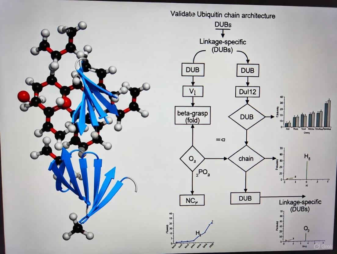
The UbiCRest methodology serves as a cornerstone technique for ubiquitin chain architecture analysis using linkage-specific DUBs. The following diagram illustrates the conceptual workflow and experimental process:
The UbiCRest protocol involves several critical steps that must be optimized for reliable results. First, the ubiquitinated substrate of interest is purified and transferred into DUB-compatible buffer. The sample is then split into equal aliquots and treated with a panel of linkage-specific DUBs in parallel reactions [10] [15]. It is crucial to include appropriate controls, including a non-specific DUB (such as USP21 or USP2) that cleaves all linkage types as a positive control, and a no-DUB negative control to establish baseline migration patterns [15].
Experimental conditions including DUB concentration, incubation time, and temperature should be optimized. Researchers typically perform assays at both low and high DUB concentrations—activity at low concentrations indicates presence of the preferred linkage type, while higher concentrations reveal whether secondary chain types remain on the substrate [10]. Following DUB treatment, samples are analyzed by SDS-PAGE and immunoblotting using ubiquitin-specific antibodies to visualize the cleavage patterns [15].
Research Reagent Solutions for Ubiquitin Analysis
The successful implementation of ubiquitin chain analysis requires specific research reagents with defined linkage specificities. The following table details key reagents used in DUB-based approaches:
Table 3: Essential Research Reagents for DUB-Based Ubiquitin Chain Analysis
| Reagent Category | Specific Examples | Key Specificity/Function | Applications |
|---|---|---|---|
| Linkage-Specific DUBs | OTUB1 (K48) [15], OTUD3 (K6/K11) [15], Cezanne (K11) [15], OTUD1 (K63) [15] | Cleave specific ubiquitin linkages [10] [15] | UbiCRest analysis [10]; Chain validation [15] |
| Non-Specific DUBs | USP21 [10] [15], USP2 [10], vOTU [15] | Cleave most or all linkage types [10] [15] | Positive controls [10]; Complete deubiquitination [15] |
| Ubiquitin Binding Reagents | Tandem Ubiquitin Binding Entities (TUBEs) [16], Linkage-specific antibodies [9] [10] | Enrich ubiquitinated proteins [16]; Detect specific chains [9] | Sample preparation [16]; Immunodetection [10] |
| Specialized Proteases | Lbpro∗ (Ub-clipping) [16], Trypsin (bottom-up MS) [9] | Leave GlyGly signature on modified residues [16]; Generate diagnostic peptides [9] | Ub-clipping [16]; Mass spectrometry [9] |
| Ubiquitin Variants | Lysine-to-arginine mutants [10] [14], Tagged ubiquitin (His-, Strep-) [9] | Restrict chain formation to specific linkages [14]; Enable affinity purification [9] | Ubiquitin replacement [10]; Substrate enrichment [9] |
Analytical Framework for Ubiquitin Chain Architecture
Interpreting UbiCRest results requires understanding how different DUBs process various chain architectures. The cleavage patterns observed on SDS-PAGE provide insights into both linkage composition and chain architecture. The following diagram illustrates the decision framework for analyzing results:
When analyzing UbiCRest results, several key patterns indicate specific architectural features. Complete cleavage to monoubiquitin by a linkage-specific DUB indicates the presence of extended homotypic chains of that linkage type. Partial cleavage with intermediate bands suggests heterotypic chains containing the DUB's preferred linkage mixed with other linkage types. For instance, when OTUB1 (K48-specific) treatment of NleL-generated chains produces intermediate bands rather than complete cleavage to monoubiquitin, this indicates the presence of both K48 and non-K48 (K6) linkages within the same polymer [14]. The electrophoretic mobility of the remaining intermediates can provide additional clues about the specific linkage types present, as different ubiquitin linkages exhibit characteristic migration patterns on SDS-PAGE [10] [14].
Applications in Drug Discovery and Therapeutic Development
Understanding ubiquitin chain architecture has profound implications for drug discovery, particularly in the development of targeted protein degradation strategies and DUB inhibitors. The ubiquitin-proteasome system regulates over 80% of cellular proteins, and its dysregulation is implicated in most cancer hallmarks [11]. E3 ligases and DUBs represent promising therapeutic targets due to their specificity and central role in controlling protein stability and signaling outputs.
PROTACs (Proteolysis-Targeting Chimeras) and molecular glues represent a breakthrough therapeutic modality that redirects E3 ubiquitin ligases to target non-traditional substrates for degradation [11]. The efficacy of these compounds depends on the formation of specific ubiquitin chain types (primarily K48-linked chains) on the target protein. UbiCRest and related methodologies can validate whether these therapeutics induce the intended ubiquitin code on their targets, providing critical mechanistic information during drug development [11]. For example, analyzing the chain architecture induced by a PROTAC molecule can explain unexpected efficacy or toxicity profiles and guide compound optimization.
DUB inhibitors represent another promising therapeutic class, with several compounds in preclinical and clinical development. Different DUB families display distinct linkage preferences—USP family DUBs generally show broad specificity but substrate selectivity, while OTU family DUBs often exhibit marked linkage preference [13] [15]. Understanding the biological functions of specific ubiquitin chain types enables rational design of DUB inhibitors with predicted physiological effects. For instance, inhibitors of K48-specific DUBs would be expected to enhance proteasomal degradation of their substrate proteins, while inhibitors of K63-specific DUBs would modulate signaling pathways such as NF-κB activation [12].
The functional outcomes of ubiquitination are fundamentally determined by the architectural complexity of ubiquitin chains. Methodologies for deciphering this ubiquitin code, particularly DUB-based approaches like UbiCRest, provide critical tools for understanding the mechanistic basis of ubiquitin signaling in health and disease. The integration of these complementary techniques—UbiCRest for architectural analysis, Ub-clipping for branching quantification, and mass spectrometry for site identification—enables researchers to obtain a comprehensive view of the ubiquitin code in specific biological contexts.
As the field advances, several emerging areas will shape future research. First, understanding the dynamics of ubiquitin chain editing—how chains are remodeled by DUBs and E3 ligases during cellular processes—will reveal how signals are integrated and terminated. Second, developing quantitative frameworks for predicting functional outcomes from chain architecture will enhance our ability to manipulate the ubiquitin system therapeutically. Finally, expanding our knowledge of heterotypic and branched chains in physiological contexts will likely uncover new regulatory mechanisms and therapeutic opportunities.
For drug development professionals, mastering these analytical approaches provides a competitive advantage in validating mechanisms of action, understanding compound selectivity, and guiding lead optimization. As targeted protein degradation and DUB modulation continue to gain therapeutic traction, methodologies for ubiquitin code validation will become increasingly essential components of the drug discovery toolkit.
The ubiquitin-proteasome system (UPS) is the major pathway for the degradation of over 80% of intracellular proteins, acting as a critical post-translational regulator of protein stability, activity, and localization [18]. The precise orchestration of protein ubiquitination—the covalent attachment of ubiquitin to target proteins—and its reversal, deubiquitination, governs virtually all aspects of eukaryotic cell biology. This guide provides a comparative analysis of the core enzymatic machinery: the E1, E2, and E3 enzymes that assemble ubiquitin chains, and the deubiquitinating enzymes (DUBs) that disassemble them. Understanding the function, specificity, and experimental analysis of these players is fundamental to research in cell signaling, protein homeostasis, and the development of therapeutics for diseases like cancer and neurodegeneration [5] [18].
The Ubiquitination Cascade: E1, E2, and E3 Enzymes
Protein ubiquitination is executed by a sequential cascade of three enzyme families. This process culminates in the attachment of a single ubiquitin or a polyubiquitin chain to a substrate protein, which can alter its function or mark it for degradation [19] [20].
- E1 (Ubiquitin-Activating Enzyme): The process initiates with a single E1 enzyme that activates ubiquitin in an ATP-dependent manner, forming a thioester bond between its active-site cysteine and the C-terminal glycine of ubiquitin [19] [21].
- E2 (Ubiquitin-Conjugating Enzyme): The activated ubiquitin is then transferred to the cysteine residue of an E2 conjugating enzyme. The human genome encodes approximately 40 E2s, which contribute to the specificity of the modification [5].
- E3 (Ubiquitin Ligase): Finally, an E3 ligase facilitates the transfer of ubiquitin from the E2 to a lysine residue on the target protein, forming an isopeptide bond. With over 600 E3s in humans, this enzyme is the primary determinant of substrate specificity. The E3 can also promote the extension of ubiquitin into polymers by catalyzing the attachment of additional ubiquitin molecules to one of the seven lysine residues (K6, K11, K27, K29, K33, K48, K63) or the N-terminal methionine (M1) of the previously attached ubiquitin [19] [5] [1].
The collaboration between these enzymes results in a diverse "ubiquitin code," including monoubiquitination, multi-monoubiquitination, and various polyubiquitin chain architectures (homotypic, mixed, and branched), each with distinct functional consequences for the modified substrate [5] [1] [18].
Table 1: Key Enzyme Classes in the Ubiquitin Cascade
| Enzyme | Number in Humans | Core Function | Key Functional Domains/Features |
|---|---|---|---|
| E1 | 2 | Activates ubiquitin via ATP hydrolysis to form E1~Ub thioester [5] [21]. | Adenylation domain, active-site cysteine [21]. |
| E2 | ~40 | Accepts ubiquitin from E1 to form E2~Ub thioester; often determines chain linkage [5]. | Catalytic cysteine residue, determines linkage specificity for some E2s [1]. |
| E3 - RING | >600 | Brings E2~Ub and substrate together for direct ubiquitin transfer [1]. | RING domain, acts as a scaffold [1]. |
| E3 - HECT | 28 | Forms E3~Ub thioester intermediate before transferring ubiquitin to substrate [1]. | HECT domain, determines linkage specificity [1]. |
| E3 - RBR | 14 | Hybrid mechanism; forms E3~Ub thioester like HECT E3s [1]. | RING1, RING2, and In-Between-Ring (IBR) domains [1]. |
| DUBs | ~100 | Cleaves ubiquitin from substrates, proofreads ubiquitination, recycles ubiquitin [18]. | Catalytic triad (Cys-based or Zn-dependent JAMM family) [18]. |
Deubiquitinases (DUBs) in Chain Disassembly
Deubiquitinating enzymes (DUBs) perform the reverse reaction of E3 ligases, removing ubiquitin from substrate proteins. This activity is essential for maintaining protein stability, proofreading ubiquitin signals, recycling ubiquitin, and controlling the dynamics of signaling complexes [18]. The human genome encodes approximately 100 DUBs, which are categorized into seven families based on their catalytic mechanisms and structural folds [18].
- Cysteine Protease DUB Families: This group includes Ubiquitin-Specific Proteases (USPs), Ubiquitin C-Terminal Hydrolases (UCHs), Ovarian Tumor Proteases (OTUs), Machado-Joseph Disease Proteases (MJDs), MINDY, and ZUFSP. They all feature a catalytic cysteine residue as part of a catalytic triad [18].
- Zinc-Dependent Metalloprotease DUB Family: The JAMM family is the only group of metalloproteases among DUBs [18].
A key feature of many DUBs is their linkage specificity, which is often imparted by ubiquitin-binding domains (UBDs) that recognize specific chain architectures. For instance, OTUD1 specifically hydrolyzes K63-linked chains, a function that is impaired if its UIM domain is missing [18]. This specificity allows DUBs to act as precise editors of the ubiquitin code, making them critical targets for therapeutic intervention. Dysregulation of DUBs is implicated in various cancers and neurodegenerative diseases [18].
Table 2: Major Deubiquitinase (DUB) Families and Their Characteristics
| DUB Family | Representative Members | Catalytic Mechanism | Notable Features / Specificity |
|---|---|---|---|
| USP | USP5, USP7, USP14 | Cysteine protease | Largest DUB family; diverse specificities and regulatory roles [18]. |
| OTU | OTUD1, A20 | Cysteine protease | Often exhibit high linkage specificity (e.g., K63, K48) [18]. |
| JAMM | UCH37, BRCC36 | Zinc metalloprotease | Requires Zn²⁺ for catalysis; often part of large complexes [1] [18]. |
| UCH | UCH-L1 | Cysteine protease | Specialized in cleaving small adducts from ubiquitin's C-terminus [18]. |
| MJD | Ataxin-3 | Cysteine protease | Often involved in protein quality control [18]. |
| MINDY | MINDY-1, MINDY-2 | Cysteine protease | Preferentially cleaves K48-linked polyUb chains [18]. |
| ZUFSP | ZUFSP/ZUP1 | Cysteine protease | Prefers K63-linked and long polyUb chains [18]. |
Experimental Methodologies for Characterization
Studying the ubiquitin system requires specialized methodologies to overcome challenges such as low endogenous stoichiometry, the multiplicity of modification sites, and the complexity of chain architectures [5]. The following protocols outline key approaches for profiling ubiquitinated proteins and characterizing chain linkage.
Profiling Ubiquitinated Proteins: Enrichment Strategies
To identify ubiquitination sites and substrates in a high-throughput manner, ubiquitinated proteins must first be enriched from complex cell lysates. The primary strategies are summarized below.
Ubiquitin Tagging-Based Enrichment: This approach involves engineering cells to express ubiquitin with an affinity tag (e.g., His, Strep, or HA). The tagged ubiquitin is incorporated into the cellular ubiquitin pool, and ubiquitinated proteins can be purified under denaturing conditions using the appropriate resin (e.g., Ni-NTA for His-tag). After purification and tryptic digestion, ubiquitination sites are identified by mass spectrometry (MS) as a 114.04 Da mass shift on modified lysine residues [5].
- Advantages: Easy to implement and relatively low-cost.
- Disadvantages: The tag may alter ubiquitin structure, creating artifacts. It is also infeasible for use in clinical or animal tissues, and co-purification of endogenous biotinylated or histidine-rich proteins can reduce specificity [5].
Ubiquitin Antibody-Based Enrichment: This method uses antibodies (e.g., P4D1, FK1/FK2) that recognize ubiquitin to immunoprecipitate endogenously ubiquitinated proteins from cell or tissue lysates. Linkage-specific antibodies (e.g., for K48, K63, K11) can be used to enrich for proteins modified with a particular chain type [5].
- Advantages: Can be applied to any sample, including clinical tissues, without genetic manipulation.
- Disadvantages: Antibodies are expensive and can have non-specific binding, leading to high background [5].
Ubiquitin-Binding Domain (UBD)-Based Enrichment: Tandem-repeated Ub-binding entities (TUBEs) are engineered proteins with multiple UBDs that exhibit high-affinity, avidity-based binding to polyubiquitin chains. TUBEs can be fused to affinity tags for purification and have the added benefit of protecting ubiquitin chains from disassembly by DUBs during lysis [5].
- Advantages: High affinity and specificity; protects ubiquitin chains.
- Disadvantages: May have inherent linkage preferences based on the UBD used.
Characterizing Ubiquitin Chain Architecture Using Linkage-Specific DUBs
A powerful method to validate ubiquitin chain architecture involves the use of linkage-specific DUBs. This assay provides functional evidence for the presence of a specific ubiquitin linkage on a protein or within a chain.
Protocol: DUB Specificity Assay for Chain Validation
Generate Ubiquitinated Substrate: Produce the ubiquitinated protein of interest. This can be achieved:
- In vivo: By immunoprecipitating the protein from cells under denaturing conditions to preserve ubiquitination and co-purify associated DUBs.
- In vitro: By reconstituting the ubiquitination reaction using purified E1, E2, and E3 enzymes.
Incubate with Recombinant DUBs: Split the purified ubiquitinated substrate into several aliquots. Incubate each aliquot with a different, purified recombinant DUB known to have high specificity for a particular ubiquitin linkage (e.g., OTUB1 for K48-linked chains, AMSH for K63-linked chains). Include a control aliquot with buffer alone.
Analyze the Results: Resolve the reactions by SDS-PAGE and perform immunoblotting with an anti-ubiquitin antibody.
- Interpretation: If a DUB cleaves the ubiquitin chains from the substrate, a loss of high-molecular-weight smearing will be observed on the blot. The disappearance of signal upon treatment with a specific DUB indicates that the substrate is modified with chains containing that particular linkage.
This method is often used in conjunction with MS-based proteomics and linkage-specific antibodies to build a comprehensive picture of the ubiquitin code.
The Scientist's Toolkit: Key Research Reagents
The following table lists essential chemical and biological tools used to interrogate the ubiquitin system in experimental settings.
Table 3: Key Research Reagents for Studying Ubiquitination and Deubiquitination
| Reagent Name | Target | Function / Effect | Key Experimental Use |
|---|---|---|---|
| TAK-243 (MLN7243) [21] | E1 Ubiquitin-Activating Enzyme (UAE) | Selective inhibitor (IC₅₀ = 1 nM); blocks ubiquitin binding and global ubiquitination. | Tool to broadly inhibit the ubiquitin system; induces apoptosis and has antitumor activity. |
| Pevonedistat (MLN4924) [21] | NEDD8-Activating Enzyme (NAE) | Potent and selective inhibitor (IC₅₀ = 4.7 nM); blocks cullin neddylation and activity of Cullin-RING E3 ligases. | Probe for studying the NEDD8 pathway and cullin-dependent ubiquitination; in clinical trials. |
| Nutlin-3 [20] [21] | MDM2-p53 Interaction | MDM2 antagonist (Kᵢ = 90 nM); stabilizes p53 and activates the p53 pathway. | Study p53-dependent apoptosis and cell cycle arrest; a classic E3 ligase inhibitor. |
| Heclin [21] | HECT E3 Ligases (e.g., Smurf2, Nedd4, WWP1) | Inhibitor of HECT E3 ligase activity (IC₅₀ ~6-7 μM). | Selective tool to probe the function of HECT-family E3 ligases. |
| Ginkgolic Acid [21] | Multiple (e.g., USP4, USP5, SENP1) | Natural compound that inhibits several deubiquitinases and SUMO proteases. | Used to study DUB and SUMOylation pathways; exhibits anti-cancer and anti-inflammatory effects. |
| TUBEs (Tandem Ubiquitin Binding Entities) [5] | Polyubiquitin Chains | High-affinity capture reagents for purifying ubiquitinated proteins; protect chains from DUBs. | High-yield enrichment of polyubiquitinated proteins for proteomics or biochemical analysis. |
| Linkage-Specific Ub Antibodies [5] | Specific Ubiquitin Linkages (K48, K63, etc.) | Antibodies that recognize a particular ubiquitin chain topology. | Detect and validate specific chain linkages via immunoblotting or immunofluorescence. |
Signaling Pathways and Logical Workflows
The following diagrams illustrate the core ubiquitination cascade and the classification of deubiquitinating enzymes, highlighting key logical relationships in this system.
The Ubiquitin Cascade: From Activation to Ligation
Diagram 1: The sequential E1-E2-E3 enzymatic cascade activates and transfers ubiquitin to a protein substrate, determining its fate.
Deubiquitinase (DUB) Family Classification
Diagram 2: DUBs are classified into seven families based on their catalytic mechanism, with the JAMM family being the only metalloproteases.
Ubiquitination is a versatile and reversible post-translational modification (PTM) that regulates fundamental aspects of protein substrates, including stability, activity, and localization [9]. This versatility stems from remarkable structural complexity—ubiquitin can modify substrates as a single monomer or form polymers of different lengths and linkage types [9]. The ubiquitin code encompasses monoubiquitination, multiple monoubiquitination, and polyubiquitin chains connected through any of eight possible linkage sites (Lys6, Lys11, Lys27, Lys29, Lys33, Lys48, Lys63, or Met1) [9] [22]. This generates homotypic chains (single linkage type), heterotypic chains (mixed linkages), and branched chains with architectural diversity that creates a sophisticated signaling system [15] [22].
Characterizing ubiquitination presents substantial analytical hurdles primarily due to two factors: the low stoichiometry of modification under normal physiological conditions and the extreme structural complexity of ubiquitin chains [9]. These challenges compound each other; the scarce ubiquitination events must be characterized against a backdrop of overwhelming non-modified proteins, while the modifications themselves can take numerous structurally distinct forms that may confer different functional outcomes. This article examines these analytical challenges and compares methodologies for validating ubiquitin chain architecture, with particular focus on deubiquitinase (DUB)-based approaches.
The Core Analytical Challenges
Low Stoichiometry: The Needle in a Haystack Problem
In typical cellular environments, ubiquitination occurs with remarkably low stoichiometry, creating significant detection and identification barriers [9]. Several factors contribute to this challenge:
- Low Abundance: Ubiquitinated proteins represent a minute fraction of the total cellular proteome, requiring highly sensitive enrichment techniques to avoid interference from non-ubiquitinated proteins [9].
- Dynamic Regulation: Ubiquitination is a highly dynamic process balanced by the opposing activities of ubiquitinating enzymes (E1, E2, E3) and deubiquitinases (DUBs) [9] [22]. This constant cycling maintains low steady-state levels of modified substrates.
- Rapid Turnover: Many ubiquitinated proteins are targeted for proteasomal degradation, resulting in transient modification states that are difficult to capture [9].
Structural Complexity: A Multi-Dimensional Challenge
The structural diversity of ubiquitin modifications creates a second layer of analytical complexity:
- Multiple Linkage Types: Eight distinct linkage types create chains with different structures and functions [9] [15]. K48-linked chains typically target substrates for proteasomal degradation, while K63-linked chains regulate protein-protein interactions and signaling pathways [9].
- Chain Architecture Diversity: Beyond homotypic chains, heterotypic mixed and branched ubiquitin chains create a "mind-boggling" number of possible architectures [15]. The functions, abundance, and importance of these complex chains remain largely undefined [15].
- Gel Electrophoresis Limitations: Ubiquitinated proteins often appear as high-molecular weight 'smears' rather than discrete bands due to heterogeneous modification patterns, chain types, and chain lengths that migrate unpredictably in SDS-PAGE [15].
Table 1: Key Challenges in Ubiquitin Characterization
| Challenge | Description | Impact on Analysis |
|---|---|---|
| Low Stoichiometry | Minimal abundance of ubiquitinated substrates under normal conditions | Requires extensive enrichment; low identification sensitivity |
| Multiple Modification Sites | Proteins can be ubiquitinated at one or several lysine residues simultaneously | Difficult to localize specific modification sites using traditional methods |
| Chain Linkage Diversity | Eight possible linkage types for polyubiquitin chain formation | Complicates functional interpretation; requires linkage-specific tools |
| Chain Architecture Complexity | Homotypic, heterotypic mixed, and branched chains possible | Standard techniques cannot resolve chain architecture |
| Dynamic Regulation | Continuous ubiquitination and deubiquitination | Capturing transient states difficult; represents a "snapshot" in time |
Methodological Approaches for Ubiquitin Characterization
Enrichment Strategies for Low-Stoichiometry Ubiquitination
To overcome stoichiometry challenges, researchers have developed several enrichment approaches:
Ubiquitin Tagging-Based Approaches: These methods involve expressing ubiquitin with affinity tags (e.g., His, Strep, FLAG) in cells. The tagged ubiquitin incorporates into cellular ubiquitination pathways, allowing purification of ubiquitinated proteins using appropriate resins [9]. While cost-effective and relatively easy to implement, these approaches may generate artifacts as tagged ubiquitin does not completely mimic endogenous ubiquitin, and co-purification of non-ubiquitinated proteins (e.g., histidine-rich proteins with His-tags) can reduce identification sensitivity [9].
Antibody-Based Enrichment: This approach utilizes anti-ubiquitin antibodies (e.g., P4D1, FK1/FK2) or linkage-specific antibodies to enrich endogenously ubiquitinated proteins without genetic manipulation [9]. This allows study of ubiquitination under physiological conditions in animal tissues or clinical samples. However, antibodies represent a high cost and may exhibit non-specific binding [9].
Ubiquitin-Binding Domain (UBD) Approaches: Proteins containing UBDs can recognize and enrich ubiquitinated proteins. While single UBDs typically have low affinity, tandem-repeated UBDs show improved binding capacity [9].
Mass Spectrometry Advances
Mass spectrometry (MS)-based proteomics has revolutionized ubiquitin research. Bottom-up approaches identify ubiquitination sites by detecting the 114.043 Da mass shift from the Gly-Gly remnant left after tryptic digestion of ubiquitinated peptides [15] [23]. Absolute quantitation techniques can assess relative abundance of various ubiquitin linkages, and enrichment techniques using antibodies against tryptic ubiquitination-site remnants enable global proteomic analysis [15]. However, MS approaches struggle to reveal details on chain architecture, which remains difficult to assess using current technologies [15].
Computational Prediction Tools
To complement experimental methods, computational approaches have been developed for predicting ubiquitination sites:
DeepUbi: A deep learning framework based on convolutional neural networks that extracts features from protein sequences and physicochemical properties. In validation studies, DeepUbi achieved an AUC of 0.9, with accuracy, sensitivity, and specificity exceeding 85% [23].
UBIPredic: This computational method predicts ubiquitinated proteins without relying on ubiquitination site prediction, using features from sequence conservation, functional domain annotation, and subcellular localization. It achieved 90.13% accuracy with Matthew's correlation coefficient of 80.34% in cross-validation [24].
Table 2: Comparison of Ubiquitin Characterization Methods
| Method | Principle | Advantages | Limitations |
|---|---|---|---|
| Tagged Ubiquitin | Expression of affinity-tagged Ub; purification and MS | Relatively low-cost; applicable to various cell types | May not mimic endogenous Ub; genetic manipulation required |
| Antibody Enrichment | Immunoaffinity purification with anti-Ub antibodies | Works with endogenous ubiquitination; applicable to tissues | High cost; potential non-specific binding |
| UbiCRest | Linkage-specific DUBs cleave specific ubiquitin linkages | Qualitative chain architecture data; works with endogenous proteins | Qualitative rather than quantitative |
| Mass Spectrometry | Detection of diGly remnants after tryptic digest | High-throughput site identification; quantitative potential | Limited architectural information; complex data analysis |
| Computational Prediction | Machine learning on sequence/structural features | No experimental work needed; high throughput | Predictive only; requires experimental validation |
DUB-Based Validation: The UbiCRest Approach
Principles of UbiCRest
UbiCRest (Ubiquitin Chain Restriction) represents a powerful approach for addressing both stoichiometry and complexity challenges by exploiting the intrinsic linkage specificity of deubiquitinating enzymes [15]. The method involves treating ubiquitinated substrates or purified ubiquitin chains with a panel of linkage-specific DUBs in parallel reactions, followed by gel-based analysis [15]. This qualitative method quickly assesses ubiquitin chain linkage types and architecture, working with western blotting quantities of endogenously ubiquitinated proteins [15].
The foundation of UbiCRest lies in the carefully characterized linkage preferences of various DUBs. Through biochemical profiling, researchers have identified DUBs with relative specificity for each of the eight ubiquitin linkage types [15]. For example, OTUB1 shows high specificity for Lys48-linked chains, while AMSH is specific for Lys63 linkages [15].
Experimental Protocol for UbiCRest
The UbiCRest protocol can be broken down into key stages:
Step 1: Sample Preparation
- Obtain ubiquitinated proteins of interest through immunoprecipitation or purification
- Alternatively, use purified polyubiquitin chains as substrate
- Prepare reaction buffers compatible with DUB activity
Step 2: DUB Panel Setup
- Set up parallel reactions with individual DUBs at optimized concentrations
- Include positive controls (e.g., USP21 which cleaves all linkages)
- Include negative controls (no enzyme, catalytically dead DUB mutants)
- Incubate at appropriate temperature (typically 37°C) for 1-2 hours
Step 3: Analysis and Interpretation
- Stop reactions with SDS-PAGE loading buffer
- Analyze by immunoblotting with anti-ubiquitin antibodies
- Interpret linkage composition based on cleavage patterns
Table 3: Linkage-Specific DUBs for UbiCRest
| Linkage Type | Recommended DUB | Working Concentration | Notes on Specificity |
|---|---|---|---|
| All linkages | USP21 or USP2 | 1-5 µM (USP21) | Positive control; cleaves all linkages including proximal ubiquitin |
| Lys48 | OTUB1 | 1-20 µM | Highly Lys48-specific; not very active but highly specific |
| Lys63 | OTUD1 | 0.1-2 µM | Very active; may become non-specific at high concentrations |
| Lys11 | Cezanne | 0.1-2 µM | Very active; may cleave Lys63 and Lys48 at high concentrations |
| Lys6 | OTUD3 | 1-20 µM | Also cleaves Lys11 chains equally well |
| Lys27 | OTUD2 | 1-20 µM | Also cleaves Lys11, Lys29, Lys33; prefers longer Lys11 chains |
| Lys29/Lys33 | TRABID | 0.5-10 µM | Cleaves Lys29 and Lys33 equally well; low bacterial expression yields |
| All except Met1 | vOTU | 0.5-3 µM | Positive control that does not cleave Met1 linkages |
Applications and Interpretation
UbiCRest provides insights into three key aspects of ubiquitination:
Identifying Ubiquitination: Treatment with broad-specificity DUBs (e.g., USP21) confirms protein ubiquitination by complete collapse of high-molecular-weight smears to discrete unmodified protein bands [15].
Determining Linkage Composition: Selective cleavage by linkage-specific DUBs identifies which chain types are present. For example, OTUB1 sensitivity indicates presence of Lys48-linked chains, while AMSH sensitivity indicates Lys63 linkages [15].
Assessing Chain Architecture: Sequential digestion with DUBs of different specificities can distinguish homotypic chains from heterotypic or branched architectures. Branched chains may require multiple DUBs for complete disassembly [15].
The Scientist's Toolkit: Essential Research Reagents
Successful characterization of ubiquitin architecture requires carefully selected reagents. The following table details key solutions for DUB-based ubiquitin analysis:
Table 4: Research Reagent Solutions for Ubiquitin Characterization
| Reagent Category | Specific Examples | Function and Application |
|---|---|---|
| Linkage-Specific DUBs | OTUB1 (K48), AMSH (K63), Cezanne (K11), OTUD2 (K27) | Cleave specific ubiquitin linkages to determine chain composition in UbiCRest |
| Broad-Specificity DUBs | USP21, USP2, vOTU (all except Met1) | Positive controls; confirm ubiquitination and cleave most linkage types |
| Ubiquitin Mutants | K48R, K63R, K48-only, K63-only | Define linkage requirements in cellular assays; study chain assembly |
| Linkage-Specific Antibodies | Anti-K48, Anti-K63, Anti-K11, Anti-Met1 | Enrich and detect specific chain types by immunoblotting or immunofluorescence |
| Activity Probes | Ubiquitin-based active site probes | Profile DUB activity and specificity in complex mixtures |
| Ubiquitin Variants (UbVs) | UbVSP.1, UbVSP.3 (STAMBP inhibitors) | Potent and selective inhibitors for JAMM family DUBs [25] |
| Mass Spectrometry Standards | Heavy-labeled ubiquitin, Tandem Ubiquitin Binding Entities (TUBEs) | Quantitative ubiquitin proteomics; affinity enrichment of ubiquitinated proteins |
The analytical challenges in ubiquitin characterization—primarily low stoichiometry and structural complexity—require sophisticated methodological approaches. While traditional techniques like immunoblotting and mass spectrometry provide valuable insights, DUB-based methods like UbiCRest offer unique advantages for deciphering ubiquitin chain architecture. The specificity of deubiquitinating enzymes for particular linkage types enables researchers to decode the complex ubiquitin signals that regulate virtually all cellular processes.
As our understanding of DUB specificity continues to expand and new tools like engineered ubiquitin variants emerge, researchers are better equipped to address the fundamental challenges in ubiquitin characterization. These advances are particularly relevant for drug development professionals seeking to target ubiquitin pathways in diseases such as cancer and neurodegenerative disorders, where ubiquitin signaling is frequently dysregulated. The continued refinement of these methodologies will undoubtedly yield deeper insights into the complex world of ubiquitin signaling and its therapeutic implications.
A Practical Guide to UbiCRest: Implementing DUBs for Linkage and Architecture Analysis
Ubiquitin Chain Restriction (UbiCRest) represents a seminal methodological advancement in the field of ubiquitin biology, providing researchers with a powerful tool to decipher the complex language of polyubiquitin signaling. This technique exploits the intrinsic linkage-specific cleavage preferences of deubiquitinating enzymes (DUBs) to characterize ubiquitin chain architecture on modified proteins. As a qualitative assay that yields insights within hours, UbiCRest has become an essential approach for validating ubiquitin chain architecture, complementing more complex mass spectrometry-based methods. This guide examines the core principles of UbiCRest, its experimental implementation, and its position within the broader toolkit for ubiquitin chain analysis, providing researchers with a comprehensive resource for studying the ubiquitin code.
Protein ubiquitination is a versatile post-translational modification that regulates virtually all cellular processes, with its functional diversity originating from the ability to form polyubiquitin chains through eight distinct linkage types [26]. These include seven lysine positions (K6, K11, K27, K29, K33, K48, K63) and the N-terminal methionine (M1), giving rise to homotypic chains (single linkage type), mixed chains (multiple linkage types in sequence), and branched chains (multiple linkages on a single ubiquitin molecule) [15]. The combinatorial complexity of these arrangements creates a sophisticated ubiquitin code that controls diverse cellular outcomes, from proteasomal degradation to DNA repair and immune signaling [27].
Traditional methods for analyzing ubiquitin chains, including linkage-specific antibodies and ubiquitin mutants, have provided valuable insights but face limitations in resolving chain architecture [26]. Mass spectrometry approaches, while powerful for identifying linkage types and ubiquitination sites, often struggle to reveal details about chain architecture and require specialized equipment and expertise [15] [26]. UbiCRest emerged to address these limitations by providing a accessible, gel-based method that uses well-characterized DUBs as "restriction enzymes" for ubiquitin chains [28].
The fundamental premise of UbiCRest is that many DUBs exhibit pronounced specificity for particular ubiquitin linkage types [29]. This specificity arises from distinct structural mechanisms, including additional ubiquitin-binding domains and specific recognition surfaces that enable DUBs to discriminate between different chain architectures [29]. By employing a panel of these linkage-specific DUBs, researchers can deduce the architecture of ubiquitin chains present on a substrate through the characteristic cleavage patterns observed on SDS-PAGE gels [15].
Fundamental Principles of the UbiCRest Methodology
Conceptual Framework and Underlying Assumptions
UbiCRest operates on the principle that DUBs with defined linkage specificities can serve as analytical tools to decompose complex ubiquitin signals into interpretable patterns [15] [26]. The method makes several key assumptions that have been validated through extensive biochemical characterization:
- Specificity Conservation: DUBs maintain their linkage preferences when acting on polyubiquitinated substrates in vitro as they do on isolated chains
- Architecture Retention: The cleavage patterns observed reflect the native architecture of ubiquitin chains rather than artifacts of the experimental conditions
- Predictable Cleavage: Complete digestion with a specific DUB indicates the presence of its preferred linkage type in the substrate
The workflow follows a logical progression from sample preparation through pattern interpretation, with controls to validate each step [26]. A critical conceptual aspect is the distinction between different chain architectures: homotypic chains will be completely digested by DUBs specific to their linkage type, while heterotypic chains will require multiple DUBs for complete disassembly, revealing a hierarchical architecture [15].
Core Workflow and Mechanism
The UbiCRest procedure can be divided into four key stages, as illustrated below:
The process begins with a polyubiquitinated substrate or purified ubiquitin chains, which are transferred into DUB-compatible buffer and divided into equal aliquots [26]. Each aliquot is then treated with a different DUB from a carefully curated panel, selected to cover the spectrum of possible linkage types [15]. Positive controls using broad-specificity DUBs like USP21 or USP2 confirm that the high-molecular-weight smears represent genuine ubiquitin modifications, while negative controls without DUB treatment establish the baseline migration pattern [26].
Following incubation, the digestion products are separated by SDS-PAGE and visualized through immunoblotting [28]. The resulting band patterns provide a fingerprint that reveals both the linkage types present and their arrangement within the ubiquitin chain [15]. Interpretation relies on comparing the digestion patterns across the DUB panel – complete collapse to monoubiquitin or specific intermediate bands indicates the presence of that DUB's preferred linkage type, while persistence of high-molecular-weight material suggests chains contain linkages resistant to that particular DUB [26].
The DUB Toolkit: Enzymatic Specificities and Experimental Considerations
Comprehensive DUB Specificity Reference Table
Successful UbiCRest analysis depends on using a well-characterized panel of DUBs with complementary specificities. The table below summarizes key DUBs used in UbiCRest, their linkage preferences, and optimal working concentrations based on established protocols [15]:
| Linkage Type | Recommended DUB | Typical Working Concentration | Specificity Notes | Key References |
|---|---|---|---|---|
| All Linkages | USP21 / USP2 | 1-5 µM (USP21) | Positive control; cleaves all linkages including proximal ubiquitin | [15] |
| All except M1 | vOTU (CCHFV) | 0.5-3 µM | Positive control; does not cleave Met1 linkages | [15] |
| K6 | OTUD3 | 1-20 µM | Also cleaves K11 chains equally well; targets other linkages at high concentrations | [15] |
| K11 | Cezanne | 0.1-2 µM | Very active; non-specific at very high concentrations (K63 > K48 > others) | [15] |
| K27 | OTUD2 | 1-20 µM | Also cleaves K11, K29, K33; prefers longer K11 chains | [15] |
| K29/K33 | TRABID | 0.5-10 µM | Cleaves K29 and K33 equally well, and K63 with lower activity | [15] |
| K48 | OTUB1 | 1-20 µM | Highly K48-specific; not very active but can be used at high concentrations | [15] |
| K63 | OTUD1 / AMSH | 0.1-2 µM (OTUD1) | Very active; non-specific at high concentrations | [15] [26] |
Concentration Optimization and Specificity Validation
A critical aspect of UbiCRest is optimizing DUB concentrations to balance specificity and activity [26]. Most DUBs exhibit their highest linkage specificity at lower concentrations, while becoming progressively promiscuous at higher concentrations [15]. The protocol typically recommends performing assays at both low and high DUB concentrations – cleavage activity at low concentrations strongly indicates the presence of that DUB's preferred linkage type, while cleavage at higher concentrations may reveal secondary specificities or complete digestion of preferred linkages [26].
The incubation conditions (typically 15-30 minutes at 37°C) represent a balance between complete digestion of preferred linkages and minimizing non-specific cleavage [26]. Researchers should empirically determine optimal conditions for their specific experimental system, particularly when working with novel DUBs or substrates. Specificity profiles for DUBs should be established using homotypic ubiquitin chains of known linkage before applying them to complex biological samples [15].
Comparative Analysis with Alternative Ubiquitin Characterization Methods
Methodological Comparison Table
UbiCRest occupies a distinct niche in the ubiquitin researcher's toolkit, complementing rather than replacing existing methodologies. The table below compares UbiCRest with other prominent approaches for ubiquitin chain analysis:
| Method | Key Principle | Linkage Information | Architecture Resolution | Throughput | Equipment Needs | Key Limitations |
|---|---|---|---|---|---|---|
| UbiCRest | Linkage-specific DUB cleavage + SDS-PAGE | Qualitative identification of major linkages | Can distinguish homotypic vs. heterotypic chains | Medium | Standard molecular biology lab | Qualitative; requires antibody detection |
| Mass Spectrometry | LC-MS/MS of tryptic peptides | Quantitative identification of all linkages | Limited for complex architectures | Low | Specialized MS equipment | Difficult for hydrophobic linkages; complex data analysis |
| Linkage-Specific Antibodies | Immunoblot with linkage-selective antibodies | Semi-quantitative for specific linkages | No architecture information | High | Standard molecular biology lab | Limited antibody availability; cross-reactivity concerns |
| Ubiquitin Mutants | In vivo expression of linkage-deficient Ub mutants | Functional implication of specific linkages | No direct architecture information | Low | Cell culture facility | Compensatory mechanisms; pleiotropic effects |
| UbiReal | Fluorescence polarization of labeled ubiquitin | Real-time kinetics of chain formation/disassembly | Limited architectural details | High | Plate reader capable of FP measurements | Requires fluorescent labeling; artificial system [30] |
Strategic Method Selection and Integration
Each method offers distinct advantages that make it suitable for particular research questions. UbiCRest excels in providing rapid, accessible analysis of ubiquitin chain architecture without requiring specialized equipment [28] [26]. Its particular strengths include:
- Architectural Insights: Ability to distinguish between homotypic, mixed, and branched chains through sequential or parallel digestion approaches [15]
- Sensitivity: Capacity to work with western blotting quantities of endogenously ubiquitinated proteins [26]
- Dynamic Range: Capacity to analyze heterogeneous ubiquitin "smears" that challenge MS-based methods [15]
UbiCRest integrates particularly well with mass spectrometry approaches – initial UbiCRest analysis can guide focused MS efforts, while MS can validate and extend UbiCRest findings [15]. Similarly, linkage-specific antibodies can confirm UbiCRest results, creating a orthogonal validation framework [26]. The recent development of highly specific DUB inhibitors further enhances UbiCRest's utility by enabling validation of findings in cellular contexts [27].
Advanced Applications in Decoding Complex Ubiquitin Signals
Architectural Analysis of Heterotypic Ubiquitin Chains
UbiCRest provides unique capabilities for deciphering the architecture of heterotypic ubiquitin chains, which incorporate multiple linkage types within a single polymer [15]. The sequential application of DUBs with different specificities can reveal whether chains are mixed (alternating linkage types) or branched (multiple linkages on single ubiquitin molecules) [26]. For example, treatment with a K48-specific DUB followed by a K63-specific DUB may completely disassemble a chain that resists either DUB alone, indicating a mixed K48/K63 architecture [15].
The interpretation of these complex digestion patterns requires careful controls and sometimes iterative experimental approaches. Including time-course experiments can help distinguish primary from secondary cleavage events, while titration of DUB concentrations helps identify the most abundant linkage types [26]. These approaches have revealed unexpected complexity in ubiquitin signaling, including the existence of branched chains that may function as specialized signals or as protective structures against disassembly [15].
Integration with Functional Genomics and Drug Discovery
UbiCRest has found important applications in functional genomics and drug discovery, particularly as CRISPR/Cas9-based screening approaches have identified novel regulators of ubiquitin pathways [31] [32] [33]. For example, when genetic screens identify E3 ligases or DUBs that regulate specific substrates, UbiCRest can characterize how their manipulation alters the ubiquitin code on those substrates [32].
In drug discovery, UbiCRest provides a valuable medium-throughput method for characterizing the effects of DUB inhibitors on cellular ubiquitin landscapes [33]. The method can determine whether specific inhibitors effectively block cleavage of their intended linkage types in cellular contexts, and identify potential off-target effects on other ubiquitin chain types [27] [33]. This application is particularly relevant as DUB inhibitors advance in clinical development, with compounds targeting USP1 and USP30 currently in clinical trials [33].
Essential Research Reagents and Experimental Solutions
Research Reagent Toolkit Table
Successful implementation of UbiCRest requires access to well-characterized reagents, particularly the linkage-specific DUBs that form the core of the assay. The following table outlines essential research reagents for establishing UbiCRest in a research setting:
| Reagent Category | Specific Examples | Function in UbiCRest | Commercial Sources | Key Considerations |
|---|---|---|---|---|
| Broad-Spectrum DUBs | USP21, USP2 | Positive controls; confirm ubiquitinated nature of samples | Boston Biochem, R&D Systems | Verify activity on all linkage types before use |
| Linkage-Specific DUBs | OTUB1 (K48), Cezanne (K11), OTUD1 (K63) | Identify specific linkage types present in samples | Boston Biochem, proprietary expression | Validate specificity under working conditions |
| Ubiquitin Chains | Homotypic chains of all 8 linkages | Specificity validation for DUB panel; positive controls | Boston Biochem, UbiQ Bio | Use as standards to establish DUB specificities |
| Detection Antibodies | Anti-ubiquitin, anti-target protein | Visualize digestion patterns after SDS-PAGE | Cell Signaling, Santa Cruz Biotechnology | Pan-ubiquitin antibodies preferred over linkage-specific for overall pattern |
| Expression Systems | E. coli, insect cell systems | Produce recombinant DUBs not commercially available | ATCC, commercial vectors | Many OTU family DUBs require eukaryotic expression for proper folding |
Implementation Practicalities
For laboratories establishing UbiCRest, several practical considerations determine success. First, DUBs can be obtained commercially or expressed recombinantly, with protocols available for purifying many OTU family DUBs from bacterial expression systems [15]. Second, the choice of detection method depends on the experimental system – while western blotting with pan-ubiquitin antibodies is most common, substrate-specific antibodies can provide more precise information when available [26].
The assay requires careful optimization of buffer conditions, particularly pH and reducing agents, which can affect different DUB families variably [15]. Including controls for non-specific proteolysis is essential, particularly when working with novel DUBs or substrates. Finally, interpretation benefits from comparison with known standards – including homotypic ubiquitin chains of known linkage in parallel experiments provides essential reference points for interpreting digestion patterns [15] [26].
UbiCRest has established itself as an indispensable method in the ubiquitin researcher's toolkit, providing unique insights into ubiquitin chain architecture that complement other analytical approaches. Its strength lies in exploiting the naturally evolved specificities of DUBs to decode complex ubiquitin signals, enabling researchers to move beyond simple linkage identification to understanding the topological arrangement of ubiquitin chains. As research continues to reveal the functional significance of heterotypic ubiquitin chains in cellular regulation, disease mechanisms, and therapeutic responses, UbiCRest will remain a vital method for connecting ubiquitin chain architecture to biological function. The ongoing development of additional linkage-specific DUBs and inhibitors will further expand its capabilities, solidifying its role in the evolving landscape of ubiquitin research methodologies.
Deubiquitinating enzymes (DUBs) have emerged as critical regulators of cellular function and promising therapeutic targets. A key to unlocking their potential lies in the specific tools researchers use to study them. This guide provides a comparative analysis of commercially available and purified DUBs, focusing on their application in validating ubiquitin chain architecture, to help you build a robust experimental toolkit.
The Ubiquitin Code and DUB Specificity
Protein ubiquitination is a versatile post-translational modification where ubiquitin molecules can form chains through eight distinct linkage types (seven lysine residues or the N-terminal methionine). This "ubiquitin code" regulates diverse cellular processes, from protein degradation to DNA repair and cell signaling [15] [34]. The reversible process of deubiquitination is carried out by approximately 100 human DUBs, which are categorized into six families: ubiquitin-specific proteases (USPs), ubiquitin C-terminal hydrolases (UCHs), ovarian tumor proteases (OTUs), Machado–Josephin domain-containing proteases (MJDs), motif interacting with ubiquitin-containing novel DUB family (MINDYs), and JAMM/MPN/MOV34 metalloproteases [35] [33].
The combinatorial complexity of homotypic (single linkage type), mixed (multiple types, one per ubiquitin), and branched ubiquitin chains (multiple linkages on a single ubiquitin) poses a significant analytical challenge [15] [34]. Linkage-specific DUBs are indispensable tools for decoding this complexity, as their intrinsic preference for cleaving particular ubiquitin linkages allows researchers to dissect chain composition and architecture.
Commercially Available DUBs: A Linkage-Specific Toolkit
The UbiCRest method exemplifies the use of a DUB panel to qualitatively analyze ubiquitin chains [15]. The table below summarizes key linkage-specific DUBs, their common sources, and their roles in a typical toolkit.
| Ubiquitin Linkage Type | Recommended DUB | Useful Final Concentration Range (1x) | Key Specificity Notes and Considerations |
|---|---|---|---|
| All eight linkages (positive control) | USP21 / USP2 [15] | 1-5 µM (USP21) [15] | Cleaves all linkage types, including the proximal ubiquitin attached to the substrate [15]. |
| All linkages except Met1 (positive control) | CCHFV viral OTU (vOTU) [15] | 0.5-3 µM [15] | Useful control as it does not cleave linear/Met1 linkages [15]. |
| Lys48 (K48) | OTUB1 [15] | 1-20 µM [15] | Highly specific for K48 linkages. Not very active, so can be used at higher concentrations [15]. |
| Lys63 (K63) | OTUD1 / AMSH [15] | 0.1-2 µM (OTUD1) [15] | Both are very active; OTUD1 can become non-specific at high concentrations [15]. |
| Lys11 (K11) | Cezanne (OTUD7B) [15] | 0.1-2 µM [15] | Very active enzyme; can become non-specific at very high concentrations (Lys63 > Lys48 > others) [15]. |
| Lys6 (K6) | OTUD3 [15] | 1-20 µM [15] | Also cleaves Lys11 chains equally well. Can target other isopeptide linkages at high concentrations [15]. |
| Lys27 (K27) | OTUD2 [15] | 1-20 µM [15] | Also cleaves Lys11, Lys29, and Lys33. Prefers longer Lys11 chains and can be non-specific at high concentrations [15]. |
| Lys29 (K29) & Lys33 (K33) | TRABID (ZRANB1) [15] | 0.5-10 µM [15] | Cleaves Lys29 and Lys33 linkages equally well, with lower activity on Lys63. May have low yields from bacterial expression [15]. |
Key Commercial Considerations:
- Source and Purity: Many DUBs for research are produced via bacterial expression (e.g., E. coli) and require purification via affinity chromatography. Mission Therapeutics, for example, emphasizes purifying full-length DUBs from mammalian cells to ensure proper folding and the presence of co-factors [36].
- Specificity vs. Selectivity: While the OTU family DUBs often display strong linkage preference, USP family DUBs are generally more substrate-specific than linkage-specific [12]. This distinction is crucial for experimental design and data interpretation.
- Concentration is Critical: The linkage specificity of a DUB can be concentration-dependent. Several DUBs, like Cezanne and OTUD1, can lose their specificity and cleave non-cognate linkages at high concentrations. Therefore, using the recommended working concentrations is essential for clean data interpretation [15].
Experimental Protocol: UbiCRest for Ubiquitin Chain Analysis
The UbiCRest assay is a primary application for a linkage-specific DUB toolkit, allowing researchers to determine the types and architecture of ubiquitin chains on a protein of interest [15].
Workflow of UbiCRest Assay
Detailed Step-by-Step Methodology
Sample Preparation: Isolate the ubiquitinated protein of interest. This can be an endogenously ubiquitinated protein purified from cells or an in vitro ubiquitinated substrate. The sample is typically divided into equal aliquots.
DUB Panel Reaction Setup: Set up parallel reactions, each containing:
- A defined amount of your ubiquitinated substrate.
- A suitable reaction buffer.
- One linkage-specific DUB from your toolkit (see table above for concentrations).
- Include control reactions: one with no DUB and one with a pan-specific DUB like USP21.
Incubation and Reaction Termination: Incubate reactions at a defined temperature (e.g., 37°C) for a specific time (e.g., 1-2 hours). The reaction is stopped by adding SDS-PAGE loading buffer.
Gel-Based Analysis: Analyze the reactions by denaturing SDS-PAGE followed by western blotting. Use antibodies specific to your protein or to ubiquitin.
Data Interpretation:
- Complete Cleavage: If a substrate's ubiquitination is completely removed by a specific DUB (e.g., OTUB1), it suggests the chains are predominantly of that linkage type (e.g., K48).
- Partial Cleavage / Band Shift: A change in the ubiquitin ladder pattern, but not a complete collapse, indicates the presence of a mixed or branched chain architecture where the DUB is cleaving one type of linkage within a more complex structure.
- No Effect: Indicates the substrate's ubiquitination does not contain the linkage type that the DUB is specific for.
The Scientist's Toolkit: Essential Research Reagents
Building a successful DUB research program requires more than just the enzymes. The table below lists other critical reagents and their functions.
| Research Reagent | Function and Importance in DUB Research |
|---|---|
| Linkage-Specific DUBs | Core tools for dissecting ubiquitin chain type and architecture in assays like UbiCRest [15]. |
| Linkage-Specific Ubiquitin Antibodies | Immunological reagents (e.g., for K48, K63, Met1) used in western blotting to confirm chain types identified via DUB panels [15]. |
| Activity-Based DUB Probes | Chemical tools that covalently bind the active site of DUBs, used to profile DUB activity and selectivity in cell lysates or in vivo [33]. |
| Selective DUB Inhibitors | Small molecules (e.g., KSQ's USP1 inhibitor, Mission's MTX325 for USP30) used for functional validation and therapeutic exploration [33] [36]. |
| Ubiquitin Mutants (K-to-R) | Mutant ubiquitins where lysines are changed to arginine to prevent specific chain types; used in vitro and in cell-based replacement strategies to validate DUB findings [15]. |
| CRISPR-Cas9 DUB Knockout Libraries | Enable genome-wide or family-wide functional screens to identify DUB essentiality and phenotypes across different cell lines, as used in the Dependency Map (DepMap) [33]. |
Strategic Application and Future Directions
The strategic use of DUB toolkits is already yielding clinical insights. For instance, the DUB USP1 is a promising target in breast cancer, and its inhibitor, pimozide, has been shown to suppress tumor metastasis in preclinical models by destabilizing key oncogenic proteins [35]. Furthermore, Mission Therapeutics has advanced DUB inhibitors targeting USP30 into clinical trials for kidney disease and Parkinson's, demonstrating the therapeutic potential of this enzyme family [36].
Emerging research on branched ubiquitin chains highlights the need for an ever-more sophisticated toolkit. Branched chains can confer stability or unique signaling properties not offered by homotypic chains [34]. Fully characterizing these complex structures requires a combination of DUB panels and advanced mass spectrometry techniques.
By integrating a well-characterized DUB toolkit with robust experimental protocols and orthogonal validation methods, researchers can continue to decode the ubiquitin landscape, accelerating both fundamental discovery and the development of novel therapeutics for cancer and other diseases.
Protein ubiquitination is a versatile post-translational modification that regulates virtually all cellular processes, with its functional diversity originating from the architecture of polyubiquitin chains. These chains can be linked in eight distinct ways through internal lysine residues (K6, K11, K27, K29, K33, K48, K63) or the N-terminal methionine (M1), creating homotypic chains (single linkage type), mixed chains (multiple linkages in series), or branched chains (multiple linkages on a single ubiquitin molecule) [15] [34]. The UbiCRest (Ubiquitin Chain Restriction) method exploits the intrinsic linkage specificity of deubiquitinating enzymes (DUBs) to decode this complex ubiquitin signature, providing researchers with a qualitative tool to identify linkage types and chain architecture within hours using standard western blotting equipment [15] [10].
This protocol guide details the experimental workflow for UbiCRest analysis, compares its performance to alternative ubiquitin characterization methods, and provides essential resources for implementing this technique in research on ubiquitin signaling in disease mechanisms and drug development.
UbiCRest Workflow: From Sample to Analysis
The UbiCRest methodology enables researchers to decipher the ubiquitin code using a panel of linkage-specific DUBs followed by gel-based analysis. The comprehensive workflow encompasses sample preparation, DUB treatment, and result interpretation.
Experimental Workflow
The diagram below illustrates the complete UbiCRest experimental procedure from initial sample preparation to final analysis:
Sample Preparation Requirements
The starting point for UbiCRest is a faithfully reproducible ubiquitinated protein sample that can be visualized by PAGE-based methods. Samples can include:
- Immunopurified ubiquitinated proteins from cellular systems
- In vitro ubiquitinated substrates from reconstituted ubiquitination reactions
- Purified ubiquitin chains of unknown linkage composition
Critical to success is obtaining sufficient material for visualization by western blotting. While the method can work with endogenously ubiquitinated proteins, the signal must be detectable above background [15] [10]. Once visualization is optimized, the protein preparation must be transferred into DUB-compatible buffer. The original protocol suggests 50 mM HEPES (pH 8.0), 50 mM NaCl, and 1 mM TCEP, though buffer conditions may need optimization for specific DUBs [37].
DUB Treatment and Controls
The treated sample is split into equal aliquots to ensure consistent protein loading across all lanes, which is critical for detecting sometimes subtle changes in polyubiquitination patterns after DUB treatment [10]. The core experimental setup includes:
- Positive control: Treatment with a non-specific DUB (USP21 or USP2 at 1-5 µM) that removes all polyubiquitin, generating monoubiquitin and confirming DUB activity
- Experimental reactions: Parallel incubation with linkage-specific DUBs at optimized concentrations
- Negative control: No DUB treatment to show original ubiquitination pattern
- Variables to optimize: Incubation time (15-120 minutes), temperature (typically 37°C), and DUB concentration (test low and high concentrations) [15]
Linkage-Specific DUB Panel
The table below details the recommended DUB panel for comprehensive chain analysis:
Table 1: Linkage-Specific DUB Toolkit for UbiCRest Analysis
| Linkage Type | Recommended DUB | Useful Final Concentration | Specificity Notes |
|---|---|---|---|
| All linkages (positive control) | USP21 or USP2 | 1-5 µM | Cleaves all linkage types including proximal ubiquitin |
| K6 | OTUD3 | 1-20 µM | Also cleaves K11 chains equally well |
| K11 | Cezanne | 0.1-2 µM | Very active; non-specific at very high concentrations |
| K27 | OTUD2 | 1-20 µM | Also cleaves K11, K29, K33; prefers longer K11 chains |
| K29/K33 | TRABID | 0.5-10 µM | Cleaves K29 and K33 equally well; lower K63 activity |
| K48 | OTUB1 | 1-20 µM | Highly K48-specific; not very active |
| K63 | OTUD1 | 0.1-2 µM | Very active; non-specific at high concentrations |
| All except M1 | CCHFV viral OTU | 0.5-3 µM | Positive control that does not cleave M1 linkages |
Source: Adapted from [15]
Interpreting UbiCRest Results
Data Interpretation Framework
After western blot analysis, band patterns are interpreted to determine linkage composition and chain architecture:
- Complete cleavage with a specific DUB indicates the predominant presence of that linkage type
- Partial cleavage suggests mixed linkage chains or heterogeneous sample populations
- Resistance to all specific DUBs but cleavage by non-specific DUBs may indicate branched chains or atypical linkages
- Differential cleavage patterns at low versus high DUB concentrations can reveal primary versus secondary linkage preferences [15]
The diagram below illustrates how different ubiquitin chain architectures respond to DUB treatment:
Advantages and Limitations
Key Advantages:
- Rapid analysis (hours versus days for mass spectrometry)
- Accessible methodology using standard laboratory equipment
- Qualitative insights into chain architecture beyond linkage identification
- Works with western blot quantities of endogenously ubiquitinated proteins
- Can distinguish between homotypic, mixed, and branched chains [15] [10]
Important Limitations:
- Qualitative rather than quantitative method
- Requires well-characterized, linkage-specific DUBs
- May miss low-abundance linkages in complex mixtures
- Dependent on antibody detection sensitivity
- Cannot pinpoint exact ubiquitination sites on substrates [15] [38]
Comparison with Alternative Ubiquitin Characterization Methods
UbiCRest represents one of several approaches for analyzing ubiquitin chains. The table below compares key methodologies for ubiquitin characterization:
Table 2: Comparison of Ubiquitin Chain Characterization Methods
| Method | Key Principle | Linkage Information | Architecture Data | Throughput | Equipment Needs |
|---|---|---|---|---|---|
| UbiCRest | Linkage-specific DUB cleavage + gel analysis | Yes, qualitative | Yes, qualitative | Medium | Standard molecular biology |
| Ubiquitin Mutants | Ubiquitin lysine mutants in conjugation assays | Yes, indirect | Limited | Low | Standard molecular biology |
| Mass Spectrometry | Detection of diglycine remnants and linkage peptides | Yes, quantitative | Limited, challenging | High | Specialized MS instrumentation |
| Linkage-Specific Antibodies | Immunodetection with linkage-selective antibodies | Yes, specific linkages only | No | Medium | Standard molecular biology |
Ubiquitin Mutant Approach
The ubiquitin mutant methodology employs ubiquitin proteins where specific lysine residues are mutated to arginine (K-to-R mutants) or where only a single lysine remains (K-only mutants) in in vitro ubiquitination reactions. When chains cannot form with a particular K-to-R mutant, that lysine is identified as essential for chain formation. Conversely, if chains form with a specific K-only mutant, that lysine is sufficient for chain formation [37].
Advantages:
- Commercially available ubiquitin mutants
- Clear interpretation for homotypic chains
Limitations:
- Challenging for mixed or branched chains
- Primarily for in vitro applications
- Does not reveal chain architecture [37] [38]
Mass Spectrometry-Based Approaches
Advanced mass spectrometry methods enable proteome-wide identification of ubiquitination sites through detection of the diglycine remnant (∼114 Da mass shift) on modified lysines after tryptic digestion. Ubiquitin remnant profiling can be combined with quantitative proteomics (SILAC, isobaric tagging) to monitor changes in ubiquitylation in response to cellular perturbations [5] [39] [40].
Advantages:
- Comprehensive, proteome-wide capability
- Precise identification of modification sites
- Quantitative data potential
Limitations:
- Requires specialized instrumentation and expertise
- Difficult to characterize chain architecture
- Limited ability to distinguish branched chains [5] [40]
Method Selection Guide
- For quick assessment of linkage types: UbiCRest or linkage-specific antibodies
- For in vitro characterization of E3 specificity: Ubiquitin mutant approach
- For proteome-wide site identification: Mass spectrometry-based remnant profiling
- For architectural analysis of complex chains: UbiCRest combined with mutagenesis
Essential Research Reagents and Materials
Successful implementation of UbiCRest requires access to well-characterized reagents. The table below summarizes essential research tools:
Table 3: Essential Research Reagent Solutions for UbiCRest
| Reagent Category | Specific Examples | Function in Protocol | Commercial Sources |
|---|---|---|---|
| Linkage-Specific DUBs | OTUB1 (K48), Cezanne (K11), OTUD1 (K63) | Selective cleavage of specific ubiquitin linkages | Boston Biochem, R&D Systems |
| Non-specific DUBs | USP21, USP2 | Positive control; complete deubiquitination | Boston Biochem, R&D Systems |
| Ubiquitin Mutants | K-to-R series, K-only series | Complementary method for linkage verification | Boston Biochem, R&D Systems |
| Antibodies | Anti-ubiquitin, linkage-specific antibodies | Detection of ubiquitin signals | Cell Signaling, Abcam |
| Enzymes/Buffers | E1, E2, E3 enzymes, reaction buffers | Generating ubiquitinated substrates in vitro | Boston Biochem, R&D Systems |
UbiCRest provides researchers with a powerful, accessible method for deciphering the ubiquitin code using linkage-specific deubiquitinating enzymes. While mass spectrometry approaches offer higher throughput and precision for site identification, and ubiquitin mutants provide complementary linkage information, UbiCRest remains uniquely valuable for its ability to provide qualitative insights into ubiquitin chain architecture, including the emerging complexity of branched chains. The methodology's relatively simple equipment requirements and rapid turnaround time make it particularly valuable for initial characterization of ubiquitin chain types and for validating findings from proteomic screens. As our understanding of the complexity of ubiquitin signaling grows, particularly regarding heterotypic and branched chains, UbiCRest will continue to serve as an essential tool in the ubiquitin researcher's toolkit for functional validation of ubiquitin chain architecture.
Ubiquitination is a critical post-translational modification that regulates virtually all cellular processes, from protein degradation to signal transduction and DNA repair [15]. The versatility of ubiquitin signaling originates from the ability of ubiquitin molecules to form diverse polymeric chains. When chains are composed of a single linkage type (e.g., all Lys48 or all Lys63), they are classified as homotypic chains. In contrast, heterotypic chains contain more than one type of ubiquitin linkage and can be further categorized as mixed (each ubiquitin modified at one site) or branched (at least one ubiquitin modified at multiple sites) [34]. The ability to accurately distinguish between these architectures is fundamental to understanding their distinct biological functions, as chain topology determines how ubiquitin signals are interpreted by cellular machinery [41] [12]. This guide compares the diagnostic patterns for homotypic versus heterotypic chains, focusing on the use of linkage-specific deubiquitinases (DUBs) as primary tools for validation.
Key Methodological Principles for Chain Analysis
The UbiCRest Approach: DUBs as Restriction Enzymes
The UbiCRest (Ubiquitin Chain Restriction) assay is a qualitative method that uses a panel of linkage-specific DUBs to probe ubiquitin chain architecture in vitro. In this approach, a ubiquitinated substrate or purified ubiquitin chains are treated with individual DUBs in parallel reactions, followed by gel-based analysis (typically SDS-PAGE and immunoblotting) to visualize the cleavage patterns [15] [42].
Core Principle: Each linkage-specific DUB acts as a "restriction enzyme" that cleaves only its preferred linkage type(s). The pattern of cleavage products generated by different DUBs reveals the types of linkages present and their arrangement within the chain [42]. For instance, a homotypic K48-linked chain will be completely disassembled to monoubiquitin by the K48-specific DUB OTUB1, whereas a heterotypic chain containing both K48 and K11 linkages would only be partially cleaved by OTUB1, leaving behind shorter K11-linked polymers [15] [41].
Workflow for Diagnostic Pattern Interpretation
The following diagram illustrates the core experimental workflow and logical decision process for distinguishing chain architecture using DUBs.
Comparative Analysis of Diagnostic Patterns
The patterns observed after DUB treatment provide a fingerprint that identifies the chain architecture. The table below summarizes the key diagnostic outcomes for homotypic and heterotypic chains.
Table 1: Diagnostic Patterns for Ubiquitin Chain Architecture Using DUBs
| Chain Architecture | Representative DUB Treatment | Observed Diagnostic Pattern | Biological Example |
|---|---|---|---|
| Homotypic | Single linkage-specific DUB (e.g., OTUB1 for K48) | Complete disassembly to monoubiquitin [15] | Pure K48-linked chains target proteins for proteasomal degradation [41] [12] |
| Heterotypic (Mixed) | Single linkage-specific DUB | Partial cleavage, leaving shorter residual chains of the other linkage type [15] [42] | NleL assembles chains with both K6 and K48 linkages; OTUB1 leaves K6-linked remnants [42] |
| Heterotypic (Branched) | Combination of DUBs | Full disassembly requires multiple DUBs targeting different linkages [34] [42] | APC/C collaborates with UBE2C and UBE2S to form branched K11/K48 chains on cyclin B1 [41] [34] |
Supporting Experimental Data and Functional Consequences
Case Study: Proteasomal Distinction Between K11 Chain Types
A critical functional difference between homotypic and heterotypic chains was demonstrated in a study on K11-linked ubiquitin. The research showed that the proteasome distinguishes between these architectures, leading to different functional outcomes for the modified substrate [41].
Table 2: Functional Consequences of K11-linked Ubiquitin Chain Architecture
| Chain Type | Proteasome Binding | Degradation Signal | Experimental Evidence |
|---|---|---|---|
| Homotypic K11 | Weak / non-significant | No | Binding assays with purified 26S proteasomes showed no significant association [41] |
| Heterotypic K11/K48 | Strong | Yes | Heterotypic chains bound proteasomes and stimulated degradation of cyclin B1 [41] |
This case study highlights that a simple readout of ubiquitination (e.g., a smear on a Western blot) is insufficient to predict biological function. Architectural analysis is necessary, as homotypic K11 chains do not signal degradation, while heterotypic K11/K48 chains are potent degradative signals [41].
Detailed Protocol: Ubiquitin Chain Restriction Analysis
The following workflow provides a detailed protocol for performing ubiquitin chain restriction analysis, based on established methodologies [15] [42].
Step 1: Sample Preparation. Generate the ubiquitinated protein or ubiquitin chains of interest. This can be achieved through in vitro ubiquitination assays using specific E2 and E3 enzyme combinations or by purifying ubiquitinated proteins from cells [15] [42].
Step 2: DUB Panel Selection and Reaction Setup.
- Prepare parallel reactions containing your substrate and a specific DUB. Key linkage-specific DUBs include [15]:
- OTUB1: Highly specific for Lys48-linked chains.
- OTUD1/Cezanne: Preferentially cleaves Lys11-linked chains.
- OTUD2: Cleaves Lys27 and other atypical linkages.
- OTUD3: Preferentially cleaves Lys6-linked chains.
- AMSH/OTUD1: Specific for Lys63-linked chains.
- vOTU/USP21: Broad-specificity DUBs (positive control).
- Incubate reactions at appropriate conditions (e.g., 37°C for 1-2 hours) [15].
Step 3: Analysis and Interpretation.
- Stop the reactions with SDS-PAGE loading buffer.
- Resolve the products by SDS-PAGE and transfer to a membrane for immunoblotting using an anti-ubiquitin antibody.
- Interpret the cleavage patterns by comparing the banding patterns across all DUB treatments, as illustrated in the workflow diagram [15] [42].
The Scientist's Toolkit: Essential Research Reagents
Successful interpretation of ubiquitin chain architecture relies on a core set of reagents. The following table details these essential tools and their functions.
Table 3: Research Reagent Solutions for Ubiquitin Chain Analysis
| Reagent / Tool | Function in Analysis | Key Characteristics & Examples |
|---|---|---|
| Linkage-Specific DUBs | Act as "restriction enzymes" to cleave specific linkages, revealing chain architecture [15] [42]. | OTUB1 (K48), Cezanne (K11), OTUD3 (K6), AMSH (K63). Can be commercially sourced or purified from bacterial expression [15]. |
| Linkage-Specific Antibodies | Enrich or detect ubiquitinated proteins with specific chain linkages without genetic manipulation [5]. | Antibodies for K11, K48, K63, and M1 linkages are available. Useful for probing blots or immunoprecipitation prior to UbiCRest [5]. |
| Ubiquitin Mutants | Used in in vitro assays to determine E2/E3 linkage specificity or to trap specific chain types [15] [42]. | Single-lysine (e.g., Ub K48R) or lysine-less (K0) mutants to restrict possible linkage formation [42]. |
| Tandem Ub-Binding Entities (TUBEs) | High-affinity tools to enrich endogenous ubiquitinated proteins from complex lysates, preserving labile chain architecture [5]. | Contain multiple ubiquitin-associated domains (UBDs), protecting chains from DUBs during isolation and enabling downstream analysis [5]. |
Distinguishing between homotypic and heterotypic ubiquitin chains is a critical step in deciphering the ubiquitin code. The UbiCRest assay, utilizing a panel of linkage-specific DUBs, provides a direct and interpretable method for this purpose. The diagnostic pattern for a homotypic chain is its complete disassembly by a single, specific DUB. In contrast, heterotypic chains require multiple DUBs for full disassembly or show partial cleavage, yielding characteristic residual fragments. As research continues to reveal the biological significance of complex chain architectures—such as the proteasome's ability to distinguish homotypic from heterotypic K11 chains—the precise application of these diagnostic principles becomes ever more vital for advancing our understanding of cellular regulation and developing targeted therapeutic strategies.
Protein ubiquitination, a fundamental post-translational modification, regulates virtually all cellular processes, from protein degradation to signal transduction and DNA repair [43]. This versatile signaling system involves the covalent attachment of ubiquitin to substrate proteins, which can be fashioned into polyubiquitin chains of different linkage types, lengths, and architectures—collectively termed the "ubiquitin code" [44]. The specificity of this code determines cellular fate; for instance, K48-linked polyubiquitin chains primarily target substrates for proteasomal degradation, whereas K63-linked chains often function in non-proteolytic signaling pathways such as DNA repair, inflammation, and endocytosis [43] [45]. Deubiquitinating enzymes (DUBs) perform the critical function of reversing these ubiquitination events by cleaving ubiquitin from substrates, thereby providing dynamic control over this signaling system [27].
Dysregulation of ubiquitin signaling represents a common pathological mechanism in diverse diseases, particularly in cancer and neurodegenerative disorders [46] [45]. In cancer, aberrant DUB activity can promote tumor growth, metastasis, and therapy resistance by stabilizing oncoproteins or disrupting immune surveillance [47] [48]. Conversely, in neurodegeneration, impaired ubiquitin-mediated protein clearance contributes to the accumulation of toxic protein aggregates, a hallmark of conditions like Alzheimer's and Parkinson's diseases [43] [45]. This guide objectively compares experimental approaches for validating ubiquitin chain architecture using linkage-specific DUBs, providing researchers with methodological frameworks applicable to both disease contexts. We present comparative data, detailed protocols, and essential toolkits to facilitate the study of ubiquitin signaling in these pathological states.
Ubiquitin Chain Architecture and Linkage Specificity
The biological outcome of ubiquitination depends critically on chain architecture, which encompasses linkage type, chain length, and topology (homotypic vs. heterotypic/branched chains) [44] [26]. Understanding these architectural nuances is essential for deciphering their roles in disease mechanisms.
Linkage Type and Cellular Fate: Different ubiquitin linkage types create distinct structural configurations that are recognized by specific effector proteins, thereby determining substrate fate [27] [43]. The K48 linkage remains the best-characterized degradation signal, while K63 linkages typically mediate signal transduction and DNA repair pathways [43]. M1-linked linear chains activate nuclear factor-κB (NF-κB) signaling, and K11-linked chains regulate proteasomal degradation and intracellular trafficking [43]. Emerging evidence indicates that branched ubiquitin chains, containing multiple linkage types within a single chain, exhibit unique properties not simply predicted from their constituent parts [44].
Chain Length as a Recognition Signal: Recent research reveals that ubiquitin chain length serves as a general factor for selective recognition by ubiquitin-binding proteins (UBPs) [49]. Using chemically-defined ubiquitin polymers of specific lengths, researchers demonstrated that chain length significantly impacts the ability of UBPs to selectively interact with ubiquitin signals, adding another layer of complexity to the ubiquitin code [49]. This length dependence has implications for both proteasomal recognition and non-proteolytic ubiquitin signaling pathways relevant to disease states.
Table 1: Ubiquitin Linkage Types and Their Primary Functions in Disease Contexts
| Linkage Type | Primary Functions | Relevance to Disease | Cleavage by DUBs |
|---|---|---|---|
| K48-linked | Proteasomal degradation [43] | Impaired in neurodegeneration; reduced in cancer for oncoprotein stabilization [45] | OTUB1 (specific); USP21 (broad) [26] |
| K63-linked | DNA repair, signaling, endocytosis [43] | Enhanced in cancer for signaling activation; DNA repair defects in neurodegeneration [47] [45] | AMSH (specific) [26] |
| M1-linked (linear) | NF-κB activation, inflammation [43] | Chronic inflammation in cancer and neurodegeneration [46] | OTULIN (specific) [26] |
| K11-linked | Proteasomal degradation, cell cycle regulation [43] | Cell cycle dysregulation in cancer [48] | Cezanne (specific) [26] |
| K6-linked | Mitochondrial homeostasis, DNA damage response [45] | Mitophagy defects in Parkinson's disease [45] | Not specified in results |
| K27-linked | Immune signaling, kinase activation [46] | Immune evasion in cancer; neuroinflammation [46] | Not specified in results |
| K29-linked | Proteasomal degradation, kinase activation [43] | Not specified in results | Not specified in results |
| Branched/K48-K63 | Regulation of degradation kinetics [44] | Potential modulation of degradation in disease contexts [44] | Substrate-anchored chain identity determines processing [44] |
DUB-Based Methodologies for Ubiquitin Chain Analysis
UbiCRest: Ubiquitin Chain Restriction Analysis
The UbiCRest technique represents a foundational biochemical approach for deciphering ubiquitin chain architecture using linkage-specific DUBs [26]. This method involves treating ubiquitinated substrates or purified ubiquitin chains with a panel of DUBs having defined linkage specificities, followed by gel electrophoresis to analyze the cleavage patterns.
Experimental Protocol:
- Prepare ubiquitinated substrate: Isolate the protein of interest from cellular systems or generate it using in vitro ubiquitination assays. Endogenous ubiquitinated proteins can be immunoprecipitated for analysis.
- Establish DUB panel: Select DUBs with complementary specificities (see Table 2). Essential DUBs include OTUB1 (K48-specific), AMSH (K63-specific), OTULIN (M1-specific), and Cezanne (K11-specific) [26].
- Set reaction conditions: Transfer ubiquitinated substrate into DUB-compatible buffer (e.g., 50 mM Tris-HCl pH 7.5, 50 mM NaCl, 1 mM DTT). Split the sample into equal aliquots for each DUB treatment.
- Perform DUB digestions: Incubate substrate with individual DUBs (varying concentration and time: typically 15-120 minutes at 37°C). Include controls with non-specific DUB (e.g., USP21) and no-DUB.
- Analyze results: Terminate reactions with SDS loading buffer, separate by SDS-PAGE, and visualize by immunoblotting with ubiquitin-specific or substrate-specific antibodies.
Data Interpretation: The cleavage pattern reveals linkage composition. Complete digestion with a linkage-specific DUB indicates presence of that linkage type. Partial digestion suggests mixed or branched chains. Resistance to specific DUBs may indicate novel linkages or architectural constraints [26].
UbiREAD: Quantitative Degradation Profiling
The recently developed UbiREAD (Ubiquitinated Reporter Evaluation After Intracellular Delivery) technology provides a quantitative framework for comparing degradation capacities of different ubiquitin chains in living cells [44]. This approach involves delivering bespoke ubiquitinated proteins into human cells and monitoring their degradation and deubiquitination at high temporal resolution.
Experimental Protocol:
- Design ubiquitinated reporters: Generate model substrate proteins modified with defined ubiquitin chains (K48, K63, or K48/K63-branched ubiquitin chains) of specific lengths.
- Intracellular delivery: Introduce ubiquitinated reporters into human cells via electroporation, ensuring consistent loading across experimental conditions.
- Time-course monitoring: Collect samples at high temporal resolution (e.g., every 15-60 minutes) post-delivery to track reporter fate.
- Quantitative analysis: Measure both degradation kinetics (via reporter loss) and deubiquitination (via chain processing) using immunoblotting or fluorescence-based detection.
- Data modeling: Apply kinetic models to derive degradation and deubiquitination rates for different chain types.
Key Findings from UbiREAD:
- K48 chains with ≥3 ubiquitins trigger rapid degradation (within minutes)
- K63-ubiquitinated substrates undergo rapid deubiquitination rather than degradation
- Branched chain fate is determined by substrate-anchored chain identity, not merely linkage composition [44]
Table 2: Comparison of Ubiquitin Chain Analysis Methodologies
| Method | Principle | Applications | Advantages | Limitations |
|---|---|---|---|---|
| UbiCRest [26] | Linkage-specific DUB cleavage + gel analysis | Identify linkage types, chain architecture on ubiquitinated proteins | Qualitative, quick (hours), accessible equipment, works with endogenous proteins | Qualitative only, requires antibody detection, may miss low-abundance linkages |
| UbiREAD [44] | Delivery of custom ubiquitinated reporters + kinetic tracking | Quantitative degradation capacity of specific chain types | Quantitative, high temporal resolution, direct functional readout in cells | Requires specialized reporter design, electroporation equipment, not for endogenous proteins |
| Mass Spectrometry [26] | Proteomic analysis of ubiquitin remnants after trypsin digestion | Global ubiquitination site mapping, relative linkage abundance | High-throughput, comprehensive linkage identification, site mapping | Technically challenging, requires specialized expertise and equipment, poor for chain architecture |
| Linkage-Specific Antibodies [26] | Immunodetection of specific linkage types | Presence/absence of specific linkages in samples or tissues | Highly specific, works in tissues and cells, commercially available | Limited to characterized linkages, potential cross-reactivity, qualitative |
Case Study 1: USP39 in Cancer Biology and Therapy Resistance
USP39 Structure and Function
Ubiquitin-Specific Protease-39 (USP39) plays a critical role in cancer progression despite lacking classical deubiquitinase activity [47]. Structurally, USP39 contains a zinc finger ubiquitin-binding domain (C2H2 ZnF) and a ubiquitin C-terminal hydrolase (UCH) domain, but in vitro analysis confirms the absence of DUB activity in its catalytic domain [47]. Instead, USP39 functions as a pivotal component of the RNA splicing machinery, specifically as part of the U4/U6.U5 tri-snRNP complex, linking ubiquitin signaling with post-transcriptional regulation.
Mechanisms in Cancer Progression
Research demonstrates that USP39 influences multiple aspects of tumor biology through its splicing regulatory function. It shows aberrant expression in various cancers and affects key cancer markers, contributing to tumor progression through several mechanisms [47]:
- DNA Damage Repair: USP39 regulates the splicing of DNA damage response genes, affecting genomic stability and response to genotoxic stress.
- Cell Cycle Control: Through splicing regulation of cell cycle regulators, USP39 influences proliferation and cell division.
- Therapy Resistance: Elevated USP39 expression contributes to resistance against checkpoint inhibitors, making it a promising target for combination immunotherapy.
Experimental Validation Approaches
Studying USP39 requires specialized methodologies that account for its non-canonical functions:
Splicing Analysis Protocol:
- USP39 Modulation: Knock down or overexpress USP39 in cancer cell lines using RNAi or expression vectors.
- RNA Sequencing: Perform transcriptome-wide RNA sequencing to identify alternative splicing events.
- Splicing Validation: Confirm specific splicing changes using RT-PCR with primers flanking alternative exons.
- Functional Assays: Evaluate impact on DNA damage sensitivity, cell cycle progression, and drug resistance.
Therapeutic Targeting Evidence: Targeting USP39 may overcome resistance to checkpoint inhibitors, offering a promising approach to enhance cancer immunotherapy efficacy [47]. This demonstrates how DUB family members, even those without catalytic activity, can represent valuable therapeutic targets in oncology.
Case Study 2: PINK1/Parkin Pathway in Neurodegeneration
Mitochondrial Quality Control in Neuronal Health
The PINK1/Parkin pathway represents a quintexample of ubiquitin signaling in neurodegenerative disease, particularly Parkinson's disease [45]. This mitochondrial quality control system involves the coordinated action of the serine/threonine kinase PINK1 and the E3 ubiquitin ligase Parkin to selectively清除 damaged mitochondria via mitophagy. Neurons are particularly dependent on this pathway due to their high energy demands and inability to dilute mitochondrial damage through cell division.
Disease Mechanisms and Ubiquitin Linkages
Loss-of-function mutations in either PINK1 or Parkin cause autosomal recessive juvenile-onset Parkinson's disease, highlighting the pathway's critical role in neuronal survival [45]. The mechanism involves:
- PINK1 Stabilization: Upon mitochondrial damage, PINK1 accumulates on the outer mitochondrial membrane where it dimerizes and autoactivates.
- Parkin Recruitment and Activation: PINK1 phosphorylates both ubiquitin and Parkin, activating Parkin's E3 ligase activity.
- Ubiquitin Chain Formation: Parkin builds ubiquitin chains (primarily K63 and K6) on mitochondrial outer membrane proteins.
- Autophagic Recognition: Ubiquitin-binding autophagy receptors (optineurin, NDP52) recognize the ubiquitin chains and recruit autophagic machinery.
Experimental Analysis of Mitophagy
Mitophagy Monitoring Protocol:
- Induce Mitochondrial Damage: Treat neuronal cells with mitochondrial uncouplers (e.g., CCCP) to trigger mitophagy.
- Monitor Parkin Translocation: Use live-cell imaging of fluorescently-tagged Parkin to visualize translocation to mitochondria.
- Assess Ubiquitination: Isolate mitochondrial fractions and analyze ubiquitin chain types using UbiCRest with linkage-specific DUBs.
- Quantify Mitophagy: Use mitochondrial turnover assays or lysosomal colocalization studies.
Linkage Analysis Findings: Research indicates that Parkin-mediated ubiquitination involves primarily K63 and K6 linkages, which serve as recognition signals for autophagy receptors rather than proteasomal degradation [45]. This exemplifies how specific ubiquitin linkage types direct distinct cellular outcomes in neurodegenerative contexts.
Comparative Analysis of DUB Functions Across Disease Contexts
DUB Family Specificities and Disease Associations
The approximately 100 human DUBs are classified into seven families based on catalytic domain structures: ubiquitin-specific proteases (USPs), ovarian tumor proteases (OTUs), ubiquitin C-terminal hydrolases (UCHs), Machado-Joseph disease protein domain proteases (MJDs), JAMM/MPN+ metalloproteases, MINDY, and ZUFSP [47] [43] [48]. Each family exhibits characteristic structural features and linkage preferences that influence their roles in disease pathogenesis.
USP Family: The largest and most heterogeneous DUB family, USPs contain conserved catalytic domains resembling a hand structure (fingers, palm, and thumb) that cleave diverse ubiquitin linkages [47]. Many USPs show elevated expression in cancers and contribute to oncogene stabilization.
OTU Family: Characterized by high linkage specificity, OTU family members often display preference for particular ubiquitin chain types [43] [26]. For example, OTUB1 preferentially cleaves K48-linked chains, while AMSH shows specificity for K63 linkages.
JAMM/MPN+ Metalloproteases: Unlike other DUB families that are cysteine proteases, JAMM proteases are zinc-dependent metalloproteases that function primarily in protein complexes [43]. The proteasome-associated DUB Rpn11 (PSMD14) recycles ubiquitin from substrates targeted for degradation.
Table 3: DUB Family Characteristics and Disease Associations
| DUB Family | Catalytic Type | Representative Members | Linkage Preference | Cancer Associations | Neurodegeneration Associations |
|---|---|---|---|---|---|
| USP [47] [48] | Cysteine protease | USP39, USP14, USP7 | Various (member-dependent) | USP39: splicing dysregulation; USP7: p53 regulation [47] | USP14: proteasomal regulation [45] |
| OTU [43] [26] | Cysteine protease | OTUB1, AMSH, Cezanne | Specific preferences (K48, K63, K11) | OTUB1: cell cycle regulation [48] | Not specified in results |
| UCH [43] [48] | Cysteine protease | UCHL1, UCHL3 | Small adducts removal | UCHL1: oncogenic role [48] | UCHL1: protein aggregation [45] |
| MJD [43] | Cysteine protease | Ataxin-3 | K63, mixed chains | Ataxin-3: cancer cell proliferation [48] | Machado-Joseph disease [43] |
| JAMM/MPN+ [43] | Metalloprotease | Rpn11, BRCC36 | Various | Proteasome function [50] | Proteasome function in neurodegeneration [45] |
| MINDY [48] | Cysteine protease | MINDY-1, MINDY-2 | Prefers long chains | Not specified in results | Not specified in results |
| ZUFSP [48] | Cysteine protease | ZUFSP | K63-specific | Not specified in results | Not specified in results |
Therapeutic Targeting of DUBs in Disease
The development of DUB inhibitors represents an emerging therapeutic strategy for both cancer and neurodegenerative disorders, though with distinct therapeutic objectives [50] [48].
Cancer Therapeutics: In oncology, DUB inhibition aims to block the stabilization of oncoproteins or enhance the degradation of pro-survival factors. USP inhibitors have shown promise in preclinical cancer models, particularly in combination with existing therapies [48]. For example:
- USP7 inhibitors combined with immunomodulatory agents enhance DNA damage effects and overcome treatment resistance [47].
- USP14 inhibition downregulates chemokine receptor CXCR4, potentially affecting inflammatory responses and metastasis [47].
- Proteasomal DUB inhibitors (targeting USP14, UCHL5) show efficacy in cancers resistant to 20S proteasome inhibitors [50].
Neurodegeneration Therapeutics: In neurodegenerative diseases, therapeutic strategies often aim to enhance DUB activity or restore proteostatic balance rather than inhibit DUB function:
- USP14 modulation may enhance proteasomal function and reduce toxic protein accumulation.
- UCHL1 activation has been proposed as a strategy to clear protein aggregates in Parkinson's disease.
The Scientist's Toolkit: Essential Research Reagents
Table 4: Essential Research Reagents for DUB and Ubiquitin Chain Analysis
| Reagent Category | Specific Examples | Function/Application | Commercial Sources |
|---|---|---|---|
| Linkage-Specific DUBs | OTUB1 (K48), AMSH (K63), OTULIN (M1), Cezanne (K11) [26] | UbiCRest analysis to decipher chain architecture | Recombinant expression or commercial vendors |
| Ubiquitin Mutants | K48R, K63R, K48-only, K63-only [26] | Define chain linkage requirements in cellular assays | Available from multiple biotechnology suppliers |
| Linkage-Specific Antibodies | Anti-K48, Anti-K63, Anti-M1 ubiquitin [26] | Detect specific linkage types in cells and tissues | Multiple commercial vendors available |
| DUB Inhibitors | USP7 inhibitors, Proteasomal DUB inhibitors [50] [48] | Functional studies of DUB inhibition in disease models | Available as research compounds |
| Ubiquitin Expression Systems | E1, E2, E3 enzyme sets [26] | In vitro ubiquitination assays | Commercial kits available |
| Mass Spectrometry Reagents | TUBE (Tandem Ubiquitin Binding Entities), diGly remnant enrichment [26] | Proteomic analysis of ubiquitination sites | Specialized proteomics suppliers |
| UbiREAD Components | Custom ubiquitinated reporters, electroporation systems [44] | Quantitative degradation profiling | Requires custom synthesis |
The strategic application of linkage-specific DUBs to decipher ubiquitin chain architecture provides powerful insights into disease mechanisms across cancer and neurodegeneration. While these fields investigate fundamentally different pathological processes—uncontrolled proliferation versus neuronal degeneration—both benefit from methodologies that elucidate the nuanced roles of specific ubiquitin linkages. UbiCRest offers an accessible qualitative approach for mapping chain architecture, while UbiREAD enables quantitative assessment of degradation kinetics for defined chain types. The case studies of USP39 in cancer and PINK1/Parkin in Parkinson's disease illustrate how understanding ubiquitin signaling mechanisms reveals potential therapeutic targets. As the toolkit of DUB-based methodologies expands and therapeutic targeting of DUBs advances, researchers are positioned to develop increasingly sophisticated approaches for manipulating the ubiquitin code in disease contexts.
Navigating Pitfalls and Enhancing Specificity in DUB-Based Analysis
In the study of ubiquitin chain architecture, the precise cleavage of ubiquitin chains is paramount. Linkage-specific deubiquitinating enzymes (DUBs) have emerged as powerful tools for deciphering the complex ubiquitin code, which regulates virtually all cellular processes, from protein degradation to DNA repair and immune signaling [15] [51]. However, the accuracy of this structural interpretation heavily depends on overcoming two major technical challenges: non-specific cleavage and incomplete digestion. These artifacts can lead to misinterpretation of ubiquitin chain architecture, potentially resulting in flawed biological conclusions. This guide objectively compares experimental approaches for validating ubiquitin chain architecture, providing researchers with methodologies to distinguish authentic ubiquitin signals from analytical artifacts, thereby ensuring data reliability in drug development research.
Understanding the Ubiquitin Code and Analytical Challenges
Protein ubiquitination represents a sophisticated post-translational modification system wherein ubiquitin molecules form chains through eight distinct linkage types (Lys6, Lys11, Lys27, Lys29, Lys33, Lys48, Lys63, and Met1) [15] [5]. The versatility of ubiquitin signaling stems from its ability to form homotypic chains (uniform linkage type), mixed chains (multiple linkage types with single modification sites), and branched chains (ubiquitin subunits modified at multiple sites) [34]. This architectural complexity enables ubiquitin to transmit specific biological information, with different chain topologies dictating diverse cellular outcomes such as proteasomal degradation, activation of signaling cascades, or DNA damage repair [51] [5].
The analytical challenge arises from the need to interpret ubiquitin chain architecture from often heterogeneous samples. Traditional gel electrophoresis of ubiquitinated proteins typically produces high-molecular weight "smears" rather than discrete bands, owing to heterogeneous ubiquitination sites, different chain types with distinct electrophoretic mobilities, and variations in polyubiquitin chain length [15]. This complexity is compounded by potential artifacts introduced during sample preparation and analysis, particularly non-specific cleavage and incomplete digestion, which can obscure the true ubiquitin chain architecture and lead to incorrect biological interpretations.
Methodological Deep Dive: UbiCRest as a Validation Tool
The UbiCRest (Ubiquitin Chain Restriction) methodology provides a robust framework for addressing these challenges through systematic application of linkage-specific DUBs [15] [28]. This approach exploits the intrinsic linkage specificity of carefully characterized DUBs to selectively cleave particular ubiquitin chain types, enabling researchers to deduce chain architecture through gel-based analysis of the resulting fragmentation patterns.
Experimental Protocol: UbiCRest Implementation
Sample Preparation: Begin with ubiquitinated proteins or purified polyubiquitin chains. These can be obtained from immunoprecipitation experiments, in vitro ubiquitination assays, or purified cellular fractions. Western blotting quantities of endogenously ubiquitinated proteins are sufficient for analysis [15].
DUB Panel Preparation: Prepare a panel of linkage-specific DUBs with characterized specificities. The core toolkit should include:
- USP2 or USP21 (broad specificity positive control)
- OTUB1 (Lys48-specific)
- OTUD1 (Lys63-specific)
- Cezanne (Lys11-specific)
- OTUD2 (Lys27-specific)
- OTUD3 (Lys6-specific)
- TRABID (Lys29/Lys33-specific)
- vOTU (broad specificity excluding Met1) [15]
Digestion Reactions: Set up parallel reactions containing the ubiquitinated substrate and individual DUBs at their optimized concentrations in appropriate buffer conditions. Incubate at 37°C for 1-2 hours [15].
Termination and Analysis: Stop reactions with SDS-PAGE loading buffer, separate proteins by SDS-PAGE, and transfer to membranes for immunoblotting with ubiquitin-specific antibodies. Analyze the resulting banding patterns to interpret linkage composition and chain architecture [15].
Expected Results and Interpretation
The table below outlines characteristic digestion patterns for different ubiquitin chain types using the UbiCRest approach:
Table 1: Expected UbiCRest Digestion Patterns for Different Ubiquitin Chain Architectures
| Chain Architecture | USP2/USP21 (Broad DUB) | Lys48-specific OTUB1 | Lys63-specific OTUD1 | Other Linkage-specific DUBs | Resulting Pattern |
|---|---|---|---|---|---|
| Homotypic Lys48 | Complete digestion | Complete digestion | No digestion | No digestion for specific types | Single cleavage pattern with specific DUB |
| Homotypic Lys63 | Complete digestion | No digestion | Complete digestion | No digestion for specific types | Single cleavage pattern with specific DUB |
| Mixed Linkage | Complete digestion | Partial digestion | Partial digestion | Varying partial digestion | Multiple intermediate fragments |
| Branched (K48/K63) | Complete digestion | Partial digestion | Partial digestion | May require multiple DUBs for full digestion | Complex fragment pattern resistant to single DUB |
Direct Comparison of Digestion Methodologies
To assist researchers in selecting appropriate analytical approaches, the table below compares key methodologies for ubiquitin chain architecture analysis:
Table 2: Comparison of Ubiquitin Chain Analysis Methodologies
| Method | Resolution | Throughput | Required Expertise | Equipment Needs | Key Advantages | Primary Limitations |
|---|---|---|---|---|---|---|
| UbiCRest with optimized DUBs | Linkage type and basic architecture | Medium | Moderate | Standard molecular biology equipment | Qualitative architecture assessment, quick results (hours) | Qualitative rather than quantitative |
| Mass spectrometry with middle-down approach | Single ubiquitin level | Low | High | Advanced LC-MS/MS instrumentation | Absolute quantification, identification of novel linkages | Difficult for chain architecture analysis |
| Linkage-specific antibodies | Specific linkage detection | High | Low | Standard immunoblotting equipment | Excellent for specific pathway analysis | Limited to known linkages with available antibodies |
| TUBE-based enrichment with MS | Global ubiquitome level | Medium | High | Advanced LC-MS/MS instrumentation | Proteome-wide ubiquitination profiling | May miss low-abundance modifications |
Research Reagent Solutions for Ubiquitin Chain Analysis
The table below catalogues essential reagents for implementing robust ubiquitin chain architecture studies:
Table 3: Essential Research Reagents for Ubiquitin Chain Architecture Studies
| Reagent Category | Specific Examples | Function in Analysis | Key Considerations |
|---|---|---|---|
| Linkage-specific DUBs | OTUB1, Cezanne, OTUD1, TRABID | Selective cleavage of specific ubiquitin linkages | Must be profiled for specificity at working concentrations [15] |
| Broad-specificity DUBs | USP2, USP21, vOTU | Positive controls for complete digestion | vOTU does not cleave Met1 linkages [15] |
| Ubiquitin mutants | Lys-to-Arg mutants | Identify linkage requirements in cellular contexts | May alter polyubiquitin structure and dynamics [15] |
| Linkage-specific antibodies | Anti-K48, Anti-K63, Anti-M1 | Detect specific chain types by immunoblotting | Excellent for pathway analysis, limited to characterized linkages [5] |
| Ubiquitin binding domains (TUBEs) | Tandem UBDs | Enrich ubiquitinated proteins from complex mixtures | Higher affinity than single UBDs; reduce deubiquitination during preparation [5] |
Visualization of Experimental Workflows
The following diagrams illustrate key experimental approaches and ubiquitin chain architectures to guide researchers in implementing these methodologies.
Ubiquitin Chain Architecture and Cleavage Specificity
UbiCRest Experimental Workflow for Ubiquitin Chain Validation
Technical Considerations for Optimal Results
Successful implementation of ubiquitin chain architecture analysis requires careful attention to several technical factors. First, DUB working concentrations must be optimized, as high enzyme concentrations can lead to loss of linkage specificity through non-specific cleavage [15]. Second, researchers should consider that protein-attached ubiquitin chains may exhibit different cleavage kinetics compared to unanchored chains, with studies demonstrating that long Lys48-linked chains show particular resistance to certain DUBs [52]. Third, sample preparation methods should minimize co-purifying contaminants that might interfere with DUB activity or subsequent analysis.
For mass spectrometry-based approaches, researchers should be aware of potential artifacts introduced during proteolytic digestion. Recent studies have demonstrated that artifactual sequence variants can arise from nonspecific cleavage-linked transpeptidation during peptide mapping, potentially leading to incorrect variant identification [53]. These artifacts can be mitigated through optimized digestion conditions and careful data analysis.
The accurate determination of ubiquitin chain architecture remains a critical challenge in ubiquitin research, with non-specific cleavage and incomplete digestion representing significant sources of potential artifacts. The UbiCRest methodology, employing carefully validated linkage-specific DUBs, provides researchers with a robust framework for overcoming these challenges and validating ubiquitin chain architecture. As research into branched and atypical ubiquitin chains continues to expand [34], the implementation of rigorous validation methodologies becomes increasingly important for establishing reliable biological conclusions. The experimental approaches and troubleshooting guidelines presented here provide researchers with practical tools to enhance the rigor and reproducibility of their ubiquitin research, ultimately supporting more confident scientific discoveries in both basic research and drug development contexts.
The validation of ubiquitin chain architecture using linkage-specific deubiquitinases (DUBs) represents a critical methodology in ubiquitin research. This experimental approach leverages the precise linkage preferences of various DUBs to decode the complex ubiquitin signals that regulate virtually all cellular processes, from protein degradation to DNA repair and immune signaling [15] [5]. The versatility of ubiquitin signaling stems from its ability to form at least eight distinct homotypic linkage types (Met1, Lys6, Lys11, Lys27, Lys29, Lys33, Lys48, Lys63) as well as heterotypic and branched chains, creating a sophisticated "ubiquitin code" that determines specific cellular outcomes [54] [55]. As research in this field advances, proper optimization of reaction conditions—including enzyme concentration, buffer composition, and incubation time—has emerged as a fundamental prerequisite for obtaining reliable, reproducible results that accurately reflect biological reality.
Core Methodologies for Ubiquitin Chain Analysis
Several established and emerging methodologies enable researchers to decipher ubiquitin chain architecture, each with distinct advantages and technical requirements.
UbiCRest (Ubiquitin Chain Restriction)
UbiCRest employs a panel of linkage-specific DUBs in parallel reactions to characterize ubiquitin chain composition and architecture [15] [28]. This method qualitatively identifies linkage types present on polyubiquitinated proteins and can assess both homotypic and heterotypic chain architectures through gel-based analysis. The technique can be performed with western blotting quantities of endogenously ubiquitinated proteins, making it particularly valuable for studying physiological ubiquitination events.
Ubiquitin Mutant-Based Linkage Determination
This biochemical approach utilizes two sets of ubiquitin mutants—Lys-to-Arg (K-to-R) mutants and "K-only" mutants—in in vitro ubiquitination reactions to determine chain linkage specificity [54]. The K-to-R mutants identify lysines essential for chain formation, while K-only mutants (containing only a single lysine) verify specific linkage requirements. This method is powerful for defining E2/E3 specificity but requires careful optimization of ubiquitination reaction components.
Advanced Mass Spectrometry Approaches
Recent innovations in mass spectrometry have enabled more comprehensive ubiquitin chain analysis. The neutron-encoded diubiquitin assay allows simultaneous profiling of all eight linkage types in a single mixture by incorporating distinct mass differences for each linkage [56]. This method provides a competitive substrate environment that better mimics cellular conditions and enables quantitative assessment of DUB activity and selectivity across multiple linkages.
Comparative Analysis of Reaction Conditions
Optimal reaction conditions vary significantly across different ubiquitin characterization methods, requiring careful consideration of enzyme concentrations, buffer components, and timing parameters.
Table 1: Optimal DUB Concentrations for UbiCRest Analysis
| Linkage Specificity | DUB Enzyme | Useful Concentration Range | Key Considerations |
|---|---|---|---|
| All linkages (positive control) | USP21 | 1-5 µM | Cleaves all linkage types including proximal ubiquitin |
| All except Met1 | vOTU (CCHFV) | 0.5-3 µM | Positive control that does not cleave Met1 linkages |
| Lys6 | OTUD3 | 1-20 µM | Also cleaves Lys11 chains equally well; targets other linkages at high concentrations |
| Lys11 | Cezanne | 0.1-2 µM | Very active; becomes non-specific at very high concentrations |
| Lys48 | OTUB1 | 1-20 µM | Highly Lys48-specific; not very active but can be used at high concentrations |
| Lys63 | OTUD1 | 0.1-2 µM | Very active; becomes non-specific at high concentrations |
Table 2: Ubiquitin Mutant Assay Reaction Conditions
| Component | Stock Concentration | Final Concentration | Volume per 25µL Reaction |
|---|---|---|---|
| 10X E3 Ligase Reaction Buffer | 10X (500 mM HEPES pH 8.0, 500 mM NaCl, 10 mM TCEP) | 1X | 2.5 µL |
| Ubiquitin (wild-type or mutant) | 1.17 mM (10 mg/mL) | ~100 µM | 1 µL |
| MgATP Solution | 100 mM | 10 mM | 2.5 µL |
| E1 Enzyme | 5 µM | 100 nM | 0.5 µL |
| E2 Enzyme | 25 µM | 1 µM | 1 µL |
| E3 Ligase | 10 µM | 1 µM | Variable |
| Substrate | Variable | 5-10 µM | Variable |
Table 3: Emerging Method Comparison
| Parameter | UbiCRest | Ubiquitin Mutant Assay | Neutron-Encoded DiUb MS Assay |
|---|---|---|---|
| Analysis Type | Qualitative | Qualitative/Semi-quantitative | Quantitative |
| Throughput | Medium | Low | High |
| Linkage Architecture Insight | Yes | Limited | Limited |
| Sample Requirements | Western blot quantities | In vitro conjugation | Purified diUb molecules |
| Key Optimization Parameters | DUB concentration, incubation time | E2/E3 specificity, ATP regeneration | Labeling efficiency, MS parameters |
| Incubation Time | 1-3 hours | 30-60 minutes | Time course (minutes to hours) |
Experimental Protocols
UbiCRest Protocol
- Sample Preparation: Immunopurify ubiquitinated proteins or isolate polyubiquitin chains. Keep samples in appropriate buffer conditions (typically 50 mM Tris pH 7.5, 50 mM NaCl, 1 mM DTT).
- DUB Panel Setup: Prepare separate reactions with each linkage-specific DUB at optimized concentrations (refer to Table 1).
- Reaction Assembly: Combine substrate with each DUB in reaction buffer. Include controls without DUB and with pan-specific DUBs (USP21 or USP2).
- Incubation: Conduct reactions at 37°C for 1-3 hours. Time course experiments may be necessary for optimal cleavage.
- Termination: Add SDS-PAGE sample buffer and heat at 95°C for 5 minutes.
- Analysis: Resolve products by SDS-PAGE followed by western blotting with ubiquitin-specific antibodies.
Ubiquitin Mutant Conjugation Assay
- Reaction Setup: Prepare nine parallel ubiquitination reactions containing wild-type ubiquitin, seven K-to-R mutants, or seven K-only mutants.
- Component Assembly: Combine components in the order specified in Table 2 to a final volume of 25µL.
- Incubation: Incubate at 37°C for 30-60 minutes.
- Termination: Add SDS-PAGE sample buffer for direct analysis or EDTA/DTT for downstream applications.
- Analysis: Analyze by western blotting with anti-ubiquitin antibodies. Lack of chain formation with a specific K-to-R mutant indicates essential lysine, while chain formation with only one K-only mutant verifies linkage specificity.
Visualization of Experimental Workflows
UbiCRest Experimental Workflow
Ubiquitin Mutant Assay Workflow
The Scientist's Toolkit: Essential Research Reagents
Table 4: Key Research Reagent Solutions
| Reagent Category | Specific Examples | Function | Key Considerations |
|---|---|---|---|
| Linkage-Specific DUBs | OTUB1 (K48), OTUD1 (K63), Cezanne (K11), TRABID (K29/K33) | Selective cleavage of specific ubiquitin linkages | Concentration-dependent specificity; validate specificity for each batch |
| Ubiquitin Mutants | K-to-R series, K-only series | Determine linkage requirements in conjugation assays | Commercial availability from suppliers like Boston Biochem/R&D Systems |
| Activity-Based Probes | Ub-PA, Ub-VS | DUB activity profiling and identification | Covalently modify active site cysteine residues |
| Linkage-Specific Antibodies | Anti-K48, Anti-K63, Anti-M1 | Enrichment and detection of specific chain types | Variable specificity and affinity between lots |
| Ubiquitin Binding Domains | TUBEs (Tandem Ubiquitin Binding Entities) | Enrich ubiquitinated proteins without linkage bias | Higher affinity than single UBDs; preserve ubiquitin chains |
| E1/E2/E3 Enzymes | Specific E2/E3 pairs (e.g., UBE2L3-HOIP for M1) | In vitro reconstitution of ubiquitination | Define linkage specificity of E2/E3 combinations |
Critical Optimization Parameters
Enzyme Concentration and Specificity
The concentration of DUBs used in UbiCRest significantly impacts linkage specificity. Many DUBs exhibit concentration-dependent activity, where higher concentrations can lead to promiscuous cleavage of non-cognate linkages [15]. For example, Cezanne demonstrates high specificity for Lys11 linkages at 0.1-2 µM but cleaves Lys63 and Lys48 linkages at higher concentrations. Similarly, OTUD1 is highly specific for Lys63 linkages at lower concentrations (0.1-2 µM) but loses specificity at elevated levels. Recent research using neutron-encoded diubiquitins has revealed that even traditionally "promiscuous" USP family DUBs can exhibit linkage selectivity at lower enzyme concentrations [56].
Buffer Composition and Reaction Environment
Optimal buffer conditions maintain enzyme stability and activity while preventing non-specific interactions. The standard UbiCRest protocol utilizes buffers containing 50 mM Tris (pH 7.5), 50 mM NaCl, and 1 mM DTT [15]. For in vitro ubiquitination assays, a specialized E3 ligase reaction buffer (50 mM HEPES pH 8.0, 50 mM NaCl, 1 mM TCEP) with 10 mM MgATP supports efficient ubiquitin transfer [54]. Reductants like DTT or TCEP are essential for maintaining catalytic cysteine residues in DUBs and E2 enzymes in their reduced, active states.
Temporal Considerations
Incubation time significantly influences reaction completeness and specificity. UbiCRest typically requires 1-3 hours at 37°C for sufficient cleavage [15], while in vitro ubiquitination reactions generally achieve completion within 30-60 minutes [54]. Time-course experiments are valuable for establishing optimal timepoints that maximize specific cleavage while minimizing non-specific activity. Advanced multiplexed assays have demonstrated that some DUBs process linkages in a specific order, cleaving certain linkages only after others have been substantially consumed [56].
Emerging Technologies and Future Directions
Recent technological advances are expanding capabilities for ubiquitin chain analysis. The development of the "Ubiquiton" system enables inducible, linkage-specific polyubiquitylation in living cells, providing a powerful tool for studying the functional consequences of specific chain types [57]. Structural studies of diverse ZUFSP family DUBs have revealed how different ubiquitin-binding domains confer linkage specificity, with implications for engineering DUBs with tailored specificities [58]. Additionally, quantitative proteomic approaches like DILUS (DUB-mediated Identification of Linkage-Specific Ubiquitinated Substrates) enable mapping of ubiquitinated substrates with specific chain linkages regulated by particular DUBs in vivo [12].
Optimizing reaction conditions for ubiquitin chain architecture analysis requires careful consideration of multiple interdependent parameters. Enzyme concentration emerges as a particularly critical factor, with many DUBs exhibiting concentration-dependent specificity that must be empirically determined for each experimental system. Buffer composition and incubation time further influence reaction specificity and completeness, necessitating systematic optimization for different substrates and biological contexts. As the ubiquitin field continues to evolve, integrating multiple complementary methodologies—from traditional biochemical approaches to emerging mass spectrometry and genetic tools—will provide the most comprehensive insights into the complex world of ubiquitin signaling. By adhering to optimized reaction conditions and validating findings through orthogonal approaches, researchers can reliably decode the ubiquitin code and advance our understanding of its crucial roles in health and disease.
Publish Comparison Guides
Ubiquitin Chain Restriction (UbiCRest) has emerged as a pivotal biochemical technique for deciphering the complex language of ubiquitin signaling. By employing a panel of linkage-specific deubiquitinating enzymes (DUBs), it provides researchers with a relatively quick and accessible method to profile ubiquitin chain linkages attached to substrate proteins. However, as this guide will elucidate through a critical comparison with alternative methodologies, UbiCRest faces significant limitations in delineating the precise architecture of heterotypic ubiquitin chains, particularly in distinguishing mixed from branched chains and in cleaving certain resistant chain architectures. This analysis is essential for researchers and drug development professionals to accurately interpret data and select the appropriate validation tools for their specific ubiquitin-related inquiries.
Protein ubiquitination is a versatile post-translational modification that regulates virtually all cellular processes, with functional diversity originating from the ability to form various polyubiquitin chains [15] [26]. These chains can be homotypic (comprising a single linkage type) or heterotypic, with the latter category including both mixed linkage chains (where each ubiquitin is modified at a single site, but the chain utilizes different linkages) and branched linkage chains (where at least one ubiquitin molecule is modified at two or more distinct sites simultaneously) [59] [60]. The architectural distinction is biologically critical, as branched chains can alter the functional output of the ubiquitin signal, influencing substrate stability, activity, and interactions with effector proteins [61].
UbiCRest was developed to qualitatively assess ubiquitin chain type and architecture by exploiting the intrinsic linkage-specificity of certain DUBs [15] [26]. The core principle involves treating ubiquitinated substrates or purified ubiquitin chains with a predefined panel of DUBs in parallel reactions, followed by gel-based analysis of the cleavage patterns. The differential susceptibility to specific DUBs provides insights into the linkage types present.
Table 1: Key DUBs Used in UbiCRest and Their Linkage Preferences
| DUB Enzyme | Favored Ubiquitin Linkages | Comments on Specificity |
|---|---|---|
| USP21 | All eight linkages | Positive control; non-specific |
| vOTU | All except Met1 | Positive control; does not cleave Met1 linkages |
| OTUD3 | K6, K11 | Cleaves K6 and K11 chains equally well [60] |
| Cezanne | K11 | Very active; can become non-specific at high concentrations |
| OTUD2 | K11, K27, K29, K33 | Prefers longer K11 chains |
| TRABID | K29, K33 | Cleaves K29 and K33 equally well |
| OTUB1 | K48 | Highly K48-specific; not very active |
| OTUD1 / AMSH | K63 | OTUD1 is very active but non-specific at high concentrations |
| OTULIN | M1 (Linear) | Specific for Met1-linked linear chains |
Core Limitations of UbiCRest
Inability to Reliably Distinguish Mixed from Branched Chains
A fundamental challenge with the UbiCRest methodology is its inherent difficulty in differentiating between mixed and branched ubiquitin chain architectures. This limitation stems from the fact that both chain types contain multiple linkage types and therefore produce similar DUB cleavage patterns on Western blots [60]. The gel-based readout of UbiCRest reveals the presence or absence of specific linkages but typically cannot reveal whether two different linkages exist within the same polymeric chain (mixed) or if one ubiquitin subunit is modified by two different chains (branched). Consequently, UbiCRest results indicating the presence of multiple linkages often require confirmation by orthogonal methods to define the topology unambiguously.
Resistance of Certain Branched Chains to DUB Cleavage
Emerging evidence indicates that some branched ubiquitin chain architectures exhibit increased resistance to hydrolysis by linkage-specific DUBs compared to their homotypic counterparts. For instance, K48/K63-branched chains have been reported to demonstrate this characteristic, which can lead to misinterpretation of UbiCRest data [60]. If a specific linkage within a branched structure is not efficiently cleaved by its canonical DUB, a researcher might incorrectly conclude that the linkage is absent from the sample. This resistance profile adds a layer of complexity to data interpretation, necessitating a cautious approach that considers potential steric or chemical hindrances at branch points.
Overlapping Linkage Specificity of DUBs
While the DUBs used in UbiCRest exhibit favored linkages, their specificity is not always absolute. Several enzymes can cleave more than one linkage type, especially at higher concentrations, which can confound the interpretation of results. For example, OTUD3 cleaves both K6- and K11-linked ubiquitin chains with similar efficacy [60]. If a substrate is fully disassembled by OTUD3, it is impossible to determine from UbiCRest alone whether it was decorated with K6-linked, K11-linked, or even K6/K11-branched chains. This overlapping activity creates a significant diagnostic challenge, particularly for understudied atypical ubiquitin linkages.
Comparative Analysis with Alternative Methods
To contextualize the limitations of UbiCRest, it is essential to compare its performance and output with other established techniques for ubiquitin chain analysis.
Table 2: Comparison of Ubiquitin Chain Architecture Analysis Methods
| Method | Key Principle | Ability to Detect Branched Chains | Key Advantages | Key Limitations / Disadvantages |
|---|---|---|---|---|
| UbiCRest | Linkage-specific DUB cleavage + gel analysis | Indirect, often ambiguous | Quick, accessible, no specialized MS equipment needed [26] | Cannot reliably distinguish mixed from branched chains; DUBs have overlapping specificity [60] |
| Antibody-Based Approaches | Immunoblotting or enrichment with linkage-specific antibodies | Indirect, cannot distinguish architecture | Useful for in vivo validation; commercial availability | Limited to characterized linkages; high cost; non-specific binding [5] |
| UbiChEM-MS | Limited proteolysis + Middle-down Mass Spectrometry | Direct, can identify branch points | Directly identifies branched ubiquitin points; can be applied proteomically [60] | Requires specialized MS expertise and instrumentation |
| Ubiquitin Variants (e.g., Flag-TEV, R54A) | Introduction of specific tags/mutations into ubiquitin + cleavage/MS | Direct, can diagnose specific branched types | Can provide unambiguous evidence for specific branch types | Designing functional variants is complex; not universally applicable to all chain types [60] |
Figure 1: Ubiquitin Chain Architectures. This diagram illustrates the three primary types of polyubiquitin modifications that can be conjugated to a substrate protein. UbiCRest struggles to distinguish between the mixed and branched architectures, a core limitation of the technique.
The Scientist's Toolkit: Essential Reagents and Protocols
Successful execution of UbiCRest and related validation experiments requires a set of key reagents. The following toolkit outlines essential materials.
Table 3: Research Reagent Solutions for Ubiquitin Chain Analysis
| Reagent / Tool | Function / Application | Example Use Case | Commercial Source Examples |
|---|---|---|---|
| Linkage-Specific DUBs | Core enzymes for UbiCRest to cleave specific ubiquitin linkages. | Profiling linkage types present on a ubiquitinated substrate. | Boston Biochem (UbiCREST Kit [62]), recombinant expression [15]. |
| Tandem Ubiquitin Binding Entities (TUBEs) | High-affinity ubiquitin binders for isolating polyubiquitinated proteins; protect chains from DUBs during extraction. | Isolation of endogenous polyubiquitinated proteins from cell lysates for downstream UbiCRest [62]. | Lifesensors (Pan-TUBEs, K48-, K63-specific TUBEs). |
| Linkage-Specific Antibodies | Detect or immunoprecipitate ubiquitin chains of a specific linkage. | Validating the presence of a specific linkage (e.g., K48) in cell lysates after a treatment. | Multiple suppliers (e.g., K11, K48, K63, M1-specific antibodies) [5]. |
| Ubiquitin Mutants (K-R, Single-Lysine) | Used in cellular assays to restrict or favor the formation of specific chain types. | In vivo testing of the functional role of a specific ubiquitin linkage. | Can be generated via site-directed mutagenesis [15]. |
Detailed UbiCRest Experimental Protocol
The standard UbiCRest protocol, as derived from the seminal Nature Protocols paper, involves several key stages [26]:
- Sample Preparation: Generate or isolate the ubiquitinated substrate of interest. This can be achieved through in vitro ubiquitination reactions, immunoprecipitation of an overexpressed protein, or using TUBEs to isolate endogenous polyubiquitinated proteins [62].
- Buffer Exchange: Transfer the isolated ubiquitinated protein into a DUB-compatible buffer (e.g., 50 mM Tris-HCl pH 7.5, 50 mM NaCl, 1 mM DTT).
- DUB Treatment: Split the sample into equal aliquots and incubate each with a specific DUB from the panel (see Table 1). A positive control using a non-specific DUB like USP21 and a negative control (no DUB) are essential. Reactions are typically carried out for 15-120 minutes at 37°C.
- Reaction Termination: Stop the reactions by adding SDS-PAGE loading buffer containing a reducing agent like DTT.
- Analysis: Analyze all samples on the same gel by SDS-PAGE and Western blotting, using antibodies against the protein substrate, an epitope tag, or ubiquitin itself.
UbiCRest remains a valuable, accessible, and rapid first-pass technique for confirming protein ubiquitination and identifying the predominant linkage types involved. Its strength lies in its simplicity and the direct biochemical insight it provides. However, the method's acknowledged limitations in resolving complex chain architectures, particularly the critical distinction between mixed and branched chains, mean that it should not be used in isolation for making definitive claims about ubiquitin chain topology. The scientific community is increasingly moving towards an integrated approach, where initial findings from UbiCRest are validated using orthogonal methods like middle-down mass spectrometry (UbiChEM-MS) or designed ubiquitin variants. For researchers in academia and drug discovery, a clear understanding of these limitations is paramount for designing robust experiments and accurately interpreting the complex code of ubiquitin signaling.
Deubiquitinating enzymes (DUBs) are crucial regulators of the ubiquitin system, counteracting the activity of E3 ligases by cleaving ubiquitin from protein substrates. As emerging drug targets, understanding their linkage specificity is essential for decoding cellular signaling and developing targeted therapies. This guide compares established and emerging methodologies for profiling DUB specificity, providing researchers with experimental data and protocols to establish these capabilities in-house.
Methodological Comparison for DUB Specificity Profiling
The following table summarizes core approaches for determining DUB linkage specificity, each offering distinct advantages for different research applications.
Table 1: Comparison of DUB Linkage Specificity Profiling Methods
| Method | Throughput | Key Advantage | Quantitative Output | Required Expertise | Best Applications |
|---|---|---|---|---|---|
| Traditional Gel-Based (UbiCRest) | Medium | Accessibility; qualitative architecture analysis | Semi-quantitative (gel densitometry) | Standard molecular biology | Initial screening; chain architecture studies [15] |
| DUB Protein Array | High | Systematic profiling of many DUBs simultaneously | Semi-quantitative to quantitative | Protein biochemistry; high-throughput screening | Comprehensive DUB family screening; inhibitor profiling [63] |
| Neutron-Encoded MS Assay | Medium-high | Direct competition measurement in native-like environment | Absolute quantification (mass spectrometry) | Chemical biology; mass spectrometry | Mechanistic studies of DUB preference under competitive conditions [64] |
| Linkage-Specific Antibodies | Low-medium | Application to endogenous cellular ubiquitination | Quantitative (if combined with SRM/MS) | Immunoassays; proteomics | Validation in cellular contexts; patient samples [5] |
Experimental Protocols for Key Profiling Methods
UbiCRest (Ubiquitin Chain Restriction) Analysis
The UbiCRest method employs linkage-specific DUBs as analytical tools to decipher ubiquitin chain composition on substrates or in purified preparations [15].
Detailed Protocol:
- Substrate Preparation: Isolate ubiquitinated proteins of interest or prepare purified ubiquitin chains (commercially available from UbiQ Bio and R&D Systems).
- DUB Panel Setup: Prepare parallel reactions containing your substrate with individual DUBs of known linkage preferences. Essential DUBs include:
- USP21 (positive control, cleaves most linkages)
- OTUB1 (K48-specific)
- Cezanne (K11-specific)
- OTUD1 (K63-specific)
- OTULIN (M1-linear specific) [15]
- Reaction Conditions: Incubate 0.1-5 μg substrate with DUBs at optimized concentrations (typically 0.1-20 μM based on DUB activity) in 20-50 μL reaction volume of DUB assay buffer (50 mM Tris-HCl pH 7.5, 5 mM DTT) for 1-3 hours at 30°C [15] [63].
- Termination and Analysis: Stop reactions with SDS-PAGE loading buffer, separate by SDS-PAGE, and visualize by immunoblotting with ubiquitin antibodies or specific chain-linkage antibodies.
Interpretation: Linkage types present in the substrate are identified by which DUBs generate cleavage patterns observed as band shifts on immunoblots.
Neutron-Encoded Diubiquitin Mass Spectrometry Assay
This innovative approach enables simultaneous assessment of all eight ubiquitin linkage types in a single reaction by incorporating mass-differentiated ubiquitins [64].
Detailed Protocol:
- Substrate Synthesis: Prepare a complete set of eight native diubiquitins (K6, K11, K27, K29, K33, K48, K63, M1) with each linkage type mass-encoded by incorporating heavy isotope-labeled amino acids (e.g., fully 13C/15N-Val, -Leu, -Ile) into the proximal ubiquitin subunit using native chemical ligation and desulfurization chemistry [64].
- Reaction Setup: Combine all eight neutron-encoded diubiquitins in equimolar ratio (e.g., 2 μM each) in DUB assay buffer. Add DUB of interest at varying concentrations (e.g., 0.1-100 nM) and incubate for different timepoints (0-180 minutes) [64].
- MS Analysis: Quench aliquots at timepoints and analyze by LC-MS. Monitor both diubiquitin substrate disappearance and monoubiquitin product formation for each linkage type by their distinct mass signatures [64].
- Data Processing: Extract ion chromatograms for each linkage-specific species. Calculate cleavage rates (Km/Vmax) and specificity ratios by comparing consumption rates across linkages under competitive conditions [64].
Key Advantage: This method reveals whether DUBs follow a specific cleavage order when all potential substrates coexist, providing physiological relevance missing from single-substrate assays [64].
DUB Protein Array Profiling
This high-throughput approach systematically characterizes linkage preferences across numerous DUBs in parallel using a unified platform [63].
Detailed Protocol:
- DUB Production: Express full-length human DUB proteins using a wheat germ cell-free system to maintain proper folding and accessory domain functions. Purify using affinity tags (e.g., AGIA tag with anti-AGIA magnetic beads) [63].
- Array Setup: Immobilize purified DUBs on magnetic beads or plates in array format. The established array includes 80 active full-length human DUBs [63].
- Linkage Specificity Screening: Incubate each DUB with individual diubiquitin linkage types (K6, K11, K27, K29, K33, K48, K63, M1) separately. Monitor cleavage by immunoblotting or AlphaScreen technology [63].
- Quantification: Calculate relative activity toward each linkage by measuring monoubiquitin generation normalized to DUB amount. Classify DUBs as specific (preference for 1-2 linkages) or promiscuous (broad activity across multiple linkages) [63].
Application Extension: This array platform can also evaluate DUB inhibitor selectivity by testing compound effects across the entire DUB panel [63].
Research Reagent Solutions
Table 2: Essential Reagents for DUB Specificity Profiling
| Reagent Category | Specific Examples | Function/Application | Commercial Sources |
|---|---|---|---|
| Diubiquitin Substrates | K48-Ub2, K63-Ub2, M1-Ub2, etc. | Linkage-specific DUB activity substrates | UbiQ Bio, R&D Systems [63] |
| Recombinant DUBs | OTUB1, Cezanne, OTUD1, USP21, etc. | Specificity controls; experimental enzymes | Commercial vendors; in-house expression [15] |
| Linkage-Specific Antibodies | α-K48, α-K63, α-M1, α-K11 ubiquitin | Detection of specific chain types in gels/blots | Multiple commercial suppliers [5] |
| DUB Inhibitors | PR-619, SJB3-019A, etc. | Control experiments; inhibitor specificity profiling | LifeSensors, MedChemExpress [63] |
| Mass Spec Standards | Neutron-encoded diubiquitins | Internal standards for multiplexed MS assays | Specialized synthesis required [64] |
Signaling Pathway and Experimental Workflow
Diagram 1: DUB Specificity Profiling Workflow
Diagram 2: Ubiquitin Signaling & DUB Regulation
Selecting the appropriate method for profiling DUB linkage specificity depends on research goals, available expertise, and required throughput. Traditional gel-based methods offer accessibility for initial studies, while neutron-encoded mass spectrometry provides unparalleled insight into competitive substrate preferences. For comprehensive profiling across multiple DUBs, array-based approaches deliver systematic data for drug discovery. By implementing these validated methodologies, researchers can advance our understanding of DUB biology and accelerate therapeutic development targeting the ubiquitin system.
Beyond UbiCRest: Corroborating Findings with Orthogonal Methodologies
Protein ubiquitination is a quintessential post-translational modification that regulates a vast array of cellular processes, from protein degradation to DNA repair and immune signaling [5] [65]. The versatility of ubiquitin signaling stems from its ability to form diverse polyubiquitin chains, wherein ubiquitin molecules are connected through one of eight possible linkage sites (M1, K6, K11, K27, K29, K33, K48, K63) [15] [5]. This complexity is further multiplied by the formation of heterotypic ubiquitin chains, which contain multiple linkage types within the same polymer. These heterotypic chains can be mixed (with ubiquitins connected in a linear fashion using different linkages) or branched (where a single ubiquitin molecule is modified at two different lysine residues, creating a fork-like structure) [15] [66]. Understanding the precise architecture of these chains is paramount, as evidence suggests that branched ubiquitin chains, particularly K11/K48 branched chains, play critical roles in regulating cell cycle progression and proteasomal degradation [67].
The analytical challenge in characterizing branched ubiquitin chains is substantial. Standard bottom-up proteomic approaches, where proteins are completely digested into peptides before mass spectrometric analysis, lose crucial connectivity information about which modifications occurred on the same ubiquitin molecule [66] [67]. This limitation has created a pressing need for innovative methods that can preserve and reveal the architecture of complex ubiquitin chains. In response to this challenge, Ubiquitin Chain Enrichment Middle-Down Mass Spectrometry (UbiChEM-MS) has emerged as a powerful technique capable of directly identifying branched ubiquitin chains in complex cellular environments [66] [67]. This guide provides a comprehensive comparison of UbiChEM-MS against alternative methodologies, with experimental data and protocols to inform researchers in the field.
Methodological Comparison: UbiChEM-MS Versus Alternative Approaches
Researchers have developed several biochemical and mass spectrometry-based strategies to decipher ubiquitin chain architecture. The following table summarizes the core principles, advantages, and limitations of UbiChEM-MS alongside other key techniques.
Table 1: Comparison of Methods for Analyzing Ubiquitin Chain Architecture
| Method | Principle | Key Advantages | Key Limitations |
|---|---|---|---|
| UbiChEM-MS | Enrichment of ubiquitin chains followed by minimal trypsin digestion and high-resolution MS analysis of large ubiquitin fragments [66] [67]. | - Direct identification of branch points on a single ubiquitin molecule [66].- Can be performed on endogenous ubiquitin [66].- Provides relative quantification of branching abundance [66]. | - Requires specialized expertise in middle-down MS [66].- Low-throughput and complex data analysis [66]. |
| Linkage-Specific DUBs (UbiCRest) | Treatment of ubiquitinated substrates with a panel of deubiquitinases (DUBs) with defined linkage preferences, followed by gel shift analysis [15]. | - Qualitative insights into chain linkage and architecture [15].- Rapid, gel-based readout accessible to most molecular biology labs [15].- Commercially available DUBs [15]. | - Indirect inference of architecture, not direct detection [15].- Resolution limited by DUB specificity and concentration [15]. |
| Bottom-Up Proteomics | Complete tryptic digestion of ubiquitinated proteins, followed by LC-MS/MS to identify peptides with di-glycine (Gly-Gly) remnants on lysines [5] [68]. | - High-throughput identification of ubiquitination sites [5] [68].- Well-established, standardized workflows [5]. | - Cannot determine if multiple modifications are on the same or different ubiquitin molecules, thus cannot characterize branch points [66] [67]. |
| Ubiquitin Mutants | Use of ubiquitin mutants (e.g., lysine-to-arginine) in cellular replacement strategies to study the function of specific linkage types [15]. | - Powerful for functional studies of specific linkages [15]. | - May alter polyubiquitin structure/ dynamics and cause compensatory effects [15]. |
UbiChEM-MS: Workflow, Data Output, and Experimental Validation
Core Experimental Protocol
The UbiChEM-MS workflow integrates biochemical enrichment with sophisticated mass spectrometry to preserve the structural information of ubiquitin chains [66] [67].
- Cell Lysis and Ubiquitin Chain Enrichment: Cells are lysed under conditions that preserve ubiquitin conjugates. Ubiquitin chains are enriched from the complex lysate using affinity reagents. Common tools include:
- On-Resin Minimal Trypsinolysis: The enriched ubiquitin chains, while still bound to the affinity resin, are subjected to a controlled, limited digestion with trypsin. Under optimized native conditions, trypsin cleaves ubiquitin predominantly at a single site (Arg74), generating a ubiquitin fragment (Ub1–74) that retains most of its structure and, crucially, all its post-translational modifications [66] [67].
- Middle-Down Mass Spectrometry Analysis: The resulting mixture of Ub1–74 fragments is analyzed by high-resolution mass spectrometry (e.g., Orbitrap technology). This "middle-down" approach analyzes large, modified polypeptides instead of small peptides [66].
- Data Interpretation:
- An unmodified Ub1–74 fragment has a calculated mass of ~8450.57 Da.
- A Ub1–74 fragment modified at one lysine with a Gly-Gly remnant (from a chain connection) has a mass of ~8564.62 Da.
- A branched ubiquitin fragment, where a single Ub1–74 is modified at two different lysines, will have two Gly-Gly remnants and a mass of ~8678.66 Da [66]. The relative abundance of these species can be quantified to determine the percentage of ubiquitin molecules that form branch points in the sample.
Key Experimental Findings and Quantitative Data
Application of UbiChEM-MS has yielded critical quantitative insights into the dynamics of branched ubiquitin chains. The following table summarizes key findings from seminal studies.
Table 2: Quantitative Findings from UbiChEM-MS Studies on Branched Ubiquitin Chains
| Experimental Condition | Enrichment Method | Key Finding on Branched Chains | Reference |
|---|---|---|---|
| Asynchronous HEK Cells | TUBEs (non-selective) | ~1% of isolated chains contained branch points. This value rose to ~4% after proteasome inhibition [66]. | [66] |
| Asynchronous HEK Cells | NZF1 (K29-selective) | ~4% of the isolated K29-enriched chains contained branch points, with no apparent dependence on proteasome inhibition [66]. | [66] |
| Cells Released from Mitotic Arrest | Lys11 Linkage-Specific Antibody | A marked accumulation of Lys11/Lys48 branched chains was observed, representing ~3-4% of the total ubiquitin population [67]. | [67] |
These findings demonstrate the power of UbiChEM-MS to not only detect but also quantify the dynamics of branched chain formation under different cellular conditions, providing evidence for their regulated formation during processes like mitosis.
Figure 1: The UbiChEM-MS Workflow for Identifying Branched Ubiquitin Chains. The process involves enrichment, controlled digestion, high-resolution mass spectrometry, and data interpretation to detect ubiquitin fragments with two Gly-Gly (GG) modifications, indicating a branch point [66] [67].
Integrating UbiChEM-MS with Linkage-Specific DUB Validation
The UbiCRest method, which uses linkage-specific deubiquitinases (DUBs) to probe chain architecture, provides a complementary biochemical approach to UbiChEM-MS [15]. The thesis that DUB specificity can be used to validate ubiquitin chain architecture is strengthened by the orthogonal validation provided by UbiChEM-MS.
For instance, the discovery of Lys11/Lys48 branched chains during mitosis via UbiChEM-MS [67] corroborates and refines the inferences that could be made from DUB profiling. A DUB like Cezanne (Lys11-specific) or OTUB1 (Lys48-specific) would cleave such a branched chain in a characteristic, partial manner, leaving behind a signature digestion pattern on a gel [15]. The direct mass spectrometric identification of the branch point provides definitive evidence for the architecture that the DUB assay can only suggest. Thus, the most robust validation of ubiquitin chain architecture comes from the convergence of evidence from both methods: DUBs offer an accessible, qualitative tool for hypothesis generation, while UbiChEM-MS provides a definitive, quantitative confirmation of complex chain architectures.
Figure 2: The Synergistic Relationship Between DUB Profiling and UbiChEM-MS. The two methods provide orthogonal and complementary data streams that, when integrated, offer the most robust validation of complex ubiquitin chain architecture.
The Scientist's Toolkit: Essential Research Reagents
Successful implementation of the discussed methodologies requires a suite of specific reagents. The table below lists key tools for studying ubiquitin chain architecture.
Table 3: Essential Research Reagents for Ubiquitin Chain Architecture Studies
| Reagent / Tool | Function / Application | Key Characteristics |
|---|---|---|
| Linkage-Specific DUBs (e.g., OTUB1, Cezanne, AMSH) [15] | Used in UbiCRest to cleave specific ubiquitin linkages, revealing linkage type and architecture. | Well-characterized specificity (e.g., OTUB1 for K48, AMSH for K63). Can be obtained commercially or purified from bacterial expression [15]. |
| TUBEs (Tandem Ubiquitin-Binding Entities) [66] [5] | High-affinity enrichment of polyubiquitinated proteins and ubiquitin chains from cell lysates under native or denaturing conditions. | Binds ubiquitin non-specifically with low nanomolar affinity, protecting chains from DUBs during purification [5]. |
| Linkage-Specific Ub Antibodies (e.g., anti-K11, anti-K48) [5] [67] | Immunoblotting and immunofluorescence to detect specific chain types; can also be used for enrichment. | Essential for validating linkage types and for specific enrichment in UbiChEM-MS [67]. |
| Ubiquitin Active-Site Probes (e.g., Ub-PA, Ub-AMC) [69] | Profiling DUB activity and specificity; covalent labeling of active DUBs for identification or inhibition studies. | Mechanism-based probes that covalently modify the active site cysteine of many DUB families [69]. |
| HaloTag-NZF1 Fusion Protein [66] | Selective enrichment of K29-linked ubiquitin chains from complex cell lysates for downstream MS analysis. | The NZF1 domain from TRABID deubiquitinase provides selectivity for K29 linkages [66]. |
Deciphering the complex language of ubiquitin signaling, particularly the role of branched chains, is essential for understanding fundamental cellular physiology and disease mechanisms. UbiChEM-MS has established itself as the premier methodology for the direct and quantitative identification of branched ubiquitin chains, filling a critical gap left by bottom-up proteomics. While techniques like UbiCRest with linkage-specific DUBs remain invaluable for accessible, qualitative architectural assessment, UbiChEM-MS provides the definitive validation. The future of ubiquitin research lies in the continued integration of these complementary techniques, coupled with advances in the affinity reagents and chemical tools that constitute the researcher's toolkit, to fully illuminate the functions and dynamics of the ubiquitin code.
The Role of Linkage-Specific Antibodies in Validation and In Vivo Analysis
Protein ubiquitination is a versatile post-translational modification that regulates virtually all cellular processes, from protein degradation to kinase activation and DNA damage response [15]. This versatility originates from the ability of ubiquitin to form polyubiquitin chains through eight distinct linkage types—connecting via Lys6, Lys11, Lys27, Lys29, Lys33, Lys48, Lys63, or the N-terminal Met1—each potentially encoding a unique functional outcome [70]. The combinatorial complexity of homotypic chains (single linkage type) and heterotypic chains (multiple linkage types or branches) creates a sophisticated signaling system that poses significant challenges for biochemical analysis [15]. Linkage-specific antibodies have emerged as indispensable tools for deciphering this complex ubiquitin code, enabling researchers to visualize and quantify specific chain types in various biological contexts. This guide objectively compares the performance of these antibodies with alternative methodologies, providing experimental data and protocols to inform reagent selection for ubiquitin research, particularly within the framework of validating ubiquitin chain architecture using linkage-specific deubiquitinases (DUBs).
Tool Comparison: Linkage-Specific Antibodies Versus Alternative Methods
Researchers have multiple approaches for studying ubiquitin chain types, each with distinct strengths and limitations. The table below provides a comparative overview of the primary technologies.
Table 1: Comparison of Methods for Ubiquitin Linkage Analysis
| Method | Key Principle | Applications | Advantages | Limitations |
|---|---|---|---|---|
| Linkage-Specific Antibodies [71] [70] | Immunorecognition of linkage-specific epitopes on ubiquitin chains | Western Blot, Immunohistochemistry, Flow Cytometry, Immunoprecipitation [71] | Technique familiarity, high throughput, accessibility for most labs | Availability not for all linkages, potential cross-reactivity, epitope masking [70] |
| DUB-based Analysis (UbiCRest) [15] [14] | Linkage-specific cleavage by purified deubiquitinases | Mapping linkage types and architecture on ubiquitinated proteins or free chains [15] | Qualitative architectural insights, identifies mixed/branched chains, uses natural enzymes | Qualitative nature, requires purified DUBs with known specificity, potential over-digestion |
| Affimer Reagents [70] | High-affinity binding via engineered non-antibody protein scaffolds | Western Blot, Confocal Microscopy, Pull-downs [70] | High specificity for challenging linkages (K6, K33), programmable binding interfaces | Novel technology with less established validation, limited commercial availability |
| Mass Spectrometry-Based Ubiquitinomics [72] | LC-MS/MS analysis of tryptic digests with diGly remnant identification | Global ubiquitination site profiling, relative quantification of linkage types [72] | Unbiased discovery, site-specific information, absolute quantitation potential | Technical complexity, expensive instrumentation, difficult chain architecture determination |
Performance Data of Key Reagents
The effectiveness of linkage-specific tools is demonstrated through rigorous validation data. The following table summarizes experimental performance characteristics for representative reagents.
Table 2: Experimental Performance of Linkage-Specific Detection Reagents
| Reagent | Target Linkage | Demonstrated Specificity | Experimental Applications | Key Validation Data |
|---|---|---|---|---|
| Anti-Ubiquitin (K63) [71] | K63 | Specific for K63 in Western blot; no cross-reactivity with K6, K11, K29, K33, K48 linkages [71] | WB (1/1000), IHC-P (1/250-1/500), Flow Cytometry (Intracellular, 1/210) [71] | Recombinant linkage-specific ubiquitin blots; observed band size 16-300 kDa in cell lysates |
| K6 Affimer [70] | K6 | High specificity for K6-diUb; weak off-target recognition with tetraUb [70] | Western Blot, Confocal Fluorescence Microscopy, Pull-downs | ITC measurements show tight binding to K6-diUb (n=0.46, suggesting 2:1 affimer:diUb complex) |
| K33 Affimer [70] | K33/K11 | Binds K33-diUb and K11-linked chains; no binding to K6-diUb [70] | ITC, potentially other applications with improvement | ITC shows binding to K33-diUb (n=0.44); initially failed in Western blot at 50 nM |
| OTUD3 (DUB) [14] | K6 | Strong activity against K6 linkages; less active against K48 chains [14] | UbiCRest restriction analysis | Cleaves K6-linked polymers at any position in chain; useful concentration 1-20 µM |
Integrated Workflow: Combining Antibodies and DUBs for Validation
The most robust approach to ubiquitin chain validation involves orthogonal methods that combine immunological and enzymatic tools. The UbiCRest method exemplifies this integration, using a panel of linkage-specific DUBs to treat ubiquitinated substrates followed by immunoblotting with linkage-specific antibodies [15]. This workflow provides a powerful framework for validating antibody specificity while simultaneously elucidating chain architecture.
Diagram: Integrated Workflow for Ubiquitin Chain Validation
Experimental Protocol: UbiCRest for Linkage Validation
The UbiCRest protocol provides a standardized methodology for analyzing ubiquitin chain linkage and architecture [15]:
Sample Preparation: Use ubiquitinated proteins or purified polyubiquitin chains. For substrate-bound ubiquitin, immunopurify the target protein to minimize contaminating signals.
DUB Panel Preparation: Prepare individual reactions with linkage-specific DUBs at optimized concentrations:
- OTUB1 (K48-specific): 1-20 µM
- Cezanne (K11-specific): 0.1-2 µM
- OTUD3 (K6-specific): 1-20 µM
- OTUD1 (K63-specific): 0.1-2 µM
- USP21 or USP2 (pan-specific): 1-5 µM as positive control [15]
Digestion Conditions: Incubate 10-20 µL of ubiquitinated sample with each DUB in appropriate buffer (e.g., 50 mM Tris-HCl, pH 7.5, 5 mM DTT) for 1-2 hours at 37°C.
Termination and Analysis: Stop reactions with SDS-PAGE loading buffer, separate by electrophoresis, and transfer to membranes for immunoblotting with linkage-specific antibodies.
Data Interpretation: Compare digestion patterns across different DUB treatments. Complete disappearance of signal with a linkage-specific DUB indicates presence of that linkage type. Partial digestion patterns suggest mixed or branched chains [14].
The Scientist's Toolkit: Essential Research Reagents
Successful ubiquitin research requires carefully selected reagents and controls. The following table details essential materials for linkage-specific analysis.
Table 3: Essential Research Reagent Solutions for Ubiquitin Analysis
| Reagent Category | Specific Examples | Function & Application | Key Considerations |
|---|---|---|---|
| Linkage-Specific Antibodies [71] | Anti-Ubiquitin (K63) ab179434 | Detection of specific ubiquitin linkages in WB, IHC, Flow Cytometry | Validate specificity using KO cells or recombinant chains; check batch consistency [73] |
| Recombinant Ubiquitin Chains [14] | K6-, K11-, K48-, K63-linked diUb and tetraUb | Positive controls for antibody and DUB specificity | Note differential electrophoretic mobility by linkage type [14] |
| Linkage-Specific DUBs [15] [14] | OTUB1 (K48), OTUD3 (K6), Cezanne (K11) | UbiCRest analysis of chain composition and architecture | Titrate enzymes to establish linkage-specific working concentrations |
| In Vivo Grade Antibodies [74] | InVivoMab, InVivoPlus, Ultra-LEAF | Functional studies in animal models | Require low endotoxin levels (<1-2 EU/mg) and absence of preservatives [74] |
| Ubiquitination System Components [75] | E1 enzyme, E2 enzymes (e.g., UBE2L3), E3 ligases | In vitro ubiquitination assays to generate substrates | Specific E2 enzymes determine linkage specificity [75] |
| CRISPR-Cas9 KO Cells [76] | Isogenic control cell lines | Antibody validation by demonstrating absence of signal | Essential for confirming antibody specificity [76] |
Validation Standards for Research Applications
Antibody validation requires rigorous application-specific testing [73] [77]. For ubiquitin linkage-specific antibodies, key validation steps include:
- Orthogonal Validation: Compare protein abundance levels determined by antibody-based methods with antibody-independent methods (e.g., mass spectrometry) across multiple samples [73].
- Genetic Validation: Use CRISPR-Cas9 to generate knockout cell lines confirming absence of signal in negative controls [76].
- Recombinant Protein Panel: Test against comprehensive panels of recombinant ubiquitin chains of known linkage to confirm specificity and identify cross-reactivity [71] [70].
- Batch Consistency Documentation: Request batch-specific validation data from vendors, as performance can vary between production lots [77].
Advanced Applications: In Vivo Analysis and Functional Studies
Linkage-specific antibodies enable critical investigations into the physiological roles of ubiquitin signals. For in vivo applications, specialized antibody formulations are required with low endotoxin levels (<1-2 EU/mg) and absence of preservatives like sodium azide [74]. These reagents allow researchers to track dynamic changes in ubiquitin signaling in disease models, such as the demonstrated 1.9- to 17.8-fold increases in specific protein expression in 5XFAD Alzheimer's model mice compared to controls [76].
When combined with DUB inhibition studies, linkage-specific antibodies can dissect regulatory mechanisms. For instance, following USP7 inhibition, ubiquitinomics approaches can simultaneously monitor ubiquitination changes and subsequent protein abundance shifts, distinguishing degradative from non-degradative ubiquitination events [72]. The integration of these advanced mass spectrometry methods with immunological tools provides unprecedented insight into ubiquitin pathway dynamics.
Linkage-specific antibodies represent powerful tools for deciphering the complex language of ubiquitin signaling, particularly when integrated with complementary approaches like DUB-based validation. As the ubiquitin field continues to evolve, the rigorous application of validation standards and orthogonal verification methods will ensure reliable research outcomes. By understanding the comparative performance characteristics, appropriate applications, and limitations of these reagents, researchers can effectively select and implement the optimal tools for their specific experimental needs, from basic biochemical characterization to in vivo functional studies.
Ubiquitination is a crucial post-translational modification that regulates diverse cellular functions, from protein degradation to signal transduction. The versatility of ubiquitin signaling stems from the ability to form various chain architectures through different linkage types. Researchers have developed multiple tools to dissect this complexity, primarily through ubiquitin mutants and tagging strategies. While these tools have revolutionized our understanding of the "ubiquitin code," each approach carries distinct strengths and potential artifacts that must be considered in experimental design. This review objectively compares the most common ubiquitin mutants and tagging strategies, with particular emphasis on their applications in validating ubiquitin chain architecture using linkage-specific deubiquitinases (DUBs).
Ubiquitin Mutants: Types and Applications
Point and Single-Lysine Mutants
Ubiquitin features seven lysine residues (K6, K11, K27, K29, K33, K48, K63) and an N-terminal methionine (M1) that serve as potential linkage sites for polyubiquitin chain formation. Point-lysine mutants, where a specific lysine is mutated to arginine, and single-lysine mutants, where all but one lysine are mutated to arginine, have been indispensable tools for studying chain-specific functions.
Table 1: Comparison of Ubiquitin Mutant Types
| Mutant Type | Design Strategy | Primary Applications | Key Advantages | Major Limitations |
|---|---|---|---|---|
| Point-Lysine | Single Lys→Arg mutation | Identify specific lysines involved in chain formation | Fully functional for activation and conjugation; specific linkage assessment | Can still form chains via remaining lysines |
| Single-Lysine | All lysines except one mutated to Arg | Study properties of specific linkage types in isolation | Rigorously defines chain type functionality; binding affinity studies | Non-physiological; potential structural alterations |
| Non-Lysine (I44A) | Mutation of hydrophobic patch residues | Study ubiquitin recognition by binding proteins | Identifies binding interfaces; disrupts specific interactions | May indirectly affect chain assembly or dynamics |
| UbG76V | C-terminal glycine to valine mutation | Create non-cleavable ubiquitin for destabilization domains | Prevents cleavage by ubiquitin C-terminal hydrolases | Affects normal processing; artificial degradation signal |
Point-lysine mutants remain fully functional for activation and thiol ester formation by E1, E2, and E3 enzymes since their C-terminal residues are intact [78]. When a polyubiquitin chain disappears or diminishes after introducing a specific lysine mutation, this strongly indicates that particular lysine's involvement in chain formation [78].
Single-lysine mutants provide even more specific utility. In these constructs, all lysines except one are mutated to arginine, forcing any chain formation to occur exclusively through the remaining lysine [78]. These mutants have been particularly valuable in binding studies to determine affinities of ubiquitin receptors for different polyubiquitin chain types.
Structural and Non-Lysine Mutants
Beyond lysine mutations, residues critical for ubiquitin's structure and interactions can be targeted. A prominent example is the hydrophobic patch formed by Leu8, Ile44, and Val70, which is essential for recognition by many ubiquitin-binding domains (UBDs) [78]. Mutation of Ile44 (I44A) is commonly used to study the recognition of ubiquitinated proteins by specific receptors [78].
Systematic analyses of ubiquitin point mutants have revealed important structural constraints. One study examining all ubiquitin point mutants found a highly sensitive cluster on the ubiquitin surface where nearly every amino acid substitution caused growth defects in yeast [79]. This sensitive face corresponds to known interfaces for binding partners, while the opposite face tolerated virtually all possible substitutions [79].
The UbG76V mutant represents a specialized tool where the C-terminal glycine is replaced with valine, creating a form of ubiquitin that cannot be cleaved by ubiquitin C-terminal hydrolases [80]. This mutant has been used to create destabilization domains that direct proteins for proteasomal degradation when fused in multiple copies [80].
Tagging Strategies for Ubiquitin Studies
Affinity Tagging Approaches
Tagged ubiquitin systems enable purification and detection of ubiquitinated proteins, with each tag offering distinct advantages and limitations.
Table 2: Comparison of Ubiquitin Tagging Strategies
| Tag Type | Examples | Enrichment Method | Key Advantages | Major Limitations/Artifacts |
|---|---|---|---|---|
| Epitope Tags | His, HA, Flag, V5, Myc | Antibody-based purification | Wide antibody availability; small size | Potential structural interference; non-specific binding |
| Protein/Domain Tags | GST, MBP, Halo | Binding domain resins | High affinity; versatile applications | Larger size may disrupt function; tags may dimerize |
| Endogenous Ub Antibodies | P4D1, FK1/FK2 | Immunoprecipitation | No genetic manipulation required; works in tissues | High cost; non-specific binding; linkage cross-reactivity |
| TUBEs | Tandem Ub-binding entities | Affinity purification | Protects from DUBs; nanomolar affinity | May preferentially bind certain linkage types |
The His-tag and Strep-tag are among the most commonly used affinity tags in protein ubiquitination profiling [5]. In one pioneering study, Peng et al. expressed 6× His-tagged ubiquitin in Saccharomyces cerevisiae, purified ubiquitinated proteins, and identified 110 ubiquitination sites on 72 proteins through detection of the characteristic 114.04 Da mass shift on modified lysine residues [5]. Similarly, the StUbEx (stable tagged ubiquitin exchange) system replaces endogenous ubiquitin with His-tagged ubiquitin, enabling identification of 277 unique ubiquitination sites on 189 proteins in HeLa cells [5].
Strep-tagged ubiquitin systems offer an alternative approach, leveraging the strong binding between Strep-tag and Strep-Tactin resin. This approach identified 753 lysine ubiquitylation sites on 471 proteins in U2OS and HEK293T cells [5].
Limitations and Artifacts of Tagging Strategies
Despite their utility, tagging approaches present several potential artifacts that must be considered. His-tagged systems can co-purify histidine-rich proteins, while Strep-tag systems may isolate endogenously biotinylated proteins, both reducing identification specificity [5]. Perhaps more importantly, the tags themselves may alter ubiquitin structure or interactions, potentially generating artifacts that don't reflect endogenous ubiquitin behavior [5].
Additionally, expressing tagged ubiquitin in animal models or patient tissues presents significant practical challenges, limiting the application of these approaches in physiologically relevant contexts [5].
Methodological Framework: UbiCRest for Chain Validation
The UbiCRest (Ubiquitin Chain Restriction) method provides a qualitative approach to assess ubiquitin chain linkage and architecture using linkage-specific DUBs [26]. This method is particularly valuable for validating findings from mutant and tagging approaches.
The procedure begins with ubiquitinated protein samples in DUB-compatible buffer, split into equal aliquots. A positive control is established using a non-specific DUB (such as USP21 or USP2) that removes all polyubiquitin, generating monoubiquitin [26]. Experimental samples are then incubated with DUBs of different linkage specificities. Variables including incubation time (typically 15-30 minutes), temperature, and DUB concentration can be adjusted based on the application [26].
Short assays at low and high DUB concentrations provide complementary information: activity at low concentrations suggests presence of the tested chain type, while higher concentrations reveal whether other chains remain on the substrate [26]. For clear interpretation, all samples should contain equal protein amounts and be run side-by-side on the same gel [26].
DUB Specificity Profiles
Different DUB families exhibit characteristic linkage preferences that can be exploited in UbiCRest:
- OTU family DUBs: Most exhibit linkage specificity, preferring one, two, or a defined subset of linkage types [6] [26]. Structural studies reveal that additional Ub-binding domains, substrate sequence, and defined S1' and S2 Ub-binding sites enable OTU DUBs to distinguish linkage types [6].
- USP family DUBs: Generally less specific, making them useful as positive controls.
- Specialized DUBs: AMSH shows preference for K63-linked chains, while OTUB1 prefers K48-linked chains [26].
The linkage specificity of DUBs stems from four primary mechanisms: additional Ub-binding domains, the ubiquitinated sequence in the substrate, and defined S1' and S2 Ub-binding sites on the OTU domain [6].
Critical Analysis of Strengths and Artifacts
Systematic Mutagenesis Insights
Large-scale mutagenesis studies provide valuable context for interpreting mutant ubiquitin experiments. One comprehensive analysis of all ubiquitin point mutants revealed that binding interfaces represent a dominant determinant of ubiquitin function, with surface positions exhibiting strong correlation between burial at structurally characterized interfaces and tolerance for amino acid substitutions [79].
Notably, some mutations that abolished yeast growth were previously shown to populate folded conformations, indicating that subtle changes to conformation or dynamics—rather than complete unfolding—can cause functional defects [79]. This finding is particularly relevant for interpreting negative results from ubiquitin mutant studies.
Practical Considerations for Experimental Design
When designing ubiquitin studies, several key considerations can mitigate artifacts:
Mutant Selection: Point-lysine mutants are ideal for initial linkage identification, while single-lysine mutants provide more definitive chain-type specificity [78].
Tag Placement: Consider C-terminal tags that minimize disruption to ubiquitin's binding interfaces, particularly the sensitive I44 face [79] [78].
Validation Strategy: Employ orthogonal methods—combining mutagenesis with UbiCRest or mass spectrometry—to confirm findings [26] [5].
Expression Level: Moderate expression of tagged ubiquitin to minimize artificial overcrowding of ubiquitination pathways.
Control Experiments: Include appropriate controls for tag-mediated artifacts, such as untagged ubiquitin and empty vector controls.
Integration with DUB-Based Validation
The true power of ubiquitin mutants and tagging strategies emerges when they're integrated with DUB-based validation methods like UbiCRest. While mutants and tags help identify potential ubiquitination events and linkage types, DUBs provide independent confirmation of chain architecture.
This integrated approach is particularly valuable for investigating heterotypic ubiquitin chains, which contain multiple linkage types and represent a particularly challenging aspect of ubiquitin signaling [26]. UbiCRest can distinguish between mixed chains (where a homotypic chain is extended by a second chain type) and branched chains (where a ubiquitin molecule is modified at multiple positions) [26].
Visualization of Experimental Workflows
Ubiquitin Study Experimental Pipeline
Ubiquitin Signaling and Detection Pathway
The Scientist's Toolkit: Essential Research Reagents
Table 3: Key Research Reagent Solutions for Ubiquitin Studies
| Reagent Category | Specific Examples | Primary Function | Considerations for Use |
|---|---|---|---|
| Linkage-Specific DUBs | OTUB1 (K48), AMSH (K63), Cezanne (K11) | Cleave specific ubiquitin linkages in UbiCRest | Validate specificity for each new application; optimize concentration |
| Ubiquitin Mutants | K48R, K63R, K48-only, K63-only | Determine linkage-specific functions | Confirm functionality in biological assays; beware of compensatory mechanisms |
| Tagged Ubiquitin | His-Ub, HA-Ub, Strep-Ub, GFP-Ub | Purify and detect ubiquitinated substrates | Monitor for tag-induced artifacts; use appropriate controls |
| Proteasome Inhibitors | MG132, Lactacystin, Bortezomib | Block degradation of ubiquitinated proteins | Use at optimized concentrations; consider off-target effects |
| Linkage-Specific Antibodies | anti-K48, anti-K63, anti-M1 | Detect specific chain types by immunoblotting | Validate specificity; be aware of cross-reactivity limitations |
| TUBEs (Tandem Ubiquitin Binding Entities) | Recombinant TUBEs | Protect ubiquitin chains from DUBs during purification | High affinity enables capture of low-abundance targets |
Ubiquitin mutants and tagging strategies provide powerful, complementary approaches for investigating the complexity of ubiquitin signaling. Point and single-lysine mutants enable linkage-specific functional studies, while tagging approaches facilitate purification and detection of ubiquitinated proteins. However, both strategies present potential artifacts that must be carefully considered in experimental design. The most robust conclusions emerge from integrating these approaches with orthogonal validation methods, particularly linkage-specific DUB analyses like UbiCRest. As our understanding of ubiquitin chain complexity continues to evolve, particularly regarding heterotypic and branched chains, this multi-faceted approach will remain essential for accurate interpretation of ubiquitin-dependent processes.
Protein ubiquitination is a fundamental post-translational modification that regulates virtually all cellular processes, from protein degradation to DNA repair and cell signaling [15] [5]. The versatility of ubiquitin signaling originates from the ability of ubiquitin molecules to form polymers (polyubiquitin chains) through eight distinct linkage types—seven via lysine residues (K6, K11, K27, K29, K33, K48, K63) and one via the N-terminal methionine (M1) [5] [81]. This combinatorial complexity creates a "ubiquitin code" that determines distinct functional outcomes for modified substrates [12]. However, this complexity poses significant challenges for biochemical analysis, as heterogeneous chain populations often appear as smears in electrophoretic analysis rather than defined bands [15].
Synthetic biology approaches address this challenge through the chemical synthesis of ubiquitin chains with defined linkage types and lengths. These synthetic chains serve as essential gold standards that enable researchers to decode ubiquitin signals with precision [82]. This review compares the performance of these synthetic reagents against alternative approaches and demonstrates their critical role in validating ubiquitin chain architecture using linkage-specific deubiquitinases (DUBs) as analytical tools.
Synthetic Ubiquitin Chains as Defined Research Reagents
Production and Advantages of Synthetic Ubiquitin Chains
Chemically synthesized ubiquitin chains provide researchers with precisely defined tools that are unavailable through biological purification methods. These synthetic approaches enable production of uniform chains of specific linkages (K11, K48, K63, M1, etc.) and discrete lengths (di-, tri-, and tetra-ubiquitin) on a preparative scale [82]. The high purity and yield of these synthetically produced chains make them indispensable for material-intensive experiments including structural studies, biophysical characterization, and high-throughput screening applications [82].
Table 1: Comparison of Ubiquitin Chain Production Methods
| Production Method | Purity & Uniformity | Linkage Specificity | Scalability | Customization Potential | Typical Applications |
|---|---|---|---|---|---|
| Chemical Synthesis | High (defined length & linkage) | Excellent | Moderate to high (milligram scale) | High (mutations, labels) | Structural studies, DUB profiling, gold standards |
| Enzymatic Assembly (in vitro) | Moderate (length heterogeneity) | Good with specific E2/E3 pairs | High | Moderate | Functional assays, proteomics |
| Biological Purification (from cells) | Low (mixed populations) | Poor | Limited | Low | General antibody validation |
| Tagged Ubiquitin Expression | Variable (depends on purification) | Moderate | Moderate | Moderate (tags) | Pull-down assays, interactome studies |
The value of synthetic ubiquitin chains is particularly evident in structural and mechanistic studies. For example, research using defined K63-linked chains revealed a relaxed, extended conformation that facilitates DNA binding during damage repair—a property not shared by compact K48-linked chains [83]. Similarly, synthetic K11-linked chains have helped elucidate their role in cell cycle regulation and endoplasmic reticulum-associated degradation (ERAD) [5] [81]. Without these defined reagents, such linkage-specific functions would remain obscured by heterogeneous chain populations.
The Scientist's Toolkit: Essential Research Reagents
Table 2: Key Research Reagent Solutions for Ubiquitin Studies
| Reagent Type | Specific Examples | Function & Application | Key Features |
|---|---|---|---|
| Defined Ubiquitin Chains | K63-linked tetra-ubiquitin; K48-linked tetra-ubiquitin [83] [82] | Gold standards for assay development; DUB specificity profiling | High purity (>95%); Defined length and linkage; Suitable for labeling |
| Linkage-Specific DUBs | OTUB1 (K48-specific); Cezanne (K11-specific); OTUD1 (K63-specific) [15] [13] | Analytical tools for chain linkage determination; UbiCRest workflow | Validated linkage preferences; Recombinantly expressible |
| Linkage-Specific Antibodies | K48-linkage specific; K63-linkage specific [5] | Immunoblotting; Immunofluorescence; Enrichment of linkage-specific conjugates | Specific recognition of particular linkage types; Variable commercial availability |
| Ubiquitin Binding Domains (UBDs) | Tandem-repeated Ub-binding entities (TUBEs) [5] | Affinity enrichment of ubiquitinated substrates; Protection from DUBs | High affinity (nanomolar); Pan-specific or linkage-selective variants |
| Mass Spectrometry Standards | DiGly peptide standards; SILAC ubiquitin [12] [81] | Quantitative proteomics; Site identification; Absolute quantification | Isotopically labeled; Enable precise quantification |
Experimental Applications: Validating Chain Architecture with DUBs
UbiCRest: A DUB-Based Restriction Analysis for Ubiquitin Chains
The UbiCRest (Ubiquitin Chain Restriction) assay represents a powerful application of synthetic ubiquitin chains as gold standards for validating chain architecture [15] [13]. This method treats ubiquitinated substrates or synthetic chains with a panel of linkage-specific DUBs in parallel reactions, followed by gel-based analysis to generate characteristic fragmentation patterns. The approach directly mirrors restriction enzyme mapping of DNA, using DUBs as "restriction enzymes" for ubiquitin chains [13].
Experimental Protocol: UbiCRest Assay
- Substrate Preparation: Incubate synthetic ubiquitin chains (50-100 ng) or immunopurified ubiquitinated proteins with linkage-specific DUBs in appropriate reaction buffers [15].
- DUB Panel Setup: Set up parallel reactions with DUBs of known specificity:
- OTUB1 (K48-specific; 1-20 μM)
- Cezanne (K11-specific; 0.1-2 μM)
- OTUD1 (K63-specific; 0.1-2 μM)
- USP21 (pan-specific; positive control; 1-5 μM)
- Include buffer-only control [15]
- Incubation Conditions: Conduct reactions at 37°C for 1-2 hours [15].
- Termination and Analysis: Stop reactions with SDS sample buffer, separate by SDS-PAGE, and visualize by immunoblotting with ubiquitin antibodies [15].
- Pattern Interpretation: Compare cleavage patterns across DUB treatments to identify linkage types present in the sample [15].
The following diagram illustrates the logical workflow and experimental design of the UbiCRest methodology:
Quantitative Performance Comparison of Methodologies
Table 3: Method Comparison for Ubiquitin Chain Characterization
| Methodology | Linkage Information | Sensitivity | Throughput | Architectural Insight | Required Expertise |
|---|---|---|---|---|---|
| UbiCRest with Synthetic Standards | High (all 8 linkages) | Moderate (Western blot) | Moderate (parallel reactions) | High (homotypic/heterotypic) | Moderate (biochemistry) |
| Mass Spectrometry (Bottom-up) | High (with enrichment) | High (attomole) | High (multiplexed) | Limited (digestion) | High (proteomics) |
| Linkage-Specific Antibodies | Limited (commercial availability) | High (Western/IF) | Low (one linkage/experiment) | Low | Low (standard techniques) |
| Genetic Approaches (Ub mutants) | Context-dependent | Variable (in vivo) | Low (multiple constructs) | Limited (indirect) | High (molecular biology) |
Synthetic chains enable rigorous validation of DUB specificity before their application to biological samples. For instance, Mevissen et al. used defined chains to comprehensively profile the linkage preferences of human ovarian tumor (OTU) family DUBs, revealing four distinct mechanisms of linkage specificity [13]. This systematic approach identified DUBs with remarkable specificity, such as OTUB1's strong preference for K48-linked chains, while others like TRABID cleave multiple linkages (K29 and K33) with similar efficiency [15] [13].
Case Studies: Applications in Biological Discovery
DNA Damage Repair: K63-Linked Chains as DNA Binders
Synthetic K63-linked ubiquitin chains were instrumental in discovering a non-canonical function for ubiquitin in directly binding to DNA to facilitate repair. Using defined chains, researchers demonstrated that K63-linked tetra-ubiquitin, but not K48-linked or other compact chains, specifically interacts with DNA through a "DNA-interacting patch" (DIP) composed of Thr9, Lys11, and Glu34 residues [83]. This finding fundamentally expanded our understanding of ubiquitin function beyond protein-protein interactions.
Experimental Protocol: DNA Binding Assay with Synthetic Ubiquitin Chains
- Chain Preparation: Prepare synthetic ubiquitin chains of various linkages (K63, K48, K11, M1) and lengths (di-, tetra-ubiquitin) at 1-5 μM in binding buffer [83].
- DNA Immobilization: Immobilize biotinylated DNA oligonucleotides (70-mer dsDNA or ssDNA) on streptavidin-coated beads [83].
- Binding Reaction: Incubate ubiquitin chains with DNA-bound beads for 1 hour at 4°C with gentle rotation [83].
- Washing and Elution: Wash beads extensively with binding buffer containing 150-300 mM NaCl to remove non-specific interactions [83].
- Analysis: Elute bound proteins and analyze by SDS-PAGE and immunoblotting with ubiquitin antibodies [83].
This assay revealed that K63-linked chains bind DNA in a length-dependent manner, with tetra-ubiquitin showing significantly stronger binding than di-ubiquitin [83]. Furthermore, cancer-derived mutations within the DIP motif impaired DNA binding, providing mechanistic insight into defective DNA repair in certain malignancies [83].
Cyclophilin Regulation: Mapping Linkage-Specific Functions
Quantitative proteomics combined with DUB specificity profiling revealed how distinct ubiquitin linkages on the same protein can dictate different functional outcomes. Research demonstrated that cyclophilin A (Cpr1) undergoes K63-linked ubiquitination at K151, regulated by Ubp2, which mediates zinc finger protein Zpr1's nuclear translocation [12]. In contrast, K48-linked ubiquitination at non-K151 sites, regulated by Ubp3, targets Cpr1 for proteasomal degradation [12]. This case study highlights how linkage-specific signaling creates functional diversity on a single substrate.
The following diagram illustrates how different ubiquitin linkages on the same protein substrate can lead to distinct cellular outcomes, using cyclophilin regulation as an example:
Chemically synthesized ubiquitin chains with defined linkage types and lengths represent indispensable gold standards in the ubiquitin field. Their use has enabled the development and validation of critical methodologies like UbiCRest, facilitated the discovery of novel ubiquitin functions in DNA binding, and allowed researchers to decipher how linkage specificity determines functional outcomes on substrate proteins. As synthetic biology approaches continue to advance, including improved chain synthesis methodologies and novel labeling strategies, these defined reagents will play an increasingly vital role in cracking the complexity of the ubiquitin code and developing targeted therapeutic interventions that modulate ubiquitin signaling pathways.
Protein ubiquitination is a versatile post-translational modification that regulates virtually all cellular processes, from protein degradation and DNA repair to cell signaling and immune responses [15] [9]. The versatility of ubiquitin signaling originates from the structural complexity of polyubiquitin chains, which can be linked through eight distinct linkages (Met1, Lys6, Lys11, Lys27, Lys29, Lys33, Lys48, Lys63) and assembled into homotypic, mixed-linkage, or branched architectures [15] [34]. This combinatorial complexity creates a sophisticated "ubiquitin code" that determines specific biological outcomes, but also poses significant challenges for biochemical analysis and validation [9] [65].
The development of robust validation workflows has become increasingly important in both basic research and drug discovery, particularly with the growing recognition of ubiquitin system components as therapeutic targets in cancer, neurodegenerative diseases, and immunological disorders [9] [65]. Researchers must navigate a landscape of methodological options, each with distinct strengths, limitations, and application-specific considerations. This guide provides an objective comparison of three principal methodological approaches—deubiquitinase-based analysis, mass spectrometry, and antibody-based techniques—for validating ubiquitin chain architecture, supported by experimental data and detailed protocols to inform integrated workflow development.
Methodological Principles and Technical Foundations
Deubiquitinase (DUB)-Based Analysis: UbiCRest
The UbiCRest (Ubiquitin Chain Restriction) methodology exploits the intrinsic linkage specificity of deubiquitinases to decipher ubiquitin chain composition [15]. In this approach, substrates (either ubiquitinated proteins or isolated polyubiquitin chains) are treated with a panel of linkage-specific DUBs in parallel reactions, followed by gel-based analysis to visualize cleavage patterns that reveal linkage types and chain architecture [15]. The method can demonstrate that a protein is ubiquitinated, identify specific linkage types present on polyubiquitinated proteins, and assess the architecture of heterotypic polyubiquitin chains, including branched structures [15].
Table 1: Linkage-Specific DUBs for UbiCRest Analysis
| Linkage Type | DUB | Useful Concentration Range | Specificity Notes |
|---|---|---|---|
| All eight linkages | USP21 or USP2 | 1-5 µM (USP21) | Positive control; cleaves all linkages including proximal ubiquitin |
| All except Met1 | vOTU (CCHFV) | 0.5-3 µM | Positive control; does not cleave Met1 linkages |
| Lys6 | OTUD3 | 1-20 µM | Also cleaves Lys11 chains equally well |
| Lys11 | Cezanne | 0.1-2 µM | Very active; non-specific at very high concentrations |
| Lys48 | OTUB1 | 1-20 µM | Highly Lys48-specific; not very active |
| Lys63 | OTUD1 | 0.1-2 µM | Very active; non-specific at high concentrations |
Experimental Protocol: UbiCRest
- Substrate Preparation: Immunopurify the ubiquitinated protein of interest or isolate polyubiquitin chains from in vitro ubiquitination reactions or cellular samples.
- DUB Panel Setup: Prepare parallel reactions containing the substrate and individual linkage-specific DUBs at optimized concentrations (see Table 1).
- Incubation: Conduct reactions in appropriate DUB buffer (typically 50 mM Tris-HCl pH 7.5, 50 mM NaCl, 1 mM DTT) for 1-2 hours at 37°C.
- Termination: Stop reactions by adding SDS-PAGE loading buffer.
- Analysis: Resolve products by SDS-PAGE followed by immunoblotting with anti-ubiquitin antibodies or substrate-specific antibodies.
UbiCRest provides qualitative insights into ubiquitin chain linkage types and architecture within hours and can be performed on western blotting quantities of endogenously ubiquitinated proteins [15]. A key limitation is its qualitative nature, and careful concentration optimization is required as some DUBs lose linkage specificity at high concentrations [15].
Mass Spectrometry-Based Approaches
Mass spectrometry-based methods have revolutionized the field of ubiquitin chain research by enabling comprehensive identification of ubiquitination sites and linkage composition [9] [84]. These approaches typically employ bottom-up proteomics where proteins are trypsin-digested into peptides, which are then subjected to liquid chromatography-tandem MS (LC-MS/MS) for identification [15] [84]. Ubiquitination sites are identified through detection of the characteristic di-glycine (Gly-Gly) remnant with a monoisotopic mass of 114.043 Da on modified lysine residues [84].
Ubiquitin Remnant Profiling Protocol:
- Protein Digestion: Digest protein samples with trypsin, which cleaves after arginine and lysine residues unless modified.
- Peptide Enrichment: Use anti-K-ε-GG antibodies to specifically enrich for peptides containing the diglycine modification.
- LC-MS/MS Analysis: Separate peptides by liquid chromatography followed by tandem mass spectrometry.
- Data Analysis: Identify ubiquitination sites by searching mass spectra against protein databases, looking for the signature Gly-Gly modification on lysine residues.
More recent innovations like Ub-clipping have expanded the architectural insights possible with MS approaches [85]. This method utilizes an engineered viral protease, Lbpro*, that cleaves ubiquitin after Arg74, leaving the signature C-terminal GlyGly dipeptide attached to the modified residue [85]. This incomplete cleavage collapses complex polyubiquitin samples to GlyGly-modified monoubiquitin species that can be further analyzed, enabling quantitation of multiply GlyGly-modified branch-point ubiquitin and assessment of coexisting ubiquitin modifications [85].
Table 2: Mass Spectrometry Approaches for Ubiquitin Analysis
| Method | Key Features | Information Obtained | Limitations |
|---|---|---|---|
| Bottom-up Proteomics | Trypsin digestion + anti-K-ε-GG enrichment | Ubiquitination sites, linkage composition | Loss of architectural information |
| Middle-down MS | Partial trypsin digestion at Arg74 | Chain length and linkage characterization | Requires optimization of digestion conditions |
| Ub-clipping | Lbpro* protease cleavage + MS | Branch-point identification, chain architecture | Specialized enzyme required |
| AQUA | Absolute quantitation with labeled peptides | Relative abundance of ubiquitin linkages | Requires synthetic labeled standards |
A significant advantage of MS approaches is their ability to identify and quantify thousands of ubiquitination sites in a single experiment [84]. However, a major disadvantage of standard tryptic digestion methods is the loss of architectural information for polyubiquitin chains, particularly regarding branching [85]. Additionally, MS-based methods require specialized instrumentation and expertise, and the stoichiometry of protein ubiquitination is typically very low under normal physiological conditions, necessitating effective enrichment strategies [9] [84].
Antibody-Based Techniques
Antibody-based approaches utilize ubiquitin-specific antibodies to detect and characterize ubiquitination through various applications including immunoblotting, immunofluorescence, and immunoprecipitation [9] [86]. Both pan-specific anti-ubiquitin antibodies that recognize all ubiquitin linkages and linkage-specific antibodies targeting particular chain types have been developed [15] [9].
Linkage-Specific Antibody Application Protocol:
- Sample Preparation: Lyse cells in appropriate buffer containing protease inhibitors and deubiquitinase inhibitors (e.g., N-ethylmaleimide or PR-619).
- Protein Separation: Resolve proteins by SDS-PAGE under denaturing conditions.
- Transfer: Transfer proteins to PVDF or nitrocellulose membranes.
- Immunoblotting: Incubate membrane with linkage-specific primary antibodies (e.g., anti-K48, anti-K63, anti-M1).
- Detection: Use appropriate HRP-conjugated secondary antibodies and chemiluminescent detection.
Recently, specialized antibody toolkits have been developed for specific ubiquitination types. For instance, antibodies that selectively recognize tryptic peptides with an N-terminal diglycine remnant (GGX peptides) corresponding to sites of N-terminal ubiquitination have been generated, enabling specific detection of this non-canonical ubiquitination without cross-reactivity with isopeptide-linked diglycine modifications on lysine [86].
While antibody-based methods are widely accessible and can be highly specific, they face challenges including potential cross-reactivity, high cost, and limited availability for some linkage types [9]. Furthermore, the affinity of ubiquitin-binding domains in antibodies is generally low, which can impact detection sensitivity [65].
Comparative Performance Analysis
Technical Comparison Across Applications
Table 3: Method Comparison for Ubiquitin Validation Applications
| Application Need | Recommended Method | Key Advantages | Experimental Considerations |
|---|---|---|---|
| Initial ubiquitination detection | Immunoblotting with pan-ubiquitin antibodies | Accessibility, rapid results | High background possible; use denaturing conditions |
| Linkage type identification | UbiCRest or linkage-specific antibodies | Linkage information; DUBs provide orthogonal validation | Antibody availability; DUB concentration optimization critical |
| Ubiquitination site mapping | MS with anti-K-ε-GG enrichment | Comprehensive site identification; high throughput | Requires peptide enrichment; specialized instrumentation |
| Chain architecture analysis | Ub-clipping + MS or limited proteolysis | Branch identification; heterotypic chain characterization | Specialized expertise required; emerging methodology |
| Dynamic ubiquitination monitoring | Quantitative MS (SILAC, TMT) | Multiplexing capability; temporal resolution | Metabolic labeling infrastructure; computational analysis |
| Low-abundance endogenous proteins | Antibody-based enrichment + MS | Sensitivity for endogenous levels | Sufficient sample input required; optimization critical |
Quantitative Performance Characteristics
Different methods exhibit distinct performance characteristics. For branched chain analysis, Ub-clipping has revealed that approximately 10-20% of ubiquitin in polymers exists as branched chains in cellular contexts, with about 4-7% of all ubiquitin in TUBE pulldowns modified with two GlyGly modifications [85]. In interactor studies, surface plasmon resonance (SPR) validation has confirmed that certain proteins like HIP1 show clear preference for K48/K63 branched ubiquitin over homotypic chains, demonstrating the functional significance of specific architectures [87].
For ubiquitination site identification, MS-based ubiquitin remnant profiling has enabled the identification of hundreds to thousands of ubiquitination sites in single experiments, with one study identifying 73 putative N-terminal ubiquitination substrates of UBE2W using specialized anti-GGX antibodies [86]. The sensitivity of such approaches continues to improve with advancements in instrumentation and enrichment strategies.
Integrated Workflow Design
Decision Framework for Method Selection
Choosing the appropriate methodological approach depends on multiple factors including the biological question, sample type and availability, technical expertise, and equipment access. The following decision framework provides guidance for method selection:
- For initial discovery and site identification: MS-based ubiquitin remnant profiling provides the most comprehensive solution for identifying ubiquitinated proteins and modification sites.
- For linkage validation: Orthogonal approaches using both linkage-specific DUBs (UbiCRest) and antibodies provide the most robust validation of chain type.
- For architectural analysis: Emerging methods like Ub-clipping offer unique insights into branched and heterotypic chains that are difficult to obtain with conventional approaches.
- For functional studies: Interaction screens with defined ubiquitin chains can identify binders with specificity for particular chain architectures.
Case Study: Integrated Approach for Branched Chain Analysis
Recent research on branched ubiquitin chains illustrates the power of integrated approaches. Studies have identified various branched chain types (K11/K48, K29/K48, K48/K63) with distinct cellular functions [34]. A comprehensive analysis of K48- and K63-linked ubiquitin chain interactomes utilized:
- Enzymatic synthesis of homotypic and branched ubiquitin chains
- UbiCRest validation of chain composition using linkage-specific DUBs (OTUB1 for K48, AMSH for K63)
- Interactor screens with immobilized chains to identify binding proteins
- SPR validation to confirm binding specificity and affinity [87]
This integrated workflow enabled the identification of branch-specific ubiquitin interactors, including histone ADP-ribosyltransferase PARP10/ARTD10, E3 ligase UBR4, and huntingtin-interacting protein HIP1 [87].
Integrated Workflow Decision Framework for Ubiquitin Validation
Essential Research Reagents and Tools
Research Reagent Solutions
Table 4: Essential Research Reagents for Ubiquitin Validation
| Reagent Category | Specific Examples | Key Applications | Commercial Sources |
|---|---|---|---|
| Linkage-specific DUBs | OTUB1 (K48), Cezanne (K11), OTUD1 (K63) | UbiCRest analysis, linkage validation | Boston Biochem, R&D Systems |
| Ubiquitin binding entities | TUBEs (Tandem Ubiquitin Binding Entities) | Ubiquitinated protein enrichment, stabilization | LifeSensors, UBiquigen |
| Pan-ubiquitin antibodies | P4D1, FK1, FK2 | Immunoblotting, immunofluorescence | Cell Signaling, Santa Cruz |
| Linkage-specific antibodies | anti-K48, anti-K63, anti-M1 (linear) | Specific linkage detection | Millipore, Abcam, CST |
| K-ε-GG remnant antibodies | Anti-diGly remnant antibodies | Ubiquitination site enrichment for MS | Cell Signaling, PTM Bio |
| Specialized antibodies | Anti-GGX (N-terminal ubiquitination) | N-terminal ubiquitination detection | Research use only |
| Activity-based probes | Ubiquitin-based DUB probes | DUB activity profiling, identification | UBPBio, Boston Biochem |
| Reference ubiquitin chains | Defined linkage chains (K48, K63, etc.) | Method standardization, controls | Boston Biochem, R&D Systems |
The complexity of the ubiquitin code demands robust, integrated validation strategies that leverage the complementary strengths of DUB-based, mass spectrometry, and antibody-based approaches. While each method has distinct advantages and limitations, their orthogonal application provides the most comprehensive insights into ubiquitin chain architecture and function.
Future methodological developments will likely focus on improving sensitivity for low-abundance modifications, enhancing architectural analysis capabilities, and enabling single-cell ubiquitin profiling. As these tools evolve, so too will our understanding of the sophisticated ubiquitin code and its roles in health and disease, ultimately informing therapeutic strategies targeting the ubiquitin-proteasome system.
Conclusion
The use of linkage-specific DUBs, primarily through the UbiCRest method, provides an indispensable and accessible tool for qualitatively decoding the complex language of ubiquitin chain architecture. When integrated with orthogonal techniques like mass spectrometry and linkage-specific antibodies, it forms a powerful validation framework that enhances the reliability of findings. As our understanding of heterotypic and branched chains in diseases like cancer and neurodegeneration deepens, the continued refinement of these methods will be crucial. Future directions will focus on quantifying ubiquitin signals, mapping architecture in vivo, and leveraging these insights to develop novel therapeutics that target specific nodes of the ubiquitin system, ultimately translating the intricacies of the ubiquitin code into clinical applications.
References
- 1. Assembly and disassembly of branched - PMC ubiquitin chains [https://pmc.ncbi.nlm.nih.gov/articles/PMC10267395/]
- 2. Analysis of the topology of ubiquitin chains - PMC [https://pmc.ncbi.nlm.nih.gov/articles/PMC6989142/]
- 3. PolyUbiquitin Chain Linkage Topology Selects the Functions from the Underlying Binding Landscape | PLOS Computational Biology [https://journals.plos.org/ploscompbiol/article?id=10.1371/journal.pcbi.1003691]
- 4. Emerging functions of branched | Cell Discovery ubiquitin chains [https://www.nature.com/articles/s41421-020-00237-y?error=cookies_not_supported&code=277468e7-2ef1-44ea-9dc0-4c6617bf90b8]
- 5. Current methodologies in protein ubiquitination characterization: from... [https://cellandbioscience.biomedcentral.com/articles/10.1186/s13578-022-00870-y]
- 6. OTU deubiquitinases reveal mechanisms of linkage and... specificity [https://pubmed.ncbi.nlm.nih.gov/23827681/]
- 7. Analysis of ubiquitin signaling and chain topology cross-talk - ScienceDirect [https://www.sciencedirect.com/science/article/abs/pii/S1874391920300026]
- 8. Frontiers | Generating a conformational landscape of ubiquitin chains at atomistic resolution by back-mapping based sampling [https://www.frontiersin.org/journals/chemistry/articles/10.3389/fchem.2022.1087963/full]
- 9. Current methodologies in protein ubiquitination characterization: from... [https://pmc.ncbi.nlm.nih.gov/articles/PMC9373315/]
- 10. Deubiquitinase-based analysis of ubiquitin using... chain architecture [https://www.nature.com/articles/nprot.2015.018?error=cookies_not_supported&code=0e08f488-301b-4ec4-b371-7a121d423f2d]
- 11. E3 ubiquitin : styles, structures and ligases - PMC functions [https://pmc.ncbi.nlm.nih.gov/articles/PMC8607428/]
- 12. Ubiquitin Linkage Specificity of Deubiquitinases Determines Cyclophilin Nuclear Localization and Degradation - PMC [https://pmc.ncbi.nlm.nih.gov/articles/PMC7115106/]
- 13. OTU Deubiquitinases Reveal Mechanisms of Linkage Specificity and Enable Ubiquitin Chain Restriction Analysis - ScienceDirect [https://www.sciencedirect.com/science/article/pii/S0092867413006521]
- 14. Assembly, analysis and architecture of atypical ubiquitin chains [https://www.nature.com/articles/nsmb.2547?error=cookies_not_supported&code=f6bd30e7-45f4-4591-9fc1-b964e41aa68d]
- 15. Deubiquitinase–based analysis of ubiquitin chain architecture using Ubiquitin Chain Restriction (UbiCRest) - PMC [https://pmc.ncbi.nlm.nih.gov/articles/PMC5011418/]
- 16. Insights into ubiquitin chain architecture using Ub-clipping | Nature [https://www.nature.com/articles/s41586-019-1482-y]
- 17. Current methodologies in protein ubiquitination characterization: from ubiquitinated protein to ubiquitin chain architecture - PubMed [https://pubmed.ncbi.nlm.nih.gov/35962460/]
- 18. Molecular Mechanisms of DUBs Regulation in Signaling and Disease... [https://pmc.ncbi.nlm.nih.gov/articles/PMC7863924/]
- 19. / E / 1 (Metabolic E /Protease) | MuseChem 2 E 3 Enzyme Enzyme [https://www.musechem.com/inhibitors/metabolic-enzyme-inhibitors/e1-e2-e3-enzyme/]
- 20. / E / 1 | Biologically Active Compounds - chemsrc E 2 E 3 Enzyme [https://www.chemsrc.com/en/CatgBio/3773.html]
- 21. / E / 1 (Inhibitors Agonists Modulators...) | TargetMol E 2 E 3 Enzyme [https://www.targetmol.com/target/E1_E2_E3_Enzyme]
- 22. Breaking the chains : deubiquitylating enzyme specificity begets function [https://www.nature.com/articles/s41580-019-0099-1?WT.ec_id=NRM-201906&sap-outbound-id=1B42A7A277BB3E03E5CFE94A7C59C2E9B4342000&mkt-key=005056B0331B1EE783A1DC70B71A8905&error=cookies_not_supported&code=2f12c694-2aee-42b9-b288-b523ca1b5956]
- 23. DeepUbi: a deep learning framework for prediction of ubiquitination sites in proteins - PMC [https://www.ncbi.nlm.nih.gov/pmc/articles/PMC6379983/]
- 24. Computational Prediction of Ubiquitination Proteins Using Evolutionary Profiles and Functional Domain Annotation - PubMed [https://pubmed.ncbi.nlm.nih.gov/32476995/]
- 25. and functional Structural of characterization variant... ubiquitin [https://pubmed.ncbi.nlm.nih.gov/34425109/]
- 26. Deubiquitinase-based analysis of ubiquitin using... chain architecture [https://www.nature.com/articles/nprot.2015.018?error=cookies_not_supported&code=423e42f6-8175-4c64-8cda-c6c525e97250]
- 27. Breaking the chains : deubiquitylating enzyme specificity begets... [https://pubmed.ncbi.nlm.nih.gov/30733604/]
- 28. Deubiquitinase-based analysis of ubiquitin chain architecture using Ubiquitin Chain Restriction (UbiCRest) - PubMed [https://pubmed.ncbi.nlm.nih.gov/25633630/]
- 29. XOTU deubiquitinases reveal mechanisms of linkage and... specificity [https://findanexpert.unimelb.edu.au/scholarlywork/1820945-xotu-deubiquitinases-reveal-mechanisms-of-linkage-specificity-and-enable-ubiquitin-chain-restriction-analysis]
- 30. Frontiers | A High-Throughput Assay for Monitoring Ubiquitination in Real Time [https://www.frontiersin.org/journals/chemistry/articles/10.3389/fchem.2019.00816/full]
- 31. A comprehensive phenotypic CRISPR - Cas screen of the 9 ... ubiquitin [https://pubmed.ncbi.nlm.nih.gov/33539788/]
- 32. / CRISPR -based genome-wide screening of the deubiquitinase... Cas 9 [https://jeccr.biomedcentral.com/articles/10.1186/s13046-023-02694-1]
- 33. Integrating multi-omics data reveals function and therapeutic potential... [https://elifesciences.org/articles/72879]
- 34. Emerging functions of branched ubiquitin chains | Cell Discovery [https://www.nature.com/articles/s41421-020-00237-y]
- 35. Research progress of DUB in breast cancer | Clinical and... enzyme [https://link.springer.com/article/10.1007/s10238-025-01878-7]
- 36. Platform – Mission Therapeutics DUB [https://missiontherapeutics.com/science/dub-platform/]
- 37. for Determining Protocol Linkage: R&D Systems Ubiquitin Chain [https://www.rndsystems.com/cn/resources/protocols/determine-ubiquitin-chain-linkage]
- 38. Getting to the Root of Branched Ubiquitin Chains: A Review of Current Methods and Functions | SpringerLink [https://link.springer.com/protocol/10.1007/978-1-0716-2859-1_2]
- 39. MS- based Approaches for Ubiquitin Characterization [https://www.labx.com/resources/ms-based-approaches-for-ubiquitin-characterization/124]
- 40. Frontiers | Mass - Spectrometry Proteomics for Investigating... Based [https://www.frontiersin.org/journals/genetics/articles/10.3389/fgene.2016.00109/full]
- 41. The Proteasome Distinguishes between Heterotypic and Homotypic Lysine-11-Linked Polyubiquitin Chains - PMC [https://www.ncbi.nlm.nih.gov/pmc/articles/PMC4533228/]
- 42. Assembly, analysis and architecture of atypical ubiquitin chains - PMC [https://www.ncbi.nlm.nih.gov/pmc/articles/PMC4176834/]
- 43. Deubiquitinases in Neurodegeneration - PMC [https://pmc.ncbi.nlm.nih.gov/articles/PMC8834042/]
- 44. UbiREAD deciphers proteasomal degradation code of homotypic and branched K48 and K63 ubiquitin chains - PubMed [https://pubmed.ncbi.nlm.nih.gov/40132582/]
- 45. Ubiquitin signalling in neurodegeneration: mechanisms and therapeutic opportunities | Cell Death & Differentiation [https://www.nature.com/articles/s41418-020-00706-7]
- 46. Frontiers | Editorial: The role of ubiquitination in disease development, progression, and prognosis [https://www.frontiersin.org/journals/immunology/articles/10.3389/fimmu.2025.1595755/full]
- 47. Frontiers | USP39 at the crossroads of cancer immunity: regulating... [https://www.frontiersin.org/journals/immunology/articles/10.3389/fimmu.2025.1665775/full]
- 48. -Specific Protease Inhibitors for Ubiquitin Therapy: Recent... Cancer [https://www.mdpi.com/2218-273X/15/2/240]
- 49. The Length of a Ubiquitin Chain: A General Factor for Selective Recognition by Ubiquitin-Binding Proteins - PubMed [https://pubmed.ncbi.nlm.nih.gov/32301549/]
- 50. Discovering proteasomal deubiquitinating enzyme inhibitors for cancer therapy: lessons from rational design, nature and old drug reposition - PubMed [https://pubmed.ncbi.nlm.nih.gov/30066579/]
- 51. Breaking the chains : deubiquitylating enzyme specificity begets function [https://www.nature.com/articles/s41580-019-0099-1?error=cookies_not_supported]
- 52. Protein-linked Ubiquitin Chain Structure Restricts Activity of Deubiquitinating Enzymes - PMC [https://pmc.ncbi.nlm.nih.gov/articles/PMC3247991/]
- 53. Sequence Variant Induced by Artifactual ... Nonspecific [https://pubmed.ncbi.nlm.nih.gov/41235648/]
- 54. Protocol for Determining Ubiquitin Chain Linkage: R&D Systems [https://www.rndsystems.com/resources/protocols/determine-ubiquitin-chain-linkage]
- 55. Resolving the Complexity of Ubiquitin Networks - PMC [https://www.ncbi.nlm.nih.gov/pmc/articles/PMC7056813/]
- 56. Neutron-encoded diubiquitins to profile linkage selectivity of... [https://www.nature.com/articles/s41467-023-37363-6?error=cookies_not_supported&code=5ead6dfe-e848-4f47-a4b9-435063f89279]
- 57. Ubiquiton-An inducible, linkage-specific polyubiquitylation tool - PubMed [https://pubmed.ncbi.nlm.nih.gov/38103558/]
- 58. A structural basis for the diverse linkage within the... specificities [https://www.nature.com/articles/s41467-022-28049-6?error=cookies_not_supported&code=5cd62bc7-48ba-4718-8dd8-bb8ad6eb8bd7]
- 59. and Branching : New Signals of the Ubiquitin... | IntechOpen Mixing [https://www.intechopen.com/chapters/71497]
- 60. Branched Ubiquitination: Detection Methods, Biological Functions and Chemical Synthesis - PMC [https://pmc.ncbi.nlm.nih.gov/articles/PMC7664869/]
- 61. Frontiers | Assembly and disassembly of branched ubiquitin chains [https://www.frontiersin.org/journals/molecular-biosciences/articles/10.3389/fmolb.2023.1197272/full]
- 62. TUBE and UbiCRest assays for elucidating polyubiquitin modifications in protein complexes - ScienceDirect [https://www.sciencedirect.com/science/article/abs/pii/S007668791930165X]
- 63. A Human DUB Protein Array for Clarification of Linkage Specificity of Polyubiquitin Chain and Application to Evaluation of Its Inhibitors [https://www.mdpi.com/2227-9059/8/6/152]
- 64. Neutron-encoded diubiquitins to profile selectivity of... linkage [https://www.nature.com/articles/s41467-023-37363-6?error=cookies_not_supported&code=3d6e5dc2-ffbd-4268-b0c1-1c8dca4ff9cc]
- 65. Frontiers | Tools for Decoding Ubiquitin Signaling in DNA Repair [https://www.frontiersin.org/journals/cell-and-developmental-biology/articles/10.3389/fcell.2021.760226/full]
- 66. Ubiquitin Chain Enrichment Middle-Down Mass Spectrometry Enables Characterization of Branched Ubiquitin Chains in Cellulo - PMC [https://pmc.ncbi.nlm.nih.gov/articles/PMC5541364/]
- 67. Ubiquitin Chain Enrichment Middle-Down Mass Spectrometry... [https://pubmed.ncbi.nlm.nih.gov/28737031/]
- 68. Dissecting the ubiquitin pathway by mass spectrometry - PMC [https://pmc.ncbi.nlm.nih.gov/articles/PMC1828906/]
- 69. Interplay between bacterial deubiquitinase and ubiquitin E3 ligase... [https://elifesciences.org/articles/58114]
- 70. Ubiquitin Linkage - Specific Affimers Reveal Insights into K6-Linked... [https://pmc.ncbi.nlm.nih.gov/articles/PMC5640506/]
- 71. Anti-Ubiquitin ( linkage - specific K63) antibody ... | Abcam [https://www.abcam.com/en-us/products/primary-antibodies/ubiquitin-linkage-specific-k63-antibody-epr8590-448-ab179434]
- 72. Time-resolved in vivo ubiquitinome profiling by DIA-MS reveals USP7 targets on a proteome-wide scale | Nature Communications [https://www.nature.com/articles/s41467-021-25454-1]
- 73. Enhanced validation of antibodies for research applications | Nature Communications [https://www.nature.com/articles/s41467-018-06642-y]
- 74. Planning In Functional Assays? Look No Further... - Nordic Biosite Vivo [https://nordicbiosite.com/blog/planning-in-vivo-functional-assays-look-no-further-for-in-vivo-antibodies]
- 75. Simple Protocol for In Vitro Ubiquitin Conjugation: R&D Systems [https://www.rndsystems.com/resources/protocols/vitro-ubiquitin-conjugation-reaction]
- 76. and Validation characterization of research... | F1000Research in vivo [https://f1000research.com/articles/12-1070]
- 77. The challenges with the validation of research antibodies - PMC [https://pmc.ncbi.nlm.nih.gov/articles/PMC5333605/]
- 78. Identification of Ubiquitinated Proteins [https://www.labome.com/method/Identification-of-Ubiquitinated-Proteins.html]
- 79. Analyses of the effects of all ubiquitin point mutants on yeast growth... [https://pmc.ncbi.nlm.nih.gov/articles/PMC3615125/]
- 80. A ubiquitin-based tagging system for controlled modulation of protein stability | Nature Biotechnology [https://www.nature.com/articles/nbt1200_1298]
- 81. Proteomic approaches to study ubiquitinomics - PMC [https://pmc.ncbi.nlm.nih.gov/articles/PMC10612121/]
- 82. (PDF) Preparation of Distinct Ubiquitin Reagents of High Purity... Chain [https://www.academia.edu/21427690/Preparation_of_Distinct_Ubiquitin_Chain_Reagents_of_High_Purity_and_Yield]
- 83. K63-linked polyubiquitin chains bind to DNA to facilitate DNA damage... [https://pmc.ncbi.nlm.nih.gov/articles/PMC6434707/]
- 84. Proteomic Identification of Protein Ubiquitination Events - PMC [https://www.ncbi.nlm.nih.gov/pmc/articles/PMC3937853/]
- 85. Insights into ubiquitin chain architecture using Ub-clipping - PMC [https://pmc.ncbi.nlm.nih.gov/articles/PMC6823057/]
- 86. toolkit reveals N-terminally... | Nature Communications Antibody [https://www.nature.com/articles/s41467-021-24669-6?error=cookies_not_supported&code=4f969dd8-dfb0-4f04-b624-3dd98cc42765]
- 87. K48- and K63-linked ubiquitin chain interactome reveals branch- and length-specific ubiquitin interactors | Life Science Alliance [https://www.life-science-alliance.org/content/7/8/e202402740]