Decoding TRABID: Structural Insights and Functional Roles of a K29/K33-Linkage Specific Deubiquitinase
This article comprehensively explores the deubiquitinase TRABID (ZRANB1), which exhibits remarkable specificity for the cleavage and recognition of atypical lysine 29 (K29)- and lysine 33 (K33)-linked polyubiquitin chains.
Decoding TRABID: Structural Insights and Functional Roles of a K29/K33-Linkage Specific Deubiquitinase
Abstract
This article comprehensively explores the deubiquitinase TRABID (ZRANB1), which exhibits remarkable specificity for the cleavage and recognition of atypical lysine 29 (K29)- and lysine 33 (K33)-linked polyubiquitin chains. We detail the structural basis for this selectivity, rooted in its N-terminal NZF1 domain, and review established methodologies for studying these linkages. The content further addresses challenges in functional validation and small-molecule inhibition, synthesizes recent findings on TRABID's role in DNA repair, autophagy, and Wnt signaling, and discusses the emerging therapeutic implications of targeting this DUB in conditions such as cancer.
Unraveling TRABID: Structural Basis and Mechanisms of K29/K33 Linkage Specificity
The deubiquitinase (DUB) TRABID (also known as ZRANB1) plays a pivotal role in decoding atypical ubiquitin signals in cellular regulation. As a member of the ovarian tumor (OTU) family DUBs, TRABID demonstrates remarkable specificity for cleaving K29- and K33-linked polyubiquitin chains [1] [2]. This linkage specificity is governed by its unique domain architecture, which enables TRABID to recognize and process ubiquitin chains that have remained poorly characterized compared to the well-studied K48 and K63 linkages. Research has established TRABID as a crucial regulator in multiple signaling pathways, including Wnt signaling, autophagy regulation, and transcriptional control [3] [2]. The functional precision of TRABID originates from the sophisticated cooperation between its catalytic OTU domain and its auxiliary ubiquitin-binding domains, providing an excellent model for understanding how DUBs achieve linkage selectivity.
Domain Architecture of TRABID
TRABID features a multi-domain architecture that integrates ubiquitin binding with catalytic activity. The protein comprises an N-terminal region containing three tandem Npl4 zinc finger (NZF) domains followed by a C-terminal OTU catalytic domain [1] [2]. This arrangement allows TRABID to selectively engage with specific ubiquitin chain types through its NZF domains and precisely cleave them via its OTU domain.
Table: TRABID Domain Organization and Functions
| Domain | Position | Key Structural Features | Functional Role |
|---|---|---|---|
| NZF1 | N-terminal | Compact zinc-binding module; hydrophobic binding interface | Primary specificity for K29/K33-linked diubiquitin recognition |
| NZF2 | Central | Classic NZF fold | Ubiquitin binding with potential auxiliary function |
| NZF3 | C-terminal to NZF2 | Classic NZF fold | Ubiquitin binding with potential auxiliary function |
| OTU Domain | C-terminal | Cysteine protease fold with variant active site (C443) | Catalytic hydrolysis of isopeptide bonds in K29/K33 chains |
The NZF domains belong to a family of compact ubiquitin-binding domains approximately 30-40 amino acids in length that coordinate zinc ions to maintain structural stability [4] [5]. These domains typically recognize the hydrophobic patch on ubiquitin centered around Ile44, though their specificity can be modulated by additional interaction surfaces [4] [6]. Among TRABID's three NZF domains, NZF1 has been identified as the primary determinant for K29/K33-linkage specificity [1] [7]. The OTU domain of TRABID contains a conserved catalytic cysteine residue (C443) that forms the active site for isopeptide bond hydrolysis [3] [2]. Unique among OTU family DUBs, TRABID possesses a conserved D>A substitution near its catalytic center, which may contribute to its distinctive linkage preference [3].
The following diagram illustrates the domain architecture of TRABID and its functional interaction with K29/K33-linked ubiquitin chains:
Structural Basis of K29 and K33 Linkage Specificity
The molecular mechanism underlying TRABID's preference for K29- and K33-linked ubiquitin chains has been elucidated through structural and biophysical studies. Solution studies indicate that both K29- and K33-linked chains adopt open, dynamic conformations similar to K63-linked polyubiquitin, rather than the compact conformations characteristic of K48-linked chains [1]. This extended architecture presents ubiquitin hydrophobic patches in an accessible orientation for domain recognition.
The NZF1 domain of TRABID emerges as the critical determinant for linkage specificity. Structural analysis reveals that NZF1 binds K29/K33-linked diubiquitin through a mechanism that exploits the unique flexibility and spacing of these chain types [7]. The crystal structure of NZF1 in complex with K33-linked diubiquitin demonstrates an intriguing filamentous binding mode where NZF1 domains interact with each ubiquitin-ubiquitin interface along the chain [1]. This binding mode is facilitated by secondary interaction surfaces in the NZF domain that complement the primary ubiquitin binding interface [5].
Key structural features of the TRABID NZF1-K29/K33 diubiquitin interaction include:
- Asymmetric engagement: NZF1 primarily contacts the distal ubiquitin moiety through its hydrophobic patch
- Linkage recognition: The interaction exploits the unique dihedral angles and flexibility of K29 and K33 linkages
- Minimal interface distortion: Both ubiquitin moieties maintain their canonical folds without significant conformational changes
- Electrostatic complementarity: Charged residues surrounding the NZF1 binding surface contribute to linkage preference
Table: Structural Features of K29 and K33-linked Diubiquitin Recognition by TRABID NZF1
| Parameter | K29-linked Diubiquitin | K33-linked Diubiquitin |
|---|---|---|
| Overall Conformation | Extended, open conformation | Extended, open conformation |
| Intermolecular Interface | Minimal Ub-Ub contact | Minimal Ub-Ub contact |
| NZF1 Binding Site | Hydrophobic patch (Ile44) of distal Ub | Hydrophobic patch (Ile44) of distal Ub |
| Key Interactions | Hydrophobic contacts with NZF1; electrostatic complementarity | Hydrophobic contacts with NZF1; similar binding mode to K29 |
| Structural Flexibility | High degree of interdomain flexibility | High degree of interdomain flexibility |
Experimental Protocols for Studying TRABID Specificity
Enzymatic Assembly of K29-linked Ubiquitin Chains
Purpose: To generate homotypic K29-linked polyubiquitin chains for biochemical and structural studies of TRABID specificity [8].
Reagents:
- Ubiquitin (wild-type and K29-only mutant)
- E1 activating enzyme (UBE1)
- E2 conjugating enzyme (UBE2D3)
- HECT E3 ligase (UBE3C)
- Viral OTU deubiquitinase (vOTU)
- ATP regeneration system
- Reaction buffer: 50mM Tris-HCl (pH 7.5), 50mM NaCl, 10mM MgCl₂, 0.5mM DTT
Procedure:
- Set up the ubiquitin chain assembly reaction containing:
- 4-8 μM E1 (UBE1)
- 20-40 μM E2 (UBE2D3)
- 2-4 μM E3 (UBE3C)
- 200-400 μM ubiquitin (wild-type or K29-only mutant)
- 4mM ATP
- ATP regeneration system (10mM creatine phosphate, 0.1μg/μL creatine kinase)
- Reaction buffer to final volume
Incubate at 30°C for 1-2 hours to allow initial chain formation.
Add vOTU deubiquitinase (1-2 μM final concentration) to the reaction.
Continue incubation at 30°C for an additional 1-2 hours to release unanchored chains from autoubiquitinated UBE3C and remove contaminating linkages.
Purify K29-linked chains by ion-exchange chromatography or size-exclusion chromatography.
Verify chain linkage by:
- Treatment with linkage-specific DUBs (TRABID for K29/K33; OTULIN for M1)
- Mass spectrometry analysis of tryptic fragments
- Immunoblotting with linkage-specific antibodies (if available)
Notes: The inclusion of vOTU is essential as it cleaves all linkage types except K29, thus enriching for K29-linked chains. The K29-only ubiquitin mutant (all lysines except K29 mutated to arginine) ensures homotypic chain formation.
TRABID Ubiquitin Binding Assay Using Catalytic Mutant Trapping
Purpose: To identify and validate physiological substrates of TRABID by trapping ubiquitinated proteins through catalytic-inactive mutants [2].
Reagents:
- Catalytic dead TRABID constructs (TRABID-C443S and TRABID-ΔOTU)
- Cell lysis buffer: 50mM HEPES (pH 7.5), 150mM NaCl, 1% NP-40, 10% glycerol, protease inhibitors
- Wash buffer: 50mM HEPES (pH 7.5), 300mM NaCl, 0.5% NP-40, 10% glycerol
- Elution buffer: 50mM HEPES (pH 7.5), 1% SDS
- Immunoprecipitation beads (anti-FLAG, anti-HA, or GST beads depending on tag)
Procedure:
- Express catalytic dead TRABID constructs (TRABID-C443S and TRABID-ΔOTU) in HEK293ET cells or other relevant cell lines.
Harvest cells 24-48 hours post-transfection and lyse in lysis buffer.
Clarify lysates by centrifugation at 16,000 × g for 15 minutes at 4°C.
Incubate supernatant with appropriate affinity beads for 2-4 hours at 4°C.
Wash beads extensively with wash buffer (4-5 washes, 5 minutes each).
Elute bound proteins with elution buffer or competitive elution with tag peptide.
Analyze trapped ubiquitinated substrates by:
- Immunoblotting with anti-ubiquitin antibodies
- Mass spectrometry for identification of interacting proteins
- Linkage-specific analysis using UbiCREST or TUBE-based assays
Validate candidate substrates through co-immunoprecipitation of endogenous proteins and functional assays.
Notes: Using two distinct catalytic dead constructs (point mutant and domain deletion) helps differentiate true substrates from non-specific interactors. The TRABID-C443S mutant maintains the OTU domain structure while ablating catalytic activity, allowing ubiquitin binding through both NZF domains and the inactive catalytic site.
The Scientist's Toolkit: Essential Research Reagents
Table: Key Research Reagents for Studying TRABID and Atypical Ubiquitin Chains
| Reagent/Category | Specific Examples | Function/Application |
|---|---|---|
| TRABID Constructs | Wild-type TRABID, TRABID-C443S (catalytic dead), TRABID-ΔOTU, NZF1 domain only | Functional studies, substrate trapping, binding assays |
| E3 Ligases for Atypical Chains | UBE3C (K29/K48-specific), AREL1 (K11/K33-specific), HECTD1 (K29/K48-branched) | Assembly of atypical ubiquitin chains |
| Linkage-Specific DUBs | TRABID (K29/K33), vOTU (cleaves most linkages except K27/K29), OTULIN (M1-specific) | Linkage verification, chain editing |
| Ubiquitin Mutants | K29-only, K33-only, K0 (all lysines mutated) | Selective chain assembly, linkage specificity controls |
| Analytical Tools | UbiCREST (Ubiquitin Chain Restriction), Ub-AQUA (Absolute QUAntification) | Linkage type identification and quantification |
| Binding Assays | NMR titration, fluorescence spectroscopy, isothermal titration calorimetry | Quantitative analysis of ubiquitin binding affinity and specificity |
Research Applications and Functional Insights
The unique linkage specificity of TRABID has enabled several key advances in understanding the cellular functions of atypical ubiquitin chains:
Regulation of HECTD1 Stability: TRABID forms a functional DUB-E3 pair with the HECTD1 ligase, which preferentially assembles K29- and K48-linked ubiquitin chains [2]. TRABID deubiquitinates HECTD1, preventing its proteasomal degradation and establishing a regulatory circuit that controls HECTD1 protein levels. This stabilization mechanism requires TRABID's catalytic activity and K29/K33 specificity, demonstrating the functional importance of these atypical linkages in maintaining E3 ligase homeostasis.
Recognition of Branched Ubiquitin Chains: TRABID's NZF domains can recognize K29/K48-branched ubiquitin chains assembled by HECTD1 [2]. This finding positions TRABID as a key regulator of heterotypic ubiquitin signals, expanding the functional repertoire of atypical ubiquitin chains beyond homotypic polymers. The ability to recognize branched chains suggests that TRABID may process complex ubiquitin architectures in cellular regulation.
Wnt Signaling Pathway Modulation: TRABID positively regulates Wnt/β-catenin/TCF-mediated transcription through interactions with the APC tumor suppressor protein [3]. While initial studies suggested K63-linkage specificity for this function, more recent evidence indicates that TRABID's canonical K29/K33 specificity likely plays a role in Wnt pathway regulation, potentially through stabilization of key pathway components.
The following diagram illustrates the experimental workflow for studying TRABID domain-specific functions:
TRABID exemplifies how the integration of specialized protein domains creates precise biological functionality. Its unique linkage specificity for K29- and K33-linked ubiquitin chains arises from the sophisticated cooperation between its catalytic OTU domain and its NZF ubiquitin-binding domains, particularly NZF1. The experimental approaches outlined here—including enzymatic chain assembly, binding assays, and substrate trapping—provide researchers with robust methodologies to investigate TRABID's functions and its roles in regulating cellular processes through atypical ubiquitin signals. As research continues to elucidate the functions of less-studied ubiquitin linkages, TRABID serves as both a model system for understanding linkage specificity and a valuable tool for probing the cellular functions of K29 and K33 ubiquitin chains.
This application note details the structural and mechanistic basis for the selective recognition of atypical K29- and K33-linked polyubiquitin chains by the Npl4-type Zinc Finger 1 (NZF1) domain of the deubiquitinase (DUB) TRABID. For researchers investigating linkage-specific ubiquitin signaling, we summarize key quantitative binding data, provide validated experimental protocols for the assembly and study of these chains, and catalog essential reagent solutions. Understanding this specificity is critical, as K29-linked ubiquitylation plays indispensable roles in diverse processes, from epigenome integrity through the regulation of SUV39H1 turnover to the formation of degradative branched chains with K48 linkages [9] [10]. The insights and tools herein are designed to accelerate drug discovery efforts targeting the ubiquitin system.
Protein ubiquitylation is a fundamental post-translational modification, with the topology of polyubiquitin chains dictating diverse cellular outcomes. While the functions of K48- and K63-linked chains are well-characterized, the roles of "atypical" linkages like K29 and K33 have remained enigmatic. A key breakthrough was the identification of the DUB TRABID as a highly specific executor for these linkages, cleaving K29 and K33 chains with marked preference [8] [1]. Central to TRABID's function is its N-terminal region, which harbors three NZF ubiquitin-binding domains. The first of these, NZF1, was identified as the primary module conferring selective binding to K29- and K33-linked diubiquitin (diUb) [8] [11]. This note collates the structural insights and methodologies that elucidate how this small, compact domain achieves remarkable linkage selectivity.
Structural Mechanism of NZF1 Linkage Selectivity
The mechanism underlying NZF1's selectivity was resolved through crystal structures of the domain in complex with K29- and K33-linked diUb. Unlike chains that form compact conformations, both K29- and K33-linked diUb adopt open and dynamic conformations in solution, presenting unique spatial arrangements of ubiquitin moieties for recognition [1].
Key Interactions in the NZF1-DiUb Complex
The crystal structure reveals a binding mode where NZF1 engages primarily with the hydrophobic patch (I44-centered surface) of the distal ubiquitin moiety. However, selectivity is achieved through additional, unique interactions with the proximal ubiquitin, a feature that distinguishes it from non-selective NZF domains [7] [11].
- Distal Ubiquitin Binding: The NZF1 domain docks onto the canonical hydrophobic patch of the distal ubiquitin via conserved residues.
- Proximal Ubiquitin Interaction: Crucially, the NZF1 domain exploits the specific path of the K29 or K33 linkage to form secondary interactions with a unique surface on the proximal ubiquitin. This composite interface is only geometrically feasible with the specific linker lengths and conformations of K29 and K33 linkages.
- Filament Model for Polymer Binding: In the case of K33-linked chains, a crystal structure of NZF1 bound to a K33-linked diUb unit within a longer chain showed an intriguing filamentous structure. The NZF1 domains pack against each ubiquitin-ubiquitin interface in the chain, suggesting a model where tandem NZF domains in TRABID could bind an extended polyubiquitin chain simultaneously at multiple sites [1].
The diagram below illustrates the mechanism of selective K29-chain recognition by TRABID NZF1.
Diagram Title: NZF1 Selective Binding to K29-diUb
Experimental Protocols
Studying atypical ubiquitin chains requires specialized methods for their production and analysis. Below are key protocols adapted from foundational studies.
Protocol 1: Enzymatic Assembly of K29-Linked Polyubiquitin Chains
This protocol describes a Ubiquitin Chain-Editing Complex method using the E3 ligase UBE3C and the DUB vOTU to produce large quantities of pure, free K29-linked chains [8].
Principle: The HECT E3 ligase UBE3C autoubiquitinates with K29/K48-mixed chains. The co-incubated vOTU DUB, which lacks activity against K29 linkages, cleaves off the free K29-linked chains from the E3 and hydrolyzes any contaminating K48 linkages, resulting in pure, unanchored K29 polymers.
Workflow:
Reaction Setup:
- Ubiquitin: 100-500 µM Wild-type Ub or K29-only Ub (all lysines except K29 mutated to Arg).
- E1 Enzyme: 100 nM human UBE1.
- E2 Enzyme: 5 µM UBE2D3.
- E3 Enzyme: 1 µM Catalytically active UBE3C.
- DUB: 1 µM catalytically active vOTU.
- Buffer: 50 mM Tris-HCl (pH 7.5), 50 mM NaCl, 10 mM MgCl₂, 5 mM ATP.
- Incubation: 37°C for 2-4 hours.
Purification:
- Stop the reaction with 10 mM DTT (to inhibit vOTU).
- Separate the reaction mixture by size-exclusion chromatography (e.g., Superdex 75).
- Pool fractions containing free polyubiquitin chains, as confirmed by SDS-PAGE and Coomassie staining.
Validation:
- Linkage Specificity: Treat purified chains with the K29/K33-specific DUB TRABID. Chains should be hydrolyzed to monoubiquitin.
- Mass Spectrometry: Confirm linkage type using Parallel Reaction Monitoring (pRM) LC-MS/MS analysis of tryptic Ub fragments [8].
Protocol 2: Isothermal Titration Calorimetry (ITC) for Binding Affinity Measurement
ITC is the gold standard for quantitatively measuring the interaction between the NZF1 domain and K29/K33-linked diUb.
Principle: A solution of purified NZF1 domain in the sample cell is titrated with injections of K29- or K33-linked diUb. The heat released or absorbed with each injection is measured, allowing direct calculation of binding affinity (K~d~), stoichiometry (N), and thermodynamics (ΔH, ΔS).
Procedure:
Sample Preparation:
- Purify the TRABID NZF1 domain (e.g., residues 7-40) and K29-/K33-diUb to >95% homogeneity.
- Dialyze both proteins extensively into the same buffer (e.g., 25 mM Tris pH 7.5, 150 mM NaCl) to perfect matching.
ITC Experiment:
- Load the NZF1 domain (e.g., 50-100 µM) into the sample cell.
- Fill the syringe with K29- or K33-diUb (e.g., 500-1000 µM).
- Program the instrument with the following parameters:
- Temperature: 25°C
- Number of Injections: 19
- Injection Volume: 2 µL
- Duration: 4 seconds per injection
- Spacing: 150 seconds between injections
- Reference Power: 10 µcal/sec
Data Analysis:
- Integrate the raw heat peaks.
- Fit the binding isotherm to a one-site binding model using the instrument's software.
- Extract the K~d~, N, and ΔH values.
Quantitative Data and Research Reagents
Quantitative Binding Data
The following table summarizes key biophysical and structural data for the TRABID NZF1 domain in complex with K29- and K33-linked ubiquitin chains.
Table 1: Biophysical and Structural Characterization of TRABID NZF1 with Atypical Ubiquitin Chains
| Parameter | K29-linked diUb | K33-linked diUb | Method & Notes | Citation |
|---|---|---|---|---|
| Chain Conformation | Extended, open, dynamic | Extended, open, dynamic | SAXS, NMR | [8] [1] |
| Primary Binding Site | I44 patch, distal Ub | I44 patch, distal Ub | Crystal Structure | [7] [1] |
| Selectivity Mechanism | Secondary interactions with proximal Ub | Secondary interactions with proximal Ub; filamentous binding | Crystal Structure | [11] [1] |
| Key Application | Cellular pull-down of K29 chains; tool to study heterotypic/branched chains | Cellular pull-down of K33 chains | Used as a selective binder (GST-NZF1) | [9] [8] |
The Scientist's Toolkit: Essential Research Reagents
Table 2: Key Reagent Solutions for Studying NZF1-K29/K33 Specificity
| Reagent / Solution | Function / Application | Example & Brief Description |
|---|---|---|
| K29/K33-specific NZF1 Domain | Selective detection and pull-down of K29/K33 chains from cell lysates or in vitro reactions. | GST-TRABID-NZF1: Recombinant fusion protein. The NZF1 domain (e.g., residues 7-40) is key for specificity [9]. |
| Linkage-Specific DUBs | Validation of chain linkage type; editing of chains during assembly. | TRABID (full-length): Hydrolyzes K29 and K33 linkages. vOTU: Cleaves most linkages except M1, K27, K29; used to purify K29 chains [8]. |
| HECT E3 Ligase Systems | Enzymatic assembly of atypical ubiquitin chains. | UBE3C + vOTU Complex: For K29-linked chain assembly [8]. AREL1 + DUB Complex: For K33-linked chain assembly [1]. |
| Defined Ubiquitin Mutants | Determining linkage specificity in assembly and binding assays. | Ub K29-only: All lysines except K29 mutated to Arg; ensures formation of only K29 linkages [8]. Ub K0: All lysines mutated to Arg; negative control for poly-chain formation. |
| K29/K48 Branched Ubiquitin | Studying heterotypic/branched chain biology and degradation signals. | TRIP12 & UBR5 E3 Ligases: Cooperatively assemble K29/K48 branched chains on substrates like OTUD5, targeting them for degradation [9]. |
Application in Contemporary Research: From Structure to Function
The foundational knowledge of NZF1 specificity has become a critical tool for unraveling the cellular functions of K29 linkages. Recent studies have leveraged the TRABID NZF1 domain as a capture reagent to demonstrate that K29 linkages frequently exist within heterotypic or branched chains, often in combination with K48 linkages [8] [9]. This has profound implications for cellular regulation.
- Targeting DUB-Protected Substrates: A 2025 study revealed that the DUB OTUD5, which cleaves K48 linkages, is counteracted by the E3 TRIP12, which adds DUB-resistant K29 linkages. This K29 foundation facilitates subsequent branching with proteasome-targeting K48 linkages by UBR5, leading to OTUD5 degradation. This combinatorial code ensures the degradation of otherwise stable, DUB-protected substrates [9].
- Regulation of Epigenome Integrity: Another major function of K29 ubiquitylation, catalyzed by TRIP12 and reversed by TRABID, is the controlled degradation of the histone methyltransferase SUV39H1. This process is essential for maintaining H3K9me3 homeostasis and, consequently, epigenome integrity [10].
The experimental workflow below outlines how the NZF1 domain is applied in modern research to uncover the biology of K29-linked ubiquitination.
Diagram Title: Workflow for Isolating K29/K33-Modified Proteins
Protein ubiquitination is a fundamental post-translational modification that regulates a vast array of cellular processes, with functional diversity encoded through structurally distinct polyubiquitin chains. While K48- and K63-linked chains have been extensively characterized, the atypical chain types linked through K29 and K33 have remained enigmatic due to challenges in studying their assembly and recognition. This application note explores the conformational dynamics of K29- and K33-linked diubiquitin, with particular emphasis on their open, extended structures and the implications for TRABID deubiquitinase specificity. Recent advances have uncovered that these atypical linkages adopt dynamic conformations in solution, which enables their selective recognition by specialized ubiquitin-binding domains, opening new avenues for therapeutic intervention in associated disease pathways.
Table 1: Key Characteristics of Atypical Ubiquitin Linkages
| Linkage Type | Primary E3 Ligase | Chain Architecture | Structural Conformation | Cellular Functions |
|---|---|---|---|---|
| K29-linked | UBE3C [1] [12] | Homotypic and heterotypic/branched [7] | Open, extended [1] [7] | Proteasomal degradation, DNA damage repair [13] |
| K33-linked | AREL1 [1] [12] | Predominantly homotypic | Open, dynamic [1] | Kinase signaling, DNA damage response |
Structural Insights into K29- and K33-Linked Diubiquitin
Conformational Features of Atypical Diubiquitin Linkages
Solution studies using nuclear magnetic resonance (NMR) spectroscopy and other biophysical techniques have revealed that both K29- and K33-linked diubiquitin adopt open conformations with significant interdomain mobility. Unlike the compact structures observed for K48-linked chains, these atypical linkages exhibit extended architectures that expose hydrophobic patches on both ubiquitin moieties, making them accessible for receptor binding [1] [7]. The conformational heterogeneity of these chains is a defining feature, as demonstrated through high-resolution NMR combined with molecular dynamics simulations, which show a broad sampling of conformational space rather than a single rigid structure [14].
For K29-linked diubiquitin specifically, crystallographic analysis confirms an extended conformation where the two ubiquitin subunits are arranged such that their hydrophobic patches (centered on Ile44) face outward, remaining available for simultaneous interaction with multiple binding partners [7]. This structural arrangement differs significantly from the closed conformations of canonical ubiquitin chains and has profound implications for how these signals are recognized and interpreted within the cell.
Structural Basis of TRABID Specificity
The deubiquitinase TRABID (encoded by ZRANB1) exhibits remarkable specificity for K29 and K33 linkages, a property conferred by its N-terminal Npl4-like zinc finger (NZF) domains [1] [13]. Structural studies have revealed that the first NZF domain (NZF1) is primarily responsible for this linkage-selective recognition [1] [11]. Crystallographic analysis of TRABID NZF1 in complex with K33-linked diubiquitin reveals an intriguing filamentous binding mode where NZF1 interacts with each ubiquitin-ubiquitin interface within the chain [1].
The molecular basis for this specificity involves NZF1 binding predominantly to the hydrophobic patch on the distal ubiquitin moiety, while leveraging unique surface features on the proximal ubiquitin that are specific to K29 and K33 linkages [7] [11]. This dual interaction mechanism explains TRABID's ability to discriminate between different ubiquitin chain types. The flexibility inherent in K29 and K33 chains enables them to adopt the specific conformations required for optimal interaction with TRABID's binding domains, illustrating how conformational dynamics and receptor specificity are intimately linked.
Figure 1: Molecular Recognition of K29/K33-Linked Diubiquitin by TRABID NZF1 Domain. The diagram illustrates the open conformation of K29/K33-linked diubiquitin and the binding mechanism of TRABID's NZF1 domain, which interacts with both ubiquitin moieties to achieve linkage specificity.
Experimental Protocols and Methodologies
Enzymatic Assembly of K29- and K33-Linked Polyubiquitin Chains
Purpose: To generate homogenous K29- and K33-linked polyubiquitin chains for biochemical and structural studies.
Principle: Utilizing linkage-specific E3 ligases in combination with deubiquitinases (DUBs) to produce defined chain types [1] [11].
Table 2: Enzymatic Systems for Atypical Ubiquitin Chain Assembly
| Component | K29-Linked Chains | K33-Linked Chains |
|---|---|---|
| E3 Ligase | UBE3C (HECT domain) [1] [12] | AREL1/KIAA0317 (HECT domain, aa 436-823) [1] [12] |
| E2 Enzyme | Specific E2 paired with UBE3C | Specific E2 paired with AREL1 |
| DUB Editing | vOTU deubiquitinase [7] | Linkage-specific DUB treatment [1] |
| Primary Linkages | K29 and K48 (without editing) [1] | K33 and K11 (without editing) [1] |
| Yield | Milligram quantities achievable [11] | Milligram quantities achievable [11] |
Procedure:
- Reaction Setup: Prepare ubiquitination reaction containing E1 enzyme, appropriate E2 enzyme, ATP, ubiquitin, and the specific HECT E3 ligase (UBE3C for K29 or AREL1 for K33 linkages) in suitable buffer.
- Chain Assembly: Incubate at 30°C for 2-4 hours to allow chain elongation.
- Linkage Editing: Treat the reaction mixture with linkage-specific DUBs (vOTU for K29 chains) to hydrolyze non-cognate linkages and enrich for desired chain type.
- Purification: Use size-exclusion chromatography or affinity-based methods to isolate chains of desired length.
- Validation: Verify linkage specificity using AQUA mass spectrometry [1] or linkage-specific antibodies.
Structural Analysis of Diubiquitin Conformations
Purpose: To determine the solution-state conformation and dynamics of K29- and K33-linked diubiquitin.
Principle: High-resolution NMR spectroscopy provides atomic-level information about protein dynamics and interdomain interactions [14].
Procedure:
- Sample Preparation: Prepare isotopically labeled (^15N, ^13C) diubiquitin using recombinant expression and purification.
- NMR Data Collection:
- Acquire ^1H-^15N HSQC spectra to assess structural integrity
- Perform paramagnetic relaxation enhancement (PRE) experiments to measure solvent exposure
- Collect residual dipolar coupling (RDC) data for orientation constraints
- Conduct spin relaxation studies to probe dynamics [14]
- Molecular Dynamics Simulations: Complement experimental data with all-atom MD simulations to explore conformational space [14].
- Data Integration: Combine NMR parameters and simulation data to derive structural ensembles using programs like Xplor-NIH or CYANA.
- Validation: Evaluate ensembles against experimental constraints and compare with crystal structures where available.
TRABID Binding and Specificity Assays
Purpose: To quantify linkage-specific binding between TRABID domains and atypical ubiquitin chains.
Principle: Surface plasmon resonance (SPR) or isothermal titration calorimetry (ITC) can measure binding affinity and kinetics.
Procedure:
- Protein Production: Express and purify TRABID NZF domains (NZF1, NZF2, NZF3) and various diubiquitin linkages.
- Binding Measurements:
- For SPR: Immobilize NZF domains on sensor chip and flow diubiquitin analytes
- For ITC: Titrate diubiquitin into NZF domain solution
- Linkage Specificity Assessment: Compare binding responses across different linkage types (K6, K11, K29, K33, K48, K63).
- Mutational Analysis: Introduce point mutations in NZF1 (based on crystal structure) to validate key interacting residues.
- Cellular Validation: Express TRABID mutants in cells and monitor localization to ubiquitin-rich puncta [1].
Figure 2: Experimental Workflow for Atypical Ubiquitin Chain Production and Analysis. The diagram outlines the sequential process for generating linkage-specific ubiquitin chains using specialized E3 ligases and DUB editing, followed by structural characterization.
The Scientist's Toolkit: Key Research Reagents
Table 3: Essential Research Tools for Studying Atypical Ubiquitin Chains
| Reagent/Category | Specific Examples | Function/Application |
|---|---|---|
| E3 Ligases | UBE3C (for K29 linkages) [1] [12] | Assembly of K29-linked chains, often with K48 mixed linkages |
| AREL1/KIAA0317 (for K33 linkages) [1] [12] | Assembly of K33-linked chains, often with K11 mixed linkages | |
| DUBs | vOTU [7] | Editing of ubiquitin chains to enrich for K29 linkages |
| TRABID (full-length or OTU domain) [1] [13] | Hydrolysis of K29 and K33 linkages; specificity studies | |
| Binding Domains | TRABID NZF1 domain [1] [11] | Linkage-specific recognition of K29 and K33 chains; affinity purification |
| FAM63A MIU domains [11] | Selective binding to K48 linkages (comparative studies) | |
| Ubiquitin Mutants | K29-only, K33-only ubiquitin [1] | Specific chain assembly in E3 ligase assays |
| K0 ubiquitin (all lysines mutated) [1] | Control for linkage specificity studies | |
| Analytical Tools | AQUA mass spectrometry [1] | Absolute quantification of linkage types in mixed chains |
| Linkage-specific antibodies | Detection of atypical chains in cells and tissues |
Application in DNA Damage Response and Therapeutic Implications
The functional significance of K29-linked ubiquitin chains and their recognition by TRABID extends to critical cellular processes, particularly DNA damage repair. Recent research has established that TRABID regulates the balance between homologous recombination (HR) and non-homologous end joining (NHEJ) repair pathways by controlling 53BP1 retention at double-strand break sites [13].
TRABID achieves this regulatory function by deubiquitinating K29-linked polyubiquitin on 53BP1, which is initially attached by the E3 ligase SPOP [13]. This deubiquitination activity prevents 53BP1 dissociation from DNA damage sites, thereby promoting NHEJ over HR repair. This mechanism has significant implications for cancer therapy, as prostate cancer cells overexpressing TRABID exhibit hypersensitivity to PARP inhibitors due to defective HR repair [13]. This synthetic lethal relationship suggests that TRABID expression levels could serve as a biomarker for predicting PARP inhibitor sensitivity, particularly in prostate cancer contexts.
The conformational flexibility of K29-linked chains enables their recognition by TRABID's NZF domains, facilitating this precise regulatory control over DNA repair pathway choice. This illustrates how the structural properties of atypical ubiquitin chains directly influence their cellular functions and potential as therapeutic targets.
The conformational dynamics of K29- and K33-linked diubiquitin, characterized by their open and flexible structures, underpin their specific recognition by specialized receptors like TRABID. The experimental methodologies outlined here—including enzymatic assembly systems, structural analysis techniques, and binding assays—provide researchers with robust tools to investigate these atypical ubiquitin signals. The emerging role of K29 linkages in DNA damage response through TRABID-mediated regulation of 53BP1 highlights the physiological importance of these structural studies. As research in this field advances, the unique structural features of atypical ubiquitin chains may offer new opportunities for therapeutic intervention in cancer and other diseases characterized by ubiquitin signaling dysregulation.
Within the intricate landscape of the ubiquitin code, the so-called "atypical" chain linkages, particularly those linked through lysine 29 (K29) and lysine 33 (K33) of ubiquitin, have long remained enigmatic. Their low cellular abundance and the initial lack of dedicated research tools posed significant challenges to understanding their specific functions. This application note, framed within broader research on the deubiquitinase TRABID and its specificity for K29 and K33 linkages, aims to synthesize current knowledge on these chains. We summarize their cellular roles, abundance, and detailed experimental methodologies that have been pivotal in moving these atypical chains from obscurity into the focus of functional ubiquitin research. The discovery that the HECT E3 ligases UBE3C and AREL1 assemble K29- and K33-linked chains, respectively, and that the N-terminal NZF1 domain of TRABID specifically recognizes them, provided the essential tools to begin their characterization [1] [15].
The following tables consolidate key quantitative and functional data for K29 and K33 linkages, providing a concise reference for their biochemical properties and cellular functions.
Table 1: Quantitative Abundance and Biochemical Properties of K29 and K33 Linkages
| Property | K29-Linked Chains | K33-Linked Chains |
|---|---|---|
| Relative Cellular Abundance | Low abundance (<1-2% of total chains) [10] | Very low abundance (<0.5% of total chains) [10] |
| Solution Conformation | Open and dynamic [1] | Open and dynamic [1] |
| Primary E3 Ligases | UBE3C, TRIP12, Ufd4, Hul5 [1] [16] [17] | AREL1 (KIAA0317) [1] |
| Key DUBs | TRABID, Ubp2, Ubp14 [1] [16] | TRABID [1] |
| Crystal Structure (diUb) | Extended conformation [7] | Information limited, adopts open conformation in solution [1] |
Table 2: Documented Cellular Functions and Associations of K29 and K33 Linkages
| Linkage | Cellular Functions & Associations | Key Substrates / Contexts |
|---|---|---|
| K29 | Proteasomal degradation signal [17] [10]; Proteotoxic stress response [17]; Ribosome biogenesis & Ribosome Assembly Stress Response (RASTR) [16]; Epigenome integrity via SUV39H1 turnover [10] | SUV39H1 [10]; Ribosomal proteins [16]; Unanchored (free) chains [16] |
| K33 | Less defined; suggested roles in kinase signaling and intracellular trafficking [1] | Information limited |
Experimental Protocols for Assembly and Analysis
A major breakthrough in studying atypical ubiquitin chains was the development of defined enzymatic assembly systems. The protocols below detail methods for generating pure homotypic chains and for profiling deubiquitinase (DUB) linkage specificity, which are foundational for biochemical and structural studies.
Enzymatic Assembly and Purification of K29-Linked Polyubiquitin Chains
This protocol describes a method for large-scale production of K29-linked chains using a ubiquitin chain-editing complex, enabling the generation of material for structural and biophysical analysis [1] [7].
Principle: The HECT E3 ligase UBE3C assembles K48/K29-branched chains on itself (autoubiquitination). The linkage-specific deubiquitinase vOTU is then used to hydrolyze these chains, releasing free K29-linked homotypic polyubiquitin chains that can be purified.
Materials:
- E1 Activating Enzyme: UBA1 (human)
- E2 Conjugating Enzyme: Specific E2 for UBE3C (e.g., UBCH7)
- E3 Ligase: Recombinant human UBE3C (HECT domain or full-length)
- DUB: Recombinant vOTU catalytic domain
- Ubiquitin: Wild-type
- Buffers: Reaction Buffer (50 mM Tris-HCl pH 7.5, 50 mM NaCl, 10 mM MgCl₂, 5 mM ATP), Size Exclusion Chromatography (SEC) columns (e.g., Superdex 75)
Procedure:
- Autoubiquitination Reaction: In a 1-10 mL reaction volume, incubate E1 (100 nM), E2 (5 µM), UBE3C (2 µM), and ubiquitin (300 µM) in Reaction Buffer. Allow the reaction to proceed for 2-3 hours at 30°C.
- Chain Hydrolysis: Add the vOTU DUB to the reaction mixture at a molar ratio of ~1:100 (DUB:Ubiquitin). Incubate for an additional 1-2 hours at 30°C. vOTU will cleave the branched chains, releasing free K29-linked polymers.
- Purification: Terminate the reaction by adding a DUB inhibitor (e.g., 10 mM iodoacetamide) or by rapid acidification. Clarify the mixture by centrifugation.
- Size Exclusion Chromatography (SEC): Load the supernatant onto an SEC column pre-equilibrated with a suitable buffer (e.g., 20 mM Tris pH 7.5, 150 mM NaCl). Collect fractions and analyze them by SDS-PAGE and immunoblotting with linkage-specific antibodies (where available).
- Characterization: Pool fractions containing the desired chain length (e.g., diUb, triUb, tetraUb). Confirm linkage specificity using AQUA mass spectrometry or by incubation with a panel of linkage-specific DUBs followed by gel analysis.
Profiling DUB Linkage Specificity Using a DUB Protein Array
This high-throughput protocol uses a comprehensive array of human DUBs to determine their activity and linkage specificity against all eight diubiquitin linkage types [18].
Principle: Full-length human DUBs are synthesized and immobilized. They are then incubated with different diubiquitin substrates. Cleavage is visualized by the appearance of monoubiquitin, indicating both activity and linkage preference.
Materials:
- DUB Array: 88 full-length human DUBs synthesized using a wheat germ cell-free protein synthesis system, tagged (e.g., AGIA-tag) for immobilization [18].
- Substrates: Eight linkage types of diubiquitin (K6, K11, K27, K29, K33, K48, K63, M1).
- Magnetic Beads: Anti-tag antibody-conjugated magnetic beads (e.g., anti-AGIA magnetic beads).
- Buffers: Wash Buffers (e.g., 20 mM Tris-HCl, pH 7.5, 500 mM then 150 mM NaCl), Reaction Buffer (50 mM Tris-HCl, pH 7.5, 5 mM DTT).
- Detection: SDS-PAGE gel stained with fluorescent protein stain (e.g., SYPRO Ruby).
Procedure:
- DUB Immobilization: Mix the crude translation mixture of each DUB (10 µL) with anti-tag magnetic beads (8 µL). Incubate for 1 hour at 4°C with rotation.
- Washing: Wash the beads sequentially with high-salt and standard-salt wash buffers to remove contaminants.
- Deubiquitination Reaction: Resuspend the DUB-bound beads in a reaction mixture containing a specific diubiquitin linkage (final concentration 2 µM) in Reaction Buffer. Incubate for 3 hours at 30°C.
- Reaction Termination: Use a magnetic stand to separate the supernatant (containing reaction products) from the beads.
- Analysis: Mix the supernatant with SDS sample buffer, separate proteins by SDS-PAGE, and stain the gel with a fluorescent protein stain.
- Data Acquisition: Image the gel using a compatible imager (e.g., Typhoon FLA imager). Linkage specificity is determined by comparing the cleavage efficiency (conversion of diUb to monoUb) across the eight different substrates for each DUB.
Visualization of the TRABID-K29/K33 Specificity Pathway
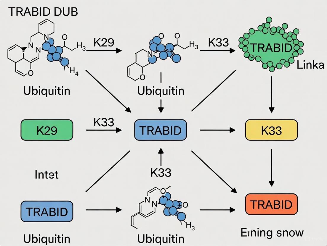
The following diagram illustrates the logical relationship between the key enzymes, the atypical ubiquitin chains they handle, and the resulting cellular outcomes, central to understanding TRABID's functional context.
Diagram Title: TRABID Specificity and Atypical Ub Chain Function
The Scientist's Toolkit: Key Research Reagents
This table catalogs essential reagents that have been critical for the experimental investigation of K29 and K33-linked ubiquitin chains.
Table 3: Essential Research Reagents for Studying K29/K33 Linkages
| Reagent / Tool | Function / Role in Research | Key Features & Examples |
|---|---|---|
| Linkage-Specific E3 Ligases | Catalyze the assembly of specific atypical chains for in vitro studies. | UBE3C: Assembles K29-linked chains [1].TRIP12: Forms K29 linkages and K29/K48-branched chains [17].AREL1: Assembles K33-linked chains [1]. |
| Linkage-Specific DUBs | Validate chain linkage identity; probe chain function by preventing cleavage. | TRABID: Highly specific for cleaving K29 and K33 linkages [1] [7]. |
| Defined Linkage Substrates | Provide standardized substrates for DUB activity assays, binding studies, and structural work. | Diubiquitin & Polyubiquitin: Commercially available K29- and K33-linked diUb (e.g., from UbiQ Bio) [18]. |
| Ubiquitin Mutants | Determine linkage specificity of E3s and DUBs in biochemical assays. | K-only (Kx-only): Ubiquitin with only one lysine available for chain formation.K-to-R: Ubiquitin where a specific lysine is mutated to arginine, blocking that linkage [1]. |
| Structural Biology Tools | Elucidate the 3D conformation of chains and their interactions with binders. | Crystal Structures: Revealed extended conformation of K29-diUb and its complex with TRABID NZF1 [7].Cryo-EM: Visualized TRIP12 E3 in action during K29/K48-branched chain formation [17]. |
| Cell-Based Ubiquitin Replacement Systems | Investigate the specific physiological roles of a linkage type in a cellular context. | Ub(K-to-R) Cell Lines: Cell lines where endogenous ubiquitin is replaced with a mutant (e.g., K29R) to abrogate formation of a specific linkage, enabling phenotypic and proteomic analysis [10]. |
The deubiquitinase TRABID (ZRANB1) is a central figure in the study of atypical ubiquitin signaling, specifically tuned for the recognition and cleavage of K29- and K33-linked ubiquitin chains [1] [13]. This linkage specificity is determined by its N-terminal Npl4-like zinc finger (NZF) domains, with NZF1 specifically binding K29/K33-linked diubiquitin, and its ovarian tumor (OTU) catalytic domain [1] [7]. A critical advancement in this field was the identification of a functional DUB-E3 pair, wherein TRABID regulates the E3 ubiquitin ligase HECTD1 [19]. This axis provides a foundational model for understanding how K29 linkages, particularly within the context of K29/K48-branched chains, are dynamically controlled and their subsequent cellular functions. This application note details the experimental approaches for studying this axis, providing key protocols and reagents for the scientific community.
The seminal study by Harris et al. (2021) established the TRABID-HECTD1 relationship through a series of key experiments [19]. The core findings are summarized in the tables below.
Table 1: Summary of Key Experimental Findings on the TRABID-HECTD1 Axis
| Experimental Finding | Description | Experimental Method(s) Used |
|---|---|---|
| HECTD1 Chain Specificity | HECTD1 preferentially assembles K29- and K48-linked ubiquitin chains in vitro. It requires branching at K29/K48 to achieve full ubiquitin ligase activity. | UbiCREST, Ub-AQUA/PRM Proteomics, In Vitro Autoubiquitination [19] |
| TRABID Substrate Trapping | HECTD1 was identified as a candidate substrate of TRABID through an interactome analysis of catalytic-dead TRABID constructs. | Co-immunoprecipitation + Mass Spectrometry (Interactome) [19] |
| Functional Regulation | Depletion of TRABID leads to the degradation of the HECTD1 protein, establishing a stabilizing function for TRABID. | Transient Knockdown, Genetic Knockout (e.g., CRISPR/Cas9) [19] |
| Linkage Specificity | TRABID is highly specific for recognizing and cleaving K29- and K33-linked ubiquitin chains. | UbiCREST DUB Profiling, Structural Studies [1] [13] [7] |
Table 2: Quantitative Data on HECTD1 Ubiquitin Chain Linkage Preference from Ub-AQUA/PRM Proteomics
| Ubiquitin Linkage Type | Relative Abundance in HECTD1 Autoubiquitination | Notes |
|---|---|---|
| K29-linked chains | High | Preferentially assembled by HECTD1 [19] |
| K48-linked chains | High | Preferentially assembled by HECTD1 [19] |
| K29/K48-branched chains | Essential for full activity | Branching is required for HECTD1's full ubiquitin ligase activity [19] |
| K33-linked chains | Not specified for HECTD1 | Primary specificity of TRABID, alongside K29 [1] [13] |
Detailed Experimental Protocols
Protocol 1: Identifying TRABID Substrates by Interactome Analysis
This protocol is used to identify candidate substrates of a DUB, such as TRABID, by "trapping" them with catalytic-dead mutants.
- Construct Design: Generate two types of catalytic-dead TRABID constructs for parallel validation:
- Cell Transfection & Lysis: Transfect mammalian cells (e.g., HEK293T) with plasmids encoding these constructs with an appropriate tag (e.g., FLAG, GFP). After 24-48 hours, lyse the cells in a non-denaturing lysis buffer (e.g., RIPA buffer supplemented with protease inhibitors and N-ethylmaleimide to inhibit endogenous DUBs).
- Affinity Purification: Incubate the cell lysates with antibody-conjugated beads (e.g., anti-FLAG M2 agarose) for several hours at 4°C. Wash the beads extensively with lysis buffer to remove non-specifically bound proteins.
- Elution and Protein Identification: Elute the bound protein complexes using a competitive peptide (e.g., 3xFLAG peptide) or by low-pH elution. Subject the eluates to tryptic digestion and analysis by liquid chromatography-tandem mass spectrometry (LC-MS/MS).
- Data Analysis: Compare the proteins identified in both the point mutant and ΔOTU traps against a control (e.g., empty vector or GFP-only). Proteins enriched in both TRABID catalytic-dead constructs are high-confidence candidate substrates, as was done to identify HECTD1 [19].
Protocol 2: Determining Ubiquitin Chain Linkage Specificity (UbiCREST)
The UbiCREST (Ubiquitin Chain Restriction) assay is used to determine the linkage specificity of a DUB like TRABID or the chains assembled by an E3 ligase like HECTD1 [19].
- Prepare Ubiquitin Chains: Source a panel of purified homotypic ubiquitin chains (K6, K11, K27, K29, K33, K48, K63, M1). These are commercially available or can be enzymatically assembled and purified.
- Set Up DUB Reactions: For a 20 µL reaction, mix 100-200 ng of a specific ubiquitin chain with the purified DUB (e.g., TRABID) in an appropriate reaction buffer (e.g., 50 mM Tris-HCl pH 7.5, 150 mM NaCl, 5 mM DTT). Incubate at 37°C for 30-60 minutes.
- Include Controls: Run parallel reactions with:
- Chain-only control (no DUB).
- Catalytic-dead DUB control (e.g., TRABID C443S).
- Well-characterized linkage-specific DUBs as controls for chain integrity (e.g., OTUB1 for K48, AMSH for K63).
- Terminate and Analyze: Stop the reaction by adding SDS-PAGE loading buffer. Analyze the cleavage products by western blotting using a pan-ubiquitin antibody or SDS-PAGE with Coomassie staining. TRABID should specifically cleave K29- and K33-linked chains [1] [13].
Protocol 3: Profiling E3 Ligase Chain Assembly with Ub-AQUA/PRM
This quantitative mass spectrometry method precisely measures the types of ubiquitin linkages an E3 ligase, like HECTD1, assembles.
- In Vitro Ubiquitination Assay: Set up an autoubiquitination reaction for HECTD1 containing E1 enzyme, E2 enzyme (e.g., UBE2L3 for HECTD1), ATP, and wild-type ubiquitin. Incubate to allow chain formation [19].
- Protein Denaturation and Digestion: Denature the reaction mixture, reduce, and alkylate cysteine residues. Digest the proteins with a specific protease, typically trypsin, which cleaves after lysine and arginine. A key point is that trypsin digestion generates a signature di-glycine (GlyGly) remnant on the modified lysine of ubiquitin, which is used for quantification.
- Spike-in AQUA Peptides: Add known quantities of heavy isotope-labeled synthetic peptides corresponding to the GlyGly-modified lysine residues for all possible ubiquitin linkages (K6, K11, K27, K29, K33, K48, K63) and an unmodified ubiquitin reference peptide.
- LC-PRM Analysis: Analyze the peptide mixture using Liquid Chromatography-Parallel Reaction Monitoring (LC-PRM) mass spectrometry. This targeted MS method allows for the precise and simultaneous quantification of the light (endogenous) and heavy (synthetic) peptides.
- Data Quantification: Calculate the absolute amount of each linkage type in the sample by comparing the peak areas of the endogenous peptides to the known quantities of their heavy analogs. This revealed HECTD1's preference for K29 and K48 linkages [19].
Pathway and Workflow Visualizations
Diagram 1: The TRABID-HECTD1 Regulatory Axis. HECTD1 assembles K29/K48-branched ubiquitin chains that promote substrate degradation. TRABID cleaves the K29 linkages, thereby attenuating the degradation signal and stabilizing the substrate.
Diagram 2: Experimental Workflow for Characterizing a DUB-E3 Pair. This workflow outlines the key steps, from initial discovery to mechanistic insight, as used to characterize the TRABID-HECTD1 axis.
The Scientist's Toolkit: Essential Research Reagents
Table 3: Key Research Reagent Solutions for Studying K29/K33 Linkages and Branched Chains
| Reagent / Tool | Function / Application | Example & Notes |
|---|---|---|
| Linkage-Specific DUBs | To selectively cleave and probe specific ubiquitin linkages in vitro and in cells. | TRABID: For K29 and K33 linkages [1] [13]. vOTU: Cleaves most linkages except K29; useful for purifying K29 chains [7] [20]. |
| Linkage-Specific Binders | To detect and enrich for specific ubiquitin chain types via immunofluorescence or pull-down. | sAB-K29: Synthetic antibody fragment for specific recognition of K29 linkages [20]. TRABID-NZF1: The isolated zinc finger domain can be used as a selective K29/K33 binder [1] [9]. |
| Ubiquitin Mutants | To restrict or identify linkage types formed during in vitro ubiquitination assays. | Ub(K0): All lysines mutated to Arg, used as a "donor" ubiquitin. Ub(Kx-only): Only one specific lysine is available for chain formation [1]. |
| HECT E3 Ligase Tools | To study the formation of atypical and branched ubiquitin chains. | TRIP12: Known to form K29 linkages and K29/K48-branched chains [17] [9]. AREL1: HECT E3 that assembles K33-linked chains [1]. HECTD1: E3 ligase that assembles K29/K48-branched chains and is stabilized by TRABID [19]. |
| Quantitative MS Standards | For absolute quantification of ubiquitin chain linkages from biological samples. | AQUA/PRM Peptides: Heavy isotope-labeled, GlyGly-modified ubiquitin peptides spiked into samples for precise quantification by mass spectrometry [1] [9] [19]. |
Tools and Techniques: Assembling, Detecting, and Probing K29/K33 Ubiquitin Chains
The functional diversity of ubiquitin signaling is largely governed by the ability of ubiquitin to form structurally distinct polymeric chains through different internal lysine residues. Among these, K29- and K33-linked ubiquitin chains represent atypical linkages whose study has been hampered by limited tools for their production and recognition [1]. This application note details standardized methodologies utilizing the HECT E3 ligases UBE3C and AREL1 for the enzymatic production of these atypical chains, providing essential tools for research focused on the deubiquitinase TRABID, which exhibits remarkable specificity for both K29 and K33 linkages [1] [7] [11].
The development of these enzymatic assembly systems has enabled biochemical and structural characterization of these previously elusive chain types, revealing that both K29- and K33-linked chains adopt open and dynamic conformations in solution, similar to K63-linked chains but distinct from the compact structures of K48-linked chains [1]. This structural insight provides the foundation for understanding recognition mechanisms by specialized binding proteins like TRABID.
Quantitative Characterization of HECT E3 Ligases and Their Linkage Specificities
Table 1: Linkage Specificity Profiles of HECT E3 Ligases
| E3 Ligase | Primary Linkages Assembled | Secondary Linkages | Chain Conformation | Key Structural Features |
|---|---|---|---|---|
| UBE3C | K29 (23%), K48 (63%) [1] | K11 (10%) [1] | Open, dynamic [1] | Extended HECT domain with open, L-shaped bilobed conformation; critical N-terminal region (aa 693-743) and loop (aa 758-762) for stability/activity [21] |
| AREL1 | K33 (36%), K11 (36%) [1] | K48 (20%) [1] | Open, dynamic [1] | Inverted T-shaped bilobed HECT conformation; unique extended N-terminal region (aa 436-482) and additional loop (aa 567-573) essential for stability [22] |
| NEDD4L | K63 (96%) [1] | Minimal other linkages [1] | N/A | Representative of NEDD4 subfamily specificity [23] |
| E6AP | K48 [23] | N/A | N/A | Representative of "other" subfamily with degradation-focused specificity [23] |
Table 2: Key Mutations and Their Functional Impact on HECT E3 Activity
| E3 Ligase | Mutation | Functional Consequence | Application |
|---|---|---|---|
| AREL1 | E701A [22] | Substantially increased autopolyubiquitination and substrate ubiquitination activity [22] | Enhanced chain production; gain-of-function applications |
| AREL1 | ΔC-terminal 3 aa [22] | Complete abrogation of autoubiquitination and reduced substrate ubiquitination [22] | Negative control; study of catalytic mechanism |
| UBE3C | K903R [21] | Reduced autoubiquitination (K903 identified as major autoubiquitination site) [21] | Control for activity assays |
| UBE3C | Q961A, S1049A [21] | Substantially decreased autoubiquitination activity [21] | Study of E2-E3 transthiolation process |
Experimental Protocols for Atypical Ubiquitin Chain Production
Protocol 1: Enzymatic Assembly of K29-Linked Ubiquitin Chains Using UBE3C
Principle: The HECT domain of UBE3C (amino acids 693-1083) exhibits intrinsic specificity for assembling K29-linked ubiquitin chains through a two-step catalytic mechanism involving transthiolation from E2 to the catalytic cysteine of UBE3C, followed by isopeptide bond formation on the lysine 29 residue of ubiquitin [1] [21].
Reagents and Equipment:
- Recombinant HECT domain of UBE3C (aa 693-1083) [21]
- E1 activating enzyme [1]
- E2 conjugating enzyme (compatible with UBE3C) [1]
- ATP regeneration system [1]
- Wild-type ubiquitin [1]
- Reaction buffer: 50 mM Tris-HCl (pH 7.5), 50 mM NaCl, 10 mM MgCl₂, 1 mM DTT [1]
- Purification system: Size exclusion chromatography or ion exchange chromatography [1]
Procedure:
- Enzyme Preparation: Express and purify the extended HECT domain of UBE3C (aa 693-1083). Confirmation of structural integrity via circular dichroism or analytical size exclusion is recommended [21].
- Reaction Setup: In a final volume of 1 mL, combine:
- 50 mM Tris-HCl buffer (pH 7.5)
- 50 mM NaCl
- 10 mM MgCl₂
- 1 mM DTT
- 2 mM ATP
- 0.1 μM E1 enzyme
- 2-5 μM E2 enzyme
- 10 μM UBE3C HECT domain
- 200 μM wild-type ubiquitin
- Incubation: Conduct the reaction at 30°C for 2-4 hours with gentle agitation [1].
- Chain Elongation Monitoring: Remove aliquots at 30-minute intervals and analyze by SDS-PAGE and immunoblotting with anti-ubiquitin antibodies to monitor chain formation.
- Reaction Termination: Stop the reaction by adding 10 mM EDTA to chelate Mg²⁺ ions required for ATP hydrolysis.
- Chain Purification: Purify K29-linked chains using size exclusion chromatography (Superdex 200) pre-equilibrated with 50 mM ammonium bicarbonate buffer (pH 7.5). Pool fractions containing polyubiquitin chains of desired length [1] [7].
- Quality Control: Verify linkage specificity using:
Protocol 2: Enzymatic Assembly of K33-Linked Ubiquitin Chains Using AREL1
Principle: The extended HECT domain of AREL1 (amino acids 436-823) preferentially assembles K33-linked ubiquitin chains, with its unique structural features including an additional loop (aa 567-573) and a critical N-terminal extension (aa 436-482) that stabilizes the catalytic domain [1] [22].
Reagents and Equipment:
- Recombinant extended HECT domain of AREL1 (aa 436-823) [22]
- E1 activating enzyme [1]
- Compatible E2 conjugating enzyme [1]
- ATP regeneration system [1]
- Wild-type ubiquitin [1]
- Reaction buffer: 50 mM HEPES (pH 7.2), 100 mM NaCl, 10 mM MgCl₂, 1 mM DTT [22]
- Size exclusion chromatography system [1]
Procedure:
- Enzyme Preparation: Express and purify AREL1 extended HECT domain (aa 436-823). Note that the N-terminal region (aa 436-482) is indispensable for stability and activity - constructs lacking this region show poor solubility and minimal activity [22].
- Reaction Setup: In a final volume of 1 mL, combine:
- 50 mM HEPES buffer (pH 7.2)
- 100 mM NaCl
- 10 mM MgCl₂
- 1 mM DTT
- 2 mM ATP
- 0.1 μM E1 enzyme
- 2-5 μM E2 enzyme
- 10 μM AREL1 HECT domain
- 200 μM wild-type ubiquitin
- Incubation: Conduct at 30°C for 2-3 hours. For enhanced yield, consider the AREL1 E701A mutant which exhibits substantially increased autopolyubiquitination activity [22].
- Chain Elongation Monitoring: Analyze progression as described for UBE3C.
- Reaction Termination: Add 10 mM EDTA.
- Purification: Purify chains using size exclusion chromatography. For structural studies, focus on fractions containing tetra-ubiquitin and longer species [1].
- Specificity Verification:
Protocol 3: Combined E3-DUB System for Linkage-Defined Chain Production
Principle: Combining HECT E3 ligases with linkage-specific deubiquitinases enables production of homogenous, linkage-defined ubiquitin chains by editing mixed chain populations [1] [7].
Procedure:
- Initial Chain Assembly: Perform large-scale ubiquitin chain assembly using either UBE3C or AREL1 as described in Protocols 1 and 2.
- DUB Selection:
- Editing Reaction: Incubate crude chain preparations with catalytic domains of appropriate DUBs at sub-complete hydrolysis conditions (typically 15-30 minutes at 37°C) to trim heterogeneous chains while preserving target linkages [1].
- Product Isolation: Purify edited chains using ion-exchange chromatography to separate by length [1].
- Validation: Confirm linkage purity via mass spectrometry and binding assays with linkage-specific domains like TRABID NZF1 [1] [11].
Workflow Visualization
The Scientist's Toolkit: Essential Research Reagents
Table 3: Key Research Reagents for K29/K33 Ubiquitin Chain Studies
| Reagent Category | Specific Examples | Function and Application | Key Characteristics |
|---|---|---|---|
| HECT E3 Ligases | UBE3C HECT domain (aa 693-1083) [21] | K29-linked chain assembly [1] | L-shaped bilobed conformation; requires N-terminal extension for full activity [21] |
| AREL1 HECT domain (aa 436-823) [22] | K33-linked chain assembly [1] | Inverted T-shaped conformation; unique loop (567-573); N-terminal region essential [22] | |
| TRABID Domains | NZF1 domain [1] [11] | K29/K33 chain detection and purification [1] [11] | Specific binding to K29/K33 diUb; structural basis for linkage selectivity [1] [11] |
| Catalytic OTU domain [1] | K29/K33 chain hydrolysis [1] | Linkage-specific deubiquitination; chain editing [1] | |
| Ubiquitin Mutants | K29-only Ub [1] | Specific chain assembly verification [1] | All lysines except K29 mutated to arginine [1] |
| K33-only Ub [1] | Specific chain assembly verification [1] | All lysines except K33 mutated to arginine [1] | |
| K0 Ub [1] | Negative control [1] | All lysines mutated to arginine; prevents chain formation [1] | |
| Analytical Tools | AQUA mass spectrometry [1] | Absolute quantification of linkage composition [1] | Spike-in isotope-labeled standards for precise quantification [1] |
| Linkage-specific DUBs [1] | Chain linkage verification [1] | Enzymatic confirmation of linkage type [1] |
Applications in TRABID DUB Specificity Research
The enzymatic assembly systems described herein have been instrumental in elucidating the structural basis of TRABID specificity for K29 and K33 linkages. Structural studies of TRABID's NZF1 domain in complex with K29- and K33-linked diubiquitin reveal an intriguing filamentous binding mode where NZF1 engages each ubiquitin-ubiquitin interface [1]. This binding mechanism exploits the unique flexibility and interface characteristics of K29 and K33 linkages, providing the molecular basis for TRABID's dual linkage specificity.
Recent research leveraging these tools has uncovered essential cellular functions for K29-linked ubiquitination, particularly in chromatin regulation and epigenome maintenance. Studies using ubiquitin replacement cell lines have demonstrated that K29-linked ubiquitylation is strongly associated with chromosome biology and is essential for proteasomal degradation of the H3K9 methyltransferase SUV39H1 [10]. Preventing K29-linkage-dependent SUV39H1 turnover deregulates H3K9me3 homeostasis, establishing a key role for this atypical ubiquitin linkage in preserving epigenome integrity [10].
Furthermore, the ability to produce homogenous K29 and K33 chains has enabled the discovery that these linkages frequently exist within heterotypic or branched chains containing other linkage types [7] [11]. This complexity expands the potential regulatory mechanisms controlled by TRABID and highlights the importance of these enzymatic tools for deciphering the ubiquitin code in physiological and pathological contexts.
Troubleshooting and Technical Considerations
- Low Chain Yield: Optimize E3:E2 ratio (typically 2:1 to 5:1), ensure adequate ATP regeneration, and verify E3 catalytic activity through autoubiquitination assays [1] [22].
- Linkage Heterogeneity: Employ combined E3-DUB editing systems or sequential purification using linkage-specific binding domains like TRABID NZF1 [1] [11].
- E3 Stability: For AREL1, ensure inclusion of the N-terminal extended region (aa 436-482); for UBE3C, include the region preceding the HECT domain (aa 693-743) [22] [21].
- Activity Enhancement: Consider AREL1 E701A mutant for increased activity, but monitor for potential alteration in linkage specificity [22].
- Chain Length Control: Use limited reaction times or sub-stoichiometric E3 concentrations to favor shorter chains; extended incubations with excess E3 produce longer polymers [1].
The study of atypical ubiquitin chains, particularly those linked through lysine 29 (K29) and lysine 33 (K33), has been historically challenging due to the inability to produce homogeneous chain preparations in sufficient quantities for biochemical and structural studies. Traditional enzymatic approaches using E2 enzymes or HECT E3 ligases alone often yield heterogeneous chain mixtures or result in predominant autoubiquitylation of the E3 ligase itself, complicating the isolation of free, defined polyubiquitin chains [8]. To address this critical methodological gap, researchers have developed ubiquitin chain-editing complexes that combine the synthetic capability of E3 ligases with the selective processing activity of deubiquitinases (DUBs) [8] [1]. This innovative approach enables the large-scale production of linkage-specific ubiquitin chains, which has been particularly valuable for investigating the specificity and function of DUBs such as TRABID that show pronounced selectivity for K29 and K33 linkages [8] [1] [24].
The fundamental principle underlying the ubiquitin chain-editing platform involves harnessing the complementary activities of an E3 ligase that preferentially assembles a specific linkage type with a DUB that selectively cleaves contaminating linkages while preserving the linkage of interest. This method has unlocked systematic research into the cellular roles of K29-linked ubiquitin chains, which are now known to function in diverse processes including proteotoxic stress responses, neuronal development, and DNA repair [17] [25] [13]. By providing researchers with a reliable source of homogeneous atypical ubiquitin chains, this technology continues to facilitate breakthroughs in our understanding of the ubiquitin code.
Experimental Protocols for K29-Linked Ubiquitin Chain Production
Core Assembly Methodology
The following protocol describes the production of K29-linked ubiquitin chains using a chain-editing complex consisting of the HECT E3 ligase UBE3C and the viral deubiquitinase vOTU [8]:
Table 1: Key Reagents for K29-Linked Ubiquitin Chain Assembly
| Reagent | Source | Function in Protocol |
|---|---|---|
| UBE3C HECT E3 Ligase | Human, recombinant | Catalyzes formation of K29-linked ubiquitin chains [8] [1] |
| UBE2D3 E2 Enzyme | Human, recombinant | Transfers ubiquitin to UBE3C during chain assembly [8] |
| vOTU Deubiquitinase | Viral, recombinant | Cleaves contaminating linkages and releases free chains from autoubiquitylated UBE3C [8] |
| Wild-type Ubiquitin | Bovine or recombinant | Primary building block for chain assembly |
| K29-only Ubiquitin Mutant | Recombinant (all lysines except K29 mutated to arginine) | Verification of linkage specificity [8] |
| ATP Regenerating System | Commercial | Provides energy for E1-mediated ubiquitin activation |
Step-by-Step Procedure:
Reaction Setup: In a total volume of 100-500 μL, combine the following components in reaction buffer (typically 50 mM Tris-HCl pH 7.5, 50 mM NaCl, 10 mM MgCl₂, 0.5 mM DTT):
- 2 μM human E1 enzyme
- 10-20 μM E2 enzyme (UBE2D3)
- 1-2 μM HECT E3 ligase (UBE3C)
- 0.5-1 μM vOTU deubiquitinase
- 200-400 μM ubiquitin (wild-type or K29-only mutant)
- 5 mM ATP
- ATP-regenerating system (10 mM creatine phosphate, 10 ng/μL creatine kinase)
Chain Assembly Incubation: Incubate the reaction mixture at 30°C for 2-4 hours to allow for polyubiquitin chain formation. The inclusion of vOTU in the initial reaction prevents the accumulation of non-K29 linkages and promotes the release of free chains from autoubiquitylated UBE3C [8].
Reaction Termination: Stop the reaction by adding EDTA to a final concentration of 10 mM to chelate magnesium ions required for E1 activity.
Product Purification: Purify the free polyubiquitin chains using size-exclusion chromatography (Superdex 75 or 200) or ion-exchange chromatography to separate the unanchained chains from enzymes, unreacted ubiquitin, and higher molecular weight aggregates [8].
Linkage Verification: Confirm the linkage specificity of the purified chains through:
Methodology Validation and Quality Control
The protocol specificity must be rigorously validated using appropriate controls:
Table 2: Validation Methods for K29 Linkage Specificity
| Method | Application | Expected Outcome |
|---|---|---|
| Ubiquitin Mutant Analysis | Assembly with K29R ubiquitin mutant | Significant reduction in chain formation [8] |
| DUB Specificity Profiling | Treatment with TRABID | Complete hydrolysis to monoubiquitin [8] [25] |
| Mass Spectrometry | Parallel reaction monitoring (pRM) LC-MS/MS | Detection of specific K29 linkage signatures [8] |
| Linkage Selectivity Assay | Comparison with other linkage-specific DUBs | Resistance to cleavage by M1-specific OTULIN [8] |
Representative results demonstrate that when this protocol is followed with wild-type ubiquitin, long polyubiquitin chains are produced that are completely hydrolyzed to monoubiquitin by TRABID, confirming the presence of K29 linkages [8]. When the K29R ubiquitin mutant is used in place of wild-type ubiquitin, chain formation is significantly impaired, further validating the linkage specificity of the approach [8].
Structural and Functional Insights Enabled by Chain-Editing Methodology
Structural Characterization of K29-Linked Ubiquitin Chains
The application of the chain-editing methodology has enabled key structural insights into K29-linked ubiquitin chains. Using chains produced via the UBE3C/vOTU system, researchers determined the crystal structure of K29-linked diubiquitin, which revealed an extended conformation with both ubiquitin moieties' hydrophobic patches exposed and available for binding interactions [8]. This structural arrangement differs significantly from the compact conformations observed for K48-linked chains and helps explain the distinct functional properties of K29 linkages.
Furthermore, this approach facilitated the structural characterization of the TRABID NZF1 domain in complex with K29-linked chains, revealing the molecular basis for linkage-specific recognition [8]. The structure demonstrated that NZF1 binding involves interaction with the hydrophobic patch on only one ubiquitin moiety while exploiting the inherent flexibility of K29 chains to achieve selective binding [8]. These structural insights would not have been possible without access to homogenous K29-linked ubiquitin chains produced via the chain-editing methodology.
Biological Applications and Discoveries
The availability of pure K29-linked ubiquitin chains through the chain-editing approach has enabled numerous biological discoveries regarding the function of these atypical chains:
The chain-editing methodology has revealed that K29 linkages frequently exist within mixed or branched chains containing other linkage types, particularly K48 linkages [8]. These heterotypic chains appear to have specialized functions distinct from homotypic chains. Recent research utilizing these tools has identified specific roles for K29-linked ubiquitination in:
Neuronal Development: TRABID-mediated deubiquitination of K29/K33-linked chains on adenomatous polyposis coli (APC) regulates its trafficking to microtubule plus-ends, which is essential for growth cone formation and neurite outgrowth [25]. Patient-derived mutations in TRABID that impair either its catalytic activity or STRIPAK complex binding disrupt this process and are associated with neurodevelopmental disorders including microcephaly [25].
DNA Damage Response: TRABID deubiquitinates K29-linked polyubiquitination on 53BP1 mediated by the E3 ligase SPOP, regulating 53BP1 retention at double-strand break sites and influencing the choice between homologous recombination and non-homologous end joining repair pathways [13]. This function has implications for cancer therapy, as TRABID overexpression creates synthetic lethality to PARP inhibitors in prostate cancer models [13].
Proteotoxic Stress Response: K29-linked chains have been associated with cellular responses to proteotoxic stress, with their abundance increasing following proteasomal inhibition [8] [17]. The HECT E3 TRIP12, which generates K29 linkages and K29/K48-branched chains, has been linked to neurodegenerative disorders [17].
The Scientist's Toolkit: Essential Research Reagents
Table 3: Key Research Reagents for Ubiquitin Chain Editing and TRABID Studies
| Reagent/Category | Specific Examples | Function and Application |
|---|---|---|
| E3 Ligases | UBE3C, AREL1, TRIP12 | Assembly of K29- and K33-linked chains [8] [1] [17] |
| Deubiquitinases | vOTU, TRABID | Linkage editing and specificity analysis [8] [25] |
| Ubiquitin Mutants | K29-only, K29R, K0 (all lysines mutated) | Linkage specificity controls [8] [1] |
| Binding Domains | TRABID NZF1 domain | K29/K33 chain detection and purification [8] [1] |
| Chemical Tools | Ubiquitin suicide probes, NCI inhibitors (NSC112200, NSC267309) | DUB activity profiling and inhibition [26] |
| Cell Models | TRABID patient variant knock-in mice, TRABID-overexpressing cancer cells | Physiological functional analysis [25] [13] |
Concluding Remarks
The development of ubiquitin chain-editing complexes represents a methodological breakthrough that has transformed our ability to study atypical ubiquitin linkages. By combining E3 ligases with complementary DUBs such as vOTU, researchers can now produce homogeneous preparations of K29-linked ubiquitin chains that were previously inaccessible. This capability has been instrumental in advancing our understanding of TRABID specificity for K29 and K33 linkages and has revealed the important roles these atypical chains play in neuronal development, DNA damage response, and human disease. As research in this field continues to evolve, the chain-editing platform will undoubtedly remain an essential tool for deciphering the complex language of the ubiquitin code.
The diversification of ubiquitin signaling is achieved through the assembly of polyubiquitin chains connected via different lysine residues, each encoding distinct cellular functions. Among these, the so-called "atypical" linkages, including lysine 29 (K29) and lysine 33 (K33), have remained particularly enigmatic due to a historical paucity of tools for their specific manipulation and detection [1]. Recent advances have identified the deubiquitinase TRABID (encoded by the ZRANB1 gene) as a highly specific reader and editor of these atypical chains [27]. TRABID contains three Npl4-type zinc finger (NZF) domains, with its N-terminal NZF1 domain demonstrating remarkable selectivity for both K29- and K33-linked ubiquitin chains [1] [7]. This application note details the development and implementation of the TRABID-NZF1 domain as a selective biochemical tool for the immunoprecipitation and detection of K29- and K33-linked ubiquitin conjugates, providing researchers with a critical reagent to decipher the functions of these understudied post-translational modifications.
Structural Basis and Mechanism of Specificity
The molecular basis for TRABID-NZF1's linkage selectivity is revealed through structural studies. Crystallographic analysis of the NZF1 domain in complex with K33-linked diubiquitin reveals an intricate binding mode that exploits the unique structural features of these atypical chains [1]. Unlike the compact conformations of K48-linked chains, both K29- and K33-linked diubiquitin adopt open and dynamic conformations in solution, making the hydrophobic patches on individual ubiquitin moieties accessible for binding [1] [7].
The NZF1 domain engages ubiquitin through the canonical Ile44 hydrophobic patch, but achieves specificity through its ability to accommodate the distinct spacing and flexibility of K29 and K33 linkages [1] [7]. In the crystal structure, NZF1 domains form continuous filaments along K33-linked ubiquitin chains, binding each ubiquitin-ubiquitin interface [1]. This structural arrangement demonstrates how TRABID achieves selective recognition, providing a blueprint for rational engineering of the domain for biochemical applications.
Table 1: Structural Features of K29/K33-linked Ubiquitin and TRABID-NZF1
| Feature | Structural Characteristic | Functional Implication |
|---|---|---|
| K29/K33 Chain Conformation | Open, extended conformations in solution [1] [7] | Exposes hydrophobic patches for domain binding |
| NZF1 Binding Interface | Canonical Ile44 hydrophobic patch engagement [7] | Provides general ubiquitin binding affinity |
| Specificity Mechanism | Accommodates unique K29/K33 linkage geometry and flexibility [1] | Enables discrimination from other linkage types |
| Observed Binding Mode | Filamentous structure along ubiquitin polymer [1] | Suggests avidity effects in longer chains |
Recombinant TRABID-NZF1 Probe Generation
Expression Construct Design and Purification
The generation of a functional TRABID-NZF1 probe begins with construct design. The human TRABID cDNA fragment encoding the NZF1 domain (approximately amino acids 1-70) should be cloned into an appropriate expression vector with an N-terminal affinity tag (such as GST or 6xHis) for purification and a C-terminal AviTag for site-specific biotinylation [9].
Detailed Protocol: Protein Expression and Purification
- Transformation and Expression: Transform the expression construct into E. coli BL21(DE3) cells. Grow cultures in LB medium at 37°C until OD600 reaches 0.6-0.8. Induce protein expression with 0.5 mM IPTG and incubate overnight at 18°C.
- Cell Lysis and Clarification: Harvest cells by centrifugation at 4,000 × g for 20 minutes. Resuspend cell pellet in lysis buffer (50 mM Tris-HCl pH 8.0, 300 mM NaCl, 1 mM DTT, 1 mM PMSF) and lyse by sonication. Clarify the lysate by centrifugation at 15,000 × g for 45 minutes at 4°C.
- Affinity Purification: Incubate the clarified lysate with glutathione-sepharose (for GST-tag) or Ni-NTA (for His-tag) resin for 1-2 hours at 4°C. Wash resin extensively with wash buffer (lysis buffer with 20-50 mM imidazole for His-tag).
- Tag Cleavage and Elution: For GST-tagged proteins, incubate with PreScission protease (1:100 w/w) overnight at 4°C to remove the tag. Elute the untagged NZF1 domain.
- Size Exclusion Chromatography: Further purify the protein using a Superdex 75 column equilibrated with storage buffer (20 mM HEPES pH 7.5, 150 mM NaCl, 1 mM DTT). Concentrate the protein to 5-10 mg/mL, aliquot, and flash-freeze in liquid nitrogen for storage at -80°C.
Biotinylation for Detection Applications
For streptavidin-based detection systems, site-specific biotinylation is critical:
- In Vitro Biotinylation: Incubate purified AviTagged NZF1 with BirA enzyme, 100 μM biotin, 10 mM ATP, and 10 mM MgOAc in biotinylation buffer (100 mM bicine, pH 8.3) for 1 hour at 30°C.
- Removal of Free Biotin: Desalt the reaction mixture using a Zeba spin desalting column to remove free biotin.
- Validation: Verify biotinylation efficiency by streptavidin blot and confirm ubiquitin binding activity by pull-down assay with K29-linked diubiquitin.
Application Protocols
Immunoprecipitation of K29/K33-Modified Proteins
The TRABID-NZF1 domain serves as a highly specific capture reagent for isolating proteins modified with K29- or K33-linked ubiquitin chains from complex cellular lysates.
Detailed Protocol: Linkage-Specific Ubiquitin Pull-Down
- Lysate Preparation: Harvest cells and lyse in IP lysis buffer (50 mM Tris-HCl pH 7.5, 150 mM NaCl, 1% NP-40, 10% glycerol, 1 mM EDTA) supplemented with protease inhibitors (including 10 μM PR619 to inhibit DUBs) and 10 mM N-ethylmaleimide to preserve ubiquitin conjugates. Clarify by centrifugation at 16,000 × g for 15 minutes at 4°C.
- Bait Immobilization: Incubate 10-50 μg of biotinylated TRABID-NZF1 with 100 μL of streptavidin magnetic beads for 1 hour at 4°C. Wash beads twice with lysis buffer.
- Binding Reaction: Incubate the immobilized NZF1 with 1-2 mg of pre-cleared cellular lysate for 2 hours at 4°C with gentle rotation.
- Washing: Wash beads extensively with lysis buffer (4-5 washes, 5 minutes each).
- Elution: Elute bound proteins with 2× Laemmli sample buffer containing 100 mM DTT at 95°C for 10 minutes, or with 2 M urea in 50 mM Tris pH 8.0 for downstream mass spectrometry analysis.
Table 2: Critical Reagents for TRABID-NZF1-Based Applications
| Reagent / Material | Function / Application | Notes and Considerations |
|---|---|---|
| Recombinant TRABID-NZF1 | Primary binding module for K29/K33 linkages | GST-tagged for purification; AviTagged for biotinylation [9] |
| Streptavidin Magnetic Beads | Solid support for immobilization | Enable efficient pull-down and washing steps [9] |
| N-Ethylmaleimide (NEM) | Deubiquitinase inhibitor | Preserves ubiquitin conjugates during lysis (10-20 mM) [27] |
| K29/K33-linked DiUbiquitin | Specificity controls | Essential for validating binding activity [1] [7] |
| Protease Inhibitor Cocktail | Prevents protein degradation | Critical for maintaining integrity of ubiquitin conjugates |
Detection of K29/K33 Linkages in Cell-Based Assays
TRABID-NZF1 can be deployed as a detection reagent in various assay formats to monitor the dynamics of K29/K33-linked ubiquitination in cellular contexts.
Detailed Protocol: Western Blot Detection
- Cell Treatment and Lysis: Treat cells with appropriate stimuli (e.g., DNA damaging agents for 53BP1 studies [27]) and lyse as described in section 4.1.
- Electrophoresis and Transfer: Separate proteins by SDS-PAGE (4-12% gradient gels recommended) and transfer to PVDF membrane.
- Blocking: Block membrane with 5% BSA in TBST for 1 hour at room temperature.
- Probe Incubation: Incubate membrane with biotinylated TRABID-NZF1 (1-2 μg/mL in blocking buffer) overnight at 4°C.
- Detection: Wash membrane and incubate with streptavidin-HRP (1:10,000) for 1 hour at room temperature. Develop with enhanced chemiluminescence substrate [28].
Controls for Specificity
- Include samples treated with linkage-specific deubiquitinases
- Compete with recombinant K29- or K33-linked diubiquitin
- Compare with catalytically inactive TRABID mutant (C443S) [27] [3]
Key Biological Applications and Case Studies
Regulation of DNA Damage Repair
TRABID and its specific recognition of K29-linked ubiquitin play a critical role in DNA damage response. TRABID deubiquitinates K29-linked polyubiquitination on 53BP1 mediated by the E3 ligase SPOP, preventing 53BP1 dissociation from double-strand break sites and promoting non-homologous end joining (NHEJ) over homologous recombination (HR) [27] [13]. The TRABID-NZF1 probe can be used to monitor this specific ubiquitination event:
Application Protocol: Monitoring 53BP1 K29-Ubiquitination
- Induce DNA damage in cells (e.g., 10 Gy ionizing radiation)
- Harvest cells at time points (0, 1, 4, 8 hours post-irradiation)
- Perform immunoprecipitation of 53BP1 using standard protocols
- Detect K29-linkages in the immunoprecipitate using biotinylated TRABID-NZF1 as described in section 4.2
- Correlate K29-ubiquitination status with 53BP1 foci formation and repair pathway choice
Chromatin Regulation and Epigenetic Control
Recent research has established that K29-linked ubiquitylation is strongly associated with chromosome biology and is essential for proteasomal degradation of the H3K9 methyltransferase SUV39H1 [10]. This K29-linked ubiquitination is catalyzed by TRIP12 and reversed by TRABID, establishing a regulatory circuit that controls H3K9me3 homeostasis and epigenome integrity [10].
Application Protocol: Studying SUV39H1 Regulation
- Treat cells with proteasome inhibitor (MG132, 10 μM, 6 hours) to accumulate ubiquitinated species
- Perform pull-down with TRABID-NZF1 to enrich K29-modified proteins
- Probe for SUV39H1 by western blot to detect K29-ubiquitinated forms
- Combine with TRIP12 knockdown to confirm specificity
Proteasomal Degradation Pathways
K29-linked ubiquitin chains frequently form heterotypic branched chains with K48-linkages, particularly on substrates targeted for proteasomal degradation [9]. The TRABID-NZF1 probe can be employed to distinguish the contribution of K29-linkages in these complex ubiquitin codes:
Application Protocol: Analyzing Branched Ubiquitin Signals
- Express substrates known to undergo K29/K48-branched ubiquitination (e.g., OTUD5 [9])
- Enrich K29-linked conjugates with TRABID-NZF1 pull-down
- Elute under mild conditions (2 M urea)
- Reprecipitate with K48-linkage specific binder (e.g., UBR5 NZF)
- Identify proteins modified with both linkages by mass spectrometry
Troubleshooting and Technical Considerations
Optimization and Validation
- Binding Specificity: Always include control pull-downs with free streptavidin beads and a mutated NZF1 domain with impaired ubiquitin binding
- Linkage Selectivity: Validate with lysates from cells expressing single-lysine ubiquitin mutants (K29-only or K33-only) where possible
- Signal-to-Noise: Optimize wash stringency by increasing salt concentration (up to 300 mM NaCl) or adding 0.1% SDS to reduce non-specific binding
Limitations and Alternative Approaches
- The TRABID-NZF1 domain recognizes both K29 and K33 linkages; discrimination between these requires additional validation methods such as linkage-specific deubiquitinases
- Binding affinity for monoubiquitination is typically lower than for polyubiquitin chains due to avidity effects
- For in vivo applications, consider overexpression of full-length TRABID or CRISPR-mediated tagging of endogenous loci
The TRABID-NZF1 domain represents a powerful and specific tool for interrogating the biological functions of K29- and K33-linked ubiquitin chains. Its application in pull-down and detection assays enables researchers to overcome previous technical limitations in studying these atypical ubiquitin linkages. As research continues to uncover the roles of these modifications in DNA damage response, epigenetic regulation, and proteasomal targeting [27] [9] [10], the methodologies outlined here will serve as essential frameworks for advancing our understanding of these complex signaling pathways. The integration of TRABID-NZF1-based probes with emerging proteomic and genomic technologies promises to illuminate the full functional spectrum of the ubiquitin code.
Within the intricate framework of the ubiquitin code, the specificity of deubiquitinases (DUBs) for particular chain types is a critical area of study. This document details the application of two key biochemical techniques—UbiCREST (Ubiquitin Chain Restriction) and Ub-AQUA (Ubiquitin Absolute QUAntification) mass spectrometry—for the verification of linkage types, with a specific focus on the K29 and K33-linked ubiquitin chain specificity of the DUB TRABID (ZRANB1). TRABID, a DUB from the ovarian tumor (OTU) family, is highly tuned for the recognition and cleavage of K29- and K33-linked ubiquitin chains, yet the cellular functions of these atypical chains remain less clear compared to the well-characterized K48 and K63 linkages [2]. The techniques described herein are foundational for validating TRABID substrates and understanding the functional impact of these atypical ubiquitin signals in pathways such as autophagy, transcriptional regulation, and H3K9me3-dependent epigenome integrity [2] [10].
Background: TRABID and Atypical Ubiquitin Chains
TRABID contains three Npl4-like zinc finger (NZF) domains and an OTU catalytic domain. Its N-terminal NZF1 domain is the minimal ubiquitin-binding domain (UBD) required for the specific recognition of K29- and K33-linked diubiquitin [1] [2]. Structural studies have revealed that this domain binds the Ub-Ub interface in K33-linked chains, explaining the molecular basis for this specificity [1] [15] [12]. A key functional relationship has been established between TRABID and the E3 ligase HECTD1, which assembles K29- and K48-linked chains, forming a DUB-E3 pair that regulates K29 linkages [2]. Furthermore, the E3 ligase TRIP12 has been identified as a specific assembler of K29-linked chains on substrates like the histone methyltransferase SUV39H1, with TRABID reversing this modification to regulate H3K9me3 homeostasis and epigenome integrity [10]. These findings underscore the importance of precise techniques to verify the linkage types assembled by these E3s and cleaved by TRABID.
The UbiCREST Technique
UbiCREST is a qualitative method that uses a panel of linkage-specific deubiquitinases (DUBs) to decipher the ubiquitin chain types present in a sample. The core principle is that treating a polyubiquitinated substrate with specific DUBs will result in a characteristic cleavage pattern that can be visualized by immunoblotting, thereby revealing the chain linkage composition [29].
Detailed UbiCREST Protocol
Sample Preparation:
- Generate Substrate: Produce the polyubiquitinated protein of interest. This can be achieved through in vitro ubiquitylation assays using purified E1, E2, E3 enzymes (e.g., TRIP12, HECTD1, or UBE3C for K29 linkages; AREL1 for K33 linkages), and ubiquitin [1] [2]. Alternatively, immunopurify the ubiquitinated protein from cell lysates.
- Purify: Isolate the ubiquitinated substrate using techniques like nickel-NTA pulldown (if His-tagged ubiquitin is used) or immunoprecipitation with a substrate-specific antibody.
DUB Treatment:
- Prepare DUB Panel: Set up parallel reactions, each containing the ubiquitinated substrate and a single, linkage-specific DUB. The recommended DUBs and their working concentrations are summarized in Table 1.
- Incubate: Conduct the reactions in appropriate DUB assay buffers (typically containing Tris-HCl pH 7.5-8.0, NaCl, and DTT) for 1-2 hours at 37°C [29].
Analysis:
- Terminate Reaction: Stop the digestion by adding SDS-PAGE loading buffer.
- Visualize: Resolve the samples by SDS-PAGE and perform immunoblotting using an anti-ubiquitin antibody.
- Interpret Results: The cleavage pattern reveals the linkage types present. Complete digestion by a specific DUB indicates the predominant presence of that linkage type. For example, cleavage of a substrate by TRABID suggests the presence of K29/K33 linkages, while resistance to OTUB1 (K48-specific) and sensitivity to TRABID would point towards a non-K48 atypical chain [29].
Table 1: Key DUBs for UbiCREST Analysis of K29/K33 Linkages
| DUB Enzyme | Linkage Specificity | Useful Final Concentration | Key Considerations for K29/K33 Research |
|---|---|---|---|
| TRABID | K29 & K33 (also cleaves K63 with lower activity) | 0.5 - 10 µM | The primary DUB for identifying K29/K33 chains. Low yields from bacterial expression [29]. |
| OTUB1 | K48 | 1 - 20 µM | Serves as a K48-specific control. Highly specific and not very active [29]. |
| Cezanne | K11 | 0.1 - 2 µM | Very active; distinguishes K11 from K29/K33 linkages [29]. |
| OTUD1 | K63 | 0.1 - 2 µM | Very active; distinguishes K63 from K29/K33 linkages [29]. |
| USP21 or USP2 | Pan-specific (all linkages) | 1 - 5 µM (USP21) | Positive control; complete digestion confirms the sample is polyubiquitinated [29]. |
The following workflow diagram illustrates the UbiCREST procedure:
The Ub-AQUA Mass Spectrometry Technique
Ub-AQUA is a quantitative mass spectrometry method that provides absolute quantification of all ubiquitin linkage types within a sample. It utilizes heavy isotope-labeled internal standard peptides corresponding to the tryptic remnants of each ubiquitin linkage, allowing for precise measurement of chain abundance [2] [30].
Detailed Ub-AQUA Protocol
Sample Preparation and Digestion:
- Generate and Purify Substrate: As described in the UbiCREST protocol (Section 3.1), generate and purify the polyubiquitinated substrate.
- Denature and Reduce: Dissolve the purified ubiquitinated material in a denaturing buffer (e.g., 8 M urea) and reduce disulfide bonds with DTT.
- Alkylate and Digest: Alkylate cysteine residues with iodoacetamide and then digest the proteins with trypsin. Trypsin cleaves ubiquitin after arginine 74, generating a signature di-glycine (GlyGly) remnant on the lysine residue of the acceptor ubiquitin that was modified [31].
Spike-in of AQUA Peptides:
- Prepare AQUA Peptides: Obtain synthetic, heavy isotope-labeled (e.g., 13C6, 15N2) peptides corresponding to the tryptic GlyGly-modified peptides for all seven lysine linkages and the N-terminus (M1).
- Spike-in: Add a known quantity of each AQUA peptide to the trypsin-digested sample. The heavy peptides are chemically identical to their endogenous counterparts but can be distinguished by mass spectrometry [2] [30].
LC-MS/MS Analysis and Quantification:
- Chromatographic Separation: Subject the peptide mixture to liquid chromatography (LC) to separate peptides based on hydrophobicity. Refined methods allow for the quantification of all chain types in short 10-minute LC-MS/MS runs, enabling high-throughput analysis [30].
- Mass Spectrometry Analysis: Analyze the eluting peptides using tandem mass spectrometry (MS/MS) in a targeted mode, such as Parallel Reaction Monitoring (PRM), which enhances sensitivity and specificity [2] [30].
- Absolute Quantification: For each linkage type, the mass spectrometer quantifies the peak areas for both the light (endogenous) and heavy (AQUA standard) peptides. The known concentration of the spiked-in heavy standard allows for the absolute quantification of the endogenous peptide, revealing the precise abundance of each ubiquitin linkage type in the original sample [2].
Table 2: Ub-AQUA Quantitative Data in TRABID-Related Research
| Experimental Context | Key Quantitative Finding (Linkage Abundance) | Technique Used | Research Implication |
|---|---|---|---|
| In vitro ubiquitylation of OTUD5 by E3 ligase TRIP12 [9] | OTUD5 was specifically modified with K29-linked chains by TRIP12. | Ub-AQUA/PRM | Confirmed TRIP12 as a specific assembler of K29 chains on its substrate. |
| In vitro autoubiquitination of HECT E3 ligase AREL1 [1] | Assembled 36% K33, 36% K11, 20% K48, and minor other linkages. | AQUA-MS | Identified AREL1 as a major assembler of K33-linked ubiquitin chains. |
| In vitro autoubiquitination of HECT E3 ligase UBE3C [1] | Assembled 63% K48, 23% K29, and 10% K11 linkages. | AQUA-MS | Established UBE3C as a mixer of K48 and K29 linkages. |
| Linkage composition in murine tissues [30] | Enrichment of K33 linkages was observed in heart and muscle tissues. | Ub-AQUA-PRM | Suggested a previously unknown role for K33 chains in contractile tissues. |
The Ub-AQUA workflow is summarized in the following diagram:
Research Reagent Solutions for K29/K33 Linkage Studies
Table 3: Essential Research Reagents for Investigating K29/K33 Linkages
| Reagent / Tool | Function / Specificity | Application in TRABID/K29/K33 Research |
|---|---|---|
| Catalytic Dead TRABID (e.g., C443S mutant) [2] | Traps ubiquitinated substrates via its NZF domains without cleaving them. | Identification of TRABID candidate substrates through immunoprecipitation and proteomics. |
| TRABID NZF1 Domain [1] [9] | Specific binder of K29- and K33-linked diubiquitin. | Used as a capture reagent to enrich for proteins modified with K29 linkages (e.g., in pull-down assays). |
| Recombinant HECT E3 Ligases (TRIP12, UBE3C, HECTD1, AREL1) [1] [2] [9] | Assemble specific ubiquitin chains (K29, K33, K48) in in vitro assays. | Used to generate defined chain types for validating TRABID specificity and substrate ubiquitylation. |
| Linkage-Specific DUBs (see Table 1) [29] | Hydrolyze specific ubiquitin linkages. | Core components of the UbiCREST assay to deconvolute chain topology. |
| K29/K33-linkage Specific Tools (e.g., TRABID-ZZ fusion) [32] | Binds K29/K33 linkages for enrichment and detection. | Alternative to NZF1 for microscopy (puncta formation) or Western blot analysis. |
| Single-Lysine Ubiquitin Mutants (e.g., K29-only, K33-only) [1] | Restricts chain formation to a single linkage type in vitro. | Determining the linkage specificity of E3 ligases (e.g., TRIP12, HECTD1) and DUBs. |
The power of UbiCREST and Ub-AQUA is maximized when they are used as complementary techniques. UbiCREST offers a rapid, qualitative assessment of chain types and architecture—for instance, revealing that HECTD1 requires branching at K29/K48 for full ligase activity [2]. Ub-AQUA provides robust, quantitative data on the absolute abundance of each linkage, confirming that TRIP12 specifically modifies OTUD5 with K29-linked chains [9]. Together, they form an indispensable toolkit for validating the specificity of enzymes like TRABID and its partner E3 ligases, moving beyond simple homotypic chains to the complex reality of branched and heterotypic ubiquitin signals. The diagrams and tables provided in this document serve as a practical guide for researchers aiming to apply these techniques to unravel the functions of atypical ubiquitin chains in cellular regulation and disease.
Within the intricate network of the ubiquitin system, the deubiquitinase (DUB) TRABID (also known as ZRANB1) emerges as a key regulator with exceptional specificity for the poorly understood atypical ubiquitin chains linked via lysine 29 (K29) and lysine 33 (K33) [1] [33] [34]. Research into these chain types has been hampered by a lack of tools to study their cellular dynamics. This application note details the use of catalytically inactive TRABID as a molecular trap to visualize and study these elusive ubiquitin signals in a cellular context, providing a method to advance our understanding of their formation and function. The protocol is framed within a broader thesis investigating the unique specificity of TRABID and the cellular roles of K29- and K33-linked ubiquitination.
The molecular structure of TRABID is key to its function. It contains three Npl4-like zinc finger (NZF) domains at its N-terminus that act as ubiquitin-binding domains (UBDs), and a C-terminal ovarian tumor (OTU) domain that confers its deubiquitinating activity [2] [13]. The first NZF domain (NZF1) has been specifically characterized as a K29/K33-linkage selective binding module [1] [7]. The catalytic domain is extended by an Ankyrin repeat domain (AnkUBD) that also contributes to linkage-specific recognition and cleavage efficiency [33].
Table 1: Key Structural Domains of TRABID and Their Functions in Ubiquitin Trapping
| Domain | Location | Function in Trapping Experiment |
|---|---|---|
| NZF1 | N-terminus (aa 1-~70) | Primary high-affinity binding site for K29- and K33-linked diubiquitin [1] [7]. |
| NZF2 & NZF3 | N-terminus (~70-200) | Contribute to polyubiquitin binding; may stabilize trapped chains [2]. |
| AnkUBD | Precedes OTU domain (~245-340) | Acts as an enzymatic S1' Ub binding site; crucial for full efficiency and linkage-specificity [33]. |
| OTU Domain | C-terminus (~340-708) | Contains catalytic site; mutation here (e.g., C443S) renders the enzyme inactive, creating the trap [2] [13]. |
The Principle of Trapping with Catalytically Inactive TRABID
The core of this methodology involves the expression of a mutant TRABID protein that can bind to its target ubiquitin chains but cannot cleave them. This creates a stable complex that can be visualized in cells and used to identify substrates. The most common and validated approach is the use of a catalytically inactive point mutant, TRABIDC443S, where the active-site cysteine is replaced by serine [2] [26] [13]. This mutation ablates the deubiquitinase activity while preserving the ability of the NZF and AnkUBD domains to bind K29- and K33-linked chains with high specificity [1] [33].
When overexpressed in cells, TRABIDC443S acts as a dominant-negative protein, sequestering K29/K33-linked polyubiquitin chains and their modified substrates. This results in the formation of prominent intracellular puncta visible by immunofluorescence microscopy [2]. These puncta represent accumulations of the trapped TRABID mutant bound to polyubiquitin chains, providing a direct visual readout of the localization of these atypical ubiquitin signals.
An alternative trapping strategy involves using a TRABID ΔOTU construct, which entirely lacks the catalytic OTU domain but retains the N-terminal NZF domains [2]. Interactome studies have confirmed that both TRABIDC443S and TRABID ΔOTU can efficiently co-precipitate a similar set of ubiquitinated proteins, validating them as effective ubiquitin traps for proteomic studies aimed at identifying novel substrates [2].
Diagram 1: Mechanism of ubiquitin chain trapping by catalytically inactive TRABID.
Experimental Protocol: Trapping and Visualization
This section provides a detailed, step-by-step protocol for expressing catalytically inactive TRABID in mammalian cells and visualizing the resulting ubiquitin-positive puncta.
Required Reagents and Constructs
Table 2: Essential Research Reagents for TRABID Trapping Experiments
| Reagent / Solution | Function / Purpose | Example / Notes |
|---|---|---|
| TRABIDC443S Plasmid | Catalytically inactive DUB expression vector | Point mutation in OTU domain (Cysteine 443 to Serine) [2] [13]. |
| TRABID ΔOTU Plasmid | Alternative trapping construct for control experiments | Deletion of the entire catalytic OTU domain [2]. |
| Transfection Reagent | For plasmid delivery into mammalian cells | Use reagent appropriate for your cell line (e.g., Lipofectamine). |
| Cell Culture Media & Supplements | For maintenance and transfection of mammalian cells | Standard media (e.g., DMEM) with serum. |
| Fixative Solution | To preserve cellular architecture for imaging | 4% Paraformaldehyde (PFA) in PBS. |
| Permeabilization Buffer | To allow antibody entry into cells | 0.1-0.5% Triton X-100 in PBS. |
| Blocking Solution | To reduce non-specific antibody binding | 1-5% BSA or serum in PBS. |
| Primary Antibody: Anti-TRABID | To detect expressed TRABID protein | Use species-specific, validated antibody. |
| Primary Antibody: Anti-Ubiquitin | To detect trapped ubiquitin chains | Linkage-specific antibodies (e.g., α-K29) can be used for validation [2]. |
| Fluorophore-Conjugated Secondary Antibodies | For visualization by fluorescence microscopy | Use antibodies against host species of primary antibodies. |
| Mounting Medium with DAPI | To preserve samples and stain nuclei | Use anti-fade mounting medium. |
Step-by-Step Methodology
Cell Seeding and Transfection:
- Plate appropriate mammalian cells (e.g., HEK293T, U2OS, HeLa) onto glass coverslips in a multi-well plate.
- Allow cells to adhere and reach 50-70% confluency.
- Transfect cells with a plasmid encoding TRABIDC443S (experimental) or wild-type TRABID (control) using a standard transfection protocol. An empty vector should be included as a negative control.
- Incubate cells for 24-48 hours to allow for sufficient protein expression.
Cell Fixation and Permeabilization:
- Aspirate the culture medium and gently wash cells with 1X Phosphate-Buffered Saline (PBS).
- Fix cells by incubating with 4% PFA in PBS for 15 minutes at room temperature.
- Remove PFA and wash cells three times with PBS.
- Permeabilize cells by incubating with 0.1-0.5% Triton X-100 in PBS for 10 minutes.
- Wash cells three times with PBS to remove the permeabilization buffer.
Immunofluorescence Staining:
- Incubate cells with a blocking solution (e.g., 3% BSA in PBS) for 1 hour at room temperature to prevent non-specific antibody binding.
- Prepare primary antibodies diluted in blocking solution.
- Incubate cells with the primary antibody mixture. A typical combination is:
- Mouse anti-TRABID (to visualize the location of the trap itself).
- Rabbit anti-Ubiquitin (pan-specific or, ideally, K29/K33-linkage specific).
- Incubate overnight at 4°C or for 1-2 hours at room temperature.
- Wash cells three times with PBS (5-10 minutes per wash) to remove unbound primary antibodies.
- Incubate cells with fluorophore-conjugated secondary antibodies (e.g., Alexa Fluor 488-conjugated anti-mouse and Alexa Fluor 555-conjugated anti-rabbit) diluted in blocking solution for 1 hour at room temperature, protected from light.
- Wash cells three times with PBS as before.
Mounting and Imaging:
- Carefully mount the coverslips onto glass slides using an anti-fade mounting medium containing DAPI to stain cell nuclei.
- Seal the coverslips with clear nail polish and allow them to dry.
- Image the cells using a confocal or high-resolution fluorescence microscope.
- In cells expressing TRABIDC443S, look for the characteristic bright cytoplasmic puncta that co-stain for both TRABID and ubiquitin [2]. These puncta are the hallmark of successful ubiquitin chain trapping.
Diagram 2: Experimental workflow for trapping and visualizing ubiquitin chains.
Data Interpretation and Analysis
Successful execution of the protocol will yield specific and interpretable results. Cells expressing the TRABIDC443S mutant should display distinct cytoplasmic puncta that show strong co-localization between the TRABID signal and the ubiquitin signal [2]. In contrast, cells expressing wild-type TRABID typically show a more diffuse cytoplasmic and nuclear staining pattern without prominent puncta formation, as the enzyme actively turns over its substrate chains [35].
The formation of these puncta can be used as a qualitative cellular readout for the presence of K29/K33-linked ubiquitin chains. Furthermore, this assay can be combined with linkage-specific ubiquitin antibodies or with the knockdown of E3 ligases known to assemble these chains (e.g., UBE3C for K29, AREL1 for K33) to further validate the signal specificity [1].
Table 3: Expected Outcomes and Controls for TRABID Trapping Experiments
| Experimental Condition | Expected Microscopy Phenotype | Interpretation |
|---|---|---|
| TRABIDC443S | Formation of bright cytoplasmic puncta positive for TRABID and ubiquitin. | Successful trapping of polyubiquitin chains by the catalytically inactive DUB [2] [13]. |
| Wild-Type TRABID | Diffuse cellular staining; absence of prominent puncta. | Active DUB cleaves ubiquitin chains, preventing accumulation [35]. |
| Empty Vector (Control) | No TRABID signal; background ubiquitin staining. | Baseline ubiquitin distribution without experimental manipulation. |
| Co-stain with K29-Ub Antibody | Puncta show positive signal for K29 linkage. | Validation that trapped chains include K29 linkages [2]. |
Applications in Research and Drug Discovery
The catalytically inactive TRABID trapping methodology extends beyond a simple localization tool and serves as a powerful technique for broader research and therapeutic discovery applications.
- Substrate Identification: TRABIDC443S and TRABID ΔOTU can be used as affinity baits in immunoprecipitation-mass spectrometry (IP-MS) experiments to identify novel cellular substrates modified with K29/K33-linked ubiquitin. This approach has successfully identified the E3 ligase HECTD1 as a bona fide TRABID substrate [2] [36].
- Functional Studies in Disease Models: This protocol can be applied in disease-relevant cell models to investigate the role of atypical ubiquitination. For instance, TRABID has been implicated in regulating DNA damage repair by controlling the retention of 53BP1 at double-strand breaks, a process with implications for cancer therapy [13].
- Validation of TRABID Mutations: The puncta formation assay has been instrumental in characterizing patient-derived mutations in the ZRANB1 gene. Studies show that mutations affecting TRABID's DUB activity or its binding to the STRIPAK complex disrupt its normal function and can impede neurite growth, linking it to neurodevelopmental disorders [35].
- Inhibitor Screening and Validation: While small-molecule inhibitors of TRABID have been reported [26], the trapping assay provides a direct cellular readout to validate candidate compounds. A true inhibitor would be expected to reduce the formation of ubiquitin-positive puncta by wild-type TRABID in a gain-of-function context.
Troubleshooting and Technical Notes
- Low or No Puncta Formation: Ensure high transfection efficiency and confirm protein expression by Western blotting. Optimization of transfection conditions and/or using a different cell line may be necessary. Overexpression of a K29/K33-specific E3 ligase (e.g., UBE3C) can be used to stimulate chain formation and enhance the signal [1].
- High Background Ubiquitin Staining: Titrate antibody concentrations to find the optimal signal-to-noise ratio. Ensure thorough washing after antibody incubations and include all necessary controls (empty vector, wild-type TRABID) to accurately interpret the specific signal.
- Co-localization Studies: To investigate if trapped ubiquitin chains co-localize with specific cellular organelles (e.g., endosomes, Golgi), include organelle-specific markers (e.g., RAB5, GM130) in the immunofluorescence staining panel.
In conclusion, the use of catalytically inactive TRABID provides a robust and specific method to probe the cellular landscape of K29- and K33-linked ubiquitination. This protocol offers researchers a powerful tool to visualize these atypical chains, identify new substrates, and elucidate their functional roles in health and disease.
Challenges and Solutions: Validating TRABID Function and Overcoming Experimental Hurdles
The deubiquitinase TRABID (ZRANB1), recognized for its specificity towards K29 and K33-linked ubiquitin chains, occupies a controversial role in the Wnt/β-catenin signaling pathway. Early research proposed it as a positive regulator that cleaves K63-linked chains on substrates like APC. In contrast, recent studies elucidate a tumor-suppressive function involving the regulation of branched K29/K48 linkages on distinct substrates, presenting a paradigm shift in understanding its mechanistic actions. This application note reconciles these conflicting datasets by contextualizing TRABID's functions within its established linkage specificity. We provide structured experimental data, detailed protocols for key assays, and visual tools to guide research into the complex role of this DUB in development and disease.
TRABID, an ovarian tumor (OTU) family deubiquitinase (DUB), is biochemically tuned for the recognition and hydrolysis of K29 and K33-linked ubiquitin chains [2] [1]. Its N-terminal NZF1 domain exhibits selective binding for K29/K33-linked diubiquitin, a specificity defined by structural analyses [7] [11]. Paradoxically, early cellular studies implicated TRABID as a positive regulator of Wnt/β-catenin signaling through the deubiquitination of K63-linked chains on the APC protein [37]. This apparent contradiction between its in vitro linkage specificity and initial reported cellular function has created a significant controversy in the field.
Subsequent investigations have begun to resolve this paradox, revealing a more complex picture. TRABID has been shown to form a functional DUB/E3 pair with the ligase HECTD1, which preferentially assembles branched K29/K48-linked ubiquitin chains [2]. Furthermore, in hepatocellular carcinoma (HCC), TRABID acts as a tumor suppressor by regulating the stability of the transcription factor Twist1, an effect that is dependent on its DUB activity [38]. This application note synthesizes these conflicting datasets and provides a methodological framework for researchers to investigate the context-dependent functions of TRABID, firmly rooted in the core thesis of its specificity for K29 and K33 linkages.
Reconciling Conflicting Data on TRABID Function
The table below summarizes the key conflicting findings and proposes reconciled interpretations based on contemporary understanding of TRABID's specificity.
Table 1: Reconciliation of Controversial TRABID Functions in Signaling
| Reported Function | Supporting Evidence | Apparent Conflict with K29/K33 Specificity | Reconciled Interpretation & Modern Context |
|---|---|---|---|
| Positive regulator of Wnt/β-catenin signaling [37] | Genetic interaction; proposed deubiquitination of APC. | Originally attributed to K63-linked chain cleavage [37]. | Initial observations may reflect regulation of an uncharacterized K29/K33 substrate in the pathway. The K63 claim is an outlier in biochemical literature. |
| Inhibitor of HCC growth & metastasis [38] | Trabid knockout promotes tumor growth and EMT in vivo; correlates with poor patient prognosis. | Aligned with K29/K33 specificity; mechanism involves K63 deubiquitination of Twist1 [38]. | The K63 role on Twist1 requires re-examination with K29/K33-specific tools. The tumor-suppressive phenotype is robust and consistent. |
| Stabilizer of E3 ligase HECTD1 [2] | TRABID depletion triggers HECTD1 degradation; HECTD1 assembles K29/K48-branched chains. | Directly aligns with and explains K29/K33 specificity. | Represents a mechanistically coherent model: TRABID cleaves K29 linkages on HECTD1 or its substrates to prevent degradation. |
Proposed Unified Model
The diagram below illustrates a unified model that reconciles TRABID's controversial roles through its established specificity for K29 and K33 linkages, contextualizing earlier findings within a modern framework.
Essential Research Toolkit
The investigation of TRABID's biology requires a specific set of reagents and tools designed to probe its unique linkage specificity.
Table 2: Essential Reagents for TRABID and Atypical Ubiquitin Chain Research
| Research Tool | Specific Example / Form | Primary Function in Experimentation |
|---|---|---|
| Catalytic Mutants | TRABIDC443S, TRABID ΔOTU [2] | Substrate trapping via affinity purification; identification of endogenous targets. |
| Linkage-Specific Ubiquitin | K29-only (K29O), K33-only Ub mutants; Defined diUb [1] [7] | In vitro DUB activity assays; determining linkage specificity of TRABID and associated E3s. |
| Linkage-Specific DUBs | vOTU (for K29 chain editing) [7] | Tool for generating pure atypical chains or validating linkage identity in samples. |
| Linkage-Specific Antibodies | Anti-K29-linkage, Anti-K33-linkage specific antibodies [1] | Detection of endogenous K29/K33 ubiquitination in cells via immunoblotting or immunofluorescence. |
| Recombinant E3 Ligases | HECTD1, UBE3C [2] [1] | In vitro ubiquitination assays to characterize chain topology and TRABID substrate relationships. |
| Mass Spectrometry Probes | TRABID NZF1 domain [11] | Affinity purification of K29/K33-linked ubiquitinated proteins from cell lysates for proteomics. |
Core Experimental Protocols & Data
Protocol: TRABID Substrate Trapping and Interactome Analysis
This protocol identifies candidate substrates by using catalytically inactive TRABID to bind ubiquitinated proteins without cleaving the ubiquitin signal [2].
- Plasmid Transfection: Transfect HEK293ET cells with constructs for catalytic dead TRABID (e.g., TRABIDC443S or TRABID ΔOTU), each with an affinity tag (e.g., FLAG).
- Cell Lysis and Immunoprecipitation: After 24-48 hours, lyse cells in a mild, non-denaturing lysis buffer (e.g., RIPA buffer with 1% NP-40). Use anti-FLAG M2 magnetic beads to immunoprecipitate the TRABID complexes for 2-4 hours at 4°C.
- Stringent Washing: Wash beads thoroughly with high-salt wash buffer (e.g., containing 500 mM NaCl) to remove non-specific interactors.
- Elution and Digestion: Elute bound proteins using FLAG peptide or low-pH elution buffer. Digest the eluates with trypsin.
- Mass Spectrometry Analysis: Analyze the resulting peptides by liquid chromatography-tandem mass spectrometry (LC-MS/MS). Compare the interactomes of the two catalytic dead constructs to identify common proteins, which represent high-confidence candidate substrates.
The workflow for this substrate identification protocol is outlined below.
Table 3: Key Findings from TRABID Trapping Experiments
| Experimental Approach | Key Identified Interactors/Substrates | Validation & Functional Insight |
|---|---|---|
| Comparative Interactome (TRABIDC443S vs. TRABID ΔOTU) [2] | 50 high-confidence candidate proteins, including E3 ligases HECTD1 and HERC2. | Validated HECTD1 as a bonafide substrate; TRABID depletion destabilizes HECTD1. |
| UbiCREST & Ub-AQUA on trapped HECTD1 | Preferential assembly of K29- and K48-linked ubiquitin chains by HECTD1. | Revealed HECTD1 requires branched K29/K48 linkages for full activity [2]. |
| Functional Knockdown/KO | Loss of TRABID leads to decreased HECTD1 protein levels. | Establishes a functional DUB/E3 pair regulating K29-linked ubiquitination. |
Protocol: Determining Ubiquitin Linkage Specificity
This methodology combines in vitro ubiquitination assays with linkage-specific deubiquitinases (DUBs) and mass spectrometry to define the topology of ubiquitin chains [2] [1] [7].
- In Vitro Ubiquitination Assay:
- Set up a reaction containing E1 enzyme, E2 enzyme, the E3 ligase of interest (e.g., HECTD1 or UBE3C), ATP, and wild-type ubiquitin.
- Incubate at 30°C for 1-3 hours.
- Stop the reaction with SDS-PAGE loading buffer or by placing on ice.
- Linkage Analysis via UbiCREST:
- Divide the ubiquitination reaction into several aliquots.
- Treat each aliquot with a different linkage-specific DUB (e.g., OTUB1 for K48, OTUD3 for K11, vOTU for K29, TRABID for K29/K33) for 30-60 minutes.
- Analyze the digestion patterns by anti-ubiquitin immunoblotting. The resistance of a chain to a specific DUB indicates its linkage composition.
- Absolute Quantification via Ub-AQUA/LC-MS:
- Subject a separate portion of the ubiquitination reaction to tryptic digestion.
- Spike the digest with isotopically labeled, GlyGly-modified internal standard peptides corresponding to each ubiquitin linkage type.
- Analyze by LC-MS/MS. The absolute abundance of each linkage type is calculated by comparing the peak areas of the endogenous peptides to their corresponding internal standards.
Table 4: Quantitative Linkage Specificity of HECT E3 Ligases via Ub-AQUA
| E3 Ubiquitin Ligase | Predominant Linkages Assembled (Percentage) | Implication for TRABID Biology |
|---|---|---|
| UBE3C [1] | K48 (63%), K29 (23%), K11 (10%) | Confirms existence of E3s producing K29 linkages, potential TRABID partners. |
| AREL1 [1] | K33 (36%), K11 (36%), K48 (20%) | Demonstrates E3 capability for K33 chain assembly, another potential TRABID substrate. |
| HECTD1 [2] | Preferentially K29 and K48; forms branched K29/K48 chains | Directly defines a TRABID-regulated E3, aligning TRABID function with K29 specificity. |
The perceived controversy surrounding TRABID's role in Wnt signaling and cancer largely dissipates when its functions are interpreted through the lens of its definitive K29 and K33-linkage specificity. The early association with K63-chain cleavage on APC represents an outlier in the biochemical data. A more consistent model positions TRABID as a key regulator of proteostasis for specific substrates, such as HECTD1 and Twist1, through its action on atypical and branched ubiquitin chains. Future research should leverage the protocols and tools detailed herein—particularly substrate trapping and linkage-specific proteomics—to identify the full repertoire of TRABID substrates and definitively establish the molecular mechanisms by which this DUB fine-tunes critical cellular signaling pathways.
Ubiquitination is a pivotal post-translational modification that regulates diverse cellular processes, from protein degradation to immune signaling. While early research focused on homotypic polyubiquitin chains, recent advances have revealed an expanded complexity involving heterotypic and branched ubiquitin chains. These complex architectures significantly increase the signaling capacity of the ubiquitin system, creating a sophisticated "ubiquitin code" that governs cellular fate decisions [39] [40].
Within this complex landscape, the deubiquitinase TRABID emerges as a critical decoder, exhibiting remarkable specificity for the atypical K29 and K33 linkages [1] [8]. This application note details the experimental strategies and methodologies for investigating these complex ubiquitin signals, with particular emphasis on approaches relevant to TRABID research. We provide structured protocols for the production, characterization, and functional analysis of heterotypic and branched chains, enabling researchers to decipher the sophisticated language of ubiquitin signaling in health and disease.
Tool Development: Essential Research Reagent Solutions
Studying atypical ubiquitin linkages and branched chains requires specialized reagents and tools. The table below summarizes key solutions for investigating K29/K33 linkages and branched architectures.
Table 1: Essential Research Reagents for Studying Atypical Ubiquitin Signals
| Reagent Category | Specific Example | Function/Application | Key Features |
|---|---|---|---|
| Linkage-Specific E3 Ligases | UBE3C (HECT E3) [1] [8] | Assembles K29-linked chains in vitro | Often used in combination with DUBs to generate specific chains |
| AREL1 (HECT E3) [1] | Assembles K33-linked chains | Preferentially forms K33 linkages on substrates and as free chains | |
| TRIP12 (HECT E3) [9] [17] | Assembles K29 linkages and K29/K48-branched chains | Key E3 for branched chain formation; implicated in degradation | |
| Linkage-Specific DUBs | TRABID (OTU Family) [1] [8] | Hydrolyzes K29 and K33 linkages | Used for linkage verification and chain editing; contains specific NZF domains |
| vOTU (Viral DUB) [8] | Cleaves most linkages except M1, K27, K29 | Useful in chain-editing complexes to purify specific linkages | |
| Specific Binding Domains | TRABID NZF1 domain [1] [8] | Selective binding to K29/K33-diubiquitin | Tool for pulldown assays and cellular detection of K29/K33 chains |
| Ubiquitin Mutants | K29-only Ub (Ub K29-only) [8] | Contains only K29 as modifiable lysine | Ensures formation of exclusively K29-linked chains in assembly systems |
| Lysine-to-Arg Ub Mutants [1] | e.g., Ub K48R, K63R | Blocks specific linkages during enzymatic assembly of branched chains | |
| Chemical Biology Tools | C-terminally blocked Ub (UbΔ77) [39] | Prevents chain elongation at specific points | Essential for stepwise assembly of defined branched trimers |
Methodological Approaches: Detailed Experimental Protocols
Enzymatic Assembly of K29-Linked Ubiquitin Chains
The production of defined, homotypic K29-linked chains is a prerequisite for biochemical and structural studies. The following protocol describes an efficient Ub chain-editing complex methodology for large-scale generation of K29-linked polyubiquitin [8].
Protocol 1: K29-Linked Chain Assembly Using UBE3C and vOTU
Principle: Co-incubate the HECT E3 ligase UBE3C with a permissive DUB (vOTU) that cleaves non-K29 linkages but preserves K29 chains, resulting in the release of free, homotypic K29-linked polymers.
Reagents:
- E1 activating enzyme (e.g., UBA1)
- E2 conjugating enzyme (e.g., UBE2D3)
- HECT E3 ligase UBE3C (wild-type, catalytic Cys required)
- Viral OTU (vOTU) DUB
- ATP regeneration system
- Wild-type ubiquitin or K29-only ubiquitin mutant
Procedure:
- Set up the ubiquitination reaction in a 50-100 µL volume containing:
- 50 mM Tris-HCl, pH 7.5
- 10 mM MgCl₂
- 0.5 mM DTT
- 2 mM ATP
- 50 µM ubiquitin
- 100 nM E1 (UBA1)
- 1 µM E2 (UBE2D3)
- 1 µM UBE3C E3 ligase
- 1 µM vOTU DUB
Incubate at 37°C for 2-4 hours to allow chain assembly and editing.
Terminate the reaction by adding 5 mM iodoacetamide to alkylate free cysteines.
Purify free ubiquitin chains by ion-exchange or size-exclusion chromatography.
Verify linkage specificity by:
- Treatment with linkage-specific DUBs (e.g., TRABID for K29 linkage cleavage)
- Mass spectrometry analysis (AQUA/PRM) of tryptic digests
- Immunoblotting with linkage-specific antibodies (if available)
Troubleshooting Notes:
- If chain length is insufficient, increase reaction time or enzyme concentrations.
- If linkage purity is compromised, use ubiquitin K29-only mutant instead of wild-type ubiquitin.
- Include controls without vOTU to demonstrate its essential role in releasing free chains.
Stepwise Enzymatic Assembly of Branched Ubiquitin Trimers
Branched ubiquitin chains containing defined linkages can be assembled through sequential enzymatic steps using strategically designed ubiquitin building blocks.
Protocol 2: Assembly of K29-K48 Branched Ubiquitin Trimers
Principle: Use a C-terminally blocked proximal ubiquitin to control the stepwise addition of distal ubiquitins through specific linkages [39].
Reagents:
- Proximal ubiquitin (Ub1-76 or Ub1-72 with C-terminal truncation)
- Distal ubiquitin mutants (e.g., Ub K48R, K63R)
- K48-specific E2/E3 pairs (e.g., UBE2R1/UBE2K)
- K29-specific E3 ligase (TRIP12 or UBE3C)
- DUBs for C-terminal processing (e.g., OTULIN for M1-linkage cleavage)
Procedure:
- Prepare the branched core:
- Start with a C-terminally blocked proximal ubiquitin (Ub1-72 or UbD77)
- Ligate the first distal ubiquitin (Ub K48R,K63R) using a K63-specific enzyme pair (UBE2N/UBE2V1)
- Purify the K63-linked dimer
Add the branch linkage:
- To the purified dimer, add a second distal ubiquitin (Ub K48R,K63R) using a K29-specific E3 ligase (TRIP12)
- Incubate with TRIP12, E1, E2, and ATP regeneration system
- Purify the resulting branched trimer
Optional chain extension:
- For more complex architectures, use a Ub-capping approach with OTULIN to remove the C-terminal block
- Expose the native C-terminus for further elongation
Validation:
- Confirm chain architecture by mass spectrometry
- Test sensitivity to linkage-specific DUBs (TRABID for K29, USP2 for K48)
- Use binding assays with specific UBDs (TRABID NZF1 for K29 recognition)
Diagram 1: Workflow for Branched Ubiquitin Trimer Assembly
Structural Analysis of TRABID-Ligand Interactions
Understanding the molecular basis of TRABID's specificity for K29 and K33 linkages provides insights for functional studies and potential therapeutic targeting.
Protocol 3: Crystallographic Analysis of TRABID NZF1 in Complex with K29/K33-diUb
Principle: Determine the high-resolution structure of TRABID's N-terminal NZF1 domain bound to K29- or K33-linked diubiquitin to elucidate the molecular mechanism of linkage recognition [1] [8].
Reagents:
- Recombinant TRABID NZF1 domain (residues 1-80)
- K29-linked diubiquitin (from Protocol 1 or commercial source)
- Crystallization screens (e.g., Hampton Research)
- Cryoprotectants (e.g., glycerol, ethylene glycol)
Procedure:
- Protein complex preparation:
- Express and purify TRABID NZF1 domain (residues 1-80) with an N-terminal His-tag
- Purify K29-linked diubiquitin using Protocol 1 or commercial sources
- Form the complex by incubating NZF1 with diubiquitin in 2:1 molar ratio
- Purify the complex by size-exclusion chromatography
Crystallization screening:
- Set up sitting-drop vapor diffusion plates
- Mix 0.1 µL protein complex (10-15 mg/mL) with 0.1 µL reservoir solution
- Incubate at 4°C and 20°C
- Optimize initial hits by grid screening around successful conditions
Data collection and structure determination:
- Flash-cool crystals in liquid nitrogen with appropriate cryoprotectant
- Collect X-ray diffraction data at synchrotron source
- Solve structure by molecular replacement using known NZF and ubiquitin structures
- Refine model through iterative cycles of manual building and computational refinement
Key Findings from Previous Structures:
- TRABID NZF1 binds the hydrophobic patch (I44-centered surface) of one ubiquitin moiety
- The flexible K29 linkage allows optimal positioning for specific recognition
- Structural insights explain why TRABID discriminates against K48 and K63 linkages
Analytical Techniques: Verification and Functional Characterization
Linkage Verification by Mass Spectrometry
Absolute quantification (AQUA) mass spectrometry provides definitive verification of ubiquitin linkage types in assembled chains or cellular samples.
Table 2: Mass Spectrometry Parameters for Ubiquitin Linkage Verification
| Parameter | Specification | Application |
|---|---|---|
| Digestion Protocol | Trypsin digestion produces linkage-specific peptides | Generates signature peptides for each linkage type |
| AQUA Peptides | Isotope-labeled GlyGly-modified internal standards | Absolute quantification of specific linkages |
| PRM (Parallel Reaction Monitoring) | Targeted MS/MS for specific peptide detection | High-sensitivity detection of low-abundance linkages |
| Quantifiable Linkages | All 8 linkage types (M1, K6, K11, K27, K29, K33, K48, K63) | Comprehensive linkage profiling |
| Detection Limit | Femtomole level for most linkages | Suitable for in vitro and cellular samples |
Procedure Summary:
- Digest ubiquitin chains with trypsin to generate signature peptides
- Spike with isotope-labeled AQUA standards for absolute quantification
- Analyze by LC-MS/MS with PRM for targeted detection
- Quantify peak areas to determine linkage composition [1] [40]
Cellular Validation of K29/K33 Chain Function
To translate in vitro findings to cellular contexts, implement the following validation strategies:
Cellular Pulldown Assays:
- Use GST-tagged TRABID NZF1 domain to capture endogenous K29/K33-modified proteins
- Identify bound proteins by mass spectrometry or immunoblotting
- Compare with catalytically inactive TRABID mutants as control [1]
Functional Disruption Studies:
- Introduce point mutations in TRABID NZF1 domain that disrupt K29/K33 binding
- Assess impact on TRABID localization to ubiquitin-rich puncta
- Measure downstream signaling consequences (e.g., Wnt pathway activity) [1] [8]
Genetic Manipulation:
- Use siRNA/shRNA to knock down TRIP12, UBE3C, or other relevant E3 ligases
- Measure changes in substrate ubiquitination patterns
- Assess effects on proteasomal degradation of specific substrates [9]
Application in Signaling Pathways: The TRABID Specificity Context
TRABID's specificity for K29 and K33 linkages positions it as a key regulator of multiple cellular processes. The diagram below illustrates its role in a integrated signaling context.
Diagram 2: TRABID in Cellular Signaling Context
Functional Implications:
- K29/K48 Branched Chains: TRIP12 generates K29 branches on K48 chains to target DUB-protected substrates like OTUD5 for proteasomal degradation [9]
- Signaling Modulation: K29 and K33 linkages regulate innate immune signaling pathways, with TRABID potentially fine-tuning these responses [41]
- Chain Architecture Recognition: TRABID's NZF domains specifically recognize the unique conformations of K29 and K33 linkages, which adopt more open, extended structures compared to compact K48 linkages [1] [8]
This application note provides a comprehensive toolkit for investigating heterotypic and branched ubiquitin signals, with special emphasis on the K29 and K33 linkages recognized by TRABID. The integrated workflow below summarizes the key steps from chain production to functional analysis.
Diagram 3: Integrated Workflow for Heterotypic/Branched Ubiquitin Studies
The methodologies outlined here enable researchers to dissect the complex world of heterotypic and branched ubiquitin signals. By combining sophisticated chain assembly techniques with rigorous analytical approaches and cellular validation, we can progressively decipher the sophisticated language of the ubiquitin code and its regulation by specialized enzymes like TRABID.
The deubiquitinating enzyme (DUB) TRABID represents a compelling yet challenging target for therapeutic intervention due to its unique specificity for K29- and K33-linked polyubiquitin chains and its established role in key cellular processes, including Wnt signaling and epigenetic regulation [1] [8]. The development of selective inhibitors for TRABID, or any DUB, requires a deep understanding of its linkage specificity, structural characteristics, and the biochemical tools available to probe its function. Early-stage inhibitor screens face significant hurdles, particularly concerning achieving specificity among the approximately 100 human DUBs, many of which share structural features in their active sites [42]. This application note details the experimental frameworks and methodologies essential for navigating these challenges, with a specific focus on TRABID, providing a protocol-centric resource for researchers in drug discovery.
TRABID Specificity Profiling and Biochemical Characterization
Quantitative Profiling of Linkage Specificity Using MALDI-TOF MS
A robust method for determining DUB specificity involves screening against a panel of all possible diubiquitin topoisomers. The MALDI-TOF DUB assay provides a sensitive and quantitative approach for this purpose [34].
Experimental Protocol: MALDI-TOF DUB Assay
- Reaction Setup: Combine recombinant TRABID (0.1–1000 ng) with a specific diubiquitin substrate (e.g., K29- or K33-linked diUb, 125 ng or 7,300 fmol) in a buffer containing 40 mM Tris-HCl (pH 7.5), 5 mM DTT, and 0.25 μg BSA in a 5 μL total reaction volume.
- Incubation: Conduct the reaction for 1 hour at 30°C.
- Termination: Stop the reaction by adding 1 μL of 10% (v/v) trifluoroacetic acid (TFA).
- Internal Standard Addition: Spike 2 μL of the terminated reaction with 2 μL (1,000 fmol) of 15N-labeled ubiquitin, the concentration of which has been predetermined by amino acid analysis.
- MALDI Matrix Preparation: Add 2 μL of a 15.2 mg mL⁻¹ solution of 2,5-dihydroxyacetophenone (DHAP) matrix and 2 μL of 2% (v/v) TFA to the sample.
- Spotting and Analysis: Spot 0.5 μL of the final mixture onto a 1,536 microtiter plate MALDI anchor target. Analyze the samples using a high-mass-accuracy MALDI-TOF mass spectrometer (e.g., Bruker UltrafleXtreme) in reflector positive ion mode.
- Quantification: Quantify the generated monoubiquitin by integrating the peak area and normalize it against the 15N-ubiquitin internal standard. The lower limit of quantification for this assay is 10 nM (2 fmol on target), providing high sensitivity with minimal enzyme and substrate consumption [34].
Application of this assay to profile 42 human DUBs confirmed that TRABID falls into a category of DUBs with moderate linkage selectivity, hydrolyzing K29 and K33 linkages with preference at low enzyme concentrations [34].
Structural Basis of TRABID Specificity
Understanding the structural determinants of TRABID's specificity is crucial for rational inhibitor design. Specificity for K29/K33 linkages is mediated by its N-terminal NZF1 domain, not just its catalytic domain [1] [7].
Experimental Protocol: Characterizing Ubiquitin Binding Domain (UBD) Interactions
- Protein Construct Design: Clone, express, and purify the individual NZF domains (NZF1, NZF2, NZF3) of TRABID as GST- or His-tagged fusion proteins.
- Binding Assays: Use techniques such as Surface Plasmon Resonance (SPR) or Isothermal Titration Calorimetry (ITC) to measure the affinity of each NZF domain for different diubiquitin linkages (K29, K33, K48, K63, etc.).
- Crystallization and Structural Determination: Concentrate the NZF1 domain in complex with K29- or K33-linked diubiquitin to 10-20 mg/mL. Screen for crystallization conditions. Flash-cool crystals in liquid nitrogen and collect X-ray diffraction data at a synchrotron source. Solve the structure by molecular replacement using known ubiquitin and NZF domain structures.
- Key Findings: The crystal structure of TRABID's NZF1 domain in complex with K33-linked diUb reveals that the domain binds the hydrophobic patch centered on Ile44 of the proximal ubiquitin moiety. The flexible, extended conformation of K29- and K33-linked chains is exploited by NZF1 to achieve linkage-selective binding, a finding corroborated by solution studies [1] [7]. This suggests that targeting the UBD-substrate interface could be a viable strategy for developing highly specific inhibitors.
Diagram 1: TRABID's dual-domain mechanism for K29/K33-linked chain recognition and cleavage.
Profiling DUB Inhibitor Specificity and Potency
A major challenge in DUB drug discovery is the high rate of non-selective hits in primary screens, often due to the reactive cysteine present in most DUB active sites.
Selectivity Screening Protocols
To assess the potency and specificity of lead compounds, a panel-based screening approach is recommended.
Experimental Protocol: DUB Inhibitor Selectivity Profiling
- DUB Panel Assembly: Generate a panel of 30-40 recombinant human DUBs encompassing multiple subfamilies (USPs, OTUs, UCHs, etc.), including TRABID and other DUBs with known or overlapping specificities (e.g., AMSH for K63, OTUB1 for K48).
- Inhibitor Titration: Pre-incubate each DUB with the candidate inhibitor across a range of concentrations (e.g., 0.1 nM to 100 μM) for 15-30 minutes at 25°C in assay buffer.
- Activity Measurement: Initiate the reaction by adding a preferred diubiquitin substrate. For TRABID, use K29-linked diUb. The MALDI-TOF assay described in Section 2.1 is ideal for this, but fluorescence-based assays using Ub-AMC can be used for initial high-throughput screening.
- Data Analysis: Calculate the remaining enzyme activity relative to a DMSO control. Determine the half-maximal inhibitory concentration (IC₅₀) for each DUB and generate a selectivity heatmap to visualize the profile of the compound across the entire DUB panel. This approach was used to profile 11 compounds against 32 DUBs, revealing widespread specificity issues [34] [42].
Table 1: Key Challenges and Solutions in Early-Stage DUB Inhibitor Screening
| Challenge | Impact on Screening | Proposed Solution |
|---|---|---|
| Active Site Cysteine Reactivity | High false-positive rate from non-specific electrophiles [42] | Use of minimal DTT (e.g., 1-5 mM) in assays; counter-screening against other cysteine proteases. |
| Low Physiological Substrate Fidelity | Hits may not inhibit cleavage of native protein substrates [34] | Use of unmodified diubiquitin isomers or endogenously ubiquitylated proteins in secondary assays [43]. |
| Pleiotropic Inhibitors | Many reported inhibitors lack selectivity [42] | Implement early selectivity profiling against large DUB panels (≥30 DUBs) [34]. |
| Rapid Ubiquitin Turnover | Kinetics difficult to track [44] | Use of TAK243 (E1 inhibitor) in combination studies to halt new ubiquitination and monitor DUB-specific substrate stabilization [44]. |
Cellular Target Engagement and Functional Validation
Confirming that an inhibitor engages its target in a cellular environment is a critical step.
Experimental Protocol: Cellular Ubiquitinome Profiling upon DUB Inhibition
- Cell Treatment: Treat U2OS cells (or a relevant cell line) with a DUB inhibitor (e.g., PR619, a broad-spectrum cysteine DUB inhibitor), a proteasome inhibitor (MG132), and a Ub E1 inhibitor (TAK243) for 1-3 hours.
- Ubiquitin Substrate Enrichment: Lyse cells and enrich for ubiquitinated proteins using either: a) His₁₀-Ub pull-down from engineered cells, or b) UbiSite immunoaffinity enrichment of endogenous ubiquitination sites using an antibody specific for a C-terminal ubiquitin motif [44].
- Quantitative Proteomics: Digest enriched samples and analyze by tandem mass tag (TMT)-based quantitative proteomics. Identify and quantify >50,000 ubiquitination sites across conditions.
- Data Interpretation: Compare the ubiquitome of DUB-inhibited cells to proteasome-inhibited cells. Substrates specifically regulated by DUBs (like TRABID) will be hyper-ubiquitinated upon DUB inhibition but show less change upon proteasome inhibition. This approach has shown that DUBs regulate distinct, large networks of substrates involved in autophagy, DNA repair, and transcription, independent of the proteasome [43] [44].
Diagram 2: Cellular pathway impact of TRABID inhibition, leading to altered signaling outputs.
The Scientist's Toolkit: Essential Research Reagents
Table 2: Key Reagent Solutions for TRABID and DUB Inhibitor Research
| Research Reagent | Function in Research | Example Application |
|---|---|---|
| K29- and K33-linked Di/Triubiquitin | Physiological substrates for specificity and inhibition assays. | In vitro DUB activity assays with TRABID [1] [7]. |
| MALDI-TOF MS with 15N-Ubiquitin Standard | Quantitative, label-free reading of DUB activity with high sensitivity. | Determining IC₅₀ values and kinetic parameters for inhibitor compounds [34]. |
| Linkage-Selective DUBs (e.g., TRABID, OTULIN, AMSH) | Controls for linkage specificity and tools for chain editing. | TRABID and vOTU form an "editing complex" with UBE3C to generate pure K29 chains [8]. |
| Activity-Based Probes (Ub-VS, Ub-AMC) | Detecting active DUBs and measuring target engagement in cells. | Confirming that an inhibitor occupies the active site of TRABID in cell lysates [42]. |
| UBiSite Antibody | Enrichment of endogenous ubiquitinated peptides for proteomics. | Identifying native cellular substrates of TRABID by profiling ubiquitome changes after inhibition [44]. |
| HECT E3 Ligases (UBE3C, AREL1, TRIP12) | Enzymes for generating atypical ubiquitin chains. | UBE3C and AREL1 assemble K29- and K33-linked chains, respectively; TRIP12 forms K29/K48-branched chains [1] [17]. |
The path to developing a selective TRABID inhibitor is complex but feasible. Success hinges on leveraging physiologically relevant substrates like K29-linked ubiquitin chains throughout the screening cascade and employing selectivity panels early to eliminate promiscuous compounds. The unique NZF1-mediated substrate recognition of TRABID offers an attractive avenue for developing allosteric or protein-protein interaction inhibitors that could achieve exceptional specificity beyond the active site. Furthermore, the emerging role of K29-linked ubiquitination in critical processes like epigenetic regulation of H3K9me3 via SUV39H1 degradation provides a clear cellular readout and therapeutic context for validating future TRABID-targeted molecules [10]. As tool compounds improve, they will not only serve as potential therapeutics but also as essential probes to further decipher the enigmatic biological functions of atypical ubiquitin chains.
The deubiquitinating enzyme (DUB) TRABID (also known as ZRANB1) is a key regulator of atypical ubiquitin signaling, with finely tuned specificity for hydrolyzing K29- and K33-linked polyubiquitin chains [2] [8]. Research into its cellular functions requires robust genetic manipulation tools to establish clear causal relationships between TRABID expression and observed phenotypes. This application note provides detailed protocols for conducting effective knockdown, knockout, and rescue experiments in mammalian cells within the context of TRABID and atypical ubiquitin chain research, enabling researchers to accurately decipher the biological roles of this specialized DUB.
The Scientist's Toolkit: Essential Research Reagents
Table 1: Key Research Reagents for TRABID and Atypical Ubiquitin Chain Research
| Reagent/Solution | Function/Application | Example from TRABID Research |
|---|---|---|
| Catalytic Mutant DUBs (e.g., TRABIDC443S) | Act as substrate traps to identify endogenous ubiquitinated proteins and validate DUB substrates [2]. | TRABIDC443S and TRABID ΔOTU used to identify HECTD1 as a substrate [2]. |
| Linkage-Specific DUBs | Cleave specific ubiquitin linkages to determine chain topology on substrates [18] [8]. | TRABID itself cleaves K29/K33 linkages; used to validate chain type in assembly reactions [8]. |
| Ubiquitin Chain Tools (e.g., K29-only Ub) | Define linkage specificity of E3 ligases and DUBs using ubiquitin mutants where all lysines except one are mutated to Arg (Kx-only) [1] [8]. | K29-only ubiquitin mutant used with UBE3C to assemble homotypic K29-linked chains [8]. |
| HECT E3 Ligase Tools (e.g., UBE3C, AREL1, HECTD1) | Assemble atypical ubiquitin chains for biochemical and functional studies [1] [2] [8]. | UBE3C assembles K29-linked chains; HECTD1 assembles branched K29/K48 chains [1] [2]. |
| Validation Antibodies | Detect protein levels and localization; assess mitotic phenotypes. | Phospho-Histone H3 (Ser28) antibody used as a marker of mitosis in HECTD1 depletion studies [45]. |
Establishing the Genetic Workflow: From Target Validation to Functional Rescue
The following diagram illustrates the core experimental workflow for genetically interrogating TRABID function, from initial targeting to final phenotypic validation.
Experimental Protocols & Best Practices
Target-Specific Knockdown using RNA Interference
Application Context: Transient knockdown is ideal for initial functional screening or studying essential genes where complete knockout could be lethal. This approach has been successfully used to investigate the TRABID-HECTD1 regulatory axis [2].
Protocol:
- Design siRNA sequences: Design 2-3 distinct siRNA sequences targeting different exons of the human TRABID (ZRANB1) mRNA to control for off-target effects.
- Cell seeding: Plate HEK293T or HeLa cells at 30-50% confluence in antibiotic-free medium 24 hours before transfection.
- Transfection: Transfect cells with 20-50 nM siRNA using an appropriate transfection reagent according to manufacturer's protocol.
- Incubation: Incubate cells for 48-72 hours to allow for protein turnover. TRABID depletion and subsequent HECTD1 reduction can be observed within this timeframe [2].
- Validation: Assess knockdown efficiency via western blotting using TRABID-specific antibodies.
Troubleshooting Tips:
- Include non-targeting siRNA as negative control
- Validate specificity by monitoring related DUBs (e.g., other OTU family members)
- Use multiple cell lines to confirm phenotype consistency
CRISPR-Cas9-Mediated Knockout
Application Context: Complete genetic ablation provides definitive evidence of gene function and enables study of long-term phenotypic consequences. CRISPR knockout has been employed to study TRABID function in mammalian cells and mice [2].
Protocol:
- gRNA design: Design 2-3 gRNAs targeting early exons of the TRABID gene to maximize frameshift probability.
- Vector delivery: Clone gRNAs into Cas9-expressing plasmids or use ribonucleoprotein (RNP) complex delivery.
- Transduction: Transfect mammalian cells (e.g., HEK293T) using appropriate method (lipofection, electroporation).
- Selection: Apply appropriate selection (e.g., puromycin) 48 hours post-transfection for 5-7 days.
- Clonal isolation: Isolate single cells by FACS or limiting dilution into 96-well plates.
- Genotype screening: Screen expanded clones by:
- Sequencing of target genomic regions
- Western blotting to confirm protein absence
- Functional validation through loss of K29/K33-linkage hydrolysis
Validation Methods:
- Surveyor or T7E1 assay to detect indel mutations
- Sanger sequencing of target region
- Western blot confirmation of protein loss
- Functional assays for K29/K33 DUB activity
Phenotypic Characterization & Rescue Experiments
Application Context: Rescue experiments confirm that observed phenotypes are specifically due to target gene loss rather than off-target effects. This approach has been critical in establishing that TRABID stabilizes HECTD1 through its deubiquitinating activity [2].
Rescue Construct Design:
- Wild-type TRABID: Functional complementation with full-length TRABID
- Catalytic mutant (C443S): Assess requirement for DUB activity
- NZF domain mutants: Evaluate importance of K29/K33-linkage recognition [1] [8]
- Tagged constructs: N-terminal tags (e.g., FLAG, GFP) for localization and detection
Rescue Experimental Workflow:
- Establish knockout/knockdown: Generate TRABID-null cells via CRISPR or siRNA-mediated knockdown.
- Re-introduce expression constructs: Transfect with rescue constructs 24 hours after siRNA treatment or use stable transduction for knockout cells.
- Assess functional recovery:
- Monitor HECTD1 protein stabilization [2]
- Evaluate cleavage of K29/K33-linked ubiquitin chains
- Assess relevant cellular phenotypes (e.g., Wnt signaling, cell proliferation)
Table 2: Quantitative Assessment of TRABID Genetic Manipulations
| Parameter | Knockdown (siRNA) | Knockout (CRISPR) | Rescue |
|---|---|---|---|
| Timeframe | 3-5 days | 3-4 weeks | Additional 2-3 days post-KD/KO |
| Efficiency | 70-90% protein reduction | Complete ablation | Variable (50-100% of endogenous) |
| HECTD1 Stability | Decreased [2] | Decreased [2] | Restored to near wild-type |
| Key Validation | Western blot, qPCR | Sequencing, Western blot, Functional assay | Phenotypic reversion, HECTD1 levels |
Molecular Relationships in TRABID Signaling
The following diagram illustrates the key molecular relationship between TRABID and its substrate HECTD1, and the consequence of genetic manipulation on this pathway.
Data Interpretation & Technical Considerations
When interpreting genetic manipulation data in TRABID research, consider these critical factors:
Linkage Specificity Validation: Always confirm that TRABID manipulations specifically affect K29/K33-linked ubiquitin signaling without broadly disrupting other ubiquitin chain types. This can be achieved using linkage-specific ubiquitin binding domains or antibodies [1] [8].
Cell Line Selection: Consider using multiple cell lines, as TRABID function may be context-dependent. HEK293T and HeLa cells have been successfully employed in previous studies [2] [45].
Phenotypic Correlation: Connect molecular findings (HECTD1 stabilization, ubiquitin chain accumulation) with functional outcomes. For example, HECTD1 has been implicated in cell proliferation and mitotic regulation [45], providing relevant phenotypic assays.
These genetic tools, when applied with the specific considerations for DUB and ubiquitin research, provide powerful approaches to deciphering the complex functions of TRABID and its regulation of atypical ubiquitin chains in cellular physiology and disease.
TRABID is an ovarian tumor (OTU) family deubiquitinase (DUB) that exhibits remarkable specificity for the atypical K29- and K33-linked ubiquitin chains [1]. Unlike the well-characterized K48-linked chains that target proteins for proteasomal degradation, the cellular functions of K29 and K33 linkages have remained less understood, creating a significant knowledge gap in ubiquitin signaling [11]. The identification of TRABID's genuine physiological substrates is therefore crucial for deciphering the biological roles of these atypical ubiquitin codes. TRABID employs a sophisticated recognition mechanism involving three Npl4-type zinc finger (NZF) domains at its N-terminus, with the first NZF domain (NZF1) primarily responsible for the selective binding to K29- and K33-linked diubiquitin [1] [7]. Structural studies have revealed that TRABID NZF1 binds the hydrophobic patch on the distal ubiquitin moiety while engaging in additional interactions with unique surfaces on the proximal ubiquitin, explaining its linkage-selective binding properties [11]. This specific recognition mechanism enables TRABID to process particular ubiquitin signals in a crowded cellular environment filled with various ubiquitin chain types. Recent research has begun to illuminate the functional significance of TRABID's activity, demonstrating its role in reversing K29-linked ubiquitylation of the histone methyltransferase SUV39H1, thereby establishing a critical connection between K29-linked ubiquitin signaling and epigenome integrity [10].
Proteomic Approaches for TRABID Substrate Identification
Proximity-Dependent Biotin Identification (BioID)
Principle and Workflow: BioID utilizes a promiscuous biotin ligase (BirA) fused to a protein of interest—in this case, TRABID—to biotinylate proximal proteins in live cells [46]. This approach captures both stable and transient interactions, which is particularly valuable for enzyme-substrate relationships that are often fleeting. Following expression of the TRABID-BirA fusion, cells are incubated with biotin for several hours (typically 6-24 hours), during which the ligase biotinylates proteins within a ~10nm radius. Cells are then lysed under denaturing conditions, and biotinylated proteins are captured using streptavidin or neutravidin beads, followed by identification via liquid chromatography tandem mass spectrometry (LC-MS/MS) [46].
Application to TRABID: For TRABID substrate identification, BioID offers the significant advantage of capturing transient enzyme-substrate interactions that might be missed by traditional co-immunoprecipitation. This method can identify proteins that come into proximity with TRABID, including both direct substrates and components of the same protein complexes. The methodology has been successfully applied to identify substrates of calcium-dependent protein kinases in Toxoplasma gondii, demonstrating its utility for identifying enzyme substrates in biological systems [46].
Critical Controls: Implementation requires careful control experiments, including:
- Parental cell lines without BirA* expression treated with biotin
- Cells expressing BirA* alone or fused to irrelevant proteins (e.g., GFP)
- Spatial controls using BirA* fused to proteins localized in the same cellular compartment as TRABID [46]
Table 1: Comparison of Proteomic Methods for TRABID Substrate Identification
| Method | Principle | Advantages for TRABID Research | Key Limitations |
|---|---|---|---|
| BioID | Proximity-dependent biotinylation in live cells | Captures transient interactions; Works in native cellular environment | Slow labeling kinetics; Potential bystander labeling |
| Affinity Purification-MS | Co-immunoprecipitation of protein complexes | Identifies direct interactors in native conformation | May miss transient substrates; False positives from post-lysis interactions |
| Phosphoproteomics | Quantitative MS of phosphorylated peptides | Can identify downstream signaling effects of TRABID activity | Indirect effects; Does not distinguish direct substrates |
| Ubiquitin Remnant Profiling | Antibody enrichment of diGly-modified peptides | Directly maps ubiquitylation sites; Can monitor TRABID-dependent changes | Cannot distinguish direct from indirect effects |
Tandem Ubiquitin-Binding Entities (TUBEs) and Substrate Trapping
Principle: Substrate trapping approaches utilize catalytically inactive mutants of DUBs to "trap" their substrates by forming stable complexes that can be isolated for identification. For TRABID, this typically involves mutation of the catalytic cysteine residue to alanine (CxxxA), rendering the enzyme unable to cleave ubiquitin chains while maintaining its binding capability [47]. When combined with TUBEs (tandem ubiquitin-binding entities)—engineered ubiquitin-binding domains with high affinity for ubiquitin chains—this approach allows for the stabilization and purification of ubiquitinated substrates specifically recognized by TRABID.
Protocol for TRABID Substrate Trapping:
- Construct Design: Generate catalytically inactive TRABID (CxxxA mutant) with an appropriate affinity tag (e.g., FLAG, HA).
- Cell Transfection: Express the TRABID trapping mutant in relevant cell lines alongside wild-type TRABID and catalytically dead controls.
- Cell Lysis: Harvest cells and lyse in mild lysis buffer containing:
- 50 mM Tris-HCl (pH 7.5)
- 150 mM NaCl
- 1% NP-40 or Triton X-100
- Protease inhibitors (including N-ethylmaleimide to inhibit endogenous DUBs)
- 10 mM N-ethylmaleimide (NEM) to preserve ubiquitin conjugates
- Affinity Purification: Incubate lysates with anti-FLAG M2 agarose beads for 2-4 hours at 4°C.
- Wash Stringently: Wash beads extensively with lysis buffer containing 300-500 mM NaCl to remove non-specific interactors.
- Elution: Competitively elute with FLAG peptide or use low-pH elution buffer.
- Protein Identification: Process eluates for LC-MS/MS analysis to identify trapped ubiquitinated substrates [47].
Ubiquitin Replacement Strategy for Functional Validation
Principle: The ubiquitin replacement strategy represents a powerful genetic approach for validating TRABID substrates and understanding the functional consequences of K29/K33 linkage abrogation [10]. This system involves conditional depletion of the endogenous ubiquitin pool followed by rescue with exogenous ubiquitin harboring specific lysine-to-arginine mutations (K-to-R) that prevent formation of specific ubiquitin chain types.
Application to TRABID Substrates: This approach was instrumental in identifying SUV39H1 as a bona fide substrate of K29-linked ubiquitylation reversed by TRABID [10]. When K29-linked ubiquitin chain formation is abrogated using the K29R ubiquitin mutant, SUV39H1 stabilization is observed, leading to deregulated H3K9me3 homeostasis. This provides both functional validation of the substrate and insight into the biological consequence of TRABID-mediated deubiquitination.
Workflow Implementation:
- Generate cell lines with doxycycline-inducible shRNAs targeting endogenous ubiquitin genes.
- Stably express wild-type ubiquitin or K29R/K33R ubiquitin mutants in these cells.
- Induce ubiquitin replacement with doxycycline treatment.
- Monitor candidate substrate stability and ubiquitination status by immunoblotting.
- Assess functional consequences on downstream pathways (e.g., H3K9me3 levels for SUV39H1) [10].
Experimental Workflows and Signaling Pathways
Integrated Workflow for TRABID Substrate Identification
The following diagram illustrates a comprehensive experimental strategy that integrates multiple proteomic approaches for the systematic identification and validation of TRABID substrates:
TRABID-K29/K33 Ubiquitin Signaling Pathway
This diagram illustrates the molecular mechanism of TRABID substrate recognition and the established functional pathway for its regulation of SUV39H1, a validated TRABID substrate:
The Scientist's Toolkit: Essential Research Reagents
Table 2: Key Research Reagents for TRABID Substrate Identification Studies
| Reagent/Tool | Specific Example | Function/Application | Key Features |
|---|---|---|---|
| Linkage-Specific UBDs | TRABID NZF1 domain | Isolation of K29/K33-linked ubiquitin chains from cell lysates | Selective binding to K29/K33-diUb; Used as affinity reagent [1] |
| Trapping Mutants | TRABID CxxxA catalytic mutant | Substrate trapping experiments; Stabilizes enzyme-substrate complexes | Catalytically inactive but retains binding capability [47] |
| Ubiquitin Mutants | Ub-K29R, Ub-K33R | Abrogation of specific linkage formation in ubiquitin replacement strategy | Prevents specific chain type formation; Validates substrate linkage [10] |
| HECT E3 Ligases | UBE3C (K29), AREL1 (K33) | Enzymatic assembly of atypical ubiquitin chains for in vitro studies | Generate defined ubiquitin chains for binding assays [1] |
| TUBEs | Tandem Ubiquitin-Binding Entities | Stabilization of ubiquitinated proteins; Protection from DUBs | High-affinity ubiquitin binding; Prevents deubiquitination during purification [47] |
| DUB Inhibitors | N-ethylmaleimide (NEM) | Preservation of ubiquitin conjugates during cell lysis | Broad-spectrum DUB inhibitor; Maintains ubiquitin landscape [47] |
The identification of genuine TRABID substrates requires a multifaceted approach that leverages complementary proteomic strategies. While BioID and substrate trapping methods provide powerful starting points for candidate identification, the ubiquitin replacement strategy offers a robust genetic validation system that directly tests the functional requirement for specific ubiquitin linkages. The recent discovery of SUV39H1 as a TRABID substrate involved in chromatin regulation exemplifies the successful application of these approaches and reveals the critical role of K29-linked ubiquitin signaling in maintaining epigenome integrity [10]. As these methodologies continue to evolve, particularly with improvements in mass spectrometry sensitivity and the development of more specific ubiquitin-binding reagents, we anticipate the rapid expansion of validated TRABID substrates. This will undoubtedly enhance our understanding of the physiological roles of atypical ubiquitin chains and may reveal novel therapeutic opportunities for diseases involving dysregulated ubiquitin signaling.
From Mechanism to Pathology: Validating TRABID's Role in Disease and Therapy
Within the broader investigation of TRABID deubiquitinase (DUB) specificity for K29 and K33 ubiquitin linkages, this application note focuses on its functional role in the DNA damage response (DDR). The precise regulation of DNA double-strand break (DSB) repair pathway choice between non-homologous end joining (NHEJ) and homologous recombination (HR) is critical for genomic integrity. The DDR protein 53BP1 is a key promoter of NHEJ and inhibitor of HR, and its retention at DSB sites must be tightly controlled [48] [49]. Recent research establishes that the K29/K33-specific DUB TRABID directly regulates 53BP1 stability at chromatin, providing a direct mechanistic link between atypical ubiquitin signaling and DNA repair pathway choice [13]. This document provides detailed methodologies and data analysis for validating TRABID's function in stabilizing 53BP1, thereby promoting error-prone NHEJ while suppressing faithful HR, with significant implications for cancer therapeutics.
Background and Significance
The DNA Repair Pathway Choice: HR vs. NHEJ
DNA double-strand breaks are repaired primarily through two major pathways: homologous recombination (HR) and non-homologous end joining (NHEJ). Homologous recombination is an error-free pathway that operates predominantly in the S and G2 phases of the cell cycle, utilizing a sister chromatid as a template for faithful repair [49]. In contrast, non-homologous end joining directly ligates broken DNA ends without a template, is active throughout the cell cycle (particularly in G1), and is inherently error-prone, often resulting in small deletions or insertions [50] [51]. The balance between these pathways is crucial for maintaining genomic stability, and its dysregulation is a hallmark of cancer cells [49].
53BP1: A Master Regulator of Pathway Choice
The protein 53BP1 is a critical determinant of DSB repair pathway choice. It promotes NHEJ and inhibits HR by [48] [49] [52]:
- Blocking DNA end resection: 53BP1 recruitment to DSBs prevents the nucleolytic processing that creates 3' single-stranded DNA overhangs essential for HR.
- Facilitating end joining: It promotes the synapsis and joining of broken DNA ends.
- Recruiting downstream effectors: 53BP1 recruits RIF1 and the shieldin complex, which further inhibit resection.
53BP1 is recruited to DSB sites through a bivalent recognition of histone marks: constitutive H4K20me2 and DNA damage-induced H2AK15ub [48].
TRABID DUB and Its Specificity for Atypical Linkages
TRABID (encoded by ZRANB1) is an ovarian tumor (OTU) family deubiquitinase characterized by its unique specificity for recognizing and cleaving K29- and K33-linked ubiquitin chains [1] [13]. Its structure includes three Npl4-like zinc finger (NZF) domains that confer linkage-specific ubiquitin binding, with the NZF1 domain particularly crucial for K29/K33 recognition [1] [13]. Prior to its implication in DNA repair, TRABID was known to regulate Wnt signaling and autophagy through various substrates [13].
TRABID-53BP1 Regulatory Axis: Key Mechanisms
Core Mechanism and Experimental Validation
Recent work demonstrates that TRABID forms a complex with 53BP1 and regulates its retention at DSB sites by counteracting ubiquitin-dependent removal [13]. The established mechanism involves:
- Direct Interaction: TRABID binds the focus-forming region (FFR; residues 1220-1712) of 53BP1 through its OTU catalytic domain (residues 340-708) [13].
- Antagonistic Deubiquitination: The E3 ubiquitin ligase SPOP promotes K29-linked polyubiquitination of 53BP1, triggering its dissociation from DSB sites. TRABID specifically reverses this modification by cleaving K29-linked chains on 53BP1 [13].
- Pathway Choice Regulation: By stabilizing 53BP1 at chromatin, TRABID favors NHEJ over HR repair, creating a cellular environment susceptible to genomic instability that can be therapeutically targeted with PARP inhibitors [13].
Table 1: Key Findings from TRABID-53BP1 Functional Studies
| Experimental Finding | Functional Significance | Experimental System |
|---|---|---|
| TRABID knockdown reduces 53BP1 IR-induced foci (IRIF) by ~60% | TRABID is required for stable 53BP1 retention at DSBs | U2OS and PC-3 cells [13] |
| TRABID deubiquitinates K29-linked chains on 53BP1 | Specific enzymatic activity against atypical ubiquitin linkages | 293T cells with K29-only ubiquitin mutant [13] |
| TRABID overexpression induces HR defects and chromosomal instability | Shifts repair balance toward error-prone NHEJ | Prostate cancer cells [13] |
| TRABID-overexpressing cells show PARP inhibitor sensitivity | Potential biomarker for targeted therapy | PC-3 prostate cancer models [13] |
Detailed Experimental Protocols
Protocol 1: Validating TRABID-53BP1 Interaction
Objective: Confirm endogenous interaction between TRABID and 53BP1 and map interaction domains.
Materials:
- Cell lines: 293T, U2OS, or PC-3 cells
- Plasmids: Full-length and truncated HA-53BP1; Full-length and truncated Myc-TRABID
- Antibodies: Anti-53BP1, Anti-TRABID, Anti-HA, Anti-Myc, species-specific IgG controls
- Lysis buffer: 50mM Tris-HCl (pH 7.5), 150mM NaCl, 1mM EDTA, 1% NP-40, protease inhibitors
Method:
- Cell Culture and Transfection: Culture cells in appropriate media. For co-immunoprecipitation (co-IP), transfect 293T cells with TRABID and 53BP1 constructs using preferred transfection method.
- Cell Lysis and Immunoprecipitation:
- At 48h post-transfection, lyse cells in ice-cold lysis buffer for 30min.
- Clear lysates by centrifugation at 16,000×g for 15min at 4°C.
- Incubate supernatant with 1μg of appropriate antibody (e.g., anti-Myc for TRABID) for 2h at 4°C.
- Add Protein A/G beads and incubate for additional 2h.
- Wash beads 4× with lysis buffer, elute with 2× Laemmli buffer at 95°C for 10min.
- Analysis: Resolve proteins by SDS-PAGE, transfer to PVDF membrane, and immunoblot with corresponding antibodies.
Expected Results: Endogenous co-IP should confirm interaction independent of DNA damage. Truncation mutants should identify 53BP1 FFR (1220-1712) and TRABID OTU domain (340-708) as necessary and sufficient for interaction [13].
Protocol 2: Monitoring 53BP1 Foci Dynamics After Irradiation
Objective: Quantify TRABID-dependent 53BP1 retention at DSB sites.
Materials:
- Cell lines: U2OS or PC-3 with control or TRABID-specific shRNA
- Irradiation source: Gamma-irradiator or X-ray machine
- Antibodies: Anti-53BP1 (immunofluorescence grade), anti-γH2AX, fluorescent secondary antibodies
- Reagents: Paraformaldehyde (4%), Triton X-100 (0.5%), DAPI, mounting medium
Method:
- Knockdown and Irradiation: Transduce cells with lentiviral shRNA targeting TRABID or non-targeting control. At 72h post-transduction, expose cells to 2 Gy ionizing radiation (IR).
- Immunofluorescence Staining:
- At specific timepoints post-IR (1h, 4h, 8h), fix cells with 4% PFA for 15min.
- Permeabilize with 0.5% Triton X-100 for 10min.
- Block with 5% BSA for 1h.
- Incubate with primary antibodies (1:1000 anti-53BP1, 1:2000 anti-γH2AX) overnight at 4°C.
- Incubate with fluorescent secondary antibodies (1:2000) for 1h at room temperature.
- Mount with DAPI-containing medium.
- Imaging and Quantification:
- Acquire ≥50 cells per condition using confocal microscopy.
- Count discrete 53BP1 foci colocalizing with γH2AX using image analysis software (e.g., ImageJ).
- Perform statistical analysis (Student's t-test) comparing foci counts between conditions.
Expected Results: TRABID knockdown should significantly reduce 53BP1 IRIF at all timepoints, particularly at later stages (4-8h), indicating impaired retention [13].
Protocol 3: Assessing K29-Linked Ubiquitination of 53BP1
Objective: Measure TRABID-mediated deubiquitination of K29-linked chains on 53BP1.
Materials:
- Plasmids: Ubiquitin mutants (WT, K29-only, K29R), TRABID (WT, catalytic dead C443S), SPOP (WT, F133V mutant)
- Reagents: MG132 (proteasome inhibitor), IR treatment equipment
- Lysis buffer: As in Protocol 1, supplemented with 10mM N-ethylmaleimide (NEM) to inhibit endogenous DUBs
Method:
- Cell-based Ubiquitination Assay:
- Co-transfect 293T cells with 53BP1, SPOP, and ubiquitin constructs ± TRABID variants.
- At 36h post-transfection, treat cells with 10μM MG132 for 6h.
- Induce DNA damage with 10 Gy IR 1h before harvesting.
- Ubiquitination Analysis:
- Lyse cells in NEM-supplemented buffer.
- Perform immunoprecipitation of 53BP1 under denaturing conditions.
- Analyze ubiquitination by immunoblotting with linkage-specific antibodies (e.g., anti-K29-linkage specific when available) or using K29-only ubiquitin mutant systems.
Expected Results: TRABID WT, but not C443S mutant, should reduce K29-linked ubiquitination of 53BP1. This effect should be abolished in SPOP F133V mutant cells, which are defective in 53BP1 ubiquitination [13].
Table 2: Quantitative Data from 53BP1 Foci and Ubiquitination Studies
| Experimental Condition | Effect on 53BP1 IRIF | Effect on K29-Ubiquitination | Impact on HR Efficiency |
|---|---|---|---|
| TRABID Knockdown | ~60% reduction [13] | Rapid increase post-IR (peaks at 1h) [13] | Increased (based on reduced 53BP1) |
| TRABID Overexpression | Prolonged retention [13] | Decreased [13] | Decreased [13] |
| SPOP F133V Mutant | Reverses TRABID knockdown effect [13] | Abolished [13] | Increased (due to reduced 53BP1) |
| Catalytic Dead TRABID (C443S) | No effect on 53BP1 retention [13] | No deubiquitination activity [13] | No significant impact |
Signaling Pathway and Experimental Workflow
TRABID Regulates DNA Repair Pathway via 53BP1 Stabilization
The Scientist's Toolkit: Essential Research Reagents
Table 3: Key Reagents for Studying TRABID-53BP1 Biology
| Reagent / Tool | Specific Example / Catalog Number | Application and Function |
|---|---|---|
| Cell Lines | U2OS (osteosarcoma), PC-3 (prostate cancer), 293T (transfection) | Model systems for DNA damage response studies [13] |
| Plasmids | HA-53BP1 (full-length & truncations), Myc-TRABID (WT & C443S) | Protein interaction mapping and functional domain analysis [13] |
| Ubiquitin Mutants | Ubiquitin K29-only (all lysines except K29 mutated to arginine) | Specific detection of K29-linked ubiquitination [13] |
| shRNA/siRNA | TRABID-specific shRNA (targeting ZRANB1) | Knockdown studies to determine TRABID loss-of-function phenotypes [13] |
| Antibodies | Anti-53BP1 (IF, IP, WB), Anti-TRABID, Anti-γH2AX, Anti-K29-linkage specific | Detection, quantification, and localization of target proteins [13] |
| Chemical Inhibitors | MG132 (proteasome inhibitor), PARP inhibitors (Olaparib) | Block protein degradation, assess therapeutic vulnerability [13] |
| K29/K33 Binders | GST-TRABID-NZF1 domain | Enrichment and detection of K29/K33-linked ubiquitin chains [9] |
The functional validation of TRABID-mediated stabilization of 53BP1 establishes a clear mechanism by which atypical ubiquitin linkages regulate DNA repair pathway choice. The protocols and data presented here provide a framework for:
- Further investigating the TRABID-53BP1 axis in different cancer contexts
- Developing TRABID as a potential biomarker for PARP inhibitor sensitivity
- Screening for small molecule modulators of TRABID DUB activity
- Exploring combinatorial treatment strategies targeting both TRABID and DNA repair pathways
The intersection of atypical ubiquitin signaling and DNA repair represents an emerging frontier with significant potential for understanding genome maintenance and developing targeted cancer therapies.
The deubiquitinase (DUB) TRABID (encoded by the ZRANB1 gene) is an emerging critical regulator of cellular homeostasis, functioning at the intersection of protein degradation and gene expression. As a member of the ovarian tumor (OTU) DUB family, TRABID possesses a unique specificity for cleaving atypical K29- and K33-linked polyubiquitin chains [1] [13] [12]. This linkage specificity is not merely a biochemical curiosity; it defines TRABID's role in directing fundamental processes, including autophagy and transcriptional regulation. This application note details the mechanistic roles of TRABID, focusing on its regulation of the autophagy protein UVRAG and the histone demethylase JMJD2D, and provides standardized protocols for studying these interactions. Understanding TRABID's function is paramount, as its dysregulation has implications for cancer therapy, particularly in predicting sensitivity to PARP inhibitors [13].
The biological significance of TRABID is rooted in its ability to interpret the complex ubiquitin code. Unlike canonical K48-linked chains that target substrates for proteasomal degradation, K29- and K33-linked chains often serve non-proteolytic functions, influencing protein-protein interactions, activity, and subcellular localization [1] [7]. TRABID is equipped to recognize and process these specific signals through its N-terminal Npl4-like zinc finger (NZF) domains and its C-terminal OTU catalytic domain. The NZF1 domain, in particular, confers high binding specificity for K29- and K33-linked diubiquitin, a key feature for its function [1] [7] [12]. By reversing the ubiquitination mediated by specific E3 ligases, TRABID fine-tunes critical signaling pathways, making it a molecule of significant interest for targeted drug development.
TRABID in Autophagy: Deubiquitination of UVRAG
Mechanism and Pathway
Autophagy, a essential catabolic process, is meticulously regulated by post-translational modifications. The core autophagy protein UVRAG plays a critical role in promoting autophagosome maturation. A key regulatory mechanism involves a phosphorylation and ubiquitination switch controlled by TRABID.
The established signaling pathway is as follows: The kinase Casein Kinase 1 Alpha 1 (CSNK1A1/CK1α) phosphorylates UVRAG at Serine 522 (S522). This phosphorylation enhances UVRAG's interaction with the negative autophagy regulator RUBCN, thereby suppressing autophagosome maturation [53]. Concurrently, the HECT-type E3 ubiquitin ligase SMURF1 ubiquitinates UVRAG at lysine residues K517 and K559 with K29- and K33-linked polyubiquitin chains. This modification promotes autophagy by reducing UVRAG's binding to RUBCN [53]. TRABID acts as the counter-regulator to SMURF1. It specifically cleaves the K29/K33-linked ubiquitin chains from UVRAG, thereby restoring the UVRAG-RUBCN interaction and inhibiting autophagic flux [53]. Furthermore, CSNK1A1 also phosphorylates and activates TRABID itself (at T35 and S209), creating a coherent regulatory circuit that potently inhibits autophagy [53].
Table 1: Key Proteins in the TRABID-UVRAG Autophagy Pathway
| Protein | Function | Role in Pathway | Key Residues/Modifications |
|---|---|---|---|
| TRABID (ZRANB1) | Deubiquitinase (DUB) | Cleaves K29/K33 chains from UVRAG, inhibiting autophagy. | OTU domain (catalytic), NZF1 (Ub binding), T35, S209 (phospho-activation) |
| UVRAG | Autophagy Protein | Promotes autophagosome maturation. | K517, K559 (Ub sites), S522 (phospho-inhibition site) |
| SMURF1 | E3 Ubiquitin Ligase | Ubiquitinates UVRAG with K29/K33 chains, promoting autophagy. | WW domains (UVRAG binding), HECT domain (catalytic) |
| CSNK1A1 (CK1α) | Kinase | Phosphorylates UVRAG (S522) and TRABID (T35/S209), inhibiting autophagy. | - |
| RUBCN | Autophagy Regulator | Binds UVRAG to negatively regulate autophagosome maturation. | - |
The following diagram illustrates this coordinated pathway and its functional impact on autophagy.
Experimental Protocol: Analyzing UVRAG Deubiquitination
This protocol allows researchers to validate the deubiquitination of UVRAG by TRABID in a cellular context.
Title: In Vivo Deubiquitination Assay for UVRAG by TRABID
Key Principles: This co-immunoprecipitation (co-IP) based assay detects changes in the ubiquitination status of UVRAG upon modulation of TRABID expression or activity. It typically involves co-transfecting cells with plasmids expressing UVRAG, ubiquitin (often a K29-only mutant to specificity), and wild-type or catalytically inactive TRABID (C443S).
Step-by-Step Procedure:
- Cell Culture and Transfection: Seed Human Embryonic Kidney 293T (HEK293T) cells in 6-well plates. At 60-70% confluence, co-transfect the cells using a standard transfection reagent with the following plasmids:
- Proteasomal Inhibition: Approximately 24 hours post-transfection, treat cells with 10 µM MG132 for 4-6 hours to inhibit the proteasome and accumulate ubiquitinated proteins.
- Cell Lysis: Lyse cells in 500 µL of modified RIPA lysis buffer (25 mM Tris-HCl pH 7.4, 150 mM NaCl, 1% NP-40, 1% sodium deoxycholate, 0.1% SDS) supplemented with 1x protease inhibitor cocktail and 10 mM N-Ethylmaleimide (NEM) to inhibit endogenous DUBs.
- Immunoprecipitation: Clarify the lysates by centrifugation. Incubate the supernatant with Anti-Flag M2 Affinity Gel for 4 hours at 4°C with gentle rotation to immunoprecipitate UVRAG and its associated proteins.
- Washing and Elution: Wash the beads 3-4 times with cold lysis buffer. Elute the bound proteins by boiling in 2x Laemmli sample buffer.
- Immunoblotting: Resolve the eluted proteins by SDS-PAGE and transfer to a PVDF membrane. Probe the membrane with the following antibodies to detect ubiquitinated UVRAG:
- Primary Antibodies: Mouse anti-HA (1:2000) to detect ubiquitin chains.
- Primary Antibodies: Rabbit anti-Flag (1:3000) to confirm UVRAG pulldown.
- Secondary Antibodies: HRP-conjugated anti-mouse and anti-rabbit IgG (1:5000).
- Detection: Visualize the bands using enhanced chemiluminescence (ECL) substrate. A reduction in the high-molecular-weight smearing of HA-Ub signal in the presence of wild-type TRABID, but not the C443S mutant, indicates deubiquitination activity [53].
TRABID in Transcription: Deubiquitination of JMJD2D
Mechanism and Pathway
Beyond autophagy, TRABID directly influences gene transcription by regulating the stability and activity of transcription-related proteins. A key substrate in this context is the histone demethylase JMJD2D (also known as KDM4D). JMJD2D demethylates repressive histone marks such as H3K9me3, thereby creating a more open chromatin state and facilitating gene activation. TRABID interacts with and deubiquitinates JMJD2D, protecting it from proteasomal degradation [13]. By stabilizing JMJD2D, TRABID promotes a transcriptional landscape conducive to the expression of genes involved in cell growth and survival. This axis represents a direct molecular link between TRABID's DUB activity, epigenetic remodeling, and transcriptional output, highlighting its multifaceted role in cell signaling.
The Scientist's Toolkit: Essential Research Reagents
The study of TRABID and its substrates requires a specific set of molecular tools. The table below details key reagents validated in the cited research.
Table 2: Essential Research Reagents for Studying TRABID Function
| Reagent / Tool | Function / Specificity | Key Application |
|---|---|---|
| Ubiquitin (K29-only) | Mutant ubiquitin where only lysine 29 is available for chain formation. | To specifically study K29-linked ubiquitination of substrates like UVRAG and 53BP1 in vivo [13] [53]. |
| TRABID (C443S) Mutant | Catalytically inactive point mutant. Acts as a dominant-negative control. | To demonstrate that TRABID's effects are dependent on its deubiquitinase activity [13] [53]. |
| Anti-K29-linkage Specific Antibody | Antibody specifically recognizing K29-linked ubiquitin chains. | To directly detect endogenous K29-linked ubiquitination in cells and tissues [13]. |
| CSNK1A1 Inhibitor (e.g., D4476) | Small molecule inhibitor of the kinase CSNK1A1. | To block inhibitory phosphorylation of UVRAG (S522) and TRABID, thereby modulating the autophagy pathway [53]. |
| siRNA/shRNA targeting TRABID | RNA interference tools for knocking down endogenous TRABID expression. | To study loss-of-function phenotypes, such as resistance to ferroptosis or defects in 53BP1 foci formation [54] [13]. |
The functional consequences of TRABID activity on its substrates can be quantified through various cellular assays. The following table consolidates key quantitative findings from the literature.
Table 3: Quantitative Functional Data of TRABID Activity
| Cellular Process | Experimental Readout | Key Quantitative Finding | Biological Implication |
|---|---|---|---|
| Ferroptosis | Cell viability upon Erastin treatment (ferroptosis inducer). | Loss of ZRANB1/TRABID confers significant resistance to Erastin-induced cell death [54]. | TRABID sensitizes cancer cells to ferroptotic cell death. |
| DNA Damage Repair | 53BP1 Foci Formation (IRIF) after irradiation (IR). | TRABID knockdown significantly reduces the number of 53BP1 foci at DNA damage sites [13]. | TRABID promotes 53BP1 retention, favoring NHEJ repair over HR. |
| Autophagy Flux | Lysosomal degradation of EGFR in HCC. | UVRAG ubiquitination at K517/K559 or inhibition of S522 phosphorylation enhances EGFR degradation and suppresses tumor growth [53]. | TRABID-mediated deubiquitination inhibits autophagic degradation of growth receptors. |
| Therapeutic Response | Sensitivity to PARP inhibitors. | Prostate cancer cells with TRABID overexpression exhibit high sensitivity to PARP inhibitors [13]. | TRABID status can serve as a biomarker for PARP inhibitor therapy. |
TRABID functions as a master regulator of K29/K33-linked ubiquitination, integrating signals from diverse cellular pathways. Its activity influences cell fate decisions through autophagy, transcription, and DNA repair, making it a compelling target for therapeutic intervention. The following research workflow diagram synthesizes the core interactions and functional outcomes detailed in this note, providing a holistic view of TRABID biology.
The deubiquitinase TRABID (encoded by the ZRANB1 gene) is an ovarian tumor (OTU) family DUB characterized by its high specificity for recognizing and cleaving the poorly understood atypical K29-linked and K33-linked polyubiquitin chains [1] [13]. Its structure includes an OTU catalytic domain and three Npl4-type zinc finger (NZF) domains, with the first NZF domain (NZF1) responsible for the specific recognition of K29/K33-linked ubiquitin chains [1] [13]. While TRABID has been implicated in various cellular processes, including autophagy, recent work has uncovered its critical function in the DNA damage response (DDR), a discovery that places it at the forefront of cancer biology and therapeutic development [13].
This application note details how TRABID overexpression dysregulates the balance between two major DNA double-strand break (DSB) repair pathways—homologous recombination (HR) and non-homologous end joining (NHEJ)—by stabilizing the repair protein 53BP1 at damage sites [13] [55] [56]. This disruption creates a homologous recombination deficient (HRD) phenotype, which is synthetically lethal with poly (ADP-ribose) polymerase (PARP) inhibition. We provide a detailed mechanistic overview, key experimental data, and standardized protocols for assessing TRABID status and its functional consequences, establishing a framework for leveraging TRABID overexpression as a novel biomarker for PARP inhibitor sensitivity in cancers such as prostate cancer.
Mechanistic Background: TRABID in the DNA Damage Response
The Critical Balance Between NHEJ and HR Repair
DSBs are among the most cytotoxic DNA lesions, and their faithful repair is essential for genomic integrity. Two principal pathways orchestrate DSB repair:
- Non-Homologous End Joining (NHEJ): An error-prone pathway that ligates broken DNA ends throughout the cell cycle, promoted by 53BP1 [13] [57].
- Homologous Recombination (HR): A high-fidelity pathway that requires a sister chromatid template and is therefore restricted to the S and G2 phases of the cell cycle [57] [58].
The proper balance between these pathways is critical. The recruitment and subsequent timely removal of 53BP1 from DSB sites is a key regulatory step that facilitates the transition from NHEJ to HR; persistent 53BP1 occupancy suppresses DNA end resection, an essential step for HR [13] [59].
The TRABID-53BP1-SPOP Regulatory Axis
The discovery of TRABID's role in this balance centers on its interaction with and regulation of 53BP1. The E3 ubiquitin ligase SPOP mediates the K29-linked polyubiquitination of 53BP1, a signal that promotes the eviction of 53BP1 from DSB sites, thereby creating a permissive environment for HR to proceed [13]. TRABID directly opposes this process.
- Specific Deubiquitination: TRABID binds to the focus-forming region of 53BP1 via its OTU domain and specifically deubiquitinates SPOP-mediated K29-linked polyubiquitin chains on 53BP1 [13].
- Prolonged Retention: By removing this ubiquitin signal, TRABID prevents the dissociation of 53BP1 from chromatin surrounding DSBs. This prolonged retention reinforces the NHEJ pathway and, critically, antagonizes DNA end resection, leading to a defect in HR [13] [55] [56].
The resulting HR deficiency is not due to a mutation in classical HR genes (e.g., BRCA1 or BRCA2) but is a functional HRD state induced by aberrant 53BP1 regulation. This state underlies the synthetic lethality with PARP inhibitors.
Diagram: TRABID Regulates DSB Repair Pathway Choice by Stabilizing 53BP1
Key Experimental Data and Supporting Evidence
The foundational study by Ma et al. (2023) provides comprehensive evidence establishing TRABID as a predictor of PARP inhibitor sensitivity [13] [55] [56]. The key findings are summarized in the table below.
Table 1: Summary of Key Experimental Findings on TRABID and PARP Inhibitor Sensitivity
| Experimental Finding | Description | Experimental Model | Implication |
|---|---|---|---|
| TRABID Regulates 53BP1 Foci | Knockdown of TRABID significantly reduced the number of irradiation-induced 53BP1 foci, indicating its role in 53BP1 retention at DSBs. | U2OS and PC-3 cell lines [13] | TRABID is a positive regulator of 53BP1 stability at DNA damage sites. |
| Direct Interaction with 53BP1 | Endogenous and exogenous co-immunoprecipitation confirmed a physical interaction. The 53BP1 focus-forming region and TRABID OTU domain mediate binding. | 293T and PC-3 cell lines [13] | The functional effect is based on a direct protein-protein interaction. |
| K29-Linkage Specific Deubiquitination | TRABID deubiquitinates K29-linked polyubiquitin chains on 53BP1, antagonizing SPOP. Catalytically inactive TRABID (C443S) lacks this activity. | 293T cells, Ubiquitin mutant assays [13] | Mechanistic basis is the specific removal of a regulatory ubiquitin signal. |
| HR Defect & Genomic Instability | TRABID overexpression prolonged 53BP1 retention, suppressed DNA end resection, impaired HR efficiency, and increased chromosomal aberrations. | DR-GFP HR reporter assay, metaphase spread [13] | TRABID overexpression creates a functional HR-deficient state. |
| Synthetic Lethality with PARPi | Prostate cancer cells with overexpressed TRABID showed heightened sensitivity to PARP inhibitors (olaparib, talazoparib). | PC-3 xenograft models [13] | TRABID overexpression is a functional biomarker for PARPi response. |
Table 2: Quantitative Data on DNA Repair and Drug Sensitivity from Ma et al.
| Measured Parameter | Control Condition | TRABID Overexpression | Experimental Context |
|---|---|---|---|
| HR Repair Efficiency | ~25% (set as baseline) | ~8% (p < 0.01) | DR-GFP assay in PC-3 cells [13] |
| 53BP1 Foci Persistence | ~20 foci/cell (at 8h post-IR) | ~45 foci/cell (at 8h post-IR) | Immunofluorescence in PC-3 cells [13] |
| Radial Chromosomes | ~0.2/cell | ~1.2/cell | Metaphase analysis post-IR [13] |
| Sensitivity to Olaparib | High IC50 | Significantly reduced IC50 | Cell viability assay in PC-3 xenograft [13] |
Application Notes: TRABID as a Biomarker and Therapeutic Target
Clinical Implications
The elucidation of the TRABID-53BP1 axis offers a novel biomarker-driven strategy for expanding the use of PARP inhibitors beyond cancers with BRCA1/2 mutations.
- Predictive Biomarker: TRABID overexpression can identify tumors with a functional HRD phenotype that are likely to respond to PARP inhibitors [13]. This is particularly relevant in cancer types like prostate cancer, where BRCA mutations are less common, but HRD may be present through alternative mechanisms.
- Therapeutic Window: The synthetic lethal interaction means PARP inhibitors selectively target TRABID-overexpressing cancer cells while sparing normal cells with functional HR, minimizing systemic toxicity [57] [60].
- Overcoming Resistance: Understanding this pathway adds to the arsenal of mechanisms that can confer PARPi sensitivity, providing new options for patients who may not qualify for PARPi therapy based on standard genetic testing.
Research Reagent Solutions
The following table compiles key reagents essential for studying TRABID biology and its role in the DNA damage response.
Table 3: Essential Research Reagents for Investigating TRABID Function
| Reagent / Tool | Function / Specificity | Example Application |
|---|---|---|
| shRNA/siRNA targeting TRABID | Knocks down endogenous TRABID expression. | Validating TRABID's role in 53BP1 foci formation and HR efficiency [13]. |
| TRABID Wild-Type (WT) Expression Plasmid | Enables overexpression of functional TRABID. | Studying gain-of-function effects on 53BP1 retention and PARPi sensitivity [13]. |
| TRABID C443S Catalytic Mutant | Serves as a catalytically inactive control. | Distinguishing between catalytic and scaffolding functions of TRABID [13]. |
| K29-/K33-only Ubiquitin Mutants | Allows specific examination of atypical ubiquitin chain formation and cleavage. | In vitro and in vivo ubiquitination assays to confirm linkage specificity [1] [13]. |
| SPOP Wild-Type & F133V Mutant | WT promotes 53BP1 ubiquitination; F133V is a substrate-binding deficient mutant used as a control. | Investigating the interplay between SPOP and TRABID in 53BP1 regulation [13]. |
| Anti-53BP1 Antibody | Detects 53BP1 protein and foci for immunofluorescence and Western blot. | Assessing 53BP1 localization and retention at DSB sites [13]. |
| PARP Inhibitors (e.g., Olaparib) | Small molecule inhibitors of PARP1/2 enzymatic activity. | Testing synthetic lethality in TRABID-overexpressing cell lines and models [13] [57]. |
Detailed Experimental Protocols
Protocol 1: Assessing TRABID-53BP1 Interaction by Co-Immunoprecipitation
Objective: To confirm the physical interaction between TRABID and 53BP1 in a cellular context.
Materials:
- Cell lines of interest (e.g., 293T, PC-3)
- Plasmids: HA-tagged 53BP1, Myc-tagged TRABID (WT and C443S)
- Transfection reagent
- Lysis Buffer (e.g., RIPA buffer supplemented with protease inhibitors and 20 mM N-Ethylmaleimide to inhibit DUBs)
- Anti-HA and Anti-Myc affinity beads
- SDS-PAGE and Western blot apparatus
- Antibodies: Anti-HA, Anti-Myc, Anti-TRABID, Anti-53BP1
Procedure:
- Transfection: Seed 293T cells and transfect with plasmids expressing HA-53BP1 and Myc-TRABID (WT or C443S) as per manufacturer's protocol. Include empty vector controls.
- Cell Lysis: 24-48 hours post-transfection, lyse cells in ice-cold lysis buffer for 30 minutes. Centrifuge at 14,000 x g for 15 minutes at 4°C to clear the lysate.
- Immunoprecipitation: Incubate the supernatant with anti-HA affinity beads for 2-4 hours at 4°C with gentle rotation.
- Washing: Pellet the beads and wash 3-5 times with cold lysis buffer to remove non-specifically bound proteins.
- Elution and Analysis: Elute bound proteins by boiling in SDS sample buffer. Resolve the proteins by SDS-PAGE and perform Western blotting. Probe the membrane with anti-Myc antibody to detect co-precipitated TRABID and with anti-HA antibody to confirm 53BP1 pull-down efficiency. Input lysates should be probed for both proteins to confirm expression.
Protocol 2: Evaluating 53BP1 Retention via Immunofluorescence (IF)
Objective: To visualize and quantify the effect of TRABID modulation on 53BP1 retention at sites of DNA damage.
Materials:
- Cells grown on glass coverslips
- Irradiation source (e.g., γ-irradiator) or DNA-damaging agent (e.g., Neocarzinostatin)
- Paraformaldehyde (PFA) 4%
- Permeabilization buffer (e.g., 0.5% Triton X-100 in PBS)
- Blocking buffer (e.g., 5% BSA in PBS)
- Primary antibody: Anti-53BP1
- Fluorescently-labeled secondary antibody
- DAPI stain
- Fluorescence microscope
Procedure:
- Induce DNA Damage: Treat cells with a defined DNA damage insult (e.g., 2 Gy IR).
- Fix and Permeabilize: At specific time points post-damage (e.g., 1, 4, 8 hours), wash cells with PBS and fix with 4% PFA for 15 minutes. Permeabilize with 0.5% Triton X-100 for 10 minutes.
- Staining: Block cells for 1 hour. Incubate with anti-53BP1 primary antibody diluted in blocking buffer overnight at 4°C. Wash and incubate with fluorescent secondary antibody for 1 hour at room temperature in the dark. Counterstain nuclei with DAPI.
- Image and Quantify: Mount coverslips and image using a fluorescence microscope. Quantify the number of 53BP1 foci per nucleus in at least 50 cells per condition. Statistical analysis (e.g., Student's t-test) should be performed to compare TRABID-knockdown or overexpressing cells with controls.
Diagram: Experimental Workflow for Validating TRABID Function
The discovery that TRABID overexpression induces a therapeutically targetable HRD state via 53BP1 stabilization significantly advances the field of precision oncology. It provides a novel, mechanistically grounded biomarker for predicting sensitivity to PARP inhibitors. The protocols and data outlined herein offer researchers a roadmap to validate TRABID's role in specific cancer models and support the development of clinical diagnostic assays to identify patients who will benefit most from PARP inhibitor therapy, thereby personalizing cancer treatment and improving outcomes.
Protein ubiquitination is a crucial post-translational modification that regulates virtually every cellular process, with diverse functional outcomes dictated by the type of polyubiquitin chain linkage. Deubiquitinases (DUBs) counterbalance ubiquitination by removing ubiquitin chains, and many exhibit remarkable specificity for particular linkage types. Among the human DUBs, those belonging to the ovarian tumor (OTU) family frequently demonstrate pronounced linkage selectivity. TRABID (also known as ZRANB1), an OTU family DUB, stands out for its unique specificity for the atypical K29 and K33-linked ubiquitin chains, which are less characterized than canonical K48 or K63 linkages [1] [2]. This application note details how TRABID's distinct enzymatic and binding properties complement the functions of other linkage-selective DUBs, creating a sophisticated system for interpreting the complex ubiquitin code. We provide a comparative analysis of DUB specificities, detailed experimental protocols for characterizing TRABID, and key reagent solutions for researchers in the field.
Linkage Specificity Landscape of Human DUBs
The human genome encodes nearly 100 DUBs, which are classified into five families: ubiquitin C-terminal hydrolases (UCH), ubiquitin-specific proteases (USP), ovarian tumor (OTU), Josephin, and JAMM metalloproteases [61]. A comprehensive analysis of the 16 human OTU DUBs revealed that most exhibit distinct linkage preferences, cleaving one, two, or a specific subset of ubiquitin chain types [62]. This specificity is fundamental to their biological functions, enabling them to precisely edit ubiquitin signals on target proteins.
Table 1: Linkage Specificity of Selected Human Deubiquitinases
| DUB Name | DUB Family | Preferred Linkage Specificity | Key Functional Domains |
|---|---|---|---|
| TRABID | OTU | K29 and K33 [1] [2] | 3xNZF, OTU catalytic domain |
| OTUD1 | OTU | K63 [63] | OTU domain |
| OTUD4 | OTU | K48 [63] | OTU domain |
| Cezanne | OTU | K11 [63] | OTU domain |
| OTUD5 | OTU | K48 and K63 [9] | OTU domain |
| USP21 | USP | Non-specific [63] | USP catalytic domain |
The specificity of OTU DUBs is mediated through multiple mechanisms, including additional ubiquitin-binding domains (UBDs), the sequence context of the ubiquitinated substrate, and defined S1' and S2 ubiquitin-binding sites within the OTU domain itself [62]. TRABID exemplifies this principle, as its linkage specificity is tuned not only by its catalytic domain but also by its N-terminal zinc finger domains.
TRABID: A Specialist for Atypical Ubiquitin Chains
Structural and Mechanistic Basis for K29/K33 Specificity
TRABID's unique ability to recognize and cleave K29- and K33-linked chains is a function of its multi-domain architecture. It contains three Npl4-like zinc finger (NZF) domains that function as UBDs, and an OTU catalytic domain responsible for hydrolysis [2]. The N-terminal NZF1 domain is the minimal unit required for selective binding to K29- and K33-linked diubiquitin [1] [11].
Structural studies have illuminated the molecular basis of this selectivity. The crystal structure of TRABID's NZF1 domain in complex with K29-linked diubiquitin reveals that the domain binds the hydrophobic patch on the distal ubiquitin moiety. Selectivity is achieved through additional interactions with a unique surface on the proximal ubiquitin, a binding mode that exploits the intrinsic flexibility of K29-linked chains [7] [11]. A similar binding mode is observed for K33 linkages. This creates a model where TRABID engages K29/K33 chains in an open conformation, with its NZF domains binding each ubiquitin-ubiquitin interface in a filamentous manner [1]. The AnkUBD, a novel UBD abutting the N-terminus of the OTU domain, is also required for full DUB activity and contributes to specificity [2].
Cellular Functions and Substrate Regulation
The specific role of K29 and K33 linkages is an emerging field, and TRABID has been instrumental in uncovering their functions. Recent studies have identified several key substrates and pathways regulated by TRABID:
- HECTD1 Regulation: TRABID forms a DUB-E3 pair with the ligase HECTD1, which preferentially assembles K29- and K48-linked ubiquitin chains, including branched K29/K48 chains. TRABID deubiquitinates and stabilizes HECTD1, establishing a key regulatory axis for K29-linked ubiquitination [2].
- Chromatin and Epigenetics: A systemic ubiquitin replacement study linked K29-linked ubiquitylation to chromosome biology. The H3K9me3 methyltransferase SUV39H1 was identified as a major substrate, with its degradation mediated by K29 chains catalyzed by the E3 ligase TRIP12 and reversed by TRABID. Disrupting this pathway deregulates H3K9me3 homeostasis, highlighting a critical role for TRABID in maintaining epigenome integrity [10].
- Autophagy and Immune Signaling: TRABID processes K29 and K33-linked chains on UVRAG, a component of the Beclin 1 complex, thereby inhibiting autophagy and impacting hepatocellular carcinoma growth [2]. Furthermore, studies in Trabid knockout mice implicate it in proteasomal stabilization of the histone demethylase Jmjd2, leading to dampened inflammatory T-cell responses [2].
Comparative Analysis: TRABID vs. Other Linkage-Selective DUBs
The following diagram illustrates how TRABID and other major linkage-selective DUBs act on different ubiquitin chain types to regulate diverse cellular processes.
Figure 1: DUB Specificity and Functional Segregation. TRABID specializes in K29/K33 linkages to regulate specific pathways like chromatin remodeling, while other DUBs target different chains and processes.
TRABID's function is complementary to other DUBs rather than redundant. While K48-specific DUBs like OTUD4 primarily oppose proteasomal degradation signals, and K63-specific DUBs like OTUD1 regulate non-proteolytic signaling pathways, TRABID governs a specialized niche involving atypical chains. A key distinction is TRABID's role in regulating branched ubiquitin chains. For instance, the DUB OTUD5 readily cleaves K48 linkages but is ineffective against K29 linkages. This allows K29 linkages, installed by TRIP12, to persist and serve as a foundation for UBR5-dependent K48-linked branching, ultimately targeting OTUD5 for proteasomal degradation. TRABID, by contrast, can directly cleave K29 linkages, placing it in a unique position to antagonize the formation of such branched degradation signals [9] [10].
Table 2: Functional Comparison of TRABID with Other DUBs in Specific Biological Contexts
| Biological Context | DUB Involved | Linkage Processed | Functional Outcome |
|---|---|---|---|
| HECTD1 Stability | TRABID | K29/K48-branched | Stabilizes HECTD1 by deubiquitination [2] |
| SUV39H1 Degradation | TRABID | K29-linked | Reverses TRIP12-mediated degradation signal [10] |
| OTUD5 Degradation | OTUD5 | K48-linked (in K29/K48 branch) | Auto-cleavage, but resistant to K29 cleavage [9] |
| Ion Channel Trafficking | TRABID (enDUB) | K29/K33-linked | Reduces ER retention/degradation of KCNQ1 [63] |
| NF-κB Signaling | A20, CYLD | K63/M1-linked | Downregulation of inflammatory signaling [61] |
Experimental Protocols for Analyzing TRABID Specificity
UbiCREST (Ubiquitin Chain Restriction) Analysis
The UbiCREST assay is a powerful method to profile the linkage specificity of DUBs like TRABID, using them as "restriction enzymes" to decipher chain types on substrates [62].
Protocol:
- Substrate Preparation: Generate ubiquitinated substrates. This can be:
- DUB Reaction: Incubate the substrate (e.g., 500 ng) with catalytic domains of TRABID or other DUBs (e.g., 100-200 nM) in a suitable reaction buffer (e.g., 50 mM Tris-HCl pH 7.5, 50 mM NaCl, 1 mM DTT) for 1-2 hours at 37°C.
- Reaction Termination: Stop the reaction by adding SDS-PAGE loading buffer.
- Analysis:
- Resolve the products by SDS-PAGE and immunoblot with anti-ubiquitin antibodies.
- Compare the cleavage patterns. The disappearance of specific high-molecular-weight smears indicates the linkage types present on the substrate that are cleaved by the DUB. For example, TRABID will cleave K29- and K33-linked chains, while OTUD4 will cleave K48-linked chains [62] [63].
Determining Linkage-Selective Binding with NZF Domains
TRABID's NZF1 domain can be used as a tool to detect K29-linked chains in vitro and in cells [9] [11].
Protocol (GST Pull-Down):
- Protein Purification: Express and purify GST-tagged TRABID NZF1 domain from E. coli.
- Binding Reaction: Incubate GST-NZF1 (e.g., 10 µg) immobilized on glutathione sepharose beads with a cell lysate or in vitro ubiquitinated protein (e.g., OTUD5 modified by TRIP12) for 1-2 hours at 4°C in a non-denaturing lysis buffer.
- Washing: Wash the beads extensively with lysis buffer to remove non-specifically bound proteins.
- Elution and Analysis: Elute the bound proteins using reduced glutathione or SDS-PAGE buffer. Analyze by immunoblotting for the protein of interest and ubiquitin. Selective enrichment indicates the presence of K29/K33 linkages [9].
Cellular Puncta Formation Assay
Catalytically inactive TRABID (e.g., TRABIDC443S) can trap polyubiquitin chains in cells, visualized as puncta, serving as a readout for its substrate engagement [2].
Protocol:
- Cell Transfection: Transfect cells (e.g., HEK293) with a plasmid encoding catalytically inactive TRABID (TRABIDC443S).
- Fixation and Immunofluorescence: At 24-48 hours post-transfection, fix cells with 4% paraformaldehyde, permeabilize with 0.1% Triton X-100, and block.
- Staining: Incubate with primary antibodies against TRABID and ubiquitin, followed by fluorescently labeled secondary antibodies.
- Imaging and Analysis: Image using confocal microscopy. Co-localization of TRABID and ubiquitin in distinct puncta indicates successful trapping of K29/K33-linked chains. Mutations in the NZF1 domain (e.g., in key hydrophobic patch-binding residues) will attenuate this puncta formation [1] [2].
The Scientist's Toolkit: Key Research Reagents
Table 3: Essential Reagents for TRABID and Linkage-Specificity Research
| Reagent / Tool | Function and Application | Example Use |
|---|---|---|
| Catalytic Domains (OTU, NZF) | Recombinant proteins for in vitro binding and cleavage assays (UbiCREST, pull-downs) [1] [63] | Defining linkage specificity and interaction partners. |
| Linkage-Specific DUBs (Panel) | OTUD1 (K63), OTUD4 (K48), Cezanne (K11), TRABID (K29/K33) for UbiCREST [62] [63] | Ubiquitin chain restriction analysis on substrates of interest. |
| Catalytically Inactive Mutants | TRABIDC443S acts as a substrate trap to identify endogenous ubiquitinated proteins [2] | Immunoprecipitation-mass spectrometry to discover novel substrates. |
| K29/K33-specific Binders | GST-tagged TRABID NZF1 domain for selective enrichment of K29/K33-linked chains [9] [11] | Detecting and purifying K29-linked ubiquitinated proteins from complex mixtures. |
| Ubiquitin Mutants | K29R, K33R, K48R ubiquitin mutants or single-lysine ubiquitin (K29-only, K48-only) for in vitro assays [1] [2] | Determining linkage dependence in E3 ligase and DUB assays. |
| Engineered DUBs (enDUBs) | GFP-nanobody fused to TRABID catalytic domain for substrate-specific deubiquitination in live cells [63] | Determining the functional consequence of removing K29/K33 chains from a specific target protein. |
TRABID occupies a unique and specialized niche within the deubiquitinase family. Its distinct specificity for K29- and K33-linked ubiquitin chains, mediated by its NZF domains and catalytic OTU domain, allows it to regulate cellular processes that are distinct from those controlled by K48- or K63-specific DUBs. The emerging roles of these atypical chains in regulating E3 ligase stability, chromatin dynamics, and the formation of branched degradation signals highlight the critical need for specialized DUBs like TRABID. The experimental tools and reagents outlined in this application note provide a roadmap for researchers to further dissect the unique biology of TRABID and its complementary role in the intricate network of ubiquitin signaling.
Ubiquitination is a crucial post-translational modification that regulates virtually all aspects of eukaryotic cell biology, with diverse outcomes dictated by the specific ubiquitin linkage types deployed [32]. Among the deubiquitinases (DUBs) that reverse this process, TRABID (encoded by the ZRANB1 gene) stands out for its exceptional specificity toward K29- and K33-linked polyubiquitin chains [13] [64]. This linkage specificity positions TRABID as a key regulator of cellular processes with emerging significance in cancer pathogenesis and treatment. TRABID belongs to the ovarian tumor (OTU) DUB family and contains three conserved Npl4 zinc finger (NZF) domains that confer its unique binding preference for K29- and K33-linked ubiquitin chains [13]. Understanding TRABID's function and regulation provides new insights into cancer biology and reveals potential therapeutic vulnerabilities.
Table: TRABID Domain Architecture and Function
| Domain/Feature | Description | Functional Role |
|---|---|---|
| OTU Domain | Catalytic domain (residues 340-708) | Mediates deubiquitinase activity |
| NZF1 Domain | First Npl4 zinc finger | Specific recognition of K29/K33-linked ubiquitin chains |
| 3xNZF Domains | Three ubiquitin-binding domains | Substrate recruitment and linkage specificity |
| Substrate Recognition | Binds 53BP1 focus-forming region (1220-1712) | Targets DNA damage response proteins |
TRABID in Prostate Cancer: PARP Inhibitor Sensitivity
Recent research has uncovered a clinically significant role for TRABID in prostate cancer, particularly through its regulation of DNA repair pathways. TRABID directly binds to and deubiquitinates 53BP1, a key DNA damage response protein that determines the balance between non-homologous end joining (NHEJ) and homologous recombination (HR) repair pathways [13].
Mechanism of 53BP1 Regulation
TRABID antagonizes the E3 ubiquitin ligase SPOP, which mediates K29-linked polyubiquitination of 53BP1 to promote its dissociation from DNA double-strand break (DSB) sites [13]. TRABID deubiquitinates 53BP1, preventing its removal from DSBs and consequently favoring NHEJ over HR repair. This regulation occurs through a precise molecular interaction where TRABID's OTU domain binds to the focus-forming region of 53BP1 (residues 1220-1712), enabling the removal of K29-linked ubiquitin chains [13].
Table: TRABID's Role in DNA Damage Response
| Component | Function in DNA Repair | Effect of TRABID Activity |
|---|---|---|
| 53BP1 | Promotes NHEJ, inhibits DNA end resection | Stabilization at DSB sites |
| SPOP | E3 ligase for K29-linked ubiquitination of 53BP1 | Antagonized by TRABID deubiquitination |
| K29-linked Ubiquitin | Signals 53BP1 removal from DSBs | Cleaved by TRABID |
| HR Repair | Error-free repair using sister chromatid | Inhibited due to persistent 53BP1 |
| NHEJ Repair | Error-prone direct ligation | Promoted by TRABID activity |
Clinical Implications and Therapeutic Opportunities
Prostate cancers with TRABID overexpression exhibit defective homologous recombination and increased sensitivity to PARP inhibitors [13]. This creates a therapeutic vulnerability, as HR-deficient cancers rely on PARP-mediated DNA repair pathways for survival. The discovery that TRABID overexpression induces HR deficiency suggests that TRABID expression levels may serve as a biomarker for predicting PARP inhibitor response in prostate cancer patients [13].
K29-Linked Ubiquitination in Breast Cancer and Chromatin Regulation
While direct evidence linking TRABID to breast cancer remains limited in the current literature, the K29-linked ubiquitination pathway that TRABID regulates has established significance in breast cancer pathogenesis. The K29-linked ubiquitination landscape is notably altered in breast cancer, with implications for chromatin regulation and tumor suppressor stability.
SUV39H1 Degradation and H3K9me3 Regulation
K29-linked ubiquitylation plays a critical role in maintaining epigenome integrity through the regulation of SUV39H1 stability [10]. SUV39H1 is the primary methyltransferase responsible for histone H3 lysine 9 trimethylation (H3K9me3), a key heterochromatin mark. The HECT E3 ligase TRIP12 catalyzes K29-linked ubiquitination of SUV39H1, targeting it for proteasomal degradation [10]. TRABID acts as the specific deubiquitinase that reverses this modification, creating a balanced regulatory system for SUV39H1 turnover. Disruption of this K29-linked ubiquitination pathway deregulates H3K9me3 homeostasis, potentially contributing to the epigenetic dysregulation observed in breast cancer and other malignancies [10].
Table: K29-Linked Ubiquitination Machinery in Chromatin Regulation
| Component | Role in K29 Signaling | Connection to Cancer |
|---|---|---|
| TRIP12 | E3 ligase for K29 chains on SUV39H1 | Regulates epigenome integrity |
| TRABID | Deubiquitinase for K29 chains | Counteracts TRIP12 activity |
| SUV39H1 | H3K9me3 methyltransferase | Controls heterochromatin formation |
| H3K9me3 | Heterochromatin mark | Deregulated in cancer |
| Cullin-RING Ligases | Prime and extend K29 linkages | Cooperate with TRIP12 |
USP Family DUBs in Breast Cancer
Multiple deubiquitinases from the USP family have established roles in breast cancer pathogenesis, highlighting the broader significance of deubiquitination in mammary tumorigenesis [65]. USP1 stabilizes KPNA2, TAZ, ERα, and KDM4A through deubiquitination, promoting breast cancer metastasis and progression [65]. The FDA-approved drug pimozide, identified as a USP1 inhibitor, suppresses breast cancer metastasis in preclinical models, demonstrating the therapeutic potential of targeting DUBs in breast cancer [65]. Additional USPs including USP2, USP7, USP21, and USP22 also contribute to breast cancer through stabilization of oncoproteins and key regulatory factors.
TRABID in Inflammatory Signaling Regulation
While TRABID's specific role in inflammatory pathways requires further characterization, several DUBs with related functions are established regulators of inflammatory signaling, particularly in the NF-κB pathway. The NF-κB transcription factor family serves as a master regulator of inflammatory and immune responses, with aberrant activation contributing to inflammatory diseases and cancer [66].
DUB Regulation of NF-κB Signaling
The DUBs CYLD and A20 are well-characterized negative regulators of NF-κB signaling [66] [67]. CYLD specifically cleaves K63- and M1-linked polyubiquitin chains from key signaling molecules including NEMO, RIP1, TRAF2, TRAF6, and TRAF7 [67]. This deubiquitination activity inhibits NF-κB activation by disrupting signal transduction from activated receptors. Although TRABID differs in its linkage specificity (preferring K29/K33 chains), it may similarly fine-tune inflammatory responses through regulation of specific substrates yet to be fully identified.
Inflammatory Bowel Disease Connections
Research on ubiquitin-specific proteases in inflammatory bowel disease (IBD) reveals that multiple USPs regulate intestinal barrier function, immune responses, and gut microbiota homeostasis [67]. Although TRABID itself has not been extensively studied in IBD contexts, the emerging pattern suggests that DUBs with linkage specificity contribute significantly to inflammatory pathology. The regulatory mechanisms controlling USP activity—including transcriptional regulation, post-translational modifications, and substrate-induced conformational changes—likely apply to TRABID as well [67].
Experimental Protocols for TRABID Research
Assessing TRABID-53BP1 Interactions in DNA Damage
Purpose: To evaluate TRABID's regulation of 53BP1 retention at DNA damage sites and its impact on DNA repair pathway choice.
Methodology:
- Cell Culture and Treatment: Maintain PC-3 prostate cancer cells or U2OS cells in appropriate media. Induce DNA damage using ionizing radiation (10-15 Gy) or pharmaceutical agents.
- TRABID Knockdown: Transfect with TRABID-specific shRNAs targeting the sequence: 5'-CCGGGCCTACACTTACCAGCTCAACTCGAGTTGAGCTGGTAAGTGTAGGCTTTTTG-3' [13].
- Immunofluorescence Staining:
- Fix cells at timepoints post-irradiation (1h, 4h, 8h)
- Permeabilize with 0.5% Triton X-100
- Block with 5% BSA for 1 hour
- Incubate with anti-53BP1 primary antibody (1:1000) overnight at 4°C
- Incubate with fluorescent secondary antibody (1:2000) for 1 hour
- Mount with DAPI-containing mounting medium
- Image Acquisition and Analysis: Capture images using confocal microscopy. Quantify 53BP1 foci number and intensity per nucleus using ImageJ software.
Expected Results: TRABID knockdown significantly reduces 53BP1 irradiation-induced foci (IRIF) formation, indicating impaired 53BP1 retention at DSB sites.
Measuring K29-Linked Deubiquitination Activity
Purpose: To specifically monitor TRABID-mediated cleavage of K29-linked ubiquitin chains from 53BP1.
Methodology:
- Plasmid Construction: Express K29 residue-only ubiquitin mutant (all lysines except K29 mutated to arginine) in 293T cells [13].
- Co-immunoprecipitation:
- Transfect cells with TRABID WT or catalytic mutant (C443S)
- Treat with IR (10 Gy) and harvest at 1h and 4h post-treatment
- Lyse cells in RIPA buffer with protease inhibitors
- Immunoprecipitate with anti-53BP1 antibody conjugated to Protein A/G beads
- Wash 3 times with lysis buffer
- Ubiquitination Detection:
- Resolve proteins by SDS-PAGE
- Transfer to PVDF membrane
- Probe with anti-K29-linkage specific antibody
- Detect with HRP-conjugated secondary antibody and ECL reagent
Expected Results: Wild-type TRABID, but not catalytic mutant C443S, significantly decreases K29-linked polyubiquitination of 53BP1 following DNA damage.
The Scientist's Toolkit: Research Reagent Solutions
Table: Essential Reagents for TRABID and K29/K33 Ubiquitin Research
| Reagent/Category | Specific Examples | Research Application |
|---|---|---|
| Linkage-Specific Antibodies | Anti-K29-linkage, Anti-K33-linkage | Detection of specific ubiquitin chain types |
| TRABID Expression Constructs | WT TRABID, C443S catalytic mutant | Functional studies and control experiments |
| Cell Lines | PC-3, U2OS, 293T | DNA damage response models |
| shRNA Sequences | TRABID-specific: CCGGGCCTACACT... | Gene knockdown studies |
| Ubiquitin Mutants | K29-only (all other K→R), K33-only | Specific linkage formation studies |
| DUB Inhibitors | General DUB inhibitors (PR-619) | Activity validation experiments |
| DNA Damage Inducers | Ionizing radiation, PARP inhibitors | DNA repair pathway activation |
The emerging clinical relevance of TRABID and its specific K29/K33-linked ubiquitin substrates highlights the importance of linkage-specific deubiquitination in human pathologies. In prostate cancer, TRABID overexpression creates a therapeutic vulnerability to PARP inhibitors through its regulation of 53BP1 and DNA repair pathway choice [13]. In breast cancer contexts, the K29-linked ubiquitination pathway regulates key epigenetic modifiers like SUV39H1, with potential implications for tumor progression [10]. The development of TRABID-specific inhibitors would enable more precise targeting of this pathway and provide research tools to further elucidate TRABID's functions in inflammatory signaling. Future research should focus on identifying additional TRABID substrates in different cancer contexts and exploring the therapeutic window for targeting TRABID in combination with existing therapies.
Conclusion
TRABID has been firmly established as a key regulator of the understudied K29 and K33 ubiquitin linkages, with its NZF1 domain providing a paradigm for linkage-selective recognition. The development of robust methodological tools has been crucial for advancing the field beyond K48 and K63 chains. While challenges remain in fully elucidating its biological functions and developing specific inhibitors, its validated roles in critical processes like DNA damage repair—where it determines the balance between HR and NHEJ—and its emerging part in cancer biology highlight its biomedical significance. Future research must focus on comprehensively mapping its substrate network, understanding the unique signaling properties of heterotypic K29/K48-branched chains, and exploiting the therapeutic potential of TRABID overexpression, particularly in sensitizing cancers to PARP inhibition. Targeting the TRABID-K29/K33 axis represents a promising frontier for innovative drug discovery.
References
- 1. Assembly and Specific Recognition of K29- and K33- ... [https://pmc.ncbi.nlm.nih.gov/articles/PMC4386031/]
- 2. The deubiquitinase TRABID stabilizes the K29/K48-specific ... [https://pmc.ncbi.nlm.nih.gov/articles/PMC7948964/]
- 3. Trabid, a new positive regulator of Wnt-induced ... [https://pmc.ncbi.nlm.nih.gov/articles/PMC2238673/]
- 4. Ubiquitin binding domains — from structures to functions - PMC [https://pmc.ncbi.nlm.nih.gov/articles/PMC7359374/]
- 5. Secondary interactions in ubiquitin-binding domains ... [https://www.sciencedirect.com/science/article/pii/S221112472400874X]
- 6. Structural Analysis of the Conserved Ubiquitin-binding Motifs ... [https://pmc.ncbi.nlm.nih.gov/articles/PMC3020744/]
- 7. Article K29-Selective Ubiquitin Binding Domain Reveals ... [https://www.sciencedirect.com/science/article/pii/S1097276515000908]
- 8. K29-Selective Ubiquitin Binding Domain Reveals Structural ... [https://pmc.ncbi.nlm.nih.gov/articles/PMC4386640/]
- 9. Combinatorial ubiquitin code degrades deubiquitylation- ... [https://www.nature.com/articles/s41467-025-57873-9]
- 10. Functional landscape of ubiquitin linkages couples K29- ... [https://link.springer.com/article/10.1038/s44318-025-00599-7]
- 11. Mechanism underlying linkage-selective polyubiquitin ... [https://discovery.dundee.ac.uk/en/studentTheses/mechanism-underlying-linkage-selective-polyubiquitin-recognition/]
- 12. Assembly and specific recognition of k29- and k33-linked ... [https://pubmed.ncbi.nlm.nih.gov/25752577/]
- 13. TRABID overexpression enables synthetic lethality to ... [https://www.nature.com/articles/s41467-023-37499-5]
- 14. Specifying conformational heterogeneity of multi-domain ... [https://www.sciencedirect.com/science/article/pii/S0969212623002502]
- 15. Assembly and specific recognition of k29- and k33-linked ... [https://elliottlab.web.ox.ac.uk/publication/976836/pubmed]
- 16. K29-linked free polyubiquitin chains affect ribosome ... [https://www.sciencedirect.com/science/article/pii/S1097276524004416]
- 17. TRIP12 structures reveal HECT E3 formation of K29 ... [https://www.nature.com/articles/s41594-025-01561-1]
- 18. A Human DUB Protein Array for Clarification of Linkage ... [https://pmc.ncbi.nlm.nih.gov/articles/PMC7344921/]
- 19. The deubiquitinase TRABID stabilizes the K29/K48-specific ... [https://pubmed.ncbi.nlm.nih.gov/33853758/]
- 20. K29-Linked Ubiquitin Signaling Regulates Proteotoxic Stress ... [https://pmc.ncbi.nlm.nih.gov/articles/PMC8717942/]
- 21. RCSB PDB - 6K2C: Extended Hect domain of UBE 3 C E 3 Ligase [https://www.rcsb.org/structure/6K2C]
- 22. Structural insights into a HECT-type E3 ligase AREL1 and ... [https://pmc.ncbi.nlm.nih.gov/articles/PMC6937569/]
- 23. HECT E3 Ligases: A Tale With Multiple Facets [https://www.frontiersin.org/journals/physiology/articles/10.3389/fphys.2019.00370/full]
- 24. DUBs' key to selectivity | Nature Reviews Molecular Cell ... [https://www.nature.com/articles/nrm3268]
- 25. Trabid patient mutations impede the axonal trafficking of ... [https://elifesciences.org/reviewed-preprints/90796]
- 26. Identification Of Small Molecule TRABID Deubiquitinase ... [https://pmc.ncbi.nlm.nih.gov/articles/PMC3475094/]
- 27. TRABID overexpression enables synthetic lethality to ... [https://pmc.ncbi.nlm.nih.gov/articles/PMC10066190/]
- 28. Overview of Detection Probes | Thermo Fisher Scientific - US [https://www.thermofisher.com/us/en/home/life-science/protein-biology/protein-biology-learning-center/protein-biology-resource-library/pierce-protein-methods/overview-detection-probes.html]
- 29. Deubiquitinase–based analysis of ubiquitin chain ... [https://pmc.ncbi.nlm.nih.gov/articles/PMC5011418/]
- 30. Targeted proteomic analysis reveals enrichment of atypical ... [https://www.sciencedirect.com/science/article/pii/S1874391920303316]
- 31. Proteomic approaches to study ubiquitinomics - PMC [https://pmc.ncbi.nlm.nih.gov/articles/PMC10612121/]
- 32. The Molecular Toolbox for Linkage Type‐Specific Analysis ... [https://pmc.ncbi.nlm.nih.gov/articles/PMC12118340/]
- 33. An Ankyrin-repeat ubiquitin binding domain determines ... - PMC [https://pmc.ncbi.nlm.nih.gov/articles/PMC5260945/]
- 34. Screening of DUB activity and specificity by MALDI-TOF ... [https://www.nature.com/articles/ncomms5763]
- 35. Trabid patient mutations impede the axonal trafficking of ... [https://elifesciences.org/articles/90796]
- 36. ProteomeXchange Dataset PXD022703-1 [http://central.proteomexchange.org/cgi/GetDataset?ID=PXD022703-1&test=no]
- 37. Trabid, a new positive regulator of Wnt-induced transcription ... [https://genesdev.cshlp.org/content/22/4/528.abstract]
- 38. Trabid inhibits hepatocellular carcinoma growth and ... [https://www.nature.com/articles/s41418-018-0119-2]
- 39. Emerging tools and methods to study cell signalling ... [https://pmc.ncbi.nlm.nih.gov/articles/PMC12224918/]
- 40. Current methodologies in protein ubiquitination characterization [https://cellandbioscience.biomedcentral.com/articles/10.1186/s13578-022-00870-y]
- 41. The Role of Atypical Ubiquitin Chains in the Regulation ... [https://www.frontiersin.org/journals/cell-and-developmental-biology/articles/10.3389/fcell.2019.00392/full]
- 42. Deubiquitylating enzymes and drug discovery - PubMed Central [https://pmc.ncbi.nlm.nih.gov/articles/PMC7097658/]
- 43. Specificity profiling of deubiquitylases against ... [https://www.sciencedirect.com/science/article/pii/S2451945624001806]
- 44. Deubiquitinating enzymes and the proteasome regulate ... [https://www.nature.com/articles/s41467-022-30376-7]
- 45. The E3 ubiquitin ligase HECTD1 contributes to cell ... [https://www.nature.com/articles/s41598-022-16965-y]
- 46. Proteomic approaches for protein kinase substrate ... [https://pmc.ncbi.nlm.nih.gov/articles/PMC11194964/]
- 47. Using proteomics to identify ubiquitin ligase–substrate pairs [https://link.springer.com/article/10.1007/s00018-019-03082-9]
- 48. 53BP1: Pro Choice in DNA Repair - PMC - PubMed Central [https://pmc.ncbi.nlm.nih.gov/articles/PMC3946699/]
- 49. How cells ensure correct repair of DNA double-strand breaks [https://pmc.ncbi.nlm.nih.gov/articles/PMC6036189/]
- 50. Non-homologous end joining [https://en.wikipedia.org/wiki/Non-homologous_end_joining]
- 51. Non-Homologous End Joining in DNA Double-Strand Break ... [https://geneglobe.qiagen.com/us/knowledge/pathways/dna-double-strand-break-repair-by-non-homologous-end-joining]
- 52. Roles for 53BP1 in the repair of radiation-induced DNA ... [https://pubmed.ncbi.nlm.nih.gov/33087281/]
- 53. Ubiquitination of UVRAG by SMURF1 promotes ... [https://pmc.ncbi.nlm.nih.gov/articles/PMC6613838/]
- 54. The deubiquitinase ZRANB1 is an E3 ubiquitin ligase for ... [https://pmc.ncbi.nlm.nih.gov/articles/PMC10570852/]
- 55. TRABID overexpression enables synthetic lethality to ... [https://pubmed.ncbi.nlm.nih.gov/37002234/]
- 56. TRABID overexpression enables synthetic lethality to ... [https://mayoclinic.elsevierpure.com/en/publications/trabid-overexpression-enables-synthetic-lethality-to-parp-inhibit]
- 57. PARP Inhibitors: The First Synthetic Lethal Targeted Therapy [https://pmc.ncbi.nlm.nih.gov/articles/PMC6175050/]
- 58. The synthetic lethality of targeting cell cycle checkpoints ... [https://jhoonline.biomedcentral.com/articles/10.1186/s13045-022-01360-x]
- 59. PARP inhibitors for HRR-deficient metastatic castration- ... [https://www.sciencedirect.com/science/article/pii/S0928098725001927]
- 60. The underlying mechanism for the PARP and BRCA ... [https://pmc.ncbi.nlm.nih.gov/articles/PMC5528309/]
- 61. Regulation and Cellular Roles of Ubiquitin-specific ... [https://pmc.ncbi.nlm.nih.gov/articles/PMC2734102/]
- 62. OTU Deubiquitinases Reveal Mechanisms of Linkage ... [https://www.sciencedirect.com/science/article/pii/S0092867413006521]
- 63. Decoding polyubiquitin regulation of KV7. 1 (KCNQ1) ... [https://www.nature.com/articles/s41467-025-60893-0]
- 64. Assembly and specific recognition of K29- and K33-linked ... [https://ohsu.elsevierpure.com/en/publications/assembly-and-specific-recognition-of-k29-and-k33-linked-polyubiqu/]
- 65. Research progress of DUB enzyme in breast cancer [https://link.springer.com/article/10.1007/s10238-025-01878-7]
- 66. Deubiquitinases in the regulation of NF-κB signaling - PMC [https://pmc.ncbi.nlm.nih.gov/articles/PMC3075605/]
- 67. Ubiquitin-specific proteases in inflammatory bowel disease ... [https://www.nature.com/articles/s41419-022-04566-6]