Decoding TRABID: Structural Insights and Functional Validation of K29/K33-Linked Ubiquitin Chain Specificity
This article provides a comprehensive analysis of the deubiquitinase TRABID (ZRANB1), established as a key regulator of atypical K29- and K33-linked polyubiquitin chains.
Decoding TRABID: Structural Insights and Functional Validation of K29/K33-Linked Ubiquitin Chain Specificity
Abstract
This article provides a comprehensive analysis of the deubiquitinase TRABID (ZRANB1), established as a key regulator of atypical K29- and K33-linked polyubiquitin chains. We synthesize foundational research that identified TRABID's unique linkage specificity with cutting-edge methodological approaches for its study. The content details the structural basis for specificity, centered on the N-terminal NZF1 domain, and explores advanced techniques for validating TRABID's interactions and functions in diverse cellular contexts, including autophagy, DNA damage repair, and the regulation of E3 ligases. Aimed at researchers and drug development professionals, this review also addresses troubleshooting in experimental workflows and offers a comparative analysis against other ubiquitin-binding domains, concluding with future directions and therapeutic implications of targeting the TRABID-K29/K33 axis.
Unraveling TRABID: The Discovery of a K29/K33-Specific Deubiquitinase
Protein ubiquitination is a crucial post-translational modification that regulates virtually every cellular process in eukaryotes. For decades, research primarily focused on two ubiquitin chain linkage types: K48-linked chains, which target substrates for proteasomal degradation, and K63-linked chains, which govern non-degradative processes like DNA repair and inflammation [1] [2]. However, the ubiquitin code is remarkably more complex. Ubiquitin contains seven lysine residues (K6, K11, K27, K29, K33, K48, K63) and an N-terminal methionine (M1), all of which can form polyubiquitin chains [3] [4].
The "atypical" ubiquitin chains—those linked via K6, K11, K27, K29, and K33—have emerged as specialized regulators of cellular signaling pathways. These non-canonical linkages are now understood to control specific immune responses, protein degradation pathways, and cell cycle events [5] [2]. This guide provides a comparative analysis of these atypical chains, focusing on their structures, functions, and the experimental tools used to decipher their roles, particularly in the context of validating TRABID specificity for K29/K33 linkages.
Comparative Analysis of Atypical Ubiquitin Chains
Table 1: Characteristics and Functions of Atypical Ubiquitin Chains
| Linkage Type | Key E3 Ligases | Deubiquitinases (DUBs) | Primary Functions | Structural Features |
|---|---|---|---|---|
| K6 | Parkin, HUWE1, RNF144A/B | USP8, USP30, OTUD1 | Mitophagy, DNA Damage Response, Innate Immunity [2] | - |
| K11 | APC/C (with UBE2C/UBE2S), RNF26 | - | Cell Cycle Regulation, STING Regulation in Innate Immunity, Proteasomal Degradation [5] [2] | - |
| K27 | TRIM23, TRIM21, RNF185, AMFR | USP13, USP21, USP19 | Antiviral Innate Immune Signaling, NF-κB and IRF3 Activation, Autophagy [5] | - |
| K29 | UBE3C, TRIM6 | TRABID, vOTU | Proteasomal Degradation (when mixed with K48), Antiviral Signaling [6] [7] [2] | Extended, open conformation in diUb; hydrophobic patches exposed [7] |
| K33 | AREL1, RNF2 | TRABID, USP38 | T-cell Receptor Signaling, Suppression of ISG Transcription, TBK1 Activation [5] [6] | Open, dynamic conformations similar to K63-linked chains [6] |
Table 2: Quantitative Analysis of Chain Assembly by Specific E3 Ligases Data obtained from AQUA-based mass spectrometry analysis of in vitro assembly reactions with wild-type Ub [6]
| E3 Ligase | K11-linked | K29-linked | K33-linked | K48-linked | Other Linkages |
|---|---|---|---|---|---|
| AREL1 | 36% | - | 36% | 20% | 8% |
| UBE3C | 10% | 23% | - | 63% | 4% |
Experimental Validation of TRABID Specificity for K29/K33 Linkages
Structural Basis of TRABID Specificity
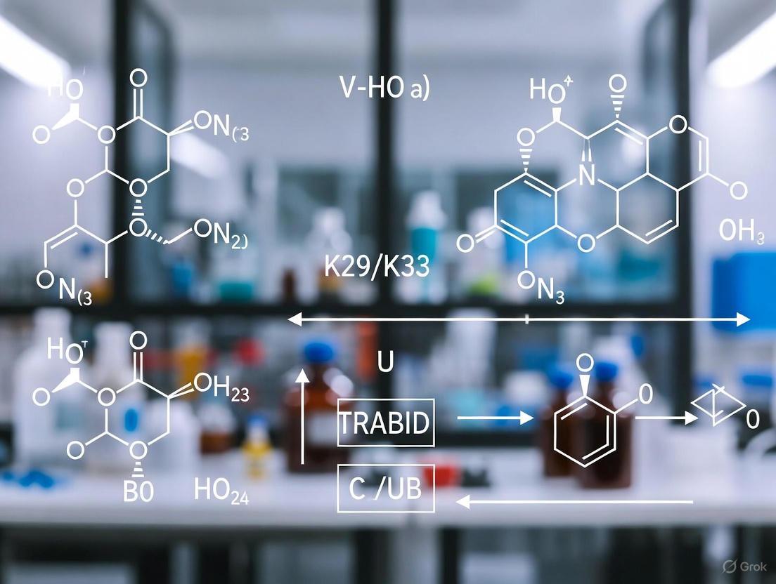
The deubiquitinase TRABID (also known as ZRANB1) possesses unique specificity for cleaving K29- and K33-linked ubiquitin chains [6]. Structural studies have revealed that its N-terminal region contains three Npl4-type zinc finger (NZF) domains, with the first domain (NZF1) responsible for selective recognition of K29/K33-diubiquitin [6] [7].
The crystal structure of TRABID's NZF1 domain bound to K33-linked diUb shows that the domain binds the hydrophobic patch centered on I44 of the proximal ubiquitin moiety. This binding mode exploits the flexibility and extended conformation of K29/K33 linkages, which adopt open structures in solution, making them distinct from the compact conformations of K48-linked chains [6] [7]. The interaction is highly specific, as mutations in the NZF1 domain disrupt binding to K29/K33 chains and impair TRABID's localization to ubiquitin-rich puncta in cells [6].
Key Methodologies for Studying K29/K33 Linkages
1. Enzymatic Assembly of Defined Chains:
- Protocol: The HECT E3 ligases UBE3C (for K29 linkages) and AREL1 (for K33 linkages) are utilized in autoubiquitination reactions with wild-type ubiquitin [6].
- Purification: Following assembly, linkage-specific deubiquitinases like vOTU (for K29 chains) are employed to digest non-specific linkages, enabling purification of homotypic K29 or K33 chains through size-exclusion chromatography [6] [7].
- Validation: Absolute quantification (AQUA) mass spectrometry verifies linkage specificity by spiking tryptic digests with isotope-labeled GlyGly-modified standard peptides corresponding to each potential linkage site [6].
2. Structural Analysis Techniques:
- Crystallography: The crystal structure of K29-diubiquitin reveals an extended conformation with exposed hydrophobic patches on both ubiquitin moieties [7].
- Solution Studies: Nuclear Magnetic Resonance (NMR) and small-angle X-ray scattering (SAXS) demonstrate that both K29- and K33-linked chains adopt open, dynamic conformations in solution, distinguishing them from compact chains like K48-linked polymers [6].
3. Cellular Localization Studies:
- Protocol: Inactive TRABID localizes to ubiquitin-rich puncta in cells, which serves as a functional cellular readout. This localization is attenuated when point mutations are introduced into the NZF1 domain, disrupting its specific binding to K29/K33 linkages [6].
The Scientist's Toolkit: Essential Research Reagents
Table 3: Key Reagents for Studying Atypical Ubiquitin Chains
| Reagent / Tool | Function / Application | Example / Source |
|---|---|---|
| Linkage-Specific E3 Ligases | Enzymatic assembly of specific atypical chains in vitro | UBE3C (K29), AREL1 (K33) [6] |
| Linkage-Specific DUBs | Validation and purification of specific chains; cellular functional studies | TRABID (K29/K33), vOTU (K29) [6] |
| Ubiquitin Mutants | Determining linkage specificity in assembly and binding assays | Kx-only mutants (e.g., K29-only, K33-only) [6] |
| AQUA Mass Spectrometry | Absolute quantification of linkage types in complex samples | Isotope-labeled GlyGly-modified peptides [6] |
| NZF1 Domain (TRABID) | Structural studies of K29/K33 linkage recognition | Recombinant protein for crystallography and binding assays [6] [7] |
Advanced Concepts: Branched Ubiquitin Chains
Beyond homotypic chains, ubiquitin can form branched structures where a single ubiquitin moiety is modified at two different lysine residues. These branched chains significantly increase the complexity of the ubiquitin code [8]. Several branched chains involving atypical linkages have been identified, including:
- K29/K48-branched chains: Assembled by UBE3C, potentially serving as potent proteasomal degradation signals [8].
- K11/K48-branched chains: Synthesized by the APC/C complex during mitosis, enhancing substrate recognition by the proteasome [8] [2].
The formation of branched chains often involves collaboration between different E3 ligases with distinct linkage specificities, providing a mechanism for spatial and temporal control of ubiquitin signals [8].
Atypical ubiquitin chains represent a sophisticated layer of regulation in cellular signaling, with distinct biological functions beyond the well-characterized K48 and K63 linkages. The specialized roles of K29 and K33 linkages in immune regulation and their specific recognition by proteins like TRABID highlight the complexity of the ubiquitin code. Continued development of tools for producing defined chains, along with advanced proteomic and structural techniques, will be essential for deciphering the full biological significance of these atypical ubiquitin signals.
TRABID (ZRANB1) is a deubiquitinating enzyme (DUB) encoded by the ZRANB1 gene in humans. It belongs to the ovarian tumor (OTU) family of deubiquitinases and plays specialized roles in cellular signaling pathways by selectively processing atypical ubiquitin chains. Its domain architecture features two critical elements: a central catalytic OTU domain and multiple Npl4-like zinc finger (NZF) domains that confer ubiquitin-binding specificity. This unique combination allows TRABID to function as a key regulator in processes such as Wnt/β-catenin signaling and epigenetic regulation through its specific recognition of K29- and K33-linked ubiquitin linkages.
Domain Architecture of TRABID
The human TRABID protein comprises 708 amino acids with a modular organization that integrates ubiquitin-binding motifs with catalytic domains. The N-terminal region contains three Npl4-like zinc finger (NZF) domains (approximately amino acids 1-200), while the C-terminal region houses the extended OTU catalytic domain (approximately amino acids 245-697). A distinctive feature of TRABID's OTU domain is an N-terminal extension consisting of two Ankyrin (Ank) repeats, forming an Ankyrin repeat ubiquitin-binding domain (AnkUBD) that precedes the canonical A20-like catalytic core [9].
Table: Domain Organization of TRABID
| Domain/Region | Position (Amino Acids) | Primary Function |
|---|---|---|
| NZF1 | 1-200 (approximately) | Specific recognition of K29/K33-linked diubiquitin |
| NZF2 | 1-200 (approximately) | Ubiquitin binding |
| NZF3 | 1-200 (approximately) | Ubiquitin binding |
| AnkUBD | 245-340 | Ubiquitin binding; contributes to linkage specificity |
| OTU Catalytic Core | 339-693 | Hydrolysis of ubiquitin chains |
Structural Basis of TRABID Specificity
The Catalytic OTU Domain and AnkUBD Extension
The crystal structure of the extended TRABID OTU domain (amino acids 245-697) reveals a triangular-shaped catalytic fold similar to A20, but with a unique 96-amino acid α-helical domain comprising two Ankyrin repeats positioned anterior to the catalytic core [9]. This AnkUBD domain forms a potential proximal Ub binding site (S1' site) that orients ubiquitin chains for preferential cleavage of Lys29 and Lys33 linkages. The catalytic triad consists of Cys443 and His585, which superpose well with the A20 catalytic center [9].
NZF Domains as Linkage-Specific Readers
The N-terminal NZF domains of TRABID function as linkage-specific ubiquitin readers. Structural studies demonstrate that the first NZF domain (NZF1) specifically recognizes K29- and K33-linked diubiquitin [6] [10]. The NZF domain forms a compact module composed of four antiparallel β-strands linked by three ordered loops, with a single zinc ion coordinated by four conserved cysteines forming two rubredoxin knuckles [11]. NZF1 binds ubiquitin using a conserved 13TF14 dipeptide to interact with the "Ile-44" surface of ubiquitin [11].
Experimental Validation of TRABID Specificity
Linkage Specificity Profiling
Comprehensive DUB activity assays against all eight ubiquitin linkage types demonstrate TRABID's marked preference for K29- and K33-linked diubiquitin over Lys63-linkages, with no cleavage activity observed for K6-, K11-, K27-, K48-linked or linear diubiquitin [9]. This dual specificity for atypical ubiquitin chains is unique among human OTU family DUBs.
Table: TRABID Activity Against Different Ubiquitin Linkages
| Ubiquitin Linkage Type | Cleavage Activity | Relative Efficiency |
|---|---|---|
| K29-linked | Yes | High |
| K33-linked | Yes | High |
| K63-linked | Yes | Moderate |
| K6-linked | No | Not detected |
| K11-linked | No | Not detected |
| K27-linked | No | Not detected |
| K48-linked | No | Not detected |
| Linear | No | Not detected |
Key Experimental Protocols
Crystallographic Analysis of TRABID OTU Domain
Methodology: The extended TRABID OTU domain (aa 245-697) was purified to homogeneity and crystallized. Phase information was obtained from a single isomorphous replacement with anomalous scattering (SIRAS) experiment using crystals derivatized with gold cyanide. The structure was resolved to 2.23 Å resolution [9].
Key Findings: The structure revealed the unexpected AnkUBD domain positioned anterior to the catalytic core, suggesting a mechanism for linkage specificity through additional ubiquitin binding sites.
NMR Mapping of Ubiquitin Binding Interfaces
Methodology: Nuclear magnetic resonance (NMR) experiments were performed to map interaction interfaces between TRABID domains and ubiquitin.
Key Findings: The AnkUBD interacts with the hydrophobic Ile44 patch of ubiquitin, while NZF1 specifically recognizes the unique interfaces presented by K29- and K33-linked ubiquitin chains [9] [6].
In Vitro DUB Activity Assays
Methodology: TRABID was incubated with synthetically generated ubiquitin chains of defined linkages. Cleavage efficiency was quantified through gel electrophoresis and mass spectrometry-based approaches [9] [6].
Key Findings: TRABID cleaves K29- and K33-linked diUb with significantly higher efficiency compared to K63-linkages, establishing its unique specificity profile among human DUBs.
Mechanism of K29/K33 Linkage Recognition
The specific recognition of K29- and K33-linked ubiquitin chains by TRABID involves a coordinated mechanism utilizing both its NZF domains and the AnkUBD:
Initial Chain Engagement: The NZF1 domain specifically binds K29- and K33-linked diubiquitin through recognition of the unique ubiquitin-ubiquitin interface presented by these linkages [6].
Enzymatic Positioning: The AnkUBD acts as an enzymatic S1' ubiquitin binding site that orients the ubiquitin chain, positioning Lys29 and Lys33 linkages optimally for cleavage by the catalytic core [9].
Catalytic Cleavage: The catalytic triad (Cys443-His585) hydrolyzes the isopeptide bond, with the spatial arrangement conferred by the auxiliary domains ensuring linkage preference.
Biological Functions and Relevance to Drug Development
TRABID's specificity for K29 and K33 linkages positions it as a key regulator in several cellular processes:
Wnt/β-catenin Signaling: TRABID acts as a positive regulator of Wnt-induced transcription through deubiquitination of APC, with its NZF domains showing preference for binding K63-linked chains in this context [12].
Epigenetic Regulation: TRABID opposes TRIP12-mediated K29-linked ubiquitylation of the histone methyltransferase SUV39H1, regulating H3K9me3 homeostasis and epigenome integrity [13].
Unfolded Protein Response: K29-linked ubiquitination is upregulated during endoplasmic reticulum stress, with TRABID potentially modulating this response through its deubiquitinating activity [14].
Research Reagent Solutions
Table: Essential Research Reagents for TRABID Studies
| Reagent/Tool | Function/Application | Specific Examples |
|---|---|---|
| Linkage-specific ubiquitin chains | Substrates for DUB activity assays | K29- and K33-linked diUb for specificity profiling [9] [6] |
| HECT E3 ligases (UBE3C, AREL1) | Generation of atypical ubiquitin chains | UBE3C for K29-linked chains; AREL1 for K33-linked chains [6] |
| TRABID-NZF1 constructs | K29/K33 linkage detection | GST-TRABID-NZF1 as linkage-specific binder in pulldown assays [15] |
| Crystallization reagents | Structural studies | Gold cyanide for SIRAS phasing of TRABID OTU domain [9] |
| Conditional ubiquitin replacement cell lines | Functional studies in cellular context | U2OS/shUb cells with inducible Ub mutants [13] |
TRABID exemplifies how the integration of multiple ubiquitin-binding domains (NZF and AnkUBD) with a catalytic OTU core enables precise recognition and processing of atypical ubiquitin linkages. Its specificity for K29 and K33 linkages, validated through structural and biochemical approaches, highlights the sophisticated mechanisms underlying ubiquitin code interpretation. For researchers and drug development professionals, TRABID represents both a potential therapeutic target and a tool for understanding the physiological roles of understudied ubiquitin chain types. The continuing elucidation of its structure-function relationships provides a framework for developing selective DUB inhibitors and probes for interrogating atypical ubiquitin signaling in health and disease.
The deubiquitinase TRABID (also known as ZRANB1) has emerged as a key regulator of atypical ubiquitin signaling, with its specificity for lysine 29 (K29) and lysine 33 (K33)-linked polyubiquitin chains now firmly established through structural, biochemical, and cellular studies. This review synthesizes pivotal findings that have transformed our understanding of TRABID's unique linkage specificity, highlighting the experimental approaches that confirmed its role as a primary reader and eraser of K29 and K33 linkages. We compare TRABID's activity and binding preferences against other deubiquitinases, detail the methodologies enabling these discoveries, and present available research tools that continue to drive this evolving field forward, offering researchers a comprehensive guide to studying these non-canonical ubiquitin signals.
Protein ubiquitination represents one of the most versatile post-translational modifications, regulating virtually every cellular process through the attachment of ubiquitin polymers of different lengths and architectures. While K48- and K63-linked chains have been extensively characterized for their roles in proteasomal degradation and signaling transduction respectively, the so-called "atypical" ubiquitin linkages—including K29 and K33—have remained enigmatic due to challenges in identifying their assembly enzymes, receptors, and cellular functions [6].
The ovarian tumor (OTU) family deubiquitinase TRABID has recently been identified as a central player in the recognition and processing of these atypical chains. Early proteomic analyses revealed that all ubiquitin chain linkages exist simultaneously in cells, yet tools to study K29 and K33 linkages specifically were limited [6]. This review documents the pivotal discoveries that established TRABID's unique specificity for K29 and K33 linkages, comparing its activity against other deubiquitinases, detailing key experimental methodologies, and presenting the specialized tools now available for investigating these unconventional ubiquitin signals.
Comparative Analysis of TRABID Specificity Versus Other DUBs
Quantitative Linkage Specificity Profiling
The specificity of TRABID for K29 and K33 linkages has been quantitatively established through multiple biochemical approaches, setting it apart from other deubiquitinases with different linkage preferences.
Table 1: Linkage Specificity Profile of TRABID Compared to Other DUBs
| Deubiquitinase | Primary Linkage Specificity | Secondary Linkage Specificity | Key Experimental Evidence |
|---|---|---|---|
| TRABID | K29 and K33 | K63 (weaker activity) | Structural studies with NZF1 domain, UbiCREST, Ub-AQUA [6] [16] |
| OTUD5 | K48 and K63 | Limited activity on K29 | Mass spectrometry, in vitro ubiquitylation assays [15] |
| USP13 | Not linkage-specific | N/A | VPS34 stabilization studies [17] |
The unique positioning of TRABID is further exemplified by its dual functionality—it serves both as a linkage-specific eraser through its catalytic OTU domain and as a specialized reader through its N-terminal zinc finger domains. This combination allows TRABID to precisely recognize and process its target chains with remarkable specificity.
Structural Basis of TRABID Specificity
The molecular mechanism underlying TRABID's specificity for K29 and K33 linkages has been elucidated through structural studies, revealing an elegant recognition system:
- NZF1 Domain Binding: The N-terminal Npl4-like zinc finger (NZF1) domain of TRABID specifically binds K29/K33-linked diubiquitin, with crystal structures showing how this domain exploits the flexibility of K29 chains to achieve linkage-selective binding [6] [7].
- Open Conformation Recognition: Both K29- and K33-linked chains adopt open and dynamic conformations in solution, similar to K63-linked polyubiquitin, with hydrophobic patches on both ubiquitin moieties exposed and available for binding [6].
- Filamentous Binding Mode: The crystal structure of NZF1 bound to K33-linked diUb reveals a filamentous structure for K33 polymers in which NZF1 binds each Ub-Ub interface, suggesting a model for how TRABID interacts with longer atypical chains [6].
Key Experimental Paradigms Establishing TRABID Specificity
Enzymatic Assembly Systems for Atypical Chains
A critical breakthrough in characterizing TRABID's specificity came from the development of methods to generate homotypic K29 and K33 chains in sufficient quantities for biochemical studies:
Diagram 1: Enzymatic systems for K29/K33 chain assembly. The HECT E3 ligases UBE3C and AREL1 assemble K29- and K33-linked chains respectively, which can be purified using linkage-specific DUBs like vOTU.
Researchers discovered that the human HECT E3 ligases UBE3C and AREL1 assemble K48/K29- and K11/K33-linked Ub chains respectively, and could be used in combination with DUBs to generate homotypic K29- and K33-linked chains for biochemical and structural analyses [6]. This enzymatic toolkit was essential for subsequent studies of TRABID specificity, as it provided the defined substrates needed for cleavage assays and binding studies.
Quantitative Mass Spectrometry Approaches
Ubiquitin-AQUA (Absolute QUAntification) proteomics has been instrumental in quantitatively establishing TRABID's specificity and identifying its cellular substrates:
- Linkage Quantification: AQUA-based mass spectrometry uses isotope-labeled GlyGly-modified standard peptides derived from each potential linkage site, allowing absolute quantification of all chain types in E3 ligase reactions [6] [16].
- Substrate Identification: Quantitative proteomics of catalytic-dead TRABID constructs (TRABIDC443S and TRABIDΔOTU) identified 50 trapped proteins representing candidate substrates, including the E3 ligase HECTD1 which preferentially assembles K29- and K48-linked ubiquitin chains [16] [18].
- Branched Chain Analysis: Middle-down mass spectrometry and Ub-clipping methods have revealed that K29 linkages frequently exist within heterotypic branched chains containing K48 linkages, with TRABID regulating these complex structures [17].
Table 2: Key Methodologies for Studying TRABID Specificity
| Methodology | Application in TRABID Research | Key Findings Enabled |
|---|---|---|
| X-ray Crystallography | Structure determination of TRABID NZF1 bound to K29/K33-diUb | Revealed molecular basis of linkage specificity [6] [7] |
| UbiCREST | Profiling DUB activity across different linkage types | Established TRABID's preference for K29 and K33 linkages [16] |
| Ub-AQUA/PRM | Quantitative analysis of linkage composition | Identified HECTD1 as K29/K48-specific E3 ligase [16] [19] |
| Cellular Puncta Formation | Visualization of polyubiquitin trapped by catalytic dead TRABID | Confirmed TRABID-ubiquitin interactions in cells [16] |
Cellular Functions and Substrates of TRABID
Regulation of Autophagy Through VPS34
TRABID plays a critical role in regulating autophagy through its action on VPS34, the catalytic subunit of the class III PI3-kinase complex:
Diagram 2: TRABID regulates autophagy via VPS34. UBE3C promotes K29/K48-branched ubiquitination of VPS34, targeting it for proteasomal degradation and inhibiting autophagy. TRABID reverses this modification by cleaving K29 linkages, thereby stabilizing VPS34 and promoting autophagy.
Under basal conditions and starvation, UBE3C and TRABID reciprocally regulate K29/K48-branched ubiquitination of VPS34, controlling its stability and consequently modulating autophagosome formation and maturation [17]. This regulation is particularly important during cellular stress responses, where the balance between these opposing enzymes determines autophagy activity to maintain proteostasis.
Control of Epigenetic Regulation Through SUV39H1
Recent research has uncovered TRABID's role in chromatin regulation through the control of histone modifier stability:
- SUV39H1 Degradation: TRABID reverses K29-linked ubiquitylation of the H3K9me3 methyltransferase SUV39H1, which is catalyzed by the E3 ligase TRIP12 [13].
- Epigenome Integrity: Preventing K29-linkage-dependent SUV39H1 turnover deregulates H3K9me3 homeostasis, establishing a key role for K29-linked ubiquitylation in maintaining epigenome integrity [13].
- Chromatin Association: Proteomic profiling revealed that K29-linked ubiquitylation is strongly associated with chromosome biology, with TRABID serving as a critical regulator of this pathway [13].
The Scientist's Toolkit: Essential Reagents for K29/K33 Research
Table 3: Key Research Reagents for Studying TRABID and Atypical Linkages
| Research Tool | Specific Application | Function and Utility |
|---|---|---|
| GST-TRABID(ZNF1) | Enrichment of K29/K33-ubiquitinated proteins | Selective precipitation of proteins modified with K29/K33 linkages for immunoblotting or mass spectrometry [20] |
| K29/K33 Polyubiquitin Chain Capture Kit | Proteomic studies of atypical ubiquitination | System-wide identification of K29/K33-modified cellular proteins [20] |
| UBE3C and AREL1 E3 Ligases | In vitro assembly of atypical chains | Generation of defined K29- and K33-linked ubiquitin chains for biochemical assays [6] |
| TRABIDC443S Mutant | Substrate trapping experiments | Identification of cellular TRABID substrates through stable interaction with ubiquitinated proteins [16] |
| Ubiquitin K29R/K33R Mutants | Specific ablation of atypical linkages | Functional studies of K29/K33-dependent cellular processes [13] |
The definitive identification of K29 and K33 linkages as primary TRABID substrates represents a significant advancement in the ubiquitin field, transforming our understanding of atypical ubiquitin signaling. Through a combination of structural biology, quantitative biochemistry, and cellular studies, researchers have established TRABID as both a dedicated reader and eraser of these unconventional chains, with important functions in autophagy, epigenetic regulation, and cellular stress responses.
The experimental approaches detailed here—from enzymatic chain assembly systems to quantitative mass spectrometry and structural analyses—provide a roadmap for investigating these complex post-translational modifications. As new tools continue to emerge, including specific binders and linkage-specific antibodies, we anticipate accelerated discovery of the full physiological and pathological roles of K29 and K33 ubiquitin signaling. TRABID continues to serve as both a valuable experimental tool and a compelling therapeutic target in the expanding landscape of ubiquitin biology.
The ubiquitin code, a pivotal post-translational regulatory mechanism, derives its complexity from the ability of ubiquitin to form eight distinct polymeric chains through different linkage types [21] [6]. Among these, the so-called "atypical" linkages, particularly those via lysine 29 (K29) and lysine 33 (K33), have remained enigmatic due to a historical lack of tools for their specific study [21] [9]. K29-linked polyubiquitin is notably abundant in resting mammalian cells and further increases upon proteasomal inhibition, suggesting significant, yet poorly understood, cellular roles [21]. A pivotal breakthrough in this field was the identification of the first Npl4-like zinc finger (NZF1) domain of the deubiquitinase TRABID as a specialized ubiquitin-binding domain (UBD) with remarkable selectivity for K29 and K33 linkages [21] [6] [22]. This discovery provided the crucial molecular tool needed to probe the formation, recognition, and function of these atypical chains. This review objectively analyzes the structural basis for NZF1's selectivity, compares its specificity profile against other UBDs, and details the experimental methodologies that validate its role as the primary structural module for K29/K33 ubiquitin binding, framing these findings within the broader thesis of validating TRABID's specificity for K29/K33 linkages.
Structural Basis of NZF1 Linkage Selectivity
The NZF domain is a compact ubiquitin-binding module of approximately 30 amino acids [23]. However, not all NZF domains are linkage-selective; many display no chain preference despite conserved secondary interaction surfaces [23]. The NZF1 domain of TRABID is a notable exception, achieving exceptional selectivity for K29 and K33 linkages through a unique binding mechanism.
Molecular Mechanism of Recognition
Biophysical and structural studies reveal that TRABID NZF1 does not bind K29- and K33-linked diubiquitin in the same manner as compact chains like K48. The key distinction lies in the open and dynamic conformations adopted by K29- and K33-linked chains in solution, which are similar to the extended conformation of K63-linked chains [6]. The crystal structure of K29-linked diubiquitin shows an extended conformation where the hydrophobic patches (Ile44 patches) on both ubiquitin moieties remain exposed and available for binding [21] [7] [24].
The crystal structure of TRABID NZF1 in complex with K29-linked diubiquitin provides the definitive molecular explanation for selectivity [21] [22]. The mechanism involves two critical aspects:
- Distal Ubiquitin Engagement: The NZF1 domain binds the hydrophobic Ile44 patch on the distal ubiquitin moiety using a conserved binding surface common to many NZF domains [21].
- Linkage-Specific Interface: Selectivity is achieved through additional, unique interactions between the NZF1 domain and a specific surface on the proximal ubiquitin moiety, an interface that is only presented in the distinct conformations of K29- and K33-linked chains [21] [22]. This exploits the intrinsic flexibility of K29 chains to achieve linkage-selective binding.
Table 1: Key Structural Features of TRABID NZF1 Binding to K29/K33 Chains
| Structural Element | Role in Ubiquitin Binding | Contribution to Linkage Selectivity |
|---|---|---|
| Conserved NZF Hydrophobic Surface | Binds the Ile44 patch of the distal ubiquitin | Necessary, but not sufficient for selectivity; common to many NZF domains |
| Secondary Interaction Surface | Engages a unique interface on the proximal ubiquitin | Primary determinant of selectivity for K29 and K33 linkages |
| Extended Conformation of K29/K33 chains | Exposes hydrophobic patches on both ubiquitin moieties | Prerequisite for the simultaneous engagement of both ubiquitin units by NZF1 |
| Flexibility of Atypical Chains | Allows the chain to adopt the precise geometry for binding | Enables the NZF1 domain to exploit conformational dynamics for specificity |
Comparative Analysis of Ubiquitin-Binding Domains
The selectivity of TRABID NZF1 is particularly striking when compared to the binding profiles of other UBDs. A comprehensive characterization of human NZF domains found that most, including several from other proteins, do not display strong chain linkage preference [23]. This highlights that linkage selectivity is a specialized property of specific NZF domains like TRABID NZF1, not a generic feature of the entire NZF family.
Table 2: Linkage Selectivity Profile of TRABID NZF1 vs. Other Domains
| Ubiquitin-Binding Domain (Protein) | Primary Linkage Specificity | Reported Affinity/Specificity Notes |
|---|---|---|
| TRABID NZF1 | K29, K33 | Highly selective; key structural basis determined [21] [6] |
| TAB2 NZF | K6, K63 (especially when phosphorylated) | Prefers phosphorylated chains on depolarized mitochondria [23] |
| HOIP NZF1 | Monoubiquitinated substrates (e.g., NEMO) | Binds ubiquitinated NEMO and linear diubiquitin; achieves specificity via simultaneous substrate/Ub recognition [23] [25] |
| FAM63A tMIU | K48 | Selective binding mediated by the second MIU (MIU2) motif [22] |
| TRABID AnkUBD | K29, K33 | Functions as an enzymatic S1' Ub binding site for the DUB, orienting the chain for preferential cleavage [9] |
Experimental Validation of TRABID NZF1 Specificity
The validation of TRABID NZG1's specificity relied on a suite of biochemical, biophysical, and structural experiments. Key to this endeavor was the development of methods to produce sufficient quantities of pure, homotypic K29 and K33-linked chains.
Key Experimental Workflows
The following diagram summarizes the integrated workflow used to assemble atypical ubiquitin chains and validate NZF1 specificity:
Detailed Methodologies
Enzymatic Assembly of Atypical Ubiquitin Chains
A major hurdle in studying atypical chains was the inability to produce them on a large scale. This was overcome using Ubiquitin Chain-Editing Complexes [21] [6].
- K29-linked Chains: The HECT E3 ligase UBE3C (which primarily assembles K29 and K48 linkages) is used in an in vitro ubiquitylation reaction alongside the viral deubiquitinase vOTU. UBE3C autoubiquitinates and builds chains, while vOTU, which lacks activity against K29 linkages, specifically cleaves contaminating linkages and releases free, homotypic K29-linked polyubiquitin from the enzyme [21] [7].
- K33-linked Chains: Similarly, the HECT E3 ligase AREL1 (apoptosis-resistant E3 ubiquitin protein ligase 1) is employed to assemble K33-linked chains, which are then purified with the aid of linkage-specific DUBs [6].
- Linkage Verification: The linkage type of the assembled chains is confirmed using:
- Ubiquitin Mutants: Using "Kx-only" ubiquitin mutants (where all lysines except one are mutated to arginine) in assembly reactions to demonstrate chain formation is dependent on a specific lysine (e.g., K29) [21] [6].
- Deubiquitinase (DUB) Specificity: Treating the chains with the DUB TRABID, which is known to hydrolyze K29 and K33 linkages, rapidly reduces them to monoubiquitin, while linkage-nonspecific or other linkage-specific DUBs (e.g., OTULIN for M1 linkages) do not [21].
- Mass Spectrometry: Using parallel reaction monitoring (pRM) mass spectrometry to quantitatively verify the presence of specific linkage types in the assembled chains [21].
Binding and Specificity Assays
- Crystallography: The crystal structure of TRABID NZF1 in complex with K29-linked diubiquitin was determined, providing atomic-level resolution of the interaction and revealing the structural basis for selectivity [21] [7] [22].
- Solution Conformation Studies: Techniques such as NMR and small-angle X-ray scattering (SAXS) confirmed that K29- and K33-linked chains adopt open and dynamic conformations in solution, which is a prerequisite for the observed NZF1 binding mode [6].
- Cellular Pull-down Assays: GST-tagged TRABID NZF1 is used as a linkage-specific capture tool to isolate K29-linked ubiquitin chains from cell lysates. This application demonstrated that K29 linkages frequently exist within mixed or branched chains containing other linkages, such as K48, in a cellular context [21] [15] [22].
The Scientist's Toolkit: Key Research Reagents
The following reagents and tools are essential for experimental research on TRABID NZF1 and K29/K33 ubiquitin chains.
Table 3: Essential Research Reagents for K29/K33 Ubiquitin Research
| Research Reagent / Tool | Function and Application | Key Experimental Use |
|---|---|---|
| Recombinant TRABID NZF1 Domain | Selective capture and detection of K29/K33 linkages. | Pull-down assays from cell lysates; sensor for immunofluorescence [21] [15]. |
| HECT E3 Ligase UBE3C | Enzymatic assembly of K29-linked polyubiquitin chains. | Large-scale in vitro production of K29 chains for biochemical and structural studies [21] [6]. |
| HECT E3 Ligase AREL1 | Enzymatic assembly of K33-linked polyubiquitin chains. | Large-scale in vitro production of K33 chains [6]. |
| Deubiquitinase vOTU | Linkage-specific editing of ubiquitin chains. | Used in chain-editing complex with UBE3C to purify homotypic K29 chains [21]. |
| K29-only Ubiquitin Mutant | Ubiquitin where only K29 is available for chain formation. | Verification of linkage specificity in E3 ligase and binding assays [21] [6]. |
| TRABID (Full-length or OTU Domain) | K29/K33-linkage specific deubiquitinase. | Enzymatic validation of linkage type; study of DUB specificity and cellular function [21] [9]. |
Functional Consequences and Broader Context
The specificity of the TRABID NZF1 domain is not an isolated phenomenon but is deeply integrated into the larger functional context of the TRABID enzyme and the biology of atypical ubiquitin chains.
Integration with TRABID DUB Activity
TRABID possesses a complex domain architecture that synergistically targets K29 and K33 linkages. In addition to its three N-terminal NZF domains, its catalytic OTU domain is extended by an Ankyrin repeat domain (AnkUBD) [9]. This AnkUBD is a unique ubiquitin-binding fold that functions as an enzymatic S1' site, orienting the ubiquitin chain to preferentially cleave K29 and K33 linkages [9]. The NZF1 domain, therefore, works in concert with the AnkUBD to ensure the enzyme's high efficiency and linkage specificity, both in vitro and in vivo.
Role in Heterotypic and Branched Ubiquitin Signaling
The use of NZF1 as a capture tool revealed a critical aspect of K29 biology: its heterotypic nature [21] [7]. K29 linkages often exist in mixed or branched chains alongside other linkages, particularly K48. This has been functionally demonstrated in several pathways:
- Targeting DUB-Protected Substrates: The K29 linkage can serve as a DUB-resistant foundation that facilitates the addition of K48-linked branches by another HECT E3 ligase, UBR5. This K29/K48-branched chain promotes the proteasomal degradation of substrates that are otherwise protected by DUBs like OTUD5 [15].
- Branched Chain Assembly: TRABID forms a DUB/E3 pair with the ligase HECTD1, which assembles branched K29/K48 chains, regulating the stability of the ligase itself and its downstream signaling [26].
This functional relationship between K29 and K48 linkages in branched chains underscores a sophisticated combinatorial ubiquitin code where the specific properties of each linkage are leveraged to create robust biological signals.
The TRABID NZF1 domain stands as a paradigm for how small, compact ubiquitin-binding modules can achieve exquisite linkage selectivity through sophisticated structural mechanisms. The experimental validation of its specificity for K29 and K33 linkages—via structural biology, bespoke biochemical chain assembly, and cellular applications—has been instrumental in transforming these atypical chains from poorly defined curiosities into decipherable elements of the ubiquitin code. The integration of NZF1's binding specificity with the cleavage activity of TRABID's catalytic domain and its role in regulating heterotypic chains like K29/K48-branched structures, solidifies the broader thesis that TRABID is a central player in the K29/K33-specific ubiquitin signaling pathway. The continued use of NZF1 as a specific research tool will undoubtedly further unravel the complex cellular functions of these enigmatic ubiquitin linkages.
Tools and Techniques: Profiling and Applying TRABID Specificity in Research
The elucidation of macromolecular structures is fundamental to understanding biological mechanisms at the molecular level. For research focused on validating TRABID specificity for K29/K33 ubiquitin linkages, structural biology techniques provide the necessary tools to visualize atomic interactions and confirm biochemical findings. X-ray crystallography and cryo-electron microscopy (cryo-EM) stand as two powerful methods for determining three-dimensional structures of biological macromolecules, each with distinct advantages and limitations [27] [28]. While X-ray crystallography has historically been the dominant technique in structural biology, accounting for approximately 84% of structures in the Protein Data Bank, cryo-EM has recently experienced a "resolution revolution" that enables near-atomic resolution for complexes previously inaccessible to structural analysis [28] [29].
The complementary nature of these techniques is particularly valuable for studying complex systems such as ubiquitin chains and their recognition by specific binding domains. X-ray crystallography provides atomic-level precision for well-ordered structures, while cryo-EM excels at visualizing larger complexes and dynamic assemblies in near-native states [30]. For researchers investigating TRABID's specificity for K29- and K33-linked ubiquitin chains, both methods can be integrated to provide a comprehensive understanding of the structural basis of recognition, from atomic contacts to overall conformational flexibility [6] [7]. This guide objectively compares the performance of these techniques specifically within the context of ubiquitin chain research, providing experimental data and protocols to inform methodological selection.
Fundamental Principles and Technical Comparison
Physical Principles and Data Collection
X-ray crystallography and cryo-EM operate on different physical principles to extract structural information from biological samples. X-ray crystallography relies on Bragg's Law of X-ray diffraction by crystalline samples, where well-ordered three-dimensional crystals scatter X-rays to produce discrete diffraction patterns [27]. The resulting spot patterns contain amplitude information about the electron density within the crystal, but phase information must be obtained through additional experimental or computational methods such as molecular replacement or anomalous dispersion [27] [29]. The quality of the final structure depends heavily on crystal order, which determines the sharpness and extent of diffraction.
Cryo-EM utilizes high-energy electrons in a transmission electron microscope to directly image macromolecules preserved in vitreous ice [27] [31]. Unlike crystallography, cryo-EM can examine non-crystalline specimens through single-particle analysis, where thousands of individual particle images are classified, aligned, and averaged to reconstruct a three-dimensional density map [27]. The magnetic objective lens in cryo-EM produces both diffraction patterns at the back-focal plane and magnified images in the image plane, with the images containing full structural information about the molecule [27]. Recent advances in direct electron detectors and image processing algorithms have enabled cryo-EM to achieve near-atomic resolution for many biologically significant complexes [28] [31].
Table 1: Fundamental Principles and Data Characteristics
| Aspect | X-ray Crystallography | Cryo-EM |
|---|---|---|
| Radiation Source | X-ray photons | High-energy electrons |
| Sample State | Crystalline lattice | Vitreous ice (near-native) |
| Primary Data | Diffraction pattern (spot intensities) | 2D projection images |
| Phase Problem | Must be solved experimentally or computationally | Built into imaging process |
| Information Obtained | Electron density map | 3D Coulomb potential map |
| Resolution Limiting Factors | Crystal order, diffraction quality | Particle alignment, detector sensitivity, microscope stability |
Resolution and Sample Requirements
The resolution achievable with each technique depends on multiple factors, with each method exhibiting distinct strengths for different sample types. X-ray crystallography routinely achieves atomic resolution (often better than 1.5-2.0 Å), providing precise atomic coordinates for well-ordered crystals [30] [29]. This high resolution makes it ideal for studying detailed molecular interactions, such as those between TRABID's NZF1 domain and K29/K33-linked diubiquitin [6] [7]. However, resolution in crystallography depends completely on crystal quality, with imperfections and disorder limiting the useful diffraction signal.
Cryo-EM typically achieves near-atomic resolution (2.5-4.0 Å) for most biological samples, with recent technological advances pushing these limits to approximately 2-3 Å for favorable cases [28] [30]. While generally not matching the highest resolutions of crystallography, cryo-EM excels for larger complexes (>100 kDa) and can tolerate some sample heterogeneity, capturing multiple conformational states within a single dataset [30]. This capability is particularly valuable for studying dynamic ubiquitin chains, which adopt open and flexible conformations in solution [6] [10].
Sample requirements differ significantly between the techniques. Crystallography typically requires highly homogeneous, purified samples at concentrations of 5-20 mg/mL, with total amounts often exceeding 5 mg to allow for crystallization trials [30] [32]. The need for crystallization presents the major bottleneck, as many biologically important targets resist crystal formation. In contrast, cryo-EM requires significantly less material (0.1-0.2 mg total) at lower concentrations (≥2 mg/mL), and can accommodate moderate heterogeneity in complex composition [30] [32].
Table 2: Resolution and Sample Requirements
| Parameter | X-ray Crystallography | Cryo-EM |
|---|---|---|
| Maximum Resolution | Sub-1.0 Å possible | Typically 2.5-4.0 Å (2-3 Å in best cases) |
| Typical Resolution Range | 1.5-2.5 Å | 3-4 Å |
| Sample Amount | >2 mg typically, >5 mg for difficult targets | 0.1-0.2 mg |
| Sample Concentration | 10-20 mg/mL | ≥2 mg/mL |
| Sample Purity | High homogeneity required | Moderate heterogeneity acceptable |
| Molecular Size | Optimal <100 kDa | Optimal >100 kDa |
| Structural Stability | Requires rigid structure | Flexible/dynamic acceptable |
Technical Workflows and Methodologies
X-ray Crystallography Workflow
The process of structure determination by X-ray crystallography follows a well-established pipeline with distinct stages. Sample preparation begins with protein purification to homogeneity, often requiring significant optimization to obtain sufficient quantities of stable protein [29]. For TRABID studies, this would involve expressing and purifying the NZF1 domain or full-length protein, along with generating K29- or K33-linked diubiquitin substrates [6] [7].
Crystallization represents the most critical and often limiting step, where purified protein is slowly brought out of solution under controlled conditions to form ordered crystals [29]. This typically involves screening hundreds to thousands of conditions varying precipitant, buffer, pH, and temperature. For protein-ligand complexes such as TRABID bound to ubiquitin chains, crystals can be obtained by co-crystallization or soaking pre-formed crystals with the ligand [29]. Successful crystal formation produces three-dimensional ordered arrays capable of diffracting X-rays.
Data collection occurs at synchrotron facilities, which provide intense, tunable X-ray sources [29]. Crystals are exposed to X-rays, and diffraction patterns are collected as the crystal is rotated. A complete dataset consists of hundreds of images capturing diffraction spot intensities across different orientations [27] [29]. For the TRABID NZF1 domain complexed with K33-linked diubiquitin, this approach revealed the structural basis of linkage specificity through a filamentous binding mode [6].
Data processing involves determining the phase information missing from diffraction measurements, typically through molecular replacement using a related structure as a search model [27]. For novel structures without homologs, experimental phasing methods such as SAD/MAD may be employed. The phased data is used to calculate an electron density map into which an atomic model is built and iteratively refined against the observed diffraction data [29].
Diagram 1: X-ray Crystallography Workflow
Cryo-Electron Microscopy Workflow
Cryo-EM single-particle analysis follows a distinct workflow designed to extract structural information from individual macromolecules. Sample preparation involves applying a purified protein solution to specialized grids followed by rapid vitrification in liquid ethane to preserve molecules in a near-native state within thin amorphous ice [31] [32]. For TRABID-ubiquitin complexes, this approach maintains the dynamic, flexible conformations of K29- and K33-linked chains observed in solution studies [6].
Data collection occurs using high-end transmission electron microscopes operating at 200-300 kV, with modern instruments equipped with direct electron detectors [32]. Thousands to millions of low-dose images are collected as movies, capturing individual particles in random orientations [31]. For a typical TRABID-ubiquitin complex, data collection might span 1-3 days to accumulate sufficient particles for high-resolution reconstruction.
Image processing begins with motion correction and contrast transfer function (CTF) estimation to account for instrument imperfections [30]. Particles are then selected from micrographs through automated picking algorithms, followed by multiple rounds of 2D and 3D classification to separate homogeneous populations from damaged particles or distinct conformational states [31]. This classification capability is particularly valuable for studying the dynamic ubiquitin chains recognized by TRABID, as it can potentially capture multiple conformational states within a single sample [6].
3D reconstruction involves iteratively refining particle orientations and positions to generate an increasingly detailed density map through algorithms such as Bayesian polishing and non-uniform refinement [30]. The final map serves as the basis for atomic model building, where existing structures can be docked as rigid bodies or de novo models can be built and refined against the density [27]. For TRABID studies, the crystal structure of the NZF1 domain bound to K33-diubiquitin could be docked into cryo-EM maps of larger complexes containing these components [6].
Diagram 2: Cryo-EM Single Particle Analysis Workflow
Application to TRABID-K29/K33 Ubiquitin Recognition Studies
Experimental Approaches for Ubiquitin Chain Characterization
The elucidation of TRABID's specificity for K29- and K33-linked ubiquitin chains required integrated structural and biochemical approaches. Ubiquitin chain assembly represents the initial critical step, with researchers identifying that human HECT E3 ligases UBE3C and AREL1 assemble K48/K29- and K11/K33-linked chains respectively [6] [10]. These enzymes can be used in combination with linkage-specific deubiquitinases (DUBs) to generate homotypic K29- and K33-linked chains for structural studies [6]. Large-scale enzymatic assembly and purification of K29-linked polyubiquitin chains enabled both biophysical characterization and structural determination [7].
Biophysical analysis of the purified chains provided initial insights into their conformational properties. Solution studies using techniques such as analytical ultracentrifugation and small-angle X-ray scattering indicated that both K29- and K33-linked ubiquitin chains adopt open and dynamic conformations, similar to K63-linked chains but distinct from the compact structures of K48-linked chains [6]. This structural information helped explain TRABID's ability to recognize these specific linkage types.
Crystallographic studies of TRABID's N-terminal NZF1 domain in complex with K33-linked diubiquitin revealed the atomic basis of recognition specificity [6] [7]. The crystal structure showed an intriguing filamentous arrangement where NZF1 domains bind each ubiquitin-ubiquitin interface within the chain [6]. This structure explained the linkage selectivity and suggested a model for how TRABID engages longer polyubiquitin chains. Similarly, the crystal structure of K29-linked diubiquitin alone confirmed its extended conformation with exposed hydrophobic patches on both ubiquitin moieties [7].
Cryo-EM applications in this field could potentially visualize how full-length TRABID engages with longer ubiquitin chains or how TRABID-containing complexes assemble on ubiquitinated substrates. While crystallography provides atomic details of isolated domains and short chains, cryo-EM could capture larger assemblies in more physiological states, potentially revealing conformational heterogeneity and dynamic aspects of recognition [27] [30].
Research Reagent Solutions for Ubiquitin Studies
Table 3: Essential Research Reagents for TRABID-Ubiquitin Studies
| Reagent/Category | Function/Application | Specific Examples |
|---|---|---|
| E3 Ligases | Assembly of specific ubiquitin linkages | UBE3C (K29/K48 linkages), AREL1 (K11/K33 linkages) [6] |
| DUBs | Linkage-specific hydrolysis for chain purification | vOTU (K29-chain editing), TRABID (K29/K33-specific) [6] [7] |
| Ubiquitin Mutants | Linkage specificity determination | Kx-only mutants (single lysine), K0 (no lysines) [6] |
| Binding Domains | Linkage-specific recognition modules | TRABID NZF1 domain (K29/K33-specific) [6] [7] |
| Expression Systems | Recombinant protein production | E. coli (uniform 15N/13C labeling for NMR), insect cells (large complexes) [29] |
| Crystallization Reagents | Crystal formation screening | Commercial sparse matrix screens, optimization reagents [29] [32] |
| Cryo-EM Grids | Sample support for vitrification | Graphene oxide grids (reduce orientation bias), UltraFoil grids [32] |
Performance Comparison for Different Research Scenarios
Scenario-Based Method Selection
The choice between X-ray crystallography and cryo-EM depends heavily on the specific research goals, sample characteristics, and available resources. For atomic-resolution mapping of specific interactions, such as determining the precise contacts between TRABID's NZF1 domain and K29/K33-linked diubiquitin, X-ray crystallography remains unsurpassed [6] [7]. The ability to achieve resolutions better than 2.0 Å reveals detailed atomic interactions, water molecules mediating contacts, and subtle conformational adjustments upon binding.
For studying dynamic complexes or conformational heterogeneity, cryo-EM offers significant advantages. The ability to classify single-particle datasets into multiple structural states allows researchers to capture conformational continua and transitional states that would be averaged out in crystallographic experiments [30]. This capability is particularly valuable for studying flexible ubiquitin chains, which adopt open and dynamic conformations in solution [6].
When sample size and properties are considered, clear distinctions emerge. Crystallography works best with well-behaved, monodisperse samples that form ordered crystals, while cryo-EM tolerates more heterogeneity and requires significantly less material [30] [32]. For difficult-to-crystallize targets such as membrane proteins or large complexes, cryo-EM often provides the only path to high-resolution structures.
Table 4: Scenario-Based Method Selection Guide
| Research Scenario | Recommended Technique | Rationale | Typical Outcome |
|---|---|---|---|
| Atomic-resolution ligand binding | X-ray crystallography | Superior resolution for precise atomic positioning | 1.5-2.5 Å structure with detailed interactions |
| Large complex architecture | Cryo-EM | No size limitations, minimal sample engineering | 3-4 Å structure of intact complex |
| Multiple conformational states | Cryo-EM | 3D classification captures structural heterogeneity | Multiple reconstructions from single dataset |
| Rapid screening of binding | X-ray crystallography (if crystals available) | Established pipeline for fragment screening | High-throughput determination of bound ligands |
| Membrane protein structure | Cryo-EM (preferred) or X-ray crystallography with LCP | Preserves native lipid environment or enables crystallization | 3-4 Å structure in near-native state |
| Small, stable domains | X-ray crystallography | Highest resolution for precise atomic details | Often sub-2.0 Å structure |
Integrated Approaches for TRABID Specificity Validation
The most comprehensive understanding of TRABID specificity for K29/K33 linkages emerges from integrating multiple structural approaches. Crystallography provided the atomic-resolution view of the NZF1 domain bound to K33-linked diubiquitin, revealing the specific interactions that confer linkage selectivity [6]. This structure showed how the NZF1 domain recognizes the unique geometry of the K33 linkage interface, with binding mediated primarily by hydrophobic patches on both ubiquitin moieties.
Solution studies complemented the crystallographic data by demonstrating that K29- and K33-linked chains adopt open conformations that make these linkage-specific interfaces accessible for recognition [6] [7]. Biochemical assays confirmed binding specificity using techniques such as pull-down assays with linkage-specific chains and mutational analysis of critical binding residues.
Cryo-EM could potentially extend these findings by visualizing how full-length TRABID, which contains multiple NZF domains, engages with longer polyubiquitin chains or ubiquitinated substrates. The technique's ability to handle flexible regions and capture heterogeneous complexes could reveal how the different domains cooperate in chain recognition and how TRABID's catalytic domain is positioned relative to bound substrates.
This multi-technique approach exemplifies the complementary nature of structural methods in modern molecular biology. While crystallography provides the precise atomic details, cryo-EM offers insights into larger assemblies and dynamic processes, together delivering a more complete mechanistic understanding of biological systems such as linkage-specific ubiquitin recognition.
X-ray crystallography and cryo-EM represent complementary rather than competing approaches for elucidating biological mechanisms. For research focused on TRABID specificity for K29/K33 ubiquitin linkages, both techniques have contributed essential insights: crystallography provided atomic-resolution structures of domain-chain interactions, while cryo-EM offers potential for studying larger complexes and dynamic aspects of recognition. The choice between methods should be guided by specific research questions, sample properties, and available resources, with many research programs benefiting from integrating both approaches.
As both technologies continue to advance, their complementary applications will further accelerate discoveries in ubiquitin biology and beyond. Crystallography continues to improve through brighter X-ray sources, advanced detectors, and data collection methods, while cryo-EM benefits from better microscopes, detectors, and processing algorithms. For researchers investigating complex molecular recognition events such as TRABID-ubiquitin interactions, this technological progress promises increasingly detailed views of the molecular machinery underlying cellular function.
In the complex field of ubiquitin research, deciphering the "ubiquitin code"—the specific linkage types and architectures of ubiquitin chains—is crucial for understanding diverse cellular signaling pathways. The validation of deubiquitinase specificity, such as TRABID's recognition of K29 and K33 linkages, relies heavily on sophisticated mass spectrometry (MS) methodologies. Among these, Ubiquitin-Absolute QUAntification (Ub-AQUA) and Ubiquitin Chain Restriction (UbiCREST) have emerged as powerful techniques for precise ubiquitin linkage identification and quantification. This guide provides a comparative analysis of these methodologies, their experimental protocols, and their application in validating DUB specificity within ubiquitin signaling networks.
The following table summarizes the core characteristics, advantages, and limitations of the Ub-AQUA and UbiCREST techniques.
| Feature | Ub-AQUA (Ubiquitin-Absolute QUAntification) | UbiCREST (Ubiquitin Chain Restriction) |
|---|---|---|
| Core Principle | MS-based absolute quantification using isotope-labeled internal standard peptides [6] [33] | Linkage-specific cleavage patterns using a panel of deubiquitinases (DUBs) [18] |
| Key Output | Absolute quantification (fmol/µg) of all ubiquitin linkage types present in a sample [6] | Pattern-based identification of linkage types through differential chain digestion [18] |
| Typical Workflow | 1. Protein digestion2. Spiking with heavy isotope-labeled GlyGly-modified peptides3. LC-MS/MS analysis4. Absolute quantification [6] [33] | 1. Incubate ubiquitinated substrate with panel of DUBs2. Analyze cleavage products via immunoblotting or MS3. Interpret linkage composition based on digestion pattern [18] |
| Throughput | Higher (can profile all linkages in a single run) | Lower (requires multiple parallel reactions) |
| Key Requirement | Access to specialized MS instrumentation and synthetic peptide libraries | Access to a panel of well-characterized, linkage-specific DUBs |
| Primary Application | Quantitative profiling of linkage abundance in complex samples [15] [6] | Rapid, functional assessment of predominant linkage types in a substrate [18] |
Experimental Protocols for Ubiquitin Linkage Analysis
Ub-AQUA Methodology
The Ub-AQUA protocol enables precise, absolute quantification of ubiquitin chain linkages through mass spectrometry [6] [33].
- Sample Preparation: Ubiquitinated proteins or purified ubiquitin chains are digested with a specific protease (typically trypsin).
- Internal Standard Addition: A defined quantity of synthetic, stable isotope-labeled (heavy) peptides is added to the digested sample. Each standard peptide corresponds to a tryptic fragment of ubiquitin containing a GlyGly modification on a specific lysine residue, representing a unique linkage type [33].
- LC-MS/MS Analysis: The peptide mixture is separated by liquid chromatography and analyzed by tandem mass spectrometry.
- Absolute Quantification: The absolute amount of each endogenous (light) ubiquitin linkage peptide in the original sample is calculated by comparing its MS signal intensity to that of the known quantity of the corresponding heavy internal standard peptide [6]. This provides data in absolute units such as femtomoles per microgram (fmol/µg).
UbiCREST Methodology
The UbiCREST assay identifies ubiquitin linkage types through their differential susceptibility to linkage-specific deubiquitinases (DUBs) [18].
- Reaction Setup: The ubiquitinated substrate is incubated in parallel reactions with a panel of DUBs, each with known linkage specificity (e.g., OTUD1 for K48, TRABID for K29/K33).
- Digestion: Reactions are allowed to proceed for a set time, during which DUBs cleave their preferred ubiquitin linkages.
- Analysis: The digestion products are analyzed, typically by immunoblotting with an anti-ubiquitin antibody. The pattern of cleavage (i.e., which DUBs disassemble the chains) reveals the linkage types present in the original sample [18].
Application in Validating TRABID Specificity for K29/K33 Linkages
The research context of validating TRABID specificity perfectly illustrates how Ub-AQUA and UbiCREST are applied, both individually and in tandem, to deliver robust biochemical evidence.
- UbiCREST Validation: TRABID's specificity was initially established using the UbiCREST platform. When incubated with an array of synthetic ubiquitin chains, TRABID selectively cleaved only K29- and K33-linked chains, demonstrating its narrow linkage preference [6] [18].
- Ub-AQUA Validation: The specificity was further quantified using Ub-AQUA/MS. For instance, this method was used to show that the E3 ligase HECTD1, a TRABID substrate, preferentially assembles K29- and K48-linked chains. The Ub-AQUA analysis provided quantitative data on the relative abundance of these linkage types on HECTD1 [18].
- Integrated Workflow: The combined use of these techniques provides a powerful validation pipeline. UbiCREST offers a rapid, functional readout of linkage types, while Ub-AQUA delivers precise, quantitative data on chain composition, together offering complementary evidence for TRABID's role in regulating K29/K33-linked ubiquitin signals [18].
Research Workflow for TRABID Validation
Key Research Reagents and Solutions
Successful execution of Ub-AQUA and UbiCREST experiments requires specific, high-quality reagents. The table below lists essential materials used in the featured research.
| Reagent / Solution | Function / Application | Example from Featured Research |
|---|---|---|
| Linkage-Specific DUBs | Core component of UbiCREST panel for linkage-specific cleavage. | TRABID (K29/K33-specific), OTUD1 (K48-specific) [18]. |
| Isotope-Labeled AQUA Peptides | Internal standards for absolute quantification in Ub-AQUA/MS. | Heavy (13C/15N) GlyGly-modified ubiquitin peptides for each linkage type [6] [33]. |
| HECT E3 Ligases | Enzymes for generating atypical ubiquitin chains for analysis. | UBE3C (assembles K29/K48 chains), AREL1 (assembles K33 chains) [6]. |
| Linkage-Binding Domains | Tools for enriching specific chain types from complex mixtures. | TRABID-NZF1 domain, used as a K29/K33-specific binder [15] [7]. |
| Tandem Ubiquitin-Binding Entity (TUBE) | Affinity reagents for enriching ubiquitinated proteins from lysates. | TUBE2 (pan-ubiquitin binder) used to assess global substrate ubiquitylation [15]. |
Ub-AQUA and UbiCREST represent complementary pillars in the functional and quantitative analysis of the ubiquitin code. UbiCREST provides an accessible, activity-based readout of linkage types ideal for initial substrate characterization, while Ub-AQUA delivers the high-precision, quantitative data required for definitive conclusions. Their combined application, as demonstrated in the validation of TRABID's specificity for K29 and K33 linkages, provides a powerful framework for elucidating the complex roles of DUBs and E3 ligases in cellular signaling, with significant implications for understanding disease mechanisms and identifying therapeutic targets.
The ubiquitin-proteasome system and autophagy represent two major quality control pathways maintaining cellular proteostasis, with their dysfunction linked to diverse diseases [17] [34]. Central to the interconnection between these pathways is the deubiquitinating enzyme TRABID (ZRANB1), which exhibits remarkable specificity for cleaving atypical lysine 29 (K29)- and lysine 33 (K33)-linked ubiquitin chains [6] [35]. This review provides a comprehensive comparison of experimental approaches for validating TRABID's function in regulating autophagy through its control of VPS34 stability and activity. Recent investigations have revealed that TRABID operates in a reciprocal relationship with the ubiquitin ligase UBE3C to govern K29/K48-branched ubiquitination of VPS34, the catalytic subunit of the class III PI3-kinase complex essential for autophagosome formation [17] [36]. Through systematic evaluation of methodologies including linkage-specific ubiquitin binding assays, mass spectrometry techniques, and functional autophagy measurements, we aim to establish a robust framework for investigating this critical regulatory axis in protein quality control and cellular homeostasis.
Comparative Analysis of TRABID-VPS34 Regulatory Axis
Table 1: Key Experimental Findings on TRABID-VPS34 Regulation
| Experimental Approach | Key Findings | Biological Outcome | Validation Methods |
|---|---|---|---|
| TRABID Loss-of-Function (shRNA) | Reduced autophagosome numbers and LC3 lipidation; p62 accumulation | Impaired autophagosome formation and cargo clearance | Immunoblotting, fluorescent puncta quantification [17] |
| TRABID Gain-of-Function (Overexpression) | Increased autophagosome number and LC3 lipidation | Enhanced autophagic flux | Immunofluorescence, immunoblotting with bafilomycin A1 treatment [17] |
| VPS34 Ubiquitination Analysis | TRABID removes K29/K48-branched ubiquitin chains from VPS34 | VPS34 stabilization and increased autophagy activity | Immunoprecipitation, K29/K48 ubiquitin mutants, chain-specific antibodies [17] |
| UBE3C Co-regulation | UBE3C installs K29/K48-branched chains on VPS34 | Enhanced proteasomal degradation of VPS34 | Ubiquitin replacement system, proteasome binding assays [36] |
| Stress Response Regulation | ER/proteotoxic stress redirects UBE3C from phagophores to proteasomes | Attenuated VPS34 ubiquitination and enhanced autophagy | Co-immunoprecipitation, subcellular fractionation [36] |
Table 2: Quantitative Data on TRABID-Mediated Autophagy Regulation
| Parameter | Basal Condition | TRABID Knockdown | TRABID Overexpression | Measurement Technique |
|---|---|---|---|---|
| LC3-II Levels | Baseline | 40-60% decrease | 60-80% increase | Immunoblot quantification [17] |
| Autophagosome Count | 10-15/cell | 5-8/cell (40-50% decrease) | 20-25/cell (60-70% increase) | Dendra-LC3 puncta counting [17] |
| VPS34 Ubiquitination | Baseline | Not reported | 50-70% reduction | AQUA mass spectrometry, immunoblot [17] |
| VPS34 Protein Half-life | ~6 hours | Not reported | Extended to >10 hours | Cycloheximide chase assay [36] |
| Proteasome Binding (K29/K48-VPS34) | 3-4 fold higher vs. K48-only chains | Not applicable | Not applicable | In vitro binding assay [36] |
Methodological Framework for TRABID-VPS34 Analysis
Experimental Protocols for Key Investigations
Protocol 1: Assessing TRABID-Mediated Deubiquitination of VPS34
- Cell Lysis and Immunoprecipitation: Lyse cells in NP-40 lysis buffer (150 mM NaCl, 50 mM Tris-HCl pH 7.5, 1% NP-40, 1 mM PMSF, protease inhibitors). Clarify lysates by centrifugation at 15,490 × g for 15 min. Immunoprecipitate VPS34 using specific antibodies conjugated to Protein A/G beads [17] [37].
- Ubiquitination Detection: Resolve immunoprecipitates by SDS-PAGE and transfer to PVDF membranes. Probe with ubiquitin antibodies, including K29/K48-linkage specific antibodies (e.g., sAB-K29) to determine chain topology [17] [14].
- Linkage Specificity Confirmation: Co-transfect with ubiquitin KR mutants (K29R, K48R, K29/48R) to identify linkage requirements. TRABID should fail to deubiquitinate VPS34 when K29 and K48 residues are mutated [17].
Protocol 2: Autophagic Flux Measurement
- LC3 Turnover Assay: Treat cells with bafilomycin A1 (100 nM for 4-6 hours) to inhibit lysosomal degradation. Compare LC3-II levels with untreated controls using immunoblotting. Increased LC3-II accumulation indicates greater autophagic flux [17].
- Tandem Fluorescence Assay: Transfert cells with mRFP-GFP-LC3 construct. Under steady state, autophagosomes display yellow signal (RFP+GFP+), while autolysosomes display red signal (RFP+GFP- due to GFP quenching in acidic pH). Calculate autophagic flux as the ratio of red to yellow puncta [37].
Protocol 3: Protein-Protein Interaction Analysis
- Endogenous Co-immunoprecipitation: Crosslink cells with DTBP (2 mM, 10 min) before lysis to preserve transient interactions. Immunoprecipitate endogenous TRABID or VPS34 using specific antibodies. Confirm interactions by immunoblotting for reciprocal proteins [17].
- In Vitro Binding Assay: Incubate baculovirally purified recombinant TRABID with VPS34 in binding buffer (25 mM Tris pH 7.5, 150 mM NaCl, 0.5% NP-40, 5% glycerol, 1 mM DTT) for 1 hour at 4°C. Pull down complexes using glutathione sepharose for GST-tagged proteins and analyze by immunoblotting [17].
Research Reagent Solutions
Table 3: Essential Research Reagents for TRABID-VPS34 Investigations
| Reagent Category | Specific Examples | Function/Application | Key Characteristics |
|---|---|---|---|
| Ubiquitin Mutants | K29R, K48R, K29/48R, K29-only, K48-only | Determining linkage specificity in ubiquitination | Enable identification of chain types involved in VPS34 regulation [17] |
| Linkage-Specific Antibodies | sAB-K29 (for K29 chains), K48-chain specific antibody | Detection of specific ubiquitin chain types | Critical for mapping chain topology on VPS34; sAB-K29 shows high specificity [17] [14] |
| Autophagy Markers | LC3, p62, DFCP1, ATG14, ATG16 | Monitoring autophagosome formation and flux | DFCP1 and ATG14 puncta indicate early autophagic structures; LC3-II levels correlate with autophagosome number [17] |
| Recombinant Proteins | Baculovirally purified TRABID and VPS34 | In vitro binding and deubiquitination assays | Enable direct interaction studies without cellular confounding factors [17] |
| Pathway Reporters | Dendra-LC3, mRFP-GFP-LC3, EGFP-2xFYVE | Live-cell imaging of autophagy dynamics | EGFP-2xFYVE binds PI3P to monitor VPS34 activity; tandem reporters track autophagic flux [17] [37] |
Visualizing the TRABID-VPS34 Regulatory Network
Diagram 1: TRABID-UBE3C Reciprocal Regulation of VPS34 in Autophagy Control. Under basal conditions, UBE3C and TRABID reciprocally regulate VPS34 stability through competitive addition and removal of K29/K48-branched ubiquitin chains. ER and proteotoxic stresses shift this balance by redirecting UBE3C to proteasomes, allowing TRABID to dominate and stabilize VPS34, thereby enhancing autophagy [17] [36] [34].
Diagram 2: Experimental Workflow for TRABID-VPS34 Functional Validation. This workflow outlines the key methodological approaches for comprehensively investigating TRABID's regulation of VPS34 and autophagy, including specific techniques and reagents employed at each stage [6] [17] [35].
The experimental data comprehensively demonstrate that TRABID serves as a critical regulatory node connecting ubiquitin signaling to autophagy through its linkage-specific deubiquitination of VPS34. The reciprocal relationship between TRABID and UBE3C establishes a dynamic control system that fine-tunes autophagic activity in response to cellular proteostasis demands [17] [36]. Under basal conditions, this system maintains balanced autophagy, while stress conditions preferentially engage TRABID to enhance VPS34 stability and promote autophagy-mediated quality control. The methodological framework presented here provides researchers with validated approaches for investigating this pathway, with particular emphasis on linkage-specific ubiquitin analysis, precise autophagy measurement techniques, and functional validation in physiologically relevant contexts. Continued refinement of these methodologies will further elucidate the therapeutic potential of modulating the TRABID-VPS34 axis in diseases characterized by proteostasis dysfunction, including neurodegenerative disorders, metabolic conditions, and cancer.
The deubiquitinase TRABID (encoded by the ZRANB1 gene) is a critical regulator within the ubiquitin system, distinguished by its high specificity for recognizing and cleaving the atypical K29 and K33-linked polyubiquitin chains [38] [16]. Its central role in DNA damage repair pathways, particularly through the regulation of the DNA damage response protein 53BP1, positions it as a protein of significant interest for basic research and therapeutic development [38]. This guide objectively compares TRABID's functional performance in experimental settings, detailing key methodologies and providing a toolkit for researchers investigating linkage-specific deubiquitinase activity and its implications for genome stability.
Molecular Basis of TRABID Specificity
The unique ability of TRABID to decode atypical ubiquitin signals is architecturally encoded within its structural domains. TRABID belongs to the ovarian tumor (OTU) deubiquitinase family and contains two primary functional segments: an N-terminal region with three Npl4 zinc finger (NZF) domains that function as ubiquitin-binding domains (UBDs), and a C-terminal OTU catalytic domain responsible for the hydrolysis of ubiquitin chains [38] [16]. The specificity for K29- and K33-linked ubiquitin chains is primarily mediated by the first NZF domain (NZF1) [6] [21].
Structural Basis for Specificity: Crystallographic studies of the TRABID NZF1 domain in complex with K29- or K33-linked diubiquitin reveal an unconventional binding mode [6] [21]. Unlike compact chain linkages, K29 and K33 chains adopt open and dynamic conformations in solution, similar to K63-linked chains [6]. The NZF1 domain exploits this flexibility, binding the hydrophobic patch centered on Ile44 of the proximal ubiquitin moiety in the chain [21]. This specific interaction, which involves a surface distinct from those used by other linkage-specific UBDs, is a key determinant of TRABID's selectivity for K29 and K33 linkages over other chain types [6] [21].
The following diagram illustrates the primary mechanism by which TRABID regulates 53BP1 retention at DNA damage sites.
TRABID in 53BP1 Regulation and DNA Repair: Experimental Comparison
A critical functional application of TRABID research lies in elucidating its role in the DNA damage response, specifically its regulation of 53BP1. The following section compares key experimental findings and provides validated protocols.
Functional Comparison in DNA Repair Pathways
Table 1: Functional Comparison of TRABID in DNA Repair Regulation
| Experimental Model | Key Finding on 53BP1 Regulation | Impact on DNA Repair Pathway | Downstream Therapeutic Consequence |
|---|---|---|---|
| Prostate Cancer (PC-3) & U2OS Cells [38] | TRABID deubiquitinates K29-linked chains on 53BP1, preventing its dissociation from DSBs. | Promotes Non-Homologous End Joining (NHEJ) over Homologous Recombination (HR). | Sensitizes to PARP inhibitors (PARPi) due to HR deficiency. |
| In Vitro Reconstitution [6] [21] | NZF1 domain specifically binds K29/K33-linked diUb, defining structural basis for 53BP1 recognition. | Provides mechanistic basis for linkage-specific recruitment or regulation at damage sites. | Informs design of selective DUB inhibitors or binders. |
| TRABID Knockdown/Knockout Models [38] [16] | TRABID loss reduces 53BP1 IR-induced foci (IRIF) and stability of substrates like HECTD1. | Shifts repair balance towards HR; leads to genomic instability and substrate degradation. | Modulates chemosensitivity; identifies potential resistance mechanisms. |
Key Experimental Protocols
The investigation of TRABID's function requires a combination of cellular, biochemical, and structural techniques. Below are detailed protocols for key experiments cited in the literature.
Protocol 1: Assessing 53BP1 Foci Formation by Immunofluorescence [38] This protocol is essential for visualizing the functional outcome of TRABID activity at DNA damage sites.
- Cell Culture and Treatment: Seed appropriate cells (e.g., U2OS or PC-3) on glass coverslips and allow to adhere.
- DNA Damage Induction: Treat cells with a DNA-damaging agent, typically ionizing radiation (IR) at 2-10 Gy. Include non-irradiated controls.
- Fixation and Permeabilization: At specific timepoints post-IR (e.g., 1, 4, 8 hours), fix cells with 4% paraformaldehyde for 15 minutes at room temperature, followed by permeabilization with 0.5% Triton X-100 for 10 minutes.
- Immunostaining:
- Block cells with 5% BSA in PBS for 1 hour.
- Incubate with primary antibody against 53BP1 (e.g., mouse or rabbit monoclonal) diluted in blocking buffer overnight at 4°C.
- Wash with PBS and incubate with fluorescently-labeled secondary antibody (e.g., Alexa Fluor 488 or 568) for 1 hour at room temperature in the dark.
- Counterstain DNA with DAPI.
- Imaging and Quantification: Mount coverslips and image using a confocal or high-content fluorescence microscope. Quantify the percentage of cells with >10 distinct 53BP1 nuclear foci per cell. Compare between control and TRABID-knockdown/knockout conditions.
Protocol 2: Analyzing 53BP1 K29-Linked Deubiquitination by Co-Immunoprecipitation [38] This protocol validates the biochemical activity of TRABID on its substrate 53BP1.
- Cell Transfection and Treatment: Transfect cells (e.g., 293T) with plasmids for 53BP1, TRABID (wild-type and catalytic mutant C443S), and a K29-only ubiquitin mutant. After 24-48 hours, treat cells with IR (e.g., 10 Gy) and allow recovery for 1-4 hours.
- Cell Lysis: Lyse cells in a denaturing lysis buffer (e.g., RIPA buffer supplemented with 1% SDS) to preserve ubiquitination states. Immediately dilute the lysate to 0.1% SDS with standard lysis buffer.
- Immunoprecipitation: Pre-clear the lysate. Incubate with an antibody against 53BP1 and Protein A/G beads for 4 hours to overnight at 4°C.
- Washing and Elution: Wash beads extensively with low-stringency wash buffer. Elute immunoprecipitated proteins by boiling in SDS-PAGE sample buffer.
- Immunoblotting: Resolve proteins by SDS-PAGE and transfer to a PVDF membrane. Probe the membrane with antibodies against:
- K29-linkage specific ubiquitin (to detect K29-polyUb on 53BP1)
- Total Ubiquitin
- 53BP1 (loading control for the IP)
- TRABID (to confirm expression)
The experimental workflow for this protocol is visualized below.
The Scientist's Toolkit: Key Research Reagents
Studying TRABID and atypical ubiquitin chains requires a specialized set of reagents. The table below details essential tools for researchers in this field.
Table 2: Key Research Reagents for Investigating TRABID and K29/K33 Linkages
| Reagent / Tool | Function & Application | Key Characteristic / Example |
|---|---|---|
| Linkage-Specific Ubiquitin Mutants | To identify linkage types assembled by E3s or cleaved by DUBs in vitro. | "K29-only" Ub (all lysines except K29 mutated to Arg); "K0" Ub (all lysines mutated to Arg) [6] [21]. |
| Catalytic Mutant TRABID (C443S) | Acts as a substrate trap; binds but cannot cleave ubiquitin chains, allowing for interactome and substrate identification via mass spectrometry [16]. | Catalytically inactive but retains binding capability via NZF domains. |
| Linkage-Specific DUBs | As enzymatic tools to validate chain linkage type in vitro (UbiCREST assay) or in enDUB approaches [39] [21]. | TRABID (K29/K33-specific); vOTU (cleaves all except M1, K27, K29) [21]. |
| Engineered DUBs (enDUBs) | To selectively remove specific ubiquitin chains from a target protein in live cells and study functional outcome [39]. | Fusion protein: GFP-nanobody + TRABID catalytic domain (K29/K33-specific editing). |
| K29/K33-Linked diUb/TriUb | For structural studies (X-ray crystallography, NMR), in vitro binding assays (SPR, ITC), and enzyme kinetics. | Produced via enzymatic assembly (E3 ligase + DUB complex) or chemical synthesis [6] [21]. |
| Linkage-Specific Antibodies | To detect endogenous levels of specific ubiquitin chain types in cells (e.g., via immunoblotting or immunofluorescence). | Anti-K29-linkage specific ubiquitin antibody [38]. |
| HECT E3 Ligases (UBE3C, AREL1) | For in vitro enzymatic assembly of atypical ubiquitin chains. UBE3C assembles K29/K48 chains; AREL1 assembles K11/K33 chains [6] [16]. | Used in combination with DUBs (vOTU) to generate free chains. |
The functional investigation of TRABID reveals its critical role as a K29/K33-specific deubiquitinase that directly regulates the DNA damage response by controlling 53BP1 retention at double-strand breaks. The experimental data and protocols consolidated in this guide provide a framework for objective comparison of its performance across different assay systems. The consistent finding that TRABID overexpression stabilizes 53BP1 at damage sites and confers PARP inhibitor sensitivity underscores its potential as a biomarker and therapeutic target, particularly in cancers like prostate cancer [38]. The continued development and application of sophisticated reagents, such as linkage-specific enDUBs and ubiquitin mutants, will be essential to further decode the complex biological functions of TRABID and the atypical ubiquitin chains it governs.
Overcoming Challenges: Ensuring Specificity and Accuracy in TRABID Research
The deubiquitinating enzyme (DUB) TRABID (ZRANB1) presents a compelling case study in linkage specificity within the ubiquitin system. Early studies suggested TRABID preferred K63-linked chains, but subsequent biochemical characterization revealed its remarkable specificity for the atypical K29 and K33 linkages [40] [16]. This evolution in understanding underscores the critical importance of implementing rigorous controls in DUB specificity assays, particularly for distinguishing activity toward atypical linkages from more common K63-linked chains. Proper experimental design is essential for accurate characterization of DUB function and avoiding misinterpretation that could misdirect research into cellular mechanisms [6] [16].
Establishing TRABID's Linkage Specificity: Key Experimental Approaches
Comprehensive Diubiquitin Cleavage Assays
The definitive establishment of TRABID's specificity relied on systematic in vitro cleavage assays using full panels of defined ubiquitin chains. Initial studies employing limited chain types suggested K63 preference, but expanded profiling revealed TRABID cleaves K29- and K33-linked diubiquitin with approximately 40-fold greater efficiency than K63-linked chains [35]. This comprehensive linkage coverage is essential for accurate specificity determination, as testing against only common chain types can generate misleading conclusions about DUB preference.
Table 1: TRABID Cleavage Efficiency Across Ubiquitin Linkage Types
| Linkage Type | Relative Cleavage Efficiency | Key Structural Features | Validation Method |
|---|---|---|---|
| K29-linked | High (reference) | Open, dynamic conformation | Mass spectrometry, X-ray crystallography |
| K33-linked | High (comparable to K29) | Extended, flexible structure | NMR, binding studies |
| K63-linked | Low (~40-fold less than K29) | Open conformation | Comparative activity assays |
| K48-linked | Minimal | Compact conformation | Specificity profiling |
| K11-linked | Minimal | Compact conformation | Specificity profiling |
Structural Basis of Specificity
TRABID achieves linkage specificity through its N-terminal NZF domains, particularly NZF1, which specifically recognizes the unique interfaces of K29- and K33-linked ubiquitin chains [6]. Crystallographic studies of NZF1 bound to K33-linked diubiquitin reveal an intricate binding mode where the domain engages both ubiquitin moieties simultaneously, explaining the strong preference for these atypical linkages [6]. This structural insight provides the mechanistic foundation for TRABID's specificity and underscores why simple assumptions based on chain conformation similarity (e.g., between K63 and K29/K33) are insufficient.
Critical Experimental Controls for K63-Linked Chain Activity
Chain-Type Specific Reagents and Assay Design
Controlling for potential K63 linkage activity requires strategic experimental design incorporating multiple validation approaches:
- Linkage-specific ubiquitin binding domains: Employ UBDs with known K63 specificity (e.g., TAB2 NZF) as competitors to identify potential K63 contamination in assays [23].
- Mutagenesis strategies: Utilize ubiquitin mutants where all lysines except K63 are mutated (K63-only) to test TRABID activity against pure K63 chains [6].
- Parallel validation with established substrates: Include known K63-specific DUBs (e.g., AMSH/LP) as controls to verify assay specificity [41].
The workflow below illustrates a comprehensive specificity validation protocol:
Technical Considerations for Specificity Profiling
Several technical factors must be addressed to ensure accurate specificity determination:
- Chain purity: Commercial ubiquitin chain preparations often contain linkage heterogeneity that can compromise results. Implement rigorous quality control using linkage-specific antibodies or UBDs [41].
- Activity normalization: Express cleavage rates relative to substrate concentration and enzyme activity, not just absolute product formation.
- Time course analysis: Initial rate measurements are essential, as extended incubations can mask specificity differences due to non-specific cleavage at high enzyme concentrations.
Research Reagent Solutions for Specificity Studies
Table 2: Essential Reagents for TRABID Specificity Research
| Reagent Category | Specific Examples | Application in Specificity Control | Key Considerations |
|---|---|---|---|
| Defined linkage diubiquitin | K29-linked diUb, K33-linked diUb, K63-linked diUb | Direct activity measurement | Verify linkage purity via MS; test susceptibility to linkage-specific DUBs |
| Linkage-specific DUBs | TRABID (K29/K33), OTUD1 (K63), Cezanne (K11) | Positive controls for assay validation | Confirm activity profiles with reference substrates |
| Ubiquitin mutants | K29-only, K33-only, K63-only, K0 (no lysines) | Specificity in cellular contexts | Assess potential compensatory effects in mutant ubiquitin backgrounds |
| Activity-based probes | HA-Ub-VS, Ub-AMC, Ub-rhodamine | Activity profiling and quantification | Optimize concentration and incubation time to avoid non-specific signal |
| Linkage-specific antibodies | K29-linkage specific, K48-linkage specific, K63-linkage specific | Western validation of chain cleavage | Verify antibody specificity with defined ubiquitin chains |
Cellular Validation of Specificity
Physiological Relevance of K29/K33 Specificity
While in vitro studies clearly establish TRABID's preference for K29/K33 linkages, cellular validation remains essential. Several approaches confirm this specificity in biological systems:
- Substrate-specific deubiquitination: TRABID removes K29/K33-branched chains from validated substrates like HECTD1 and VPS34, while K63-specific DUBs show minimal activity toward these targets [16] [17].
- Pathway-specific functional studies: TRABID regulates autophagy through VPS34 deubiquitination, specifically targeting K29/K48-branched chains rather than K63 linkages [17].
- Patient-derived mutations: Disease-associated TRABID mutations (R438W, A451V) that impair K29/K33 cleavage cause neuronal trafficking defects, establishing the physiological relevance of this specificity [35].
The cellular role of TRABID in regulating specific substrates through its K29/K33-linkage preference can be visualized as:
Based on the evolving understanding of TRABID specificity, researchers should implement multi-layered approaches to control for K63-linked chain activity:
- Employ comprehensive linkage panels beyond K48 and K63 to uncover preferences for atypical linkages.
- Combine multiple orthogonal methods including biochemical, structural, and cellular approaches.
- Verify physiological relevance through substrate identification and pathophysiological validation.
- Account for branched chains in cellular contexts, as heterotypic ubiquitin arrays represent important biological signals.
The case of TRABID demonstrates how rigorous specificity controls can reveal unexpected biological functions and prevent misassignment of DUB activities, ultimately advancing our understanding of the complex ubiquitin code.
In the functional characterization of deubiquitinases (DUBs) like TRABID (ZRANB1), a central challenge lies in unequivocally distinguishing direct deubiquitination of substrate proteins from effects mediated through intermediate players. This distinction is fundamental to establishing true physiological substrates and understanding the mechanistic basis of DUB specificity, particularly for atypical ubiquitin linkages such as K29 and K33. As with ecological systems where direct effects involve immediate impacts between two species while indirect effects are transmitted through a third intermediary [42], DUB-substrate relationships can be obscured by complex cellular networks. For TRABID, which demonstrates remarkable specificity for K29- and K33-linked ubiquitin chains [6] [21] [18], rigorous validation is essential to advance research into these poorly understood ubiquitin signals and their potential therapeutic applications.
Methodological Framework for Direct Substrate Validation
Biochemical Reconstitution with Purified Components
The most definitive evidence for direct deubiquitination comes from experiments recreating the reaction with isolated components, eliminating cellular complexity where indirect effects can occur.
Protocol for In Vitro Deubiquitination Assay:
- Step 1: Purify recombinant TRABID (wild-type and catalytic mutant C443A/C443S) [18] [43].
- Step 2: Generate ubiquitinated substrate using purified E1 (UBE1), E2 (UBE2D3), and E3 (HECTD1 or UBE3C) enzymes with wild-type ubiquitin or linkage-specific mutants (K29-only, K33-only Ub) [6] [18].
- Step 3: Incubate ubiquitinated substrate with TRABID in reaction buffer (50 mM Tris-HCl pH 7.5, 50 mM NaCl, 5 mM DTT, 0.1 mg/mL BSA) at 37°C for 30-60 minutes [21].
- Step 4: Terminate reaction with SDS-PAGE loading buffer, analyze by immunoblotting with anti-ubiquitin and anti-substrate antibodies [18].
Key Controls:
- Include catalytically inactive TRABID (C443A/C443S) to demonstrate enzyme dependence [43].
- Test linkage specificity using ubiquitin mutants (K29R, K33R, K48-only, K63-only) [6] [21].
- Use established TRABID substrates (HECTD1, 53BP1) as positive controls [18] [43].
Table 1: Expected Outcomes for TRABID Specificity Validation
| Experimental Condition | Expected Ubiquitin Chain Cleavage | Interpretation |
|---|---|---|
| TRABID WT + K29-linked chains | Significant cleavage | Confirms K29 activity |
| TRABID WT + K33-linked chains | Significant cleavage | Confirms K33 activity |
| TRABID WT + K48-linked chains | Minimal cleavage | Validates linkage specificity |
| TRABID WT + K63-linked chains | Minimal cleavage | Validates linkage specificity |
| TRABID C443S + K29/K33 chains | No cleavage | Confirms enzymatic requirement |
Genetic Interaction and Epistasis Analysis
Genetic approaches help position TRABID within functional pathways and distinguish direct from indirect relationships.
Epistasis Experimental Design:
- Strategy: Manipulate TRABID expression in combination with putative substrate or pathway components [40] [43].
- Methodology: Use siRNA/shRNA knockdown or CRISPR-Cas9 knockout of TRABID in relevant cell lines (e.g., HEK293T, U2OS, PC-3) [43].
- Rescue Experiments: Re-express wild-type or catalytically dead TRABID in knockout cells to confirm phenotype specificity [43].
- Downstream Analysis: Monitor substrate ubiquitination status, protein stability, and pathway activity.
Exemplar Finding: TRABID depletion reduces HECTD1 protein levels, which is rescued by wild-type but not catalytic mutant TRABID, supporting a direct stabilization mechanism through deubiquitination [18].
Affinity Purification and Interaction Mapping
Characterizing stable complexes helps distinguish direct binding from transient associations.
Co-immunoprecipitation Protocol:
- Step 1: Express tagged TRABID (e.g., FLAG-, HA-, or GFP-tagged) with putative substrate in appropriate cell line [18] [43].
- Step 2: Lyse cells in mild lysis buffer (20 mM Tris-HCl pH 7.5, 150 mM NaCl, 1% NP-40, protease inhibitors) to preserve interactions.
- Step 3: Immunoprecipitate with anti-tag beads for 2-4 hours at 4°C [43].
- Step 4: Wash with lysis buffer, elute with SDS-PAGE buffer or competitive peptide.
- Step 5: Analyze by immunoblotting for putative substrate and associated proteins.
Domain Mapping: Identify specific interacting regions through truncation mutants. For TRABID-53BP1 interaction, the OTU domain (residues 340-708) of TRABID binds the focus-forming region (residues 1220-1712) of 53BP1 [43].
Advanced Proteomic Strategies for Substrate Identification
Modern proteomic methods enable systematic discovery of direct DUB substrates.
Proximal-Ubiquitomics Workflow [44]:
- Principle: Combines proximity labeling (APEX2) with ubiquitin remnant enrichment to capture ubiquitination events near the DUB of interest.
- Step 1: Fuse TRABID with APEX2 biotin ligase.
- Step 2: Express in cells, stimulate with biotin-phenol and H₂O₂ to biotinylate proximal proteins.
- Step 3: Streptavidin purification of biotinylated proteins.
- Step 4: Trypsin digestion and enrichment of K-ε-GG-containing peptides (ubiquitination sites).
- Step 5: Quantitative mass spectrometry to identify TRABID-dependent ubiquitination changes.
Comparative Interactomics:
- Approach: Express both full-length catalytic dead TRABID (C443S) and OTU-domain deleted TRABID [18].
- Methodology: Purify associated proteins from both constructs and identify common interactors by mass spectrometry.
- Rationale: Proteins trapped by both constructs likely represent direct substrates rather than indirect associations.
Experimental Data and Case Studies
TRABID-HECTD1 Axis Validation
Comprehensive studies of the TRABID-HECTD1 interaction demonstrate a rigorous approach to establishing direct DUB-substrate relationships:
Table 2: Multi-level Validation of TRABID-HECTD1 Interaction
| Validation Method | Key Finding | Evidence Level |
|---|---|---|
| Co-immunoprecipitation | Endogenous interaction in multiple cell lines | Direct interaction |
| Ubiquitin chain restriction (UbiCREST) | HECTD1 assembles K29/K48-branched chains | Substrate characterization |
| In vitro deubiquitination | TRABID cleaves HECTD1-generated chains in purified systems | Direct activity |
| Genetic knockout | TRABID depletion reduces HECTD1 stability | Functional consequence |
| Linkage-specific proteomics | TRABID regulates cellular K29/K33 levels | Pathway validation |
TRABID-53BP1 in DNA Damage Response
The TRABID-53BP1 relationship illustrates how to position a DUB within a specific cellular pathway:
- Functional Context: TRABID deubiquitinates 53BP1 to promote its retention at DNA damage sites, influencing repair pathway choice [43].
- Specificity Mechanism: TRABID counteracts SPOP-mediated K29-linked ubiquitination of 53BP1 [43].
- Pathway Positioning: TRABID operates downstream of 53BP1 stabilization but upstream of repair pathway execution.
Diagram: TRABID regulates DNA repair pathway choice by controlling 53BP1 retention at damage sites through antagonizing SPOP-mediated K29-linked ubiquitination [43].
The Scientist's Toolkit: Essential Research Reagents
Table 3: Key Reagents for TRABID Substrate Validation
| Reagent/Solution | Specific Example | Research Application |
|---|---|---|
| Catalytic Mutants | TRABID C443S/A | Distinguish catalytic vs. scaffolding functions |
| Linkage-Specific Ub Mutants | K29-only, K33-only, K29R, K33R Ub | Determine linkage specificity requirements |
| Specific E3 Ligases | UBE3C (K29), AREL1 (K33), HECTD1 (K29/K48) | Generate specific chain types for assays [6] [18] |
| TRABID Inhibitors | Currently undeveloped | Potential therapeutic and research tools |
| Structural Tools | NZF1 domain (K29/K33-binding) | Probe chain recognition mechanisms [21] |
| Cell Line Models | U2OS, PC-3, HEK293T | DNA damage response and signaling studies |
Integration of Evidence: Establishing Direct Relationships
To confidently establish direct deubiquitination, researchers should integrate multiple lines of evidence:
- Biochemical Evidence: Demonstration of activity in purified systems with defined components.
- Genetic Evidence: Specific and rescurable phenotypes upon TRABID manipulation.
- Physical Evidence: Direct molecular interactions between TRABID and substrate.
- Functional Evidence: Concordance between TRABID activity and substrate function/pathway output.
- Specificity Evidence: Selective activity toward K29/K33 linkages versus other ubiquitin chain types.
The most compelling substrate validations satisfy all five criteria, as demonstrated in the TRABID-HECTD1 and TRABID-53BP1 case studies [18] [43].
Rigorous differentiation between direct deubiquitination and indirect effects requires a multifaceted experimental approach combining biochemical, genetic, proteomic, and functional analyses. For TRABID, the integration of linkage-specific tools with classical validation methods has revealed its unique specificity for K29 and K33 ubiquitin linkages and identified physiological substrates in diverse cellular processes. As research progresses toward therapeutic applications, particularly in cancer contexts where TRABID overexpression creates synthetic lethality with PARP inhibitors [43], these validation principles will remain essential for distinguishing true mechanistic insights from observational correlations in the complex landscape of ubiquitin signaling.
The deubiquitinating enzyme TRABID (ZRANB1) is a critical regulator of cellular signaling, with its specificity for the atypical K29 and K33-linked ubiquitin chains placing it at the center of emerging research on ubiquitin coding. Validation of TRABID's specificity and identification of its physiological substrates present significant methodological challenges for researchers. The development of catalytically inactive mutant traps has revolutionized this process, enabling the precise capture and identification of TRABID substrates through stable interaction complexes. This guide compares the performance of two primary trapping strategies—active site point mutations and domain deletion constructs—within the broader context of validating TRABID specificity for K29/K33 linkages. We provide objective experimental data and optimized protocols to empower researchers in selecting the most appropriate methodological approach for their specific research questions, thereby advancing our understanding of TRABID's biological functions and its potential as a therapeutic target.
TRABID Trapping Mutants: Mechanism and Design Rationale
Fundamental Principles of Substrate Trapping
The substrate trapping technique originated from phosphatase research, where mutating the catalytic aspartate residue (D181A) in PTP1B created an enzyme that could bind but not release its substrates [45]. This principle has been successfully adapted for deubiquitinases (DUBs) like TRABID, leveraging the understanding that catalytic inactivation allows the formation of stable enzyme-substrate complexes that can withstand stringent purification conditions. For TRABID, this approach is particularly valuable given its unique specificity for K29- and K33-linked ubiquitin chains, which are less characterized than canonical K48- and K63-linked chains [9] [6]. The trapping strategy allows researchers to overcome the transient nature of DUB-substrate interactions, facilitating the identification of physiological targets that might be missed by conventional proteomic approaches.
TRABID Trapping Mutant Constructs
Two principal catalytic dead TRABID constructs have been experimentally validated for substrate trapping applications, each with distinct advantages for different experimental scenarios:
TRABID C443S Point Mutant: This construct involves a single amino acid substitution where the catalytic cysteine residue at position 443 is replaced with serine [16]. This mutation abolishes the nucleophilic attack capability essential for isopeptide bond hydrolysis while maintaining the overall structural integrity of the OTU domain. The TRABID C443S mutant retains its ability to bind ubiquitin chains through its NZF domains but cannot cleave them, creating a stable enzyme-substrate complex.
TRABID ΔOTU Deletion Mutant: This construct completely removes the ovarian tumor (OTU) catalytic domain while preserving the N-terminal zinc finger domains (NZF1-3) that mediate ubiquitin chain binding [16]. The deletion approach eliminates any potential residual catalytic activity and may enhance substrate trapping efficiency by removing conformational constraints imposed by the inactive catalytic domain.
Table 1: Comparison of TRABID Trapping Mutants
| Feature | TRABID C443S | TRABID ΔOTU |
|---|---|---|
| Catalytic Residue Modification | Point mutation (C443S) | Complete domain deletion |
| Ubiquitin Binding Capacity | Retained via NZF domains | Retained via NZF domains |
| Structural Integrity | Maintains full-length protein structure | Altered protein architecture |
| Experimental Applications | Co-immunoprecipitation, proteomics, cellular localization | Proteomic identification, interaction validation |
| Key Advantage | Preserves potential regulatory interactions | Eliminates any residual catalytic function |
Experimental Protocols for TRABID Substrate Trapping
Cell Culture and Transfection
Cell Lines: HEK293T and HEK293ET cells have been successfully utilized in TRABID trapping studies due to their high transfection efficiency and robust protein expression [16]. Maintain cells in Dulbecco's Modified Eagle Medium (DMEM) supplemented with 10% fetal bovine serum (FBS) and 1% penicillin-streptomycin at 37°C in a 5% CO₂ atmosphere.
Transfection Protocol: Transiently transfect cells with plasmids encoding TRABID trapping mutants (C443S or ΔOTU) using calcium phosphate or polyethyleneimine (PEI) methods. For a 10-cm dish, use 20 μg of CsCl-purified DNA per transfection [45]. Harvest cells 44-48 hours post-transfection for subsequent analysis.
Immunoprecipitation and Complex Isolation
Cell Lysis: Lyse transfected cells in 50 mM Tris-HCl (pH 7.5), 150 mM NaCl, 1% Triton X-100, 5 mM EDTA, supplemented with protease inhibitors (5 μg/ml leupeptin, 5 μg/ml aprotinin, 1 mM benzamidine) and 5 mM iodoacetic acid to inhibit endogenous DUB activity [45].
Immunoprecipitation: Incubate cell lysates with anti-TRABID antibody or appropriate tag-specific antibodies (e.g., HA, FLAG) for 2-4 hours at 4°C with gentle rotation. Add protein A/G agarose beads and continue incubation for an additional 1-2 hours.
Washing Conditions: Wash immunoprecipitates four times with ice-cold lysis buffer to remove non-specifically bound proteins. Increase stringency by including 0.1% SDS in wash buffers if necessary to reduce background interactions.
Proteomic Identification of Trapped Substrates
Sample Preparation: Elute trapped complexes in 2× Laemmli sample buffer by heating at 95°C for 5 minutes [45]. Separate proteins by SDS-PAGE and excise gel bands for in-gel digestion.
Mass Spectrometry Analysis: Digest proteins with trypsin and analyze resulting peptides by liquid chromatography-tandem mass spectrometry (LC-MS/MS). Use label-free quantification or tandem mass tag (TMT) labeling for comparative analysis between trapping mutants and controls.
Data Analysis: Process raw files using MaxQuant or similar software against human protein databases. Identify significantly enriched proteins in trapping mutant samples compared to control immunoprecipitations.
The experimental workflow for identifying TRABID substrates through catalytic trapping mutants involves sequential stages from cellular transfection to final proteomic analysis, with multiple validation checkpoints to ensure specificity.
Performance Comparison of TRABID Trapping Methodologies
Efficiency in Substrate Identification
Comparative analysis of trapping methodologies reveals distinct performance characteristics for each approach. The TRABID C443S point mutant demonstrates superior performance in trapping certain E3 ubiquitin ligases, including HECTD1, which has been validated as a bona fide TRABID substrate involved in assembling K29/K48-branched ubiquitin chains [16]. In contrast, the ΔOTU deletion mutant may provide advantages in comprehensive substrate profiling by eliminating potential steric hindrance from the inactive catalytic domain. Proteomic studies utilizing both constructs have identified approximately 50 high-confidence candidate substrates that are consistently trapped by both TRABID trapping mutants, suggesting a core set of physiological TRABID targets [16].
Table 2: Trapping Efficiency and Experimental Outcomes
| Performance Metric | TRABID C443S | TRABID ΔOTU | Control Conditions |
|---|---|---|---|
| Candidate Substrates Identified | ~50 proteins | ~50 proteins | 0-5 non-specific binders |
| Validation Rate | >80% for tested candidates | Similar validation rate | N/A |
| Cellular Puncta Formation | Strong puncta formation [16] | Strong puncta formation [16] | No specific puncta |
| Interaction Stability | Withstands stringent washing | Withstands stringent washing | Lost during washing |
| Notable Identified Substrates | HECTD1, E3 ligases [16] | Components of STRIPAK complex [16] | Non-specific interactions |
Specificity and Background Considerations
Both trapping mutants exhibit high specificity for K29/K33-linked ubiquitin chains, consistent with TRABID's known linkage preference [9] [6]. The TRABID C443S mutant maintains the Ankyrin repeat Ub-binding domain (AnkUBD) that is crucial for recognizing K29- and K33-linked ubiquitin chains, ensuring that trapped substrates reflect physiological specificity [9]. Control experiments are essential for both approaches, including:
- Wild-type TRABID transfection to identify non-specifically bound proteins
- Empty vector transfections to account for background interactions
- Competition experiments with free ubiquitin chains to verify linkage specificity
The similar substrate profiles obtained with both C443S and ΔOTU mutants provide strong validation for identified candidates, as proteins trapped by both constructs are highly likely to represent genuine TRABID substrates.
Technical Considerations and Optimization Strategies
Experimental Design Optimization
Cellular Context Selection: Consider cell types relevant to TRABID biology, such as neuronal cells (given TRABID's association with neurological disorders) or liver cells (where TRABID regulates lipid metabolism through VPS34 stabilization [17]).
Stimulation Conditions: Include cellular stress conditions such as proteotoxic stress, ER stress, or metabolic challenges that may modulate TRABID-substrate interactions, as TRABID function has been linked to stress response pathways [13] [17].
Validation Approaches: Employ orthogonal validation methods including:
- In vitro deubiquitination assays with wild-type TRABID
- Knockdown/rescue experiments to confirm functional relationships
- Ubiquitin chain restriction (UbiCREST) analysis to verify linkage specificity [16]
Troubleshooting Common Issues
High Background Signal: Increase washing stringency by incorporating mild denaturants (0.1% SDS) or high salt concentrations (up to 500 mM NaCl) in wash buffers. Include iodoacetic acid in lysis buffers to inhibit endogenous DUB activity [45].
Low Trapping Efficiency: Verify catalytic mutation completeness through mass spectrometry analysis. Ensure proper folding of trapping mutants by comparing their cellular localization with wild-type TRABID.
Specificity Validation: Confirm that identified interactions are dependent on TRABID's ubiquitin-binding domains by introducing point mutations in NZF domains (e.g., mutations that abolish ubiquitin binding) [6].
Research Reagent Solutions
Table 3: Essential Research Reagents for TRABID Substrate Trapping Studies
| Reagent Category | Specific Examples | Function/Application | Key Considerations |
|---|---|---|---|
| Expression Plasmids | TRABID C443S, TRABID ΔOTU, Wild-type TRABID | Substrate trapping, control experiments | Include epitope tags (HA, FLAG) for detection |
| Cell Lines | HEK293T, HEK293ET, HeLa | Protein expression, interaction studies | Select based on transfection efficiency and relevance to biological question |
| Ubiquitin Reagents | K29-linked diUb, K33-linked diUb, K48-linked diUb [6] | Specificity validation, competition assays | Verify linkage specificity and purity |
| Antibodies | Anti-TRABID, Anti-HA, Anti-FLAG, Linkage-specific ubiquitin antibodies | Immunoprecipitation, detection, validation | Validate antibody specificity for intended applications |
| Proteomic Tools | LC-MS/MS systems, TMT labels, Trypsin | Substrate identification and quantification | Optimize for ubiquitinated peptide detection |
The strategic application of catalytically inactive TRABID mutants has significantly advanced our understanding of TRABID biology, enabling the identification of physiological substrates and clarifying its specificity for K29/K33-linked ubiquitin chains. Both the C443S point mutant and ΔOTU deletion mutant provide robust and complementary approaches for substrate trapping, with similar performance in identification of genuine TRABID substrates. The optimal choice between these methodologies depends on specific research objectives: the C443S mutant may be preferable for studies requiring full-length protein context, while the ΔOTU construct offers advantages for comprehensive substrate profiling.
Future methodological developments will likely focus on combining trapping approaches with emerging techniques such as Ub-clipping for branched chain analysis [17] and proximity labeling for mapping subcellular-specific substrates. Additionally, the development of inducible trapping systems could enable temporal control over substrate capture, providing insights into dynamic regulation of TRABID-substrate interactions under different physiological conditions. These methodological advances will continue to drive our understanding of TRABID's roles in cellular homeostasis and human disease, potentially revealing novel therapeutic opportunities for conditions linked to dysregulated ubiquitin signaling.
Addressing Technical Pitfalls in Detecting Branched K29/K48 Ubiquitin Chains
Within the complex landscape of the ubiquitin code, branched K29/K48 ubiquitin chains represent a sophisticated signal that integrates proteasomal degradation with non-degradative functions. This guide examines the principal technical challenges in accurately detecting these heterotypic ubiquitin structures and systematically compares the experimental approaches available to overcome them. Framed within the broader validation of TRABID specificity for K29/K33 linkages, we provide a detailed analysis of methodological pitfalls and solutions, supported by quantitative data and standardized protocols to enhance reproducibility in ubiquitin research.
Branched ubiquitin chains, wherein a single ubiquitin moiety is modified at two distinct lysine residues, substantially expand the signaling capacity of the ubiquitin system beyond homotypic chains [46]. The K29/K48-branched chain represents a particularly intriguing heterotypic signal that has been implicated in proteasomal degradation pathways, yet its detection remains technically challenging due to methodological limitations and the predominance of homotypic chain analytical tools [16].
The context for understanding these chains is deeply intertwined with research validating TRABID specificity for K29/K33 linkages. TRABID (also known as ZRANB1) is an ovarian tumor (OTU) family deubiquitinase (DUB) highly tuned for recognizing and processing K29 and K33 linkages [16]. Recent studies have revealed that TRABID stabilizes the E3 ligase HECTD1, which preferentially assembles K29- and K48-linked ubiquitin chains, establishing a functional DUB/E3 pair regulating K29 linkages [16]. This relationship positions TRABID as a critical component in the cellular machinery that interprets and modulates branched K29/K48 signaling, making methodological advances in detecting these chains essential for elucidating their biological functions.
Technical Pitfalls in Branched Chain Detection
Specific Pitfalls for K29/K48 Branched Chains
Antibody Cross-Reactivity: Commercially available linkage-specific antibodies primarily developed for homotypic chains often demonstrate significant cross-reactivity with mixed or branched chains containing their preferred linkage, leading to misinterpretation of chain architecture [47] [48]. For K29/K48 branched chains, this is particularly problematic as antibodies against either K29 or K48 linkages may detect the branched structure but fail to distinguish it from homotypic chains.
Proteomic Misidentification: Mass spectrometry-based approaches struggle to differentiate branched isopeptide peptides from linear peptides with similar masses, and the low abundance of branched chains further complicates their identification against a background of abundant homotypic chains [46] [16].
Enzymatic Assembly Challenges: Many E3 ligases with reported activity for K29 or K48 linkages produce heterogeneous chain mixtures rather than defined branched architectures. For instance, UBE3C assembles both K29- and K48-linked chains but primarily as homotypic or mixed chains rather than specific branched structures [6] [21].
Deubiquitinase Specificity Limitations: While DUBs like TRABID show preference for K29 and K33 linkages, their activity toward branched K29/K48 chains remains poorly characterized, limiting their utility for linkage validation in complex mixtures [16].
Method-Specific Limitations and Solutions
Table 1: Comparison of Detection Methods for Branched K29/K48 Ubiquitin Chains
| Method | Key Limitations | Recommended Solutions | Suitability for K29/K48 |
|---|---|---|---|
| Linkage-Specific Antibodies | High cross-reactivity with mixed/branched chains; cannot distinguish branching | Combine with DUB validation (TRABID for K29; Cezanne for K48) | Low reliability alone; moderate with confirmation |
| Mass Spectrometry | Difficult detection of branched isopeptides; low abundance; sample complexity | Enrich branched chains using TUBEs or UBDs prior to analysis; use heavy isotope-labeled internal standards | Low for standard protocols; moderate with enrichment |
| DUB-based Profiling | Most DUBs have undefined activity toward branched chains | Use TRABID for K29 validation in combination with K48-specific DUBs | High when used in combination approaches |
| TUBE-based Enrichment | Limited availability of branched chain-specific TUBEs | Use pan-TUBEs followed by linkage-specific characterization | High for initial enrichment step |
| Genetic Code Expansion | Technically challenging; requires specialized expertise | Incorporate protected lysine variants for controlled branching | High for defined in vitro synthesis |
Experimental Approaches for Specific Detection
Recombinant Assembly of Defined Branched Chains
The controlled synthesis of branched ubiquitin chains with defined architecture is a prerequisite for developing reliable detection methods and reference standards. Multiple sophisticated approaches have been developed for this purpose:
Enzymatic Assembly Using Sequential Ligation: This method uses C-terminally blocked proximal ubiquitin (Ub1-72 or UbD77) with mutant distal ubiquitins ligated sequentially using specific enzymes for each linkage [46]. For K29/K48 branched trimers, the protocol involves:
- Generating a K48 dimer from Ub1-72 and UbK29R,K48R using UBE2R1 or UBE2K
- Performing K29 linkage of UbK29R,K48R to the proximal Ub1-72 using UBE3C
- Purifying the resulting branched trimer using size-exclusion chromatography
Ubiquitin-Capping Strategy for Extended Branched Chains: To enable assembly of more complex tetrameric branched structures, our lab adapted a ubiquitin-capping approach using the yeast DUB Yuh1 to trim the C-terminus of a D77-blocked ubiquitin [46]. This method initiates assembly with an M1-linked dimer containing a proximal Ub1-72, K29R, K48R mutant. Following K29 and K48 ligation to the distal ubiquitin, the M1-specific DUB OTULIN removes the proximal cap, exposing the native C-terminus for further chain extension.
Chemical Synthesis Approaches: Total chemical synthesis via native chemical ligation (NCL) enables production of branched K29/K48 ubiquitin with various modifications. The 'isoUb' core strategy has been successfully employed, synthesizing a core consisting of residues 46-76 of the distal ubiquitin linked via a pre-formed K29 or K48 isopeptide bond to residues 1-45 of the proximal ubiquitin [46].
Analytical Validation of Branched Architecture
Deubiquitinase Specificity Profiling: The combination of TRABID (K29/K33-specific) with other linkage-specific DUBs provides a powerful method for branched chain validation. The recommended protocol includes:
- Incubating purified ubiquitin chains with TRABID (K29-specific) and OTUD2 (K48-specific) separately and in combination
- Analyzing cleavage patterns by immunoblotting with pan-ubiquitin and linkage-specific antibodies
- Comparing cleavage efficiency against homotypic K29 and K48 chain controls
Tandem Ubiquitin Binding Entities (TUBEs): TUBEs with affinity for multiple ubiquitin linkages can be employed to capture branched species from complex mixtures [49] [48]. The experimental workflow involves:
- Immobilizing pan-specific TUBEs on magnetic beads
- Incubating with cell lysates prepared with N-ethylmaleimide (NEM) to inhibit endogenous DUBs
- Eluting bound ubiquitinated proteins for downstream analysis
- Probing with linkage-specific antibodies following TUBE enrichment
Figure 1: Experimental workflow for TUBE-based enrichment of branched ubiquitin chains from complex mixtures.
Mass Spectrometry with Advanced Enrichment
Modern proteomic approaches for branched ubiquitin chain detection incorporate sequential enrichment and specialized data analysis:
- Sample Preparation: Use TUBE-based enrichment followed by tryptic digestion
- Peptide Identification: Utilize parallel reaction monitoring (pRM) liquid chromatography tandem mass spectrometry (LC-MS/MS) analysis of tryptic fragments
- Absolute Quantification: Implement AQUA (absolute quantification) with isotope-labeled GlyGly-modified standard peptides for each linkage site [6] [16]
Table 2: Quantitative Analysis of E3 Ligase Linkage Specificity by Ub-AQUA Mass Spectrometry
| E3 Ligase | K29 Linkage (%) | K48 Linkage (%) | K11 Linkage (%) | K33 Linkage (%) | Branched Chain Activity |
|---|---|---|---|---|---|
| HECTD1 | 45% | 38% | 5% | 2% | Confirmed K29/K48 branching |
| UBE3C | 23% | 63% | 10% | <1% | Primarily homotypic chains |
| AREL1 | 4% | 20% | 36% | 36% | K11/K33 branching suspected |
| NEDD4L | <1% | <1% | 2% | <1% | K63 specificity (96%) |
The TRABID Connection: Specificity Validation and Technical Applications
The validation of TRABID's specificity for K29 and K33 linkages provides both methodological insights and tools for studying branched K29/K48 chains. Key findings from TRABID research include:
Structural Basis of Specificity: The N-terminal NZF1 domain of TRABID specifically recognizes K29/K33-linked diubiquitin, with crystal structures revealing how this domain exploits the flexibility of K29 chains to achieve linkage-selective binding [21] [50]. This structural understanding enables rational design of mutants for specificity controls.
Functional Relationship with HECTD1: TRABID stabilizes the E3 ligase HECTD1, which preferentially assembles K29- and K48-linked ubiquitin chains [16]. This establishes TRABID-HECTD1 as a DUB/E3 pair regulating K29 linkages, with TRABID depletion leading to HECTD1 degradation.
Cellular Puncta Formation: Catalytically inactive TRABID localizes to ubiquitin-rich puncta in cells, and this localization is attenuated when K29/K33-specific binding is disrupted by point mutations, providing a cellular readout for K29/K33 chain engagement [6].
Figure 2: The TRABID-HECTD1 regulatory axis in K29/K48 branched ubiquitin signaling. HECTD1 assembles branched chains on substrates, while TRABID edits these modifications, creating a dynamic regulatory system.
The Scientist's Toolkit: Essential Research Reagents
Table 3: Key Research Reagents for Studying Branched K29/K48 Ubiquitin Chains
| Reagent Category | Specific Examples | Function in Research | Key Characteristics |
|---|---|---|---|
| E3 Ligases | HECTD1, UBE3C | Branch assembly | HECTD1 confirms K29/K48 branching; UBE3C for K29 homotypic control |
| Deubiquitinases | TRABID (K29/K33), Cezanne (K11), OTUD2 (K48) | Linkage validation | TRABID essential for K29 specificity confirmation; use in combination |
| Ubiquitin Mutants | K29-only (K29R), K48-only (K48R), K29R/K48R | Specificity controls | Critical for determining linkage requirements in assembly assays |
| Binding Domains | TRABID NZF1, Linkage-specific TUBEs | Enrichment and detection | NZF1 for K29-specific pull-downs; TUBEs for broad capture |
| Antibodies | Anti-K29, Anti-K48, Anti-branched (developing) | Detection | High cross-reactivity concern; require DUB validation |
| Chemical Tools | NVOC-protected ubiquitin, Non-hydrolysable analogs | Controlled assembly | Photo-controlled enzymatic assembly; resistant to DUB cleavage |
The accurate detection of branched K29/K48 ubiquitin chains remains challenging but essential for understanding their distinct biological functions beyond homotypic chains. Methodological advances centered on TRABID specificity validation provide increasingly sophisticated tools to overcome these technical hurdles. The integration of enzymatic assembly, TUBE-based enrichment, DUB specificity profiling, and advanced mass spectrometry represents the current state-of-the-art approach.
Future methodological developments should focus on generating truly branched chain-specific antibodies, expanding the repertoire of characterized E3 ligases with branching capability, and developing more accessible chemical biology tools for cellular studies. As these technical capabilities advance, our understanding of the functional significance of K29/K48-branched ubiquitin signals in proteostasis, cell signaling, and disease pathogenesis will undoubtedly expand, potentially revealing new therapeutic opportunities targeting this sophisticated layer of ubiquitin coding.
TRABID (encoded by the ZRANB1 gene) is a deubiquitinating enzyme (DUB) belonging to the ovarian tumor (OTU) family with remarkable specificity for cleaving K29- and K33-linked ubiquitin chains [51] [6]. This linkage specificity is primarily mediated by its N-terminal Npl4-like zinc finger (NZF) domains, with NZF1 demonstrating selective binding to K29/K33-linked diubiquitin [6] [22]. The catalytic OTU domain then cleaves these atypical ubiquitin linkages, regulating various cellular processes by modulating substrate ubiquitination status.
Understanding the phenotypes resulting from TRABID depletion requires investigating its specific substrates and the pathways they regulate. This guide systematically compares cellular defects observed upon TRABID loss-of-function, linking them to disrupted molecular mechanisms through integrated experimental data and pathway visualization.
Comparative Analysis of Cellular Phenotypes Following TRABID Depletion
Table 1: Comprehensive Phenotypic Comparison Following TRABID Depletion or Mutation
| Phenotypic Category | Specific Observed Defects | Affected Pathway/Process | Key Molecular Players | Experimental Models |
|---|---|---|---|---|
| Mitotic Division | Extended metaphase-anaphase duration [52] [53], lagging/mis-segregated chromosomes [52] [53], cytokinesis failure [52] [53], micronuclei formation [52] [53] | Chromosomal passenger complex (CPC) stabilization, mitotic progression | Aurora B, Survivin, INCENP [52] [53] | Trabid KO MEFs [52] [53], TRABID-depleted HeLa cells [52] [53] |
| Neuronal Development | Reduced neurite outgrowth [51] [54], impaired growth cone formation [51] [54], aberrant axonal trafficking [51] [54], reduced brain cell density [51] | STRIPAK-APC cytoskeletal regulation, Wnt signaling | APC, STRIPAK complex, β-catenin [51] [40] [54] | ZRANB1 patient mutant knock-in mice [51] [54], midbrain progenitor cultures [51] [54] |
| DNA Damage Response | Decreased 53BP1 foci retention at DSBs [43], impaired non-homologous end joining (NHEJ) [43], increased chromosomal instability [43] | 53BP1 ubiquitination dynamics, DNA repair pathway choice | 53BP1, SPOP E3 ligase [43] | TRABID-depleted U2OS/PC-3 cells [43], ionizing radiation models [43] |
| Immune Signaling | Micronuclei accumulation [52] [53], cGAS/STING pathway activation [52] [53], enhanced anti-tumor immunity [52] [53] | Innate immune activation, mitotic fidelity, autophagy | cGAS, STING, Aurora B [52] [53] | Trabid KO MEFs [52] [53], preclinical tumor models [52] [53] |
| Cancer Pathobiology | Context-dependent: HCC suppression [55] vs. breast/colorectal cancer promotion [52] [53], EMT regulation [55] | Twist1 ubiquitination, Wnt/β-catenin signaling [40] [55] | Twist1, β-catenin, APC [40] [55] | HCC cell lines [55], xenograft models [55] |
Table 2: Quantitative Biochemical Changes in TRABID-Depleted Cells
| Affected Process | Parameter Measured | Change vs. Control | Experimental Method | Citation |
|---|---|---|---|---|
| CPC Stability | Aurora B protein level | ~40% decrease | Quantitative LC-MS/MS [52] [53] | [52] [53] |
| CPC Stability | Survivin protein level | ~35% decrease | Quantitative LC-MS/MS [52] [53] | [52] [53] |
| CPC Stability | INCENP protein level | ~50% decrease | Quantitative LC-MS/MS [52] [53] | [52] [53] |
| Mitotic Fidelity | Cells with mitotic defects | 14.0% (control) → 54.6% (KO) | Live-cell imaging [52] [53] | [52] [53] |
| DNA Repair | 53BP1 foci formation | ~60% reduction | Immunofluorescence [43] | [43] |
| Neuronal Development | Neurite length | ~70% reduction | Microscopy of mutant neurons [51] [54] | [51] [54] |
Experimental Protocols for Investigating TRABID Function
Assessing Mitotic Progression and Chromosome Stability
Purpose: To quantify TRABID's role in ensuring proper mitotic division and chromosome segregation [52] [53].
Workflow:
- Cell Model Generation: Create TRABID-deficient cells using CRISPR/Cas9 knockout or RNAi in HeLa cells or mouse embryonic fibroblasts (MEFs).
- Live-Cell Imaging: Transfect cells with H2B-mCherry to visualize chromosomes. Record mitosis using time-lapse microscopy.
- Metric Quantification: Measure time from nuclear envelope breakdown (NEBD) to metaphase and metaphase to mitotic exit. Score lagging chromosomes, mis-segregation, and chromosome bridges.
- Immunofluorescence Analysis: Fix cells and stain for CPC components (Aurora B, Survivin) and mitotic markers. Quantify protein levels and localization.
- Biochemical Validation: Perform co-immunoprecipitation and ubiquitination assays to confirm TRABID-mediated deubiquitination of CPC components.
Key Controls: Include catalytically inactive TRABID (C443S) and STRIPAK-binding deficient (A451V) mutants to distinguish mechanistic requirements [51] [54].
Analyzing Neurite Outgrowth and APC Trafficking
Purpose: To evaluate TRABID's function in neuronal development through APC regulation [51] [54].
Workflow:
- Patient Mutation Modeling: Generate knock-in mice or primary neurons expressing TRABID patient mutations (R438W - catalytic impaired; A451V - STRIPAK binding deficient).
- Neurite Outgrowth Assay: Culture midbrain progenitors and measure neurite length and trajectory over 48-72 hours using time-lapse imaging.
- APC Localization Analysis: Immunostain for APC and microtubule plus-end markers (+TIPs). Quantify APC accumulation at growth cones and neurite tips.
- Ubiquitination Status: Assess APC ubiquitination in mutant neurons using ubiquitin pulldowns under denaturing conditions.
- STRIPAK Interaction: Confirm TRABID-STRIPAK complex formation by co-immunoprecipitation in neuronal cells.
Key Controls: Include wild-type TRABID rescue experiments to confirm phenotype specificity [51] [54].
Profiling DNA Damage Response Dynamics
Purpose: To characterize TRABID's role in regulating 53BP1 retention at DNA damage sites [43].
Workflow:
- DNA Damage Induction: Treat control and TRABID-depleted cells with ionizing radiation (e.g., 2-8 Gy) or laser microirradiation.
- Immunofluorescence Timing: Fix cells at various timepoints post-irradiation (1, 4, 8 hours) and immunostain for 53BP1 and damage markers (γH2AX).
- Foci Quantification: Count 53BP1 foci per nucleus across cell populations. Measure foci size and intensity.
- Ubiquitination Analysis: Examine K29-linked ubiquitination of 53BP1 using linkage-specific ubiquitin antibodies or Ub-AQUA mass spectrometry.
- Interaction Mapping: Verify TRABID-53BP1 interaction via co-immunoprecipitation, mapping binding domains through truncation mutants.
Key Controls: Utilize SPOP mutants (F133V) to demonstrate pathway specificity [43].
Figure 1: TRABID Mechanism of Action. TRABID cleaves K29/K33-linked ubiquitin chains from specific substrates, regulating their function and causing distinct cellular phenotypes when disrupted.
Pathway Visualization: Connecting Molecular Function to Phenotype
Figure 2: Cellular Phenotypes from TRABID Depletion. Loss of TRABID function disrupts multiple substrates, leading to distinct but interconnected pathological outcomes.
The Scientist's Toolkit: Essential Research Reagents
Table 3: Key Reagents for TRABID Research
| Reagent Category | Specific Examples | Research Application | Key Features/Citations |
|---|---|---|---|
| TRABID Mutants | C443S (Catalytic dead) [51] [54], R438W (Patient, catalytic impaired) [51] [54], A451V (Patient, STRIPAK-binding deficient) [51] [54] | Mechanistic studies distinguishing catalytic vs. scaffolding functions | Patient-derived hypomorphs [51] [54] |
| Linkage-Specific Tools | K29-linked diUb [6] [22], K33-linked diUb [6] [22], TRABID NZF1 domain [6] [22], K29-only ubiquitin mutants [43] | Biochemical specificity assays, ubiquitin binding studies | Enable linkage-specific interaction analysis [6] [22] |
| Cell Models | Trabid KO MEFs [52] [53], ZRANB1 patient mutant knock-in mice [51] [54], TRABID-depleted cancer cells [43] [55] | Phenotypic screening, pathway analysis | Provide physiologically relevant contexts [52] [51] [53] |
| Antibodies | Anti-53BP1 (for foci quantification) [43], Anti-Aurora B [52] [53], Anti-APC [51] [54], Linkage-specific ubiquitin (K29/K33) | Immunofluorescence, Western blot, IP | Critical for substrate visualization and monitoring [52] [51] [43] |
| Assay Systems in vitro DUB assays with defined chains [6] [22], AQUA mass spectrometry [6], Live-cell imaging of mitosis [52] [53] | Activity measurement, ubiquitin linkage quantification, dynamic process tracking | Provide quantitative readouts of TRABID function [52] [6] [53] |
TRABID depletion produces distinct but interconnected cellular phenotypes rooted in its specific biochemical function as a K29/K33-linkage deubiquitinase. The mitotic defects originate from failed CPC stabilization; neuronal impairments stem from disrupted APC trafficking; DNA repair deficiencies arise from dysregulated 53BP1 dynamics; and immune activation results from combined mitotic and autophagic failure.
This comparative analysis demonstrates that precise phenotypic interpretation requires understanding both TRABID's catalytic specificity and its spatial regulation through complexes like STRIPAK. The experimental frameworks provided here enable researchers to dissect these relationships systematically, supporting therapeutic development targeting TRABID in cancer, neurological disorders, and immune modulation.
Confirming the Role of TRABID: From Structural Validation to Functional Comparison
The deubiquitinase TRABID (also known as ZRANB1) is a pivotal regulator in the ubiquitin system, distinguished by its high specificity for recognizing and cleaving K29- and K33-linked ubiquitin chains [56] [18]. Unlike the well-characterized K48 and K63 linkages, the cellular functions of these atypical ubiquitin signals are still emerging. TRABID contains three Npl4-type zinc finger (NZF) domains that facilitate ubiquitin binding and an ovarian tumor (OTU) catalytic domain that hydrolyzes ubiquitin chains [43]. Its NZF1 domain specifically binds K29- and K33-linked diubiquitin, providing the molecular basis for its linkage selectivity [6]. This review objectively compares the experimental validation of three proposed TRABID substrates—HECTD1, 53BP1, and VPS34—evaluating the strength of evidence supporting their regulation via TRABID-mediated deubiquitination.
Comparative Analysis of TRABID Substrate Validation
Table 1: Comparative overview of TRABID substrate validation
| Substrate | Validation Level | Ubiquitin Linkage | Functional Consequences | Key Experimental Methods |
|---|---|---|---|---|
| HECTD1 | High (In vitro & cellular) | K29/K48-branched | Stabilization; E3 ligase activity regulation | Interactome profiling, UbiCREST, Ub-AQUA, knockout/knockdown [56] [18] |
| 53BP1 | High (Cellular) | K29-linked | Retention at DNA damage sites; HR/NHEJ balance regulation | Co-immunoprecipitation, deubiquitination assays, IRIF analysis [43] |
| VPS34 | Not validated | Not established | Not established | No supporting evidence in search results |
Quantitative Data from Key Studies
Table 2: Key quantitative findings from substrate validation studies
| Experimental Readout | HECTD1 | 53BP1 | VPS34 |
|---|---|---|---|
| Interaction Detection | Identified in TRABID interactome (50 candidates) [56] | Co-IP at endogenous levels [43] | No data available |
| Linkage Specificity | Preferentially assembles K29/K48-branched chains (Ub-AQUA) [56] | K29-linked polyubiquitination [43] | No data available |
| Effect of TRABID Depletion | HECTD1 degradation [56] | Reduced IR-induced foci (U2OS: ~60% reduction; PC-3: ~50% reduction) [43] | No data available |
| Deubiquitination Confirmation | In vitro and cellular validation [56] | Specific deubiquitination of K29-linked chains [43] | No data available |
Experimental Protocols for TRABID Substrate Validation
TRABID Interactome Profiling for Substrate Identification
Purpose: To identify potential TRABID substrates in an unbiased manner through proteomic approaches.
Methodology:
- Catalytic Mutant Trapping: Express catalytic dead TRABID constructs (either C443S point mutation or OTU domain deletion) in mammalian cells [56]
- Affinity Purification: Isclude TRABID-containing protein complexes using affinity tags
- Mass Spectrometry Analysis: Identify co-purifying proteins by quantitative proteomics
- Candidate Selection: Select proteins trapped by both constructs (50 identified for TRABID) as high-confidence potential substrates [56]
Key Controls: Compare with wild-type TRABID to exclude non-specific interactions; use multiple catalytic dead constructs to reduce false positives.
Ubiquitin Linkage Characterization (UbiCREST/Ub-AQUA)
Purpose: To determine the specificity of ubiquitin linkages assembled by E3 ligases or cleaved by DUBs.
UbiCREST Protocol:
- Substrate Preparation: Generate ubiquitinated substrates using recombinant E3 ligases (e.g., HECTD1 autoubiquitination) [56]
- DUB Incubation: Treat ubiquitinated substrates with panel of linkage-specific DUBs (e.g., TRABID for K29/K33, OTUD1 for K48, etc.)
- Gel Analysis: Visualize cleavage patterns by immunoblotting to infer linkage types
Ub-AQUA Mass Spectrometry:
- Trypsin Digestion: Digest ubiquitinated samples with trypsin, which cleaves after ubiquitin K63 and K48
- Spike-in Standards: Add stable isotope-labeled GG-modified ubiquitin peptides for absolute quantification
- LC-MS/MS Analysis: Quantify all possible ubiquitin linkage types simultaneously [56] [6]
Cellular Validation via Knockdown/Knockout
Purpose: To validate functional relationships between TRABID and candidate substrates in cellular contexts.
Methodology:
- TRABID Depletion: Use siRNA/shRNA-mediated knockdown or CRISPR/Cas9 knockout in relevant cell lines (e.g., U2OS, PC-3, HEK293) [56] [43]
- Substrate Monitoring: Assess candidate substrate levels by immunoblotting and cellular localization by immunofluorescence
- Functional Assays: Perform substrate-specific functional readouts:
- Rescue Experiments: Re-express wild-type or catalytic dead TRABID to confirm phenotype specificity
Signaling Pathways and Molecular Relationships
TRABID-HECTD1 Regulatory Axis
The TRABID-HECTD1 axis represents a DUB-E3 pair that co-regulates K29 linkages in cells. HECTD1 is a K29/K48-specific E3 ubiquitin ligase that preferentially assembles branched K29/K48 chains, requiring this branching for full ubiquitin ligase activity [56]. TRABID stabilizes HECTD1 by removing its K29-linked ubiquitin chains, preventing HECTD1 degradation [56] [18]. This reciprocal relationship exemplifies how DUB-E3 pairs can fine-tune ubiquitin signaling through atypical linkages.
TRABID-53BP1 in DNA Damage Response
TRABID regulates DNA repair pathway choice by controlling 53BP1 retention at double-strand breaks. The E3 ligase SPOP mediates K29-linked polyubiquitination of 53BP1, triggering its removal from damage sites and promoting homologous recombination (HR) over non-homologous end joining (NHEJ) [43]. TRABID antagonizes SPOP by deubiquitinating 53BP1, leading to prolonged 53BP1 retention at break sites and favoring NHEJ repair [43]. This pathway has clinical implications, as TRABID overexpression induces HR defects and sensitizes prostate cancer cells to PARP inhibitors [43].
The Scientist's Toolkit: Essential Research Reagents
Table 3: Key reagents for studying TRABID and atypical ubiquitin linkages
| Reagent Category | Specific Examples | Application | Key Features |
|---|---|---|---|
| Ubiquitin Mutants | K29-only, K33-only, K0 (all lysines mutated) | Linkage specificity assays | Enable determination of chain linkage preference in enzymatic assays [6] |
| TRABID Constructs | Catalytic dead (C443S), OTU domain deletion, Wild-type | Substrate trapping, Functional studies | C443S mutant traps substrates; OTU deletion identifies interacting proteins [56] |
| Linkage-Specific DUBs | TRABID (K29/K33), OTUD1 (K48) | UbiCREST assay | Cleave specific ubiquitin linkages to determine chain topology [56] |
| Mass Spectrometry Standards | Isotope-labeled GG-modified ubiquitin peptides | Ub-AQUA proteomics | Enable absolute quantification of ubiquitin linkage types [56] [6] |
| Cell Lines | U2OS, PC-3, HEK293 | Cellular validation | Used for DNA damage, knockout, and protein interaction studies [56] [43] |
The experimental evidence robustly supports HECTD1 and 53BP1 as bona fide TRABID substrates, while validation for VPS34 is lacking in the available literature. Both validated substrates highlight the functional significance of K29-linked ubiquitination in diverse cellular processes, from protein stability regulation to DNA damage response.
The TRABID-HECTD1 axis exemplifies regulation of E3 ligase activity through atypical ubiquitin chains, where TRABID-mediated stabilization of HECTD1 enables the formation of branched K29/K48 chains on downstream substrates [56] [18]. In the DNA damage response, the opposing activities of SPOP and TRABID fine-tune 53BP1 dynamics at double-strand breaks, ultimately influencing repair pathway choice between HR and NHEJ [43].
Methodologically, comprehensive substrate validation requires a multi-step approach combining interactome studies, in vitro ubiquitination/deubiquitination assays, linkage-specific profiling, and cellular functional tests. The consistent use of catalytic dead TRABID mutants for substrate trapping, alongside linkage-specific ubiquitin proteomics, has been instrumental in establishing TRABID's substrate specificity and mechanistic actions.
Future research should address the identification of additional TRABID substrates, explore the structural basis of its preference for K29/K33 linkages, and investigate the therapeutic potential of modulating TRABID activity in diseases such as cancer, where its overexpression affects DNA repair proficiency and PARP inhibitor sensitivity [43].
Ubiquitination is a crucial post-translational modification that regulates virtually every cellular process, from protein degradation to DNA repair and immune signaling [6] [57]. The versatility of ubiquitin signaling arises from the ability of ubiquitin to form diverse polyubiquitin chains through eight different linkage types: M1 (linear), K6, K11, K27, K29, K33, K48, and K63 [58]. While K48- and K63-linked chains are well-characterized, the so-called "atypical" linkages, including K29 and K33, have remained enigmatic due to limited tools for their study [6]. Deciphering this "ubiquitin code" requires specialized "reader" domains—ubiquitin-binding domains (UBDs) that can recognize specific chain linkages with high selectivity. This review objectively compares the specificity of TRABID's NZF domain against other UBDs, focusing on experimental validation of its unique preference for K29- and K33-linked ubiquitin chains—a critical advancement for researchers investigating these atypical ubiquitin signals in health and disease.
Ubiquitin-Binding Domains: Diversity and Specificity Mechanisms
Ubiquitin-binding domains (UBDs) are modular elements that bind non-covalently to ubiquitin, enabling the decoding of ubiquitin signals [57]. Over 150 UBDs have been identified, spanning more than 20 distinct structural folds including α-helical motifs, zinc fingers, pleckstrin homology (PH) domains, and Ubc-like structures [58] [57]. These domains recognize ubiquitin's surface features, particularly a common hydrophobic patch centered around Ile44, though they employ diverse binding mechanisms to achieve specificity [57].
Table 1: Major Classes of Ubiquitin-Binding Domains (UBDs) and Their Characteristics
| Structural Fold | UBD Class | Example Proteins | Primary Ubiquitin Signaling Role | Linkage Preference (Where Known) |
|---|---|---|---|---|
| α-helical | UIM (Ubiquitin-Interacting Motif) | RAP80, Vps27, S5a/Rpn10 | DNA repair, Endocytosis, Proteasomal degradation | Typically none (binds hydrophobic patch) |
| α-helical | UBA (Ubiquitin-Associated) | Rad23/HR23A, Dsk2 | Proteasome targeting, Kinase regulation | K48 (some members, e.g., hHR23A) |
| α-helical | UBAN (Ubiquitin Binding in ABIN and NEMO) | NEMO, OPTINEURIN | NF-κB signaling | M1/Linear (high specificity) |
| Zinc Finger | NZF (Npl4-type Zinc Finger) | NPL4, Vps36, TAB2/3 | ERAD, MVB biogenesis, Kinase regulation | K63 (TAB2/3), K29/K33 (TRABID NZF1) |
| Zinc Finger | UBZ (Ubiquitin-Binding Zinc Finger) | POLη, POLκ, Tax1BP1 | DNA damage tolerance, NF-κB signaling | Typically none |
| Ubc-like | UEV (Ubc-E2 Variant) | Uev1/Mms2 | DNA repair, MVB biogenesis | K63 (in complex with Ubc13) |
| PH domain | PRU (Pleckstrin-like Receptor for Ubiquitin) | RPN13 | Proteasome function | Typically none |
Specificity for particular ubiquitin chain linkages arises from several mechanisms. Many UBDs achieve selectivity through multimeric interactions, simultaneously engaging multiple ubiquitin subunits within a chain [57]. Others make critical contacts with the linkage region itself—the unique structural elements that connect two ubiquitin moieties via a specific lysine [21]. The inherent conformational dynamics of different chain types also contributes significantly to selective recognition [6] [21].
TRABID's NZF1 Domain: A Specific Reader for Atypical Ubiquitin Chains
Structural Basis of K29/K33 Linkage Recognition
The deubiquitinase TRABID (also known as ZRANB1) exhibits a striking preference for hydrolyzing K29- and K33-linked ubiquitin chains [6] [21]. This specificity is mediated primarily by its N-terminal Npl4-like zinc finger (NZF1) domain, which selectively binds these atypical linkages [6]. Crystallographic studies reveal that TRABID's NZF1 domain binds K33-linked diubiquitin in an intriguing filamentous arrangement where NZF1 interacts with each ubiquitin-ubiquitin interface [6]. The domain exploits the unique flexibility and extended conformation of K29/K33 linkages to achieve specificity, primarily engaging the canonical hydrophobic patch (Ile44) on only one of the ubiquitin moieties while making additional contacts unique to these linkage types [21].
Unlike compact chains like K48, K29-linked diubiquitin adopts an open conformation with both ubiquitin hydrophobic patches exposed and available for binding [21] [7]. This structural feature is exploited by TRABID's NZF1 domain, which recognizes the unique spatial arrangement of ubiquitins in K29 and K33 chains. The specificity is remarkable, as NZF domains from other proteins (such as TAB2 and NPL4) show preference for K63-linked chains or lack linkage specificity altogether [6].
Functional Validation of Specificity
The linkage specificity of TRABID's NZF1 domain has been rigorously validated through multiple experimental approaches. Quantitative binding assays demonstrate strong, preferential interaction with K29- and K33-linked diubiquitin compared to other linkage types [6]. Cellular studies show that catalytically inactive TRABID localizes to ubiquitin-rich puncta, and this localization is disrupted when the K29/K33-specific binding mode is compromised by point mutations in the NZF1 domain [6]. Furthermore, TRABID efficiently hydrolyzes K29- and K33-linked chains while showing minimal activity against other linkage types, confirming functional specificity [21].
Table 2: Experimentally Determined Linkage Specificity of TRABID NZF1 Versus Other Common UBDs
| UBD | Source Protein | Primary Function | Linkage Specificity | Key Structural Features for Specificity | Experimental Evidence |
|---|---|---|---|---|---|
| NZF1 | TRABID | K29/K33-specific DUB | K29 and K33 | Filamentous binding to Ub-Ub interface; exploits open conformation | Crystal structure with K33-diUb; binding assays; cellular localization [6] [21] |
| NZF | TAB2/3 | NF-κB signaling | K63 | Tryptophan-dependent binding to single Ub moiety | Pull-down assays; ITC; functional studies in NF-κB pathway |
| UBAN | NEMO | NF-κB signaling | M1/Linear | Dimeric domain inserts into M1-diUb groove | Crystal structure with M1-diUb; EMSA; functional assays |
| UBA | hHR23A | Proteasomal targeting | K48 | Binds closed conformation of K48 chains | NMR chemical shift perturbations; X-ray crystallography |
| UEV | Mms2-Ubc13 | DNA repair | K63 (catalyzes) | Forms specific complex with Ubc13 to synthesize K63 chains | Enzyme activity assays; structure of Mms2-Ubc13~Ub complex |
| CUE | Vps9 | Endocytosis | Typically none (binds monoUb) | Dimeric organization for avidity | ITC; NMR; endocytic function assays |
Diagram 1: TRABID NZF1 Domain Recognition Pathway for Atypical Ubiquitin Chains. The NZF1 domain specifically binds K29- and K33-linked ubiquitin chains, leading to cellular recruitment and engagement with substrate proteins modified with these atypical linkages.
Experimental Approaches for Validating UBD Specificity
Methodologies for Assessing TRABID NZF1 Specificity
Ubiquitin Chain Binding Assays: Researchers typically employ quantitative binding measurements such as isothermal titration calorimetry (ITC) or surface plasmon resonance (SPR) to determine affinity and specificity [6]. These techniques quantify interactions between purified NZF1 domains and different diubiquitin linkage types. For TRABID NZF1, these assays demonstrated preferential binding to K29- and K33-linked diubiquitin with significantly weaker interaction to other linkages [6].
Structural Analysis: X-ray crystallography of TRABID NZF1 in complex with K33-linked diubiquitin provided atomic-level insight into the specificity mechanism [6]. The structure revealed how NZF1 recognizes the unique ubiquitin-ubiquitin interface in K33 chains, explaining the linkage preference. Solution NMR studies further characterized K29-linked diubiquitin conformation, showing it adopts an open, dynamic structure that facilitates specific recognition by TRABID NZF1 [6] [21].
Cellular Validation: To confirm physiological relevance, researchers introduced point mutations in TRABID's NZF1 domain that disrupt ubiquitin binding (e.g., mutating key hydrophobic residues) [6]. These mutants showed reduced localization to ubiquitin-rich puncta in cells, demonstrating that the specific binding activity observed in vitro is functionally important in a cellular context [6].
Enzymatic Assembly of Atypical Ubiquitin Chains
A significant challenge in studying atypical ubiquitin chains has been obtaining sufficient quantities of homogenous K29- or K33-linked polymers. Recent breakthroughs have established ubiquitin chain-editing complexes that combine specific E3 ligases with complementary deubiquitinases (DUBs) to produce linkage-pure chains [6] [21].
Protocol for K29-linked Chain Assembly:
- Enzyme Components: Combine the HECT E3 ligase UBE3C (which naturally assembles K29 and K48 linkages) with the viral DUB vOTU (which cleaves all linkages except M1, K27, and K29) [21].
- Reaction Setup: Incubate UBE3C with E1 activating enzyme, E2 conjugating enzyme (UBE2D3), ubiquitin, and ATP in appropriate buffer [21].
- Editing Process: vOTU simultaneously removes contaminating linkages (like K48) while releasing free K29-linked chains from autoubiquitinated UBE3C [21].
- Purification: Isolate free K29-linked polyubiquitin chains using size-exclusion or ion-exchange chromatography [21].
- Validation: Verify linkage purity using linkage-specific DUBs (e.g., TRABID) and mass spectrometric analysis [21].
Diagram 2: Enzymatic Workflow for Atypical Ubiquitin Chain Production. A ubiquitin chain-editing approach utilizes specific E3 ligases (UBE3C for K29, AREL1 for K33) with complementary DUBs to generate linkage-pure atypical chains for biochemical studies.
For K33-linked chains, a similar approach uses the HECT E3 ligase AREL1 (KIAA0317), which assembles K11/K33-linked chains, combined with appropriate DUBs to generate homogenous K33 polymers [6]. These methodologies have been essential for producing the quality material needed for structural and biophysical characterization of atypical ubiquitin chains and their interactions with specific UBDs like TRABID's NZF1.
The Scientist's Toolkit: Essential Reagents for Studying Atypical Ubiquitin Chains
Table 3: Key Research Reagents for Investigating TRABID Specificity and Atypical Ubiquitin Signaling
| Reagent/Tool | Function/Application | Example Source/Usage |
|---|---|---|
| HECT E3 Ligase UBE3C | Assembles K29-linked chains (with K48) | Used in chain-editing complexes with vOTU DUB to generate pure K29 chains [6] [21] |
| HECT E3 Ligase AREL1 | Assembles K33-linked chains (with K11) | Essential for producing K33-linked ubiquitin polymers for binding studies [6] |
| vOTU Deubiquitinase | Cleaves most linkages except M1, K27, K29 | Editing DUB that removes contaminating linkages during K29 chain assembly [21] |
| Linkage-Specific Ub Mutants | Ubiquitin with single lysines (Kx-only) or specific lysine knockouts (KxR) | Determining linkage specificity of E3 ligases and UBDs; verifying chain composition [6] |
| TRABID NZF1 Domain Constructs | Recombinant protein for in vitro binding assays | ITC, SPR, and crystallography studies to quantify K29/K33 specificity [6] [21] |
| AQUA Mass Spectrometry | Absolute quantification of ubiquitin chain linkages | Mass spectrometry with isotope-labeled standards to determine linkage abundance [6] |
| TRABID Catalytic Mutant | Catalytically inactive TRABID (Cys mutant) for cellular localization | Visualizes recruitment to K29/K33-rich cellular structures without chain hydrolysis [6] |
The discovery of TRABID's NZF1 domain as a specific reader for K29- and K33-linked ubiquitin chains represents a significant advancement in the ubiquitin field. Its remarkable selectivity provides researchers with a critical tool for detecting and manipulating these atypical ubiquitin signals in cellular contexts. When compared to other UBDs, TRABID's NZF1 demonstrates how domains can evolve to recognize the unique structural features of specific linkage types, particularly the open conformations adopted by K29 and K33 chains.
For drug development professionals, these findings offer intriguing possibilities. The specific interface between TRABID's NZF1 domain and atypical ubiquitin chains could represent a target for therapeutic intervention in diseases where K29/K33 signaling is dysregulated. Furthermore, the structural insights gleaned from this system may inform the engineering of synthetic ubiquitin-binding domains with customized specificities for research and therapeutic applications. As research tools continue to improve—particularly methods for producing homogenous atypical chains—our understanding of these poorly characterized ubiquitin signals will undoubtedly expand, potentially revealing new regulatory pathways amenable to pharmacological manipulation.
The functional relationship between the deubiquitinase TRABID and the E3 ubiquitin ligase HECTD1 represents a pivotal regulatory axis controlling branched K29/K48-linked ubiquitin chains. This guide compares the key experimental evidence validating this specific E3-DUB pair against other HECT E3 ligases and their mechanisms, providing a framework for understanding linkage-specific ubiquitination machinery.
Within the ubiquitin system, E3 ubiquitin ligases and deubiquitinases (DUBs) function in precise pairs to maintain cellular homeostasis. The partnership between TRABID (DUB) and HECTD1 (E3 ligase) has emerged as a critical regulator of atypical K29-linked ubiquitin chains, which have been increasingly linked to proteotoxic stress responses, transcriptional regulation, and cell cycle progression [14] [59]. Unlike the well-characterized K48 and K63 linkages, K29 and K33 chains represent a sophisticated layer of ubiquitin coding that requires specialized enzymatic machinery for assembly and disassembly. This guide objectively compares the experimental validation of the TRABID-HECTD1 relationship against other HECT E3 ligases, providing researchers with a comprehensive analysis of methodologies, specificity data, and functional insights relevant to drug discovery targeting the ubiquitin system.
Comparative Analysis of HECT E3 Ligases and Their DUB Partners
Table 1: Comparison of HECT E3 Ligases and Their Linkage Specificities
| E3 Ligase | Primary Linkages | Catalytic Mechanism | Validated DUB Partner | Cellular Functions |
|---|---|---|---|---|
| HECTD1 | K29, K48 (branched) [56] | E3~Ub thioester intermediate [6] | TRABID [56] | Cell proliferation, mitosis [26] |
| UBE3C | K29, K48, K11 [6] | E3~Ub thioester intermediate [6] | vOTU [7] | ER-associated degradation, proteostasis |
| AREL1 | K33, K11 [6] | E3~Ub thioester intermediate [6] | TRABID (potential) [6] | Apoptosis regulation, uncharacterized |
| TRIP12 | K29, K29/K48 branched [59] | E3~Ub thioester intermediate [59] | Not fully characterized | Neurodevelopment, DNA damage response [59] |
| NEDD4 | K63 [6] | E3~Ub thioester intermediate [6] | Various DUBs | Endocytosis, vesicular trafficking [60] |
Table 2: Quantitative Analysis of Linkage Specificity for Atypical Ubiquitin Chain Assembly
| E3 Ligase | Experimental Method | K29 Linkage | K33 Linkage | K48 Linkage | K63 Linkage | Other Notable Linkages |
|---|---|---|---|---|---|---|
| HECTD1 | Ub-AQUA proteomics [56] | 23% [6] | Not detected | 63% [6] | Minimal | K11 (10%) [6] |
| UBE3C | Ub-AQUA proteomics [6] | 23% | Not detected | 63% | Minimal | K11 (10%) |
| AREL1 | Ub-AQUA proteomics [6] | Not detected | 36% | 20% | Minimal | K11 (36%) |
| TRIP12 | Biochemical assays [59] | Primary linkage | Not detected | Secondary (branching) | Minimal | Specialization in K29/K48 branches |
Experimental Validation of the TRABID-HECTD1 Relationship
Mechanistic Basis of TRABID-HECTD1 Specificity
The TRABID-HECTD1 partnership exemplifies a highly specific E3-DUB pair based on structural complementarity and linkage recognition. TRABID contains an N-terminal NZF1 domain that specifically recognizes K29- and K33-linked diubiquitin, providing the molecular basis for its substrate selection [6] [7]. Structural studies reveal that NZF1 domains bind K29/K33 linkages by engaging the hydrophobic patches on ubiquitin moieties while exploiting the inherent flexibility of these atypical chains [7]. This binding mode differs significantly from DUBs recognizing canonical linkages, highlighting the specialized nature of the TRABID-HECTD1 axis.
HECTD1 preferentially assembles K29- and K48-linked ubiquitin chains, with a unique capacity to form branched K29/K48 ubiquitin structures [56]. Biochemical assays demonstrate that while HECTD1 can assemble short homotypic K29 and K48-linked chains, it requires branching at K29/K48 to achieve full ubiquitin ligase activity [56]. This reliance on branched chain formation creates a functional dependency on TRABID for regulation, establishing a tightly coupled E3-DUB pair.
Diagram 1: TRABID-HECTD1 Regulatory Cycle. HECTD1 assembles K29- and K48-linked chains (yellow), forming branched ubiquitin structures (black) on substrate proteins. TRABID (green) cleaves these chains and stabilizes HECTD1, creating a feedback loop.
Functional Validation Through Loss-of-Function Studies
Genetic knockout and transient knockdown experiments demonstrate that TRABID depletion leads to rapid degradation of HECTD1, establishing a clear functional relationship where TRABID stabilizes its partner E3 ligase [56]. This stabilization mechanism differs from typical E3-DUB relationships where DUBs primarily remove ubiquitin chains from E3 substrates rather than regulating the E3 stability itself. The dependency of HECTD1 on TRABID for stability suggests a co-evolution of this E3-DUB pair specialized for handling branched K29/K48 ubiquitin signals.
Cellular studies reveal that upon TRABID depletion, HECTD1 is readily degraded, establishing a non-redundant relationship between this specific E3-DUB pair [56]. This functional dependency contrasts with other HECT E3 ligases like UBE3C, which can be regulated by multiple DUBs including vOTU [7], indicating that the TRABID-HECTD1 relationship represents a more specialized and exclusive pairing within the ubiquitin system.
Methodological Framework for Validating E3-DUB Relationships
Experimental Protocols for E3-DUB Validation
Interactome Trapping for DUB Substrate Identification: The identification of HECTD1 as a TRABID substrate employed a sophisticated interactome trapping approach using catalytic dead TRABID constructs (either through point mutation in the catalytic cysteine residue or removal of the OTU catalytic domain) [56]. This methodology enabled the capture of 50 candidate TRABID substrates, from which HECTD1 was subsequently validated. The protocol involves:
- Expression of catalytic dead DUB mutants in mammalian cells
- Affinity purification of trapped DUB-substrate complexes
- Quantitative mass spectrometry to identify interacting proteins
- Validation of candidates using orthogonal biochemical approaches
UbiCREST and Ub-AQUA Proteomics for Linkage Specificity: The linkage specificity of HECTD1 was determined using UbiCREST (Ubiquitin Chain Restriction) analysis and Ub-AQUA (Absolute QUantitation) proteomics [56]. This combined approach provides quantitative data on ubiquitin chain linkage preferences:
- In vitro autoubiquitination assays with wild-type and mutant ubiquitins
- Digestion of ubiquitinated proteins with trypsin
- Spike-in with isotope-labeled GlyGly-modified standard peptides
- LC-MS/MS analysis for absolute quantification of all chain types
- Linkage specificity profiling across different E3 ligases
Diagram 2: Interactome Trapping Workflow. Experimental pipeline for identifying DUB substrates using catalytic dead mutants, leading to HECTD1 validation.
Advanced Methodologies for E3-DUB Research
BioE3 Platform for Substrate Identification: The recently developed BioE3 system represents a cutting-edge approach for identifying specific substrates of E3 ligases, including HECT-type E3s [60]. This method utilizes:
- BirA-E3 ligase fusions for proximity-dependent biotinylation
- BioUb (biotinylated ubiquitin) for tagging substrates
- Streptavidin capture and LC-MS/MS for substrate identification
- Controlled biotin labeling conditions to minimize false positives
Deep Learning Approaches for E3-DUB Interaction Prediction: DeepUSI is a deep learning-based framework that predicts E3-substrate interactions (ESIs) and DUB-substrate interactions (DSIs) using protein sequence information [61]. This computational approach complements experimental methods by:
- Utilizing convolutional neural networks (CNN) to analyze sequence features
- Training on comprehensive gold standard datasets
- Achieving high performance metrics (AUROC >0.9 in validation studies)
- Providing predictions for difficult-to-characterize E3 ligases
The Scientist's Toolkit: Essential Research Reagents
Table 3: Key Research Reagents for Studying TRABID-HECTD1 and Related E3-DUB Pairs
| Reagent/Solution | Specific Example | Research Application | Key Function |
|---|---|---|---|
| Catalytic Dead DUB Mutants | TRABID (Cys mutant) [56] | Interactome trapping | Substrate identification through stable complex formation |
| Linkage-Specific Ubiquitin Antibodies | K29-linkage specific sAB-K29 [14] | Immunofluorescence, CUT&Tag | Detection of specific ubiquitin chain types in cellular contexts |
| Ubiquitin Mutant Series | K-only and R-only ubiquitin mutants [6] | In vitro ubiquitination assays | Determination of linkage specificity for E3 ligases |
| Isotope-Labeled Standard Peptides | GlyGly-modified AQUA peptides [6] | Ub-AQUA proteomics | Absolute quantification of ubiquitin linkage types |
| BioE3 System | BirA-E3 fusions + bioGEFUb [60] | Substrate identification | Proximity-dependent labeling of E3-specific substrates |
| HECT Domain Constructs | TRIP12ΔN (truncated) [59] | Structural studies | Cryo-EM analysis of linkage-specific ubiquitylation mechanisms |
The validation of the TRABID-HECTD1 E3-DUB pair establishes a paradigm for how specialized enzyme partnerships regulate atypical ubiquitin signaling. The experimental data demonstrate that this relationship is characterized by exceptional specificity for K29/K48-branched ubiquitin chains, a stabilization mechanism where the DUB protects its partner E3 from degradation, and non-redundant cellular functions. Compared to other HECT E3 ligases like UBE3C, AREL1, and TRIP12, HECTD1 displays unique dependency on its cognate DUB TRABID, suggesting a co-evolved regulatory unit within the broader ubiquitin system.
For researchers and drug development professionals, targeting specialized E3-DUB pairs like TRABID-HECTD1 offers potential therapeutic strategies for conditions linked to aberrant ubiquitin signaling, including cancer, neurodegenerative disorders, and cellular stress response pathologies. The methodological framework presented here provides a roadmap for validating additional E3-DUB relationships, potentially uncovering new regulatory nodes in the complex ubiquitin signaling network.
Ubiquitin chains, classified by their linkage topology, function as distinct cellular signals. Among these, the atypical K29- and K33-linked chains have emerged as important regulators, yet their mechanisms remain less characterized than the canonical K48 and K63 linkages. The deubiquitinase (DUB) TRABID (ZRANB1) is a central player tuned for the recognition and hydrolysis of these atypical chains [16]. This guide provides a systematic comparison of TRABID's activity, detailing its pronounced specificity for homotypic K29 and K33 linkages and its emerging role in regulating complex branched ubiquitin chains, particularly K29/K48-branched structures. We dissect the experimental evidence and methodologies that validate TRABID as a critical tool and target for researchers deciphering the ubiquitin code in health and disease.
TRABID's Structural Mechanism for Linkage Specificity
TRABID's high specificity for K29 and K33 linkages is not a function of its catalytic OTU domain alone. Instead, specificity is achieved through a multi-domain architecture where specialized ubiquitin-binding domains (UBDs) work in concert with the catalytic core.
- The Role of the NZF1 Domain: The N-terminal Npl4-like zinc finger 1 (NZF1) domain is the primary determinant of TRABID's linkage selectivity [6] [10] [50]. Structural studies reveal that NZF1 specifically binds K29- and K33-linked diubiquitin (diUb). The crystal structure of NZF1 bound to K33-linked diUb shows the domain engaging the hydrophobic patch centered on Ile44 of the proximal ubiquitin moiety [6] [7]. This binding mode exploits the unique flexibility and extended conformation of K29 and K33 chains to achieve selectivity [7].
- Collaboration with the AnkUBD: The Ankyrin repeat domain (AnkUBD), located immediately N-terminal to the OTU catalytic domain, is required for TRABID's full DUB activity [16]. While the NZF1 domain confers binding specificity, the AnkUBD likely enhances catalytic efficiency and contributes to the overall specificity profile.
- Open Conformation of Target Chains: Biochemical and structural analyses indicate that both K29- and K33-linked homotypic chains adopt open and dynamic conformations in solution [6] [50]. This extended architecture differs from the compact conformations of K48-linked chains and more closely resembles K63-linked chains, making the linkage sites accessible to TRABID's binding and catalytic domains.
The diagram below illustrates how TRABID's domains coordinate to recognize and cleave its preferred ubiquitin chains.
Comparative Activity Analysis: Homotypic vs. Branched Chains
A critical question is whether TRABID's activity is preserved when its preferred linkages are part of more complex, branched ubiquitin architectures. The data, summarized in the table below, indicate that while TRABID is highly specific for homotypic K29 and K33 chains, its activity on branched chains is more context-dependent and can be modulated by the chain's overall topology.
Table 1: Comparative Analysis of TRABID Activity on Homotypic vs. Branched Ubiquitin Chains
| Chain Topology | TRABID Activity | Key Experimental Evidence | Functional Consequence |
|---|---|---|---|
| Homotypic K29 | High activity; primary target [16] [7] | In vitro DUB assays with synthetic diUb; UbiCREST [16] | Canonical cleavage; chain editing/disassembly |
| Homotypic K33 | High activity; primary target [6] [50] | In vitro DUB assays; structural studies with NZF1 [6] | Canonical cleavage; chain editing/disassembly |
| Branched K29/K48 | Activity present but potentially modulated; processes chains on substrates like HECTD1 [16] | Validation of HECTD1 as a TRABID substrate; DUB activity prevents HECTD1 degradation [16] | Stabilizes the E3 ligase HECTD1 by removing K29/K48-branched chains |
| Branched K29/K48 (on VPS34) | Activity present; counteracts UBE3C [17] | TRABID deubiquitinates VPS34, removing K29/K48 chains to stabilize it [17] | Promotes autophagosome formation by stabilizing VPS34 |
Key Insights from Comparative Data
- Specificity for Atypical Linkages: TRABID exhibits a clear preference for K29- and K33-linked chains over other linkage types, with its NZF1 domain being the minimal unit required for this specific recognition [16].
- Activity on Branched Chains: TRABID can process branched K29/K48 chains on specific physiological substrates such as HECTD1 and VPS34 [16] [17]. This demonstrates that its specificity is not limited to homotypic chains but extends to heterotypic architectures containing its preferred linkages.
- Functional Impact of Deubiquitination: The cleavage of branched K29/K48 chains by TRABID is not merely a catalytic event; it leads to significant functional outcomes, including stabilization of key regulatory proteins and subsequent modulation of downstream pathways like autophagy [17].
Essential Experimental Workflows for Profiling TRABID
To rigorously characterize TRABID's specificity and activity, researchers employ a suite of biochemical and proteomic techniques. The workflow below outlines the key steps for a UbiCREST assay, a standard method for profiling DUB activity and linkage specificity.
Detailed Experimental Protocols
UbiCREST (Ubiquitin Chain Restriction) Assay
This assay is ideal for a direct, visual readout of DUB linkage specificity [16].
- Reaction Setup: In a suitable buffer, incubate a panel of purified ubiquitin chains (e.g., homotypic K29, K33, K48, K63, and branched K29/K48) with active, recombinant TRABID protein. A catalytically inactive TRABID mutant (e.g., C443S) serves as a negative control.
- Time-Course Experiment: Aliquot the reaction mixture at various time points (e.g., 0, 5, 15, 30, 60 minutes) and immediately stop the reaction by adding SDS-PAGE loading buffer containing DTT.
- Product Analysis: Resolve the samples by SDS-PAGE and visualize using ubiquitin-specific immunoblotting or Coomassie staining. The disappearance of specific chain types over time indicates TRABID's substrate preference.
- Data Interpretation: Compare the degradation kinetics of different chain linkages. TRABID is expected to rapidly cleave K29 and K33 chains while showing little to no activity against K48 or K63 chains.
Ub-AQUA (Absolute QUAntification) Mass Spectrometry
For a precise, quantitative analysis of chain linkage composition, Ub-AQUA proteomics is the gold standard [6] [16].
- In Vitro Ubiquitination: Set up an ubiquitination reaction with an E3 ligase (e.g., UBE3C for K29, AREL1 for K33, or HECTD1 for branched K29/K48), E1, E2, and ubiquitin.
- Protein Digestion: Purify the ubiquitinated product and digest it with trypsin. This cleaves ubiquitin after arginine residues, generating a signature di-glycine (GlyGly) remnant on the modified lysine of the acceptor ubiquitin.
- Spike-in of Heavy Standards: Add known quantities of synthetic, stable isotope-labeled internal standard peptides corresponding to the GlyGly-modified lysine residues (K-ε-GG) for all ubiquitin linkages.
- LC-MS/MS Analysis: Analyze the peptide mixture by liquid chromatography coupled to tandem mass spectrometry (LC-MS/MS).
- Absolute Quantification: The absolute amount of each linkage type in the sample is determined by comparing the peak areas of the native peptides to the peak areas of the known, heavy standard peptides. This method confirmed that UBE3C assembles chains with 63% K48, 23% K29, and 10% K11 linkages [6].
The Scientist's Toolkit: Key Research Reagents
The following reagents are indispensable for experimental research into TRABID biology and K29/K33 ubiquitin chain dynamics.
Table 2: Essential Research Reagents for TRABID and Atypical Ubiquitin Chain Studies
| Research Reagent | Function and Utility | Key Application Examples |
|---|---|---|
| Recombinant TRABID (WT & Catalytic Mutant) | Active enzyme and substrate-trapping mutant for DUB assays and interactome studies. | UbiCREST assays; identification of candidate substrates by immunoprecipitation [16]. |
| Linkage-Specific Ubiquitin Binders (e.g., TRABID-NZF1) | High-affinity domains for selective pulldown of K29/K33-linked chains from cell lysates. | Enriching and detecting endogenous K29/K33-modified proteins; validating chain topology [15]. |
| Defined Ubiquitin Chains (Homotypic & Branched) | Substrates for in vitro DUB and ubiquitination assays. Commercially available or enzymatically synthesized. | Profiling DUB specificity (UbiCREST); characterizing E3 ligase activity [16] [62]. |
| Linkage-Specific Ubiquitin Antibodies | Immunodetection of specific chain types in cells and cell extracts via immunofluorescence and Western blot. | Assessing cellular levels and localization of K29/K33 chains [14]. |
| UBE3C, AREL1, HECTD1 E3 Ligases | Enzymes for the in vitro assembly of K29-, K33-, and K29/K48-branched chains, respectively [6] [16]. | Generating specific chain types for downstream assays; reconstituting ubiquitylation pathways. |
This analysis establishes that TRABID is a master regulator of atypical ubiquitin chains, with a well-defined specificity for homotypic K29 and K33 linkages driven by its multi-domain architecture. Furthermore, its capacity to regulate branched K29/K48 chains on specific substrates like HECTD1 and VPS34 positions it as a crucial node in integrating different ubiquitin signals. The experimental frameworks and tools detailed herein provide a roadmap for continued investigation into the complex biology of atypical ubiquitin chains and their role in cellular homeostasis and disease. Understanding the precise mechanics of how TRABID discriminates between homotypic and branched chains containing the same primary linkages remains a fertile area for future structural and biochemical studies.
The identification of reliable biomarkers for predicting response to Poly (ADP-ribose) polymerase (PARP) inhibitors (PARPi) represents a critical area of precision oncology research. While homologous recombination repair (HRR) deficiency, particularly through BRCA1/2 mutations, remains the primary validated biomarker, a significant proportion of patients exhibit de novo or acquired resistance. Recent investigations into the DNA damage response (DRR) network have revealed novel regulatory proteins that induce a functional HR-deficient state. This guide examines the therapeutic validation of TRABID (ZRANB1) overexpression as a promising biomarker for PARPi sensitivity. We objectively compare this emerging biomarker against established and alternative mechanisms, supported by experimental data on its unique role in regulating the balance between non-homologous end joining (NHEJ) and homologous recombination (HR) repair pathways.
PARP inhibitors have revolutionized cancer treatment for tumors with deficient homologous recombination repair, exploiting the concept of synthetic lethality to selectively target cancer cells while sparing healthy tissues [63] [64]. The efficacy of PARPi relies on creating an intolerable burden of DNA damage in cells already compromised in their ability to repair double-strand breaks (DSBs). Currently, four PARPi—olaparib, rucaparib, niraparib, and talazoparib—have received FDA approval for various cancer types including ovarian, breast, pancreatic, and prostate cancer [65] [63]. Despite these advances, a pressing clinical challenge remains the identification of robust biomarkers that can accurately predict which patients will benefit from PARPi therapy.
The current biomarker landscape for PARPi sensitivity primarily centers on:
- Germline and somatic BRCA1/2 mutations: The most established predictors, with approximately 20% of epithelial ovarian cancer cases associated with BRCA1/2 pathogenic variants [65].
- Homologous Recombination Deficiency (HRD): A broader genomic signature of HR dysfunction, present in approximately 41.4% of ovarian cancer patients across all histologic types [65].
- Emerging biomarkers: Including nuclear RAD51 levels, ADP-ribosylation, HOXA9 promoter methylation, patient-derived organoids, KELIM, and SLFN11 [65].
However, not all patients with these biomarkers respond to PARPi, and resistance frequently develops through various mechanisms [63]. This clinical reality has driven the search for novel biomarkers that capture different aspects of HR dysfunction. In this context, TRABID overexpression has emerged as a compelling candidate that mechanistically disrupts HR repair through a previously unrecognized pathway involving the regulation of 53BP1 dynamics at DNA damage sites [38] [43].
TRABID in DNA Repair: Mechanistic Insights
TRABID Specificity for Atypical Ubiquitin Linkages
TRABID, encoded by the ZRANB1 gene, belongs to the ovarian tumor (OTU) deubiquitinase family and exhibits remarkable specificity for recognizing and cleaving atypical ubiquitin chains [6]. Structural and biochemical studies have revealed that:
- Linkage Specificity: TRABID specifically targets K29- and K33-linked polyubiquitin chains through its N-terminal Npl4-like zinc finger (NZF) domains [6].
- Domain Architecture: TRABID contains three highly conserved Npl4 zinc finger domains (3xNZF) that function as ubiquitin-binding domains, plus an OTU catalytic domain responsible for hydrolyzing ubiquitin polymers [38] [43].
- Structural Basis: The NZF1 domain of TRABID exploits the flexibility of K29/K33 chains to achieve linkage-selective binding, as demonstrated by crystal structure analyses [6].
This unique linkage specificity positions TRABID as a key regulator of cellular processes that depend on K29- and K33-linked ubiquitination, including the DNA damage response.
TRABID-Mediated Regulation of 53BP1 Dynamics
The core mechanism through which TRABID influences DNA repair pathway choice involves its regulation of 53BP1 retention at DNA double-strand break sites:
Pathway Diagram: TRABID Overexpression Promotes PARPi Sensitivity via 53BP1 Regulation
This diagram illustrates the mechanistic relationship between TRABID overexpression and subsequent PARP inhibitor sensitivity. The crucial regulatory loop involves:
- SPOP-Mediated Ubiquitination: The E3 ubiquitin ligase SPOP promotes K29-linked polyubiquitination of 53BP1, triggering its dissociation from DSB sites [38] [43].
- TRABID Counteraction: TRABID deubiquitinates K29-linked polyubiquitination on 53BP1, preventing its dissociation from DSBs [38] [43].
- Pathway Balance Disruption: Prolonged 53BP1 retention at break sites suppresses DNA end resection, a critical step in HR initiation, thereby promoting error-prone NHEJ over precise HR repair [38].
- Functional HR Defect: The resulting HR deficiency creates a synthetic lethal interaction with PARP inhibition, rendering TRABID-overexpressing cells sensitive to PARPi [38] [43].
This mechanism is particularly significant because it represents a functional HR defect that occurs independently of traditional HRR gene mutations, potentially expanding the population of patients who might benefit from PARPi therapy.
Experimental Validation of TRABID as a Predictive Biomarker
Key Methodologies for Investigating TRABID Function
Research into TRABID's role in DNA repair and PARPi sensitivity has employed several sophisticated experimental approaches:
1. Immunofluorescence Microscopy for IR-Induced Foci (IRIF) Formation
- Purpose: To visualize and quantify 53BP1 recruitment and retention at DNA damage sites.
- Protocol: Cells grown on coverslips are exposed to ionizing radiation (typically 2-10 Gy), fixed at various timepoints post-irradiation (1, 4, and 8 hours), and immunostained with 53BP1-specific antibodies. Foci are quantified by fluorescence microscopy [38] [43].
- Key Findings: TRABID knockdown significantly reduced 53BP1 IRIF, while TRABID overexpression prolonged 53BP1 retention at DSB sites [38].
2. Co-Immunoprecipitation (Co-IP) and Domain Mapping
- Purpose: To characterize protein-protein interactions and identify functional domains.
- Protocol: Cells are transfected with tagged constructs (e.g., HA-53BP1 and Myc-TRABID), lysed, and subjected to immunoprecipitation with tag-specific antibodies. For domain mapping, truncated versions of both proteins are used to identify interacting regions [38] [43].
- Key Findings: The focus-forming region (FFR; residues 1220-1712) of 53BP1 interacts with the OTU domain (residues 340-708) of TRABID [38].
3. In Vivo Ubiquitination Assays
- Purpose: To detect and characterize ubiquitination events on specific substrates.
- Protocol: Cells are transfected with 53BP1, TRABID, and ubiquitin constructs (including K29-only ubiquitin mutant). After IR treatment, 53BP1 is immunoprecipitated under denaturing conditions and probed with ubiquitin-specific antibodies [38] [43].
- Key Findings: TRABID deubiquitinates K29-linked polyubiquitination of 53BP1 specifically in SPOP WT but not F133V mutant cells [38].
4. PARP Inhibitor Sensitivity Assays
- Purpose: To quantify cellular response to PARP inhibition.
- Protocol: Prostate cancer cells (e.g., PC-3) with modulated TRABID expression are treated with increasing concentrations of PARP inhibitors (e.g., olaparib). Cell viability is measured using assays like MTT or clonogenic survival [38] [43].
- Key Findings: Prostate cancer cells with TRABID overexpression exhibited high sensitivity to PARP inhibitors [38].
Comparative Quantitative Data Analysis
The experimental validation of TRABID as a biomarker for PARPi sensitivity has generated substantial quantitative data, summarized in the table below:
Table 1: Quantitative Experimental Data Supporting TRABID as a PARPi Sensitivity Biomarker
| Experimental Measure | Control Conditions | TRABID Modulation | Biological Impact | Citation |
|---|---|---|---|---|
| 53BP1 IRIF Formation | Normal 53BP1 foci persistence at DSBs | Significant reduction after TRABID knockdown | Impaired 53BP1 retention at damage sites | [38] |
| K29-linked Ubiquitination | Slow increase post-IR (peaks at 4h) | Rapid increase (peaks at 1h) with TRABID knockdown | Accelerated 53BP1 removal from DSBs | [38] [43] |
| PARPi Sensitivity | Baseline sensitivity in HR-proficient cells | Enhanced sensitivity with TRABID overexpression | Synthetic lethality in TRABID-high contexts | [38] |
| SPOP F133V Rescue | TRABID knockdown reduces 53BP1 IRIF | SPOP F133V reverses TRABID depletion effects | Confirms TRABID-SPOP opposition on 53BP1 | [38] |
Table 2: Comparison of PARPi Sensitivity Biomarkers in Cancer
| Biomarker | Prevalence in Cancer | Mechanistic Basis | Clinical Validation Status | Advantages/Limitations |
|---|---|---|---|---|
| BRCA1/2 Mutations | ~20% of EOC [65] | Impaired HRR due to core HR gene defects | FDA-approved companion diagnostic | Well-validated but limited population |
| Genomic HRD Scores | ~41.4% of EOC [65] | Genomic scarring from historical HR deficiency | Clinical use in ovarian cancer | Broad detection but heterogeneous |
| TRABID Overexpression | Under investigation | Functional HR defect via 53BP1 misregulation | Preclinical validation | Novel mechanism; may expand eligible patients |
| SLFN11 Expression | Various cancers [65] | Replication stress response | Emerging clinical evidence | Independent of HR status |
The Scientist's Toolkit: Essential Research Reagents
Investigating TRABID's role in DNA repair and its potential as a biomarker requires specialized research tools:
Table 3: Essential Research Reagents for TRABID and DNA Repair Studies
| Reagent/Category | Specific Examples | Research Application | Key Function |
|---|---|---|---|
| Cell Line Models | U2OS, PC-3, 293T | IRIF formation, protein interaction studies | Validated models for DNA damage response studies |
| Antibodies | Anti-53BP1, Anti-TRABID, Anti-yH2AX, Anti-Ubiquitin (K29-linkage specific) | Immunofluorescence, Western blotting, Immunoprecipitation | Detection and localization of DNA repair proteins |
| Expression Constructs | HA-53BP1, Myc-TRABID, TRABID C443S (catalytically dead), K29-only Ubiquitin | Mechanistic studies, domain mapping, ubiquitination assays | Functional dissection of specific pathways |
| PARP Inhibitors | Olaparib, Rucaparib, Niraparib, Talazoparib | Sensitivity assays, synthetic lethality validation | Therapeutic agents for efficacy testing |
| Knockdown Tools | TRABID-specific shRNA, Non-targeting control shRNA | Loss-of-function studies, pathway analysis | Genetic validation of protein function |
Clinical Implications and Future Directions
The mechanistic and experimental data supporting TRABID overexpression as a biomarker for PARPi sensitivity highlights several promising clinical implications:
Expanding the PARPI-Sensitive Population TRABID overexpression represents a novel mechanism for inducing functional HR deficiency that occurs independently of mutations in classical HRR genes. This could potentially expand the population of cancer patients eligible for PARPi therapy, particularly in cancer types like prostate cancer where HRR mutations are less frequent [38] [66].
Combinatorial Therapeutic Strategies Understanding TRABID's role in the DNA damage response may inform rational combination therapies. For instance, combining PARPi with agents that modulate 53BP1 dynamics or ubiquitin signaling might overcome certain forms of PARPi resistance [63].
Diagnostic Development Challenges Several challenges must be addressed before TRABID can transition to clinical application:
- Standardization of TRABID expression assessment methods
- Determination of optimal cutoff values for overexpression
- Validation in prospective clinical trials across multiple cancer types
Future research should focus on correlating TRABID expression levels with clinical outcomes in PARPi-treated patients, developing robust clinical-grade assays for TRABID detection, and further elucidating the transcriptional and post-translational regulation of TRABID expression in different cancer contexts.
TRABID overexpression represents a mechanistically distinct biomarker that induces functional HR deficiency through prolonged 53BP1 retention at DNA damage sites, creating a synthetic lethal interaction with PARP inhibition. The experimental evidence from well-controlled studies demonstrates that TRABID deubiquitinates K29-linked polyubiquitination of 53BP1, preventing its dissociation from double-strand breaks and consequently shifting DNA repair balance toward error-prone NHEJ at the expense of HR. While currently at the preclinical validation stage, TRABID offers promise for expanding the population of patients who may benefit from PARPi therapy, particularly in cancer types with limited prevalence of canonical HRR gene mutations. Further validation in clinical cohorts and development of robust diagnostic assays will be essential to determine its ultimate utility in precision oncology.
Conclusion
The validation of TRABID's specificity for K29- and K33-linked ubiquitin chains has unveiled a critical layer of regulation within the ubiquitin system. Foundational structural work on the NZF1 domain provides a mechanistic blueprint for this specificity, which has been robustly confirmed through diverse methodological and comparative analyses. The functional consequences of TRABID activity—ranging from the stabilization of specific E3 ligases like HECTD1 and the regulation of autophagy through VPS34 to the control of DNA repair via 53BP1—highlight its significance in key cellular pathways. Future research should focus on mapping the full landscape of TRABID substrates, elucidating the distinct physiological roles of K29 versus K33 linkages, and exploring the therapeutic potential of modulating the TRABID-K29/K33 axis in diseases such as cancer and neurodegeneration. The tools and validation frameworks discussed herein provide a solid foundation for these next-generation investigations.
References
- 1. Breaking the K48-Chain: Linking Ubiquitin Beyond Protein ... [https://pmc.ncbi.nlm.nih.gov/articles/PMC11730971/]
- 2. Beyond K48 and K63: non-canonical protein ubiquitination [https://cmbl.biomedcentral.com/articles/10.1186/s11658-020-00245-6]
- 3. Review Ubiquitin—A structural perspective [https://www.sciencedirect.com/science/article/abs/pii/S1097276524010104]
- 4. Proteomic approaches to study ubiquitinomics - PMC [https://pmc.ncbi.nlm.nih.gov/articles/PMC10612121/]
- 5. The Role of Atypical Ubiquitin Chains in the Regulation ... [https://pmc.ncbi.nlm.nih.gov/articles/PMC6987411/]
- 6. Assembly and Specific Recognition of K29- and K33- ... [https://pmc.ncbi.nlm.nih.gov/articles/PMC4386031/]
- 7. Article K29-Selective Ubiquitin Binding Domain Reveals ... [https://www.sciencedirect.com/science/article/pii/S1097276515000908]
- 8. Emerging functions of branched ubiquitin chains [https://www.nature.com/articles/s41421-020-00237-y]
- 9. An Ankyrin-repeat ubiquitin binding domain determines ... - PMC [https://pmc.ncbi.nlm.nih.gov/articles/PMC5260945/]
- 10. Assembly and specific recognition of k29- and k33-linked ... [https://pubmed.ncbi.nlm.nih.gov/25752577/]
- 11. Structure and Ubiquitin Interactions of the Conserved Zinc ... [https://pmc.ncbi.nlm.nih.gov/articles/PMC3366119/]
- 12. Trabid, a new positive regulator of Wnt-induced transcription ... [https://genesdev.cshlp.org/content/22/4/528.full]
- 13. Functional landscape of ubiquitin linkages couples K29- ... [https://link.springer.com/article/10.1038/s44318-025-00599-7]
- 14. K29‐Linked Ubiquitination of Transcription Regulators ... [https://pmc.ncbi.nlm.nih.gov/articles/PMC12622412/]
- 15. Combinatorial ubiquitin code degrades deubiquitylation- ... [https://www.nature.com/articles/s41467-025-57873-9]
- 16. The deubiquitinase TRABID stabilizes the K29/K48-specific ... [https://pmc.ncbi.nlm.nih.gov/articles/PMC7948964/]
- 17. VPS34 K29/K48 branched ubiquitination governed by ... [https://www.nature.com/articles/s41467-021-21715-1]
- 18. The deubiquitinase TRABID stabilizes the K29/K48-specific ... [https://www.sciencedirect.com/science/article/pii/S0021925821000119]
- 19. ProteomeXchange Dataset PXD022703-1 [http://central.proteomexchange.org/cgi/GetDataset?ID=PXD022703-1&test=no]
- 20. K29/K33 Polyubiquitin Chain Capture Kit [https://www.ubpbio.com/index.php/product/ubiquitin-sumo-chains/k29-k33-polyubiquitin-chain-capture-kit.html]
- 21. K29-Selective Ubiquitin Binding Domain Reveals Structural ... [https://pmc.ncbi.nlm.nih.gov/articles/PMC4386640/]
- 22. Mechanism underlying linkage-selective polyubiquitin ... [https://discovery.dundee.ac.uk/en/studentTheses/mechanism-underlying-linkage-selective-polyubiquitin-recognition/]
- 23. Secondary interactions in ubiquitin-binding domains ... [https://www.sciencedirect.com/science/article/pii/S221112472400874X]
- 24. K29-selective ubiquitin binding domain reveals structural ... [https://pubmed.ncbi.nlm.nih.gov/25752573/]
- 25. Structural basis for the simultaneous recognition of NEMO ... [https://www.nature.com/articles/s41598-022-16193-4]
- 26. The deubiquitinase TRABID stabilizes the K29/K48-specific ... [https://www.semanticscholar.org/paper/The-deubiquitinase-TRABID-stabilizes-the-K29-E3-Harris-Pen/7bbabe9de7b67e6d759dc5aa7065a777fae58d8e]
- 27. How cryo‐electron microscopy and X‐ray crystallography ... [https://pmc.ncbi.nlm.nih.gov/articles/PMC5192981/]
- 28. JEOL USA blog | Cryo-EM vs. X-ray Crystallography [https://www.jeolusa.com/NEWS-EVENTS/Blog/cryo-em-vs-x-ray-crystallography]
- 29. Comparison Of Structural Techniques: X-ray, NMR, Cryo-EM [https://peakproteins.com/a-comparison-of-the-structural-techniques-used-at-sygnature-discovery-x-ray-crystallography-nmr-and-cryo-em/]
- 30. X-ray Crystallography vs Cryo-EM: Which is Best for Your ... [https://www.creative-biostructure.com/resource-cryo-em-vs-xray-crystallography-comparison.htm?srsltid=AfmBOoouJZa-BUJIxIcGBoIkv6ciP47YfMgs3qgq3X_n-1dx20TGmDmR]
- 31. Cryo Electron Microscopy: Principle, Strengths, Limitations ... [https://www.technologynetworks.com/analysis/articles/cryo-electron-microscopy-principle-strengths-limitations-and-applications-377080]
- 32. Cryo-EM vs. X-Ray: Which Structural Method Is Best? [https://shuimubio.com/blogs/cryo-em-vs-x-ray-which-structural-method-is-best]
- 33. Current methodologies in protein ubiquitination ... [https://pmc.ncbi.nlm.nih.gov/articles/PMC9373315/]
- 34. VPS34 K29/K48 branched ubiquitination governed by ... [https://pubmed.ncbi.nlm.nih.gov/33637724/]
- 35. Trabid patient mutations impede the axonal trafficking of ... [https://elifesciences.org/reviewed-preprints/90796]
- 36. Regulation of autophagy by VPS34 branched ... [https://pmc.ncbi.nlm.nih.gov/articles/PMC8128189/]
- 37. Long noncoding RNA BCRP3 stimulates VPS34 and ... [https://jbiomedsci.biomedcentral.com/articles/10.1186/s12929-022-00815-0]
- 38. TRABID overexpression enables synthetic lethality to ... [https://pmc.ncbi.nlm.nih.gov/articles/PMC10066190/]
- 39. Decoding polyubiquitin regulation of KV7. 1 (KCNQ1) ... [https://www.nature.com/articles/s41467-025-60893-0]
- 40. Trabid, a new positive regulator of Wnt-induced ... [https://pmc.ncbi.nlm.nih.gov/articles/PMC2238673/]
- 41. Assay Systems for Profiling Deubiquitinating Activity [https://www.mdpi.com/1422-0067/21/16/5638]
- 42. Abiotically-Mediated Direct and Indirect | Learn Science at... Effects [https://www.nature.com/scitable/knowledge/library/abiotically-mediated-direct-and-indirect-effects-17359885/?error=cookies_not_supported&code=369bfebf-46ab-4712-a5e9-cdf2ba81d167]
- 43. TRABID overexpression enables synthetic lethality to ... [https://www.nature.com/articles/s41467-023-37499-5]
- 44. Integrative proximal-ubiquitomics profiling for ... [https://pubmed.ncbi.nlm.nih.gov/40328249/]
- 45. Development of “substrate-trapping” mutants to identify ... [https://pmc.ncbi.nlm.nih.gov/articles/PMC19976/]
- 46. Emerging tools and methods to study cell signalling ... [https://pmc.ncbi.nlm.nih.gov/articles/PMC12224918/]
- 47. Ubiquitination detection techniques - PMC - PubMed Central [https://pmc.ncbi.nlm.nih.gov/articles/PMC10625345/]
- 48. Recent progress in dissecting ubiquitin signals with ... [https://www.sciencedirect.com/science/article/abs/pii/S1367593122000722]
- 49. Unraveling chain specific ubiquitination in cells using ... [https://www.nature.com/articles/s41598-025-07242-9]
- 50. Assembly and Specific Recognition of K29- and K33- ... [https://www.sciencedirect.com/science/article/pii/S109727651500091X]
- 51. Trabid patient mutations impede the axonal trafficking of ... [https://pmc.ncbi.nlm.nih.gov/articles/PMC10723793/]
- 52. TRABID inhibition activates cGAS/STING-mediated anti- ... [https://pmc.ncbi.nlm.nih.gov/articles/PMC10220035/]
- 53. TRABID inhibition activates cGAS/STING-mediated anti- ... [https://www.nature.com/articles/s41467-023-38784-z]
- 54. Trabid patient mutations impede the axonal trafficking of ... [https://elifesciences.org/articles/90796]
- 55. Trabid inhibits hepatocellular carcinoma growth and ... [https://www.nature.com/articles/s41418-018-0119-2]
- 56. The deubiquitinase TRABID stabilizes the K29/K48-specific ... [https://pubmed.ncbi.nlm.nih.gov/33853758/]
- 57. Ubiquitin binding domains — from structures to functions - PMC [https://pmc.ncbi.nlm.nih.gov/articles/PMC7359374/]
- 58. Ubiquitin and its binding domains - PMC [https://pmc.ncbi.nlm.nih.gov/articles/PMC6295927/]
- 59. TRIP12 structures reveal HECT E3 formation of K29 ... [https://www.nature.com/articles/s41594-025-01561-1]
- 60. BioE3 identifies specific substrates of ubiquitin E3 ligases [https://www.nature.com/articles/s41467-023-43326-8]
- 61. Deep-learning based approach to identify substrates of ... [https://pmc.ncbi.nlm.nih.gov/articles/PMC9883182/]
- 62. Assembly and disassembly of branched ubiquitin chains [https://pmc.ncbi.nlm.nih.gov/articles/PMC10267395/]
- 63. Biomarkers beyond BRCA: promising combinatorial treatment ... [https://jbiomedsci.biomedcentral.com/articles/10.1186/s12929-022-00870-7]
- 64. The basi(c)s of PARP inhibitor efficacy and resistance [https://www.sciencedirect.com/science/article/pii/S0093775423000611]
- 65. Predictive biomarkers for the efficacy of PARP inhibitors in ... [https://pmc.ncbi.nlm.nih.gov/articles/PMC11897386/]
- 66. PARP inhibitors for HRR-deficient metastatic castration- ... [https://www.sciencedirect.com/science/article/pii/S0928098725001927]