Decoding Ubiquitin Signals: A Comprehensive Guide to TUBEs for K63- and M1-Linked Chain Enrichment
This article provides a targeted resource for researchers and drug discovery professionals working with non-degradative ubiquitin signaling.
Decoding Ubiquitin Signals: A Comprehensive Guide to TUBEs for K63- and M1-Linked Chain Enrichment
Abstract
This article provides a targeted resource for researchers and drug discovery professionals working with non-degradative ubiquitin signaling. We explore the foundational science behind Tandem Ubiquitin Binding Entities (TUBEs) and their critical role in selectively enriching for K63-linked and linear (M1) ubiquitin chains—key regulators of immune signaling, DNA repair, and protein trafficking. The guide progresses from the basic principles and design of TUBEs to detailed methodological protocols for pulldown and proteomic applications. It addresses common experimental challenges, optimization strategies, and validation techniques, while comparing TUBEs to alternative enrichment methods like diGly antibody and UBD-fused scaffolds. The conclusion synthesizes how optimized TUBE use accelerates the characterization of ubiquitin-dependent pathways and informs therapeutic intervention in inflammation and cancer.
Understanding Ubiquitin Chains and the TUBE Technology: From K63/M1 Biology to Affinity Tools
This Application Note is framed within a broader research thesis utilizing Tandem Ubiquitin Binding Entities (TUBEs) to selectively enrich and analyze K63-linked and linear (M1-linked) polyubiquitin chains. Understanding the distinct signaling roles of these specific ubiquitin linkages is critical for deciphering pathological states and identifying novel therapeutic targets in cancer, neurodegeneration, and inflammation.
Biological Functions & Signaling Pathways
K63-Linked Ubiquitin Chains: Primarily non-proteolytic signaling molecules. Key roles include:
- NF-κB Activation: K63 chains on RIP1, RIP2, and TRAF6 recruit the TAB/TAK1 and IKK complexes.
- DNA Damage Repair: K63 chains on PCNA and histones facilitate error-free repair and recruitment of repair complexes.
- Endocytosis & Trafficking: K63 ubiquitination of plasma membrane receptors tags them for lysosomal degradation.
Linear (M1-Linked) Ubiquitin Chains: Assembled by the LUBAC complex (HOIP, HOIL-1L, SHARPIN), these chains are crucial in innate immunity and inflammation.
- Canonical NF-κB Signaling: M1 chains on NEMO and RIPK1 provide a rigid platform for strong, sustained IKK complex activation.
- Regulation of Cell Death: Linear ubiquitination modulates TNFα-induced apoptosis and necroptosis.
- Inflammatory Signaling: Essential for signaling downstream of TNF, IL-1β, and TLR agonists.
Diagram 1: K63 vs M1 Chains in NF-κB Activation
Quantitative Comparison of K63 vs. M1 Chains
Table 1: Comparative Properties of K63-Linked and Linear (M1) Ubiquitin Chains
| Property | K63-Linked Chains | Linear (M1-Linked) Chains |
|---|---|---|
| Linkage Chemistry | Isopeptide bond (Lys63-Gly76) | Peptide bond (Met1-Gly76) |
| Primary Assembly E2/E3 | UBC13/UEV1A (E2), TRAF6, cIAPs (E3) | LUBAC complex (HOIP is the catalytic E3) |
| Key Deubiquitinases (DUBs) | CYLD, AMSH, USP30 | OTULIN, CYLD |
| Major Cellular Functions | Signal transduction, endocytosis, DNA repair | NF-κB activation, inflammation, cell death regulation |
| Structural Conformation | Extended, open conformation | Compact, linear "head-to-tail" conformation |
| Affinity for TUBEs (e.g., K63-TUBE) | High: Selective binding via ubiquitin-binding domains (UBDs) with linkage-specific avidity. | Variable: Some TUBEs (e.g., M1-specific) bind with high selectivity; generic TUBEs may have lower affinity. |
| Role in Disease | Neurodegeneration (e.g., Parkinson's), cancer progression | Autoimmunity, chronic inflammation, oncogenic signaling |
Protocols for TUBE-Based Enrichment and Analysis
Protocol 1: Enrichment of K63/M1 Ubiquitinated Proteins from Cultured Cells using Agarose-TUBEs
Objective: To selectively isolate proteins modified with K63 or M1 ubiquitin chains from whole-cell lysates for downstream analysis.
Materials & Reagents:
- Cells of interest, treated appropriately (e.g., TNFα, IL-1β, genotoxic stress)
- Lysis Buffer: 50 mM Tris-HCl (pH 7.5), 150 mM NaCl, 1% NP-40, 1 mM EDTA, 10% glycerol, supplemented with 1x Protease Inhibitor Cocktail, 10 mM N-Ethylmaleimide (NEM), and 1x PR-619 (broad DUB inhibitor).
- Agarose-Conjugated TUBEs (specific for K63 or M1 linkages; e.g., K63-TUBE, Linear Ubiquitin TUBE).
- Control Agarose Beads.
- Wash Buffer: Same as lysis buffer but with 0.1% NP-40.
- Elution Buffer: 2x Laemmli SDS-sample buffer with 100 mM DTT.
Detailed Procedure:
- Lysis: Harvest 1x10^7 cells, wash with cold PBS. Lyse in 1 mL of ice-cold lysis buffer for 30 min with gentle rotation. Clarify by centrifugation at 16,000 x g for 15 min at 4°C.
- Pre-clearing: Incubate clarified supernatant with 50 μL of control agarose beads for 1h at 4°C. Pellet beads and retain supernatant.
- TUBE Capture: Incubate the pre-cleared lysate with 25-50 μL of packed agarose-TUBE beads overnight at 4°C with gentle rotation.
- Washing: Pellet beads and wash 5 times with 1 mL of wash buffer.
- Elution: Resuspend beads in 50 μL of Elution Buffer. Heat at 95°C for 10 min. Centrifuge and collect the supernatant containing the eluted ubiquitinated proteins.
- Downstream Analysis: Analyze by SDS-PAGE and Western Blotting with anti-ubiquitin or target-specific antibodies, or by mass spectrometry.
Protocol 2: Detection of Endogenous K63/M1 Chains by Western Blot after TUBE Pull-Down
Objective: To verify the presence and relative abundance of specific ubiquitin linkages in a sample.
Procedure:
- Perform TUBE enrichment as per Protocol 1.
- Separate eluted proteins on a 4-12% Bis-Tris gradient gel (for optimal resolution of ubiquitin smears).
- Transfer to PVDF membrane.
- Probe with linkage-specific antibodies:
- Anti-K63-linkage specific antibody (e.g., clone Apu3)
- Anti-linear (M1) linkage specific antibody (e.g., clone 1E3)
- Pan-ubiquitin antibody (control)
- Use secondary antibodies conjugated to HRP and develop with ECL reagent.
The Scientist's Toolkit: Key Research Reagent Solutions
Table 2: Essential Reagents for K63/M1 Ubiquitin Research
| Reagent / Material | Function & Application | Key Considerations |
|---|---|---|
| K63-Specific TUBEs (Agarose/Matrigel) | Selective high-affinity enrichment of K63-polyubiquitinated proteins from complex lysates. Protects chains from DUBs. | Crucial for proteomic identification of K63 substrates or monitoring chain dynamics. |
| Linear (M1)-Specific TUBEs | Selective enrichment of linear polyubiquitinated proteins (e.g., NEMO, RIPK1). | Essential for studying LUBAC-mediated signaling in inflammation and cell death. |
| Linkage-Specific Antibodies (K63, M1) | Detection of specific chain types in Western blot (WB), immunofluorescence (IF), or immuno-precipitation (IP). | Confirm specificity using linkage-defined di-ubiquitin standards. Limited utility in direct IP from lysates compared to TUBEs. |
| LUBAC Inhibitors (e.g., HOIPINs) | Small molecule inhibitors of the linear ubiquitin chain assembly complex (LUBAC). | Used to dissect M1 chain-specific functions in cellular signaling pathways. |
| DUB Inhibitors (NEM, PR-619, G5) | Broad-spectrum deubiquitinase inhibitors added to lysis buffers. | Critical for preserving labile ubiquitin chains during sample preparation. |
| Non-hydrolyzable Di-Ubiquitin Standards (K63, M1) | Positive controls for antibody specificity and TUBE binding assays. | Validate the selectivity of your detection/enrichment tools. |
| Activity-Based DUB Probes (e.g., HA-Ub-VS) | To profile active deubiquitinases in cell lysates, which may target K63/M1 chains. | Identify DUBs that regulate your pathway of interest. |
| UBC13/UEV1A Inhibitors | Inhibit the E2 enzyme responsible for K63 chain synthesis. | Tool for probing K63-specific signaling events. |
Diagram 2: TUBE Workflow for Ubiquitin Chain Analysis
Application Notes on K63 and M1 Ubiquitin Chains in Cellular Signaling
Within the framework of TUBE (Tandem Ubiquitin-Binding Entity)-based research, the enrichment and study of Lys63 (K63) and Met1 (M1) linear ubiquitin chains have revealed their central, proteolysis-independent roles in key cellular processes. These chains function as sophisticated scaffolds for the assembly of protein complexes that regulate signaling outcomes.
1. NF-κB Activation: K63 and M1 chains are master regulators of canonical NF-κB signaling. Upon TNFα stimulation, K63 chains assembled by cIAP1/2 on RIPK1 recruit the TAB2/3-TAK1 kinase complex and the LUBAC complex. LUBAC then synthesizes M1 chains on components of the NEMO/IKK complex. The unique ability of NEMO to selectively bind M1 chains via its UBAN domain, and of TAB2/3 to bind K63 chains, creates a dual-chain scaffold that facilitates TAK1-mediated IKK activation. TUBEs specific for K63 or M1 chains are indispensable for capturing and visualizing this sequential and cooperative chain assembly.
2. DNA Damage Repair: The response to DNA double-strand breaks is orchestrated by K63 ubiquitin chains. Key E3 ligases like RNF8 and RNF168 catalyze K63 ubiquitination on histones H2A and H2AX surrounding the break site. These chains serve as landing platforms for repair effector proteins such as BRCA1 and 53BP1 through their UIM and UDR domains, respectively. Enrichment with K63-specific TUBEs allows for the monitoring of this critical signaling cascade independent of the proteasome.
3. Endocytic Trafficking: K63 chains are the primary ubiquitin signal regulating cargo sorting in the endosomal-lysosomal system. Monoubiquitination or K63-linked polyubiquitination of cell surface receptors (e.g., EGFR) acts as a sorting signal recognized by ESCRT-0 components (HRS/STAM) containing UIM domains. This targets cargo for lysosomal degradation. TUBE-based pulldowns can isolate ubiquitinated cargo and associated machinery to dissect trafficking kinetics.
Table 1: Quantitative Roles of K63 and M1 Chains in Key Pathways
| Pathway | Primary Chain Type | Key E3 Ligase(s) | Key Reader/Effector Domain(s) | Primary Functional Outcome |
|---|---|---|---|---|
| NF-κB Activation (Canonical) | K63 & M1 (cooperative) | cIAP1/2, LUBAC | TAB2/3 (K63), NEMO UBAN (M1) | IKK complex activation, pro-inflammatory gene transcription |
| DNA Double-Strand Break Repair | K63 | RNF8, RNF168 | UIM (in BRCA1 complex), UDR (in 53BP1) | Recruitment of repair complexes, choice of repair pathway (HR vs. NHEJ) |
| Receptor Endocytosis/Lysosomal Sorting | K63 (or mono-Ub) | Various (e.g., c-Cbl) | UIM, UBA (in ESCRT-0, -I, -II) | Cargo internalization, MVBi sorting, lysosomal degradation |
Detailed Experimental Protocols
Protocol 1: Enrichment and Analysis of K63/M1 Chains using K63- and M1-Specific TUBEs for NF-κB Signaling Studies
Objective: To immunoprecipitate endogenous K63- and M1-linked ubiquitin chains and associated proteins from TNFα-stimulated cells.
Key Research Reagent Solutions:
- K63-Specific TUBE Agarose: High-affinity resin to selectively enrich K63-linked polyubiquitinated proteins.
- M1-Specific TUBE Agarose: Resin designed for specific pulldown of linear (M1-linked) ubiquitin chains.
- Deubiquitinase (DUB) Inhibitors (e.g., N-Ethylmaleimide, PR-619): Added fresh to lysis buffers to preserve ubiquitin chains.
- Protease & Phosphatase Inhibitor Cocktails: To maintain protein integrity and phosphorylation states.
- Lysis Buffer (Non-denaturing): 50 mM Tris-HCl (pH 7.5), 150 mM NaCl, 1% NP-40, 10% glycerol, 1 mM EDTA.
- Chain-Specific Ubiquitin Antibodies: For western blot validation (e.g., anti-K63-linkage specific, anti-M1-linkage specific).
Procedure:
- Cell Stimulation & Lysis: Seed HEK293T or HeLa cells. Stimulate with TNFα (e.g., 10-20 ng/mL) for a time course (0, 5, 15, 30 min). Rapidly wash cells with ice-cold PBS and lyse in 1 mL of lysis buffer supplemented with DUB and protease/phosphatase inhibitors. Rotate at 4°C for 30 min. Clear lysate by centrifugation (16,000 x g, 15 min, 4°C).
- TUBE-Mediated Affinity Purification: Aliquot 500 µg of clarified lysate per condition. Incubate with 20 µL of pre-washed K63-TUBE Agarose or M1-TUBE Agarose slurry overnight at 4°C with gentle rotation.
- Washing: Pellet beads (1000 x g, 1 min) and wash 4 times with 1 mL of ice-cold lysis buffer.
- Elution & Analysis:
- For Western Blot: Elute bound proteins in 40 µL of 2X Laemmli buffer by boiling for 10 min. Resolve by SDS-PAGE. Probe with antibodies against ubiquitin, K63 chains, M1 chains, and pathway components (RIPK1, NEMO, IKKγ).
- For Mass Spectrometry (MS): Perform on-bead trypsin digestion or elute under mild, non-denaturing conditions (e.g., with excess free TUBE protein) for subsequent LC-MS/MS analysis to identify interacting proteins.
Protocol 2: Assessing DNA Damage-Induced K63 Ubiquitination via TUBE Enrichment
Objective: To isolate K63-ubiquitinated chromatin-associated proteins after induction of DNA damage.
Procedure:
- DNA Damage Induction & Fractionation: Treat U2OS cells with ionizing radiation (IR, e.g., 10 Gy) or a DNA-damaging agent (e.g., 1 µM Camptothecin). Harvest cells after 1-2 hours. Perform subcellular fractionation to isolate the chromatin-enriched fraction using a commercial kit or standard protocols.
- Chromatin Solubilization: Solubilize the chromatin pellet in a buffer containing 50 mM Tris (pH 8.0), 150 mM NaCl, 1% SDS, and inhibitors. Sonicate briefly to shear DNA and reduce viscosity.
- Dilution and TUBE Pulldown: Dilute the solubilized chromatin 10-fold with lysis buffer (without SDS) to reduce SDS concentration below 0.1%. Incubate with K63-TUBE Agarose as in Protocol 1.
- Analysis: Proceed with washing and elution. Analyze by western blot for K63 chains, γH2AX (damage marker), and repair factors (e.g., BRCA1, 53BP1).
Signaling Pathway & Experimental Workflow Diagrams
Title: K63 & M1 Chains in NF-κB Activation
Title: TUBE-Based Ubiquitin Chain Enrichment Workflow
The Scientist's Toolkit: Key Research Reagents
| Reagent / Material | Function / Application |
|---|---|
| K63-Linkage Specific TUBE (Agarose/Resin) | High-affinity affinity purification of proteins modified with K63-linked polyubiquitin chains. Essential for isolating endogenous K63-chain conjugates. |
| M1-Linkage Specific TUBE (Agarose/Resin) | Selective enrichment of proteins modified with linear (M1-linked) ubiquitin chains. Critical for studying LUBAC and NF-κB signaling. |
| Pan-TUBE (Agarose/Resin) | Binds all ubiquitin chain linkages with high affinity. Used for general ubiquitome enrichment and to assess total ubiquitination levels. |
| Deubiquitinase (DUB) Inhibitors (e.g., NEM, PR-619) | Added to all lysis and purification buffers to prevent the cleavage and loss of ubiquitin chains by endogenous DUBs during sample preparation. |
| Linkage-Specific Ubiquitin Antibodies (K63, M1, K48) | Validate the identity of enriched chains by western blot. Note: Many have cross-reactivity; validation with knockdown/knockout is advised. |
| Tandem Ubiquitin-Binding Entity (TUBE) Recombinant Protein | Soluble form used for competitive elution in MS protocols or in vitro binding assays. |
| Non-denaturing Lysis Buffer (NP-40/Triton-based) | Preserves protein-protein interactions and the native structure of ubiquitin chain-signaling complexes during immunoprecipitation. |
| Cell Lines with Perturbed Ubiquitination (KO, KD) | Control cell lines (e.g., HOIP-/-, Ubc13-/-) are crucial for verifying the specificity of TUBE pulldowns and antibody signals. |
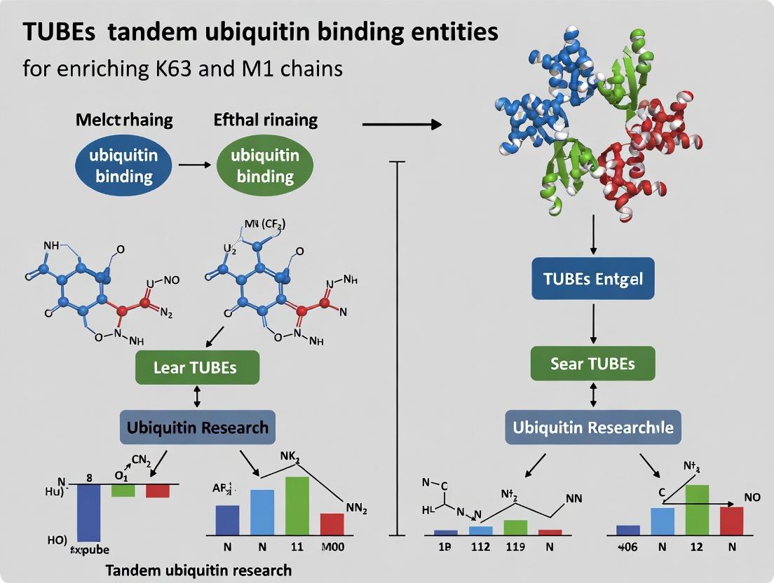
Tandem Ubiquitin-Binding Entities (TUBEs) are engineered reagents composed of multiple ubiquitin-associated (UBA) domains connected in tandem. They are central to research within a broader thesis focused on enriching and studying K63- and M1-linked polyubiquitin chains. Unlike monomeric UBA domains with low micromolar affinity, TUBEs exploit avidity effects to achieve nanomolar affinity for polyubiquitin chains. Crucially, specific UBA domain sequences confer selectivity for distinct ubiquitin linkage types, enabling the isolation of specific chain topologies (e.g., K63, M1) from complex biological samples for downstream analysis. This has profound implications for studying ubiquitin signaling in pathways like NF-κB activation, DNA damage repair, and proteostasis, which are often dysregulated in cancer and neurodegenerative diseases.
Mechanisms of High-Affinity and Linkage-Selective Capture
The superior performance of TUBEs is explained by two key principles:
Avidity-Driven High Affinity: A single UBA domain binds a single ubiquitin moiety with modest affinity (Kd ~10-100 µM). By linking multiple UBA domains with flexible linkers, a single TUBE molecule can simultaneously engage multiple ubiquitin units within a polyubiquitin chain. This multivalent interaction results in a dramatic increase in functional affinity (avidity), achieving effective Kd values in the low nanomolar range. This allows TUBEs to efficiently capture polyubiquitinated proteins even in the presence of deubiquitinases (DUBs), as the TUBE physically shields the chain from enzymatic cleavage.
Linkage Selectivity: Linkage selectivity is determined by the intrinsic preference of the source UBA domain. For example, the UBA domain from the protein Rabex-5 shows strong preference for K63-linked di-ubiquitin, while the UBAN motif from NEMO (IKKγ) selectively binds linear (M1-linked) ubiquitin chains. By constructing TUBEs from these selective domains, researchers can create tools that preferentially enrich specific chain architectures.
Table 1: Affinity and Selectivity Profiles of Common UBA Domains Used in TUBEs
| UBA Domain Source | Preferred Linkage Type | Monomeric Kd (for di-Ub) | Tandem Construct (TUBE) Effective Kd | Primary Application |
|---|---|---|---|---|
| Rabex-5 | K63-linked | ~30 µM | < 10 nM | Enrichment of K63-linked chains involved in DNA repair, endocytosis. |
| NEMO (UBAN) | M1-linked (Linear) | ~5 µM | ~1-5 nM | Isolation of linear ubiquitin chains in NF-κB and TNF signaling. |
| hHR23A | K48-linked | ~90 µM | ~20 nM | Capture of K48-linked chains targeting substrates for proteasomal degradation. |
| SQSTM1/p62 | K63-linked, unanchored | Variable | ~10-50 nM | Study of autophagy and aggregates. |
Application Notes
Key Applications in Research
- Protection from Deubiquitinases (DUBs): Adding TUBEs to cell lysates stabilizes labile ubiquitin signals by inhibiting DUB activity competitively.
- Enrichment of Polyubiquitinated Proteins: TUBEs coupled to matrices (e.g., agarose beads) enable affinity purification of ubiquitinated proteins for mass spectrometry (Ubiquitin Proteomics) or western blot analysis.
- Linkage-Specific Signaling Analysis: Selective TUBEs allow researchers to dissect the role of specific chain types in cellular pathways, such as K63 chains in kinase activation or M1 chains in inflammatory responses.
- Immunofluorescence/Histochemistry: Fluorescently tagged TUBEs can be used to visualize endogenous polyubiquitin chains in fixed cells or tissues.
Critical Considerations for Experimental Design
- Selectivity is Relative: No TUBE is absolutely specific. K63-TUBEs may still bind K48 chains at very high concentrations. Use appropriate controls (e.g., free ubiquitin or monoubiquitin for competition).
- Buffer Composition: Use strong lysis buffers (e.g., with 1% SDS) to disrupt non-covalent interactions and then dilute for capture to ensure access to true ubiquitin conjugates. Include DUB and protease inhibitors (N-ethylmaleimide, iodoacetamide, complete protease inhibitors).
- Elution Conditions: For downstream analysis like western blotting, elution with Laemmli buffer at 95°C is standard. For functional studies, competitive elution with free polyubiquitin chains (specific linkage) is possible.
Detailed Protocols
Protocol 1: Linkage-Selective Enrichment of Ubiquitinated Proteins using Agarose-Conjugated TUBEs
Objective: To isolate K63- or M1-linked polyubiquitinated proteins from mammalian cell lysates for detection by immunoblotting.
Materials:
- Cells treated with relevant stimulus (e.g., TNF-α for M1 chains, DNA damaging agent for K63 chains).
- Lysis Buffer: 50 mM Tris-HCl pH 7.5, 150 mM NaCl, 1% NP-40, 0.5% Sodium deoxycholate, 0.1% SDS, 10 mM N-ethylmaleimide (NEM), 5 mM iodoacetamide, 1x protease inhibitor cocktail.
- Linkage-specific TUBE Agarose (e.g., K63-TUBE Agarose, M1-TUBE Agarose).
- Control Agarose (beads without TUBE).
- Wash Buffer: 50 mM Tris-HCl pH 7.5, 150 mM NaCl, 0.5% NP-40.
- 2X Laemmli Sample Buffer.
- Antibodies: Anti-Ubiquitin (linkage-specific optional, e.g., anti-K63-Ub, anti-M1-Ub), Anti-Target Protein (e.g., RIPK1, TRAF6).
Procedure:
- Lysis: Harvest 5-10 x 10^6 cells. Lyse cells in 0.5-1 mL of pre-chilled Lysis Buffer for 30 minutes on ice. Vortex briefly every 10 minutes.
- Clarification: Centrifuge lysates at 16,000 x g for 15 minutes at 4°C. Transfer supernatant to a new tube. Note: The lysate can be snap-frozen at -80°C at this stage.
- Pre-clearing (Optional but Recommended): Incubate lysate with 20 µL of control agarose beads for 30 minutes at 4°C on a rotator. Centrifuge at 1,000 x g for 2 minutes and transfer supernatant to a new tube.
- TUBE Capture: Add 20-30 µL of settled TUBE Agarose beads to the lysate. Incubate for 2-4 hours at 4°C on a rotator.
- Washing: Pellet beads at 1,000 x g for 2 minutes. Carefully remove supernatant.
- Wash beads 3 times with 1 mL of Lysis Buffer (without inhibitors).
- Wash beads 2 times with 1 mL of Wash Buffer.
- Elution: Completely aspirate supernatant. Add 40 µL of 2X Laemmli Sample Buffer to the beads. Boil at 95°C for 10 minutes.
- Analysis: Centrifuge briefly, load supernatant onto an SDS-PAGE gel. Proceed to western blotting with relevant antibodies.
Protocol 2: DUB Protection Assay using Soluble TUBEs
Objective: To preserve endogenous polyubiquitin chains during cell lysis and sample preparation.
Materials:
- Soluble TUBEs (e.g., GST- or MBP-tagged K63-TUBE) at 5-10 µM stock.
- Standard Cell Lysis Buffer (without NEM/iodoacetamide if assessing DUB activity).
- 4X Laemmli Sample Buffer.
Procedure:
- Treatment: Add soluble TUBE to the standard lysis buffer at a final concentration of 1-2 µM just before use.
- Lysis: Lyse cells directly in the TUBE-containing lysis buffer. Incubate on ice for 10-15 minutes.
- Immediate Denaturation: Mix lysate 1:1 with 4X Laemmli Sample Buffer. Boil immediately at 95°C for 10 minutes to denature proteins and fix the ubiquitination state.
- Analysis: Analyze by western blot for total or linkage-specific ubiquitin. Compare to samples lysed without TUBEs to visualize the protective effect.
Diagrams
Diagram 1: TUBE Avidity vs. Monomeric UBA Binding
Diagram 2: Workflow for TUBE-Based Enrichment & Analysis
Diagram 3: Linkage-Selective TUBEs in NF-κB Pathway Analysis
The Scientist's Toolkit: Key Research Reagent Solutions
Table 2: Essential Materials for TUBE-Based Ubiquitin Research
| Reagent / Material | Function & Purpose | Key Considerations |
|---|---|---|
| Linkage-Specific TUBE Agarose | Affinity matrix for pull-down of polyubiquitinated proteins with defined linkage (K63, M1, K48, etc.). | Choice depends on pathway studied. Check manufacturer's data for selectivity profile. Pre-clearing reduces non-specific binding. |
| Soluble TUBEs (GST-, MBP-, Halo- tagged) | In-solution capture or DUB protection. Useful for co-immunoprecipitation, fluorescence imaging, or stabilizing chains before pull-down. | Tag can influence solubility and may need removal for some applications. Concentration is critical for effective DUB protection. |
| Deubiquitinase (DUB) Inhibitors (N-ethylmaleimide, Iodoacetamide, PR-619, Ubiquitin Aldehydes) | Preserve the endogenous ubiquitinome by inhibiting cysteine protease DUB activity during lysis. | NEM is common but must be prepared fresh. Some inhibitors are broad-spectrum, others are specific to DUB families. |
| Linkage-Specific Ubiquitin Antibodies (Anti-K63-Ub, Anti-M1-Ub, Anti-K48-Ub) | Validate enrichment specificity and detect specific chain types by western blot. | Quality varies greatly. Always confirm with appropriate controls (e.g., linkage-specific DUB treatment). May have cross-reactivity. |
| Recombinant Linkage-Specific Di-/Poly-Ubiquitin | As standards for binding assays, for competitive elution from TUBEs, or to validate antibody/TUBE specificity. | Essential positive control. K48- and K63-linked chains are most common. M1-linked (linear) chains are also available. |
| Ubiquitin Activating Enzyme (E1) Inhibitor (e.g., TAK-243, PYR-41) | Negative control to confirm signals are due to ubiquitination. Depletes cellular ubiquitin pools pre-treatment. | Useful for dynamic studies to block new ubiquitination events. |
| Proteasome Inhibitor (e.g., MG132, Bortezomib) | Accumulates polyubiquitinated proteins, often K48-linked, by blocking their degradation. Enhances signal for capture. | Can alter signaling dynamics. Use with clear experimental rationale. |
| Strong Denaturing Lysis Buffer (with 1% SDS) | Effectively disrupts all non-covalent protein complexes, ensuring TUBEs access genuine ubiquitin conjugates. | Must be diluted (to ≤0.1% SDS) before incubation with TUBE beads to prevent protein denaturation and bead damage. |
Tandem Ubiquitin-Binding Entities (TUBEs) are engineered scaffolds containing multiple Ubiquitin-Associated (UBA) domains in series. They exhibit high avidity for polyubiquitin chains, protecting them from deubiquitinating enzymes (DUBs) and enabling enrichment from complex biological samples. Within research focused on K63- and M1-linked polyubiquitin chains—key signals in NF-κB activation, inflammation, and DNA damage repair—selective TUBEs are indispensable. The specificity of a TUBE is dictated by the intrinsic linkage preference of its constituent UBA domains. This document outlines the properties of common UBA domains used in TUBE scaffolds and provides protocols for their application in enriching K63 and M1 chains.
UBA Domain Binding Specificities
The binding affinity and linkage preference of UBA domains are quantified by techniques like Isothermal Titration Calorimetry (ITC) and Surface Plasmon Resonance (SPR). The following table summarizes key data for UBA domains commonly incorporated into TUBE scaffolds.
Table 1: Binding Affinities of Common UBA Domains for Ubiquitin Linkages
| UBA Domain (Source Protein) | Preferred Linkage(s) | Kd for MonoUb / DiUb (µM) | Key Structural Feature Influencing Specificity | Utility in TUBEs for K63/M1 Research |
|---|---|---|---|---|
| UBA2 (hHR23A) | K48, K63 (broad) | K48-diUb: ~0.6 | Ubiquitin interaction motif (MGF, LVL) | General polyUb enrichment; not linkage-specific. |
| UBAN (NEMO/IKKγ) | M1 (Linear), K63 | M1-diUb: ~0.2 - 1.0 | Specific groove recognizing M1-diUb N-terminus | Critical for selective M1-chain enrichment. |
| NZF (HOIL-1L) | M1 (Linear) | M1-diUb: ~15 - 20 | Dedicated linear Ubiquitin-binding NZF (LUBAN) | Used in tandem for high-avidity M1 capture. |
| UBAN (OPTN) | M1, K63 | M1-diUb: ~0.4 | Similar but distinct from NEMO UBAN | Selective for M1 and K63 chains. |
| UBA (SQSTM1/p62) | K63 (preferential) | K63-diUb: ~4.5; K48: ~14 | UBA dimerization enhances avidity | Preferential enrichment of K63-linked chains. |
Core Protocols
Protocol 1: Enrichment of K63- and M1-Linked Polyubiquitin Chains using Linkage-Specific TUBEs
Objective: To isolate and concentrate K63- or M1-linked polyubiquitinated proteins from cell lysates for downstream analysis (e.g., Western blot, mass spectrometry).
Research Reagent Solutions & Materials:
- TUBE Agarose Beads: GST- or MBP-tagged TUBE proteins immobilized on agarose. Common constructs: Tandem UBAN (from NEMO) for M1, Tandem UBA (from p62) for K63.
- Lysis Buffer (with inhibitors): 50 mM Tris-HCl (pH 7.5), 150 mM NaCl, 1% NP-40, 1 mM EDTA. Supplements (added fresh): 10 mM N-Ethylmaleimide (NEM), 1 mM PMSF, 10 μM PR-619 (broad DUB inhibitor), 1x protease inhibitor cocktail.
- Wash Buffer: Lysis buffer without DUB inhibitors (NEM/PR-619 can be retained).
- Elution Buffer: 1x LDS sample buffer containing 50 mM DTT (for direct gel loading) or competitive elution with 1 M Glycine (pH 2.5) neutralized with Tris base.
- Control Beads: Beads conjugated with GST/MBP alone.
- Pre-cleared Cell Lysate: From stimulated cells (e.g., TNFα for NF-κB pathway activation).
Methodology:
- Cell Lysis: Harvest 5x10^6 - 1x10^7 cells. Lyse in 500 μL - 1 mL of ice-cold Lysis Buffer with inhibitors. Incubate on ice for 20 min, vortexing intermittently.
- Clarification: Centrifuge at 16,000 x g for 15 min at 4°C. Transfer supernatant to a new tube. Determine protein concentration.
- Pre-clearing: Incubate lysate (1 mg total protein) with 20 μL of control beads for 30 min at 4°C with rotation. Pellet beads (800 x g, 2 min) and transfer supernatant.
- TUBE Capture: Add 20-30 μL of settled TUBE Agarose Beads to the pre-cleared lysate. Incubate for 2-4 hours at 4°C with rotation.
- Washing: Pellet beads (800 x g, 2 min). Aspirate supernatant. Wash beads 4 times with 1 mL of Wash Buffer. Perform a final quick wash with PBS or Tris buffer.
- Elution: Aspirate all wash buffer. Add 30-50 μL of 1x LDS sample buffer with DTT. Heat at 95°C for 10 min to elute bound proteins. Centrifuge and load supernatant on SDS-PAGE gel.
Protocol 2: Validation of Enriched Chain Linkage by Western Blot
Objective: To confirm the specificity of the enrichment using linkage-specific ubiquitin antibodies.
Methodology:
- Separate eluates from Protocol 1 by SDS-PAGE (4-12% Bis-Tris gel).
- Transfer to PVDF membrane.
- Probe with the following antibodies in sequence (with stripping in between):
- Pan-ubiquitin: (e.g., FK2/P4D1) to confirm total polyUb pull-down.
- Linkage-specific: Anti-K63-linkage (e.g., Apu3) and Anti-M1/Linear-linkage (e.g., 1E3) specific antibodies.
- Target protein: Antibody against a known substrate (e.g., RIPK1 for M1/K63 signaling).
- Compare signals from TUBE pull-down vs. control bead pull-down.
Visualization
Title: TUBE-Based Strategy to Enrich K63 & M1 Chains in TNFα/NF-κB Signaling
Title: Workflow for Ubiquitin Chain Enrichment Using TUBEs
The Scientist's Toolkit
Table 2: Essential Research Reagents for TUBE-Based Ubiquitin Enrichment
| Item | Function & Role in Experiment |
|---|---|
| TUBE Agarose (M1-specific) | Recombinant scaffold of tandem NEMO UBAN domains. High-affinity capture of linear/M1-linked chains, protecting them from DUBs. |
| TUBE Agarose (K63-preferential) | Recombinant scaffold of tandem p62 UBA domains. Preferentially enriches K63-linked polyubiquitin chains over other types. |
| PR-619 (DUB Inhibitor) | Cell-permeable, broad-spectrum DUB inhibitor. Preserves global ubiquitination levels in lysates prior to TUBE capture. |
| N-Ethylmaleimide (NEM) | Irreversible cysteine protease/DUB inhibitor. Used in lysis buffers to instantly halt DUB activity upon cell disruption. |
| Anti-Linear/M1 Ubiquitin (1E3) | Monoclonal antibody specifically recognizing the linear (M1) diubiquitin linkage motif. Critical for validating M1 enrichment. |
| Anti-K63 Linkage (Apu3) | Monoclonal antibody with high specificity for K63-linked polyubiquitin chains. Used to validate K63 enrichment. |
| Pan-Ubiquitin Antibody (FK2) | Recognizes mono- and polyubiquitinated proteins regardless of linkage. Confirms total ubiquitin pull-down efficiency. |
| Recombinant Linkage-Specific DiUb | Defined K63-, K48-, M1-diubiquitin. Essential as standards for competitive elution or SPR/ITC validation of TUBE specificity. |
Why Enrich K63/M1? Linking Chain-Specific Analysis to Disease Mechanisms in Oncology and Immunology
K63-linked and linear/M1-linked polyubiquitin chains are non-degradative ubiquitin modifications central to inflammatory and oncogenic signaling pathways. Enrichment and analysis of these specific chains are critical for elucidating disease mechanisms. Tandem Ubiquitin Binding Entities (TUBEs) are indispensable tools for this purpose, allowing high-affinity, chain-specific pulldowns from complex biological samples. This application note details protocols and analytical frameworks for using K63/M1-specific TUBEs to link ubiquitinomics to oncology and immunology research.
The Role of K63 and M1 Ubiquitination in Disease Pathways
Oncology
K63-linked ubiquitination is a key driver of oncogenic signaling, primarily through regulation of Protein Kinase B (AKT) and NF-κB pathways. It modulates receptor tyrosine kinase (RTK) trafficking, DNA damage response, and cell survival.
Immunology & Inflammation
Both K63 and M1 linkages are pivotal in innate immunity. K63 chains regulate signaling adaptors like TRAF6 downstream of Toll-like Receptors (TLRs) and Interleukin-1 Receptor (IL-1R). M1/linear chains, assembled by the Linear Ubiquitin Chain Assembly Complex (LUBAC), are essential for optimal NF-κB activation and inflammatory gene expression.
Table 1: Key Disease Associations of K63 and M1 Ubiquitination
| Ubiquitin Linkage | Key E3 Ligase(s) | Primary Signaling Pathways | Associated Disease Contexts |
|---|---|---|---|
| K63-linked | TRAF6, cIAP1/2, BRCA1-BARD1 | NF-κB, AKT, DNA Repair, RTK Trafficking | Breast & Ovarian Cancers, Lymphoma, Autoimmunity |
| M1-linked (Linear) | LUBAC (HOIP, HOIL-1, Sharpin) | NF-κB (TNFR1, TLRs), Inflammation, Necroptosis | Rheumatoid Arthritis, Inflammatory Bowel Disease, Skin Disorders |
Research Reagent Solutions Toolkit
Table 2: Essential Reagents for K63/M1 Ubiquitin Research
| Reagent | Function & Specificity | Example Application |
|---|---|---|
| Agarose-TUBE (K63-specific) | High-affinity resin for selective enrichment of K63-linked polyubiquitinated proteins from lysates. | Pulldown of K63-ubiquitinated RIPK1 in TNFα signaling studies. |
| Agarose-TUBE (M1-specific) | Selective enrichment of linear polyubiquitin chains. | Isolation of LUBAC-modified NEMO in NF-κB pathway analysis. |
| K63-linkage Specific Antibody (e.g., anti-Ubiquitin (K63-linkage specific)) | Detects endogenous K63 chains in WB, IHC, or IF without cross-reactivity with other linkages. | Validation of TUBE enrichments; monitoring K63 chain dynamics. |
| M1-linkage Specific Antibody (e.g., anti-Linear Ubiquitin) | Detects endogenous M1 chains. | Confirmation of linear ubiquitination in immunoprecipitates or tissue samples. |
| Deubiquitinase (DUB) Inhibitors (e.g., PR619, N-Ethylmaleimide) | Broad-spectrum DUB inhibitors preserve the endogenous ubiquitinome during cell lysis. | Added to lysis buffer to prevent chain disassembly. |
| Proteasome Inhibitor (e.g., MG132) | Inhibits 26S proteasome, prevents degradation of polyubiquitinated proteins. | Used in cell pre-treatment to stabilize ubiquitin conjugates. |
| Isopeptidase T (USP5) Inhibitor | Selective inhibitor of K63-chain disassembly by USP5. | Enhances recovery of K63-linked conjugates in enrichment protocols. |
| LUBAC Complex Recombinant Protein | Active enzyme complex for in vitro ubiquitination assays. | Generating positive controls for M1-linkage detection. |
Detailed Protocols
Protocol 1: Enrichment of K63/M1-Linked Polyubiquitinated Proteins Using Agarose-TUBEs
Objective: To selectively isolate proteins modified with K63 or M1 ubiquitin chains from mammalian cell lysates for downstream analysis (WB, MS).
Materials:
- Cells of interest, treated as required.
- Ice-cold PBS.
- Lysis Buffer: 50 mM Tris-HCl (pH 7.5), 150 mM NaCl, 1 mM EDTA, 1% NP-40, 10% glycerol. Add fresh: 1x protease inhibitor cocktail, 5 mM N-Ethylmaleimide (DUB inhibitor), 10 μM MG132.
- Agarose-TUBE (K63-specific) and Agarose-TUBE (M1-specific).
- Control Agarose (unspecific).
- Wash Buffer: 50 mM Tris-HCl (pH 7.5), 150 mM NaCl, 1 mM EDTA, 0.5% NP-40.
- Elution Buffer: 1X SDS-PAGE Sample Buffer (reducing, with 100 mM DTT).
- Rotating mixer at 4°C, microcentrifuge.
Method:
- Cell Lysis: Harvest ~1x10^7 cells per condition. Wash with ice-cold PBS. Lyse cells in 500 μL Lysis Buffer on ice for 30 min with occasional vortexing. Clarify by centrifugation at 16,000 x g for 15 min at 4°C. Transfer supernatant to a fresh tube.
- Pre-clearing: Incubate lysate with 20 μL control agarose slurry for 30 min at 4°C on a rotator. Pellet beads (2,500 x g, 5 min, 4°C) and transfer supernatant.
- TUBE Capture: Aliquot lysate (e.g., 400 μL for enrichment, 50 μL as "Input" control). Incubate the 400 μL aliquot with 30 μL of packed Agarose-TUBE resin (either K63- or M1-specific) for 3 hours at 4°C on a rotator.
- Washing: Pellet beads (2,500 x g, 5 min, 4°C). Carefully aspirate supernatant. Wash beads 4 times with 500 μL Wash Buffer, rotating for 2 min per wash.
- Elution: After final wash, completely aspirate buffer. Add 40 μL of 1X SDS-PAGE Sample Buffer with DTT to the beads and the saved Input sample. Boil at 95°C for 10 min.
- Analysis: Load eluates and Input on SDS-PAGE. Perform Western Blotting using anti-ubiquitin, linkage-specific antibodies (K63 or M1), or antibodies against your target protein of interest.
Protocol 2: Quantitative Mass Spectrometry Workflow Following TUBE Enrichment
Objective: To identify and quantify the proteome modified by K63 or M1 chains under specific disease-relevant conditions.
Materials:
- TUBE eluates from Protocol 1, Step 5 (use non-reducing, non-denaturing elution like 100 mM NH4OH, pH 11.5, for MS).
- Equipment for SDS-PAGE and in-gel digestion, or FASP filter-aided sample preparation.
- Trypsin/Lys-C protease mix.
- StageTips for desalting.
- LC-MS/MS system.
- Bioinformatics software (MaxQuant, Perseus).
Method:
- Sample Preparation: Combine eluates from multiple TUBE enrichments of the same condition to obtain sufficient material (aim for >10 μg protein). Separate proteins by short-run SDS-PAGE (entire lane) or proceed with in-solution digestion after buffer exchange.
- Proteolytic Digestion: Reduce with DTT, alkylate with iodoacetamide, and digest with Trypsin/Lys-C overnight at 37°C.
- Peptide Clean-up: Desalt peptides using C18 StageTips according to manufacturer's instructions.
- LC-MS/MS Analysis: Analyze peptides on a high-resolution tandem mass spectrometer coupled to nanoflow liquid chromatography. Use data-dependent acquisition (DDA) or data-independent acquisition (DIA/SWATH) methods.
- Data Analysis: Process raw files with MaxQuant, searching against the human UniProt database. Specify 'GlyGly (K)' as a variable modification to identify ubiquitination sites. For DIA data, use Spectronaut or DIA-NN. Use Perseus for statistical analysis: filter for contaminants, reverse hits, and proteins only identified by site. Compare enrichment in K63/M1 TUBE samples vs. control agarose or vs. different treatment conditions.
Data Presentation: Quantitative Insights
Table 3: Example MS Data: Proteins Enriched with K63-TUBEs in TNFα-Stimulated HEK293T Cells
| Protein Gene Symbol | Protein Name | Log2 Fold Change (TNFα/Untreated) | -log10(p-value) | Known K63 Substrate? | Proposed Function in Pathway |
|---|---|---|---|---|---|
| RIPK1 | Receptor-interacting serine/threonine-protein kinase 1 | 3.2 | 5.7 | Yes | Necroptosis/NF-κB signaling scaffold |
| TRAF6 | TNF receptor-associated factor 6 | 2.8 | 4.5 | Yes | E3 ligase for K63 chains in IL-1R/TLR signaling |
| TAX1BP1 | Tax1-binding protein 1 | 2.5 | 3.9 | Yes (binds) | Autophagy adaptor, negative regulator of inflammation |
| NEMO (IKBKG) | NF-kappa-B essential modulator | 2.1 | 3.2 | Yes (M1 also) | Regulatory subunit of IKK complex |
| MYD88 | Myeloid differentiation primary response protein MyD88 | 1.9 | 2.8 | Indirect | TLR/IL-1R adaptor, recruits IRAKs and TRAF6 |
Signaling Pathway Visualizations
Diagram Title: K63/M1 Ubiquitin in Immune and Cancer Signaling & Analysis
Diagram Title: K63/M1 TUBE Enrichment and Analysis Protocol Workflow
Step-by-Step Protocols: Implementing TUBEs for K63/M1 Enrichment in Pulldown and Mass Spectrometry
Within the broader thesis on TUBEs (Tandem Ubiquitin-Binding Entities) for enriching K63 and M1 (Met1-linked linear) polyubiquitin chains, selecting the optimal TUBE format is critical. Recombinant TUBE proteins and agarose bead-conjugated TUBEs offer distinct advantages tailored to specific experimental goals in ubiquitin proteomics and signaling research.
Application Notes & Comparative Analysis
Core Distinction: Recombinant TUBEs are soluble proteins used in pull-downs when the eluted ubiquitinated targets must be free of antibody interference (e.g., for mass spectrometry). Agarose bead-conjugated TUBEs offer convenience and are ideal for rapid immunoblotting analysis and repeated use.
Quantitative Performance Comparison:
Table 1: Format Comparison for K63/M1 Chain Enrichment
| Parameter | Recombinant TUBE Protein | Agarose Bead-Conjugated TUBE |
|---|---|---|
| Typical Binding Capacity | ~2-5 µg ubiquitin conjugates per µg TUBE | ~10-20 µg ubiquitin conjugates per mL bead slurry |
| Elution Compatibility | Gentle, non-denaturing (e.g., low pH, competitive elution) | Denaturing (SDS sample buffer) or gentle |
| Best for Mass Spectrometry (MS) | Excellent (minimal contamination) | Possible, but bead leaching can increase background |
| Best for Immunoblotting | Good | Excellent (direct bead boiling) |
| Re-usability | No | Yes (typically 3-5 cycles) |
| Handling Speed | Slower (requires coupling to beads per experiment) | Faster (ready-to-use) |
| Relative Cost per Experiment | Higher | Lower |
Table 2: Recommended Application Selection
| Primary Application Goal | Recommended Format | Key Rationale |
|---|---|---|
| Ubiquitinome Profiling (MS) | Recombinant Protein | Cleanest eluate, reduced contaminant carryover. |
| Monitoring Chain Dynamics (K63/M1) | Agarose Bead-Conjugated | Rapid processing, multiple sequential pulldowns from same sample. |
| Identifying UB-binding Partners | Recombinant Protein | Avoids false positives from bead matrix interactions. |
| Routine Analysis of PolyUbylation | Agarose Bead-Conjugated | Workflow simplicity and cost-effectiveness for blotting. |
Detailed Protocols
Protocol A: Enrichment of K63/M1 Chains Using Recombinant TUBEs for Mass Spectrometry
Objective: Isolate endogenous K63/M1-linked ubiquitinated proteins for subsequent proteomic analysis.
Materials: Recombinant K63/M1-specific TUBE (e.g., TAB2 NZF domain tandem), Magnetic Agarose Beads (e.g., Streptavidin or Anti-FLAG), Cell Lysis Buffer (50 mM Tris-HCl pH 7.5, 150 mM NaCl, 1% NP-40, 10% glycerol, 1 mM EDTA, supplemented with 1x Protease Inhibitor Cocktail, 10 mM N-Ethylmaleimide, and 1x Deubiquitinase Inhibitor PR-619).
Method:
- Lysate Preparation: Harvest and lyse cells (5-10 mg total protein) in ice-cold lysis buffer. Centrifuge at 16,000 x g for 15 min at 4°C.
- Bead Coupling: Incubate 20 µg recombinant TUBE with 50 µL of appropriate pre-washed magnetic beads for 1 hour at 4°C.
- Pull-Down: Incubate cleared lysate with TUBE-bound beads for 2 hours at 4°C with rotation.
- Washing: Wash beads 4x with 1 mL lysis buffer (without inhibitors).
- Competitive Elution for MS: Elute ubiquitinated complexes with 50 µL of 0.2 M glycine pH 2.5 for 5 min. Immediately neutralize with 5 µL 1 M Tris-HCl pH 8.0.
- Processing: Analyze eluate by SDS-PAGE followed by in-gel tryptic digestion and LC-MS/MS, or proceed to solution-based digestion.
Protocol B: Rapid Detection Using Agarose Bead-Conjugated TUBEs
Objective: Quickly assess global K63/M1 polyubiquitination levels or ubiquitination of a high-abundance target.
Materials: Agarose Bead-Conjugated TUBE (K63/M1-specific), RIPA Lysis Buffer, 2x Laemmli Sample Buffer.
Method:
- Lysate Preparation: Lyse cells in RIPA buffer with inhibitors (as in Protocol A). Clarify by centrifugation.
- Pull-Down: Incubate 500 µg – 1 mg lysate with 20 µL bead-conjugated TUBE slurry for 90 min at 4°C.
- Washing: Wash beads 3x with 1 mL cold lysis buffer.
- Direct Immunoblot Analysis: Add 40 µL of 2x Laemmli buffer directly to beads. Boil for 5 min. Load supernatant directly onto SDS-PAGE gel. Probe with antibodies against ubiquitin (K63-linkage specific), your protein of interest, or common signaling proteins in NF-κB or DNA damage pathways.
Pathway & Workflow Visualizations
TUBE Selection and Experimental Workflow
K63/M1 Hybrid Chains in NF-κB Activation
The Scientist's Toolkit: Essential Research Reagents
Table 3: Key Reagents for TUBE-Based Ubiquitin Enrichment
| Reagent / Material | Function / Role | Critical Note |
|---|---|---|
| K63/M1-Specific TUBE | Core affinity reagent. Binds K63 & M1 linkages with high avidity, protecting chains from DUBs. | Specificity must be validated. M1-binding requires unique structural motifs (e.g., NZF1 of HOIL-1L). |
| N-Ethylmaleimide (NEM) | Alkylating agent; irreversibly inhibits cysteine proteases, including deubiquitinases (DUBs). | Essential in lysis buffer to preserve ubiquitination state. Must be fresh. |
| PR-619 (DUB Inhibitor) | Broad-spectrum, cell-permeable DUB inhibitor. Used in cell pre-treatment or lysis. | Complements NEM by inhibiting a wider range of DUB classes. |
| Protease Inhibitor Cocktail | Inhibits serine, cysteine, and metalloproteases to prevent general protein degradation. | Standard addition, but does not protect against DUBs specifically. |
| Glycine (pH 2.5) Elution Buffer | Low-pH competitive elution. Disrupts TUBE-Ubiquitin interaction gently. | Ideal for MS. Must be neutralized immediately post-elution. |
| Anti-K63-linkage Specific Ab | Antibody used to validate enrichment in western blot. | Does not bind M1 chains. Confirms K63 component of enriched pools. |
| Streptavidin Magnetic Beads | For coupling biotinylated recombinant TUBE proteins. | Enables flexible, clean pulldowns with recombinant TUBEs. |
This application note outlines critical sample preparation protocols for the analysis of labile ubiquitin (Ub) conjugates, with a specific focus on enriching for Lys63 (K63)- and Met1 (M1)-linked polyubiquitin chains using Tandem Ubiquitin Binding Entities (TUBEs). Within the broader thesis context of TUBE-based research on K63 and M1 chains—key regulators of NF-κB signaling and inflammation—preserving the native ubiquitome during cell lysis is paramount. The labile nature of these modifications, particularly M1 linear chains, necessitates stringent lysis conditions to prevent deubiquitinase (DUB)-mediated cleavage and preserve chain topology for downstream enrichment and analysis.
Key Challenges in Ubiquitin Conjugate Preservation
The primary obstacles during lysis are:
- Deubiquitinating Enzyme (DUB) Activity: Endogenous DUBs remain active post-cell disruption, rapidly cleaving ubiquitin conjugates.
- Proteasomal Degradation: The proteasome continues to degrade polyubiquitinated substrates.
- Denaturation of Epitopes: Harsh denaturants preserve conjugates but can disrupt native protein interactions and TUBE binding epitopes.
- Chain Rearrangement: Changes in pH or temperature can promote non-enzymatic chain disassembly.
Optimized Lysis Buffers: Composition & Rationale
The choice of lysis buffer represents a compromise between complete inhibition of enzymatic activity and preservation of native interactions for affinity enrichment. Based on current literature, the following formulations are recommended.
Table 1: Comparative Analysis of Lysis Buffer Formulations for Ubiquitin Preservation
| Component | Mild RIPA (Compromise) | Fully Denaturing (Maximal Preservation) | Native (for Functional Studies) | Primary Function in Context |
|---|---|---|---|---|
| Base Buffer | 50 mM Tris, 150 mM NaCl | 50 mM Tris, 150 mM NaCl | 50 mM HEPES, 150 mM NaCl | Maintains ionic strength & pH. HEPES offers better pH stability. |
| Detergent | 1% NP-40 or Triton X-100 | 1% SDS | 0.5-1% NP-40 | Membrane solubilization. SDS fully denatures and inactivates enzymes. |
| DUB/Protease Inhibitors | 10 mM N-Ethylmaleimide (NEM), 5 mM EDTA, 1x cOmplete | 20-50 mM NEM, 5 mM EDTA, 1x cOmplete, 10 µM PR-619 | 10 mM NEM, 5 mM EDTA, 1x cOmplete | NEM is critical—alkylates active site cysteines of DUBs. PR-619 is a broad-spectrum DUB inhibitor. |
| Proteasome Inhibitor | 10 µM MG-132 (optional) | 10 µM MG-132 | 10 µM MG-132 | Prevents degradation of ubiquitinated substrates. |
| Additional Agents | Glycerol (5-10%) | 8M Urea or 2% SDS | Glycerol (5%), ATP (1 mM) | Denaturants (Urea/SDS) ensure complete enzyme inactivation. Glycerol stabilizes complexes. |
| pH | 7.4-7.6 | 7.4-8.0 | 7.4-7.6 | Slightly basic pH reduces acid-driven DUB activity. |
| Key Advantage | Preserves protein-protein interactions for native pulldowns. | Gold standard for preserving total ubiquitin conjugates; halts all enzymatic activity. | Maintains protein complex integrity and activity. | |
| Key Disadvantage | Potential for residual DUB activity. | Requires dilution/ dialysis before TUBE pulldown; may disrupt some epitopes. | High risk of conjugate loss; not recommended for topological studies. | |
| Best for TUBEs | Suitable for Agarose-TUBE pulldowns. | Recommended for initial validation. Must dilute SDS to <0.1% for Magnetic TUBE pulldowns. | Not ideal for conjugate preservation studies. |
Detailed Protocol: Lysis for TUBE-Based Enrichment of K63/M1 Chains
Materials & Reagents (The Scientist's Toolkit)
Table 2: Essential Research Reagent Solutions
| Item | Function & Rationale |
|---|---|
| N-Ethylmaleimide (NEM), 500 mM stock in EtOH | Irreversible cysteine protease/DUB inhibitor. The single most important reagent for preserving labile ubiquitin conjugates. |
| EDTA (0.5 M stock, pH 8.0) | Chelates divalent cations, inhibiting metalloprotease DUBs and proteasomes. |
| Broad-Spectrum DUB Inhibitor (e.g., PR-619) | Potent, cell-permeable inhibitor of a wide range of DUB families, used in addition to NEM. |
| Proteasome Inhibitor (e.g., MG-132) | Reversible inhibitor of the 26S proteasome, preventing substrate degradation during lysis. |
| SDS (20% stock solution) | Ionic denaturant that inactivates all enzymes immediately upon lysis. |
| Magnetic or Agarose-TUBEs | Recombinant tandem ubiquitin-binding entities with high affinity for polyubiquitin chains. Select TUBEs with specificity for K63/M1 linkages if available. |
| Lysis Buffer (Denaturing): 50 mM Tris-HCl (pH 7.5), 150 mM NaCl, 1% SDS, 10 mM NEM, 5 mM EDTA, 10 µM MG-132, 1x protease inhibitor cocktail. | Complete preservation buffer. Prepare fresh, adding NEM and MG-132 from stock solutions immediately before use. |
Protocol Steps
A. Cell Harvest and Lysis (All steps performed on ice or at 4°C unless stated)
- Pre-treatment (Optional but Recommended): Treat cultured cells directly with 10 µM MG-132 and 10 µM PR-619 for 30-60 minutes before harvest to inhibit degradation and DUB activity in vivo.
- Harvest & Wash: Rapidly aspirate media. Wash cells once with 10 mL of ice-cold PBS containing 10 mM NEM.
- Immediate Lysis: Aspirate PBS-NEM. Immediately add Denaturing Lysis Buffer (recommended: 100-200 µL per 10⁶ cells). For adherent cells, add buffer directly to the plate/dish and scrape cells into a pre-cooled microcentrifuge tube.
- Homogenization: Pass the lysate through a 21-27 gauge needle 10-15 times to shear DNA and ensure complete mixing. Note: The solution will become viscous.
- Complete Denaturation: Heat the lysate at 95°C for 5-10 minutes to ensure full protein denaturation and complete inactivation of all enzymes.
- Clarification: Centrifuge the lysate at 20,000 x g for 15 minutes at 4°C to remove insoluble debris. Transfer the clear supernatant to a new tube.
B. Lysate Preparation for TUBE Pulldown
- For Magnetic TUBEs: SDS must be diluted to a concentration below its critical micelle concentration (CMC, typically <0.1-0.2%) to prevent interference with binding. Dilute the clarified lysate 1:10 with a non-ionic detergent-based buffer (e.g., 50 mM Tris, 150 mM NaCl, 1% Triton X-100, pH 7.5).
- For Agarose-TUBEs: The lysate can be diluted similarly, or SDS can be removed via dialysis or spin-column buffer exchange into a compatible buffer.
C. TUBE-Based Affinity Enrichment
- Incubation: Incubate the prepared lysate with the appropriate amount of Magnetic or Agarose-TUBE beads for 2-4 hours at 4°C under gentle rotation.
- Washing: Wash beads extensively with a mild wash buffer (e.g., 50 mM Tris, 150 mM NaCl, 0.1% Tween-20, pH 7.5).
- Elution: Elute enriched ubiquitinated proteins and conjugates by boiling in 2x Laemmli SDS-PAGE sample buffer containing 50-100 mM DTT for 5-10 minutes. DTT will reduce the NEM modification but conjugates are now stable.
D. Downstream Analysis Proceed with SDS-PAGE and Western blotting using linkage-specific antibodies (e.g., anti-K63, anti-M1) or mass spectrometric analysis to identify ubiquitinated substrates and chain topology.
Visualizations
Workflow for Preserving Ubiquitin Conjugates
Research Context: Lysis in Thesis
Within the broader thesis on Tandem Ubiquitin Binding Entities (TUBEs) for enriching K63- and M1-linked polyubiquitin chains, this protocol details a core, reproducible workflow. K63 and M1 (linear) linkages are critical signals in inflammatory, DNA damage, and cell death pathways, often competing for the same substrates or complexes. TUBEs, with their high avidity for ubiquitin, enable the capture of labile, endogenously modified proteins, protecting them from deubiquitinases (DUBs) and the proteasome. This application note provides a detailed, step-by-step protocol for the selective enrichment of proteins modified with these chain types, followed by analytical workflows.
Key Research Reagent Solutions (The Scientist's Toolkit)
| Item | Function & Rationale |
|---|---|
| K63/M1-Specific TUBE Agarose | Core reagent. Recombinant tandem ubiquitin-binding domains (e.g., from UBQLN1, UBAN motifs) coupled to beads, with high selectivity for K63 and/or M1 linkages over other types (K48, K11). |
| Control TUBE Agarose (K48-specific or Wild-Type) | Essential negative control to distinguish non-specific binding and assess linkage specificity of observed enrichments. |
| Deubiquitinase (DUB) Inhibitors (e.g., PR-619, N-Ethylmaleimide) | Added fresh to all lysis and wash buffers to preserve the native ubiquitinome by inhibiting ubiquitin cleavage. |
| Proteasome Inhibitors (e.g., MG-132, Bortezomib) | Prevents degradation of polyubiquitinated proteins, increasing yield for pulldown. |
| Crosslinker (DSS or DTBP) | Optional. For stabilizing weak or transient ubiquitin-dependent interactions prior to lysis. |
| Lysis Buffer (Non-denaturing) | Typically contains Tris-HCl (pH 7.5-8.0), NaCl, glycerol, NP-40 or Triton X-100, and EDTA, maintaining native protein complexes. |
| Competitive Elution Buffer (Ubiquitin Probes) | Contains free Lys63-linked di-ubiquitin or linear di-ubiquitin for specific, gentle elution of bound proteins. |
| Denaturing Elution Buffer (2X Laemmli Buffer) | For complete elution of all bound material for downstream immunoblotting. |
| Antibodies: Anti-K63-linkage, Anti-M1-linkage, Anti-pan-ubiquitin | For validation of enrichment specificity via western blot. |
Detailed Experimental Protocol
A. Cell Culture, Treatment, and Lysis
- Culture & Treatment: Grow cells (e.g., HEK293, HeLa, or relevant primary cells) to 70-90% confluence. Apply relevant stimuli (e.g., TNF-α for NF-κB/M1 signaling, DNA damaging agents for K63 signaling).
- Inhibition: 1-2 hours pre-lysis, add proteasome inhibitor (e.g., 10 µM MG-132). Add DUB inhibitor (e.g., 10 µM PR-619) directly to lysis buffer.
- Harvest & Lysis: Wash cells with ice-cold PBS. Scrape cells into PBS and pellet (500 x g, 5 min, 4°C). Lyse cell pellet (approx. 10⁷ cells per 1 mL) in Non-denaturing Lysis Buffer (50 mM Tris-HCl pH 7.5, 150 mM NaCl, 1% NP-40, 10% glycerol, 1 mM EDTA) supplemented with DUB and protease inhibitors. Incubate on ice for 30 min with gentle vortexing.
- Clarification: Centrifuge lysate at 16,000 x g for 15 min at 4°C. Transfer supernatant to a new tube. Determine protein concentration (e.g., via BCA assay).
B. TUBE-Mediated Affinity Pulldown
- Bead Preparation: For each sample, aliquot 30 µL of settled K63/M1-TUBE Agarose and Control TUBE Agarose slurry into separate microcentrifuge tubes. Wash beads twice with 1 mL of ice-cold lysis buffer.
- Incubation: Incubate 1-2 mg of clarified total cell lysate with the pre-washed TUBE beads. Perform binding for 2-4 hours at 4°C with end-over-end rotation.
- Washing: Pellet beads (1000 x g, 1 min, 4°C). Aspirate supernatant. Wash beads sequentially:
- Wash 1: 1 mL Lysis Buffer (high salt: increase NaCl to 300 mM).
- Wash 2: 1 mL Lysis Buffer (standard salt).
- Wash 3: 1 mL PBS or Tris buffer (pH 7.5).
- Incubate for 5 min with rotation at 4°C for each wash.
C. Elution and Analysis
- For Mass Spectrometry (MS) Analysis:
- Perform a final wash with 50 mM ammonium bicarbonate (pH 8.0).
- Elute bound proteins competitively using 2-3 bead volumes of buffer containing 0.5 mg/mL of Lys63-linked or linear di-ubiquitin for 30 min at 25°C. Alternatively, elute by on-bead digestion.
- For Immunoblot Analysis:
- After final PBS wash, directly add 50 µL of 2X Laemmli SDS sample buffer to the beads.
- Heat at 95°C for 10 min to denature and elute all bound material.
- Resolve eluates by SDS-PAGE and proceed to western blotting.
Data Presentation: Expected Outcomes & Validation
Table 1: Expected Western Blot Results from TUBE Pulldown Validation
| Target | Input Lysate | K63/M1-TUBE Eluate | Control TUBE Eluate | Interpretation |
|---|---|---|---|---|
| K63-linkage (e.g., HA-Ub K63-only) | Weak signal | Strong Enrichment | No/Low signal | Successful specific capture of K63 chains. |
| M1-linkage (e.g., HOIP output) | Weak signal | Strong Enrichment | No/Low signal | Successful specific capture of linear chains. |
| K48-linkage | Detectable | Low/Undetectable | Enriched (if K48-TUBE control) | Specificity of the K63/M1-TUBE reagent. |
| Known Substrate (e.g., RIPK1) | Detectable | Enriched | Not Enriched | Identification of specifically modified proteins. |
Table 2: Typical Yield Metrics from Pulldown for MS Sample Prep
| Parameter | Typical Range | Notes |
|---|---|---|
| Input Protein | 1 - 5 mg | Higher input improves detection of low-abundance ubiquitinated species. |
| Eluted Protein (Competitive) | 5 - 50 µg | Highly variable; depends on stimulus and cell type. |
| Estimated Ubiquitinated Fraction | 0.5 - 5% of eluate | Majority of eluted protein may be associated complexes. |
Pathway & Workflow Visualization
Title: Experimental Workflow for TUBE-Based Enrichment
Title: K63 and M1 Ubiquitin Signaling Pathways Crosstalk
Application Notes
This protocol details the downstream analytical workflow following the enrichment of polyubiquitinated proteins using Tandem Ubiquitin-Binding Entities (TUBEs), specifically those selective for K63- and M1-linked chains, within a thesis investigating K63/M1 hybrid chains in inflammatory signaling. The process begins with the validation of TUBEs pulldown specificity via Western Blot (WB) using linkage-specific antibodies and culminates in sample preparation for mass spectrometric (MS) identification of ubiquitinated substrates and modification sites.
- Objective 1: Validation of Enrichment Specificity: Following TUBEs pulldown, WB analysis with antibodies against specific ubiquitin linkages (e.g., anti-K63, anti-M1) confirms the successful and selective enrichment of the targeted chain topology. This is a critical quality control step before committing samples to MS.
- Objective 2: Target Protein Investigation: WB with antibodies against proteins of interest (e.g., RIPK1, NEMO) determines if they are ubiquitinated with the relevant chain type under the studied conditions.
- Objective 3: MS Sample Preparation: For unbiased discovery, the entire enriched protein pool is subjected to in-gel or in-solution trypsin digestion, generating peptides for LC-MS/MS analysis to identify ubiquitination sites via the detection of Gly-Gly (diGly) remnant peptides.
Key Quantitative Data from TUBEs Enrichment and Validation
Table 1: Typical Yield and Enrichment Metrics from TUBEs Protocol
| Parameter | Typical Range/Result | Measurement Method |
|---|---|---|
| Enriched Ubiquitin-Conjugates | 50-500 µg | BCA assay post-elution |
| Fold-Enrichment (vs. control bead) | 10- to 100-fold | Anti-Ubiquitin WB densitometry |
| K63-Specific TUBEs Efficiency | >90% selectivity for K63 chains over K48 chains | WB with linkage-specific antibodies |
| M1-Specific TUBEs Efficiency | >95% selectivity for M1 chains | WB with anti-M1 (linear) antibody |
| Detection Limit for Ubiquitinated Proteins via WB | 1-10 ng | Chemiluminescence |
Table 2: Critical Antibodies for Western Blot Validation
| Antibody Specificity | Clone/Cat. Example | Key Application in Thesis Context |
|---|---|---|
| K63-linkage Specific | Apu3 (Apu3.AS.27) | Confirms enrichment of K63-linked chains by TUBEs. |
| M1-linkage Specific | Anti-linear Ubiquitin (1E3) | Confirms enrichment of M1-linked chains by TUBEs. |
| Pan-Ubiquitin | P4D1 | Total ubiquitinated protein load control. |
| Target Protein (e.g., RIPK1) | D94C12 | Detects ubiquitination status of specific substrate. |
Experimental Protocols
Protocol 1: Western Blot Analysis of TUBEs Eluates with Chain-Specific Antibodies
Materials: TUBEs eluate in 2X Laemmli buffer, Precast 4-20% Tris-Glycine gels, PVDF membrane, TBST, Blocking buffer (5% BSA in TBST), Primary antibodies (see Table 2), HRP-conjugated secondary antibodies, ECL substrate.
Methodology:
- Sample Preparation: Denature TUBEs eluates at 95°C for 5 min. Load 20-30 µL per well alongside a prestained protein ladder.
- Electrophoresis: Run gel at 120-150 V until dye front reaches bottom.
- Transfer: Activate PVDF membrane in methanol. Transfer proteins at 100 V for 60 min (or 30 V overnight) at 4°C using wet transfer system.
- Blocking: Block membrane with 5% BSA/TBST for 1 hour at RT.
- Primary Antibody Incubation: Dilute linkage-specific antibody (1:1000) in blocking buffer. Incubate membrane overnight at 4°C with gentle agitation.
- Washing: Wash membrane 3 x 10 min with TBST.
- Secondary Antibody Incubation: Incubate with HRP-conjugated anti-mouse/rabbit IgG (1:5000) in blocking buffer for 1 hour at RT.
- Final Wash: Wash 3 x 10 min with TBST.
- Detection: Apply ECL substrate evenly and image using a chemiluminescence detector.
Protocol 2: In-Gel Trypsin Digestion for LC-MS/MS Analysis
Materials: Coomassie Brilliant Blue stain, Destaining solution (40% ethanol, 10% acetic acid), 100 mM ammonium bicarbonate (ABC), Acetonitrile (ACN), 10 mM DTT in ABC, 55 mM iodoacetamide in ABC, Sequencing-grade trypsin, 0.1% formic acid.
Methodology:
- Gel Electrophoresis: Separate the entire TUBEs eluate on a 1D SDS-PAGE gel (4-12% Bis-Tris). Stain with Coomassie Blue.
- Gel Slicing: Excise entire lane as a single band or multiple molecular weight regions. Dice into 1 mm³ pieces.
- Destaining: Wash gel pieces with 200 µL of destaining solution, vortex, incubate at 37°C for 15 min. Repeat until clear. Remove liquid.
- Dehydration: Add 200 µL ACN, incubate until pieces shrink and turn white (~5 min). Remove ACN.
- Reduction: Add 100 µL of 10 mM DTT in 100 mM ABC. Incubate at 56°C for 45 min. Cool to RT. Remove liquid.
- Alkylation: Add 100 µL of 55 mM iodoacetamide in 100 mM ABC. Incubate in the dark at RT for 30 min. Remove liquid.
- Wash/Dehydrate: Add 200 µL of 100 mM ABC, incubate 10 min. Remove. Add 200 µL ACN, incubate 5 min. Remove ACN. Air dry pieces for 5-10 min.
- Trypsin Digestion: Rehydrate gel pieces with 20-50 µL of 12.5 ng/µL trypsin in 50 mM ABC on ice for 30 min. Add enough 50 mM ABC to cover gel pieces. Incubate overnight at 37°C.
- Peptide Extraction: Transfer supernatant to a new tube. Add 50 µL of 0.1% formic acid to gel pieces, sonicate 10 min, combine extracts. Add 50 µL of 50% ACN/0.1% formic acid, sonicate 10 min, combine. Dry down combined extracts in a vacuum concentrator.
- MS Sample Preparation: Resuspend dried peptides in 20 µL of 0.1% formic acid for LC-MS/MS analysis.
Diagrams
Title: Downstream Analysis Workflow After TUBEs Enrichment
Title: Thesis Context: TUBEs in TNF-NFκB Pathway Analysis
The Scientist's Toolkit
Table 3: Essential Research Reagent Solutions for TUBEs Downstream Analysis
| Item | Function | Example Product/Note |
|---|---|---|
| K63- & M1-specific TUBEs | High-affinity, linkage-selective enrichment of polyubiquitinated proteins from complex lysates. | LifeSensors (UM-604M/K63, UM-801M/M1) or in-house GST-tagged TUBEs. |
| Linkage-Specific Ub Antibodies | Validation of TUBEs pulldown specificity and chain-type presence on substrates via WB. | Millipore (Apu3 for K63), Millipore (1E3 for M1). |
| Protease Inhibitor Cocktail (Ub-specific) | Prevents deubiquitinase (DUB) activity during lysis and pulldown to preserve ubiquitin chains. | N-ethylmaleimide (NEM) or PR-619. |
| SDS-PAGE Gel (4-12% Bis-Tris) | Optimal separation of high MW ubiquitin conjugates for both WB analysis and in-gel digestion. | Invitrogen NuPAGE or Bio-Rad Criterion. |
| Sequencing-Grade Modified Trypsin | Highly pure, specific protease for generating peptides for MS; minimizes autolysis. | Promega Trypsin Gold, MS grade. |
| DiGly-Lysine Remnant Antibody | Alternative method to validate ubiquitination in WB by detecting the tryptic remnant. | Cell Signaling Technology (Clone mAb #39205). |
| Strong Cation Exchange (SCX) StageTips | Desalting and fractionation of complex peptide mixtures pre-LC-MS/MS to enhance depth. | Thermo Scientific or homemade with Empore disks. |
| LC-MS/MS System with High Resolution | Identifies and quantifies tryptic peptides, enabling diGly remnant site localization. | Orbitrap-based systems (Exploris, Fusion). |
Within the broader thesis exploring TUBEs (Tandem Ubiquitin-Binding Entities) as critical tools for dissecting the roles of K63-linked and M1-linked (linear) ubiquitin chains in cellular signaling, this application note details their practical implementation in proteomics. The selective enrichment of these chain types, often underrepresented in conventional ubiquitin proteomics, is paramount for understanding their distinct roles in inflammatory signaling, proteostasis, and DNA damage response. This document provides current protocols and data analysis frameworks for leveraging TUBEs in ubiquitin profiling and interactome studies.
The Scientist's Toolkit: Research Reagent Solutions
| Reagent / Material | Function & Rationale |
|---|---|
| High-Affinity TUBEs (e.g., K63-specific, M1-specific, Pan-Selective) | Recombinant proteins with multiple ubiquitin-associated (UBA) domains in tandem. They bind polyubiquitin chains with high avidity, protecting them from proteasomal and deubiquitinase (DUB) activity during lysis. |
| TUBE Agarose/ Magnetic Beads | TUBEs immobilized on solid support for pull-down assays. Magnetic beads facilitate easy washing and elution. |
| Deubiquitinase (DUB) Inhibitors (e.g., PR-619, N-Ethylmaleimide) | Added fresh to cell lysis buffers to prevent artifivial chain disassembly during sample preparation. |
| Proteasome Inhibitors (e.g., MG132, Bortezomib) | Used in cell pre-treatment to stabilize ubiquitinated substrates, enhancing detection. |
| Crosslinkers (e.g., DSS, DTBP) | Optional. For stabilizing weak or transient interactions prior to lysis for interactome studies. |
| Competitive Elution Buffer (1xSDS + 8M Urea) | Harsh elution to disrupt TUBE-ubiquitin interaction. Alternative: Low pH glycine buffer. |
| Trypsin/Lys-C Protease Mix | For on-bead or in-solution digestion of eluted proteins for LC-MS/MS analysis. |
| Anti-Ubiquitin Remnant Motif (diGly) Antibody | For western blot validation or enrichment of ubiquitinated peptides prior to MS (for ubiquitin profiling). |
Application Note 1: Global Ubiquitinome Profiling Using TUBEs
This protocol is designed for the large-scale identification of ubiquitinated proteins, with enhanced recovery of K63/M1-linked substrates.
Protocol: TUBE-based Enrichment for Mass Spectrometry
- Cell Treatment & Lysis: Pre-treat cells (e.g., HEK293, HeLa) with 10 µM MG132 for 4-6 hours. Wash with PBS and lyse in TUBE Lysis Buffer (50 mM Tris-HCl pH 7.5, 150 mM NaCl, 1% NP-40, 1 mM EDTA, 10% glycerol, 1x DUB Inhibitor cocktail, 1x Protease Inhibitor cocktail) for 30 min on ice.
- Clarification: Centrifuge at 16,000 x g for 15 min at 4°C. Transfer supernatant to a new tube and measure protein concentration.
- TUBE Pull-down: Incubate 1-2 mg of total protein lysate with 50 µL of washed Pan-TUBE Magnetic Beads for 2 hours at 4°C with gentle rotation.
- Washing: Place tube on a magnetic rack. Discard flow-through. Wash beads sequentially with:
- Wash Buffer 1: Lysis Buffer (3 x 1 mL)
- Wash Buffer 2: High-Salt Buffer (50 mM Tris-HCl pH 7.5, 500 mM NaCl, 0.1% NP-40; 2 x 1 mL)
- Wash Buffer 3: No-Detergent Buffer (50 mM Tris-HCl pH 7.5, 150 mM NaCl; 1 x 1 mL)
- On-Bead Digestion (Recommended): Resuspend beads in 50 µL of 50 mM Tris-HCl (pH 8.0) with 2 M urea. Add 1 µg Trypsin/Lys-C mix and digest overnight at 37°C with shaking.
- Peptide Cleanup: Acidify peptides with 1% TFA, desalt using C18 StageTips, and dry for LC-MS/MS analysis.
- LC-MS/MS & Data Analysis: Analyze peptides on a high-resolution tandem mass spectrometer. Search data against a human protein database, specifying diGly (K-ε-GG) as a variable modification on lysine to identify ubiquitination sites.
Quantitative Data Summary: TUBE vs. Conventional IP Table 1: Comparative performance of enrichment strategies in a model study of TNF-α stimulated cells.
| Enrichment Method | Total Ubiquitinated Proteins Identified | Unique K63-Linked Substrates | Unique M1-Linked Substrates | Average Fold-Enrichment (Ubiquitin Signal) |
|---|---|---|---|---|
| Pan-TUBE Pull-down | ~3,200 | ~450 | ~85 | >100x |
| Anti-diGly Antibody (Post-Lysis) | ~2,800 | ~120 | ~15 | ~50x |
| Single-UBA Domain Pull-down | ~950 | ~30 | <5 | ~20x |
| No Enrichment (Total Lysate) | <50 | N/A | N/A | 1x |
Application Note 2: Chain-Specific Interactome Analysis
This protocol isolates protein complexes associated with specific ubiquitin chain linkages (K63 or M1).
Protocol: K63/M1 Chain-Specific Interactome Capture
- Stabilization (Optional): Treat cells with a mild, membrane-permeable crosslinker (e.g., 2 mM DTBP for 30 min) to capture transient interactions. Quench with 100 mM Tris-HCl pH 7.5.
- Lysis: Lyse cells in Native Lysis Buffer (as above, but with 0.5% NP-40 or 0.25% Digitonin) to preserve complexes.
- Chain-Specific Enrichment: Split lysate. Incubate equal amounts with K63-TUBE Beads, M1-TUBE Beads, or Mutant TUBE Control Beads for 90 min at 4°C.
- Gentle Washing: Wash beads 3 times with 1 mL of Native Lysis Buffer.
- Elution: Elute bound proteins and complexes with 50 µL of 1x Laemmli SDS sample buffer by heating at 70°C for 10 min.
- Downstream Analysis: Analyze by:
- Western Blot: Probe for known interactors (e.g., RIPK1, NEMO for M1; TRAF6, OPTN for K63).
- Mass Spectrometry: Separate by SDS-PAGE, perform in-gel digestion, and analyze by LC-MS/MS to identify co-enriched interaction partners.
Quantitative Data Summary: Chain-Specific Interactors Table 2: Representative interactors enriched in a TNF-α/NF-κB pathway study.
| Interactor Protein | Function | Fold-Enrichment (K63-TUBE) | Fold-Enrichment (M1-TUBE) | Known Primary Chain Linkage |
|---|---|---|---|---|
| RIPK1 | Kinase in TNF signaling | 5x | 45x | M1 |
| NEMO (IKBKG) | Regulatory subunit of IKK | 8x | 62x | M1 |
| TRAF6 | E3 ubiquitin ligase | 40x | 3x | K63 |
| OPTN | Autophagy adaptor | 35x | 2x | K63 |
| MYD88 | TLR/IL-1R adaptor | 22x | 1x | K63 |
Mandatory Visualizations
Title: K63 and M1 Ubiquitin Chain Signaling Pathways
Title: TUBE Enrichment Workflow for Mass Spectrometry
Title: Thesis Research Framework Using TUBE Technology
Solving Common TUBE Challenges: Optimization Strategies for Specificity, Yield, and Reproducibility
Within the broader thesis on Tandem Ubiquitin Binding Entities (TUBEs) for enriching K63 and M1 polyubiquitin chains, a recurring experimental challenge is low yield during affinity purification. This application note systematically addresses three critical, tunable parameters: buffer composition, incubation time, and bead binding capacity. Optimizing these factors is essential for maximizing the recovery of endogenously polyubiquitinated proteins, particularly for downstream proteomic analysis or functional studies of K63/M1-linked chain signaling in disease contexts.
Optimizing Buffer Composition
The lysis and binding buffer must effectively solubilize proteins, preserve native ubiquitin conjugates, and maintain the activity of TUBEs while minimizing non-specific binding.
Key Considerations & Quantitative Data:
- Denaturants vs. Native Conditions: Mild denaturants (e.g., 0.1-1% SDS) can improve extraction of insoluble ubiquitinated targets but must be diluted below critical micelle concentration (<0.1%) for TUBE binding. Guanidine HCl (2-4 M) is effective but requires dialysis or dilution prior to incubation with TUBEs.
- Protease and Deubiquitinase (DUB) Inhibitors: Essential additives. Omitting a DUB inhibitor cocktail can reduce yield by >90%.
- pH and Ionic Strength: Tris or HEPES buffers at pH 7.5-8.5 are standard. High NaCl (>500 mM) can reduce specific binding; optimal range is 150-300 mM.
- Reducing Agents: DTT or TCEP (1-5 mM) is necessary to break disulfide bonds but should be added fresh.
Table 1: Impact of Buffer Components on Enrichment Yield
| Buffer Component | Tested Range | Optimal Concentration for TUBE (K63/M1) | Effect on Yield vs. Suboptimal Condition |
|---|---|---|---|
| SDS | 0 - 1% | 0.1% (in lysis, diluted to <0.1% for binding) | +300% vs. no SDS (for membrane proteins) |
| NaCl | 0 - 1 M | 150 mM | +150% vs. 1 M NaCl (reduced non-specific) |
| DTT | 0 - 10 mM | 2 mM (fresh) | Prevents yield loss from aggregation |
| DUB Inhibitor (PR-619) | 0 - 50 µM | 10 µM | +>1000% vs. no inhibitor |
| Glycerol | 0 - 10% | 5% | +25% (stabilizes interactions) |
Protocol 1: Preparation of Optimized TUBE Lysis/Binding Buffer
- Prepare base buffer: 50 mM Tris-HCl (pH 7.5), 150 mM NaCl.
- Add non-ionic detergent to 0.5-1% (v/v) Triton X-100 or NP-40.
- Add glycerol to 5% (v/v).
- Immediately before use, add:
- SDS to 0.1% (v/v) [optional, for difficult extracts].
- DTT to a final concentration of 2 mM.
- Protease inhibitor cocktail (e.g., EDTA-free).
- Deubiquitinase inhibitor (e.g., PR-619 to 10 µM, or N-Ethylmaleimide to 5 mM).
- For lysis, use this buffer at 5-10x volume relative to cell pellet. For binding, dilute lysate 1:1 with base buffer (without added SDS) if SDS was used.
Optimizing Incubation Time
Binding between TUBEs and polyubiquitin chains is rapid, but equilibrium for complex protein conjugates within a cell lysate may require longer incubation.
Experimental Findings:
- Kinetic Analysis: >80% of binding occurs within 30 minutes at 4°C with gentle rotation. However, maximum yield for high molecular weight complexes or low-abundance targets is often achieved between 2-4 hours.
- Prolonged Incubation Risk: Incubation beyond 4-6 hours can increase non-specific binding, especially with agarose-based matrices.
Table 2: Yield vs. Incubation Time for K63-Ubiquitin Chain Enrichment
| Incubation Time | Relative Yield (vs. 1 hr) | Note on Background |
|---|---|---|
| 30 min | 75% | Lowest background |
| 1 hr | 100% (reference) | Good balance |
| 2 hr | 115% | Recommended for most uses |
| 4 hr | 120% | Slight increase in background |
| Overnight (16 hr) | 125% | Significant non-specific binding |
Protocol 2: Determining Optimal Incubation Time
- Prepare identical aliquots of cell lysate (e.g., 1 mg total protein each) using the optimized buffer.
- Add equal amounts of TUBE-coupled beads (e.g., 20 µl slurry) to each aliquot.
- Incubate at 4°C with rotation for: 30 min, 1 hr, 2 hr, 4 hr, and overnight (16 hr).
- Process samples in parallel: wash 3x with ice-cold optimized binding buffer (without inhibitors/DTT).
- Elute proteins with 2x Laemmli buffer containing 10 mM DTT at 95°C for 5 min.
- Analyze eluates by immunoblotting for a known K63- or M1-ubiquitinated target (e.g., RIPK1, NEMO) and a common non-specifically binding protein (e.g., GAPDH or tubulin).
Optimizing Bead Capacity
Exceeding the binding capacity of the immobilized TUBE matrix is a primary cause of low yield. Capacity depends on TUBE density, bead type, and target abundance.
Capacity Determination:
- Bead Types: Magnetic agarose vs. sepharose beads have different surface areas and coupling efficiencies.
- Saturation Point: Must be determined empirically per lysate type.
Table 3: Bead Capacity Load Test for TUBE-Agarose Beads
| Input Lysate (mg protein) | Bead Volume (µl slurry) | Yield (Relative) | Recommendation |
|---|---|---|---|
| 0.25 mg | 20 µl | 100% (saturating) | For precious samples |
| 0.5 mg | 20 µl | 100% | Standard load |
| 1.0 mg | 20 µl | 95% | Efficient use |
| 2.0 mg | 20 µl | 70% | Capacity exceeded |
| 1.0 mg | 40 µl | 100% | Scale up beads, not load |
Protocol 3: Bead Capacity Saturation Assay
- Prepare a large volume of clarified cell lysate (e.g., 10 mg total protein).
- Aliquot increasing amounts of total protein (e.g., 0.25, 0.5, 1.0, 2.0 mg) into separate tubes. Adjust volumes to be equal with lysis buffer.
- To each aliquot, add a constant, recommended volume of TUBE-bead slurry (e.g., 20 µl). Ensure beads are well-resuspended.
- Follow standard binding (2 hr, 4°C) and washing steps.
- Elute and analyze by SDS-PAGE followed by ubiquitin immunoblotting (e.g., FK2 antibody which detects poly-Ub). Stain gel with Coomassie to assess total bound protein.
- Plot signal intensity of poly-Ub smear or a specific target against input protein. The point where the signal plateaus or declines indicates saturation.
The Scientist's Toolkit: Research Reagent Solutions
| Item | Function in TUBE-based Enrichment |
|---|---|
| K63- or M1-linkage Specific TUBEs (GST- or Agarose-coupled) | High-affinity bait proteins for selective enrichment of specific polyubiquitin chains from complex lysates. |
| Pan-Specific TUBEs (GST- or Agarose-coupled) | Enrich all polyubiquitinated species regardless of linkage, useful for comparison or total ubiquitome analysis. |
| PR-619 (Broad-Spectrum DUB Inhibitor) | Potently inhibits isopeptidases in lysates, preventing chain disassembly and dramatically improving yield. |
| N-Ethylmaleimide (NEM) | Irreversible cysteine protease/DUB inhibitor; an alternative/complement to PR-619. |
| FK2 (or similar anti-polyUb Antibody) | Immunoblot detection of enriched mono/polyubiquitinated conjugates (does not bind free Ub). |
| Linkage-Specific Ub Antibodies (e.g., anti-K63, anti-M1) | Confirm specificity of enrichment and probe chain topology on target proteins. |
| Control Agarose/GST Beads | Essential for distinguishing specific TUBE binding from non-specific background binding to the matrix. |
| Magnetic Separation Rack | Facilitates rapid and efficient bead washing when using magnetic TUBE beads, reducing sample loss. |
Visualizations
Title: TUBE Workflow for K63/M1 Chain Enrichment in Signaling
Title: Low Yield Troubleshooting Strategy Map
Title: Optimized TUBE Enrichment Protocol Flowchart
In the study of ubiquitin signaling, particularly for enriching specific polyubiquitin chains like K63- and M1-linked chains using Tandem Ubiquitin Binding Entities (TUBEs), assay specificity is paramount. Non-specific binding (NSB) presents a significant challenge, leading to high background noise, false positives, and compromised data. This application note details the critical role of competitor proteins, such as Bovine Serum Albumin (BSA), and stringent wash protocols in mitigating NSB within the context of TUBE-based affinity enrichment. Effective implementation is essential for generating reliable data in ubiquitin proteomics and drug discovery efforts targeting ubiquitin pathways.
Mechanisms of Non-Specific Binding & Mitigation Strategies
NSB arises from hydrophobic, ionic, or charge-based interactions between assay components (e.g., the solid-phase matrix, antibodies, or the TUBE molecule itself) and non-target proteins or biomolecules.
- Competitor Proteins (e.g., BSA): Act as "blocking agents" by saturating non-specific binding sites on surfaces (e.g., plastic wells, beads, membranes). They also occupy low-affinity sites on capture molecules. BSA is a common choice due to its stability, low cost, and lack of interference with most biological interactions.
- Stringent Washes: Remove loosely bound, non-specifically adsorbed materials while retaining high-affinity, specific interactions. Stringency is controlled by buffer composition (ionic strength, detergents), pH, and wash frequency/duration.
Table 1: Common Mitigation Agents and Their Functions
| Agent/Reagent | Typical Concentration | Primary Function in TUBE Assays |
|---|---|---|
| BSA (Bovine Serum Albumin) | 1-5% (w/v) | Blocks non-specific sites on beads/plates; stabilizes proteins. |
| Non-Fat Dry Milk | 3-5% (w/v) | Cost-effective blocking agent; contains caseins that block broadly. |
| Tween-20 (Detergent) | 0.05-0.1% (v/v) | Reduces hydrophobic interactions in wash buffers. |
| NaCl (Salt) | 150-500 mM | Disrupts weak ionic interactions in wash buffers. |
| Carrier tRNA/BSA | 0.1 mg/mL | Added to hybridization/binding buffers to reduce NSB of nucleic acids/proteins. |
Protocol: TUBE-Based Enrichment of K63/M1 Chains with Optimized Blocking and Washes
This protocol is designed for the pull-down of endogenous K63- and/or M1-linked polyubiquitin conjugates from cell lysates using agarose-conjugated TUBEs.
A. Materials & Reagents (The Scientist's Toolkit)
Table 2: Essential Research Reagent Solutions
| Item | Function in Protocol |
|---|---|
| K63/M1-Specific TUBE Agarose | Affinity resin for selective binding of target polyubiquitin chains. |
| Control Agarose (e.g., GST) | Bead control for identifying non-specific interactions. |
| Lysis Buffer (with inhibitors) | Extracts proteins while preserving ubiquitination state (e.g., 50mM Tris, 150mM NaCl, 1% NP-40, 1mM DTT, 10mM NEM, protease inhibitors). |
| Blocking Buffer | 3% BSA in TBS-T (Tris-Buffered Saline with 0.1% Tween-20). Saturates NSB sites on beads. |
| Low-Stringency Wash Buffer | TBS-T (20mM Tris, 150mM NaCl, 0.1% Tween-20, pH 7.5). Removes unbound material. |
| High-Stringency Wash Buffer | TBS-T with 500mM NaCl. Disrupts moderate-strength NSB. |
| Elution Buffer | 2X Laemmli Sample Buffer with 5% β-mercaptoethanol. Denatures and releases bound proteins. |
| Pre-clearing Matrix | Unconjugated agarose beads. Removes proteins that bind non-specifically to the bead matrix. |
B. Step-by-Step Procedure
- Bead Preparation: For each sample, aliquot 20 µL of TUBE-agarose slurry and 20 µL of control agarose slurry into separate microcentrifuge tubes.
- Blocking: Wash beads twice with 500 µL TBS-T. Resuspend beads in 200 µL of Blocking Buffer (3% BSA). Rotate for 1 hour at 4°C.
- Cell Lysis: Harvest cells in ice-cold Lysis Buffer. Clarify lysate by centrifugation at 16,000 x g for 15 minutes at 4°C. Determine protein concentration.
- Pre-clearing (Critical): Incubate 500-1000 µg of clarified lysate with 10 µL of unconjugated agarose beads for 30 minutes at 4°C. Centrifuge and collect supernatant.
- Incubation with TUBEs: Transfer the pre-cleared lysate to the tube containing blocked TUBE-agarose. Rotate for 2-4 hours at 4°C.
- Stringent Washes:
- a. Wash 3x with 500 µL Low-Stringency Wash Buffer.
- b. Wash 2x with 500 µL High-Stringency Wash Buffer (500mM NaCl).
- c. Final wash: 1x with 500 µL Low-Stringency Wash Buffer.
- Centrifuge at 2000 x g for 1 minute between washes.
- Elution: Remove all supernatant. Add 30-40 µL of pre-heated Elution Buffer. Vortex and heat at 95°C for 10 minutes. Centrifuge and collect eluate for downstream WB/MS analysis.
Data Presentation: Impact of Mitigation Strategies
Table 3: Effect of Blocking and Wash Stringency on TUBE Enrichment Specificity
| Condition | Background (Control Bead Signal) | Target Ub-Conjugate Yield | Specificity Index (Target/Background) |
|---|---|---|---|
| No Blocking, Low Salt Washes | High | High | Low (1.5) |
| BSA Blocking, Low Salt Washes | Moderate | High | Moderate (5.2) |
| BSA Blocking, High Salt Washes | Low | Moderate-High | High (12.7) |
| Milk Blocking, High Salt Washes | Low | Moderate | High (10.1) |
Data is representative; Specificity Index = band intensity from TUBE pulldown / intensity from control bead pulldown for a target protein.
Visualizing the Workflow and Pathway Context
TUBE Enrichment Workflow with Mitigation Steps
TNFα Pathway & TUBE Enrichment Context
Within the broader thesis on Tandem Ubiquitin Binding Entities (TUBEs) for enriching K63 and M1 ubiquitin chains, a critical prerequisite is the preservation of ubiquitin chain integrity during sample preparation. Deubiquitinating enzymes (DUBs), which are active during cell lysis, can rapidly degrade ubiquitin chains, leading to loss of signal and erroneous conclusions. This application note details strategies and protocols to irreversibly inhibit DUB activity from the moment of cell lysis through the enrichment process, ensuring accurate analysis of K63- and M1-linked polyubiquitin chains using TUBEs.
The Challenge of DUB Activity
DUBs are cysteine proteases or metalloproteases that remain catalytically active under standard lysis conditions. Their activity can cleave polyubiquitin chains off substrates (deubiquitination) or disassemble chains (deconjugation), directly opposing the goal of TUBE-based enrichment. Effective inhibition requires a combination of chemical inhibitors, rapid processing, and controlled buffer conditions.
Research Reagent Solutions Toolkit
| Reagent | Function & Rationale |
|---|---|
| Broad-Spectrum DUB Inhibitor Cocktail (e.g., PR-619) | Cell-permeable, reversible pan-DUB inhibitor. Used in pre-lysis culture medium to inhibit DUBs prior to harvest. |
| N-Ethylmaleimide (NEM) | Irreversible alkylating agent that modifies active-site cysteine residues of cysteine-based DUBs. Critical additive in lysis buffer. |
| Iodoacetamide (IAA) | Alternative irreversible alkylating agent to NEM. Used in lysis or immediately post-lysis to modify cysteines. |
| Ubiquitin Aldehyde (Ub-al) | Potent, reversible competitive inhibitor that mimics the ubiquitin C-terminus and binds tightly to the active site of many DUBs. |
| 1,10-Phenanthroline | Chelating agent that inhibits metalloprotease DUBs (e.g., JAMM/MPN+ family). |
| Protease Inhibitor Cocktail (without EDTA) | Inhibits standard proteases (serine, cysteine, aspartic proteases). EDTA-free versions are used to avoid chelation of metalloprotease inhibitors. |
| TUBE Agarose (K63/M1 specific) | Tandem Ubiquitin Binding Entities immobilized on agarose beads. High-affinity matrices for enriching specific ubiquitin chain linkages while protecting them from DUBs. |
| Denaturing Lysis Buffer (e.g., with 1% SDS) | Rapidly denatures all enzymes, including DUBs. Required for certain protocols but necessitates dilution for subsequent TUBE pull-down. |
Table 1: Comparison of DUB Inhibitors and Their Effects on Ubiquitin Chain Recovery.
| Inhibitor (in Lysis Buffer) | Target DUB Classes | Working Concentration | % Recovery of K63 Chains (vs. No Inhibitor)* | % Recovery of M1 Chains (vs. No Inhibitor)* | Key Considerations |
|---|---|---|---|---|---|
| None (Control) | N/A | N/A | 100% (Baseline) | 100% (Baseline) | Rapid chain degradation. |
| NEM | Cysteine Proteases | 10-25 mM | 320% | 290% | Irreversible. Toxic. Must be quenched (e.g., with DTT) after lysis. |
| IAA | Cysteine Proteases | 10-20 mM | 280% | 260% | Irreversible. Less odor than NEM. |
| PR-619 | Broad Spectrum (Cysteine) | 10-50 µM | 400% | 380% | Reversible, cell-permeable. Often used in combination. |
| Ubiquitin Aldehyde | Ubiquitin-Specific Proteases (USPs) | 1-10 µM | 250% | 240% | Expensive. Highly specific. |
| NEM + 1,10-Phenanthroline | Cysteine + Metalloproteases | 10 mM + 5 mM | 410% | 395% | Broad coverage. Standard recommended combination. |
| Denaturing Lysis (1% SDS) | All Enzymes | N/A | >500% | >500% | Most effective. Requires buffer exchange/dilution for pull-down. |
*Representative data based on immunoblot quantification of TUBE-enriched material. Actual recovery varies by cell type and stimulus.
Table 2: Impact of Lysis Delay on Ubiquitin Chain Integrity.
| Delay Time Post-Lysis (at 4°C) | DUB Inhibitors Present | Remaining K63 Chains | Remaining M1 Chains |
|---|---|---|---|
| Immediate Processing (0 min) | Yes | 100% | 100% |
| 5 minutes | Yes | 95% | 92% |
| 15 minutes | Yes | 85% | 80% |
| 30 minutes | Yes | 70% | 65% |
| Immediate Processing (0 min) | No | 100% | 100% |
| 5 minutes | No | 40% | 35% |
| 30 minutes | No | <10% | <10% |
Detailed Protocols
Protocol 1: Rapid Denaturing Lysis for Maximum DUB Inhibition
This method is optimal for preserving the in vivo ubiquitome state but requires an extra dilution step before TUBE enrichment.
- Pre-treatment (Optional): Incubate cells with 10-50 µM PR-619 in culture medium for 30-60 minutes prior to harvest.
- Lysis Buffer Preparation: Prepare a strong denaturing buffer: 1% SDS, 50 mM Tris-HCl (pH 7.5), 150 mM NaCl. Immediately before use, add NEM to 25 mM from a fresh 500 mM stock in ethanol, and 1,10-Phenanthroline to 5 mM.
- Cell Lysis:
- Aspirate culture medium from adherent cells (or pellet suspension cells).
- Rapidly add pre-heated (95°C) denaturing lysis buffer (e.g., 100 µL per 1x10^6 cells).
- Immediately scrape adherent cells and transfer the lysate to a pre-heated microcentrifuge tube.
- Vortex vigorously for 10 seconds and incubate at 95°C for 5 minutes.
- Clearing and Dilution:
- Centrifuge at 20,000 x g for 10 minutes at room temperature.
- Transfer the supernatant to a fresh tube.
- Dilute the lysate 10-fold with a non-denaturing TUBE-compatible buffer (e.g., 50 mM Tris, 150 mM NaCl, 0.5% NP-40, pH 7.5) to reduce SDS concentration to 0.1%, which is compatible with TUBE binding.
- Proceed to Protocol 3 for TUBE enrichment.
Protocol 2: Non-Denaturing Lysis with Potent DUB Inhibition
For native co-immunoprecipitation studies where protein complexes must be preserved.
- Lysis Buffer Preparation (Ice-cold): 50 mM Tris-HCl (pH 7.5), 150 mM NaCl, 1% NP-40 or Triton X-100, 10% glycerol. Add fresh inhibitors: 10 mM NEM, 5 mM 1,10-Phenanthroline, 10 µM PR-619, 1x EDTA-free protease inhibitor cocktail, 1-5 µM Ubiquitin Aldehyde (if available).
- Cell Harvest & Lysis:
- Place culture dish on ice. Aspirate medium and wash once with ice-cold PBS containing 1 mM NEM.
- Add cold lysis buffer (e.g., 500 µL per 10 cm dish).
- Scrape cells and transfer suspension to a pre-chilled microcentrifuge tube.
- Lyse by end-over-end rotation for 15 minutes at 4°C.
- Clearing and Quenching:
- Centrifuge at 20,000 x g for 15 minutes at 4°C.
- Transfer supernatant (cleared lysate) to a fresh tube.
- Quench excess NEM by adding fresh DTT to a final concentration of 5-10 mM to prevent alkylation of downstream reagents (e.g., TUBE beads).
- Proceed immediately to enrichment.
Protocol 3: TUBE-Based Enrichment of K63/M1 Chains
Following lysis by Protocol 1 (diluted) or Protocol 2.
- Prepare Beads: Wash 20-50 µL of settled K63/M1-specific TUBE-agarose beads twice with 1 mL of appropriate binding buffer (e.g., the diluted/lysis buffer from previous steps).
- Incubate Lysate with Beads: Add the prepared lysate (up to 1-2 mg total protein) to the washed beads. Incubate with end-over-end rotation for 2-4 hours at 4°C.
- Wash: Pellet beads briefly (500 x g, 1 min). Aspirate supernatant. Wash beads 4 times with 1 mL of wash buffer (e.g., 50 mM Tris, 150 mM NaCl, 0.1% NP-40, pH 7.5). Include 1 mM DTT in the first wash if NEM was used.
- Elution:
- For Western Blot: Add 40-60 µL of 2X Laemmli SDS-PAGE sample buffer (with 50-100 mM DTT). Heat at 95°C for 5-10 minutes. Centrifuge and load supernatant.
- For Mass Spectrometry: Elute with 50-100 µL of 0.1-0.5% SDS, 50 mM Tris (pH 8.5), and 10 mM DTT, followed by alkylation and trypsin digestion.
Visualizations
DUB Cleavage During Standard Lysis
Workflow for Preserving Ubiquitin Chains
Within the broader research on Tandem Ubiquitin Binding Entities (TUBEs), a critical challenge is validating their linkage-specific enrichment capabilities. TUBEs are engineered proteins with multiple ubiquitin-associated (UBA) domains, offering high affinity for polyubiquitin chains. However, claims of specificity for Lys63 (K63) or linear Met1 (M1) linkages over other types (e.g., K48, K11) require rigorous experimental confirmation. This application note details essential protocols and controls to unequivocally demonstrate that your TUBE preparation selectively enriches K63 or M1 chains, ensuring data integrity for downstream analysis in signaling studies and drug discovery.
Key Validation Strategies and Quantitative Benchmarks
Validation relies on a combination of defined ubiquitin standards and targeted detection methods. The following table summarizes the core approaches and expected outcomes for a K63/M1-specific TUBE.
Table 1: Validation Strategies for Linkage Specificity of TUBEs
| Validation Method | Core Principle | Key Reagents / Controls | Expected Result for Specific TUBE | Common Pitfall/Cross-Reactivity |
|---|---|---|---|---|
| In Vitro Chain Pull-Down | Use homogeneous di- or polyubiquitin chains of defined linkage. | Recombinant K48-, K63-, M1-, K11-, K29-linked diUb/tetraUb. | High recovery of K63/M1 chains; ≤5% recovery of K48/K11 chains. | TUBE may have residual affinity for K48 chains, especially at high concentration. |
| Spiked Cell Lysate Assay | Spike endogenous lysate with defined linkage chains. | HeLa or HEK293 lysate + recombinant chains (e.g., 100 ng K63-Ub₄ vs. K48-Ub₄). | Enriched TUBE eluate shows strong signal for spiked K63/M1, minimal for K48. | Endogenous ubiquitin can compete; use ubiquitin-free (ΔUb) cell lysate if available. |
| Linkage-Specific DUB Treatment | Treat TUBE eluates with linkage-specific deubiquitinases (DUBs). | OTUB1 (K48-specific), AMSH (K63-specific), OTULIN (M1-specific). | Signal diminished only by the corresponding DUB (AMSH for K63, OTULIN for M1). | Incomplete digestion; verify DUB activity on controls. |
| Mass Spectrometry (MS) Analysis | Quantitative analysis of tryptic digests from enriched material. | SILAC-labeled samples, trypsin, LC-MS/MS. | >80% of identified Ub-Ub linkages are K63 or M1. | Background from monolubiquitin or other linkages if specificity is imperfect. |
Quantitative data from recent literature suggests that high-quality K63-specific TUBEs should exhibit a ≥20-fold enrichment ratio for K63 chains over K48 chains in in vitro pull-down assays using equimolar mixtures. For M1-specific TUBEs, the selectivity over K48 can be even higher (≥50-fold), but cross-reactivity with K63 can occur and must be tested.
Detailed Experimental Protocols
Protocol 1: In Vitro Specificity Pull-Down with Recombinant Ubiquitin Chains
Objective: To test the inherent linkage preference of the TUBE in a clean system.
- Immobilization: Incubate 20 µg of GST-tagged TUBE (or TUBE coupled to agarose resin) with 50 µL of glutathione-Sepharose beads in PBS + 0.1% Triton X-100 for 1 hour at 4°C.
- Equilibration: Wash beads 3x with cold PBS-T (PBS + 0.1% Tween-20).
- Binding Reaction: Prepare individual tubes each containing 200 ng of a single linkage type of recombinant tetraubiquitin (K48, K63, M1, K11) in 200 µL of binding buffer (50 mM Tris-HCl pH 7.5, 150 mM NaCl, 0.1% NP-40, 1 mM DTT, 0.5 mg/mL BSA). Add to the TUBE beads.
- Incubation: Rotate for 2 hours at 4°C.
- Washing: Wash beads 5x with 500 µL of wash buffer (binding buffer without BSA).
- Elution: Elute bound ubiquitin chains with 40 µL of 2x Laemmli buffer containing 50 mM DTT by heating at 95°C for 5 min.
- Analysis: Resolve eluates by SDS-PAGE (12-15% gel), transfer to PVDF, and immunoblot with anti-ubiquitin antibody (e.g., P4D1). Quantify band intensity.
Protocol 2: Validation in a Cellular Context using DUB Sensitivity
Objective: To confirm the linkage type of polyubiquitin chains enriched from cell lysates.
- Enrichment: Perform standard TUBE-based enrichment from 1-2 mg of cell lysate (e.g., TNFα-stimulated for M1/K63 signaling) using your protocol.
- Elution: Elute enriched ubiquitinated proteins not with boiling SDS, but with a gentle elution buffer (e.g., 50 mM Tris pH 7.5, 150 mM NaCl, 20 mM DTT) to preserve DUB activity. Split eluate into 3 equal aliquots.
- DUB Treatment: Treat each aliquot for 1 hour at 37°C as follows:
- Aliquot 1: 100 nM recombinant AMSH (K63-specific).
- Aliquot 2: 100 nM recombinant OTULIN (M1-specific).
- Aliquot 3: Buffer-only control.
- Termination: Add 2x Laemmli buffer to stop the reaction.
- Analysis: Perform immunoblotting for ubiquitin and proteins of interest (e.g., RIPK1, NEMO). Specific chain removal indicates enrichment of that linkage.
Visualization of Workflows and Pathways
Title: DUB-Based Validation Workflow for TUBE Specificity
Title: K63/M1 vs. K48 Ubiquitin in TNF Signaling
The Scientist's Toolkit: Essential Research Reagents
Table 2: Key Reagents for Validating TUBE Specificity
| Reagent / Material | Supplier Examples | Function in Validation |
|---|---|---|
| Recombinant Di-/Tetra-Ubiquitin (K48, K63, M1, K11) | R&D Systems, Ubiquigent, Boston Biochem | Gold-standard controls for in vitro specificity assays. |
| GST- or Tag-Specific Affinity Resins | Cytiva (Glutathione Sepharose), Sigma (Anti-FLAG M2) | For immobilizing tagged TUBEs for pull-down experiments. |
| Linkage-Specific Deubiquitinases (DUBs): AMSH, OTULIN, OTUB1 | Enzo Life Sciences, Ubiquigent | Enzymatic tools to confirm linkage identity in eluates. |
| Anti-Ubiquitin Linkage-Specific Antibodies | MilliporeSigma (K48- & K63-specific), Abcam | Complementary method to verify TUBE-enriched chain types by western blot. |
| Ubiquitin-Modified Cell Lysate (e.g., TNFα-treated) | Homebrew, SignaGen | Biologically relevant positive control for enrichment efficiency. |
| ΔUb (Ubiquitin-Free) Cell Lysate | Homebrew (using CRISPR/UBA1 inhibition) | Allows clean spiking experiments without endogenous Ub competition. |
| Quantitative Mass Spectrometry Service/Kit | Thermo Fisher (TMT), PTM Biolabs | Definitive analysis of linkage composition in enriched samples. |
This Application Note addresses the critical need to adapt and scale Tandem Ubiquitin Binding Entity (TUBE)-based affinity purification protocols for the study of K63 and M1 ubiquitin chains in challenging sample types. Operating within the broader thesis on deciphering the role of atypical ubiquitin linkages in cellular signaling and disease, we provide optimized workflows to overcome limitations posed by low-input samples and complex tissue lysates. These protocols enhance detection sensitivity and specificity, enabling robust ubiquitome profiling in translational research and drug discovery contexts.
The study of specific ubiquitin chain topologies, particularly K63-linked and linear/M1-linked chains, is pivotal for understanding inflammatory signaling, protein trafficking, and DNA damage repair. Standard TUBE protocols, while powerful, are often calibrated for abundant cell line models. Applying these methods to primary cells, biopsy samples, or complex organ tissues presents significant hurdles in yield, purity, and specificity. This document details systematic optimizations to scale down input requirements and scale up analytical depth.
Optimized Protocols for Low-Abundance Samples
Protocol 2.1: Micro-Scale TUBE Affinity Purification (for samples < 1 mg total protein)
Objective: To enrich ubiquitinated proteins and specific chains from limited starting material.
Key Reagents & Solutions:
- Lysis Buffer: 50 mM Tris-HCl (pH 7.5), 150 mM NaCl, 1% NP-40, 0.25% Sodium Deoxycholate, 1 mM EDTA, supplemented fresh with 1x Complete ULTRA protease inhibitor (EDTA-free), 10 mM N-Ethylmaleimide (NEM), 5 μM PR-619, and 1 mM PMSF.
- Agarose-TUBE (K63/M1 preferential binding) or MagneSphere Streptavidin-Beads coupled with Biotin-TUBE.
- High-Salt Wash Buffer: 50 mM Tris-HCl (pH 7.5), 500 mM NaCl, 1 mM EDTA, 0.1% NP-40.
- Low-Salt Wash Buffer: 50 mM Tris-HCl (pH 7.5), 50 mM NaCl, 1 mM EDTA, 0.1% NP-40.
- Acid Elution Buffer: 0.1 M Glycine-HCl (pH 2.5).
Detailed Procedure:
- Sample Preparation: Lyse cells/tissue in ice-cold lysis buffer (100 μL per 0.5-1 mg estimated protein). Use mechanical homogenization for tissues. Clarify by centrifugation at 16,000 x g for 15 min at 4°C.
- Pre-Clearance: Incubate supernatant with 20 μL of control agarose beads for 30 min at 4°C. Pellet beads and retain supernatant.
- TUBE Incubation: Transfer supernatant to a fresh tube containing 20-30 μL of pre-washed Agarose-TUBE slurry. Incubate with end-over-end rotation for 3 hours at 4°C.
- Washing: Pellet beads and wash sequentially:
- 3x with 500 μL ice-cold Lysis Buffer.
- 2x with 500 μL High-Salt Wash Buffer.
- 2x with 500 μL Low-Salt Wash Buffer.
- 1x with 500 μL 50 mM Tris-HCl (pH 7.5).
- Elution: For downstream MS, elute with 2x 50 μL of Acid Elution Buffer, neutralizing immediately with 1 M Tris-HCl (pH 8.0). For immunoblotting, elute directly in 2x Laemmli sample buffer by boiling for 10 min.
Protocol 2.2: On-Bead Tryptic Digestion for Mass Spectrometry
Objective: To maximize peptide recovery for LC-MS/MS analysis from micro-scale TUBE pulldowns.
- After final wash, resuspend beads in 50 μL of 50 mM Ammonium Bicarbonate.
- Add 1 μL of 0.5 M TCEP (final 10 mM) and incubate at 55°C for 30 min.
- Add 2.2 μL of 0.5 M IAA (final 20 mM) and incubate in the dark for 20 min.
- Add 1 μg of sequencing-grade trypsin and incubate overnight at 37°C with shaking.
- Quench with 1 μL formic acid. Collect supernatant. Wash beads with 50 μL 50% Acetonitrile/2% FA, pool with initial supernatant, and dry in a vacuum concentrator.
Adaptation for Complex Tissue Lysates
Protocol 3.1: Pre-Fractionation and Cleanup for Tissue Homogenates
Objective: To reduce complexity and non-specific binding from lipid-rich and fibrous tissue lysates.
- Differential Centrifugation: Homogenize tissue in a mild detergent buffer (1% Digitonin). Centrifuge at 1,000 x g to remove nuclei/debris. Take supernatant and ultracentrifuge at 100,000 x g for 45 min to separate membrane-bound and soluble fractions.
- Protein Cleanup: Perform protein precipitation on the supernatant using a methanol-chloroform method to remove lipids and detergents. Resuspend dried pellet in standard TUBE lysis buffer.
- Enhanced Nuclease Treatment: To reduce viscosity from DNA/RNA, add Benzonase (125 U/mL) to the lysate and incubate for 30 min at 4°C before TUBE incubation.
Table 1: Performance Metrics of Optimized vs. Standard TUBE Protocol
| Parameter | Standard Protocol | Optimized Low-Abundance Protocol | Optimized Tissue Protocol |
|---|---|---|---|
| Minimum Input Requirement | 2-5 mg total protein | 0.2-0.5 mg total protein | 5-10 mg tissue weight |
| Estimated Yield (Ub-proteins) | ~1.5% of input | ~0.8% of input (but absolute ID ↑) | ~2.0% of input (after cleanup) |
| K63 Chain Enrichment Fold* | 15-25x | 12-20x | 18-30x |
| M1 Chain Enrichment Fold* | 10-20x | 8-15x | 15-25x |
| MS ID Success Rate (≥5 Ub sites) | 60-70% | 75-85% | 70-80% |
| Processing Time | ~6 hours | ~8 hours | ~12 hours |
*Fold enrichment over background (IgG control) as measured by quantitative immunoblotting for K63- or M1-specific diUb signals.
Table 2: Key Reagent Solutions for Optimized TUBE Workflows
| Reagent / Material | Supplier Examples | Function & Critical Note |
|---|---|---|
| Agarose-TUBE (K63/M1 preferential) | LifeSensors, Merck | Core affinity matrix. K63/M1 variants offer selectivity while capturing most chain types. |
| Biotin-TUBE + Streptavidin Beads | - | Flexible alternative. Allows bead choice (magnetic) and adjustable binding capacity. |
| Complete ULTRA Protease Inhibitors (EDTA-free) | Roche | Broad-spectrum inhibition, EDTA-free to preserve metal-dependent DUB activity if needed. |
| N-Ethylmaleimide (NEM) | Sigma-Aldrich | Irreversible cysteine protease/DUB inhibitor. CRITICAL: Freshly prepare in ethanol. |
| PR-619 (Broad DUB Inhibitor) | LifeSensors | Cell-permeable, broad-spectrum DUB inhibitor. Used in lysis to preserve ubiquitin chains. |
| Digitonin | Calbiochem | Mild, cholesterol-selective detergent for tissue lysis; preserves protein complexes. |
| Recombinant K63-diUb Standard | R&D Systems, Ubiquigent | Essential quantitative standard for MS and blot to calibrate enrichment efficiency. |
| Anti-K63-linkage Specific Antibody | Millipore, Cell Signaling | For validation. Clone Apu3 recommended for low background. |
| Anti-M1/Lin48 Antibody | Millipore | For validation of linear chain enrichment. |
Visualized Workflows & Pathways
Title: Optimized TUBE Workflow for Low-Input Samples
Title: Tissue Sample Pre-Fractionation for TUBE
Title: K63 & M1 Ubiquitin Chain Signaling Roles
Benchmarking TUBEs: Validation Techniques and Comparison to Alternative Enrichment Methods
Within the broader thesis on Tandem Ubiquitin Binding Entities (TUBEs) for enriching K63- and M1-linked polyubiquitin chains, robust validation controls are non-negotiable. Specificity claims require rigorous verification using two complementary tools: linkage-specific deubiquitinases (DUBs) and chemically defined ubiquitin chain standards. This application note details protocols for employing these controls to validate enrichment specificity, assess cleavage efficiency, and calibrate experimental systems.
Key Research Reagent Solutions
The following table lists essential reagents for implementing these validation controls.
Table 1: Essential Reagents for Validation Controls
| Reagent | Supplier Examples | Function |
|---|---|---|
| Linkage-Specific DUBs | R&D Systems, LifeSensors, Ubiquigent | Enzymatic probes to selectively cleave a specific ubiquitin linkage, confirming its presence. |
| Defined Ubiquitin Chain Standards (K48, K63, M1) | Boston Biochem, UBPBio, MedChemExpress | Pure, homotypic chains of known length and linkage to serve as positive controls and calibration standards. |
| TUBEs (K63/M1-specific) | LifeSensors, Merck, Thermo Fisher | Affinity matrices for the enrichment of K63- and/or M1-linked polyUb chains from complex lysates. |
| Anti-Ubiquitin Linkage Antibodies | Cell Signaling, Abcam, Millipore | Antibodies specific for K48-, K63-, or M1-linkages for detection by western blot. |
| General DUB Inhibitor (e.g., N-Ethylmaleimide, PR-619) | Sigma-Aldrich, LifeSensors | Added to lysis buffers to preserve endogenous ubiquitin conjugates during sample preparation. |
| Recombinant Ubiquitin (WT, Mutants) | Boston Biochem | Used in competition assays or to generate custom standards. |
Application Notes & Quantitative Data
Role of Defined Ubiquitin Chain Standards
Chemically defined homotypic chains (e.g., K63-tetraUb, M1-diUb) are critical for establishing assay parameters. They are used to:
- Verify TUBE Enrichment Specificity: Spiking defined chains into naive lysate followed by pull-down confirms which linkages are captured.
- Calibrate Detection Systems: Generate standard curves for western blot or MS quantification.
- Optimize DUB Reactions: Serve as positive substrates for DUB activity validation.
Table 2: Typical Performance Metrics for K63/M1-Specific TUBEs using Defined Standards
| Defined Standard Spiked | TUBE Type | % Recovery (by WB) | Limit of Detection (fmol) | Cross-Reactivity (vs. other chains) |
|---|---|---|---|---|
| K63-tetraUb (100 fmol) | K63-specific TUBE | >90% | ~10 fmol | <5% with K48-tetraUb |
| M1-tetraUb (100 fmol) | M1-specific TUBE | >85% | ~15 fmol | <5% with K63-tetraUb |
| K48-tetraUb (100 fmol) | K63/M1 TUBE | <5% | N/A | Serves as negative control |
Role of Linkage-Specific DUBs
Linkage-specific DUBs provide orthogonal, enzymatic validation of chain identity post-enrichment.
- OTULIN: Highly specific for cleaving M1 (linear) ubiquitin chains.
- AMSH/AMSH-LP: Preferentially cleaves K63-linked chains.
- USP2: A non-specific DUB that cleaves all linkages, serving as a control for total ubiquitin.
Table 3: Characterized Linkage-Specific DUBs for Validation
| DUB | Primary Linkage Specificity | Recommended Activity Buffer | Typical Incubation ( [Enzyme]:[Substrate]) | Expected Outcome for Valid Enrichment |
|---|---|---|---|---|
| OTULIN | M1 (Linear) | 50 mM Tris-HCl, pH 7.5, 50 mM NaCl, 10 mM DTT | 1:50, 37°C, 1 hr | Complete cleavage of M1-enriched material. No cleavage of K63-enriched material. |
| AMSH-LP | K63 | 50 mM HEPES, pH 7.5, 100 mM NaCl, 5 mM DTT, 0.1 mg/mL BSA | 1:100, 37°C, 2 hr | Complete cleavage of K63-enriched material. Minimal cleavage of M1-enriched material. |
| USP2 | Pan-specific | 50 mM Tris-HCl, pH 7.5, 150 mM NaCl, 10 mM DTT, 1 mM EDTA | 1:500, 37°C, 1 hr | Complete cleavage of all enriched polyUb signals. |
Detailed Experimental Protocols
Protocol: Validating TUBE Specificity with Defined Chain Standards
Objective: To confirm that K63/M1-specific TUBEs selectively enrich their target linkages. Materials: K63-specific TUBE agarose, Defined ubiquitin chain standards (K48-, K63-, M1-tetraUb), HEK293T cell lysate, TBS-T wash buffer, 2X Laemmli sample buffer.
Procedure:
- Prepare Input Mixtures: In three separate tubes, mix 500 µg of HEK293T lysate with 100 fmol of a single defined chain standard (K48, K63, or M1).
- Enrichment: Add 50 µL of K63-specific TUBE agarose slurry to each mixture. Rotate at 4°C for 2 hours.
- Washing: Pellet beads (1000 x g, 1 min). Wash 3x with 1 mL cold TBS-T.
- Elution: Elute bound proteins with 40 µL of 2X Laemmli buffer by boiling for 10 min.
- Analysis: Resolve input (5%), unbound flow-through (5%), and eluate (50%) samples by SDS-PAGE. Perform western blot using anti-K63-linkage and anti-M1-linkage specific antibodies.
- Interpretation: The K63-specific TUBE should show strong recovery of the spiked K63-standard, minimal recovery of the M1-standard, and no recovery of the K48-standard.
Protocol: Orthogonal Validation with Linkage-Specific DUBs
Objective: To enzymatically verify the linkage identity of material enriched by TUBEs from a biological sample. Materials: Enriched ubiquitinated proteins (on beads from TUBE pull-down), Recombinant OTULIN and AMSH-LP, Appropriate DUB activity buffers (see Table 3).
Procedure:
- Split Enriched Sample: After the final wash of the TUBE pulldown, split the bead slurry into three equal aliquots.
- Set Up DUB Reactions:
- Tube 1 (M1 Test): Resuspend beads in 50 µL OTULIN reaction buffer + 1 µg OTULIN.
- Tube 2 (K63 Test): Resuspend beads in 50 µL AMSH-LP reaction buffer + 0.5 µg AMSH-LP.
- Tube 3 (Control): Resuspend beads in 50 µL appropriate buffer only (no DUB).
- Incubate: Place all tubes in a thermomixer at 37°C with gentle shaking (500 rpm) for 2 hours.
- Terminate and Analyze: Pellet beads. Carefully transfer the supernatants (containing cleaved ubiquitin chains) to new tubes. Add Laemmli buffer to both supernatant and bead fractions. Analyze by western blot using anti-ubiquitin (total) and linkage-specific antibodies.
- Interpretation: Valid K63/M1 co-enrichment is indicated by the loss of polyUb signal in the bead fraction specifically after treatment with its corresponding DUB (e.g., K63-signal cleaved by AMSH-LP, M1-signal cleaved by OTULIN).
Visualization Diagrams
Validation Workflow with Linkage-Specific DUBs
TUBE Specificity Test with Defined Standards
Within the broader context of advancing research on TUBEs (Tandem Ubiquitin Binding Entities) for the specific enrichment of K63- and M1-linked polyubiquitin chains, selecting the optimal method for global ubiquitin proteomics is critical. This application note provides a detailed, practical comparison of two cornerstone techniques: TUBE-based affinity enrichment and diGly remnant immunoaffinity enrichment. We present quantitative data, detailed protocols, and reagent toolkits to guide researchers and drug development professionals in implementing these methods for comprehensive ubiquitome profiling.
Table 1: Comparison of TUBE and diGly Antibody Enrichment Methodologies
| Feature | TUBE-based Enrichment | diGly Antibody-based Enrichment |
|---|---|---|
| Target | Polyubiquitinated proteins/protein complexes | Lysine residues with diglycine remnant (K-ε-GG) |
| Chain Linkage Specificity | Possible (e.g., K63/M1-specific TUBEs) | No specificity; captures all ubiquitination events |
| Enrichment Scope | Full-length ubiquitin conjugates | Tryptic peptides containing modified lysines |
| Typical MS Approach | Affinity enrichment followed by protein-level digestion (bottom-up) | Peptide-level immunoaffinity enrichment (bottom-up) |
| Key Advantage | Preserves ubiquitin chain architecture and interacting proteins | Direct, global mapping of ubiquitination sites |
| Key Limitation | Less effective for monoubiquitination; complex downstream analysis | Requires efficient trypsin digestion; misses non-diglycined linkages |
| Typical Yield (Ubiquitinated Species) | 500-2000 proteins | 10,000-20,000 unique modification sites |
Table 2: Performance Metrics in a Model System (HeLa Cells, TNFα Stimulation)
| Metric | TUBE (K63/M1-focused) | diGly Antibody |
|---|---|---|
| Total Ubiquitin Targets Identified | ~1,200 proteins | ~8,500 K-ε-GG sites |
| Specific K63/M1-linked Proteins | ~350 proteins | Not discernible |
| Fold-Enrichment over Input | >500-fold | >1000-fold (for modified peptides) |
| Reproducibility (CV) | 15-20% | 10-15% |
| Required Starting Material | 2-5 mg protein lysate | 5-10 mg protein lysate |
Experimental Protocols
Protocol 1: TUBE-based Enrichment for K63/M1-linked Polyubiquitin Chains
Objective: To isolate and identify proteins modified with K63- and/or M1-linked polyubiquitin chains from cell lysates.
Materials:
- Cells of interest, treated as required.
- Lysis Buffer: 50 mM Tris-HCl pH 7.5, 150 mM NaCl, 1 mM EDTA, 1% NP-40, 10% glycerol. Supplement fresh with: 1 mM DTT, 1x protease inhibitors (without EDTA), 10 mM N-Ethylmaleimide (NEM), 1x deubiquitinase (DUB) inhibitors (e.g., PR-619).
- K63/M1-linkage specific TUBE agarose beads (e.g., LifeSensors).
- Wash Buffer: Lysis buffer without NP-40.
- Elution Buffer: 2x Laemmli buffer with 100 mM DTT, or 0.1 M Glycine pH 2.5 for gentle elution.
- Pre-clearing beads: Control agarose beads.
Procedure:
- Harvest & Lysis: Wash cells with cold PBS. Lyse cells in ice-cold lysis buffer (1 mL per 10⁷ cells) for 30 min on ice. Centrifuge at 16,000 x g for 15 min at 4°C. Collect supernatant.
- Pre-clearing: Incubate lysate with control agarose beads (50 μL slurry per mg protein) for 1 hr at 4°C. Centrifuge to remove beads.
- TUBE Enrichment: Incubate pre-cleared lysate with K63/M1-TUBE agarose beads (25 μL slurry per mg protein) for 2-4 hrs at 4°C with gentle rotation.
- Washing: Pellet beads by gentle centrifugation. Wash 4 times with 10 bead-volumes of cold wash buffer.
- Elution: Elute bound proteins with 2 bead-volumes of elution buffer for 10 min at 95°C (for denaturing elution) or with 0.1 M Glycine pH 2.5 (neutralize immediately with 1 M Tris pH 8.0). Analyze by Western blot or process for MS.
- Mass Spec Sample Prep: Resolve eluted proteins by short SDS-PAGE (e.g., 1 cm into gel). Excise the entire lane, digest with trypsin, and desalt peptides for LC-MS/MS analysis.
Protocol 2: diGly Remnant Immunoaffinity Enrichment (IAE)
Objective: To globally identify and quantify endogenous ubiquitination sites via enrichment of tryptic peptides containing the K-ε-GG remnant.
Materials:
- Cell pellet.
- Urea Lysis Buffer: 8 M Urea, 50 mM Tris-HCl pH 8.0, 75 mM NaCl. Supplement fresh with: 10 mM NEM, 1x protease & phosphatase inhibitors, 10 mM DTT (added just before use).
- diGly Remnant (K-ε-GG) Motif Antibody, agarose-conjugated (e.g., PTMScan).
- IAP Buffer: 50 mM MOPS-NaOH pH 7.2, 10 mM Na₂HPO₄, 50 mM NaCl.
- Sequencing-grade modified trypsin.
- C18 StageTips for desalting.
Procedure:
- Lysis & Reduction/Alkylation: Lyse cell pellet in Urea Lysis Buffer. Sonicate. Reduce with 10 mM DTT (30 min, RT), then alkylate with 50 mM iodoacetamide (30 min, RT in dark). Quench with DTT.
- Protein Digestion: Dilute lysate to 2 M urea with 50 mM Tris pH 8.0. Digest with trypsin (1:50 w/w) overnight at 37°C. Acidify with TFA to pH < 3.
- Peptide Clean-up: Desalt peptides on C18 columns. Dry in vacuo.
- Immunoaffinity Enrichment: a. Resuspend peptides in 1.4 mL IAP Buffer. b. Incubate with diGly antibody beads (30 μL slurry per mg peptide starting material) for 2 hrs at 4°C with rotation. c. Wash beads 3x with IAP Buffer, then 3x with HPLC-grade water.
- Elution & MS Prep: Elute peptides from beads with 2 x 50 μL 0.15% TFA. Combine eluates, desalt using C18 StageTips, and dry for LC-MS/MS analysis.
Visualizations
Diagram Title: TUBE vs diGly Ubiquitin Proteomics Workflow Comparison
Diagram Title: K63 and M1 Ubiquitin Chain Roles in NF-κB Signaling
The Scientist's Toolkit: Research Reagent Solutions
Table 3: Essential Reagents for Ubiquitin Proteomics
| Reagent | Function & Role | Example/Supplier |
|---|---|---|
| Linkage-specific TUBEs | Recombinant tandem ubiquitin-binding entities for affinity purification of polyubiquitinated proteins with defined linkage specificity (e.g., K63, M1). | LifeSensors, Merck |
| diGly Remnant Antibody (K-ε-GG) | High-affinity antibody for immunoaffinity purification of tryptic peptides containing the ubiquitin remnant. Core reagent for global site mapping. | Cell Signaling Technology (PTMScan) |
| Deubiquitinase (DUB) Inhibitors | Covalent or non-covalent inhibitors (e.g., N-Ethylmaleimide, PR-619) added to lysis buffers to prevent ubiquitin chain disassembly during sample prep. | Sigma-Aldrich, LifeSensors |
| Protease Inhibitor Cocktails (EDTA-free) | Broad-spectrum inhibitors to prevent protein degradation, formulated without EDTA to preserve ubiquitin-binding metal-dependent domains. | Roche cOmplete, EDTA-free |
| Ubiquitin Active-Site Probes | Activity-based probes (e.g., Ub-PA, Ub-VS) to label and monitor active E1, E2, or DUB enzymes in lysates. | UbiQ Bio |
| Recombinant Ubiquitin Variants (M1, K63-only) | Defined chain types for assay controls, competition experiments, and in vitro reconstitution studies. | R&D Systems, Boston Biochem |
| Tryptic Digestion Enhancers | MS-compatible surfactants (e.g., RapiGest, ProteaseMAX) to improve protein solubilization and digestion efficiency for deep coverage. | Waters, Promega |
| Heavy Labeled Ubiquitin SILAC/Spike-in Standards | Isotopically labeled ubiquitin for quantitative mass spectrometry, enabling precise comparison of ubiquitination dynamics across conditions. | Cambridge Isotope Laboratories |
Within the broader thesis on Tandem Ubiquitin Binding Entities (TUBEs) for enriching K63 and M1 (Met1-linked linear) polyubiquitin chains, the selection of the appropriate affinity reagent is paramount. These tools are essential for isolating, detecting, and studying the dynamics of specific ubiquitin chain linkages, which regulate critical processes like NF-κB signaling, proteasomal degradation, and DNA repair. This application note provides a comparative analysis of three principal affinity reagent classes: TUBEs, Ubiquitin Interacting Motifs (UIMs), and UBAN (Ubiquitin Binding in ABIN and NEMO) domains. It details their mechanisms, specificity, and provides actionable protocols for their use in selective K63 and M1 chain capture.
Comparative Analysis of Affinity Reagents
The table below summarizes the key characteristics of each affinity reagent class based on current literature and experimental data.
Table 1: Comparison of Affinity Reagents for K63 and M1 Ubiquitin Chains
| Feature | Tandem Ubiquitin Binding Entities (TUBEs) | Ubiquitin Interacting Motif (UIM) | UBAN Domain (e.g., from NEMO/ABIN proteins) |
|---|---|---|---|
| Structural Basis | Tandem repeats of ubiquitin-associated (UBA) domains or other Ub-binding modules. | Single α-helix that binds a hydrophobic patch on ubiquitin (I44, L8). | Coiled-coil dimer that forms a specialized groove for ubiquitin binding. |
| Primary Linkage Preference | Broad affinity for polyubiquitin chains (K48, K63, M1). Can be engineered. | Prefers K63-linked chains in vitro; also binds monoUb. | High specificity for linear (M1) and K63-linked diubiquitin. |
| Reported Affinity (K~d~) | ~0.1 - 20 µM for polyUb (avidity effect). | ~100-400 µM for monoUb. Weak for isolated motifs. | ~1 - 10 µM for linear diUb; ~10-100 µM for K63 diUb. |
| Key Utility | Protection from deubiquitinases, enrichment of polyubiquitinated proteins from lysates. | Often used in proteomic screens; component of E3 ligases and DUBs. | Gold standard for specific detection/pull-down of M1-linked chains. |
| Advantages | High avidity, stabilizes ubiquitin conjugates, versatile platform for engineering. | Small, simple motif; useful as a biosensor. | Exceptional specificity for M1 and K63 linkages. |
| Disadvantages | Lower inherent linkage specificity unless engineered. | Low affinity alone; linkage preference can be context-dependent. | Lower affinity for K63 than for M1; may require dimerization. |
Experimental Protocols
Protocol: Enrichment of K63/M1-Modified Proteins Using Agarose-Conjugated TUBEs
Objective: To isolate and identify proteins modified with K63 or M1 polyubiquitin chains from mammalian cell lysates.
Key Reagent Solutions:
- Lysis Buffer: 50 mM Tris-HCl (pH 7.5), 150 mM NaCl, 1% NP-40, 1 mM EDTA, 10% Glycerol. Supplement freshly with 1x protease inhibitor cocktail, 1 mM PMSF, 10 mM N-Ethylmaleimide (NEM), and 5 µM PR-619 (DUB inhibitors).
- Wash Buffer: 50 mM Tris-HCl (pH 7.5), 150 mM NaCl, 0.5% NP-40, 10% Glycerol.
- Elution Buffer: 1x SDS-PAGE Loading Buffer with 100 mM DTT.
Procedure:
- Cell Lysis: Harvest 5-10 x 10^6 cells per condition. Wash with PBS and lyse in 0.5-1 mL of ice-cold Lysis Buffer for 30 min on a rotator at 4°C.
- Clarification: Centrifuge lysates at 16,000 x g for 15 min at 4°C. Transfer the supernatant to a fresh tube. Determine protein concentration.
- Pre-Clearing: Incubate 1 mg of total protein lysate with 20 µL of bare agarose beads for 1 hour at 4°C. Pellet beads and collect supernatant.
- TUBEs Capture: Incubate the pre-cleared lysate with 20-30 µL of agarose-conjugated TUBEs slurry (e.g., K63-specific or pan-selective TUBEs) for 2-4 hours at 4°C with gentle rotation.
- Washing: Pellet beads and wash 4 times with 1 mL of Wash Buffer.
- Elution: Resuspend beads in 40 µL of Elution Buffer. Heat at 95°C for 10 min. Analyze eluates by immunoblotting for ubiquitin (linkage-specific antibodies recommended), or process for mass spectrometry.
Protocol:In VitroBinding Assay for UBAN Domain Specificity
Objective: To validate the specificity of a purified GST-UBAN domain for linear (M1) vs. K63-linked diubiquitin.
Key Reagent Solutions:
- Binding Buffer: 25 mM Tris-HCl (pH 7.5), 150 mM NaCl, 0.1% Tween-20, 1 mM DTT, 0.5 mg/mL BSA.
- Purified Proteins: GST-UBAN (NEMO 257-346) immobilized on glutathione beads, K48-, K63-, and M1-linked diubiquitin.
Procedure:
- Bead Preparation: Equilibrate 10 µg of GST or GST-UBAN bound to glutathione-Sepharose beads in 200 µL Binding Buffer.
- Binding Reaction: Incubate the beads with 1 µg of the specified diubiquitin chain (K48, K63, or M1) in a total volume of 300 µL Binding Buffer for 1 hour at 4°C with rotation.
- Washing: Wash beads 3 times with 500 µL of Binding Buffer (without BSA).
- Elution & Detection: Elute bound proteins with 30 µL of 1x SDS buffer + 20 mM reduced glutathione. Separate eluates by SDS-PAGE and perform Coomassie staining or immunoblotting with an anti-ubiquitin antibody.
Visualizations
Diagram 1: Workflow for selective K63/M1 capture.
Diagram 2: M1 ubiquitin in TNFα/NF-κB signaling.
The Scientist's Toolkit: Essential Research Reagents
Table 2: Key Reagents for K63/M1 Ubiquitin Research
| Reagent | Function & Utility | Example/Supplier |
|---|---|---|
| Linkage-Specific TUBEs | High-avidity reagents for enrichment and stabilization of polyubiquitinated proteins from cell lysates. Can be pan-specific or engineered for K63/M1 preference. | LifeSensors (UM-101 K63, UM-302 M1), Boston Biochem. |
| Linear (M1) & K63 DiUbiquitin | Defined ubiquitin chains for in vitro binding assays, calibration, and competition experiments to validate reagent specificity. | Boston Biochem, UBPBio, R&D Systems. |
| Anti-Ubiquitin Linkage Antibodies | Critical for validating pull-down efficiency and detecting specific chain types by immunoblotting (e.g., anti-K63, anti-M1). | MilliporeSigma (Apu3, Apu2), Cell Signaling Technology. |
| Deubiquitinase (DUB) Inhibitors | Essential additives to lysis buffers to preserve the native ubiquitome by preventing chain cleavage during sample preparation. | PR-619 (pan-DUB inhibitor), N-Ethylmaleimide (NEM). |
| Recombinant UBAN Domain Proteins | Purified proteins (e.g., GST-NEMO-UBAN) for high-specificity pull-down of M1/K63 chains or as blocking agents in assays. | Available as clones from Addgene; purify in-house or from specialty suppliers. |
| UIM-Containing Peptide/Protein | Synthetic peptides or recombinant proteins for probing ubiquitin interactions in SPR, NMR, or as competitive inhibitors. | Custom synthesis from peptides vendors, recombinant expression. |
This application note, framed within a broader thesis on Tandem Ubiquitin Binding Entities (TUBEs) for enriching K63- and M1-linked polyubiquitin chains, details critical performance metrics for mass spectrometry (MS) studies. Robust assessment of sensitivity, dynamic range, and reproducibility is paramount for generating high-quality, translatable data in ubiquitin proteomics and drug development.
Key Performance Metrics: Definitions and Targets
Performance in targeted and discovery MS proteomics for TUBE-based studies is quantified using the following core metrics.
Table 1: Core MS Performance Metrics for TUBE-Based Ubiquitinomics
| Metric | Definition | Ideal Target for TUBE-Pulldown/MS |
|---|---|---|
| Sensitivity | Ability to detect low-abundance ubiquitinated peptides. | Limit of Detection (LOD) in low attomole range for synthetic ubiquitin peptides. |
| Dynamic Range | Ratio between the most and least abundant ubiquitin linkage peptides reliably quantified. | ≥ 4 orders of magnitude to capture endogenous chain diversity. |
| Reproducibility | Precision of measurement across replicates. | CV < 20% for peptide abundance across technical replicates. |
| Recovery | Efficiency of TUBE-based enrichment. | >70% for targeted K63/M1 chains from a spiked lysate. |
| Specificity | Selectivity of TUBEs for K63/M1 over other linkage types. | ≥50-fold enrichment for K63/M1 vs. K48 chains in a mixed sample. |
Detailed Experimental Protocols
Protocol 3.1: Assessing Sensitivity and Dynamic Range Using a Stable Isotope-Labeled Ubiquitin Spike-In
Objective: To empirically determine the limit of detection (LOD) and linear dynamic range for quantifying specific ubiquitin linkages following TUBE enrichment.
Materials:
- Cell lysate (untreated control)
- Recombinant, stable isotope-labeled (SIL) K63- or M1-linked di-ubiquitin (e.g., (^{13})C(6), (^{15})N(4)-Arg/Lys)
- Agarose-conjugated TUBEs (specific for K63/M1 chains)
- MS-grade Trypsin/Lys-C
- C18 StageTips
- LC-MS/MS system (Q-Exactive HF or TimsTOF preferred)
Procedure:
- Spike-In Series Preparation: Prepare a dilution series of SIL di-ubiquitin (K63 or M1) in a constant background of 1 mg of control cell lysate. Range: 0.1 fmol to 10 pmol.
- TUBE Enrichment: Incubate each spiked lysate with 20 µL of TUBE-agarose beads for 2h at 4°C. Wash 3x with lysis buffer and 2x with PBS.
- On-Bead Digestion: Reduce with 5 mM DTT, alkylate with 10 mM IAA, and digest with 1 µg trypsin/Lys-C overnight at 37°C.
- Peptide Cleanup: Acidify peptides, desalt using C18 StageTips, and dry in a vacuum concentrator.
- LC-MS/MS Analysis: Reconstitute in 0.1% formic acid. Inject equal volumes for Data-Dependent Acquisition (DDA) or use parallel reaction monitoring (PRM) targeting the unique signature peptides for K63 (TTGGR) and M1 (TLSDYNIQK) from both light (endogenous) and heavy (spiked) ubiquitin.
- Data Analysis: Plot the measured heavy/light ratio (or heavy peptide intensity) against the spiked amount. LOD is the point where signal/noise ≥ 3. Dynamic range is the linear portion of the curve (R² > 0.98).
Protocol 3.2: Assessing Reproducibility and Technical Variability
Objective: To measure the coefficient of variation (CV) across the entire workflow from sample preparation to MS detection.
Materials: As in Protocol 3.1. Procedure:
- Replicate Sample Preparation: Prepare 6-8 identical replicates of a treated cell lysate (e.g., TNF-α stimulated for K63/M1 signaling).
- Parallel Processing: Subject each replicate to the full, independent TUBE enrichment and digestion workflow (Protocol 3.1, steps 2-4).
- LC-MS/MS Run: Analyze replicates in randomized order within a single MS batch to avoid batch effects.
- Data Analysis: Using label-free quantification (MaxLFQ) or spectral counting, extract the abundance of identified ubiquitin linkage peptides. Calculate the CV (% standard deviation / mean) for each peptide across all replicates. The median CV of all quantified K63/M1 peptides reports overall workflow reproducibility.
Research Reagent Solutions Toolkit
Table 2: Essential Research Reagents for TUBE-Based Ubiquitin Enrichment Studies
| Reagent / Material | Function & Rationale |
|---|---|
| Agarose-Conjugated TUBEs (K63/M1 specific) | Core enrichment tool. High-affinity, linkage-specific capture of endogenous polyubiquitin chains while protecting them from deubiquitinases (DUBs). |
| Stable Isotope-Labeled Di-Ubiquitin Standards (K63, M1, K48) | Critical for absolute quantification, determining recovery, and assessing specificity of enrichment in spike-in experiments. |
| Deubiquitinase (DUB) Inhibitors (e.g., PR-619, N-Ethylmaleimide) | Added fresh to lysis buffers to prevent artifivial cleavage of ubiquitin chains during sample preparation. |
| MS-Grade Trypsin/Lys-C | Provides specific, efficient digestion of ubiquitinated proteins into peptides amenable to LC-MS/MS. Lys-C/Trypsin combo reduces missed cleavages. |
| Anti-Ubiquitin Remnant Motif (K-ε-GG) Antibody | Used post-enrichment/digestion to further isolate ubiquitinated peptides, increasing coverage and sensitivity in discovery-mode studies. |
| TiSH-Ubiquitin Kit (Optional) | A tandem immunoaffinity separation approach (TUBEs + K-ε-GG) for maximal depth in ubiquitinome analysis. |
Visualized Workflows and Pathways
Title: TUBE-MS Performance Assessment Workflow
Title: K63/M1 Signaling and TUBE Capture
Tandem Ubiquitin Binding Entities (TUBEs) are engineered protein scaffolds with high affinity and specificity for polyubiquitin chains. Within the broader thesis on TUBEs for enriching K63- and M1-linked chains, this application note explores integrative strategies that combine TUBE-based enrichment with crosslinking mass spectrometry (XL-MS) or proximity-dependent labeling (e.g., BioID, APEX). These combinations address the dynamic, weak, and transient nature of ubiquitin signaling interactions, allowing for the capture, stabilization, and identification of ubiquitinated protein complexes and their proximal interactors in native cellular contexts. This provides unprecedented insights into the spatial organization and functional outcomes of specific ubiquitin code signals.
Key Application Notes
TUBE-XL-MS for Structural and Interaction Mapping
Combining TUBE enrichment with chemical crosslinking (e.g., DSSO, BS3) prior to mass spectrometry analysis stabilizes protein-protein interactions within ubiquitinated complexes. This allows for the identification of lysine residues involved in both ubiquitination and crosslinks, offering constraints for modeling complex architectures.
Quantitative Data Summary: Table 1: Comparison of TUBE Enrichment vs. TUBE-XL-MS Outcomes
| Parameter | Standard TUBE Enrichment + MS | TUBE Enrichment + XL-MS |
|---|---|---|
| Identified Ubiquitination Sites | High yield | Maintained, with validated proximity |
| Transient Interactor Recovery | Low | Significantly Improved (2-5 fold increase) |
| Structural Information | None | Crosslink-derived distance constraints (∼10-30 Å) |
| Complex Stoichiometry Data | Indirect | Supported by crosslink networks |
| Key Challenge | Loss of weak interactors during lysis/wash | Data complexity; specialized software needed (e.g., MeroX, XlinkX) |
TUBE-Proximity Labeling for Spatial Proteomics
Fusing TUBEs to engineered peroxidases (APEX2) or biotin ligases (TurboID, BioID2) enables the selective biotinylation of proteins in the immediate vicinity (<20 nm) of TUBE-bound ubiquitin chains. Subsequent streptavidin capture reveals the proximal proteome of specific ubiquitin signals.
Quantitative Data Summary: Table 2: Proximity Labeling Enzymes for TUBE Fusion
| Enzyme | Catalysis Time | Biotin Type | Primary Advantage | Best Paired with TUBE for |
|---|---|---|---|---|
| APEX2 | 1 min (H₂O₂) | Biotin-Phenol | Excellent temporal control | Rapid, stimulus-responsive processes |
| TurboID | 10 min (Endogenous) | Biotin | Extreme sensitivity | Steady-state, low-abundance complexes |
| BioID2 | ∼18 hrs (Endogenous) | Biotin | Low background | Stable, long-lived interactions |
Detailed Protocols
Protocol 1: TUBE Pull-down followed by On-bead Crosslinking for MS
Objective: To stabilize and identify components of K63/M1-linked ubiquitin chain-associated complexes.
Materials:
- Cell Line: Stimulated HEK293T (e.g., TNF-α for NF-κB pathway).
- Lysis Buffer: 50 mM Tris-HCl pH 7.5, 150 mM NaCl, 1% NP-40, 1 mM EDTA, supplemented with 1x protease inhibitors (e.g., cOmplete, Roche) and 10 mM N-ethylmaleimide (NEM) to block deubiquitinases.
- TUBE Agarose: K63- or M1-linkage selective TUBE resin (e.g., from LifeSensors, UM401/UM501).
- Crosslinker: Membrane-permeable, MS-cleavable DSSO (Disuccinimidyl sulfoxide) or non-cleavable BS³.
Method:
- Lysis: Harvest ∼1x10⁷ cells, lyse in 1 mL cold lysis buffer for 30 min on ice. Centrifuge at 20,000 x g for 15 min at 4°C.
- TUBE Enrichment: Incubate cleared lysate with 50 µL TUBE agarose slurry for 2 hrs at 4°C with gentle rotation.
- Wash: Pellet beads (500 x g, 2 min). Wash 3x with 1 mL lysis buffer without inhibitors/NEM.
- On-bead Crosslinking: Resuspend beads in 200 µL PBS. Add DSSO to a final concentration of 2 mM. Incubate for 30 min at room temperature with rotation. Quench with 20 mM Tris-HCl (pH 7.5) for 15 min.
- Elution & Digestion: Elute proteins with 100 µL 2x Laemmli buffer with 100 mM DTT at 95°C for 10 min. Resolve briefly by SDS-PAGE (short run, ~1 cm into gel). Excise entire lane, digest in-gel with trypsin.
- MS Analysis: Analyze peptides by LC-MS/MS using a instrument capable of MS³ (e.g., Orbitrap Fusion Lumos). Use search software compatible with crosslink identification (e.g., Proteome Discoverer with XlinkX node or MeroX).
Protocol 2: In-cell TUBE-APEX2 Proximity Labeling
Objective: To map proteins proximal to K63/M1 chains in living cells upon pathway activation.
Materials:
- Construct: Mammalian expression vector for fusion protein: TUBE (linkage-specific) - APEX2 - FLAG/HA tag.
- Labeling Reagents: Biotin-phenol (500 µM stock), Hydrogen Peroxide (1 mM final), Quencher Solution (10 mM Sodium ascorbate, 10 mM Sodium azide, 5 mM Trolox in PBS).
- Streptavidin Beads: High-capacity magnetic streptavidin beads (e.g., Pierce).
Method:
- Transfection & Expression: Transfect HEK293 cells with TUBE-APEX2 construct. Culture for 24-36 hrs.
- Stimulation & Labeling: Stimulate pathway (e.g., IL-1β for 15 min). Add biotin-phenol to media (500 µM) for 1 min. Initiate labeling by adding H₂O₂ to 1 mM for exactly 1 min.
- Quenching & Lysis: Rapidly aspirate media and wash cells twice with cold quencher solution. Lyse cells in RIPA buffer with quencher cocktail.
- Biotinylated Protein Capture: Clarify lysate. Incubate with pre-washed streptavidin magnetic beads (50 µL slurry) for 1 hr at 4°C.
- Stringent Washes: Wash beads sequentially: 2x RIPA, 1x 1M KCl, 1x 0.1M Na₂CO₃, 1x 2M Urea in 10mM Tris-HCl, 2x PBS.
- On-bead Digestion & TUBE Elution: Digest proteins directly on beads with trypsin/Lys-C. Alternatively, elute TUBE-bound ubiquitinated proteins first using 2% SDS, 10 mM DTT, then capture biotinylated proteins from eluate with streptavidin beads.
- MS Sample Prep: Desalt peptides and analyze by high-resolution LC-MS/MS. Use streptavidin controls from untransfected/no-H₂O₂ cells for background subtraction.
The Scientist's Toolkit: Research Reagent Solutions
Table 3: Essential Materials for Integrative TUBE Experiments
| Reagent/Solution | Supplier Examples | Function in Protocol |
|---|---|---|
| Linkage-specific TUBE Agarose | LifeSensors, Boston Biochem | Selective enrichment of K63, M1, or other polyUb chains from lysates. |
| MS-cleavable Crosslinker (DSSO) | Thermo Fisher, Sigma | Stabilizes protein interactions for MS; cleavable spacer simplifies spectra. |
| cOmplete Protease Inhibitor Cocktail | Roche | Prevents proteolytic degradation of ubiquitin conjugates during lysis. |
| N-Ethylmaleimide (NEM) | Sigma-Aldrich | Irreversibly inhibits deubiquitinating enzymes (DUBs), preserving ubiquitome. |
| Biotin-Phenol | Iris Biotech, APExBIO | Substrate for APEX2 enzyme. Becomes reactive radical upon H₂O₂ addition. |
| TurboID / APEX2 Expression Vectors | Addgene | Genetic source for proximity labeling enzymes for fusion protein generation. |
| High-Capacity Streptavidin Magnetic Beads | Pierce, Cytiva | Efficient capture of biotinylated proteins under stringent wash conditions. |
| Trolox | Sigma-Aldrich | Antioxidant in quencher; reduces background labeling in APEX experiments. |
Visualizations
TUBE-APEX2 Proximity Labeling Workflow
M1 Ubiquitin in NF-κB Signaling Pathway
TUBE-XL-MS Experimental Workflow
Conclusion
Mastering TUBE technology for K63 and M1 ubiquitin chain enrichment provides researchers with a powerful, specific tool to dissect complex signaling networks central to human health and disease. As outlined, success requires a solid grasp of ubiquitin biology, a meticulous and optimized experimental protocol, rigorous troubleshooting, and thorough validation against gold standards. When implemented correctly, TUBEs offer superior selectivity for these non-degradative chains compared to pan-ubiquitin approaches, enabling clearer insights into pathways driving inflammation, genomic instability, and immune regulation. Future directions will involve the development of next-generation TUBEs with even greater specificity, their application in single-cell proteomics, and the direct translation of findings into drug discovery—particularly in targeting ubiquitin enzymes in immuno-oncology. By standardizing and refining these methodologies, the research community can accelerate the decoding of the ubiquitin code and its therapeutic exploitation.