Decoding Ubiquitination: A Comprehensive Guide to SILAC in Quantitative Proteomics
This article provides a comprehensive overview of Stable Isotope Labeling by Amino Acids in Cell Culture (SILAC) and its powerful application in studying ubiquitination.
Decoding Ubiquitination: A Comprehensive Guide to SILAC in Quantitative Proteomics
Abstract
This article provides a comprehensive overview of Stable Isotope Labeling by Amino Acids in Cell Culture (SILAC) and its powerful application in studying ubiquitination. Aimed at researchers, scientists, and drug development professionals, we explore the foundational principles of this metabolic labeling technique, detail advanced methodological workflows for capturing dynamic ubiquitin signaling, and offer practical troubleshooting and optimization strategies. Furthermore, we present a critical validation of current SILAC data analysis platforms based on the latest 2025 benchmarking studies and compare SILAC with alternative quantitative proteomics methods. This guide synthesizes established protocols with cutting-edge developments to empower robust and accurate analysis of the ubiquitinated proteome.
Ubiquitin Signaling and SILAC Fundamentals: Principles of Quantitative Proteomics
The Ubiquitin-Proteasome System: Core Components and Functions
The Ubiquitin-Proteasome System (UPS) is the major selective intracellular proteolytic machinery responsible for regulating the turnover of the vast majority of eukaryotic proteins [1] [2]. This system maintains cellular homeostasis by eliminating damaged, misfolded, and short-lived regulatory proteins, thereby playing crucial roles in virtually all cellular processes, including cell cycle progression, signal transduction, stress responses, and immune activation [1] [2].
The Ubiquitination Cascade
Protein ubiquitination occurs through a sequential enzymatic cascade [1] [2]:
- E1 (Ubiquitin-Activating Enzyme): Activates ubiquitin in an ATP-dependent reaction.
- E2 (Ubiquitin-Conjugating Enzyme): Accepts the activated ubiquitin from E1.
- E3 (Ubiquitin-Protein Ligase): Recognizes specific substrate proteins and facilitates ubiquitin transfer from E2 to the target substrate.
The human genome encodes an estimated 500-1000 E3 ubiquitin ligases, which provide the system with its remarkable substrate specificity [1].
Proteasome Complex and Protein Degradation
The 26S proteasome is a massive 2.5 MDa multi-subunit complex that degrades ubiquitinated proteins [1]. It consists of:
- 20S Core Particle (CP): A barrel-shaped structure containing the proteolytic active sites.
- 19S Regulatory Particle (RP): Recognizes ubiquitinated proteins, deubiquitinates them, and unfolds them before translocation into the 20S core.
The proteasome exists in several specialized forms, including immunoproteasomes containing inducible catalytic subunits (β1i, β2i, β5i) that are constitutively expressed in immune cells and enhance the production of antigenic peptides for MHC class I presentation [1] [2].
Table 1: Core Components of the Ubiquitin-Proteasome System
| Component | Key Functions | Examples/Subtypes |
|---|---|---|
| Enzymatic Cascade | ||
| E1 (Activating) | Ubiquitin activation via ATP hydrolysis | UBA1, UBA6 |
| E2 (Conjugating) | Ubiquitin carrier | ~40 enzymes in humans |
| E3 (Ligating) | Substrate recognition | HECT-type, RING-type |
| Proteasome Complex | ||
| 20S Core Particle | Proteolytic degradation | β1, β2, β5 subunits |
| 19S Regulatory Particle | Substrate recognition, deubiquitination | Rpn1-13, Rpt1-6 |
| Alternative Regulators | ||
| Immunoproteasome | Antigen processing | β1i/LMP2, β2i/MECL-1, β5i/LMP7 |
| 11S/PA28 regulator | Proteasome activation | PA28αβ, PA28γ |
SILAC-Based Methodologies for Ubiquitination Research
Stable Isotope Labeling by Amino Acids in Cell Culture (SILAC) has emerged as a powerful quantitative proteomic approach for studying dynamic changes in protein abundance and post-translational modifications, including ubiquitination [3] [4] [5]. The SILAC methodology enables accurate relative quantification by incorporating stable isotopically labeled amino acids (e.g., deuterated leucine) into the entire proteome of growing cells [4].
Global Ubiquitination Analysis by SILAC
The protocol for global ubiquitination analysis combines SILAC labeling with immunoaffinity enrichment of ubiquitinated peptides and high-resolution mass spectrometry [3]. This approach allows for:
- Comprehensive identification of ubiquitination sites across the proteome
- Relative quantification of changes in ubiquitination levels under different conditions
- Detection of endogenous ubiquitination sites without overexpression of tagged ubiquitin
Table 2: Key Steps in SILAC-Based Ubiquitination Analysis
| Step | Procedure | Duration | Key Considerations |
|---|---|---|---|
| Cell Culture & Labeling | Grow cells in SILAC media | 5-6 cell doublings | Ensure complete label incorporation |
| Protein Preparation | Lysis, reduction, alkylation, digestion | 1-2 days | Prevent protein degradation |
| Ubiquitin Peptide Enrichment | Anti-K-ε-GG antibody immunoprecipitation | 1-2 days | Cross-link antibody to beads |
| Fractionation | High-pH reverse-phase chromatography | 1 day | Reduces sample complexity |
| LC-MS/MS Analysis | Liquid chromatography-mass spectrometry | 1-2 days | High-resolution instrumentation |
| Data Analysis | Database search, quantification | 1-2 days | Specialized software tools |
Detailed Protocol: Ubiquitination Site Mapping
This protocol enables the identification of tens of thousands of distinct ubiquitination sites from cell lines or tissue samples [6]:
Sample Preparation
- Culture cells in SILAC media containing light (R0K0) or heavy (R6K4/R10K8) amino acids for at least five population doublings to ensure complete label incorporation.
- Harvest cells and lyse in denaturing buffer (e.g., 8 M urea, 50 mM Tris-HCl pH 8.0) supplemented with protease and phosphatase inhibitors.
- Reduce disulfide bonds with 5 mM dithiothreitol (37°C, 30 min) and alkylate with 10 mM iodoacetamide (room temperature, 30 min in darkness).
- Digest proteins sequentially with Lys-C (4 hours) and trypsin (overnight) at 37°C.
- Acidify digests with trifluoroacetic acid to pH < 3 and desalt using C18 solid-phase extraction cartridges.
Ubiquitinated Peptide Enrichment
- Cross-link anti-K-ε-GG antibody to protein A/G beads using dimethyl pimelimidate.
- Incubate desalted peptide samples with antibody-conjugated beads for 2-4 hours at 4°C with gentle agitation.
- Wash beads sequentially with ice-cold IAP buffer (50 mM MOPS/NaOH pH 7.2, 10 mM Na2HPO4, 50 mM NaCl) and HPLC-grade water.
- Elute ubiquitinated peptides with 0.1% trifluoroacetic acid.
Mass Spectrometric Analysis
- Fractionate enriched peptides using high-pH reverse-phase chromatography.
- Analyze fractions by LC-MS/MS on a high-resolution mass spectrometer.
- Acquire data in data-dependent acquisition mode with higher-energy collisional dissociation fragmentation.
- Search data against appropriate protein databases using search engines that can accommodate SILAC quantification and ubiquitin remnant motif identification.
Research Reagent Solutions for UPS Studies
Table 3: Essential Research Reagents for UPS and Ubiquitination Studies
| Reagent/Category | Specific Examples | Function/Application |
|---|---|---|
| SILAC Reagents | L-lysine-2HCl, L-arginine-HCl, isotope-labeled variants | Metabolic labeling for quantitative proteomics |
| Ubiquitin Enrichment Tools | Anti-K-ε-GG motif antibodies, ubiquitin-binding domains | Immunoaffinity purification of ubiquitinated peptides |
| Proteasome Inhibitors | Bortezomib, carfilzomib, MG132 | Specific inhibition of proteasome activity for functional studies |
| E3 Ligase Modulators | Small molecule inhibitors/activators of specific E3s | Targeted perturbation of ubiquitination pathways |
| Cell Lines | HEK293, HCT116, pluripotent stem cells, immune cells | Model systems for UPS function in different contexts |
| Mass Spectrometry | High-resolution LC-MS systems, database search software | Identification and quantification of ubiquitination sites |
UPS Regulation in Physiological and Pathological Contexts
Immune Regulation
The UPS plays a fundamental role in regulating both innate and adaptive immune responses [1]. Key immune-related functions include:
- Signal Transduction: Regulation of pattern recognition receptor (PRR) signaling pathways, including Toll-like receptors (TLR) and RIG-I-like receptors (RLR) [1].
- Cytokine Production: Control of NF-κB and IRF3/IRF7 activation, which drive proinflammatory cytokine and interferon production [1].
- Antigen Presentation: Immunoproteasomes optimize the generation of antigenic peptides for MHC class I presentation [1] [2].
In viral myocarditis, the UPS participates in multiple phases of disease progression, from initial viral replication through immune activation and transition to dilated cardiomyopathy [2].
Circadian Rhythm Regulation
The UPS ensures precise timing of clock protein degradation, which is essential for maintaining robust circadian rhythms [7]. Key mechanisms include:
- PERIOD Protein Turnover: Ubiquitination of phosphorylated PER proteins by E3 ligases (β-TrCP1/β-TrCP2, slmb) targets them for proteasomal degradation [7].
- Feedback Loop Timing: Regulated degradation of clock proteins creates necessary delays in transcription-translation feedback loops.
- Light Entrainment: In Drosophila, the E3 ligase Jetlag mediates light-induced degradation of Timeless, resetting the molecular clock [7].
Stem Cell Maintenance and Differentiation
Human embryonic stem cells (hESCs) exhibit enhanced proteasome activity that is crucial for maintaining pluripotency and self-renewal capacity [8]. Specific E3 ubiquitin ligases such as HERC2, UBE3A, and RNF181 are highly expressed in hESCs and decrease during differentiation, suggesting specialized roles in maintaining stem cell identity [8].
Therapeutic Targeting of the UPS
Several UPS-targeting therapies have been successfully developed, particularly for cancer treatment [9]. Current approaches include:
- Proteasome Inhibitors: Drugs like bortezomib and carfilzomib are approved for multiple myeloma treatment.
- Immunoproteasome Inhibitors: Selective targeting of immunoproteasomes for inflammatory and autoimmune conditions.
- E3 Ligase Modulators: Development of small molecules that target specific E3 ligases for more precise therapeutic intervention.
Emerging research also explores targeting UPS components for circadian rhythm disorders, cardiac diseases, and neurodegenerative conditions [10] [7] [2].
Stable Isotope Labeling by Amino Acids in Cell Culture (SILAC) is a powerful metabolic labeling technique that has revolutionized quantitative proteomics since its introduction in 2002. As a high-throughput approach, SILAC enables accurate comparison of protein abundance across different biological states by incorporating non-radioactive isotopic labels directly into the cellular proteome during protein synthesis [11] [12]. The fundamental principle relies on metabolic encoding, where cells cultured in media containing stable isotope-labeled amino acids integrate these "heavy" forms into all newly synthesized proteins. When combined with mass spectrometric analysis, this method allows precise quantification of protein expression changes, post-translational modifications, and protein-protein interactions [13] [14].
The significance of SILAC lies in its simplicity and accuracy—by mixing labeled and unlabeled samples early in the experimental workflow, it minimizes quantitative errors that can arise from parallel processing of samples. This technique has been successfully applied to diverse research areas including cell signaling studies, characterization of post-translational modifications, protein turnover measurements, and ubiquitination research [11] [3] [13]. Unlike chemical labeling methods that modify peptides after digestion, SILAC occurs at the cellular level through normal metabolic processes, making it particularly valuable for studying dynamic cellular events [14].
Core Principles and Methodological Framework
Fundamental Working Mechanism
SILAC operates on an elegantly simple principle where two or more cell populations are cultured in isotopically distinct media—one containing normal "light" amino acids and the other(s) containing "heavy" amino acids incorporated with stable isotopes such as 13C, 15N, or 2H [11] [12]. As cells proliferate and undergo protein synthesis, they metabolically incorporate these amino acids into their entire proteome. After complete labeling is achieved (typically after 5-6 cell doublings), the different cell populations are subjected to experimental conditions, combined, and processed together for mass spectrometric analysis [15] [14].
The critical advantage of this approach is that any handling variations affect all samples equally since they are combined prior to processing. In mass spectrometry, peptides from identical protein sequences appear as distinct but predictable ion clusters separated by the mass difference imposed by the isotopic labels. The ratio of peak intensities between these heavy and light peptide pairs directly reflects the relative abundance of their parent proteins in the original samples [11]. This quantitative accuracy, combined with the ability to multiplex experimental conditions, has established SILAC as a gold standard in quantitative proteomics.
Amino Acid Selection Strategy
The choice of amino acids for SILAC labeling is strategically important for achieving comprehensive proteome coverage. Lysine and arginine have emerged as the most commonly used amino acids in SILAC workflows because trypsin, the most frequently employed protease in mass spectrometry-based proteomics, cleaves specifically at the C-terminal side of these residues [15] [14]. This enzymatic specificity ensures that nearly all tryptic peptides (except the C-terminal peptide of proteins) contain at least one labeled amino acid, enabling accurate quantification across most of the proteome [14].
Table 1: Commonly Used Amino Acids in SILAC Labeling
| Amino Acid | Isotopic Form | Mass Difference (Da) | Application Context |
|---|---|---|---|
| Arginine | 13C6-Arg (Arg6) | +6 | Standard duplex/triplex SILAC |
| Arginine | 13C6,15N4-Arg (Arg10) | +10 | Standard duplex/triplex SILAC |
| Lysine | 2H4-Lys (Lys4) | +4 | Medium labeling in triplex SILAC |
| Lysine | 13C6,15N2-Lys (Lys8) | +8 | Heavy labeling in triplex SILAC |
| Tyrosine | 13C9-Tyr | +9 | Tyrosine kinase substrate studies |
Different isotopic forms of these amino acids create predictable mass shifts that can be resolved by modern mass spectrometers. For basic duplex experiments, two forms (light and heavy) suffice, while triplex SILAC utilizes three distinct isotopic forms (light, medium, and heavy) to compare multiple conditions simultaneously [11] [14]. The selection of specific labeled amino acids depends on experimental design, required multiplexing capacity, and the mass spectrometry platform being used.
SILAC in Ubiquitination Research
Analyzing the Ubiquitinated Proteome
Ubiquitination is a versatile and dynamic post-translational modification that regulates nearly all cellular processes, including protein degradation, cell signaling, and DNA repair [3]. SILAC has emerged as a particularly powerful method for global ubiquitination analysis, enabling researchers to profile changes in the ubiquitinated proteome (ubiquitome) under different physiological conditions or in response to perturbations [3]. The quantitative capabilities of SILAC make it ideal for capturing the often transient nature of ubiquitination events and for distinguishing specific ubiquitination targets from background proteins.
In a typical ubiquitination study employing SILAC, cells are metabolically labeled with light or heavy amino acids and subjected to different conditions—such as proteasomal inhibition, genetic manipulation of ubiquitin pathway components, or environmental stressors. Following treatment, cells are lysed and ubiquitinated proteins are enriched using antibody-based capture reagents specific for ubiquitin or ubiquitin remnants [3]. The enriched samples are then analyzed by high-resolution mass spectrometry, with SILAC ratios providing precise quantification of changes in ubiquitination levels [3] [16].
Identifying Unconventional Ubiquitination Sites
Beyond conventional lysine ubiquitination, SILAC has proven invaluable for investigating non-canonical ubiquitination events occurring on non-lysine residues. This application was demonstrated in a study of the T-cell receptor α subunit (TCRα), where researchers used a novel peptide-based SILAC approach to identify unconventional ubiquitination sites on a lysine-less mutant of TCRα [16]. This innovative methodology revealed that specific peptides near the C-terminus of lysine-less TCRα became modified, suggesting that the cellular protein degradation machinery can target non-lysine residues when conventional ubiquitination sites are unavailable [16].
The ability of SILAC to detect these unconventional modifications highlights its utility in discovering novel regulatory mechanisms in ubiquitination. By comparing heavy and light labeled samples, researchers can identify peptides with altered masses that may correspond to unconventional ubiquitination events, even when traditional bioinformatics tools might miss these modifications [16]. This approach has opened new avenues for understanding the remarkable flexibility of the ubiquitination system in targeting proteins for degradation.
Experimental Protocols and Workflows
Core SILAC Protocol for Ubiquitination Studies
The following protocol outlines the key steps for implementing SILAC in ubiquitination research, based on established methodologies with modifications for ubiquitome analysis [3] [15]:
Step 1: Preparation of SILAC Media
- Prepare Dulbecco's Modified Eagle Medium (DMEM) deficient in lysine and arginine
- Supplement with either light (natural) or heavy (13C6, 15N4-arginine and 13C6, 15N2-lysine) amino acids to create distinct labeling media
- Add dialyzed fetal bovine serum (10%) and penicillin/streptomycin (1×)
- Filter media using 0.22-μm filter flasks to ensure sterility [15]
Step 2: Cell Culture and Metabolic Labeling
- Split cells into separate culture dishes containing light, medium, or heavy SILAC media
- Culture cells for at least 5-6 population doublings to achieve >97% incorporation of labeled amino acids [15]
- Verify complete incorporation by mass spectrometry analysis of a small sample
Step 3: Experimental Treatment and Cell Lysis
- Subject differentially labeled cells to experimental conditions (e.g., proteasome inhibition, cytokine stimulation)
- Harvest cells by centrifugation and wash with ice-cold phosphate buffered saline (PBS)
- Lyse cells using appropriate lysis buffer supplemented with protease and phosphatase inhibitors
- Sonicate lysates to reduce viscosity and clarify by centrifugation [15]
Step 4: Protein Digestion and Peptide Preparation
- Reduce disulfide bonds with dithiothreitol (5 mM final concentration)
- Alkylate cysteine residues with iodoacetamide (14 mM final concentration)
- Digest proteins with trypsin (enzyme:substrate ratio 1:200) overnight at 37°C
- Acidify peptides with trifluoroacetic acid to pH <2 [15]
Step 5: Enrichment of Ubiquitinated Peptides
- Perform antibody-based enrichment of ubiquitinated peptides using ubiquitin remnant motif antibodies
- Alternatively, purify ubiquitinated proteins prior to digestion using ubiquitin-binding domains
- Desalt enriched peptides using C18 solid-phase extraction cartridges [3]
Step 6: LC-MS/MS Analysis and Data Processing
- Analyze peptides by liquid chromatography coupled to tandem mass spectrometry
- Use high-resolution instruments (Orbitrap platforms) for accurate quantification
- Identify proteins and quantify SILAC ratios using specialized software (MaxQuant, Proteome Discoverer, etc.)
- Apply statistical analysis to identify significant changes in ubiquitination [3] [17]
Protocol Variations for Specialized Applications
Dynamic SILAC for Protein Turnover Studies: Pulsed SILAC (pSILAC) involves exposing cells to labeled amino acids for only a short duration, enabling monitoring of de novo protein synthesis rather than steady-state abundance [11]. This approach is particularly useful for studying the dynamics of ubiquitination and subsequent degradation of proteins, as it can distinguish newly synthesized proteins from pre-existing pools.
Triple SILAC for Complex Experimental Designs: For more complex experimental designs, such as studying caspase-dependent cleavage events during apoptosis, triple SILAC can be employed. This approach uses three distinct isotopic forms (light, medium, and heavy) to compare multiple conditions simultaneously, as demonstrated in a study identifying substrates of caspase-dependent cleavage during TRAIL-induced apoptosis [18].
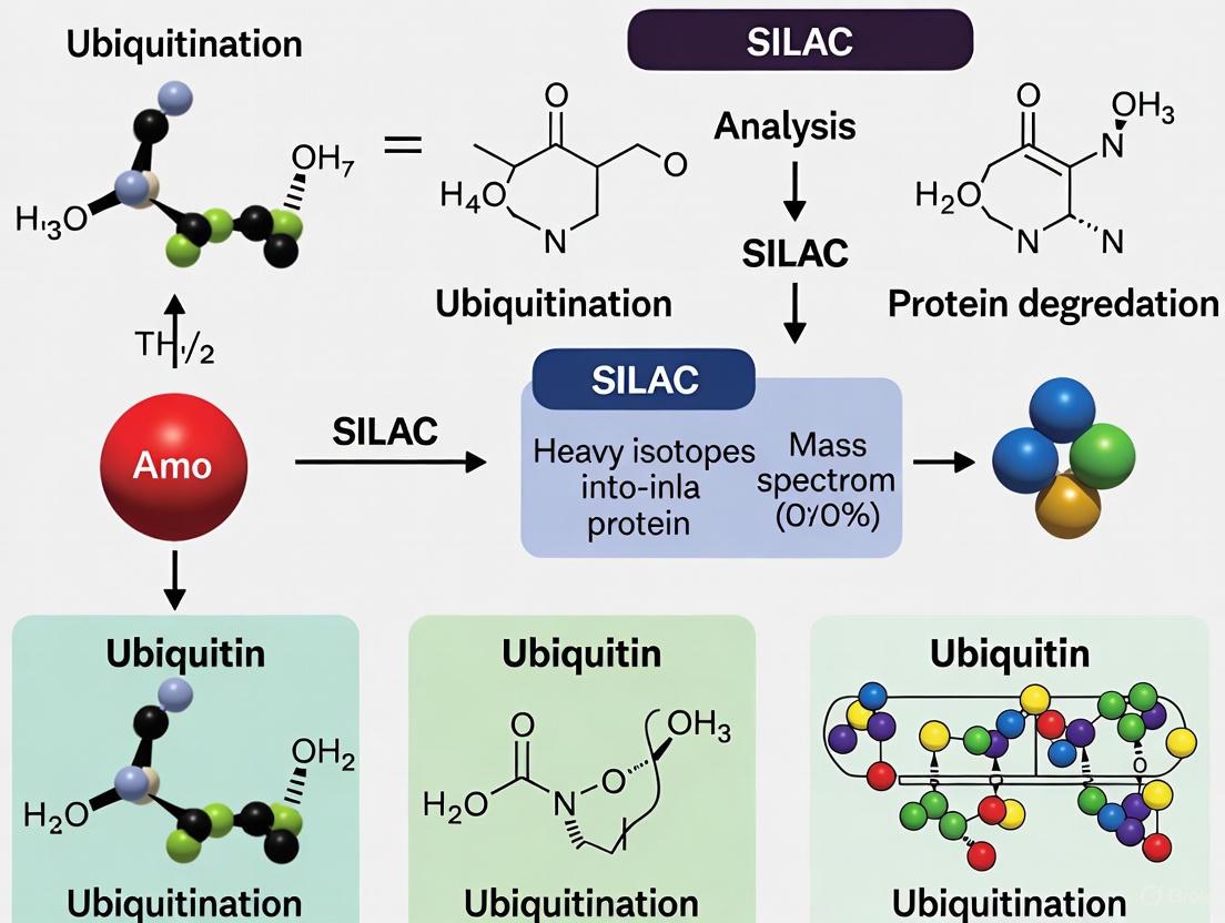
Visualization of SILAC Workflows
Research Reagent Solutions
Table 2: Essential Research Reagents for SILAC-Based Ubiquitination Studies
| Reagent/Category | Specific Examples | Function in SILAC Ubiquitination Research |
|---|---|---|
| SILAC Amino Acids | L-lysine (13C6, 15N2), L-arginine (13C6, 15N4) | Metabolic labeling for quantitative comparison |
| Cell Culture Media | DMEM deficient in lysine and arginine | Base medium for preparing SILAC media |
| Protease Inhibitors | EDTA-free protease inhibitor cocktails | Prevent protein degradation during sample preparation |
| Ubiquitin Enrichment Reagents | Ubiquitin remnant motif antibodies, ubiquitin-binding domains | Selective isolation of ubiquitinated peptides |
| Digestion Enzymes | Trypsin (mass spectrometry grade) | Protein digestion into analyzable peptides |
| Mass Spectrometry | High-resolution LC-MS/MS systems (Orbitrap platforms) | Peptide identification and quantification |
Technical Considerations and Limitations
Practical Implementation Challenges
While SILAC offers exceptional quantitative accuracy, researchers must consider several technical aspects to ensure successful experiments. Complete incorporation of labeled amino acids requires approximately five cell doublings, which may be challenging for slow-growing cells or primary cultures with limited division capacity [15]. For non-dividing cells such as neurons, specialized approaches like multiplex SILAC labeling have been developed, utilizing two different sets of heavy amino acids to enable accurate quantitation [15].
Another significant consideration is the arginine-to-proline conversion phenomenon, wherein some cell types metabolize labeled arginine to proline, leading to unexpected labeling patterns and complicating data analysis [14]. This issue can be mitigated by using lower concentrations of arginine, utilizing proline-free media, or selecting cell lines with minimal conversion activity. Additionally, the cost of isotope-labeled amino acids and specialized media components can be substantial, particularly for large-scale experiments [14].
Data Analysis and Quality Control
The analysis of SILAC data requires specialized software capable of accurately detecting and quantifying peptide pairs with defined mass differences. Commonly used platforms include MaxQuant, Proteome Discoverer, FragPipe, DIA-NN, and Spectronaut [17]. A recent benchmarking study evaluated these software tools and revealed that most can accurately quantify light/heavy ratios within a dynamic range of up to 100-fold, but struggle with greater differences [17]. The study also recommended against using Proteome Discoverer for SILAC data-dependent acquisition analysis despite its popularity in label-free proteomics [17].
Quality control measures should include assessment of labeling efficiency, which can be determined by analyzing a small aliquot of labeled cells before proceeding with full experiments. Additionally, researchers should implement label swapping (where experimental conditions are reversed between light and heavy labels in biological replicates) to account for any potential bias introduced by the labels themselves [15]. For ubiquitination studies specifically, careful optimization of enrichment conditions is crucial to maximize specificity and recovery of ubiquitinated peptides while minimizing background.
Advanced SILAC Methodologies
Specialized SILAC Variants
The basic SILAC approach has been adapted into several specialized variants to address specific research questions:
Super-SILAC involves creating a mixture of SILAC-labeled proteins from multiple cell lines to serve as an internal standard for analyzing complex samples like tissues [15] [14]. This approach was introduced by Matthias Mann's group in 2010 and has proven particularly valuable for quantifying tumor tissues, where the super-SILAC mix provides a representative reference that accounts for tissue heterogeneity [15].
NeuCode SILAC utilizes amino acids with subtle mass differences that can only be resolved with high-resolution mass spectrometers, enabling a higher degree of multiplexing (up to 4-plex) without compromising quantitative accuracy [11] [14]. This approach leverages the small mass defects created by extra neutrons in stable isotopes, expanding the multiplexing capabilities of metabolic labeling strategies [11].
BONLAC combines SILAC with bioorthogonal noncanonical amino acid tagging (BONCAT) to specifically analyze newly synthesized proteins [15]. This integrated approach enables researchers to measure protein synthesis rates and degradation simultaneously, providing comprehensive insights into protein turnover dynamics.
SILAC in Host-Pathogen Interactions
SILAC has been adapted for studying host-microbe interactions through a 'forward+reverse' labeling strategy that simultaneously labels host and microbial proteins [11]. This innovative approach enables researchers to track molecular exchanges between host cells and pathogens, revealing how microbes manipulate host cellular processes and how hosts respond to infection. The method provides a powerful tool for understanding the complex interplay between infectious agents and their hosts at the proteome level.
SILAC represents a robust and versatile platform for quantitative proteomics that continues to evolve and adapt to new research challenges. Its application in ubiquitination research has provided unprecedented insights into the dynamics and scope of this crucial post-translational modification, revealing both conventional and unconventional ubiquitination events. The continuous development of SILAC variants—including pulsed SILAC for dynamics, super-SILAC for tissue analysis, and NeuCode for enhanced multiplexing—ensures that this methodology remains at the forefront of quantitative proteomics.
For researchers investigating ubiquitination, SILAC offers a powerful approach to capture the transient and regulated nature of this modification, identify novel substrates, and quantify changes in response to cellular perturbations. When combined with advanced mass spectrometry instrumentation and sophisticated data analysis tools, SILAC provides a comprehensive solution for deciphering the complex landscape of protein ubiquitination in health and disease. As the field advances, integration of SILAC with emerging technologies such as data-independent acquisition methods and single-cell proteomics will likely further expand its utility in ubiquitination research and beyond.
Why Lysine and Arginine are the Cornerstones of SILAC Labeling
Stable Isotope Labeling by Amino Acids in Cell Culture (SILAC) has emerged as a powerful methodology in quantitative mass spectrometry-based proteomics. Among the various amino acids that can be metabolically incorporated, lysine and arginine have become the established cornerstones for robust and accurate quantification. This application note delineates the biochemical and practical rationale for their preferential use, provides detailed protocols for their implementation in ubiquitination research, and visualizes key workflows and considerations for researchers and drug development professionals.
SILAC operates on the principle of metabolic incorporation of stable isotope-labeled amino acids into the entire proteome of a cell during normal cell growth and division [19]. This incorporation occurs through protein synthesis, resulting in proteins that are chemically identical but distinguishable by mass spectrometry (MS) due to their mass differences [19]. While several amino acids can be used for labeling, lysine and arginine are overwhelmingly the preferred choices, particularly in trypsin-based proteomic workflows.
The selection is primarily driven by the specificity of the proteolytic enzyme trypsin, which cleaves peptide bonds C-terminal to arginine and lysine residues [20]. This enzymatic specificity ensures that nearly all generated peptides (except the C-terminal peptide of a protein) will contain a single labeled C-terminus, thereby simplifying quantitative analysis and maximizing the number of peptides suitable for quantification [20].
Core Biochemical Principles
Trypsin Specificity and Peptide Labeling Efficiency
Trypsin's cleavage specificity guarantees that when lysine and arginine are used as labeled amino acids, the mass tag is consistently located at the C-terminus of most peptides. This creates predictable "SILAC pairs" in mass spectra—peptide ions from different samples that differ by a known mass increment but otherwise have identical physicochemical properties. This predictability is crucial for automated data processing and improves the accuracy and depth of quantification.
Advantages Over Other Amino Acids
Using lysine and arginine in tandem offers significant advantages:
- Complete Proteome Coverage: Ensures all tryptic peptides, barring the C-terminal peptide, are quantifiable.
- Prevents Quantification Errors: Labeling with a single amino acid like leucine would leave many peptides unlabeled, complicating ratio calculations and reducing quantitative precision.
- Multiplexing Capabilities: The availability of multiple heavy isotope forms enables experimental designs comparing several conditions simultaneously (e.g., 3-plex experiments) [20].
Table 1: Common Heavy Isotope Forms of Lysine and Arginine for SILAC
| Amino Acid | Isotope Form | Mass Shift (Da) | Typical Use |
|---|---|---|---|
| L-Lysine | ¹³C₆ | +6 | Standard 2-plex labeling [20] |
| L-Lysine | ¹³C₆¹⁵N₂ | +8 | 3-plex labeling; Trypsin kits [20] |
| L-Lysine | D₄ (²H₄) | +4 | 3-plex labeling [20] |
| L-Arginine | ¹³C₆ | +6 | 3-plex labeling [20] |
| L-Arginine | ¹³C₆¹⁵N₄ | +10 | Standard 2-plex & Trypsin kits [20] |
Figure 1: The foundational SILAC workflow. Metabolic incorporation of heavy Lys and Arg, followed by tryptic digestion, ensures that most resulting peptides contain a single, predictable label, enabling accurate LC-MS/MS quantification.
Application in Ubiquitination Research
SILAC has proven to be a particularly powerful tool for investigating post-translational modifications, especially ubiquitination. Ubiquitin is typically conjugated to target proteins via an isopeptide bond to lysine ε-amino groups [16]. This makes SILAC labeling with heavy lysine indispensable for studying this modification.
Protocol: Investigating Ubiquitination Pathways with SILAC
The following protocol, adapted from studies on endoplasmic reticulum-associated degradation (ERAD), outlines a method to identify proteins undergoing ubiquitination and to characterize novel ubiquitination sites, including non-lysine ubiquitination [16].
I. Cell Culture and SILAC Labeling
- Cell Line: HEK293T cells or other relevant cell lines.
- SILAC Media Preparation:
- Use DMEM deficient in lysine and arginine.
- "Light" Medium: Supplement with normal L-lysine and L-arginine.
- "Heavy" Medium: Supplement with heavy ¹³C₆, ¹⁵N₂ L-lysine and ¹³C₆, ¹⁵N₄ L-arginine [16].
- Add 10% dialyzed fetal bovine serum to both media to avoid unlabeled amino acid contamination [19].
- Add 2.6 mM L-proline to the media to prevent metabolic conversion of arginine to proline, which can compromise quantification accuracy [21].
- Labeling: Culture cells in their respective SILAC media for at least five cell doublings to ensure >99% incorporation of the labeled amino acids [19].
II. Experimental Treatment and Cell Lysis
- Transfection: Transfect cells with plasmids encoding your protein of interest (e.g., wild-type or lysine-less mutant TCRα for ERAD studies) and relevant E3 ligases (e.g., Hrd1) [16].
- Proteasome Inhibition: Treat cells with 10 μM MG132 for 3-4 hours before lysis to accumulate ubiquitinated species [16].
- Cell Lysis: Lyse cells in a suitable lysis buffer (e.g., RIPA buffer: 50 mM Tris-HCl pH 8, 150 mM NaCl, 1% NP-40, 0.5% sodium deoxycholate, 0.1% SDS) supplemented with protease inhibitors (e.g., Complete EDTA-free tablet) and deubiquitinase inhibitors [16].
III. Immunoprecipitation (IP) and Sample Preparation
- IP of Target Protein: Incubate 1 mg of total protein lysate with a specific antibody (e.g., anti-HA for HA-tagged TCRα) and protein A/G agarose beads overnight at 4°C [16].
- Washing: Wash beads stringently to remove non-specifically bound proteins.
- Elution: Elute bound proteins using a low-pH elution buffer or by boiling in SDS-PAGE sample buffer.
IV. Protein Digestion and LC-MS/MS Analysis
- Separation: Separate proteins by SDS-PAGE. Excise the entire protein band of interest.
- In-gel Digestion: Destain, reduce, alkylate, and digest proteins in-gel with sequencing-grade trypsin (e.g., 12.5 ng/μL) overnight at 37°C [19].
- Peptide Extraction: Extract peptides from the gel using 5% formic acid/50% acetonitrile, and desalt using C18 StageTips [16].
- LC-MS/MS Analysis:
- Use a nanoflow HPLC system coupled to a high-resolution mass spectrometer (e.g., Orbitrap series).
- Peptides are separated on a C18 reversed-phase column.
- Acquire data in data-dependent acquisition (DDA) mode, where the MS automatically selects the most intense precursor ions for fragmentation (MS/MS).
V. Data Analysis
- Database Searching: Search raw MS data against a protein sequence database using software (e.g., MaxQuant, FragPipe) configured for SILAC (Lys+8, Arg+10) [17].
- Ubiquitination Site Identification: Include GlyGly (di-glycine) remnant (+114.0429 Da on lysine) as a variable modification to identify lysine ubiquitination sites.
- Quantification and Validation: Use SILAC ratios (Heavy/Light) to quantify changes in protein abundance or ubiquitination levels. For novel ubiquitination sites, validate findings through mutagenesis and follow-up biochemical assays [16].
Critical Considerations and Troubleshooting
The Arginine Conversion Problem and Mitigation
A well-documented challenge in SILAC is the metabolic conversion of labeled arginine to labeled proline in some cell lines [21]. This occurs via the arginase pathway and leads to the incorporation of heavy labels into proline residues, complicating the MS spectrum and skewing quantification ratios [22] [21].
Solution: The recommended and most effective mitigation strategy is the addition of excess unlabeled (light) proline to the SILAC culture media. Studies have shown that increasing the proline concentration to 2.6 mM successfully suppresses this conversion without leading to detectable back-conversion to arginine or other side effects [21].
Figure 2: The metabolic conversion of arginine to proline and its solution. Adding excess light proline to the culture medium is a critical step to ensure data quality in many cell lines.
The Scientist's Toolkit: Essential Reagents for SILAC
Table 2: Key Research Reagent Solutions for SILAC Experiments
| Reagent / Material | Function / Role | Example & Notes |
|---|---|---|
| SILAC Amino Acids | Metabolic incorporation into proteins for mass encoding. | ¹³C₆ L-Lysine & ¹³C₆¹⁵N₄ L-Arginine [20]. Isotope purity >99% is critical. |
| Amino Acid-Deficient Media | Base medium for preparing light and heavy SILAC media. | DMEM, RPMI-1640, or DMEM/F-12, lacking Lys and Arg [19] [20]. |
| Dialyzed FBS | Serum source with low molecular weight contaminants removed. | Prevents contamination with unlabeled amino acids, ensuring high incorporation efficiency [19]. |
| L-Proline | Prevents metabolic conversion of Arg to Pro. | Add to 2.6 mM final concentration in SILAC media [21]. |
| Trypsin / Lys-C | Proteolytic enzyme for protein digestion. | Sequencing grade, specific cleavage C-terminal to Arg/Lys ensures labeled peptides [19] [20]. |
| Phosphatase/Protease Inhibitors | Preserves post-translational modification states during lysis. | Sodium orthovanadate (pTyr inhibitor), sodium fluoride, Complete protease inhibitor [19]. |
Lysine and arginine are the foundational pillars of SILAC due to their synergistic compatibility with tryptic digestion, which ensures comprehensive and simplified quantitative analysis of the proteome. Their application is particularly potent in ubiquitination research, allowing for the precise identification and quantification of dynamic modification events. By adhering to optimized protocols—including the critical step of supplementing media with excess proline—researchers can leverage the full power of SILAC to drive discoveries in basic biology and targeted drug development.
Ubiquitination is a critical post-translational modification (PTM) that regulates diverse cellular functions, including protein degradation, activity, and localization [23]. The versatility of ubiquitination stems from the complexity of ubiquitin (Ub) conjugates, which can range from a single Ub monomer to polymers of different lengths and linkage types [23]. Dysregulation of ubiquitination pathways leads to numerous pathologies, including cancer and neurodegenerative diseases, making comprehensive mapping of ubiquitination events a crucial research objective [23] [24].
Stable Isotope Labeling by Amino Acids in Cell Culture (SILAC) has emerged as a powerful quantitative proteomic approach for studying dynamic cellular processes like ubiquitination [4]. This metabolic labeling technique incorporates non-radioactive, isotopically labeled amino acids into all mammalian proteins during cell culture, enabling accurate relative quantitation of protein expression and modifications between experimental conditions [4]. By combining SILAC with advanced enrichment strategies and mass spectrometry, researchers can now precisely identify ubiquitination sites and quantify changes in the ubiquitin landscape, providing unprecedented insights into the molecular mechanisms governed by the ubiquitin code.
Technical Foundations of SILAC in Ubiquitination Research
Principles of SILAC Technology
SILAC functions as a metabolic labeling strategy where cell lines are grown in media lacking a standard essential amino acid but supplemented with a non-radioactive, isotopically labeled form of that amino acid [4]. In practice, mammalian cell lines grown in SILAC media exhibit normal growth characteristics, including cell morphology, doubling time, and differentiation capacity [4]. Complete incorporation of labeled amino acids, such as deuterated leucine (Leu-d3), typically occurs after five population doublings, ensuring that all proteins are fully labeled [4].
The fundamental strength of SILAC lies in its ability to mix protein populations from experimental and control conditions directly after harvesting, simplifying downstream processing and minimizing quantitative variability [4]. During mass spectrometric analysis, every peptide containing the labeled amino acid incorporates either all normal or all heavy isotopes, making identification and quantification straightforward and accurate [4].
Ubiquitination Complexity and Analytical Challenges
The ubiquitination landscape presents substantial analytical challenges that SILAC helps overcome. Ubiquitin can modify substrate proteins at one or several lysine residues simultaneously and can itself serve as a substrate, forming complex chains that vary in length, linkage type, and overall architecture [23]. With eight possible linkage sites (K6, K11, K27, K29, K33, K48, K63, and M1) and the potential for heterotypic branched chains, the combinatorial complexity is enormous [23] [24].
Furthermore, the stoichiometry of protein ubiquitination is typically low under normal physiological conditions, necessitating effective enrichment strategies to identify ubiquitinated substrates against the background of non-modified proteins [23]. The dynamic nature of ubiquitination, with continuous writing by E1-E2-E3 enzyme cascades and erasing by deubiquitinases (DUBs), adds another layer of complexity to its study [23] [24].
Experimental Workflow for SILAC-Based Ubiquitination Site Mapping
Cell Culture and Metabolic Labeling
The initial phase involves cultivating appropriate cell lines in SILAC media. A typical approach utilizes "heavy" SILAC media containing 13C6 15N4 l-arginine and 13C6 15N2 l-lysine for experimental conditions (e.g., PC3 cells expressing Parkin), while control cells (e.g., expressing vector only) are maintained in "light" media with normal amino acids [25]. Cells are harvested after sufficient doublings to ensure complete isotope incorporation, which is critical for accurate quantification.
Sample Processing and Ubiquitinated Peptide Enrichment
Following cell lysis, heavy and light labeled lysates are combined in a 1:1 ratio based on protein weight (e.g., 20 mg total) [25]. The mixed lysate undergoes reduction with dithiothreitol, alkylation with iodoacetamide, and in-solution digestion with trypsin [25]. Digested peptides are desalted using C18 cartridges before ubiquitinated peptide enrichment.
A key step involves enriching for ubiquitinated peptides using antibodies specific to the diglycine remnant left on modified lysines after tryptic digestion. The PTMScan Ubiquitin Remnant Motif (K-ε-GG) Kit is commonly employed for this purpose [25]. This antibody-based enrichment specifically isolates peptides containing the K-ε-GG motif, representing former ubiquitination sites.
Mass Spectrometric Analysis and Data Processing
Enriched ubiquitinated peptides are analyzed using liquid chromatography-tandem mass spectrometry (LC-MS/MS) with instruments such as the Q-Exactive HF mass spectrometer, typically using extended LC gradients (e.g., 4-hour) to enhance peptide separation and identification [25]. For global proteome analysis without immunoaffinity enrichment, digested samples can be fractionated into multiple fractions (e.g., 10 fractions using high pH reversed-phase fractionation) to increase proteome coverage [25].
MS data processing utilizes specialized software such as MaxQuant for database searching against human protein databases [25]. Critical search parameters include:
- Full tryptic specificity with up to five missed cleavages
- Static carbamidomethylation of cysteine
- Variable modifications including methionine oxidation and diglycine addition to lysine
- False discovery rates of 1% at protein, peptide, and site levels
Ubiquitinated sites are specifically determined from the GlyGly (K)Sites.txt output table [25].
Figure 1: Experimental workflow for SILAC-based ubiquitination site mapping, showing key steps from metabolic labeling to site identification.
Key Research Reagents and Solutions
Table 1: Essential research reagents for SILAC-based ubiquitination studies
| Reagent/Category | Specific Examples | Function and Application |
|---|---|---|
| SILAC Amino Acids | 13C6 15N4 l-arginine, 13C6 15N2 l-lysine [25] | Metabolic labeling for quantitative proteomics |
| Enrichment Kits | PTMScan Ubiquitin Remnant Motif (K-ε-GG) Kit [25] | Immunoaffinity enrichment of ubiquitinated peptides |
| Mass Spectrometers | Q-Exactive HF Mass Spectrometer [25] | High-resolution LC-MS/MS analysis |
| Data Analysis Software | MaxQuant, Proteome Discoverer, FragPipe, DIA-NN, Spectronaut [17] | Identification and quantification of ubiquitination sites |
| Chromatography | High pH Reversed-Phase Peptide Fractionation Kit [25] | Peptide fractionation to increase proteome coverage |
| Cell Lines | PC3, HeLa, HEK293T, U2OS [25] [23] | Model systems for ubiquitination studies |
Quantitative Data Analysis and Benchmarking
Performance Metrics and Software Considerations
Recent benchmarking studies have evaluated multiple SILAC data analysis platforms across 12 performance metrics, including identification, quantification, accuracy, precision, reproducibility, filtering criteria, missing values, false discovery rate, protein half-life measurement, data completeness, unique software features, and analysis speed [17]. This comprehensive evaluation revealed that most software reaches a dynamic range limit of approximately 100-fold for accurate quantification of light/heavy ratios [17].
Critical considerations for SILAC data analysis include:
- Removal of low-abundance peptides and outlier ratios to improve quantification accuracy [17]
- Careful selection of labeling time points for dynamic SILAC experiments [17]
- Using multiple software packages for cross-validation to achieve greater confidence in quantification [17]
- Specific recommendations against using Proteome Discoverer for SILAC DDA analysis despite its utility in label-free proteomics [17]
Data Filtering and Validation Criteria
Effective SILAC ubiquitome analysis requires stringent data filtering. Consensus identification lists should be generated with false discovery rates of 1% at protein, peptide, and site levels [25]. Reverse hits, contaminants, and identifications without any heavy/light ratio should be systematically removed from all datasets [25].
For global proteome network analysis, proteins typically require a consistent minimum fold change of 1.45 and a minimum SILAC ratio count of 2, while mitochondrial proteins may be analyzed with slightly relaxed criteria (fold change of 1.3 with ratio count of 2) [25].
Table 2: Quantitative data analysis parameters for SILAC ubiquitination studies
| Analysis Parameter | Typical Setting | Purpose and Rationale |
|---|---|---|
| False Discovery Rate | 1% at protein, peptide, and site levels [25] | Control false positive identifications |
| Minimum Fold Change | 1.45 (global), 1.3 (mitochondrial) [25] | Ensure biological significance of changes |
| SILAC Ratio Count | Minimum of 2 [25] | Ensure quantification reliability |
| Dynamic Range Limit | ~100-fold [17] | Practical limit for accurate quantification |
| Peptide Filtering | Remove low-abundance peptides and outlier ratios [17] | Improve quantification accuracy |
| Software Cross-Validation | Use of multiple packages recommended [17] | Increase confidence in quantification |
Advanced Methodologies and Emerging Approaches
Alternative Enrichment Strategies
While K-ε-GG antibody-based enrichment remains widely used, emerging approaches address certain limitations. The UbiSite antibody recognizes a 13-amino-acid remnant specific to ubiquitin after LysC digestion, providing greater specificity and reduced bias toward certain sequences [26]. This approach has enabled identification of over 63,000 ubiquitination sites on more than 9,000 proteins in human cell lines [26].
Other enrichment strategies include:
- Ub tagging-based approaches: Expression of epitope-tagged Ub (e.g., His, Strep) for affinity purification [23]
- Ubiquitin binding domain (UBD)-based approaches: Tandem-repeated Ub-binding entities (TUBEs) with enhanced affinity for ubiquitinated proteins [23]
- Linkage-specific antibodies: Antibodies targeting specific Ub chain linkages (M1-, K11-, K27-, K48-, K63-linkage) [23]
Structural and Functional Implications of Site-Specific Ubiquitination
Recent advances have revealed that the biophysical consequences of ubiquitination are site-specific and regulate signaling and function [24]. Ubiquitination can affect the energy landscape of a protein in a site-specific manner, allowing substrates to access high-energy, partially unfolded states only when modified at certain sites [24]. The proteasome selectively recognizes and degrades substrates ubiquitinated at these destabilizing sites, revealing a new regulatory layer for proteasomal degradation [24].
The exact thermodynamic mechanism driving this destabilization depends on the ubiquitination site, with entropic versus enthalpic mechanisms operating at different sites within the same substrate [24]. This site-specific modulation of protein dynamics represents a crucial aspect of the ubiquitin code that extends beyond simple degradation tagging.
The integration of SILAC with advanced ubiquitination enrichment strategies and mass spectrometry has revolutionized our ability to decode the complex language of ubiquitin signaling. This powerful combination enables researchers not only to identify ubiquitination sites but also to quantitatively monitor dynamic changes in the ubiquitin landscape in response to cellular perturbations, disease states, or therapeutic interventions.
As methodologies continue to advance, with improvements in enrichment specificity, mass spectrometry sensitivity, and data analysis algorithms, our understanding of the ubiquitin code will deepen. These technical advances promise to uncover novel regulatory mechanisms and therapeutic opportunities in the vast landscape of ubiquitin-mediated signaling, particularly in pathological conditions where ubiquitination pathways are disrupted. The continued refinement of SILAC-based ubiquitination mapping approaches will undoubtedly play a central role in these discoveries, providing increasingly sophisticated tools to elucidate the complexities of the ubiquitin code.
Stable Isotope Labeling by Amino acids in Cell culture (SILAC) has emerged as a powerful technique in mass spectrometry-based quantitative proteomics for analyzing protein dynamics and post-translational modifications. The fundamental principle involves metabolic incorporation of stable isotope-labeled amino acids into the entire proteome of growing cells, creating a mass shift that enables precise quantification of protein abundance changes across multiple samples. This methodology provides a robust framework for investigating cellular processes in dynamic biological systems, particularly for measuring protein turnover rates and mapping PTM regulation across the proteome.
The SILAC technique involves cultivating two cell populations in parallel—one in normal "light" medium and another in "heavy" medium containing amino acids with stable isotopes (e.g., 13C6-arginine). After several cell divisions, the labeled amino acids are fully incorporated into all newly synthesized proteins. The protein populations are then combined, digested, and analyzed by mass spectrometry, where peptide pairs appear as doublets with a predictable mass difference. The ratio of their peak intensities directly reflects the relative protein abundance between the two conditions [11]. This approach has been successfully adapted for studying various biological phenomena, including cell signaling pathways, protein-protein interactions, and secretory pathways [11].
SILAC Methodologies for Protein Turnover Analysis
Pulsed SILAC (pSILAC) for Dynamic Measurements
Pulsed SILAC represents a significant methodological advancement that enables researchers to monitor temporal changes in protein synthesis and degradation. Unlike conventional SILAC that measures relative protein abundance at a single endpoint, pSILAC tracks the kinetics of label incorporation over multiple time points, providing direct insight into protein turnover dynamics [27]. In this approach, cells previously grown in light medium are switched to heavy isotope-containing medium, and samples are collected at specific intervals post-switch. The increasing incorporation of heavy isotopes into proteins over time reflects newly synthesized proteins, while the decreasing light signal represents pre-existing proteins.
The experimental workflow for pSILAC typically involves:
- Culturing cells to near-confluence in light medium
- Exchanging with heavy SILAC medium at time zero
- Harvesting cells at multiple time points (e.g., 0, 1, 3, 6, 10, 16, 24, 34, 48 hours)
- Processing samples for LC-MS/MS analysis
- Quantifying light-to-heavy ratios for each peptide across time points
- Calculating turnover rates using kinetic modeling [27]
For protein turnover studies, the clearance rate of pre-existing proteins is quantified by measuring the decrease in light peptide signals over time. In a steady-state system, the rate of clearance equals the rate of synthesis, enabling calculation of protein half-lives. pSILAC has revealed that protein half-lives vary considerably, ranging from minutes to several weeks, depending on cell type and physiological conditions [27].
Site-Resolved Protein Turnover (SPOT) Profiling
A recent innovation in turnover analysis, Site-resolved Protein Turnover (SPOT) profiling, combines pSILAC with PTM-specific enrichment to measure turnover rates at the level of individual modification sites [27]. This approach has been applied to phosphorylation, acetylation, and ubiquitination, revealing that PTMs can significantly influence protein stability and function. SPOT analysis demonstrates that different modification sites on the same protein can exhibit distinct turnover rates, suggesting complex regulatory mechanisms operating at the proteoform level.
SPOT methodology employs two complementary workflows:
- Dynamic SILAC (dSILAC): Cells are pulsed with SILAC medium and harvested at 6, 24, and 40-hour time points, followed by sequential enrichment of phosphorylated, acetylated, and ubiquitinated peptides [27].
- dSILAC-TMT: This multiplexed approach uses tandem mass tags to analyze 10 pulse time points simultaneously, significantly increasing throughput and temporal resolution [27].
SPOT profiling has disclosed global differences in turnover associated with specific PTMs. Phosphorylated peptidoforms generally show slightly faster turnover compared to unmodified peptides, while ubiquitinated peptides exhibit significantly increased turnover rates, consistent with ubiquitin's established role in targeting proteins for proteasomal degradation. Surprisingly, acetylated peptidoforms often demonstrate considerably slower turnover compared to other modifications and their corresponding proteins [27].
Table 1: Protein Turnover Methodologies Comparison
| Method | Principle | Time Points | Applications | Advantages |
|---|---|---|---|---|
| Standard SILAC | Metabolic labeling with heavy amino acids | Single endpoint | Protein expression profiling, PTM quantification | Simple workflow, high accuracy |
| pSILAC | Pulse-chase with heavy amino acids | Multiple time points | Protein synthesis/degradation kinetics | Direct measurement of turnover rates |
| SPOT Profiling | pSILAC with PTM enrichment | Multiple time points | PTM-specific turnover analysis | Site-specific resolution of modification dynamics |
| NeuCode SILAC | Multiplexing with neutron encoding | Flexible | High-throughput turnover studies | Increased multiplexing capacity (up to 4-plex) |
SILAC Applications in Ubiquitination Analysis
Global Ubiquitinome Profiling
Ubiquitination represents a crucial post-translational modification that regulates diverse cellular processes, including protein degradation, DNA repair, and signal transduction. SILAC-based approaches have revolutionized the large-scale analysis of the ubiquitinome by enabling quantitative assessment of ubiquitination dynamics under different physiological conditions. The standard protocol for global ubiquitination analysis involves SILAC labeling followed by immunoaffinity enrichment of ubiquitinated peptides using specific antibodies directed against the di-glycine remnant that remains after tryptic digestion of ubiquitinated proteins [3].
The detailed methodology includes:
- Growing light and heavy SILAC-labeled cells under experimental conditions
- Combining cell populations in equal ratios
- Harsh lysis under denaturing conditions to preserve PTMs
- Trypsin digestion to generate di-glycine-modified peptides
- Immunoaffinity purification using di-glycine remnant antibodies
- LC-MS/MS analysis and quantitative data processing [3]
This approach has been successfully applied to identify ubiquitination sites regulated in various biological contexts, including DNA damage response, growth factor signaling, and protein quality control. The high specificity of di-glycine antibody enrichment allows for comprehensive mapping of ubiquitination sites, with studies routinely identifying thousands of modified sites in a single experiment.
Temporal Analysis of Ubiquitination Dynamics
The combination of pSILAC with ubiquitin remnant enrichment enables researchers to investigate the turnover kinetics of ubiquitinated proteins, providing insights into the temporal regulation of ubiquitin-mediated processes. This approach has revealed that ubiquitinated peptides generally display faster turnover compared to non-modified peptides, with this difference becoming more pronounced after normalization to the corresponding protein turnover rates [27]. This observation aligns with ubiquitin's primary role in targeting proteins for proteasomal degradation.
Interestingly, SPOT profiling has identified significant heterogeneity in turnover rates among different ubiquitination sites, suggesting distinct functional consequences depending on the specific site modified. Some ubiquitination events appear associated with rapid degradation, while others may serve non-proteolytic functions such as regulating protein activity or localization. This methodological approach has proven particularly valuable for identifying regulatory ubiquitination events that control the stability of key cellular regulators.
Table 2: Key Research Reagent Solutions for SILAC-Based PTM Analysis
| Reagent/Category | Specific Examples | Function in Experimental Workflow |
|---|---|---|
| SILAC Amino Acids | L-arginine:U-13C6, L-lysine:U-13C6-15N2 | Metabolic labeling for quantitative comparison |
| Affinity Capture Reagents | Anti-di-glycine remnant antibody, PTM-specific antibodies | Enrichment of modified peptides prior to MS analysis |
| Proteases | Trypsin, Lys-C | Protein digestion with specific cleavage sites |
| Chromatography Materials | C18 StageTips, HPLC columns | Peptide separation and desalting |
| Mass Spectrometry Tags | Tandem Mass Tags (TMT) | Multiplexing for high-throughput turnover studies |
Advanced SPOT Profiling for PTM Function Discovery
Revealing PTM Regulatory Networks
SPOT profiling has emerged as a powerful tool for investigating the functional relationships between multiple PTMs and their collective impact on protein behavior. By measuring turnover rates for thousands of modified sites, this approach can identify PTMs with potential regulatory significance based on their divergent turnover compared to unmodified counterpart peptides [27]. Statistical analysis of SPOT data has revealed that approximately 20% of all annotated regulatory sites exhibit differential turnover behavior, with many previously uncharacterized modification sites showing even larger turnover differences than known regulatory sites [27].
This methodology has uncovered several key principles governing PTM dynamics:
- PTMs with significantly faster turnover often represent early events in protein maturation
- Modifications with slower turnover typically occur later in a protein's lifetime
- The relative clearance rates primarily reflect the temporal ordering of modification events rather than direct effects on protein stability [28]
These insights have fundamentally changed the interpretation of metabolic labeling data in the context of PTM analysis, shifting focus from stability effects to temporal ordering of modification events along a protein's lifespan.
Protein-Peptide Turnover Profiling (PPToP) for Modification Kinetics
Protein-Peptide Turnover Profiling represents a specialized application of pSILAC combined with PTM enrichment that enables detailed investigation of modification kinetics and order [28]. PPToP leverages theoretical kinetic modeling to interpret experimental pSILAC data, demonstrating that clearance rates measured for different proteoforms are not straightforward indicators of proteolytic stability but primarily reflect the relative order of PTM addition and removal during a protein's lifetime [28].
The key insights from PPToP analysis include:
- Most phosphorylated peptides exhibit slower clearance than corresponding protein medians, characteristic of modifications occurring later in a protein's lifetime
- Faster-clearing phosphopeptides often represent early intermediates in protein maturation
- Relative differences in clearance rates are not predictive of effects on protein stability but reveal the sequence of modification events [28]
This approach has been successfully applied to identify temporal phosphorylation patterns on cell cycle regulators and protein complex assembly intermediates, providing unprecedented insight into the kinetic aspects of PTM regulation.
Experimental Protocols
Detailed Protocol: Global Ubiquitination Analysis by SILAC
Cell Culture and Labeling
- Grow two populations of mammalian cells in SILAC medium—one with light lysine and arginine, the other with heavy isotopes (13C6-lysine and 13C6-arginine)
- Culture for at least five cell doublings to ensure complete incorporation of labeled amino acids
- Treat cells according to experimental design (e.g., proteasome inhibition, growth factor stimulation)
Sample Preparation
- Combine light and heavy cell populations in equal protein amounts
- Lyse cells in urea buffer (8 M urea, 50 mM Tris-HCl, pH 8.0) containing protease and phosphatase inhibitors
- Reduce disulfide bonds with 5 mM dithiothreitol (30 min, 25°C)
- Alkylate with 10 mM iodoacetamide (30 min, 25°C in darkness)
- Dilute urea concentration to 2 M and digest with trypsin (1:50 enzyme-to-substrate ratio) overnight at 37°C
Ubiquitinated Peptide Enrichment
- Acidify digests to pH < 3 with trifluoroacetic acid
- Desalt peptides using C18 solid-phase extraction
- Resuspend peptides in immunoaffinity purification buffer (50 mM MOPS, 10 mM Na2HPO4, 50 mM NaCl, pH 7.2)
- Incubate with anti-di-glycine remnant antibody-conjugated beads for 2 hours at 4°C
- Wash beads extensively with IAP buffer followed by water
- Elute ubiquitinated peptides with 0.1% trifluoroacetic acid
Mass Spectrometric Analysis
- Analyze enriched peptides by LC-MS/MS using a high-resolution instrument
- Acquire data in data-dependent acquisition mode with dynamic exclusion
- Identify and quantify peptides using search engines (e.g., MaxQuant) with SILAC doublet quantification
- Apply appropriate statistical filters to identify significantly regulated ubiquitination sites [3]
Detailed Protocol: SPOT Profiling for Multi-PTM Turnover Analysis
Dynamic SILAC Labeling
- Culture HeLa cells in light medium until 70% confluent
- Replace medium with heavy SILAC medium (K8R10 or K10R10)
- Harvest cells at multiple time points (6, 24, 40 hours) post-medium exchange
- Include label-swap experiments for technical validation
Sequential PTM Enrichment
- Digest proteins as described in the ubiquitination protocol
- Subject peptides to sequential enrichment:
- First, enrich for phosphorylated peptides using TiO2 or IMAC beads
- Second, enrich for acetylated peptides using anti-acetyllysine antibody
- Third, enrich for ubiquitinated peptides using di-glycine remnant antibody
- Use the flow-through from sequential enrichment for whole proteome analysis
Multiplexed Analysis (dSILAC-TMT)
- Label cells with heavy SILAC medium as above
- Harvest at 10 time points (0, 1, 3, 6, 10, 16, 24, 34, 48, >200 hours)
- Label peptides from each time point with different TMT tags
- Combine TMT-labeled samples in equal ratios
- Perform sequential PTM enrichment as described above
- Analyze by LC-MS/MS using synchronous precursor selection for accurate TMT quantification [27]
Data Processing and Turnover Calculation
- Extract SILAC and TMT intensities using proteomics software
- Calculate heavy-to-light ratios for each time point
- Fit turnover curves using exponential decay models
- Compute half-lives for proteins and modified peptidoforms
- Identify statistically significant differences between modified and unmodified peptide turnover [27]
Data Analysis and Interpretation
Key Considerations for SILAC-based Turnover Studies
The interpretation of SILAC-based turnover data requires careful consideration of several analytical factors. For pSILAC experiments, shorter pulse times may exhibit substantial amino acid recycling and inferior correlation between label-swap experiments, leading to potentially erroneous turnover estimations [27]. Additionally, the clearance rates measured for PTM-defined proteoforms are influenced not only by degradation but also by interconversion between different modified states, complicating direct interpretation of half-life measurements [28].
Statistical analysis of SPOT data typically involves:
- Testing for significant differences between modified peptidoforms and their corresponding proteins
- Applying multiple testing correction to account for the large number of comparisons
- Setting appropriate thresholds for fold-change and statistical significance
- Validating findings with orthogonal approaches for selected targets [27]
Recent theoretical work has established that the relative order of PTM addition and removal, rather than the modification's direct effect on stability, primarily determines the observed clearance rates in metabolic labeling experiments [28]. This insight necessitates reevaluation of previous interpretations regarding PTM effects on protein half-lives.
Table 3: Quantitative Findings from SILAC-Based PTM Turnover Studies
| PTM Type | Turnover Relative to Unmodified Protein | Key Biological Implications | Representative Statistical Findings |
|---|---|---|---|
| Phosphorylation | Slightly faster (normalizes with protein adjustment) | More prevalent on higher turnover proteins; regulates activity, localization | ~20% of known regulatory sites show significant turnover differences |
| Ubiquitination | Significantly faster (even after normalization) | Primarily targets proteins for degradation; non-proteolytic functions for some sites | Strong enrichment in proteasomal degradation pathways |
| Acetylation | Considerably slower | Potential stabilizing effect; competitive with ubiquitination | Large fraction shows >50% slower turnover than protein median |
| Mixed PTMs | Site-specific variations | Complex regulatory networks with temporal ordering | 10,050 peptidoforms across 2,788 proteins show significant regulation |
Visualizing Experimental Workflows and Signaling Pathways
Advanced SILAC Workflows for Ubiquitin Proteomics: From Cell Culture to Data Acquisition
Stable Isotope Labeling by Amino Acids in Cell Culture (SILAC) has emerged as a powerful quantitative proteomic method since its introduction in 2002. As a metabolic labeling strategy, it utilizes isotope-labeled amino acids that are incorporated in vivo into proteins during translation [15]. This technique provides a robust framework for comparing proteomes across different experimental conditions, such as disease states or drug treatments, with minimal quantitative errors since samples are mixed immediately after cell lysis prior to processing [15]. Within ubiquitination research, SILAC offers particular advantage for investigating the dynamics of this crucial post-translational modification, which regulates protein degradation, endocytosis, and cell cycle progression [16]. The versatility of SILAC has led to the development of multiple strategic approaches—duplex, triplex, and super-SILAC—each designed to address specific experimental questions in proteome dynamics and ubiquitination pathways.
Core SILAC Methodologies: Principles and Applications
Duplex SILAC: Basic Comparative Proteomics
Duplex SILAC represents the fundamental application of the SILAC methodology, enabling direct comparison between two proteomic states. This approach utilizes two distinct forms of amino acids: "light" (natural abundance) and "heavy" (isotope-labeled) [15].
Key Protocol Steps [15]:
- Prepare SILAC media using DMEM deficient in lysine and arginine, supplemented with either light (L-lysine and L-arginine) or heavy (13C6-lysine and 13C6-arginine) amino acids
- Culture cells in their respective media for at least five population doublings to ensure ≥97% label incorporation
- Harvest cells, mix light and heavy labeled cell lysates in equal protein amounts
- Process combined samples through digestion, desalting, and LC-MS/MS analysis
In ubiquitination research, duplex SILAC effectively identifies substrates with altered ubiquitination states under different conditions, though it provides limited mechanistic insight into whether changes result from synthesis or degradation alterations.
Triplex SILAC: Complex Experimental Designs
Triplex SILAC expands experimental capability by incorporating a third isotopic label, enabling simultaneous comparison of three proteomic states within a single experiment. This approach is particularly valuable for time-course studies, dose-response analyses, or investigations involving multiple variables [15].
Advanced Application: Non-Dividing Cells Traditional SILAC requires cell division for efficient label incorporation, presenting challenges for non-dividing cells like primary neurons. Triplex SILAC addresses this limitation through a specialized multiplex strategy where two different sets of heavy amino acids are introduced [15]. This ensures that compared cell populations remain equally labeled, allowing accurate quantitation by comparing medium and heavy labeled peptides from partially labeled cells.
Experimental Workflow:
- Different cell populations are labeled with light, medium, and heavy amino acids
- For non-dividing cells, labeling efficiency is confirmed through partial incorporation measurements
- Cells are processed similarly to duplex SILAC, with triple mixtures analyzed by LC-MS/MS
Super-SILAC: Tissue Proteomics and Quantitative Accuracy
Super-SILAC represents an innovative approach for analyzing complex tissue samples that cannot be metabolically labeled. Introduced by Matthias Mann's group in 2010, this method utilizes a labeled internal standard created by mixing multiple heavy isotope-labeled cell lines [15].
Implementation Strategy [15]:
- Generate a "super-SILAC mix" from various heavy-labeled cell lines relevant to the tissue of interest
- Combine this mix 1:1 with the tissue sample being analyzed
- Use the heavy labeled peptides as internal standards for accurate quantification of endogenous light peptides
This approach significantly improves quantification accuracy in tissue samples by accounting for sample-specific variation in processing and analysis, making it particularly valuable for clinical proteomics and ubiquitination studies in patient-derived materials.
Specialized SILAC Applications in Ubiquitination Research
Novel Peptide-Based SILAC for Unconventional Ubiquitination
Ubiquitination traditionally occurs on lysine residues, but emerging evidence reveals unconventional ubiquitination on serine, threonine, and cysteine residues [16]. Standard proteomic workflows often miss these modifications due to their labile nature and limitations in informatics algorithms.
A novel peptide-based SILAC approach enables detection of non-lysine ubiquitination events, as demonstrated in research on TCRα ubiquitination [16]. This method identified specific lysine-less TCRα peptides that become modified, providing insights into alternative degradation pathways for misfolded proteins.
Pulse-Chase SILAC for Protein Turnover Analysis
Pulse-chase SILAC (pcSILAC) represents a powerful strategy for measuring protein synthesis and decay rates, providing mechanistic insights beyond simple abundance changes [29]. This approach is particularly relevant for ubiquitination studies, as it can distinguish whether altered protein levels result from changes in synthesis or degradation.
Application in Proteostasis Research: pcSILAC has been successfully implemented to study Hsp90 inhibition effects, revealing drug-induced generalized slowdown of protein synthesis alongside increased decay of specific client proteins [29]. This enables researchers to dissect how ubiquitination pathways respond to proteostatic challenges.
BONLAC: Analyzing Newly Synthesized Proteins
The BONLAC method combines Bioorthogonal Noncanonical Amino Acid Tagging (BONCAT) with SILAC to specifically measure newly synthesized proteins [15]. This approach utilizes methionine analogs incorporated into nascent peptides, which are then detected using click chemistry, while SILAC provides quantitative information through differential isotopic labeling.
Comparative Analysis of SILAC Strategies
Table 1: SILAC Strategy Applications and Characteristics
| SILAC Strategy | Comparative Capacity | Optimal Application | Key Advantages | Technical Considerations |
|---|---|---|---|---|
| Duplex SILAC | 2 conditions | Basic comparative studies; Ubiquitination state changes | Simple design; High quantification accuracy | Limited to binary comparisons |
| Triplex SILAC | 3 conditions | Time-course studies; Multi-factor experiments; Non-dividing cells | Reduced analytical variation; Complex experimental designs | Requires triple labeling; More complex data analysis |
| Super-SILAC | Multiple tissues vs reference | Tissue proteomics; Clinical samples; Heterogeneous samples | Enables tissue quantification; Improved accuracy in complex samples | Requires appropriate cell line mix preparation |
| pcSILAC | Synthesis vs decay rates | Protein turnover studies; Mechanistic ubiquitination studies | Distinguishes synthesis from degradation | Complex experimental timeline |
| BONLAC | Newly synthesized proteins | Nascent proteome analysis; Acute cellular responses | Specificity for newly synthesized proteins | Requires non-canonical amino acids |
Experimental Design and Protocol
Critical Considerations for SILAC Experimental Design
Labeling Efficiency Validation A fundamental requirement for accurate SILAC quantification is ensuring complete label incorporation. Cells must undergo at least five population doublings in SILAC media to achieve ≥97% incorporation [15]. For non-dividing cells like primary neurons, specialized triplex approaches with partial labeling confirmation are necessary [15].
Amino Acid Selection Lysine and arginine are the preferred amino acids for SILAC labeling because trypsin cleaves specifically at these residues, ensuring that most tryptic peptides contain a single labeled amino acid for reliable quantification [15]. Use of dialyzed serum is essential to prevent unlabeled amino acids from compromising labeling efficiency.
Benchmarking and Data Analysis Considerations Recent benchmarking studies reveal that SILAC proteomics has a dynamic range limit of approximately 100-fold for accurate light/heavy ratio quantification [17]. Software selection significantly impacts results, with MaxQuant, FragPipe, DIA-NN, and Spectronaut representing viable options, while Proteome Discoverer is not recommended for SILAC DDA analysis despite its label-free capabilities [17]. To enhance quantification confidence, researchers should consider using multiple software packages for cross-validation [17].
Detailed Protocol: Triplex SILAC for Global Phosphoproteomics
Materials Preparation [15]
- DMEM deficient in lysine and arginine
- Light (L-lysine, L-arginine), medium (13C6-lysine, 13C6-arginine), and heavy (13C6,15N2-lysine, 13C6,15N4-arginine) amino acid stocks
- Dialyzed fetal bovine serum
- Cell lysis buffer (with fresh protease and phosphatase inhibitors)
- Dithiothreitol (DTT), iodoacetamide, trypsin
- Sep-Pak tC18 cartridges for desalting
Procedure [15]
- Prepare three SILAC media types supplemented with light, medium, and heavy amino acids
- Split cells into three culture dishes, each containing one SILAC medium type
- Culture for ≥5 doublings, confirming confluence <80% to maintain logarithmic growth
- For phosphoproteomics, transfer cells to serum-free SILAC media for 24 hours to reduce basal phosphorylation
- Apply experimental treatments, ensuring label switching between biological replicates
- Harvest cells via washing with ice-cold PBS and lysis
- Measure protein concentration, mix equal amounts from each condition
- Reduce proteins with 5mM DTT (50°C, 20 minutes)
- Alkylate with 14mM iodoacetamide (room temperature, 20 minutes, dark)
- Quench excess iodoacetamide with additional DTT
- Dilute with 25mM ammonium bicarbonate to reduce urea concentration
- Digest with trypsin (enzyme:substrate 1:200) at 37°C overnight
- Acidify with TFA to pH<2 and centrifuge to remove precipitates
- Desalt peptides using C18 cartridges
- Analyze by LC-MS/MS using appropriate instrumentation
Research Reagent Solutions
Table 2: Essential SILAC Reagents and Their Functions
| Reagent/Category | Specific Examples | Function in SILAC Workflow |
|---|---|---|
| Specialized Media | DMEM deficient in Lys/Arg | Base medium preventing unlabeled amino acid competition |
| Isotopic Amino Acids | L-lysine (light), 13C6-lysine (heavy), 13C6,15N2-lysine (medium) | Metabolic incorporation creating mass-differentiated proteomes |
| Digestion Reagents | Trypsin (mass spectrometry grade) | Specific proteolysis generating quantifiable peptides |
| Reduction/Alkylation | DTT, iodoacetamide | Cysteine bond reduction and alkylation for consistent digestion |
| Desalting Materials | Sep-Pak tC18 cartridges, C18 StageTips | Peptide purification and concentration pre-MS analysis |
| Chromatography | EASY-nLC 1000 System, self-packed C18 columns | Peptide separation reducing sample complexity |
| Mass Spectrometry | Q Exactive Hybrid Quadrupole-Orbitrap | High-resolution mass analysis for accurate quantification |
Visualizing SILAC Experimental Workflows
Diagram 1: Core SILAC Experimental Workflow
Diagram 2: SILAC Strategy Selection Guide
The selection of appropriate SILAC strategy—whether duplex, triplex, or super-SILAC—fundamentally shapes the experimental questions that can be addressed in ubiquitination research. Duplex SILAC provides a solid foundation for basic comparative studies, while triplex SILAC enables more complex experimental designs including investigations in non-dividing cells. Super-SILAC extends quantitative proteomics to clinically relevant tissue samples, and specialized approaches like pcSILAC and peptide-based SILAC offer unique insights into protein turnover dynamics and unconventional ubiquitination events. By carefully matching experimental design to biological questions, and adhering to established protocols for labeling, sample processing, and data analysis, researchers can leverage the full power of SILAC methodologies to advance our understanding of ubiquitination pathways in health and disease.
This protocol details the application of Stable Isotope Labeling by Amino Acids in Cell Culture (SILAC) for the quantitative mass spectrometric analysis of protein ubiquitination. SILAC is a powerful metabolic labeling approach that allows for highly accurate protein quantitation by incorporating stable isotope-labeled amino acids into the entire proteome of growing cells [30]. When applied to ubiquitination studies, it enables the precise tracking and quantification of ubiquitin-modified proteins and the dynamics of this essential post-translational modification under different experimental conditions, such as response to drugs or specific stressors. This methodology is particularly valuable in drug development for identifying novel ubiquitination-dependent pathways and therapeutic targets [11].
The following sections provide a complete workflow, from cell culture preparation to the generation of protein lysates ready for ubiquitin enrichment and subsequent LC-MS/MS analysis.
Research Reagent Solutions
The table below lists the essential materials required to execute this protocol successfully.
Table 1: Key Research Reagents and Materials
| Item | Function & Specification |
|---|---|
| SILAC Media Kits | Ready-to-use cell culture media formulations lacking specific amino acids (e.g., Lysine, Arginine) for reconstitution with isotope-labeled forms. |
| Lysine & Arginine Isotopes | Light (L): ( ^{12}C6, ^{14}N2 )-L-Lysine, ( ^{12}C6, ^{14}N4 )-L-Arginine.Heavy (H): ( ^{13}C6, ^{15}N2 )-L-Lysine, ( ^{13}C6, ^{15}N4 )-L-Arginine.Medium (M): ( ^{2}H4 )-L-Lysine or ( ^{13}C6 )-L-Lysine (for triple-labeling). |
| Dialyzed Fetal Bovine Serum (FBS) | Essential to prevent unlabeled amino acids from standard serum from competing with the labeled amino acids in the SILAC medium, which would dilute labeling efficiency [11]. |
| Lysis Buffer | A modified RIPA buffer, typically containing 50mM Tris-HCl (pH 7.5), 150mM NaCl, 1% NP-40, 0.5% sodium deoxycholate, 0.1% SDS, and protease/phosphatase inhibitors. |
| Proteasome Inhibitor (e.g., MG132) | Added during cell treatment to prevent degradation of poly-ubiquitinated proteins, thereby enriching for ubiquitinated species prior to lysis. |
| Ubiquitin Enrichment Reagents | Immunoaffinity matrices such as anti-ubiquitin remnant motif (K-ε-GG) antibodies conjugated to beads for enriching ubiquitinated peptides after tryptic digestion. |
Experimental Protocol
SILAC Media Preparation and Cell Culture
Reconstitute SILAC Media: Prepare two (doublet) or three (triplet) separate batches of SILAC media. Supplement the base media with dialyzed FBS (typically 10%), L-glutamine, penicillin/streptomycin, and the respective labeled amino acids.
- Light (L) Medium: Add normal L-lysine and L-arginine.
- Heavy (H) Medium: Add heavy ( ^{13}C6, ^{15}N2 )-L-lysine and ( ^{13}C6, ^{15}N4 )-L-arginine.
- Medium (M) Medium (for triple-SILAC): Add medium ( ^{2}H4 )-L-lysine and ( ^{13}C6 )-L-arginine.
Cell Adaptation: Culture your cell lines of interest (e.g., HeLa) separately in the Light and Heavy media. Passage the cells at least five times in their respective SILAC media to ensure >99% incorporation of the labeled amino acids. This complete incorporation is critical for accurate quantification [11].
Experimental Treatment: After full labeling, subject the cell populations to the experimental conditions. For ubiquitination studies, treat cells with a proteasome inhibitor like MG132 (e.g., 10µM for 4-6 hours) to accumulate ubiquitinated proteins. Include a DMSO vehicle control in the reciprocal population.
Cell Lysis and Protein Extraction
Washing and Harvesting: Place culture dishes on ice. Aspirate the media and wash cells twice with ice-cold phosphate-buffered saline (PBS). Harvest cells using a cell scraper in a minimal volume of PBS and transfer to a microcentrifuge tube.
Lysis: Centrifuge the cell suspension at 500 x g for 5 minutes at 4°C to pellet cells. Aspirate the PBS completely. Lyse the cell pellet by resuspending in ice-cold lysis buffer (e.g., 100 µL per 1x10^6 cells) supplemented with protease/phosphatase inhibitors and 10mM N-ethylmaleimide (NEM) to inhibit deubiquitinases.
Clarification: Vortex the lysate vigorously and incubate on ice for 30 minutes, with brief vortexing every 10 minutes. Clarify the lysate by centrifugation at 17,000 x g for 15 minutes at 4°C. Carefully transfer the supernatant (containing the solubilized proteins) to a new pre-chilled tube.
Protein Digestion and Ubiquitin Peptide Enrichment
Protein Quantification and Mixing: Determine the protein concentration of each SILAC-labeled lysate using a Bradford or BCA assay. Combine the Light and Heavy lysates in a 1:1 protein ratio. Combining the samples at this stage ensures that any subsequent processing variability affects all samples equally, maximizing quantification accuracy [11].
Reduction, Alkylation, and Digestion: Reduce disulfide bonds with dithiothreitol (DTT), alkylate with iodoacetamide (IAA), and digest the combined protein mixture with trypsin overnight at 37°C.
Ubiquitinated Peptide Enrichment: Desalt the resulting peptide mixture. Enrich for ubiquitinated peptides using anti-K-ε-GG antibody-conjugated beads according to the manufacturer's instructions. This step is crucial for isolating the low-abundance ubiquitin remnants.
LC-MS/MS Analysis: The enriched peptides are analyzed by LC-MS/MS. The mass spectrometer will distinguish Light and Heavy peptide pairs based on their mass difference, and the relative abundance of ubiquitination events is determined by the ratio of the peak intensities [30] [11].
Quantitative Data Standards and Analysis
The quantitative data derived from SILAC-based ubiquitination experiments must be processed and filtered carefully to ensure accuracy.
Table 2: Key Performance Metrics for SILAC-based Ubiquitination Analysis
| Metric | Typical Benchmark | Notes & Filtering Strategy |
|---|---|---|
| Labeling Efficiency | >99% | Must be confirmed before main experiment via MS analysis of a test digest. Incomplete labeling compromises quantitation. |
| Dynamic Range for Accurate Quantification | Up to 100-fold [17] | SILAC proteomics is unable to accurately quantify differences greater than 100-fold. Be cautious of ratios beyond this limit. |
| Data Filtering | N/A | Remove low-abundant peptides and outlier ratios to improve quantification accuracy and reliability [17]. |
| Software for Data Analysis | MaxQuant, FragPipe, DIA-NN, Spectronaut [17] | Proteome Discoverer is not recommended for SILAC DDA analysis. Using more than one software for cross-validation can increase confidence [17]. |
Experimental Workflow Visualization
The following diagram illustrates the complete experimental workflow from cell culture to data analysis.
SILAC Quantification Logic
The core of SILAC quantification relies on the simultaneous measurement of light and heavy peptide pairs from the combined sample. The following chart outlines the decision-making process for interpreting the resulting quantitative data.
Enrichment Techniques for Ubiquitinated Peptides Prior to MS Analysis
In the field of quantitative proteomics, stable isotope labeling with amino acids in cell culture (SILAC) has revolutionized our ability to accurately measure changes in protein dynamics and post-translational modifications (PTMs) [14]. Among PTMs, protein ubiquitination is a critical regulator of diverse cellular functions, targeting proteins for degradation and influencing signaling pathways, DNA repair, and immune responses [31]. The analysis of ubiquitination presents significant challenges due to its low stoichiometry, the complexity of ubiquitin chain architectures, and the transient nature of the modification [31]. Effective enrichment of ubiquitinated peptides is therefore an essential prerequisite for their successful identification and characterization by mass spectrometry (MS). This protocol details the most current and effective enrichment strategies, framed specifically within the context of SILAC-based ubiquitination research.
Key Enrichment Strategies for Ubiquitinated Peptides
Several powerful strategies have been developed to isolate ubiquitinated peptides from complex protein digests. The table below summarizes the core methodologies, their applications, and key considerations for SILAC-based workflows.
Table 1: Overview of Ubiquitinated Peptide Enrichment Techniques
| Enrichment Strategy | Principle | SILAC Compatibility | Key Advantages | Key Limitations |
|---|---|---|---|---|
| Antibody-based Enrichment | Uses anti-ubiquitin antibodies (e.g., P4D1, FK1/FK2) or linkage-specific antibodies to immunoaffinity purify ubiquitinated peptides [31]. | High | • Enables analysis of endogenous ubiquitination without genetic manipulation• Linkage-specific antibodies provide chain architecture data [31] | • High cost of quality antibodies• Potential for non-specific binding [31] |
| Ubiquitin Branch Motif (K-ε-GG) Enrichment | Affinity purification using antibodies specific for the di-glycine (K-ε-GG) remnant left on lysine residues after tryptic digestion [31]. | Excellent | • Gold standard for site identification• Excellent compatibility with SILAC-mixed samples pre-digestion [14] [31] | • Does not enrich intact ubiquitin chains• Cannot distinguish between ubiquitin and other UBL modifiers |
| Tandem Ubiquitin-Binding Entity (TUBE) | Utilizes engineered tandem-repeated ubiquitin-associated domains (UBDs) with high affinity for polyubiquitin chains [31]. | High | • Captures endogenous ubiquitin chains• Can be used for functional studies beyond proteomics [31] | • Lower affinity for monoubiquitination• Requires careful control for binding specificity |
| SCASP-PTM Tandem Enrichment | A serial enrichment protocol from one sample that can isolate ubiquitinated, phosphorylated, and glycosylated peptides without intermediate desalting [32]. | High | • Multiplexing of PTM analysis from a single sample• Streamlined workflow reduces sample loss [32] | • Protocol complexity may require optimization• Potential for cross-contamination between PTM fractions |
Experimental Protocol: Tandem Enrichment with SCASP-PTM
This protocol is adapted from a recently published detailed method for the tandem enrichment of ubiquitinated, phosphorylated, and glycosylated peptides from a single sample, which is highly compatible with SILAC-labeled material [32].
Materials and Reagents
Table 2: Essential Research Reagent Solutions
| Item | Function / Application |
|---|---|
| SILAC Media | Cell culture media lacking lysine and arginine, supplemented with "light" (K0/R0), "medium" (K4/R6), or "heavy" (K8/R10) isotope-labeled amino acids for quantitative proteomics [14]. |
| Lysis Buffer | Contains SDS and cyclodextrin (SCASP) for effective protein extraction and digestion, while maintaining compatibility with downstream PTM enrichment [32]. |
| Anti-K-ε-GG Antibody Beads | For the immunoaffinity enrichment of peptides containing the di-glycine remnant left after tryptic digestion of ubiquitinated proteins [31]. |
| TiO2 or IMAC Beads | For the subsequent enrichment of phosphorylated peptides from the flowthrough of the ubiquitin enrichment [32]. |
| HILIC Tips or Beads | For the enrichment of glycosylated peptides from the subsequent flowthrough [32]. |
| StageTips (C18) | For clean-up and desalting of enriched peptides prior to LC-MS/MS analysis [32]. |
Step-by-Step Procedure
Protein Extraction and Digestion:
- Lyse SILAC-labeled cells (e.g., light control, heavy treated) in SCASP lysis buffer.
- Perform protein reduction and alkylation.
- Digest proteins to peptides using trypsin. Trypsin is particularly suitable for SILAC as it cleaves C-terminal to lysine and arginine, ensuring each peptide contains a single labeled amino acid for accurate quantification [14].
- Combine the SILAC-labeled samples in a 1:1 protein ratio.
Ubiquitinated Peptide Enrichment (without prior desalting):
- Dilute the peptide digest to reduce SDS concentration.
- Incubate the peptide mixture with anti-K-ε-GG antibody beads overnight at 4°C with gentle agitation.
- Centrifuge and collect the bead-bound fraction (ubiquitinated peptides).
- Retain the flowthrough for subsequent PTM enrichments.
Wash and Elute Ubiquitinated Peptides:
- Wash the beads extensively with cold PBS followed by cold water to remove non-specifically bound peptides.
- Elute the ubiquitinated peptides from the beads using a low-pH elution buffer (e.g., 0.1-0.5% TFA).
Enrichment of Other PTMs from Flowthrough:
Cleanup and MS Analysis:
- Desalt each fraction of enriched peptides (ubiquitin, phospho, glyco) using C18 StageTips.
- Analyze by LC-MS/MS. For SILAC-based quantification, the MS1-level intensity ratios of the light and heavy peptide pairs are calculated to determine relative changes in ubiquitination [14].
The following diagram illustrates the logical workflow of this tandem enrichment protocol.
Data Analysis and Integration with SILAC Quantification
Following MS data acquisition, the identification and quantification of ubiquitination sites are processed.
- Database Search: Peak lists are searched against the appropriate protein database using search engines (e.g., MaxQuant, which has built-in support for SILAC quantification). The search parameters must include the di-glycine (K-ε-GG) remnant (+114.042 Da) as a variable modification on lysine [31].
- SILAC Quantification: The software extracts the MS1 intensity ratios for the light and heavy forms of each identified ubiquitinated peptide. A significant change in this ratio (e.g., >2-fold increase or <0.5-fold decrease) between conditions indicates regulation of that specific ubiquitination site.
- Data Filtering: Apply statistical criteria (e.g., p-value < 0.05, fold-change threshold) and remove known common contaminants to generate a high-confidence list of regulated ubiquitination sites.
- Bioinformatic Analysis: Use tools for pathway analysis (e.g., Gene Ontology, KEGG) to determine the biological processes and pathways most affected by changes in ubiquitination.
The following chart visualizes the post-MS data analysis workflow.
Concluding Remarks
The enrichment of ubiquitinated peptides is a critical step in the MS-based analysis of this complex PTM. When integrated with the SILAC methodology, it provides a powerful and accurate system for quantifying dynamic changes in the ubiquitinome in response to cellular stimuli or disease states. The choice of enrichment strategy—whether it is the highly specific K-ε-GG antibody approach, the more functional TUBE-based method, or the newly developed multiplexed SCASP-PTM protocol—depends on the specific research question and the need for information on ubiquitin chain linkage or parallel analysis of other PTMs. As mass spectrometry technology continues to advance, these enrichment techniques will remain foundational for cracking the molecular code of ubiquitination signaling in health and disease.
Stable Isotope Labeling by Amino Acids in Cell Culture (SILAC) has emerged as a powerful metabolic labeling technique for quantitative proteomics, particularly for studying ubiquitination dynamics. SILAC enables the in vivo incorporation of specific amino acids with heavy isotopes into all mammalian proteins, providing a robust foundation for comparative analysis of protein abundance and post-translational modifications [4] [5]. When applied to ubiquitinomics—the system-wide study of protein ubiquitination—SILAC facilitates precise quantification of ubiquitinated peptides across multiple experimental conditions, enabling researchers to investigate ubiquitin signaling dynamics in response to cellular perturbations or drug treatments [33].
The integration of SILAC with mass spectrometry has evolved significantly, with two primary data acquisition strategies dominating current research: data-dependent acquisition (DDA) and data-independent acquisition (DIA). Each approach offers distinct advantages and limitations for SILAC ubiquitinomics, requiring researchers to make informed decisions about instrument configuration based on their specific research goals, sample availability, and required proteomic depth [17] [34]. This application note provides a comprehensive comparison of DDA and DIA configurations for SILAC ubiquitinomics, supported by experimental data and detailed protocols to guide researchers in optimizing their LC-MS/MS workflows.
Performance Comparison: DDA vs. DIA for SILAC Ubiquitinomics
Table 1: Comprehensive Performance Metrics for DDA and DIA in Ubiquitinomics
| Performance Metric | DDA (Data-Dependent Acquisition) | DIA (Data-Independent Acquisition) |
|---|---|---|
| Typical K-ε-GG Peptide Identifications | ~21,434 peptides [33] | ~68,429 peptides (≥3x improvement) [33] |
| Quantitative Precision (Median CV) | Variable, generally higher CV [35] | ~10% median CV [33] |
| Data Completeness | ~50% without missing values [33] | >90% without missing values [33] [35] |
| Reproducibility | Moderate; suffers from stochastic sampling [33] | Excellent; consistent identification across replicates [33] |
| Dynamic Range Limit | ~100-fold for accurate SILAC ratios [17] [34] | Potentially broader due to MS2 quantification [36] |
| Throughput | Suitable for small-scale studies | Ideal for large sample series and time-course experiments [33] |
| Best Suited For | Targeted studies with limited samples | System-wide discovery studies requiring depth and precision [33] |
Table 2: Software Performance for SILAC Data Analysis
| Software Platform | DDA SILAC Support | DIA SILAC Support | Key Strengths | Notable Limitations |
|---|---|---|---|---|
| MaxQuant | Excellent [17] [34] | Limited | Comprehensive SILAC feature set; user-friendly | Not recommended for DIA-SILAC [17] [34] |
| DIA-NN | Not primary function | Excellent [17] [33] | High sensitivity; neural network-based scoring; library-free mode | Steeper learning curve |
| FragPipe | Good [17] [34] | Limited | Integrated workflow | Less established for SILAC-DIA |
| Spectronaut | Moderate | Excellent [17] [36] | Robust DIA quantification | Commercial license required |
| Proteome Discoverer | Not recommended for SILAC [17] [34] | Limited | Wide use in label-free proteomics | Poor performance for SILAC-DDA |
The comparative data reveals that DIA significantly outperforms DDA in key metrics critical for ubiquitinomics, particularly in identification depth, quantitative precision, and data completeness. DIA's ability to quantify over 68,000 ubiquitinated peptides in single runs represents a substantial advancement for comprehensive ubiquitin signaling profiling [33]. This enhanced performance is attributed to DIA's systematic fragmentation of all precursors within defined mass windows, eliminating the stochastic sampling limitation inherent to DDA methods [33] [35].
For SILAC-specific quantification, DIA provides an order of magnitude improvement in quantitative accuracy and precision compared to DDA, which is crucial for detecting subtle changes in ubiquitination dynamics [36]. The implementation of neural network-based data processing in tools like DIA-NN has further enhanced the sensitivity and accuracy of SILAC-DIA workflows, particularly for modified peptides like K-ε-GG remnants [33].
Experimental Protocols
Optimized Sample Preparation for SILAC Ubiquitinomics
SDC-Based Lysis and Protein Extraction
- Prepare SDC lysis buffer: 5% sodium deoxycholate, 50 mM Tris pH 8.5, 150 mM NaCl, 20 mM chloroacetamide (CAA)
- Aspirate culture medium and wash cells twice with ice-cold PBS
- Add SDC lysis buffer directly to cells (e.g., 1 mL per 10⁶ cells) and immediately boil at 95°C for 5 minutes
- Sonicate samples using a probe sonicator (3 × 30-second pulses at 30% amplitude, resting on ice between pulses)
- Clarify lysates by centrifugation at 16,000 × g for 15 minutes at 4°C
- Determine protein concentration using BCA assay [33]
Key Advantage: SDC-based lysis increases K-ε-GG peptide identification by approximately 38% compared to conventional urea-based methods while maintaining high enrichment specificity. Immediate boiling with CAA rapidly inactivates deubiquitinases, preserving the ubiquitinome landscape [33].
Protein Digestion and K-ε-GG Peptide Enrichment
- Reduce proteins with 5 mM DTT at 37°C for 45 minutes
- Alkylate with 15 mM CAA at room temperature for 30 minutes in the dark
- Dilute SDC concentration to <1% using 50 mM Tris pH 8.5
- Digest with trypsin/Lys-C mix (1:50 enzyme-to-protein ratio) at 37°C for 18 hours
- Acidify with TFA to pH <3 and precipitate SDC by centrifugation
- Desalt peptides using Oasis HLB cartridges and dry under vacuum
- Resuspend peptides in immunoaffinity purification (IAP) buffer: 50 mM MOPS pH 7.2, 10 mM Na₂HPO₄, 50 mM NaCl
- Enrich K-ε-GG peptides using anti-K-ε-GG antibody-conjugated beads (commercially available) [33]
Critical Considerations: For SILAC ubiquitinomics, incorporate heavy lysine (13C6, 15N2-Lys) and heavy arginine (13C6, 15N4-Arg) during cell culture for complete metabolic labeling. Ensure >97% incorporation efficiency by passaging cells at least five times in heavy SILAC medium before experimentation [4].
LC-MS/MS Configuration for DDA SILAC Ubiquitinomics
Liquid Chromatography Parameters
- Column: 75 cm × 75 μm i.d. PepMap C18 column (2 μm particles)
- Mobile Phase A: 0.1% formic acid in water
- Mobile Phase B: 0.1% formic acid in acetonitrile
- Gradient: 2-22% B over 180 minutes, 22-32% B over 60 minutes, 32-95% B over 5 minutes
- Flow Rate: 300 nL/min
- Column Temperature: 55°C [34]
Mass Spectrometry Configuration - DDA Mode
- Instrument: Q-Exactive HF-X or similar high-resolution mass spectrometer
- MS1 Resolution: 120,000
- MS1 Scan Range: m/z 400-1000
- AGC Target: 1 × 10⁶
- Maximum Injection Time: 60 ms
- TopN: 40 most abundant precursors
- Isolation Window: 1.4 m/z
- Fragmentation: HCD with 28% normalized collision energy
- MS2 Resolution: 15,000
- Dynamic Exclusion: 30 seconds [34]
Data Analysis Pipeline for DDA SILAC
- Process raw files using MaxQuant (version 2.0 or higher)
- Set SILAC multiplicity to 2 or 3 depending on labeling strategy
- Enable "Match Between Runs" to enhance identification
- Use integrated Andromeda search engine with UniProt database
- Set FDR to 1% at both peptide and protein levels
- Filter for K-ε-GG peptide remnants as variable modification [17] [34]
LC-MS/MS Configuration for DIA SILAC Ubiquitinomics
Liquid Chromatography Parameters
- Utilize the same LC configuration as DDA method for direct comparison
- Consider shorter gradients (75-120 minutes) for higher throughput without significant coverage loss [33]
Mass Spectrometry Configuration - DIA Mode
- Instrument: Q-Exactive HF-X or Orbitrap Exploris 480
- MS1 Resolution: 120,000
- MS1 Scan Range: m/z 400-1000
- DIA Windows: 30-60 variable windows covering m/z 400-1000
- Window Placement: Optimize based on precursor density
- MS2 Resolution: 30,000
- AGC Target: 1 × 10⁶
- Maximum Injection Time: Auto
- HCD Collision Energy: Stepped 25, 27.5, 30% [33]
Advanced DIA Method: Scheduled-DIA For improved sensitivity in targeted ubiquitinomics applications:
- Perform initial DDA survey run to identify high-confidence K-ε-GG peptides
- Generate inclusion list with optimized retention time windows
- Configure DIA method with narrow isolation windows (4-8 m/z) targeting specific m/z and RT ranges
- Implement dynamic retention time scheduling to maximize duty cycle [37]
Data Analysis Pipeline for DIA SILAC
- Process DIA files using DIA-NN (version 1.8 or higher)
- Use library-free mode with .fasta database and K-ε-GG as variable modification
- Enable "Deep Neural Network" classification for enhanced K-ε-GG identification
- Set protein-level FDR to 1%
- Utilize "Match Between Runs" with stringent alignment parameters
- Export SILAC ratios for heavy/light quantification [33]
Figure 1: Integrated Workflow for SILAC Ubiquitinomics Using DDA and DIA Approaches
The Scientist's Toolkit: Essential Research Reagents and Materials
Table 3: Key Research Reagent Solutions for SILAC Ubiquitinomics
| Reagent/Material | Function/Purpose | Recommended Specifications |
|---|---|---|
| SILAC Amino Acids | Metabolic labeling of proteins | 13C6,15N2-Lysine and 13C6,15N4-Arginine (>97% isotopic enrichment) |
| SDC Lysis Buffer | Protein extraction with DUB inhibition | 5% sodium deoxycholate, 50 mM Tris pH 8.5, 20 mM chloroacetamide |
| Chloroacetamide (CAA) | Cysteine alkylation; prevents lysine di-carbamidomethylation | 500 mM stock in water, fresh preparation |
| Anti-K-ε-GG Antibody | Immunoaffinity enrichment of ubiquitinated peptides | High-affinity monoclonal antibody conjugated to agarose beads |
| Trypsin/Lys-C Mix | Protein digestion with specific K-ε-GG remnant generation | Sequencing grade, 1:50 enzyme-to-protein ratio |
| Magnetic Alkyne Agarose (MAA) Beads | Automated enrichment of AHA-labeled nascent proteins | High capacity (10-20 μmol/mL); magnetic properties for automation |
| DIA-NN Software | Data processing for DIA-SILAC ubiquitinomics | Library-free mode with neural network-based scoring |
Application Note: USP7 Inhibition Case Study
To demonstrate the practical application of DIA-SILAC ubiquitinomics, we present a case study profiling the deubiquitinase USP7, an important oncology target. This study exemplifies the power of combining optimized sample preparation with DIA-SILAC for comprehensive ubiquitin signaling analysis.
Experimental Design
- Treat HCT116 cells with USP7 inhibitor for varying durations (0, 15, 30, 60, 120 minutes)
- Incorporate heavy SILAC amino acids for quantitative precision
- Process samples using SDC lysis protocol and K-ε-GG enrichment
- Analyze by DIA-MS with 75-minute LC gradient
- Process data using DIA-NN with library-free approach [33]
Key Findings
- Identification of >70,000 ubiquitinated peptides in single runs
- Quantification of ubiquitination changes for >8,000 proteins simultaneously with proteome abundance
- Discrimination between degradative and regulatory ubiquitination events
- Rapid ubiquitination increases within minutes of USP7 inhibition for hundreds of proteins
- Only a subset of ubiquitinated proteins underwent degradation, delineating USP7 scope of action [33]
This case study highlights how DIA-SILAC ubiquitinomics enables rapid mode-of-action profiling for drug targets with unprecedented precision and throughput, providing insights that would be challenging to obtain with conventional DDA approaches.
Concluding Recommendations
Based on comprehensive benchmarking and practical applications, we recommend:
For Discovery Ubiquitinomics: Implement DIA-SILAC workflows for maximum coverage, reproducibility, and quantitative precision. The combination of SDC lysis, optimized DIA methods, and DIA-NN processing provides the most comprehensive solution for system-wide ubiquitin signaling studies.
For Targeted Ubiquitinomics: Consider Scheduled-DIA approaches when focusing on specific ubiquitination events or when sample quantity is limited. This method provides enhanced sensitivity for predefined targets while maintaining DIA quantification benefits.
For Dynamic Studies: Leverage DIA-SILAC for time-course experiments investigating ubiquitination dynamics, such as drug treatment responses or signaling pathway activation. The superior data completeness enables reliable tracking of ubiquitination changes across multiple time points.
Software Selection: Utilize DIA-NN for DIA-SILAC data processing, particularly for its advanced neural network-based scoring specifically optimized for modified peptides like K-ε-GG remnants.
The continued evolution of DIA methodologies, combined with robust SILAC quantification, positions SILAC ubiquitinomics as a powerful approach for deciphering the complexities of ubiquitin signaling in health and disease.
This application note provides a comprehensive framework for employing * Stable Isotope Labeling by Amino Acids in Cell Culture (SILAC) *-based methodologies to identify E3 ubiquitin ligase substrates and profile ubiquitination in disease models. We present detailed protocols for * quantitative ubiquitin proteomics *, highlighting innovative approaches that overcome traditional limitations in throughput, sensitivity, and applicability to primary tissues. Designed for researchers and drug development professionals, these methods enable systematic mapping of ubiquitination events, offering critical insights into disease mechanisms and therapeutic development.
Protein ubiquitylation regulates diverse cellular processes including * protein degradation *, * signal transduction *, and * immune response * [38] [39]. With over 600 E3 ubiquitin ligases in humans, identifying specific substrates remains a central challenge [39] [40]. SILAC has emerged as a powerful technique for * accurate proteome-wide comparative quantification * by metabolic incorporation of isotope-coded "heavy" amino acids into proteins during cell growth [41] [42]. This methodology allows for precise relative quantification of ubiquitylated peptides across different experimental conditions, enabling discovery of novel E3 ligase substrates and dysregulated ubiquitination pathways in disease models.
Recent technological advances have significantly expanded SILAC applications in ubiquitination research. While traditional SILAC enables comparison of up to three samples, novel approaches like the * UbiFast * method now permit highly multiplexed measurement of ubiquitylation sites from limited biological samples, including primary tissues [39]. Furthermore, methods like * 2nSILAC * have overcome the challenge of applying SILAC to prototrophic organisms, enabling global studies of protein function in models like Saccharomyces cerevisiae [42]. These innovations provide researchers with powerful tools to investigate ubiquitination dynamics at unprecedented depth and scale.
Methodologies and Experimental Protocols
SILAC-Based Substrate Identification for Transmembrane E3 Ligases
This protocol identifies novel substrates for transmembrane E3 ubiquitin ligases using * SILAC-based quantitative mass spectrometry * combined with * high-throughput flow cytometry * validation [38].
- Cell Culture and SILAC Labeling: Culture B cells in SILAC media containing either "light" (L-arginine and L-lysine) or "heavy" (13C6/15N4-arginine and 13C6/15N2-lysine) amino acids. Generate a MARCH9-expressing cell line and corresponding control cell line.
- Plasma Membrane Isolation: Harvest cells and isolate plasma membrane fractions using ultracentrifugation or commercial membrane isolation kits.
- Protein Digestion and Peptide Preparation: Solubilize membrane proteins, reduce disulfide bonds, alkylate cysteine residues, and digest proteins with trypsin/Lys-C.
- Mass Spectrometric Analysis: Combine equal amounts of peptides from "heavy" (MARCH9-expressing) and "light" (control) samples. Analyze peptides by LC-MS/MS on a high-resolution mass spectrometer.
- Data Analysis: Process raw data using quantitative proteomics software (e.g., MaxQuant). Identify potential MARCH9 substrates by decreased abundance in the "heavy" channel.
- Validation by Flow Cytometry: Confirm identified substrates by staining cells with antibodies against candidate proteins and analyzing surface expression by flow cytometry.
UbiFast: Multiplexed Ubiquitylation Profiling for Tissue Samples
The * UbiFast * protocol enables highly sensitive, rapid, and multiplexed quantification of ~10,000 ubiquitylation sites from as little as 500 μg peptide per sample from cells or tissue [39].
- Sample Preparation: Lyse cells or tissue in appropriate buffer. Digest proteins to peptides using trypsin.
- K-ɛ-GG Peptide Enrichment: Incubate peptides with anti-K-ɛ-GG antibody-conjugated beads. Wash beads to remove non-specifically bound peptides.
- On-Antibody TMT Labeling: While peptides are bound to antibody beads, resuspend in 100 mM HEPES (pH 8.5) and add TMT reagent (0.4 mg per sample). Incubate for 10 minutes with shaking. Quench reaction with 5% hydroxylamine.
- Peptide Elution and Clean-up: Combine TMT-labeled peptides from multiple samples. Elute peptides from antibody beads using low pH buffer. Desalt peptides using C18 solid-phase extraction.
- LC-MS/MS Analysis with FAIMS: Analyze peptides by LC-MS/MS using High-field Asymmetric Waveform Ion Mobility Spectrometry (FAIMS) to improve quantitative accuracy.
- Data Analysis: Process data using appropriate software, quantifying TMT reporter ions to determine relative abundance of ubiquitylation sites across samples.
Table 1: Comparison of SILAC-Based Methods for Ubiquitination Profiling
| Method | Throughput | Sample Requirements | Key Applications | Limitations |
|---|---|---|---|---|
| Traditional SILAC [38] | 2-3 plex | Standard cell culture | E3 substrate identification, membrane proteomics | Limited to cell culture models |
| 2nSILAC [42] | 2-3 plex | Prototrophic organisms | Yeast mitochondrial studies, functional genomics | Requires optimization for different growth conditions |
| UbiFast [39] | 10-11 plex | 500 μg peptide/sample | Primary tissues, patient-derived xenografts, translational research | Requires specialized FAIMS instrumentation |
| Peptide-based SILAC [16] | 2-3 plex | Standard cell culture | Non-lysine ubiquitination, unconventional modifications | Limited to specific research questions |
Identification of Non-Lysine Ubiquitination Sites
This specialized protocol detects * non-lysine ubiquitination * events using a novel peptide-based SILAC approach [16].
- Cell Culture and Transfection: Culture HEK293T cells in SILAC DMEM lacking lysine and arginine, supplemented with isotopically enriched leucine, lysine, and arginine. Transfect cells with plasmids expressing wild-type or lysine-less TCRα along with Hrd1 E3 ligase.
- Immunoprecipitation: Treat cells with proteasome inhibitor (MG132) for 3 hours prior to lysis. Lyse cells in RIPA buffer and immunoprecipitate TCRα using anti-HA antibody.
- Sample Preparation for MS: Digest immunoprecipitated proteins with trypsin. For glycoprotein analysis, treat peptides with PNGase F.
- Mass Spectrometry with SILAC Analysis: Analyze peptides by LC-MS/MS. Use SILAC ratios to identify peptides showing differential modification in wild-type versus lysine-less TCRα.
- Data Interpretation: Manually inspect spectra for evidence of non-lysine ubiquitination, focusing on serine, threonine, or cysteine residues with potential -GG modification.
The Scientist's Toolkit: Essential Research Reagents
Table 2: Key Research Reagent Solutions for SILAC-Based Ubiquitination Studies
| Reagent/Category | Specific Examples | Function/Application |
|---|---|---|
| SILAC Amino Acids | L-lysine (13C6, 15N2), L-arginine (13C6, 15N4) | Metabolic labeling for quantitative proteomics [38] [42] |
| E3 Ligase Expression Systems | MARCH9 plasmids, Hrd1-mycHis constructs | Ectopic expression of E3 ligases for substrate identification [38] [16] |
| Ubiquitin Enrichment Reagents | Anti-K-ɛ-GG antibody beads | Immunoaffinity purification of ubiquitylated peptides [39] |
| Isobaric Labeling Reagents | Tandem Mass Tag (TMT) 10/11-plex | Multiplexed quantification of ubiquitylation sites [39] |
| Protease Inhibitors | MG132, proteasome inhibitor | Stabilization of ubiquitylated proteins by blocking degradation [16] |
| Protein Digestion Enzymes | Trypsin, Lys-C | Proteolytic digestion for mass spectrometry analysis [38] [39] |
Data Analysis and Interpretation
Quantitative Data Processing
Process SILAC and TMT-based ubiquitination data using specialized software such as * MaxQuant * [42]. For SILAC experiments, calculate heavy:light ratios for all identified ubiquitylated peptides. For TMT-based UbiFast data, extract TMT reporter ion intensities for quantification. Normalize data across samples and experiments using statistical methods.
Substrate Validation and Prioritization
Prioritize potential E3 ligase substrates based on * statistical significance * (typically p < 0.05) and * magnitude of change * (fold-change > 2). Validate candidates using orthogonal methods such as:
- * Flow cytometry * for cell surface proteins [38]
- * Immunoblotting * following immunoprecipitation
- * Functional assays * to confirm biological relevance
Advanced Applications in Disease Models
Cancer Proteomics
Apply UbiFast to profile ubiquitination in * breast cancer xenograft models * to identify dysregulated ubiquitylation events in basal versus luminal subtypes [39]. This approach can reveal novel therapeutic targets and biomarkers for cancer progression.
Neurodegenerative Disease Models
Utilize SILAC-based methods to study the * PARKIN-dependent ubiquitylome * in response to mitochondrial depolarization, relevant to Parkinson's disease mechanisms [40].
PROTAC Mechanism of Action
Employ novel methods like * E3-substrate tagging by ubiquitin biotinylation (E-STUB) * to identify direct ubiquitylated targets of protein degraders, including collateral targets and non-degradative ubiquitylation events [40].
Troubleshooting and Technical Considerations
- * Arginine-to-Proline Conversion *: Add unlabeled proline (200 mg/L) to SILAC media to minimize arginine conversion, which can compromise quantification accuracy [42].
- * Complete Labeling Verification *: Analyze a small aliquot of SILAC-labeled cell lysate by MS to confirm >95% incorporation of heavy amino acids before proceeding with experiments.
- * Antibody Enrichment Efficiency *: Optimize antibody-to-peptide ratios during K-ɛ-GG enrichment to maximize yield while minimizing non-specific binding.
- * Sample Multiplexing Balance *: Ensure equal peptide amounts from each sample are combined in TMT experiments to avoid ratio compression.
SILAC-based methodologies for ubiquitination profiling have evolved into sophisticated tools that enable * comprehensive mapping * of E3 ligase substrates and ubiquitination dynamics in disease models. The protocols outlined herein—from traditional SILAC to advanced multiplexed approaches—provide researchers with powerful strategies to investigate the ubiquitin system in physiological and pathological contexts. These techniques continue to drive discoveries in basic ubiquitin biology and facilitate the development of targeted therapeutics, particularly in the expanding field of targeted protein degradation.
Optimizing SILAC for Ubiquitination Studies: Troubleshooting Common Pitfalls
In stable isotope labeling with amino acids in cell culture (SILAC)-based ubiquitination research, achieving complete isotopic incorporation is a critical prerequisite for generating accurate quantitative proteomic data. Incomplete labeling leads to erroneous quantification and compromised conclusions regarding protein turnover and ubiquitination dynamics. This application note provides detailed protocols for calculating cell doublings and measuring incorporation efficiency, specifically framed within ubiquitination research. We present a comprehensive framework that enables researchers to verify complete labeling through both standard metabolic incorporation tracking and a novel ratiometric fluorescence method adapted for SILAC systems.
The fundamental principle of SILAC involves the metabolic incorporation of stable isotope-labeled amino acids into the entire proteome during cell culture [4]. For ubiquitination studies, where often subtle changes in post-translational modifications are quantified, the incorporation efficiency must be exceptionally high to avoid confounding signals from partially labeled species. Proper calculation of cell doublings ensures sufficient time for complete label incorporation, while precise measurement of incorporation efficiency validates the success of the labeling process before proceeding with complex ubiquitination pull-down assays and mass spectrometric analysis.
Theoretical Framework: Cell Growth Dynamics in SILAC
The Cell Doubling Concept in Metabolic Labeling
Cell doubling refers to the process wherein a cell population undergoes division, effectively doubling its number. In SILAC protocols, each doubling provides an opportunity for newly synthesized proteins to incorporate the stable isotope-labeled amino acids from the culture medium. Over multiple generations, the proportion of labeled proteins increases exponentially while the unlabeled protein pool becomes progressively diluted through cell division.
The relationship between doublings and labeling efficiency follows first-order kinetics, where the fraction of labeled proteins (F) after n doublings can be described as F = 1 - (1/2)^n, assuming perfect incorporation and no degradation of pre-existing unlabeled proteins. According to foundational SILAC research, approximately five cell doublings are required to achieve >97% incorporation of labeled amino acids, which is generally considered complete for most proteomic applications [4].
Key Parameters for Tracking Cell Proliferation
Population Doubling Time (PDT): The time required for a cell population to double in number, typically measured in hours. This parameter varies significantly between cell lines and culture conditions.
Population Doubling Level (PDL): The cumulative number of doublings a population has undergone, calculated using the formula: PDL = log₂(Nₜ/N₀), where N₀ is the initial cell number and Nₜ is the cell number at time t.
Confluency Considerations: The percentage of surface area covered by cells, which affects growth kinetics. Most mammalian cell lines exhibit optimal growth and incorporation at subconfluent conditions (typically 70-85% confluency).
Table 1: Typical Cell Culture Parameters for Common Model Systems in SILAC-based Ubiquitination Research
| Cell Line/Vessel | Doubling Time (hours) | Seeding Density (cells/vessel) | Confluent Density (cells/vessel) | Recommended SILAC Duration (days)* |
|---|---|---|---|---|
| HeLa (T-75 flask) | 24-30 | 2.1 × 10⁶ | 8.4 × 10⁶ | 5-7 |
| HEK293 (T-75 flask) | 20-26 | 2.0 × 10⁶ | 8.0 × 10⁶ | 4-6 |
| U2OS (T-75 flask) | 30-36 | 1.8 × 10⁶ | 7.2 × 10⁶ | 6-8 |
| Mouse Neurons (6-well) | 48-72 | 0.3 × 10⁶ | 1.2 × 10⁶ | 10-14 |
*Assumes 5 doublings required for complete incorporation [4] [43]
Protocol 1: Calculating Cell Doublings in SILAC Culture
Materials and Equipment
- SILAC medium (heavy and light forms)
- Appropriate cell line for ubiquitination studies (e.g., HeLa, HEK293)
- Cell culture vessels (T-25, T-75, or T-175 flasks)
- Hemocytometer or automated cell counter
- Inverted phase-contrast microscope
- CO₂ incubator (37°C, 5% CO₂)
- Centrifuge
- Phosphate-buffered saline (PBS)
- Trypsin-EDTA solution (0.05%)
Experimental Procedure
Step 1: Initial Seeding
- Harvest and count cells from a pre-culture using standard trypsinization methods.
- Seed cells at an appropriate density in SILAC medium containing the heavy isotope-labeled amino acids (e.g., Lys⁸, Arg¹⁰). For a T-75 flask, seed at approximately 2.1 × 10⁶ cells for HeLa cells [43].
- Include a control culture in light SILAC medium processed in parallel.
- Incubate at 37°C with 5% CO₂.
Step 2: Monitoring Cell Growth
- Observe cells daily using an inverted microscope to assess morphology, confluency, and potential contamination.
- Record confluency percentage estimates each day.
- Once cells reach 80-90% confluency (typically 2-3 days for most continuous cell lines), passage the culture.
Step 3: Subculturing and Doubling Calculation
- Remove medium, wash with PBS, and add trypsin-EDTA solution (5 mL for T-75 flask) [43].
- Incubate until cells detach (3-5 minutes at 37°C).
- Neutralize trypsin with complete medium and collect cell suspension.
- Count cells using a hemocytometer or automated cell counter.
- Calculate the population doubling level using the formula: PDL = log₂(Nₜ/N₀), where N₀ is the initial seeded cell number and Nₜ is the harvested cell number.
- Re-seed cells at the appropriate density for continued culture in heavy SILAC medium.
- Repeat steps 2-6 until a minimum of 5 cumulative doublings have been achieved [4].
Step 4: Documentation and Quality Control
- Maintain a detailed log of seeding densities, harvest counts, confluence observations, and calculated doublings.
- Note any morphological changes or signs of stress that might indicate issues with the SILAC medium adaptation.
Figure 1: Workflow for calculating cell doublings during SILAC labeling
Data Analysis and Interpretation
Calculating Cumulative Doublings: The cumulative number of population doublings (CPD) is calculated as the sum of individual passage doublings: CPD = Σ log₂(Nₜ/N₀). A minimum of 5 cumulative doublings should be achieved before proceeding with experiments [4].
Growth Curve Analysis: Plot cell numbers against time to generate growth curves. The doubling time can be calculated from the exponential phase of growth using the formula: DT = t × log(2)/log(Nₜ/N₀), where t is the time interval between measurements.
Troubleshooting:
- Extended doubling times: May indicate poor adaptation to SILAC medium; consider preparing fresh medium or increasing serum concentration.
- Poor cell viability: Check osmotic balance of SILAC medium and ensure dialyzed serum is used.
- Inconsistent counts: Standardize counting methods between operators; use automated counters for improved reproducibility.
Protocol 2: Measuring Incorporation Efficiency
Mass Spectrometric Method
Principle: Incorporation efficiency is determined by analyzing the relative abundance of light, medium, and heavy peptide forms in a protein digest using mass spectrometry. The percentage of heavy amino acid incorporation is calculated from the isotope pattern observed.
Procedure:
- Harvest SILAC-labeled cells and prepare protein extracts.
- Perform protein digestion using trypsin or other specific proteases.
- Analyze peptides by LC-MS/MS using appropriate instrumentation.
- Process raw data using SILAC-compatible software (MaxQuant, FragPipe, Proteome Discoverer, DIA-NN, or Spectronaut) [44].
- Calculate incorporation efficiency as: Heavy/(Light + Heavy) × 100%.
Data Interpretation:
- Incorporation >97% is considered complete for most applications [4].
- Lower incorporation may require additional cell doublings in SILAC medium.
- Software-specific considerations: Benchmarking studies indicate that most software reaches a dynamic range limit of 100-fold for accurate quantification of light/heavy ratios [44].
Adaptation of Ratiometric Fluorescence Method for SILAC
Principle: While originally developed for fluorescent labeling efficiency quantification [45], this ratiometric method can be adapted to SILAC by treating heavy and light amino acids as two different "labels" and using mass spectrometry instead of fluorescence detection.
Materials:
- Heavy and light SILAC-labeled cell populations
- Standard cell culture reagents
- Mass spectrometry-compatible buffers
- LC-MS/MS system
Procedure:
- Culture two separate populations: Population A in heavy SILAC medium and Population B in light SILAC medium.
- Mix populations at known ratios (e.g., 1:1, 1:3, 3:1 heavy:light).
- Prepare protein extracts from each mixture and analyze by LC-MS/MS.
- Measure the observed heavy:light ratio (r) for each mixture.
- Calculate the actual incorporation efficiency using the adaptation of the formula: eheavy = (r × elight)/(1 + r × elight), where elight is known or assumed to be 1 for the light population [45].
Figure 2: Ratiometric method for determining SILAC incorporation efficiency
Data Analysis and Quality Control
Calculation of Incorporation Efficiency: For each peptide, calculate the incorporation efficiency as: % Incorporation = [Heavy/(Heavy + Light)] × 100. Report the mean and standard deviation across multiple peptides and proteins.
Threshold for Complete Labeling: In ubiquitination research, where detection of subtle changes is critical, we recommend a minimum of 98% incorporation efficiency before proceeding with experiments.
Software Considerations: Recent benchmarking of SILAC data analysis platforms indicates that software selection significantly impacts results [44]. For DDA approaches, MaxQuant and FragPipe generally perform well, while for DIA, DIA-NN and Spectronaut are recommended. Cross-validation using multiple software packages is advised for critical ubiquitination studies.
Table 2: Comparison of SILAC Data Analysis Software Performance Metrics
| Software | Identification Performance | Quantification Accuracy | Dynamic Range | Recommended Use Case | Considerations for Ubiquitination Research |
|---|---|---|---|---|---|
| MaxQuant | High | High | ~100-fold | Static SILAC, DDA | Excellent for complex ubiquitin remnant digests |
| FragPipe | High | High | ~100-fold | Both static and dynamic | Fast processing for large-scale ubiquitome studies |
| DIA-NN | Moderate to High | High | ~100-fold | Dynamic SILAC, DIA | Enhanced quantification for low-abundance ubiquitinated peptides |
| Spectronaut | High | High | ~100-fold | Dynamic SILAC, DIA | Excellent for multiplexed ubiquitination time courses |
| Proteome Discoverer | Moderate | Moderate | Limited | Not recommended for SILAC DDA | Limited performance for SILAC quantification [44] |
The Scientist's Toolkit: Essential Research Reagent Solutions
Table 3: Key Research Reagents for SILAC-based Ubiquitination Studies
| Reagent/Category | Specific Examples | Function in SILAC Ubiquitination Research | Implementation Notes |
|---|---|---|---|
| SILAC Amino Acids | L-Lysine-⁸13C₆¹⁵N₂, L-Arginine-¹⁰13C₆¹⁵N₄ | Metabolic incorporation for heavy labeling; essential for quantification | Use concentrations matching standard amino acids in formulation; ensure isotope purity >98% |
| SILAC Media Kits | DMEM/F-12 SILAC Ready, RPMI-1640 SILAC | Optimized basal media lacking specific amino acids for SILAC | Use dialyzed FBS to prevent unlabeled amino acid contamination |
| Cell Culture Vessels | T-75 flasks, 100 mm dishes, 6-well plates | Provide appropriate surface for cell growth and doubling | Surface area of 75 cm² (T-75) supports ~8.4×10⁶ HeLa cells at confluency [43] |
| Protease Inhibitors | Complete ULTRA Tablets, EDTA-free | Preserve ubiquitination signatures during extraction | Essential for ubiquitination studies to prevent deubiquitinase activity |
| Ubiquitin Enrichment Reagents | K-ε-GG Antibody Conjugates, TUBE (Tandem Ubiquitin Binding Entity) | Selective enrichment of ubiquitinated peptides | Critical for reducing sample complexity and enhancing ubiquitin remnant detection |
| Trypsin/Lys-C Mix | Sequencing grade modified trypsin | Protein digestion for mass spectrometry analysis | Cleaves C-terminal to arginine and lysine; essential for SILAC quantification |
| Data Analysis Software | MaxQuant, FragPipe, DIA-NN, Spectronaut | Identification and quantification of SILAC pairs | Choice depends on acquisition method (DDA vs. DIA); cross-validation recommended [44] |
Application to Ubiquitination Research: Special Considerations
Ubiquitination research presents unique challenges for SILAC applications due to the substoichiometric nature of this modification and the complexity of ubiquitin chain architectures. When planning SILAC experiments for ubiquitination studies:
Sample Complexity Management: Ubiquitinated peptides represent a small fraction of the total proteome. Implement efficient enrichment strategies (e.g., di-Gly remnant immunoaffinity purification) prior to MS analysis to enhance detection of ubiquitination sites.
Dynamic Range Considerations: The 100-fold accurate quantification limit of most SILAC software [44] may be challenged by the extreme abundance range in ubiquitination studies. Consider fractionation or extended LC gradients to improve detection of low-abundance ubiquitinated species.
Data Validation: Given the critical nature of ubiquitination quantification in signaling studies, implement rigorous validation procedures including:
- Technical replicates of MS analyses
- Cross-validation using multiple software platforms
- Independent validation of key findings by immunoblotting
- Calculation of false discovery rates specifically for ubiquitination sites
Accurate calculation of cell doublings and precise measurement of incorporation efficiency are fundamental to generating reliable SILAC data in ubiquitination research. The protocols outlined herein provide a comprehensive framework for ensuring complete labeling before proceeding with complex ubiquitination experiments. By implementing these methods and maintaining rigorous quality control, researchers can minimize quantification artifacts and enhance the validity of their findings in the dynamic field of ubiquitin biology.
The integration of classical cell culture techniques with modern mass spectrometry and emerging computational approaches creates a robust pipeline for SILAC-based ubiquitination studies. As the field advances towards more complex multiplexed designs and single-cell applications, these fundamental principles of ensuring complete labeling will remain essential for quantitative accuracy.
Addressing the Arginine-to-Proline Conversion Artifact
Stable Isotope Labeling by Amino acids in Cell culture (SILAC) has revolutionized quantitative proteomics by enabling precise comparison of protein abundance across different cellular states. However, a significant metabolic artifact—the conversion of isotope-labeled arginine to proline—compromises data accuracy in SILAC experiments. This conversion occurs through intracellular metabolic pathways where arginine is catabolized to ornithine and subsequently converted to glutamate semialdehyde, which is then reduced to proline [46] [47]. The resulting "heavy" proline incorporates into peptides, generating satellite peaks that skew quantitative measurements and complicate mass spectrometric analysis [46] [48]. In ubiquitination research, where SILAC is extensively employed to study dynamics of protein modification, this artifact is particularly problematic as it can obscure true biological changes in ubiquitination sites and protein turnover rates, potentially leading to erroneous conclusions about pathway regulation in drug development contexts.
Understanding the Metabolic Basis of Arginine-to-Proline Conversion
The arginine-to-proline conversion represents a fundamental metabolic intersection within cellular nitrogen metabolism. Arginine, as the most nitrogen-rich amino acid, serves as a key precursor in multiple biochemical pathways [49]. The conversion occurs through an intermediate pathway where arginine is first broken down to ornithine via arginase, with ornithine subsequently undergoing transformation to glutamate semialdehyde via ornithine aminotransferase (OAT) [47]. Glutamate semialdehyde then spontaneously cyclizes to form Δ1-pyrroline-5-carboxylate, which is finally reduced to proline by pyrroline-5-carboxylate reductase [49] [47].
This metabolic pathway directly links the urea cycle with amino acid synthesis, creating a challenging scenario for SILAC experiments. When heavy isotope-labeled arginine (e.g., 13C6-arginine) is used in SILAC media, it undergoes the same catabolic conversions, resulting in the production of heavy proline that incorporates into newly synthesized proteins [46]. This leads to the appearance of proline-containing peptides with unexpected mass shifts, which complicates mass spectrometric analysis and quantification. The diagram below illustrates this metabolic relationship and its impact on SILAC experiments:
It is worth noting that a reverse conversion phenomenon has also been hypothesized. Recent research suggests that under oxidative stress conditions, protein-incorporated arginine residues may undergo post-translational conversion to proline through a redox mechanism involving reactive oxygen species [50]. This process, which remains distinct from the metabolic conversion of free amino acids, begins with oxidation of arginine to glutamyl semialdehyde, which can then be reduced to proline within the protein structure [50]. While this post-translational modification represents a different biological process, its potential impact on protein function and SILAC analysis merits consideration in experimental design.
Quantitative Assessment of the Conversion Impact
The arginine-to-proline conversion artifact presents a substantial quantitative challenge in SILAC experiments. Research indicates that approximately 30-40% of all observable proline-containing peptides exhibit some degree of conversion from heavy arginine [46]. This widespread effect is particularly concerning given that roughly 50% of all tryptic peptides between 700 and 6000 Da in the human proteome contain at least one proline residue, magnifying the potential impact on quantitative measurements [46]. The resulting proline-converted peptides distribute the heavy peptide ion signal, causing systematic underestimation of heavy-to-light ratios and consequently leading to inaccurate quantification of protein abundance and post-translational modifications, including ubiquitination sites.
The table below summarizes the key quantitative aspects of this artifact and the efficacy of different intervention strategies:
Table 1: Quantitative Impact of Arginine-to-Proline Conversion and Intervention Efficacy
| Parameter | Impact Level | Experimental Consequences | Intervention Efficacy |
|---|---|---|---|
| Peptides Affected | ~50% of all tryptic peptides contain proline [46] | Widespread quantification errors across proteome | >95% reduction with proline supplementation [46] |
| Conversion Rate | 30-40% of proline-containing peptides show conversion [46] | Underestimation of heavy/light ratios | Computational correction restores accurate ratios [48] |
| Spectral Complexity | Additional satellite peaks for each proline residue [48] | Reduced signal-to-noise, overlapping peaks | Genetic ablation eliminates conversion [51] |
| Ratio Distortion | Up to 30-40% signal reduction for heavy peptides [48] | Systematic quantitative bias | All methods restore central ratio distribution [48] |
Solutions and Methodologies
Proline Supplementation Protocol
The most straightforward and widely adopted approach to prevent arginine-to-proline conversion involves supplementing SILAC media with excess unlabeled proline. This method operates on the principle of feedback inhibition, where elevated extracellular proline levels suppress endogenous proline synthesis from arginine through metabolic regulation [46]. The protocol below details the implementation of this strategy:
Materials Required:
- SILAC DMEM lacking lysine, arginine, and proline (custom ordered or prepared)
- Dialyzed fetal bovine serum (FBS) to eliminate unlabeled amino acids
- L-proline (Sigma, or equivalent molecular biology grade)
- Isotope-labeled L-lysine and L-arginine (e.g., 13C6-Arg, 13C6-15N4-Arg)
- Standard tissue culture reagents and equipment
Procedure:
- Prepare SILAC Media Base: Begin with arginine-, lysine-, and proline-free DMEM. Supplement with standard concentrations of other essential components including glucose, glutamine, antibiotics, and 10% dialyzed FBS [46].
Add Isotope-labeled Amino Acids: Supplement with heavy isotope-labeled lysine (e.g., 13C6,15N2-Lys) at 0.798 mM final concentration and heavy arginine (e.g., 13C6-Arg) at 0.398 mM final concentration. For light media, use equivalent concentrations of normal lysine and arginine [46].
Introduce Proline Supplement: Add L-proline to a final concentration of 200 mg/L (approximately 1.74 mM). Higher concentrations (up to 800 mg/L) may be used for particularly sensitive cell lines or those with high metabolic activity, though 200 mg/L typically achieves complete suppression [46].
Filter Sterilize and Quality Control: Sterilize media through 0.2 μm filtration. Validate efficacy by testing a small batch of cells and analyzing proline-containing peptides for conversion artifacts via mass spectrometry before proceeding with large-scale experiments.
Cell Culture and Adaptation: Culture cells for at least 5-6 population doublings in the supplemented media to ensure complete incorporation of heavy amino acids. For challenging cell types like embryonic stem cells, additional optimization of serum replacement components may be necessary [46].
This method's key advantage is its simplicity and cost-effectiveness, requiring no specialized reagents or genetic manipulation. Studies demonstrate that proline supplementation renders arginine-to-proline conversion completely undetectable while exhibiting no observable back-conversion from proline to arginine [46].
Computational Correction Approach
For experimental scenarios where media modification is impractical, computational correction provides an effective post-acquisition solution. This method leverages high-resolution mass spectrometry data to identify and mathematically adjust for proline conversion artifacts [48]. The workflow is particularly valuable for primary cells or sensitive cell types that cannot tolerate media modifications.
Experimental Workflow:
- SILAC Labeling and Sample Preparation: Perform standard SILAC labeling without media modification. Mix light and heavy samples in 1:1 ratio based on protein concentration. Prepare tryptic digests using standard protocols [48].
LC-MS/MS Analysis: Analyze samples using high-resolution mass spectrometry (e.g., LTQ-Orbitrap) with settings optimized to resolve isotopic clusters. Ensure method parameters include: survey scans 400-1800 m/z, 1-second scan cycles, and data-dependent selection of +2, +3, and +4 charge state precursors [48].
Data Processing and Peak Extraction: Process raw data using appropriate software (e.g., Protein Lynx Global Server) to generate peak lists. Extract individual isotope peaks rather than summing intensities across m/z ranges to maintain precision in distinguishing converted proline clusters [48].
Ratio Correction Algorithm:
- Identify proline-containing peptides and their isotopic distributions
- Calculate the contribution of heavy proline satellites to the overall signal
- Apply correction factors to restore accurate heavy-to-light ratios
- For peptides with multiple proline residues, apply secondary corrections (though these contribute less significantly) [48]
Validation and Quality Control: Compare corrected ratio distributions of proline-containing peptides against non-proline-containing peptides from the same experiment. Successful correction should centralize ratio distributions and eliminate systematic bias [48].
This computational approach successfully restores accurate protein ratios without modifying culture conditions, though it requires high-quality mass spectrometry data and specialized software tools available from resources such as http://fields.scripps.edu [48].
Genetic Engineering Solution
For organisms amenable to genetic manipulation, targeted disruption of arginine catabolism genes offers a permanent solution to the conversion problem. This approach has been successfully demonstrated in fission yeast (Schizosaccharomyces pombe) by deleting genes encoding arginase and ornithine transaminase enzymes [51].
Implementation Strategy:
- Identify Target Genes: Determine key enzymes in arginine catabolism specific to your model organism. In fission yeast, this involved deleting two arginase genes (SPAC13G7.02c and SPBC29B5.02c) and the single ornithine transaminase gene (carA1+) [51].
Genetic Modification: Using standard genetic techniques (e.g., CRISPR-Cas9, homologous recombination), generate knockout strains lacking the identified enzymes. Verify deletions through PCR and sequencing.
Media Optimization: Supplement minimal media with standard arginine concentrations (approximately 225 mg/L in Edinburgh minimal medium 2) to ensure adequate uptake in mutant strains [51].
Validation: Confirm elimination of conversion by SILAC analysis comparing wild-type and engineered strains. The genetically modified strains should show complete absence of heavy proline incorporation [51].
While this method provides a robust and permanent solution, its applicability is limited to genetically tractable organisms and may not be feasible for primary human cells or complex mammalian systems.
The Scientist's Toolkit: Research Reagent Solutions
Successful implementation of SILAC experiments requiring control of arginine-to-proline conversion relies on several key reagents. The following table outlines essential materials and their specific functions:
Table 2: Essential Research Reagents for Addressing Arginine-to-Proline Conversion
| Reagent Category | Specific Examples | Function & Application Notes |
|---|---|---|
| Heavy Amino Acids | 13C6-Arg, 13C6-15N4-Arg, 13C6-15N2-Lys [46] [52] | Metabolic labeling; 13C6-15N4-Arg allows distinction from proline conversion products |
| SILAC Media Base | Custom DMEM lacking Arg, Lys, Pro [46] | Foundation for controlled supplementation; must use dialyzed serum |
| Proline Supplement | L-proline (200-800 mg/L) [46] | Prevents conversion via feedback inhibition; 200 mg/L typically sufficient |
| Specialized Cell Culture Reagents | Knockout Serum Replacement (KOSR) [46] | Essential for sensitive cells (e.g., embryonic stem cells); contains ~600-800 mg/L proline |
| Computational Tools | Correction algorithms [48] | Post-acquisition data processing for experiments without media modification |
| Genetic Engineering Tools | CRISPR-Cas9, homologous recombination systems [51] | Permanent solution via knockout of arginase and ornithine transaminase genes |
Integrated Workflow for Ubiquitination Studies
For SILAC-based ubiquitination research, implementing a comprehensive strategy that addresses the arginine-to-proline conversion artifact is essential for data quality. The diagram below outlines a recommended integrated workflow:
This integrated approach emphasizes preventative measures through proline supplementation while incorporating computational verification to ensure data integrity. For ubiquitination site mapping specifically, combining this SILAC optimization with peptide-level immunoaffinity enrichment using K-ε-GG antibodies significantly enhances ubiquitination site identification compared to protein-level enrichment methods [53]. This combined methodology has proven particularly effective for mapping ubiquitination sites on challenging targets such as membrane-associated receptors and proteins affected by endoplasmic reticulum stress [53].
The arginine-to-proline conversion artifact presents a significant challenge in SILAC-based quantitative proteomics, particularly for ubiquitination studies where accurate quantification is essential. Researchers can effectively address this challenge through proactive supplementation of SILAC media with unlabeled proline, which represents the most straightforward and cost-effective solution for most experimental systems. When media modification is impractical, computational correction methods provide a reliable alternative, while genetic engineering approaches offer permanent solutions for genetically tractable organisms. By implementing these strategies, researchers can eliminate quantitative artifacts and ensure the reliability of their SILAC data, thereby advancing our understanding of ubiquitination dynamics in health, disease, and drug response.
The study of non-dividing cells, particularly neurons, presents unique challenges and opportunities in cell biology and proteomic research. Primary neuronal cultures and neuronal cell lines provide indispensable models for investigating neuronal function, signaling pathways, and protein dynamics [54]. Within the specific context of stable isotope labeling with amino acids in cell culture (SILAC) ubiquitination research, these cellular models enable precise tracking of protein turnover and post-translational modifications in quiescent cells [55] [3]. This application note details optimized strategies for cultivating neuronal models and implementing SILAC-based ubiquitination protocols to study the proteomics of non-dividing cells.
The principal challenge in studying mature neurons stems from their post-mitotic nature—they exit the cell cycle and cease division upon maturation [54]. This fundamental characteristic necessitates specialized approaches for long-term culture, metabolic labeling, and experimental analysis. Furthermore, the intricate polarization of neurons into axons, dendrites, and somata creates compartmentalized proteomic environments that must be considered in experimental design [56].
Cell Model Systems for Neuronal Research
Primary Neuronal Cultures
Primary neuronal cultures, directly isolated from neural tissue, most closely mimic the in vivo environment and provide physiologically relevant data [57]. These cultures are particularly valuable for investigating neuron-neuron interactions, neuron-glial cell relationships, and synapse formation [57].
Table 1: Sources and Applications of Primary Neuronal Cultures
| Neural Tissue Source | Developmental Stage | Key Applications | Considerations |
|---|---|---|---|
| Cortex | Embryonic Day 17-18 (E17-E18) [57] | Studies of neurodegeneration, synaptic plasticity [57] | High neuronal yield; less extensive processes facilitate dissociation [56] |
| Hippocampus | Postnatal Days 1-2 (P1-P2) [57] | Learning and memory research, neurogenesis | Enhanced connectivity; susceptible to damage during dissociation |
| Spinal Cord | Embryonic Day 15 (E15) [57] | Motor neuron disease, neuropathic pain models | Requires precise dissection to isolate specific neuronal populations |
| Dorsal Root Ganglia (DRG) | Young adult (6-week-old) [57] | Peripheral neuropathy, pain signaling | Contain mixed neuronal populations; require NGF in culture media [57] |
Optimized Dissection and Culture Conditions: Successful primary neuron culture requires rapid processing to maintain cell viability [56]. Embryonic tissue (E16-E18) is preferred as neurons possess less extensive processes, reducing susceptibility to damage during dissociation [56]. Proper dissociation using a combination of enzymatic (e.g., collagenase) and mechanical methods is critical to prevent aggregation and enable proper neuronal connections in culture [56]. Serum-free media supplemented with B-27 and appropriate antibiotics is essential to prevent astrocytic differentiation and maintain neuronal health [57] [56].
Immortalized Neuronal Cell Lines
Immortalized cell lines provide unlimited, homogeneous cell populations that are more amenable to standardized protocols and large-scale experiments, such as proteomic analyses requiring significant protein yield [54].
SH-SY5Y Neuroblastoma Cells: This human cell line, derived from a metastatic bone tumor biopsy, can be continuously grown as undifferentiated cells with a neuroblast-like morphology [54]. Upon differentiation using agents such as retinoic acid, phorbol esters, or dibutyryl cAMP, SH-SY5Y cells exit the cell cycle, enter G0, extend long processes, and exhibit increased expression of mature neuronal markers including βIII-tubulin, synaptophysin, MAP2, and NeuN [54].
Other Neuronal Model Systems: Additional cell lines include NT2 (NTERA-2), a human neuronally committed teratocarcinoma cell line that differentiates into neurons following retinoic acid treatment, and PC12 cells derived from rat adrenal medulla pheochromocytoma, which undergo terminal neuronal differentiation upon nerve growth factor (NGF) administration [54].
SILAC Adaptation for Non-Dividing Cells
Fundamental SILAC Principles
Stable Isotope Labeling by Amino Acids in Cell Culture (SILAC) is a mass spectrometry-based quantitative proteomic technique that allows for precise comparison of protein expression between different cell states [55] [4]. The method involves culturing cells in media lacking a standard essential amino acid but supplemented with a non-radioactive, isotopically labeled form of that amino acid (e.g., deuterated leucine or 13C15N-labeled lysine and arginine) [55] [4].
In traditional SILAC applications, cells are maintained in the labeled medium for at least five population doublings to ensure complete incorporation of heavy amino acids into the entire proteome [55] [4]. However, this requirement presents a significant challenge for post-mitotic primary neurons, which do not undergo cell division.
Pulsed SILAC (pSILAC) and Dynamic Labeling
For non-dividing cells, pulsed SILAC (pSILAC) and dynamic labeling approaches have been developed to study protein synthesis and degradation dynamics without requiring cell division [55]. These methods monitor the initial incorporation of heavy labels over time (pSILAC) or follow the loss of heavy signal after switching to unlabeled medium (pulse-chase SILAC) [55].
Table 2: SILAC Strategies for Non-Dividing Cells
| Method | Principle | Application in Neurons | Key Readout |
|---|---|---|---|
| Traditional SILAC | Complete metabolic replacement with heavy amino acids [4] | Limited utility; requires cell division | Relative protein abundance between conditions |
| pSILAC (pulsed SILAC) | Monitoring incorporation rate of heavy label over time [55] | Protein synthesis rates in mature neurons | Rate of protein label integration |
| Pulse-Chase SILAC | Heavy label incorporation followed by chase with light medium [55] | Protein degradation and turnover dynamics | Loss of heavy signal over time |
| Spatial SILAC | Incorporation of heavy amino acids in specific cellular compartments | Regional protein synthesis in neuronal processes | Compartment-specific protein turnover |
Implementation Considerations for Neurons: Successful SILAC labeling in primary neurons requires optimization of several parameters. Labeling medium must use specialized neuronal base media (e.g., Neurobasal) supplemented with SILAC-quality heavy amino acids and appropriate neuronal supplements [57] [56]. The labeling duration must be extended to account for slower protein turnover in non-dividing cells, typically ranging from several days to weeks depending on the protein population of interest. Efficient incorporation can be facilitated by using a combination of heavy lysine and arginine to ensure all tryptic peptides are labeled [55].
SILAC-Based Ubiquitination Analysis in Neurons
Ubiquitination is a versatile and dynamic post-translational modification that regulates nearly all cellular events, including protein degradation, synaptic function, and receptor trafficking in neurons [3]. Global ubiquitination analysis using SILAC enables quantitative assessment of changes in the ubiquitinated proteome (ubiquitome) under different physiological and pathological conditions [3].
The ubiquitin system is particularly relevant in neuronal cells for maintaining protein homeostasis, regulating synaptic strength, and controlling neurotransmitter receptor density. Dysregulation of ubiquitination is implicated in various neurodegenerative diseases, including Alzheimer's, Parkinson's, and Huntington's diseases.
Experimental Workflow for Ubiquitome Analysis
The following diagram illustrates the comprehensive workflow for SILAC-based ubiquitination analysis in primary neurons:
Detailed Protocol: Ubiquitinated Peptide Enrichment
Step 1: SILAC Labeling of Primary Neurons
- Culture primary neurons in SILAC media containing "heavy" (13C6, 15N2) lysine and "light" (12C6, 14N2) lysine for 10-14 days to ensure complete labeling [55] [57].
- Use Neurobasal-based SILAC medium supplemented with B-27, GlutaMAX, and heavy lysine/arginine at concentrations equivalent to standard neuronal culture media [57] [56].
- Treat labeled neurons with experimental conditions (e.g., proteasome inhibition, receptor activation) while maintaining control cells in light medium.
Step 2: Protein Extraction and Digestion
- Combine heavy- and light-labeled cell populations in a 1:1 ratio based on protein concentration.
- Lyse cells in urea buffer (6-8 M urea, 2 M thiourea, 50 mM Tris-HCl, pH 8.0) supplemented with protease inhibitors and deubiquitinase inhibitors (e.g., N-ethylmaleimide).
- Reduce disulfide bonds with 5 mM dithiothreitol (60°C, 30 min) and alkylate with 10 mM iodoacetamide (room temperature, 30 min in darkness).
- Digest proteins with Lys-C (1:100 enzyme-to-substrate ratio, 37°C, 4 h) followed by trypsin digestion (1:50 ratio, 37°C, overnight) after 4-fold dilution with 50 mM ammonium bicarbonate.
Step 3: Enrichment of Ubiquitinated Peptides
- Use anti-diGly remnant antibodies (e.g., K-ε-GG antibody) for immunoaffinity purification of ubiquitinated peptides following trypsin digestion, which generates a di-glycine remnant on modified lysines [3].
- Incubate digested peptides with antibody-conjugated beads in PBS (4°C, 2 h to overnight).
- Wash beads sequentially with PBS, ice-cold water, and 50 mM ammonium bicarbonate.
- Elute ubiquitinated peptides with 0.1-0.5% trifluoroacetic acid or 0.1 M glycine (pH 2.5).
Step 4: LC-MS/MS Analysis and Data Processing
- Analyze enriched peptides by high-resolution LC-MS/MS using a data-dependent acquisition method.
- Identify ubiquitination sites using database search algorithms (MaxQuant, Proteome Discoverer) with specific parameters for SILAC quantification [55].
- Quantify heavy-to-light ratios for identified ubiquitination sites to determine changes in ubiquitination under experimental conditions.
The Scientist's Toolkit: Essential Research Reagents
Table 3: Key Reagent Solutions for Neuronal SILAC-Ubiquitination Studies
| Reagent/Category | Specific Examples | Function/Application | Considerations for Neuronal Cells |
|---|---|---|---|
| Cell Culture Substrates | Poly-D-lysine, Poly-L-lysine, Laminin [56] | Promotes neuronal attachment and differentiation | Use higher molecular weight (>30,000-70,000) poly-L-lysine; shorter polymers can be toxic [56] |
| Neuronal Media | Neurobasal Plus, B-27 Supplement, GlutaMAX [57] | Supports neuronal growth and maintenance | Serum-free to prevent astrocytic differentiation; contains antioxidants and neuron-specific factors [56] |
| SILAC Amino Acids | L-Lysine-13C6, 15N2; L-Arginine-13C6, 15N4 [55] | Metabolic labeling for quantitative proteomics | Use "SILAC-grade" purity; concentration must match neuronal metabolic requirements |
| Ubiquitin Enrichment | K-ε-GG Antibody Beads [3] | Immunoaffinity purification of ubiquitinated peptides | Specific for tryptic diglycine remnant; efficiency varies by vendor; requires optimization |
| Protease Inhibitors | Deubiquitinase inhibitors (N-ethylmaleimide) | Preserves ubiquitination states during processing | Critical for accurate ubiquitome mapping; prevents artifactual deubiquitination |
| Neuronal Markers | βIII-Tubulin, MAP2, Tau, NeuN [56] | Characterization of neuronal cultures and differentiation | βIII-Tubulin: immature and mature neurons; MAP2: dendrites and soma; NeuN: neuronal nuclei [56] |
Troubleshooting and Technical Considerations
Common Challenges in Neuronal SILAC
Incomplete Labeling Efficiency: While dividing cells typically achieve >99% labeling after five doublings, non-dividing neurons may require extended labeling periods (10-14 days) to approach similar efficiency for most proteins [55]. Verification through MS analysis of representative peptides is essential.
Neuronal Viability in Defined Media: Primary neurons are sensitive to medium composition. Adaptation of commercial SILAC media to neuronal requirements through supplementation with B-27, creatine, and cholesterol may be necessary to maintain long-term viability during labeling [56].
Low Abundance of Ubiquitinated Proteins: Ubiquitinated peptides represent a small fraction of the total proteome. Sufficient starting material (≥1-2 mg of total protein) is recommended for ubiquitination enrichment experiments to ensure comprehensive coverage [3].
Optimization Strategies
- Labeling Time Course: Perform pilot experiments with different labeling durations (7, 10, 14 days) to determine optimal incorporation for your specific neuronal preparation.
- Metabolic Stress Monitoring: Assess neuronal health through morphological examination and viability assays throughout the labeling period, as metabolic perturbations can affect ubiquitination profiles.
- Enrichment Efficiency Controls: Include positive controls (e.g., cells treated with proteasome inhibitors) to verify ubiquitinated peptide enrichment efficiency.
The integration of primary neuronal cultures with advanced SILAC methodologies provides a powerful platform for investigating the ubiquitin-proteasome system and protein dynamics in non-dividing cells. The strategies outlined in this application note enable researchers to overcome the inherent challenges of working with post-mitotic neurons while generating quantitative, system-wide data on neuronal proteostasis. As SILAC techniques continue to evolve, including the development of more sensitive mass spectrometry platforms and improved ubiquitin enrichment methods, these approaches will further enhance our understanding of neuronal protein regulation in health and disease.
In the specialized field of ubiquitination research using Stable Isotope Labeling by Amino Acids in Cell Culture (SILAC), quantitative accuracy faces unique challenges due to the dynamic nature of ubiquitin signaling and the stoichiometric limitations of modified peptides. Recent systematic benchmarking studies have revealed that SILAC proteomics encounters a fundamental dynamic range limit of approximately 100-fold for accurate light/heavy ratio quantification [17]. This constraint becomes particularly problematic in ubiquitination studies where low-abundance ubiquitinated peptides often represent the most biologically significant species, yet frequently escape reliable quantification due to their transient expression and sub-stoichiometric presence relative to their non-modified counterparts.
The implementation of strategic data filtering approaches—specifically the removal of low-abundance peptides and outlier ratios—has emerged as a critical methodology for rescuing quantitative information and maximizing dynamic range in SILAC ubiquitination studies. These techniques address the core limitation observed in spike-in SILAC experiments where the full protein and peptide profiles of biological samples are not completely represented in internal standards, leading to "orphan analytes" that lack heavy cognates for comparison [58]. In tissue-specific ubiquitination studies, this problem can affect up to 40% of peptide analytes, potentially obscuring crucial regulatory events.
Theoretical Foundation: Dynamic Range Limitations in SILAC Quantification
The 100-Fold Dynamic Range Barrier in SILAC Proteomics
Comprehensive benchmarking of five software tools with both Data-Dependent Acquisition (DDA) and Data-Independent Acquisition (DIA) workflows has established that SILAC proteomics cannot accurately quantify differences greater than 100-fold, creating an absolute boundary for quantitative measurements [17]. This limitation stems from multiple technical factors including ionization efficiency disparities, detector saturation effects, and the inherent challenges of accurately integrating peptide isotope envelopes with significantly different abundances.
In the context of ubiquitination research, this dynamic range constraint manifests particularly in the quantification of:
- Low-abundance ubiquitinated peptides relative to their non-modified counterparts
- Transient ubiquitination events with rapid turnover kinetics
- Tissue-specific ubiquitination where spike-in standards lack complete representation
- Dynamic SILAC (dSILAC) experiments measuring protein half-life and turnover rates
Table 1: Dynamic Range Limitations in SILAC Ubiquitination Studies
| Quantification Scenario | Impact on Dynamic Range | Consequence for Ubiquitination Research |
|---|---|---|
| Low-abundance ubiquitinated peptides | Reduced quantitative accuracy for low stoichiometry modifications | Inability to detect biologically significant regulatory events |
| Orphan analytes without heavy cognates | Complete loss of quantitative information | Up to 40% data loss in tissue ubiquitination studies [58] |
| Extreme ratio compression | Underestimation of true abundance differences | Misinterpretation of ubiquitination fold-changes |
| Protein turnover measurements | Inaccurate half-life determination | Flawed kinetic models of ubiquitin-mediated degradation |
The Orphan Analyte Problem in Ubiquitination Research
The "orphan analyte" phenomenon presents a particularly challenging aspect of SILAC ubiquitination research. These are peptides confidently identified in biological samples but lacking corresponding heavy versions in the spike-in standards, preventing quantitative comparison across samples [58]. The frequency of orphan analytes increases as the diversity of the standard decreases, with particularly high rates observed in tissue-specific ubiquitination studies where appropriate cell culture models may not exist to reflect complex protein profiles.
Experimental Protocols: Implementing Effective Filtering Strategies
Protocol 1: Identification and Filtering of Low-Abundance Peptides
Purpose: To systematically identify and filter low-abundance peptides that contribute disproportionately to quantitative noise in SILAC ubiquitination datasets.
Materials and Reagents:
- SILAC-labeled ubiquitin-enriched samples
- C18 solid-phase extraction cartridges (Grace Davidson)
- Trypsin (Promega, modified sequencing grade)
- Urea, Tris-HCl, ammonium bicarbonate, and protease inhibitors (Sigma-Aldrich)
- Phosphatase inhibitors (sodium orthovanadate, sodium fluoride, beta-glycerophosphate)
- HPLC-MS grade solvents (Honeywell Burdick and Jackson)
Methodology:
- Sample Preparation and Ubiquitin Enrichment:
- Prepare SILAC-labeled cell cultures using [13C6,15N2]lysine and [13C6,15N4]arginine (Cambridge Isotope Laboratories) in arginine- and lysine-free DMEM [58].
- Perform ubiquitin enrichment using specific ubiquitin remnant motifs or antibody-based approaches.
- Digest enriched samples with trypsin (1:100 w/w) overnight at 37°C following reduction with DTT and alkylation with iodoacetamide.
Data Acquisition Parameters:
- Utilize both DDA and DIA methods for comprehensive coverage.
- Set automatic gain control (AGC) targets to optimize for low-abundance species.
- Employ maximum injection time (MaxIT) parameters that balance sensitivity and quantitative accuracy.
Low-Abundance Peptide Identification:
- Process data through multiple software platforms (MaxQuant, Proteome Discoverer, FragPipe, DIA-NN, Spectronaut) for cross-validation [17].
- Apply intensity-based filtering using the 10th percentile of peptide intensity distributions as an initial cutoff.
- Remove peptides with signal-to-noise ratios below 5:1 in both light and heavy channels.
Validation of Filtering Efficacy:
- Compare coefficient of variation (CV) distributions pre- and post-filtering.
- Assess dynamic range using defined mixtures with known ratios.
- Verify retention of biologically relevant ubiquitination events through spike-in validation.
Protocol 2: Statistical Identification and Treatment of Outlier Ratios
Purpose: To implement robust statistical methods for identifying and addressing outlier ratios that distort quantitative measurements in SILAC ubiquitination data.
Theoretical Basis: Outlier ratios in SILAC data typically arise from multiple sources including measurement errors, sampling problems, and natural variation [59]. In ubiquitination studies, additional sources include incomplete ubiquitin enrichment, variable digestion efficiency of ubiquitin remnants, and stochastic ionization of modified peptides.
Methodology:
- Initial Data Processing:
- Generate light/heavy ratios for all quantified ubiquitinated peptides.
- Apply log2 transformation to normalize ratio distributions.
- Calculate median absolute deviation (MAD) for robust dispersion estimation.
Outlier Identification Using Tukey's Method:
- Calculate interquartile range (IQR) for log2-transformed ratios.
- Define mild outlier thresholds at Q1 - 1.5×IQR and Q3 + 1.5×IQR.
- Define extreme outlier thresholds at Q1 - 3×IQR and Q3 + 3×IQR [60].
- Flag peptides falling beyond extreme outlier thresholds for further investigation.
Ratio-Based Filtering Criteria:
- Remove ratios where heavy or light channel intensities approach background levels.
- Apply minimum count requirements for peptide spectral matches.
- Implement signal-to-noise thresholds specific to ubiquitinated peptides.
Contextual Evaluation of Outliers:
- Investigate outlier ratios for potential biological significance before exclusion.
- Correlate outlier status with peptide properties (hydrophobicity, modification site, sequence context).
- Perform parallel analysis with and without outliers to determine impact on biological conclusions.
Data Imputation Strategies:
- For orphan analytes without heavy cognates, employ surrogate quantification methods using non-orphan internal standard peptides to generate analysis-specific correction factors [58].
- Use k-nearest neighbor imputation for missing values in replicate measurements.
- Apply maximum likelihood estimation for censored data points.
Table 2: Statistical Thresholds for Outlier Management in SILAC Ubiquitination Data
| Filtering Parameter | Threshold Value | Rationale |
|---|---|---|
| Intensity-based filtering | 10th percentile of distribution | Removes inherently noisy low-abundance measurements |
| Signal-to-noise ratio | 5:1 minimum | Ensures reliable peak detection and integration |
| Ratio variability (CV) | <30% across replicates | Maintains quantitative precision |
| Tukey's outlier bounds | 1.5×IQR (mild), 3×IQR (extreme) | Balanced approach to preserve biological variability |
| Minimum peptide count | 2 unique peptides/protein | Increases confidence in protein-level quantification |
Computational Implementation and Workflow Integration
The integration of filtering strategies into standardized SILAC ubiquitination workflows requires careful computational implementation. The following diagram illustrates the complete analytical pipeline incorporating these critical filtering steps:
SILAC Ubiquitination Data Analysis Workflow with Integrated Filtering Steps
The Scientist's Toolkit: Essential Research Reagent Solutions
Successful implementation of these filtering strategies requires specific reagents and computational tools optimized for SILAC ubiquitination research:
Table 3: Essential Research Reagent Solutions for SILAC Ubiquitination Studies
| Reagent/Tool | Supplier/Source | Function in SILAC Ubiquitination Research |
|---|---|---|
| [13C6,15N2]lysine and [13C6,15N4]arginine | Cambridge Isotope Laboratories | Metabolic incorporation for heavy labeling in SILAC experiments [58] |
| Modified trypsin | Promega | Specific digestion while preserving ubiquitin remnant motifs |
| C18 solid-phase extraction cartridges | Grace Davidson | Sample cleanup and peptide desalting pre-MS analysis |
| Ubiquitin enrichment antibodies | Various commercial sources | Immunoaffinity enrichment of ubiquitinated peptides |
| MaxQuant software | Open source | Comprehensive SILAC data analysis with built-in quantification [17] |
| DIA-NN software | Open source | Data-independent acquisition processing for enhanced quantification |
| FragPipe platform | Open source | Computational workflow for peptide identification and quantification |
| Phosphatase/protease inhibitors | Sigma-Aldrich | Preservation of ubiquitination states during sample preparation |
Validation and Quality Control Metrics
Implementation of these filtering strategies must be accompanied by rigorous quality control to ensure biological meaningfulness is preserved while improving quantitative accuracy:
Performance Metrics for Filtering Efficacy
Quantitative Accuracy Assessment:
- Analyze defined mixtures with known ratios to establish precision and accuracy.
- Calculate root mean square error (RMSE) for ratio recovery.
- Determine false discovery rates (FDR) for ubiquitination site identification.
Completeness of Data:
- Monitor percentage of orphan analytes rescued through surrogate methods.
- Track data completeness across biological replicates.
- Assess quantitative coverage of ubiquitination pathway components.
Biological Validation:
- Correlate filtered quantitative data with orthogonal validation methods.
- Verify expected behavior of known ubiquitination regulators.
- Confirm consistency across multiple software platforms [17].
The strategic implementation of low-abundance peptide filtering and outlier ratio management represents a critical advancement in maximizing the dynamic range of SILAC ubiquitination studies. By systematically addressing the 100-fold quantification barrier and orphan analyte problem, researchers can rescue previously intractable quantitative information while maintaining the statistical rigor required for robust biological conclusions. As ubiquitination research continues to reveal the complexity of this post-translational regulatory system, these refined quantitative protocols will enable deeper insights into the dynamics of ubiquitin signaling in health and disease.
The integration of these filtering approaches with emerging techniques in data-independent acquisition and targeted proteomics promises to further extend the quantitative boundaries of SILAC ubiquitination research, ultimately providing unprecedented views of the ubiquitin code in action.
Stable Isotope Labeling by Amino Acids in Cell Culture (SILAC) has revolutionized quantitative proteomics by enabling precise measurement of differential protein expression and post-translational modifications (PTMs) [61]. However, the analysis of ubiquitination—a key regulatory PTM—presents unique challenges due to its low stoichiometry, dynamic nature, and complexity of the cellular proteome. Ubiquitylation site occupancy spans over four orders of magnitude, with a median occupancy three orders of magnitude lower than that of phosphorylation [62]. This fundamental property necessitates sophisticated fractionation and enrichment strategies to detect and quantify ubiquitination events reliably.
The biological significance of ubiquitination extends far beyond protein degradation, encompassing critical roles in transcriptional regulation, DNA repair, and cell signaling. For instance, H2AK119ub is a well-established mark of transcriptional repression with a genome-wide occupancy of approximately 10% in mammals, written by RING1A/B of the Polycomb repressive complex 1 [63]. In contrast, H2BK120ub is associated with active transcription but is present at even lower levels (approximately 1% in mammals) [63]. These low-abundance modifications require highly optimized workflows to overcome sample complexity and achieve meaningful biological insights.
Critical Methodologies: Integrated Workflows for SILAC Ubiquitination Analysis
Subcellular Fractionation for Organelle-Specific Ubiquitome Analysis
Subcellular fractionation represents the first critical step in reducing sample complexity for ubiquitination studies. An optimized mammalian cell fractionation protocol enables the separation of cytoplasmic, membrane, and nuclear compartments while maintaining nuclear and nuclear envelope integrity [64]. This method utilizes polyvinylpyrrolidone (PVP) in lysis buffers to stabilize nuclei against disintegration and can be completed in approximately 3 hours [64].
Protocol: Subcellular Fractionation for Ubiquitination Studies
- Cell Lysis: Harvest and wash cells with ice-cold PBS. Allow cells to swell on ice in lysis buffer (containing 0.015-0.045% non-ionic detergent). The ratio of lysis buffer to cell slurry can be adjusted between 8:1 and 5:1 depending on cell line [64].
- Membrane Disruption: Lyse cells through gentle shearing with a syringe and needle. Monitor lysis progress by phase contrast microscopy to ensure sufficient lysis while preserving organellar integrity [64].
- Differential Centrifugation: Underlay lysed cells with 20% sucrose in PVP-containing buffer and centrifuge. The resulting supernatant represents the cytoplasmic fraction [64].
- Nuclear Isolation: Resuspend the pellet (containing membranous material and nuclei) in PVP-containing buffer and homogenize with a polytron to separate membranous non-nuclear material from nuclei [64].
- Density Gradient Centrifugation: Overlay the suspension on 2.01 M sucrose in PVP-containing buffer and subject to ultracentrifugation. The interphase contains the "membrane" fraction while nuclei pellet at the bottom [64].
- Nuclear Subfractionation: For nuclear-specific ubiquitination studies, resuspend nuclei in a DNase I-containing buffer and centrifuge to separate released nuclear envelopes from the nucleoplasmic suspension [64].
This protocol has been successfully validated in multiple cell lines including HEK293T, HeLa, HOS, HT1080, and N2A, with verification through immunoblotting for subcellular markers [64].
Immunoaffinity Enrichment of Ubiquitinated Peptides
Following fractionation, specific enrichment of ubiquitinated peptides is essential due to their low abundance. The most widely adopted approach utilizes antibodies specific for the di-glycine remnant (K-ε-GG) left on tryptic peptides after ubiquitination [25].
Protocol: K-ε-GG Immunoaffinity Enrichment
- Protein Processing: Reduce and alkylate proteins from fractionated samples using dithiothreitol and iodoacetamide respectively [25].
- Trypsin Digestion: Digest proteins in solution with trypsin [25].
- Peptide Desalting: Desalt tryptic peptides using C18 columns (e.g., Sep-Pak C18) [25].
- Immunoaffinity Enrichment: Incubate peptides with K-ε-GG antibody-conjugated beads. Commercial kits such as the PTMScan Ubiquitin Remnant Motif Kit are available for this purpose [25].
- Stringent Washing: Wash beads extensively to remove non-specifically bound peptides [25].
- Peptide Elution: Elute enriched ubiquitinated peptides using mild acidic conditions [25].
This enrichment strategy typically requires substantial protein input (e.g., 20 mg of protein lysate per enrichment as used in one protocol) [25] and is compatible with subsequent SILAC-based quantification.
Automated Enrichment Strategies for Enhanced Reproducibility
Recent advances have focused on automating enrichment procedures to improve reproducibility and throughput. For instance, automated phosphopeptide enrichment protocols offer valuable parallels for ubiquitination workflows, with systematic optimization of parameters such as binding time, peptide-to-beads ratios, and elution conditions [65]. Similarly, the QuaNPA (Quantitative Newly synthesized Proteome Analysis) workflow incorporates automated enrichment using magnetic alkyne agarose (MAA) beads on a liquid handling platform, enabling parallel processing of 8-96 samples with volumes <200 µL per sample [66].
Quantitative Data: Performance Metrics and Optimization Parameters
Fractionation and Enrichment Efficiency Metrics
Table 1: Performance Metrics of Subcellular Fractionation and Enrichment Methods
| Method | Key Performance Metrics | Optimal Results | Critical Parameters |
|---|---|---|---|
| Subcellular Fractionation [64] | Nuclear integrity, Marker enrichment, Cross-contamination | High enrichment of compartment-specific markers (e.g., GAPDH in cytoplasm, Histone H2A.Z in nucleus) | Detergent concentration (0.015-0.045%), Buffer-to-cell ratio (5:1 to 8:1), Shearing force |
| K-ε-GG Immunoaffinity [25] | Enrichment specificity, Ubiquitinated peptide yield, SILAC ratio accuracy | >99% labeling efficiency, Identification of thousands of ubiquitination sites | Antibody specificity, Input material (≥20 mg protein), Wash stringency |
| Automated Magnetic Bead Enrichment [66] | Process reproducibility, Throughput, Sample loss | Parallel processing of 96 samples, <200 µL volume per sample | Bead capacity (10-20 µmol/mL), Binding time, Acetonitrile concentration in SP3 |
SILAC Quantification Performance and Dynamic Range
The integration of fractionation and enrichment strategies with SILAC quantification must account for technical limitations in dynamic range and accuracy. Benchmarking studies indicate that most SILAC proteomics software reaches a dynamic range limit of approximately 100-fold for accurate quantification of light/heavy ratios [17]. Furthermore, the quantitative accuracy depends heavily on appropriate filtering criteria, with recommendations to remove low-abundance peptides and outlier ratios to improve SILAC quantification [17].
Table 2: SILAC Data Acquisition and Analysis Performance
| Platform/Software | Quantification Dynamic Range | Strengths | Limitations |
|---|---|---|---|
| MaxQuant [17] | Up to 100-fold | Comprehensive identification, User-friendly | Limited to DDA, Lower precision in extreme ratios |
| DIA-NN [17] [67] | Up to 100-fold | High reproducibility, Library-free capability | Complex parameter optimization |
| Spectronaut [17] [67] | Up to 100-fold | High sensitivity, Advanced machine learning | Commercial license required |
| FragPipe [17] | Up to 100-fold | Open-source, Integrated workflow | Less established for SILAC |
| Proteome Discoverer [17] | Not recommended for SILAC DDA | Wide label-free use | Suboptimal for SILAC quantification |
Research Reagent Solutions: Essential Materials for SILAC Ubiquitination Studies
Table 3: Key Research Reagents for SILAC Ubiquitination Workflows
| Reagent/Category | Specific Examples | Function in Workflow |
|---|---|---|
| SILAC Amino Acids | 13C6 15N4 L-arginine, 13C6 15N2 L-lysine [25] | Metabolic labeling for quantitative comparison between experimental conditions |
| Cell Fractionation Reagents | Polyvinylpyrrolidone (PVP) [64], Sucrose gradients [64] [68], Percoll [68] | Stabilize organelles during lysis and create density barriers for separation |
| Ubiquitin Enrichment Materials | K-ε-GG antibody beads [25], Magnetic alkyne agarose (MAA) beads [66] | Specific isolation of ubiquitinated peptides from complex mixtures |
| Protease Inhibitors | EDTA-free protease inhibitor tablets [68], Pefabloc, Pepstatin A [68] | Prevent protein degradation during sample preparation |
| Digestion & Clean-up | Trypsin [25], C18 desalting columns [25], SP3 beads [66] | Protein digestion to peptides and sample purification before MS analysis |
Workflow Integration: From Fractionation to Quantitation
The complete integration of fractionation, enrichment, and SILAC quantification requires careful planning and execution. A robust multiplex-DIA workflow has been developed that incorporates machine learning algorithms and channel-specific statistical filtering to enhance both coverage and accuracy of protein turnover profiling [67]. This approach dynamically adapts to systematic changes in channel ratios across multiplexed experiments, which is particularly valuable for quantifying ubiquitination dynamics.
Figure 1: Integrated Workflow for SILAC-Based Ubiquitination Studies. This comprehensive pipeline begins with metabolic labeling and progresses through fractionation, enrichment, and quantitative mass spectrometry analysis to identify and quantify ubiquitination events.
Advanced Applications: Addressing Specific Biological Questions
Histone Ubiquitination Analysis
The development of specialized workflows for histone ubiquitination marks demonstrates how targeted method optimization can address specific biological questions. A recently published approach enables the detection and improved quantification of canonical histone ubiquitination marks H2AK119ub and H2BK120ub through fully tryptic digestion of acid-extracted histones followed by derivatization with heavy or light propionic anhydride [63]. This method uses parallel reaction monitoring (PRM)-based nanoLC-MS/MS and has been validated with synthetic peptides and treatments known to modulate the levels of these specific ubiquitination marks [63].
Protein Turnover Regulation Studies
The integration of pulse-SILAC (pSILAC) with optimized fractionation enables the investigation of protein turnover regulations in complex biological systems. For example, applying a robust multiplex-DIA workflow to cisplatin-resistant ovarian cancer cells revealed strong proteome buffering of key protein complex subunits encoded by the aneuploid genome, mediated by protein degradation [67]. This approach identified resistance-associated turnover signatures, including mitochondrial metabolic adaptation via accelerated degradation of respiratory complexes I and IV [67].
Overcoming sample complexity in SILAC ubiquitination research requires methodical optimization at each stage of the workflow. Effective subcellular fractionation reduces initial complexity, while highly specific enrichment strategies target the low-abundance ubiquitinome. Integration with appropriate SILAC quantification methods and data analysis platforms ensures accurate and biologically meaningful results. As mass spectrometry technologies continue to advance with instruments like the Orbitrap Astral achieving >32,000 phosphopeptide identifications from 0.5 million cells in short gradients [65], similar advances can be anticipated for ubiquitination studies. The continued refinement of these integrated workflows will undoubtedly expand our understanding of the multifaceted roles of ubiquitination in cellular regulation and disease pathogenesis.
Benchmarking SILAC Data and Comparative Analysis with Other Proteomic Methods
Stable Isotope Labeling by Amino acids in Cell culture (SILAC) remains a powerful metabolic labeling technique in quantitative proteomics, often referred to as the "gold standard" for its accuracy and precision [69]. In ubiquitination research, SILAC provides a robust framework for quantifying post-translational modifications and protein turnover dynamics, enabling researchers to investigate complex cellular processes. The biological context for this analysis focuses on SILAC ubiquitome research, where understanding protein degradation and regulatory mechanisms is fundamental to drug discovery and disease mechanism elucidation.
The evolution of mass spectrometry acquisition methods, particularly the shift from Data-Dependent Acquisition (DDA) to Data-Independent Acquisition (DIA), has significantly transformed SILAC data analysis requirements [17] [70]. DIA methods provide superior reproducibility and quantitative accuracy for complex samples but demand more sophisticated computational approaches for deconvoluting highly multiplexed spectra. This technological progression has driven the development of specialized software platforms, each with unique strengths, weaknesses, and optimal application scenarios for SILAC proteomics.
This application note provides a comprehensive evaluation of four leading software platforms—MaxQuant, FragPipe, DIA-NN, and Spectronaut—for SILAC-based ubiquitination studies. We present systematic benchmarking data, detailed experimental protocols, and practical recommendations to guide researchers in selecting appropriate informatics tools for their specific research objectives and experimental designs in drug development and basic research contexts.
Comparative Software Performance Analysis
Systematic Benchmarking of SILAC Platforms
A comprehensive 2025 benchmarking study evaluated five software packages using both static and dynamic SILAC labeling with DDA and DIA methods across 12 performance metrics [17] [44]. The evaluation utilized HeLa and neuron culture samples to assess identification capabilities, quantification accuracy, precision, reproducibility, false discovery rates, protein half-life measurement, and data completeness. The results demonstrated that each software platform possesses distinct strengths and weaknesses, with most reaching a dynamic range limit of approximately 100-fold for accurate quantification of light/heavy ratios [17].
A critical finding from this systematic evaluation was the strong recommendation against using Proteome Discoverer for SILAC DDA analysis despite its widespread application in label-free proteomics [17]. The study also revealed that removing low-abundance peptides and outlier ratios significantly improves SILAC quantification accuracy across all platforms. For dynamic SILAC experiments specifically designed to measure protein turnover, appropriate selection of labeling time points emerged as a crucial factor for obtaining reliable kinetic measurements [17].
Table 1: Overall Software Performance for SILAC Ubiquitination Research
| Software | Optimal SILAC Application | Quantification Accuracy | Handling of Ubiquitin Remnants | Data Completeness | Recommended Use Case |
|---|---|---|---|---|---|
| MaxQuant | Static SILAC (DDA) | High for standard ratios | Excellent with GlyGly(K) setting | Moderate (improves with MBR) | Established ubiquitome protocols |
| FragPipe | Dynamic SILAC (DDA/DIA) | High with IonQuant | Supported in MSFragger | High with MBR | High-throughput turnover studies |
| DIA-NN | Multiplex SILAC (DIA) | Superior for extreme ratios | Library-free capability | Excellent | Novel or complex ubiquitination studies |
| Spectronaut | Dynamic SILAC (DIA) | Highest precision | Robust with directDIA+ | Highest with multiplexing | Regulated environments requiring audit trails |
Quantitative Performance Metrics
The benchmarking analysis revealed significant differences in quantitative performance across platforms, particularly when processing data-independent acquisition (DIA) SILAC data. DIA-NN demonstrated exceptional processing speed and efficiency, utilizing deep neural networks for interference correction and spectral prediction [71]. Its library-free capabilities make it particularly valuable for ubiquitination studies where comprehensive spectral libraries may not be available for modified peptides.
Spectronaut consistently delivered high quantitative precision, especially in multiplexed DIA-SILAC experiments [67]. Its directDIA+ algorithm, enhanced with machine learning, provides dynamic adaptation to systematic changes in channel ratios across experiments, a critical feature for accurate protein turnover measurements [67]. The software's ability to perform cross-channel peak picking and elution group scoring significantly improves detection sensitivity for low-abundance ubiquitinated peptides.
MaxQuant remains the benchmark for traditional SILAC-DDA analysis, with robust quantification algorithms and specialized support for ubiquitin remnant motif analysis (K-ε-GG) [25]. Its integrated workflow, combined with the Perseus platform for statistical analysis, provides a complete solution for standard SILAC ubiquitination studies. However, its performance with DIA-SILAC data, while improved with the MaxDIA module, may not match the precision of specialized DIA platforms [72].
FragPipe, particularly through its MSFragger search engine, offers exceptional speed and sensitivity for identifying modified peptides, including ubiquitinated species [71]. Its open-search capabilities are valuable for detecting unexpected modifications in ubiquitination studies. When combined with IonQuant for quantification, it provides a robust solution for high-throughput SILAC experiments, though its graphical interface is less polished than commercial alternatives [71].
Table 2: Technical Specifications and Performance Benchmarks
| Parameter | MaxQuant | FragPipe | DIA-NN | Spectronaut |
|---|---|---|---|---|
| SILAC DDA Accuracy | High | High | Not Primary | Not Recommended |
| SILAC DIA Accuracy | Moderate (MaxDIA) | High (MSFragger-DIA) | Very High | Highest |
| Ubiquitin Site ID | Excellent | Excellent | High | High |
| Processing Speed | Moderate | Very Fast | Fast (GPU accelerated) | Moderate to Slow |
| Missing Value Handling | Good (with MBR) | Excellent (with MBR) | Good | Excellent |
| Dynamic Range | ~100-fold | ~100-fold | >100-fold | >100-fold |
| Cost Model | Free | Free | Free | Commercial License |
| Usability | Intermediate | Intermediate | Intermediate | Beginner-Friendly |
Experimental Protocols for SILAC Ubiquitome Analysis
Sample Preparation and Ubiquitin Enrichment
The following protocol outlines a standardized approach for SILAC-based ubiquitome analysis, adapted from established methodologies with optimization for compatibility across multiple software platforms [25]. This procedure has been validated in human cell line models (e.g., PC-3, A2780, HeLa) and can be adapted to other systems with appropriate optimization.
Cell Culture and SILAC Labeling:
- Culture cells in SILAC media containing either "light" (L-arginine, L-lysine) or "heavy" (13C6 15N4 L-arginine, 13C6 15N2 L-lysine) amino acids [25].
- For dynamic turnover studies, perform pulse-chase labeling with heavy amino acids for multiple time points (e.g., 2h, 8h, 24h) to capture protein degradation kinetics [67].
- Passage cells for at least five doublings to achieve >99% incorporation of isotopic amino acids.
- Treat cells according to experimental design (e.g., proteasome inhibition, cytokine stimulation, drug treatment).
Cell Lysis and Protein Extraction:
- Lyse cells in urea-based buffer (6-8M urea, 2M thiourea, 50mM Tris-HCl pH8.0) supplemented with protease and deubiquitinase inhibitors.
- Reduce proteins with 5mM dithiothreitol (37°C, 30min) and alkylate with 10mM iodoacetamide (room temperature, 30min in darkness).
- Dilute urea concentration to <2M and digest with trypsin (1:50 enzyme-to-protein ratio) overnight at 37°C.
Ubiquitinated Peptide Enrichment:
- Desalt digested peptides using C18 Sep-Pak cartridges.
- Reconstitute peptides in immunoaffinity purification buffer (50mM MOPS-NaOH pH7.3, 10mM Na2HPO4, 50mM NaCl).
- Enrich ubiquitinated peptides using anti-K-ε-GG antibody-conjugated beads (PTMScan Ubiquitin Remnant Motif Kit) with incubation for 2 hours at 4°C [25].
- Wash beads extensively with buffer I (50mM Tris-HCl pH7.5, 150mM NaCl, 1mM EDTA) and buffer II (50mM Tris-HCl pH7.5, 500mM NaCl, 1mM EDTA).
- Elute ubiquitinated peptides with 0.15% trifluoroacetic acid.
Sample Preparation for LC-MS/MS:
- Desalt enriched peptides using StageTips or similar micro-scale purification methods.
- Lyophilize and reconstitute in 0.1% formic acid for LC-MS/MS analysis.
- For deep proteome coverage, fractionate samples using high-pH reversed-phase chromatography (10-12 fractions) prior to enrichment.
Mass Spectrometry Data Acquisition
Optimal instrument configuration depends on the specific software platform and SILAC method employed. The following methods have been validated for ubiquitination studies:
DDA Method for MaxQuant/FragPipe Analysis:
- Instrument: Q-Exactive HF or Orbitrap Fusion Lumos
- MS1 Resolution: 120,000 at m/z 200
- Scan Range: m/z 300-1650
- AGC Target: 3e6
- Maximum Injection Time: 100ms
- MS2 Resolution: 30,000 at m/z 200
- AGC Target: 1e5
- Isolation Window: m/z 1.6
- Fragmentation: HCD with 28-30% normalized collision energy
- Dynamic Exclusion: 30s
DIA Method for Spectronaut/DIA-NN Analysis:
- Instrument: timsTOF Pro, Orbitrap Astral, or Exploris 480
- MS1 Resolution: 120,000 at m/z 200
- DIA Windows: 20-40 variable windows covering m/z 400-1000
- MS2 Resolution: 30,000 (Orbitrap) or mobility-IMS (timsTOF)
- Collision Energy: Stepped 25-35%
- Ion Mobility Separation (if available): 1/K0 range 0.6-1.4 V·s/cm²
For protein turnover studies using dynamic SILAC, implement multiplexed DIA methods with narrow isolation windows to separate light and heavy peptide forms into different MS2 scans [67]. This significantly improves quantification accuracy for extreme H/L ratios encountered in early or late labeling time points.
Software-Specific Configuration Guidelines
MaxQuant for SILAC Ubiquitination Analysis
MaxQuant provides specialized support for SILAC ubiquitination studies through its integrated workflow:
Parameter Configuration:
- Set "Type" to "Standard" for DDA-SILAC experiments
- Define "Multiplicity" based on labeling strategy (2 for light/heavy, 3 for triple-SILAC)
- Specify "Heavy labels" as Arg10, Lys8 for standard heavy amino acids
- Fixed modifications: Carbamidomethylation (C)
- Variable modifications: Oxidation (M), Acetylation (Protein N-term)
- Add "GlyGly (K)" to variable modifications for ubiquitination site identification
- Set "Digestion" to Trypsin/P with maximum 2 missed cleavages
- Enable "Match between runs" to improve data completeness
Ubiquitin-Specific Settings:
- In the "Group-specific parameters" tab, add "GlyGly (K)" to variable modifications
- Adjust "Min. score for modified peptides" to 40 for confident ubiquitination site localization
- Set "Site FDR" to 1% for modified peptides
- In the "Tables" output, ubiquitination sites are reported in "GlyGly (K)Sites.txt"
Data Interpretation:
- Filter results to remove reverse hits, contaminants, and identifications without SILAC ratios
- Use "Ratio H/L" or "Ratio M/L" columns for quantification
- Apply minimum ratio count of 2 for protein-level quantification
- For ubiquitination site analysis, use "GlyGly (K)Sites.txt" table with localization probability >0.75
FragPipe Configuration for SILAC Ubiquitination
FragPipe's MSFragger search engine provides exceptional speed for ubiquitination studies:
Workflow Setup:
- Select "LFQ-MBR" or "SILAC" workflow in the GUI
- Configure MSFragger for closed search with specific modifications
- Set "Precursor mass tolerance" to 20 ppm and "Fragment mass tolerance" to 0.05 Da
- Fixed modifications: Carbamidomethylation (C)
- Variable modifications: Oxidation (M), Acetylation (Protein N-term)
- Add "GlyGly (K)" (114.042927 Da) to variable modifications
- Enable "Output report approximate posterior probabilities" for localization confidence
IonQuant for SILAC Quantification:
- Set "MBR" to true for match-between-runs
- Enable "Use isotope distributions" for SILAC pairs
- Define "SILAC labels" in IonQuant tab (Lys0/Arg0 for light, Lys8/Arg10 for heavy)
- Set "Max SILAC label intensity ratio" to 100 for wide dynamic range
- Adjust "FDR" to 0.01 at peptide and protein levels
DIA-NN for SILAC DIA Analysis
DIA-NN provides advanced capabilities for library-free DIA-SILAC analysis:
Library Generation:
- For library-free analysis, use "Library generation" with "Smart profiling"
- Provide species-specific FASTA database with reversed decoys
- Enable "Deep learning-based spectra and RTs prediction"
- For ubiquitination studies, add "GlyGly (K)" as variable modification
- Set "Cysteine alkylation" to Carbamidomethylation
SILAC-Specific Parameters:
- In "Quantification options," set "Mass accuracy" to 10 ppm
- Enable "Matrix-based correction" for improved quantification precision
- Set "Protein inference" to "Genes" or "Proteins" based on research question
- For multiplexed SILAC, define "Channels" with appropriate mass shifts
- Enable "Cross-run normalization" using robust LFQ
Output Filtering:
- Apply 1% FDR at both precursor and protein levels
- Filter "Protein.Q.Value" and "Global.PG.Q.Value" < 0.01
- For ubiquitination sites, use "Modified.Sequence" column containing "K(GlyGly)"
Spectronaut for Multiplexed SILAC DIA
Spectronaut's directDIA+ workflow with machine learning optimization excels at multiplexed SILAC experiments:
Experimental Setup:
- Select "directDIA" for library-free analysis or "Library" if project-specific library exists
- Define "Labeling" as "SILAC" with appropriate channels (Light/Heavy or multi-plex)
- For dynamic SILAC, enable "Labeled Workflow (LBL)" for optimal channel utilization [67]
Search Settings:
- Set "Search effort" to "High" for comprehensive modification detection
- Fixed modifications: Carbamidomethyl (C)
- Variable modifications: Oxidation (M), Acetylation (Protein N-term)
- Add "GlyGly (K)" to variable modifications with "Maximum number" of 3 per peptide
- Enable "PTM Localization" with minimum probability of 0.75
Quantification Parameters:
- Set "Cross-run normalization" to "Local normalization"
- Enable "Interference correction" for accurate SILAC ratio calculation
- For protein turnover studies, apply "Channel-specific Q-value" filtering
- Use "Min Q-value" option where at least one channel must be independently identified
Advanced Applications in Ubiquitination Research
Protein Turnover Analysis with Dynamic SILAC
Dynamic SILAC (pSILAC) combined with DIA enables precise measurement of protein degradation kinetics, particularly valuable for studying ubiquitination dynamics:
Experimental Design:
- Implement pulse-chase labeling with heavy SILAC amino acids across multiple time points
- Include early (2-4h), intermediate (8-12h), and late (24-48h) time points to capture degradation kinetics
- Combine with ubiquitination enrichment for pathway-specific turnover analysis
Data Processing with KdeggeR:
- Utilize the KdeggeR package (R environment) for streamlined analysis of pSILAC-DIA data [67]
- Perform data formatting, quality control, and calculation of peptide/protein turnover rates (k_loss)
- Generate degradation rates (k_deg) through nonlinear regression modeling
- Implement comparative analysis between experimental conditions
- Create publication-ready visualizations of protein degradation profiles
Software-Specific Considerations:
- For Spectronaut: Export results with channel-specific quantities for all time points
- For DIA-NN: Use "Match-between-runs" to maximize data completeness across time series
- For FragPipe: Enable "IonQuant" with MBR for consistent quantification across time points
- For MaxQuant: Process all time points together with appropriate multiplicity settings
Cross-Platform Validation Strategy
Given the unique strengths and potential biases of each software platform, implementing cross-platform validation enhances confidence in ubiquitination study results:
Recommended Approach:
- Process identical datasets through at least two software platforms (e.g., MaxQuant + FragPipe or DIA-NN + Spectronaut)
- Compare ubiquitination site identification overlap (expect 60-80% consensus for high-confidence sites)
- Evaluate correlation of SILAC ratios for commonly identified ubiquitination events
- Resolve discrepancies through manual validation of spectral evidence
Validation Metrics:
- Calculate coefficient of variation for technical replicates within each platform
- Assess false localization rates for ubiquitination sites
- Evaluate quantitative precision across dilution series or mixing ratios
- Monitor false discovery rates using target-decoy approaches
Research Reagent Solutions
Table 3: Essential Research Reagents for SILAC Ubiquitination Studies
| Reagent Category | Specific Product | Application Purpose | Software Compatibility Notes |
|---|---|---|---|
| SILAC Amino Acids | "Light" L-lysine/L-arginine; "Heavy" 13C6 15N2 L-lysine/13C6 15N4 L-arginine | Metabolic labeling for quantification | All platforms: Define mass differences in search parameters |
| Ubiquitin Enrichment Kit | PTMScan Ubiquitin Remnant Motif Kit (K-ε-GG) | Immunoaffinity enrichment of ubiquitinated peptides | All platforms: Add GlyGly(K) as variable modification (114.042927 Da) |
| Protease Inhibitors | Complete ULTRA Tablets (Roche) or equivalent | Prevent protein degradation during sample preparation | Critical for accurate turnover measurements in dynamic SILAC |
| Deubiquitinase Inhibitors | PR-619 or N-ethylmaleimide | Preserve ubiquitination states during lysis | Essential for maintaining endogenous ubiquitination levels |
| Protein Lysis Buffer | Urea/thiourea buffer with SDS compatibility | Efficient extraction of hydrophobic ubiquitinated proteins | MaxQuant: May require special handling for protein quantification |
| Digestion Enzymes | Sequencing-grade trypsin/Lys-C | Specific proteolysis for mass spectrometry analysis | All platforms: Set enzyme specificity in search parameters |
| Chromatography Columns | C18 StageTips or commercial desalting columns | Sample cleanup and peptide concentration | Affects signal-to-noise in MS data across all platforms |
| LC-MS Buffers | Mass spectrometry-grade solvents and acids | Optimal chromatographic separation and ionization | Critical for reproducible results across all software platforms |
The comprehensive evaluation of MaxQuant, FragPipe, DIA-NN, and Spectronaut for SILAC ubiquitination research reveals distinct application profiles for each platform. MaxQuant remains the gold standard for traditional DDA-SILAC studies with established ubiquitination protocols, while FragPipe offers exceptional speed and sensitivity for high-throughput applications. DIA-NN provides superior performance for library-free DIA-SILAC experiments, and Spectronaut delivers the highest quantification precision for multiplexed dynamic SILAC designs.
For researchers investigating ubiquitination dynamics in drug development contexts, we recommend:
- For established ubiquitination studies: MaxQuant provides the most validated workflow with extensive documentation
- For high-throughput screening: FragPipe offers the optimal balance of speed and sensitivity
- For novel ubiquitination discovery: DIA-NN's library-free approach enables comprehensive profiling
- For regulated environments: Spectronaut delivers audit-friendly reporting and exceptional precision
The integration of machine learning approaches in Spectronaut's directDIA+ and DIA-NN's neural networks represents the future of SILAC data analysis, particularly for complex experimental designs involving protein turnover measurements [67]. As mass spectrometry instrumentation continues to evolve with increased sensitivity and acquisition speeds, these computational approaches will become increasingly essential for extracting biologically meaningful insights from SILAC ubiquitination datasets.
Regardless of software selection, rigorous quality control, appropriate statistical thresholds, and cross-platform validation remain essential components of robust SILAC ubiquitination research. By aligning software capabilities with specific research objectives, scientists can maximize the potential of SILAC methodologies to unravel the complexities of ubiquitin-mediated cellular regulation.
Ubiquitination is a versatile and dynamic post-translational modification that regulates virtually all cellular processes, from cell cycle progression to signal transduction [73] [74]. Within the context of stable isotope labeling with amino acids in cell culture (SILAC) ubiquitination research, the accurate interpretation of performance metrics—accuracy, precision, and reproducibility—is paramount for drawing meaningful biological conclusions. These metrics serve as the foundation for validating findings in ubiquitin signaling, identifying therapeutic targets, and understanding disease mechanisms. The expansion of mass spectrometry (MS)-based ubiquitinomics has dramatically increased the scale at which ubiquitinated proteins can be profiled, making robust quantitative assessment more crucial than ever [74]. This protocol details the methodologies for acquiring, calculating, and interpreting these essential performance metrics, specifically framed within SILAC-based ubiquitination studies, to empower researchers in generating high-quality, reproducible data.
Core Concepts: Defining Key Performance Metrics
In ubiquitinomics, specific quantitative metrics are used to gauge the reliability and quality of the dataset. These metrics are particularly crucial when dealing with the low stoichiometry of ubiquitination and the complexity of ubiquitin signaling.
Accuracy refers to how close a measured value of ubiquitinated peptide abundance is to its true value. In SILAC ubiquitination research, this is often assessed by spiking known quantities of synthetic ubiquitinated peptides into samples and measuring the deviation from expected values [74].
Precision describes the closeness of repeated measurements of the same ubiquitinated peptide under unchanged conditions. It is typically reported as the coefficient of variation (CV), which is the standard deviation expressed as a percentage of the mean. High-precision ubiquitinome data shows minimal variation between technical replicates.
Reproducibility measures the consistency of ubiquitinated peptide identification and quantification across different biological replicates, sample preparations, or instrument runs. A highly reproducible workflow yields a high degree of overlap in ubiquitinated peptide identifications across replicates.
Experimental Protocols for Metric Assessment
Optimized Sample Preparation for Ubiquitinomics
The following protocol, adapted from current methodologies, is designed to maximize reproducibility and precision in SILAC-based ubiquitination studies [74]:
Cell Lysis and Protein Extraction:
- Use sodium deoxycholate (SDC)-based lysis buffer supplemented with 40 mM chloroacetamide (CAA) for immediate cysteine protease inhibition.
- Avoid iodoacetamide, which can cause di-carbamidomethylation artifacts that mimic ubiquitin remnant peptides [74].
- Immediately boil samples after lysis to further denature proteins and inactivate enzymes.
Protein Digestion and Peptide Clean-up:
- Digest proteins using sequencing-grade trypsin (1:50 enzyme-to-protein ratio) at 37°C for 12-16 hours.
- Acidify peptides to a final concentration of 1% formic acid to precipitate SDC, followed by centrifugation at 12,000 × g for 10 minutes.
- Desalt peptides using C18 solid-phase extraction cartridges or StageTips.
DiGly Peptide Enrichment:
- Use anti-diGly remnant antibody-based enrichment (e.g., PTMScan Ubiquitin Remnant Motif Kit).
- Incubate 1-2 mg of peptide material with 31.25 μg of anti-diGly antibody for 2 hours at 4°C with gentle rotation.
- Wash beads extensively with ice-cold PBS, and elute diGly peptides with 0.15% trifluoroacetic acid.
Mass Spectrometric Analysis:
- For data-independent acquisition (DIA), use optimized window schemes (e.g., 46 windows of variable width) covering the 400-1000 m/z range.
- Set MS2 resolution to 30,000 for improved ubiquitinated peptide identification [74].
- For SILAC quantification, use appropriate heavy amino acids (e.g., Lys8, Arg10) and ensure full incorporation (>97%) through quantitative proteomics analysis prior to ubiquitinome profiling.
Quantification and Statistical Analysis
Data Processing for SILAC Ubiquitinomics:
- Process DIA data using specialized software (e.g., DIA-NN) with neural network-based scoring optimized for ubiquitinated peptide identification [74].
- Generate comprehensive spectral libraries by combining fractionated samples from multiple cell types or conditions.
- For SILAC data, calculate heavy-to-light ratios for each ubiquitinated peptide, applying correction factors for isotope impurities.
Metric Calculation:
- Precision: Calculate CVs for ubiquitinated peptide abundances across technical replicates. Aim for median CVs <20% for high-quality data.
- Reproducibility: Determine the percentage overlap of identified ubiquitinated peptides across biological replicates. High-quality datasets typically show >70% overlap.
- Accuracy: Assess by spiking known concentrations of synthetic ubiquitinated peptides and calculating the percentage recovery and deviation from expected values.
Comparative Performance of Ubiquitinomics Methods
Quantitative Comparison of DDA and DIA Performance
Table 1: Performance metrics comparison between DDA and DIA methods in ubiquitinomics
| Performance Metric | Data-Dependent Acquisition (DDA) | Data-Independent Acquisition (DIA) |
|---|---|---|
| Identified diGly Peptides (single run) | ~21,434 peptides [74] | ~68,429 peptides [74] |
| Median CV | >20% [75] | ~10% [74] |
| Peptides with CV <20% | ~15% [75] | ~45% [74] |
| Data Completeness | ~50% without missing values [74] | >90% without missing values [74] |
| Reproducibility | Moderate | High |
Impact of Lysis Buffer on Ubiquitinated Peptide Identification
Table 2: Effect of lysis buffer composition on ubiquitinome coverage and reproducibility
| Parameter | Urea-Based Lysis Buffer | SDC-Based Lysis Buffer |
|---|---|---|
| Average K-GG Peptide Identifications | 19,403 [74] | 26,756 [74] |
| Improvement over Urea | Baseline | 38% increase [74] |
| Enrichment Specificity | Moderate | High |
| Reproducibility (CV <20%) | Lower | Higher |
The Scientist's Toolkit: Essential Research Reagents
Table 3: Key research reagents for SILAC-based ubiquitinomics
| Reagent/Category | Specific Examples | Function in Ubiquitinomics |
|---|---|---|
| Lysis Buffers | SDC buffer with CAA [74] | Efficient protein extraction with immediate protease inhibition |
| Enrichment Reagents | Anti-K-ε-GG antibody [73] [75] | Immunoaffinity purification of ubiquitinated peptides |
| Mass Spec Standards | SILAC amino acids (Lys8, Arg10) [73] | Metabolic labeling for accurate quantification |
| Protease Inhibitors | MG-132 [74] | Proteasome inhibition to stabilize ubiquitinated proteins |
| Data Analysis Tools | DIA-NN, MaxQuant [74] | Ubiquitinated peptide identification and quantification |
Workflow Visualization: Integrated Ubiquitinomics with Metric Assessment
This workflow integrates metric assessment directly into the sample preparation pipeline, allowing for quality control before proceeding to mass spectrometric analysis, thus conserving valuable instrument time and resources.
Advanced Applications in Drug Discovery
The application of rigorous performance metrics in ubiquitinomics has profound implications for drug development, particularly for targeting deubiquitinases (DUBs) and ubiquitin ligases. A recent study demonstrated this power by applying high-precision ubiquitinome profiling to investigate USP7 inhibition, an oncology target [74]. The researchers simultaneously monitored ubiquitination changes and protein abundance for over 8,000 proteins at high temporal resolution, revealing that while ubiquitination of hundreds of proteins increased within minutes of USP7 inhibition, only a small fraction underwent degradation. This critical distinction, only possible with precise quantitative metrics, helps dissect the therapeutic scope of USP7 inhibition and identify which ubiquitination events actually drive protein degradation versus those that modulate non-proteolytic functions.
Furthermore, the implementation of optimized DIA methods with enhanced reproducibility enables the detection of subtle ubiquitination changes in response to candidate therapeutics, providing unprecedented insights into drug mechanism of action. This approach is particularly valuable for assessing the specificity of DUB inhibitors and understanding potential off-target effects at a systems level, ultimately guiding more effective therapeutic development.
The rigorous assessment of accuracy, precision, and reproducibility is fundamental to advancing SILAC-based ubiquitination research. As detailed in this protocol, implementation of optimized sample preparation methods, particularly SDC-based lysis coupled with DIA-MS analysis, significantly enhances these critical performance metrics. The frameworks provided for metric calculation and interpretation empower researchers to validate their ubiquitinome datasets robustly, fostering increased reliability in the field. By adopting these standardized approaches, the scientific community can accelerate discoveries in ubiquitin signaling and more effectively translate these findings into therapeutic applications for human disease.
Understanding the 100-Fold Dynamic Range Limit for Accurate SILAC Quantification
Stable Isotope Labeling by Amino Acids in Cell Culture (SILAC) has established itself as a cornerstone technique in quantitative proteomics, enabling precise measurement of protein expression, post-translational modifications, and protein dynamics [4]. However, like all analytical techniques, SILAC is subject to specific technical limitations that researchers must understand to properly design experiments and interpret results. A critical finding from recent comprehensive benchmarking studies reveals that SILAC proteomics encounters a fundamental dynamic range limit, typically unable to accurately quantify light/heavy ratios exceeding 100-fold differences [44] [34] [17]. This application note examines the basis of this limitation within the context of ubiquitination research and provides detailed protocols to optimize SILAC experimental design and data analysis for confident quantification.
Experimental Evidence of the 100-Fold Dynamic Range Limit
Benchmarking Study Design
A systematic evaluation of SILAC workflows was conducted using five major proteomics software packages (MaxQuant, Proteome Discoverer, FragPipe, DIA-NN, and Spectronaut) analyzing both static and dynamic SILAC labeling with both Data-Dependent Acquisition (DDA) and Data-Independent Acquisition (DIA) methods [34]. The study utilized multiple datasets from HeLa and induced pluripotent stem cell (iPSC)-derived neuron cultures, assessing 12 performance metrics including identification, quantification, accuracy, precision, reproducibility, and false discovery rate [44] [34].
Key Quantitative Findings on Dynamic Range
The benchmarking data revealed a consistent pattern across platforms and experimental setups, demonstrating that accurate quantification becomes challenging when light-to-heavy protein ratios exceed approximately 100-fold [44] [17]. This limitation manifests as a compression of reported ratios at the extreme ends of the dynamic range, potentially leading to underestimation of true biological effects in ubiquitination studies where substantial changes in substrate abundance may occur.
Table 1: Performance Metrics of SILAC Data Analysis Platforms
| Software Platform | Optimal Acquisition Method | Quantification Dynamic Range | Strengths | Limitations for SILAC |
|---|---|---|---|---|
| MaxQuant | DDA | Up to 100-fold | Comprehensive SILAC feature set; Well-established | Not evaluated for DIA in this study |
| FragPipe | DDA | Up to 100-fold | High identification rates; Fast processing | Less established for dynamic SILAC |
| DIA-NN | DIA | Up to 100-fold | High reproducibility; Low missing values | Complex parameter optimization |
| Spectronaut | DIA | Up to 100-fold | High quantification precision; User-friendly | Commercial license required |
| Proteome Discoverer | Not recommended for SILAC DDA | Limited accuracy at extremes | Wide label-free use | Not recommended for SILAC DDA |
Protocols for Optimizing SILAC Quantification
Protocol 1: Experimental Design for Ubiquitination Studies
The following protocol outlines steps to maximize quantification accuracy within the 100-fold dynamic range limit for ubiquitination research:
Pilot Experimentation: Conduct preliminary time-course experiments to determine the expected range of substrate turnover rates, particularly when studying E3 ligase activity like MARCH9 [38].
Sample Mixing Optimization: Prior to full-scale experimentation, test multiple mixing ratios (e.g., 1:1, 1:2, 1:5 light:heavy) to ensure target proteins fall within the quantifiable range.
Time Point Selection: For dynamic SILAC measuring protein turnover, select time points that capture the linear incorporation phase, typically 0, 1, 2, 4, and 6 days after medium switch for neuronal cultures [34].
Replication Strategy: Implement both technical triplicates for DDA/DIA static SILAC and biological replicates (n=3-4) for dynamic SILAC experiments to ensure statistical power [34].
Cell Culture Maintenance: For heavy amino acid incorporation, confirm complete labeling after five doubling times in dividing cells like HeLa, and validate labeling efficiency before experimentation [34] [4].
Protocol 2: Data Processing and Quality Control
Software Selection: Utilize multiple software platforms (e.g., FragPipe with MaxQuant, or DIA-NN with Spectronaut) for cross-validation of SILAC quantification [44] [17].
Data Filtering: Remove low-abundance peptides and outlier ratios to improve quantification accuracy, as these contribute disproportionately to ratio compression at extremes [34] [17].
Missing Value Handling: Implement match-between-runs (MBR) or similar functions to maximize data completeness while maintaining false discovery rate (FDR) control.
Ratio Validation: Manually inspect extracted ion chromatograms for protein ratios approaching the 100-fold limit to confirm accurate peak detection and integration.
Dynamic SILAC Modeling: For protein half-life calculations, use multiple time points within the linear range of heavy amino acid incorporation to avoid extrapolation beyond validated limits.
The Scientist's Toolkit: Research Reagent Solutions
Table 2: Essential Reagents and Materials for SILAC Ubiquitination Studies
| Item | Function | Specifications | Application Notes |
|---|---|---|---|
| Heavy Amino Acids | Metabolic labeling | L-lysine (13C6 15N2) and/or L-arginine (13C6 15N4) | Use amino acid-deficient medium as base; Confirm isotope incorporation |
| Cell Culture Medium | SILAC-compatible growth | Custom formulation lacking standard essential amino acids | Supplement with dialyzed FBS to prevent unlabeled amino acid introduction |
| Digestion Enzymes | Protein processing | Trypsin/LysC mix (Promega) | Enzyme:protein ratio of 1:20 (μg); 18h digestion at 37°C |
| Lysis Buffer | Protein extraction | 8M Urea, 50mM Tris, 150mM NaCl | Include protease and deubiquitinase inhibitors for ubiquitination studies |
| Chromatography Column | Peptide separation | C18 reversed-phase (75μm x 75cm) | Maintain at 55°C for optimal separation efficiency |
| Mass Spectrometer | Data acquisition | High-resolution instrument (e.g., Q-Exactive HF-X) | DDA and DIA methods both applicable with appropriate software |
Visualizing SILAC Workflows and Data Analysis
SILAC Experimental and Computational Workflow
SILAC Quantification Range and Limits
The 100-fold dynamic range limit for accurate SILAC quantification represents a fundamental technical constraint that researchers must incorporate into experimental design, particularly in ubiquitination studies where substantial changes in substrate abundance may occur. By implementing the protocols outlined in this application note—including careful sample mixing, appropriate time point selection, rigorous data filtering, and cross-platform validation—researchers can maximize the reliability of their SILAC quantification. Furthermore, understanding this limitation helps in properly interpreting results, especially for potential E3 ligase substrates that may show dramatic abundance changes. As SILAC methodologies continue to evolve with advancements in DIA and computational tools, awareness of these fundamental boundaries ensures that biological conclusions remain firmly grounded in technical reality.
Ubiquitination is a versatile and dynamic post-translational modification (PTM) that regulates almost all cellular events, including protein degradation, activity modulation, and signal transduction [3] [23]. The dysregulation of ubiquitination pathways is implicated in numerous pathologies, from cancer to neurodegenerative diseases, making its comprehensive study a priority in biomedical research [23]. Mass spectrometry (MS)-based quantitative proteomics has emerged as an indispensable tool for profiling the "ubiquitinome" – the complete set of ubiquitinated proteins in a biological system. Among the various quantification strategies, Stable Isotope Labeling by Amino acids in Cell culture (SILAC) and the isobaric chemical tagging methods TMT (Tandem Mass Tags) and iTRAQ (Isobaric Tags for Relative and Absolute Quantitation) represent three of the most prominent techniques. Each method employs a unique approach to protein quantification with distinct advantages and limitations for ubiquitination studies. This application note provides a detailed comparison of these methodologies and offers structured protocols to guide researchers in selecting and implementing the optimal approach for their specific ubiquitination research objectives.
Technology Comparison: SILAC, TMT, and iTRAQ
Fundamental Principles and Technical Specifications
SILAC is a metabolic labeling technique where cells are cultured in media containing either 'light' (natural isotope) or 'heavy' (stable isotope-labeled, e.g., 13C, 15N) essential amino acids, typically lysine and arginine. During protein synthesis, these labeled amino acids are incorporated into the entire proteome. After several cell divisions, proteins from differentially labeled populations are combined, digested, and analyzed by MS. The relative abundance is determined by comparing the peak intensities of 'light' and 'heavy' peptide pairs in mass spectra [76] [61].
In contrast, TMT and iTRAQ are chemical labeling methods that use isobaric tags to label peptides after protein digestion. These tags consist of a mass reporter region, mass normalization region, and peptide reactive group. Although the total mass of the tags remains identical across samples (isobaric), each tag variant has a unique distribution of heavy isotopes between its reporter and normalization regions. During MS/MS analysis, the tags fragment to produce reporter ions with distinct mass-to-charge ratios, whose intensities reflect the relative abundance of the peptides across samples [76] [77].
Table 1: Core Characteristics of SILAC, TMT, and iTRAQ
| Parameter | SILAC | TMT | iTRAQ |
|---|---|---|---|
| Labeling Type | Metabolic (in vivo) | Chemical (in vitro) | Chemical (in vitro) |
| Labeling Stage | Living cells, during protein synthesis | Peptides, after digestion | Peptides, after digestion |
| Quantification Level | MS1 | MS2 | MS2 |
| Multiplexing Capacity | 2-5 plex [77] | Up to 16-18 plex [76] | 4-8 plex [76] |
| Typical Application | Cell culture systems, dynamic processes | Global proteomics, PTM analysis, high-throughput screening | Global proteomics, PTM analysis |
| Key Advantage | High quantitative accuracy; minimal chemical artifacts | High multiplexing; reduced run-to-run variation | Good multiplexing for moderate sample numbers |
| Key Challenge | Limited to viable cells; long incorporation time | Ratio compression; high cost | Ratio compression; high cost [76] |
Advantages, Limitations, and Suitability for Ubiquitination Studies
SILAC Advantages and Limitations: The primary advantage of SILAC is its high quantitative accuracy and robustness. As a metabolic method, it avoids potential chemical artifacts and ensures that labeled and unlabeled peptides are chemically identical, behaving identically during chromatographic separation and MS analysis [76]. This is particularly valuable for ubiquitination studies, where accurate quantification of low-stoichiometry modifications is crucial. SILAC also allows for early sample combination, minimizing experimental variability [61]. However, its major limitation is the restriction to cell culture systems or specially engineered model organisms (SILAM - Stable Isotope Labeling in Mammals) [76]. Furthermore, achieving complete incorporation of labeled amino acids requires several cell generations, making SILAC time-consuming for slow-growing cells [76].
TMT/iTRAQ Advantages and Limitations: The most significant advantage of TMT and iTRAQ is their high multiplexing capacity, enabling the parallel analysis of multiple conditions (e.g., time courses, dose responses, multiple patient samples) in a single experiment. This drastically reduces analytical time and minimizes run-to-run variability [76] [77]. They are also applicable to a wider range of sample types, including tissues and primary cells, which cannot be metabolically labeled. The main drawback is the ratio compression effect, where quantitative accuracy is compromised due to co-isolation and co-fragmentation of near-isobaric peptides, a particular concern in complex ubiquitinome analyses [76] [77]. Both techniques are also more expensive than label-free approaches and generate complex data requiring sophisticated bioinformatics resources [76].
Table 2: Suitability Assessment for Ubiquitination Profiling
| Research Scenario | Recommended Method | Rationale |
|---|---|---|
| Comparing ubiquitination in 2-3 cell culture conditions | SILAC | Superior accuracy; early sample mixing reduces variability for low-stoichiometry ubiquitin peptides. |
| High-throughput screening of ubiquitination (e.g., drug library) | TMT | High multiplexing (up to 16 samples) enables screening many conditions in one run. |
| Ubiquitination dynamics in patient tissues or primary cells | TMT/iTRAQ | Metabolic labeling is not feasible for these samples. |
| Studying ubiquitin chain linkage types | SILAC or TMT | Both can be combined with linkage-specific antibodies or UBDs for enrichment. |
| Protein turnover / pulse-chase ubiquitination studies | SILAC (pSILAC) | pSILAC is the gold standard for distinguishing newly synthesized vs. preexisting proteins [78]. |
| Limited sample amount for ubiquitinome analysis | TMT (with UbiFast protocol) | The UbiFast method allows profiling from ~500 μg peptide per sample [39]. |
Experimental Protocols for Ubiquitination Analysis
Protocol 1: Global Ubiquitination Profiling Using SILAC
This protocol is adapted from Wu et al. and studies on aging and protein aggregation, which utilized SILAC to profile ubiquitinated proteins in mammalian cells and mouse liver tissue [3] [78].
Workflow Overview:
- Cell Culture and Metabolic Labeling: Culture two populations of cells in SILAC "light" (natural Arg and Lys) and "heavy" (13C6, 15N4 Arg and 13C6, 15N2 Lys) media, respectively. Ensure complete labeling by passaging cells for at least 5-6 cell doublings and verifying >99% incorporation via MS [61].
- Treatment and Cell Lysis: Subject the labeled cell populations to different experimental conditions (e.g., drug treatment, oxidative stress). Harvest cells and lyse them in a cold, denaturing buffer (e.g., containing 8M urea) to preserve ubiquitination states and inhibit deubiquitinases [78].
- Sample Mixing and Digestion: Combine light and heavy cell lysates in a 1:1 protein ratio. This early pooling ensures that subsequent processing steps affect both samples identically, maximizing quantitative accuracy. Digest the pooled proteins into peptides using trypsin.
- Enrichment of Ubiquitinated Peptides: Use anti-K-ε-GG remnant antibodies to immunoaffinity-enrich for peptides containing the di-glycine remnant left after tryptic digestion of ubiquitinated proteins [78] [39]. This critical step isolates the ubiquitinome from the complex background proteome.
- LC-MS/MS Analysis and Data Processing: Analyze the enriched peptides using high-resolution LC-MS/MS. In the MS1 spectrum, paired light and heavy peptides appear as distinct peaks. Quantify the relative ubiquitination abundance by calculating the heavy-to-light peptide intensity ratio. Use software like MaxQuant for identification and quantification [71].
SILAC Ubiquitination Workflow
Protocol 2: Multiplexed Ubiquitinome Profiling Using TMT with UbiFast
The UbiFast method, described in "Rapid and deep-scale ubiquitylation profiling for biology and translational research," overcomes the key limitation of TMT for ubiquitination analysis by enabling efficient on-bead labeling, making it ideal for tissue and primary cell samples [39].
Workflow Overview:
- Sample Preparation and Digestion: Prepare protein lysates from up to 10 different conditions (e.g., patient tissue samples, primary cell cultures). Digest the proteins into peptides using trypsin. Note: Samples are processed separately until after TMT labeling.
- Peptide-Level Enrichment for Ubiquitin Remnants: For each sample individually, enrich the K-ε-GG-containing peptides using anti-K-ε-GG antibody conjugated to beads.
- On-Antibody TMT Labeling (UbiFast Innovation): While the K-ε-GG peptides are still bound to the antibody beads, resuspend the beads in a TMT labeling solution. The antibody protects the di-glycine remnant amine, directing the TMT label to react specifically with the N-termini and lysine side chains of the enriched peptides. Quench the reaction with hydroxylamine [39].
- Sample Pooling and Clean-up: Combine the TMT-labeled samples from all conditions. Elute the pooled, labeled K-ε-GG peptides from the antibody beads.
- LC-MS/MS Analysis and Data Processing: Analyze the pooled sample by LC-MS/MS. During MS/MS fragmentation, the TMT tags generate reporter ions whose intensities reflect the relative abundance of each ubiquitinated peptide across the multiplexed samples. Use software like Proteome Discoverer or FragPipe for data analysis, being mindful of the ratio compression effect [71].
TMT UbiFast Ubiquitination Workflow
The Scientist's Toolkit: Essential Reagents and Materials
Successful ubiquitination profiling relies on a suite of specialized reagents and tools. The following table details key solutions for designing your experiment.
Table 3: Essential Research Reagent Solutions for Ubiquitination Proteomics
| Reagent / Material | Function | Examples & Notes |
|---|---|---|
| SILAC Media & Amino Acids | Metabolic incorporation of stable isotopes into the proteome of living cells. | "Light" L-Arg and L-Lys; "Heavy" 13C6,15N4-L-Arg and 13C6,15N2-L-Lys. Kits are available from vendors like Silantes [76]. |
| TMT or iTRAQ Reagents | Isobaric chemical tags for multiplexed quantification of peptides. | TMTpro (16-plex) or iTRAQ (4/8-plex). The choice depends on required multiplexing level [76] [77]. |
| Anti-K-ε-GG Antibody | Immunoaffinity enrichment of tryptic peptides derived from ubiquitinated proteins. | Commercial monoclonal antibodies (e.g., from Cell Signaling Technology) are essential for specific pulldown of ubiquitin remnants [39]. |
| Linkage-Specific Ub Antibodies | Enrichment of polyubiquitinated proteins or chains with a specific linkage (e.g., K48, K63). | Used for studying the biology of specific chain types. Examples include K48- and K63-linkage specific antibodies [23]. |
| Ubiquitin Binding Domains (UBDs) | As an alternative to antibodies, UBDs like Tandem-repeated Ub-binding Entities (TUBEs) can enrich ubiquitinated proteins. | TUBEs have high affinity for polyUb chains, protect chains from DUBs, and can be used for purification and detection [23]. |
| Proteomics Software | Identification and quantification of proteins/peptides from raw MS data. | MaxQuant (for SILAC), Proteome Discoverer (for TMT/iTRAQ), FragPipe, and DIA-NN are widely used platforms [71]. |
Concluding Recommendations
The selection between SILAC, TMT, and iTRAQ for ubiquitination studies is not a matter of identifying a universally superior technique, but rather of matching the method to the specific biological question and experimental system.
- For hypothesis-driven research in cell lines, where quantitative accuracy and minimal artifacts are paramount, SILAC remains the gold standard. Its application in pulse-chase (pSILAC) experiments is particularly powerful for studying the dynamics of ubiquitination and protein turnover [78].
- For translational research and high-throughput screening, where sample number, throughput, and applicability to tissues are primary concerns, TMT-based methods, particularly the UbiFast protocol, are recommended. The ability to profile thousands of ubiquitination sites from sub-milligram amounts of patient tissue represents a significant advance for clinical proteomics [39].
- iTRAQ provides a solid middle ground for studies requiring moderate multiplexing where the latest TMT technology is not accessible.
Ultimately, the chosen method must align with the research goals, sample type, and available resources. By leveraging the protocols and comparisons outlined in this application note, researchers can make an informed decision to effectively characterize the ubiquitinome and advance our understanding of this critical regulatory system.
Quantitative proteomics is indispensable for elucidating changes in protein expression levels that occur in response to disease, environmental stressors, or other biological stimuli, playing a crucial role in biomedical research, drug development, and biomarker discovery [79]. Within this field, researchers must frequently choose between two primary methodologies: Stable Isotope Labeling by Amino acids in Cell culture (SILAC) and label-free quantification. This decision is particularly critical in specialized applications such as ubiquitination research, where accurately profiling dynamic post-translational modifications can reveal insights into cellular regulation and disease mechanisms [3].
This guide provides a structured comparison of SILAC and label-free approaches, with specific application to ubiquitination studies. We present summarized comparative data, detailed experimental protocols, visualization of workflows, and a curated list of essential research reagents to inform experimental design for researchers, scientists, and drug development professionals.
Technical Comparison at a Glance
The choice between SILAC and label-free quantification involves trade-offs across multiple experimental parameters. The table below provides a high-level comparison to guide initial methodological selection:
Table 1: Core Characteristics of SILAC and Label-Free Quantitative Proteomics
| Feature | SILAC | Label-Free |
|---|---|---|
| Fundamental Principle | Metabolic incorporation of stable isotopic amino acids (e.g., ¹³C₆-lysine) [80] | Measurement of peptide ion intensity or spectral count without labels [79] [81] |
| Sample Throughput | High multiplexing capability (typically 2-3 samples simultaneously) [79] | Virtually unlimited; samples processed individually [82] |
| Quantitative Accuracy & Precision | High (internal standard reduces variability) [79] [83] | Moderate; requires stringent standardization [79] [83] |
| Proteome Coverage | Lower due to increased sample complexity [83] | Higher (up to 3x more proteins identified in some studies) [83] |
| Cost Considerations | Higher (cost of isotopic reagents) [79] | Lower (no labeling reagents needed) [79] [83] |
| Sample Compatibility | Limited to cell culture and adaptable animal models (SILAM) [79] | Universal (tissue, biofluids, FFPE samples) [82] [83] |
| Experimental Flexibility | Lower (requires planning labeling strategy upfront) | High (easy to add samples post-hoc) [83] |
| Optimal Application Scope | Dynamic time-course studies, precise relative quantification [83] | Large-scale screening, biomarker discovery, complex clinical samples [82] [83] |
Table 2: Performance Comparison in Practical Applications
| Performance Metric | SILAC | Label-Free |
|---|---|---|
| Typical Proteins Identified | ~1,000 (varies with sample and instrumentation) [83] | ~3,000 (broader dynamic range) [83] |
| Detection of Low-Abundance Proteins | Enhanced via internal standardization [83] | More challenging; requires high reproducibility [79] |
| Inter-Study Reproducibility | High (samples mixed early, minimizing run-to-run variation) [79] [83] | Moderate (requires careful normalization and controls) [79] |
| Statistical Power | Achieved with fewer replicates [79] | Requires more biological replicates [79] |
| Technical Variability | Reduced (combined analysis eliminates LC-MS/MS variation) [83] | Higher (each sample analyzed separately) [79] [83] |
Experimental Protocols for Ubiquitination Research
SILAC-Based Protocol for Global Ubiquitination Analysis
This protocol enables comprehensive profiling of the ubiquitinome in mammalian cells, adapted from established methodologies [3].
Step 1: Metabolic Labeling with SILAC
- Prepare SILAC media: "Light" media containing normal L-arginine and L-lysine; "Heavy" media with ¹³C₆-labeled L-arginine and L-lysine [80].
- Culture mammalian cells in respective media for at least 5-6 cell divisions to ensure >99% incorporation of isotopic amino acids.
- Treat labeled cells with experimental conditions (e.g., proteasome inhibition, stress stimuli).
Step 2: Cell Lysis and Protein Extraction
- Harvest cells and wash with ice-cold PBS.
- Lyse cells using urea-based or RIPA buffer supplemented with:
- Deubiquitinase inhibitors (e.g., N-ethylmaleimide)
- Protease inhibitor cocktail
- Phosphatase inhibitors
- Clarify lysates by centrifugation at 16,000 × g for 15 minutes at 4°C.
Step 3: Ubiquitinated Peptide Enrichment
- Digest proteins with trypsin (1:50 w/w) overnight at 37°C.
- Desalt peptides using C18 solid-phase extraction.
- Immunoaffinity purification of ubiquitinated peptides using anti-di-glycine remnant antibody resin [3].
- Wash resin extensively to remove non-specifically bound peptides.
- Elute ubiquitinated peptides with 0.1% trifluoroacetic acid.
Step 4: LC-MS/MS Analysis and Data Processing
- Analyze peptides using high-resolution LC-MS/MS (e.g., Orbitrap platform).
- Acquire data in data-dependent acquisition (DDA) or data-independent acquisition (DIA) mode.
- Process raw data using specialized software (MaxQuant, FragPipe, DIA-NN, or Spectronaut) [17].
- Identify ubiquitination sites and quantify heavy/light ratios.
- Apply appropriate statistical filters (remove low-abundance peptides, outlier ratios) [17].
Label-Free Protocol for Ubiquitination Profiling
This approach is suitable for sample types where metabolic labeling is impractical, such as clinical specimens [82] [83].
Step 1: Sample Preparation and Normalization
- Homogenize tissue samples or isolate proteins from biofluids.
- Quantify total protein using Bradford or BCA assay.
- Normalize samples to equal protein concentrations across conditions.
Step 2: Protein Digestion and Peptide Cleanup
- Reduce disulfide bonds with dithiothreitol (5mM, 30 minutes, 60°C).
- Alkylate cysteine residues with iodoacetamide (15mM, 30 minutes, dark).
- Digest with trypsin (1:50 enzyme-to-substrate ratio, 37°C, 12-16 hours).
- Desalt peptides using C18 cartridges or plates.
- Lyophilize and reconstitute in appropriate LC-MS loading buffer.
Step 3: Ubiquitinated Peptide Enrichment
- Resuspend peptides in immunoaffinity purification buffer.
- Incubate with anti-di-glycine remnant antibody resin for 2 hours at 4°C.
- Wash with PBS followed by water to remove non-specific binders.
- Elute ubiquitinated peptides with 0.1% TFA.
- Concentrate eluents using vacuum centrifugation.
Step 4: LC-MS/MS Analysis and Data Processing
- Analyze each sample individually using nanoLC-MS/MS.
- Use data-dependent acquisition for discovery proteomics.
- Include quality control samples (pooled reference) throughout runs.
- Process data with label-free quantification software (MaxQuant, Progenesis LC-MS).
- Normalize using reference peptides or total ion current.
- Apply statistical tests (ANOVA, t-tests) with multiple testing correction.
Essential Research Reagents and Materials
Successful implementation of ubiquitination proteomics requires specific reagents and materials. The following table outlines critical components for both SILAC and label-free approaches:
Table 3: Research Reagent Solutions for Ubiquitination Proteomics
| Reagent/Material | Function/Application | Specific Examples/Notes |
|---|---|---|
| Stable Isotope-Labeled Amino Acids | Metabolic labeling in SILAC | ¹³C₆-L-lysine, ¹³C₆-¹⁵N₂-L-lysine, ¹³C₆-L-arginine [80] |
| Anti-di-glycine Remnant Antibody | Enrichment of ubiquitinated peptides | Monoclonal antibody recognizing K-ε-GG motif; crucial for ubiquitome studies [3] |
| Deubiquitinase Inhibitors | Preservation of ubiquitination state | N-ethylmaleimide, PR-619; essential in lysis buffers [3] |
| Trypsin, Sequencing Grade | Protein digestion | High-purity trypsin ensures complete, reproducible digestion [3] |
| C18 Solid-Phase Extraction Material | Peptide desalting and cleanup | Cartridges, plates, or tips for sample preparation [3] |
| Chromatography Columns | Peptide separation | Nanoflow C18 columns (75µm ID) for high-separation efficiency |
| Mass Spectrometry Instruments | Peptide identification and quantification | High-resolution instruments (Orbitrap, Q-TOF) with nanoESI sources [17] |
| Data Analysis Software | Identification and quantification | MaxQuant, FragPipe, DIA-NN, Spectronaut [17] |
Decision Framework and Concluding Recommendations
Selecting between SILAC and label-free quantification requires systematic consideration of experimental goals and constraints. The following decision framework provides guidance:
For ubiquitination research specifically, we recommend:
SILAC when studying ubiquitination dynamics in cell culture models, particularly for time-course experiments tracking protein turnover or modification changes in response to stimuli. The internal standardization provided by SILAC enhances quantification accuracy for lower-abundance ubiquitinated peptides [3] [17].
Label-free quantification when analyzing clinical specimens, tissue samples, or when screening large sample cohorts for ubiquitination signature discovery. The unlimited sample multiplexing and higher proteome coverage facilitate biomarker identification [83].
Cross-validation for critical findings, where both methods can be applied to complementary experiments to increase confidence in results, as evidenced by studies showing that different quantification strategies can yield complementary data on differentially abundant proteins [84].
The optimal choice ultimately depends on the specific research question, sample type, and available resources. By understanding the strengths and limitations of each approach, researchers can design more robust ubiquitination studies that generate biologically meaningful insights into this crucial regulatory pathway.
Conclusion
SILAC remains a gold-standard methodology in quantitative proteomics, offering unparalleled accuracy for dissecting the complex landscape of protein ubiquitination. By integrating robust foundational principles with optimized experimental workflows and rigorous data validation, researchers can confidently uncover novel ubiquitin-mediated regulatory mechanisms. Future directions will focus on integrating SILAC with emerging technologies like high-resolution DIA mass spectrometry and spatial proteomics, pushing the boundaries to enable single-cell ubiquitinomics and direct clinical translation. This progression will undoubtedly deepen our understanding of disease pathogenesis and accelerate the development of novel therapeutics targeting the ubiquitin-proteasome system.
References
- 1. The Ubiquitin–Proteasome System in Immune Cells - PMC [https://pmc.ncbi.nlm.nih.gov/articles/PMC7824874/]
- 2. The Ubiquitin-Proteasome System: A Key Regulatory Hub in ... [https://pmc.ncbi.nlm.nih.gov/articles/PMC12497386/]
- 3. Global ubiquitination analysis by SILAC in mammalian cells [https://pubmed.ncbi.nlm.nih.gov/25059610/]
- 4. Stable Isotope Labeling by Amino Acids in Cell Culture ... [https://www.sciencedirect.com/science/article/pii/S1535947620344406]
- 5. Stable isotope labeling with amino acids in cell culture (SILAC ... [https://pubmed.ncbi.nlm.nih.gov/15657263/]
- 6. Large-scale identification of ubiquitination sites by mass ... [https://pubmed.ncbi.nlm.nih.gov/24051958/]
- 7. The ubiquitin-proteasome system in circadian regulation [https://www.frontiersin.org/journals/neuroscience/articles/10.3389/fnins.2025.1632905/full]
- 8. Insights into the ubiquitin-proteasome system of human ... [https://www.nature.com/articles/s41598-018-22384-9]
- 9. From Discovery to Bedside: Targeting the Ubiquitin System [https://www.sciencedirect.com/science/article/pii/S2451945618303829]
- 10. Modulation of the Ubiquitin-Proteasome System by ... [https://www.sciencedirect.com/science/article/pii/S095528632500289X]
- 11. Stable isotope labeling by amino acids in cell culture [https://en.wikipedia.org/wiki/Stable_isotope_labeling_by_amino_acids_in_cell_culture]
- 12. Stable isotope labeling by amino acids in cell culture ... [https://pubmed.ncbi.nlm.nih.gov/12118079/]
- 13. Stable isotope labeling by amino acids in cell culture ... [https://pubmed.ncbi.nlm.nih.gov/24952180/]
- 14. Stable Isotope Labeling by Amino Acids in Cell Culture ... [https://chempep.com/stable-isotope-labeling-by-amino-acids-in-cell-culture-silac/]
- 15. Quantitative Comparison of Proteomes Using SILAC - PMC [https://pmc.ncbi.nlm.nih.gov/articles/PMC6342620/]
- 16. A Novel Peptide-Based SILAC Method to Identify the ... [https://pmc.ncbi.nlm.nih.gov/articles/PMC3574754/]
- 17. Benchmarking SILAC Proteomics Workflows and Data ... [https://www.sciencedirect.com/science/article/pii/S1535947625000787]
- 18. A SILAC-based Approach Identifies Substrates of Caspase ... [https://pmc.ncbi.nlm.nih.gov/articles/PMC3650350/]
- 19. Use of Stable Isotope Labeling by Amino Acids in Cell ... [https://pmc.ncbi.nlm.nih.gov/articles/PMC3757925/]
- 20. SILAC Metabolic Labeling Systems [https://www.thermofisher.com/us/en/home/life-science/protein-biology/protein-mass-spectrometry-analysis/protein-quantitation-mass-spectrometry/silac-metabolic-labeling-systems.html]
- 21. A systematic approach to assess amino acid conversions ... [https://www.sciencedirect.com/science/article/abs/pii/S0039914011000841]
- 22. Quantitative analysis of SILAC data sets using spectral ... [https://pmc.ncbi.nlm.nih.gov/articles/PMC4326228/]
- 23. Current methodologies in protein ubiquitination characterization [https://cellandbioscience.biomedcentral.com/articles/10.1186/s13578-022-00870-y]
- 24. Site-specific ubiquitination: Deconstructing the degradation ... [https://pmc.ncbi.nlm.nih.gov/articles/PMC9208700/]
- 25. SILAC ubiquitome and global proteomic analyses [https://bio-protocol.org/exchange/minidetail?id=10514314&type=30]
- 26. Comprehensive mapping of ubiquitination | Nature Methods [https://www.nature.com/articles/s41592-018-0122-z]
- 27. Linking post-translational modifications and protein ... [https://pmc.ncbi.nlm.nih.gov/articles/PMC8748498/]
- 28. Protein-Peptide Turnover Profiling reveals the order of PTM ... [https://www.nature.com/articles/s41467-022-35054-2]
- 29. A Novel Pulse-Chase SILAC Measures Changes... | PLOS One Strategy [https://journals.plos.org/plosone/article?id=10.1371/journal.pone.0080423]
- 30. by Stable in Cell Culture ( Isotope ) for... Labeling Amino Acids SILAC [https://pubmed.ncbi.nlm.nih.gov/31347069/]
- 31. Current methodologies in protein ubiquitination ... [https://pmc.ncbi.nlm.nih.gov/articles/PMC9373315/]
- 32. Protocol for tandem enrichment of ubiquitinated, ... [https://www.sciencedirect.com/science/article/pii/S2666166725003508]
- 33. Time-resolved in vivo ubiquitinome profiling by DIA-MS ... [https://www.nature.com/articles/s41467-021-25454-1]
- 34. Benchmarking SILAC Proteomics Workflows and Data ... [https://pmc.ncbi.nlm.nih.gov/articles/PMC12159905/]
- 35. Tear fluid proteomics: a comparative study of DIA and DDA ... [https://pubmed.ncbi.nlm.nih.gov/41158730/]
- 36. Improved SILAC quantification with data independent ... [https://pmc.ncbi.nlm.nih.gov/articles/PMC8256668/]
- 37. Scheduled data-independent acquisition mass ... [https://www.sciencedirect.com/science/article/abs/pii/S1387380625001010]
- 38. Stable isotope labeling by amino acids in cell culture and differential... [https://pubmed.ncbi.nlm.nih.gov/19457934/]
- 39. Rapid and deep-scale ubiquitylation profiling for biology ... [https://www.nature.com/articles/s41467-019-14175-1]
- 40. -specific proximity labeling for the Ubiquitin of identification ... E 3 ligase [https://www.nature.com/articles/s41589-024-01590-9?error=cookies_not_supported&code=655d1011-53be-4d1f-9e90-2086ede80aa3]
- 41. Stable Isotope Labeling by Amino Acids in Cell Culture ... [https://link.springer.com/book/10.1007/978-1-4939-1142-4]
- 42. Complete Native Stable Isotope Labeling by Amino Acids ... [https://pmc.ncbi.nlm.nih.gov/articles/PMC6300314/]
- 43. Useful Numbers for Cell | Thermo Fisher Scientific - RU Culture [https://www.thermofisher.com/ru/ru/home/references/gibco-cell-culture-basics/cell-culture-protocols/cell-culture-useful-numbers.html]
- 44. Benchmarking SILAC Proteomics Workflows and Data ... [https://pubmed.ncbi.nlm.nih.gov/40315959/]
- 45. determination of fluorescence Quantitative implemented in... labeling [https://bmcbiol.biomedcentral.com/articles/10.1186/s12915-023-01685-0]
- 46. Prevention of Amino Acid Conversion in SILAC ... [https://pmc.ncbi.nlm.nih.gov/articles/PMC2556023/]
- 47. Arginine Metabolism in Cancer Biology and Immunotherapy [https://www.immunenetwork.org/DOIx.php?id=10.4110/in.2025.25.e30]
- 48. A Computational Approach to Correct Arginine-to-Proline ... [https://pmc.ncbi.nlm.nih.gov/articles/PMC2946183/]
- 49. Arginine: at the crossroads of nitrogen metabolism - PMC [https://pmc.ncbi.nlm.nih.gov/articles/PMC11876448/]
- 50. Post-Translationally Regulated Protein Arginine-to-Proline ... [https://pmc.ncbi.nlm.nih.gov/articles/PMC9318405/]
- 51. A Genetic Engineering Solution to the “Arginine ... [https://www.sciencedirect.com/science/article/pii/S1535947620309476]
- 52. SILAC Reagents and Sets [https://isotope.com/silac-reagents-and-sets/]
- 53. Peptide Level Immunoaffinity Enrichment Enhances ... [https://www.sciencedirect.com/science/article/pii/S1535947620342663]
- 54. General overview of neuronal cell culture - PMC [https://pmc.ncbi.nlm.nih.gov/articles/PMC4052554/]
- 55. dynamic labeling for spatial and temporal proteomic analysis [https://pmc.ncbi.nlm.nih.gov/articles/PMC9378559/]
- 56. 7 Tips to successfully culture primary rodent neurons [https://www.ptglab.com/news/blog/7-tips-to-successfully-culture-primary-rodent-neurons/?srsltid=AfmBOor0XF_gd4NfSZ6knhMwedwRLKiNA5_ETOvUaCdRvYJXyFO2SPj3]
- 57. Optimized Protocols for the Isolation and Culture of Primary ... [https://www.integrmed.org/journal/view.php?number=64]
- 58. SILAC Surrogates: Rescue of quantitative information for ... [https://pmc.ncbi.nlm.nih.gov/articles/PMC3936786/]
- 59. Guidelines for Removing and Handling Outliers in Data [https://statisticsbyjim.com/basics/remove-outliers/]
- 60. Outliers in Statistics: How to Find and Deal with Them ... [https://cxl.com/blog/outliers/]
- 61. SILAC - Based Proteomics Analysis [https://www.creative-proteomics.com/services/silac-based-proteomics-analysis-2.htm]
- 62. Global, site-resolved analysis of ubiquitylation occupancy ... [https://www.sciencedirect.com/science/article/pii/S0092867424003155]
- 63. PMC - PubMed Central [https://pmc.ncbi.nlm.nih.gov/articles/PMC11932154/]
- 64. A general method for quantitative fractionation of ... [https://pmc.ncbi.nlm.nih.gov/articles/PMC10040634/]
- 65. Systematic Optimization of Automated Phosphopeptide ... [https://www.sciencedirect.com/science/article/pii/S1535947624000446]
- 66. An integrated workflow for quantitative analysis of the ... [https://www.nature.com/articles/s41467-023-43919-3]
- 67. A robust multiplex-DIA workflow profiles protein turnover ... [https://www.nature.com/articles/s41467-025-60319-x]
- 68. Subcellular Fractionation of Hela Cells for Lysosome ... [https://en.bio-protocol.org/en/bpdetail?id=3362&type=0]
- 69. Benchmarking SILAC workflows in 2025! Wait. What? [https://proteomicsnews.blogspot.com/2025/05/benchmarking-silac-workflow.html]
- 70. Benchmarking informatics workflows for data-independent ... [https://www.nature.com/articles/s41467-025-65174-4]
- 71. Choosing the Right Proteomics Data Analysis Software [https://www.metwarebio.com/proteomics-software-comparison-lc-msms-lfq-tmt-silac-dia/]
- 72. MaxQuant Software: Comprehensive Guide for Mass ... [https://www.metwarebio.com/maxquant-software-guide-mass-spectrometry-data-analysis/]
- 73. Global Ubiquitination Analysis by SILAC in Mammalian... | SpringerLink [https://link.springer.com/protocol/10.1007/978-1-4939-1142-4_11]
- 74. Time-resolved in vivo ubiquitinome profiling by DIA-MS ... [https://pmc.ncbi.nlm.nih.gov/articles/PMC8438043/]
- 75. Data-independent acquisition method for ubiquitinome ... [https://www.nature.com/articles/s41467-020-20509-1]
- 76. Comparing iTRAQ, TMT and SILAC [https://silantes.com/itraq-tmt-silac/]
- 77. Quantitative Proteomics Using Isobaric Labeling [https://pmc.ncbi.nlm.nih.gov/articles/PMC9170757/]
- 78. Stable Isotope Labeling Reveals Novel Insights Into ... [https://pmc.ncbi.nlm.nih.gov/articles/PMC6380815/]
- 79. Label-Free vs. Label-Based Quantitative Proteomics [https://silantes.com/label-vs-label-free-quantitative-proteomics/]
- 80. Isotope Labeling Techniques: A Comprehensive Guide ... [https://www.metwarebio.com/isotope-labeling-guide-applications/]
- 81. Label-free quantification [https://en.wikipedia.org/wiki/Label-free_quantification]
- 82. Label-Free Quantitative Proteomics [https://www.creative-proteomics.com/blog/label-free-quantitative-proteomics.htm]
- 83. Label-free vs Label-based Proteomics [https://www.creative-proteomics.com/resource/label-free-vs-labe-proteomics.htm]
- 84. Direct comparison of MS-based label-free and SILAC ... [https://pubmed.ncbi.nlm.nih.gov/22623344/]