diGly Peptide Enrichment for Ubiquitination Studies: A Comprehensive Guide from Fundamentals to Advanced Applications
This article provides a comprehensive overview of diGly peptide enrichment, a cornerstone method for ubiquitination site mapping in proteomics.
diGly Peptide Enrichment for Ubiquitination Studies: A Comprehensive Guide from Fundamentals to Advanced Applications
Abstract
This article provides a comprehensive overview of diGly peptide enrichment, a cornerstone method for ubiquitination site mapping in proteomics. Tailored for researchers and drug development professionals, it covers foundational principles, detailed methodological protocols, troubleshooting strategies, and comparative validation of enrichment techniques. The content synthesizes current methodologies including antibody-based enrichment, mass spectrometry analysis with DDA and DIA approaches, and emerging techniques like TUBE-MS and DRUSP. Practical optimization guidelines and applications in disease research and drug discovery are emphasized to equip scientists with the knowledge needed to implement robust ubiquitinome profiling in their research.
Understanding Ubiquitination and the diGly Signature: Foundational Principles
The Ubiquitin-Proteasome System (UPS) is a crucial hierarchical enzymatic cascade responsible for regulating the degradation of intracellular proteins in eukaryotes, thereby maintaining cellular protein homeostasis (proteostasis). This system governs the controlled breakdown of short-lived, misfolded, oxidized, or otherwise damaged proteins, and in doing so, it regulates a vast array of cellular processes, including immune response, apoptosis, cell cycle progression, cell differentiation, and signal transduction [1] [2]. The UPS operates through a consecutive process: proteins are first tagged with a chain of ubiquitin molecules, and then this tag is recognized by the proteasome, a massive protease complex that degrades the target protein into small peptides, recycling its amino acids [2].
The discovery of this system was so foundational that it was awarded the Nobel Prize in Chemistry in 2004 to Aaron Ciechanover, Avram Hershko, and Irwin Rose [2] [3]. The importance of the UPS extends far beyond mere waste disposal; its dysregulation is implicated in the development of numerous human diseases, including cancers, autoimmune diseases, and neurodegenerative disorders [1] [4] [3]. Consequently, components of the UPS have become attractive targets for therapeutic intervention, with several drugs, such as the proteasome inhibitor Bortezomib, already in clinical use [5].
Core Enzymatic Machinery of the UPS
The ubiquitination process is mediated by a cascade of three types of enzymes that work in sequence to attach ubiquitin to specific protein substrates. This cascade is counterbalanced by a family of enzymes known as deubiquitinases.
The Enzymatic Cascade: E1, E2, and E3
Table 1: The Enzymatic Cascade of the Ubiquitin-Proteasome System
| Enzyme | Number in Humans | Primary Function | Key Characteristics |
|---|---|---|---|
| E1 (Ubiquitin-Activating Enzyme) | 2 [3] [5] | Activates ubiquitin in an ATP-dependent manner [2] [6]. | Initiates the entire UPS cascade; forms a thioester bond with ubiquitin [6]. |
| E2 (Ubiquitin-Conjugating Enzyme) | ~35 [3] [5] | Accepts activated ubiquitin from E1 and carries it during the conjugation process [2]. | Contains a conserved catalytic domain; determines the type of ubiquitin chain that can be formed [1] [6]. |
| E3 (Ubiquitin Ligase) | >600 [3] [5] | Recognizes specific protein substrates and catalyzes the transfer of ubiquitin from E2 to the substrate [1] [2]. | Provides substrate specificity; large and diverse family, often containing specialized protein-protein interaction domains [1]. |
The process of ubiquitination involves a precise, three-step enzymatic reaction [2] [6]:
- Activation (E1): The E1 enzyme activates ubiquitin in an ATP-dependent reaction, forming a high-energy thioester bond between its active site cysteine and the C-terminal glycine of ubiquitin (E1~Ub) [2] [6].
- Conjugation (E2): The activated ubiquitin is then transferred to the active site cysteine of an E2 conjugating enzyme, forming an E2~Ub thioester complex [2].
- Ligation (E3): An E3 ubiquitin ligase binds both the E2~Ub complex and the target protein substrate. It then catalyzes the transfer of ubiquitin from the E2 to the ε-amino group of a lysine residue on the substrate, forming an isopeptide bond [2]. The E3 ligase is primarily responsible for recognizing the specific substrate, ensuring the precision of the system [1].
After the first ubiquitin is attached, the process repeats to form a polyubiquitin chain. E3 ligases can be classified into two major families based on their mechanism: RING E3s act as scaffolds that bring the E2~Ub and substrate into proximity, facilitating direct transfer, while HECT E3s form a transient thioester intermediate with ubiquitin before transferring it to the substrate [5].
[caption] Diagram 1: The E1-E2-E3 enzymatic cascade of ubiquitination.
Deubiquitinating Enzymes (DUBs)
Ubiquitination is a reversible modification. Deubiquitinating enzymes (DUBs) comprise a family of over 100 enzymes responsible for cleaving ubiquitin from substrate proteins [2] [5]. DUBs perform several critical functions:
- Reverse Signaling: They can terminate ubiquitin-dependent signaling events by removing ubiquitin marks [1].
- Rescue Substrates: They can edit ubiquitin chains or remove them entirely to rescue specific target proteins from degradation by the proteasome [2].
- Ubiquitin Recycling: They cleave and recycle ubiquitin molecules from proteins degraded by the proteasome or from ubiquitin fusion proteins, maintaining the free ubiquitin pool in the cell [2].
DUBs are categorized into five main subfamilies: ubiquitin-specific proteases (USP), ubiquitin C-terminal hydrolases (UCH), Josephine domain proteases, ovarian tumour proteases (OTU), and JAMM metallo-enzyme proteases [5]. Like E3 ligases, many DUBs show specificity for particular substrates or types of ubiquitin chain linkages, making them promising therapeutic targets [5].
The Ubiquitin Code and Proteasomal Degradation
The functional consequences of ubiquitination are determined by the topology of the ubiquitin modification, a concept often referred to as the "ubiquitin code" [4].
Types of Ubiquitin Modifications
Ubiquitin can be attached to substrates in different forms, each sending a distinct cellular signal [4] [3]:
- Monoubiquitination: Attachment of a single ubiquitin molecule to one lysine residue. This can regulate processes like DNA repair, endocytosis, and chromatin remodeling [3].
- Multi-Monoubiquitination: Attachment of single ubiquitin molecules to multiple lysine residues on the same substrate.
- Polyubiquitination: Attachment of a chain of ubiquitin molecules, where each subsequent ubiquitin is linked to a lysine residue on the preceding one.
Table 2: Major Types of Polyubiquitin Linkages and Their Primary Functions
| Linkage Type | Primary Known Functions |
|---|---|
| K48-linked | The canonical proteasomal degradation signal; targets substrates to the 26S proteasome for destruction [1] [3]. |
| K63-linked | Predominantly involved in non-proteolytic signaling, such as in the NF-κB pathway, DNA damage repair, endocytosis, and inflammation [1] [5]. |
| K11-linked | Involved in cell cycle regulation and targeting misfolded proteins for ER-associated degradation (ERAD) [1]. |
| K33-linked | Less common; implicated in T cell receptor signaling and kinase suppression [3]. |
| M1-linked (Linear) | Assembled by the LUBAC complex; crucial for regulating inflammatory signaling and the NF-κB pathway [4] [5]. |
| K6, K27, K29-linked | Associated with DNA damage repair (K6), mitochondrial autophagy (K27), and others, though functions are less characterized [1] [3]. |
The 26S Proteasome
The 26S proteasome is the final destination for proteins marked with primarily K48-linked polyubiquitin chains. It is a massive 2.5 MDa multi-subunit complex composed of two main particles [2] [5]:
- 20S Core Particle: A barrel-shaped structure containing the proteolytically active sites (caspase-like, trypsin-like, and chymotrypsin-like) on its interior surfaces. It is responsible for the actual degradation of proteins into short peptides [2].
- 19S Regulatory Particle: One or two particles that cap one or both ends of the 20S core. The 19S particle recognizes polyubiquitinated substrates, removes the ubiquitin chain for recycling, unfolds the target protein, and threads it into the degradation chamber of the 20S core [2] [6].
DiGly Proteomics: Profiling the Ubiquitinated Proteome
Understanding the global landscape of protein ubiquitination is essential for deciphering its role in biology and disease. Mass spectrometry (MS)-based proteomics, particularly methods that enrich for ubiquitin-derived peptides, has become the primary tool for this purpose.
The DiGly Enrichment Methodology
A major breakthrough in ubiquitinomics was the development of antibodies that specifically recognize the diglycine (diGly) remnant left on modified lysine residues after tryptic digestion of ubiquitinated proteins [7] [8]. This allows for the direct enrichment and identification of ubiquitination sites from complex protein mixtures.
Table 3: Key Steps in a Typical DiGly Enrichment Protocol
| Step | Description | Purpose |
|---|---|---|
| 1. Cell Lysis & Protein Extraction | Lyse cells under denaturing conditions (e.g., with SDS) [7]. | To preserve the ubiquitination state and inactivate endogenous proteases and DUBs. |
| 2. Protein Digestion | Digest the protein mixture to peptides using trypsin. | Trypsin cleaves after lysine and arginine, leaving the K-ε-GG signature on the modified lysine. |
| 3. DiGly Peptide Enrichment | Incubate the peptide pool with anti-diGly remnant motif antibodies (e.g., PTMScan Kit) immobilized on beads [7]. | To selectively isolate the low-abundance ubiquitinated peptides from the vast background of unmodified peptides. |
| 4. LC-MS/MS Analysis | Analyze the enriched peptides using Liquid Chromatography coupled to Tandem Mass Spectrometry (LC-MS/MS). | To identify and quantify the isolated diGly peptides. |
Recent advancements have shown that Data-Independent Acquisition (DIA) MS methods significantly outperform traditional Data-Dependent Acquisition (DDA) for diGly proteomics. DIA provides greater sensitivity, quantitative accuracy, and data completeness, enabling the identification of over 35,000 distinct diGly peptides in a single measurement from proteasome inhibitor-treated cells [7].
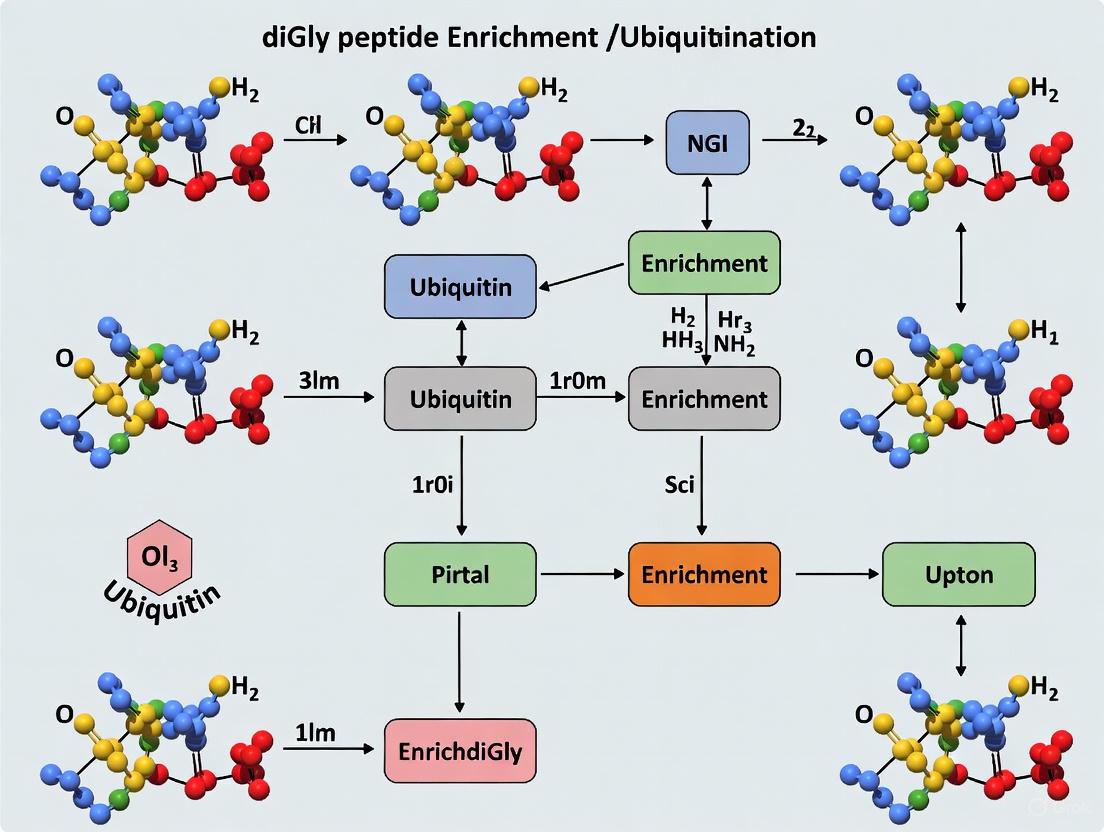
[caption] Diagram 2: Workflow for diGly peptide enrichment and ubiquitinome analysis.
The Scientist's Toolkit: Key Reagents for Ubiquitination Research
Table 4: Essential Research Reagents for Ubiquitination Studies
| Research Reagent / Tool | Function / Application |
|---|---|
| Anti-diGly Remnant Motif Antibody | Core reagent for immunoaffinity enrichment of ubiquitinated peptides from trypsin-digested samples for MS analysis [7]. |
| Proteasome Inhibitors (e.g., MG132) | Treating cells with these inhibitors causes accumulation of polyubiquitinated proteins, thereby increasing the yield of diGly peptides for detection [2] [7]. |
| Tandem Mass Tag (TMT) Reagents | Enable multiplexed, quantitative proteomics. Multiple samples are labeled with different isotopic tags, combined, and analyzed simultaneously by MS, allowing precise relative quantification [2]. |
| Click-iT Plus Technology | Utilized for pulse-chase experiments to label nascent proteins and study protein synthesis and degradation dynamics in real-time [2]. |
| Ubiquitin Enrichment Kits | Kits containing high-binding affinity resins (e.g., agarose beads with ubiquitin-binding domains) for the isolation of polyubiquitinated proteins from cell or tissue lysates, which can then be probed for specific proteins of interest [2]. |
| LanthaScreen Conjugation Assay Reagents | High-throughput screening reagents used to monitor the rate and extent of ubiquitin conjugation to a protein of interest in vitro, useful for drug discovery [2]. |
UPS in Disease and Therapeutic Targeting
Given its central role in controlling cellular processes, it is unsurprising that dysregulation of the UPS is a contributor to many diseases. This has made its components prime targets for drug development.
UPS in Disease Biology
- Cancer: Mutations in or altered expression of E3 ligases (e.g., HDM2) and DUBs can lead to the destabilization of tumor suppressors like p53 or the stabilization of oncoproteins, driving tumorigenesis. The UPS also regulates tumor metabolism, the immunological tumor microenvironment, and cancer stem cell stemness [3].
- Neurodegenerative Diseases: Impaired UPS function can lead to the accumulation of toxic, misfolded protein aggregates, a hallmark of diseases like Alzheimer's, Parkinson's, and Huntington's [2] [4].
- Immune and Inflammatory Disorders: The UPS is integral to signaling pathways in both innate and adaptive immunity. For example, E3 ligases like TRAF and Cbl-b are key regulators of NF-κB signaling and T-cell activation, respectively [1]. Dysregulation can lead to autoimmunity or immunodeficiency.
Targeted Therapies and Drug Discovery
Targeting the UPS for therapy has moved beyond the successful proteasome inhibitors. New strategies aim for greater specificity by targeting upstream components [5]:
- E1 Inhibitors: e.g., MLN4924 (Pevonedistat), which inhibits the NEDD8-activating E1 enzyme, is in clinical trials for various cancers [3] [5].
- E2 Inhibitors: e.g., CC0651, an allosteric inhibitor of the E2 enzyme Cdc34, shows potential in inhibiting cancer cell proliferation [5].
- E3 Ligase-Targeting: This is a highly active area due to the potential for specificity. Examples include:
- DUB Inhibitors: Inhibitors for DUBs like USP7 are in development to modulate the stability of key proteins like p53 and HDM2 [5].
- PROTACs (Proteolysis-Targeting Chimeras): A revolutionary class of bifunctional molecules that re-purpose E3 ligases. A PROTAC consists of one ligand that binds an E3 ligase, another that binds a target protein of interest, and a linker. It brings the E3 ligase into proximity with the target protein, leading to its ubiquitination and degradation [1]. This approach allows for the targeted degradation of proteins previously considered "undruggable."
Ubiquitination represents one of the most versatile post-translational modifications, governing virtually all cellular processes through a complex coding system. This regulatory diversity stems from the ability of ubiquitin to form various chain architectures through its seven internal lysine residues and N-terminal methionine. The development of diGly peptide enrichment methodologies has revolutionized our capacity to decipher this ubiquitin code, enabling proteome-wide mapping of ubiquitination sites with unprecedented depth and precision. This technical guide explores the complexity of ubiquitin chain signaling—from mono-ubiquitination to diverse polyubiquitin linkages—and details the advanced proteomic workflows that now allow researchers to systematically interrogate the ubiquitin-modified proteome. Within the context of diGly peptide enrichment research, we provide comprehensive experimental frameworks, quantitative assessments, and practical toolkits to advance the study of ubiquitin signaling in health and disease.
The Ubiquitin Code: Architectural Diversity and Functional Consequences
Ubiquitination entails the covalent attachment of the 76-amino acid protein ubiquitin to substrate proteins, fundamentally altering their fate and function [4]. This modification exhibits remarkable architectural diversity, generating a complex biological code that cells utilize to coordinate physiological processes.
Types of Ubiquitin Modifications
- Mono-ubiquitination: Involves attachment of a single ubiquitin molecule to a substrate lysine, typically regulating non-proteolytic functions including DNA repair, endocytosis, and histone activity [9] [10]. Unlike degradation signals, mono-ubiquitination often serves as a scaffold for protein-protein interactions.
- Multi-mono-ubiquitination: Occurs when single ubiquitin molecules attach to multiple different lysine residues within the same substrate protein [11]. This pattern creates distinct signaling outcomes compared to polyubiquitin chains.
- Polyubiquitination: Features ubiquitin polymers formed through sequential attachment of ubiquitin molecules to one another, creating chains with diverse linkage types that determine biological function [9] [10] [11]. The seven lysine residues (K6, K11, K27, K29, K33, K48, K63) and N-terminal methionine (M1) of ubiquitin serve as linkage points, generating functionally distinct signals.
Linkage-Specific Functions in Cellular Signaling
Different ubiquitin linkage types create structurally distinct surfaces that are recognized by specific effector proteins, enabling precise control over cellular processes [10] [4].
Table: Ubiquitin Linkage Types and Their Primary Cellular Functions
| Linkage Type | Primary Cellular Functions |
|---|---|
| K48-linked | Proteasomal degradation, protein turnover [10] |
| K63-linked | DNA repair, kinase activation, NF-κB signaling, endocytosis [9] [10] |
| K11-linked | Cell cycle regulation, ER-associated degradation [10] |
| K6-linked | DNA damage response, mitochondrial homeostasis [10] |
| K27-linked | Innate immune response, Wnt signaling [10] |
| K29-linked | Proteasomal degradation, innate immune response [10] |
| K33-linked | Intracellular trafficking, kinase regulation [10] |
| M1-linked (linear) | NF-κB activation, inflammation, immunity [10] [4] |
The ubiquitination cascade involves three enzyme classes: E1 (activating), E2 (conjugating), and E3 (ligating) enzymes [10] [12]. E3 ubiquitin ligases provide substrate specificity, with over 600 human E3 ligases categorized into distinct families including RING, HECT, RBR, and U-box types based on their structural features and catalytic mechanisms [10].
Diagram: The ubiquitination enzymatic cascade and chain formation pathways. The three-step enzymatic process involves E1 activation, E2 conjugation, and E3 ligation, resulting in diverse ubiquitin modifications.
Methodological Framework: diGly Peptide Enrichment for Ubiquitinome Analysis
The tryptic digestion of ubiquitinated proteins generates a characteristic diglycine (diGly) remnant on modified lysine residues, with a detectable mass shift of 114.042 Da [13] [12]. This signature enabled development of antibody-based enrichment strategies that revolutionized ubiquitinome research.
Core Principle of diGly Remnant Recognition
Upon trypsin digestion, ubiquitinated proteins yield peptides containing the Lys-ε-Gly-Gly (K-ε-GG) motif, where the C-terminal glycine residues of ubiquitin remain attached to the modified lysine residue of the substrate [13]. Specific antibodies developed against this diGly remnant enable highly selective enrichment of previously ubiquitinated peptides from complex proteomic samples, allowing identification of ubiquitination sites without genetic manipulation of the ubiquitin system [13] [14].
Advanced Mass Spectrometry Workflows
Contemporary diGly proteomics employs sophisticated mass spectrometry approaches to achieve unprecedented depth of ubiquitinome coverage:
Data-Independent Acquisition (DIA): This method fragments all co-eluting peptide ions within predefined m/z windows simultaneously, resulting in superior quantitative accuracy, fewer missing values, and higher identification rates across samples [7]. A recent optimized DIA workflow identified approximately 35,000 distinct diGly peptides in single measurements of proteasome inhibitor-treated cells—doubling the coverage achievable with traditional data-dependent acquisition (DDA) methods [7].
Spectral Library Generation: Comprehensive spectral libraries containing >90,000 diGly peptides enable precise identification and quantification [7]. These libraries are typically constructed from multiple cell types and treatment conditions (e.g., proteasome inhibition) to maximize coverage of the ubiquitinome.
Fractionation Strategies: Basic reversed-phase separation into 96 fractions concatenated into 8 pools, with separate processing of fractions containing highly abundant K48-linked ubiquitin-chain derived diGly peptides, significantly reduces signal interference and improves detection sensitivity [7].
Table: Comparative Performance of Mass Spectrometry Methods in diGly Proteomics
| Parameter | Data-Dependent Acquisition (DDA) | Data-Independent Acquisition (DIA) |
|---|---|---|
| Typical diGly peptides identified | ~20,000 in single measurements [7] | ~35,000 in single measurements [7] |
| Quantitative precision (CV) | 15% of peptides with CV <20% [7] | 45% of peptides with CV <20% [7] |
| Missing values | Higher rate across sample sets | Fewer missing values [7] |
| Spectral library requirement | Not required | Essential (>90,000 diGly peptides) [7] |
| Dynamic range | Limited | Broader [7] |
Experimental Design Considerations
Several critical factors optimize diGly enrichment efficiency and data quality:
- Sample Input: 1 mg of peptide material typically yields optimal results with 31.25 μg of anti-diGly antibody [7].
- Proteasome Inhibition: Treatment with MG132 (10 μM, 4 hours) increases ubiquitinated protein abundance, enhancing detection sensitivity for low-stoichiometry modifications [7].
- Lysis Conditions: 8M urea buffer supplemented with 5mM N-Ethylmaleimide (NEM) effectively denatures proteins and inhibits deubiquitinating enzymes [13].
- Specificity Controls: The diGly antibody captures <6% non-ubiquitin modifications (primarily NEDD8 and ISG15), ensuring high specificity for ubiquitinome analysis [7].
Diagram: Core workflow for diGly remnant enrichment and mass spectrometry analysis. The process leverages tryptic digestion signatures and antibody-based enrichment for comprehensive ubiquitinome mapping.
Distinguishing Polyubiquitination from Multi-mono-ubiquitination
Differentiating between these ubiquitin modification types is essential for understanding their biological consequences, as they mediate distinct functional outcomes.
Experimental Approach Using Ubiquitin Mutants
A definitive method to distinguish polyubiquitination from multi-mono-ubiquitination employs "Ubiquitin No K"—a ubiquitin mutant where all seven lysine residues are substituted with arginines [11]. This modified ubiquitin can be conjugated to substrates but cannot form polyubiquitin chains due to the absence of acceptor lysine residues.
The experimental protocol involves parallel in vitro ubiquitination reactions [11]:
- Reaction 1: Wild-type ubiquitin + E1/E2/E3 enzymes + substrate
- Reaction 2: Ubiquitin No K + E1/E2/E3 enzymes + substrate
After incubation at 37°C for 30-60 minutes, reactions are terminated with SDS-PAGE sample buffer or DTT/EDTA, followed by Western blot analysis using anti-ubiquitin antibodies.
Interpretation of Results
- Polyubiquitination: High molecular weight smears appear in Reaction 1 (wild-type ubiquitin) but not Reaction 2 (Ubiquitin No K), since chain formation requires lysine residues in ubiquitin [11].
- Multi-mono-ubiquitination: Discrete high molecular weight bands appear in both reactions, as single ubiquitin molecules attach to multiple substrate lysines regardless of ubiquitin's lysine status [11].
- Mixed Modification Patterns: Some substrates display both patterns, evidenced by reduced high molecular weight species in Reaction 2 but persistent lower molecular weight bands [11].
The Scientist's Toolkit: Essential Reagents and Methodologies
Key Research Reagent Solutions
Table: Essential Reagents for Ubiquitination Studies
| Reagent / Method | Function | Application Examples |
|---|---|---|
| Ubiquitin No K | Lysine-less ubiquitin mutant distinguishes polyubiquitination from multi-mono-ubiquitination [11] | Mechanism of action studies |
| Linkage-specific Antibodies | Immunoenrichment of ubiquitin chains with specific linkages (K48, K63, K11, M1, etc.) [12] | Pathway-specific ubiquitination analysis |
| diGly Remnant Antibodies | Enrich ubiquitinated peptides for MS-based ubiquitinome mapping [13] [7] [14] | Proteome-wide ubiquitination site identification |
| Tandem Ubiquitin Binding Entities (TUBEs) | High-affinity enrichment of ubiquitinated proteins while protecting against deubiquitinases [12] | Native ubiquitin conjugate analysis |
| Proteasome Inhibitors (MG132) | Increase ubiquitinated protein abundance by blocking degradation [7] | Enhancing detection sensitivity |
| E1/E2/E3 Enzyme Systems | Reconstitute ubiquitination cascades in vitro [11] | Enzyme mechanism studies |
Emerging Technologies and Applications
Recent methodological advances continue to expand our ability to decipher the ubiquitin code:
- Ubiquitin Interactor Affinity Enrichment-MS (UbIA-MS): Uses chemically synthesized diubiquitin of specific linkages to identify linkage-selective ubiquitin interactors from cell lysates, revealing how different chain types are decoded by cellular effectors [15].
- Stable Tagged Ubiquitin Exchange (StUbEx): Cellular system replacing endogenous ubiquitin with affinity-tagged variants for purification of ubiquitinated proteins under near-physiological conditions [12].
- Cross-linking Approaches: Stabilize transient ubiquitin-enzyme interactions for structural insights into ubiquitination mechanisms.
- DIA-Optimized diGly Proteomics: Recent advances combining diGly antibody-based enrichment with optimized Orbitrap-based DIA methods achieve identification of 35,000+ diGly peptides in single measurements [7].
Biological Insights and Research Applications
Deciphering the ubiquitin code through diGly proteomics has yielded profound insights into cellular regulation and disease mechanisms.
Systems-Level Analysis of Ubiquitin Signaling
Large-scale diGly proteomics has identified approximately 19,000 ubiquitination sites across ~5,000 human proteins, revealing the remarkable scope of this regulatory modification [14]. Quantitative diGly proteomics enables monitoring temporal dynamics of ubiquitination in response to cellular perturbations, classifying substrates based on their degradation kinetics and revealing distinct functional classes within the ubiquitinome [14].
Disease Relevance and Therapeutic Targeting
Dysregulated ubiquitination underlies numerous pathologies, making its comprehensive analysis clinically relevant:
- Cancer: Altered E3 ligase activity and ubiquitination patterns contribute to uncontrolled proliferation, disrupted DNA repair, and aberrant signaling in tumorigenesis [10] [4].
- Neurodegenerative Disorders: Impaired ubiquitin-proteasome function and accumulated ubiquitinated proteins (e.g., tau, α-synuclein, TDP-43) represent pathological hallmarks of Alzheimer's, Parkinson's, and related diseases [4] [16].
- Aging: Quantitative diGly proteomics reveals extensive rewiring of the ubiquitinome in aged mouse brains, with 29% of altered ubiquitination sites showing changes independent of protein abundance shifts [16].
- Inflammation and Immunity: Met1-linked and K63-linked ubiquitin chains play critical roles in NF-κB activation and inflammatory signaling, with dysregulation contributing to autoinflammatory and immune disorders [10] [4].
The depth and precision of modern diGly proteomics continue to illuminate the astonishing complexity of ubiquitin signaling, providing researchers with powerful methodological frameworks to explore this essential regulatory system in health and disease. As methodologies evolve toward even greater sensitivity and throughput, our capacity to decipher the subtleties of the ubiquitin code will undoubtedly expand, revealing new biological insights and therapeutic opportunities.
This technical guide provides a comprehensive overview of the critical role of trypsin digestion in generating the signature di-glycine (diGly) remnant peptides essential for ubiquitination studies. We detail the biochemical principles, optimized experimental protocols, and key analytical considerations for researchers employing bottom-up mass spectrometry to investigate the ubiquitin-modified proteome. Framed within the broader context of enriching for diGly peptides, this whitepequin serves as a fundamental resource for scientists and drug development professionals aiming to achieve robust, reproducible, and high-coverage mapping of ubiquitination sites.
Ubiquitination is a pivotal post-translational modification (PTM) that regulates diverse cellular functions, including protein degradation, signaling, and trafficking [17]. This process involves the covalent attachment of the 76-amino-acid protein ubiquitin to lysine residues on substrate proteins. The enzymatic cascade, involving E1 activating, E2 conjugating, and E3 ligase enzymes, results in an isopeptide bond between the C-terminal carboxyl group of glycine 76 (G76) of ubiquitin and the ε-amino group of the target lysine [18] [17].
In bottom-up mass spectrometry-based proteomics, proteins are digested into peptides prior to analysis. Trypsin digestion is the cornerstone of this process for ubiquitination studies. When a ubiquitinated protein is digested with trypsin, a short peptide remnant derived from the C-terminus of ubiquitin remains attached to the modified lysine on the substrate peptide. This remnant features a di-glycine (diGly or K-ε-GG) motif, which results in a characteristic mass shift of +114.0429 Da on the modified lysine residue [13] [18]. This diGly signature serves as a highly specific mass spectrometry-detectable handle for the definitive identification of ubiquitination sites, enabling the global profiling of the "ubiquitinome" [14].
The Central Role of Trypsin in diGly Peptide Generation
Biochemical Mechanism of Trypsin Cleavage
Trypsin is a serine protease that demonstrates stringent specificity, cleaving peptide bonds predominantly on the C-terminal side of the amino acids arginine (R) and lysine (K) [19] [20]. Its catalytic mechanism relies on a catalytic triad of histidine, aspartate, and serine, and a negatively charged aspartate residue in its S1 binding pocket that attracts and stabilizes the positively charged residues, arginine and lysine [20]. This high specificity is a primary reason for its widespread use in proteomics, as it generates peptides with C-terminal basic residues that are ideal for positive-ion mode mass spectrometry.
In the context of ubiquitinated proteins, trypsin cleavage occurs not only within the protein substrate but also within the ubiquitin modifier itself. The C-terminal sequence of ubiquitin is ...-Arg-Gly-Gly (R-G-G). Trypsin cleaves after the arginine residue, liberating the final two glycine residues (G75 and G76). While G76 is the site of the isopeptide bond with the substrate lysine, the G75-G76 moiety remains attached to the modified lysine as the diGly remnant (K-ε-GG), ready for immunoaffinity enrichment and mass spectrometry analysis [13] [14].
Specificity and Common Deviations
While the canonical trypsin cleavage rule is "after R and K," several nuances and deviations are critical for accurate interpretation of diGly peptide data:
- Proline Inhibition: Trypsin typically does not cleave before a proline (P) residue [20]. If an R or K is immediately followed by a proline, cleavage will not occur at that site.
- Semi-tryptic Peptides: These peptides result from a single tryptic cleavage or from non-tryptic activity elsewhere in the system. Their identification can complicate database searches but may also provide valuable biological insights [21].
- Missed Cleavages: Incomplete digestion can result in peptides containing internal K or R residues that have not been cleaved. The efficiency of cleavage can be influenced by adjacent amino acids, protein folding, and digestion conditions [21] [19]. The presence of missed cleavages within a diGly peptide must be accounted for during data analysis.
Table 1: Factors Influencing Trypsin Digestion Efficiency and diGly Peptide Yield
| Factor | Impact on Digestion | Consideration for diGly Studies |
|---|---|---|
| Digestion Time | Longer times (e.g., 18 hours) can increase completeness but may promote non-specific cleavage or deamidation [21]. | Shorter times (2-4 hours) with optimized protocols may be sufficient and reduce artifacts [22]. |
| Enzyme Origin | Bovine and porcine trypsins show subtle but significant differences in missed cleavage rates and semi-tryptic peptide generation [21]. | Use a single, consistent source of trypsin for reproducible results within a study. |
| Denaturants & pH | Denaturants (e.g., Urea, Guanidine HCl) increase accessibility of cleavage sites. Optimal pH is ~7.8-8.5 [19]. | High urea concentrations must be diluted (<2M) before trypsin addition to avoid enzyme inhibition. |
| Enzyme:Protein Ratio | A ratio of 1:100 (w/w) is standard; increasing trypsin concentration can accelerate digestion [19] [22]. | Higher ratios (e.g., 1:20) can be used for faster digestion without adversely affecting yield for many peptides [22]. |
| Reduction & Alkylation | Breaking disulfide bonds (DTT) and alkylating cysteines (Iodoacetamide) is crucial for complete digestion [19]. | Essential step to ensure full protein denaturation and access to all potential cleavage sites. |
Optimized Experimental Protocols
The following protocols are consolidated from best practices in the field to ensure efficient generation of diGly-modified peptides.
Standard In-Solution Trypsin Digestion for Ubiquitinome Analysis
This protocol is designed for digesting complex protein lysates prior to diGly peptide enrichment [13] [19].
Materials:
- Lysis Buffer: 8 M Urea, 150 mM NaCl, 50 mM Tris-HCl, pH 8.0.
- Reducing Agent: 1 M Dithiothreitol (DTT).
- Alkylating Agent: 500 mM Iodoacetamide (IAA).
- Protease: Sequencing-grade, TPCK-treated trypsin (e.g., Promega).
- Quenching Agent: 100% Formic Acid or Trifluoroacetic Acid (TFA).
Procedure:
- Protein Denaturation and Reduction: Dilute protein extract (e.g., 1-10 mg) in lysis buffer. Add DTT to a final concentration of 5-10 mM and incubate at 37°C for 30-60 minutes.
- Cysteine Alkylation: Add IAA to a final concentration of 15-20 mM. Incubate at room temperature in the dark for 30 minutes.
- Dilution and Trypsin Addition: Dilute the sample with 50 mM Tris-HCl (pH 8.0) or 50 mM Ammonium Bicarbonate (pH ~7.8) to reduce the urea concentration to <2 M. Add trypsin at an enzyme-to-protein ratio of 1:50 to 1:100 (w/w).
- Digestion: Incubate at 37°C for 6-18 hours.
- Reaction Quenching: Acidify the digest by adding formic acid or TFA to a final concentration of 1-5% (v/v). This drops the pH to ~2-3, effectively stopping tryptic activity.
- Peptide Clean-up: Desalt the resulting peptide mixture using a C18 solid-phase extraction column (e.g., Waters Sep-Pak) according to the manufacturer's instructions. The cleaned-up peptides are now ready for the subsequent diGly enrichment step.
Two-Step Digestion with Trypsin/Lys-C Mix for Challenging Proteins
For tightly folded or difficult-to-digest proteins, a two-step protocol using a Trypsin/Lys-C mix can enhance digestion efficiency and reduce missed cleavages [19].
Materials:
- Trypsin/Lys-C Mix, Mass Spec Grade (e.g., Promega #V5071).
- Buffer A: 8 M Urea, 50 mM Tris-HCl, pH 8.0.
Procedure:
- Initial Lys-C Digestion: Denature and reduce the protein sample in Buffer A. Alkylate cysteines as in the standard protocol. Add the Trypsin/Lys-C mix. Lys-C remains active in 8 M urea. Incubate at 37°C for 1-4 hours.
- Dilution and Tryptic Digestion: Dilute the reaction mixture fourfold with 50 mM Tris-HCl (pH 8.0) to reduce the urea concentration to 2 M, creating optimal conditions for trypsin activity.
- Continued Digestion: Continue the incubation at 37°C for a further 4-18 hours to allow trypsin to complete the proteolysis.
- Quenching and Clean-up: Proceed with acidification and desalting as described in Section 3.1.
The Scientist's Toolkit: Essential Reagents for diGly Workflows
Table 2: Key Research Reagent Solutions for diGly Peptide Studies
| Reagent / Kit | Function / Role in Workflow | Key Characteristics |
|---|---|---|
| Sequencing Grade Trypsin | Protein digestion to generate diGly-modified peptides. | Reductive methylation to suppress autolysis; TPCK-treated to inhibit chymotryptic activity [19]. |
| Trypsin/Lys-C Mix | Enhanced digestion of difficult proteins. | Lys-C cleaves before K, is urea-tolerant; mix reduces missed cleavages [19]. |
| diGly Remnant Motif Antibody | Immunoaffinity enrichment of K-ε-GG peptides. | monoclonal antibody specific for the diGly lysine remnant; core of ubiquitinome studies [13] [14]. |
| PTMScan Ubiquitin Remnant Kit | Integrated solution for diGly peptide enrichment. | Contains antibodies, beads, and buffers for a standardized workflow [13]. |
| Stable Isotope Labeling (SILAC) | Quantitative proteomics for comparing ubiquitination across conditions. | Metabolic labeling with heavy/light Lys and Arg allows precise quantification [13]. |
| C18 Desalting Cartridges | Peptide clean-up post-digestion and pre-enrichment. | Removes salts, detergents, and other impurities incompatible with LC-MS/MS [13]. |
Critical Considerations for Data Interpretation
The generation of diGly peptides via trypsin digestion presents unique analytical challenges. Firstly, researchers must be aware that the diGly antibody also enriches for peptides modified by other ubiquitin-like proteins (UBLs), such as NEDD8 and ISG15, which leave an identical C-terminal diGly remnant upon trypsin digestion [13]. While studies indicate that ~95% of identified diGly peptides originate from ubiquitin, this distinction is important for precise mechanistic follow-up [13] [16].
Secondly, the location of lysine and arginine residues within both the substrate and ubiquitin itself dictates the resulting diGly peptide. For example, if a substrate's ubiquitinated lysine is followed by a proline, it can lead to a missed cleavage and a longer-than-expected diGly peptide. Furthermore, trypsin cleavage within a ubiquitin chain can generate various branched peptides, complicating spectral interpretation and requiring advanced search algorithms for confident identification.
The following diagram illustrates the core workflow from ubiquitinated protein to diGly peptide identification.
Workflow for diGly Peptide Identification
Trypsin digestion is a non-negotiable and defining step in the mass spectrometry-based analysis of protein ubiquitination, as it is directly responsible for generating the signature diGly remnant peptide. The quality and reproducibility of this enzymatic step are paramount to the success of any subsequent enrichment and quantitative analysis. By understanding the biochemical principles, implementing optimized and controlled digestion protocols, and being cognizant of the nuances in data interpretation, researchers can reliably uncover the vast and functionally critical landscape of protein ubiquitination, driving discoveries in basic biology and therapeutic development.
The ubiquitin system, once recognized primarily as a mechanism for targeting proteins to the proteasome for degradation, is now understood to be a versatile post-translational modification system that regulates a vast array of cellular processes through both degradative and non-degradative signaling. This transformation in understanding has been propelled by technological advances in mass spectrometry-based proteomics, particularly the development of diGly peptide enrichment strategies that enable system-wide identification of ubiquitination sites. This review explores the biological significance of ubiquitination, detailing the molecular mechanisms that distinguish proteasomal targeting from non-degradative signaling functions, and provides a comprehensive technical guide to contemporary methodologies for ubiquitinome analysis. Within the context of a broader thesis on diGly peptide enrichment, we examine how these techniques have revealed the astonishing diversity of the ubiquitin code and its functional consequences in cellular regulation, disease pathogenesis, and therapeutic development.
The Expanding Landscape of the Ubiquitin Code
Ubiquitination is a major post-translational modification (PTM) in eukaryotic cells involving the covalent attachment of ubiquitin, a 76-amino acid protein, to target substrates. Originally discovered as a signal for energy-dependent protein degradation, ubiquitination is now recognized as a structurally diverse and dynamic modification involved in a myriad of signaling pathways [23]. The modification is executed through a sequential enzymatic cascade involving ubiquitin-activating (E1), conjugating (E2), and ligating (E3) enzymes, and is reversed by deubiquitinases (DUBs) [23] [24].
The functional diversity of ubiquitination stems from the variety of ways ubiquitin can be conjugated to substrates:
- Monoubiquitination: Attachment of a single ubiquitin molecule to a substrate lysine, typically involved in non-proteolytic processes such as endocytosis, histone regulation, and DNA repair [24].
- Multi-monoubiquitination: Multiple single ubiquitin molecules attached to different lysine residues on the same substrate protein [23].
- Polyubiquitination: Chains of ubiquitin molecules linked through specific lysine residues or the N-terminus, creating structurally and functionally distinct signals [23].
Table 1: Types of Ubiquitin Linkages and Their Primary Functions
| Linkage Type | Primary Functions | Structural Features |
|---|---|---|
| K48-linked | Proteasomal degradation [25] [24] | Canonical degradation signal |
| K63-linked | DNA repair, endocytosis, inflammation, kinase activation [25] [26] | Extended chain conformation |
| M1-linked (Linear) | NF-κB activation, inflammation, cell death [27] | Head-to-tail linear linkage |
| K6-linked | Mitophagy, DNA damage response [26] | Associated with mitochondrial quality control |
| K11-linked | Cell cycle regulation, ER-associated degradation [23] | Similar structure to K48 chains |
| K27-linked | Innate immunity, DNA damage response [26] | Role in inflammatory signaling |
| K29-linked | Wnt signaling, neurodegenerative disorders [26] | Proteasomal and non-proteolytic functions |
| K33-linked | Protein trafficking, kinase regulation [26] | Endosomal sorting and receptor internalization |
The ubiquitin code is further complicated by atypical modifications, including non-canonical ubiquitination on cysteine, serine, or threonine residues, and modifications to ubiquitin itself through phosphorylation, acetylation, SUMOylation, and neddylation [23]. This complexity allows ubiquitination to serve as a sophisticated regulatory system controlling virtually every cellular process.
Analytical Methodologies: diGly Enrichment and Proteomic Strategies
The study of ubiquitination at a systems level has been revolutionized by mass spectrometry-based proteomics, particularly through methods that exploit the signature diglycine (diGly) remnant left on trypsinized peptides from ubiquitinated proteins [23] [7]. When ubiquitinated proteins are digested with trypsin, a signature diGly remnant (K-ε-GG) remains attached to the modified lysine residue, serving as a specific marker for ubiquitination sites that can be recognized by antibodies [7].
diGly Peptide Enrichment Workflow
The standard workflow for ubiquitinome analysis involves multiple critical steps designed to maximize the identification of low-abundance ubiquitination sites:
- Cell Culture and Treatment: Cells are cultured under experimental conditions and often treated with proteasome inhibitors (e.g., MG132) to stabilize ubiquitinated substrates [7].
- Protein Extraction and Digestion: Proteins are extracted under denaturing conditions and digested with trypsin, which cleaves after arginine and lysine residues but leaves the diGly-modified lysine intact [7].
- Peptide Fractionation: Basic reversed-phase (bRP) chromatography separates peptides into 96 fractions, which are concatenated into 8-12 pools to reduce complexity [7].
- diGly Peptide Enrichment: Immunoaffinity purification using anti-diGly remnant motif antibodies selectively enriches for modified peptides [7].
- Mass Spectrometry Analysis: Enriched peptides are analyzed by LC-MS/MS using either Data-Dependent Acquisition (DDA) or Data-Independent Acquisition (DIA) methods [7].
Advanced Mass Spectrometry Approaches
Recent methodological advances have significantly improved the depth and quantitative accuracy of ubiquitinome analyses:
Data-Independent Acquisition (DIA) has emerged as a powerful alternative to traditional DDA methods. DIA fragments all co-eluting peptide ions within predefined m/z windows simultaneously, leading to more precise quantification with fewer missing values across samples [7]. A recent optimized DIA workflow for diGly proteome analysis demonstrated remarkable sensitivity, identifying approximately 35,000 distinct diGly peptides in single measurements of proteasome inhibitor-treated cells—double the number achievable with DDA methods [7].
Spectral Library Generation is critical for DIA analysis. Comprehensive libraries can be constructed by combining diGly peptide enrichments from multiple cell lines and conditions. One study created a library containing more than 90,000 diGly peptides from HEK293 and U2OS cells, representing the deepest diGly proteome to date [7]. According to their data, 57% of identified diGly sites had not been previously reported in databases, highlighting the expanding knowledge of the ubiquitinome.
Optimized Experimental Parameters include:
- Antibody and Peptide Input: Enrichment from 1 mg of peptide material using 31.25 μg of anti-diGly antibody provides optimal results [7].
- Fractionation Strategies: Separate handling of fractions containing highly abundant K48-linked ubiquitin-chain derived diGly peptide reduces competition during enrichment and improves detection of co-eluting peptides [7].
- MS Instrument Settings: Methods with high MS2 resolution (30,000) and 46 precursor isolation windows have been shown to improve diGly peptide identification by 13% compared to standard proteome methods [7].
Table 2: Quantitative Performance Comparison of DDA vs. DIA Methods for diGly Proteome Analysis
| Parameter | Data-Dependent Acquisition (DDA) | Data-Independent Acquisition (DIA) |
|---|---|---|
| Typical diGly Peptides Identified | ~20,000 in single measurements [7] | ~35,000 in single measurements [7] |
| Coefficient of Variation (CV) | 15% of peptides with CV <20% [7] | 45% of peptides with CV <20% [7] |
| Quantitative Reproducibility | Lower reproducibility across replicates [7] | Higher reproducibility (77% of peptides with CV <50%) [7] |
| Dynamic Range | Limited for low-abundance ubiquitination events | Enhanced detection across wider dynamic range [7] |
| Spectral Libraries | Not required | Essential, requiring comprehensive library generation [7] |
Biological Functions of Ubiquitin Signaling
Proteasomal Degradation: The Canonical Pathway
The best-characterized function of ubiquitination is targeting proteins for degradation by the 26S proteasome. This process primarily involves K48-linked polyubiquitin chains, which are recognized by proteasomal subunits [25] [24]. A minimum of four ubiquitin molecules attached in a chain is required to trigger degradation [25]. The ubiquitin-proteasome system plays a vital role in protein homeostasis, rapidly removing damaged, misfolded, or regulatory proteins to control their abundance [24]. Key examples include:
- Cell Cycle Regulation: The anaphase-promoting complex/cyclosome (APC/C) and SCF (Skp1-Cul1-F-box) E3 ligase complexes control cell cycle progression by targeting cyclins and other regulators for degradation [4].
- NF-κB Signaling: IκBα, an inhibitor of NF-κB, is phosphorylated and ubiquitinated, leading to its proteasomal degradation and subsequent release of NF-κB for nuclear translocation and inflammatory gene activation [24].
- Hypoxia Response: The von Hippel-Lindau (VHL) E3 ligase targets hypoxia-inducible factor-alpha (HIF-α) for degradation under normoxic conditions [24].
Non-degradative Ubiquitination in Cellular Signaling
Beyond proteasomal targeting, ubiquitination serves numerous non-proteolytic functions that regulate protein activity, interactions, and localization:
Kinase Regulation: Ubiquitination can directly modulate kinase activity through conformational changes. Molecular dynamics simulations of ZAP-70 kinase revealed that monoubiquitination at specific sites (K377 vs. K476) exerts site-dependent effects on the kinase conformational ensemble, either stabilizing inactive or active states [28].
DNA Damage Response (DDR): Multiple ubiquitin linkages coordinate the cellular response to DNA damage:
- K63-linked chains serve as recruitment platforms for repair proteins at DNA damage sites [26].
- K27-linked ubiquitylation of histones H2A and H2A.X by RNF168 generates docking sites for DDR mediators including 53BP1 and BRCA1 [26].
- K29-linked polyubiquitylation of 53BP1 by the CUL3/SPOP complex regulates its exclusion from chromatin during S phase [26].
Inflammatory Signaling:
- Linear (M1-linked) ubiquitination assembled by the LUBAC complex is essential for TNFα- and IL-1-mediated NF-κB signaling pathways [27].
- K63-linked ubiquitination of signaling intermediates like TRAF6 activates downstream inflammatory cascades [25].
Membrane Trafficking: Monoubiquitination and K63-linked polyubiquitination serve as signals for endocytosis and lysosomal trafficking, controlling the surface expression and turnover of membrane receptors and transporters [24].
Pathophysiological Significance and Therapeutic Targeting
Dysregulation of ubiquitin signaling is implicated in numerous human diseases, making components of the ubiquitin system attractive therapeutic targets:
Cancer: Aberrations in ubiquitin signaling are hallmarks of many cancers:
- Von Hippel-Lindau (VHL) Disease: Loss-of-function mutations in the VHL E3 ligase lead to stabilization of HIF-α and uncontrolled expression of growth factors, promoting renal cell carcinoma and other tumors [24].
- Colorectal Cancer: Mutations in the APC (adenomatous polyposis coli) tumor suppressor disrupt the degradation of β-catenin, leading to uncontrolled proliferation of colorectal epithelial cells [24].
- Glioblastoma: Dysregulation of E3 ubiquitin ligases contributes to the development and progression of this aggressive brain cancer [4].
Neurodegenerative Disorders: Impaired ubiquitin-proteasome function leads to accumulation of toxic protein aggregates:
- Parkinson's and Alzheimer's Diseases: Failure to degrade misfolded proteins like α-synuclein and amyloid-β contributes to disease pathology [4].
- Angelman Syndrome: Mutations in UBE3A, which codes for an E3 ubiquitin ligase, cause this rare neurodevelopmental disorder [24].
Inflammatory and Immune Disorders:
- A20 Dysfunction: The A20 protein, a dual ubiquitin-editing enzyme that limits NF-κB signaling, is crucial for preventing inflammatory pathologies; A20 deficiency leads to severe inflammation and sepsis [25].
- Linear Ubiquitination in Immunity: Proper regulation of linear ubiquitination by LUBAC and OTULIN is essential for controlled immune activation; mutations in these components are linked to autoinflammatory and immunodeficiency syndromes [27].
Research Reagent Solutions for Ubiquitination Studies
Table 3: Essential Research Reagents for Ubiquitinome Studies
| Reagent Category | Specific Examples | Function and Application |
|---|---|---|
| Enrichment Antibodies | Anti-K-ε-GG Ubiquitin Remnant Motif Kit (CST) [7] | Immunoaffinity enrichment of diGly-modified peptides for MS analysis |
| Proteasome Inhibitors | MG132, Bortezomib [7] | Stabilize ubiquitinated proteins by blocking proteasomal degradation |
| E1 Enzyme Inhibitors | TAK-243, PYR-41 [4] | Block ubiquitin activation, globally inhibiting ubiquitination |
| DUB Inhibitors | PR-619, G5, NSC632839 [4] | Inhibit deubiquitinating enzymes, stabilizing ubiquitination events |
| Linkage-Specific Binders | TUBE (Tandem Ubiquitin Binding Entities) [23] | Enrich for specific polyubiquitin chain linkages |
| LUBAC Inhibitors | HOIPIN-8, Benzodiazepine derivatives [27] | Specifically inhibit linear ubiquitination by targeting HOIP |
| Cell Line Models | HEK293, U2OS, Jurkat T-cells [7] [28] | Commonly used model systems for ubiquitinome profiling |
| Ubiquitin Mutants | K48R, K63R, K0 (all lysines mutated) [23] | Study chain-type specific functions and generate linkage-defined ubiquitin |
The understanding of ubiquitin signaling has evolved remarkably from a simple degradation signal to a complex post-translational modification system regulating virtually every cellular process. This paradigm shift has been driven largely by technical advances in proteomics, particularly the development and refinement of diGly peptide enrichment methodologies that enable comprehensive ubiquitinome profiling. The distinction between proteasomal and non-proteolytic ubiquitin signaling is now well-established, with specific ubiquitin chain linkages encoding distinct functional outcomes.
Future directions in ubiquitin research include the development of more specific reagents for distinguishing ubiquitin chain linkages, improved computational tools for analyzing complex ubiquitinome datasets, and the continued development of therapeutics targeting specific components of the ubiquitin system. As these methodologies advance, our understanding of the biological significance of ubiquitination will continue to expand, revealing new connections to human disease and novel therapeutic opportunities.
The study of protein ubiquitination has undergone a revolutionary transformation, evolving from low-throughput biochemical techniques to sophisticated, high-throughput proteomic methodologies. This evolution has been pivotal for understanding the intricate role of ubiquitination in cellular regulation, protein homeostasis, and disease pathogenesis. Central to this progress has been the development and refinement of techniques for enriching and analyzing peptides containing the diglycine (diGLY) remnant—a signature of ubiquitination. This whitepaper traces this historical development, detailing the key technological breakthroughs that have established diGLY peptide enrichment as a cornerstone of modern ubiquitinome research, providing drug development professionals and scientists with a comprehensive technical guide.
Part 1: The Foundational Shift in Ubiquitination Analysis
The initial methods for detecting protein ubiquitination were functional but limited in scale and precision. Early biochemical approaches relied on protein-level immunoprecipitation followed by Western blot analysis using ubiquitin-specific antibodies. While diagnostic, this method could not identify the exact sites of modification [18]. To pinpoint specific lysine residues, researchers turned to site-directed mutagenesis, substituting lysines with arginines to assess the loss of ubiquitination. This process was often laborious, especially for proteins with many lysine residues, and could be confounded by functional redundancy where mutation of one lysine would lead to ubiquitination at an alternative site [18].
A pivotal conceptual and technical shift occurred with the realization that tryptic digestion of ubiquitylated proteins generates peptides with a characteristic diGLY modification on the target lysine residue, resulting in a defined mass shift of +114.0429 Da detectable by mass spectrometry (MS) [13] [18]. Although the existence of this remnant was reported as early as 1977 [13], its widespread application for proteome-wide analysis remained challenging due to the extremely low abundance of these modified peptides within a complex proteomic background.
The critical breakthrough came in the early 2010s with the development and commercialization of robust antibodies specifically targeting the Lys-ε-Gly-Gly (diGLY) remnant motif [13] [14]. This innovation enabled the immunoaffinity enrichment of diGLY-containing peptides from complex tryptic digests, dramatically improving the sensitivity and scale of ubiquitination site identification. This approach, often referred to as ubiquitin remnant motif profiling, transformed the field, allowing for the systematic and unbiased interrogation of the ubiquitin-modified proteome, or "ubiquitinome" [29].
Table 1: Evolution of Key Methodologies in Ubiquitination Site Mapping
| Era | Methodology | Key Principle | Limitations | Throughput & Scale |
|---|---|---|---|---|
| Pre-Proteomics | Site-directed mutagenesis + Western Blot | Indirect inference via lysine-to-arginine mutation and immunoblotting. | Cannot confirm exact site; functionally redundant sites can confound results [18]. | Low (single protein, single site) |
| Early MS | Gel-based protein IP & in-gel digest | Protein immunoprecipitation, SDS-PAGE separation, in-gel digestion, and MS analysis of diGLY peptides. | Low sensitivity; limited success for many substrates; labor-intensive [18]. | Medium (single protein, multiple sites) |
| Modern High-Throughput | diGLY Peptide Immunoaffinity Enrichment | Antibody-based enrichment of diGLY-modified peptides from whole-cell lysate digests for LC-MS/MS. | Minimal; potential for very low-level enrichment of NEDD8/ISG15 peptides [13]. | Very High (proteome-wide, thousands of sites) |
Part 2: The High-Throughput Proteomics Revolution
The advent of high-throughput proteomics, driven largely by advances in mass spectrometry, has provided the tools necessary to realize the full potential of diGLY enrichment. Proteomics has evolved from studying individual proteins to analyzing entire proteomes, a shift enabled by technological breakthroughs like soft ionization techniques (MALDI, ESI) and improved mass analyzers (Orbitrap) [30] [31].
The standard workflow for diGLY proteomics involves several key steps, which have been continuously optimized for depth and throughput [13] [32]:
- Cell Lysis & Protein Digestion: Cells or tissues are lysed under denaturing conditions (e.g., 8M Urea buffer) in the presence of protease inhibitors and N-Ethylmaleimide (NEM) to preserve ubiquitination and deubiquitination states [13]. Proteins are then digested, typically with LysC and trypsin, to generate peptides.
- Peptide Fractionation (Optional): To increase coverage, complex peptide mixtures can be pre-fractionated using high-pH reverse-phase chromatography prior to diGLY enrichment. This helps reduce sample complexity and minimizes competition for antibody binding sites from highly abundant peptides, such as the K48-linked ubiquitin chain-derived diGLY peptide [7] [32].
- diGLY Peptide Immunoaffinity Enrichment: The tryptic peptides are incubated with anti-diGLY antibodies conjugated to beads. This step selectively isolates the low-abundance diGLY-modified peptides from the bulk of unmodified peptides [13] [29].
- Mass Spectrometric Analysis: The enriched peptides are separated by nanoflow liquid chromatography and analyzed by tandem MS (LC-MS/MS). Quantitative methods, such as Stable Isotope Labeling with Amino acids in Cell culture (SILAC) or label-free quantification, are employed to compare ubiquitination levels across different conditions [13] [18].
A more recent advancement is the adoption of Data-Independent Acquisition (DIA) MS. Unlike traditional Data-Dependent Acquisition (DDA), which selectively fragments the most intense ions, DIA systematically fragments all ions within predefined mass windows, leading to more comprehensive and reproducible data acquisition. When applied to diGLY proteomics, DIA has been shown to double the number of diGLY peptides identified in a single measurement (e.g., ~35,000 sites) compared to DDA, while also significantly improving quantitative accuracy and data completeness [7].
Diagram 1: High-Throughput Workflow for diGLY Proteomics. This diagram outlines the key steps in a modern ubiquitinome study, from sample preparation to MS analysis.
Part 3: Experimental Protocols for Key Applications
Protocol 1: Basic diGLY Immunoaffinity Enrichment and Quantitative Analysis
This protocol is adapted from established methods for identifying and quantifying ubiquitination sites from cultured cells using SILAC [13].
Cell Culture and Lysis:
- Grow cells in SILAC "light" (normal Lys/Arg) and "heavy" (13C6,15N2-Lys; 13C6,15N4-Arg) media for at least five cell doublings to achieve complete labeling [13].
- Treat cells according to experimental design (e.g., with a proteasome inhibitor like MG132 or a specific ligand).
- Lyse cells in a denaturing buffer (e.g., 8M Urea, 50mM Tris-HCl pH 8, 150mM NaCl) supplemented with complete protease inhibitors, phosphatase inhibitors, and 5mM NEM (freshly prepared) to inhibit deubiquitinases [13].
- Mix light and heavy lysates in a 1:1 ratio based on total protein amount.
Protein Digestion and Cleanup:
- Reduce proteins with 5mM DTT (30 min at 50°C) and alkylate with 10mM iodoacetamide (15 min in the dark) [32].
- Digest proteins first with LysC (4 hours) followed by trypsin (overnight at 30°C) [13] [32].
- Acidify the digest with Trifluoroacetic acid (TFA) to a final concentration of 0.5% to precipitate and remove detergents. Centrifuge and collect the peptide-containing supernatant [32].
- Desalt the peptides using a C18 reverse-phase SepPak column [13].
diGLY Peptide Enrichment:
- Use a commercial PTMScan Ubiquitin Remnant Motif Kit or equivalent anti-diGLY antibody conjugated to beads.
- Incubate the peptide mixture with the antibody beads for 2 hours at 4°C with rotation [32].
- Wash the beads extensively with ice-cold Immunoaffinity Purification (IAP) buffer and then with water.
- Elute the bound diGLY peptides with 0.15% TFA [32].
LC-MS/MS Analysis and Data Processing:
- Analyze the enriched peptides on a high-sensitivity nanoLC-MS/MS system (e.g., an Orbitrap instrument).
- Operate the MS in data-dependent acquisition (DDA) mode, with MS1 scans at high resolution (e.g., 60,000) and top-speed MS2 fragmentation of precursors.
- For deeper coverage, a second run using a "least intense first" fragmentation regime can be performed to capture low-abundance peptides [32].
- Process the raw data using search engines like MaxQuant, specifying diGLY (K-ε-GG) as a variable modification and the appropriate SILAC labels for quantification [32].
Protocol 2: Advanced DIA-based Ubiquitinome Profiling
For the most comprehensive and quantitative analysis, a DIA-based workflow is recommended [7].
Library Generation:
- Generate a deep, sample-specific spectral library by fractionating a peptide digest (e.g., into 96 fractions concatenated into 8-9 pools) after basic reversed-phase chromatography.
- Enrich each fraction for diGLY peptides and analyze them using a standard DDA method to build a library containing tens of thousands of diGLY peptide spectra [7].
Single-Run DIA Analysis:
- For biological samples, enrich diGLY peptides from 1 mg of peptide input using an optimized amount of anti-diGLY antibody.
- Analyze only 25% of the total enriched material on the LC-MS/MS system.
- Use an optimized DIA method with ~46 variable windows and high-resolution MS2 scans (30,000) to maximize identifications.
- Interrogate the single-run DIA data against the pre-acquired comprehensive spectral library for identification and quantification [7].
Table 2: Performance Comparison of MS Acquisition Methods for diGLY Proteomics
| Parameter | Data-Dependent Acquisition (DDA) | Data-Independent Acquisition (DIA) |
|---|---|---|
| Identification Depth (Single Run) | ~20,000 diGLY peptides [7] | ~35,000 diGLY peptides [7] |
| Quantitative Reproducibility | 15% of peptides with CV < 20% [7] | 45% of peptides with CV < 20% [7] |
| Data Completeness | Higher rate of missing values across samples [7] | Fewer missing values across samples [7] |
| Workflow Requirement | Simpler; no library always required | Benefits from a comprehensive spectral library |
| Primary Advantage | Simplicity and wide adoption | Superior sensitivity, reproducibility, and quantitative accuracy |
Part 4: The Scientist's Toolkit: Essential Reagents and Materials
Successful diGLY proteomics relies on a set of critical reagents and tools. The following table details key components for a typical experiment.
Table 3: Essential Research Reagent Solutions for diGLY Proteomics
| Item | Function / Principle | Example / Specification |
|---|---|---|
| diGLY Motif-specific Antibody | Immunoaffinity enrichment of ubiquitin remnant-containing peptides from complex digests. | PTMScan Ubiquitin Remnant Motif (K-ε-GG) Kit; monoclonal antibodies [13] [14]. |
| Cell Culture Media for Labeling | Enables metabolic labeling for accurate quantification via mass spectrometry. | SILAC DMEM, lacking Lysine and Arginine; supplemented with heavy isotopes (K8, R10) and dialyzed FBS [13]. |
| Lysis Buffer Components | Effective denaturation and inactivation of enzymes to preserve the native ubiquitinome. | 8M Urea, 50mM Tris-HCl, 150mM NaCl. Must be supplemented with Protease Inhibitors, 5mM NEM [13]. |
| Chromatography Media | Desalting and cleaning up peptide mixtures pre- and post-enrichment. | C18 reverse-phase SepPak columns (e.g., 500mg for 30mg digest) for bulk cleanup; C18 StageTips for final sample preparation [13] [32]. |
| Proteases for Digestion | Generation of peptides with C-terminal diGLY remnant on modified lysines. | LysC (Wako) and Trypsin (TPCK-treated, Sigma). Sequential digestion improves efficiency [13]. |
| Mass Spectrometer | High-sensitivity identification and quantification of enriched diGLY peptides. | High-resolution instrument coupled to nanoflow HPLC (e.g., Orbitrap series) capable of DDA and DIA acquisition [30] [7] [32]. |
The journey from focused biochemical assays to global, high-throughput proteomics has fundamentally reshaped our understanding of the ubiquitin system. The development of diGLY remnant immunoaffinity enrichment was a pivotal milestone, providing a powerful and specific tool to probe the ubiquitinome at an unprecedented scale. Subsequent advancements in mass spectrometry, particularly the adoption of DIA strategies, have further pushed the boundaries of sensitivity, reproducibility, and quantitative accuracy. This powerful toolkit now allows researchers to not only catalog ubiquitination sites but also to dynamically monitor changes in response to cellular stimuli, identify substrates of specific E3 ligases, and uncover novel regulatory mechanisms in health and disease. As these high-throughput methodologies continue to be refined and integrated with other omics data, they will undoubtedly remain a fundamental driver of discovery in basic research and drug development.
diGly Enrichment Protocols and Cutting-Edge Applications in Research
Antibody-based enrichment represents a cornerstone technique in modern proteomics, enabling the selective isolation of low-abundance proteins or specific post-translational modifications (PTMs) from complex biological samples. This methodology addresses a critical bottleneck in biomarker validation and PTM analysis by providing the necessary sensitivity and specificity to study rare analytes that would otherwise be undetectable amid high-abundance interfering proteins [33]. The technique operates on the principle of immunoaffinity purification (IAP), where immobilized antibodies capture target antigens from a digested peptide mixture, followed by washing and elution steps to yield a purified sample for downstream analysis [34].
The significance of this technology is particularly evident in the field of ubiquitination studies, where the identification of ubiquitylated proteins has been revolutionized by antibodies specific to the diglycine (diGLY) remnant that remains on modified lysine residues after tryptic digestion [13] [7]. This approach has enabled researchers to systematically interrogate the ubiquitin-modified proteome with site-level resolution, leading to the identification of over 50,000 ubiquitylation sites in human cells and providing unprecedented insights into how ubiquitination regulates virtually all cellular processes [13].
Core Principles of Antibody-Based Enrichment
Fundamental Mechanisms
Antibody-based enrichment exploits the specific binding interaction between an antibody and its target epitope. In proteomic applications, this typically occurs after proteins have been digested into peptides, allowing for highly selective isolation of specific peptide sequences or PTM-bearing peptides from complex mixtures [34]. The process involves three fundamental steps: binding, where the peptide mixture is incubated with immobilized antibodies; washing, to remove non-specifically bound contaminants; and elution, to recover the purified target peptides [33] [35].
The exceptional specificity of antibody-antigen interactions enables remarkable enrichment factors. Optimized magnetic bead-based platforms for peptide capture can achieve ion signal enhancements on the order of 10³, with precision (coefficients of variation <10%) and accuracy (relative error ~20%) sufficient for quantifying biomarkers in the physiologically relevant ng/mL range [33]. This level of performance is crucial for applications like biomarker validation, where target proteins may be present at orders of magnitude lower concentration than abundant serum proteins like albumin (50 mg/mL) and globulin (35 mg/mL) [33].
Antibody Types and Their Applications
Different antibody types are employed based on the specificity required for the research question:
- Standard site-specific antibodies: Recognize a modified amino acid in the context of a specific sequence of surrounding amino acids [34].
- PTM-specific antibodies: Bind to any residue with a particular PTM (e.g., acetyl-lysine), regardless of flanking sequences [34].
- PTM-sequence motif antibodies: Recognize a modified amino acid within a specific motif, such as substrates of particular kinases [34].
In ubiquitination research, diGLY remnant-specific antibodies have become indispensable. These antibodies recognize the Lys-ϵ-Gly-Gly (diGLY) motif generated when trypsin cleaves after arginine (R) and lysine (K) residues of ubiquitylated proteins, leaving a Gly-Gly remnant attached to the modified lysine [13]. It is important to note that while this antibody primarily captures ubiquitylated peptides, the identical C-terminal sequences of ubiquitin-like proteins (Nedd8 and ISG15) mean that a small percentage (<6%) of identified diGLY peptides may arise from neddylation or ISGylation [7].
Table: Comparison of Antibody Types Used in Proteomic Enrichment
| Antibody Type | Target Specificity | Applications | Advantages |
|---|---|---|---|
| Standard Site-Specific | Defined amino acid sequence with PTM | PhosphoScan, targeted pathway analysis | High specificity for known sites |
| PTM-Specific | Modified amino acid regardless of context | Ubiquitin, acetylome, methylome studies | Comprehensive coverage of all modified sites |
| Motif-Specific | PTM within a characteristic sequence motif | Kinase substrate identification | Captures related family of substrates |
diGLY Enrichment for Ubiquitination Studies
The diGLY Signature and Ubiquitinome Analysis
Protein ubiquitylation involves the covalent attachment of the 76-amino acid ubiquitin protein to lysine residues on substrate proteins. This modification typically targets substrates for proteasomal degradation but can also modulate protein function, localization, and activity without impacting turnover [13]. When ubiquitylated proteins are digested with trypsin, a characteristic diGLY remnant (K-ε-GG) is left attached to the modified lysine residue, serving as a signature for prior ubiquitination [13] [7].
The diGLY proteomics approach has transformed the study of ubiquitin signaling by enabling:
- Site-level resolution of ubiquitylation events across the proteome
- Quantitative assessment of how ubiquitylation changes under different conditions
- Identification of specific ubiquitin ligase targets
- Discovery of novel regulatory mechanisms in cellular physiology and pathophysiology [13]
This methodology has been successfully applied to identify >50,000 ubiquitylation sites in human cells and to quantify how these sites are altered upon exposure to diverse proteotoxic stressors [13].
Experimental Workflow for diGLY Enrichment
The standard workflow for diGLY enrichment proteomics involves multiple critical steps that must be carefully optimized for successful results. The following diagram illustrates this process:
Sample Preparation and Lysis Cells or tissues are lysed in a denaturing buffer containing 8M urea, 150mM NaCl, 50mM Tris-HCl (pH 8), supplemented with complete protease inhibitors and 5mM N-Ethylmaleimide (NEM) to preserve ubiquitination by inhibiting deubiquitinating enzymes (DUBs) [13]. The inclusion of NEM is critical as it irreversibly alkylates cysteine residues, preventing the activity of cysteine-dependent DUBs that would otherwise remove ubiquitin chains during sample processing.
Protein Digestion and Desalting Proteins are digested first with LysC (which cleaves at lysine residues) followed by trypsin (cleaves at arginine and lysine) to generate peptides with the diGLY signature [13]. Trypsin digestion of ubiquitylated proteins produces peptides with the characteristic diGLY remnant on modified lysines. Following digestion, peptides are desalted using C18 reverse-phase columns such as Sep-Pak cartridges to remove salts and contaminants that could interfere with subsequent enrichment steps [13] [36].
diGLY Immunoaffinity Purification The core enrichment process involves incubating the digested peptide mixture with diGLY remnant motif-specific antibodies immobilized on beads. The PTMScan Ubiquitin Remnant Motif (K-ε-GG) Kit is commonly used for this purpose [13] [7]. Typical protocols use 1mg of peptide material with approximately 31.25μg of anti-diGLY antibody, incubating overnight at 4°C to maximize capture efficiency [7]. After binding, beads are washed with buffer to remove non-specifically bound peptides, and bound diGLY peptides are eluted with dilute acid (e.g., 0.1-0.5% trifluoroacetic acid or 5% acetic acid) [33] [13].
Mass Spectrometry Analysis Enriched peptides are analyzed by liquid chromatography coupled to tandem mass spectrometry (LC-MS/MS). Recent advances have demonstrated the superiority of Data-Independent Acquisition (DIA) methods over traditional Data-Dependent Acquisition (DDA) for diGLY proteomics. DIA provides greater data completeness across samples, higher quantitative accuracy, and identifies approximately 35,000 distinct diGLY peptides in single measurements—nearly double the identification rate of DDA methods [7].
Commercial Kits and Platforms
Specialized diGLY Enrichment Kits
Several commercial platforms have been developed to standardize and optimize antibody-based enrichment for ubiquitination studies. The most widely adopted system is the PTMScan technology from Cell Signaling Technology (CST), which offers specialized kits for ubiquitin remnant enrichment:
The PTMScan Ubiquitin Remnant Motif (K-ε-GG) Kit is specifically designed for comprehensive ubiquitinome analysis. This kit employs monoclonal antibodies that recognize the diGLY remnant left on modified lysines after tryptic digestion of ubiquitylated proteins [7] [35]. The protocol involves:
- Immunoaffinity purification using bead-conjugated diGLY-specific antibodies
- Optimized binding and washing buffers to minimize non-specific binding
- Elution conditions that preserve peptide integrity for downstream MS analysis
- Compatibility with both label-free and isotope-labeled quantification approaches [35]
For researchers interested in profiling multiple signaling pathways simultaneously, the PTMScan Multi-Pathway Enrichment Kit provides an array of site-specific antibodies conjugated to protein A beads. This kit enables screening, discovery, and quantitation of thousands of proteins and phosphorylation sites across multiple signaling pathways, including cell cycle control, PI3K/Akt signaling, and MAPK cascades [35].
Supporting Purification Technologies
Successful antibody-based enrichment often relies on supporting purification technologies that prepare samples for the specific capture step:
Protein A/G/L Purification Systems These bacterial immunoglobulin-binding proteins are fundamental tools for antibody purification and can also be used to immobilize antibodies for enrichment procedures [37] [38]. Each has distinct binding properties:
- Protein A: Binds the Fc region of IgG from multiple species, particularly effective for human, mouse, goat, and rabbit IgG [37]
- Protein G: Broader species reactivity than Protein A, binds all human, goat, and murine IgG subclasses [37]
- Protein A/G: Recombinant fusion protein combining binding profiles of both Protein A and Protein G [37]
- Protein L: Binds kappa light chains of immunoglobulins without interfering with antigen binding, useful for capturing antibody fragments [37]
These proteins are available in multiple formats including magnetic beads, loose resins, spin columns, and FPLC cartridges to accommodate different processing scales from microgram to kilogram quantities [37].
Abundant Protein Depletion Kits For complex samples like serum or plasma, where a few abundant proteins (e.g., albumin, immunoglobulins) can dominate the proteome and obscure detection of low-abundance analytes, abundant protein depletion kits can significantly improve detection of rare species. Commercial options include:
- High Select HSA/Immunoglobulin Depletion Mini Spin Columns: Specifically target albumin and immunoglobulins for removal [36]
- High Select Top14 Abundant Protein Depletion Mini Spin Columns: Remove the 14 most abundant plasma proteins [36]
- ProteoExtract and Proteome PurifyTM 2: Multi-component depletion systems [36]
These depletion methods can increase the identification of low-abundance disease biomarkers by reducing signal suppression from highly abundant proteins during MS analysis [36].
Technical Considerations and Optimization
Critical Experimental Parameters
Successful implementation of antibody-based enrichment requires careful optimization of several parameters:
Sample Input and Antibody Ratio Titration experiments have determined that optimal diGLY enrichment is achieved using 1mg of peptide material with approximately 31.25μg of anti-diGLY antibody [7]. Excessive peptide input leads to competition for antibody binding sites, while insufficient input reduces detection sensitivity.
Enrichment Specificity and Competition A particular challenge in ubiquitinome analysis is the presence of extremely abundant ubiquitin-derived peptides, especially the K48-linked ubiquitin-chain derived diGLY peptide, which can dominate the enrichment and compete with less abundant peptides for antibody binding sites [7]. To address this, researchers can employ pre-fractionation strategies such as basic reversed-phase chromatography to separate the most abundant diGLY peptides before enrichment, improving coverage of less prevalent ubiquitination sites [7].
Mass Spectrometry Acquisition Methods Comparative studies have demonstrated significant advantages of Data-Independent Acquisition (DIA) over traditional Data-Dependent Acquisition (DDA) for diGLY proteomics:
- DIA identifies ~35,000 diGLY sites in single measurements versus ~20,000 for DDA
- DIA provides superior quantitative accuracy with 45% of diGLY peptides having coefficients of variation (CVs) below 20% compared to only 15% for DDA
- DIA achieves greater data completeness with fewer missing values across samples [7]
Table: Performance Comparison of DIA vs. DDA for diGLY Proteomics
| Parameter | DIA (Data-Independent Acquisition) | DDA (Data-Dependent Acquisition) |
|---|---|---|
| diGLY Peptides Identified | 35,000 ± 682 | 20,000 |
| Quantitative Precision (CV <20%) | 45% of peptides | 15% of peptides |
| Quantitative Precision (CV <50%) | 77% of peptides | Not reported |
| Required Sample Amount | 25% of enriched material | 100% of enriched material |
| Data Completeness | Higher, fewer missing values | Lower, more missing values |
The Scientist's Toolkit: Essential Research Reagents
Table: Key Reagents for Antibody-Based Enrichment Experiments
| Reagent Category | Specific Examples | Function/Purpose |
|---|---|---|
| Cell Lysis Reagents | 8M Urea, 150mM NaCl, 50mM Tris-HCl (pH 8) | Protein denaturation and extraction |
| Protease Inhibitors | Complete Protease Inhibitor Cocktail | Prevent protein degradation during processing |
| DUB Inhibitors | 5mM N-Ethylmaleimide (NEM) | Preserve ubiquitination by inhibiting deubiquitinating enzymes |
| Digestion Enzymes | Trypsin, LysC | Protein digestion to generate analyzable peptides |
| Desalting Media | C18 Sep-Pak columns | Peptide cleanup and buffer exchange |
| Enrichment Antibodies | PTMScan Ubiquitin Remnant Motif (K-ε-GG) Antibody | Specific capture of diGLY-modified peptides |
| Binding Beads | Magnetic Protein G beads | Solid support for antibody immobilization |
| Chromatography Media | Protein A, G, A/G, L | Antibody purification and immobilization |
| Depletion Kits | High Select Top14, ProteoExtract | Remove abundant proteins to enhance detection of low-abundance targets |
| LC-MS/MS Columns | C18 reverse-phase nano-capillary columns | Peptide separation before mass spectrometry |
Applications in Biological Research
Advancing Ubiquitination Studies
Antibody-based diGLY enrichment has enabled groundbreaking discoveries across diverse areas of biology:
TNF Signaling Pathway Mapping Comprehensive application of diGLY proteomics to Tumor Necrosis Factor (TNF) signaling has captured known ubiquitination sites while adding many novel ones, revealing the extensive regulation of this pathway by ubiquitin [7]. The technology has identified both expected components (like NF-κB pathway members) and previously unrecognized ubiquitination events that expand our understanding of TNF signal transduction.
Circadian Biology Regulation An in-depth, systems-wide investigation of ubiquitination across the circadian cycle uncovered hundreds of cycling ubiquitination sites and dozens of cycling ubiquitin clusters within individual membrane protein receptors and transporters [7]. This revealed unexpected connections between metabolism and circadian regulation, demonstrating how ubiquitination dynamics contribute to temporal organization of cellular processes.
Ubiquitin Ligase Substrate Identification The diGLY antibody-based approach has proven particularly valuable for identifying substrates of specific ubiquitin ligases, which has been a longstanding challenge in the field due to the transient nature of enzyme-substrate interactions and the complexity of the ubiquitin system [13]. By comparing diGLY profiles between wild-type and ligase-deficient cells, researchers have identified specific ligase targets that contribute to various physiological and pathological processes [13].
Biomarker Discovery and Validation
Beyond fundamental biology, antibody-based enrichment holds tremendous potential for translational applications. The SISCAPA (Stable Isotope Standards with Capture by Anti-Peptide Antibodies) method exemplifies this approach, where anti-peptide antibodies enrich specific target peptides along with spiked stable-isotope-labeled internal standards for highly precise quantification by mass spectrometry [33]. This technology provides a desperately needed bridging methodology between biomarker discovery and clinical application, offering a more cost-effective and rapid alternative to traditional ELISA development for biomarker validation [33].
Antibody-based enrichment represents a powerful and versatile methodology that has revolutionized proteomic analysis, particularly in the study of post-translational modifications like ubiquitination. The development of diGLY remnant-specific antibodies has enabled comprehensive mapping of the ubiquitin-modified proteome, revealing the remarkable scope and regulatory complexity of this modification. Commercial kits such as the PTMScan platform have standardized and democratized these approaches, making sophisticated proteomic analyses accessible to a broader research community.
As mass spectrometry technologies continue to advance, particularly with the adoption of Data-Independent Acquisition methods, the sensitivity, depth, and quantitative accuracy of antibody-based enrichment workflows will further improve. These advancements promise to accelerate discoveries in basic biology while simultaneously enabling the translation of proteomic findings into clinically applicable biomarkers and therapeutic targets. The integration of antibody-based enrichment with emerging proteomic technologies ensures this methodology will remain a cornerstone of proteome research for the foreseeable future.
In the study of ubiquitination, a pivotal post-translational modification (PTM), sample preparation is a critical foundational step that directly determines the success and depth of downstream analysis. The ubiquitin-modified proteome, or "ubiquitinome," presents unique challenges for mass spectrometry (MS)-based investigation due to the low stoichiometry of modified proteins, the dynamic nature of the modification, and the complexity of ubiquitin chain architectures [7]. The core objective of sample preparation in ubiquitination studies is to efficiently extract, digest, and fractionate protein samples to enable the specific isolation of ubiquitin-derived peptides for subsequent LC-MS/MS analysis. This technical guide details the essential methodologies for lysis, digestion, and fractionation, framed within the context of preparing samples for diGly peptide enrichment—the gold-standard approach for system-wide ubiquitination site mapping [39] [40] [7]. The guidance provided herein is designed to equip researchers with the protocols needed to achieve deep, reproducible, and biologically meaningful coverage of the ubiquitinome.
Lysis Conditions
The lysis step must achieve complete disruption of cells or tissues to release proteins while preserving the native state of ubiquitination and other PTMs. Harsh conditions are often necessary for challenging samples, but they must be compatible with subsequent enzymatic digestion and MS analysis.
Core Principles of Lysis for Ubiquitinome Studies
- Complete Protein Solubilization: The lysis buffer must effectively solubilize hydrophobic membrane proteins, which are frequent targets of ubiquitination. Incomplete lysis leads to significant and biased protein loss [41].
- PTM Preservation: The lysis conditions must rapidly inactivate endogenous enzymes, particularly proteases and deubiquitinases (DUBs), to prevent the degradation of the ubiquitin signature. This is typically achieved with denaturing conditions and the inclusion of protease and DUB inhibitors [7].
- MS-Compatibility: While detergents are highly effective for extraction, they often interfere with downstream tryptic digestion and LC-MS, necessitating extensive and potentially loss-prone cleanup steps [41].
Lysis Buffer Formulations and Protocols
Two primary strategies are employed, each with distinct advantages and trade-offs, summarized in Table 1.
Table 1: Comparison of Lysis Buffer Strategies for Ubiquitinome Analysis
| Lysis Strategy | Key Components | Recommended Protocol | Advantages | Disadvantages |
|---|---|---|---|---|
| Detergent-Based Lysis | SDS, SDC, Urea, Protease Inhibitor Cocktails | 1. Suspend cell pellet in lysis buffer (e.g., 1-2% SDS).2. Incubate at 95°C for 5-10 min.3. Sonicate to reduce viscosity and shear DNA.4. Centrifuge to clarify lysate. | Powerful denaturation and solubilization, effective protease inhibition. | Requires detergent removal (e.g., via filter-based methods like FASP) before digestion, adding steps and time. |
| Detergent-Free Lysis (SPEED) | Pure Trifluoroacetic Acid (TFA) | 1. Add pure TFA directly to cell pellet or tissue.2. Vortex vigorously until the sample is fully dissolved.3. Neutralize with a pre-calculated volume of Tris base.4. Proceed directly to digestion [41]. | Universal application, highly reproducible, rapid, and avoids detergent-removal steps. | Highly acidic conditions require careful handling and precise neutralization. |
The SPEED (Sample Preparation by Easy Extraction and Digestion) protocol is a notable detergent-free method that uses pure trifluoroacetic acid (TFA) for highly efficient protein extraction by complete sample dissolution. This protocol consists of three mandatory steps: acidification, neutralization, and digestion. It has been demonstrated to be superior to detergent-based methods like FASP, ISD-Urea, and SP3 in terms of quantitative reproducibility and proteome coverage, especially for challenging samples [41].
Digestion Protocols
Following lysis, proteins must be digested into peptides suitable for LC-MS/MS analysis. The goal is to generate peptides with high efficiency and reproducibility, paying particular attention to the C-terminal cleavage behavior of ubiquitin-modified lysine residues.
Tryptic Digestion and the diGly Signature
The hallmark of ubiquitination site identification in bottom-up proteomics is the tryptic-digest-derived diGly remnant. After tryptic digestion of ubiquitinated proteins, a signature diglycine (diGly) remnant remains conjugated to the epsilon-amino group of the modified lysine residue [39]. This "K-ε-diglycine" or "diGly" motif, with a mass shift of +114.0429 Da, serves as a mass-taggable surrogate for the original ubiquitination site. This diGly remnant is the target for immunopurification using specific antibodies [39] [40] [7].
Detailed In-Solution Digestion Protocol
A robust, standard protocol for protein digestion prior to diGly enrichment is as follows:
- Protein Denaturation and Reduction: Dilute the protein lysate in a compatible buffer (e.g., 50 mM Tris-HCl, pH 8.0). Add a reducing agent such as dithiothreitol (DTT) to a final concentration of 5-10 mM and incubate at 55°C for 30-45 minutes to break disulfide bonds.
- Alkylation: Add an alkylating agent like iodoacetamide (IAA) to a final concentration of 15-20 mM and incubate at room temperature in the dark for 30 minutes. This step prevents the reformation of disulfide bonds.
- Digestion: Add trypsin (typically sequencing-grade, modified) at an enzyme-to-protein ratio of 1:50 (w/w) and incubate at 37°C for a minimum of 6 hours, or overnight for complete digestion. To quench the digestion, acidify the sample with TFA to a final concentration of 0.5-1%.
- Desalting and Cleanup: Desalt the resulting peptide mixture using a C18 solid-phase extraction (SPE) cartridge or column. Elute peptides in a solution compatible with the next step (e.g., 30-80% acetonitrile with 0.1% TFA). Lyophilize or vacuum concentrate the eluate and reconstitute in an appropriate immunoaffinity enrichment buffer [39] [7].
Fractionation
To achieve deep coverage of the ubiquitinome, fractionation is essential to reduce sample complexity and alleviate the dynamic range limitation of the mass spectrometer, thereby increasing the number of identifiable diGly peptides.
High-pH Reverse-Phase Fractionation
A highly effective fractionation strategy, used prior to diGly enrichment, is basic reversed-phase (bRP) chromatography.
- Protocol: The digested peptide mixture is separated on a C18 column using a gradient of increasing organic solvent (acetonitrile) at high pH (e.g., pH 10). The eluent is collected into a series of fractions (e.g., 96 fractions) which are then concatenated into a smaller number of pools (e.g., 8-12) to save analysis time [7].
- Strategic Handling of Abundant Peptides: In samples treated with proteasome inhibitors (e.g., MG132), the abundance of K48-linked ubiquitin-chain derived diGly peptides can be extremely high. These peptides can compete for antibody binding sites during enrichment and obscure the detection of co-eluting peptides. To mitigate this, fractions containing these highly abundant K48-peptides can be identified and processed separately, which significantly improves the overall depth of analysis [7]. This workflow is illustrated in Figure 1.
Post-Enrichment Fractionation
As an alternative, fractionation can also be performed after the diGly peptide enrichment step. While this approach is feasible, pre-enrichment fractionation generally yields a greater number of identified ubiquitination sites because it reduces the complexity of the sample presented to the antibody, improving enrichment efficiency [39].
Figure 1: A high-level workflow for ubiquitinome analysis via diGly peptide enrichment.
The Scientist's Toolkit: Key Research Reagents
Successful execution of the ubiquitinome analysis workflow relies on a set of specific reagents and materials. Table 2 details essential items and their functions.
Table 2: Essential Research Reagents for diGly Peptide Enrichment Workflow
| Reagent / Material | Function / Application | Key Considerations |
|---|---|---|
| Anti-K-ε-GG Antibody | Immunoaffinity enrichment of diGly-modified peptides from complex digests. | Commercial kits are available (e.g., PTMScan Ubiquitin Remnant Motif Kit). The amount of antibody must be titrated against peptide input (e.g., 31.25 µg antibody per 1 mg peptide) [7]. |
| Protease Inhibitor Cocktail | Prevents protein degradation and preserves ubiquitin modifications during lysis. | Should include inhibitors of deubiquitinating enzymes (DUBs). |
| Sequencing-Grade Trypsin | Proteolytic enzyme for digesting proteins into peptides, generating the diGly signature. | Essential for consistent and complete cleavage. |
| Trifluoroacetic Acid (TFA) | Used for acidification in SPEED protocol and as a mobile phase modifier in LC-MS. | High-purity grade is required for MS compatibility [41]. |
| C18 Solid-Phase Extraction Cartridge | Desalting and cleanup of peptide samples after digestion and before enrichment. | Critical for removing salts, detergents, and other impurities. |
| High-pH Stable C18 Chromatography Column | For off-line or on-line high-pH reverse-phase fractionation of peptides. | Enables deep ubiquitinome coverage by reducing sample complexity [39] [7]. |
| Proteasome Inhibitor (e.g., MG132) | Treatment of live cells to accumulate ubiquitinated proteins, enhancing detection. | Commonly used at 10-20 µM for 4-6 hours prior to lysis [7]. |
The journey to a comprehensive ubiquitinome profile begins at the bench with meticulous sample preparation. The choices made during lysis, digestion, and fractionation profoundly impact the sensitivity, depth, and accuracy of the final results. The adoption of efficient, reproducible, and MS-compatible methods—such as the detergent-free SPEED protocol for lysis, rigorous tryptic digestion, and strategic high-pH fractionation—provides a robust foundation for the specific enrichment of diGly peptides. By adhering to these detailed protocols and utilizing the appropriate toolkit of reagents, researchers can standardize and optimize their workflows to uncover the vast and dynamic landscape of protein ubiquitination, thereby enabling discoveries in fundamental biology and drug development.
Protein ubiquitylation is one of the most prevalent post-translational modifications (PTMs) within cells, imparting critical regulatory control over nearly every cellular, physiological, and pathophysiological process [13]. While ubiquitin modification typically marks substrates for proteasome-dependent degradation, it can also alter protein function through modulation of protein complexes, localization, or activity without impacting protein turnover [13]. The identification of ubiquitylation sites has been revolutionized by an antibody-based affinity approach that recognizes the Lys-ϵ-Gly-Gly (diGLY) remnant generated following trypsin digestion of ubiquitylated proteins [13]. This diGLY proteomics approach has led to the identification of >50,000 ubiquitylation sites in human cells and provides quantitative information about how these sites are altered upon exposure to diverse proteotoxic stressors [13]. When performing such analyses, mass spectrometry (MS) is one of the most popular methods, with data-dependent acquisition (DDA) and data-independent acquisition (DIA) representing the two broad approaches for generating bottom-up or "shotgun" MS proteomic data [42]. This technical guide examines the comparative advantages, limitations, and applications of both approaches specifically within the context of diGly peptide analysis for ubiquitination studies.
Core Principles of DDA and DIA Mass Spectrometry
Data-Dependent Acquisition (DDA)
In tandem MS (MS/MS), the DDA approach operates by first surveying all peptides within a certain mass range during an initial MS scan [42] [43]. The instrument then selects only the most intense peptide ions (typically the "top N" precursors, where N is usually 10-15 peptides) within a narrow range of mass-to-charge (m/z) signal intensity for further fragmentation and analysis in a second stage of tandem mass spectrometry [42]. This selection process occurs sequentially for each peptide, and the resulting data are used to search an existing protein database [42]. The fundamental characteristic of DDA is this selective, intensity-driven approach to precursor selection, which introduces a level of bias toward more abundant peptides but simplifies data analysis through more straightforward spectral interpretation [42].
Data-Independent Acquisition (DIA)
In contrast, DIA takes a comprehensive approach where all peptides within predefined m/z windows are fragmented and analyzed during the second stage of tandem mass spectrometry [42] [44]. Rather than selecting specific intense precursors, the mass spectrometer divides the overall mass range (typically 400-1200 m/z) into small, consecutive mass windows (e.g., 5-25 Da wide) [45] [43]. All precursors within each isolation window are fragmented simultaneously, and all resulting product ions are systematically recorded [45]. A common DIA method is Sequential Windowed acquisition of All THeoretical fragment ion Mass Spectra (SWATH-MS), which steps through these mass windows across the entire mass range, systematically collecting MS/MS data from every mass and from all detected precursors throughout the chromatographic separation [43] [44]. This creates a complete, time-resolved recording of fragment ions for all peptide precursors, providing an unbiased and comprehensive dataset [43].
Comparative Analysis: DDA vs. DIA for diGly Proteomics
Table 1: Technical comparison between DDA and DIA approaches for diGly proteomics
| Parameter | Data-Dependent Acquisition (DDA) | Data-Independent Acquisition (DIA) |
|---|---|---|
| Precursor Selection | Selective; chooses most intense "top N" precursors | Comprehensive; fragments all precursors in predefined m/z windows |
| Bias | Biased toward high-abundance peptides | Less biased; includes low-abundance peptides |
| Reproducibility | Lower precision and reproducibility | Higher precision and better reproducibility |
| Dynamic Range | Limited; low-abundance peptides under-represented | Large; can quantify proteins in complex mixtures over wide dynamic range |
| Data Complexity | Simpler spectra; easier interpretation | Highly multiplexed spectra; complex interpretation |
| Computational Demand | Lower demand on computational resources | High demand due to large, complex datasets |
| Quantification Sensitivity | More sensitive for targeted analysis | Lower sensitivity due to scanning complete spectrum |
| Best Application | Targeted analysis, beginners, when target peptides are in existing databases | Discovery proteomics, large sample cohorts, little-studied organisms |
Advantages and Limitations in diGly Research
For diGly peptide analysis specifically, each approach presents distinct considerations. DDA's primary advantages include simpler setup and analysis, lower computational requirements, and more sensitive quantification for targeted analyses where peptides of interest are already documented in databases [42]. This can be particularly valuable when studying well-characterized ubiquitination pathways. However, DDA suffers from lower precision and reproducibility, under-representation of low-abundance diGly peptides, and inherent bias in precursor selection that may miss important but less abundant ubiquitination events [42].
DIA offers significant advantages for comprehensive ubiquitinome mapping, including less biased sampling of all peptides, higher precision, better reproducibility, and the ability to detect low-abundance diGly peptides that might be missed by DDA [42] [45]. This is particularly valuable for discovery-oriented ubiquitination studies aiming to identify novel regulatory sites. The method also allows greater temporal resolution, which benefits studies examining changes in ubiquitylation patterns over time [42]. Furthermore, DIA data can be retrospectively re-analyzed with improved algorithms as they become available, potentially uncovering additional diGly peptides from existing datasets [42]. The main limitations include substantially larger data files, higher computational demands, more challenging data analysis due to highly multiplexed MS2 spectra, and generally lower sensitivity for quantification [42] [45].
Workflow Integration for diGly Peptide Analysis
diGly Peptide Enrichment Experimental Workflow
The standard workflow for diGly proteomics begins with sample preparation, which varies depending on the experimental design. For cell culture studies, Stable Isotope Labeling with Amino acids in Cell culture (SILAC) can be employed for quantification, while label-free approaches offer alternatives for tissue samples or in vivo studies [13]. Following sample collection, proteins are extracted using a denaturing lysis buffer (typically containing 8M Urea, 150mM NaCl, 50mM Tris-HCl pH 8, plus protease inhibitors and 5mM N-Ethylmaleimide to deubiquitinating enzymes) [13]. After reduction and alkylation, proteins are digested first with LysC protease and then with trypsin [13]. The resulting peptides are desalted using reverse-phase columns before the critical enrichment step.
diGLY-modified peptides are isolated using ubiquitin remnant motif (K-ε-GG) antibodies, which specifically recognize the diGly modification left on lysine residues after tryptic digestion of ubiquitylated proteins [13] [46]. Recent methodological improvements have significantly enhanced this enrichment, including offline high-pH reverse-phase fractionation of tryptic peptides into multiple fractions prior to immunopurification, more efficient wash steps to reduce non-specific binding, and the use of filter plugs to retain antibody beads [46]. These modifications have enabled the routine detection of over 23,000 diGly peptides from HeLa cells upon proteasome inhibition and have proven effective for in-depth analysis of endogenous ubiquitinomes from in vivo samples such as mouse brain tissue [46] [47].
Figure 1: Experimental workflow for diGly peptide enrichment and analysis
Mass Spectrometry Data Acquisition Strategies
Following diGly enrichment, samples are analyzed by liquid chromatography coupled to tandem mass spectrometry (LC-MS/MS). The choice between DDA and DIA approaches depends on the research goals, with DIA increasingly favored for comprehensive ubiquitinome mapping due to its superior reproducibility and depth [45]. In DIA mode, the mass spectrometer systematically steps through predefined m/z windows (typically 5-25 Da wide) across the entire mass range, fragmenting all precursors within each window and recording all resulting fragment ions [45] [43]. This creates a complete record of all eluting peptides, including diGly-modified species, throughout the chromatographic separation.
Recent advances in DIA methodology have significantly improved its application to diGly proteomics. These include the use of variable isolation window schemes adjusted based on precursor m/z distribution, which enhances selectivity; overlapping windows to improve coverage; and optimized instrument settings that balance scan speed with mass resolution [45]. Furthermore, the development of more sophisticated data analysis tools has addressed earlier limitations in dealing with the highly complex, multiplexed spectra generated by DIA, making it increasingly suitable for diGly peptide identification and quantification [42] [45].
Advanced Integrated Approaches and Methodological Innovations
Hybrid DDA/DIA Strategies
Emerging evidence suggests that integrating DDA and DIA approaches can yield significant benefits for sensitive applications like immunopeptidomics, and these principles extend to diGly proteomics [48]. Recent research has led to the development of "Data dependent-independent acquisition proteomics" (DDIA), which combines DDA and DIA in a single LC-MS/MS run and uses deep-learning tools for more streamlined data analysis [42]. This hybrid approach aims to capture the complementary strengths of both methods - the sensitive identification of abundant peptides characteristic of DDA with the comprehensive, reproducible quantification of DIA.
Integrated platforms like PEAKS Online exemplify this trend, seamlessly combining DDA and DIA data acquisition with multiple computational approaches (spectral library search, database search, and de novo sequencing) under a unified framework [48]. Such platforms have demonstrated impressive performance, identifying 5-30% more peptide precursors than other state-of-the-art systems on multiple benchmark datasets and 1.7-4.1 times more peptides from DDA immunopeptidomics data than previously reported results [48]. The application of deep learning throughout the analysis pipeline, from basic tasks like spectrum or retention time predictions to complex processes like de novo sequencing, further enhances identification capabilities [48].
Enhanced Data Analysis Workflows
Figure 2: Integrated DIA data analysis workflow for maximal sensitivity
The complex, multiplexed nature of DIA data requires sophisticated analysis strategies, particularly for challenging applications like diGly proteomics. Modern approaches typically employ a targeted data extraction strategy, where previously identified spectral libraries are used to mine the DIA data for specific peptides of interest [45] [44]. However, recent innovations have enabled direct analysis of DIA data without the need for project-specific DDA-based spectral libraries, though organism-specific libraries (e.g., Pan-Human library) can still improve identifications [45].
The most advanced workflows now integrate multiple computational approaches consecutively to achieve maximum sensitivity [48]. As illustrated in Figure 2, this involves performing spectral library search, database search, and de novo sequencing on the same DIA dataset, then using the combined peptide identifications to build a project-specific spectral library, followed by a final search of the entire dataset against this consolidated library [48]. This integrated approach provides a unified global false discovery rate (FDR) estimation while maximizing the identification of diGly peptides across the dynamic range.
Essential Research Reagents and Materials
Table 2: Key research reagents for diGly proteomics studies
| Reagent/Category | Specific Examples | Function in diGly Proteomics |
|---|---|---|
| Cell Culture Media | DMEM lacking lysine/arginine (Thermo Fisher #88364) | Base for SILAC labeling with heavy isotopes |
| Heavy Isotopes | L-Lysine:2HCL (13C6, 99%; 15N2, 99%), L-Arginine:HCL (13C6, 99%; 15N4, 99%) | Metabolic labeling for quantification |
| Lysis Buffer Components | 8M Urea, 150mM NaCl, 50mM Tris-HCl (pH 8), Protease Inhibitors, 5mM N-Ethylmaleimide (NEM) | Protein extraction while preserving ubiquitin modifications |
| Proteolytic Enzymes | LysC protease (Wako #125-02543), Trypsin (Sigma #T1426) | Protein digestion while generating diGly remnant |
| Enrichment Antibodies | Ubiquitin Remnant Motif (K-ε-GG) Antibody, PTMScan Ubiquitin Remnant Motif Kit | Specific immunopurification of diGly-modified peptides |
| Chromatography | SepPak tC18 reverse phase column (Waters #WAT036815) | Peptide desalting and cleanup |
| Mass Spectrometry | Q-Orbitrap, Q-TOF instruments with DIA capabilities (SWATH-MS) | High-resolution detection and quantification |
The choice between DDA and DIA approaches for diGly peptide analysis depends largely on the specific research objectives. DDA remains valuable for targeted analyses where the primary interest is in well-characterized ubiquitination sites, particularly when sample amounts are limited or computational resources are constrained [42]. Its simpler data interpretation and more sensitive quantification for known targets make it appropriate for hypothesis-driven research on specific ubiquitination events.
In contrast, DIA offers significant advantages for discovery-oriented ubiquitinome mapping, where the goal is comprehensive identification of ubiquitylation sites across the proteome [42] [45]. Its superior reproducibility, minimal bias, and ability to detect low-abundance diGly peptides make it increasingly the method of choice for large-scale studies of ubiquitination dynamics in physiological and pathophysiological processes [46] [47]. The ability to retrospectively re-analyze DIA data as improved computational tools emerge provides an additional long-term benefit.
Looking forward, the distinction between DDA and DIA is likely to blur further with the development of hybrid approaches that capture the benefits of both methods [42] [48]. The integration of advanced computational approaches, particularly deep learning-based tools for spectrum prediction and data analysis, will continue to enhance the sensitivity and accuracy of both DDA and DIA for diGly proteomics [48]. These technological advances, combined with optimized experimental protocols for diGly peptide enrichment, will undoubtedly deepen our understanding of the extensive regulatory roles played by protein ubiquitylation in health and disease.
Protein ubiquitination is a crucial post-translational modification (PTM) involved in virtually all cellular processes, from protein degradation to signal transduction and circadian regulation [7] [12]. The versatility of ubiquitin signaling arises from its complex conjugation patterns, which can range from single ubiquitin monomers to diverse polyubiquitin chains with different linkage types and architectures [12]. This complexity presents significant analytical challenges for researchers seeking to understand ubiquitination at a systems level. Two advanced methodologies have emerged to address these challenges: TUBE-MS (Tandem Ubiquitin Binding Entities coupled to Mass Spectrometry) for enriching intact ubiquitinated proteins, and DRUSP (Data-Independent Acquisition [DIA]-based Ubiquitin Site Profiling) for deep, quantitative analysis of ubiquitination sites. These techniques represent complementary approaches within the broader context of ubiquitin enrichment strategies, each offering unique advantages for specific research applications in drug development and basic research.
Core Principles and Technological Foundations
The diGLY Remnant: A Signature for Ubiquitination
At the heart of many ubiquitin enrichment strategies is the recognition of a unique structural feature generated during sample preparation. When ubiquitinated proteins undergo tryptic digestion, they yield peptides containing a characteristic diglycine (diGLY) remnant conjugated to the ε-amino group of modified lysine residues [13]. This K-ε-GG motif serves as a specific signature for previously ubiquitinated peptides. Antibodies developed to recognize this diGLY remnant have become indispensable tools, enabling the affinity-based enrichment of these modified peptides from complex biological samples [13] [39]. It is important to note that while this approach is highly effective, identical diGLY remnants can theoretically be generated from the ubiquitin-like modifiers NEDD8 and ISG15, though studies indicate that approximately 95% of identified diGLY peptides originate from genuine ubiquitination events [13].
TUBE Technology: Preserving Native Ubiquitin Architecture
Tandem Ubiquitin Binding Entities (TUBEs) represent a fundamentally different approach to ubiquitin enrichment. These engineered, high-affinity reagents are composed of multiple ubiquitin-associated (UBA) domains arranged in tandem, enabling them to bind polyubiquitin chains with exceptional avidity [49] [50]. Unlike antibody-based methods that target proteolytic fragments, TUBEs interact with intact ubiquitin chains, allowing them to capture polyubiquitinated proteins while preserving their native architecture. This capability is particularly valuable for studying ubiquitin chain topology and dynamics, as TUBEs shield polyubiquitinated proteins from deubiquitinating enzymes (DUBs) and proteasomal degradation during sample processing [49]. Furthermore, TUBEs can be designed with pan-selectivity to capture all ubiquitin chain linkage types, or with linkage specificity to isolate particular chain architectures such as K48 or K63 linkages [49] [50].
Table 1: Comparison of Core Ubiquitin Enrichment Principles
| Feature | diGLY Antibody Approach | TUBE Approach |
|---|---|---|
| Target | DiGLY remnant on tryptic peptides | Intact ubiquitin chains on proteins |
| Specificity | Site-specific resolution | Protein-level resolution |
| Chain Information | Lost during digestion | Preserved during enrichment |
| Primary Application | Ubiquitination site mapping | Polyubiquitinated protein capture |
| DUB Protection | No | Yes [49] |
| Linkage Specificity | Limited | Available (pan-specific or linkage-specific) [49] |
The DRUSP Methodology: Deep, Quantitative Ubiquitin Site Profiling
Workflow Optimization and Spectral Library Generation
The DRUSP methodology represents a significant advancement in ubiquitination site analysis by combining diGLY antibody-based enrichment with optimized data-independent acquisition (DIA) mass spectrometry. This approach addresses key limitations of traditional data-dependent acquisition (DDA) methods, particularly regarding quantitative accuracy, data completeness, and sensitivity in single-run analyses [7]. The foundational step in establishing a robust DRUSP workflow involves creating comprehensive spectral libraries to facilitate accurate peptide identification in DIA mode. In a landmark study, researchers treated HEK293 and U2OS cell lines with the proteasome inhibitor MG132 to enhance ubiquitinated protein levels, followed by protein extraction, digestion, and extensive fractionation using basic reversed-phase chromatography into 96 fractions concatenated into 8 pools [7]. A critical innovation involved separating fractions containing the highly abundant K48-linked ubiquitin-chain derived diGLY peptide, which competes for antibody binding sites and interferes with detection of co-eluting peptides [7]. This refined approach enabled the identification of more than 67,000 and 53,000 diGLY peptides from MG132-treated HEK293 and U2OS cells, respectively, ultimately generating spectral libraries containing over 90,000 diGLY peptides [7].
DIA Method Optimization and Performance Metrics
The unique characteristics of diGLY-containing peptides—often longer with higher charge states due to impeded C-terminal cleavage of modified lysine residues—necessitated specific optimization of DIA parameters [7]. Researchers empirically optimized DIA window widths and determined that a method with 46 precursor isolation windows and high MS2 resolution (30,000) significantly improved diGLY peptide identification [7]. Additionally, systematic titration experiments established that enrichment from 1 mg of peptide material using 31.25 μg of anti-diGLY antibody provided optimal results, with only 25% of the total enriched material required for injection due to the enhanced sensitivity of the DIA approach [7]. The performance metrics of the optimized DRUSP workflow are impressive, identifying approximately 35,000 distinct diGLY sites in single measurements of proteasome inhibitor-treated cells—doubling the number achievable with DDA methods [7]. Quantitative accuracy showed substantial improvement, with 45% of diGLY peptides exhibiting coefficients of variation (CVs) below 20% across replicates, compared to only 15% with DDA methods [7].
Diagram 1: DRUSP workflow for deep ubiquitinome analysis.
The TUBE-MS Methodology: Capturing Polyubiquitinated Proteins
TUBE Design and Enrichment Optimization
The TUBE-MS methodology centers on the use of engineered tandem ubiquitin-binding entities to enrich polyubiquitinated proteins directly from complex biological samples. These reagents typically consist of four repeats of ubiquitin-binding UBA domains derived from proteins such as ubiquilin-1, arranged in tandem to achieve high avidity for polyubiquitin chains of various linkage types [50]. A critical advancement in TUBE technology involves site-specific biotinylation of recombinantly expressed TUBEs using BirA enzyme on an N-terminal Avi-tag, facilitating modular immobilization on streptavidin-coated magnetic beads [50]. This design enables efficient pulldown of free ubiquitin chains while maintaining compatibility with downstream LC-MS/MS analysis. To preserve ubiquitin chains during sample processing, the TUBE-MS workflow incorporates semi-denaturing lysis conditions with 4M urea and complete inhibition of deubiquitinating enzymes through additives such as N-ethylmaleimide (NEM) at 20mM concentration, which has been shown to be essential for full DUB inhibition upon cell lysis [50].
Elution Strategy and Application to Compound Profiling
A key innovation in the TUBE-MS workflow is the development of an elution strategy that selectively liberates ubiquitinated proteins while the TUBE reagent remains bound to the beads [50]. This approach minimizes TUBE contamination in the final eluate, thereby reducing background interference during mass spectrometric analysis. The utility of TUBE-MS has been demonstrated in multiple pharmacological contexts, including the profiling of small molecule-induced changes in protein polyubiquitination. When applied to characterize the effects of PROTAC MZ1 (which induces ubiquitination and degradation of BET family proteins), TUBE-MS enabled robust detection of polyubiquitinated BRD2 in a manner dependent on both MZ1 treatment and proteasomal inhibition [50]. Similarly, application of TUBE-MS to compounds inhibiting the deubiquitinase USP7 revealed induction of non-degradative ubiquitination on the E3 ligase UBE3A, highlighting the method's ability to detect ubiquitination events that do not necessarily target proteins for degradation [50].
Diagram 2: TUBE-MS workflow for polyubiquitinated protein enrichment.
Comparative Performance and Applications
Quantitative Comparison of Method Capabilities
The strategic selection between DRUSP and TUBE-MS methodologies depends heavily on research objectives, as each approach offers distinct advantages for different aspects of ubiquitin research. The following table summarizes key performance characteristics and application strengths of each method based on current implementations:
Table 2: Performance Comparison of DRUSP vs. TUBE-MS Methodologies
| Parameter | DRUSP (DIA diGLY) | TUBE-MS |
|---|---|---|
| Identification Depth | ~35,000 diGLY sites (single run) [7] | Protein-level identification (fewer specific sites) |
| Quantitative Precision | 45% of peptides with CV < 20% [7] | Compatible with quantitative methods |
| Chain Architecture | Limited information | Preserved during enrichment [49] [50] |
| Site Resolution | Single lysine resolution | Protein-level resolution |
| Primary Strengths | Comprehensive site mapping, high quantitative accuracy | Detection of non-degradative ubiquitination, chain topology [50] |
| Therapeutic Applications | Ubiquitination site dynamics, PTM crosstalk | PROTAC mechanism studies, DUB inhibitor profiling [50] |
Biological Applications and Case Studies
Both methodologies have demonstrated significant utility in addressing complex biological questions. The DRUSP approach has been successfully applied to systems-wide investigations of ubiquitination dynamics across the circadian cycle, uncovering hundreds of cycling ubiquitination sites and revealing ubiquitin clusters within individual membrane protein receptors and transporters [7]. This application highlighted new connections between metabolic regulation and circadian biology, demonstrating the power of comprehensive site-specific ubiquitinome analysis. When applied to TNFα signaling, the DRUSP methodology comprehensively captured known ubiquitination sites while adding many novel ones, validating its utility for mapping signaling-related ubiquitination events [7].
In contrast, TUBE-MS has proven particularly valuable in drug discovery contexts, especially for characterizing compounds that modulate the ubiquitin-proteasome system. The method has been effectively used to profile changes induced by PROTACs, p97 inhibitors, and deubiquitinase inhibitors, providing direct evidence of compound-induced polyubiquitination changes [50]. Unlike whole proteome analyses that indirectly infer ubiquitination through protein abundance changes, TUBE-MS directly detects polyubiquitination events, enabling differentiation between degradative and non-degradative ubiquitination—a critical distinction for understanding the mechanisms of emerging therapeutic modalities [50].
Essential Research Reagent Solutions
The successful implementation of either DRUSP or TUBE-MS methodologies requires specific reagent systems optimized for their respective workflows. The following table catalogues key reagents and their applications in advanced ubiquitin enrichment:
Table 3: Essential Research Reagents for Advanced Ubiquitin Enrichment
| Reagent / Kit | Type | Primary Application | Key Features |
|---|---|---|---|
| PTMScan Ubiquitin Remnant Motif Kit | diGLY antibody | DRUSP | Enrichment of K-ε-GG peptides; optimized for MS [7] [13] |
| Pan-selective TUBEs | Tandem UBA domains | TUBE-MS | Broad specificity for all linkage types; DUB protection [49] |
| Linkage-specific TUBEs | Engineered UBA domains | TUBE-MS | K48 or K63 chain enrichment; specific ubiquitination profiling [49] |
| Ubiquitin Mass Spectrometry Kit (UM420) | Complete kit | TUBE-MS | Includes TUBEs and reagents for full workflow [49] |
| Heavy SILAC Amino Acids | Stable isotopes | Quantitative ubiquitomics | K8/R10 for metabolic labeling in quantitative studies [13] |
| DUB Inhibitor Cocktails | Small molecules | Sample preparation | NEM, PR-619; preserve ubiquitination during lysis [13] [50] |
TUBE-MS and DRUSP methodologies represent complementary advanced approaches for ubiquitinome profiling, each with distinct strengths and applications. DRUSP methodology, combining diGLY enrichment with optimized DIA mass spectrometry, provides unprecedented depth and quantitative accuracy for ubiquitination site mapping, making it ideal for systems-level investigations of ubiquitination dynamics. In contrast, TUBE-MS excels at capturing intact polyubiquitinated proteins while preserving chain architecture, offering unique insights into ubiquitin chain topology and non-degradative ubiquitination events particularly relevant to drug mechanism studies. As the ubiquitin field continues to evolve, these methodologies will play increasingly important roles in elucidating the complex roles of ubiquitination in cellular regulation and in accelerating the development of novel therapeutics targeting the ubiquitin-proteasome system.
Protein ubiquitination is a fundamental post-translational modification (PTM) that regulates virtually every cellular process in eukaryotes, from protein degradation and cell cycle progression to DNA repair and signal transduction [17]. The critical role of ubiquitination in maintaining cellular homeostasis makes its dysregulation a central feature in numerous disease pathologies. The development of diGLY proteomics—an affinity enrichment method utilizing antibodies specific to the diglycine (K-ε-GG) remnant left on trypsinized peptides from ubiquitinated proteins—has revolutionized our ability to study ubiquitination sites at a systems-wide level [13]. This technical guide explores the transformative application of diGLY proteomics in three key research areas: cancer biology, neurodegenerative disorders, and circadian regulation, providing researchers with detailed methodologies and analytical frameworks for implementing these approaches in disease-specific contexts.
The versatility of ubiquitination signaling stems from its complex architecture. Beyond serving as a degradation signal via K48-linked polyubiquitin chains, ubiquitination can modulate protein function, localization, and interactions through various chain linkages and monoubiquitination events [51] [17]. The diGLY proteomics approach has enabled researchers to move beyond single protein analyses to global ubiquitinome profiling, generating unprecedented insights into disease mechanisms and potential therapeutic targets. Here, we detail the experimental and computational strategies that make these discoveries possible.
Core Methodological Framework
Fundamental Workflow of diGLY Proteomics
The standard diGLY proteomics workflow consists of several critical stages, each requiring optimization for specific disease research applications. The foundational process begins with sample preparation from relevant biological sources (cell cultures, animal models, or human tissues), followed by protein digestion using trypsin or alternative proteases, affinity enrichment of diGLY-modified peptides using motif-specific antibodies, and finally liquid chromatography-mass spectrometry (LC-MS/MS) analysis with quantitative profiling [13] [52]. The following diagram illustrates this core workflow:
Figure 1: Core workflow for diGLY proteomics analysis of ubiquitination sites, highlighting key stages from sample preparation through data analysis.
Critical Optimization Strategies
Recent methodological advances have significantly enhanced the sensitivity and depth of ubiquitinome coverage. Offline high-pH reverse-phase fractionation prior to immunoenrichment dramatically improves diGLY peptide identification by reducing sample complexity. One optimized protocol separates peptides into just three fractions (7%, 13.5%, and 50% acetonitrile in 10mM ammonium formate, pH 10) before enrichment, enabling identification of over 23,000 diGLY peptides from a single HeLa cell sample [52] [46]. This approach is particularly valuable when analyzing limited clinical samples or rare cell populations.
The implementation of data-independent acquisition (DIA) mass spectrometry represents another major advancement. Traditional data-dependent acquisition (DDA) methods typically identify 15,000-20,000 diGLY peptides, with approximately 15% showing coefficients of variation (CVs) below 20% across replicates. In contrast, optimized DIA methods can identify over 35,000 distinct diGLY peptides in single measurements, with 45% demonstrating CVs below 20% [7]. This dramatic improvement in reproducibility and coverage is particularly valuable for detecting subtle ubiquitination changes in disease states. For comprehensive analysis, researchers are building extensive spectral libraries; one recent effort compiled over 90,000 diGLY peptides from multiple cell lines and conditions, creating an unprecedented resource for ubiquitinome studies [7].
The Scientist's Toolkit: Essential Research Reagents
Table 1: Essential reagents for diGLY proteomics applications in disease research
| Reagent Category | Specific Examples | Function & Application Notes |
|---|---|---|
| Cell Culture Media | SILAC DMEM (Thermo Fisher #88364), Heavy Lysine (K8) & Arginine (R10) (Cambridge Isotopes) | Metabolic labeling for quantitative comparisons between disease states [13] |
| Lysis Buffers | 8M Urea, 150mM NaCl, 50mM Tris-HCl (pH 8) with protease inhibitors, 5mM N-Ethylmaleimide (NEM) | Effective protein extraction while preserving ubiquitination states by inhibiting deubiquitinases [13] |
| Digestion Enzymes | LysC (Wako #125-02543), Trypsin (Sigma #T1426) | Sequential digestion for efficient protein cleavage and diGLY remnant generation [13] [52] |
| Enrichment Reagents | PTMScan Ubiquitin Remnant Motif Kit (CST #), Ubiquitin Remnant Motif Antibody | Immunoaffinity enrichment of diGLY-modified peptides; optimal ratio: 31.25μg antibody per 1mg peptide input [13] [7] |
| Chromatography | SepPak tC18 cartridges (Waters), High pH RP C18 material (300Å, 50μm) | Peptide clean-up and fractionation to reduce complexity before enrichment [52] [53] |
| Mass Spectrometry | Orbitrap-based LC-MS systems with HCD fragmentation | High-resolution detection and quantification of diGLY peptides; DIA methods preferred for comprehensive coverage [7] [46] |
Applications in Cancer Research
Unveiling Ubiquitination Networks in Oncogenesis
DiGLY proteomics has emerged as a powerful tool for deciphering the complex ubiquitination networks that drive oncogenesis and tumor progression. In a comprehensive study of ubiquitination in cancer hallmarks, researchers have identified specific ubiquitination events that regulate critical processes including evading growth suppressors, reprogramming energy metabolism, and unlocking phenotypic plasticity [54]. For instance, the E3 ligase RNF2 facilitates monoubiquitination of histone H2A at lysine 119, leading to transcriptional repression of E-cadherin and enhanced metastatic potential in hepatocellular carcinoma [54]. Similarly, linear ubiquitination mediated by the LUBAC complex activates NF-κB signaling in B-cell lymphomas, suggesting LUBAC components as viable therapeutic targets [54].
The integration of diGLY proteomics with drug mechanism studies has proven particularly insightful. Research on metformin, a first-line type 2 diabetes drug with anticancer properties, revealed its profound impact on the cellular ubiquitinome. Through integrated ubiquitinome profiling with pulsed metabolic labeling, metformin was found to suppress global protein ubiquitination, including various ubiquitin chain linkages, while concurrently inhibiting both protein synthesis and degradation [55]. Notably, metformin induces a marked reduction in the ubiquitination of histone H4 at K92, a modification closely associated with DNA damage repair, thereby establishing a mechanistic link between ubiquitination regulation and metformin's effects on cell cycle progression [55].
Experimental Protocol for Cancer Cell Ubiquitinome Analysis
Cell Culture & Treatment: Culture cancer cell lines of interest in SILAC media for at least six population doublings to ensure complete labeling. Treat cells with experimental compounds (e.g., 10μM proteasome inhibitor MG132 for 4-8 hours to enhance ubiquitination signals) versus vehicle controls [52] [7].
Sample Preparation: Lyse cells in urea-based lysis buffer (8M urea, 50mM Tris-HCl pH 8.0, 150mM NaCl) supplemented with protease inhibitors and 5mM N-ethylmaleimide (NEM) to preserve ubiquitination sites. Determine protein concentration using BCA assay [13] [53].
Protein Digestion & Cleanup: Reduce proteins with 5mM DTT (30min, 50°C), alkylate with 10mM iodoacetamide (15min, dark), and digest sequentially with LysC (1:200 w/w, 4h) and trypsin (1:50 w/w, overnight). Desalt peptides using C18 SepPak cartridges [52] [53].
High-pH Fractionation: Fractionate peptides using basic reversed-phase chromatography (pH 10) into 3-8 fractions based on sample complexity. For deep coverage, 96 fractions concatenated into 8-12 pools is optimal. Lyophilize fractions completely [52] [7].
diGLY Peptide Enrichment: Resuspend peptides in immunoaffinity purification (IAP) buffer and incubate with ubiquitin remnant motif (K-ε-GG) antibody-conjugated beads (10-15μL antibody per 1-2mg peptide input) for 2 hours at 4°C. Wash beads extensively with IAP buffer and cold PBS before elution [13] [7].
LC-MS/MS Analysis: Analyze enriched peptides on an Orbitrap mass spectrometer coupled to a nanoLC system. For DIA methods, use 30,000 resolution MS2 scans with 46 variable windows covering 400-1000m/z range. For DDA, use top20 method with HCD fragmentation [7].
Applications in Neurodegeneration
Profiling Ubiquitination in Protein Aggregation Disorders
In neurodegenerative disorders, diGLY proteomics has been instrumental in characterizing the ubiquitination patterns associated with pathological protein aggregation. Huntington's Disease (HD), caused by polyglutamine expansion in the huntingtin (Htt) protein, serves as a prominent model for studying ubiquitination in protein misfolding disorders [51]. The N-terminal region of Htt contains three critical lysine residues (K6, K9, K15) that undergo complex ubiquitination, which modulates the subcellular localization, aggregation propensity, and clearance of mutant Htt [51]. Mass spectrometry analyses have revealed that soluble oligomeric Htt species, considered the most toxic forms, display distinct ubiquitination patterns compared to insoluble aggregates, providing insights for therapeutic strategies aimed at enhancing degradation of pathogenic species.
The application of diGLY proteomics extends to other neurodegenerative conditions characterized by protein aggregation. In Alzheimer's Disease, linkage-specific ubiquitin antibodies have revealed abnormal accumulation of K48-linked polyubiquitination on tau proteins, implicating impaired proteasomal degradation in disease pathogenesis [17]. The ability to profile these modifications in patient-derived samples and animal models provides unprecedented opportunities to understand disease mechanisms and identify potential biomarkers.
Experimental Protocol for Neural Tissue Ubiquitinome Analysis
Tissue Homogenization: Rapidly dissect brain regions of interest and homogenize in ice-cold lysis buffer containing 100mM Tris-HCl (pH 8.5), 12mM sodium deoxycholate (DOC), and 12mM sodium N-lauroylsarcosinate [52]. Sonicate for 10 minutes at 4°C and boil at 95°C for 5 minutes to ensure complete lysis and inactivation of enzymes.
Protein Handling: Centrifuge lysates at 10,000 × g for 10 minutes to remove insoluble material. Determine protein concentration and process 5-10mg of protein for digestion. The high protein input compensates for lower ubiquitination levels in tissue samples.
Detergent Removal: After digestion, add trifluoroacetic acid (TFA) to a final concentration of 0.5% and centrifuge at 10,000 × g for 10 minutes to precipitate and remove detergents that interfere with subsequent MS analysis [52]. Collect the supernatant containing peptides for cleanup.
Enrichment Optimization: For tissue samples with limited material, use filter-based cleanup methods to retain antibody beads more efficiently, reducing non-specific binding. Employ staged elution with increasing acetonitrile concentrations to recover differentially hydrophobic diGLY peptides [46].
Data Analysis Considerations: When working with post-mortem tissue, account for potential post-mortem modifications using appropriate control samples. Normalize to total protein levels rather than cell number, and consider regional differences in ubiquitination patterns within complex neural tissues.
Applications in Circadian Biology
Systems-wide Analysis of Circadian Ubiquitination
The application of diGLY proteomics to circadian biology has revealed an extensive, previously unappreciated layer of post-translational regulation governing circadian rhythms. A groundbreaking study employing optimized DIA-based diGLY proteomics uncovered hundreds of cycling ubiquitination sites across the circadian cycle, demonstrating that ubiquitination extends far beyond its traditional role in protein degradation to include precise temporal regulation of diverse cellular processes [7]. This systems-wide investigation identified dozens of cycling ubiquitin clusters within individual membrane protein receptors and transporters, highlighting novel connections between metabolic regulation and circadian control.
The analytical power of diGLY proteomics enabled researchers to detect closely spaced ubiquitination clusters that exhibited synchronous circadian phasing, suggesting coordinated regulatory mechanisms acting on specific protein regions. These clusters were particularly prevalent in proteins involved in nutrient sensing and transport, providing mechanistic insights into the known connections between circadian disruption and metabolic disorders. The implementation of DIA mass spectrometry was crucial for capturing these dynamic changes, as its superior quantitative accuracy and reproducibility enabled reliable detection of oscillation patterns that would be missed with traditional DDA approaches [7].
Experimental Protocol for Circadian Ubiquitinome Profiling
Synchronization & Time-Course Design: Synchronize cells (e.g., U2OS, HEK293) using serum shock or dexamethasone treatment. Collect samples at 4-6 hour intervals across at least two circadian cycles (48+ hours) to robustly detect oscillations. Include biological replicates for each time point.
Sample Processing Consistency: Process all time-course samples simultaneously using identical reagent batches to minimize technical variation. Use SILAC-labeled reference standards spiked into each sample to normalize across time points and account for sample processing variability.
Fractionation Strategy: Employ moderate fractionation (8-12 fractions) to balance depth of coverage with sample throughput needed for multiple time points. Specifically isolate fractions containing abundant K48-linked ubiquitin chain-derived diGLY peptides and process them separately to prevent signal suppression [7].
MS Data Acquisition: Utilize DIA methods with 30,000 resolution MS2 scans and 46 variable windows for optimal diGLY peptide quantification. Include quality control samples (pooled from all time points) run intermittently throughout the acquisition sequence to monitor instrument performance.
Circadian Analysis: Process raw data using spectral library-based DIA analysis tools. Identify oscillating ubiquitination sites using algorithms such as JTK_Cycle or RAIN that are specifically designed for circadian time-series data. Validate key findings with orthogonal methods during peak and trough expression phases.
Comparative Data Across Disease Applications
Quantitative Profiling of Disease-Associated Ubiquitination
Table 2: Comparative ubiquitinome profiling across disease models and experimental conditions
| Disease/System | Experimental Condition | diGLY Peptides Identified | Key Functional Pathways Affected | Reference |
|---|---|---|---|---|
| Cancer (CHO Cells) | ER stress + proteasome inhibition (TM+MG132) | >4,000 | Proteasome, ER protein processing, N-glycan biosynthesis, ubiquitin-mediated proteolysis [53] | |
| Circadian Biology | Cycling ubiquitination across circadian cycle | 35,000+ (single measurement) | Metabolic regulation, membrane receptor trafficking, nutrient sensing [7] | |
| TNF Signaling | Pathway activation in human cells | Comprehensive known site coverage + novel sites | NF-κB signaling, inflammatory response, cell survival [7] | |
| Metformin Treatment | Global ubiquitination suppression | Not specified | All ubiquitin linkage types, histone H4-K92 ubiquitination, DNA damage repair [55] | |
| Neurodegeneration (HD) | Mutant Htt aggregation | Site-specific (K6, K9, K15 on Htt) | Protein clearance, aggregation propensity, subcellular localization [51] |
Integrated Analysis of Ubiquitination Landscapes
The comparative analysis of ubiquitinome datasets across disease contexts reveals both shared and distinct regulatory mechanisms. Proteasome inhibition emerges as a common experimental manipulation that significantly expands detectable ubiquitination events across disease models, from cancer to neurodegeneration [52] [53]. However, disease-specific patterns are equally evident: in circadian biology, tightly clustered ubiquitination sites with synchronous phasing point to novel regulatory mechanisms, while in cancer, specific ubiquitination events on histones and signaling molecules drive pathogenic processes [7] [54].
The following diagram illustrates the interconnected signaling pathways and biological processes regulated by ubiquitination across the disease contexts discussed in this review:
Figure 2: Interconnected signaling pathways regulated by ubiquitination across cancer, neurodegeneration, and circadian biology, highlighting key modification sites and functional outcomes.
DiGLY proteomics has fundamentally transformed our ability to investigate ubiquitination signaling in disease contexts, providing unprecedented insights into the molecular mechanisms driving cancer progression, neurodegenerative processes, and circadian regulation. The methodological refinements detailed in this guide—including optimized sample preparation, advanced fractionation strategies, and implementation of DIA mass spectrometry—enable researchers to capture the remarkable complexity of ubiquitin-based signaling with increasing sensitivity and precision.
As these methodologies continue to evolve, particularly through integration with other omics technologies and advances in single-cell proteomics, diGLY proteomics will undoubtedly uncover new dimensions of ubiquitin signaling in health and disease. The growing appreciation of ubiquitination as a dynamic regulatory mechanism, rather than simply a degradation signal, opens new avenues for therapeutic intervention across diverse disease contexts. By providing this comprehensive technical framework, we aim to empower researchers to implement these cutting-edge approaches in their own investigations of disease mechanisms and treatment strategies.
Optimizing diGly Enrichment: Troubleshooting Common Challenges
The low stoichiometry of protein ubiquitination presents a significant challenge for its comprehensive analysis. Enrichment of ubiquitinated peptides is essential, yet the basal levels of these peptides are often insufficient for deep coverage. Strategic inhibition of the proteasome is a critical and widely adopted method to increase the abundance of ubiquitinated substrates prior to mass spectrometry analysis. This technical guide details the rationale, methodologies, and practical protocols for using proteasome inhibitors to enhance the depth and quality of ubiquitination site mapping via diGly peptide enrichment. It is framed within the broader context of a thesis on the fundamentals of ubiquitination research, providing researchers and drug development professionals with the experimental knowledge to effectively study the ubiquitin-proteasome system.
Protein ubiquitination is one of the most prevalent post-translational modifications (PTMs), regulating nearly every cellular process from protein degradation to signal transduction [13]. Despite its biological significance, the systematic identification of ubiquitination sites has been hampered by characteristically low stoichiometry, where only a tiny fraction of any given protein is ubiquitinated at a specific site at any moment [56] [17]. This low abundance is physiologically logical; for degradation signals in particular, high efficiency is essential, and a ubiquitinated protein is typically rapidly degraded by the proteasome, leaving a very small steady-state population of modified molecules.
The development of antibodies specific to the diGly remnant (K-ε-GG)—a signature motif left on trypsin-digested peptides from ubiquitinated proteins—revolutionized the field by enabling immunoaffinity enrichment of these rare peptides [13] [52]. However, even with effective enrichment, the initial low abundance of diGly peptides can limit coverage. To circumvent this, researchers employ proteasome inhibition, a strategic intervention that artificially increases the pool of ubiquitinated proteins by blocking their ultimate degradation pathway. This guide explores the foundational principles and detailed protocols for leveraging proteasome inhibition to overcome the stoichiometry barrier, thereby enabling deep, system-wide profiling of the ubiquitinome.
The Role of Proteasome Inhibition in Ubiquitinome Profiling
Rationale and Mechanistic Basis
The ubiquitin-proteasome system (UPS) is the primary endpoint for many ubiquitin-signaling cascades, particularly those involving K48-linked polyubiquitin chains [57]. The 26S proteasome complex recognizes and degrades ubiquitinated proteins, thereby maintaining a low steady-state level of these substrates. Administering a proteasome inhibitor creates a "traffic jam" in this system: proteins continue to be ubiquitinated by E1, E2, and E3 enzymes, but their degradation is stalled. This leads to a rapid and marked accumulation of polyubiquitinated proteins within the cell [52] [58] [57].
For diGly proteomics, this accumulation directly translates into a higher starting concentration of ubiquitinated substrates. After cell lysis and tryptic digestion, this results in a significantly larger pool of diGly-containing peptides. This increased input material allows the subsequent antibody-based enrichment step to capture a greater number and diversity of ubiquitination sites, dramatically improving the depth of analysis. Studies have shown that proteasome inhibitor treatment can lead to the identification of over 35,000 distinct diGly peptides in a single mass spectrometry run, a number that is often double what can be identified from untreated cells [58].
Biological Context and Implications
It is crucial to recognize that proteasome inhibition perturbs cellular physiology. The accumulation of ubiquitinated proteins induces proteotoxic stress and can trigger downstream responses such as the Unfolded Protein Response (UPR) and the heat shock response [57]. Furthermore, cells activate compensatory mechanisms, most notably the bounce-back response mediated by the transcription factor NRF1 (NFE2L1). Under proteasome impairment, NRF1 is processed and translocates to the nucleus, upregulating the transcription of all proteasome subunit genes to restore proteasome capacity [57].
Therefore, while inhibition is a powerful tool for discovery, the resulting ubiquitinome snapshot is not purely physiological. It is a dynamically altered state that captures both steady-state ubiquitination and the accumulating degradation targets. Researchers must interpret their findings with this context in mind. The strategy is ideal for cataloging the maximum number of potential ubiquitination sites, including those with very rapid turnover, but subsequent experiments without inhibition are often needed to understand the native regulation of specific sites.
The following diagram illustrates the core mechanism of how proteasome inhibition leads to diGly peptide accumulation.
Quantitative Impact of Proteasome Inhibitors
The use of proteasome inhibitors has a demonstrable and substantial quantitative impact on the depth of ubiquitinome analysis. The table below summarizes key data from seminal studies that benchmarked this effect.
Table 1: Quantitative Impact of Proteasome Inhibition on diGly Peptide Identification
| Cell Line / Tissue | Inhibitor Used (Concentration, Duration) | Number of diGly Peptides Identified | Experimental Context | Source |
|---|---|---|---|---|
| HeLa (cervical cancer) | Bortezomib (10 µM, 8 hrs) | >23,000 diGly peptides | Deep fractionation workflow | [52] |
| HEK293 (human embryonic kidney) | MG132 (10 µM, 4 hrs) | ~35,000 diGly peptides (in single DIA measurement) | Optimized Data-Independent Acquisition (DIA) | [58] |
| U2OS (osteosarcoma) | MG132 (10 µM, 4 hrs) | Libraries of >53,000 - 67,000 diGly peptides | For deep spectral library generation | [58] |
| Untreated HeLa cells | None | ~10,000 diGly peptides | Baseline for comparison | [52] |
The data unequivocally shows that proteasome inhibition can increase the number of identifiable ubiquitination sites by two to threefold or more. The advanced DIA-based workflow, which benefits from the increased material provided by inhibition, allows for the identification of around 35,000 distinct diGly peptides in a single measurement with high quantitative accuracy, a significant improvement over traditional Data-Dependent Acquisition (DDA) methods [58]. This makes inhibition indispensable for projects aimed at creating comprehensive ubiquitinome maps or for studying low-abundance or rapidly turned-over substrates.
Detailed Experimental Protocols
Standard Cell Culture Inhibition and Lysis Protocol
This protocol is adapted from established methodologies for diGly proteomics [13] [52].
Reagents and Solutions:
- Proteasome Inhibitor Stock: Prepare a 10 mM stock of MG132 or Bortezomib in DMSO. Aliquot and store at -20°C or -80°C.
- Lysis Buffer: 8 M Urea, 150 mM NaCl, 50 mM Tris-HCl (pH 8.0).
- Supplement Lysis Buffer fresh with:
- Complete Protease Inhibitor (e.g., Roche)
- 1 mM Sodium Fluoride (NaF)
- 1 mM β-glycerophosphate (β-Gly)
- 1 mM Sodium Orthovanadate (NaV)
- 5 mM N-Ethylmaleimide (NEM) - a critical deubiquitinase (DUB) inhibitor. Dissolve fresh in ethanol before use [13].
Procedure:
- Cell Culture and Inhibition: Grow the cells of interest (e.g., HeLa, HEK293) to ~70-80% confluency.
- Cell Harvesting: After treatment, place the culture dish on ice. Wash cells twice with ice-cold Phosphate-Buffered Saline (PBS).
- Cell Lysis: Aspirate PBS completely and add ice-cold lysis buffer (e.g., 2 mL for a 150 cm² culture plate). Scrape the cells thoroughly and transfer the lysate to a pre-chilled microcentrifuge tube.
- Sonication and Clarification: Sonicate the lysate on ice to shear DNA and reduce viscosity (e.g., 3-5 cycles of 10-15 seconds pulses with rest on ice).
- Centrifugation: Centrifuge the lysate at 14,000-20,000 x g for 15 minutes at 4°C to pellet insoluble debris.
- Protein Quantification: Carefully transfer the clear supernatant to a new tube. Quantify the protein concentration using a compatible assay (e.g., BCA assay).
Protein Digestion and diGly Peptide Enrichment
Following lysis, the proteins are digested, and diGly peptides are isolated.
Reagents:
- LysC protease (Wako)
- Trypsin protease (TPCK-treated)
- Trifluoroacetic Acid (TFA)
- Ubiquitin Remnant Motif (K-ε-GG) Antibody Beads (PTMScan Kit from Cell Signaling Technology)
Procedure:
- Protein Digestion:
- Reduce proteins with 5 mM dithiothreitol (DTT) for 30 min at 50°C.
- Alkylate with 10 mM iodoacetamide (IAA) for 15 min in the dark.
- Digest first with LysC (1:200 enzyme-to-substrate ratio) for 4 hours at room temperature.
- Dilute the urea concentration to below 2 M with 50 mM Tris-HCl (pH 8.0).
- Digest with trypsin (1:50 enzyme-to-substrate ratio) overnight at 30°C [52].
- Peptide Cleanup: Acidify the digest with TFA to a final concentration of 0.5% to precipitate and remove the sodium deoxycholate (DOC) detergent. Centrifuge and collect the supernatant. Desalt the peptides using a C18 Solid-Phase Extraction (SPE) column (e.g., Waters Sep-Pak) [13].
- diGly Peptide Immunoaffinity Enrichment:
- Resuspend the dried peptide pellet in immunoaffinity purification (IAP) buffer (provided in PTMScan kit or use 50 mM MOPS-NaOH, pH 7.2).
- Incubate the peptide solution with the anti-K-ε-GG antibody-conjugated beads for 1.5-2 hours at 4°C with gentle agitation [13] [52].
- Wash the beads extensively with IAP buffer and then with water.
- Elute the bound diGly peptides with 0.15% TFA.
- Mass Spectrometry Analysis: Desalt the eluted peptides and analyze by LC-MS/MS. The use of Data-Independent Acquisition (DIA) is highly recommended for its superior quantitative accuracy and data completeness compared to traditional DDA [58].
The complete experimental workflow, from cell treatment to data acquisition, is visualized below.
The Scientist's Toolkit: Essential Reagents and Materials
Successful execution of a proteasome inhibition-enhanced diGly proteomics experiment requires a set of key reagents. The following table details these essential components and their functions.
Table 2: Research Reagent Solutions for diGly Proteomics
| Reagent / Kit | Function / Purpose | Key Considerations |
|---|---|---|
| MG132 / Bortezomib | Reversible proteasome inhibitors that block the chymotrypsin-like activity of the proteasome, leading to ubiquitinated protein accumulation. | MG132 is commonly used for research. Bortezomib is a clinical-grade drug. Both are typically used at ~10 µM for 4-8 hours. |
| N-Ethylmaleimide (NEM) | Deubiquitinase (DUB) inhibitor. Critical to prevent the removal of ubiquitin chains by DUBs during cell lysis and sample preparation. | Must be prepared fresh. Used at 5-20 mM in lysis buffer [13]. |
| Ubiquitin Remnant Motif (K-ε-GG) Kit (CST) | Contains antibody-conjugated beads specifically designed to immunopurify diGly-modified peptides from complex digests. | The gold-standard reagent for this application. Antibody amount per vial is proprietary [13] [52]. |
| LysC & Trypsin Proteases | Proteases for sequential protein digestion. LysC is active in high urea, improving digestion efficiency before dilution and trypsin addition. | High sequencing grade purity is required to minimize non-specific cleavage [13]. |
| Stable Isotope Labeling (SILAC) | Metabolic labeling for quantitative comparison of ubiquitination changes between conditions (e.g., treated vs. control). | Requires culture in "light" or "heavy" lysine/arginine for >6 cell doublings [13]. |
| C18 Desalting Columns | For cleaning up peptide digests and enriching eluates, removing salts, and detergents prior to MS. | Essential for removing acid-precipitated detergents like DOC after digestion [52]. |
Advanced Strategies: Integration with Proximity Labeling
While proteasome inhibition enriches for the global ubiquitinome, alternative and complementary methods have been developed to study proteins in the immediate vicinity of the proteasome itself. ProteasomeID is a proximity-dependent labeling strategy that utilizes engineered proteasome subunits (e.g., PSMA4) fused to a promiscuous biotin ligase (BirA*) [59] [60].
In this approach, upon induction and biotin supplementation, the BirA* fusion protein biotinylates proteins in close proximity (~10 nm) to the proteasome. These biotinylated proteins—which include proteasome-interacting proteins, regulators, and endogenous substrates—can then be efficiently captured using streptavidin beads and identified by mass spectrometry. When combined with proteasome inhibition, this method is particularly powerful for identifying transient or low-abundance endogenous proteasome substrates, as these substrates are trapped in the act of engaging with the proteasome [59] [60]. This integrated approach provides a more targeted view of the proteasome's interaction network and its immediate substrates, offering a different lens through which to study ubiquitin-proteasome biology.
The fidelity of mass spectrometry-based ubiquitinome analysis fundamentally depends on preserving the in vivo state of ubiquitylated proteins at the moment of cell lysis. The highly dynamic nature of the ubiquitin system, driven by the opposing actions of ubiquitin ligases and deubiquitinating enzymes (DUBs), presents a significant methodological challenge. DUBs remain enzymatically active during sample preparation and can rapidly remove ubiquitin modifications, thereby erasing critical biological signals and introducing analytical artifacts. Similarly, the use of DUB inhibitors themselves can produce confounding effects that must be carefully controlled. This technical guide examines the core principles and methodologies for preventing artifacts through strategic application of DUB inhibition and denaturing conditions, framed within the essential context of diGly peptide enrichment workflows. A comprehensive understanding of these foundational aspects is crucial for generating reliable, biologically relevant ubiquitinome data, particularly for research applications in drug discovery and mechanistic biology.
The Dynamic Ubiquitin-DUB Equilibrium
The ubiquitin-proteasome system represents a constant equilibrium between protein ubiquitylation and deubiquitylation. This balance is maintained by the hierarchical activity of E1 (activating), E2 (conjugating), and E3 (ligase) enzymes that install ubiquitin modifications, counterbalanced by approximately 100 human deubiquitinating enzymes that remove these modifications [61] [62]. DUBs are categorized into six major families: ubiquitin-specific proteases (USPs), ubiquitin C-terminal hydrolases (UCHs), ovarian tumor proteases (OTUs), Machado-Josephin domain proteases (MJDs), motif interacting with Ub-containing novel DUB family (MINDY), and JAB1/MPN/Mov34 metalloenzyme (JAMM) domain proteases [61]. This enzymatic diversity creates a complex regulatory network that is highly vulnerable to experimental perturbation.
The kinetic capacity of DUBs is remarkable, with studies demonstrating that DUBs can process the bulk of cellular ubiquitin conjugates within 1-3 hours when new ubiquitination is blocked [62]. This rapid turnover means that even brief periods of non-denaturing conditions during sample preparation can significantly alter the ubiquitinome landscape. Furthermore, different DUB families exhibit distinct linkage specificities, with some showing high specificity for particular ubiquitin chain topologies (e.g., OTULIN for M1/linear, OTUB1 for K48, AMSH/AMSH-LP/BRCC3 for K63), while others like most USPs display broad linkage selectivity [61]. This specificity means that artifact generation during sample preparation is not random but follows specific patterns that can potentially mislead biological interpretation.
Artifacts from DUB Inhibitor Application
Pharmacological DUB inhibition, while necessary for preserving ubiquitin signals, can itself introduce artifacts that researchers must recognize and control. A critical consideration is the discrepancy often observed between genetic and pharmacological perturbation of DUB function. While RNAi-mediated knockdown of individual DUBs typically reduces substrate abundance through sustained depletion, acute pharmacological inhibition frequently results in substrate accumulation, contrary to initial expectations [63]. This paradox may be explained by several factors: inhibitor binding without catalytic activity may sequester substrates; multi-day knockdowns permit compensatory mechanisms not seen with acute inhibition; and broad-specificity DUB inhibitors often target proteasome-associated DUBs essential for substrate degradation, thereby impairing proteolysis itself [63].
The interpretation of DUB inhibitor experiments is further complicated by the fact that commonly used broad-spectrum inhibitors like PR619 (which targets cysteine proteases but not metalloproteases) produce effects that overlap with yet are distinct from proteasome inhibition [62]. Studies comparing proteasome inhibition with MG132 and DUB inhibition have revealed "large dynamic ubiquitin signalling networks with substrates and sites preferentially regulated by DUBs or by the proteasome," highlighting the role of DUBs in degradation-independent ubiquitination [62]. This underscores that DUB inhibition artifacts are not merely technical but can reflect the complex biology of ubiquitin signaling.
Practical Implementation: Methodological Safeguards
Denaturing Lysis and DUB-Inactive Buffers
Immediate and irreversible denaturation of endogenous DUB activity at the moment of cell lysis represents the most critical step in preserving the native ubiquitinome. The standard approach employs high concentrations of chaotropic agents, typically 8M urea or 6M guanidine hydrochloride, in the initial lysis buffer to disrupt protein structure and enzymatic activity [13]. These denaturing conditions must be applied consistently throughout subsequent processing steps until proteolytic digestion.
Essential Lysis Buffer Components:
- 8M Urea: Primary denaturant for irreversible DUB inactivation [13]
- 50mM Tris-HCl (pH 8.0): Buffering system compatible with downstream digestion
- 150mM NaCl: Maintains ionic strength
- Protease Inhibitors: Broad-spectrum cocktail (e.g., Complete Protease Inhibitor) [13]
- 5mM N-Ethylmaleimide (NEM): Freshly prepared cysteine alkylating agent to inhibit cysteine protease DUBs [13]
- Phosphatase Inhibitors: (e.g., NaF, β-Glycerophosphate, NaV) to preserve phosphorylation states [13]
The inclusion of N-ethylmaleimide (NEM) at 5mM concentration is particularly crucial as it covalently modifies the catalytic cysteine residue of cysteine protease DUBs (which constitute the majority of DUBs), providing an additional layer of protection beyond physical denaturation [13]. Fresh preparation of NEM is essential as it can degrade in aqueous solution, reducing its efficacy.
Table 1: Key Research Reagent Solutions for Artifact Prevention
| Reagent | Function | Technical Specification | Considerations |
|---|---|---|---|
| Urea (8M) | Protein denaturant | Irreversibly denatures DUBs; lysis buffer component | Must be fresh; avoid cyanate formation |
| N-Ethylmaleimide (NEM) | Cysteine protease inhibitor | Alkylates catalytic cysteine; 5mM in lysis buffer [13] | Prepare fresh in ethanol; light-sensitive |
| Iodoacetamide | Cysteine alkylator | Alkylates reduced cysteines; digestion step | Used after DTT reduction; prevents disulfide bonds |
| PR619 | Broad DUB inhibitor | Cell-permeable; used pre-lysis | Inhibits cysteine proteases, not metalloproteases [62] |
| diGLY Antibody | Ubiquitin remnant enrichment | Immunoaffinity purification of diGLY peptides | Sequence bias reported; cross-linking improves yield [63] |
Experimental Design Considerations
Strategic experimental design is paramount for distinguishing genuine biological effects from artifacts introduced by DUB manipulation:
Temporal Considerations: The kinetics of ubiquitin turnover necessitate careful timing of interventions. Studies comparing proteasome inhibition, DUB inhibition, and E1 inhibition have revealed that DUBs process ubiquitin conjugates with rapid kinetics (within 1-3 hours) [62]. Experimental timecourses should be designed with this rapid turnover in mind, and synchronization of treatments becomes critical for meaningful comparisons.
Inhibitor Selection and Validation: The choice between specific and broad-spectrum DUB inhibitors should be guided by the experimental question. For general ubiquitinome stabilization, broad inhibitors like PR619 are appropriate, but researchers should be aware that these inhibit cysteine proteases but not metalloproteases [62]. For mechanistic studies of specific DUBs, increasingly selective inhibitors are becoming available [64]. Critical validation steps include concentration gradients to establish optimal working concentrations and comparison with genetic DUB depletion where possible to identify inhibitor-specific effects.
Multi-level Controls: A robust experimental design incorporates multiple control strategies:
- Compare DUB inhibition with proteasome inhibition (e.g., MG132) to distinguish stabilization of ubiquitinated substrates from general disruption of protein degradation [62].
- Include E1 inhibition (TAK243) to assess baseline ubiquitination in the absence of new ubiquitin conjugation [62].
- Utilize USP2 catalytic domain treatments to distinguish ubiquitin-derived diGly peptides from those derived from NEDD8 or ISG15, which generate identical diGly remnants after trypsinization [65].
Troubleshooting and Quality Assessment
Recognizing Common Artifacts
Systematic quality control measures are essential for identifying potential artifacts in ubiquitinome datasets:
Ubiquitin Depletion Signatures: A characteristic artifact occurs when proteasome inhibition triggers widespread ubiquitin accumulation that depletes the free ubiquitin pool. This can be identified by quantifying the ratio of free ubiquitin to conjugated ubiquitin and by observing decreased ubiquitylation on a subset of putative monoubiquitylated proteins despite overall increases in ubiquitin signal [65]. This signature suggests insufficient free ubiquitin to maintain basal ubiquitylation while accommodating accumulated substrates.
NEDD8/ISG15 Misassignment: Since trypsin digestion of NEDD8- and ISG15-modified proteins generates identical diGly remnants, misassignment can occur. While studies indicate that typically <6% of diGly peptides result from neddylation in unperturbed cells [63] [13], conditions that deplete ubiquitin (such as prolonged proteasome inhibition) may increase spurious charging of NEDD8 by the ubiquitin E1 enzyme [63] [65]. The use of linkage-specific enzymes like USP2cc (which removes ubiquitin but not NEDD8) or UbiSite technology (which uses antibodies targeting longer ubiquitin-specific remnants) can resolve this ambiguity [65] [62].
Inhibitor-specific Patterns: Different inhibition strategies produce distinct ubiquitinome patterns that should be recognized as expected biological responses rather than technical artifacts. Proteasome inhibition primarily increases K48-linked ubiquitylation, while DUB inhibition affects broader linkage types [62]. Furthermore, combination treatments with proteasome and DUB inhibitors produce additive effects that reflect the complex regulation of ubiquitin dynamics [62].
Table 2: Quantitative Comparison of Ubiquitination Patterns Under Different Inhibitor Treatments
| Treatment | Effect on K48 Linkages | Effect on K63 Linkages | Primary Artifacts | Key Applications |
|---|---|---|---|---|
| Proteasome Inhibition (MG132) | Strong increase (>2-fold) [65] | Largely unaffected [65] | Ubiquitin depletion; NEDD8 mischarging [65] | Identifying proteasome substrates |
| DUB Inhibition (PR619) | Increase [62] | Increase [62] | Substrate sequestration; compensatory mechanisms [63] | Degradation-independent ubiquitination |
| E1 Inhibition (TAK243) | Depletion [62] | Depletion [62] | Altered UBL usage [65] | Establishing ubiquitination baseline |
| Combination (MG132+PR619) | Additive accumulation [62] | Additive accumulation [62] | Complex signaling disruption | Comprehensive ubiquitinome mapping |
Protocol Optimization and Validation
Sample Input and Antibody Ratio: The efficiency of diGly peptide enrichment is highly dependent on the ratio of antibody to peptide input. Titration experiments have demonstrated that optimal coverage is typically achieved using 1mg of peptide material with approximately 31.25μg of anti-diGly antibody [7]. Excessive antibody can increase non-specific binding, while insufficient antibody reduces yield.
Cross-linking Strategies: Antibody cross-linking to beads prior to immunoprecipitation has been shown to increase enrichment yield and specificity by reducing antibody leaching and improving accessibility to diGly motifs [63]. This is particularly important when working with limited sample amounts or when analyzing low-abundance ubiquitination events.
Digestion Optimization: The use of LysC protease, which cleaves C-terminal to lysine residues, for initial protein digestion can be advantageous as it generates longer ubiquitin remnants that are less likely to be confused with NEDD8 or ISG15 modifications [62]. Additionally, multi-enzyme digestion strategies (e.g., trypsin with LysC) can improve sequence coverage and ubiquitin site identification.
The prevention of artifacts in ubiquitinome studies through appropriate DUB inhibition and denaturing conditions is not merely a technical consideration but a fundamental requirement for biological accuracy. The methodologies outlined here—comprising immediate protein denaturation, strategic inhibitor use, robust experimental design, and systematic quality control—provide a framework for preserving the native ubiquitinome state. As ubiquitin proteomics continues to evolve with more sensitive detection methods like data-independent acquisition [7] and more selective pharmacological tools [64], these foundational principles will remain essential for distinguishing genuine biological regulation from experimental artifact. Through rigorous application of these approaches, researchers can ensure their ubiquitinome data accurately reflects the complex physiology of ubiquitin signaling in health and disease.
Diagram 1: Sample Processing Workflow. This diagram outlines the critical steps for preventing artifacts during sample preparation for diGly proteomics, highlighting where improper technique can introduce artifacts.
Diagram 2: Artifact Prevention Logic. This diagram illustrates the logical relationship between proper technique and data quality, showing how denaturing conditions and strategic inhibition prevent both technical and biological artifacts.
Within the fundamental workflow of diGly peptide enrichment for ubiquitination studies, achieving sufficient analytical depth requires powerful fractionation and identification strategies. The low stoichiometry of ubiquitination sites amidst complex biological samples presents a significant challenge, often necessitating pre-enrichment fractionation to reduce sample complexity and the use of comprehensive spectral libraries for confident peptide identification. This technical guide details the synergistic application of high-pH reversed-phase fractionation and advanced spectral library generation to substantially enhance the coverage, sensitivity, and quantitative accuracy of ubiquitinome analyses. These techniques are foundational for researchers aiming to uncover the vast regulatory networks controlled by protein ubiquitination in health, disease, and drug response.
The Critical Role of Fractionation and Libraries in diGly Proteomics
Mass spectrometry-based analysis of the ubiquitinome relies on the enrichment of peptides containing the diGly (K-ε-GG) remnant, a signature left on modified lysine residues after tryptic digestion of ubiquitinated proteins [13]. Despite effective enrichment, the resulting peptide mixture remains highly complex. A single-dimensional liquid chromatography-tandem MS (LC-MS/MS) analysis is often insufficient to resolve and identify thousands of co-eluting diGly peptides, leading to limited proteome coverage [66] [67].
This limitation creates a demand for orthogonal, high-resolution separations that increase analytical dynamic range. Furthermore, the unique characteristics of diGly peptides—such as longer peptide lengths and higher charge states due to impeded C-terminal cleavage at modified lysines—require tailored analytical workflows for optimal identification [67]. High-pH reversed-phase liquid chromatography (RPLC) coupled with fraction concatenation has emerged as a superior first-dimension separation method. When combined with the generation of deep, sample-specific spectral libraries for data-independent acquisition (DIA) methods, it enables an unprecedented depth of analysis, allowing researchers to identify tens of thousands of ubiquitination sites in a single study [66] [67].
High-pH Reversed-Phase Fractionation with Concatenation
Principles and Advantages over SCX
Strong-cation exchange (SCX) chromatography has been a traditional first-dimension separation in proteomics. However, it has limitations, including reduced peptide resolution, lower sample recovery due to required desalting steps, and a tendency to group tryptic peptides by charge, leading to non-uniform use of the two-dimensional separation space [66].
High-pH RPLC operates on the same hydrophobic interaction principles as the second-dimension low-pH RPLC but with a different selectivity due to the change in peptide charge distribution at high pH. This provides separation orthogonality comparable to SCX-RPLC but with critical advantages [66]:
- Higher Peak Capacity: RPLC generally resolves peptides better than SCX due to faster chromatographic partitioning.
- Reduced Sample Loss: The use of low-salt or salt-free buffers eliminates the need for desalting between dimensions, preserving analytical sensitivity. Studies note that sample losses during desalting can often reach 50% or more, making this a crucial advantage for precious samples [66].
- Improved Orthogonality: When combined with fraction concatenation, high-pH RPLC provides a more uniform coverage of the separation space compared to SCX (see Figure 1).
dot code for High-pH Fractionation Workflow
Figure 1. Workflow of concatenated high-pH RPLC fractionation. Peptides are separated by high-pH RPLC, and many fractions are collected. These are then pooled (concatenated) in a non-adjacent manner to create fractions that cover a wide elution range, improving orthogonality for the second dimension.
Detailed Protocol: Concatenated Fractionation
The following protocol is adapted for diGly peptide analysis prior to immunoenrichment [67] [46].
Materials:
- Solvent A: 5-10 mM Ammonium Bicarbonate (or Ammonium Formate), pH 10.
- Solvent B: Acetonitrile (ACN), HPLC-grade.
- LC System: High-pressure LC system capable of generating precise high-pH gradients.
- Column: C18 reversed-phase column (e.g., 2.1 mm i.d. x 15 cm, 3 µm particle size).
- Collection Plates: 96-well plates or similar.
Method:
- Sample Preparation: After tryptic digestion of your protein sample, desalt the peptides if necessary. Note that high-pH RPLC is more tolerant of salts and urea than SCX, potentially eliminating this step [66].
- Column Equilibration: Equilibrate the C18 column with 95% Solvent A / 5% Solvent B.
- Sample Loading: Load the peptide sample onto the column.
- Chromatographic Separation: Run a linear gradient from 5% to 35% Solvent B over 60-120 minutes at a flow rate of 0.2-0.4 mL/min. The gradient length can be scaled based on sample complexity.
- Fraction Collection: Collect fractions at fixed time intervals (e.g., every 1 minute for a 60-minute gradient yields 60 fractions).
- Fraction Concatenation: Pool the fractions in a concatenated (non-adjacent) manner. For example, if 60 fractions are collected and the goal is 15 final pools:
- Speed Vac Concentration: Reduce the volume of the concatenated pools by vacuum centrifugation to remove ACN.
- Proceed to diGly Enrichment: The pooled fractions are now ready for the anti-diGly antibody enrichment step.
This approach effectively compensates for imperfect orthogonality and makes more efficient use of the second-dimension separation window, leading to significant gains in peptide and protein identifications [66].
Spectral Library Generation for Deep Ubiquitinome Coverage
The Need for Comprehensive Libraries in DIA
Data-independent acquisition (DIA) has become a compelling alternative to data-dependent acquisition (DDA) for ubiquitinome analysis due to its superior quantitative accuracy, fewer missing values, and higher sensitivity across a large dynamic range [67]. However, DIA requires a comprehensive spectral library to deconvolve the complex fragment ion spectra, as all peptides in a predefined m/z window are fragmented simultaneously.
For diGly proteomics, generating a deep, sample-specific library is paramount because diGly peptides have unique properties. The missed cleavage at the modified lysine often results in longer peptide sequences and higher charge states, which influences their chromatographic behavior and fragmentation patterns [67]. A library built from generic whole proteome data will not optimally cover the diGly peptide landscape.
Protocol: Building a Deep diGly Spectral Library
This protocol describes the generation of a deep spectral library using pre-fractionation and DDA, which can then be used for DIA analysis of single-shot samples [67].
Materials:
- Cell or Tissue Sample: Use a biologically relevant sample. Treatment with a proteasome inhibitor (e.g., 10 µM MG132 for 4 hours) can increase the abundance of many ubiquitinated substrates, deepening library coverage [67].
- Lysis Buffer: 8 M Urea, 50 mM Tris-HCl, pH 8.0, 150 mM NaCl, supplemented with protease inhibitors and 5 mM N-Ethylmaleimide (NEM) to inhibit deubiquitinating enzymes [13].
- diGly Antibody: Anti-K-ε-GG antibody beads (commercially available).
- LC-MS/MS System: High-resolution Orbitrap mass spectrometer.
Method:
- Prepare a Complex Sample: Process a large amount of starting material (e.g., 10-20 mg of protein) from your model system. This can include multiple cell lines or conditions to maximize diversity.
- Protein Digestion: Reduce, alkylate, and digest the proteins with trypsin.
- High-pH Fractionation: Desalt the resulting peptides and subject them to high-pH RPLC with concatenation as described in Section 2.2. Use a high number of fractions (e.g., 96 fractions concatenated into 8-12 pools) for maximum depth [67].
- Handle Abundant Ubiquitin Peptides: To prevent highly abundant ubiquitin-chain-derived diGly peptides (e.g., the K48-linked peptide) from saturating the antibody and masking other peptides, consider isolating and enriching fractions containing these peptides separately [67].
- diGly Peptide Enrichment: Immunopurify the diGly peptides from each fraction pool using the anti-K-ε-GG antibody. rigorous wash steps are critical to reduce non-specific binding [46].
- LC-MS/MS Analysis (DDA): Analyze each enriched fraction pool using a standard DDA method on a high-resolution mass spectrometer.
- Data Processing and Library Assembly: Process the raw DDA files with a database search engine (e.g., Sequest, MaxQuant). Combine all identified diGly peptides to create a unified spectral library.
- Library Validation: The resulting library should contain tens of thousands of diGly sites. For example, one study combined libraries from two cell lines and a tissue sample to create a library of over 90,000 diGly peptides [67].
Table 1: Performance Comparison of Ubiquitinome Analysis Methods
| Method | Typical diGly Peptides ID (Single Run) | Quantitative Precision (Median CV) | Key Advantages |
|---|---|---|---|
| DDA (Label-Free) | ~20,000 [67] | ~15-20% CV [67] | Established workflow, lower computational demand |
| DIA with Comprehensive Library | ~35,000 [67] | <20% CV [67] | Higher sensitivity, fewer missing values, superior quantitative accuracy |
| Direct DIA (Library-Free) | ~27,000 [67] | Similar to DIA | No need for extensive library generation |
Integrated Workflow and Application in Biological Research
The Integrated DIA Workflow for Ubiquitinome Analysis
Combining the techniques above creates a powerful workflow for ubiquitinome characterization. The integrated process begins with generating a deep spectral library via extensive fractionation and DDA. This library then enables high-quality DIA analysis of single-enrichment samples, which are optionally fractionated at high-pH to a much lower degree (e.g., into 3 fractions) to further boost coverage without drastically reducing throughput [46].
dot code for Integrated Workflow
Figure 2. Integrated workflow using a spectral library for DIA analysis. The deep library generation path (top, yellow) informs the quantitative analysis of experimental samples (bottom, green).
Optimization of DIA parameters for diGly peptides is crucial. This includes adjusting isolation window widths and using a higher MS2 resolution (e.g., 30,000) to account for the complex fragmentation spectra. Such optimizations can improve identifications by over 10% compared to standard proteome methods [67].
Impact on Biological Discovery
The application of these enhanced techniques has directly enabled large-scale, high-quality studies of the ubiquitinome in various biological contexts:
- Circadian Biology: A DIA-based workflow with a comprehensive library identified 35,000 diGly peptides in single measurements, uncovering hundreds of ubiquitination sites that cycle across the circadian cycle in membrane receptors and transporters [67].
- Aging and Neurodegeneration: These methods have been used to profile ubiquitination changes in the aging mouse brain, revealing thousands of sites altered independently of protein abundance, and in Huntington's disease models, identifying differential ubiquitination of wild-type and mutant Huntingtin [68] [16].
- Drug Mechanism of Action: Integrated ubiquitinome and proteostasis analysis revealed that the drug metformin suppresses global protein ubiquitination, providing insights into its complex pharmacological activities [55].
The Scientist's Toolkit: Essential Research Reagents
Table 2: Key Reagents for High-pH Fractionation and Spectral Library Generation
| Reagent / Material | Function | Example |
|---|---|---|
| Anti-K-ε-GG Antibody | Immunoaffinity enrichment of diGly-modified peptides post-trypsin digestion | PTMScan Ubiquitin Remnant Motif Kit [13] [67] |
| High-pH Stable C18 Column | Primary dimension separation of peptides based on hydrophobicity at pH 10 | 2.1 mm i.d. x 15 cm, 3 µm particle size [66] |
| Stable Isotope Amino Acids (SILAC) | For metabolic labeling and quantitative comparison of multiple conditions in library generation | 13C6,15N4-L-Arg (R10) & 13C6,15N2-L-Lys (K8) [69] [13] |
| Proteasome Inhibitor | Increases abundance of ubiquitinated proteins by blocking their degradation, deepening library coverage | MG132 [67] |
| N-Ethylmaleimide (NEM) | Alkylating agent that inhibits deubiquitinating enzymes (DUBs), preserving the endogenous ubiquitinome during lysis | Freshly prepared in ethanol [13] [68] |
| LysC / Trypsin Proteases | Sequential digestion for efficient and specific protein cleavage, generating diGly-modified peptides | Wako LysC, TPCK-treated Trypsin [13] |
The integration of concatenated high-pH fractionation and comprehensive spectral library generation represents a significant technical advancement in the field of ubiquitinome research. These techniques directly address the core challenges of complexity and low stoichiometry inherent to diGly proteomics. By implementing these enhanced protocols, researchers can achieve a dramatic increase in sensitivity, coverage, and quantitative reliability, transforming our ability to decipher the complex language of ubiquitin signaling in fundamental biology and disease pathology.
In the analysis of protein ubiquitination using diGly peptide enrichment, the specificity of the assay is paramount. Non-specific binding (NSB) and co-enrichment of contaminants present significant challenges, potentially leading to inaccurate identification and quantification of ubiquitination sites. NSB occurs when molecules interact with surfaces or components of the experimental system through unintended non-covalent forces, such as hydrophobic interactions, hydrogen bonding, or electrostatic attractions, rather than through specific, targeted binding [70]. In diGly proteomics, this can manifest as the enrichment of non-ubiquitin peptides containing the diGly motif or non-specific adherence of proteins to solid supports, leading to increased background noise and reduced sensitivity [7]. This technical guide outlines systematic strategies to mitigate these issues, enhancing the reliability of ubiquitination studies critical for drug development and biological research.
Understanding Non-specific Binding in Enrichment Assays
The fundamental goal of diGly antibody-based enrichment is to isolate peptides containing the characteristic diglycine remnant left on lysine residues after tryptic digestion of ubiquitinated proteins [7]. The specificity of this process is critical, as the low stoichiometry of ubiquitination means target peptides are often scarce compared to the broader peptidome [7]. Non-specific binding in this context can arise from multiple sources:
- Hydrophobic Interactions: Non-polar regions of peptides or proteins can adhere to plastic surfaces, chromatography media, or other hydrophobic interfaces in the sample preparation workflow [70] [71].
- Charge-Based Interactions: Positively charged analytes may interact with negatively charged sensor surfaces or silica-based materials, especially when the buffer pH is inappropriate for the specific biomolecules being used [70].
- Antibody Cross-Reactivity: The anti-diGly antibody may exhibit some affinity for peptides with similar structural motifs or for abundant non-target peptides that compete for binding sites [7] [72].
Understanding these mechanisms is the first step in selecting appropriate countermeasures to improve data quality.
Strategic Optimization to Minimize NSB
Buffer Composition and Additives
Optimizing the buffer environment is one of the most effective ways to reduce NSB. The composition can be tailored to disrupt the unwanted interactions responsible for non-specific binding.
- pH Adjustment: The pH of running and sample buffers dictates the net charge of biomolecules. If the analyte is positively charged at a given pH, it may non-specifically interact with negatively charged surfaces. Adjusting the buffer to a pH within the isoelectric point (pI) range of your target protein can neutralize its overall charge and minimize these electrostatic interactions [70] [71].
- Increased Ionic Strength: Adding salts such as NaCl at varying concentrations (e.g., 150-200 mM) can produce a shielding effect that reduces charge-based NSB by neutralizing attractive forces between charged proteins and surfaces [70]. The data in [70] demonstrates a clear reduction in NSB for rabbit IgG with the addition of 200 mM NaCl.
- Non-ionic Surfactants: Detergents like Tween 20, used at low concentrations (e.g., 0.01-0.1%), effectively disrupt hydrophobic interactions without denaturing most proteins or interfering with specific antibody-antigen binding [70] [73] [71]. They also prevent analyte loss to tubing and container walls.
- Protein Blockers: Adding blocking proteins like bovine serum albumin (BSA, typically at 1%) to buffer and sample solutions can shield the analyte from non-specific protein-protein interactions and interactions with charged surfaces [70] [71]. BSA occupies non-specific binding sites on surfaces, thereby reducing non-specific adsorption of the target analyte.
Sample Preparation and Cleanup
The initial sample condition profoundly impacts the potential for NSB and contamination in downstream enrichment.
- Abundant Protein Depletion: Biofluids like plasma and serum have a high dynamic range of protein concentrations. Depleting the top 2-14 most abundant proteins (e.g., albumin, IgG) can reduce signal masking and non-specific carryover, improving the detection of low-abundance ubiquitinated peptides [72]. However, the choice of depletion kit can bias the observed proteoform distribution and must be carefully considered [72].
- Contaminant and Detergent Removal: Sample preparation often requires additives like SDS for solubilization. However, these can suppress MS signal and must be removed prior to analysis. Cleanup procedures such as solid-phase extraction (SPE), filter-aided sample preparation (FASP), and precipitation protocols are critical for desalting and detergent removal, thereby minimizing artefacts and improving MS sensitivity [72]. It is important to note that detergent removal can introduce bias based on proteoform solubility [72].
- Controlled Sample Handling: Artefactual proteoform modifications can be introduced during sample processing. For instance, heating samples in unbuffered solutions can lead to artefactual truncation, and storage at elevated temperatures can alter modification states like phosphorylation. Standardizing protocols to use buffered solutions and controlled temperatures is essential to minimize these artefacts [72].
Experimental Protocols for Key Experiments
Basic diGly Peptide Enrichment Workflow with NSB Reduction
This protocol is adapted from methodologies used in large-scale ubiquitinome analyses [7].
Protein Extraction and Digestion:
- Lyse cells or tissues in a denaturing buffer (e.g., 8 M urea, 50 mM Tris-HCl, pH 8.0) supplemented with protease inhibitors and 10 µM MG132 (proteasome inhibitor) to preserve ubiquitinated proteins.
- Reduce disulfide bonds with 5 mM dithiothreitol (DTT) at 37°C for 45 minutes.
- Alkylate with 15 mM iodoacetamide (IAA) at room temperature in the dark for 30 minutes.
- Dilute the urea concentration to below 2 M and digest proteins first with Lys-C (4 hours) followed by trypsin (overnight) at 37°C.
Sample Cleanup:
- Desalt the resulting peptides using a C18 solid-phase extraction (SPE) cartridge. Elute peptides with 50% acetonitrile (ACN) / 0.1% trifluoroacetic acid (TFA).
- Lyophilize the eluate to dryness.
diGly Peptide Enrichment:
- Reconstitute the peptide pellet in IAP Buffer (50 mM MOPS/NaOH, pH 7.2, 10 mM Na₂HPO₄, 50 mM NaCl). The inclusion of 50 mM NaCl in this buffer helps mitigate charge-based NSB [70] [7].
- Incubate the peptide solution with anti-K-ε-GG antibody-coupled beads for 1.5-2 hours at 4°C with gentle agitation. To further combat NSB, the IAP buffer can be supplemented with 0.1% Tween 20 [70] or 1% BSA [71].
Washing and Elution:
- Wash the beads extensively with cold IAP Buffer to remove non-specifically bound peptides.
- Perform a second wash with ice-cold water to remove salts.
- Elute the bound diGly peptides with 0.1-0.2% TFA or 0.1% formic acid.
- Desalt the eluate using C18 StageTips or similar micro-SPE columns before LC-MS/MS analysis.
Protocol for Testing and Optimizing NSB Reducers
A systematic approach to optimizing conditions for a specific experimental system is recommended [74].
Preliminary NSB Test:
- Run the analyte (e.g., a complex peptide mixture) over the bare enrichment support (e.g., bare beads or an unmodified sensor surface) without immobilized ligand. A significant response indicates a high level of NSB that must be addressed [70].
Design of Experiments (DOE) Setup:
- Select factors to test, such as NaCl concentration (0-250 mM), Tween 20 concentration (0-0.1%), BSA concentration (0-1%), and buffer pH.
- Use a DOE software tool (e.g., MODDE) to design an efficient screening experiment that varies all factors simultaneously.
Evaluation:
- Perform the enrichment or binding assay under the conditions specified by the DOE.
- Quantify the outcome based on the signal-to-noise ratio, the number of unique diGly peptides identified, and the specificity of enrichment (e.g., ratio of diGly peptides to total identified peptides).
Data Presentation and Analysis
Quantitative Comparison of NSB Reduction Strategies
The table below summarizes the effects of different buffer additives on key metrics in a diGly enrichment experiment, based on data from foundational protocols [70] [7] [71].
Table 1: Efficacy of Different Buffer Additives for Reducing Non-specific Binding
| Additive | Recommended Concentration | Primary Mechanism | Effect on diGly Peptide ID | Considerations |
|---|---|---|---|---|
| NaCl | 50 - 200 mM | Shields charged groups, reducing electrostatic interactions. | Prevents loss of charged diGly peptides; can improve yield. | Very high concentrations may cause salting-out of proteins. |
| Tween 20 | 0.01 - 0.1% | Disrupts hydrophobic interactions. | Reduces background; can increase specificity and total IDs. | Must be MS-compatible; use high-purity versions. |
| BSA | 0.5 - 1% | Blocks non-specific sites on surfaces and beads. | Can improve recovery of low-abundance diGly peptides. | May introduce keratin contamination; requires extra washes. |
| pH Adjustment | Match protein pI | Neutralizes net charge of analyte. | Minimizes charge-based NSB, improving data clarity. | Must be within stable pH range of antibody and proteins. |
The Scientist's Toolkit: Essential Reagents for diGly Enrichment
Table 2: Key Research Reagent Solutions for diGly Pe enrichment
| Reagent / Kit | Function / Application | Key Characteristics |
|---|---|---|
| Anti-K-ε-GG Antibody | Immunoaffinity enrichment of diGly-modified peptides from complex digests. | High specificity and affinity for the diglycine lysine remnant; available conjugated to agarose beads for PTMScan kits [7]. |
| PTMScan Ubiquitin Remnant Motif (K-ε-GG) Kit | A complete solution for the enrichment and sample preparation of diGly peptides for MS analysis. | Includes buffers and antibody-coupled beads, standardizing the protocol for reproducibility [7]. |
| Proteasome Inhibitors (e.g., MG132) | Prevents degradation of ubiquitinated proteins, increasing their abundance prior to extraction. | Treatment (e.g., 10 µM for 4 hours) is often essential to build a deep spectral library [7]. |
| IAP Buffer | The standard immunoaffinity purification buffer for diGly enrichments. | Typically contains MOPS, phosphate, and NaCl at a neutral pH to maintain antibody integrity during binding [7]. |
| Solutol HS15 (Kolliphor HS15) | A non-ionic surfactant used to mitigate NSB of lipophilic compounds to labware. | Shown to prevent NSB to dialysis membranes and plastic in other assay formats; potential application in sample prep [75]. |
| C18 Solid-Phase Extraction Cartridges/StageTips | For sample cleanup, desalting, and detergent removal post-enrichment and prior to LC-MS/MS. | Critical for removing contaminants that suppress ionization and interfere with MS analysis [72]. |
Workflow Visualization
The following diagram illustrates the integrated workflow for diGly peptide enrichment, highlighting key stages where strategies to reduce non-specific binding and contaminants are critical.
Achieving high specificity in diGly peptide enrichment is an iterative process that requires careful attention to buffer composition, sample handling, and systematic optimization. By implementing the strategies outlined in this guide—including the use of salt and detergent additives, rigorous sample cleanup, and controlled experimental conditions—researchers can significantly reduce non-specific binding and contaminants. This enhances the sensitivity and reliability of ubiquitination data, thereby strengthening downstream analyses in drug development and fundamental biological research. The provided protocols, data tables, and reagent toolkit offer a practical foundation for scientists to refine their workflows and produce more robust, interpretable results.
Protein ubiquitination, a pivotal post-translational modification, regulates virtually all cellular processes, from protein degradation and cell signaling to stress responses and circadian biology [7]. The study of the "ubiquitinome" via mass spectrometry (MS) has been revolutionized by antibody-based enrichment of tryptic peptides containing the diglycine (diGly)-modified lysine remnant [13] [14]. However, the low stoichiometry of ubiquitination and the complex nature of the ubiquitin-modified proteome present significant challenges for quantitative accuracy. Achieving high precision and reproducibility in quantification is not merely a technical goal but a fundamental prerequisite for drawing meaningful biological conclusions, such as identifying substrates of specific E3 ligases or understanding dynamics in response to cellular stressors like endoplasmic reticulum (ER) stress [53] [7]. This guide details strategies for optimizing MS parameters and replication to maximize quantitative accuracy in diGly proteomics, framed within the essential context of ubiquitination research.
Core Experimental Protocol: diGly Peptide Enrichment and Analysis
A robust, widely adopted protocol for diGly proteome analysis involves specific steps for sample preparation, enrichment, and mass spectrometry, optimized for quantitative accuracy [13] [7].
Cell Culture and Lysis
- Culture Conditions: Cells (e.g., HEK293, U2OS, or CHO-DP12) are often treated with proteasome inhibitors like MG132 (10 µM for 4 hours) to stabilize ubiquitinated proteins by preventing their degradation [7]. ER stress inducers such as Tunicamycin may also be used [53].
- Lysis Buffer: A denaturing buffer is critical for effective lysis and inhibition of deubiquitinating enzymes (DUBs). A standard formulation is: 8M Urea, 150mM NaCl, 50mM Tris-HCl (pH 8), supplemented with protease inhibitors and, importantly, 5mM N-Ethylmaleimide (NEM) to inhibit DUB activity [13].
Protein Digestion
A two-step digestion protocol is recommended for complete and specific cleavage:
- LysC Digestion: Incubate with LysC (enzyme-to-protein ratio of 1:100) in 4M Urea, 100mM Tris/HCl (pH 8) for 3 hours at 37°C [7] [76].
- Trypsin Digestion: Dilute to 2M urea and digest with trypsin (enzyme-to-protein ratio of 1:100) overnight at 37°C [76]. Trypsin cleaves C-terminal to arginine and lysine, generating the characteristic diGly remnant on modified lysines [13].
Peptide Desalting
Desalt digested peptides using C18 solid-phase extraction (SPE) cartridges (e.g., Sep-Pak from Waters) before enrichment. Elute peptides with 40% acetonitrile (ACN) in 0.1% trifluoroacetic acid (TFA) [76].
diGly Peptide Immunoaffinity Enrichment
This is the core step for isolating ubiquitinated peptides.
- Antibody: Use a monoclonal antibody specific for the Lys-ε-Gly-Gly (diGLY) remnant (e.g., PTMScan Ubiquitin Remnant Motif Kit from Cell Signaling Technology) [13] [53] [7].
- Optimal Input: For deep coverage from single measurements, enrich from 1 mg of peptide material using 31.25 µg of anti-diGly antibody [7].
- Fractionation for Deep Libraries: For creating comprehensive spectral libraries, fractionate peptides by basic reversed-phase chromatography (e.g., into 96 fractions concatenated into 8-9 pools) prior to enrichment to reduce complexity and increase depth [7].
Mass Spectrometric Analysis
The choice of acquisition method is paramount for quantitative accuracy, with Data-Independent Acquisition (DIA) showing superior performance [7].
- Liquid Chromatography: Use a nanoflow UHPLC system with a C18 reversed-phase column (e.g., 75 µm x 150 mm, 1.7 µm particle size) and a gradient of 90-120 minutes for optimal separation [76].
- Mass Spectrometer: Orbitrap-based instruments, such as the Orbitrap Exploris or Astral, are standard. The use of ion mobility (e.g., FAIMS) can improve sensitivity [77] [76].
- Acquisition Mode: DIA is highly recommended over Data-Dependent Acquisition (DDA). It fragments all peptides within pre-defined isolation windows, leading to fewer missing values, higher reproducibility, and superior quantitative accuracy across samples [7].
Optimizing MS Parameters for Superior Quantification
Fine-tuning MS parameters specifically for diGly peptides, which are often longer and carry higher charge states, is crucial for maximizing quantitative accuracy [7].
Table 1: Optimized DIA Parameters for diGly Proteome Analysis
| Parameter | Recommended Setting | Impact on Quantitative Accuracy |
|---|---|---|
| MS1 Resolution | 120,000 | High resolution for accurate precursor quantification [76]. |
| MS2 Resolution | 30,000 | Provides high-quality fragment spectra for reliable identifications [7]. |
| Precursor Isolation Range | 400-1200 m/z | Covers the typical mass range of tryptic peptides [76]. |
| Number of DIA Windows | 46 | Optimized balance between coverage and cycle time for diGly peptides [7]. |
| Window Width | Variable, optimized based on precursor density | Ensures efficient fragmentation across the m/z range; can use 5 Th windows with 1 Th overlap [76]. |
| Maximum Injection Time | Automatic / "Auto" | Allows the instrument to accumulate sufficient ions for accurate quantification. |
| Normalized Collision Energy (NCE) | Stepped (e.g., 25, 30, 35) | Improves fragmentation efficiency and spectrum quality for diverse peptides [76]. |
| Cycle Time | Aim for ~6 seconds | Ensures sufficient data points (~5-10) across chromatographic peaks for accurate integration [77]. |
Strategic Replication and Experimental Design
The choice of replication strategy and the use of spectral libraries are foundational for reliable quantification.
The Critical Role of Spectral Libraries
DIA analysis typically requires a spectral library for the most confident peptide identification and quantification.
- Library Construction: Create a comprehensive, sample-specific library from a pooled fraction of your samples. This can be achieved through extensive fractionation (e.g., 8-24 fractions) and DDA analysis [7].
- Library Depth: A high-quality library can contain over 90,000 diGly peptides, enabling the identification of more than 35,000 distinct diGly sites in a single DIA run [7].
- Library-Free Alternatives: DirectDIA (or library-free) searches are feasible and can identify ~27,000 sites, though with lower numbers than library-based searches. A hybrid approach, merging library and directDIA searches, yields the highest identification rates [7].
Replication Strategies to Mitigate Variability
- Technical Replicates: Performing multiple injections of the same enriched sample is essential to account for variability in LC-MS performance. The high reproducibility of DIA results in a high degree of overlap (>90% of proteins identified in all replicates) [77].
- Biological Replicates: The number of independent biological samples (e.g., cell cultures, animal models) must be sufficient to account for natural biological variability. The appropriate number is context-dependent but should be determined by power analysis where possible.
- Carrier Strategies in Multiplexed Experiments: In single-cell or low-input proteomics using isobaric tags (e.g., TMT), a "carrier" channel of abundant, congruent peptide sample (e.g., 100-200 cells) is often used to boost identification. However, high carrier ratios (>20x) can distort quantitative accuracy due to ion suppression and ratio compression. It is recommended to use a modest carrier ratio (<20x) or alternative multiplexing strategies like DIA-TMT to preserve accuracy while maintaining sensitivity [76].
Table 2: Comparison of Key Quantitative Performance Metrics Across Methodologies
| Methodological Aspect | Quantitative Performance Metric | DDA (Typical) | DIA (Optimized) |
|---|---|---|---|
| Identification Depth | Distinct diGly peptides (single run) | ~20,000 [7] | ~35,000 [7] |
| Quantitative Reproducibility | % of peptides with CV < 20% | 15% [7] | 45% [7] |
| Data Completeness | Missing values across replicates | Higher | Lower (Fewer missing values) [7] |
| Spectral Library | Requirement & Complexity | Not required, but possible | Required for maximum depth; can be >90,000 peptides [7] |
The Scientist's Toolkit: Essential Reagents and Materials
Table 3: Key Research Reagent Solutions for diGly Proteomics
| Reagent / Kit | Function / Application | Example Product / Component |
|---|---|---|
| diGLY Motif-Specific Antibody | Immunoaffinity enrichment of ubiquitinated peptides from complex digests. | PTMScan Ubiquitin Remnant Motif (K-Ɛ-GG) Kit (Cell Signaling Technology) [13] [53] |
| Cell Lysis Reagent | Effective protein extraction and denaturation while preserving ubiquitin modifications. | 8M Urea Lysis Buffer with 5mM N-Ethylmaleimide (NEM) [13] |
| * Protease Inhibitors* | Essential for sample preparation to prevent protein degradation. | e.g., Complete Protease Inhibitor Cocktail (Roche) [13] |
| Endoproteinases | Specific protein digestion to generate diGly-modified peptides for MS analysis. | LysC (Wako) & Trypsin (Promega) [13] [76] |
| Chromatography Column | High-resolution nanoflow UHPLC separation of peptides prior to MS injection. | C18 reversed-phase column (e.g., 75µm x 150mm, 1.7µm) [77] [76] |
| Proteasome Inhibitor | Stabilizes the ubiquitinome by blocking degradation of ubiquitinated proteins. | MG132 (e.g., 10 µM, 4 hours) [53] [7] |
| ER Stress Inducer | Used to perturb the ubiquitinome for functional studies. | Tunicamycin (inhibits N-linked glycosylation) [53] |
Visualizing Workflows and Signaling Context
The following diagrams illustrate the core experimental workflow and the biological context of ubiquitin signaling, highlighting key steps and regulatory nodes where quantitative accuracy is critical.
Experimental Workflow for Quantitative diGly Proteomics
Ubiquitin Signaling and Proteasome Pathway
Validating Ubiquitination Sites and Comparative Method Analysis
Protein ubiquitination, a fundamental post-translational modification, regulates diverse cellular processes including protein degradation, DNA repair, and cell signaling. The complexity of the "ubiquitin code"—encompassing monoubiquitination, multiple monoubiquitination, and various polyubiquitin chain topologies—presents significant challenges for comprehensive analysis. To decipher this code, researchers have developed multiple enrichment strategies, primarily falling into three categories: immunoaffinity enrichment of ubiquitin remnant (diGly) peptides, affinity purification using Tandem Ubiquitin-Binding Entities (TUBEs), and tagged ubiquitin systems. Orthogonal validation, which cross-references results from these independent methodological approaches, has emerged as a critical practice for verifying ubiquitination events and overcoming the limitations inherent to any single technique. This whitepaper provides an in-depth technical comparison of these core methodologies and establishes a framework for their orthogonal integration in ubiquitination studies.
Core Methodologies in Ubiquitin Enrichment
diGly Peptide Immunoaffinity Enrichment
The diGly method leverages the signature diglycine remnant left on modified lysine residues after tryptic digestion of ubiquitinated proteins. Specific antibodies recognize this GlyGly motif, enabling immunoaffinity enrichment of ubiquitin-derived peptides for subsequent mass spectrometry (MS) analysis [63] [78].
Key Technical Protocol:
- Cell Lysis & Digestion: Lyse cells in denaturing buffer (e.g., 50 mM Tris HCl with 0.5% sodium deoxycholate), boil samples, and digest proteins with Lys-C followed by trypsin [78].
- Peptide Fractionation: Fractionate peptides using high-pH reverse-phase C18 chromatography to reduce complexity. This step is crucial for separating the highly abundant K48-linked ubiquitin-chain derived diGly peptide, which can compete for antibody binding sites [58] [78].
- Immunoprecipitation: Incubate peptides with anti-diGly antibodies conjugated to protein A agarose beads. Sequential incubation with multiple batches of beads increases yield [78].
- MS Analysis: Analyze enriched peptides using LC-MS/MS. Advanced data-independent acquisition (DIA) methods significantly improve sensitivity, enabling identification of >35,000 distinct diGly peptides in single measurements [58].
Tandem Ubiquitin-Binding Entities (TUBEs)
TUBEs are engineered recombinant proteins containing multiple ubiquitin-binding domains (UBDs) that interact with polyubiquitin chains with high avidity. They are particularly valuable for protecting polyubiquitinated proteins from deubiquitinating enzymes (DUBs) during purification [79] [12].
Key Technical Protocol:
- Cell Lysis with DUB Inhibition: Lyse cells in the presence of TUBEs (typically 3 μM) or DUB inhibitors like N-ethylmaleimide (NEM) to preserve ubiquitin conjugates [79].
- Affinity Purification: Incubate lysates with resin-bound TUBEs (e.g., MBP-fused TUBEs on amylose resin). MBP-3xOtUBD, incorporating three tandem OtUBD domains, shows higher capacity than single-domain constructs [79].
- Protein Elution & Analysis: Elute bound proteins and analyze by immunoblotting or MS. TUBEs efficiently enrich polyubiquitinated proteins but show limited efficacy for monoubiquitinated species [79].
Tagged Ubiquitin Systems
This approach involves genetic engineering of cells to express epitope-tagged ubiquitin (e.g., His, HA, FLAG, or Strep tags), enabling purification of ubiquitinated proteins under denaturing conditions [63] [12].
Key Technical Protocol:
- Stable Cell Line Generation: Create cell lines stably expressing tagged ubiquitin using lentiviral transduction. The Strep-tag II system (binding to Strep-Tactin) offers high specificity [12] [80].
- Denaturing Lysis & Purification: Lyse cells in denaturing buffers (e.g., 6 M guanidine HCl) to disrupt non-covalent interactions and isolate ubiquitin conjugates via tag-specific affinity resins [12].
- Orthogonal Ubiquitin Transfer (OUT): For substrate identification, engineer orthogonal pairs of ubiquitin and E1 enzymes (e.g., xUB-xUba1) that function exclusively together, eliminating background from endogenous ubiquitination [80].
Comparative Performance Analysis
Table 1: Methodological Comparison of Ubiquitin Enrichment Techniques
| Parameter | diGly Enrichment | TUBEs | Tagged Ubiquitin |
|---|---|---|---|
| Enrichment Target | Diglycine-modified tryptic peptides | Polyubiquitinated proteins | Full ubiquitinated proteins |
| Detection Level | Site-specific (lysine) | Protein-level | Protein-level |
| Sensitivity | High (>35,000 sites in single runs) [58] | Moderate for polyUb, low for monoUb [79] | Variable (identified 750 sites in single study) [63] |
| Monoubiquitination Detection | Excellent [63] | Poor [79] | Good |
| Polyubiquitination Detection | Excellent, but loses chain architecture | Excellent, preserves chain architecture [79] | Excellent |
| Linkage Specificity | No, except with linkage-specific antibodies | Yes, with engineered TUBEs [12] | No |
| Non-canonical Sites | No (lysine-specific) [79] | Yes (detects non-lysine ubiquitination) [79] | Yes |
| Throughput | High (single-shot analysis possible) | Moderate | Low (requires genetic manipulation) |
| Key Limitations | Cannot distinguish ubiquitin from UBL modifiers [63] | Bias against monoubiquitination [79] | May alter endogenous ubiquitination [63] |
Table 2: Quantitative Performance Benchmarking
| Metric | diGly (DIA) | diGly (DDA) | TUBE (OtUBD) | His-Tagged Ub |
|---|---|---|---|---|
| Typical Identifications | 35,000+ diGly sites [58] | 20,000 diGly sites [58] | N/A (protein-level) | 753 ubiquitination sites [12] |
| Quantitative Precision (CV) | 45% of sites <20% CV [58] | 15% of sites <20% CV [58] | N/A | N/A |
| Input Material | 1 mg peptides [58] | 1 mg peptides [58] | Whole cell lysate | Denatured lysate |
| Protection from DUBs | No (post-lysis) | No (post-lysis) | Yes [79] | No (post-lysis) |
Orthogonal Validation Strategies
Orthogonal validation strengthens research findings by cross-referencing antibody-based results with data from non-antibody methods [81]. In ubiquitination studies, this involves correlating datasets from diGly, TUBE, and tagged ubiquitin approaches.
Systematic Validation Framework:
- Initial Discovery: Conduct large-scale ubiquitinome profiling using diGly enrichment with DIA-MS for comprehensive site identification [58].
- Independent Verification: Verify specific targets using TUBE-based pulldown followed by immunoblotting, particularly for polyubiquitinated proteins where chain architecture is important [79].
- Functional Confirmation: Employ tagged ubiquitin systems (e.g., OUT) in living cells to validate substrates without interference from endogenous ubiquitination [80].
- Correlation with Orthogonal Data: Cross-reference ubiquitination data with public resources such as the Human Protein Atlas or mass spectrometry datasets to confirm expression patterns and modification status [81].
Research Reagent Solutions
Table 3: Essential Research Reagents for Ubiquitination Studies
| Reagent Category | Specific Examples | Function & Application |
|---|---|---|
| diGly Antibodies | PTMScan Ubiquitin Remnant Motif Kit [58] | Immunoaffinity enrichment of diGly peptides for MS-based ubiquitinome profiling |
| TUBE Reagents | MBP-3xOtUBD [79], 4xTR-TUBE [79] | High-avidity capture of polyubiquitinated proteins with DUB protection |
| Tagged Ubiquitin | HBT-xUB (His-Biotin-Tagged) [80], Strep-tagged Ub [12] | Purification of ubiquitinated proteins under denaturing conditions for substrate identification |
| Linkage-Specific Reagents | K48-linkage specific antibody [12], linkage-specific TUBEs [12] | Selective enrichment of specific ubiquitin chain types |
| Engineered Enzymes | xUba1, xUba6 [80] | Orthogonal ubiquitin transfer for specific substrate profiling in OUT cascades |
| Mass Spec Standards | TMT/SILAC labeling reagents | Quantitative comparison of ubiquitination changes across conditions |
Advanced Technical Considerations
Methodological Integration Workflow
The following diagram illustrates how these methodologies can be integrated in an orthogonal validation workflow:
Emerging Technologies and Future Directions
Recent advancements are pushing the boundaries of ubiquitination research:
- Engineering Novel Binding Domains: The discovery of OtUBD from Orientia tsutsugamushi provides an exceptionally high-affinity ubiquitin-binding domain (Kd ≈ 5 nM) that efficiently enriches both monoubiquitinated and polyubiquitinated proteins, addressing a key TUBE limitation [79].
- Advanced Mass Spectrometry: Data-independent acquisition (DIA) methods double ubiquitination site identifications compared to traditional data-dependent acquisition (DDA), with significantly improved quantitative accuracy [58].
- Orthogonal Ubiquitin Transfer: OUT cascades enable specific substrate profiling for individual E3 ligases like Parkin by engineering exclusive enzyme-ubiquitin pairs, revealing novel substrates including Rab GTPases and CDK5 [82] [80].
The orthogonal integration of diGly, TUBE, and tagged ubiquitin methodologies provides a powerful framework for comprehensive ubiquitination analysis. While diGly enrichment excels at site-specific profiling with exceptional depth, TUBEs preserve ubiquitin chain architecture and protect labile modifications, and tagged systems enable specific substrate identification in living cells. The strategic combination of these approaches, coupled with emerging technologies like high-affinity OtUBD-based tools and advanced DIA-MS, empowers researchers to decode the complex ubiquitin code with unprecedented accuracy and biological insight. As the field advances, orthogonal validation will remain essential for distinguishing true ubiquitination events from methodological artifacts and building robust models of ubiquitin-mediated cellular regulation.
The enrichment of peptides containing a lysine residue modified by a diglycine (diGly) remnant has become an indispensable tool for the large-scale, site-specific analysis of protein ubiquitination. This methodology exploits the signature motif (K-ε-GG) left on trypsinized peptides that were formerly conjugated to ubiquitin or ubiquitin-like proteins (UBLs). The central challenge, however, lies in the fact that this diGly remnant is not unique to ubiquitin. Several UBLs, most notably NEDD8 and ISG15, share a C-terminal glycine-glycine motif and undergo analogous conjugation cascades, resulting in identical diGly signatures upon tryptic digestion. Consequently, diGly enrichment experiments capture a mixed population of modified peptides, and subsequent mass spectrometry analysis cannot, on its own, unequivocally distinguish the originating modification. For researchers focused exclusively on the ubiquitin-modified proteome, this lack of inherent specificity can confound data interpretation. This guide details the molecular and experimental strategies required to deconvolute these distinct post-translational modifications within diGly proteomics datasets.
Table 1: Core Characteristics of Ubiquitin and Ubiquitin-like Proteins in diGly Proteomics
| Feature | Ubiquitin | NEDD8 | ISG15 |
|---|---|---|---|
| Protein Size | 8.6 kDa [83] | ~9 kDa | 17-18 kDa (two UBL domains) [84] |
| C-terminal Motif | LRLRGG | LRGG | LRLRGG [84] |
| DiGly Remnant | K-ε-GG | K-ε-GG | K-ε-GG |
| Primary Physiological Role | Protein degradation, signaling, trafficking [23] | Regulation of cullin-RING ligases (CRLs) | Innate immune response, antiviral defense [84] [83] |
| Estimated Contribution to diGly Peptides | ~95% [13] | Low (<6%) [7] [13] | Low (<6%) [7] [13] |
| Inducing Stimuli | Constitutive; diverse cellular stresses | Constitutive | Type I Interferons, infection, LPS, DNA damage [84] |
Molecular and Biochemical Strategies for Discrimination
Genetic and Pharmacological Perturbations
A highly effective strategy to isolate ubiquitin-specific signals involves modulating the expression or activity of UBL-specific enzymes. Given that ISG15 expression is highly inducible, its contribution to the diGly proteome can be significantly amplified under specific conditions.
- IFN Stimulation: Treating cells with type I interferon (e.g., IFN-α, IFN-β) potently induces ISG15 expression and global protein ISGylation [84]. Comparing the diGly proteome of IFN-treated versus untreated cells can reveal ISG15-dependent sites.
- ISG15 Knockout: Using siRNA, shRNA, or CRISPR-Cas9 to deplete ISG15 provides a definitive control. DiGly peptides that disappear or show significant reduction in ISG15-deficient cells, especially after IFN stimulation, are high-confidence ISGylation sites [84].
- UBE1L Inhibition: The E1 activating enzyme for ISG15, UBE1L (UBA7), is unique to the ISGylation pathway [84]. Specific inhibitors of UBE1L, while less common than ubiquitin E1 inhibitors, can be used to suppress ISGylation without affecting ubiquitination.
Table 2: Key Enzymes in Ubiquitin and UBL Pathways for Specificity Control
| Enzyme Type | Ubiquitin System | ISGylation System | NEDDylation System |
|---|---|---|---|
| E1 Activating Enzyme | UBA1, UBA6 | UBE1L (UBA7) [84] | NAE1-APPBP1 (NAE) |
| E2 Conjugating Enzyme | ~60 different E2s (e.g., UBE2L3, UBE2D3) [85] | UbcH8 (UBE2L6) [84] | UBE2M (Ubc12) |
| E3 Ligase Examples | HUWE1 [85], Nedd4 [86], TRIM25 [83] | HERC5, TRIM25, ARIH1 [84] | DCN1, RNF111 |
| Deconjugating Enzymes (DUBs) | ~100 DUBs (e.g., USP14, CYLD, A20) [83] | USP18, UBP43 [86] [84] [83] | NEDP1, DEN1 |
Immunoaffinity Enrichment with Extended Remnant Motifs
A critical biochemical advancement for improving specificity is the use of antibodies that target an extended remnant motif. While conventional diGly antibodies recognize the minimal K-ε-GG motif, the C-terminal sequences of ubiquitin (LRLRGG) and NEDD8 (LRGG) are distinct. Using the protease LysC instead of trypsin for protein digestion generates a longer remnant peptide. Antibodies have been developed that target this extended ubiquitin-derived remnant, which excludes the shorter remnants generated from NEDD8 or ISG15, thereby providing higher specificity for ubiquitin [7]. Incorporating this into the sample preparation workflow is a powerful method to reduce false-positive ubiquitination assignments.
Experimental Workflows for Specific DiGly Proteomics
Detailed Protocol for Specific DiGly Enrichment
The following protocol is adapted for the specific discrimination of ubiquitination from ISGylation and NEDDylation, incorporating the strategies discussed above [13].
Cell Culture and Stimulation:
- Culture two sets of cells (e.g., HEK293 or U2OS). For the test condition, stimulate cells with 1000 U/mL of universal type I interferon for 24 hours to induce ISGylation. Leave the second set unstimulated as a control.
- Optional: To enrich for ubiquitinated peptides, treat cells with a proteasome inhibitor like 10 µM MG132 for 4 hours prior to harvesting [7].
Cell Lysis and Protein Extraction:
- Lyse cells in a denaturing buffer (e.g., 8 M Urea, 50 mM Tris-HCl pH 8.0, 150 mM NaCl) supplemented with complete protease inhibitors and 5-10 mM N-Ethylmaleimide (NEM) to inhibit deubiquitinating enzymes [13].
- Sonicate lysates to shear DNA and reduce viscosity. Clarify by centrifugation at 20,000 x g for 15 minutes.
Protein Digestion:
Peptide Desalting:
- Acidify digested peptides with trifluoroacetic acid (TFA) to pH < 3.
- Desalt peptides using a C18 solid-phase extraction cartridge (e.g., Sep-Pak) according to the manufacturer's instructions. Dry peptides completely using a vacuum concentrator.
diGly Peptide Immunoaffinity Enrichment:
- Reconstitute peptides in immunoaffinity purification (IAP) buffer.
- Incubate the peptide solution with the anti-diGly antibody (e.g., PTMScan Ubiquitin Remnant Motif Kit) for 2 hours at 4°C with gentle agitation [13].
- Use protein A/G beads to capture the antibody-diGly peptide complexes. Wash beads extensively with IAP buffer and then with water.
- Elute diGly peptides with 0.15% TFA.
Mass Spectrometric Analysis:
- Analyze enriched peptides by LC-MS/MS using either Data-Dependent Acquisition (DDA) or, for greater depth and quantitative accuracy, Data-Independent Acquisition (DIA) [7].
- For DIA, generate a comprehensive spectral library from deep fractionation of diGly-enriched samples.
Data Analysis and Validation
- Bioinformatic Filtering: Cross-reference identified diGly sites with databases like PhosphoSitePlus to check for previously reported ISGylation or NEDDylation sites.
- Quantitative Comparison: In the interferon stimulation experiment, sites that show a significant (>5-fold) increase in the stimulated sample are likely ISGylation targets.
- Orthogonal Validation: Confirm critical ubiquitination sites using traditional methods such as immunoprecipitation of the target protein followed by immunoblotting with an anti-ubiquitin antibody that does not cross-react with ISG15.
The Scientist's Toolkit: Essential Research Reagents
Table 3: Key Reagents for Discriminating Ubiquitination in diGly Studies
| Reagent / Tool | Function / Specificity | Key Consideration |
|---|---|---|
| Anti-K-ε-GG Antibody | Immunoaffinity enrichment of all diGly-containing peptides (Ub, NEDD8, ISG15) [13] | The standard workhorse; lacks inherent specificity. |
| Anti-Extended Ub Remnant Antibody | Enrichment of ubiquitin-derived diGly peptides with higher specificity [7] | Requires LysC digestion; reduces NEDD8/ISG15 carryover. |
| Recombinant Type I Interferon | Induces expression of ISG15 and the ISGylation machinery [84] | Positive control for inducing ISGylation background. |
| siRNA/shRNA vs. ISG15 | Genetic knockout/knockdown to eliminate ISGylation background [84] | Definitive method to confirm ISG15-dependent diGly sites. |
| Proteasome Inhibitors (MG132) | Increases abundance of ubiquitinated proteins, enhancing coverage [7] | Also increases K48-linked Ub-diGly peptides, which can dominate MS signals. |
| N-Ethylmaleimide (NEM) | Alkylating agent that inhibits DUBs and de-ISGylating enzymes, preserving modifications [13] | Must be added fresh to lysis buffer. |
Visualizing the Specificity Challenge and Solutions
The following diagrams, created using Graphviz DOT language, illustrate the core problem and the strategic pathways to achieve specificity.
Diagram 1: The Core Specificity Challenge in diGly Proteomics. Trypsin digestion of ubiquitin, ISG15, and NEDD8-modified proteins generates peptides with an indistinguishable K-ε-GG remnant, leading to a mixed pool upon standard immunoaffinity enrichment.
Diagram 2: Experimental Strategies to Resolve Specificity. Two primary approaches are shown: using extended remnant antibodies for direct ubiquitin enrichment (left), and using genetic/pharmacological perturbations with quantitative MS to identify and filter out ISGylation events (right).
Protein ubiquitination, the covalent attachment of ubiquitin to lysine residues on target proteins, regulates critical cellular processes including proteasomal degradation, cell signaling, and DNA repair. The study of ubiquitination relies on specialized proteomic techniques to identify and quantify the diGly remnant (K-ε-GG) left on modified peptides after tryptic digestion. This technical guide explores the three principal quantitative frameworks—SILAC, label-free, and chemical labeling—within the context of ubiquitinome research. Each approach offers distinct advantages and limitations for detecting dynamic ubiquitination changes, mapping modification sites, and understanding the functional consequences of ubiquitin signaling. The selection of an appropriate quantification strategy directly impacts data quality, depth of ubiquitinome coverage, and biological insights in studies ranging from fundamental mechanism discovery to drug target validation.
For ubiquitination studies specifically, the extremely low stoichiometry of modified peptides presents unique challenges. Even with effective diGly antibody-based enrichment, ubiquitinated peptides remain low in abundance compared to their unmodified counterparts. This technical reality necessitates quantitative methods with high sensitivity, reproducibility, and dynamic range to accurately capture biologically relevant changes in ubiquitination status across different experimental conditions.
Core Quantitative Methodologies
Stable Isotope Labeling by Amino Acids in Cell Culture (SILAC)
SILAC represents a metabolic labeling approach where cells are cultured in media containing stable isotope-labeled "heavy" amino acids (typically lysine and arginine), which are incorporated into the entire proteome during cell growth and division. Following at least 5-6 cell doublings to ensure complete incorporation (>95%), differentially labeled cell populations are subjected to experimental treatments, mixed at predetermined ratios, and processed together for downstream analysis. This early pooling minimizes technical variability, as samples from different conditions are processed identically throughout protein extraction, digestion, and analysis [87].
The most significant advantage of SILAC for ubiquitination studies is its high quantitative accuracy and precision. Since samples are combined prior to any processing steps, variations in digestion efficiency, enrichment yield, and instrument performance affect all samples equally, resulting in highly reproducible measurements. SILAC is particularly well-suited for cell culture models where complete metabolic labeling is feasible. Common variants include triple SILAC (comparing three conditions simultaneously using light, medium, and heavy labels) and pulsed SILAC (pSILAC) for studying protein turnover dynamics [87]. For ubiquitination site mapping, SILAC enables direct comparison of modification levels under different conditions, such as proteasome inhibition or pathway stimulation.
However, SILAC has notable limitations. It is not directly applicable to clinical samples or primary tissues without modification. The Super-SILAC approach addresses this limitation by creating a heavily labeled internal standard from multiple cell lines that is spiked into tissue samples, enabling more accurate quantification of complex tissue proteomes [88]. Additional challenges include the cost of labeled amino acids and potential arginine-to-proline conversion in some cell lines, which can complicate quantification if not properly accounted for [87].
Label-Free Quantification (LFQ)
Label-free quantification encompasses two primary techniques: intensity-based methods, which measure peak areas of precursor ions in mass spectrometry, and spectral counting, which quantifies proteins based on the number of associated fragment spectra. Unlike labeling approaches, LFQ analyzes each sample in separate MS runs, providing greater flexibility in experimental design [89].
The principal advantages of LFQ include its simplicity, cost-effectiveness, and unlimited multiplexing capability. There are no labeling reagents required, making it accessible for laboratories with limited budgets. The approach can handle virtually any sample type, including clinical specimens, tissues, and body fluids, without requiring metabolic incorporation of labels. This flexibility allows researchers to add samples to an ongoing study or compare large numbers of conditions, which is particularly valuable for clinical biomarker discovery or large-scale ubiquitinome profiling [89] [88].
The main limitations of LFQ stem from its higher susceptibility to technical variability. Since each sample is processed and analyzed separately, inconsistencies in sample preparation, chromatographic performance, and instrument calibration can introduce quantification errors. Consequently, LFQ typically requires more biological replicates to achieve statistical power comparable to label-based methods and is generally less precise for measuring subtle changes in ubiquitination [89] [88]. Advanced normalization algorithms and stringent quality control measures are essential for reliable label-free ubiquitinome analysis.
Chemical Labeling Strategies (TMT/iTRAQ)
Chemical labeling approaches utilize isobaric tags that covalently modify peptide amines after protein digestion. The most common platforms are Tandem Mass Tags (TMT) and Isobaric Tags for Relative and Absolute Quantitation (iTRAQ). These tags consist of a mass reporter region, balance group, and reactive group that attaches to peptides. The key innovation is that tags for different samples have identical overall mass, but generate distinct reporter ions during MS/MS fragmentation that enable quantification [87].
The standout advantage of chemical labeling is its high multiplexing capacity. Modern TMT protocols allow simultaneous comparison of up to 16-18 samples in a single experiment, significantly increasing throughput while reducing instrument time per sample. This multiplexing capability makes TMT/iTRAQ ideal for complex experimental designs, such as time-course studies of ubiquitination dynamics or dose-response experiments. Since labeling occurs after digestion, these methods can be applied to any protein sample, including those from tissues or body fluids [87].
However, chemical labeling suffers from the ratio compression phenomenon, where co-isolation of nearly identical precursor ions leads to attenuated quantification ratios. This effect is particularly problematic for ubiquitination studies where modification stoichiometry is often low. Newer approaches like synchronous precursor selection have mitigated but not eliminated this issue. Additional limitations include increased sample handling complexity and the substantial cost of labeling reagents, especially for large-scale studies [87].
Table 1: Comparison of Quantitative Proteomics Methods for Ubiquitination Studies
| Feature | SILAC | Label-Free | Chemical Labeling (TMT/iTRAQ) |
|---|---|---|---|
| Labeling Principle | Metabolic incorporation | No labeling | Chemical tagging of peptides |
| Multiplexing Capacity | 2-3 conditions (up to 5 with NeuCode) | Unlimited | 2-18 conditions |
| Sample Compatibility | Cell culture only | All sample types | All sample types |
| Quantitative Accuracy | High (early sample mixing) | Moderate | Moderate (ratio compression) |
| Throughput | Moderate | Lower (runs samples separately) | High (multiplexing) |
| Cost Considerations | Expensive labeled amino acids | Cost-effective (no labels) | Expensive labeling reagents |
| Technical Variation | Low (samples processed together) | Higher (run-to-run variation) | Moderate |
| Best Applications | Cell signaling, protein turnover, interaction studies | Large cohort studies, clinical samples, any sample type | High-throughput screening, time courses |
Table 2: Performance Characteristics for Ubiquitination Site Mapping
| Performance Metric | SILAC | Label-Free | Chemical Labeling |
|---|---|---|---|
| Typical Sites Identified (Single Shot) | ~20,000 (DDA) [90] | ~20,000 (DDA) [90] | Varies with multiplexing |
| Reproducibility (CV) | ~15-25% [90] | ~20-30% [90] | ~15-25% |
| Dynamic Range | ~100-fold for accurate light/heavy ratios [90] | Limited by enrichment efficiency | Affected by ratio compression |
| Recommended Software | MaxQuant, FragPipe [90] | DIA-NN, Spectronaut [91] | Spectronaut, Proteome Discoverer |
| Compatibility with DIA | Yes (SILAC-DIA) [90] | Excellent (native DIA) [67] | Limited |
Experimental Design and Workflow Integration
Method Selection Guidelines
Choosing the appropriate quantification method requires careful consideration of experimental goals, sample types, and available resources. For cell culture experiments where high quantification accuracy is paramount, SILAC is generally preferred. [88] Its internal reference design provides superior precision for detecting subtle changes in ubiquitination, such as those occurring in signaling cascades or in response to targeted inhibitors. When studying clinical specimens, animal tissues, or other samples where metabolic labeling is impossible, the choice falls between label-free and chemical labeling approaches. Label-free quantification is ideal for large cohort studies or when sample availability is unpredictable, as it allows retrospective addition of samples to an analysis. [88] Chemical labeling (TMT) excels in medium-throughput studies with well-defined sample sets, such as time courses or dose responses, where multiplexing provides significant efficiency gains. [87]
Technical resources also influence method selection. Laboratories with limited mass spectrometer access may benefit from the reduced instrument time required by multiplexed chemical labeling approaches. [88] Conversely, labs with constrained wet lab resources might prefer label-free methods that minimize complex sample preparation steps. [88] Regardless of the chosen method, recent benchmarking studies emphasize that cross-validation using multiple software platforms can increase confidence in ubiquitination site quantification. [90]
Ubiquitination-Specific Workflow Considerations
Ubiquitination studies require specialized sample preparation steps regardless of the quantification method employed. The critical step is peptide-level immunoaffinity enrichment using antibodies specific to the diGly remnant motif. This enrichment is essential because ubiquitinated peptides typically represent <1% of the total peptide population. Standard protocols recommend starting with 1-2 mg of protein digest and using high-specificity anti-K-ε-GG antibodies to isolate ubiquitinated peptides prior to LC-MS/MS analysis. [18] [92]
Recent advances have demonstrated that offline high-pH reverse-phase fractionation prior to diGly enrichment significantly improves ubiquitinome coverage by reducing sample complexity. [92] Additionally, specialized handling of high-abundance ubiquitin-derived peptides (particularly the K48-linked chain peptide) through separate fractionation pools prevents these species from dominating the analysis and masking lower-abundance ubiquitination sites. [67] For all workflows, including appropriate controls (e.g., untreated samples, no-enrichment controls) is essential for distinguishing true ubiquitination events from non-specific background.
The following workflow diagram illustrates a comprehensive DIA-based ubiquitinome analysis that can be adapted for different quantification methods:
Advanced Applications and Future Directions
Emerging Technologies and Applications
Data-independent acquisition (DIA) mass spectrometry represents a significant advancement for ubiquitination studies. Unlike traditional data-dependent acquisition (DDA), which randomly selects precursors for fragmentation, DIA systematically fragments all ions within predefined m/z windows. When combined with diGly enrichment, DIA methods have demonstrated remarkable performance, identifying over 35,000 distinct diGly peptides in single measurements—approximately double the coverage achieved with DDA. [67] This enhanced sensitivity is particularly valuable for capturing dynamic ubiquitination events in signaling pathways. For example, when applied to TNFα signaling, DIA-based ubiquitinome analysis comprehensively captured known regulatory sites while identifying numerous novel modifications. [67]
Another emerging application is the systems-wide investigation of ubiquitination dynamics across biological cycles. In a groundbreaking study of circadian biology, quantitative ubiquitinome profiling uncovered hundreds of cycling ubiquitination sites and multiple ubiquitin clusters within individual membrane receptors and transporters. [67] These findings revealed unexpected connections between metabolic regulation and circadian timing, demonstrating how quantitative ubiquitinome approaches can uncover novel biological mechanisms. For drug discovery, quantitative ubiquitination profiling is increasingly used for target validation in proteolysis-targeting chimera (PROTAC) development and for understanding the mechanisms of drugs that target the ubiquitin system. [8]
The Scientist's Toolkit: Essential Research Reagents
Table 3: Essential Reagents for Quantitative Ubiquitinome Studies
| Reagent/Category | Specific Examples | Function & Application | Considerations |
|---|---|---|---|
| diGly Enrichment Antibodies | PTMScan Ubiquitin Remnant Motif Kit [92] | Immunoaffinity purification of K-ε-GG containing peptides | Critical for sufficient depth; requires 1mg peptide input [67] |
| Stable Isotope Labels | SILAC: 13C6-Lysine, 13C6-Arginine [87] | Metabolic labeling for accurate quantification | Require 5-6 cell doublings for complete incorporation [87] |
| Isobaric Chemical Tags | TMT (Tandem Mass Tags), iTRAQ [87] | Peptide-level multiplexing for higher throughput | Susceptible to ratio compression effects |
| Protease Inhibitors | MG132, Bortezomib [92] | Proteasome inhibition to stabilize ubiquitinated proteins | Can increase K48-linked peptides significantly [67] |
| Data Analysis Software | MaxQuant, DIA-NN, Spectronaut, FragPipe [90] | Identification and quantification of ubiquitination sites | Cross-validation with multiple software recommended [90] |
| Spectral Libraries | Custom-built or public repositories [67] | Enhanced identification for DIA analysis | Libraries >90,000 diGly peptides reported [67] |
The following diagram illustrates the specialized data analysis workflow required for DIA-based ubiquitinome studies, which represents the current state-of-the-art:
The selection of an appropriate quantitative framework—SILAC, label-free, or chemical labeling—fundamentally shapes the depth and quality of ubiquitination studies. SILAC provides superior accuracy for cell culture models through metabolic incorporation of stable isotopes. Label-free approaches offer maximum flexibility for diverse sample types including clinical specimens. Chemical labeling enables highly multiplexed designs for medium-throughput applications. Recent technological advances, particularly the adoption of DIA mass spectrometry and improved diGly enrichment protocols, have dramatically enhanced ubiquitinome coverage and quantification accuracy across all platforms. By understanding the principles, strengths, and limitations of each quantitative framework, researchers can design optimized ubiquitinome studies that yield biologically meaningful insights into this crucial regulatory pathway.
Within the broader framework of diGly peptide enrichment for ubiquitination studies, the systematic assessment of performance metrics is paramount for advancing our understanding of the ubiquitinome. The characterization of protein ubiquitination, a pivotal post-translational modification regulating virtually all cellular processes, relies heavily on mass spectrometry (MS)-based proteomics following the immunoaffinity enrichment of peptides containing the lysine-ε-glycyl-glycine (K-ε-GG) remnant [7] [18] [13]. This technical guide provides an in-depth examination of the core metrics—sensitivity, reproducibility, and coverage—used to evaluate and refine these methodologies, providing researchers and drug development professionals with the criteria necessary to select and optimize protocols for their specific biological questions.
Core Performance Metrics in diGly Proteomics
The efficacy of diGly proteomics workflows is quantitatively gauged through three interdependent metrics, each reflecting a critical aspect of experimental success and data quality.
Sensitivity
Sensitivity refers to the ability of a workflow to detect low-abundance diGly peptides from complex mixtures. It is fundamentally limited by the low stoichiometry of ubiquitination and the high dynamic range of the cellular proteome [7] [17]. Key methodological improvements have focused on overcoming this challenge:
- Antibody and Input Optimization: Titration experiments have determined that enrichment from 1 mg of peptide material using a defined amount of anti-diGly antibody (31.25 µg) provides an optimal balance for maximizing peptide yield and depth of coverage from endogenous cellular levels [7].
- Advanced MS Acquisition: The adoption of Data-Independent Acquisition (DIA) methods has marked a significant leap in sensitivity. Unlike traditional Data-Dependent Acquisition (DDA), DIA fragments all co-eluting peptides within predefined m/z windows, leading to higher identification rates across a wider dynamic range [7]. One study demonstrated that a DIA-based workflow could identify 35,000 distinct diGly peptides in single measurements of proteasome inhibitor-treated cells, doubling the number identified by DDA from the same samples [7].
- Sample Fractionation: Fast, offline high-pH reverse-phase fractionation of peptides into a minimal number of fractions (e.g., three) prior to immunopurification drastically reduces sample complexity, allowing for more efficient enrichment and subsequent identification of over 23,000 diGly peptides from cell lysates [46].
Reproducibility
Reproducibility measures the quantitative consistency of diGly peptide identification and measurement across technical and biological replicates. It is typically reported as the coefficient of variation (CV) for peptide abundances.
The DIA methodology demonstrates superior reproducibility compared to DDA. In a systematic evaluation, six replicate DIA experiments of MG132-treated HEK293 cells yielded nearly 48,000 distinct diGly peptides, with 45% of quantified peptides exhibiting CVs below 20% and 77% below 50% [7]. In stark contrast, parallel DDA experiments identified only 24,000 peptides, with a mere 15% achieving CVs below 20% [7]. This enhanced reproducibility stems from DIA's comprehensive and systematic data acquisition, which minimizes missing values and improves quantitative accuracy [7] [93].
Coverage
Coverage denotes the total number of unique ubiquitination sites identified from a given sample, reflecting the depth and comprehensiveness of the ubiquitinome analysis. The pursuit of deeper coverage has driven the development of extensive spectral libraries and sophisticated fractionation schemes.
Deep spectral libraries, such as one containing over 90,000 diGly peptides [7], are now used to match identifications in single-run DIA analyses. Furthermore, a workflow involving basic reversed-phase separation of peptides into 96 fractions, concatenated into 8 pools, successfully identified more than 67,000 diGly peptides from a single human cell line, representing one of the deepest diGly proteomes reported to date [7]. Of these identified sites, 57% were not previously recorded in public databases, highlighting the potential for novel discovery [7].
Table 1: Key Performance Metrics of Modern diGly Proteomics Workflows
| Methodology | Reported Coverage (Sites/Peptides) | Quantitative Reproducibility (% of peptides with CV < 20%) | Key Innovation |
|---|---|---|---|
| DIA with deep library [7] | ~35,000 diGly sites (single shot) | 45% | Optimized DIA windows and spectral library matching |
| DDA with extensive fractionation [7] | >67,000 diGly peptides (from 96 fractions) | 15% | Deep fractionation prior to enrichment |
| On-antibody TMT (UbiFast) [93] | ~10,000 ubiquitylation sites (from 500 µg input) | Not explicitly stated | TMT labeling while peptides are bound to antibody beads |
| Improved HCD fragmentation [46] | >23,000 diGly peptides (single sample) | Not explicitly stated | Offline fractionation and optimized fragmentation settings |
Experimental Protocols for Performance Assessment
To ensure the rigorous evaluation of the metrics described above, standardized experimental protocols and benchmark datasets are essential.
Protocol for DIA-based diGly Analysis
The following protocol, adapted from a foundational Nature Communications paper, is designed for high-sensitivity and high-reproducibility ubiquitinome profiling [7].
- Cell Culture and Lysis: Grow HEK293 or U2OS cells. Treat with 10 µM MG132 (proteasome inhibitor) for 4 hours to stabilize ubiquitinated proteins. Lyse cells in a buffer containing 8 M Urea, 150 mM NaCl, 50 mM Tris-HCl (pH 8), and complete protease inhibitors. Include 5 mM N-Ethylmaleimide (NEM) to inhibit deubiquitinases [7] [13].
- Protein Digestion: Digest proteins first with LysC (Wako, 2AU) at an enzyme-to-protein ratio of 1:100, followed by trypsin (Sigma, TPCK-treated) at a ratio of 1:50. Desalt the resulting peptides using a SepPak tC18 cartridge [7] [13].
- Peptide Fractionation (for library generation): For in-depth spectral library generation, fractionate peptides using basic reversed-phase chromatography (e.g., into 96 fractions) and concatenate them into a smaller number of pools (e.g., 8). To improve coverage, separate fractions containing the highly abundant K48-linked ubiquitin chain-derived diGly peptide [7].
- diGly Peptide Enrichment: Enrich diGly peptides from 1 mg of total peptide material using 31.25 µg of anti-K-ε-GG antibody (PTMScan Ubiquitin Remnant Motif Kit, Cell Signaling Technology). Perform the enrichment in an appropriate buffer system [7] [18] [13].
- LC-MS/MS Analysis with DIA: Analyze the enriched peptides on an Orbitrap mass spectrometer. Use an optimized DIA method with 46 precursor isolation windows and a fragment scan resolution of 30,000. A cycle time of ~3 seconds ensures sufficient sampling of chromatographic peaks [7].
- Data Analysis: Use a comprehensive, project-specific spectral library (e.g., >90,000 diGly peptides) for peptide identification. Software like Spectronaut or Skyline can be used for DIA data analysis and quantification. Calculate CVs from replicate runs to assess reproducibility [7].
Protocol for Multiplexed Ubiquitylation Profiling (UbiFast)
For scenarios with limited sample material, such as patient tissue, the UbiFast protocol enables highly multiplexed quantification [93].
- Sample Preparation and Digestion: Process cells or tissue samples. Lyse and digest proteins as described in Section 3.1.
- diGly Peptide Enrichment: Enrich diGly peptides from a smaller amount of input material (e.g., 500 µg) using the anti-K-ε-GG antibody.
- On-bead TMT Labeling: While the K-ɛ-GG peptides are still bound to the antibody beads, label them with Tandem Mass Tag (TMT) reagents. Use 0.4 mg of TMT reagent per sample and incubate for 10 minutes. This approach protects the diGly remnant from derivatization. Quench the reaction with 5% hydroxylamine [93].
- Peptide Pooling and Cleanup: Combine the TMT-labeled samples from up to 10 different conditions. Elute the pooled peptides from the antibody beads.
- LC-MS/MS Analysis: Analyze the pooled sample using a single-shot LC-MS/MS method on an Orbitrap instrument equipped with High-Field Asymmetric Waveform Ion Mobility Spectrometry (FAIMS) to improve quantitative accuracy [93].
The following diagram illustrates the core logical and procedural differences between the DIA-based workflow and the UbiFast approach.
The Scientist's Toolkit: Essential Research Reagents and Materials
The successful implementation of the protocols above depends on a suite of specialized reagents and tools.
Table 2: Essential Reagents and Materials for diGly Proteomics
| Item | Function/Description | Example Sources/Notes |
|---|---|---|
| Anti-K-ε-GG Antibody | Immunoaffinity enrichment of tryptic peptides containing the diGly remnant; the core of the workflow. | PTMScan Ubiquitin Remnant Motif Kit (Cell Signaling Technology) [7] [18] [13]. |
| Proteasome Inhibitor | Stabilizes ubiquitinated proteins by blocking their degradation, increasing yield for detection. | MG132 is commonly used at 10-25 µM for 2-4 hours [7] [18]. |
| Deubiquitinase (DUB) Inhibitor | Prevents the removal of ubiquitin during sample preparation, preserving the native ubiquitinome. | N-Ethylmaleimide (NEM), added fresh to lysis buffer [13]. |
| LysC & Trypsin Proteases | Sequential digestion of proteins to generate peptides. LysC handles denaturing conditions well. | Wako (LysC), Sigma (Trypsin, TPCK-treated) [7] [13]. |
| Tandem Mass Tags (TMT) | Isobaric chemical labels for multiplexed relative quantification of peptides across multiple samples. | Used in the UbiFast protocol for on-bead labeling [93]. |
| Orbitrap Mass Spectrometer | High-resolution mass spectrometer for accurate mass measurement and identification of diGly peptides. | Q-Exactive series, Orbitrap Fusion Tribrid series [7] [93] [94]. |
| C18 Desalting Cartridges | Clean-up and desalting of peptides after digestion and prior to enrichment or MS analysis. | Waters SepPak tC18 [13]. |
The field of diGly proteomics has undergone a transformative shift, moving from the identification of a limited number of ubiquitination sites to the systematic, large-scale profiling of the ubiquitinome. This evolution has been propelled by quantifiable improvements in three key areas: the sensitivity to detect over 35,000 diGly sites in a single run, the reproducibility afforded by DIA with the majority of peptides now quantifiable with low variance, and the coverage achieved through deep spectral libraries and sophisticated fractionation, uncovering tens of thousands of novel sites. The continued refinement of these performance metrics, guided by standardized protocols and a well-characterized toolkit of reagents, is fundamental to cracking the molecular mechanisms of ubiquitin signaling in health and disease. Future advances will likely focus on further enhancing sensitivity for minute clinical samples, improving the quantification of specific ubiquitin chain linkages, and fully integrating ubiquitinome data with other layers of proteomic information.
Ubiquitination is a crucial post-translational modification (PTM) that regulates virtually all cellular processes, from protein degradation and DNA repair to cell signaling and immune responses [23] [17]. This modification involves a sequential enzymatic cascade comprising E1 (activating), E2 (conjugating), and E3 (ligating) enzymes that collectively coordinate the covalent attachment of ubiquitin to substrate proteins [23]. E2 conjugating enzymes occupy a central position in this cascade, responsible for receiving activated ubiquitin from E1 enzymes and cooperating with E3 ligases to facilitate its transfer to specific substrate proteins [23] [17]. The human genome encodes approximately 40 E2 enzymes, which partner with more than 600 E3 ligases to create a sophisticated regulatory network that governs substrate specificity and ubiquitin chain topology [23].
Understanding E2 enzyme biology is particularly valuable for drug mechanism elucidation, as these enzymes represent promising therapeutic targets in various diseases, especially cancer [95]. For instance, the neddylation E2 enzymes UBE2M and UBE2F are overactivated in many cancers, leading to increased levels of tumor-promoting factors and decreased tumor suppressors [95]. The clinical development of MLN4924 (pevonedistat), a first-in-class NEDD8-activating enzyme (NAE) inhibitor, underscores the therapeutic potential of targeting this pathway [95]. This review explores contemporary methodologies for E2 substrate identification, with particular emphasis on diGly peptide enrichment techniques that have revolutionized ubiquitinome profiling and facilitated the elucidation of drug mechanisms in therapeutic development.
Fundamental Principles of diGly Peptide Enrichment
The identification of ubiquitination sites has been transformed by the discovery that tryptic digestion of ubiquitinated proteins leaves a characteristic diGly remnant (diglycine signature) on the modified lysine residues [23] [7]. This -GG signature produces a predictable 114.04 Da mass shift that can be detected by mass spectrometry (MS), serving as a diagnostic feature for ubiquitination site identification [17]. The development of specific antibodies that recognize this diGly remnant has enabled highly selective enrichment of ubiquitinated peptides from complex protein digests, dramatically improving the sensitivity and coverage of ubiquitinome analyses [7] [17].
The fundamental workflow for diGly-based ubiquitinome analysis typically involves the following steps: (1) cell lysis and protein extraction under denaturing conditions to preserve ubiquitination states; (2) protein digestion with trypsin, which cleaves ubiquitin after arginine 74 to generate the characteristic diGly-modified lysine residues; (3) immunoaffinity enrichment of diGly-containing peptides using specific antibodies; (4) liquid chromatography-tandem mass spectrometry (LC-MS/MS) analysis; and (5) computational processing and database searching to identify ubiquitination sites based on the diagnostic 114.04 Da mass shift [7] [17]. This approach has become the gold standard for large-scale ubiquitinome profiling, enabling the identification of tens of thousands of ubiquitination sites in single experiments [7].
Table 1: Key Research Reagents for diGly Peptide Enrichment
| Reagent/Solution | Function/Application | Considerations |
|---|---|---|
| Anti-diGly Antibodies | Immunoaffinity enrichment of ubiquitinated peptides from tryptic digests | Commercial kits available (e.g., PTMScan Ubiquitin Remnant Motif Kit); critical for sensitivity and specificity |
| Proteasome Inhibitors (MG132) | Increases ubiquitinated protein abundance by blocking degradation | Essential for enhancing signal; used at 10-20 μM for 4-6 hours |
| Strep- or His-Tagged Ubiquitin | Affinity purification of ubiquitinated proteins in live cells | Enables alternative enrichment strategy; may alter Ub structure |
| Linkage-Specific Ub Antibodies | Enrichment of ubiquitinated proteins with specific chain linkages (K48, K63, etc.) | Enables linkage-specific analysis; valuable for mechanistic studies |
| Tandem Ub-Binding Entities (TUBEs) | High-affinity enrichment of polyubiquitinated proteins using engineered ubiquitin-binding domains | Preserves labile ubiquitination; allows analysis under native conditions |
Case Study 1: E2~dID Method for Targeted E2/E3 Substrate Identification
Experimental Principles and Protocol
The E2-thioester-driven identification (E2~dID) method represents a versatile approach for identifying substrates modified by specific E2 and E3 enzyme pairs [96]. This innovative technique exploits the central positioning of E2 conjugating enzymes in the ubiquitination cascade by utilizing in vitro generated biotinylated E2~ubiquitin thioester conjugates as the exclusive ubiquitination source in cell extracts [96]. The E2~dID methodology proceeds through several key stages, beginning with the production of biotinylated E2~ubiquitin thioesters, followed by their introduction into cell extracts as the sole ubiquitination source, affinity purification of ubiquitinated proteins using streptavidin beads, and finally identification of modified substrates by mass spectrometry under stringent conditions [96].
A significant advantage of the E2~dID approach is its independence from the biological source of the extract, enabling substrate identification for specific E2/E3 pairs across different cellular contexts [96]. Furthermore, the method's modular design allows adaptation to various ubiquitin-like modifiers, as demonstrated by its successful application in identifying SUMOylation targets in S. cerevisiae [96]. The technique has proven particularly valuable for studying enzymes like the Anaphase-Promoting Complex/Cyclosome (APC/C), where it has identified and validated novel substrates in human cells with remarkable sensitivity and specificity [96].
Technical Workflow and Applications
The experimental workflow for E2~dID involves the following detailed steps:
Preparation of Biotinylated E2~Ub Thioesters: In vitro generation of active E2~ubiquitin conjugates using recombinant E1, E2, biotin-ubiquitin, and ATP in appropriate reaction buffer. The biotin tag enables subsequent affinity purification.
Cell Extract Preparation: Generation of cell extracts from the biological system of interest under conditions that preserve enzymatic activities while minimizing endogenous ubiquitination.
in extracto Ubiquitination Reaction: Incubation of biotinylated E2~Ub thioesters with cell extracts supplemented with specific E3 ligases of interest, allowing ubiquitination of endogenous substrates.
Affinity Purification: Capture of ubiquitinated proteins using streptavidin-coated beads under stringent washing conditions to reduce nonspecific binding.
Sample Processing for MS: On-bead tryptic digestion of purified proteins followed by LC-MS/MS analysis to identify ubiquitination sites through detection of diGly-modified peptides.
Data Analysis and Validation: Computational processing of MS data to identify ubiquitination sites, followed by orthogonal validation using biochemical methods such as immunoblotting or functional assays.
This methodology has demonstrated exceptional utility in mapping substrates for challenging E2/E3 pairs, providing insights into the specificity determinants of ubiquitination cascades [96]. The approach has been successfully applied to both ubiquitin and ubiquitin-like modifiers, highlighting its adaptability across different modification systems [96].
Diagram 1: E2~dID workflow for targeted substrate identification
Case Study 2: Advanced Mass Spectrometry Methods for Ubiquitinome Profiling
Data-Independent Acquisition for Comprehensive Ubiquitinome Analysis
Recent advances in mass spectrometry have dramatically improved the depth and quantitative accuracy of ubiquitinome profiling. Data-independent acquisition (DIA) has emerged as a particularly powerful alternative to traditional data-dependent acquisition (DDA) for diGly proteome analysis [7]. Unlike DDA, which selects intense precursors for fragmentation, DIA systematically fragments all ions within predefined m/z windows, resulting in more complete data acquisition with fewer missing values across samples [7].
The implementation of DIA for ubiquitinome studies requires careful optimization of several parameters. Key considerations include appropriate window sizing to account for the unique characteristics of diGly peptides, which often exhibit higher charge states due to impeded C-terminal cleavage at modified lysine residues [7]. Additionally, method optimization must balance scan resolution and cycle time to ensure sufficient sampling of eluting chromatographic peaks [7]. Through systematic optimization, researchers have achieved remarkable performance, identifying approximately 35,000 distinct diGly peptides in single measurements of proteasome inhibitor-treated cells—doubling the identification rates previously achievable with DDA methods [7].
Table 2: Quantitative Comparison of DDA vs. DIA Performance in diGly Proteomics
| Performance Metric | Data-Dependent Acquisition (DDA) | Data-Independent Acquisition (DIA) |
|---|---|---|
| Distinct diGly Peptides Identified | ~20,000 | ~35,000 |
| Coefficient of Variation (CV) <20% | 15% of peptides | 45% of peptides |
| Quantitative Accuracy | Moderate | High |
| Data Completeness | Moderate | High (fewer missing values) |
| Required Spectral Library | Not essential but beneficial | Essential (comprehensive library needed) |
| Sensitivity in Single-Shot Analysis | Limited | Excellent |
Protocol for DIA-based Ubiquitinome Analysis
The optimized workflow for DIA-based ubiquitinome profiling comprises the following steps:
Sample Preparation and Proteomic Digestion:
- Treat cells with proteasome inhibitor (e.g., 10 μM MG132 for 4 hours) to enhance ubiquitinated protein accumulation
- Lyse cells in denaturing buffer (e.g., 8 M urea, 50 mM Tris-HCl, pH 8.0)
- Reduce disulfide bonds with dithiothreitol (5 mM, 30 minutes, 37°C)
- Alkylate cysteine residues with iodoacetamide (15 mM, 30 minutes, room temperature in darkness)
- Digest proteins with trypsin (1:50 enzyme-to-substrate ratio, overnight, 37°C)
diGly Peptide Enrichment:
- Use anti-diGly antibody (e.g., 31.25 μg antibody per 1 mg peptide material)
- Incubate peptides with antibody-conjugated beads for 2 hours at 4°C with gentle rotation
- Wash beads stringently to remove non-specifically bound peptides
- Elute diGly peptides with low-pH buffer (0.15% TFA)
Spectral Library Generation:
- Fractionate peptides by basic reversed-phase chromatography (96 fractions concatenated to 8)
- Process K48-linked ubiquitin-chain derived diGly peptides separately to avoid interference
- Generate comprehensive spectral libraries containing >90,000 diGly peptides from multiple cell lines and conditions
DIA Mass Spectrometry Analysis:
- Use optimized DIA method with 46 precursor isolation windows
- Set MS2 resolution to 30,000
- Inject 25% of total enriched material for analysis
- Acquire data using 90-minute LC gradients
Data Processing and Analysis:
- Process raw data using spectral library-based extraction (e.g., Spectronaut, DIA-NN)
- Validate ubiquitination sites based on diGly signature (114.04 Da mass shift)
- Perform statistical analysis to identify significantly regulated sites
This optimized workflow has been successfully applied to investigate diverse biological systems, including TNFα signaling and circadian regulation, uncovering novel ubiquitination events with remarkable depth and quantitative precision [7].
Case Study 3: Machine Learning Approaches for E2 Substrate Prediction
Integrating Experimental Data with Computational Prediction
The growing complexity of ubiquitination signaling has motivated the development of computational approaches to complement experimental methods for E2 substrate identification. Machine learning (ML) has emerged as a particularly powerful strategy for predicting enzyme-substrate relationships, leveraging high-throughput experimental data to train predictive models [97]. These approaches typically combine in vitro enzyme activity assays on peptide arrays with computational modeling to identify sequence and structural features that dictate substrate specificity [97].
A key advantage of ML-based methods is their ability to integrate multiple data types and identify complex patterns that may not be apparent through conventional biochemical approaches. For instance, ML models can incorporate information about sequence context, structural accessibility, evolutionary conservation, and physicochemical properties to generate accurate substrate predictions [97]. Furthermore, these approaches can be adapted to different enzyme classes, as demonstrated by their successful application to both methyltransferases (SET8) and deacetylases (SIRT1-7) [97].
Implementation Workflow for ML-Based Substrate Prediction
The typical workflow for machine learning-driven substrate prediction involves:
Training Data Generation:
- Synthesize peptide arrays representing known modification sites and sequence variants
- Measure enzyme activity toward each peptide using high-throughput assays
- Quantify modification signals to generate labeled training data
Feature Engineering:
- Encode sequence features including amino acid identity, physicochemical properties, and position-specific scoring matrices
- Incorporate structural features such as solvent accessibility, secondary structure, and disorder probability
- Include evolutionary features from multiple sequence alignments
Model Training and Validation:
- Train multiple classifier types (e.g., random forests, support vector machines, neural networks)
- Optimize hyperparameters using cross-validation
- Validate model performance on independent test datasets
- Compare against conventional motif-based prediction methods
Experimental Validation:
- Test high-confidence predictions using targeted biochemical assays
- Validate in cellular contexts using genetic or pharmacological perturbation
- Assess functional consequences of modification on substrate function
This integrated approach has demonstrated remarkable performance, correctly predicting 37-43% of proposed modification sites in validation experiments—a substantial improvement over traditional in vitro methods [97]. The method has also revealed disease-associated perturbations in enzyme-substrate networks, such as altered SET8-regulated networks in breast cancer missense mutations, providing insights into differential enzyme function in pathogenesis [97].
Diagram 2: Machine learning workflow for substrate prediction
The multifaceted approaches to E2 substrate identification—ranging from biochemical methods like E2~dID to advanced mass spectrometry and machine learning—provide complementary tools for elucidating the complex networks of ubiquitination signaling. The integration of these methodologies offers a powerful framework for comprehensive E2 substrate characterization, enabling researchers to bridge the gap between enzyme activity and biological function.
For drug development professionals, these methodologies provide critical insights into therapeutic mechanisms of action. The ability to profile ubiquitination changes in response to pharmacological intervention, particularly using sensitive DIA-MS methods, facilitates target engagement assessment and identification of mechanism-based biomarkers [7] [95]. Furthermore, the expanding toolkit for E2 substrate identification continues to reveal novel therapeutic opportunities, as exemplified by the clinical development of neddylation pathway inhibitors [95].
As these technologies continue to evolve, particularly with advances in artificial intelligence and single-cell omics, we anticipate unprecedented resolution in mapping E2 enzyme networks. These advances will undoubtedly accelerate both fundamental understanding of ubiquitination biology and the development of targeted therapeutics for diseases driven by dysregulated ubiquitination signaling.
Conclusion
diGly peptide enrichment has revolutionized ubiquitinome studies by enabling comprehensive, site-specific mapping of ubiquitination events across diverse biological systems. The methodology's evolution from basic antibody-based approaches to sophisticated workflows integrating optimized DIA-MS and novel enrichment strategies has dramatically improved sensitivity, reproducibility, and quantitative accuracy. As research continues to unravel the complexity of ubiquitin signaling in disease mechanisms, further advancements in single-cell ubiquitinomics, clinical sample applications, and integration with other omics technologies will expand our understanding of ubiquitin biology. These developments will undoubtedly accelerate therapeutic innovation in targeted protein degradation and DUB inhibition, solidifying diGly proteomics as an indispensable tool in biomedical research and drug development.
References
- 1. Ubiquitin proteasome system in immune regulation and ... [https://pmc.ncbi.nlm.nih.gov/articles/PMC10163937/]
- 2. Protein Degradation using the Ubiquitin-Proteasome ... [https://www.thermofisher.com/us/en/home/life-science/cell-analysis/cell-analysis-learning-center/protein-degradation-resource-center/protein-degradation-using-ubiquitin-proteasome-pathway.html]
- 3. The role of ubiquitination in tumorigenesis and targeted ... [https://www.nature.com/articles/s41392-020-0107-0]
- 4. The ubiquitin system: from cell signalling to disease ... [https://www.nature.com/articles/s41418-020-00703-w]
- 5. Targeting the ubiquitin-proteasome system [https://www.drugtargetreview.com/article/34078/targeting-the-ubiquitin-proteasome-system/]
- 6. Perilous journey: a tour of the ubiquitin–proteasome system [https://www.sciencedirect.com/science/article/abs/pii/S0962892413002274]
- 7. Data-independent acquisition method for ubiquitinome ... [https://www.nature.com/articles/s41467-020-20509-1]
- 8. Proteomic approaches for the profiling of ubiquitylation ... [https://www.sciencedirect.com/science/article/abs/pii/S187439192030364X]
- 9. Mechanisms of mono- and poly-ubiquitination: Ubiquitination ... [https://pmc.ncbi.nlm.nih.gov/articles/PMC2927562/]
- 10. E3 ubiquitin ligases: styles, structures and functions - PMC [https://pmc.ncbi.nlm.nih.gov/articles/PMC8607428/]
- 11. Distinguish Between Poly-ubiquitination and Multi-mono ... [https://www.rndsystems.com/resources/protocols/distinguish-between-poly-ubiquitination-and-multi-mono-ubiquitination]
- 12. Current methodologies in protein ubiquitination ... [https://pmc.ncbi.nlm.nih.gov/articles/PMC9373315/]
- 13. Ubiquitin diGLY proteomics as an approach to identify and ... [https://pmc.ncbi.nlm.nih.gov/articles/PMC6791129/]
- 14. Systematic and Quantitative Assessment of the Ubiquitin- ... [https://www.sciencedirect.com/science/article/pii/S1097276511006757]
- 15. An Interaction Landscape of Ubiquitin Signaling [https://www.sciencedirect.com/science/article/pii/S1097276517300047]
- 16. Aging and diet alter the protein ubiquitylation landscape in ... [https://www.nature.com/articles/s41467-025-60542-6]
- 17. Current methodologies in protein ubiquitination characterization [https://cellandbioscience.biomedcentral.com/articles/10.1186/s13578-022-00870-y]
- 18. Peptide Level Immunoaffinity Enrichment Enhances ... [https://pmc.ncbi.nlm.nih.gov/articles/PMC3879610/]
- 19. Protease Digestion for Mass Spectrometry | Protein ... [https://www.promega.com/resources/guides/protein-analysis/protease-digestion-for-mass-spec/]
- 20. Trypsin [https://en.wikipedia.org/wiki/Trypsin]
- 21. Comprehensive analysis of protein digestion using six ... [https://pmc.ncbi.nlm.nih.gov/articles/PMC4076643/]
- 22. Cheaper, faster, simpler trypsin digestion for high- ... [https://www.sciencedirect.com/science/article/pii/S2667145X23000378]
- 23. Proteomic approaches to study ubiquitinomics - PMC [https://pmc.ncbi.nlm.nih.gov/articles/PMC10612121/]
- 24. Biochemistry, Ubiquitination - StatPearls - NCBI Bookshelf - NIH [https://www.ncbi.nlm.nih.gov/books/NBK556052/]
- 25. Regulation of Signaling by Non-degradative Ubiquitination [https://pmc.ncbi.nlm.nih.gov/articles/PMC2659175/]
- 26. Non-proteolytic ubiquitylation in cellular signaling and ... [https://www.nature.com/articles/s42003-022-03060-1]
- 27. Mechanisms underlying linear ubiquitination and implications ... [https://biosignaling.biomedcentral.com/articles/10.1186/s12964-023-01239-5]
- 28. Non-degradative Ubiquitination of Protein Kinases [https://journals.plos.org/ploscompbiol/article?id=10.1371/journal.pcbi.1004898]
- 29. Profiling Lysine Ubiquitination by Selective Enrichment ... | SpringerLink [https://link.springer.com/protocol/10.1007/978-1-4939-0944-5_4]
- 30. High-throughput proteomics: a methodological mini-review [https://www.nature.com/articles/s41374-022-00830-7]
- 31. perspective and major milestones in Historical | Fiveable proteomics [https://fiveable.me/proteomics/unit-1/historical-perspective-major-milestones-proteomics/study-guide/ohxorTymmeKXdWQi]
- 32. Video: Detection of Protein Ubiquitination Sites by Peptide ... [https://app.jove.com/v/59079/detection-protein-ubiquitination-sites-peptide-enrichment-mass]
- 33. Antibody-based enrichment of peptides on magnetic beads ... [https://pmc.ncbi.nlm.nih.gov/articles/PMC1852426/]
- 34. PTM Enrichment: Antibody-Based Proteomics Analysis [https://blog.cellsignal.com/simplifying-proteomics-part-2]
- 35. PTMScan® Multi-Pathway Enrichment | Cell Signaling Technology Kit [https://www.cellsignal.com/products/proteomic-analysis-products/multi-pathway-enrichment-kit/75676]
- 36. Proteomics evaluation of five economical commercial abundant... [https://pmc.ncbi.nlm.nih.gov/articles/PMC11166006/]
- 37. Purification | Thermo Fisher Scientific - ID Antibody [https://www.thermofisher.com/id/en/home/life-science/antibodies/antibody-purification-kits-reagents.html]
- 38. Antibody Purification Methods [https://www.thermofisher.com/us/en/home/life-science/antibodies/antibodies-learning-center/antibodies-resource-library/antibody-methods/antibody-purification-methods.html]
- 39. Detection of Protein Ubiquitination Sites by Peptide ... [https://pubmed.ncbi.nlm.nih.gov/32250355/]
- 40. Ubiquitin diGly peptide enrichment and LC-MS ... [https://www.sciencedirect.com/science/article/pii/B9780323903950000061]
- 41. Sample Preparation by Easy Extraction and Digestion ... [https://www.sciencedirect.com/science/article/pii/S1535947620300165]
- 42. - Data vs. Data-independent... | Technology Networks dependent [https://www.technologynetworks.com/proteomics/lists/data-dependent-vs-data-independent-proteomic-analysis-331712]
- 43. vs DIA DDA ... - Creative Proteomics Blog Mass Spectrometry [https://www.creative-proteomics.com/blog/data-dependent-acquisition-data.htm]
- 44. Data-independent acquisition [https://en.wikipedia.org/wiki/Data-independent_acquisition]
- 45. Data-independent acquisition (DIA) [https://pmc.ncbi.nlm.nih.gov/articles/PMC8674493/]
- 46. Improvement of ubiquitylation site detection by Orbitrap ... [https://www.sciencedirect.com/science/article/pii/S1874391917303718]
- 47. Proteomic Analyses Reveal Divergent Ubiquitylation Site ... [https://www.sciencedirect.com/science/article/pii/S153594762033454X]
- 48. A streamlined platform for analyzing tera-scale DDA and DIA mass ... [https://www.nature.com/articles/s41467-022-30867-7?error=cookies_not_supported&code=b044532c-d60d-46d8-847f-406da5d201be]
- 49. Mass Spectrometry Proteomics using TUBEs [https://lifesensors.com/mass-spectromtry-proteomics-using-tubes/]
- 50. Small Molecule‐Induced Alterations of Protein ... [https://pmc.ncbi.nlm.nih.gov/articles/PMC12322627/]
- 51. Frontiers | Strategies to Investigate Ubiquitination in... [https://www.frontiersin.org/journals/chemistry/articles/10.3389/fchem.2020.00485/full]
- 52. Detection of Protein Ubiquitination Sites by Peptide and... Enrichment [https://www.jove.com/t/59079/detection-protein-ubiquitination-sites-peptide-enrichment-mass]
- 53. Characterization of the Ubiquitin‐Modified Proteome of ... [https://pmc.ncbi.nlm.nih.gov/articles/PMC11612545/]
- 54. Ubiquitination and deubiquitination in cancer [https://molecular-cancer.biomedcentral.com/articles/10.1186/s12943-024-02046-3]
- 55. Multi-dimensional Proteomics Reveal Metformin's Impact ... [https://www.sciencedirect.com/science/article/pii/S1535947625005390]
- 56. Global, site-resolved analysis of ubiquitylation occupancy ... [https://www.sciencedirect.com/science/article/pii/S0092867424003155]
- 57. Impact of proteostasis workload on sensitivity to proteasome ... [https://pmc.ncbi.nlm.nih.gov/articles/PMC12106538/]
- 58. Data-independent acquisition method for... | Nature Communications [https://www.nature.com/articles/s41467-020-20509-1?error=cookies_not_supported&code=2fbaa3f0-2f8e-4419-9956-ce65834bc697]
- 59. Quantitative mapping of proteasome interactomes and ... [https://pmc.ncbi.nlm.nih.gov/articles/PMC11374303/]
- 60. ProteasomeID: quantitative mapping of proteasome ... [https://elifesciences.org/reviewed-preprints/93256v2]
- 61. Screening of DUB activity and specificity by MALDI-TOF ... [https://pmc.ncbi.nlm.nih.gov/articles/PMC4147353/]
- 62. Deubiquitinating enzymes and the proteasome regulate ... [https://pmc.ncbi.nlm.nih.gov/articles/PMC9117253/]
- 63. Using the Ubiquitin-modified Proteome to Monitor Protein ... [https://pmc.ncbi.nlm.nih.gov/articles/PMC3861705/]
- 64. Accelerating inhibitor discovery for deubiquitinating enzymes [https://www.nature.com/articles/s41467-023-36246-0]
- 65. Systematic and quantitative assessment of the ubiquitin ... [https://pmc.ncbi.nlm.nih.gov/articles/PMC3200427/]
- 66. High pH reversed-phase chromatography with fraction ... [https://pmc.ncbi.nlm.nih.gov/articles/PMC3337716/]
- 67. Data-independent acquisition method for... | Nature Communications [https://www.nature.com/articles/s41467-020-20509-1?error=cookies_not_supported&code=59745945-750a-4761-86ee-a711920c295e]
- 68. Global Proteome and Ubiquitinome Changes in the ... [https://pmc.ncbi.nlm.nih.gov/articles/PMC6731087/]
- 69. Analysis of Ubiquitinated Proteome by Quantitative Mass ... [https://pmc.ncbi.nlm.nih.gov/articles/PMC3467951/]
- 70. 4 Ways to Reduce Non-Specific Binding in SPR Experiments [https://nicoyalife.com/blog/4-ways-reduce-non-specific-binding-spr/]
- 71. How to eliminate non-specific binding? [https://www.aatbio.com/resources/faq-frequently-asked-questions/how-to-eliminate-non-specific-binding]
- 72. Sample preparation and cleanup methods for clinical top- ... [https://pmc.ncbi.nlm.nih.gov/articles/PMC12433189/]
- 73. Remove Non-specific Binding For Great Western Blot images [https://bitesizebio.com/26042/non-specific-binding-tips-to-sharpen-up-your-western-blot/]
- 74. Overcoming Non-Specific Binding in Biolayer ... [https://www.sartorius.com/en/products/biolayer-interferometry/bli-resources/overcoming-non-specific-binding-in-bli-article]
- 75. A Novel Method for Preventing Non-specific Binding in ... [https://www.sciencedirect.com/science/article/abs/pii/S0022354920307474]
- 76. Quantitative Accuracy and Precision in Multiplexed Single- ... [https://pmc.ncbi.nlm.nih.gov/articles/PMC8829824/]
- 77. Challenging the Astral mass analyzer to quantify up ... [https://www.nature.com/articles/s41592-024-02559-1]
- 78. Video: Detection of Protein Ubiquitination Sites by Peptide ... [https://www.jove.com/v/59079/detection-protein-ubiquitination-sites-peptide-enrichment-mass]
- 79. A versatile new tool derived from a bacterial deubiquitylase to ... [https://pmc.ncbi.nlm.nih.gov/articles/PMC9278747/]
- 80. Orthogonal ubiquitin transfer identifies ... [https://www.nature.com/articles/ncomms14286]
- 81. Antibody Validation Essentials: Orthogonal Strategy - CST Blog [https://blog.cellsignal.com/hallmarks-of-validation-orthogonal-strategy]
- 82. Engineering a cell-based orthogonal ubiquitin transfer ... [https://www.sciencedirect.com/science/article/pii/S2589004225011745]
- 83. Targeting the Ubiquitylation and ISGylation Machinery for the ... [https://pmc.ncbi.nlm.nih.gov/articles/PMC8869442/]
- 84. The diverse repertoire of ISG15: more intricate than initially ... [https://www.nature.com/articles/s12276-022-00872-3]
- 85. Selective ubiquitination of drug-like small molecules by the ... [https://www.nature.com/articles/s41467-025-63442-x]
- 86. ISG15 Inhibits Nedd4 Ubiquitin E3 Activity and Enhances the ... [https://pmc.ncbi.nlm.nih.gov/articles/PMC2276364/]
- 87. Stable Isotope Labeling by Amino Acids in Cell Culture ( SILAC ) [https://chempep.com/stable-isotope-labeling-by-amino-acids-in-cell-culture-silac/]
- 88. Hands-on: Label - free versus Labelled - How to Choose Your... [https://training.galaxyproject.org/training-material/topics/proteomics/tutorials/labelfree-vs-labelled/tutorial.html]
- 89. Label-Free vs. Label-Based Quantitative Proteomics [https://silantes.com/label-vs-label-free-quantitative-proteomics/]
- 90. Benchmarking SILAC Proteomics Workflows and Data ... [https://pubmed.ncbi.nlm.nih.gov/40315959/]
- 91. Benchmarking informatics workflows for data-independent ... [https://www.nature.com/articles/s41467-025-65174-4]
- 92. Detection of Protein Ubiquitination Sites by Peptide and... Enrichment [https://app.jove.com/t/59079/detection-protein-ubiquitination-sites-peptide-enrichment-mass]
- 93. Rapid and deep-scale ubiquitylation profiling for biology ... [https://www.nature.com/articles/s41467-019-14175-1]
- 94. Ubiquitinated Proteomics Analysis Service [https://www.creative-proteomics.com/services/ubiquitination-2.htm]
- 95. Targeting neddylation E s: a novel therapeutic strategy in cancer 2 [https://jhoonline.biomedcentral.com/articles/10.1186/s13045-021-01070-w]
- 96. E2~dID: A versatile approach to identify substrates ... [https://www.omicsdi.org/dataset/pride/PXD008624]
- 97. Machine learning-driven prediction of substrates for ... [https://www.nature.com/articles/s42004-025-01717-6]