His-Tag vs. Strep-Tag Ubiquitin Purification: A Comprehensive Guide from Principles to Practice
This article provides researchers, scientists, and drug development professionals with a definitive guide to purifying ubiquitinated proteins using His-tag and Strep-tag affinity systems.
His-Tag vs. Strep-Tag Ubiquitin Purification: A Comprehensive Guide from Principles to Practice
Abstract
This article provides researchers, scientists, and drug development professionals with a definitive guide to purifying ubiquitinated proteins using His-tag and Strep-tag affinity systems. It covers the foundational principles of ubiquitination and affinity tags, delivers detailed step-by-step protocols for both methods under denaturing and native conditions, addresses common troubleshooting and optimization challenges, and offers a comparative analysis of tag performance for validation and downstream applications. The content synthesizes current methodologies to enable informed tag selection, efficient purification of high-quality ubiquitin conjugates, and successful application in functional studies and drug discovery.
Ubiquitination Biology and Affinity Tag Fundamentals: Laying the Groundwork for Purification
Ubiquitin is a small, 76-amino-acid regulatory protein found ubiquitously in eukaryotic tissues [1]. The post-translational modification of substrate proteins with ubiquitin, known as ubiquitination or ubiquitylation, represents one of the most versatile regulatory mechanisms in cell biology, controlling virtually all aspects of eukaryotic physiology [2]. This modification involves a sequential enzymatic cascade comprising ubiquitin-activating enzymes (E1), ubiquitin-conjugating enzymes (E2), and ubiquitin ligases (E3), which collectively facilitate the covalent attachment of ubiquitin to target proteins [1] [3]. The complexity of ubiquitin signaling arises from the diversity of ubiquitin modifications, which can range from single ubiquitin molecules (monoubiquitination) to complex polyubiquitin chains of different lengths, linkage types, and architectures [4] [5]. These diverse ubiquitin modifications constitute a sophisticated "ubiquitin code" that can be interpreted by cellular machinery to determine substrate fate and function [2].
The ubiquitin-proteasome system (UPS) has traditionally been associated with targeted protein degradation, but recent research has revealed numerous non-proteolytic functions of ubiquitination in regulating kinase activation, endocytosis, epigenetic processes, intracellular trafficking, and mRNA stability [4]. This functional diversity is mediated by the ability of ubiquitin to form at least eight distinct linkage-specific polyubiquitin chains through its internal lysine residues (K6, K11, K27, K29, K33, K48, K63) or N-terminal methionine (M1), resulting in structurally and functionally distinct signals that control different cellular processes [2]. Understanding this complex ubiquitin signaling landscape requires sophisticated research tools, including tagged ubiquitin systems that enable the purification and characterization of ubiquitinated proteins and their modifications.
The Ubiquitin Code: Structural and Functional Diversity
Types of Ubiquitin Modifications
Ubiquitin modifications exhibit remarkable structural diversity, which underpins their functional specificity. The major types of ubiquitin modifications include:
- Monoubiquitination: A single ubiquitin molecule attached to a substrate lysine residue, typically regulating non-proteolytic processes such as endocytic trafficking, histone function, and DNA repair [3].
- Multi-monoubiquitination: Multiple single ubiquitin molecules attached to different lysine residues on the same substrate protein, often serving as signals for endocytosis and sorting decisions [5].
- Homotypic Polyubiquitination: Chains composed of ubiquitin molecules linked through a single specific lysine residue or the N-terminal methionine, creating structurally uniform chains with distinct functions [4] [2].
- Heterotypic and Branched Polyubiquitination: Mixed linkage chains containing more than one type of ubiquitin-ubiquitin connection, creating complex combinatorial signals that expand the coding potential of the ubiquitin system [4] [2].
Linkage-Specific Functions of Polyubiquitin Chains
The functional consequences of ubiquitination depend critically on the specific linkage type within polyubiquitin chains. The table below summarizes the known functions of the eight major ubiquitin linkage types:
Table 1: Functions of Ubiquitin Linkage Types
| Linkage Type | Primary Functions | Cellular Processes |
|---|---|---|
| K48-linked | Proteasomal degradation [5] [3] | Protein turnover, cell cycle progression, transcription [4] |
| K63-linked | Protein-protein interactions, kinase activation [5] | DNA damage repair, endocytosis, NF-κB signaling, inflammation [3] |
| K11-linked | Proteasomal degradation, ER-associated degradation (ERAD) [3] | Cell cycle regulation, mitotic progression [3] |
| K29-linked | Proteasomal degradation [3] | Innate immune response, AMPK signaling [3] |
| K33-linked | Non-proteolytic signaling [3] | Intracellular trafficking, T-cell receptor signaling [3] |
| K6-linked | DNA damage response [3] | Mitochondrial quality control [3] |
| K27-linked | Protein secretion, immune signaling [3] | DNA damage repair, mitochondrial damage response, innate immunity [3] |
| M1-linked (Linear) | NF-κB activation, inflammatory signaling [2] [3] | Immunity, cell death, prevention of type I IFN signaling [3] |
This linkage-specific functionality enables ubiquitin to serve as a precise regulatory mechanism controlling diverse cellular pathways. The specificity is achieved through linkage-specific E3 ubiquitin ligases that assemble particular chain types, deubiquitinases (DUBs) that disassemble them, and ubiquitin-binding domains (UBDs) that recognize and translate the signals into functional outcomes [2].
Experimental Approaches for Ubiquitin Research
Tagged Ubiquitin Systems for Protein Purification
The study of ubiquitination requires specialized methodologies for enriching and detecting ubiquitinated proteins. Tagged ubiquitin systems have become indispensable tools in this field, with His-tagged and Strep-tagged ubiquitin representing the most widely used approaches [5].
Table 2: Comparison of Tagged Ubiquitin Purification Systems
| Tag Type | Purification Method | Advantages | Limitations | Applications |
|---|---|---|---|---|
| His-tag | Ni-NTA affinity chromatography [5] | Easy implementation, low cost, high yield | Co-purification of histidine-rich proteins, potential artifacts from tag interference | Identification of ubiquitination sites, proteomic profiling [5] |
| Strep-tag | Strep-Tactin affinity chromatography [5] | High specificity, mild elution conditions | Co-purification of endogenously biotinylated proteins | Interaction studies, functional assays [5] |
| Tandem Affinity Tags | Sequential purification steps | High purity, reduced background | More complex protocol, lower yield | High-confidence identifications, structural studies |
The experimental workflow for tagged ubiquitin approaches typically involves:
- Expression of tagged ubiquitin in cell systems, either transiently or stably
- Replacement of endogenous ubiquitin pool with tagged version (e.g., using StUbEx system) [5]
- Purification of ubiquitinated proteins under denaturing conditions to preserve modifications
- Trypsin digestion generating di-glycine remnants on modified lysines (K-ε-GG)
- Identification of ubiquitination sites by mass spectrometry [5]
Alternative Enrichment Strategies
Beyond tagged ubiquitin systems, researchers have developed additional enrichment methodologies:
Ubiquitin Antibody-Based Approaches: Utilization of pan-specific anti-ubiquitin antibodies (e.g., P4D1, FK1/FK2) or linkage-specific antibodies to immunoprecipitate endogenous ubiquitinated proteins without genetic manipulation [5]. This approach is particularly valuable for clinical samples and animal tissues where genetic tagging is infeasible.
Ubiquitin-Binding Domain (UBD)-Based Approaches: Exploitation of natural ubiquitin receptors containing UBDs to enrich ubiquitinated proteins. Tandem-repeated UBDs show enhanced affinity and have been successfully used in proteomic studies to capture endogenous ubiquitin conjugates [5].
Di-glycine Remnant Profiling: Mass spectrometry-based identification of the characteristic K-ε-GG signature left after trypsin digestion of ubiquitinated proteins, enabling site-specific mapping of ubiquitination events [5].
Research Reagent Solutions for Ubiquitin Studies
Table 3: Essential Research Tools for Ubiquitin Signaling Studies
| Reagent/Category | Specific Examples | Function/Application |
|---|---|---|
| Tagged Ubiquitin | His-Ub, Strep-II-Ub, HA-Ub, FLAG-Ub [5] | Purification and detection of ubiquitinated proteins |
| Linkage-Specific Antibodies | K48-linkage specific, K63-linkage specific, M1-linkage specific [5] | Detection and enrichment of specific ubiquitin chain types |
| E3 Ligase Inhibitors/Activators | Mdm2 inhibitors, LUBAC activators | Pathway-specific manipulation of ubiquitination |
| Deubiquitinase Inhibitors | PR-619, P22077, linkage-specific DUB inhibitors | Stabilization of ubiquitin signals, pathway analysis |
| Activity-Based Probes | Ubiquitin-based suicide substrates, HA-Ub-VS | Profiling deubiquitinase activities, enzyme characterization |
| Proteasome Inhibitors | MG132, Bortezomib, Carfilzomib | Blockade of proteasomal degradation, accumulation of ubiquitinated proteins |
| Mass Spec Standards | Heavy-labeled ubiquitin, AQUA peptides | Quantitative proteomics, absolute quantification of ubiquitination |
These research tools enable the comprehensive analysis of ubiquitin signaling from multiple angles, facilitating both discovery-based proteomics and hypothesis-driven mechanistic studies. The selection of appropriate reagents depends on the specific research question, with tagged ubiquitin systems being particularly valuable for initial discovery efforts, while linkage-specific reagents enable functional validation of specific ubiquitin-dependent processes.
Experimental Protocols
His-Tagged Ubiquitin Purification Protocol
Principle: Expression of N-terminal or C-terminal His-tagged ubiquitin enables purification of ubiquitinated proteins using nickel-nitrilotriacetic acid (Ni-NTA) affinity chromatography under denaturing conditions to disrupt non-covalent interactions and preserve ubiquitin modifications.
Materials:
- Plasmid encoding His-tagged ubiquitin
- Ni-NTA agarose resin
- Lysis Buffer: 6 M Guanidine-HCl, 100 mM NaH₂PO₄, 10 mM Tris-HCl, pH 8.0
- Wash Buffer I: 8 M Urea, 100 mM NaH₂PO₄, 10 mM Tris-HCl, pH 8.0
- Wash Buffer II: 8 M Urea, 100 mM NaH₂PO₄, 10 mM Tris-HCl, pH 6.3
- Elution Buffer: 200 mM Imidazole, 150 mM Tris-HCl, pH 6.7, 30% Glycerol, 0.72 M β-mercaptoethanol, 5% SDS
Procedure:
- Cell Transfection and Lysis: Transfert cells with His-tagged ubiquitin plasmid and incubate for 24-48 hours. Harvest cells and lyse in Lysis Buffer (1-2 mL per 10⁷ cells) by sonication or mechanical disruption.
- Affinity Purification: Clarify lysates by centrifugation (16,000 × g, 20 minutes). Incubate supernatant with Ni-NTA resin (50 μL bed volume per 1 mg total protein) for 3-4 hours at room temperature with gentle agitation.
- Washing: Pellet resin (800 × g, 5 minutes) and wash sequentially with 10 bed volumes of Wash Buffer I (twice) and Wash Buffer II (twice).
- Elution: Elute bound proteins with 2-3 bed volumes of Elution Buffer at 95°C for 5-10 minutes.
- Analysis: Analyze eluates by SDS-PAGE and immunoblotting or process for mass spectrometry analysis.
Technical Notes:
- Include control samples expressing untagged ubiquitin to identify non-specific binders
- For mass spectrometry, alkylate cysteine residues with iodoacetamide before purification
- Optimize imidazole concentration in wash buffers to balance purity and yield
- Consider adding protease and deubiquitinase inhibitors to preserve ubiquitin conjugates
Strep-Tagged Ubiquitin Purification Protocol
Principle: Strep-tag II (8-amino acid peptide with Trp-Ser-His-Pro-Gln-Phe-Glu-Lys sequence) binds with high affinity and specificity to Strep-Tactin, enabling gentle purification under native or denaturing conditions.
Materials:
- Plasmid encoding Strep-tagged ubiquitin
- Strep-Tactin Sepharose resin
- Buffer W: 100 mM Tris-HCl, 150 mM NaCl, 1 mM EDTA, pH 8.0
- Elution Buffer: Buffer W supplemented with 2.5 mM desthiobiotin
- Denaturing Buffer: 6 M Guanidine-HCl, 100 mM NaH₂PO₄, 10 mM Tris-HCl, pH 8.0
Procedure:
- Cell Culture and Lysis: Express Strep-tagged ubiquitin in appropriate cell system. Harvest cells and lyse in either native lysis buffer (for interaction studies) or Denaturing Buffer (for ubiquitome analysis).
- Affinity Purification: Clarify lysates by centrifugation. Incubate supernatant with Strep-Tactin Sepharose (25 μL bed volume per 1 mg total protein) for 2 hours at 4°C with gentle rotation.
- Washing: Pellet resin (800 × g, 5 minutes) and wash with 10-15 bed volumes of Buffer W.
- Elution: Elute bound proteins with 3-5 bed volumes of Elution Buffer. Incubate 5-10 minutes between each elution step.
- Buffer Exchange and Concentration: Use centrifugal filter devices to exchange buffer and concentrate samples as needed.
Technical Notes:
- Strep-tag system enables milder elution conditions compared to His-tag purification
- For sequential purification with other tags, consider tandem affinity purification strategies
- Regenerate Strep-Tactin resin with 1 mM HABA solution for reuse
- Desthiobiotin can be removed by dialysis or buffer exchange for functional studies
Ubiquitin Linkage Analysis Using Linkage-Specific Antibodies
Principle: Linkage-specific ubiquitin antibodies recognize unique structural features of particular ubiquitin chain types, enabling specific detection and enrichment.
Materials:
- Linkage-specific antibodies (commercially available for K48, K63, K11, M1 linkages)
- Protein A/G agarose beads
- IP Lysis Buffer: 25 mM Tris-HCl, pH 7.4, 150 mM NaCl, 1 mM EDTA, 1% NP-40, 5% glycerol
- Complete protease inhibitor cocktail
- Deubiquitinase inhibitors (N-ethylmaleimide or PR-619)
Procedure:
- Cell Lysis: Lyse cells in IP Lysis Buffer containing protease and deubiquitinase inhibitors. Clarify lysates by centrifugation (16,000 × g, 15 minutes).
- Pre-clearing: Incubate lysates with control IgG and Protein A/G beads for 30 minutes at 4°C.
- Immunoprecipitation: Incubate pre-cleared lysates with linkage-specific antibody (1-5 μg per mg total protein) overnight at 4°C with rotation.
- Bead Capture: Add Protein A/G beads and incubate for 2-4 hours at 4°C.
- Washing: Pellet beads and wash 3-5 times with IP Lysis Buffer.
- Elution: Elute bound proteins with 2× Laemmli sample buffer at 95°C for 10 minutes.
- Analysis: Analyze by immunoblotting with pan-ubiquitin or substrate-specific antibodies.
Visualization of Ubiquitin Signaling Pathways
Ubiquitin Signaling Cascade and Functional Outcomes
The complexity of ubiquitin signaling extends far beyond its initial characterization as a degradation signal, encompassing a sophisticated code of mono-ubiquitination and diverse polyubiquitin chains that regulate virtually all cellular processes. The development of tagged ubiquitin systems, particularly His-tagged and Strep-tagged ubiquitin approaches, has revolutionized our ability to decipher this code by enabling the purification and characterization of ubiquitinated proteins. As research in this field advances, the integration of these purification methodologies with emerging technologies in mass spectrometry, structural biology, and chemical biology will continue to unravel the complexities of ubiquitin signaling, offering new insights into cellular regulation and novel therapeutic opportunities for human diseases.
Ubiquitination is a pivotal post-translational modification regulating protein stability, activity, and localization. However, the low stoichiometry of endogenous ubiquitination and the rapid degradation of ubiquitinated substrates present significant challenges for their purification and identification. This application note details how the use of His-tagged and Strep-tagged ubiquitin overcomes these hurdles by enabling high-affinity, selective enrichment of ubiquitinated proteins from complex cell lysates. Framed within broader research on tagged ubiquitin purification protocols, we provide a comparative analysis of tag methodologies, detailed experimental workflows, and key reagent solutions to guide researchers in effectively capturing the elusive ubiquitinome.
Protein ubiquitination is a versatile post-translational modification (PTM) involved in nearly all aspects of eukaryotic biology, governing processes such as proteasomal degradation, DNA damage repair, and immune response [6] [7]. A central challenge in studying ubiquitination is its inherently low abundance within the cellular proteome. Most ubiquitinated proteins are rapidly degraded or deubiquitinated, resulting in a low stoichiometry that makes them difficult to detect against a background of non-modified proteins [5]. Furthermore, the ubiquitin code is complex; ubiquitin itself can form polymers (polyubiquitin chains) through eight distinct linkage types (Lys6, Lys11, Lys27, Lys29, Lys33, Lys48, Lys63, and Met1), each capable of encoding different functional outcomes [7].
To profile this landscape, enrichment is a mandatory first step. While methods like antibody-based enrichment or the use of ubiquitin-binding domains (UBDs) exist, they present limitations including high cost, linkage bias, and the inability to be used in all sample types, such as animal tissues [6] [5]. The expression of tagged ubiquitin variants—specifically His-tagged and Strep-tagged ubiquitin—provides a powerful and accessible alternative, circumventing these issues by enabling robust, high-yield purification under denaturing conditions that preserve unstable modifications.
Tagged Ubiquitin vs. Alternative Enrichment Strategies
The selection of an enrichment strategy is critical for successful ubiquitinome profiling. The table below summarizes the core methodologies, highlighting the advantages of tagged ubiquitin approaches.
Table 1: Comparison of Ubiquitinated Protein Enrichment Methods
| Method | Principle | Advantages | Limitations | Best For |
|---|---|---|---|---|
| Tagged Ubiquitin (e.g., His, Strep) | Ectopic expression of affinity-tagged ubiquitin; purification via immobilized metal or Strep-Tactin affinity chromatography [5]. | Cost-effective; applicable to a wide range of linkage types; enables use of denaturing conditions to preserve complexes [5]. | Requires genetic manipulation; potential for artifacts from tag or overexpression [5]. | Global ubiquitinome profiling from cultured cells. |
| Ubiquitin Antibody-Based | Immunoaffinity purification using antibodies against ubiquitin (e.g., P4D1, FK1/FK2) or specific linkages (e.g., K48, K63) [5]. | Applicable to endogenous ubiquitination and clinical samples; linkage-specific options available [6] [5]. | High cost of quality antibodies; potential for non-specific binding and co-purification of antibody-reactive proteins [6] [5]. | Targeted studies on endogenous proteins or specific chain linkages. |
| Ubiquitin-Binding Domain (UBD)-Based | Enrichment using engineered protein domains with high affinity for ubiquitin moieties (e.g., tandem hybrid UBDs) [6]. | Can be highly sensitive and relatively unbiased towards different chain types; no genetic manipulation needed [6]. | Requires production of recombinant UBD proteins; binding efficiency can vary [6]. | Unbiased profiling of diverse ubiquitin chain architectures. |
| Di-Gly Antibody (for MS) | Enrichment of tryptic peptides containing a di-glycine remnant left on modified lysine after digestion [6]. | Directly identifies modification sites; can be highly specific. | Cannot distinguish ubiquitination from other Ubl modifications (NEDD8, ISG15); bias in peptide affinity and recovery [6]. | High-throughput mapping of ubiquitination sites by mass spectrometry. |
As illustrated, tagged ubiquitin provides a unique balance of cost-effectiveness, versatility, and experimental control, making it a cornerstone technique for systematic ubiquitinome studies.
Quantitative Data on Tagged Ubiquitin Performance
The efficacy of tagged ubiquitin protocols is demonstrated by their success in large-scale proteomic studies. The following table compiles key performance metrics from seminal research utilizing these methods.
Table 2: Performance Metrics from Tagged Ubiquitin Proteomic Studies
| Study (System) | Tag Used | Identified Ubiquitinated Proteins | Identified Ubiquitination Sites | Key Findings |
|---|---|---|---|---|
| Peng et al., 2003 (S. cerevisiae) [5] | 6x-His | 72 proteins | 110 sites | Pioneered the method, demonstrating feasibility for global ubiquitinome analysis. |
| Danielsen et al., 2011 (HEK293T/U2OS) [5] | Strep | 471 proteins | 753 sites | Showed high efficiency of Strep-tag purification in mammalian cells. |
| Akimov et al., 2018 (HeLa) [5] | His (StUbEx system) | 189 proteins | 277 sites | Developed a stable system for replacing endogenous Ub with tagged Ub, identifying novel substrates. |
| Xu et al., 2010 (Human Cell Line) [6] | - (Used Di-Gly antibody) | - | - | Highlights an alternative MS-based method, noting inherent biases that tagged Ub can help circumvent. |
These data underscore the substantial coverage of the ubiquitinome achievable with tagged ubiquitin approaches, forming a critical foundation for hypothesis generation and validation.
Essential Reagents for Tagged Ubiquitin Purification
A successful tagged ubiquitin experiment relies on a core set of specialized reagents and materials.
Table 3: Research Reagent Solutions for Tagged Ubiquitin Purification
| Reagent/Material | Function/Description | Example Use in Protocol |
|---|---|---|
| His-Tagged Ubiquitin | Recombinant ubiquitin with a polyhistidine (6x-His) tag; enables purification via binding to Ni²⁺-charged resins [5]. | Purification of ubiquitin conjugates under denaturing conditions using Ni-NTA agarose [5]. |
| Strep-Tagged Ubiquitin | Recombinant ubiquitin with a Strep-tag II; enables purification via high-affinity binding to Strep-Tactin resin [5]. | Gentle, high-specificity purification of ubiquitinated complexes under native or denaturing conditions. |
| Ni-NTA Agarose | Nickel-charged affinity resin that chelates the His-tag. | The primary solid support for immobilizing and washing His-tagged ubiquitin-protein conjugates. |
| Strep-Tactin Sepharose | Engineered streptavidin resin with high affinity for the Strep-tag. | Used for the pull-down of Strep-tagged ubiquitin and its conjugated substrates. |
| Lysis Buffer (Denaturing) | Buffer containing SDS and urea to denature proteins, inactivate DUBs, and dissolve complexes. | Critical for preserving low-abundance ubiquitination events by halting enzymatic degradation during cell lysis [6]. |
| Imidazole | A competitive inhibitor of His-tag binding to Ni-NTA. | Used in washing buffers to reduce non-specific binding and in elution buffers to release bound proteins. |
| Desthiobiotin | A biotin analog with reversible binding to Strep-Tactin. | Used for the gentle and efficient elution of Strep-tagged ubiquitin conjugates. |
| Anti-Ubiquitin Antibodies | Antibodies for western blot validation (e.g., P4D1, FK2). | Used post-purification to confirm the enrichment of ubiquitinated proteins. |
Detailed Protocol: His-Tagged Ubiquitin Purification under Denaturing Conditions
This protocol is adapted from established methodologies for the purification of ubiquitinated proteins from yeast and mammalian cells for downstream mass spectrometry analysis [6] [5].
Cell Culture and Lysis
- Culture and Harvest: Grow cells (e.g., MHCC97-H, HEK293) stably expressing 6x-His-tagged ubiquitin to the desired confluence. Harvest cells by centrifugation and discard the supernatant.
- Denaturing Lysis: Resuspend the cell pellet in a denaturing lysis buffer (e.g., 6 M Guanidine-HCl, 0.1 M Na₂HPO₄/NaH₂PO₄, 10 mM Imidazole, pH 8.0). Add fresh protease inhibitors and DUB inhibitors (e.g., N-ethylmaleimide). The denaturing conditions are crucial for inactivating DUBs and the proteasome.
- Homogenize and Clarify: Lyse cells by sonication on ice or by vigorous vortexing with glass beads for yeast. Centrifuge the lysate at >15,000 × g for 30 minutes at room temperature to remove insoluble debris.
Affinity Purification with Ni-NTA Agarose
- Equilibrate Beads: Equilibrate Ni-NTA agarose beads in the lysis buffer.
- Incubate Lysate with Beads: Incubate the clarified supernatant with the Ni-NTA beads for 2-4 hours at room temperature with gentle end-over-end mixing.
- Wash Beads:
- Wash 1: Transfer beads to a column and wash with 10-20 column volumes of wash buffer 1 (8 M Urea, 0.1 M Na₂HPO₄/NaH₂PO₄, 10 mM Imidazole, pH 8.0).
- Wash 2: Wash with 10-20 column volumes of wash buffer 2 (8 M Urea, 0.1 M Na₂HPO₄/NaH₂PO₄, 10 mM Imidazole, pH 6.3).
- Optional Wash: A final wash with a mild detergent buffer (e.g., containing 0.1% Triton X-100) can reduce non-specific binding.
- Elution: Elute the bound His-tagged ubiquitin conjugates with elution buffer (200-250 mM Imidazole, 0.1 M Na₂HPO₄/NaH₂PO₄, pH 6.3-8.0) or by boiling in SDS-PAGE loading buffer.
Downstream Processing
- Desalting and Cleaning: For MS analysis, the eluate must be desalted and cleaned, for example by acetone precipitation or using commercial spin columns.
- Trypsin Digestion: Digest the purified protein mixture with trypsin.
- Mass Spectrometry Analysis: Analyze the resulting peptides by LC-MS/MS. Ubiquitination sites are identified by searching for a diagnostic mass shift (+114.042 Da) on lysine residues, corresponding to the diglycine remnant [5].
The use of His-tagged and Strep-tagged ubiquitin is an indispensable strategy for penetrating the dynamic and low-abundance world of the ubiquitinome. By providing a mechanism for high-affinity purification, often under denaturing conditions that stabilize modifications, this approach allows researchers to overcome the central challenge of stoichiometry. The detailed protocols and reagent solutions outlined here provide a robust framework for researchers to effectively capture and analyze ubiquitination, driving discovery in fundamental cell biology and drug development. As the field progresses, combining these powerful purification tools with increasingly sophisticated quantitative proteomics will continue to decode the complex language of ubiquitin signaling.
In recombinant protein research, affinity tags are indispensable tools for the purification and study of proteins. Within the context of ubiquitin research, which is critical for understanding cellular signaling pathways like the DNA damage response, selecting the appropriate affinity tag is paramount for obtaining functional, high-purity protein. The polyhistidine tag (His-tag) and the Strep-tag II represent two of the most widely used systems, each with distinct chemical properties and operational mechanisms. This Application Note provides a detailed comparison of their size, structure, and binding chemistry, and outlines robust protocols tailored for the purification of ubiquitinated proteins, enabling researchers to make an informed choice for their specific experimental needs.
Tag Chemistry and Structure
His-Tag Characteristics
The His-tag is a short sequence typically consisting of six to ten consecutive histidine residues (e.g., 6xHis-tag, His10-tag) [8] [9]. Its minimal size ensures a low impact on the tertiary structure of the fused target protein, making it one of the smallest affinity tags available [8]. The tag's functionality centers on the imidazole side chain of its histidine residues, which, at a pH of 7-8, becomes deprotonated and capable of coordinating divalent metal ions such as Ni²⁺ or Co²⁺ immobilized on a resin [9]. This interaction forms the basis of Immobilized Metal Affinity Chromatography (IMAC).
Strep-Tag II Characteristics
The Strep-tag II is an 8-amino acid peptide with the sequence Trp-Ser-His-Pro-Gln-Phe-Glu-Lys (WSHPQFEK) [10] [11]. Its small size similarly minimizes potential interference with the structure and function of the recombinant protein [10]. The tag is engineered to bind specifically to an engineered streptavidin called Strep-Tactin [10] [11]. It functions by occupying the biotin-binding pocket of Strep-Tactin, a mechanism that mimics the natural, high-affinity biotin-streptavidin interaction but is purposefully designed to be reversible [12] [11].
Table 1: Fundamental Characteristics of His-Tag and Strep-Tag II
| Feature | His-Tag | Strep-Tag II |
|---|---|---|
| Typical Sequence | H6-H10 | WSHPQFEK |
| Size (Amino Acids) | 6-10 [9] | 8 [10] [11] |
| Binding Ligand | Ni²⁺-NTA / IMAC Resin [9] | Strep-Tactin Resin [11] |
| Primary Binding Mechanism | Coordination of Ni²⁺ by deprotonated histidine imidazole rings [9] | Occupation of the biotin-binding pocket [12] |
| Key Structural Motif | Poly-imidazole chain | Linear peptide sequence |
Quantitative Comparison of Binding Properties
A critical differentiator between these tags is the affinity and specificity of their interaction with the respective ligand. The His-tag system, while robust, is often prone to unspecific binding of host cell proteins that contain surface-exposed histidine or cysteine clusters, which can compromise purity and necessitate protocol optimization [8]. In contrast, the Strep-tag II/Strep-Tactin system is characterized by highly specific binding, typically resulting in purities exceeding 95% in a single step without further optimization [10].
The following table summarizes key quantitative and operational metrics for the two systems, with data relevant to the Strep-TactinXT variant included for completeness.
Table 2: Quantitative and Operational Comparison of Purification Systems
| Parameter | His-Tag / Ni²⁺-NTA | Strep-Tag II / Strep-Tactin | Strep-Tag II / Strep-TactinXT |
|---|---|---|---|
| Dissociation Constant (Kd) | ~14 nM (for His6) [9] | ~1 µM [10] [11] | ~nM range [11] |
| Typical Elution Agent | Imidazole (150-250 mM) [9] | Desthiobiotin (2.5 mM) [10] | Biotin or Desthiobiotin [11] |
| Elution Principle | Competitive displacement | Competitive displacement [10] | Competitive displacement |
| Buffer Compatibility | Native and denaturing conditions (e.g., 6 M Urea) [10] | Physiological, non-denaturing conditions only [10] | Physiological, non-denaturing conditions |
| Resin Reusability | Yes | Yes (3-5 times after regeneration) [10] | Yes |
Application in Ubiquitin Purification Protocols
The gentle, non-denaturing purification conditions of the Strep-tag II system make it particularly suitable for isolating functionally active mono-ubiquitinated proteins, which are essential for studying DNA repair pathways.
Protocol: Purification of Mono-ubiquitinated Proteins using a Biotin/Avi-tag Strategy
This protocol leverages a dual-tagging strategy to achieve pure, natively mono-ubiquitinated proteins, as demonstrated for FANCI:FANCD2 complex and other substrates [13].
Principle: A modified ubiquitin, N-terminally fused to a 10xHis tag, an AviTag (for biotinylation), and a protease cleavage site (e.g., HRV 3C), is used in in vitro ubiquitination reactions. The biotinylated, ubiquitinated proteins are captured and then gently eluted via protease cleavage.
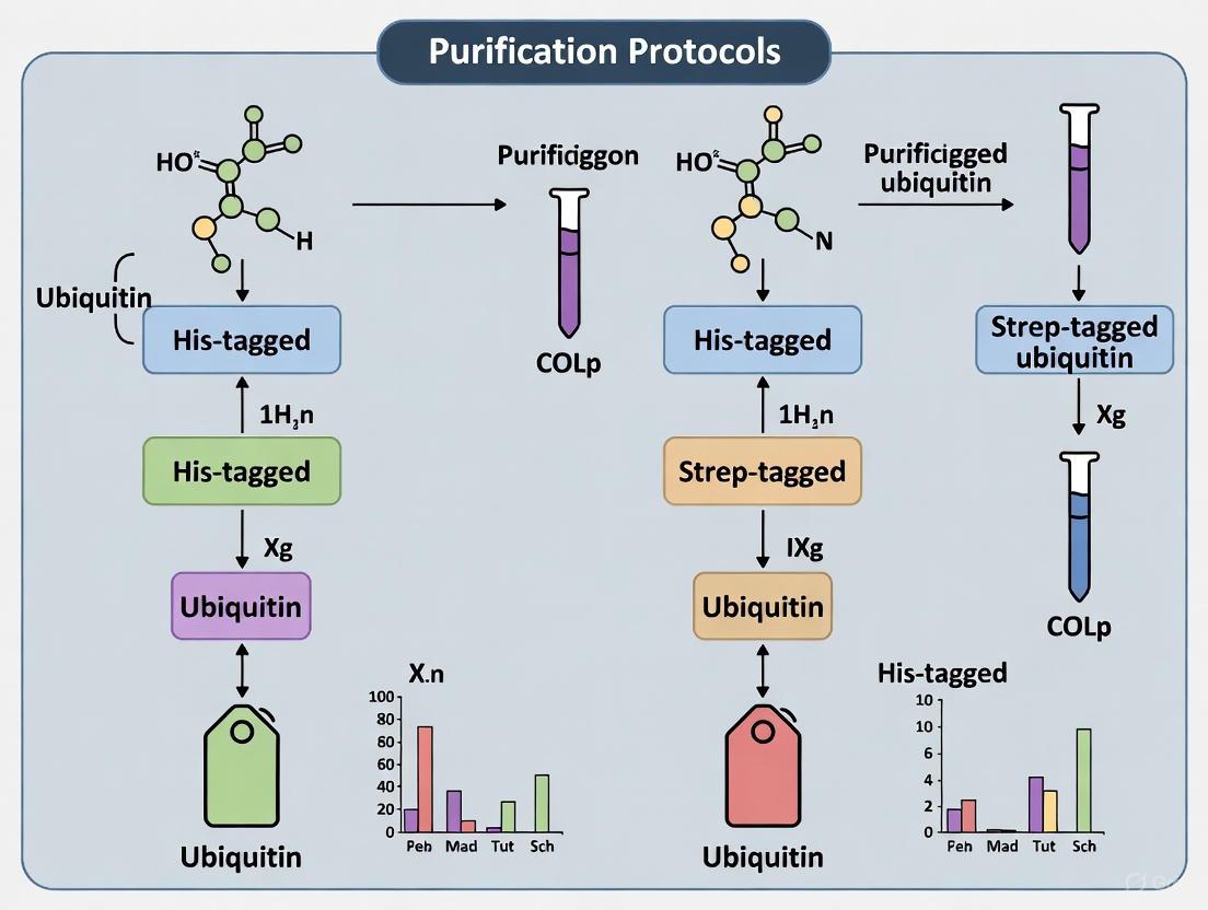
Diagram 1: Workflow for purifying mono-ubiquitinated proteins.
Key Reagents and Solutions
- pET16b-Avi-ubiquitinrbsBirA Plasmid: Plasmid for co-expression of Avi-tagged ubiquitin and BirA ligase [13].
- Biotin: Supplemented in culture media for in vivo biotinylation of the AviTag.
- Ni-NTA Resin: For initial immobilization of the His-tagged Avi-ubiquitin.
- Strep-Tactin or Streptavidin Resin: For high-affinity capture of biotinylated, ubiquitinated proteins.
- HRV 3C Protease: For specific, tag-less elution of the mono-ubiquitinated target.
Step-by-Step Procedure
- Expression and Biotinylation: Express the Avi-ubiquitin construct in E. coli BL21 cells using auto-induction media supplemented with biotin. Co-expression with BirA ensures specific biotinylation of the AviTag [13].
- Initial Purification: Lyse cells and clarify the lysate. Purify the biotinylated Avi-ubiquitin using Ni-NTA affinity chromatography under native conditions [13].
- In Vitro Ubiquitination: Use the purified Avi-ubiquitin as the substrate in a reconstituted E1-E2-E3 enzymatic reaction with your target protein and other required components [13].
- Affinity Capture: Incubate the ubiquitination reaction mixture with Strep-Tactin or streptavidin resin. The biotin moiety on the ubiquitin attached to your target protein will bind with high affinity.
- Washing: Wash the resin extensively with a compatible buffer (e.g., 150 mM NaCl, 100 mM Tris-HCl, 1 mM EDTA, pH 8.0) to remove non-specifically bound proteins [10].
- Elution: Incubate the resin with HRV 3C protease to cleave the AviTag, releasing the purified, natively mono-ubiquitinated protein without any affinity tag remnant [13].
Protocol: Tandem Affinity Purification for Membrane Proteins
For challenging targets like G protein-coupled receptors (GPCRs), a tandem His-/Strep-tag strategy can yield high-purity, functional protein [9].
Principle: A target protein (e.g., human cannabinoid receptor CB2) is engineered with an N-terminal Twin-Strep-tag and a C-terminal His10-tag. Sequential purification first on Ni-NTA and then on Strep-Tactin resin efficiently removes contaminants and degraded fusion products.
Key Reagents and Solutions
- Detergent Solutions: Critical for solubilizing and stabilizing membrane proteins. For CB2, a mix of DDM, CHAPS, and cholesteryl hemisuccinate (CHS) is used [9].
- Ni-NTA Agarose: For the first purification step.
- StrepTactin Resin: For the second, polishing purification step.
- TEV Protease: For removing the His-tag after the first purification step, if required.
- Desthiobiotin Elution Buffer: 5 µM desthiobiotin in appropriate detergent-containing buffer for gentle elution from Strep-Tactin resin [9].
Step-by-Step Procedure
- Solubilization: Extract the expressed membrane protein from membranes using a optimized detergent cocktail (e.g., 1% DDM, 0.5% CHAPS, 0.1% CHS) to stabilize the protein in micelles [9].
- First Purification (IMAC): Load the solubilized lysate onto a Ni-NTA column. Wash with buffer containing low-concentration imidazole (e.g., 20-40 mM) and elute with high-concentration imidazole (e.g., 250 mM) [9].
- Tag Cleavage (Optional): Treat the eluate with TEV protease to remove the C-terminal His-tag.
- Second Purification (Strep-Tactin): Dialyze or exchange the buffer to remove imidazole. Load the sample onto a Strep-Tactin column. Wash and then elute with buffer containing 2.5 mM desthiobiotin [9].
- Concentration and Buffer Exchange: Use centrifugal concentrators to concentrate the purified protein into the final storage or assay buffer.
Research Reagent Solutions
The following table lists essential materials for experiments utilizing the Strep-tag II and His-tag systems.
Table 3: Essential Research Reagents for Affinity Purification
| Reagent / Material | Function | Example Use Case |
|---|---|---|
| Strep-TactinXT Resin | High-affinity affinity resin for Strep-tag II and Twin-Strep-tag purification; offers nM-pM affinity [11]. | One-step purification of functional protein complexes. |
| Ni-NTA Agarose | Immobilized metal affinity chromatography resin for purifying His-tagged proteins [9]. | Initial capture and purification under native or denaturing conditions. |
| Desthiobiotin | Biotin analog used for competitive and gentle elution of Strep-tagged proteins from Strep-Tactin resin [10]. | Elution of Strep-tagged proteins under physiological conditions. |
| Imidazole | Competitive agent for elution of His-tagged proteins from Ni-NTA resin [9]. | Elution in His-tag IMAC protocols. |
| HRV 3C Protease | Highly specific protease for removing affinity tags; recognizes the Leu-Glu-Val-Leu-Phe-Gln↓Gly-Pro sequence [13]. | Cleavage of tags to yield tag-less, native protein after purification. |
| AviTag & BirA Ligase | System for site-specific biotinylation of a 15-amino acid peptide (AviTag) in vivo or in vitro [13]. | Creating high-affinity biotin handles on recombinant proteins. |
The choice between His-tag and Strep-tag II is dictated by the specific requirements of the downstream application. The His-tag system offers robustness, low cost, and compatibility with denaturing conditions, making it ideal for initial protein capture and when cost is a primary factor. However, for applications demanding high purity and retained biofunctionality in a single step—such as the isolation of enzymatically active ubiquitinated complexes for biochemical assays or structural studies—the Strep-tag II system is superior. Its gentle, competitive elution with desthiobiotin and high specificity minimize protein damage and co-purifying contaminants. For the most challenging targets, including membrane proteins and fragile complexes, a tandem affinity approach that leverages the strengths of both tags provides a powerful strategy to achieve the purity and homogeneity required for advanced research and drug development.
In the study of ubiquitination—a crucial post-translational modification regulating protein stability, activity, and localization—the selection of an appropriate affinity tag for purifying ubiquitinated proteins is a critical decision that directly determines experimental success. The complex nature of ubiquitin signaling, where substrates can be modified by single ubiquitin molecules or complex polyubiquitin chains of different linkages, presents unique challenges for purification [5]. Within this research landscape, His-tags and Strep-tags have emerged as predominant tools for isolating ubiquitinated proteins, yet each possesses distinct characteristics that align with specific research objectives. This application note examines how defined purification goals should guide the selection between His-tagged and Strep-tagged ubiquitin systems, providing structured comparisons and detailed protocols to inform researchers' experimental design.
The Ubiquitination System: Complexity and Analytical Challenges
Ubiquitination involves a sophisticated enzymatic cascade where E1 activating enzymes, E2 conjugating enzymes, and E3 ligases work in concert to attach the 76-amino acid ubiquitin protein to substrate proteins. This system can generate remarkable diversity through:
- Monoubiquitination: Single ubiquitin modification on a substrate
- Multi-monoubiquitination: Multiple single ubiquitin modifications on different sites
- Polyubiquitination: Ubiquitin chains of varying lengths connected through different lysine residues (K6, K11, K27, K29, K33, K48, K63) or methionine (M1)
- Heterotypic chains: Mixed linkage types creating complex branching patterns [5]
This complexity is further compounded by the low stoichiometry of ubiquitination under normal physiological conditions and the challenge of precisely localizing modification sites on substrates [5]. Consequently, affinity tag selection must accommodate these biological realities to ensure accurate characterization of the ubiquitin code.
Affinity Tag Selection Criteria for Ubiquitin Research
Selecting between His-tag and Strep-tag systems requires evaluating multiple performance parameters against research objectives. The table below summarizes key comparative characteristics:
Table 1: Comparison of Affinity Tags for Ubiquitination Studies
| Parameter | His-Tag | Strep-Tag II |
|---|---|---|
| Tag Size | 6-10 histidine residues | 8 amino acids (WSHPQFEK) |
| Binding Principle | Coordination with immobilized metal ions | Molecular recognition by engineered streptavidin (Strep-Tactin) |
| Affinity | μM-mM range | pM-nM range (varies with tag and resin version) |
| Resin Cost | Low | Moderate to high |
| Purification Purity | Moderate, with co-purification of endogenous proteins [14] [15] | High (>95%), minimal non-specific binding [16] |
| Elution Conditions | Imidazole or low pH | Desthiobiotin (gentle, competitive elution) |
| Buffer Flexibility | Limited by chelating agents and reducing agents | High tolerance to detergents, chelators, salts, and reducing agents [16] |
| Impact on Protein Function | Can influence biofunctionality in some cases [14] | Minimal influence due to balanced amino acid composition [16] |
| Suitability for Structural Studies | Moderate | Excellent (preserves protein bioactivity) [16] |
Beyond these general characteristics, tag selection must align with specific research goals in ubiquitination studies:
Target Identification & Interactome Studies: For initial discovery-phase research aiming to identify novel ubiquitination substrates or interacting proteins, preservation of native complexes is essential. The gentle elution conditions of the Strep-tag system (using desthiobiotin) better maintain protein-protein interactions and complex integrity [16].
Structural & Functional Characterization: When the research objective involves biochemical activity assays or structural analysis, protein purity and bioactivity become paramount. The Strep-tag system yields >95% pure proteins with maintained functionality, making it preferable for these applications [16].
High-Throughput Screening: For projects requiring rapid processing of multiple samples, the robustness and scalability of the purification system determines efficiency. While both systems can be scaled, the His-tag system offers cost advantages for large-scale applications, though with potential purity compromises [15].
Low-Abundance Protein Studies: When investigating low-stoichiometry ubiquitination events, the binding affinity and specificity of the purification tag directly impact yield. The Twin-Strep-tag combined with Strep-TactinXT provides picomolar affinity, enabling efficient pull-down of low-abundance targets [16].
Quantitative Performance Comparison Across Expression Systems
The performance of affinity tags varies significantly across different expression systems, an important consideration when planning ubiquitination studies. Research comparing tag efficiency reveals systematic differences:
Table 2: Tag Performance Across Expression Systems
| Expression System | His-Tag Performance | Strep-Tag II Performance |
|---|---|---|
| E. coli | Good yield, moderate purity [15] | Excellent purification, good yield [15] |
| Yeast | Relatively poor purification [15] | Good purification and yield [15] |
| Drosophila | Relatively poor purification [15] | Good purification and yield [15] |
| HeLa (Mammalian) | Relatively poor purification [15] | Good purification and yield [15] |
| Microalgae | Co-purification of endogenous proteins; can impact biofunctionality [14] | Has succeeded where His-tag failed; good specificity [14] |
This comparative data demonstrates that while His-tags provide satisfactory results in prokaryotic systems, their performance diminishes in more complex eukaryotic environments where metal-binding proteins are more prevalent. Conversely, the Strep-tag system maintains consistent performance across diverse expression platforms, making it particularly valuable for ubiquitination studies in mammalian cells or when comparing results across model systems.
Experimental Protocols for Ubiquitinated Protein Purification
His-Tagged Ubiquitin Purification Protocol
Principle: Recombinant His-tagged ubiquitin is expressed in the system of choice, and the tagged protein is purified via immobilized metal affinity chromatography (IMAC) using nickel-nitrilotriacetic acid (Ni-NTA) resin.
Materials:
- Lysis Buffer: 50 mM Tris-HCl (pH 8.0), 300 mM NaCl, 10 mM imidazole, 0.1% NP-40, protease inhibitors
- Wash Buffer: 50 mM Tris-HCl (pH 8.0), 300 mM NaCl, 20-40 mM imidazole
- Elution Buffer: 50 mM Tris-HCl (pH 8.0), 300 mM NaCl, 250-500 mM imidazole
- Ni-NTA Resin
- Desalting Column (for imidazole removal if needed)
Procedure:
- Cell Lysis: Resuspend cell pellet in ice-cold Lysis Buffer. Lyse cells by sonication or mechanical homogenization. Centrifuge at 15,000 × g for 30 minutes at 4°C to remove insoluble material.
- Column Preparation: Equilibrate Ni-NTA resin with 5 column volumes (CV) of Lysis Buffer.
- Binding: Incubate clarified lysate with equilibrated Ni-NTA resin for 1-2 hours at 4°C with gentle agitation.
- Washing: Wash resin with 10-20 CV of Wash Buffer to remove non-specifically bound proteins.
- Elution: Elute His-tagged ubiquitin with 5-10 CV of Elution Buffer, collecting multiple fractions.
- Buffer Exchange: If needed, remove imidazole using desalting columns equilibrated with storage buffer.
Critical Considerations:
- Imidazole concentration in wash buffers should be optimized to balance purity and yield
- Include protease inhibitors to prevent ubiquitin degradation
- Consider adding 5-10% glycerol to buffers for protein stability
- For ubiquitinated substrate identification, use denaturing conditions (6-8 M urea or guanidine hydrochloride) to disrupt non-covalent interactions [5]
Strep-Tagged Ubiquitin Purification Protocol
Principle: Strep-tagged ubiquitin binds with high specificity to Strep-Tactin resin, with gentle competitive elution using desthiobiotin under physiological conditions that preserve protein complex integrity.
Materials:
- Buffer W: 100 mM Tris-HCl (pH 8.0), 150 mM NaCl, 1 mM EDTA
- Buffer E: Buffer W containing 2.5 mM desthiobiotin
- Strep-Tactin resin (Superflow or XT for higher affinity)
- Regeneration Buffer: 1 mM HABA (for standard resin) or 3 M MgCl₂ (for XT resin)
Procedure:
- Cell Lysis: Prepare cell lysate in Buffer W using mild detergents if needed for membrane proteins. Centrifuge at 15,000 × g for 30 minutes at 4°C.
- Resin Preparation: Equilibrate Strep-Tactin resin with 5 CV of Buffer W.
- Binding: Incubate clarified lysate with equilibrated resin for 1 hour at 4°C with gentle agitation. For low-abundance targets, extend binding time to 2 hours.
- Washing: Wash with 10-15 CV of Buffer W. For challenging purifications, increase salt concentration to 500 mM NaCl to reduce non-specific binding.
- Elution: Elute with 5-10 CV of Buffer E, collecting 0.5-1 CV fractions.
- Regeneration: Regenerate resin with Regeneration Buffer, then re-equilibrate with Buffer W for reuse.
Critical Considerations:
- The Twin-Strep-tag version provides higher affinity for low-abundance targets
- Strep-TactinXT resin offers enhanced binding capacity (up to 7 mg/ml) for high-yield purifications [16]
- Buffer conditions can be widely adapted (HEPES, PBS) without affecting binding efficiency
- For ubiquitin interactome studies, use native conditions throughout to preserve non-covalent interactions
Research Reagent Solutions for Ubiquitination Studies
Table 3: Essential Reagents for Ubiquitin Affinity Purification
| Reagent / Material | Function / Application | Key Features |
|---|---|---|
| Strep-TactinXT 4Flow | Affinity resin for Strep-tag purification | High binding capacity (14 mg/ml), suitable for FPLC/HPLC, pH range 4-10 [16] |
| Ni-NTA Superflow | IMAC resin for His-tag purification | High flow rate, binding capacity ~5-10 mg/ml, compatible with denaturing conditions |
| Desthiobiotin | Competitive elution agent for Strep-tag system | Gentle elution under physiological conditions, reversible binding |
| Imidazole | Competitive elution agent for His-tag system | Effective displacement, but may require removal for downstream applications |
| Protease Inhibitor Cocktails | Prevent protein degradation during purification | Essential for preserving ubiquitin conjugates, especially DUB-sensitive linkages |
| HABA Solution | Regeneration indicator for Strep-Tactin resin | Colorimetric verification of resin regeneration status [16] |
Workflow Visualization for Tag Selection and Application
The following diagram illustrates the decision pathway for selecting between His-tag and Strep-tag systems based on research objectives, and the general workflow for ubiquitinated protein purification:
The selection between His-tag and Strep-tag systems for ubiquitination studies represents a strategic decision that should be guided by specific research objectives rather than convenience or cost considerations alone. For discovery-phase research aiming to identify novel ubiquitination substrates or interacting partners under native conditions, the Strep-tag system provides superior performance with its high specificity, gentle elution conditions, and consistent performance across diverse expression systems. Conversely, for large-scale screening projects where budget constraints outweigh purity requirements, the His-tag system offers a cost-effective alternative despite limitations in complex biological samples. By aligning affinity tag selection with well-defined purification goals, researchers can optimize experimental outcomes in characterizing the complex ubiquitin code and its functional consequences in health and disease.
Step-by-Step Purification Protocols: From Cell Lysis to Elution for Both Tags
The ubiquitin fusion technique is a powerful tool in molecular biology for enhancing the yield and simplifying the purification of recombinant proteins. This approach involves fusing the gene of interest to the C-terminus of ubiquitin (Ub), which can be tagged with affinity handles like polyhistidine (His) or Strep-tag for streamlined purification. A primary benefit of this system is the ability to subsequently cleave off the ubiquitin moiety using highly specific deubiquitylating enzymes (DUBs), yielding the target protein with its authentic N-terminus, a critical feature for functional and structural studies [17].
These systems are particularly valuable for expressing unstable or poorly expressed proteins, and their application has been demonstrated across a wide range of proteins and peptides, showing suitability for high-throughput applications [17]. The choice between a His-tag and a Strep-tag is pivotal, as it influences the purification strategy, potential for tag removal, and ultimately, the quality and authenticity of the final protein product. This application note provides a detailed guide for researchers designing constructs for His-ubiquitin and Strep-ubiquitin expression, framed within the broader context of optimizing ubiquitin-based purification protocols.
Vector Design and Key Considerations
Core Elements of Ubiquitin Expression Vectors
The design of ubiquitin fusion vectors requires careful consideration of several genetic elements to ensure high-yield expression and successful recovery of the target protein. A representative backbone is the pHUE vector, an E. coli expression vector constructed for the expression of His-tagged ubiquitin fusion proteins. It contains an inducible T7 RNA polymerase promoter, a histidine tag at the 5' end of the Ub open reading frame, and an extended polylinker to facilitate the cloning of the gene of interest [17].
A critical feature in vector design is the inclusion of a specific protease recognition site. While other fusion systems use proteases like TEV or Factor Xa, which can leave behind extraneous residues, the ubiquitin system leverages endogenous DUBs. These enzymes cleave precisely after the final glycine residue (Gly76) at the C-terminus of ubiquitin, irrespective of the amino acid that follows, with the sole exception of proline, which is cleaved inefficiently [17]. For researchers requiring a precise N-terminus, the SacII site can be engineered into the 3' end of the Ub sequence. A ligated DNA fragment must encode the Leu-Arg-Gly-Gly sequence (with Gly75-Gly76 being essential for cleavage), which can be achieved via PCR amplification with a primer containing the appropriate 5' extension [17].
Comparative Analysis of Affinity Tags
The choice of affinity tag is a fundamental decision that dictates the purification workflow. The table below summarizes the key characteristics of His-tag and Strep-tag within the context of ubiquitin fusion systems.
Table 1: Comparison of His-Tag and Strep-Tag for Ubiquitin Fusion Systems
| Feature | His-Tag (e.g., in pHUE vector) | Strep-Tag |
|---|---|---|
| Affinity Resin | Immobilized metal affinity chromatography (IMAC); e.g., Ni-NTA agarose [17] | Strep-Tactin resin [18] |
| Purification Conditions | Native or denaturing conditions [17] | Native conditions |
| Typical Elution | Imidazole or low pH | Desthiobiotin |
| Key Advantage | Robust, high-capacity, cost-effective; allows purification of insoluble proteins under denaturing conditions [17] | High specificity, gentle elution, lower background co-purification |
| Potential Drawback | Co-purification of histidine-rich proteins or metal-binding contaminants [18] | Co-purification of endogenously biotinylated proteins [18]; generally more expensive resin |
| Tag Size | Small (~0.8 kDa for a 6xHis tag) | Small (a few amino acids) |
Both tags are small, which minimizes structural interference with the ubiquitin moiety or the fused protein of interest. This is a significant advantage over larger fusion partners like GST or MBP [19].
Quantitative Performance Data
The utility of the ubiquitin fusion system, particularly the His-tagged version, is demonstrated by its ability to produce high yields of a diverse range of proteins. The following table compiles experimental data from the pHUE system, showcasing yields for proteins of varying size and complexity.
Table 2: Protein Yields from a His-Tagged Ubiquitin Fusion System (pHUE vector) [17]
| Purified Ub-Fusion Protein | Size (kD) | Structure | Yield (mg/L of E. coli culture) |
|---|---|---|---|
| His₆Ub–SUMO | 24.5 | Monomer | 22.83 |
| His₆Ub–M-GSTP1 | 34.0 | Dimer | 25.56 |
| His₆Ub–P-GSTP1 | 33.9 | Dimer | 26.85 |
| His₆Ub–GSH-S | 63.1 | Dimer | 3.58 |
| His₆Ub–β-gal | 129.2 | Tetramer | 3.30 |
The data indicates that the system is highly efficient for many proteins, with yields often exceeding 20 mg/L. However, yields can be significantly lower for larger and more complex proteins like glutathione synthetase (GSH-S) and β-galactosidase (β-gal), primarily due to reduced solubility. The ubiquitin fusion generally enhances solubility, but it does not guarantee a completely soluble product. The presence of the poly-histidine tag allows for purification under denaturing conditions if necessary, followed by refolding steps to recover active protein [17].
Detailed Experimental Protocols
Protocol A: Purification of His-Tagged Ubiquitin Fusion Proteins
This protocol details the expression and purification of proteins using the pHUE vector system, which employs a His-tagged ubiquitin fusion and a companion His-tagged deubiquitylating enzyme for cleavage [17].
Materials
- Expression Vector: pHUE or similar His-Ub vector [17]
- E. coli Strain: BL21(DE3) or similar for T7 promoter-driven expression [17] [19]
- Affinity Resin: Ni-NTA (Nickel-Nitrilotriacetic acid) agarose beads [17]
- Lysis Buffer: 50 mM Na₂HPO₄, pH 8.0, 500 mM NaCl, 0.01% SDS, 5% glycerol, supplemented with protease inhibitors [17] [6]
- Wash Buffer: Lysis buffer with 20-50 mM imidazole
- Elution Buffer: Lysis buffer with 250-500 mM imidazole
- Deubiquitylating Enzyme (DUB): Purified His-tagged catalytic domain of mouse Usp2 or equivalent [17]
Workflow
Step-by-Step Procedure
- Cloning and Expression: Subclone the gene of interest into the pHUE vector's polylinker downstream of the His-Ub sequence. Transform the construct into an appropriate E. coli expression strain like BL21(DE3). Induce protein expression with IPTG when the culture reaches mid-log phase [17] [19].
- Cell Lysis and Clarification: Harvest cells by centrifugation. Resuspend the cell pellet in lysis buffer and lyse using sonication or a homogenizer. Centrifuge the lysate at a high speed (e.g., 70,000 × g for 30 minutes) to remove insoluble debris [17] [6].
- IMAC Purification: Incubate the clarified lysate with Ni-NTA agarose beads. Wash the beads extensively with wash buffer to remove non-specifically bound contaminants. Elute the purified His-Ub fusion protein with elution buffer [17].
- Ubiquitin Cleavage and Final Purification: Incubate the eluted fusion protein with the purified His-tagged DUB. To obtain the authentic target protein, pass the cleavage reaction mixture over a fresh Ni-NTA column. The His-tagged DUB, the cleaved His-Ub moiety, and any uncleaved fusion protein will bind to the resin, while the untagged target protein will be found in the flow-through, ready for further analysis or use [17].
Protocol B: Purification of Strep-Tagged Ubiquitin Fusion Proteins
This protocol outlines an alternative method using Strep-tagged ubiquitin for high-specificity purification.
Materials
- Expression Vector: Vector encoding N- or C-terminal Strep-tagged ubiquitin (e.g., modified from commercial Strep-tag vectors)
- E. coli Strain: BL21(DE3) or similar
- Affinity Resin: Strep-Tactin Sepharose or similar
- Lysis/Wash Buffer: 100 mM Tris-HCl, pH 8.0, 150 mM NaCl, 1 mM EDTA
- Elution Buffer: Lysis buffer containing 2.5-10 mM desthiobiotin
- DUB Enzyme: Appropriate deubiquitylating enzyme (not necessarily tagged)
Workflow
Step-by-Step Procedure
- Cloning and Expression: Clone the gene of interest into a Strep-Ubiquitin vector. Express the fusion protein in E. coli as described in Protocol A [18].
- Cell Lysis and Clarification: Lyse cells in a compatible buffer (e.g., 100 mM Tris-HCl, pH 8.0, 150 mM NaCl, 1 mM EDTA) and clarify the lysate by centrifugation [18].
- Strep-Tactin Affinity Purification: Apply the clarified lysate to a Strep-Tactin column. Wash the column thoroughly with the lysis buffer to remove contaminants. Elute the bound Strep-Ub fusion protein gently using a buffer containing desthiobiotin [18].
- Ubiquitin Cleavage and Final Purification: Incubate the eluted fusion protein with a DUB enzyme. To separate the target protein from the cleaved Strep-Ub tag, pass the cleavage mixture over a fresh Strep-Tactin column. The pure, authentic target protein will be collected in the flow-through [18].
The Scientist's Toolkit: Research Reagent Solutions
Table 3: Essential Reagents for Ubiquitin Fusion Experiments
| Reagent / Material | Function / Application | Examples & Notes |
|---|---|---|
| pHUE Vector | Expression vector for His-tagged ubiquitin fusions in E. coli [17] | Provides T7 promoter, His-tag, and Ub sequence; available through academic repositories. |
| Strep-Tag II Ubiquitin Vector | Expression vector for Strep-tagged ubiquitin fusions. | Can be constructed by subcloning Ub into commercial Strep-tag vectors. |
| Ni-NTA Agarose | Immobilized metal affinity chromatography resin for purifying His-tagged proteins [17]. | High binding capacity; suitable for native or denaturing purification. |
| Strep-Tactin Sepharose | Affinity resin for highly specific purification of Strep-tagged proteins [18]. | Provides high purity; elution with desthiobiotin is gentle and non-denaturing. |
| Deubiquitylating Enzyme (DUB) | Protease for cleaving ubiquitin from the fusion protein post-purification [17]. | His-tagged mouse Usp2 catalytic domain allows easy removal post-cleavage [17]. Commercial DUBs available. |
| Ubiquitin Mutants (e.g., K48R) | Used in specialized applications to study ubiquitination or prevent chain formation [20]. | Ubiquitin K48R mutant prevents formation of K48-linked chains, often used in ubiquitin-reference technique (URT) [20]. |
Application Notes in Research and Drug Development
The His-Ub and Strep-Ub expression systems are not merely purification tools but are integral to various advanced research and screening applications. A key application is in high-throughput screening (HTS) for ubiquitin system modulators. For instance, the Ubiquitin-Reference Technique (URT) uses a linear fusion protein where ubiquitin is located between a protein of interest and a reference protein moiety. This construct is cleaved by endogenous DUBs to produce equimolar amounts of the protein of interest and the reference. By integrating this with a Dual-Luciferase system, researchers can screen for small-molecule inhibitors of E3 ubiquitin ligases, as demonstrated for SMURF1, with the internal reference compensating for sample-to-sample variation [20].
Furthermore, the Ub fusion technique has proven effective for expressing challenging proteins. A prominent example is the single-step purification of Cas9 protein. Cas9 was expressed as an N-terminal fusion to poly-histidine-tagged ubiquitin. This strategy enhanced soluble production in E. coli and enabled purification of functional, high-purity Ub-Cas9 with a yield of over 8 mg/L using a single metal affinity chromatography step, bypassing the need for tag removal in many genome-editing applications [19]. This underscores the system's utility in producing large, complex proteins for therapeutic and research applications.
Within the framework of thesis research focused on optimizing purification protocols for His-tagged ubiquitin and Strep-tagged ubiquitin, a critical and often determinative step occurs prior to purification: the pre-analytical treatment of cell cultures. The integrity of the ubiquitin conjugates being studied is perpetually threatened by the endogenous activity of deubiquitinases (DUBs) and the proteasomal degradation machinery. This article details the essential application of the cell-permeable proteasome inhibitor, MG-132, as a cornerstone method for preserving the cellular ubiquitinome during cell culture and transfection experiments, thereby ensuring the successful purification of high-quality ubiquitinated proteins for downstream analysis.
The Scientific Rationale: Why Proteasome Inhibition is Indispensable
Ubiquitination is a reversible post-translational modification that regulates diverse cellular functions, from protein degradation to signal transduction. The process is orchestrated by a cascade of E1, E2, and E3 enzymes and is reversed by deubiquitinating enzymes (DUBs) [5]. A primary fate of K48-linked polyubiquitinated proteins is degradation by the 26S proteasome [5]. When the objective is to purify ubiquitinated proteins, this constitutive degradation presents a significant challenge.
MG-132 (carbobenzoxy-Leu-Leu-leucinal) is a potent, reversible peptide aldehyde that inhibits the proteolytic activity of the 26S proteasome complex [21]. Its application during cell culture and prior to cell lysis serves a dual purpose:
- Prevents Substrate Degradation: By blocking the proteasome, MG-132 halts the degradation of polyubiquitinated proteins, leading to their accumulation within cells and thereby increasing the yield for purification [22] [21].
- Stabilizes Regulatory Proteins: Many proteins involved in cell signaling, such as transcription factors and tumor suppressors, are short-lived and regulated by proteasomal degradation. MG-132 stabilizes these proteins, which can be crucial for studying their ubiquitination status or for maintaining physiological conditions during transfection experiments [22] [23].
The use of MG-132 is particularly critical when working with tagged ubiquitin constructs (His-Ub or Strep-Ub). Transfection with these constructs allows for the pulldown of ubiquitinated proteins, but without proteasome inhibition, a significant portion of the conjugates of interest may be lost before purification can occur.
Essential Research Reagent Solutions
The table below catalogues the key reagents and materials required for experiments involving MG-132 and ubiquitin purification.
Table 1: Key Research Reagents and Their Applications
| Reagent/Material | Function/Description | Application in Ubiquitin Research |
|---|---|---|
| MG-132 (Proteasome Inhibitor) | Reversible inhibitor of the 26S proteasome's chymotrypsin-like activity. | Stabilizes polyubiquitinated proteins in cell culture prior to lysis, increasing yield for purification [22] [21]. |
| His-Tagged Ubiquitin | Ubiquitin with a polyhistidine affinity tag (e.g., 6x-His). | Enables enrichment of ubiquitinated proteins from cell lysates using Ni-NTA affinity chromatography [5]. |
| Strep-Tagged Ubiquitin | Ubiquitin with a Strep-tag II affinity tag. | Allows for purification of ubiquitinated proteins via high-affinity binding to Strep-Tactin resin [5]. |
| Ubiquitin-Binding Domain (UBD) Resins | Affinity resins coupled to high-affinity UBDs (e.g., OtUBD). | Enables enrichment of endogenous, non-tagged ubiquitinated proteins from crude lysates under native or denaturing conditions [24]. |
| Avi-Tagged/Biotinylated Ubiquitin | Ubiquitin fused to an AviTag for site-specific biotinylation. | Facilitates purification of natively mono-ubiquitinated substrates using streptavidin resin and elution via protease cleavage [13]. |
| Linkage-Specific Ub Antibodies | Antibodies recognizing specific ubiquitin chain linkages (e.g., K48, K63). | Used in immunoblotting to characterize the chain topology of enriched ubiquitin conjugates [5]. |
Experimental Protocols and Workflows
Protocol 1: MG-132 Treatment of Cell Cultures
This protocol describes the application of MG-132 to mammalian cell cultures to inhibit proteasomal degradation before harvesting for ubiquitin purification.
Materials:
- Cell line of interest (e.g., HEK293T, HeLa, PANC-1, SW1990)
- Complete cell culture medium
- MG-132 stock solution (e.g., 10 mM in DMSO)
- Dimethyl sulfoxide (DMSO), sterile
Procedure:
- Cell Culture & Transfection: Culture cells to ~70-90% confluency. Perform transfection with your His-tagged or Strep-tagged ubiquitin plasmid using your preferred method.
- MG-132 Preparation: Dilute the 10 mM MG-132 stock in pre-warmed culture medium to a final working concentration of 10-20 µM. Ensure uniform mixing.
- Treatment:
- Aspirate the existing culture medium from the cells.
- Gently add the medium containing MG-132 to the cells.
- Incubate the cells for a duration of 4 to 6 hours in a standard humidified incubator (37°C, 5% CO₂) [22].
- Control Setup: Prepare a vehicle control by adding an equivalent volume of DMSO to the culture medium without MG-132.
- Cell Harvesting: After incubation, immediately place the culture dish on ice. Aspirate the medium and wash the cells twice with ice-cold phosphate-buffered saline (PBS). Proceed to cell lysis using your chosen buffer.
Notes:
- The optimal concentration and treatment time may require empirical determination for specific cell lines and experimental goals.
- Prolonged exposure (>12-24 hours) to MG-132 can induce apoptosis and may not be suitable for all applications [21].
Protocol 2: Tandem Affinity Purification of Tagged Ubiquitin Conjugates
This workflow follows MG-132 treatment and describes two parallel paths for purifying ubiquitinated proteins using the two most common tags.
Materials:
- Lysis Buffer (e.g., RIPA buffer supplemented with 1% protease inhibitor cocktail)
- Affinity Resins: Ni-NTA Agarose (for His-Ub) or Strep-Tactin Resin (for Strep-Ub)
- Wash Buffers:
- For Ni-NTA: Buffer with 20-50 mM imidazole, PBS, pH 8.0 [13]
- For Strep-Tactin: PBS or manufacturer's recommended buffer
- Elution Buffers:
- For Ni-NTA: Buffer with 250-500 mM imidazole
- For Strep-Tactin: Buffer with 10-50 mM biotin
Procedure:
- Cell Lysis: Lyse the harvested, MG-132-treated cells in an appropriate lysis buffer. For subsequent proteomic analysis, consider using denaturing lysis conditions (e.g., with SDS) to inactivate DUBs and preserve ubiquitination.
- Clarification: Centrifuge the lysate at >16,000 × g for 15 minutes at 4°C. Transfer the clear supernatant to a new tube.
- Affinity Purification:
- Washing: Pellet the resin and wash thoroughly with 10-20 column volumes of the appropriate wash buffer to remove non-specifically bound proteins.
- Elution: Elute the bound ubiquitinated proteins using the specific elution buffer. Collect multiple fractions for analysis.
- Analysis: Analyze the eluates by SDS-PAGE and immunoblotting using anti-ubiquitin antibodies (e.g., P4D1, FK2) or antibodies against your protein of interest.
The following diagram visualizes the core experimental workflow, integrating the critical MG-132 treatment step with the subsequent purification pathways.
Diagram 1: Workflow for tagged ubiquitin conjugate purification.
Data Presentation and Analysis
Quantitative data from key experiments utilizing MG-132 are summarized below for easy comparison of its effects across different biological contexts.
Table 2: Quantitative Effects of MG-132 Treatment in Various Experimental Models
| Cell Type / System | MG-132 Concentration & Duration | Observed Effect (Quantitative) | Experimental Readout |
|---|---|---|---|
| PDAC Cells (PANC-1, SW1990) [22] | 10 µM, 4-6 hrs | Upregulation of E-cadherin; Inhibition of invasion/migration by ~40-60% | Western Blot, Transwell Assay |
| Human Pulmonary Fibroblast (HPF) [21] | 10-20 µM, 24 hrs | Induced growth inhibition and cell death (~30-50%); Increased ROS levels | MTT Assay, Flow Cytometry |
| Vero & Human Cell Lines (HepG2, etc.) [23] | 0.75 µM, 24-48 hrs | Decreased HSV-1 plaque formation by ~35%; Reduced extracellular virus yield to 0.01-10% | Plaque Assay, qPCR |
| HEK293T & U2OS Cells [5] | Not specified, standard treatment | Enabled identification of 753 ubiquitination sites on 471 proteins | Mass Spectrometry Proteomics |
The Scientist's Toolkit: Alternative and Advanced Enrichment Strategies
While tagged ubiquitin expression is powerful, several complementary biochemical tools are available for studying ubiquitination, each with unique strengths.
Table 3: Comparison of Ubiquitinated Protein Enrichment Methodologies
| Enrichment Method | Principle | Advantages | Limitations |
|---|---|---|---|
| Tagged Ubiquitin (His/Strep) [5] | Expression of affinity-tagged Ub in cells; purification under denaturing conditions. | High-yield; cost-effective; broad applicability. | May not mimic endogenous Ub perfectly; requires genetic manipulation. |
| Ubiquitin-Binding Domains (UBDs) [24] | Use of high-affinity domains (e.g., OtUBD) to purify endogenous ubiquitinated proteins. | Works with native tissue samples; no genetic tag needed. | Can co-purify interacting proteins; requires optimization. |
| Anti-Ubiquitin Antibodies [5] | Immunoaffinity purification using pan- or linkage-specific Ub antibodies. | Can be used on any sample, including clinical specimens; linkage-specific options. | High cost; potential for non-specific binding. |
| Chemical/Semi-Synthesis [25] [13] | Generation of homogeneously ubiquitinated proteins through chemical ligation. | Absolute homogeneity; atomic-level control over modification. | Technically challenging; low throughput; yield can be limiting. |
The strategic decision-making process for selecting the optimal ubiquitin enrichment method based on experimental goals is illustrated below.
Diagram 2: Decision pathway for ubiquitin enrichment method selection.
The integration of MG-132 treatment into cell culture and transfection workflows is a critical pre-purification step that is non-negotiable for the successful study of labile ubiquitinated proteins. When combined with robust purification protocols for His-tagged or Strep-tagged ubiquitin, it enables researchers to capture a more authentic snapshot of the cellular ubiquitinome. As the field advances, coupling these foundational methods with emerging tools—such as high-affinity UBDs, linkage-specific probes, and chemical biology approaches—will provide unprecedented insights into the complex and dynamic role of ubiquitination in health and disease, a central pursuit of thesis research in this domain.
Within the broader research on ubiquitin purification protocols, the isolation of His-tagged ubiquitin and related constructs under denaturing conditions represents a critical methodology for studying the ubiquitin-proteasome system and protein biochemistry. The small size and high stability of ubiquitin make it an excellent candidate for affinity purification, yet the study of its modified substrates or its own polymeric chains often requires harsh conditions to disrupt strong non-covalent interactions and preserve labile modifications. Immobilized Metal-Affinity Chromatography (IMAC) utilizing Ni-NTA resin under denaturing conditions enables researchers to efficiently purify recombinant His-tagged proteins that are insoluble, aggregated in inclusion bodies, or possess tertiary structures that occlude the polyhistidine affinity tag [26]. This technical note provides a detailed protocol for the purification of His-tagged proteins under denaturing conditions using guanidinium hydrochloride or urea buffers, with specific considerations for applications in ubiquitin research.
Background and Principles
The Polyhistidine Affinity Tag
The polyhistidine tag is one of the most widely utilized affinity tags in biochemical research due to its small size and minimal impact on protein structure and function. Affinity tags consisting of six histidine residues are most commonly employed in IMAC, as they generally provide sufficient length to yield high-affinity interactions with immobilized metal matrices while minimizing potential perturbation of protein function [26]. These tags can be placed on either the N or C terminus of recombinant proteins, with optimal placement being protein-specific. For ubiquitin and ubiquitin-like modifiers, the C-terminal placement is typically preferred to avoid interference with the conjugation machinery and substrate recognition.
Denaturing Conditions in Protein Purification
Denaturing conditions are particularly valuable in ubiquitin research for several applications: (1) purification of insoluble ubiquitinated proteins or aggregates; (2) disruption of ubiquitin-binding domains that non-covalently associate with ubiquitin or ubiquitin chains; (3) inactivation of deubiquitinases (DUBs) and proteases that might otherwise process ubiquitin tags or ubiquitinated substrates during purification. The use of 6 M guanidinium hydrochloride or 8 M urea effectively solubilizes inclusion bodies, disrupts non-covalent protein-protein interactions, and depresses the activity of phosphatases and proteolytic enzymes [26]. This is especially relevant when working with ubiquitin-binding domains such as the high-affinity OtUBD from Orientia tsutsugamushi, which exhibits dissociation constants in the low nanomolar range and requires strong denaturants for complete dissociation [27].
Materials and Reagents
Essential Research Reagent Solutions
Table 1: Key reagents and materials for His-tag purification under denaturing conditions
| Reagent/Material | Function/Application | Specifications/Alternatives |
|---|---|---|
| Ni-NTA Agarose | IMAC resin for His-tag binding | Binding capacity: 5-10 mg protein/mL; Kd ~10⁻¹³ M for His₆-tag [26] |
| Guanidinium HCl | Protein denaturant | 6 M concentration for complete denaturation [26] |
| Urea | Protein denaturant | 8 M concentration; preferable for SDS-PAGE compatibility [26] |
| Imidazole | Competitive elution agent | 20-50 mM in wash buffers; 150-250 mM in elution buffers [26] |
| Protease Inhibitor Cocktail | Prevent protein degradation | EDTA-free formulations required for IMAC [26] |
| 2-Mercaptoethanol or DTT | Reducing agent | Prevents disulfide bond formation; 10 mM concentration [26] |
| Triton X-100 or Tween 20 | Non-ionic detergent | Reduces hydrophobic interactions; up to 1% concentration [26] |
Buffer Composition
Table 2: Denaturing buffer compositions for His-tag purification
| Buffer Type | Components | Concentration | Purpose |
|---|---|---|---|
| Denaturing Lysis Buffer | Guanidinium HCl or Urea, Sodium Phosphate, NaCl, Imidazole, pH | 6 M GuHCl or 8 M Urea, 50 mM NaPO₄, 300 mM NaCl, 10-20 mM imidazole, pH 8.0 [26] | Cell lysis and protein solubilization |
| Denaturing Wash Buffer | Guanidinium HCl or Urea, Sodium Phosphate, NaCl, Imidazole, pH | 6 M GuHCl or 8 M Urea, 50 mM NaPO₄, 300 mM NaCl, 20-50 mM imidazole, pH 8.0 [26] | Remove weakly bound contaminants |
| Denaturing Elution Buffer | Guanidinium HCl or Urea, Sodium Phosphate, NaCl, Imidazole, pH | 6 M GuHCl or 8 M Urea, 50 mM NaPO₄, 300 mM NaCl, 150-250 mM imidazole, pH 8.0 [26] | Competitive elution of His-tagged proteins |
Experimental Protocol
Detailed Step-by-Step Procedure
Step 1: Cell Lysis and Lysate Preparation
- Resuspend cell pellets in Denaturing Lysis Buffer containing either 6 M guanidinium hydrochloride or 8 M urea.
- For bacterial cultures expressing His-tagged ubiquitin constructs, include lysozyme (1 mg/mL) and incubate for 30 minutes with gentle mixing.
- Add DNase I (10 µg/mL) to reduce viscosity and protease inhibitor cocktail (EDTA-free) to prevent degradation [27].
- For mammalian cells expressing Strep-tagged ubiquitin constructs, mechanical disruption may be required prior to denaturation [28].
Step 2: Lysate Clarification
- Centrifuge the lysate at 15,000 × g for 30 minutes at 4°C.
- Collect the supernatant containing the solubilized His-tagged protein.
- Determine protein concentration using Bradford or BCA assay adapted for denaturing conditions [27].
Step 3: Binding to Ni-NTA Resin
- Equilibrate Ni-NTA resin with 5 column volumes of Denaturing Lysis Buffer.
- Incubate the clarified lysate with Ni-NTA resin using either:
- Use approximately 1 mL resin per 5-10 mg of total protein for optimal binding.
Step 4: Washing Non-Specifically Bound Proteins
- Wash resin with 10-20 column volumes of Denaturing Wash Buffer.
- Include 20-50 mM imidazole in wash buffer to remove weakly bound contaminants [26].
- For additional stringency, include low concentrations of non-ionic detergent (0.1-1% Triton X-100 or Tween 20) to reduce hydrophobic interactions [26].
Step 5: Elution of His-Tagged Proteins
- Elute bound proteins with Denaturing Elution Buffer containing 150-250 mM imidazole.
- Use 5-10 column volumes total, collecting fractions of 1 column volume each.
- Alternative elution can be performed using pH reduction (to 4.0-5.0), though this may be less effective under denaturing conditions [26].
Step 6: Analysis and Refolding
- Analyze fractions by SDS-PAGE and immunoblotting with anti-ubiquitin or anti-His tag antibodies [27].
- For functional studies, refold purified proteins by gradual removal of denaturants through dialysis or dilution.
- In some cases, proteins can be refolded while still bound to the resin before elution [26].
Troubleshooting and Optimization
Common Issues and Solutions
Optimization Strategies
- Tag Accessibility: If binding efficiency is low due to tag occlusion, try moving the affinity tag to the opposite terminus of the protein [26].
- Reducing Non-Specific Binding: Include 10 mM 2-mercaptoethanol in all buffers to prevent disulfide bond formation with contaminating proteins [26].
- Detergent Compatibility: Ni-NTA resin tolerates most non-ionic and zwitterionic detergents, which can be included at 0.1-1% to reduce hydrophobic interactions [26].
- Resin Selection: For higher purity with reduced non-specific binding, consider Co²⁺-CMA resin (sold as Talon resin) as an alternative to Ni²⁺-NTA [26].
Applications in Ubiquitin Research
The purification of His-tagged ubiquitin under denaturing conditions enables several advanced applications in ubiquitin signaling research. When combined with modern proteomic approaches, this methodology facilitates comprehensive analysis of the ubiquitinome - the complete set of ubiquitinated proteins in a biological system [27]. Specifically, denaturing conditions are essential for distinguishing directly ubiquitinated proteins from those that merely associate with ubiquitin or ubiquitinated proteins, a critical distinction when studying ubiquitin-binding domains and their interactors [27] [5].
This protocol is compatible with downstream applications including immunoblotting, differential proteomics, and ubiquitin chain restriction (UbiCREST) analysis [27]. When working with different ubiquitin chain types or studying ubiquitin in pathological contexts, the denaturing conditions help preserve labile modifications and prevent artifactual deubiquitination during purification. The methodology described here for His-tagged ubiquitin purification complements other affinity approaches such as Strep-tag systems [28] and Ub-binding domain based methods [27] [5], providing researchers with a versatile toolkit for interrogating the complex landscape of ubiquitin signaling.
Within the broader research on His-tagged and Strep-tagged ubiquitin purification, selecting an appropriate affinity tag is critical for obtaining functional, high-quality protein for downstream applications. While the His-tag system is popular for its high yield and low costs, it is often plagued by issues such as low target purity due to unspecific binding of host cell proteins and requires careful optimization of purification conditions [8]. These challenges can be particularly problematic when purifying proteins for sensitive biochemical assays or structural studies.
The Strep-tag system presents a superior alternative for purifications requiring high specificity and maintenance of protein bioactivity. The core of this technology is the engineered streptavidin, Strep-Tactin, which allows affinity purification of Strep-tagII and Twin-Strep-tag fusion proteins under physiological conditions [29]. The high specificity of the interaction between the Strep-tag and Strep-Tactin can yield over 99% purity after a single chromatographical step, while the mild purification parameters preserve the bioactivity of the target protein [8] [29]. This application note details a standardized protocol for the purification of Strep-tagged proteins using Strep-Tactin resin and biotin elution, with specific considerations for researchers comparing purification strategies for ubiquitin and other proteins.
Principle of the Method
The Strep-tag purification system leverages the highly specific interaction between a short peptide tag (Strep-tag II or Twin-Strep-tag) and an engineered streptavidin derivative called Strep-Tactin. The Strep-tag II (WSHPQFEK) is a short 8-amino acid tag that binds reversibly to Strep-Tactin [30]. The Twin-Strep-tag, comprising two Strep-tag II motifs connected by a linker, offers higher affinity for Strep-Tactin due to avidity effects, allowing more efficient protein purification [31].
Unlike the ionic interaction between His-tags and immobilized metal ions, the Strep-tag/Strep-Tactin interaction is a specific affinity binding, which significantly reduces non-specific binding of host cell proteins [8]. The binding is reversible under mild conditions using biotin as a competitive elution agent, which displaces the tagged protein from Strep-Tactin by binding to the same pocket [32]. This gentle elution method is a key advantage for preserving the native structure and function of sensitive proteins.
The following diagram illustrates the core workflow and molecular interactions involved in this purification method.
Comparative Analysis of Affinity Tags
The selection of an appropriate affinity tag depends on the specific requirements of the downstream application. The following table provides a quantitative comparison of the most commonly used affinity tags, highlighting the distinct advantages of the Strep-tag system for applications requiring high purity under physiological conditions.
Table 1: Performance Comparison of Common Affinity Tags in Purification Applications
| Tag | Size (kDa) | Binding Specificity | Expected Purity | Elution Conditions | Key Advantages |
|---|---|---|---|---|---|
| Strep-tag II | 1.06 [33] | High [8] | 90-99% [33] | Mild (Biotin) [33] [32] | High specificity, physiological conditions |
| Twin-Strep-tag | ~2.1 (2x tag) | Very High [31] | >99% [8] [31] | Mild (Biotin) or SDS [31] | Enhanced affinity, superior for protein complexes |
| His-tag (6x) | 0.84 [33] | Low [8] | 70-95% [33] | Imidazole or low pH [8] | Small size, works in denaturing conditions, high yield |
| FLAG-tag | 1.01 [33] | High [33] | 95%+ [33] | Low pH, EDTA, or peptide [32] | High specificity, well-characterized antibodies |
| GST-tag | 26 [33] | Moderate | 80-90% | Reduced glutathione [33] | Enhances solubility, but large size may interfere with function |
As evidenced in the table, the Strep-tag system offers a favorable balance of small size, high specificity, and mild elution conditions. While the His-tag remains valuable for initial protein isolation, particularly when yield is the primary concern, the Strep-tag system is ideal when high purity under native conditions is required [8] [33]. This makes it particularly suitable for purifying proteins for functional studies, interaction analyses, and structural biology.
Materials and Reagents
The Scientist's Toolkit: Essential Research Reagents
Table 2: Key Reagents for Strep-tag Purification Protocols
| Reagent/Resource | Function/Description | Example Supplier |
|---|---|---|
| Strep-Tactin Resin | Engineered streptavidin matrix for affinity purification; binds Strep-tag II and Twin-Strep-tag | IBA Lifesciences [29] |
| Biotin | Competitive elution agent; displaces Strep-tagged proteins from Strep-Tactin | Various (e.g., Sigma-Aldrich) |
| Buffer W (Wash Buffer) | Physiological buffer for washing; typically 100 mM Tris, 150 mM NaCl, 1 mM EDTA, pH 8.0 | Standard laboratory preparation |
| Buffer E (Elution Buffer) | Wash Buffer supplemented with biotin (e.g., 2.5 mM) for gentle protein elution | Standard laboratory preparation |
| Protease Inhibitors | Prevent proteolytic degradation of target protein during purification | Various (e.g., Sigma, Millipore) |
| Magnetic Strep-Tactin Beads | Enable high-throughput purification in 96-well plates using magnetic separators | IBA Lifesciences [33] |
Step-by-Step Protocol
Resin Preparation
- Resin Equilibration: Gently resuspend the Strep-Tactin resin and transfer the desired volume to a chromatography column. For standard resins, 1 mL of settled resin can typically bind 1-5 mg of target protein. Wash the resin with 5-10 column volumes of Buffer W (100 mM Tris, 150 mM NaCl, 1 mM EDTA, pH 8.0) to equilibrate it to the binding conditions.
Sample Preparation and Binding
- Cell Lysis: Prepare a cell lysate containing your Strep-tagged protein using a mild lysis buffer compatible with the Strep-tag system (e.g., 50 mM Tris-HCl pH 7.4, 150 mM NaCl, 0.1-0.5% IGEPAL-CA630, protease inhibitors). Avoid strong denaturants or high concentrations of reducing agents that might disrupt the Strep-Tactin tetramer [34] [31].
- Clarification: Centrifuge the lysate at 15,000 × g for 20 minutes at 4°C to remove insoluble debris. Transfer the clear supernatant to a fresh tube.
- Binding Incubation: Incub the clarified lysate with the equilibrated Strep-Tactin resin for 30-60 minutes at 4°C with gentle end-over-end mixing. For batch purification, this can be done directly in the slurry; for column purification, slowly pass the lysate through the column.
Washing and Elution
- Wash Steps: Wash the resin with 10-15 column volumes of Buffer W to remove non-specifically bound contaminants. The high specificity of the Strep-tag/Strep-Tactin interaction typically requires fewer wash steps compared to His-tag purifications [8].
- Elution: Elute the bound Strep-tagged protein with 3-5 column volumes of Buffer E (Buffer W supplemented with 2.5 mM biotin). Incubate the resin with elution buffer for 5-10 minutes for each elution step to maximize yield. For difficult-to-elute Twin-Strep-tag fusions, consider cross-linking the Strep-Tactin resin with BS3 to prevent subunit dissociation during alternative elution methods [31].
- Collection: Collect the eluate in fractions and analyze them for protein content via SDS-PAGE or spectrophotometry.
Troubleshooting and Optimization
Common Challenges and Solutions
Low Yield After Elution: For Twin-Strep-tag fusions, the higher affinity can sometimes lead to incomplete elution with biotin [31]. Solutions include increasing biotin concentration (up to 5-10 mM), extending incubation time with elution buffer, or using a desthiobiotin elution followed by standard biotin. For analytical applications where recovery is paramount, cross-linking the Strep-Tactin resin with BS3 allows efficient SDS elution without contaminating Strep-Tactin subunits [31].
Sample Contamination with Strep-Tactin: Harsh elution conditions can cause Strep-Tactin to leach from the resin and contaminate the sample. Using cross-linked Strep-Tactin resin prevents this issue and is essential for downstream applications like mass spectrometry [31].
Reduced Binding Capacity: Ensure that detergents and buffer components in your lysis buffer are compatible with Strep-Tactin. While mild non-ionic detergents are generally acceptable, high concentrations of SDS can interfere with binding [34].
Applications in High-Throughput Workflows
The Strep-tag system is particularly suited for automated, high-throughput protein purification. Strep-Tactin magnetic beads are commercially available and compatible with liquid handling systems, enabling parallel purification of hundreds of protein variants in 96- or 384-well formats [33]. While the His-tag remains the workhorse for high-throughput applications due to its faster purification time and higher yield, the Strep-tag II is a valuable backup option when higher purity is required [33]. A typical high-throughput Strep-tag purification takes approximately 30 minutes and yields protein with 90-99% purity [33], making it an excellent choice for functional genomics and structural proteomics initiatives where protein quality is paramount.
In the pursuit of highly pure, functional recombinant proteins for structural and functional studies, tandem affinity purification strategies have emerged as a powerful methodology. Within the context of ubiquitin research—where understanding post-translational modifications is critical—the combination of His-tag and Strep-tag technologies offers researchers a robust system for achieving exceptional purity while maintaining protein functionality. This approach is particularly valuable when working with challenging proteins such as Gα subunits of heterotrimeric G proteins and various actin-binding proteins, where conventional single-step purification often yields insufficient purity or compromises activity [35] [36].
The fundamental advantage of the His-Strep-tag system lies in its orthogonal binding and elution principles. The Strep-tag interacts with Strep-Tactin matrices through gentle, physiological elution with desthiobiotin, while the His-tag binds to immobilized metal ions requiring imidazole for displacement. This orthogonality enables sequential purification steps that effectively remove both nonspecifically bound contaminants and tag-interacting proteins, resulting in substantially improved final protein purity [37] [36].
Strategic Design and Molecular Engineering
Tag Configuration and Vector Design
The strategic placement of affinity tags significantly influences both the yield and functionality of purified proteins. Modern vector systems, including the pSTTa vector used in G protein research, often incorporate an N-terminal dual-StrepII tag followed by a TEV protease cleavage site and the protein of interest, with an optional C-terminal His-tag [35]. This configuration capitalizes on the high binding affinity of the Strep-tag system for initial capture, while positioning the His-tag for subsequent purification or immobilization applications.
For ubiquitination studies, researchers have successfully employed both N-terminal and C-terminal tag arrangements on ubiquitin itself, enabling the purification of ubiquitinated substrates under both native and denaturing conditions [5]. The small size of the Strep-tag II (8 amino acids) minimizes structural interference with the folded ubiquitin protein, while the His-tag provides versatility for different purification conditions.
Table: Comparative Characteristics of Affinity Tags Used in Tandem Purification
| Tag Type | Size | Binding Partner | Elution Condition | Key Advantages |
|---|---|---|---|---|
| Strep-tag II | 8 amino acids | Strep-Tactin | 2.5 mM desthiobiotin [37] | Gentle physiological conditions, high specificity |
| His-tag | 6-10 histidines | Ni-NTA/IMAC | Imidazole (10-250 mM) [36] | Works under denaturing conditions, high capacity |
| Twin-Strep-tag | ~28 amino acids | Strep-Tactin XT | Desthiobiotin [38] | Very high affinity, excellent for low-abundance proteins |
Protease Cleavage Sites for Tag Removal
Incorporating specific protease cleavage sites between the affinity tags and the target protein enables tag removal after purification, which is crucial for structural and functional studies. The most commonly used sites include:
- TEV protease site: Highly specific, cleaves after Gln in the sequence ENLYFQ↓G/S, with activity in various buffer conditions [36] [35]
- HRV 3C protease site: Cleaves at LEVLFQ↓GP, efficient cleavage under native conditions [37]
- Thrombin site: Recognizes LVPR↓GS, though less specific than TEV or 3C protease [37]
The inclusion of these sites in the polyprotein linker region allows for the generation of tag-free native protein after the tandem purification process, which is particularly important for biophysical and enzymatic characterization.
Experimental Protocols and Workflows
Comprehensive Two-Column Purification Protocol
The following protocol, adapted from successful purification of actin-binding proteins and Gα subunits, details the complete tandem purification process [36] [35]:
Cell Lysis and Initial Capture
- Prepare cell lysate: Resuspend cell pellets in lysis buffer (100 mM Tris-HCl, 150 mM NaCl, 1 mM EDTA, pH 8.0) supplemented with protease inhibitors. For E. coli expressions, include 1 mg/mL lysozyme.
- Clarify lysate: Centrifuge at 20,000 × g for 30 minutes at 4°C to remove cellular debris.
- Initial binding: Apply clarified supernatant to a Strep-Tactin column (1 mL resin per 10-20 mg total protein) pre-equilibrated with wash buffer (150 mM NaCl, 100 mM Tris-HCl, 1 mM EDTA, pH 8.0).
- Wash: Pass 10-15 column volumes of wash buffer through the column to remove nonspecifically bound proteins.
Strep-Tag Elution and Tag Cleavage
- Elute with desthiobiotin: Apply 5-10 column volumes of elution buffer (wash buffer supplemented with 2.5 mM desthiobiotin) to collect the Strep-tag fusion protein [37].
- Concentrate protein: Use centrifugal concentrators (10-30 kDa MWCO) to concentrate the eluate to 1-5 mg/mL.
- TEV protease cleavage: Add His-tagged TEV protease at 1:20 to 1:50 (protease:substrate) mass ratio and incubate at 4°C for 16 hours or room temperature for 2-4 hours [36].
- Buffer exchange: Dialyze or desalt the cleavage reaction into binding buffer compatible with IMAC purification (50 mM NaH₂PO₄, 300 mM NaCl, 10 mM imidazole, pH 8.0).
Reverse IMAC Purification
- Bind to Ni-NTA: Apply the TEV-digested protein to a Ni-NTA column pre-equilibrated with binding buffer.
- Collect flow-through: The tag-free target protein does not bind to the resin and is collected in the flow-through fraction.
- Wash: Use 10-15 column volumes of binding buffer to ensure complete recovery of the target protein.
- Strip columns: Elute bound His-tagged TEV protease and cleaved tags with high imidazole (250-500 mM).
The following diagram illustrates this complete experimental workflow:
Key Reagent Solutions for Tandem Purification
Table: Essential Research Reagents for His-Strep-Tag Tandem Purification
| Reagent/Resource | Specification/Function | Application Notes |
|---|---|---|
| Strep-Tactin Resin | Superflow or MacroPrep formats; binding capacity: 50-100 nmol/mL [37] | Compatible with gravity flow, spin columns, or FPLC; reusable 3-5 times with proper regeneration |
| Desthiobiotin | 2.5 mM in wash buffer; specific competitor for Strep-tag binding [37] | Enables gentle elution under physiological conditions (pH > 7.0) |
| Ni-NTA Resin | Immobilized metal affinity chromatography matrix [36] | Binds His-tag with high capacity; compatible with denaturing conditions if needed |
| TEV Protease | His-tagged recombinant enzyme; specific activity > 50,000 units/mg [36] | Can be removed after cleavage via reverse IMAC; active in various buffer conditions |
| Protease Inhibitors | EDTA-free cocktails for metal-dependent proteases [27] | Essential for preventing target protein degradation during purification |
| Imidazole | 10-500 mM gradient for binding and elution [36] | Low concentrations (10 mM) in binding buffer reduce nonspecific binding |
Applications in Protein Research
Case Study: Purification of Actin-Binding Proteins
Researchers developing a unified purification method for diverse actin-binding proteins (capping protein, cofilin, ADF, profilin, fascin, and VASP) successfully employed the His-Strep-tag approach. The methodology involved initial purification through a Strep-Tactin column followed by tag removal and subsequent reverse purification by Ni-NTA chromatography. This strategy yielded functionally validated proteins as confirmed through biochemical and microscopic assays [36].
The particular advantage noted in this application was the system's ability to handle diverse protein families with varying biochemical properties while maintaining their structural integrity and biological activity. This demonstrates the robustness of the tandem purification approach for challenging protein classes that may not tolerate harsh purification conditions.
Case Study: G Protein Alpha Subunits
In heterotrimeric G protein research, the conventional purification of functional Gα subunits from E. coli has presented historical challenges, with some family members consistently aggregating in inclusion bodies [35]. Researchers addressed this by developing a robust expression and rapid purification protocol utilizing dual StrepII-tags, which allowed Gα elution in buffers directly compatible with downstream nucleotide binding and GTP hydrolysis assays.
This approach eliminated the need for protracted buffer component removal (e.g., by dialysis) and resulted in substantially higher yields of active protein compared to conventional methods. The successful application to both plant GPA1 and human GNAI1 subunits demonstrates the broad applicability of this methodology across homologous protein families [35].
Troubleshooting and Optimization Strategies
Addressing Common Challenges
- Biotin Interference: Cell culture media can contain significant biotin levels that compete for Strep-Tactin binding sites. While many bacterial expression systems show minimal interference, mammalian and insect cell cultures may require avidin pretreatment to neutralize free biotin [37].
- Proteolytic Degradation: Include EDTA-free protease inhibitors during cell lysis and initial purification steps, particularly when working with susceptible proteins like ubiquitin conjugates or Gα subunits [27] [35].
- Nonspecific Binding: If nonspecific contamination persists, switch between Strep-Tactin Superflow and MacroPrep resins, as they exhibit different nonspecific protein binding properties [37].
- Denaturing Conditions: The Strep-Tactin system is incompatible with strong denaturants (6 M urea or guanidine hydrochloride). For proteins requiring denaturing conditions, employ the His-tag for initial purification under denaturing conditions, followed by refolding and subsequent Strep-tag purification [37].
Buffer Composition and Additives
Optimizing buffer composition is crucial for maintaining protein stability throughout the tandem purification process:
- Reducing Agents: DTT (1-5 mM) or TCEP (0.5-2 mM) can be added to prevent oxidation, though concentrations should be optimized as they may affect certain Strep-Tactin preparations [27].
- Detergents: Mild nonionic detergents (0.01-0.1% Tween-20 or Triton X-100) can be included to solubilize membrane-associated proteins or prevent aggregation [27].
- Glycerol: Including 5-10% glycerol helps stabilize proteins during purification, particularly for enzymes like Gα subunits that require structural integrity for GTP binding and hydrolysis activities [35].
The implementation of His-Strep-tag tandem purification represents a sophisticated yet accessible methodology for researchers demanding high-purity protein preparations. The strategic combination of these orthogonal affinity tags creates a versatile system that accommodates diverse protein classes, from challenging ubiquitin conjugates to enzymatically sensitive G proteins. The protocols outlined herein provide a foundation for adapting this approach to specific experimental needs, with flexibility in vector design, tag placement, and purification conditions. As protein research continues to address increasingly complex biological questions, such refined purification strategies become indispensable tools for generating reliable, reproducible results in structural biology, enzymology, and drug development contexts.
Downstream processing is a critical phase in the production and purification of recombinant proteins, ensuring that samples are compatible with analytical techniques, functional assays, and long-term storage. For research involving His-tagged and Strep-tagged ubiquitin, processes like buffer exchange, concentration, and proper storage are indispensable for maintaining protein stability, functionality, and integrity. Buffer exchange replaces the solution environment of a protein sample to remove unwanted contaminants like salts, detergents, or imidazole from elution buffers, and to adjust conditions such as ionic strength or pH [39] [40]. Sample concentration is often necessary to achieve the required protein levels for downstream applications, while optimized storage conditions prevent degradation and preserve activity [39]. This application note provides detailed protocols and data-driven comparisons of these essential techniques, framed within the context of purifying tagged ubiquitin proteins.
Buffer Exchange Methods: Principles and Quantitative Comparison
Buffer exchange is a fundamental step to desalt a protein sample or transfer it into a specific buffer formulation suitable for subsequent experiments like chromatography, crystallization, or kinetic assays [39] [40]. The three primary methods employed in laboratories are dialysis, gel filtration (size-exclusion chromatography), and diafiltration (ultrafiltration).
Dialysis relies on passive diffusion of small molecules and salts across a semi-permeable membrane into a large volume of dialysate (typically 200-500 times the sample volume) [39]. While economical, this process is slow, often requiring hours or even days, and generates significant buffer waste [40].
Gel Filtration separates molecules based on size using porous resin-packed columns. Macromolecules like proteins pass through the column quickly, while smaller molecules enter the pores and are delayed. This method is faster than dialysis but is limited in sample capacity (typically 0.1-10 mL in research settings) and can have high equipment costs, especially for FPLC-driven systems [39] [40].
Diafiltration, a type of ultrafiltration, uses a semi-permeable membrane to retain macromolecules while buffer and low-molecular-weight solutes are forced through by pressure or centrifugation. It enables rapid buffer exchange with minimal buffer consumption and is highly scalable from microliters to liters [40] [41]. Diafiltration can be performed in either a discontinuous (sequential concentration and dilution steps) or continuous mode (exchange buffer added at the same rate as filtrate is removed) [41].
Table 1: Quantitative Comparison of Buffer Exchange Methods
| Method | Typical Sample Volume Range | Process Time | Relative Buffer Consumption | Key Advantages | Key Limitations |
|---|---|---|---|---|---|
| Dialysis [40] | 0.1 - 100 mL | Slow (hours to days) | High (200-500x sample volume) | Low equipment cost; economical consumables | Time-consuming; high buffer waste; sample dilution |
| Gel Filtration [39] [40] | 0.1 - 10 mL (research scale) | Fast (minutes to hours) | Low to Medium | Fast for small volumes; simple operation | Limited sample capacity; high cost for larger scales |
| Diafiltration (Ultrafiltration) [40] [41] | 0.1 mL to >500 mL | Fast (minutes to hours) | Low (3-5x sample volume for ≥99% exchange) | High speed and efficiency; scalable; combines concentration and exchange | Membrane fouling potential; requires centrifugal force or pressure |
Visualizing Buffer Exchange Workflows
The following diagram illustrates the operational principles of the three main buffer exchange methods, aiding in the selection of the appropriate technique for a specific application.
Detailed Experimental Protocols
Protocol 1: Rapid Buffer Exchange via Centrifugal Diafiltration
This protocol uses devices like Vivaspin or Amicon Ultra centrifugal filters for efficient buffer exchange of His- or Strep-tagged ubiquitin samples, combining concentration and buffer exchange [40] [41].
Materials:
- Protein sample (e.g., purified His-tagged ubiquitin in elution buffer)
- Target exchange buffer (e.g., storage buffer or assay-compatible buffer)
- Vivaspin or Amicon Ultra centrifugal filter (appropriate NMWC; e.g., 5kDa for ubiquitin)
- Laboratory centrifuge
- Pipettes and microcentrifuge tubes
Procedure:
- Select an Ultrafiltration Device: Choose a device with a Nominal Molecular Weight Cut-Off (NMWCO) 3-5 times smaller than the molecular weight of your protein. For ubiquitin (~8.5 kDa), a 3kDa NMWCO membrane is appropriate [41].
- Condition the Membrane (Optional): Pre-wet the membrane by adding a small amount of purified water or buffer and centrifuging briefly. This removes membrane preservatives.
- Load the Sample: Pipette the protein sample (up to the maximum volume recommended by the device manufacturer) into the sample reservoir.
- Initial Concentration: Centrifuge the device at the recommended speed and duration (e.g., 4000 x g for Amicon Ultra devices) until the sample volume is reduced by ~80-90%. This step removes the original buffer.
- Discontinuous Diafiltration: a. Dilution: Add a volume of your target exchange buffer equal to the current concentrated sample volume. b. Concentration: Centrifuge again until the volume is reduced back to the concentrated level. c. Repetition: Repeat steps 5a and 5b for a total of 3-5 cycles. Each cycle reduces the concentration of the original buffer components by approximately 50%. Five cycles will achieve >97% buffer exchange [41].
- Recovery: Retrieve the final, buffer-exchanged, and concentrated protein by pipetting from the sample reservoir. For maximum recovery, invert the device into a fresh collection tube and centrifuge briefly (e.g., 1000 x g for 1 minute).
Protocol 2: Buffer Exchange via Gravity-Flow Gel Filtration
This protocol uses disposable desalting columns for fast buffer exchange of smaller sample volumes.
Materials:
- Protein sample (e.g., Strep-tagged ubiquitin, volume ≤ 5% of column bed volume)
- Gravity-flow desalting column (e.g., Zeba Spin Columns or PD-10)
- Target exchange buffer
- Collection tubes
Procedure:
- Column Equilibration: Prepare the column according to the manufacturer's instructions. This typically involves rinsing the resin with at least 3 bed volumes of your target exchange buffer.
- Sample Application: Carefully load your protein sample onto the center of the compact resin bed. Allow the sample to fully enter the resin.
- Elution: Add the target exchange buffer to the column and collect the flow-through. The protein of interest will elute in the void volume (first fraction to emerge after the sample enters the resin), while smaller molecules will be retained in the resin pores [39].
- Analysis: Assess protein recovery and buffer exchange efficiency using spectrophotometry (A280) and SDS-PAGE.
Table 2: Troubleshooting Common Issues in Downstream Processing
| Problem | Potential Cause | Solution |
|---|---|---|
| Low Protein Recovery (Diafiltration) | Non-specific binding to membrane; over-concentration | Use low-protein-binding membranes; include a non-ionic detergent (e.g., 0.01% Tween-20) in the exchange buffer; avoid concentrating to a sticky paste [41]. |
| Poor Buffer Exchange Efficiency | Insufficient diafiltration volumes; membrane fouling | Ensure 3-5 diafiltration cycles (DV=5) for ≥99% exchange [41]. Pre-clear sample by centrifugation to remove particulates. |
| Protein Aggregation/Precipitation | Rapid concentration; shear stress; incompatible buffer | Use continuous diafiltration for gentler exchange [41]. Screen different storage buffer components (e.g., salts, stabilizers). |
Considerations for Tagged Ubiquitin Proteins
The choice of affinity tag influences the downstream strategy. While His-tags are small and widely used, purification can be complicated by co-purification of host proteins and interference from chelating agents or imidazole in the sample, often necessitating a buffer exchange step before storage or analysis [8]. In contrast, Strep-tag purification offers high specificity under physiological conditions, often yielding purer protein in the final elution buffer, which may require less extensive processing [8] [42] [43]. A dual His-Strep-tag strategy, as demonstrated in a unified purification method for actin-binding proteins, can be highly effective. This involves initial purification via a Strep-Tactin column followed by tag removal and simultaneous buffer exchange during reverse purification using a Ni-NTA column [36].
Sample Concentration and Storage
After buffer exchange, protein concentration is often required. The same diafiltration devices used for buffer exchange are highly effective for concentration [41]. The key is to select the correct NMWCO and follow the manufacturer's guidelines for centrifugal force and time.
For long-term storage of tagged ubiquitin proteins:
- Buffer Composition: Use buffers that maximize stability, typically at or near physiological pH (e.g., 20-50 mM Tris or phosphate buffers, pH 7.0-8.0). Include stabilizing agents like 10-20% glycerol, 100-150 mM NaCl, and 1-2 mM DTT to prevent aggregation and maintain reducing conditions if needed.
- Storage Temperature: For short-term storage (days to weeks), keep at 4°C. For long-term storage (months to years), flash-freeze aliquots in liquid nitrogen and store at -80°C. Avoid repeated freeze-thaw cycles by aliquoting the protein.
- Concentration: Store proteins at a concentration that prevents dissociation or aggregation. For ubiquitin, concentrations of 0.5-5 mg/mL are typically stable.
The Scientist's Toolkit: Research Reagent Solutions
Table 3: Essential Materials for Downstream Processing
| Item | Function | Example Products |
|---|---|---|
| Ultrafiltration Devices | For concentration and diafiltration of samples via centrifugation. | Amicon Ultra Centrifugal Filters (Merck Millipore), Vivaspin (Sartorius) [40] [41] |
| Desalting Columns | For rapid buffer exchange and desalting via gel filtration. | Zeba Spin Desalting Columns (Thermo Fisher), PD-10 Columns (Cytiva) [39] |
| Dialysis Cassettes | For slow, passive buffer exchange over several hours or days. | Slide-A-Lyzer Dialysis Cassettes (Thermo Fisher) [39] |
| Stirred Cells | For pressure-driven concentration and diafiltration of large sample volumes (>50 mL). | Amicon Stirred Cells (Merck Millipore) [41] |
| Chromatography Resins | For affinity purification of tagged proteins, a prerequisite for downstream processing. | Ni-NTA Resin (for His-tags), Strep-TactinXT Resin (for Strep-tags) [8] [36] |
Efficient downstream processing is vital for successful research on tagged ubiquitin and other recombinant proteins. Buffer exchange via diafiltration offers significant advantages in speed, efficiency, and scalability over traditional dialysis and gel filtration. By implementing the detailed protocols and considerations outlined here—from selecting the appropriate method and membrane to optimizing storage conditions—researchers can ensure their His-tagged and Strep-tagged ubiquitin samples retain high quality, stability, and functionality for all downstream applications.
Solving Common Problems and Optimizing Yield, Purity, and Protein Activity
In the purification of affinity-tagged proteins such as His-tagged ubiquitin and Strep-tagged ubiquitin, achieving high yield and purity is paramount for downstream biochemical and structural analyses. However, researchers frequently encounter substantial losses at various stages—expression, lysis, and binding—compromising experimental outcomes and project timelines. This application note systematically addresses the common pitfalls leading to low yield, providing evidence-based troubleshooting strategies and optimized protocols. Framed within broader research on ubiquitin purification, this guide equips scientists with practical methodologies to enhance efficiency, drawing clear distinctions between the requirements for His-tag and Strep-tag systems. By integrating quantitative data on buffer compositions, affinity tag performance, and enzymatic efficiency, we present a consolidated resource for improving purification workflows in both academic and drug development settings.
Critical Factors Impacting Protein Yield
Low protein yield during affinity purification is rarely attributable to a single cause. Instead, it often results from the cumulative effect of suboptimal conditions across the workflow. The following table summarizes the primary culprits, their manifestations, and the underlying causes, with particular relevance to ubiquitin purification.
Table 1: Common Causes of Low Yield in Affinity-Tagged Protein Purification
| Stage | Problem Manifested | Potential Root Cause |
|---|---|---|
| Expression | Low expression of tagged ubiquitin | Poor plasmid construct, incorrect inducer concentration, non-optimal host cell line, low cell viability pre-lysis. |
| Lysis | Incomplete cell disruption | Inefficient lysis method for cell type (e.g., bacterial vs. mammalian), use of weak detergents for robust cell walls, insufficient lysis time. |
| Lysis | Target protein degradation | Omission of protease or phosphatase inhibitors, lysis performed at elevated temperatures, overly prolonged lysis incubation [44]. |
| Binding | Inefficient binding to resin | Non-specific binding to host proteins (common with His-tag) [8], incorrect buffer pH or composition, imidazole or biotin contamination in samples. |
| Binding | Protein precipitation | Incompatible buffer components, lack of stabilizing agents, removal of essential detergents [8]. |
| Elution | Low recovery from resin | Overly stringent elution conditions (e.g., low pH) denaturing the protein, insufficient elution volume or time. |
Affinity Tag Selection: His-tag vs. Strep-tag
The choice of affinity tag is a fundamental decision that profoundly impacts purity, yield, and downstream applicability. While the 6xHis-tag is popular for its small size and low cost, it is often prone to low target purity due to unspecific binding of host proteins [8]. This is a significant drawback when high purity is required for sensitive assays. Furthermore, sample or buffer components can interfere with protein binding to the immobilized metal ions, and the required pH for the target protein can cause premature elution from the resin [8].
The Strep-tag system presents a powerful alternative, offering highly specific binding to Strep-Tactin matrices. The interaction is based on a peptide tag (e.g., Strep-tagII or Twin-Strep-tag) binding to an engineered streptavidin, which avoids the issue of non-specific binding [8]. This system is particularly advantageous for purifying proteins expressed in mammalian systems, where the culture media can interfere with His-tag binding or cause metal ion leakage from the resin [42]. Elution under gentle, physiological conditions using biotin-containing buffer helps preserve the function and activity of the purified protein, making it ideal for functional studies of ubiquitin and its complexes [42].
Table 2: Quantitative Comparison of His-tag and Strep-tag Purification Systems
| Feature | His-tag | Strep-tag |
|---|---|---|
| Tag Size | Small (6xHis, ~0.8 kDa) [8] | Small (Strep-tagII, 8 aa; Twin-Strep-tag, 28 aa) [30] |
| Binding Specificity | Low to moderate; prone to non-specific binding [8] | Very high; minimal non-specific binding [8] |
| Typical Purity | Often requires further optimization and polishing [8] | High purity in a single step [8] |
| Elution Conditions | Imidazole (denaturing) or low pH (can denature protein) | Biotin derivatives (gentle, physiological) [42] |
| Impact on Protein Function | Risk of denaturation during elution | Gentle elution preserves structure and function [42] |
| Best For | High-yield production where ultra-high purity is not critical | Applications requiring high purity and functional protein, including fragile complexes [42] |
Figure 1: A logical workflow for diagnosing the root cause of low protein yield and implementing targeted solutions.
Optimized Protocols for Enhanced Yield
Protocol 1: High-Efficiency Cell Lysis for Ubiquitin Recovery
Efficient cell lysis is the critical first step to ensure the target protein is fully released and solubilized. The following protocol is optimized for maximum ubiquitin recovery from E. coli, a common expression host.
Materials:
- Lysis Buffer: 50 mM Tris-HCl (pH 8.0), 150 mM NaCl, 1% Triton X-100, 1 mg/mL Lysozyme.
- Protease Inhibitor Cocktail (e.g., EDTA-free).
- Benzonase Nuclease (optional, for reducing viscosity).
- Physical Disruption Tool: Sonicator or high-pressure homogenizer.
- Refrigerated Centrifuge.
Method:
- Harvesting: Pellet cells by centrifugation (5,000 x g, 10 min, 4°C). Discard the supernatant and resuspend the pellet in an appropriate volume of ice-cold Lysis Buffer (e.g., 5-10 mL per gram of cell paste).
- Inhibition: Add protease inhibitor cocktail immediately before use.
- Enzymatic Digestion: Incubate the resuspended cells with gentle agitation for 30 minutes on ice. This allows lysozyme to degrade the bacterial cell wall.
- Physical Disruption: For sonication, use a probe sonicator on ice. Perform 3-5 cycles of 30-second pulses at 30-40% amplitude, with 30-second rest intervals between pulses to prevent overheating. For high-pressure homogenization, pass the suspension through the homogenizer at 15,000-20,000 PSI for 2-3 passes.
- Clarification: Centrifuge the lysate at >15,000 x g for 30 minutes at 4°C to pellet insoluble cell debris.
- Collection: Carefully transfer the clear supernatant (soluble fraction) to a fresh tube. The clarified lysate is now ready for affinity purification.
Troubleshooting Notes:
- For mammalian cells, which are more fragile, detergent-based lysis buffers like RIPA or NP-40 may be sufficient without vigorous physical disruption [45].
- If extracting membrane-bound proteins, use stronger ionic detergents like SDS or specialized kits designed for membrane protein extraction [44].
- The efficiency of different lysis and digestion workflows is quantifiable. Research shows that the SP3 protocol combined with SDS lysis quantifies significantly more proteins (6,131 ± 20) from HeLa cells compared to in-solution digestion (ISD) with GnHCl (4,851 ± 44) [46].
Protocol 2: Maximizing Binding & Elution Efficiency for His-Tagged Ubiquitin
This protocol addresses common binding inefficiencies with His-tagged proteins.
Materials:
- Equilibration Buffer: 50 mM Sodium Phosphate, 300 mM NaCl, 10 mM Imidazole, pH 8.0.
- Wash Buffer: 50 mM Sodium Phosphate, 300 mM NaCl, 25-50 mM Imidazole, pH 8.0.
- Elution Buffer: 50 mM Sodium Phosphate, 300 mM NaCl, 250-500 mM Imidazole, pH 8.0.
- IMAC Resin: Ni-NTA or Co²⁺-based resin.
Method:
- Equilibration: Wash the IMAC resin with 5-10 column volumes (CV) of Equilibration Buffer.
- Binding: Incub the clarified lysate with the pre-equilibrated resin for 30-60 minutes at 4°C with end-over-end mixing. Ensure the lysate is free of strong chelators (e.g., EDTA) and high concentrations of reducing agents (e.g., DTT).
- Washing: Wash the resin with 10-15 CV of Wash Buffer to remove weakly bound contaminants. The imidazole concentration can be optimized (20-50 mM) to balance purity and yield.
- Elution: Elute the bound His-tagged ubiquitin with 3-5 CV of Elution Buffer. Collect multiple fractions to maximize recovery.
Troubleshooting Notes:
- If purity is low, increase the imidazole concentration in the wash buffer step-wise.
- For problematic proteins, switch to a Strep-tag system. The near-covalent affinity of the Twin-Strep-tag can achieve nanomolar dissociation constants (Kd), drastically improving binding and purity in a single step [30].
Protocol 3: High-Purity Purification for Strep-Tagged Ubiquitin
This protocol leverages the high specificity of the Strep-tag system.
Materials:
- Equilibration/Wash Buffer: 100 mM Tris-HCl, 150 mM NaCl, 1 mM EDTA, pH 8.0.
- Elution Buffer: 100 mM Tris-HCl, 150 mM NaCl, 1 mM EDTA, 2.5 mM Desthiobiotin, pH 8.0.
- Strep-Tactin Affinity Resin.
Method:
- Equilibration: Wash the Strep-Tactin resin with 5-10 CV of Equilibration Buffer.
- Binding: Load the clarified lysate onto the resin. A flow-through rate of 1-2 mL/min is typically suitable for gravity-flow columns.
- Washing: Wash with 10-15 CV of Equilibration Buffer to remove non-specifically bound proteins.
- Elution: Elute the Strep-tagged ubiquitin with 5-7 CV of Elution Buffer containing desthiobiotin. Collect fractions.
Troubleshooting Notes:
- The gentle, physiological conditions of this system are ideal for preserving the activity of ubiquitin and its enzymatic complexes [42].
- If binding capacity is low, ensure the lysate does not contain biotin or biotin-containing proteins from the expression host, as these will compete for binding sites.
The following table lists key reagents and their critical functions in optimizing your purification workflow.
Table 3: Essential Research Reagent Solutions for Protein Purification
| Reagent / Resource | Function / Application | Key Considerations |
|---|---|---|
| Ni-NTA Resin | Immobilized metal-ion affinity chromatography (IMAC) for His-tag purification. | Cost-effective; prone to metal ion leakage and non-specific binding [8]. |
| Strep-Tactin XT Resin | Affinity matrix for purification of Strep-tagII and Twin-Strep-tag fusion proteins. | High specificity and purity; gentle elution with desthiobiotin [42]. |
| RIPA Lysis Buffer | Effective lysis buffer for total protein extraction, including membrane proteins. | Contains ionic detergents (SDS) which can denature proteins; may interfere with some downstream assays [45]. |
| Protease Inhibitor Cocktail | Prevents proteolytic degradation of the target protein during and after lysis. | Essential addition to all lysis buffers; choose EDTA-free if purifying metal-dependent proteins [44]. |
| SP3 Paramagnetic Beads | Single-pot, solid-phase-enhanced sample preparation for proteomics. | Highly efficient digestion and clean-up; compatible with SDS-containing lysis buffers [46]. |
| Equilibrium Dialysis Device | Gold-standard method for measuring protein-binding interactions in drug development. | Used to determine unbound drug fraction in plasma for PK/PD studies [47]. |
Successful purification of His-tagged and Strep-tagged ubiquitin hinges on a meticulous and informed approach to the entire workflow. While the His-tag system offers simplicity and low cost, the Strep-tag system consistently delivers superior purity and maintains protein function under gentle elution conditions, making it particularly suitable for sensitive downstream applications. By critically evaluating expression levels, employing a rigorous and optimized lysis protocol, and selecting the appropriate affinity tag and purification strategy, researchers can significantly overcome the challenge of low yield. The protocols and data-driven strategies outlined here provide a robust framework for enhancing the efficiency and reliability of ubiquitin purification, thereby accelerating progress in related biochemical and drug discovery research.
In the study of the ubiquitin-proteasome system (UPS), the ability to purify ubiquitinated proteins or ubiquitin fusion proteins is foundational. Researchers commonly employ affinity tags, primarily the polyhistidine-tag (His-tag) and the Strep-tag, to facilitate this purification [5]. However, a significant and frequently encountered obstacle is non-specific binding, where host cell proteins interact with the purification resin, co-eluting with the target protein and compromising sample purity [8]. This application note details the inherent challenges of both His-tag and Strep-tag systems in ubiquitin research and provides validated, detailed protocols to achieve high-purity outcomes suitable for demanding downstream applications in drug development and basic research.
Understanding the Tags and Their Associated Purity Issues
His-Tagged Ubiquitin Purification
The His-tag system is prized for its high yield and low cost. Purification relies on Immobilized Metal Affinity Chromatography (IMAC), where the polyhistidine tag binds to immobilized nickel ions (Ni²⁺) on a resin [17]. Despite its widespread use, the IMAC process is notoriously prone to non-specific binding. Endogenous histidine-rich proteins within the cell lysate can bind to the Ni-NTA resin, and metal-cheating agents or imidazole in the buffer can displace nickel ions, reducing binding capacity [8]. Furthermore, the relatively low specificity of the interaction means that achieving high purity often requires extensive protocol optimization and multiple purification steps.
Strep-Tagged Ubiquitin Purification
The Strep-tag system is based on the highly specific, reversible interaction between the Strep-tag II peptide (8 amino acids) and an engineered streptavidin known as Strep-Tactin [48]. This system offers several advantages for ubiquitination studies, including the ability to perform purifications under gentle, physiological conditions and the attainment of high purity in a single step [49]. However, it is not without its drawbacks. The binding capacity of Strep-Tactin resin is generally lower than that of Ni-NTA resin, and the system's performance can be sensitive to high-salt concentrations in the loading buffer, which can interfere with the tag-ligand interaction [49]. Additionally, biotin present in cell culture media can compete for binding sites on the resin, reducing its effective capacity [48].
Table 1: Comparative Analysis of His-tag and Strep-tag Systems for Ubiquitin Purification
| Feature | His-Tag System | Strep-Tag II System |
|---|---|---|
| Tag Size | ~6-10 amino acids (small) | 8 amino acids (small) [48] |
| Binding Principle | Coordination with Ni²⁺ ions | High-affinity interaction with Strep-Tactin [48] |
| Typical Purity | Moderate, often requires optimization | High, often >95% in one step [48] |
| Elution Conditions | Imidazole or low pH | Desthiobiotin (gentle, physiological) [48] |
| Common Purity Issues | Binding of host histidine-rich proteins; metal ion leakage | Sensitivity to high-salt buffers; biotin interference [8] [49] |
| Best Applications | High-yield expression; denaturing purification | Functional studies; isolation of protein complexes [5] [48] |
Optimized Protocols to Minimize Non-Specific Binding
Protocol for High-Purity His-Tagged Ubiquitin Purification
This protocol is adapted from the pHUE (His-tagged Ubiquitin Expression) system, designed for the expression and purification of ubiquitin fusion proteins in E. coli [17].
Step 1: Cell Lysis and Lysate Preparation
- Lyse cells using a suitable buffer such as Native Binding Buffer (50 mM NaH₂PO₄, 500 mM NaCl, 10 mM imidazole, pH 8.0). The inclusion of 10 mM imidazole is critical as it helps reduce non-specific binding of weakly interacting host proteins.
- Clarify the lysate by centrifugation at ≥10,000 × g for 30 minutes at 4°C to remove cellular debris.
Step 2: Chromatography and Washes
- Load the clarified lysate onto a column containing Ni-NTA Agarose resin that has been pre-equilibrated with Native Binding Buffer.
- Wash the resin with 10-20 column volumes (CV) of Native Binding Buffer to remove unbound and weakly bound proteins.
- Perform a second wash with 5-10 CV of High-Stringency Wash Buffer (50 mM NaH₂PO₄, 500 mM NaCl, 20-50 mM imidazole, pH 8.0). This intermediate imidazole concentration is highly effective at displacing contaminating proteins without eluting the target His-tagged ubiquitin fusion.
Step 3: Elution
- Elute the purified His-tagged ubiquitin protein using Elution Buffer (50 mM NaH₂PO₄, 500 mM NaCl, 250-500 mM imidazole, pH 8.0).
- Collect fractions and analyze them via SDS-PAGE to identify those containing the pure target protein.
Protocol for High-Purity Strep-Tagged Ubiquitin Purification
This protocol utilizes the high specificity of the Strep-Tactin system to isolate Strep-tagged ubiquitin with minimal background [48].
Step 1: Cell Lysis and Lysate Preparation
- Prepare lysate using 1X Strep-Tactin Wash Buffer (100 mM Tris-HCl, 150 mM NaCl, 1 mM EDTA, pH 8.0). It is vital to avoid high salt concentrations (>200 mM) at this stage to prevent interference with binding.
- If the culture medium is rich in biotin (e.g., mammalian cell media), consider adding avidin to the lysate to sequester free biotin before purification, thus preventing a reduction in resin capacity [48].
Step 2: Chromatography and Washes
- Incubate the lysate with Strep-Tactin XT resin, which offers superior binding affinity and capacity compared to earlier versions.
- For column purification, wash the resin with 10-15 CV of Wash Buffer. The gentle, physiological conditions of this wash preserve the activity of the target protein and associated complexes.
Step 3: Elution and Regeneration
- Elute the bound Strep-tagged ubiquitin protein using Wash Buffer supplemented with 2.5 mM desthiobiotin. Desthiobiotin is a biotin analog that competes with the tag for binding, allowing for gentle and specific elution.
- To regenerate the resin, strip any remaining desthiobiotin with a solution containing hydroxy-azophenylbenzoic acid (HABA), which can be monitored by a color change. The resin can typically be regenerated 3-5 times [48].
Table 2: Troubleshooting Guide for Common Purity Issues
| Problem | Potential Cause | Recommended Solution |
|---|---|---|
| High background in His-tag purification | Host proteins binding via histidines; insufficient washing | Increase imidazole concentration (20-50 mM) in wash buffer; include a low-level imidazole (10 mM) in the binding buffer. |
| Low yield in His-tag purification | Protein not binding due to overloading or incorrect pH | Ensure pH of binding buffer is 8.0; do not exceed the binding capacity of the resin. |
| Poor binding in Strep-tag purification | High salt in loading buffer; biotin interference | Ensure NaCl concentration is ≤150 mM; add avidin to lysate to neutralize free biotin [48]. |
| Impurities in Strep-tag elution | Nonspecific binding to resin | Switch resin type (e.g., from Superflow to MacroPrep or vice versa); ensure desthiobiotin is used for specific elution [48]. |
The Scientist's Toolkit: Essential Research Reagents
Table 3: Key Reagents for Ubiquitin Affinity Purification
| Reagent / Material | Function / Application | Example & Notes |
|---|---|---|
| Ni-NTA Agarose | IMAC resin for purifying His-tagged proteins. | Available from multiple suppliers (e.g., Thermo Fisher). Prone to non-specific binding without optimized washes [8]. |
| Strep-Tactin XT | Engineered affinity resin for purifying Strep-tagged proteins. | Higher binding capacity for Twin-Strep-tag; gentle elution with desthiobiotin [49] [48]. |
| Imidazole | Key buffer component for His-tag purification. | Low concentration (10-20 mM) in binding/wash buffers reduces background; high concentration (250-500 mM) elutes target protein. |
| Desthiobiotin | Biotin analog for eluting Strep-tagged proteins. | Competes with Strep-tag for binding to Strep-Tactin, enabling gentle, specific elution under native conditions [48]. |
| pHUE Vector | Bacterial expression vector for His-tagged ubiquitin fusions. | Allows expression of proteins as N-terminal fusions to 6xHis-Ubiquitin for enhanced yield and solubility [17]. |
| pET-51b Vector | Example vector for Strep-tag II expression. | E. coli expression vector providing an N-terminal Strep-tag II for affinity purification [48]. |
Non-specific binding presents a significant hurdle in the purification of tagged ubiquitin proteins, but it is not an insurmountable one. For His-tag-based protocols, the strategic use of imidazole in a graded wash strategy is the most effective method for mitigating purity issues. For Strep-tag-based systems, maintaining low-salt conditions and guarding against biotin interference are paramount for achieving optimal results. By understanding the mechanistic basis of these common problems and implementing the optimized protocols detailed herein, researchers can consistently obtain high-purity ubiquitinated proteins, thereby ensuring the reliability of subsequent functional and structural analyses.
Managing Protein Precipitation and Stability After Elution
In the context of research on His-tagged ubiquitin and Strep-tagged ubiquitin purification protocols, the successful elution of the target protein marks a critical transition point. The elution process abruptly changes the protein's microenvironment, potentially exposing it to conditions that can induce aggregation, precipitation, or denaturation [5]. For ubiquitin-related research—where maintaining structural integrity is paramount for studying its role as a versatile post-translational modification—implementing robust stabilization strategies is essential [5]. This application note provides detailed methodologies to mitigate these risks, ensuring that purified proteins retain their stability and functionality for downstream applications in drug development and basic research.
The challenges are particularly pronounced when working with recombinant ubiquitin, as its biological activity depends on preserving its native conformation for proper recognition by E1, E2, and E3 enzymes as well as deubiquitinases (DUBs) [5]. Furthermore, the choice between His-tag and Strep-tag systems introduces distinct considerations for post-elution handling, as summarized in Table 1.
Table 1: Comparison of Post-Elution Challenges and Stabilization Needs for Different Affinity Tags
| Affinity Tag | Common Elution Conditions | Primary Stability Risks Post-Elution | Key Stabilization Strategies |
|---|---|---|---|
| His-tag | High imidazole concentrations (150-300 mM) [50] | • Imidazole-induced protein denaturation• Protein precipitation after purification due to incompatible buffer components [8] | • Rapid imidazole removal via buffer exchange• Optimization of buffer composition to prevent precipitation |
| Strep-tag | Desthiobiotin (gentle competitive elution) [8] | • Sensitivity to physical and chemical changes during concentration and storage [51] | • Maintenance of compatible buffer components• Use of stabilizing additives in storage buffers |
Core Principles of Protein Stabilization
Proteins are fragile molecules that require careful handling to remain intact and fully active after purification [51]. Three fundamental principles govern successful post-elution stabilization:
Buffer Composition and Optimization
The buffer system must maintain pH stability, provide essential cofactors, and protect against surface adsorption and agitation-induced denaturation. For ubiquitin studies, maintaining physiological pH is particularly crucial as extreme pH conditions can alter its conformation and affect its recognition by binding partners [5]. Buffers should be selected based on the protein's isoelectric point and optimized to avoid any conditions that might promote precipitation or aggregation.
Temperature Management
Consistent low-temperature handling (+4°C to +8°C) is recommended throughout post-elution procedures to slow enzymatic degradation and reduce thermal denaturation [50]. However, researchers must be cautious of cold denaturation phenomena for some proteins. For ubiquitin, which is relatively stable, maintaining cool temperatures helps preserve its activity during extended procedures.
Timely Processing
Rapid processing is essential—delays between purification steps significantly increase the risk of degradation and denaturation [50]. This is especially critical for ubiquitin variants used in enzymatic assays, where prolonged exposure to suboptimal conditions can compromise functional integrity.
Strategic Approaches to Prevent Precipitation
Buffer Exchange and Desalting
Imidazole removal is crucial for His-tagged proteins. Several effective methods are available:
- Dialysis: Traditional but effective; suitable for large sample volumes but time-consuming
- Size Exclusion Chromatography (SEC): Rapid desalting with minimal sample dilution; ideal for transitioning to storage buffers
- Diafiltration: Efficient for concentration and buffer exchange simultaneously; scalable for various sample sizes
For Strep-tagged ubiquitin, desthiobiotin removal may be necessary for certain applications, though it typically poses fewer stability concerns than imidazole.
Additives for Stabilization
Carefully selected additives can significantly enhance protein stability:
- Reducing agents (DTT, TCEP): Prevent inappropriate disulfide bond formation
- Detergents (CHAPS, Triton X-100): Reduce surface-induced denaturation and aggregation
- Stabilizing salts (NaCl, KCl): Optimize ionic strength to maintain solubility
- Polyols (glycerol, sucrose): Preferable to sugars for reducing protein dynamics without chemical modification risks
Table 2: Optimization Guide for Addressing Common Post-Elution Problems
| Problem | Possible Causes | Optimization Strategies | Recommended Additives/Techniques |
|---|---|---|---|
| Low purity after elution | • Unspecific binding of host proteins [8]• Co-elution of contaminants | • Optimize binding and wash buffers – increase imidazole concentration in wash steps [50]• Use high-quality imidazole (white powder) [50]• Add a second purification step (SEC or IEX) [50] | • NTA ligand for immobilization of metal ions [50]• Cobalt-based resins for higher purity [50] |
| Protein precipitation | • Incompatible buffer components [8]• Too rapid removal of denaturants• Concentration-induced aggregation | • Decrease imidazole concentration in buffers [50]• Change to pH gradient elution [50]• Incorporate stabilizing additives (glycerol, sugars) | • Glycerol (5-10%)• Mild detergents (e.g., CHAPS, Tween-20)• Amino acids (e.g., glycine, proline) as stabilizers |
| Inefficient elution | • Too tight binding to resin [50]• Suboptimal elution buffer composition | • Optimize metal ion (Ni2+, Co2+, Cu2+, Zn2+) for His-tag [50]• Optimize imidazole concentration or change to pH gradient [50]• Ensure tag is exposed for binding [50] | • IDA ligand for immobilization of metal ions for His-tag [50]• Consider tag relocation (N-/C-terminal) or alternative tag [50] |
| Loss of biological activity | • Protein denaturation during elution• Removal of essential cofactors• Oxidative damage | • Include stabilizing cofactors in buffers• Add reducing agents for cysteine-rich proteins• Implement gentle processing conditions | • Reducing agents (DTT, TCEP)• Essential metal ions• Substrate analogues for enzymes |
The following workflow diagram illustrates the key decision points in managing protein stability after elution:
Concentration Techniques and Optimization
Ultrafiltration Strategies
Ultrafiltration provides efficient concentration while enabling buffer exchange. Key considerations include:
- Membrane selection: Choose appropriate molecular weight cut-off (typically 1/3 to 1/2 of protein MW)
- Pressure optimization: Use moderate pressures to prevent membrane fouling and protein damage
- Temperature control: Maintain +4°C throughout the process
- Additive incorporation: Include stabilizers before concentration begins
Preventing Concentration-Induced Aggregation
As protein concentration increases, so does the risk of aggregation. Strategies to mitigate this include:
- Progressive concentration: Avoid over-concentrating beyond required levels
- Stabilizer addition: Include non-specific stabilizers like glycerol (5-10%) or trehalose (100-250 mM)
- Surface protection: Add mild detergents (0.01-0.1% CHAPS) or carrier proteins (0.1 mg/mL BSA) for low-concentration samples
Lyophilization for Long-Term Stability
Lyoprotectant Selection and Formulation
Lyophilization, or freeze-drying, is widely used to preserve proteins in a dry state for long-term storage and transportation [52]. The following diagram illustrates the key steps in developing a successful lyophilization protocol:
Lyoprotectants are added to protein formulations to protect them from freeze-drying stresses [52]. They achieve this by reducing the water content and maintaining proper protein structure and stability during the process [52]. Systematic studies have identified several effective categories:
- Disaccharides (trehalose, sucrose): Most effective; form stable amorphous matrices
- Polyols (mannitol, sorbitol): Provide bulk properties and crystallinity
- Amino acids (glycine, proline): Offer specific stabilizing interactions
- Surfactants (polysorbate 20, 80): Prevent surface-induced denaturation
Table 3: Lyoprotectant Effectiveness for Protein Stabilization During Freeze-Drying
| Lyoprotectant Category | Representative Examples | Mechanism of Action | Effectiveness Notes | Recommended Concentrations |
|---|---|---|---|---|
| Sugars" | Trehalose, Sucrose, Raffinose [52] | Form amorphous glass matrix; replace hydrogen bonds with water [52] | Disaccharides are most effective [52] | 5-10% (w/v) for disaccharides |
| Polyols" | Mannitol, Sorbitol [52] | Provide cryoprotection; some crystallize providing structural support | Best used in combination with other lyoprotectants [52] | 2-5% (w/v) |
| Amino Acids" | Glycine, Glutamate, Proline [52] | Osmoprotection; specific side chain interactions with protein | Effective in combination systems; may buffer pH | 1-3% (w/v) |
| Surfactants" | Polysorbate 20, Polysorbate 80 | Reduce surface denaturation at interfaces; prevent aggregation | Primarily used in combination with sugars | 0.005-0.1% (w/v) |
| Polymers" | Ficoll, Dextran [52] | Provide bulk properties; increase glass transition temperature | Effective in multi-component systems | 1-3% (w/v) |
Lyophilization Protocol Development
A successful lyophilization protocol requires optimization of multiple parameters:
Pre-freezing: Implement controlled freezing rates (1°C/min) to ensure formation of uniformly distributed ice crystals, which minimizes damage to protein structures [53]
Primary drying: Conduct at temperatures below the collapse temperature (typically -30°C to -10°C) with appropriate vacuum levels
Secondary drying: Gradually increase temperature to 20-25°C under high vacuum to remove bound water
Storage: Package under inert gas or vacuum with desiccants to maintain low moisture content
For ubiquitin proteins, formulation with 5-7% trehalose in a phosphate buffer (pH 7.0-7.5) typically provides excellent stability, preserving both structure and function through the lyophilization process and subsequent storage.
The Scientist's Toolkit: Essential Research Reagents
Table 4: Key Research Reagent Solutions for Post-Elution Protein Stabilization
| Reagent/Category | Specific Examples | Function/Application | Considerations for Ubiquitin Research |
|---|---|---|---|
| Affinity Resins | Ni-NTA, Co-NTA, Strep-TactinXT [50] [8] | Purification of tagged proteins | Co2+ generally gives higher purity; Ni2+ gives higher yield [50] |
| Buffer Components | HEPES, Tris, Phosphate buffers | pH Maintenance during and after elution | Avoid phosphate with His-tag in IMAC; compatible with Strep-tag |
| Stabilizing Additives" | Glycerol, Trehalose, Sucrose [52] | Prevent aggregation and denaturation | Disaccharides are most effective lyoprotectants [52] |
| Reducing Agents | DTT, TCEP, β-mercaptoethanol | Maintain cysteine residues in reduced state | TCEP more stable than DTT for long-term storage |
| Protease Inhibitors | PMSF, EDTA-free cocktails | Prevent proteolytic degradation | Essential for ubiquitin studies to prevent DUB activity |
| Lyophilization Excipients" | Trehalose, Sucrose, Mannitol [52] | Stabilize proteins during freeze-drying | Form amorphous matrices that protect protein structure [52] |
| Detergents | CHAPS, Tween-20, Triton X-100 | Reduce surface-induced denaturation | Use at critical micelle concentration or below |
Downstream Application Considerations
Activity Assessment
After implementing stabilization protocols, verify protein functionality through:
- Enzymatic assays: For ubiquitin-conjugating enzymes and DUBs
- Binding studies: Surface plasmon resonance or pull-down assays for interaction partners
- Structural analysis: Circular dichroism or NMR to confirm proper folding
Troubleshooting Common Issues
- Rapid activity loss: Check for missing cofactors or oxidative damage; add reducing agents or metal ions
- Unexpected precipitation: Review buffer transition history; ensure proper salt and pH conditions
- Poor recovery after lyophilization: Optimize lyoprotectant ratio and freezing protocol
Successful management of protein precipitation and stability after elution requires careful attention to buffer composition, timely processing, appropriate concentration techniques, and strategic use of stabilizers. For ubiquitin research specifically, maintaining structural integrity through these post-elution processes is essential for obtaining reliable data in downstream applications. By implementing the protocols outlined in this application note, researchers can significantly improve the stability and functionality of both His-tagged and Strep-tagged ubiquitin proteins, enhancing the reproducibility and quality of their experimental results.
The purification of recombinant proteins using affinity tags is a cornerstone of modern biochemical research and therapeutic development. Within this domain, the optimization of purification buffers is not merely a procedural step but a critical determinant of success, impacting yield, purity, and biological activity. This application note details the optimized purification protocols for His-tagged ubiquitin and Strep-tagged ubiquitin, framing the discussion within a broader research thesis on ubiquitin signaling. The precise control of buffer components—specifically pH, imidazole concentration, and reducing agents—is essential to address common challenges such as low purity, inefficient elution, and protein precipitation. Herein, we provide a structured, data-driven guide to buffer optimization, complete with quantitative comparisons and detailed protocols, designed for researchers and scientists engaged in high-stakes drug development.
The Critical Role of Buffer Components in Affinity Purification
Affinity purification relies on highly specific interactions between a tag and its immobilized ligand. The buffer environment directly modulates these interactions, influencing everything from binding affinity to the structural integrity of the purified protein.
- pH Dependence: The binding of a polyhistidine tag to immobilized metal ions is profoundly sensitive to pH. Histidine residues must be deprotonated to coordinate with metal ions like Ni²⁺, a state favored at a pH of 7.0 to 8.0 [9]. Operating outside this optimal window significantly reduces binding capacity and final yield.
- Competitive Elution with Imidazole: Imidazole, a constituent of the histidine side chain, competes with the His-tag for binding sites on the immobilized metal resin. While low concentrations (e.g., 5-50 mM) are used in binding and wash buffers to minimize non-specific binding of host proteins, high concentrations ( 250-500 mM) are required for efficient elution of the target protein [50] [54].
- Challenge of Reducing Agents: Disulfide bond stability is crucial for many proteins, necessitating reducing agents. However, traditional agents like dithiothreitol (DTT) can reduce the metal ions on IMAC resins, stripping them from the column and destroying its binding capacity [55] [50]. This has led to the adoption of Tris(2-carboxyethyl)phosphine (TCEP), which is more stable, operates in a wider pH range, and crucially, does not reduce metal ions under standard IMAC conditions [55].
Table 1: Common Problems and Solutions in His-Tag Purification Buffer Optimization
| Problem | Root Cause | Optimization Strategy | Expected Outcome |
|---|---|---|---|
| Low Purity | Non-specific binding of host proteins with polyhistidine stretches [8] [54]. | Increase imidazole in wash buffer (e.g., 10-50 mM); Use high-purity, white powdered imidazole; Screen metal ions (e.g., Co²⁺ for higher purity) [50]. | Higher specificity, reduced co-elution of contaminants. |
| Low Yield (Inefficient Elution) | Target protein binding too tightly to resin; protein precipitation during elution [50]. | Optimize elution imidazole concentration; Use a step or linear gradient; Switch to IDA-based resins for weaker metal chelation [50]. | Improved recovery of target protein. |
| Low Yield (Poor Binding) | Tag inaccessibility; interfering substances (EDTA, DTT) in buffer [50] [9]. | Purify under denaturing conditions; Use resins resistant to EDTA/DTT; Desalt lysate; Reduce imidazole in binding buffer [50]. | Enhanced binding efficiency. |
| Metal Ion Leaching | Use of reducing agents like DTT; presence of strong chelators like EDTA [55] [50]. | Replace DTT with TCEP; Use EDTA-free protease inhibitor cocktails [55] [50]. | Preserved resin capacity and longevity. |
Quantitative Data and Buffer Formulations
Selecting the optimal metal ion and chelating ligand for the IMAC resin is a primary strategic decision. Furthermore, the choice of reducing agent must balance efficacy with compatibility.
Metal Ion and Chelating Ligand Selection
The choice of metal ion and chelating ligand for the IMAC resin creates a system with distinct selectivity and binding strength, directly influencing the purity and yield of the purification [50].
Table 2: Comparison of Immobilized Metal Affinity Chromatography (IMAC) Resins
| Metal Ion | Typical Ligand | Binding Strength | Best Use Case | Pros | Cons |
|---|---|---|---|---|---|
| Nickel (Ni²⁺) | NTA, IDA | High | Standard first choice; high yield [50]. | High yield; extensive references [50]. | Lower purity; not environmentally friendly [50]. |
| Cobalt (Co²⁺) | NTA, IDA | Moderate | When high purity is the priority [50]. | Higher purity than Ni²⁺ [50]. | Lower yield; not environmentally friendly [50]. |
| Zinc (Zn²⁺) | NTA, IDA | Weak | Bioprocess scale; therapeutic protein production [50]. | Least toxic; environmentally friendly [50]. | Often overlooked in screening [50]. |
| Copper (Cu²⁺) | NTA, IDA | Variable | When Ni²⁺/Co²⁺ fail [50]. | Environmentally acceptable [50]. | Less characterized; not commonly used [50]. |
Reducing Agent Compatibility and Selection
The choice of reducing agent is critical for proteins requiring disulfide bond stability. The following table compares the properties of common agents in the context of IMAC.
Table 3: Properties and Compatibility of Common Reducing Agents
| Reducing Agent | Compatibility with Ni-NTA | Stability in Buffer | Odor | Key Advantage | Key Disadvantage |
|---|---|---|---|---|---|
| TCEP | High (does not reduce metal ions) [55]. | High; stable in air and across a wide pH range [55]. | Odorless [55]. | Irreversible reduction; ideal for IMAC [55]. | Higher cost. |
| DTT | Low (strips metal ions from resin) [55] [50]. | Low; oxidizes rapidly in air [55]. | Strong, unpleasant odor [55]. | Standard, well-understood reducer. | Unsuitable for standard IMAC; requires special EDTA-resistant resins [50]. |
| β-Mercaptoethanol | Low (strips metal ions from resin). | Low. | Strong, unpleasant odor. | Low cost. | High toxicity; unsuitable for IMAC. |
Standardized Buffer Formulations
Based on the optimization data, the following buffer formulations are recommended for the purification of His-tagged ubiquitin.
Table 4: Optimized Buffer Compositions for His-Tagged Ubiquitin Purification
| Buffer Component | Lysis/Binding Buffer | Wash Buffer | Elution Buffer |
|---|---|---|---|
| Base Buffer | 50 mM Sodium Phosphate, 300 mM NaCl, pH 8.0 | 50 mM Sodium Phosphate, 300 mM NaCl, pH 8.0 | 50 mM Sodium Phosphate, 300 mM NaCl, pH 8.0 |
| Imidazole | 10-20 mM | 20-50 mM | 250-500 mM |
| Glycerol | 10% (v/v) (optional, for stability) | 10% (v/v) | 10% (v/v) |
| Reducing Agent | 1 mM TCEP (freshly added) | 1 mM TCEP | 1 mM TCEP |
| Protease Inhibitors | EDTA-free cocktail (e.g., PMSF, Pepstatin A) | - | - |
| Detergent (if needed) | 0.1-1% (e.g., Triton X-100, DDM) | 0.1-1% | 0.1-1% |
Experimental Protocols
Protocol A: Purification of His-Tagged Ubiquitin fromE. coli
This protocol is designed for a gravity-flow column and a cell lysate prepared via sonication or chemical lysis.
Materials & Reagents:
- E. coli cell pellet expressing 6xHis-Ubiquitin
- Ni-NTA or Co-NTA Resin (e.g., 1 mL bed volume)
- Lysis, Wash, and Elution Buffers (as in Table 4)
- Benchtop centrifuge and chromatography column
Procedure:
- Cell Lysis: Resuspend the cell pellet in 10-20 volumes of chilled Lysis Buffer. Lyse cells by sonication (e.g., 3 cycles of 30 seconds on/off) or chemical lysis. Clarify the lysate by centrifugation at 15,000 x g for 30 minutes at 4°C. Retain the supernatant.
- Column Preparation: Equilibrate 1 mL of Ni-NTA resin in the chromatography column with 5-10 column volumes (CV) of Lysis Buffer.
- Binding: Incubate the clarified lysate with the equilibrated resin for 30-60 minutes at 4°C with gentle end-over-end mixing. Allow the lysate to flow through by gravity.
- Washing: Wash the resin with 10-15 CV of Wash Buffer to remove non-specifically bound proteins. Monitor the UV absorbance (A280) until the signal returns to baseline.
- Elution: Elute the His-tagged ubiquitin with 5 CV of Elution Buffer. Collect the eluate in small fractions (e.g., 1 mL).
- Analysis: Analyze the fractions via SDS-PAGE and Western Blotting with an anti-His antibody. Pool the pure fractions.
Troubleshooting Notes:
- Low Purity: Increase the imidazole concentration in the Wash Buffer in 10 mM increments. Consider switching from Ni²⁺ to Co²⁺ resin [50].
- Low Yield: If binding is poor, ensure the pH is 8.0 and that no EDTA or DTT is present. If elution is inefficient, use a linear gradient of imidazole (e.g., 0-500 mM over 20 CV) to find the optimal elution concentration [50].
Protocol B: One-Step Purification of Strep-Tagged Ubiquitin
The Strep-tag system offers high specificity and gentle elution, often achieving >95% purity in a single step [56].
Materials & Reagents:
- Cell lysate expressing Strep-tag II-Ubiquitin
- StrepTactin Superflow column (e.g., 1 mL bed volume)
- Wash Buffer: 100 mM Tris-HCl, 150 mM NaCl, 1 mM EDTA, pH 8.0
- Elution Buffer: Wash Buffer supplemented with 2.5 mM desthiobiotin
Procedure:
- Lysate Preparation: Prepare a clarified cell lysate in a compatible buffer (e.g., Wash Buffer). Avoid strong reducing agents and biotin-containing media, which can interfere with binding [56].
- Column Equilibration: Equilibrate the StrepTactin column with 10 CV of Wash Buffer.
- Loading and Washing: Load the clarified lysate onto the column. Wash with 10-15 CV of Wash Buffer until the A280 signal stabilizes.
- Elution: Elute the Strep-tagged ubiquitin with 5 CV of Elution Buffer. The gentle, competitive elution with desthiobiotin helps preserve protein activity.
- Column Regeneration: Regenerate the resin according to the manufacturer's instructions, typically using a solution of HABA dye to displace bound desthiobiotin [56].
The Scientist's Toolkit: Essential Research Reagents
Table 5: Key Reagents for Affinity Purification and Analysis
| Reagent / Kit | Supplier Examples | Function in Protocol |
|---|---|---|
| Ni-NTA Superflow Resin | Qiagen, Bio-Works | High-capacity IMAC resin for His-tag purification. |
| Strep-Tactin XT Resin | IBA Lifesciences | Engineered streptavidin resin for high-purity Strep-tag II purification. |
| TCEP-HCl | GoldBio, Thermo Fisher | Metal-ion compatible reducing agent for stabilizing disulfide bonds in IMAC buffers. |
| EDTA-free Protease Inhibitor Cocktail | Roche, Sigma | Prevents proteolytic degradation without chelating metal ions from IMAC resins. |
| Desthiobiotin | IBA Lifesciences, Sigma | Competitive elution agent for gentle and specific release of Strep-tagged proteins from StrepTactin resin. |
| Recombinant TEV Protease | Invitrogen, in-house | Highly specific protease for removing affinity tags after purification, leaving a native sequence. |
Workflow and Data Interpretation
Logical Workflow for Tag Selection and Buffer Optimization
The following diagram illustrates the strategic decision-making process for selecting an affinity tag and the corresponding optimization pathway for the purification buffer.
Technical Note: Quantitation in the Presence of Imidazole
A critical, often-overlooked aspect of His-tag purification is the impact of imidazole on protein quantification. Imidazole absorbs UV light at 280 nm, meaning standard Nanodrop or spectrophotometer readings of elution fractions will be artificially inflated [57]. For example, an elution buffer containing 250 mM imidazole can have an A280 of 0.2-0.4, leading to significant overestimation of protein concentration [57].
Recommended Solution: For accurate quantitation, use the Bradford protein assay, which is based on a colorimetric shift of Coomassie dye upon protein binding and is more tolerant of imidazole. The Lowry and biuret assays, which involve copper reduction, are more susceptible to interference [57]. Always use elution buffer without protein as the reagent blank.
The path to purifying high-quality ubiquitin, or any recombinant protein, is paved with meticulous buffer optimization. As detailed in this note, the precise tuning of pH, the strategic use of imidazole, and the judicious selection of reducing agents like TCEP are not minor details but fundamental to success. By adopting the data-driven protocols and troubleshooting strategies outlined herein, researchers can systematically overcome the common pitfalls of affinity purification, thereby ensuring the production of pure, active protein essential for advancing structural studies, functional assays, and therapeutic development.
The purification of recombinant proteins, such as His-tagged and Strep-tagged ubiquitin, is a foundational technique in biochemical and drug discovery research. A critical decision in any purification workflow is whether to use native or denaturing elution conditions, a choice that directly impacts the yield, stability, and, most importantly, the biological activity of the purified protein. Under native conditions, a protein's structure and function are preserved, but this approach can be challenging with insoluble proteins or when the affinity tag is inaccessible. Denaturing conditions, in contrast, efficiently solubilize recalcitrant proteins from inclusion bodies and fully expose the affinity tag, but they come at the cost of loss of biological activity, necessitating a subsequent, often complex, refolding step [58] [26].
This application note provides a structured framework for selecting between native and denaturing purification conditions, with a specific focus on ubiquitinomics research. We summarize key decision criteria in a comparative table, provide detailed protocols for both pathways, and introduce an advanced denatured-refolded method that enhances the robustness of ubiquitinome profiling.
Decision Framework: Native vs. Denaturing Conditions
The choice between native and denaturing conditions hinges on multiple factors related to the protein of interest, the expression system, and the downstream application. The following table outlines the primary considerations to guide this decision.
Table 1: Key decision criteria for choosing between native and denaturing purification conditions.
| Criterion | Native Conditions | Denaturing Conditions |
|---|---|---|
| Protein Solubility | Suitable for soluble proteins in the cytoplasm or secreted proteins. | Essential for purifying insoluble proteins from inclusion bodies. [58] [26] |
| Tag Accessibility | May be hindered if the tag is occluded by the protein's tertiary structure. | Full exposure of the polyhistidine tag, ensuring efficient binding. [26] |
| Biological Activity | Preserved; the most efficient way to obtain a functional protein. [58] | Lost during purification; requires refolding post-elution to regain function. [26] |
| Downstream Application | Ideal for functional assays, activity studies, and co-purification of complexes. [58] | Suitable for applications where only the protein sequence matters (e.g., antibodies for immunization). |
| Handling of Contaminants | Higher risk of copurifying contaminants and maintaining protease activity. | Suppresses protease and phosphatase activity, reducing protein degradation. [26] |
| Typical Buffers | Mild, non-denaturing buffers (e.g., with low imidazole, NaCl). | Strong chaotropic agents like 6-8 M Urea or 6 M Guanidine-HCl. [58] [26] |
The following decision flowchart provides a step-by-step guide for selecting the appropriate purification pathway.
Detailed Purification Protocols
Protocol A: Native Purification of His-Tagged Ubiquitin
This protocol is designed to maintain the native structure and biological function of ubiquitin throughout the purification process.
Materials & Reagents:
- Lysis Buffer: 50 mM Sodium Phosphate, 300 mM NaCl, 10 mM Imidazole, pH 8.0.
- Protease Inhibitor Cocktail (without EDTA).
- Wash Buffer: 50 mM Sodium Phosphate, 300 mM NaCl, 20-25 mM Imidazole, pH 8.0.
- Elution Buffer: 50 mM Sodium Phosphate, 300 mM NaCl, 250-500 mM Imidazole, pH 8.0.
- TALON (Co²⁺-CMA) or Ni-NTA Resin. TALON resin is noted for higher specificity under native conditions. [26]
- FastBreak Cell Lysis Reagent (optional, for efficient lysis). [59]
Procedure:
- Cell Lysis: Resuspend the cell pellet in ice-cold Lysis Buffer containing protease inhibitors. Lyse cells using sonication, a French press, or a detergent-based lysis reagent. Maintain samples at 4°C throughout.
- Clarification: Centrifuge the lysate at >12,000 × g for 30 minutes at 4°C to pellet insoluble debris. Retain the supernatant (soluble fraction).
- Binding: Incub the clarified lysate with pre-equilibrated TALON or Ni-NTA resin for 30-60 minutes at 4°C with gentle agitation. The batch binding method often yields higher efficiency. [26]
- Washing: Pack the resin into a column and wash with 10-20 column volumes of Wash Buffer. The low concentration of imidazole removes weakly bound contaminants.
- Elution: Elute the purified His-tagged ubiquitin with 5-10 column volumes of Elution Buffer. Collect the eluate in small fractions.
- Buffer Exchange & Storage: Dialyze or use desalting columns to exchange the eluted protein into a storage buffer (e.g., 50 mM Tris-HCl, 100 mM NaCl, pH 7.5) to remove imidazole. Concentrate if necessary, aliquot, and flash-freeze in liquid nitrogen for storage at -80°C.
Protocol B: Denaturing Purification and Refolding of Insoluble Ubiquitin
This protocol is used when the target ubiquitin protein is expressed in inclusion bodies, ensuring solubilization and tag accessibility, followed by a refolding step.
Materials & Reagents:
- Denaturing Lysis Buffer: 6 M Guanidine-HCl (or 8 M Urea), 100 mM Sodium Phosphate, 10 mM Imidazole, pH 8.0.
- Denaturing Wash Buffer: 6 M Guanidine-HCl, 100 mM Sodium Phosphate, 20-25 mM Imidazole, pH 8.0.
- Denaturing Elution Buffer: 6 M Guanidine-HCl, 100 mM Sodium Phosphate, 250-500 mM Imidazole, pH 8.0.
- Refolding Buffer: 50 mM Tris-HCl, 100 mM NaCl, 0.5 M Arginine, 2 mM Reduced Glutathione, 0.2 mM Oxidized Glutathione, pH 8.5. Note: Urea is often preferred over guanidine-HCl for refolding as it is easier to dialyze away and does not precipitate with SDS. [26]
Procedure:
- Cell Lysis & Solubilization: Resuspend the cell pellet in Denaturing Lysis Buffer. Stir for 30-60 minutes at room temperature to completely solubilize the inclusion bodies.
- Clarification: Centrifuge the lysate at >12,000 × g for 30 minutes at room temperature to remove any remaining insoluble material.
- Binding & Washing: Incubate the clarified, denatured lysate with the pre-equilibrated resin. Wash with Denaturing Wash Buffer. All steps are performed at room temperature.
- Elution: Elute the denatured protein with Denaturing Elution Buffer.
- Refolding (Dialytic Method): Dialyze the eluted protein against a large volume of Refolding Buffer at 4°C. Perform sequential dialysis steps, gradually reducing the concentration of the denaturant to allow the protein to refold slowly and correctly.
- Concentration & Characterization: Concentrate the refolded protein. Analyze the success of refolding by assessing monodispersity (via Size Exclusion Chromatography) and, if possible, functional activity.
Advanced Application: DRUSP for Enhanced Ubiquitinomics
Traditional native lysis for ubiquitinomics faces challenges like insufficient protein extraction, deubiquitinating enzyme (DUB) activity, and poor reproducibility [60] [61]. The Denatured-Refolded Ubiquitinated Sample Preparation (DRUSP) method overcomes these limitations by combining the robustness of denaturing conditions with a subsequent refolding step to restore ubiquitin chain structure for efficient enrichment.
Key Advantages of DRUSP:
- Enhanced Ubiquitin Signal: Yields a significantly stronger ubiquitin signal, nearly three times greater than control methods under native conditions. [60]
- Superior Enrichment: Improves overall ubiquitin signal enrichment by approximately 10-fold compared to standard methods. [60] [61]
- Versatility and Reproducibility: Effectively restores eight types of ubiquitin chains for recognition by ubiquitin-binding domains (UBDs) without bias, significantly enhancing the stability and reproducibility of ubiquitinomics research. [60]
Table 2: Quantitative performance comparison of DRUSP versus a control native method.
| Performance Metric | DRUSP Method | Control (Native) Method |
|---|---|---|
| Ubiquitin Signal Strength | ~3x stronger [60] | Baseline |
| Overall Ubiquitin Signal Enrichment | ~10x improvement [60] [61] | Baseline |
| Ubiquitin Chain Restoration | Efficient restoration of 8 chain types [60] | Dependent on native structure |
| Reproducibility | Significantly enhanced [60] | Undermined by DUBs/proteasomes |
The workflow for the DRUSP method is illustrated below.
The Scientist's Toolkit: Essential Research Reagents
Table 3: Key reagents and materials for His-tagged protein purification.
| Reagent / Material | Function / Application |
|---|---|
| TALON Superflow Resin | Cobalt-based resin for IMAC; offers higher specificity and milder elution than some nickel resins, ideal for native purifications. [58] [26] |
| Ni-NTA Resin | Nickel Nitrilotriacetic Acid resin; high-affinity, widely used matrix for immobilizing Ni²⁺ for His-tag purification. [26] |
| FastBreak Cell Lysis Reagent | A ready-to-use, detergent-based reagent for efficient lysis of bacterial, mammalian, and insect cells. [59] |
| Protease Inhibitor Cocktail | Added to lysis and binding buffers to prevent proteolytic degradation of the target protein during purification. [26] |
| β-Mercaptoethanol | A reducing agent used to prevent disulfide bond formation and protein aggregation. Note: TALON resin is stable with β-mercaptoethanol, but not with DTT or DTE. [58] |
| Imidazole | Competes with the His-tag for binding to the metal ion; used at low concentrations in wash buffers to remove impurities and at high concentrations for elution. [26] |
Assessing Purity, Function, and Selecting the Right Tag for Your Research
The study of protein ubiquitination is a critical area in molecular biology and drug discovery, playing essential regulatory roles in diverse cellular processes such as proteasome-mediated degradation, protein sorting, inflammation, and DNA repair [62]. For researchers investigating ubiquitination pathways, the purification of ubiquitinated proteins using tagged ubiquitin systems—specifically His-tagged and Strep-tagged ubiquitin—has become a fundamental methodology. However, purification alone is insufficient without robust validation techniques to confirm the identity and functionality of the isolated conjugates. This application note provides detailed protocols and methodologies for validating ubiquitinated proteins using three cornerstone techniques: Western blotting, mass spectrometry, and functional assays. The content is framed within the context of His-tagged and Strep-tagged ubiquitin purification protocols, providing researchers, scientists, and drug development professionals with comprehensive workflows to ensure accurate characterization of ubiquitinated proteins in their experimental systems.
Western Blotting Validation Techniques
Fundamental Principles and Applications
Western blotting remains a widely employed technique for validating ubiquitinated proteins following affinity purification. The fundamental principle underlying Western blot validation of ubiquitin conjugates leverages two key characteristics of ubiquitination: (1) the modification causes a dramatic increase in apparent molecular weight, with mono-ubiquitination adding approximately 8 kDa and polyubiquitination creating even larger shifts; and (2) ubiquitination often generates heterogeneous modified substrates that appear as characteristic ladders on Western blots [62]. These properties allow researchers to distinguish true ubiquitin conjugates from co-purified contaminants that often plague ubiquitin enrichment protocols, particularly endogenous His-rich proteins in nickel affinity chromatography or highly abundant non-specific binders [62] [5].
For researchers utilizing tagged ubiquitin systems, Western blotting provides a straightforward method to confirm successful purification. When working with His-tagged ubiquitin, antibodies against the His tag can confirm the presence of the tag in purified fractions, while anti-ubiquitin antibodies can detect both tagged and endogenous ubiquitin conjugates. Similarly, for Strep-tagged ubiquitin systems, anti-Strep tag antibodies serve the same purpose. The power of Western blotting in ubiquitination studies lies in its ability to provide semi-quantitative data on protein expression levels and modification states while confirming the identity of the target protein through molecular weight comparison [63].
Detailed Western Blot Protocol for Ubiquitin Conjugate Analysis
Sample Preparation:
- Extract proteins from cells or tissues using ice-cold lysis buffer containing protease inhibitors to prevent protein degradation [63]. For ubiquitination studies, include deubiquitinase inhibitors (such as N-ethylmaleimide or PR-619) in the lysis buffer to preserve ubiquitin conjugates.
- Clarify lysates by centrifugation at 12,000-15,000 × g for 10-15 minutes at 4°C [63].
- Determine protein concentration using a spectrophotometer and prepare samples containing 50-100 μg of protein [63].
- Dilute samples in loading buffer containing glycerol and tracking dye (bromophenol blue), then denature by heating at 95-100°C for 5 minutes [63].
Gel Electrophoresis:
- Prepare a polyacrylamide gel system with a stacking gel (pH 6.8, lower acrylamide concentration) and a separating gel (pH 8.8, higher acrylamide concentration) [63]. For optimal resolution of ubiquitin conjugates, use gradient gels (e.g., 6-12%) to separate proteins across a broad molecular weight range [62].
- Load samples and molecular weight markers into wells [63].
- Run gel electrophoresis initially at low voltage (60 V) through the stacking gel, then increase to higher voltage (100-140 V) for the separating gel until the dye front approaches the bottom [63].
Electrotransfer:
- Cut PVDF membrane to gel dimensions and pre-wet in methanol [63].
- Prepare transfer sandwich in the following order: fiber pad, three filter papers, gel, PVDF membrane, three filter papers, fiber pad [63]. Ensure no air bubbles are trapped between gel and membrane.
- Transfer proteins to membrane using wet transfer system at 4°C for 90 minutes (adjust time based on gel thickness) [63]. Place membrane between gel and positive electrode to facilitate transfer of negatively charged proteins [63].
Blocking and Antibody Incubation:
- Block membrane with 5% non-fat dry milk or BSA in TBST for 1 hour at room temperature [63].
- Incubate with primary antibody diluted in blocking buffer overnight at 4°C with gentle shaking [63]. For ubiquitin detection, use anti-ubiquitin antibodies (e.g., P4D1, FK1/FK2) or antibodies against tags (anti-His or anti-Strep).
- Wash membrane three times for 5 minutes each with TBST [63].
- Incubate with HRP-conjugated or fluorescently labeled secondary antibody for 1 hour at room temperature [63].
- Wash membrane three times for 5 minutes each with TBST [63].
Detection:
- For chemiluminescent detection, incubate membrane with ECL substrate for 1-2 minutes [63].
- Visualize signals using appropriate imaging system, adjusting exposure time to optimize signal-to-noise ratio [63].
Virtual Western Blot Analysis
An innovative approach for large-scale validation of ubiquitinated proteins involves reconstructing virtual Western blots from mass spectrometry data. This method computes the experimental molecular weight of putative ubiquitin conjugates from the value and distribution of spectral counts in the gel using Gaussian curve fitting. The difference between experimental and expected molecular weight serves as confirmation of ubiquitination status. This strategy has demonstrated that approximately 95% of proteins with defined ubiquitination sites show convincing molecular weight increases on virtual Western blots, providing a reliable computational validation method that complements experimental approaches [62].
Antibody Validation for Western Blotting
The reliability of Western blot data critically depends on antibody specificity. Implement at least two of these validation strategies:
- Genetic Strategies: Use knockout or knockdown cells (via CRISPR-Cas9 or RNAi) to confirm absence of signal in cells lacking the target protein [64].
- Orthogonal Strategies: Compare antibody-based quantification with antibody-independent methods (e.g., mass spectrometry) [64].
- Independent Antibody Strategies: Use two or more antibodies against different epitopes on the same target [64].
- Expression of Tagged Proteins: Express endogenous genes with affinity tags (FLAG, V5, GFP) and compare antibody signal with tag detection [64].
Table 1: Key Antibodies for Ubiquitin Research
| Antibody Target | Common Clones/Examples | Applications | Considerations |
|---|---|---|---|
| Ubiquitin | P4D1, FK1/FK2 | General ubiquitin detection, immunoblotting | FK1 recognizes polyubiquitin; FK2 recognizes mono- and polyubiquitin |
| Linkage-specific Ubiquitin | K48-specific, K63-specific | Detecting specific ubiquitin chain linkages | Varying specificity and affinity between commercial sources |
| His-tag | Multiple commercial sources | Detection of His-tagged ubiquitin and conjugates | Can detect endogenous His-rich proteins; confirm with additional methods |
| Strep-tag | Multiple commercial sources | Detection of Strep-tagged ubiquitin and conjugates | Generally high specificity with low background |
Mass Spectrometry Validation Techniques
Fundamental Principles and Applications
Mass spectrometry has revolutionized the identification and validation of ubiquitinated proteins by enabling direct mapping of ubiquitination sites with high precision and specificity. The core principle underlying MS-based ubiquitination site identification involves the tryptic digestion of ubiquitinated proteins, which generates a characteristic di-glycine remnant (-GG) on modified lysine residues with a monoisotopic mass shift of 114.0429 Da [62] [5]. This unique mass signature produces distinctive MS/MS spectra that can be matched by database-searching algorithms, allowing for unambiguous identification of ubiquitination sites [62] [5].
The application of mass spectrometry to ubiquitin research addresses several critical challenges in the field. First, the low stoichiometry of protein ubiquitination under normal physiological conditions necessitates highly sensitive detection methods [5]. Second, the ability of ubiquitin to modify substrates at multiple lysine residues simultaneously requires techniques capable of mapping multiple modification sites [5]. Third, the complexity of ubiquitin chains themselves, which vary in length, linkage, and architecture, demands analytical approaches that can decipher this structural diversity [5]. Mass spectrometry, particularly when coupled with enrichment strategies for ubiquitinated proteins, provides solutions to these challenges by enabling comprehensive profiling of ubiquitination sites and, in some cases, ubiquitin chain architecture.
Detailed Mass Spectrometry Protocol for Ubiquitin Site Mapping
Sample Preparation and Digestion:
- Following affinity purification of ubiquitinated proteins (using His-tag or Strep-tag systems), precipitate proteins using acetone or TCA to remove interfering substances [62].
- Resuspend protein pellets in denaturing buffer (e.g., 8 M urea or 6 M guanidine-HCl) [62].
- Reduce disulfide bonds with 10 mM dithiothreitol (DTT) at 56°C for 30-45 minutes [62].
- Alkylate cysteine residues with 50 mM iodoacetamide in the dark for 30 minutes [62].
- Dilute sample to reduce denaturant concentration, then digest with trypsin (typically 1:20-1:50 enzyme-to-substrate ratio) overnight at 37°C [62].
- Acidify digests with formic acid or TFA to stop digestion, then desalt using C18 solid-phase extraction columns.
Liquid Chromatography-Mass Spectrometry Analysis:
- Reconstitute peptides in loading solvent (typically 0.1% formic acid in water) [62].
- Separate peptides using reverse-phase nanoLC with a C18 column (75 μm i.d.) at flow rates of 200-300 nL/min [62].
- Employ a gradient elution (typically 2-35% acetonitrile in 0.1% formic acid over 60-120 minutes) [62].
- Analyze eluting peptides using a high-resolution mass spectrometer (e.g., ion trap, Orbitrap, or Q-TOF instruments) [62].
- Operate instrument in data-dependent acquisition mode, selecting top N most intense ions for MS/MS fragmentation [65].
Data Analysis:
- Search MS/MS spectra against appropriate target/decoy protein databases using algorithms such as SEQUEST, MaxQuant, or FragPipe [62].
- Include the following dynamic modifications: ubiquitinated Lys (+114.0429 Da), oxidized Met (+15.9949 Da), and fixed modification of carbamidomethylated Cys (+57.0215 Da) [62].
- Set mass tolerance for precursor ions to ±10-20 ppm and fragment ions to ±0.5-1.0 Da (instrument-dependent) [62].
- Filter peptide matches using appropriate criteria (XCorr, ΔCn, FDR) to reduce false discovery rates to 1% or less [62].
- Manually verify ubiquitination sites when necessary, particularly for peptides with multiple lysine residues where algorithm assignment may be ambiguous [62].
Quantitative Mass Spectrometry Approaches
For comparative ubiquitination studies, several quantitative mass spectrometry approaches can be employed:
Label-Based Quantification:
- SILAC (Stable Isotope Labeling with Amino Acids in Cell Culture): Incorporate heavy amino acids (e.g., (^{13})C(6)-Lys, (^{13})C(6)(^{15})N(_4)-Arg) during cell culture to enable precise quantification [65].
- Chemical Labeling (TMT, iTRAQ): Use isobaric tags to label peptides from different conditions, allowing multiplexed analysis [65].
- AQUA (Absolute Quantification): Synthesize stable isotope-labeled peptide standards corresponding to specific ubiquitinated peptides for absolute quantification [66].
Label-Free Quantification:
- Spectral Counting: Use the number of MS/MS spectra identifying a particular protein as a quantitative measure [62] [65].
- Peptide Intensity: Measure extracted ion chromatograms for specific peptides across multiple runs [65].
Table 2: Mass Spectrometry Parameters for Ubiquitination Site Mapping
| Parameter | Recommended Setting | Notes |
|---|---|---|
| Database Search | Target/Decoy Approach | Essential for accurate FDR estimation |
| Ubiquitination Modification | +114.0429 Da on Lysine | Di-glycine remnant mass |
| Mass Tolerance (Precursor) | ±10-20 ppm | Depends on instrument resolution |
| Mass Tolerance (Fragment) | ±0.5-1.0 Da | Depends on instrument type |
| Enzyme Specificity | Trypsin (Full or Partial) | Trypsin is most common |
| Fixed Modifications | Carbamidomethyl (Cys) | Alkylation with iodoacetamide |
| Dynamic Modifications | Oxidation (Met) | Common biological modification |
| False Discovery Rate | ≤1% | Standard for confident identifications |
Functional Assays for Validation
Fundamental Principles and Applications
Functional assays provide critical validation of ubiquitination by demonstrating the biological consequences of this modification rather than merely confirming its presence. These approaches are particularly valuable in drug development contexts where understanding the functional impact of ubiquitination on protein stability, activity, or interaction is essential. The fundamental principle underlying functional assays for ubiquitination validation involves monitoring downstream effects of ubiquitination, such as protein degradation, altered subcellular localization, changes in interaction partners, or modulation of enzymatic activity [5]. For researchers working with tagged ubiquitin systems, functional assays offer a means to connect biochemical observations with relevant biological outcomes.
Functional assays play several important roles in ubiquitination research. First, they can confirm the functional significance of ubiquitination events identified through Western blotting or mass spectrometry. Second, they enable researchers to investigate the consequences of manipulating ubiquitination, such as through enzyme inhibition or mutation of ubiquitination sites. Third, they provide tools for screening compounds that modulate ubiquitination pathways, which has significant implications for therapeutic development [67]. In the context of tagged ubiquitin purification protocols, functional assays serve as the ultimate validation that the purified conjugates are not only biochemically verifiable but also biologically relevant.
Deubiquitinase (DUB) Inhibition Assays
Deubiquitinase inhibition assays measure the activity of DUBs, which reverse protein ubiquitination, providing functional insights into ubiquitination dynamics [5] [67].
High-Throughput DUB Assay Protocol:
- Generate substrate by bacterial expression of His-tagged SUMO2 conjugated to Strep-tagged SUMO3 using a bacterial SUMOylation system [67].
- Purify substrate using three-step purification strategy (affinity chromatography followed by additional polishing steps) [67].
- Set up cleavage reactions in 384-well format containing substrate, DUB enzyme, and inhibitors in appropriate reaction buffer [67].
- Incubate reactions for predetermined time period (typically 30-120 minutes) at room temperature or 37°C [67].
- Detect cleavage using AlphaScreen technology with anti-His and anti-Strep detection antibodies [67].
- Quantify signal reduction compared to uninhibited controls to determine inhibition efficacy [67].
- Calculate Z' factor to confirm assay suitability for HTS applications (Z' > 0.5 is acceptable; >0.7 is excellent) [67].
ERAD Functional Assays
For studies focused on endoplasmic reticulum-associated degradation (ERAD), functional assays can monitor the degradation of specific substrates:
- Express ERAD substrate (e.g., CD147) in appropriate cell line [68].
- Co-express HRD1 complex components (HRD1, SEL1L, XTP3B) to enhance degradation efficiency [68].
- Treat cells with proteasome inhibitor (e.g., MG132) or DUB inhibitors as positive controls [68].
- Measure substrate levels over time by Western blotting after inhibiting protein synthesis with cycloheximide [68].
- Quantify band intensity to determine substrate half-life and degradation kinetics [68].
Proteasome Degradation Assays
Direct assessment of proteasome-mediated degradation:
- Isate ubiquitinated proteins using His-tag or Strep-tag affinity purification under denaturing conditions [62] [5].
- Incubate purified ubiquitinated proteins with purified 26S proteasome in degradation buffer (containing ATP) [5].
- Monitor degradation over time by Western blotting for target protein or by measuring released peptides [5].
- Include controls with proteasome inhibitors (e.g., MG132, bortezomib) to confirm proteasome dependence [5].
Integrated Workflows and Research Tools
Comprehensive Validation Workflow
A robust validation strategy for ubiquitinated proteins integrates multiple techniques to leverage their complementary strengths:
Research Reagent Solutions
Table 3: Essential Research Reagents for Ubiquitination Studies
| Reagent Category | Specific Examples | Function/Application |
|---|---|---|
| Tagged Ubiquitin Plasmids | 6xHis-myc-ubiquitin, Strep-tagged ubiquitin | Enable affinity purification of ubiquitinated proteins |
| Affinity Resins | Ni-NTA agarose (His-tag), Strep-Tactin (Strep-tag) | Isolation of tagged ubiquitin conjugates |
| Ubiquitin Antibodies | P4D1, FK1, FK2, linkage-specific antibodies | Detection of ubiquitin conjugates in Western blot |
| Mass Spectrometry Standards | Stable isotope-labeled ubiquitinated peptides (AQUA) | Absolute quantification of ubiquitination |
| DUB Inhibitors | PR-619, MG132, bortezomib | Positive controls for functional assays |
| Proteasome Inhibitors | MG132, bortezomib, carfilzomib | Confirming proteasome-dependent degradation |
| Ubiquitin-Activating Enzyme Inhibitors | PYR-41, TAK-243 | Inhibiting ubiquitination cascade |
| Cell Lines | SUB592 (His-myc-ubiquitin yeast), U2OS/HEK293T (Strep-tagged ubiquitin) | Expression of tagged ubiquitin systems |
Troubleshooting Common Issues
High Background in Affinity Purification:
- Increase stringency of wash buffers (include 8 M urea, imidazole for His-tag, or competitive ligands for Strep-tag) [62] [5].
- Optimize binding conditions (pH, salt concentration, detergent type) [62].
- Use two-step affinity purification (tandem tags) to increase specificity [5].
Low Ubiquitinated Protein Yield:
- Add deubiquitinase inhibitors (N-ethylmaleimide, PR-619) to lysis and purification buffers [62] [5].
- Use proteasome inhibitors (MG132) to prevent degradation of ubiquitinated proteins [5].
- Optimize expression levels of tagged ubiquitin to avoid saturation of ubiquitination machinery [5].
Incomplete Trypsin Digestion for MS:
- Ensure adequate denaturation (8 M urea) and reduction/alkylation [62].
- Use trypsin-to-protein ratio of 1:20-1:50 with overnight digestion [62].
- Consider alternative proteases (Lys-C, Glu-C) for complementary coverage [62].
The comprehensive validation of ubiquitinated proteins requires an integrated approach combining Western blotting, mass spectrometry, and functional assays. Each technique provides complementary information that collectively builds a robust validation framework. Western blotting offers rapid assessment of molecular weight shifts and modification patterns, mass spectrometry delivers precise mapping of modification sites, and functional assays confirm biological relevance. For researchers utilizing His-tagged and Strep-tagged ubiquitin purification systems, implementing these validation techniques ensures accurate identification and characterization of ubiquitin conjugates, forming a solid foundation for subsequent functional studies and drug development efforts. As ubiquitination research continues to evolve, these validation methodologies will remain essential tools for advancing our understanding of this crucial post-translational modification.
The selection of an appropriate affinity tag is a critical first step in the design of experiments aimed at purifying and studying ubiquitinated proteins. Within the context of ubiquitination research—a versatile post-translational modification regulating protein stability, activity, and localization—this choice directly influences the success of downstream profiling and analysis [5]. Researchers employing tagged ubiquitin variants must navigate the complex trade-offs between purity, yield, and cost to effectively capture the ubiquitinome. This application note provides a direct, data-driven comparison between two prevalent purification systems: His-tagged and Strep-tagged ubiquitin. We summarize quantitative performance metrics and provide detailed, citable protocols to guide researchers, scientists, and drug development professionals in selecting the optimal strategy for their specific experimental and budgetary constraints.
Quantitative Comparison of His-tag and Strep-tag
The following table summarizes the key performance characteristics of His-tag and Strep-tag for ubiquitin purification, based on comparative studies [43].
Table 1: Direct Comparison of His-tag and Strep-tag for Ubiquitinated Protein Purification
| Feature | His-tag | Strep-tag |
|---|---|---|
| Typical Purity (from E. coli) | Moderate [43] | Excellent [43] |
| Yield | Good [43] | Good [43] |
| Cost | Low (high-capacity, inexpensive resins) [43] | Moderate [43] |
| Resin Capacity | High [43] | Low [43] |
| Purification from Complex Extracts (e.g., HeLa) | Relatively Poor [43] | Excellent [43] |
| Typical Resin | Ni-NTA (Nickel-Nitrilotriacetic acid) Agarose [5] | Strep-Tactin [5] |
| Co-purifying Contaminants | Histidine-rich proteins [5] | Endogenously biotinylated proteins [5] |
Experimental Protocols
Protocol 1: Purification of His-Tagged Ubiquitin Conjugates
This protocol is adapted from the StUbEx (Stable Tagged Ub Exchange) cellular system and other established methodologies [6] [5].
Materials
- Lysis Buffer: 6 M Guanidine HCl, 0.1 M Na₂HPO₄/NaH₂PO₄, 0.01 M Tris-HCl, pH 8.0
- Wash Buffer 1: 8 M Urea, 0.1 M Na₂HPO₄/NaH₂PO₄, 0.01 M Tris-HCl, pH 8.0
- Wash Buffer 2: 8 M Urea, 0.1 M Na₂HPO₄/NaH₂PO₄, 0.01 M Tris-HCl, pH 6.3
- Elution Buffer: 0.15 M Tris-HCl, pH 6.7, 30% Glycerol, 0.72 M β-mercaptoethanol, 5% SDS
- Resin: Ni-NTA (Nickel-Nitrilotriacetic acid) Agarose [5]
Procedure
- Cell Lysis: Harvest cells and lyse in a denaturing Lysis Buffer. Use sonication to ensure complete disruption and DNA shearing.
- Clarification: Centrifuge the lysate at >10,000 × g for 15 minutes to remove insoluble debris.
- Immobilization: Incubate the clarified supernatant with pre-equilibrated Ni-NTA agarose resin for 45-60 minutes at room temperature with gentle end-over-end mixing.
- Washing:
- Transfer the resin to a column and let the buffer drain.
- Wash with 10-15 column volumes of Wash Buffer 1 (pH 8.0).
- Wash with 10-15 column volumes of Wash Buffer 2 (pH 6.3).
- Elution: Elute the bound His-tagged ubiquitin conjugates with Elution Buffer. Analyze the eluate by SDS-PAGE and Western Blotting or proceed to mass spectrometry analysis.
Protocol 2: Purification of Strep-Tagged Ubiquitin Conjugates Under Native Conditions
This protocol enables the isolation of ubiquitinated proteins for interaction studies under native conditions [5].
Materials
- Lysis Buffer: 50 mM Na₂HPO₄, pH 8.0, 500 mM NaCl, 0.01% SDS, 5% Glycerol, supplemented with protease inhibitors.
- Resin: Strep-Tactin affinity resin [5].
Procedure
- Cell Lysis: Lyse cells in native Lysis Buffer using a preferred mechanical method (e.g., Dounce homogenization, sonication, or glass beads for yeast [6]).
- Clarification: Centrifuge the lysate at a high speed (e.g., 70,000 × g for 30 minutes) to remove insoluble material [6].
- Immobilization: Incubate the clarified lysate with Strep-Tactin resin for 1-2 hours at 4°C with gentle agitation.
- Washing: Wash the resin extensively with Lysis Buffer (e.g., 10-20 column volumes) to remove non-specifically bound proteins.
- Elution: Elute the bound Strep-tagged ubiquitin conjugates using a buffer containing a competitive ligand like desthiobiotin. The eluate can be used directly for functional studies or prepared for MS analysis.
Visualizing the Ubiquitin Purification Workflow
The following diagram illustrates the key decision points and procedural steps in the two purification protocols.
The Scientist's Toolkit: Key Research Reagents
Table 2: Essential Materials for Tagged Ubiquitin Purification Experiments
| Reagent / Material | Function / Application | Key Considerations |
|---|---|---|
| Ni-NTA Agarose | Immobilized metal affinity chromatography resin for purifying His-tagged proteins [5]. | High capacity, cost-effective; can co-purify histidine-rich endogenous proteins [5]. |
| Strep-Tactin Resin | Affinity resin based on engineered streptavidin for purifying Strep-tagged proteins [5]. | High specificity, gentle elution; more expensive than Ni-NTA; can bind endogenous biotinylated proteins [5]. |
| Anti-diGly Antibody | Immunoaffinity reagent for enriching ubiquitinated peptides (after trypsin digestion) for mass spectrometry [69]. | Enables system-wide ubiquitinome profiling; may have varying affinity for different peptide epitopes, introducing bias [6]. |
| Linkage-Specific Ub Antibodies | Antibodies (e.g., FK1, FK2, or K48/K63-specific) for immunoblotting or enriching ubiquitinated proteins with specific chain types [5]. | Useful for validating chain linkage; high cost; potential for non-specific binding [5]. |
| Tandem Hybrid UBDs (ThUBDs) | Artificial ubiquitin-binding domains with high affinity and minimal linkage bias for enriching native ubiquitinated proteins [6]. | Useful for purifying ubiquitinated proteins from tissues where genetic tagging is infeasible; requires custom production [6]. |
| Proteasome Inhibitor (e.g., MG132) | Blocks degradation of proteasomal substrates, increasing yield of K48-linked ubiquitinated proteins [69]. | Essential for deeper ubiquitinome coverage; alters cellular physiology. |
In the study of protein ubiquitination, a crucial post-translational modification regulating diverse cellular functions from protein degradation to kinase activation, the choice of affinity tag is not merely a convenience but a critical experimental variable [5]. Ubiquitination research presents unique challenges, including the need to preserve the delicate architecture of ubiquitin chains, which can be homotypic or heterotypic and linked through any of eight different lysine residues, each potentially conferring distinct functional outcomes [5]. Traditional methods like immunoblotting have given way to more sophisticated tag-based purification strategies coupled with mass spectrometry, enabling high-throughput mapping of ubiquitination sites and chain topology [5]. Within this context, the longstanding preference for polyhistidine (His-tag) tags is increasingly being reevaluated against the specific advantages offered by Strep-tag systems. This application note provides a data-driven comparison of these technologies, framing them within the specific methodological demands of ubiquitination studies to guide researchers in selecting the optimal tool for their experimental objectives.
Systematic Comparison of Tag Technologies
Performance Metrics and Characteristics
Table 1: Quantitative Comparison of His-tag and Strep-tag Performance Characteristics
| Parameter | His-tag | Strep-tag II | Twin-Strep-tag |
|---|---|---|---|
| Typical Tag Size | 6-10 amino acids (~0.8 kDa) [70] | 8 amino acids (~1 kDa) [71] | 28 amino acids (~3.1 kDa) [72] |
| Binding Ligand | Ni²⁺ or Co²⁺ ions [70] | Engineered Streptavidin (Strep-Tactin) [71] | Strep-TactinXT [72] |
| Dissociation Constant (Kd) | Not specified in results | 1 µM [71] | Sub-nanomolar to picomolar range [72] |
| Typical Purity | Low to moderate, requires optimization [8] | >95% in one step [72] | >99% in one step [72] |
| Elution Condition | Imidazole (0.25-1 M) [70] | Desthiobiotin (2.5 mM) [71] | Biotin [72] |
| Impact on Protein Solubility | Minimal [73] | Minimal; balanced/inert composition [72] | Minimal; balanced/inert composition [72] |
| Buffer Compatibility | Incompatible with chelators (EDTA/EGTA); problematic with reducing agents [70] | High tolerance to detergents, chelators, metal ions, and reducing agents [72] | High tolerance to detergents, chelators, metal ions, and reducing agents; wide pH range (4-10) [72] |
Advantages and Limitations at a Glance
Table 2: Qualitative Analysis of Advantages and Limitations
| Aspect | His-tag | Strep-tag |
|---|---|---|
| Primary Advantages | Small size; low cost; high yield [8] | High purity and specificity; gentle, physiological elution; broad buffer compatibility [71] [72] |
| Key Limitations | Low specificity requiring optimized imidazole washes; co-purification of host proteins with polyhistidine stretches; buffer restrictions [8] [70] | Higher cost of specialized resins and ligands; potential biotin interference in certain culture media [71] |
| Ideal Use Cases | Initial protein production where high yield is prioritized over ultimate purity; large-scale industrial applications with cost constraints [8] | Purification of functional proteins for activity assays, structural studies, and interaction analyses; challenging proteins like metalloproteins and membrane proteins [72] |
Experimental Protocols for Ubiquitination Studies
His-tagged Ubiquitin Purification Protocol
Principle: His-tagged ubiquitin binds to immobilized nickel ions (Ni²⁺) via coordinate bonds with the imidazole side chains of the tag. Elution is achieved by competitive displacement with free imidazole [70].
Protocol:
- Cell Lysis: Lyse cells expressing His-tagged ubiquitin in a suitable native lysis buffer (e.g., 50 mM NaH₂PO₄, 300 mM NaCl, 10 mM imidazole, pH 8.0). Critical: Ensure the buffer is free of EDTA, EGTA, or other metal chelators, as they will strip nickel from the resin [70].
- Clarification: Centrifuge the lysate at >15,000 × g for 30 minutes to remove insoluble debris.
- Binding: Incubate the clarified lysate with Ni-NTA resin for 60 minutes at 4°C with gentle agitation. The low imidazole concentration in the lysis buffer helps minimize non-specific binding of host proteins with endogenous polyhistidine stretches [70].
- Washing: Wash the resin with 10-20 column volumes of wash buffer (e.g., 50 mM NaH₂PO₄, 300 mM NaCl, 20-50 mM imidazole, pH 8.0) to remove weakly bound contaminants.
- Elution: Elute the purified His-tagged ubiquitin with elution buffer containing 250 mM - 1 M imidazole [70].
- Buffer Exchange: Remove imidazole from the eluted protein via dialysis or desalting chromatography, as imidazole can inhibit downstream enzymatic assays or interfere with MS analysis.
Strep-tagged Ubiquitin Purification Protocol
Principle: The Strep-tag II peptide binds with high specificity to an engineered streptavidin (Strep-Tactin). Elution is performed under gentle, physiological conditions using desthiobiotin, a biotin analog [71].
Protocol:
- Cell Lysis: Lyse cells expressing Strep-tagged ubiquitin in 1x Strep-Tactin Wash Buffer (150 mM NaCl, 100 mM Tris-HCl, 1 mM EDTA, pH 8.0) or a buffer of choice compatible with the target protein. The system tolerates a wide range of additives, including detergents and reducing agents [72].
- Clarification: Centrifuge the lysate to remove insolubles.
- Binding: Apply the clarified lysate to a column containing Strep-Tactin affinity resin. For optimal results, use gravity flow or a low-pressure liquid chromatography system. Avoid batch purification methods [71].
- Washing: Wash with 10 column volumes of 1x Wash Buffer to remove non-specifically bound proteins. The high specificity of the interaction typically results in very clean backgrounds [70].
- Elution: Elute the pure Strep-tagged ubiquitin with Wash Buffer supplemented with 2.5 mM desthiobiotin [71]. The physiological conditions preserve the protein's native structure and activity.
- Regeneration (Optional): The resin can be regenerated 3-5 times. Flush with a solution of HABA to displace desthiobiotin, then wash with Buffer W until the red color disappears. The resin is then ready for reuse [71].
Tag Selection Workflow and Strategic Application
The following diagram illustrates the decision-making process for selecting the appropriate affinity tag in ubiquitination studies, integrating the technical comparisons from the previous sections.
The Scientist's Toolkit: Essential Reagents for Ubiquitin Tagging
Table 3: Key Research Reagent Solutions
| Reagent / Material | Function & Application in Ubiquitin Studies |
|---|---|
| Strep-Tactin XT Resin | An engineered streptavidin with picomolar affinity for the Twin-Strep-tag. Ideal for purifying low-abundance ubiquitinated proteins or fragile ubiquitin complexes from mammalian expression systems [72]. |
| Desthiobiotin | A biotin analog used for gentle, competitive elution of Strep-tagged proteins from Strep-Tactin resin under physiological conditions, helping to maintain ubiquitin-conjugate activity [71]. |
| Linkage-Specific Ub Antibodies | Antibodies (e.g., for K48, K63 linkages) used to validate the chain topology of purified ubiquitinated proteins via immunoblotting, a critical step after tag-based purification [5]. |
| SUMO Fusion Tags | A protein tag that enhances the solubility of hard-to-express ubiquitination enzymes (E1, E2, E3) or ubiquitin itself in E. coli, increasing functional yield [74]. |
| Haloferax volcanii System | A halophilic archaeal expression host optimized for extremophilic protein expression; engineered H1895 strain reduces background in His-tag purifications [75]. |
| Thioether-mediated Ligation Kits | Chemical ubiquitination kits that facilitate the generation of homogeneous mono- and poly-ubiquitinated protein probes for detailed biochemical studies, bypassing enzymatic methods [76]. |
The choice between His-tag and Strep-tag systems for ubiquitination studies is not a simple binary but a strategic decision with profound implications for experimental outcomes. For ubiquitin research, where understanding precise biological function is paramount, the Strep-tag system offers compelling advantages. Its ability to deliver highly pure, functional protein under physiological conditions in a single step makes it particularly suited for downstream applications including activity assays, structural biology, and the study of protein-protein interactions within the ubiquitin system. While the His-tag remains a valuable tool for initial high-yield production, the Strep-tag technology, especially in its advanced XT and Twin versions, provides a superior platform for researchers demanding the highest data quality and biological relevance in their exploration of the ubiquitin code.
The selection of an appropriate affinity tag is a critical strategic decision in recombinant protein production, directly influencing the success of downstream applications such as mass spectrometry (MS) and structural studies. While the His-tag is renowned for its robustness and cost-effectiveness, the Strep-tag system is increasingly recognized for delivering superior purity under mild, physiological conditions [8] [77] [78]. For research requiring the highest sample integrity—including the analysis of ubiquitination or the structural characterization of fragile complexes—this choice becomes paramount. These tags facilitate not only purification but also detection and immobilization, making them versatile tools in the scientist's toolkit [79] [77]. This application note provides a structured comparison and detailed protocols to guide researchers in aligning their tag selection with their analytical endpoints, framed within ubiquitination research.
Comparative Analysis of His-tag and Strep-tag Systems
The table below summarizes the core characteristics of the His-tag and Strep-tag systems, highlighting their performance in contexts relevant to advanced protein analysis.
Table 1: Key Characteristics of His-tag and Strep-tag Systems
| Feature | His-tag | Strep-tag II / Twin-Strep-tag |
|---|---|---|
| Tag Sequence | Typically 6xHis (HHHHHH) [8] | Strep-tag II: WSHPQFEKTwin-Strep-tag: Tandem version of Strep-tag II [80] |
| Binding Affinity | Micromolar (µM) range [80] | Strep-tag II: µM to nM rangeTwin-Strep-tag: nM to picomolar (pM) range [77] [78] [80] |
| Typical Purity | Moderate; often requires optimization to reduce background [8] | High to very high (>95%) in a single step [8] [77] |
| Elution Conditions | Imidazole or low pH [32] | Gentle competition with desthiobiotin (Strep-Tactin) or biotin (Strep-TactinXT) [77] [78] |
| Ideal for MS | Good, but co-purification of host proteins can complicate spectra [8] [18] | Excellent; high specificity minimizes contaminating proteins, simplifying analysis [8] [18] |
| Ideal for Structural Studies | Suitable, but elution conditions or metal ions may interfere [80] | Excellent; mild elution preserves native protein function and complex integrity [77] [80] |
Key Comparative Insights:
- Purity and Specificity: The Strep-tag system leverages an engineered streptavidin (Strep-Tactin) for highly specific binding, typically resulting in higher purity straight from crude lysates without the need for further optimization [8]. This is particularly advantageous for MS, where contaminating proteins can obscure results.
- Binding Affinity and Application Range: The Twin-Strep-tag combined with Strep-TactinXT offers affinity in the picomolar range [77] [78]. This is crucial for purifying low-abundance proteins, such as certain ubiquitinated substrates or G protein-coupled receptors (GPCRs), and is ideal for immobilization techniques used in interaction studies like Surface Plasmon Resonance (SPR) [80].
- Functional Yield: Strep-tag elution under mild, physiological conditions with desthiobiotin or biotin helps ensure the recovered protein is native, functional, and suitable for sensitive enzymatic assays or structural biology [77] [42].
Table 2: Suitability for Key Research Applications
| Application | His-tag Recommendation | Strep-tag Recommendation |
|---|---|---|
| General Protein Purification | Strong - Cost-effective for high-yield production | Strong - Superior for routine high-purity needs |
| Mass Spectrometry (MS) | Moderate - Can be used; may require additional cleanup steps | High - High purity reduces background interference in MS analysis [18] |
| X-Ray Crystallography/NMR | Moderate - Good for initial purification; check for metal ion interference | High - Mild purification preserves native state; ideal for structural studies [80] |
| Protein-Protein Interaction Studies | Moderate - Dimerization is not typically an issue | High - Gentle elution maintains complex integrity; used for SPR/BLI [77] [80] |
| Membrane Protein Purification | Strong - Widely used with various detergents | High - Functions well in detergents; high affinity valuable for low-expression targets [80] |
| Ubiquitination Studies | Moderate - Used in tagged-ubiquitin pulldowns (e.g., His-Ub) [18] | High - High specificity reduces background in identifying ubiquitinated substrates [18] |
Research Reagent Solutions
The following table lists key reagents essential for implementing the Strep-tag purification system, which is featured in the experimental context below.
Table 3: Essential Reagents for Strep-tag-Based Protein Purification
| Reagent Name | Function/Description |
|---|---|
| Strep-Tactin XT Resin | An engineered streptavidin matrix with very high affinity (pM range) for the Twin-Strep-tag, ideal for high-purity purification from complex samples [77] [80]. |
| Strep-Tactin XT 4Flow | A high-capacity resin formulation designed for FPLC/HPLC systems, offering dynamic binding capacities up to 14 mg/ml [77]. |
| Desthiobiotin | A biotin analog used for gentle, competitive elution of Strep-tagII proteins from the standard Strep-Tactin resin [77] [78]. |
| Biotin | Used for competitive elution from the high-affinity Strep-Tactin XT resin [77] [78]. |
| Twin-Strep-tag Capture Kit | Provides components for immobilizing Twin-Strep-tag fusion proteins on sensor chips for interaction analysis like SPR [78]. |
Experimental Protocol: Purification of Strep-Tagged Ubiquitin Complexes for Mass Spectrometry
This protocol is designed for the isolation of ubiquitinated proteins or ubiquitin chain architectures using the Strep-tag system, yielding samples of high purity suitable for subsequent mass spectrometric analysis [18].
Workflow Overview:
Detailed Methodology:
1. Construct Generation and Cell Culture:
- Clone the gene for your protein of interest (POI), or ubiquitin (Ub), in-frame with the Twin-Strep-tag at either the N- or C-terminus into an appropriate expression vector [18] [80].
- Transform the construct into your expression host (e.g., E. coli, HEK293). Culture the cells and induce protein expression using standard methods.
2. Cell Lysis and Lysate Preparation:
- Harvest cells by centrifugation.
- Resuspend the cell pellet in Buffer W (100 mM Tris-HCl pH 8.0, 150 mM NaCl, 1 mM EDTA) or a physiologically compatible buffer of your choice. The Strep-tag system is tolerant of various additives like salts, detergents, and reducing agents, which can be included if needed for your target protein [77] [78].
- Lyse the cells using an appropriate method (e.g., sonication, homogenization, or hypotonic lysis for certain systems [75]).
3. Lysate Clarification:
- Centrifuge the lysate at high speed (e.g., >20,000 × g for 30 min at 4°C) to remove insoluble debris.
- Filter the supernatant through a 0.45 µm filter to prevent clogging the chromatography column.
4. Strep-Tactin XT Affinity Chromatography:
- Equilibrate the Strep-Tactin XT resin with several column volumes of Buffer W.
- Load the clarified lysate onto the column by gravity flow, peristaltic pump, or FPLC system.
- Wash the column with 10-15 column volumes of Buffer W to remove non-specifically bound proteins thoroughly. The high specificity of the Strep-Tactin XT interaction allows for stringent washing with minimal loss of the target protein [77] [80].
5. Elution:
- Elute the bound Twin-Strep-tag fusion protein using Buffer BXT (Buffer W supplemented with 50 mM biotin) [77] [78].
- Collect 1 mL fractions and analyze them via SDS-PAGE to identify protein-containing fractions.
6. Buffer Exchange and Analysis:
- Pool the fractions containing the purified protein.
- Perform buffer exchange into a mass spectrometry-compatible buffer (e.g., ammonium bicarbonate) using centrifugal filter units or dialysis.
- Validate the purity and identity of the sample by SDS-PAGE and Western blotting using an anti-Strep-tag or anti-ubiquitin antibody.
7. Mass Spectrometry Analysis:
- The highly pure sample is now ready for MS analysis. The low background contamination facilitates clearer identification of ubiquitination sites and Ub chain linkages [18].
Decision Framework for Tag Selection
Use the following workflow to guide your choice of affinity tag based on the primary goal of your research project.
Framework Guidance:
- Prioritize His-tag for high-throughput or cost-sensitive production where ultimate purity is not the primary driver [8].
- Choose the Strep-tag system when sample quality is critical, such as for functional enzymology, structural biology (NMR, X-ray crystallography), or sensitive protein-protein interaction studies [77] [80].
- Opt for the Twin-Strep-tag/Strep-TactinXT combination for the most challenging targets, including low-abundance proteins, membrane proteins, and applications requiring ultra-stable immobilization like SPR [80].
No single affinity tag is universally superior; the optimal choice is dictated by the specific requirements of the downstream analysis. The His-tag remains a powerful, economical workhorse for many applications. However, for research where high purity, native functionality, and compatibility with sophisticated analytical techniques are paramount—such as detailed ubiquitination profiling by mass spectrometry or structural determination of macromolecular complexes—the Strep-tag system, particularly the Twin-Strep-tag with Strep-TactinXT, offers a definitive advantage. By applying the comparative data and decision framework provided, researchers can make informed, strategic decisions that maximize the success and impact of their protein science endeavors.
Ubiquitination, the process by which the small protein ubiquitin is covalently attached to substrate proteins, is a fundamental regulatory mechanism controlling protein stability, activity, and localization [5]. Traditional research has relied heavily on affinity-tagged ubiquitin variants, primarily His-tagged ubiquitin and Strep-tagged ubiquitin, to purify and study ubiquitinated proteins [5] [81]. These tags function by allowing the selective enrichment of ubiquitin conjugates from complex cell lysates using immobilized metal affinity chromatography (Ni-NTA for His-tags) or Strep-Tactin chromatography (for Strep-tags) [5] [82] [81].
However, the field is rapidly evolving beyond these foundational tools. Recent discoveries have revealed that ubiquitin's substrate range extends far beyond proteins to include lipids, sugars, and even synthetic drug-like molecules [83] [84]. This expansion necessitates the development of novel tags, innovative enrichment strategies, and more sophisticated protocols to fully capture the complexity of the ubiquitin code. This Application Note details these emerging technologies and provides detailed protocols for their implementation, framed within the ongoing research on tagged ubiquitin purification.
Emerging Tags and Substrates in Ubiquitination
Beyond Proteins: The Diversity of Ubiquitin Substrates
The conventional paradigm of ubiquitination targeting solely protein lysine residues has been fundamentally challenged. Key recent discoveries have unveiled a surprising breadth of non-protein substrates, as detailed in the table below.
Table 1: Emerging Non-Protein Substrates for Ubiquitination
| Substrate Category | Specific Examples | Relevant E3 Ligase(s) | Functional Implications |
|---|---|---|---|
| Saccharides | Glycogen, Maltose, other mono-/disaccharides [84] | HOIL-1 [84] | Links ubiquitin signaling to glucose metabolism and energy storage [84]. |
| Nucleic Acids | Single-stranded RNA and DNA [84] | To be fully characterized | Potential role in nucleic acid sensing, repair, or viral defense mechanisms. |
| Lipids | Phospholipids [84] | To be fully characterized | May regulate membrane dynamics, signaling, and trafficking. |
| Synthetic Molecules | Drug-like compounds (e.g., HUWE1 ligands) [83] | HUWE1 and others [83] | Offers new avenues for drug discovery and therapeutic modulation of the ubiquitin system [83]. |
| ADP-ribose | ADP-ribosylated proteins/nucleic acids [85] [84] | To be fully characterized | Forms a dual modification process ("MARUbylation") integrating ubiquitin and ADP-ribose signaling [85]. |
The discovery that E3 ligases like HOIL-1 can ubiquitinate serine residues and diverse saccharides in vitro highlights a significant shift. HOIL-1 lacks activity for canonical lysine substrates, utilizing a critical catalytic histidine residue (His510) that enables O-linked ubiquitination while prohibiting lysine targeting [84]. Furthermore, the finding that synthetic molecules can be ubiquitinated by human E3 ligases like HUWE1 suggests this phenomenon may be more widespread than previously imagined, opening tangible strategies for developing new molecular tools and therapeutics [83].
Advanced Affinity Tools: Moving Beyond Basic Tags
While His and Strep tags remain workhorses for purification, new affinity tools with superior properties are being developed to address their limitations, such as co-purification of endogenous biotinylated or histidine-rich proteins [5].
Table 2: Comparison of Ubiquitin Affinity Enrichment Tools
| Tool | Principle | Key Features | Advantages | Limitations |
|---|---|---|---|---|
| His-Tagged Ubiquitin [5] [81] | Affinity to Ni-NTA resin via polyhistidine tag. | - Relies on metal chelation- Elution with imidazole or low pH. | - Low cost- Easy to use. | - Co-purification of histidine-rich proteins- Non-physiological expression levels can cause artifacts [5]. |
| Strep-Tagged Ubiquitin [5] [82] | Affinity to Strep-Tactin resin via Strep-tagII (8AA) or Twin-Strep-tag. | - Binds reversibly to engineered streptavidin- Elution with desthiobiotin (Strep-Tactin) or biotin (Strep-TactinXT). | - High specificity and purity (>95%)- Works under physiological conditions- Compatible with diverse buffers [82]. | - Can co-purify endogenously biotinylated proteins [5]. |
| Ubiquitin-Binding Domain (UBD) Tools (e.g., OtUBD) [27] | High-affinity binding of UBDs to ubiquitin moieties themselves. | - Recombinant OtUBD coupled to resin- Enriches endogenous ubiquitination without genetic tags. | - No need for tagged ubiquitin expression- Enriches both mono- and polyubiquitinated proteins- Works with all chain types [27]. | - May co-purify proteins that non-covalently associate with ubiquitinated proteins. |
| Linkage-Specific Antibodies [5] | Antibodies specific to Ub chain linkages (e.g., K48, K63). | - Immunoaffinity purification. | - Provides linkage-specific information- Works on endogenous proteins and clinical samples [5]. | - Very high cost- May not recognize all ubiquitinated proteins equally. |
A standout emerging technology is the OtUBD affinity resin, which utilizes a high-affinity ubiquitin-binding domain from Orientia tsutsugamushi [27]. This tool robustly enriches both mono- and polyubiquitinated proteins from crude lysates without requiring ectopic expression of tagged ubiquitin, thus providing a more native view of the "ubiquitinome" [27]. Its versatility is demonstrated by the availability of both native and denaturing workflows to distinguish covalently modified proteins from non-covalent interactors.
The Scientist's Toolkit: Key Research Reagent Solutions
The following table catalogs essential reagents for implementing modern ubiquitin research protocols, emphasizing tools that complement traditional His- and Strep-tagged ubiquitin approaches.
Table 3: Essential Reagents for Advanced Ubiquitin Research
| Reagent / Tool | Function / Application | Key Characteristics |
|---|---|---|
| Twin-Strep-tag Ubiquitin [82] | High-affinity purification of ubiquitin conjugates for analytical applications. | - Binds Strep-TactinXT with low pM affinity- Enables sharp elution profiles and high protein concentration. |
| OtUBD Plasmid (e.g., pRT498-OtUBD) [27] | Production of recombinant OtUBD for creating affinity resin to study endogenous ubiquitination. | - High affinity for ubiquitin (low nM Kd)- Enriches mono- and polyUb proteins. |
| Strep-TactinXT 4Flow Resin [82] | Affinity resin for FPLC/HPLC purification of Strep-tagged ubiquitin conjugates. | - High dynamic binding capacity (14 mg/ml for high capacity)- Wide pH tolerance (4-10). |
| Linkage-Specific Ub Antibodies (e.g., K48-, K63-specific) [5] | Immunoblotting and enrichment of ubiquitin chains with specific architectures. | - Reveals chain topology- Critical for understanding functional consequences of ubiquitination. |
| Engineered Constitutively Active HOIL-1 [84] | In vitro generation of O-linked ubiquitinated substrates and ubiquitinated saccharides. | - Simplifies production of tool compounds and standards for non-protein ubiquitination. |
| E-STUB (E3-substrate tagging by ubiquitin biotinylation) [86] | Proximity-labeling method to identify direct substrates of E3 ubiquitin ligases. | - Identifies collateral targets of PROTACs and molecular glues- Reveals non-degradative ubiquitination. |
Detailed Experimental Protocols
Protocol 1: Enrichment of Ubiquitinated Proteins Using OtUBD Affinity Resin
This protocol provides a powerful alternative to tagged ubiquitin approaches by enriching endogenous ubiquitinated proteins from cell lysates [27].
Workflow Overview:
Materials:
- pET21a-cys-His6-OtUBD plasmid (Addgene #190091) [27]
- SulfoLink coupling resin [27]
- Lysis Buffer: 50 mM Tris-HCl (pH 7.5), 150 mM NaCl, 1% NP-40, 1 mM EDTA, 10 mM N-ethylmaleimide (NEM), 1 mM PMSF, and complete EDTA-free protease inhibitor cocktail [27]
- Wash Buffer: 50 mM Tris-HCl (pH 7.5), 150 mM NaCl, 0.1% NP-40
- Elution Buffer: 50 mM Tris-HCl (pH 7.5), 2% SDS
Method:
- Lysate Preparation: Harvest yeast or mammalian cells. Resuspend cell pellet in Lysis Buffer. Lyse cells using mechanical disruption (e.g., bead beating for yeast, sonication for mammalian cells). Clarify the lysate by centrifugation at 20,000 × g for 15 minutes at 4°C. Retain the supernatant.
- OtUBD Resin Preparation: Express and purify recombinant His6-OtUBD from E. coli using standard Ni-NTA chromatography [27]. Couple the purified OtUBD to SulfoLink resin via cysteine residues according to the manufacturer's instructions. Block any remaining reactive groups. The resin can be stored in PBS with 0.02% sodium azide at 4°C.
- Affinity Pulldown: Equilibrate the OtUBD resin in Wash Buffer. Incubate the clarified cell lysate with the equilibrated resin for 1-2 hours at 4°C with gentle rotation.
- Washing: Pellet the resin by gentle centrifugation. Remove the flow-through. Wash the resin 3-4 times with 10 column volumes of Wash Buffer to remove non-specifically bound proteins.
- Elution: For subsequent immunoblotting, elute bound proteins by boiling the resin in 1-2 column volumes of SDS-PAGE sample buffer. For proteomics, elute with 2-4 column volumes of Elution Buffer (2% SDS).
- Analysis: Analyze the eluate by immunoblotting with anti-ubiquitin antibodies (e.g., P4D1) or by liquid chromatography-tandem mass spectrometry (LC-MS/MS) for proteomic identification.
Protocol 2: Analysis of Non-Protein Ubiquitination Using HOIL-1
This in vitro assay allows for the investigation of ubiquitination on novel substrates like saccharides, leveraging the unique activity of the E3 ligase HOIL-1 [84].
Workflow Overview:
Materials:
- Purified recombinant human HOIL-1 (wild-type or engineered constitutive active variant) [84]
- E1 activating enzyme, E2 conjugating enzyme (specific for HOIL-1)
- Ubiquitin
- ATP-regenerating system
- Candidate non-protein substrate (e.g., maltose, glycogen)
- Reaction Buffer: 50 mM Tris-HCl (pH 7.5), 50 mM NaCl, 10 mM MgCl₂
Method:
- Enzyme Preparation: Express and purify wild-type or constitutively active HOIL-1, E1, and the cognate E2 from E. coli using standard affinity tags (e.g., His-tag or GST-tag).
- Reaction Setup: In a tube, combine on ice:
- 2 µM E1 enzyme
- 5-10 µM E2 enzyme
- 5-10 µM HOIL-1 E3 ligase
- 50-100 µM Ubiquitin
- 5 mM ATP
- ATP-regenerating system (e.g., creatine phosphate and creatine kinase)
- Reaction Buffer to volume.
- Initiation and Incubation: Add the non-protein substrate (e.g., 1-10 mM maltose). Mix gently and incubate the reaction at 30°C for 60-90 minutes.
- Termination and Analysis: Stop the reaction by adding SDS-PAGE sample buffer and boiling, or by freezing. Analyze the products by:
- Immunoblotting: Use anti-ubiquitin antibodies to detect higher molecular weight smears, indicating ubiquitinated species.
- Mass Spectrometry (MS): Use LC-MS/MS to confirm the covalent attachment of ubiquitin to the non-protein substrate and identify the exact site of modification.
Protocol 3: Mapping E3 Ligase Substrates with E-STUB
E-STUB (E3-substrate tagging by ubiquitin biotinylation) is a cutting-edge proximity labeling method that identifies direct substrates of E3 ubiquitin ligases, providing a solution to a long-standing challenge in the field [86].
Workflow Overview:
Materials:
- Plasmid encoding E3 ligase of interest fused to a promiscuous biotin ligase (e.g., BioID2, TurboID)
- Biotin
- Streptavidin-coated magnetic beads
- Lysis Buffer: As in Protocol 1, but may require inclusion of strong detergents like SDS
- Wash Buffers: Including a buffer with 2% SDS to remove non-specifically bound proteins
Method:
- Cell Engineering: Stably or transiently express the E3 ligase-biotin ligase fusion construct in your cell line of interest.
- Proximity Labeling: Incubate cells with biotin (e.g., 50 µM) for the required time to allow the ligase to biotinylate proteins in its immediate vicinity, including its direct substrates.
- Cell Lysis and Capture: Lyse the cells. To ensure complete solubilization and disruption of non-covalent interactions, a lysis buffer containing 1-2% SDS is recommended, which can later be diluted for the pulldown. Incubate the clarified lysate with streptavidin-coated magnetic beads.
- Stringent Washing: Wash the beads extensively. Washes should include a round with a buffer containing 2% SDS to eliminate proteins that are not covalently biotinylated.
- On-Bead Digestion and Proteomic Analysis: Either elute the bound proteins or perform tryptic digestion directly on the beads. The resulting peptides are then identified and quantified using LC-MS/MS. Substrates are identified by their significant enrichment in samples expressing the E3 fusion compared to controls.
Concluding Remarks
The field of ubiquitin research is undergoing a profound transformation, moving from a protein-centric view to a more holistic understanding that encompasses a diverse array of cellular molecules. The continued use and refinement of His-tagged and Strep-tagged ubiquitin purification protocols provide a strong foundation. However, the integration of emerging tools—such as high-affinity UBDs (OtUBD) for native enrichment, linkage-specific antibodies for functional decoding, and innovative methods like E-STUB for substrate identification—is crucial for future discoveries.
Furthermore, the recognition that ubiquitin ligases like HOIL-1 and HUWE1 can modify non-protein substrates opens exciting new frontiers in cell signaling and therapeutic development. As these technologies mature, they will undoubtedly unravel the full complexity of the ubiquitin code, driving innovations in drug discovery for cancer, neurodegenerative diseases, and beyond.
Conclusion
The strategic selection between His-tag and Strep-tag ubiquitin purification systems is pivotal for successful research outcomes. While the His-tag system offers a robust, cost-effective solution for high-yield isolation, often under denaturing conditions, the Strep-tag system provides superior purity and specificity under gentle, physiological conditions, better preserving protein function. The choice ultimately depends on the specific research goals, balancing the need for yield, purity, cost, and the requirements of downstream applications. As the field of ubiquitin research advances toward more complex questions involving specific chain linkages and interactome analysis, the development of more refined purification strategies, including tandem affinity tags and linkage-specific tools, will be essential. Mastering these purification protocols directly empowers research in targeted protein degradation therapeutics and the deciphering of ubiquitin codes in cancer and neurodegenerative diseases.
References
- 1. Ubiquitin [https://en.wikipedia.org/wiki/Ubiquitin]
- 2. The ubiquitin system: from cell signalling to disease ... [https://www.nature.com/articles/s41418-020-00703-w]
- 3. E3 ubiquitin ligases: styles, structures and functions - PMC [https://pmc.ncbi.nlm.nih.gov/articles/PMC8607428/]
- 4. The diversity of linkage-specific polyubiquitin chains and their ... [https://pmc.ncbi.nlm.nih.gov/articles/PMC7484030/]
- 5. Current methodologies in protein ubiquitination ... [https://pmc.ncbi.nlm.nih.gov/articles/PMC9373315/]
- 6. Enhanced Purification of Ubiquitinated Proteins by ... [https://pmc.ncbi.nlm.nih.gov/articles/PMC4824862/]
- 7. Ubiquitin modifications | Cell Research [https://www.nature.com/articles/cr201639]
- 8. Benefits of Strep-tag® vs His-tag purification [https://www.iba-lifesciences.com/strep-tag-vs-his-tag]
- 9. Use of Tandem Affinity Chromatography for Purification ... [https://pmc.ncbi.nlm.nih.gov/articles/PMC4784266/]
- 10. Strep•Tag® II Fusion Tag Provides Rapid One-Step Affinity ... [https://www.sigmaaldrich.com/US/en/technical-documents/technical-article/protein-biology/protein-purification/streptag-ii-fusion-tag?srsltid=AfmBOopM65VRz_t_6HGiTpRH14xUQ00fO4GkazhoEKjZPiJ7pFnUM0O7]
- 11. Strep-tag® technology - One affinity tag for all applications [https://www.iba-lifesciences.com/strep-tag]
- 12. Improved affinity of engineered streptavidin for the Strep ... [https://pmc.ncbi.nlm.nih.gov/articles/PMC2373521/]
- 13. Preparation and purification of mono-ubiquitinated proteins ... [https://pmc.ncbi.nlm.nih.gov/articles/PMC7039436/]
- 14. A game of tag: A review of protein tags for the successful ... [https://pmc.ncbi.nlm.nih.gov/articles/PMC12178573/]
- 15. Comparison of affinity tags for protein purification [https://pure.psu.edu/en/publications/comparison-of-affinity-tags-for-protein-purification/]
- 16. Strep-tag® Technology for Protein Production [https://www.neuromics.com/strep-tag-technology-for-protein-production?srsltid=AfmBOoqETHzF8936ZI-TB58rOGvaAe2TwEmv3iUjCqRm1gQZkRxCPZPW]
- 17. An efficient system for high-level expression and easy ... [https://pmc.ncbi.nlm.nih.gov/articles/PMC2286746/]
- 18. Current methodologies in protein ubiquitination characterization [https://cellandbioscience.biomedcentral.com/articles/10.1186/s13578-022-00870-y]
- 19. Single-step purification of functional Cas9 protein via the ... [https://www.sciencedirect.com/science/article/abs/pii/S0141813025081474]
- 20. A cell-based high-throughput screening method ... [https://pmc.ncbi.nlm.nih.gov/articles/PMC6393603/]
- 21. MG132, a proteasome inhibitor, induces human pulmonary ... [https://pmc.ncbi.nlm.nih.gov/articles/PMC3583605/]
- 22. Proteasome inhibitor MG132 suppresses pancreatic ductal ... [https://pmc.ncbi.nlm.nih.gov/articles/PMC6924158/]
- 23. MG132 exerts anti-viral activity against HSV-1 by ... [https://www.nature.com/articles/s41598-020-63438-1]
- 24. Use of a High-Affinity Ubiquitin-Binding Domain to Detect ... [https://pubmed.ncbi.nlm.nih.gov/40948893/]
- 25. Chemical approaches to explore ubiquitin-like proteins [https://pubs.rsc.org/en/content/articlehtml/2025/cb/d4cb00220b]
- 26. [16] Purification of Proteins Using Polyhistidine Affinity Tags [https://pmc.ncbi.nlm.nih.gov/articles/PMC2909483/]
- 27. Use of a High-Affinity Ubiquitin-Binding Domain to Detect and ... [https://pmc.ncbi.nlm.nih.gov/articles/PMC12423275/]
- 28. The strep-tag system for one-step affinity purification ... [https://portal.fis.tum.de/en/publications/the-strep-tag-system-for-one-step-affinity-purification-of-protei/]
- 29. Protein purification with Strep-Tactin® [https://www.iba-lifesciences.com/strep-tag/strep-tactin/]
- 30. Optimisation of a multivalent Strep tag for protein detection [https://www.sciencedirect.com/science/article/abs/pii/S0301462210002292]
- 31. One-step purification of twin-strep-tagged proteins and ... [https://pubmed.ncbi.nlm.nih.gov/24796313/]
- 32. Overview of Affinity Tags for Protein Purification - PMC [https://pmc.ncbi.nlm.nih.gov/articles/PMC4527311/]
- 33. Next-level high throughput workflows: Gotta tag 'em all [https://www.cradle.bio/blog/how-protein-tags-revolutionize-lab-automation]
- 34. A Simple Elution Strategy for Biotinylated Proteins Bound ... [https://pmc.ncbi.nlm.nih.gov/articles/PMC5693302/]
- 35. Influence of expression and purification protocols on Gα ... [https://pmc.ncbi.nlm.nih.gov/articles/PMC12009698/]
- 36. A unified purification method for actin-binding proteins ... [https://www.sciencedirect.com/science/article/pii/S2215016124003364]
- 37. Strep•Tag® II Fusion Tag Provides Rapid One-Step Affinity ... [https://www.sigmaaldrich.com/US/en/technical-documents/technical-article/protein-biology/protein-purification/streptag-ii-fusion-tag?srsltid=AfmBOopOJHyGrgriLz3OYny9o6eBb--_ZWV5ueL1-fidQuXOL7UTHqSC]
- 38. Efficient purification of recombinant Strep-tag® fusion ... [https://www.labroots.com/webinar/efficient-purification-recombinant-strep-tag-fusion-proteins-expi-supernatants-using-strep-tactin-xt?srsltid=AfmBOoqqBlK-lVJp4Mz0-iinHvxHvR3gqV_tVpdejp-Y-Sa_C8leM3Xs]
- 39. Overview of dialysis, desalting, buffer exchange and ... [https://www.thermofisher.com/us/en/home/life-science/protein-biology/protein-biology-learning-center/protein-biology-resource-library/pierce-protein-methods/overview-buffer-exchange.html]
- 40. Diafiltration for Rapid Buffer Exchange [https://www.sartorius.com/en/products/lab-filtration-purification/buffer-exchange]
- 41. Introduction to Concentration and Buffer Exchange using ... [https://www.merckmillipore.com/INTL/en/life-science-research/protein-sample-preparation/protein-concentration/large-volumes-samples/Amicon-Stirred-Cell-Application-Note/introduction-concentration-buffer-exchange/DCyb.qB..ssAAAFTjW5X5xnw,nav]
- 42. Benefits of Strep-tag ® vs His-tag purification [https://www.stratech.co.uk/iba/strep-tag-vs-his-tag/]
- 43. Comparison of affinity tags for protein purification - PubMed [https://pubmed.ncbi.nlm.nih.gov/15802226/]
- 44. Cell Lysis Methods: A Guide to Efficient Protein Extraction [https://www.bosterbio.com/blog/post/cell-lysis-the-first-and-crucial-step-in-molecular-biology?srsltid=AfmBOooHffpyqHH6I0UgCbJmm-Knl7GzJD1Sv8IZU3cqA-ozUuUNDSGN]
- 45. Cell Lysis Buffers | Thermo Fisher Scientific - US [https://www.thermofisher.com/us/en/home/life-science/protein-biology/protein-purification-isolation/cell-lysis-fractionation/cell-lysis-total-protein-extraction.html]
- 46. Comparing Efficiency of Lysis Buffer Solutions and Sample ... [https://pmc.ncbi.nlm.nih.gov/articles/PMC9181984/]
- 47. Protein Binding Assays [https://www.bioagilytix.com/solutions/capabilities/assay-development-validation/protein-binding-assays/]
- 48. Strep•Tag® II Fusion Tag Provides Rapid One-Step Affinity ... [https://www.sigmaaldrich.com/US/en/technical-documents/technical-article/protein-biology/protein-purification/streptag-ii-fusion-tag?srsltid=AfmBOooFEAz1p3DasrtY1OKEFW_2YpiPFGjTyz2115nV74o9ub0EBrxA]
- 49. Common protein tags explained- Strep [https://trialtusbioscience.com/articles/common-protein-tags-explained-strep]
- 50. Addressing the challenge of optimizing His-tagged protein ... [https://www.bio-works.com/blog/addressing-the-challenge-of-optimizing-his-tagged-protein-purifications]
- 51. Maintaining protein stability [https://pubmed.ncbi.nlm.nih.gov/19892170/]
- 52. Effectiveness of Lyoprotectants in Protein Stabilization ... [https://pmc.ncbi.nlm.nih.gov/articles/PMC11510631/]
- 53. Protein Lyophilization [https://catachem.com/protein-lyophilization/]
- 54. 3 Small Peptide Tags for Affinity Protein Purification [https://www.goldbio.com/blogs/articles/3-small-peptide-tags-for-affinity-protein-purification?srsltid=AfmBOorw2mHKzSECf_ID72k9KTCUMLpnoASIu042wh2V0jVAfQsSyHG6]
- 55. Reducing Agents (Part 4 of 4) - TCEP-HCl (Tris- ... [https://www.goldbio.com/blogs/articles/reducing-agents-part-4-of-4-tcep-tris-carboxyethyl-phosphine-hydrochloride?srsltid=AfmBOoqheuQz8VXL5li6BaJQqbsKKiCoAHthOGM5o7vI2N8UZgmsv9Db]
- 56. Strep•Tag® II Fusion Tag Provides Rapid One-Step Affinity ... [https://www.sigmaaldrich.com/US/en/technical-documents/technical-article/protein-biology/protein-purification/streptag-ii-fusion-tag?srsltid=AfmBOooIPCT1Opnw_hHZDEVQGXNbqzfbNdyfX4kzQqQVHGjquL1qhRBK]
- 57. How does imidazole affect my quantitation of protein? [https://www.qiagen.com/us/resources/faq/132]
- 58. Choice of native or denaturing purification conditions and ... [https://www.takarabio.com/learning-centers/protein-research/his-tag-purification/protocols/talon-native-vs-denaturing-purification?srsltid=AfmBOookfu6AAX6d-XUfVdd1bMwDb2jR_bUjfUQUAuCXHPlWYOYsFttQ]
- 59. An Introduction to Protein Purification Methods [https://www.promega.com/resources/guides/protein-analysis/protein-purification-methods/]
- 60. Super Enhanced Purification of Denatured-Refolded ... [https://www.sciencedirect.com/science/article/pii/S1535947624001427]
- 61. Super Enhanced Purification of Denatured-Refolded ... [https://pubmed.ncbi.nlm.nih.gov/39362602/]
- 62. Systematic approach for validating the ubiquitinated proteome [https://pmc.ncbi.nlm.nih.gov/articles/PMC2673951/]
- 63. Western Blot: Technique, Theory, and Trouble Shooting [https://pmc.ncbi.nlm.nih.gov/articles/PMC3456489/]
- 64. How and why you should validate your antibodies [https://azurebiosystems.com/blog/quantitative-western-blotting-how-and-why-you-should-validate-your-antibodies/]
- 65. Quantitative mass spectrometry-based techniques for clinical ... [https://pmc.ncbi.nlm.nih.gov/articles/PMC7185464/]
- 66. Enhanced Purification of Ubiquitinated Proteins by ... [https://www.sciencedirect.com/science/article/pii/S1535947620336288]
- 67. Development of a High-Throughput Screening Assay for ... [https://www.sciencedirect.com/science/article/pii/S2472555222075128]
- 68. Structural insights into the human HRD1 ubiquitin ligase ... [https://www.nature.com/articles/s41467-025-61143-z]
- 69. Data-independent acquisition method for ubiquitinome ... [https://www.nature.com/articles/s41467-020-20509-1]
- 70. 3 Small Peptide Tags for Affinity Protein Purification [https://www.goldbio.com/blogs/articles/3-small-peptide-tags-for-affinity-protein-purification?srsltid=AfmBOorSKwRhfxjDopK7TnBrFiYsJSx2hpPXeRHC6obWZb7mCvJW8Adm]
- 71. Strep•Tag® II Fusion Tag Provides Rapid One-Step Affinity ... [https://www.merckmillipore.com/GW/en/technical-documents/technical-article/protein-biology/protein-purification/streptag-ii-fusion-tag]
- 72. Strep-tag® Technology for Protein Production [https://www.neuromics.com/strep-tag-technology-for-protein-production?srsltid=AfmBOopqFIrxUvfsEtdw6ukd5noPpwWC9S67HDj8UFCl7QwREMgS_kwY]
- 73. Protein tags: Advantages and disadvantages [https://www.ptglab.com/news/blog/protein-tags-advantages-and-disadvantages/?srsltid=AfmBOoodMMtelJbagqqIZbSu44xwLmnIm2La7OjoPOxmV7QDYz6Zid67]
- 74. Beyond Purification: Evolving Roles of Fusion Tags in ... [https://www.mdpi.com/1467-3045/47/9/768]
- 75. A systematic analysis of affinity tags in the haloarchaeal ... [https://www.frontiersin.org/journals/microbiology/articles/10.3389/fmicb.2024.1403623/full]
- 76. Thioether-mediated protein ubiquitination in constructing affinity [https://pubmed.ncbi.nlm.nih.gov/40281337/]
- 77. Strep-tag® Technology for Protein Production [https://www.neuromics.com/strep-tag-technology-for-protein-production?srsltid=AfmBOopbinlv9jselRj7vcs677tYLzBW9cycq8D2cXjtw4V2pY-8v-bq]
- 78. IBA-Lifesciences - Strep-tag® Technology [https://lucerna-chem.ch/strep-tag-the-leading-affinity-tag-in-recombinant-protein-production/]
- 79. Beyond Purification: Evolving Roles of Fusion Tags in ... [https://pmc.ncbi.nlm.nih.gov/articles/PMC12468823/]
- 80. Application of Strep-Tactin XT for affinity purification of Twin ... [https://www.sciencedirect.com/science/article/abs/pii/S1046592816303321]
- 81. Utility of polyhistidine-tagged ubiquitin in the purification ... [https://www.sciencedirect.com/science/article/pii/S0021925820805900]
- 82. Strep-tag® Technology for Protein Production [https://www.neuromics.com/strep-tag-technology-for-protein-production?srsltid=AfmBOorVgWdG_QgBB_9xY6fZc0YSYGILaxW1hwiQgZv_HuP60-imXd-y]
- 83. A surprising tag - MPI - Max-Planck-Gesellschaft [https://www.mpinat.mpg.de/5119533/pr_2519]
- 84. The RBR E3 ubiquitin ligase HOIL-1 can ... [https://www.life-science-alliance.org/content/8/6/e202503243]
- 85. Researchers discover new way to study protein, could lead ... [https://news.ohsu.edu/2025/03/31/researchers-discover-new-way-to-study-protein-could-lead-to-new-treatments-for-immune-system-diseases]
- 86. Identifying the targets of E3 ubiquitin ligases [https://www.ludwigcancerresearch.org/ludwig-link/september-2024/identifying-the-targets-of-e3-ubiquitin-ligases/]