K29 and K33 Ubiquitin Chains: Decoding Linkage-Specific DUBs from Mechanism to Therapeutic Application
This comprehensive review synthesizes current knowledge on deubiquitinating enzymes (DUBs) with specificity for the atypical K29 and K33 ubiquitin chain linkages.
K29 and K33 Ubiquitin Chains: Decoding Linkage-Specific DUBs from Mechanism to Therapeutic Application
Abstract
This comprehensive review synthesizes current knowledge on deubiquitinating enzymes (DUBs) with specificity for the atypical K29 and K33 ubiquitin chain linkages. We explore the fundamental biology of these understudied ubiquitin signals, including their structural conformations and the HECT E3 ligases UBE3C and AREL1 responsible for their assembly. The article details methodological approaches for studying K29/K33-specific DUBs like TRABID, addresses common experimental challenges in chain characterization, and validates linkage specificity through comparative analysis with other DUB families. By connecting basic mechanisms to emerging pathophysiological roles in neurodegeneration and cancer, this resource provides researchers and drug development professionals with both foundational knowledge and practical tools to advance therapeutic targeting of these specialized enzymes.
The Biology of Atypical Ubiquitin Chains: Understanding K29 and K33 Linkages
Protein ubiquitination represents one of the most versatile post-translational modifications in eukaryotic cells, governing virtually every cellular process through a complex "ubiquitin code." While the canonical K48- and K63-linked polyubiquitin chains have been extensively characterized for their roles in proteasomal degradation and signal transduction, respectively, the so-called "atypical" ubiquitin chains linked through K6, K11, K27, K29, and K33 residues have remained enigmatic. These atypical chains constitute a sophisticated layer of regulatory complexity that extends far beyond the traditional degradation-signaling paradigm [1] [2].
The structural diversity of atypical ubiquitin chains arises from the ability of ubiquitin's seven lysine residues (K6, K11, K27, K29, K33, K48, K63) and N-terminal methionine (M1) to form distinct isopeptide linkages. This linkage diversity generates polymers with unique three-dimensional conformations that are specifically recognized by linkage-selective ubiquitin-binding domains (UBDs) and deubiquitinases (DUBs) [3] [4]. The K29 and K33 linkages, which form the focus of this application note, have been particularly challenging to study due to their low abundance and the historical lack of specific research tools. Recent advances have begun to illuminate their unique structural properties and biological functions, revealing critical roles in immune regulation, protein trafficking, and quality control pathways [5] [6].
Table 1: Classification of Atypical Ubiquitin Chains
| Chain Type | Structural Features | Cellular Abundance | Known Functions |
|---|---|---|---|
| K29-linked | Extended, open conformation | Low | Proteasomal degradation, autophagy |
| K33-linked | Open, dynamic conformations | Very low | Post-Golgi trafficking, immune regulation |
| K27-linked | Not fully characterized | Low | Immune signaling, mitophagy |
| K11-linked | Compact conformations | High (∼30% in yeast) | Cell cycle regulation, ERAD |
| K6-linked | Variable conformations | Low | DNA damage response, mitophagy |
Key Experimental Protocols for K29/K33 Chain Research
Enzymatic Assembly of K29- and K33-Linked Ubiquitin Chains
Principle: The HECT family E3 ligases UBE3C and AREL1 specifically assemble K29- and K33-linked ubiquitin chains, respectively. When combined with linkage-specific DUBs, these enzymes enable the production of homotypic chains for biochemical and structural studies [5].
Protocol:
- Reaction Setup: Prepare a 500 μL reaction containing: 50 mM Tris-HCl (pH 7.5), 5 mM MgCl₂, 2 mM ATP, 0.2 mM DTT, 10 μM ubiquitin, 100 nM E1 enzyme (UBA1), 2 μM E2 enzyme (UBE2L3 for AREL1 or UBCH7 for UBE3C), and 500 nM of either AREL1 (aa 436-823) or UBE3C HECT domain.
- Incubation: Conduct the reaction at 37°C for 3 hours with gentle agitation.
- Chain Termination: Add 10 mM EDTA to stop the reaction.
- DUB Treatment: Add 200 nM vOTU DUB (for K29 chains) or TRABID (for K33 chains) and incubate at 30°C for 1 hour to trim heterogeneous chains and enrich for specific linkages.
- Purification: Apply the reaction to a Superdex 75 10/300 GL size-exclusion chromatography column pre-equilibrated with 50 mM ammonium acetate (pH 6.5). Collect fractions containing diUb or polyUb chains.
- Verification: Analyze chain linkage by AQUA mass spectrometry using isotope-labeled GlyGly-modified standard peptides for absolute quantification of linkage types [5].
Critical Parameters: ATP regeneration systems may enhance chain elongation. Linkage specificity should be verified routinely by mass spectrometry, as E3 ligases can exhibit promiscuity under suboptimal conditions.
Structural Analysis of K29- and K33-Linked Diubiquitin
Principle: K29- and K33-linked diubiquitin adopt extended, open conformations in solution, making them amenable to crystallographic and solution NMR studies [5] [6].
Crystallization Protocol:
- Complex Formation: Incubate 200 μM K29- or K33-linked diubiquitin with 300 μM TRABID NZF1 domain (residues 1-60) for 1 hour on ice.
- Crystallization Screening: Use the sitting-drop vapor diffusion method with commercial screens (Hampton Research). Optimal crystals typically form in 0.1 M HEPES (pH 7.5), 20% PEG 6000.
- Cryoprotection: Transfer crystals to mother liquor supplemented with 20% glycerol before flash-freezing in liquid nitrogen.
- Data Collection: Collect X-ray diffraction data at 100 K using synchrotron radiation. K33-diUb/NZF1 complexes typically diffract to 2.1 Å resolution.
- Structure Determination: Solve structures by molecular replacement using monomeric ubiquitin (PDB: 1UBQ) as a search model [5].
Solution NMR Analysis:
- Sample Preparation: Prepare 300 μL of 0.5 mM ¹⁵N/¹³C-labeled diubiquitin in 20 mM phosphate buffer (pH 6.5), 50 mM NaCl, 0.02% NaN₃, 10% D₂O.
- Data Collection: Acquire 2D ¹H-¹⁵N HSQC, 3D HNCO, HNCA, HNCACB, and ¹⁵N-edited NOESY spectra at 298 K.
- Structure Calculation: Use CYANA or XPLOR-NIH for iterative structure calculations incorporating NOE distance restraints, torsion angle restraints, and hydrogen bonding restraints.
- Validation: Analyze final structures using MolProbity and PROCHECK [6].
Identification of K29/K33-Linked Substrates in Innate Immune Signaling
Principle: K29 and K33 linkages regulate antiviral innate immune responses through modification of key signaling components. This protocol enables identification of endogenous substrates using linkage-specific tools [1] [7].
Protocol:
- Cell Stimulation: Treat HEK293T or THP-1 cells with 1 μg/mL poly(I:C) for 6 hours or infect with Sendai virus (80 HA units/mL) for 12 hours to activate RIG-I-like receptor (RLR) signaling.
- Cell Lysis: Harvest cells in RIPA buffer (50 mM Tris-HCl pH 7.5, 150 mM NaCl, 1% NP-40, 0.5% sodium deoxycholate, 0.1% SDS) supplemented with 10 mM N-ethylmaleimide, 1× protease inhibitor cocktail, and 1× phosphatase inhibitors.
- Affinity Enrichment: Incubate lysates with 20 μL of K29/K33 linkage-specific TRABID NZF1 domain immobilized on NHS-activated Sepharose for 2 hours at 4°C.
- Washing: Wash beads extensively with lysis buffer followed by TBS (50 mM Tris-HCl pH 7.5, 150 mM NaCl).
- Elution: Elute bound proteins with 2× SDS sample buffer containing 100 mM DTT at 95°C for 10 minutes.
- Western Blotting: Analyze eluates by SDS-PAGE and immunoblot for candidate innate immune proteins (MAVS, STING, NEMO, TBK1).
- Validation:
Signaling Pathways and Experimental Workflows
K29/K33 Ubiquitin Chains in Antiviral Innate Immune Signaling
Experimental Workflow for K29/K33 Ubiquitin Chain Production and Characterization
The Scientist's Toolkit: Research Reagent Solutions
Table 2: Essential Research Reagents for K29/K33 Ubiquitin Chain Studies
| Reagent Category | Specific Examples | Function/Application | Key Characteristics |
|---|---|---|---|
| E3 Ligases | UBE3C (HECT domain), AREL1/KIAA0317 | Specific assembly of K29- and K33-linked chains | UBE3C produces K29/K48 chains; AREL1 produces K11/K33 chains [5] |
| DUBs | TRABID, vOTU | Linkage-specific hydrolysis and chain validation | TRABID NZF1 domain specifically binds K29/K33 linkages [5] [6] |
| Ubiquitin Mutants | K29-only, K33-only, K29R, K33R | Linkage specificity controls | Enable selective assembly or blockade of specific chain types [5] |
| Binding Domains | TRABID NZF1 domain | Affinity enrichment of K29/K33 chains | Crystal structure available for rational mutagenesis [5] |
| Mass Spectrometry | AQUA quantification | Absolute measurement of linkage abundance | Uses isotope-labeled GlyGly-modified standard peptides [5] |
| Cell-based Systems | Ubiquitin replacement strains | Functional studies in physiological context | Yeast strains expressing K-to-R ubiquitin mutants [8] |
Table 3: Quantitative Analysis of E3 Ligase Linkage Specificity
| E3 Ligase | K6 | K11 | K27 | K29 | K33 | K48 | K63 | Primary Applications |
|---|---|---|---|---|---|---|---|---|
| UBE3C | <5% | 10% | <5% | 23% | <5% | 63% | <5% | K29 chain assembly, branched chain studies [5] |
| AREL1 | <5% | 36% | <5% | <5% | 36% | 20% | <5% | K33 chain assembly, immune signaling studies [5] |
| RNF26 | NR | Primary | NR | NR | NR | NR | NR | STING regulation, K11 chain biology [1] |
| TRIM23 | NR | NR | Primary | NR | NR | Secondary | NR | NEMO ubiquitination, IRF3 activation [1] [7] |
Note: Values represent percentage of total linkages formed in vitro based on AQUA mass spectrometry. NR = Not reported or minimal activity.
Functional Implications and Research Applications
The emerging understanding of K29 and K33 ubiquitin linkages has revealed their significance across multiple cellular pathways. In antiviral innate immunity, K29/K33 linkages contribute to the precise regulation of signaling amplitude and duration through their effects on key adaptor proteins. The identification of specific E3 ligases and DUBs that target these linkages has enabled the development of pharmacological tools to modulate immune responses [1] [7].
Beyond immune regulation, K29-linked chains have been implicated in proteasomal degradation pathways, often functioning in conjunction with K48 linkages to form branched degradation signals. The collaboration between UBE3C and other E3 ligases creates heterogeneous chains that may enhance proteasomal recognition or regulate the processing of specific substrates. Similarly, K33 linkages participate in trafficking decisions through their ability to modulate protein-protein interactions in endosomal sorting [9] [2].
The experimental approaches outlined in this application note provide a foundation for deciphering the complex biological functions of these atypical ubiquitin chains. As research tools continue to evolve, particularly in the areas of linkage-specific antibodies and chemical biology probes, our understanding of K29 and K33 ubiquitin signaling will undoubtedly expand, potentially revealing new therapeutic opportunities for immune disorders, neurodegenerative diseases, and cancer.
Structural Conformations of K29- and K33-Linked Ubiquitin Chains
Ubiquitin chains linked through lysine 29 (K29) and lysine 33 (K33) represent two of the least understood "atypical" ubiquitin modifications. Despite their detection in yeast and mammalian cells, research into their cellular functions has been hampered by the historical lack of tools for their specific production and detection [10]. K29-linked ubiquitin is notably abundant in resting mammalian cells, with levels increasing following proteasomal inhibition, suggesting roles in protein homeostasis and stress response pathways [10] [11]. The HECT family E3 ligases UBE3C and AREL1 have been identified as key enzymes assembling K29- and K33-linked chains, respectively [5]. Furthermore, the deubiquitinase TRABID exhibits specificity for hydrolyzing K29 and K33 linkages, and its N-terminal NZF1 domain provides a critical binding module for selective recognition of these chains [10] [5]. This application note details the structural features, production methodologies, and research tools essential for advancing the study of these atypical ubiquitin chains.
Structural Conformations and Dynamics
The three-dimensional structures of K29- and K33-linked ubiquitin chains dictate their specific interactions with cellular machinery. Unlike the compact conformations of K48-linked chains, both K29- and K33-linked diubiquitin adopt extended, open conformations in solution, characterized by high flexibility and dynamic behavior [10] [5]. Crystallographic analysis of K29-linked diubiquitin reveals an arrangement where the hydrophobic patches (centered on I44) on both ubiquitin moieties remain exposed and available for protein interactions [10] [6]. This structural presentation differs significantly from the closed conformations of K48-linked chains where these hydrophobic patches participate in inter-ubiquitin contacts.
The solution studies using NMR and other biophysical techniques confirm that K29- and K33-linked chains sample multiple conformational states, existing in equilibrium between open and more compact forms [5]. This intrinsic flexibility enables them to be specifically recognized in various signaling pathways through a conformational selection mechanism, whereby binding proteins select and stabilize pre-existing conformational states from the dynamic ensemble [12]. The structural plasticity of these chains represents a critical feature for their biological functions and distinguishes them from other ubiquitin linkage types.
Table 1: Structural Properties of K29- and K33-Linked Ubiquitin Chains
| Property | K29-Linked Chains | K33-Linked Chains | Comparison to K48-Linked Chains |
|---|---|---|---|
| Overall Conformation | Extended, open conformation [10] | Extended, open conformation [5] | Predominantly compact conformations [13] |
| Inter-ubiquitin Interface | No extensive hydrophobic interface [10] | No extensive hydrophobic interface [5] | Defined hydrophobic interface [13] |
| Structural Dynamics | Dynamic, flexible chains [10] | Dynamic, flexible chains [5] | Less dynamic, stable compact states [13] |
| Hydrophobic Patch Accessibility | Exposed on both ubiquitin moieties [10] | Exposed on both ubiquitin moieties [5] | Partially obscured in compact states [13] |
| NZF1 Domain Binding | Yes, with linkage selectivity [10] | Yes, with linkage selectivity [5] | No selective binding by NZF1 [5] |
Figure 1: Conformational Dynamics of K29/K33-linked ubiquitin chains and their functional implications. Chains exist in equilibrium between open and closed states, with the open conformation facilitating specific recognition by the TRABID NZF1 domain, leading to deubiquitinase recruitment and downstream signaling outcomes.
Experimental Protocols for Chain Production
Enzymatic Assembly of K29-Linked Polyubiquitin Chains
The production of pure K29-linked ubiquitin chains requires a specialized ubiquitin chain-editing complex that combines synthetic and degradative activities [10]. The following protocol details the large-scale assembly of K29-linked chains using the HECT E3 ligase UBE3C in combination with the viral deubiquitinase vOTU.
Materials Required:
- Ubiquitin (wild-type and K29-only mutant)
- E1 activating enzyme (UBA1)
- E2 conjugating enzyme (UBE2D3)
- HECT E3 ligase (UBE3C)
- Viral OTU deubiquitinase (vOTU)
- Reaction buffer: 50 mM Tris-HCl (pH 7.5), 50 mM NaCl, 10 mM MgCl₂, 0.5 mM DTT, 2 mM ATP
- anion exchange chromatography resin
Procedure:
- Setup Assembly Reaction:
- Combine 100 μM ubiquitin, 100 nM E1 (UBA1), 1.5 μM E2 (UBE2D3), and 1.5 μM E3 (UBE3C) in reaction buffer
- Incubate at 37°C for 1 hour to allow polyubiquitin chain formation
- Include 1 μM vOTU in the reaction to cleave contaminating non-K29 linkages [10]
Chain Purification:
- Terminate reaction by placing on ice
- Remove enzymes via centrifugation with molecular weight cut-off filters
- Separate unanchored polyubiquitin chains using anion exchange chromatography
- Identify K29-linked chain fractions by SDS-PAGE and western blotting
Linkage Verification:
- Confirm linkage specificity by treatment with linkage-specific DUBs
- Incubate purified chains with TRABID (K29/K33-specific) and OTULIN (M1-specific) as control
- Analyze cleavage products by SDS-PAGE - TRABID should hydrolyze chains to monoubiquitin, while OTULIN should not affect K29 chains [10]
- Verify linkage type further by mass spectrometry analysis of tryptic fragments
Critical Considerations:
- The K29-only ubiquitin mutant (all lysines except K29 mutated to arginine) can be used to ensure exclusive K29 linkage formation [10]
- vOTU is essential as it cleaves all linkage types except M1, K27, and K29, thereby enriching for K29 linkages [10]
- UBE3C autoubiquitination occurs in trans; including vOTU releases free chains from autoubiquitylated UBE3C [10]
Enzymatic Assembly of K33-Linked Polyubiquitin Chains
For K33-linked chain assembly, the HECT E3 ligase AREL1 (apoptosis-resistant E3 ubiquitin protein ligase 1, also known as KIAA0317) serves as the primary catalyst [5].
Materials Required:
- Ubiquitin (wild-type and K33-only mutant)
- E1 activating enzyme
- E2 conjugating enzyme
- HECT E3 ligase (AREL1, residues 436-823)
- Linkage-specific DUBs for purification
- Size exclusion chromatography matrix
Procedure:
- Assembly Reaction:
- Combine 50-100 μM ubiquitin with E1, E2, and AREL1 in reaction buffer
- Incubate at 37°C for 60-90 minutes
- Purification and Validation:
- Purify chains using size exclusion chromatography
- Analyze linkage composition using AQUA-based mass spectrometry
- Verify K33 linkage specificity using TRABID-mediated hydrolysis [5]
Critical Considerations:
- AREL1 assembles both K11 and K33 linkages; additional purification steps may be necessary to isolate K33-linked chains [5]
- Absolute quantification (AQUA) mass spectrometry provides precise determination of linkage composition [5]
Specific Recognition and Detection Methods
TRABID NZF1 Domain as a Specific Binder
The N-terminal NZF1 domain of the deubiquitinase TRABID provides exceptional specificity for recognizing both K29- and K33-linked ubiquitin chains [10] [5]. Structural studies of NZF1 in complex with K29-linked diubiquitin reveal a binding mode that exploits the flexibility of K29 chains and involves the hydrophobic patch on only one of the ubiquitin moieties [10]. Similarly, the crystal structure of NZF1 bound to K33-linked diubiquitin shows an intriguing filamentous arrangement where NZF1 binds each Ub-Ub interface [5].
Application Notes for NZF1 Utilization:
- The NZF1 domain can be expressed as a GST-fusion protein for pull-down assays to identify K29/K33-linked substrates
- Fluorescently tagged NZF1 constructs enable visualization of K29/K33 chains in cellular contexts
- Mutational analysis of the NZF1 binding interface (particularly residues contacting the linkage region) abolishes binding specificity [5]
- NZF1 shows negligible binding to other linkage types, making it an excellent specificity control [10]
Synthetic Antibody Fragments for K29 Detection
The development of a synthetic antigen-binding fragment (sAB-K29) through phage display screening provides a highly specific tool for recognizing K29-linked ubiquitin chains [11]. This binder recognizes K29-linked diubiquitin at nanomolar concentrations through three distinct binding interfaces that simultaneously engage the proximal ubiquitin, distal ubiquitin, and the isopeptide linker region [11].
Application Protocol for sAB-K29:
- Immunoprecipitation:
- Immobilize sAB-K29 on resin support
- Incubate with cell lysates containing ubiquitinated proteins
- Wash with mild buffer to remove non-specifically bound proteins
- Elute bound K29-ubiquitinated proteins for downstream analysis
- Immunofluorescence:
- Fix and permeabilize cells
- Incubate with sAB-K29 followed by fluorescent secondary antibody
- Image using standard fluorescence microscopy
- sAB-K29 has revealed K29 enrichment in midbodies during cytokinesis and in puncta under proteotoxic stress [11]
Table 2: Key Research Reagents for K29/K33-Linked Ubiquitin Chain Research
| Reagent | Type | Specificity/Function | Key Applications |
|---|---|---|---|
| UBE3C E3 Ligase | HECT-family E3 ubiquitin ligase | Assembles K29- and K48-linked chains [10] | In vitro production of K29-linked chains; study of K29 chain assembly mechanisms |
| AREL1 E3 Ligase | HECT-family E3 ubiquitin ligase | Assembles K33- and K11-linked chains [5] | In vitro production of K33-linked chains; investigation of K33 chain formation |
| TRABID | OTU family deubiquitinase | Hydrolyzes K29 and K33 linkages [10] [5] | Linkage verification; cellular manipulation of K29/K33 chain levels |
| TRABID NZF1 Domain | Ubiquitin binding domain (NZF) | Selectively binds K29- and K33-linked chains [10] [5] | Affinity purification; cellular imaging; interaction studies |
| vOTU | Viral deubiquitinase | Cleaves all linkages except M1, K27, K29 [10] | Enrichment of K29-linked chains during production |
| sAB-K29 | Synthetic antibody fragment | Specifically recognizes K29 linkage [11] | Immunoprecipitation; immunofluorescence; Western blotting |
Functional Implications and Research Applications
K29- and K33-linked ubiquitin chains play significant roles in cellular physiology, particularly in stress response pathways and cell cycle regulation. Research using the sAB-K29 tool has demonstrated that K29-linked ubiquitination is enriched in cellular puncta under various proteotoxic stress conditions, including unfolded protein response, oxidative stress, and heat shock response [11]. Furthermore, K29-linked ubiquitination shows prominent enrichment in the midbody during telophase of mitosis, and experimental reduction of K29-linked ubiquitination causes cell cycle arrest at the G1/S phase transition [11].
The discovery that K29 linkages frequently exist within mixed or branched chains containing other linkages, particularly K48 linkages, adds another layer of complexity to their functional characterization [10] [14]. TRIP12, a HECT E3 ligase associated with neurodegenerative disorders and autism spectrum disorders, specifically generates K29-linked branches off K48-linked chains, creating heterotypic signals with potentially distinct functions [14]. This branching activity depends on precise geometric constraints, as demonstrated by experiments showing that TRIP12 requires exactly four methylene groups in the acceptor lysine side chain for efficient K29/K48-branched chain formation [14].
Figure 2: Functional Context of K29/K33-linked ubiquitin chains. Multiple cellular signals and E3 ligases drive the formation of K29/K33 linkages, which can exist as homotypic chains or as part of branched ubiquitin signals, ultimately influencing diverse cellular outcomes.
The structural and methodological insights presented in this application note provide researchers with essential tools for investigating the biologically significant yet understudied realms of K29- and K33-linked ubiquitin signaling. The extended, dynamic conformations of these chains distinguish them from classical ubiquitin linkages and enable unique interaction networks within the cell. The development of specific E3 ligase-based production systems, coupled with selective binding modules like the TRABID NZF1 domain and sAB-K29 antibody fragment, has finally enabled rigorous biochemical and cellular investigation of these atypical ubiquitin signals. As research in this field advances, these foundational protocols and reagents will continue to be invaluable for deciphering the complex ubiquitin code and its implications for cellular regulation and disease pathogenesis.
Within the intricate ubiquitin code, the specific topology of a polyubiquitin chain is a primary determinant of its functional outcome. While the roles of K48-linked chains in proteasomal degradation and K63-linked chains in signal transduction are well-established, the biological functions of several "atypical" ubiquitin chain linkages remain enigmatic [15]. Among these, K29- and K33-linked polyubiquitin chains have been particularly challenging to study due to a historical lack of identified enzymes for their assembly and specific receptors for their recognition [5]. This application note addresses this gap by detailing the experimental characterization of two human HECT-type E3 ubiquitin ligases—UBE3C and AREL1—that specifically assemble K29- and K33-linked chains, respectively [5] [16]. These findings provide essential tools for researchers investigating these unstudied post-translational modifications within the broader context of linkage-specific deubiquitinase (DUB) research.
The HECT family of E3 ligases is particularly notable for its ability to dictate linkage specificity independent of E2 enzymes [16]. Unlike RING E3 ligases that primarily facilitate the direct transfer of ubiquitin from E2 to substrate, HECT E3s form an obligate thioester intermediate with ubiquitin before catalyzing its transfer to the substrate, providing greater control over chain linkage type [5] [16]. This mechanistic feature makes HECT E3s especially valuable for studying linkage-specific ubiquitination. Recent research has confirmed that different HECT E3 subfamilies exhibit distinct linkage specificities: the NEDD4 subfamily predominantly assembles K63-linked chains, while members of the "other" subfamily, including UBE3C and AREL1, specialize in atypical linkages such as K29 and K33 [5] [16].
Table 1: Key HECT E3 Ligases for Atypical Ubiquitin Chain Assembly
| E3 Ligase | Full Name | HECT Subfamily | Primary Linkages Assembled | Cellular Functions |
|---|---|---|---|---|
| UBE3C | E6AP Homolog | Other | K29, K48 [5] | Proteotoxic stress responses [14] |
| AREL1 | Apoptosis-Resistant E3 Ligase 1 | Other | K33, K11 [5] [16] | Apoptosis inhibition, SMAC degradation [16] |
| TRIP12 | Thyroid Hormone Receptor Interactor 12 | Other | K29, K29/K48-branched [14] | Cell division, DNA damage response [14] |
| NEDD4L | Neural Precursor Cell Expressed Developmentally Down-regulated 4-Like | NEDD4 | K63 [5] | Protein trafficking, membrane transport |
Quantitative Profiling of E3 Ligase Linkage Specificity
Absolute Quantification of Ubiquitin Linkages
Determining the precise linkage specificity of E3 ligases requires quantitative methodologies beyond conventional ubiquitination assays. Absolute quantification (AQUA)-based mass spectrometry has emerged as a powerful technique for this purpose, utilizing stable isotope-labeled GlyGly-modified peptides as internal standards to quantify all possible ubiquitin linkage types present in E3 ligase assembly reactions [5] [15]. When applied to UBE3C and AREL1, this approach revealed distinct linkage specificities:
Table 2: Linkage Specificity of HECT E3 Ligases by AQUA Mass Spectrometry
| E3 Ligase | K29-linkage | K33-linkage | K48-linkage | K11-linkage | Other Linkages |
|---|---|---|---|---|---|
| UBE3C | 23% [5] | Not detected | 63% [5] | 10% [5] | <4% combined |
| AREL1 | Not detected | 36% [5] | 20% [5] | 36% [5] | <8% combined |
| NEDD4L | Not detected | Not detected | <2% | Not detected | >96% K63 [5] |
For UBE3C, the AQUA analysis confirmed its ability to assemble not only K48-linked chains but also significant amounts of K29-linked chains, with approximately one-quarter of all linkages being K29-specific [5]. This dual specificity suggests potential functional relationships between these two chain types that warrant further investigation. Meanwhile, AREL1 demonstrated a striking preference for K33-linked chains, which represented over one-third of all linkages formed, establishing it as a primary E3 ligase for this atypical chain type [5]. Biochemical studies further confirmed that the extended HECT domain of AREL1 (amino acids 436-823) assembles K33-, K48-, and K63-linked polyubiquitin chains, with K33 linkages being predominant [16].
Structural Basis for Linkage Specificity
The molecular mechanisms underlying K29 and K33 linkage specificity have been illuminated through structural studies. The HECT domain typically adopts a bilobed architecture, with the N-lobe responsible for E2 binding and the C-lobe containing the catalytic cysteine that forms a thioester intermediate with ubiquitin [16]. Structural analyses reveal that AREL1 possesses an extended HECT domain (amino acids 436-823) with distinctive features, including an additional N-terminal region (amino acids 436-482) that is indispensable for its stability and activity, and a unique loop (amino acids 567-573) absent in other HECT family members [16]. This extended HECT domain adopts an inverted T-shaped conformation that likely contributes to its linkage specificity [16].
Recent cryo-EM structures of TRIP12, another HECT E3 that generates K29 linkages, reveal a "pincer-like" architecture that directs K29 of the acceptor ubiquitin toward the active site [14]. This structural arrangement precisely juxtaposes the donor and acceptor ubiquitins to ensure linkage specificity, with one side of the pincer comprising tandem ubiquitin-binding domains that engage the proximal ubiquitin, while the opposite side consists of the catalytic HECT domain [14]. Structural comparisons between UBE3C, AREL1, and TRIP12 will further elucidate the conserved and divergent mechanisms of linkage-specific chain assembly among HECT E3 ligases.
Experimental Protocols for K29- and K33-linked Chain Assembly and Analysis
Enzymatic Assembly of K29- and K33-linked Chains
Protocol 1: In Vitro Assembly of Atypical Ubiquitin Chains Using HECT E3 Ligases
Principle: Recombinant HECT E3 ligases are combined with E1, E2, ubiquitin, and ATP to generate linkage-specific polyubiquitin chains in a cell-free system.
Reagents and Equipment:
- Purified E1 activating enzyme (UBA1)
- Purified E2 conjugating enzyme (UBE2L3 for AREL1; UBE2E1 for UBE3C)
- Recombinant HECT E3 ligases (AREL1 aa 436-823; UBE3C catalytic domain)
- Wild-type ubiquitin or ubiquitin mutants (K0, Kx-only)
- ATP regeneration system (ATP, creatine phosphate, creatine kinase)
- Reaction buffer: 50 mM Tris-HCl (pH 7.5), 50 mM NaCl, 10 mM MgCl₂, 0.5 mM DTT
- SDS-PAGE equipment and immunoblotting apparatus
Procedure:
- Set up a 50 µL reaction mixture containing reaction buffer, 2.5 mM ATP, 10 mM creatine phosphate, 0.1 U creatine kinase, 0.1 µM E1, 2.5 µM E2, 5 µM E3 ligase, and 50 µM ubiquitin.
- Incubate at 30°C for 2 hours to allow chain assembly.
- Stop the reaction by adding SDS-PAGE sample buffer and heating at 95°C for 5 minutes.
- Analyze the products by immunoblotting with anti-ubiquitin antibodies.
- For linkage verification, treat aliquots with linkage-specific DUBs (TRABID for K29/K33) before analysis.
Troubleshooting:
- If chain formation is inefficient, verify the activity of each enzyme component and ensure proper ATP regeneration.
- To confirm linkage specificity, perform parallel reactions with ubiquitin K0 (all lysines mutated to arginine) and Kx-only (only one lysine available) mutants.
- For large-scale chain production, scale up the reaction 10-20 fold and purify chains using ion-exchange or size-exclusion chromatography.
Purification of Homogeneous K29- and K33-linked Chains
Protocol 2: Generation of Homotypic Atypical Chains Using DUBs
Principle: Following initial chain assembly with wild-type ubiquitin, linkage-specific deubiquitinases (DUBs) are employed to hydrolyze non-target linkages, yielding homotypic chains.
Reagents and Equipment:
- Crude ubiquitin chain assembly reaction (from Protocol 1)
- Recombinant linkage-specific DUBs (TRABID for K29/K33 chains)
- Size-exclusion chromatography columns (Superdex 75)
- Ion-exchange chromatography equipment (MonoQ column)
- DUB reaction buffer: 50 mM Tris-HCl (pH 7.5), 150 mM NaCl, 1 mM DTT
Procedure:
- After E3 ligase assembly reactions, concentrate the mixture using a 10 kDa molecular weight cut-off centrifugal filter.
- Incubate the concentrated chains with catalytic domain of TRABID (1:100 molar ratio) for 30 minutes at 37°C to cleave mixed and non-specific linkages.
- Heat-inactivate the DUB at 65°C for 15 minutes.
- Separate the chains by anion-exchange chromatography using a 0-500 mM NaCl gradient in 20 mM Tris-HCl (pH 7.5).
- Pool fractions containing the desired chain length and further purify by size-exclusion chromatography.
- Confirm chain homogeneity and linkage type by mass spectrometry and DUB sensitivity profiling.
Applications: The purified homotypic chains are suitable for structural studies, in vitro binding assays with UBDs, and biochemical characterization of DUB specificity.
Diagram 1: Experimental Workflow for Atypical Ubiquitin Chain Production. This diagram illustrates the enzymatic cascade for assembling K29- and K33-linked ubiquitin chains using specific E2-E3 pairs, followed by TRABID DUB treatment to obtain homotypic chains for downstream applications.
The Scientist's Toolkit: Essential Research Reagents
Table 3: Key Research Reagents for Studying K29 and K33 Ubiquitin Linkages
| Reagent Category | Specific Examples | Function and Application | Key Features |
|---|---|---|---|
| E3 Ligases | AREL1 (aa 436-823) [16] | Assemblies K33-linked chains in autoubiquitination and on substrates | Extended HECT domain required for activity |
| UBE3C catalytic domain [5] | Assemblies K29- and K48-linked chains | Dual specificity for K29 and K48 linkages | |
| DUBs | TRABID [5] | Linkage-specific hydrolysis of K29- and K33-linked chains | Contains NZF domains for specific chain recognition |
| Ubiquitin Mutants | Ubiquitin K0 (all K→R) [5] | Controls for linkage specificity in assembly assays | Prevents polyubiquitin chain formation |
| Ubiquitin K29-only [5] | Specific assembly of K29-linked chains | All lysines except K29 mutated to arginine | |
| Ubiquitin K33-only [5] | Specific assembly of K33-linked chains | All lysines except K33 mutated to arginine | |
| Binding Reagents | TRABID NZF1 domain [5] | Specific recognition of K29/K33-linked diubiquitin | Crystal structure with K33-diUb available |
| K33-linkage Affimers [17] | Detection and pull-down of K33-linked chains | Some cross-reactivity with K11 linkages | |
| Analytical Tools | AQUA Mass Spectrometry [5] [15] | Absolute quantification of ubiquitin linkages | Uses isotope-labeled internal standards |
| Linkage-specific DUB profiling [5] | Verification of chain linkage type | TRABID cleaves K29 and K33 linkages |
Recognition Mechanisms for K29 and K33 Ubiquitin Chains
The biological functions of ubiquitin chains are executed through recognition by specific ubiquitin-binding domains (UBDs). For K29- and K33-linked chains, the N-terminal Npl4-like zinc finger (NZF1) domain of TRABID serves as a specific receptor [5]. Structural studies reveal that this domain specifically binds K29- and K33-linked diubiquitin, with a crystal structure of NZF1 bound to K33-linked diubiquitin demonstrating an intriguing filamentous arrangement where NZF1 binds each ubiquitin-ubiquitin interface [5].
Biophysical analyses indicate that both K29- and K33-linked chains adopt open and dynamic conformations in solution, similar to K63-linked chains, rather than the compact structures characteristic of K48-linked chains [5]. This structural arrangement likely facilitates specific protein-protein interactions distinct from those of other chain types. The identification of TRABID's NZF1 domain as a specific reader for these atypical linkages provides a crucial tool for detecting and studying these modifications in cellular contexts.
Diagram 2: K33-linked Ubiquitin Chain Recognition by TRABID. The NZF1 domain of TRABID specifically recognizes K33-linked chains, directing the full-length DUB for cleavage or facilitating signaling outputs through receptor functions.
Concluding Remarks and Research Applications
The identification of UBE3C and AREL1 as specific assemblers of K29- and K33-linked ubiquitin chains, respectively, provides critical tools for deciphering the biological functions of these atypical ubiquitin modifications [5] [16]. Combined with the recognition properties of TRABID's NZF1 domain, these findings enable a more comprehensive exploration of the ubiquitin code's complexity. The experimental protocols outlined in this application note establish robust methodologies for generating and analyzing these chain types, facilitating their study in various cellular contexts.
Future research directions should focus on identifying physiological substrates of these E3 ligases, elucidating the structural features that dictate linkage specificity, and developing additional high-affinity reagents for detecting these modifications in cellular environments. Furthermore, understanding the interplay between different chain types—including the formation of heterotypic and branched chains containing K29 and K33 linkages—represents an important frontier in ubiquitin research [14] [15]. The tools and methodologies described herein provide a solid foundation for these investigations, advancing our understanding of these enigmatic post-translational modifications.
Cellular Functions and Physiological Roles of K29 and K33 Ubiquitination
Ubiquitination is a crucial post-translational modification that regulates virtually every cellular process in eukaryotes. The versatility of ubiquitin signaling arises from its ability to form diverse polyubiquitin chains through different linkage types between ubiquitin monomers. While K48- and K63-linked chains are the most extensively studied, atypical ubiquitin chains linked through K29 and K33 residues have remained enigmatic due to challenges in studying their assembly and recognition. These atypical linkages represent important but understudied components of the ubiquitin code that expand the functional complexity of ubiquitin signaling beyond canonical degradation and inflammatory pathways.
The structural and functional characterization of K29- and K33-linked ubiquitin chains has been hampered by the limited availability of tools and reagents for their specific detection and production. However, recent methodological advances have begun to illuminate the unique properties and physiological functions of these atypical chains, revealing their roles in critical processes including transcriptional regulation, immune signaling, and cellular stress responses [5] [18]. This application note synthesizes current methodologies and findings to provide researchers with practical frameworks for investigating K29 and K33 ubiquitination, with particular emphasis on their study in the context of linkage-specific deubiquitinases (DUBs).
Physiological Functions of K29 and K33 Linkages
Cellular Roles of K29-Linked Ubiquitination
K29-linked ubiquitination has emerged as a multifunctional signal involved in both proteolytic and non-proteolytic cellular pathways. Recent research has illuminated its diverse physiological functions, which span from protein degradation to transcriptional regulation.
Table 1: Key Physiological Functions of K29-Linked Ubiquitin Chains
| Function | Biological Process | Key Proteins/Complexes | Experimental Evidence |
|---|---|---|---|
| Transcriptional Regulation | Unfolded Protein Response (UPR) | Cohesin complex (SMC1A, SMC3) | CUT&Tag, RNA-seq [19] |
| Cell Cycle Control | Mitotic progression, G1/S arrest | Midbody proteins | sAB-K29 imaging [18] |
| Proteotoxic Stress Response | Cellular stress adaptation | Unidentified substrates | Proteomic analysis [18] |
| Ribosome Biogenesis | Ribosome assembly, INQ sequestration | Ufd4, Hul5, Ubp2, Ubp14 | Ribosome profiling [20] |
| Proteasomal Degradation | Alternative degradation signal | UBE3C, UFD pathway | AQUA mass spectrometry [5] |
During the unfolded protein response (UPR), K29-linked ubiquitination of the cohesin complex increases significantly, particularly on SMC1A and SMC3 proteins [19]. This modification recruits the cohesin release factor WAPL, leading to cohesin release from chromatin and subsequent transcriptional downregulation of cell proliferation-related genes such as SERTAD1 and NUDT16L1. This mechanism allows cells to redirect energy resources toward stress recovery by temporarily halting proliferation.
In cell cycle regulation, K29-linked ubiquitination is enriched in the midbody during cytokinesis, and its downregulation arrests cells at the G1/S phase transition [18]. This suggests an important role for K29 linkages in coordinating cell division, potentially through the regulation of key cell cycle regulators. Additionally, K29-linked unanchored polyubiquitin chains (chains not attached to a substrate) have been found to associate with maturing ribosomes, where they disrupt ribosomal assembly and activate the ribosome assembly stress response (RASTR) [20]. This leads to sequestration of orphan ribosomal proteins at the intranuclear quality control compartment (INQ), revealing a quality control mechanism for managing ribosomal assembly defects.
Cellular Roles of K33-Linked Ubiquitination
K33-linked ubiquitination primarily functions in non-proteolytic signaling pathways, particularly in immune regulation and protein trafficking.
Table 2: Key Physiological Functions of K33-Linked Ubiquitin Chains
| Function | Biological Process | Key Proteins/Complexes | Experimental Evidence |
|---|---|---|---|
| T Cell Signaling | TCR signal transduction | TCR-ζ, Zap-70 | Immunoblotting, genetic models [21] |
| Protein Trafficking | Coronin 7 regulation | Cul3-KLHL20 E3 ligase | Immunoprecipitation [21] |
| Autoimmunity Regulation | T cell activation, tolerance | Cbl-b, Itch E3 ligases | Mouse knockout models [21] |
In T cell signaling, K33-linked polyubiquitination of the T cell receptor-ζ (TCR-ζ) chain at the juxtamembrane K54 residue regulates its phosphorylation and association with Zap-70, without affecting TCR endocytosis or stability [21]. This non-proteolytic function represents a novel mechanism for modulating receptor signaling through ubiquitination. Genetic studies in mice have revealed that deficiency in both Cbl-b and Itch E3 ligases results in spontaneous autoimmunity with augmented T cell activation, suggesting that K33 linkages participate in maintaining immune tolerance [21].
K33 linkages also regulate protein trafficking, as demonstrated by the Cul3-KLHL20 ubiquitin E3 ligase-mediated K33-linked ubiquitination of coronin 7, which controls its intracellular trafficking [21]. This expands the functional repertoire of K33 linkages beyond immune signaling to include broader roles in cellular organization and transport.
Experimental Protocols for Studying K29 and K33 Ubiquitination
Enzymatic Assembly of K29 and K33-linked Ubiquitin Chains
The production of homogeneous K29 and K33-linked ubiquitin chains requires specialized enzymatic systems due to the linkage specificity of the involved E3 ligases.
Figure 1: Workflow for enzymatic assembly of atypical ubiquitin chains
Protocol: Large-scale production of K29-linked ubiquitin chains
- Reaction Setup: Combine 50 μM ubiquitin, 100 nM E1 (UBA1), 1 μM E2 (UBE2D1), and 500 nM UBE3C HECT E3 ligase in reaction buffer (50 mM Tris-HCl pH 7.5, 50 mM KCl, 5 mM MgCl2, 2 mM ATP).
- Incubation: Conduct the reaction at 37°C for 4 hours to allow chain elongation.
- DUB Treatment: Add the viral ovarian tumor (vOTU) domain DUB at 1:100 molar ratio to hydrolyze non-K29 linkages and incubate at 30°C for 1 hour.
- Purification: Apply the reaction to a Superdex 75 size-exclusion chromatography column pre-equilibrated with 20 mM Tris-HCl pH 7.5, 150 mM NaCl.
- Characterization: Analyze chain length by SDS-PAGE and verify linkage specificity by AQUA mass spectrometry [5] [6].
Protocol: Generation of K33-linked ubiquitin chains
- Reaction Setup: Combine 50 μM ubiquitin, 100 nM E1 (UBA1), 1 μM E2 (UBCH5B), and 500 nM AREL1 HECT E3 ligase (amino acids 436-823) in reaction buffer.
- Incubation: Conduct the reaction at 37°C for 4 hours.
- DUB Treatment: Add TRABID DUB at 1:100 molar ratio to hydrolyze non-K33 linkages.
- Purification: Apply the reaction to a MonoQ anion exchange column with a 0-500 mM NaCl gradient over 20 column volumes.
- Characterization: Verify linkage specificity using K33-linkage specific antibodies and mass spectrometry [5].
Detection and Validation of K29 and K33 Linkages in Cells
Protocol: Immunofluorescence detection of K29-linked chains
- Cell Culture and Fixation: Culture HEK293FT cells on glass coverslips. Induce UPR with 2 μg/mL tunicamycin or 1 μg/mL thapsigargin for 24 hours. Fix with 4% paraformaldehyde for 15 minutes.
- Permeabilization and Blocking: Permeabilize with 0.1% Triton X-100 for 10 minutes, then block with 5% BSA for 1 hour.
- Staining: Incubate with sAB-K29 synthetic antibody fragment (1:500) overnight at 4°C [18].
- Secondary Detection: Incubate with fluorescently-labeled anti-IgG (1:1000) for 1 hour at room temperature.
- Imaging and Analysis: Mount and image using confocal microscopy. Quantify fluorescence intensity in nuclear and cytoplasmic compartments [19].
Protocol: CUT&Tag for chromatin-associated K29 ubiquitination
- Cell Preparation: Harvest 5×10^5 HEK293FT cells and bind to concanavalin A-coated magnetic beads.
- Antibody Binding: Incubate with sAB-K29 primary antibody in antibody buffer (20 mM HEPES pH 7.5, 150 mM NaCl, 0.5 mM Spermidine, 0.05% Digitonin, 2 mM EDTA) for 2 hours at room temperature.
- Secondary Antibody Binding: Wash with Dig-wash buffer (20 mM HEPES pH 7.5, 150 mM NaCl, 0.5 mM Spermidine, 0.05% Digitonin) and incubate with anti-IgG secondary antibody for 1 hour.
- pA-Tn5 Binding: Incubate with protein A-Tn5 transposase preloaded with adapters for 1 hour.
- Tagmentation: Activate tagmentation by adding 10 mM MgCl2 and incubating at 37°C for 1 hour.
- DNA Purification and Sequencing: Extract DNA using phenol-chloroform, purify, and prepare libraries for high-throughput sequencing [19].
The Scientist's Toolkit: Research Reagent Solutions
Table 3: Essential Research Reagents for K29 and K33 Ubiquitin Research
| Reagent | Type | Specificity/Function | Application Examples | Key Features |
|---|---|---|---|---|
| UBE3C E3 Ligase | Enzyme | Assembles K29/K48-branched chains | In vitro chain assembly, autoubiquitination assays | HECT family E3, requires E2 (UBE2D1) [5] |
| AREL1 E3 Ligase | Enzyme | Assembles K11/K33-linked chains | In vitro K33-chain production, substrate identification | HECT family E3 (aa 436-823), E2: UBCH5B [5] |
| TRABID DUB | Enzyme | K29/K33-linkage specific deubiquitinase | Chain validation, linkage specificity assays | Contains K29/K33-specific NZF1 domain [5] [6] |
| sAB-K29 | Synthetic antibody | K29-linkage specific binder | Immunofluorescence, CUT&Tag, immunoblotting | High specificity vs. other linkages [18] |
| K29/K33-diUb | Chemical tool | Structurally defined chains | Structural studies, binding assays | Chemically synthesized [18] |
| Ub Mutants (K29R, K33R) | Mutant proteins | Linkage site disruption | Specificity controls, mechanistic studies | Eliminates specific linkage formation [5] |
Structural Insights and Recognition Mechanisms
Structural studies have revealed that both K29- and K33-linked ubiquitin chains adopt open and dynamic conformations in solution, similar to K63-linked chains, which distinguishes them from the compact structures of K48-linked chains [5]. This extended architecture provides accessible surfaces for interaction with specific binding proteins.
The N-terminal NZF1 domain of the K29/K33-specific deubiquitinase TRABID provides a paradigm for linkage-specific recognition of atypical ubiquitin chains [5] [6]. Crystal structures of NZF1 bound to K29- and K33-linked diubiquitin reveal an intriguing filamentous binding mode in which NZF1 domains bind each Ub-Ub interface along the chain. The specificity is achieved through interactions with both ubiquitin moieties and the isopeptide bond, exploiting the unique flexibility and spacing of K29 and K33 linkages.
Figure 2: K33 ubiquitination in T cell receptor signaling
For K29-linked chains, the crystal structure of K29-linked diubiquitin reveals an extended conformation with both hydrophobic patches exposed and available for binding interactions [6]. This structural arrangement facilitates the formation of mixed or branched chains containing K29 linkages together with other linkage types, increasing the combinatorial complexity of ubiquitin signals in cellular regulation.
Emerging Research Applications and Future Directions
The study of K29 and K33 ubiquitination is rapidly evolving with several emerging research applications:
Branched ubiquitin chains: There is growing evidence that K29 and K33 linkages can form heterotypic branched chains in combination with other linkage types [9]. For example, K29/K48-branched chains are synthesized by UBE3C, while K29/K33-branched chains have been detected in cellular contexts [9] [6]. These branched architectures may confer unique properties and recognition specificities that differ from homotypic chains.
Therapeutic targeting opportunities: The linkage-specific enzymes involved in K29 and K33 ubiquitination pathways represent potential therapeutic targets. The development of specific inhibitors for enzymes like UBE3C, AREL1, or TRABID could provide new avenues for modulating cellular processes in disease contexts, particularly in cancer and autoimmune disorders where these pathways are implicated.
Proteomic mapping: Advanced proteomic approaches are being applied to comprehensively map the cellular substrates and interaction networks of K29 and K33 ubiquitination. These efforts will elucidate the full scope of physiological processes regulated by these atypical chains and potentially reveal novel disease mechanisms.
As research tools continue to improve, particularly with the development of more specific antibodies and chemical probes, our understanding of K29 and K33 ubiquitination will undoubtedly expand, potentially revealing new opportunities for therapeutic intervention in various disease contexts.
Ubiquitination is a crucial post-translational modification that regulates virtually every cellular process, from protein degradation to DNA damage response and immune signaling. The versatility of ubiquitin signaling stems from the ability of ubiquitin to form diverse polyubiquitin chains through different linkage types between its amino group and one of its seven lysine residues (K6, K11, K27, K29, K33, K48, K63) or its N-terminal methionine (M1). While K48-linked chains are well-established as proteasomal degradation signals and K63-linked chains play key non-degradative roles, the so-called "atypical" chain types—including K29- and K33-linked chains—have remained poorly characterized due to limited tools for their study [5].
The deubiquitinase TRABID (also known as ZRANB1) has emerged as a master regulator specifically targeting these atypical ubiquitin chains. As a member of the ovarian tumor (OTU) family deubiquitinases, TRABID exhibits remarkable specificity for cleaving K29- and K33-linked polyubiquitin chains [5] [22]. This linkage specificity positions TRABID as a critical signaling node that controls the cellular functions of these poorly understood ubiquitin signals. Recent advances have uncovered the structural basis for TRABID's specificity and developed methodologies to study K29 and K33 chains, opening new avenues for understanding their roles in cellular regulation and disease pathogenesis [6] [23].
Structural Basis of TRABID Specificity for K29 and K33 Linkages
Domain Architecture and Linkage Recognition
TRABID contains three Npl4-like zinc finger (NZF) domains at its N-terminus, with the first NZF domain (NZF1) responsible for the specific recognition of K29- and K33-linked diubiquitin [5] [24]. Structural studies have revealed that TRABID NZF1 binds to the hydrophobic patch centered around Ile44 on the distal ubiquitin moiety of K29- or K33-linked diubiquitin [6]. This binding mode exploits the unique flexibility and extended conformations of K29 and K33 linkages to achieve linkage-selective recognition [5].
The crystal structure of TRABID NZF1 in complex with K33-linked diubiquitin reveals an intriguing filamentous structure where NZF1 binds each ubiquitin-ubiquitin interface within K33 polymers [5]. Similarly, solution studies indicate that TRABID NZF1 engages K29-linked chains through a comparable mechanism, involving additional interactions with unique surfaces on the proximal ubiquitin moiety [23]. This dual recognition mechanism—targeting both the canonical hydrophobic patch and linkage-specific features—enables TRABID to achieve exceptional specificity for K29 and K33 linkages over other ubiquitin chain types.
Table 1: Key Structural Features of TRABID and its Interaction with Atypical Ubiquitin Chains
| Structural Element | Feature Description | Functional Significance |
|---|---|---|
| NZF1 Domain | N-terminal Npl4-like zinc finger domain | Specifically binds K29/K33-linked diubiquitin |
| Hydrophobic Patch Binding | Interaction with Ile44-centered patch on distal ubiquitin | Provides fundamental ubiquitin binding affinity |
| Linkage-Selective Interface | Additional interactions with proximal ubiquitin | Confers specificity for K29 and K33 linkages over other chain types |
| K29/K33 Chain Conformation | Extended, open conformations in solution | Enables unique binding mode distinct from compact K48 chains |
Structural Determinants of Linkage Selectivity
The remarkable linkage specificity of TRABID for K29 and K33 chains stems from precise molecular complementarity between its NZF1 domain and the unique structural features of these atypical linkages. K29-linked diubiquitin adopts an extended conformation in crystal structures, with the hydrophobic patches on both ubiquitin moieties exposed and available for binding interactions [6]. This open conformation differs significantly from the compact structures of K48-linked chains and creates a distinct binding surface that TRABID exploits for selective recognition.
The binding mode of TRABID NZF1 involves contacts with both ubiquitin molecules in K29- or K33-linked diubiquitin, but with a critical asymmetry: while the interaction with the distal ubiquitin primarily involves the canonical hydrophobic patch, the interaction with the proximal ubiquitin targets linkage-specific surfaces unique to K29 and K33 connections [23]. This asymmetric engagement allows TRABID to discriminate between different linkage types based on the precise spatial orientation of ubiquitin molecules in the chain, rather than just recognizing generic ubiquitin features.
Experimental Protocols for Studying K29 and K33 Ubiquitin Chains
Enzymatic Assembly of K29- and K33-Linked Polyubiquitin Chains
The study of linkage-specific ubiquitin chains requires methods to produce homogeneously linked polyubiquitin chains in sufficient quantities for biochemical and structural analyses. The following protocol describes the enzymatic assembly of K29- and K33-linked chains using identified HECT E3 ligases in combination with linkage-specific deubiquitinases [5].
Materials and Reagents:
- Recombinant human E1 activating enzyme (UBE1)
- Recombinant E2 conjugating enzyme (UbCH7 or similar)
- Recombinant HECT E3 ligases: UBE3C for K29-linked chains, AREL1 for K33-linked chains
- Recombinant viral OTU (vOTU) deubiquitinase for K29 chain editing
- Wild-type ubiquitin and mutant ubiquitin (K29-only or K33-only)
- ATP regeneration system
- Reaction buffer: 50 mM Tris-HCl (pH 7.5), 50 mM NaCl, 10 mM MgCl₂, 0.5 mM DTT
Procedure:
- E3 Autoubiquitination Reaction:
- Set up a 500 μL reaction containing 2 μM E1, 10 μM E2, 5 μM E3 (UBE3C for K29 or AREL1 for K33), 100 μM wild-type ubiquitin, and ATP regeneration system in reaction buffer.
- Incubate at 37°C for 2 hours to allow extensive chain formation.
Chain Editing with Linkage-Specific DUBs:
- Add vOTU DUB (for K29 chains) at 1:100 molar ratio to E3 and incubate for an additional 30 minutes at 37°C.
- This editing step trims heterologous linkages and enriches for the desired chain type.
Chain Purification:
- Terminate the reaction by adding 10 mM DTT to reduce thioester bonds.
- Purify polyubiquitin chains using ion-exchange chromatography (MonoQ column) with a 0-500 mM NaCl gradient in 20 mM Tris-HCl (pH 7.5).
- Pool fractions containing high-molecular-weight polyubiquitin and concentrate using centrifugal concentrators.
Quality Assessment:
- Analyze chain linkage specificity by AQUA mass spectrometry [5].
- Verify chain length by SDS-PAGE and western blotting with linkage-specific antibodies (if available).
This methodology enables the production of milligram quantities of homogeneously linked K29- or K33-linked polyubiquitin chains suitable for structural studies, binding assays, and functional characterization [5] [23].
TRABID Binding Assays Using Purified K29/K33 Chains
Once purified K29- and K33-linked chains are obtained, the linkage specificity of TRABID can be assessed through binding assays. The following protocol describes quantitative measurement of TRABID interaction with atypical ubiquitin chains.
Materials and Reagents:
- Purified K29-, K33-, K48-, and K63-linked diubiquitin and tetraubiquitin
- Recombinant TRABID NZF1 domain (residues 1-80) or full-length TRABID
- Surface Plasmon Resonance (SPR) chip (e.g., CM5) or ITC instrument
- Running buffer: 10 mM HEPES (pH 7.4), 150 mM NaCl, 0.005% Tween-20, 1 mM DTT
SPR Binding Assay Procedure:
- Ligand Immobilization:
- Immobilize different linkage types of diubiquitin (K29, K33, K48, K63) on separate flow cells of a CM5 SPR chip using standard amine coupling chemistry.
- Target immobilization level of 100-500 response units (RU) for accurate kinetic measurements.
Analyte Binding:
- Inject serial dilutions of TRABID NZF1 (0.1-50 μM) over the ubiquitin-modified surfaces at a flow rate of 30 μL/min.
- Monitor association for 120 seconds and dissociation for 300 seconds.
Data Analysis:
- Subtract responses from a reference flow cell to account for bulk refractive index changes.
- Fit sensorgrams to a 1:1 binding model to determine kinetic parameters (kₐ, kḍ, K𝙳).
Linkage Specificity Assessment:
- Compare binding responses and affinities across different ubiquitin linkage types.
- TRABID NZF1 should show significantly higher affinity for K29- and K33-linked diubiquitin compared to K48 or K63 linkages [5].
This binding assay quantitatively establishes the linkage specificity of TRABID for K29 and K33 chains and can be used to characterize mutants or small molecule inhibitors that modulate these interactions.
Research Reagent Solutions for K29/K33 Ubiquitin Research
Table 2: Essential Research Reagents for Studying K29/K33 Ubiquitin Chains and TRABID Function
| Reagent/Category | Specific Examples | Function and Application |
|---|---|---|
| E3 Ligases for Chain Assembly | UBE3C, AREL1, TRIP12 | Catalyze formation of K29- and K33-linked chains in autoubiquitination reactions [5] [14] |
| Linkage-Specific DUBs | TRABID, vOTU | Cleave K29/K33 chains (TRABID) or edit chain mixtures (vOTU) to produce homogeneous chains [5] |
| Ubiquitin Mutants | K29-only, K33-only, K0 (no lysines) | Control linkage specificity in assembly reactions and binding studies [5] |
| Binding Domains/Probes | TRABID NZF1 domain | Detect and purify endogenous K29/K33 chains; study structural basis of recognition [6] [23] |
| Analytical Tools | AQUA mass spectrometry, linkage-specific antibodies | Quantify chain linkage composition and abundance in complex mixtures [5] |
Visualization of TRABID Mechanism and Experimental Workflows
Diagram 1: TRABID recognizes K29/K33 ubiquitin chains through its NZF1 domain, leading to linkage-specific hydrolysis.
Diagram 2: Experimental workflow for producing homogeneous K29/K33 ubiquitin chains using HECT E3 ligases and linkage-specific DUB editing.
The discovery of TRABID as a K29/K33-specific deubiquitinase has opened new avenues for understanding the cellular functions of these atypical ubiquitin chains. The methodologies and reagents described here provide researchers with essential tools to investigate the assembly, recognition, and disassembly of K29 and K33 linkages in cellular signaling. Recent structural work on HECT E3 ligases like TRIP12 has further illuminated how K29-linked chains and K29/K48-branched chains are formed, revealing specialized geometric arrangements that ensure linkage specificity [14].
Future research directions should focus on identifying the full complement of cellular substrates modified with K29 and K33 linkages, elucidating the signaling pathways regulated by these modifications, and understanding how TRABID-mediated cleavage of these chains contributes to pathway dynamics. The development of chemical tools and genetically encoded sensors for K29 and K33 chains in live cells would represent a significant advance. Furthermore, given the association of TRIP12 with neurodegenerative disorders and autism spectrum disorders [14], investigating potential connections between TRABID function and these disease states may reveal novel therapeutic opportunities.
As our tools for studying atypical ubiquitin chains continue to improve, we anticipate that K29 and K33 signaling will emerge as important regulatory modules in cellular homeostasis, stress response, and disease pathogenesis, with TRABID standing as a master regulator of these signaling pathways.
Within the intricate landscape of ubiquitin signaling, the specific recognition of atypical polyubiquitin chains by specialized binding domains is a fundamental regulatory mechanism. This application note focuses on the structural basis for the recognition of K29- and K33-linked polyubiquitin chains by the Npl4 zinc finger (NZF) domain of the deubiquitinase TRABID. Unlike generic NZF domains that bind ubiquitin without linkage preference, TRABID's NZF1 domain exhibits remarkable specificity for these atypical linkages [25]. Understanding this specificity provides crucial insights for researchers investigating the cellular roles of these poorly characterized ubiquitin signals and for drug development professionals targeting linkage-specific ubiquitin pathways. Framed within a broader thesis on linkage-specific deubiquitinases (DUBs), this note provides detailed protocols and structural insights to advance research on K29 and K33 ubiquitin chains.
Structural Basis of NZF Domain Specificity for K29/K33 Linkages
Key Structural Features of TRABID NZF1 Domain
The N-terminal NZF1 domain of TRABID (residues 1-30) coordinates a single zinc ion and contains a conserved Thr-Phe (TF) motif that mediates ubiquitin binding [25]. However, unlike non-specific NZF domains, TRABID NZF1 possesses unique structural characteristics that enable its selective interaction with K29- and K33-linked chains.
- Extended Interaction Surface: TRABID NZF1 engages both ubiquitin moieties in K29- or K33-linked diubiquitin, contacting the hydrophobic patch centered on I44 of the distal ubiquitin while simultaneously forming linkage-specific interactions with the proximal ubiquitin [26] [6].
- Linkage Flexibility Exploitation: The domain exploits the inherent flexibility and extended conformation of K29 and K33 linkages to achieve optimal binding geometry [6].
- Unique Binding Mode: In K33-linked chains, TRABID NZF1 binds each ubiquitin-ubiquitin interface in a filamentous arrangement, with the binding mode for K29 linkages being similar in solution studies [5].
Table 1: Key Characteristics of K29- and K33-linked Ubiquitin Chains
| Characteristic | K29-Linked Chains | K33-Linked Chains |
|---|---|---|
| Primary Assembly E3 | UBE3C (assembles K48/K29-linked chains) [5] | AREL1 (assembles K11/K33-linked chains) [5] |
| Chain Conformation | Extended, open, and dynamic [5] [6] | Extended, open, and dynamic [5] |
| TRABID NZF1 Affinity | Specific binding [5] [26] | Specific binding [5] [26] |
| Cellular Occurrence | Found in heterotypic chains with other linkages [26] [6] | Research ongoing |
Structural Comparison with Other Linkage-Specific NZF Domains
The NZF domain family exhibits remarkable linkage discrimination through variations in their binding interfaces.
- TAB2-NZF: Specifically binds K63-linked chains but also displays affinity for K6-linked chains due to flexible C-terminal regions of ubiquitin [25].
- HOIL-1L NZF: Unique specificity for M1-linked linear chains [25].
- TRABID NZF1: Distinct binding mode for K29/K33 linkages, differing from both TAB2 and HOIL-1L [27].
Table 2: Linkage Specificity of Different NZF Domains
| NZF Domain | Linkage Specificity | Structural Basis of Specificity |
|---|---|---|
| TRABID NZF1 | K29 and K33 [5] | Binds hydrophobic patch on distal Ub and unique surface on proximal Ub [26] |
| TAB2-NZF | K63 (primary) and K6 [25] | Dual specificity enabled by flexible C-terminal tail of distal Ub [25] |
| HOIL-1L NZF | M1 (linear) [25] | Specific recognition of linear diUb conformation [25] |
Visualizing the TRABID NZF1-K33-linked DiUbiquitin Complex
The following diagram illustrates the structural basis for K33-linkage recognition by TRABID NZF1 domain, based on crystal structure data [5]:
Experimental Protocols
Protocol 1: Production of K29- and K33-linked Polyubiquitin Chains
Purpose: To generate milligram quantities of atypical K29- and K33-linked ubiquitin chains for biochemical and structural studies [5] [26].
Materials:
- Recombinant human E1 activating enzyme
- Appropriate E2 conjugating enzyme (UBCH7 for K33 chains)
- HECT E3 ligases: UBE3C (for K29 chains) and AREL1 (for K33 chains)
- Deubiquitinases: vOTU (for K29 chains) or linkage-specific DUBs for trimming
- Ubiquitin mutants (K0, Kx-only variants for specificity assessment)
- Reaction buffer: 50 mM Tris-HCl (pH 9.0), 10 mM ATP, 10 mM MgCl₂, 0.6 mM DTT
- Chromatography equipment (ion exchange, size exclusion)
Procedure:
- E3 Ligase Autoubiquitination:
Linkage-Specific Trimming:
Chain Purification:
- Terminate reaction by rapid cooling to 4°C.
- Purify chains using sequential chromatography:
- Analyze chain linkage and purity by AQUA mass spectrometry and SDS-PAGE [5].
Quality Control:
- Verify linkage specificity using linkage-specific DUBs in analytical digestions.
- Confirm chain length by mass spectrometry and gel electrophoresis.
Protocol 2: Analyzing NZF Domain-Ubiquitin Chain Interactions
Purpose: To characterize the binding specificity and affinity between TRABID NZF1 domain and K29/K33-linked diubiquitin.
Materials:
- Purified TRABID NZF1 domain (residues 1-30)
- K29- and K33-linked diubiquitin (from Protocol 1)
- Control diubiquitin linkages (K48, K63, M1)
- Surface Plasmon Resonance (SPR) instrument or Isothermal Titration Calorimetry (ITC)
- 20 mM HEPES (pH 7.5), 150 mM NaCl, 0.005% Tween-20
- Crystallization reagents for structural studies
Procedure: A. Binding Affinity Measurements (SPR):
- Immobilize NZF1 domain on CMS SPR chip via amine coupling.
- Flow various diubiquitin linkages (0.1-100 μM) in HEPES buffer over the surface.
- Measure association and dissociation rates at 25°C.
- Analyze data using 1:1 binding model to calculate Kd values [27].
B. Crystallization and Structure Determination:
- Form complex by incubating NZF1 with K29- or K33-diUb in 1:1.5 molar ratio.
- Set up crystallization screens using vapor diffusion method.
- Optimize crystal growth for data collection (K33-diUb/NZF1 crystals were obtained in 0.1 M MES pH 6.5, 25% PEG 4000 [5]).
- Collect X-ray diffraction data at synchrotron source.
- Solve structure by molecular replacement using known ubiquitin and NZF structures.
C. Cellular Localization Studies:
- Express GFP-tagged TRABID (catalytically inactive) in HEK293 cells.
- Introduce point mutations in NZF1 domain (TF motif mutations) to disrupt ubiquitin binding.
- Image cells for localization to ubiquitin-rich puncta using fluorescence microscopy [5].
- Quantify puncta formation with and without binding-disrupting mutations.
Visualizing the Experimental Workflow
The following diagram outlines the key stages in studying NZF domain recognition of atypical ubiquitin chains:
The Scientist's Toolkit: Research Reagent Solutions
Table 3: Essential Research Reagents for K29/K33 Ubiquitin Research
| Reagent | Type | Function/Application | Example Sources |
|---|---|---|---|
| UBE3C E3 Ligase | HECT E3 Ligase | Assembling K29-linked chains (with K48) [5] | Recombinant expression |
| AREL1 E3 Ligase | HECT E3 Ligase | Assembling K33-linked chains (with K11) [5] | Recombinant expression |
| TRABID NZF1 Domain | Ubiquitin Binding Domain | K29/K33 linkage-specific recognition studies [5] [26] | Peptide synthesis or recombinant |
| vOTU DUB | Deubiquitinase | Trimming K29-linked chains to homogeneous length [6] | Recombinant expression |
| Linkage-Specific DUBs | Deubiquitinase | Analytical verification of chain linkage purity [5] | Commercial sources |
| Ubiquitin Mutants (Kx-only) | Modified Ubiquitin | Determining linkage specificity in assembly reactions [5] | Recombinant expression |
| K29/K33-diubiquitin | Defined Ubiquitin Chain | Structural and biophysical binding studies [26] [6] | Enzymatic synthesis |
Application in Broader Research Context
The linkage-specific recognition of K29 and K33 chains by TRABID NZF1 represents a paradigm for how ubiquitin-binding domains achieve specificity for atypical ubiquitin signals. Within a broader thesis on linkage-specific DUBs, these structural insights provide:
- Tool Development: TRABID NZF1 can be exploited as a capture tool for isolating K29/K33 chains from cellular lysates, enabling proteomic identification of endogenous substrates [26].
- Signal Decoding Mechanisms: The structural principles revealed—extended conformation recognition, dual ubiquitin moiety engagement, and flexibility accommodation—inform our understanding of how other atypical linkage receptors may operate.
- Therapeutic Targeting: The unique binding interface offers potential for developing specific inhibitors that disrupt pathological K29/K33 signaling without affecting other ubiquitin-dependent processes.
The ability to specifically produce, manipulate, and study these previously elusive ubiquitin linkages through the protocols outlined here opens new avenues for understanding their roles in cellular regulation and disease pathogenesis.
The ubiquitin code, a pivotal post-translational regulatory system, encompasses a diverse array of signals encoded by different polyubiquitin chain linkages. While K48- and K63-linked chains represent the most extensively studied ubiquitin signals, atypical ubiquitin chains linked through K29, K33, K6, and K27 residues have emerged as crucial regulators of specialized cellular processes [5] [9]. The decoding of this complex ubiquitin language depends significantly on deubiquitinating enzymes (DUBs), which cleave ubiquitin modifications with remarkable linkage specificity [28] [29]. DUBs are categorized into seven structurally distinct families, with cysteine proteases comprising six families (USP, OTU, UCH, MJD, MINDY, ZUFSP) and metalloproteases forming the JAMM family [30] [31]. This application note provides a comparative analysis of DUB families exhibiting specificity for atypical ubiquitin linkages, with particular emphasis on K29 and K33 chains, and details experimental methodologies for their investigation in the context of drug discovery and basic research.
Table 1: Major DUB Families and Their General Characteristics
| DUB Family | Catalytic Mechanism | Representative Members | General Linkage Preferences |
|---|---|---|---|
| USP | Cysteine protease | USP53, USP54, BAP1 | Diverse; often broad specificity [32] |
| OTU | Cysteine protease | TRABID, OTUD1-4 | Highly linkage-specific [29] |
| MJD | Cysteine protease | ATXN3, ATXN3L | K48, K63 [28] |
| MINDY | Cysteine protease | MINDY1-2 | K48-specific [28] |
| ZUFSP | Cysteine protease | ZUP1 | K63-specific [30] |
| JAMM | Zinc metalloprotease | AMSH, AMSH-LP | K63-specific [28] |
Mechanisms of Linkage Specificity in DUB Families
Structural Basis for Atypical Linkage Recognition
Linkage specificity in DUBs is governed by sophisticated structural mechanisms that enable discrimination between chemically similar ubiquitin chain architectures. OTU family DUBs employ at least four distinct mechanisms for linkage specificity, utilizing specialized ubiquitin-binding sites (S1, S1', S2) and auxiliary domains that collectively recognize unique topological features of specific chain types [29]. The N-terminal NZF1 domain of TRABID, for instance, confers specificity for K29- and K33-linked diubiquitin through a unique binding interface that recognizes the ubiquitin-ubiquitin junction in these atypical chains [5]. Structural analyses reveal that TRABID's NZF1 domain binds each Ub-Ub interface in K33-linked chains, forming an extended filamentous structure that explains its remarkable specificity for atypical linkages [5].
Surprisingly, recent research has discovered that certain USP family members, previously considered catalytically inactive, exhibit pronounced linkage specificity. USP53 and USP54, once annotated as pseudoenzymes, demonstrate exceptional specificity for K63-linked polyubiquitin through cryptic S2 ubiquitin-binding sites within their catalytic domains [32]. USP53 catalyzes K63-linkage-directed en bloc deubiquitination, while USP54 cleaves within K63-linked chains, representing previously uncharacterized DUB activities [32]. This revised understanding expands the functional repertoire of USP family DUBs and demonstrates that atypical linkage specificity exists beyond the OTU family.
Regulation of DUB Activity and Specificity
DUB activity is tightly regulated through multiple mechanisms to ensure proper substrate targeting and prevent promiscuous deubiquitination. Most DUBs exhibit cryptic activity, requiring conformational changes induced by substrate binding, interacting partners, or post-translational modifications to achieve catalytic competence [33] [34]. Intramolecular interactions can promote DUB stability, influence subcellular localization, and modulate enzymatic activity, sometimes through auto-deubiquitination mechanisms that counter E3-mediated ubiquitination [33]. Additionally, many DUBs form obligate or facultative complexes with regulatory partners that dramatically influence their substrate specificity, enzymatic activity, and cellular functions [33]. For example, the collaboration between DUBs and E3 ligases within the same protein complexes creates sophisticated regulatory circuits that enable precise control of ubiquitin signaling dynamics [34].
Experimental Approaches for Studying Atypical Linkage Specificity
Biochemical Assays for DUB Activity and Specificity
Comprehensive characterization of DUB linkage specificity requires integrated biochemical approaches. The following protocol outlines a standardized methodology for determining DUB specificity toward atypical ubiquitin chains:
Table 2: Key Research Reagents for DUB Specificity Studies
| Reagent | Function/Application | Example/Linkage Specificity |
|---|---|---|
| Linkage-specific tetraubiquitin panels | Substrates for cleavage assays | K29-, K33-, K48-, K63-linked chains [32] |
| Activity-based probes (UB-PA) | DUB activity profiling and enrichment | HA-Ubiquitin-PA for active site labeling [32] |
| Fluorogenic ubiquitin substrates (Ub-RhoG) | Kinetic analysis of DUB activity | Ubiquitin-rhodamine 110 for real-time monitoring [32] |
| TAMRA-labeled triubiquitin | Fluorescent chain cleavage assays | K63-linked chains with fluorescent detection [32] |
| DUB inhibitors (PR-619) | Pan-DUB inhibition studies | Cysteine protease inhibition [31] |
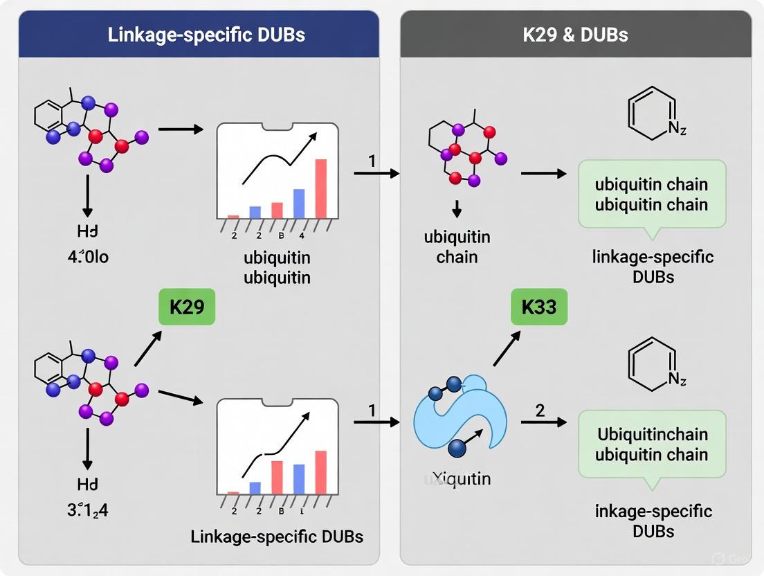
Protocol 1: DUB Linkage Specificity Profiling Using Tetraubiquitin Panels
Substrate Preparation: Prepare or commercially source homotypic tetraubiquitin chains of all eight linkage types (K6, K11, K27, K29, K33, K48, K63, M1) at 0.5-1 mg/mL concentration in assay buffer (50 mM Tris-HCl pH 7.5, 150 mM NaCl, 1 mM DTT).
Reaction Setup: Combine 2 μg of each tetraubiquitin substrate with 100-500 nM purified DUB in 20 μL reaction volume. Include negative controls without enzyme and without substrate.
Time-Course Incubation: Incubate reactions at 37°C for 0, 15, 30, 60, and 120 minutes. Terminate reactions by adding SDS-PAGE loading buffer with 20 mM N-ethylmaleimide.
Product Analysis: Resolve reaction products by SDS-PAGE (12-16% gels) and visualize by silver staining or immunoblotting with ubiquitin antibodies.
Specificity Scoring: Quantify cleavage efficiency by densitometry, calculating the ratio of cleaved product to remaining substrate for each time point. DUBs with strong preference for atypical linkages will show rapid cleavage of K29/K33 chains with minimal activity against other linkage types.
Diagram 1: DUB specificity profiling workflow
Ubiquitin Chain Restriction Analysis (UCRA)
Ubiquitin chain restriction analysis adapts principles from molecular biology restriction mapping to characterize ubiquitin chain composition on physiological substrates [29]. This approach utilizes linkage-specific DUBs as "restriction enzymes" to decipher the architecture and abundance of atypical chains on substrate proteins:
Protocol 2: Ubiquitin Chain Restriction Analysis for Substrate-Modified Chains
Substrate Immunoprecipitation: Purify the ubiquitinated protein of interest from cells under denaturing conditions (e.g., 1% SDS with subsequent dilution) to preserve ubiquitin modifications and prevent deubiquitination during purification.
DUB Restriction Digestion: Divide purified ubiquitinated substrate into aliquots. Treat each aliquot with 200-500 nM of linkage-specific DUBs (e.g., TRABID for K29/K33 chains, USP53/USP54 for K63 chains, OTUD1 for K48 chains) for 60 minutes at 37°C in appropriate reaction buffers.
Product Separation and Detection: Resolve restriction products by SDS-PAGE and perform immunoblot analysis with ubiquitin antibodies and substrate-specific antibodies.
Pattern Interpretation: Interpret the restriction pattern based on the molecular weight shifts observed with different DUB treatments. Complete cleavage with a linkage-specific DUB indicates presence of that linkage type; partial cleavage suggests mixed or branched chains containing that linkage.
Diagram 2: Ubiquitin chain restriction analysis
Functional Roles and Pathophysiological Implications
Cellular Functions of Atypical Linkage-Specific DUBs
DUBs with specificity for atypical ubiquitin chains regulate diverse cellular processes through selective editing of ubiquitin signals. TRABID, with its specificity for K29 and K33 linkages, localizes to ubiquitin-rich puncta in cells and regulates pathways including Wnt signaling and autophagic flux [5]. Disease-associated mutations in atypical linkage-specific DUBs provide compelling evidence for their physiological importance. In USP53, mutations located within its catalytic domain (R99S, G31S, C303Y, H132Y) cause progressive familial intrahepatic cholestasis, a hereditary liver disorder in children, through abrogation of DUB activity toward K63-linked chains [32]. This establishes a direct mechanistic connection between loss of DUB activity and human disease pathology.
Global ubiquitinome analyses reveal that DUBs regulate substrates via at least 40,000 unique ubiquitination sites, forming extensive dynamic ubiquitin signaling networks that control autophagy, apoptosis, genome integrity, telomere maintenance, cell cycle progression, mitochondrial function, and vesicle transport [31]. These findings highlight that DUBs have profound roles in degradation-independent ubiquitination, expanding their functions beyond merely reversing proteasomal targeting signals.
Table 3: DUBs with Atypical Linkage Specificity and Functional Associations
| DUB | Family | Linkage Specificity | Cellular Functions/Processes | Disease Associations |
|---|---|---|---|---|
| TRABID | OTU | K29, K33 | Wnt signaling, autophagy, Ub-rich puncta formation | Not specified |
| USP53 | USP | K63 | Tricellular junction regulation, protein stabilization | Progressive familial intrahepatic cholestasis [32] |
| USP54 | USP | K63 | Substrate-specific deubiquitination | Not specified |
| UCH-L1 | UCH | MonoUb | Ubiquitin recycling, neuronal function | Parkinson's disease [30] |
| A20 | OTU | K63 | NF-κB signaling, inflammatory responses | Inflammation, autoimmunity |
Therapeutic Targeting Opportunities
The exquisite linkage specificity of certain DUB families, particularly OTU and USP members, positions them as attractive therapeutic targets for pathological conditions involving dysregulated ubiquitin signaling. Small-molecule inhibitors targeting the catalytic domains of disease-relevant DUBs represent promising therapeutic candidates for cancer, neurodegenerative disorders, and inflammatory conditions [28] [30]. The development of activity-based probes that form covalent complexes with the catalytic cysteine of cysteine protease DUBs enables pharmacological profiling of DUB activity in cell lysates and intact cells, providing valuable tools for drug discovery campaigns [32] [31]. Furthermore, the regulation of DUB activity by oxidative stress through direct modification of catalytic cysteines reveals potential for therapeutic interventions that modulate DUB function in pathological contexts characterized by redox imbalance [28].
DUB families demonstrate remarkable diversity in their recognition and hydrolysis of atypical ubiquitin linkages, with the OTU family exhibiting particularly refined specificity for K29 and K33 chains. The experimental methodologies outlined in this application note provide robust frameworks for characterizing novel DUB activities, deciphering complex ubiquitin chain architectures, and validating physiological substrates. As research continues to elucidate the functions of atypical ubiquitin chains in cellular regulation and disease pathogenesis, linkage-specific DUBs will undoubtedly emerge as critical regulatory nodes and promising therapeutic targets in the ubiquitin signaling network.
Research Tools and Techniques for Studying K29/K33-Specific DUBs
Enzymatic Generation of Homotypic K29 and K33 Ubiquitin Chains
Ubiquitination is a fundamental post-translational modification that regulates virtually all cellular processes, with specificity determined by the architecture of polyubiquitin chains. Among the eight possible linkage types, the atypical K29 and K33 linkages have remained particularly enigmatic despite their detection in yeast and mammalian cells [5] [6]. These non-canonical chains constitute important regulatory signals with emerging roles in proteotoxic stress responses, autophagy, Wnt signaling regulation, and potentially neurodegenerative disorders [14] [35]. Recent advances have identified specific enzymatic machinery for assembling these chains, unlocking new opportunities for biochemical and structural characterization [5] [14].
This application note provides detailed methodologies for the enzymatic generation of homotypic K29 and K33 ubiquitin chains, framed within the context of linkage-specific deubiquitinase (DUB) research. The ability to produce well-defined chains of these linkage types is foundational for investigating their recognition by DUBs such as TRABID, which exhibits specificity for both K29 and K33 linkages [5] [36].
Key Enzymes and Assembly Strategies
HECT E3 Ligases for Atypical Chain Assembly
The identification of specific HECT E3 ligases capable of assembling K29 and K33 linkages has been pivotal to advancing this field. These enzymes provide the foundation for both in vitro chain assembly and cellular studies.
Table 1: HECT E3 Ligases for K29 and K33 Ubiquitin Chain Assembly
| E3 Ligase | Primary Linkage Specificity | Additional Linkages | Applications | Key References |
|---|---|---|---|---|
| UBE3C | K29-linked chains | K48 (63%), K11 (10%) in autoubiquitination | In vitro chain assembly; proteasomal degradation studies | Michel et al., 2015 [5] |
| AREL1 (KIAA0317) | K33-linked chains | K11 (36%), K48 (20%) in autoubiquitination | Biophysical studies; structural characterization | Michel et al., 2015 [5] |
| TRIP12 | K29 linkages and K29/K48 branched chains | Preferentially branches from K48-linked diUb | Cellular stress response studies; neurodegenerative disease models | Nature Structural & Molecular Biology, 2025 [14] |
The selection of an appropriate E3 ligase represents the most critical decision in experimental design. UBE3C predominantly generates K29-linked chains in combination with K48 linkages [5], while AREL1 shows strong preference for K33 linkages, particularly when assembling free chains without substrate [5]. TRIP12 has recently emerged as a key enzyme for K29-linked chain formation, with structural studies revealing its unique mechanism for ensuring linkage specificity [14].
Quantitative Analysis of E3 Specificity
Absolute quantification (AQUA)-based mass spectrometry analysis of E3 ligase autoubiquitination reactions reveals the following linkage distribution:
Table 2: Linkage Distribution in E3 Autoubiquitination Reactions
| E3 Ligase | K29 Linkage | K33 Linkage | K48 Linkage | K11 Linkage | Other Linkages |
|---|---|---|---|---|---|
| UBE3C | 23% | Not detected | 63% | 10% | 4% |
| AREL1 | Not detected | 36% | 20% | 36% | 8% |
Data derived from Michel et al. (2015) using AQUA mass spectrometry [5].
Experimental Protocols
Enzymatic Assembly of K29-Linked Ubiquitin Chains
This protocol utilizes the HECT E3 ligase UBE3C to generate homotypic K29-linked chains, with optional purification using the DUB vOTU [5] [6].
Materials and Reagents
- E1 enzyme: UBA1 (100 nM working concentration)
- E2 enzyme: Use E2s compatible with HECT E3s, such as UBE2D family (1 μM working concentration)
- E3 enzyme: UBE3C HECT domain (1 μM working concentration)
- Ubiquitin: Wild-type (approximately 100 μM working concentration)
- 10X Reaction Buffer: 500 mM HEPES (pH 8.0), 500 mM NaCl, 10 mM TCEP
- MgATP solution: 100 mM
- Protease inhibitors: Complete EDTA-free cocktail
- Deubiquitinase: vOTU (0.5-3 μM for purification)
Step-by-Step Procedure
Reaction Setup: In a 250 μL reaction volume, combine the following components in order:
- Nuclease-free water to volume
- 25 μL of 10X Reaction Buffer
- 10 μL of ubiquitin (1.17 mM stock)
- 25 μL of MgATP solution (100 mM stock)
- 5 μL of E1 enzyme (5 μM stock)
- 10 μL of E2 enzyme (25 μM stock)
- X μL of UBE3C (10 μM stock) to 1 μM final concentration
Incubation: Mix thoroughly and incubate at 37°C for 2-4 hours to allow chain elongation.
Purification (Optional): For homotypic K29 chain purification:
- Add vOTU DUB to 0.5-3 μM final concentration
- Incubate at 37°C for 1 hour to cleave mixed linkages
- vOTU cleaves all linkages except M1, enriching for K29 chains [36]
Termination: Add EDTA to 20 mM final concentration or SDS-PAGE sample buffer for direct analysis.
Validation: Analyze chain linkage by:
- Western blot with linkage-specific antibodies (where available)
- UbiCRest analysis with linkage-specific DUBs [36]
- Mass spectrometry for definitive verification
Diagram 1: K29 ubiquitin chain assembly workflow.
Enzymatic Assembly of K33-Linked Ubiquitin Chains
This protocol employs AREL1 to generate homotypic K33-linked chains, with the OTU DUB TRABID serving as both a purification and validation tool [5].
Materials and Reagents
- E1 enzyme: UBA1 (100 nM working concentration)
- E2 enzyme: Use E2s compatible with HECT E3s (1 μM working concentration)
- E3 enzyme: AREL1 HECT domain (436-823, 1 μM working concentration)
- Ubiquitin: Wild-type (approximately 100 μM working concentration)
- 10X Reaction Buffer: 500 mM HEPES (pH 8.0), 500 mM NaCl, 10 mM TCEP
- MgATP solution: 100 mM
- DUB: TRABID (0.5-10 μM for validation/purification)
Step-by-Step Procedure
Reaction Setup: Prepare reaction mixture as described for K29 chains, substituting AREL1 for UBE3C.
Incubation: Mix thoroughly and incubate at 37°C for 2-4 hours.
Linkage Verification via UbiCRest:
- Aliquot reaction mixture into separate tubes
- Add specific DUBs to each tube:
- Tube 1: TRABID (0.5-10 μM) - cleaves K29/K33
- Tube 2: OTUB1 (1-20 μM) - cleaves K48
- Tube 3: Cezanne (0.1-2 μM) - cleaves K11
- Tube 4: USP21 (1-5 μM) - positive control (cleaves all linkages)
- Incubate at 37°C for 1 hour [36]
Analysis: Analyze cleavage patterns by SDS-PAGE and Western blotting. Specific cleavage by TRABID confirms K33 linkage presence.
The Scientist's Toolkit: Essential Research Reagents
Table 3: Key Reagents for K29/K33 Ubiquitin Chain Research
| Reagent Category | Specific Examples | Function/Application | Linkage Specificity |
|---|---|---|---|
| E3 Ligases | UBE3C, AREL1, TRIP12 | Assembly of K29/K33 chains in vitro and in cells | K29 (UBE3C, TRIP12), K33 (AREL1) [5] [14] |
| DUBs | TRABID, vOTU | Linkage validation and chain purification | K29/K33 (TRABID), all except M1 (vOTU) [36] |
| Ubiquitin Binding Domains | TRABID NZF1 domain | Detection and pull-down assays | K29/K33 diUb [5] |
| Ubiquitin Mutants | K-only and R mutants | Linkage determination assays | Varies by mutation [37] |
| Mass Spectrometry | AQUA standards | Absolute quantification of linkage abundance | All linkages [5] |
Application in Linkage-Specific DUB Research
The enzymatic generation of well-defined K29 and K33 chains enables sophisticated studies of DUB specificity and function. TRABID (ZRANB1), which contains three NZF ubiquitin-binding domains, demonstrates specific activity toward K29 and K33 linkages [5] [36]. Structural studies reveal that TRABID's N-terminal NZF1 domain specifically recognizes K29/K33-linked diubiquitin, with crystal structures showing how this domain achieves linkage-selective binding [5] [6].
When planning DUB specificity studies, consider these key applications:
- Specificity Profiling: Use panels of homotypic chains to comprehensively characterize DUB linkage preferences
- Kinetic Analysis: Employ defined chain preparations to determine kinetic parameters (Km, kcat) for DUB-chain interactions
- Structural Studies: Generate sufficient quantities of chains for crystallography or cryo-EM studies of DUB-chain complexes
- Cellular Function Mapping: Express chain assembly enzymes to investigate DUB functions in specific cellular pathways
Troubleshooting and Technical Considerations
- Low Chain Yield: Optimize E3 concentration and reaction time; ensure adequate ATP regeneration system
- Linkage Heterogeneity: Implement DUB-based purification steps or use ubiquitin mutants to restrict linkage options
- Chain Length Control: For shorter chains, limit reaction time or ubiquitin concentration
- Validation Challenges: Employ orthogonal validation methods (DUB cleavage, MS, and binding domains)
- Cellular Expression: Use inducible systems for E3 expression to prevent cellular toxicity from atypical chain accumulation
The methods outlined herein provide a robust foundation for investigating the assembly and recognition of K29 and K33 ubiquitin chains, with particular relevance for understanding linkage-specific DUBs in both biochemical and cellular contexts.
Ubiquitination is a crucial post-translational modification that regulates virtually all cellular processes. The diversity of ubiquitin signals arises from the ability to form polyubiquitin chains through different lysine linkages, each encoding distinct functional outcomes. Among these, K29 and K33-linked chains belong to the "atypical" linkage class and are involved in various non-proteolytic signaling pathways. Research into these specific chains has been accelerated by the development of sophisticated affinity enrichment tools, particularly Ubiquitin Binding Domains (UBDs) and Tandem Ubiquitin Binding Entities (TUBEs). These tools enable the selective isolation and study of ubiquitin chains and ubiquitinated proteins from complex biological mixtures, providing critical insights into the roles of linkage-specific deubiquitinases (DUBs) in cellular homeostasis and disease. This application note details the practical methodologies for employing these reagents in the study of K29 and K33 ubiquitin chains.
Key Research Reagent Solutions
The following table summarizes essential reagents for affinity enrichment of ubiquitin chains, with a focus on tools validated for K29 and K33 linkages.
Table 1: Key Research Reagents for Ubiquitin Affinity Enrichment
| Reagent / Tool | Type | Linkage Specificity | Key Application | Example Use in K29/K33 Research |
|---|---|---|---|---|
| TRABID NZF1 | Ubiquitin Binding Domain (UBD) | Selective for K29 and K33 linkages [5] [6] | Affinity enrichment; Middle-down MS analysis | Selective pulldown of K29/K33 chains from cell lysates for mass spectrometry [38] |
| HaloTag-NZF1 Fusion | Immobilizable UBD | Selective for K29 and K33 linkages [38] | On-resin enrichment and digestion | Covalent immobilization on HaloLink resin for UbiChEM-MS workflow [38] |
| Non-hydrolyzable Diubiquitin | Synthetic Ubiquitin Bait | Defined linkage (e.g., K29, K33) [39] | UbIA-MS affinity enrichment | Serves as hydrolysis-resistant bait to capture linkage-specific interactors from lysates [39] [40] |
| TUBEs (Tandem Ubiquitin Binding Entities) | Engineered Tandem UBDs | Broad-specificity, pan-ubiquitin [38] | Protection from DUBs; enrichment of diverse chains | Isolation of total ubiquitinated material, including branched chains containing K29/K33 [38] |
| UBE3C & AREL1 E3 Ligases | Enzymatic Assembly Tools | Generate K29- and K33-linked chains [5] | In vitro chain assembly for reagent production | Production of defined K29 (UBE3C) and K33 (AREL1) chains for in vitro studies [5] |
Quantitative Profiling of Ubiquitin Interactors with UbIA-MS
The Ubiquitin Interactor Affinity Enrichment-Mass Spectrometry (UbIA-MS) method enables proteome-wide, linkage-specific profiling of ubiquitin-binding proteins [39] [40]. This approach is particularly powerful for characterizing proteins that interact with understudied chains like K29 and K33.
Core Principle and Workflow
UbIA-MS uses chemically synthesized, non-hydrolyzable diubiquitin of defined linkages as bait to enrich for interacting proteins from crude cell lysates. The non-hydrolyzable property makes the baits resistant to cleavage by endogenous deubiquitinases (DUBs), preserving bait integrity during the assay and reducing background [40]. The workflow involves bait immobilization, incubation with lysates, thorough washing, on-bead tryptic digestion of captured proteins, and subsequent identification and quantification via LC-MS/MS [39].
Key Applications and Insights
This technology has been successfully applied to map the interaction landscape of all ubiquitin linkages. For K29 and K33 chains, it identified the DUB TRABID as a highly selective interactor [39]. Furthermore, UbIA-MS can reveal stimulus-dependent changes in the ubiquitin interactome, such as the identification of DNA damage-induced monoubiquitin and K6 diubiquitin interactors, demonstrating its utility for discovering dynamic signaling events [39].
Table 2: Representative Ubiquitin Linkage-Selective Interactors Identified by UbIA-MS
| Ubiquitin Linkage | Identified Interactor | Function of Interactor | Biological Context |
|---|---|---|---|
| K6-linked diUb | TAB2, TAB3 | Adaptors in TAK1 kinase complex | DNA damage response [39] |
| K27-linked diUb | UCHL3 | Deubiquitinase (DUB) | Regulates K27 polyubiquitin formation [39] |
| K29/K33-linked diUb | TRABID (ZRANB1) | Linkage-specific DUB | Putative regulator of Wnt signaling and cell proliferation [5] [6] |
Figure 1: UbIA-MS Workflow for identifying linkage-specific ubiquitin interactors. The protocol involves bait synthesis, affinity enrichment, and LC-MS/MS analysis [39] [40].
Detection of Branched Ubiquitin Chains with UbiChEM-MS
Branched ubiquitin chains, which incorporate multiple linkage types, represent a complex layer of ubiquitin signaling. UbiChEM-MS (Ubiquitin Chain Enrichment Middle-down Mass Spectrometry) was developed to identify and characterize these branched chains, including those containing K29 and K33 linkages [38].
Methodology
The UbiChEM-MS protocol involves several key stages:
- Enrichment: Ubiquitin chains are isolated from cell lysates using linkage-specific UBDs (e.g., TRABID's NZF1 for K29/K33) or broad-specificity TUBEs immobilized on resin [38].
- On-resin Minimal Trypsinolysis: The enriched chains are subjected to limited proteolysis with trypsin under nondenaturing conditions. This cleaves ubiquitin specifically at Arg74, generating a ubiquitin 1-74 fragment (Ub~1-74~). A Ub moiety modified with a single GlyGly remnant (from a chain linkage) has a calculated mass of 8564.62 Da (GG-Ub~1-74~). A branch point, where a single ubiquitin is modified at two lysines, results in a 2xGG-Ub~1-74~ fragment with a calculated mass of 8678.66 Da [38].
- Middle-down MS Analysis: The resulting peptide mixtures are analyzed by high-resolution mass spectrometry (e.g., Orbitrap Fusion). The relative abundance of mono-, single-, and double-modified Ub~1-74~ fragments is quantified to determine the proportion of branched chains in the sample [38].
Key Findings Using K29/K33-Selective Tools
Applying UbiChEM-MS with the K29-selective NZF1 domain of TRABID revealed that approximately 4% of the enriched ubiquitin chains contained branch points. Interestingly, this level of branching was independent of proteasome inhibition, suggesting a constitutive and potentially regulatory role for branched chains involving K29 linkages. In contrast, enrichment with TUBEs showed that only ~1% of total cellular chains were branched, a figure that increased to ~4% upon proteasome inhibition [38].
Figure 2: UbiChEM-MS Workflow for detecting branched ubiquitin chains, leveraging linkage-specific UBDs and middle-down MS [38].
Detailed Experimental Protocols
This protocol outlines the steps for identifying proteins that specifically bind to K29- or K33-linked diubiquitin.
Stage 1: Preparation of Biotinylated Non-hydrolyzable Diubiquitin Baits
- Synthesis: Chemically synthesize ubiquitin precursors containing a C-terminal propargylamine and an internal lysine (e.g., K29 or K33) with a δ-azido-α-aminobutyric acid (Abu) side chain. The Abu side chain acts as a non-hydrolyzable isopeptide linkage mimic.
- Click Chemistry: Conjugate the two ubiquitin monomers via copper-catalyzed azide-alkyne cycloaddition (CuAAC) to form the non-hydrolyzable diubiquitin.
- Biotinylation: Introduce a biotin tag, for example via a maleimide-biotin conjugate to a cysteine residue, for subsequent immobilization on streptavidin beads. This synthesis process typically requires 3 weeks.
Stage 2: Affinity Purification from Cell Lysate
- Lysate Preparation: Prepare clarified lysate from the cell or tissue of interest using a nondenaturing lysis buffer (e.g., 50 mM Tris pH 7.5, 150 mM NaCl, 1% NP-40, supplemented with protease inhibitors).
- Immobilization: Couple 1-5 nmol of biotinylated diubiquitin bait to streptavidin-conjugated magnetic beads.
- Enrichment: Incubate the bait-bound beads with 1-10 mg of cell lysate protein for 1-2 hours at 4°C with gentle rotation.
- Washing: Pellet beads and wash stringently (e.g., 3-5 times with cold lysis buffer) to remove nonspecifically bound proteins.
Stage 3: On-bead Digestion and MS Sample Preparation
- Reduction and Alkylation: On-bead, reduce proteins with dithiothreitol (DTT) and alkylate with iodoacetamide.
- Tryptic Digestion: Add sequencing-grade trypsin directly to the beads and incubate overnight at 37°C to digest captured proteins.
- Peptide Recovery: Acidify the supernatant with trifluoroacetic acid (TFA) to stop digestion. Desalt the resulting peptides using C18 StageTips or cartridges.
Stage 4: LC-MS/MS Analysis and Data Processing
- Chromatography: Separate peptides using a reverse-phase C18 nano-liquid chromatography (LC) system.
- Mass Spectrometry: Analyze eluting peptides with a high-resolution tandem mass spectrometer (e.g., Q-Exactive, Orbitrap Fusion) operating in data-dependent acquisition mode.
- Data Analysis: Identify proteins by searching MS/MS spectra against a canonical protein sequence database (e.g., using MaxQuant, Proteome Discoverer). Use quantitative metrics (e.g., spectral counts, label-free quantification intensity) from control (monoubiquitin) and experimental (diubiquitin) baits to determine significantly enriched, linkage-specific interactors. The entire enrichment and identification process requires approximately 2 weeks.
This protocol describes the use of the TRABID NZF1 domain to enrich and detect branched chains containing K29 linkages.
Stage 1: Preparation of HaloTag-NZF1 Resin
- Protein Expression: Express and purify a HaloTag fusion of the TRABID NZF1 domain from E. coli.
- Immobilization: Incubate the purified HaloTag-NZF1 protein with HaloLink resin (Promega) overnight at 4°C in binding buffer (50 mM Tris, 150 mM NaCl, 10% glycerol, 0.05% IGEPAL CA-630). Wash the resin extensively to remove unbound protein.
Stage 2: Isolation of Ubiquitin Chains from Cell Lysate
- Incubation: Incubate 200 μL of NZF1-resin (as a 50% slurry) with 45 mg of total protein from HEK293 cell lysate overnight at 4°C with rotation.
- Washing: Pellet the resin and wash three times with binding buffer, followed by two washes with a minimal buffer (50 mM Tris, 150 mM NaCl, pH 7.5) to remove detergents and glycerol.
Stage 3: On-resin Minimal Trypsinolysis
- Digestion: Resuspend the washed resin in minimal buffer. Add sequencing-grade trypsin at an empirically determined ratio (e.g., 1:20 w/w trypsin-to-lysate protein) and incubate at room temperature for 16 hours.
- Peptide Recovery: Acidify the supernatant to pH ~2 with acetic acid to deactivate trypsin. Centrifuge to remove precipitates and concentrate the supernatant by speedvac.
Stage 4: Middle-down Mass Spectrometry and Quantitative Analysis
- Direct Infusion or LC-MS: Analyze the minimally digested sample by direct infusion into a high-resolution mass spectrometer (e.g., Orbitrap Fusion) or via nano-LC separation.
- Quantification: Identify the different ubiquitin species based on their accurate mass: Ub~1-74~ (8450.57 Da), GG-Ub~1-74~ (8564.62 Da), and 2xGG-Ub~1-74~ (8678.66 Da). The relative abundance of each species is calculated by integrating the corresponding peak intensities across relevant charge states. The percentage of branched chains is determined as [2xGG-Ub~1-74~ / (Ub~1-74~ + GG-Ub~1-74~ + 2xGG-Ub~1-74~)] * 100.
Affinity enrichment strategies using UBDs and TUBEs are indispensable for deciphering the complex biology of atypical ubiquitin chains. The linkage-specific UBDs, such as the NZF1 domain of TRABID, provide the precision needed to isolate and study K29 and K33 chains specifically. In parallel, broad-specificity TUBEs are invaluable for capturing the global ubiquitome and revealing the presence of heterotypic and branched chains. When coupled with advanced mass spectrometry techniques like UbIA-MS and UbiChEM-MS, these tools form a powerful platform for discovering novel interactors, quantifying chain dynamics, and uncovering the regulatory roles of DUBs in K29/K33-linked ubiquitin signaling. These detailed protocols provide a roadmap for researchers to apply these cutting-edge methods in their own investigations of the ubiquitin code.
Protein ubiquitination is a crucial post-translational modification that regulates virtually all aspects of eukaryotic cell biology. The versatility of ubiquitin signaling arises from its ability to form structurally and functionally distinct polyubiquitin chains through different linkage types. Among these, the so-called "atypical" linkages, particularly K29- and K33-linked chains, have remained poorly characterized due to the historical lack of tools for their specific detection and manipulation [5] [7]. Linkage-specific antibodies represent a fundamental toolset for deciphering this complex ubiquitin code, yet their development and validation present significant challenges that must be addressed to advance our understanding of these modifications in cellular signaling and disease contexts.
The research landscape for K29 and K33 linkages has recently evolved with the identification of specialized enzymes that assemble and recognize these chains. The human HECT E3 ligases UBE3C and AREL1 have been identified as key assemblers of K29- and K33-linked polyubiquitin, respectively [5] [24]. Furthermore, the deubiquitinase TRABID specifically cleaves K29 and K33 linkages, with its N-terminal NZF1 domain demonstrating specific binding to K29/K33-linked diubiquitin [5] [6]. These discoveries have unlocked new possibilities for studying these previously elusive chain types, yet they also highlight the critical need for highly specific detection reagents, particularly antibodies that can distinguish these linkages in complex biological samples.
The Molecular Toolbox for K29 and K33 Ubiquitin Chain Research
Table 1: Key Research Reagents for K29 and K33 Ubiquitin Chain Studies
| Reagent Category | Specific Examples | Function and Application | Key Characteristics |
|---|---|---|---|
| E3 Ligases | UBE3C, AREL1, TRIP12 | Enzymatic assembly of K29- and K33-linked chains in vitro and in cells | UBE3C assembles K48/K29-linked chains; AREL1 assembles K11/K33-linked chains; TRIP12 forms K29 linkages and K29/K48-branched chains [5] [14] |
| Deubiquitinases (DUBs) | TRABID (OTUD1) | Linkage-specific cleavage of K29 and K33 chains; validation of antibody specificity | Contains NZF1 domain with specific binding to K29/K33-diubiquitin; useful as counter-screen for antibody validation [5] [6] [41] |
| Ubiquitin-Binding Domains (UBDs) | TRABID NZF1 domain | Structural studies of K29/K33 chain recognition; potential affinity reagents | Crystal structure reveals binding mode involving hydrophobic patch on ubiquitin; exploits chain flexibility for specificity [5] [6] |
| Engineered Ubiquitin Mutants | K29-only, K33-only, Ub(K0) | Controlled chain assembly; specificity controls for antibody validation | Lysine-to-arginine mutations restrict linkage formation; essential for testing antibody linkage-specificity [5] |
| Molecular Tools for Detection | Affimers, macrocyclic peptides, catalytically inactive DUBs | Alternative recognition elements for ubiquitin chain detection | Emerging tools that may complement or surpass antibodies in specificity; useful for independent verification [42] |
Fundamental Limitations of Linkage-Specific Antibodies
Structural and Epitope Accessibility Challenges
The structural conformations adopted by K29- and K33-linked ubiquitin chains present fundamental challenges for antibody development. Solution studies indicate that both K29- and K33-linked chains adopt open and dynamic conformations similar to K63-linked polyubiquitin, in contrast to the compact structures of K48-linked chains [5] [6]. This structural flexibility means that linkage-specific epitopes may be transient or poorly accessible, reducing antibody binding affinity and consistency.
The crystal structure of K29-linked diubiquitin reveals an extended conformation with the hydrophobic patches on both ubiquitin moieties exposed and available for binding [6]. While this exposes potential epitopes, the dynamic nature of these chains means that the precise spatial orientation of linkage-defining residues may be inconsistent, complicating the development of antibodies that reliably recognize the specific isopeptide linkage without cross-reacting with similar structures in other linkage types.
Cross-Reactivity and Specificity Validation
Comprehensive validation studies have revealed alarming rates of non-specificity in commercially available antibodies. Systematic validation using the ShGE platform, which employs gene knockdown approaches, demonstrated that fewer than 20% of commercially available monoclonal antibodies and only 1% of polyclonal antibodies are truly specific for their intended targets [43]. This concerning statistic highlights the critical importance of rigorous validation for linkage-specific ubiquitin antibodies, where the potential for cross-reactivity is even greater due to the high sequence identity between different ubiquitin linkage types.
The challenge is further compounded when antibodies validated for one application (e.g., Western blot) are used in another (e.g., immunocytochemistry). Research has shown that among antibodies considered highly specific based on Western blot results, only 70% showed equivalent specificity in immunocytochemistry or flow cytometry applications [43]. This application-dependent variability represents a significant limitation for comprehensive studies of K29 and K33 ubiquitin chain biology, which often require multiple experimental approaches to fully characterize chain functions and dynamics.
Experimental Validation Strategies and Protocols
Comprehensive Specificity Validation Workflow
Diagram 1: Comprehensive antibody validation workflow. A multi-step approach is essential for establishing linkage-specificity.
Genetic Knockdown Validation Protocol
Principle: Gene knock-down (KD) or knock-out (KO) technologies are internationally recognized gold standard methods for validating antibody specificity. KD reduces target gene expression at the RNA level, while KO completely removes or disrupts the target gene at the DNA level [43].
Procedure:
- Cell Line Preparation: Select appropriate cell lines (e.g., HeLa) with confirmed expression of the target ubiquitin linkage machinery.
- Gene Silencing: Use lentivirus-mediated shRNA to silence specific mRNAs targeting components of the K29/K33 ubiquitination system. The ShGE platform simplifies this process from 16 steps to 4, reducing time from 6 months to 2 weeks while increasing success rates from 40% to over 70% [43].
- Efficiency Validation: Confirm knockdown efficiency through qRT-PCR for mRNA levels and functional assays for protein reduction.
- Antibody Testing: Apply the candidate linkage-specific antibody to both wild-type and KD/KO cells across multiple applications (Western blot, immunocytochemistry, flow cytometry).
- Specificity Confirmation: A specific antibody shows strong signal in wild-type samples that disappears or is greatly reduced in KD/KO samples [43].
Troubleshooting:
- Cellular heterogeneity may result in subpopulations with residual target protein. Use flow cytometry to select KD cell populations with high silencing efficiency.
- Changes in subcellular localization may affect protein function; evaluate by ICC.
- Always include positive and negative controls, including cells with defined ubiquitin chain linkages.
Multi-Application Validation Case Studies
Table 2: Multi-Application Validation Results for Candidate Antibodies
| Target Protein | Western Blot Results | Flow Cytometry Results | Immunocytochemistry Results | Validation Conclusion |
|---|---|---|---|---|
| CREB1 | Band disappears completely in KD cells (100% reduction) | No significant difference between WT and KD | No significant difference between WT and KD | Non-specific - Potential false negative in WB or false positive in ICC/FCM [43] |
| RAB9A | Complete loss of band in KD cells | Leftward shift of population signal in KD | No detectable signal in KD cells | High specificity - Consistent results across all platforms [43] |
| RABEPK | Signal completely disappears in KD cells | No significant difference between WT and KD | Abnormal nuclear localization (expected: membrane) | Non-specific - WB may fail to detect issues revealed by ICC/FCM [43] |
Orthogonal Validation Using Mass Spectrometry
Principle: Orthogonal validation compares protein abundance levels obtained using antibody-dependent methods with levels determined by antibody-independent methods across a set of samples [44].
Procedure:
- Sample Panel Preparation: Establish a panel of cell lines with highly variable expression of the target ubiquitin linkages based on transcriptomics data.
- Parallel Analysis:
- Antibody-based: Perform Western blot analysis with candidate linkage-specific antibodies.
- MS-based proteomics: Conduct either unbiased tandem mass tag (TMT) shotgun proteomics or targeted Parallel Reaction Monitoring (PRM) with internal standard spike-in.
- Correlation Analysis: Compare band intensities from Western blot with quantitative results from mass spectrometry across the cell line panel.
- Validation Criteria: Antibodies with Pearson correlation coefficients >0.5 are considered validated, while those below this threshold require further investigation [44].
Application Notes:
- This method requires a minimum 5-fold difference in expression levels across the cell line panel for reliable validation.
- For targets with low expression variability, genetic knockdown provides a more reliable validation approach.
- Always include reference standards of defined linkage types (K29- or K33-linked ubiquitin chains) when available.
Alternative and Complementary Detection Methodologies
Domain-Based Recognition Tools
Given the challenges with antibody specificity, researchers have developed alternative recognition strategies that exploit naturally occurring ubiquitin-binding domains with intrinsic linkage selectivity. The NZF1 domain of TRABID provides an excellent example, as structural studies have revealed the molecular basis for its specificity toward K29- and K33-linked chains [5] [6]. The crystal structure of NZF1 bound to K33-linked diubiquitin reveals a binding mode that involves the hydrophobic patch on only one of the ubiquitin moieties and exploits the flexibility of K29/K33 chains to achieve linkage-selective binding [6].
These domain-based tools can be engineered as affinity reagents with several advantages over traditional antibodies:
- Defined specificity: Structural understanding of binding mechanisms allows for rational design
- Consistent production: Recombinant expression in bacterial systems
- Modular design: Can be fused to detection tags or solid supports
DUB-Based Detection Strategies
Catalytically inactive deubiquitinases (DUBs) represent another emerging tool for linkage-specific ubiquitin detection. The DUB protein array platform has enabled systematic profiling of linkage specificity across nearly all human DUBs [41]. This resource can be leveraged to identify DUBs with high specificity for K29 and K33 linkages, which can then be engineered as detection reagents by eliminating catalytic activity while preserving binding capability.
Protocol for DUB Array Specificity Screening:
- Protein Synthesis: Produce full-length recombinant human DUB proteins using wheat cell-free protein synthesis system [41].
- Array Setup: Immobilize DUBs on solid support using affinity tag systems.
- Linkage Screening: Incubate array with eight linkage types of diubiquitins as substrates.
- Activity Detection: Measure cleavage activity using AlphaScreen technology or similar detection methods.
- Specificity Profiling: Identify DUBs with selective activity toward K29 and K33 linkages for tool development.
Application Notes for K29 and K33 Chain Research
Special Considerations for Atypical Ubiquitin Chains
Research on K29 and K33 linkages requires special methodological considerations distinct from more well-characterized ubiquitin chain types:
Cellular Context: K29-linked chains have been associated with proteotoxic stress responses and exist within mixed or branched chains containing other linkages [6] [14]. K33-linked chains play important roles in immune signaling and regulation of intracellular trafficking [7]. These biological contexts should inform experimental design and interpretation.
Chain Preparation: For controlled studies, K29- and K33-linked ubiquitin chains can be generated using identified E3 ligases (UBE3C for K29, AREL1 for K33) in combination with linkage-specific DUBs for purification [5]. These defined chains serve as essential standards for antibody validation and functional studies.
Detection Limitations: Current evidence suggests that K29 and K33 linkages frequently occur in heterotypic or branched configurations rather than extended homotypic chains [6] [14]. This complexity may limit detection by antibodies that recognize only linear epitopes in homotypic chains, highlighting the need for validation in physiologically relevant contexts.
Integrated Experimental Design
Diagram 2: Integrated experimental approach. No single method is sufficient for comprehensive analysis of atypical ubiquitin chains.
The field of atypical ubiquitin chain research continues to evolve rapidly, with recent structural studies of TRIP12 providing new insights into the mechanisms of K29-linked chain formation [14]. As our understanding of the biological functions of K29 and K33 linkages expands, the demand for highly specific detection tools will only increase. The development of linkage-specific antibodies faces significant challenges, but rigorous validation using the multi-platform approaches described here can identify reagents with sufficient specificity for meaningful biological discovery.
Emerging technologies, including affimers, macrocyclic peptides, and engineered ubiquitin-binding domains, offer promising alternatives to traditional antibodies [42]. Additionally, advances in mass spectrometry sensitivity and methodology continue to improve our ability to detect and quantify these low-abundance modifications. By employing an integrated approach that combines multiple complementary methods, researchers can overcome the current limitations of individual detection technologies and advance our understanding of the complex roles played by K29- and K33-linked ubiquitin chains in health and disease.
Mass Spectrometry-Based Approaches for Ubiquitinome Analysis
Ubiquitinome analysis represents a specialized branch of proteomics that focuses on the system-wide identification and quantification of protein ubiquitination, a crucial post-translational modification (PTM) regulating virtually all cellular processes. This modification involves the covalent attachment of ubiquitin, a 76-amino acid protein, to target substrates, primarily via an isopeptide bond between the C-terminal glycine of ubiquitin and the ε-amino group of a lysine residue on the substrate protein. The versatility of ubiquitination arises from the ability of ubiquitin itself to form polyubiquitin chains through any of its seven internal lysine residues (K6, K11, K27, K29, K33, K48, K63) or its N-terminal methionine (M1), with each linkage type potentially conferring distinct functional consequences to the modified protein.
Within the context of a broader thesis on linkage-specific deubiquitinases (DUBs) for K29 and K33 ubiquitin chains, mass spectrometry-based ubiquitinome analysis provides the essential tools to decipher the roles of these less-characterized "atypical" chains. K29- and K33-linked ubiquitin chains have remained poorly understood due to the historical scarcity of known enzymes mediating their assembly and receptors with specific binding properties. Recent research has uncovered that the human HECT E3 ligases UBE3C and AREL1 assemble K29- and K33-linked Ub chains, respectively, providing crucial tools for their biochemical and structural characterization [5]. Furthermore, the discovery that the N-terminal NZF1 domain of the DUB TRABID specifically binds K29/K33-linked diUb has opened new avenues for investigating the cellular functions of these atypical ubiquitin signals [5] [6].
This application note details established mass spectrometry-based methodologies for comprehensive ubiquitinome analysis, with particular emphasis on techniques relevant to studying K29 and K33 ubiquitin linkages, and provides a framework for integrating these approaches into research on linkage-specific DUBs.
Key Quantitative Strategies in Ubiquitinome Analysis
Quantitative mass spectrometry strategies for ubiquitinome profiling can be broadly categorized based on their acquisition methods and labeling approaches, each with distinct advantages and limitations that must be considered during experimental design.
Table 1: Comparison of Quantitative Mass Spectrometry Strategies for Ubiquitinome Analysis
| Strategy | Principle | Advantages | Limitations | Suitable for K29/K33 Studies |
|---|---|---|---|---|
| Data-Dependent Acquisition (DDA) | Selects most intense precursor ions for fragmentation | Well-established, sensitive identification | Limited dynamic range, stochastic sampling | Yes, for discovery-phase studies |
| Data-Independent Acquisition (DIA) | Fragments all ions within predefined m/z windows | Comprehensive data recording, excellent reproducibility | Complex data deconvolution | Ideal for targeted verification |
| Label-Free Quantification | Compares peptide abundances across runs | Low cost, unlimited multiplexing | Requires highly reproducible chromatography | Suitable for sample comparison |
| Isobaric Labels (TMT, iTRAQ) | Uses isobaric mass tags for multiplexing | High multiplexing capacity, reduces MS1 interference | Reporter ion compression may affect accuracy | Effective for multi-condition studies |
| Metabolic Labeling (SILAC) | Incorporates stable isotopes during cell growth | High quantification accuracy, early pooling | Limited to cell culture models | Excellent for controlled experiments |
The selection of an appropriate quantification strategy depends heavily on the research objectives. For discovery-phase studies aiming to identify novel K29/K33 ubiquitination events, label-free DIA methods offer particular advantages due to their comprehensive data recording, which enables retrospective analysis of previously unanticipated ubiquitination sites [45]. For well-controlled mechanistic studies investigating the effects of specific DUBs on K29/K33 chain dynamics, SILAC-based approaches provide superior quantification accuracy, though they are restricted to cell culture models [46].
Software tools such as Census and quantms provide flexible solutions for analyzing quantitative ubiquitinome data, supporting various labeling strategies and acquisition methods [45] [46]. These platforms incorporate robust algorithms for addressing poor-quality measurements, improving quantitative efficiency, and performing statistical analysis of differential ubiquitination, which is particularly valuable when investigating the subtle regulatory effects mediated by K29- and K33-specific DUBs like TRABID.
Experimental Protocols for Ubiquitinome Analysis
SCASP-PTM Protocol for Tandem Enrichment of Ubiquitinated Peptides
The SDS-cyclodextrin-assisted sample preparation-post-translational modification (SCASP-PTM) approach enables the tandem enrichment of ubiquitinated, phosphorylated, and glycosylated peptides from a single sample in a serial manner without intermediate desalting steps, maximizing the recovery of valuable PTM peptides from limited material [47].
Protocol Steps:
Protein Extraction and Digestion:
- Extract proteins using SDS-containing buffer with cyclodextrin to assist in detergent removal.
- Perform reduction and alkylation of cysteine residues.
- Digest proteins using trypsin or Lys-C overnight at 37°C.
Ubiquitinated Peptide Enrichment:
- Without prior desalting, incubate the protein digest with K-ε-GG antibody-conjugated beads to specifically enrich ubiquitinated peptides containing the di-glycine remnant motif.
- Wash beads extensively to remove non-specifically bound peptides.
- Elute ubiquitinated peptides using acidified buffer.
Sequential PTM Enrichment:
- Use the flowthrough from the ubiquitin enrichment for subsequent phosphorylated peptide enrichment using TiO2 or IMAC beads.
- Subsequently, use the flowthrough from phosphorylation enrichment for glycosylated peptide enrichment using hydrazide chemistry or lectin affinity.
Sample Cleanup and Mass Spectrometry Analysis:
- Desalt each enriched PTM peptide fraction using C18 StageTips or columns.
- Analyze by LC-MS/MS using appropriate acquisition methods (DDA or DIA) [47].
This protocol is particularly valuable for studying K29 and K33 ubiquitination as it conserves precious sample material, allowing for parallel analysis of multiple PTMs from the same biological source, thereby providing a more comprehensive view of the cellular signaling landscape.
K-ε-GG Antibody-Based Enrichment for Ubiquitinome Profiling
The K-ε-GG antibody, which specifically recognizes the di-glycine remnant left on ubiquitinated lysine residues after tryptic digestion, has become the cornerstone of modern ubiquitinome analysis [48].
Detailed Methodology:
Sample Preparation:
- Lyse tissues or cells in denaturing buffer (e.g., 6M guanidine-HCl) to inactivate endogenous DUBs and proteases.
- Perform protein precipitation to remove interfering compounds.
- Redissolve and digest proteins with trypsin, which cleaves after arginine residues but leaves the Gly-Gly modification on modified lysines.
Immunoaffinity Enrichment:
- Incubate the peptide mixture with anti-K-ε-GG antibody-conjugated beads for several hours at 4°C.
- Use stringent washing conditions (e.g., high-salt, organic solvent-containing buffers) to minimize non-specific binding.
- Elute bound peptides with 0.1-0.5% trifluoroacetic acid or low-pH formic acid.
Mass Spectrometric Analysis:
- Analyze enriched peptides by nanoLC-MS/MS using either DDA or DIA methods.
- For DDA, use topN methods with dynamic exclusion to maximize identifications.
- For DIA, implement variable window designs optimized for ubiquitinated peptide coverage.
Data Analysis:
- Search data against appropriate protein databases using search engines such as MaxQuant, Spectronaut, or DIA-NN.
- Include GlyGly modification (+114.04293 Da) on lysine as a variable modification.
- Apply strict false discovery rate thresholds (typically <1%) at both peptide and protein levels [48].
This approach has been successfully applied in diverse biological contexts, including recent research on maize viral infections, where ubiquitinome analysis revealed that viral infection significantly alters host protein ubiquitination patterns, with particular implications for metabolic enzymes [48].
Structural Characterization of K29 and K33 Ubiquitin Chains
For specialized studies focusing on the structural aspects of K29 and K33 ubiquitin chains, specific biochemical approaches have been developed:
Enzymatic Assembly of K29 and K33 Chains:
K29-Linked Chains:
- Utilize the HECT E3 ligase UBE3C in autoubiquitination reactions to assemble K29-linked chains.
- Treat assembly reactions with the viral deubiquitinase vOTU (which cleaves various linkages except K29) to purify homogeneous K29-linked polyubiquitin.
K33-Linked Chains:
- Employ the HECT E3 ligase AREL1 (also known as KIAA0317) to assemble K33-linked chains.
- Use linkage-specific DUBs to purify K33-linked chains from mixed assembly reactions [5].
Structural Analysis:
Crystallography:
- Generate diubiquitin of specific linkages for crystallization trials.
- Determine crystal structures to reveal conformational features, as demonstrated for K29-linked diubiquitin which adopts an extended conformation with exposed hydrophobic patches on both ubiquitin moieties [6].
Solution Studies:
- Employ NMR and small-angle X-ray scattering (SAXS) to analyze chain conformations in solution.
- Studies indicate that both K29- and K33-linked chains adopt open and dynamic conformations, similar to K63-linked chains [5].
Binding Studies:
- Use surface plasmon resonance (SPR) or isothermal titration calorimetry (ITC) to characterize interactions with specific binding domains.
- The NZF1 domain of TRABID shows specific binding to K29- and K33-linked diUb, with structural studies revealing how this domain exploits the flexibility of K29 chains to achieve linkage-selective binding [6].
Data Analysis Workflow
The computational analysis of ubiquitinome data requires specialized workflows to ensure accurate identification and quantification of ubiquitination sites, particularly when studying atypical linkages like K29 and K33.
Diagram 1: Ubiquitinome Data Analysis Workflow. This workflow outlines the key computational steps for analyzing mass spectrometry-based ubiquitinome data, from raw data processing to biological interpretation.
The quantms workflow, part of the nf-core framework, provides a comprehensive, scalable solution for ubiquitinome data analysis [45]. This workflow enables:
- Peptide Identification: Matching fragment spectra with protein databases containing known ubiquitination sites (K-ε-GG modification).
- Peptide Quantification: Using feature detection on MS1 level for label-free quantification or reporter ion intensities for isobaric labeling approaches.
- Retention Time Alignment: Correcting for chromatographic shifts between runs to improve quantification accuracy.
- Statistical Analysis: Identifying significantly changed ubiquitination sites using tools like MSstats.
- Protein Inference: Grouping proteins based on peptide evidence and aggregating quantitative information to the protein level.
For K29 and K33-specific analyses, researchers can incorporate spectral libraries containing linkage-specific ubiquitin peptides to enhance detection and quantification of these atypical chains.
The Scientist's Toolkit: Research Reagent Solutions
Table 2: Essential Research Reagents for K29/K33 Ubiquitin Chain Research
| Reagent/Category | Specific Examples | Function/Application | Relevance to K29/K33 Studies |
|---|---|---|---|
| E3 Ligases | UBE3C, AREL1 | Assembly of specific ubiquitin chains | UBE3C generates K29 linkages; AREL1 generates K33 linkages [5] |
| Deubiquitinases | TRABID, vOTU | Cleavage of specific ubiquitin linkages | TRABID is K29/K33-specific; vOTU edits chains for K29 purification [5] [6] |
| Ubiquitin Binding Domains | NZF1 domain of TRABID | Detection and purification of specific chains | Specifically binds K29/K33 linkages for pull-down assays [5] [6] |
| Antibodies | K-ε-GG antibody | Enrichment of ubiquitinated peptides | Essential for ubiquitinome studies; recognizes diglycine remnant [48] |
| Mass Spec Software | Census, quantms, MaxQuant | Data analysis and quantification | Flexible tools for various quantification strategies [45] [46] |
| Ubiquitin Mutants | K29-only, K33-only, K0 | Defining linkage specificity in assays | Critical for determining linkage specificity of enzymes and binders [5] |
This toolkit provides the essential components for designing experiments focused on K29 and K33 ubiquitin chain biology, from chain assembly and detection to functional analysis.
Application in Disease Research: Case Study
Ubiquitinome analysis has proven particularly valuable in understanding the molecular mechanisms of disease processes, including viral pathogenesis. A recent integrated proteome and ubiquitinome analysis of maize infected with sugarcane mosaic virus (SCMV) and maize chlorotic mottle virus (MCMV) revealed dramatic alterations in host protein ubiquitination during viral infection [48].
Key Findings:
- Viral infection significantly increased global ubiquitination levels, with co-infection showing the most pronounced effect.
- Most down-regulated differentially accumulated proteins with up-regulated ubiquitination sites were involved in photosynthesis and metabolic pathways.
- Functional validation demonstrated that glycolate oxidase 1 (ZmGOX1), an enzyme in the glyoxylate metabolism pathway with altered ubiquitination, played a protective role against viral infection.
- Overexpression of ZmGOX1 and its ubiquitination site mutants enhanced maize resistance to SCMV infection, suggesting a direct functional link between ubiquitination of this metabolic enzyme and antiviral defense [48].
This case study illustrates how integrated ubiquitinome and proteome approaches can identify functionally important ubiquitination events in disease contexts, providing a roadmap for similar applications in other biological systems.
Mass spectrometry-based ubiquitinome analysis has matured into a powerful set of technologies for comprehensively profiling protein ubiquitination at a system-wide level. The specialized protocols and analytical strategies outlined in this application note provide researchers with a solid foundation for investigating the complex landscape of ubiquitin signaling, with particular utility for studying the poorly characterized K29 and K33 linkage types. By integrating specific enrichment methods, advanced mass spectrometric acquisition techniques, and sophisticated computational tools, researchers can now address fundamental questions about the assembly, recognition, and functional consequences of these atypical ubiquitin chains. Furthermore, the application of these methodologies in disease models continues to yield novel insights into the pathological significance of altered ubiquitination, opening new avenues for therapeutic intervention targeting the ubiquitin system.
Determining the three-dimensional structures of proteins is fundamental to understanding their function. For researchers studying linkage-specific deubiquitinases (DUBs) for K29 and K33 ubiquitin chains, a combination of structural biology techniques is essential to unravel the molecular basis of recognition and specificity. X-ray crystallography provides high-resolution atomic snapshots of DUBs bound to ubiquitin chains, enabling the precise mapping of interaction interfaces [49] [50]. Solution techniques, such as Nuclear Magnetic Resonance (NMR) spectroscopy, reveal the dynamics and conformational flexibility of these complexes under near-physiological conditions, complementing the static pictures from crystals [51] [50]. Furthermore, Mass Spectrometry (MS) has become an integral component of the structural biology toolkit, particularly for characterizing stoichiometry, interactions, and structural organization of complexes that are challenging to study by other methods [52]. This application note details standardized protocols for employing these techniques within a research program focused on K29- and K33-linked ubiquitin chains.
Key Structural Methods and Their Applications
The following table summarizes the primary structural biology techniques, their fundamental principles, and their specific utility in the study of K29/K33 ubiquitin chains and their cognate DUBs.
Table 1: Core Structural Biology Techniques in Ubiquitin Research
| Method | Fundamental Principle | Key Application in K29/K33 Ubiquitin & DUB Research |
|---|---|---|
| X-ray Crystallography | Analyses diffraction patterns from protein crystals to determine electron density and atomic positions [50]. | Determining high-resolution structures of DUBs (e.g., TRABID NZF1 domain) in complex with K29- or K33-linked diubiquitin to reveal molecular basis of linkage specificity [5] [6]. |
| Serial Femtosecond Crystallography (SFX) | Uses ultrashort, bright X-ray Free-Electron Laser (XFEL) pulses to obtain diffraction from microcrystals before radiation damage occurs [50]. | Studying microcrystals of radiation-sensitive ubiquitin-DUB complexes at room temperature to capture physiologically relevant states [53] [54]. |
| Nuclear Magnetic Resonance (NMR) Spectroscopy | Probes local chemical environment and distances between atoms in solution using magnetic fields and radio waves [50]. | Characterizing the dynamic, open conformations of K29- and K33-linked polyubiquitin chains in solution and mapping binding epitopes upon DUB interaction [5] [51]. |
| Mass Spectrometry (MS) | Determines mass-to-charge ratios of ions in the gas phase to infer identity and structure [51] [52]. | Identifying linkage types in assembled chains via AQUA mass spectrometry, mapping binding interfaces using HDX-MS or crosslinking-MS, and determining complex stoichiometry [5] [52]. |
| Integrative/Hybrid Methods | Combines data from multiple low- to medium-resolution techniques to build a coherent structural model [51] [50]. | Determining architectures of large, flexible ubiquitin-DUB complexes by integrating restraints from NMR, MS, SAXS, and computational modeling [51]. |
Experimental Protocols for Ubiquitin Chain Production and Analysis
Protocol 1: Enzymatic Assembly and Purification of Atypical Ubiquitin Chains
Objective: To generate homotypic K29- or K33-linked polyubiquitin chains in vitro for biochemical and structural studies [5] [6].
Materials:
- Recombinant Ubiquitin: Wild-type and mutant (e.g., K29-only, K33-only) ubiquitin.
- E1 Activating Enzyme: UBE1.
- E2 Conjugating Enzyme: Specific E2(s) working with the chosen HECT E3s.
- E3 Ligases: UBE3C for K29/K48-linked chains; AREL1 (KIAA0317) for K11/K33-linked chains [5].
- Deubiquitinase (DUB): vOTU (for editing chains during UBE3C assembly) or other linkage-specific DUB for purification [5] [6].
- Buffers: ATP-containing reaction buffer, ion-exchange chromatography buffers, size-exclusion chromatography (SEC) buffer.
Method:
- Reaction Setup: In a 1 mL reaction volume, combine 50 µM ubiquitin, 100 nM E1, 1-5 µM E2, and 1-5 µM HECT E3 (UBE3C or AREL1) in reaction buffer (50 mM Tris-HCl pH 7.5, 50 mM NaCl, 10 mM MgCl₂, 2 mM ATP).
- Incubation: Incubate the reaction at 30°C for 2-4 hours.
- Chain Editing (for UBE3C): To enrich for K29 linkages, add the linkage-specific DUB vOTU to the UBE3C assembly reaction. vOTU will cleave other linkages, leaving K29 chains intact [6].
- Purification:
- Stop the reaction with 20 mM DTT.
- Acidity the mixture to pH 4.5 and load onto a Source S cation-exchange column.
- Elute with a linear gradient of 0-500 mM NaCl.
- Pool fractions containing polyubiquitin and concentrate.
- Perform final purification via SEC (Superdex 200) in a low-salt buffer (e.g., 20 mM Tris-HCl pH 7.5, 50 mM NaCl).
- Validation: Analyze chain length and linkage specificity by SDS-PAGE and AQUA mass spectrometry [5].
Protocol 2: Crystallography of Ubiquitin-DUB Complexes
Objective: To determine the crystal structure of a DUB's ubiquitin-binding domain (e.g., TRABID NZF1) in complex with K29- or K33-linked diubiquitin [5] [6].
Materials:
- Purified Proteins: K29- or K33-linked diubiquitin and the recombinant NZF1 domain.
- Crystallization Screens: Commercial sparse matrix screens (e.g., Hampton Research).
- Cryoprotectants: Glycerol, ethylene glycol, or other cryoprotective solutions.
- Synchrotron Access: Access to a microfocus beamline (e.g., VMXm) for data collection [54].
Method:
- Complex Formation: Mix the NZF1 domain with diubiquitin in a 1:1.2 molar ratio. Incubate on ice for 30 minutes.
- Crystallization: Using the vapor-diffusion sitting-drop method, mix 100 nL of protein complex with 100 nL of reservoir solution. A typical condition leading to crystals might be 0.1 M HEPES pH 7.5 and 25% PEG 3350 [6].
- Cryo-protection: Soak crystals briefly in reservoir solution supplemented with 20-25% glycerol before flash-cooling in liquid nitrogen.
- Data Collection:
- Structure Determination:
- Process diffraction data (indexing, integration, scaling) with software like XDS or autoPROC.
- Solve the structure by molecular replacement using a known ubiquitin structure (PDB: 1UBQ) as a search model.
- Perform iterative cycles of model building and refinement using Coot and Phenix.
Protocol 3: Analyzing Chain Conformation and Dynamics via NMR
Objective: To characterize the solution structure and dynamics of free K29- or K33-linked polyubiquitin and their complexes with DUBs.
Materials:
- Isotope-Labeled Ubiquitin: 15N- and/or 13C-labeled ubiquitin for NMR signal assignment.
- NMR Buffer: 20 mM Sodium Phosphate pH 6.5, 50 mM NaCl, 0.02% NaN₃, 10% D₂O.
- NMR Spectrometer: High-field spectrometer (≥600 MHz) equipped with a cryoprobe.
Method:
- Sample Preparation: Prepare 0.2-0.5 mM samples of 15N-labeled K29- or K33-linked diubiquitin in NMR buffer. For titration experiments, prepare a concentrated stock of the unlabeled DUB domain.
- Data Acquisition:
- Record 2D 1H-15N HSQC spectra of the free diubiquitin chain.
- Titrate the DUB domain into the diubiquitin sample, recording a 2D 1H-15N HSQC spectrum after each addition.
- Data Analysis:
- Assign chemical shift perturbations (CSPs) to specific ubiquitin residues.
- Map CSPs onto the surface of ubiquitin to identify the DUB binding interface.
- Analyze relaxation parameters (T1, T2) to infer conformational dynamics and flexibility of the chains, which for K29 and K33 linkages indicate open and dynamic conformations similar to K63-linked chains [5].
Quantitative Data and Reagent Solutions
The application of these structural methods has yielded key quantitative insights into the properties of atypical ubiquitin chains, as summarized below.
Table 2: Experimentally Determined Structural Parameters of Atypical Ubiquitin Chains
| Linkage Type | Solution Conformation (from NMR/SAXS) | Crystal Structure Insights | Key Interacting Proteins / Domains |
|---|---|---|---|
| K29-linked | Extended and dynamic conformation in solution [5]. | Extended conformation in crystal structure; hydrophobic patches on both ubiquitin moieties exposed and available for binding [6]. | TRABID (NZF1 domain): Binds K29/K33-diUb with high specificity. UBE3C (HECT E3): Assembles K29- and K48-linked chains [5] [6]. |
| K33-linked | Adopts open and dynamic conformations in solution, similar to K63-linked chains [5]. | Filamentous structure when bound to TRABID NZF1 domains; NZF1 binds each Ub-Ub interface in the crystal [5]. | TRABID (NZF1 domain): Specific binder. AREL1 (HECT E3): Assembles K11- and K33-linked chains [5]. |
Table 3: Research Reagent Solutions for Structural Studies of Atypical Ubiquitin Chains
| Reagent / Material | Function / Application | Example & Notes |
|---|---|---|
| Linkage-Specific E3 Ligases | Enzymatic assembly of defined ubiquitin chain linkages. | UBE3C: For K29-linked chains. AREL1: For K33-linked chains [5]. |
| Linkage-Specific DUBs | Editing and purification of specific chains; validation of linkage type. | vOTU: Used with UBE3C to enrich K29 linkages. TRABID: K29/K33-specific DUB [5] [6]. |
| Ubiquitin Mutants | Determining linkage specificity in assembly and binding assays. | Kx-only Mutants: Ubiquitin with only one lysine (e.g., K29, K33) to restrict chain linkage [5]. |
| Isotope-Labeled Ubiquitin | Enables NMR spectroscopy for structural and dynamic studies. | 15N-, 13C-labeled Ubiquitin: For backbone assignment and CSP mapping in solution [51]. |
| Crystallization Screens | Initial screening of conditions for protein crystal formation. | Sparse Matrix Screens (e.g., Hampton Index): Identify initial hits for protein, complex, or microcrystal growth [54] [55]. |
Workflow and Pathway Visualizations
Diagram 1: Structural Biology Workflow for Atypical Ubiquitin Chain Analysis. This diagram outlines the integrated experimental workflow for discovering and characterizing DUBs specific to K29 and K33 ubiquitin chains, highlighting the convergence of biochemical and structural techniques.
Diagram 2: Ubiquitination Pathway for Atypical Chains. This diagram illustrates the enzymatic cascade for assembling K29- and K33-linked ubiquitin chains and their subsequent recognition by a linkage-specific DUB, highlighting the key enzymes involved.
Cellular Assays for Monitoring K29/K33 Chain Dynamics
Within the intricate landscape of post-translational modifications, the ubiquitin code represents a language of remarkable complexity. Among its least understood dialects are the atypical ubiquitin chains linked through lysine 29 (K29) and lysine 33 (K33). Once considered poorly characterized entities, these chain types are now emerging as critical regulators of cellular homeostasis, proteotoxic stress response, and signal transduction [5] [11]. K29-linked ubiquitination has been identified as one of the most abundant atypical linkages in eukaryotic cells, with roles in cell cycle regulation and cellular response to proteotoxic stresses, including unfolded protein response, oxidative stress, and heat shock [11]. Meanwhile, K33-linked chains have been implicated in intracellular trafficking and signal transduction of cell surface receptors [11]. The study of these chains has been hampered by a historical scarcity of dedicated research tools; however, recent advances in linkage-specific reagents and enzymatic methodologies now enable researchers to decipher their dynamic cellular regulation.
This application note provides a comprehensive framework for investigating K29 and K33 ubiquitin chain dynamics, with particular emphasis on their regulation by deubiquitinating enzymes (DUBs). We detail specific methodologies for chain assembly, detection, and functional characterization, providing life science researchers and drug development professionals with the necessary tools to advance this emerging field.
Key Research Tools for K29/K33 Ubiquitin Research
The study of K29 and K33 ubiquitin chain biology requires specialized reagents. The table below summarizes essential research tools for investigating these atypical ubiquitin chains.
Table 1: Key Research Reagent Solutions for K29/K33 Ubiquitin Chain Studies
| Reagent Type | Specific Example | Function and Application | Key Characteristics |
|---|---|---|---|
| E3 Ligases | UBE3C (HECT E3) | Assembles K29- and K48-linked chains in autoubiquitination reactions [5]. | Used in combination with DUBs to generate K29-linked chains for analysis [5]. |
| AREL1 (HECT E3) | Assembles K11/K33- and K33-linked chains on substrates and as unanchored chains [5]. | Predominantly generates K33-linkages on free chains and reported substrates [5]. | |
| Linkage-Specific DUBs | TRABID (OTU Family) | K29/K33-specific deubiquitinase; used as a restriction enzyme for linkage identification [5] [29]. | Contains N-terminal NZF1 domain that specifically binds K29/K33-diUb [5]. |
| Ubiquitin-Binding Domains | TRABID NZF1 Domain | Specifically recognizes K29/K33-linked diubiquitin; tool for pull-down assays [5]. | Crystal structure with K33-diUb reveals basis for specificity [5]. |
| Linkage-Specific Binders | sAB-K29 (Synthetic Fab) | Synthetic antigen-binding fragment for specific recognition of K29-linked chains [11]. | Binds K29-linked diUb with nanomolar affinity; enables immunofluorescence and pull-down [11]. |
Experimental Protocols for K29/K33 Chain Assembly and Detection
Enzymatic Assembly of K29- and K33-Linked Ubiquitin Chains
The production of pure, homotypic K29- or K33-linked ubiquitin chains is a prerequisite for biochemical and structural studies. The following protocol describes an enzymatic assembly system utilizing identified HECT E3 ligases, adapted from Michel et al. (2015) [5].
Materials:
- Ubiquitin: Wild-type and mutant (K29-only, K33-only) ubiquitin
- Enzyme System: UBA1 (E1), UBE2L3/UBE2D2 (E2), UBE3C (for K29 chains) or AREL1 (for K33 chains)
- Reaction Buffer: 50 mM Tris-HCl (pH 7.5), 50 mM NaCl, 10 mM MgCl₂, 5 mM ATP
- Purification: vOTU DUB (for removing non-K29 chains) or other linkage-specific DUBs, anion exchange chromatography columns
Procedure:
- Chain Assembly Reaction:
- Set up a 1 mL reaction mixture containing reaction buffer, 5 µM E1, 10 µM E2, 5 µM E3 (UBE3C for K29; AREL1 for K33), and 100 µM ubiquitin.
- Incubate at 30°C for 3 hours to allow polyubiquitin chain formation.
Linkage-Specific Cleavage (for K29 chains):
- For K29 chain purification, add vOTU DUB (final concentration 1 µM) to the assembly reaction.
- Incubate at 37°C for 1 hour. vOTU cleaves K48 and other linkages but leaves K29 chains intact [11].
Chain Purification:
- Separate the reaction products by anion exchange chromatography using a linear salt gradient (0-500 mM NaCl).
- Collect fractions and analyze by SDS-PAGE. K29- or K33-linked diUb and polyUb chains elute at distinct conductivities.
- Concentrate purified chains using centrifugal filter units and store at -80°C.
Validation: Verify chain linkage and purity using absolute quantification (AQUA) mass spectrometry [5] or linkage-specific DUB profiling [29].
Detection of K29-Linked Chains in Cells Using sAB-K29
The synthetic antigen-binding fragment sAB-K29 enables specific detection of endogenous K29-linked ubiquitination, overcoming a major historical limitation in the field [11].
Materials:
- Detection Reagent: sAB-K29 (purified)
- Cell Culture Reagents: Appropriate cell line culture media, transfection reagents, proteotoxic stress inducers (e.g., tunicamycin for ER stress, sodium arsenite for oxidative stress)
- Lysis Buffer: RIPA buffer supplemented with 10 mM N-Ethylmaleimide (NEM) to inhibit endogenous DUBs
- Immunofluorescence: Fixation buffer (4% PFA), permeabilization buffer (0.1% Triton X-100), blocking buffer (5% BSA)
Procedure for Immunofluorescence and Pull-Down:
- Cell Treatment and Fixation:
- Culture cells on glass coverslips and treat with desired stressors (e.g., 2 µg/mL tunicamycin for 8 hours to induce ER stress).
- Wash cells with PBS and fix with 4% PFA for 15 minutes at room temperature.
Immunofluorescence Staining:
- Permeabilize cells with 0.1% Triton X-100 for 10 minutes.
- Block with 5% BSA for 1 hour.
- Incubate with sAB-K29 (1-5 µg/mL) in blocking buffer overnight at 4°C.
- Wash and incubate with appropriate fluorescent secondary antibody.
- Image using a confocal microscope. K29 signal is often observed in cytoplasmic puncta under proteotoxic stress and in the midbody during cytokinesis [11].
Pull-Down for Proteomic Analysis:
- Lyse cells in RIPA buffer with NEM and protease inhibitors.
- Incubate cleared lysate with sAB-K29-conjugated beads for 2 hours at 4°C.
- Wash beads extensively and elute bound proteins with Laemmli buffer for Western blotting or under denaturing conditions for mass spectrometry analysis.
Figure 1: Experimental workflow for the assembly, detection, and functional analysis of K29- and K33-linked ubiquitin chains.
Methodologies for Profiling DUB Specificity Toward K29/K33 Linkages
Determining the linkage specificity of deubiquitinating enzymes is crucial for understanding their biological functions. The following approaches enable comprehensive DUB specificity profiling.
DUB Protein Array for Linkage Specificity Screening
The human DUB protein array provides a high-throughput platform for determining the linkage specificity of nearly all human DUBs against eight different diubiquitin linkages [56].
Materials:
- DUB Array: 88 full-length recombinant human DUB proteins synthesized using a wheat cell-free system
- Substrates: Eight linkage types of diubiquitins (K6, K11, K27, K29, K33, K48, K63, M1)
- Assay Buffer: 50 mM Tris-HCl (pH 7.5), 5 mM DTT
- Detection: SDS-PAGE equipment, SYPRO Ruby protein gel stain
Procedure:
- DUB Immobilization:
- Incubate 10 µL of each crude DUB translation mixture with 8 µL of anti-AGIA magnetic beads for 1 hour at 4°C.
- Wash beads with high-salt buffer (500 mM NaCl) followed by standard buffer (150 mM NaCl).
Deubiquitination Reaction:
- Prepare substrate mixtures containing 2 µM of each diubiquitin linkage in assay buffer.
- Add 10 µL of substrate mixture to DUB-bound beads.
- Incubate for 3 hours at 30°C with gentle agitation.
Product Analysis:
- Separate supernatant from beads using a magnetic stand.
- Mix supernatant with SDS sample buffer and separate by SDS-PAGE.
- Stain proteins with SYPRO Ruby and image using a fluorescence scanner.
- Cleavage activity is indicated by the appearance of monoubiquitin bands.
Data Interpretation: DUBs are classified as K29/K33-specific if they cleave these linkages efficiently while showing minimal activity against other linkage types. TRABID is a known example identified through such methods [56] [29].
Ubiquitin Chain Restriction Analysis (UCRA)
Ubiquitin chain restriction analysis adapts the principle of DNA restriction digest to ubiquitin chains, using linkage-specific DUBs as "restriction enzymes" to decipher the architecture of ubiquitin signals on substrates [29].
Materials:
- Substrate: Ubiquitinated protein of interest
- Restriction DUBs: Panel of linkage-specific DUBs (e.g., TRABID for K29/K33, OTULIN for M1, etc.)
- Reaction Buffer: 50 mM Tris-HCl (pH 7.5), 150 mM NaCl, 5 mM DTT, 0.1 mg/mL BSA
- Analysis: SDS-PAGE and Western blot equipment, anti-ubiquitin antibodies
Procedure:
- Prepare Ubiquitinated Substrate:
- Immunoprecipitate the ubiquitinated protein of interest from cell lysates under denaturing conditions to preserve ubiquitin modifications.
DUB Restriction Digest:
- Divide the purified ubiquitinated substrate into multiple aliquots.
- Incubate each aliquot with a different linkage-specific DUB (e.g., TRABID for K29/K33) or combination of DUBs.
- Include a no-DUB control and a non-specific DUB control (e.g., USP2).
- Incubate at 37°C for 2 hours.
Analysis of Cleavage Products:
- Stop reactions with SDS sample buffer and analyze by Western blotting.
- Probe with anti-ubiquitin antibody and substrate-specific antibody.
- A shift to lower molecular weight upon TRABID treatment indicates the presence of K29/K33 linkages on the substrate.
Table 2: Quantitative Cleavage Profiles of Selected DUBs Against Atypical Linkages
| DUB | Family | K29-Linked Chain Cleavage | K33-Linked Chain Cleavage | Other Relevant Linkage Preferences | Application in K29/K33 Research |
|---|---|---|---|---|---|
| TRABID | OTU | High [5] [29] | High [5] [29] | Prefers K29 and K33 over K48, K63 | Primary restriction enzyme for K29/K33 chain identification [29] |
| OTUD1 | OTU | Not Detected | Not Detected | Specific for K63 linkages | Negative control for K29/K33 assays [29] |
| Cezanne | OTU | Not Detected | Not Detected | Specific for K11 linkages | Negative control for K29/K33 assays [29] |
| vOTU | OTU (Viral) | None (Resistant) [11] | Not Reported | Cleaves K48, K63, others | Tool for purifying K29 chains by removing contaminating linkages [11] |
Functional Characterization of K29/K33 Chains in Cellular Pathways
Investigating K29/K33 Chains in Proteotoxic Stress and Cell Cycle
The functional roles of K29 and K33 chains can be elucidated through targeted cellular assays that monitor their dynamics under specific physiological conditions.
Monitoring K29-Linked Ubiquitination During Proteotoxic Stress:
- Induction of Proteotoxic Stress:
- Treat cells with various stressors: 2 µM thapsigargin (ER stress), 0.5 mM sodium arsenite (oxidative stress), or heat shock (42°C for 1 hour).
- Include controls with proteasome inhibitor (MG132) to distinguish degradation-related functions.
Assessment of K29 Chain Dynamics:
- Fix cells at time intervals and stain with sAB-K29 for immunofluorescence [11].
- Observe formation of K29-positive puncta, which indicate recruitment to stress-induced aggregates.
- Perform co-staining with stress granule markers (G3BP1) or autophagy markers (LC3) to determine compartmentalization.
Functional Validation:
- Knock down K29-specific E3s (UBE3C) or DUBs (TRABID) using siRNA.
- Assess impact on cell viability under stress conditions using MTT or CellTiter-Glo assays.
- Monitor cell cycle progression by flow cytometry to identify potential arrest, particularly at G1/S phase as reported with K29 signal disruption [11].
Figure 2: Cellular signaling pathways involving K29-linked ubiquitin chains in response to proteotoxic stress and during cell cycle progression. Disruption of K29/K33 signaling leads to measurable cellular phenotypes.
The methodologies detailed in this application note provide a comprehensive toolkit for investigating the dynamics of K29 and K33-linked ubiquitin chains in cellular contexts. From enzymatic assembly of defined chains to linkage-specific detection and functional analysis, these protocols enable researchers to move beyond correlation and establish causative relationships between these atypical ubiquitin modifications and their cellular functions. The integration of these approaches—particularly when combining specific reagents like sAB-K29 with DUB restriction analysis and cellular phenotyping—will accelerate our understanding of how K29 and K33 ubiquitin chains contribute to proteotoxic stress response, cell cycle regulation, and intracellular signaling. As these tools continue to evolve, they will undoubtedly uncover new therapeutic opportunities for manipulating the ubiquitin system in human disease.
The ubiquitin code, a complex post-translational language, regulates virtually every cellular process through the assembly of polyubiquitin chains with distinct linkage specificities. Among these, the atypical K29 and K33 linkages represent particularly enigmatic components of this signaling system. Research into these chains has been hampered by a historical lack of tools for their specific generation and detection [5]. Recent advances have uncovered dedicated enzymatic systems, with the HECT E3 ligases UBE3C and AREL1 identified as specific assemblers of K29- and K33-linked chains, respectively [5]. Parallel discoveries have revealed that the ovarian tumor (OTU) family deubiquitinase TRABID specifically recognizes and cleaves these atypical linkages [5] [29]. This application note details integrated methodologies for profiling deubiquitinase (DUB) activity, with a specialized focus on investigating K29- and K33-linkage specificity from purified biochemical systems to live-cell contexts, providing researchers with a comprehensive toolkit for deciphering the biological functions of these atypical ubiquitin signals.
Linkage-Specific Reagents and Tools for K29/K33 Research
The functional characterization of K29- and K33-specific DUBs requires a suite of specialized reagents that enable the controlled production, manipulation, and analysis of these atypical ubiquitin chains. The table below summarizes the core components of this toolkit.
Table 1: Essential Research Reagents for K29/K33 Ubiquitin Chain Research
| Reagent Type | Specific Example | Function in K29/K33 Research |
|---|---|---|
| E3 Ligases | UBE3C (HECT family) | Assembler of K48/K29-branched and K29-linked ubiquitin chains [5] |
| E3 Ligases | AREL1 (HECT family) | Assembler of K11/K33-branched and K33-linked ubiquitin chains [5] |
| Linkage-Specific DUBs | TRABID (OTU family) | DUB with specificity for cleaving K29 and K33 linkages; contains K29/K33-binding NZF1 domain [5] [29] |
| Ubiquitin Binding Domains (UBDs) | TRABID NZF1 domain | Selective binder of K29/K33-linked diubiquitin; used for linkage detection and validation [5] [6] |
| Defined Ubiquitin Chains | Enzymatically synthesized K29- or K33-linked polyubiquitin | Substrate for in vitro DUB activity assays and structural studies [5] [6] |
| Activity-Based Probes | Covalent ubiquitin-based probes | Chemoproteomic profiling of DUB activity and inhibitor engagement in live cells [57] |
Biochemical Profiling of DUB Activity and Linkage Specificity
Biochemical assays provide the foundation for quantifying DUB enzyme kinetics, determining linkage specificity, and establishing structure-activity relationships.
In Vitro Deubiquitination Assay with Defined Chains
Purpose: To directly measure the cleavage activity and linkage preference of a DUB using purified, linkage-defined ubiquitin chains as substrate [58].
Protocol:
- Reaction Setup: In a 20 µL reaction volume, combine:
- 1 µM of purified DUB (e.g., TRABID)
- 5 µM of purified ubiquitin chain (e.g., K29- or K33-linked tetraubiquitin)
- Assay Buffer: 50 mM Tris-HCl (pH 7.5), 150 mM NaCl, 1 mM DTT [5]
- Incubation and Time-point Collection: Incubate the reaction at 37°C. Aliquot 5 µL of the reaction mixture at defined time points (e.g., 0, 5, 15, 30, 60 minutes) and immediately quench by adding SDS-PAGE loading buffer containing DTT.
- Analysis:
- Gel Electrophoresis: Resolve time-point samples by SDS-PAGE (4-12% Bis-Tris gradient gel).
- Visualization: Stain the gel with Coomassie Blue or conduct Western blotting using an anti-ubiquitin antibody to visualize the disappearance of the substrate chain and the appearance of lower molecular weight cleavage products (e.g., diUb, monoUb) [5] [6].
Quantitative Fluorescence-Based DUB Activity Assay
Purpose: To achieve high-throughput, quantitative kinetic analysis of DUB activity using ubiquitin substrates conjugated to fluorogenic tags.
Protocol:
- Substrate Preparation: Use commercially available ubiquitin substrates (e.g., Ub-AMC, Ub-Rhodamine) or label purified K29/K33-linked diubiquitin with a suitable fluorescent tag [59].
- Assay Configuration: In a 384-well plate, mix:
- A range of DUB concentrations (nM to µM)
- Fixed concentration of fluorogenic substrate (e.g., 100 nM)
- Assay Buffer: 50 mM HEPES (pH 7.5), 0.1 mg/mL BSA, 0.5 mM TCEP [59]
- Readout and Data Analysis: Monitor fluorescence increase (Ex/Em: 355/460 for AMC) in real-time using a plate reader. Calculate initial velocities, determine Km and kcat values, and generate IC50 curves for inhibitor characterization using 4- or 5-parameter logistic (4PL/5PL) curve fitting [59].
The following diagram illustrates the strategic workflow for determining DUB linkage specificity, from biochemical screening to mechanistic validation.
Cell-Based DUB Activity and Inhibition Profiling
Translating biochemical findings into a cellular context is critical for validating DUB function and assessing the efficacy of inhibitors under physiologically relevant conditions.
Flow Cytometry-Based Cellular DUB Activity Assay
Purpose: To sensitively quantify DUB activity and inhibition in living cells, bridging the gap between biochemical and cellular studies [60].
Protocol:
- Reporter Construction: Create a DUB recruitment system by fusing a substrate protein (e.g., a ubiquitin-GFP fusion) with a specific degradation signal. Co-express this with a DUB (e.g., TRABID, USP7) fused to a GFP-targeting nanobody (Nb) to recruit the DUB to the substrate [60].
- Cell Transfection and Treatment: Transfect the DUB-Nb construct into cells expressing the ubiquitin-GFP reporter. Treat cells with a DUB inhibitor (e.g., small molecule compound) or vehicle control for a specified duration.
- Analysis by Flow Cytometry: Harvest cells and analyze GFP fluorescence intensity via flow cytometry. DUB activity stabilizes the reporter, resulting in high GFP fluorescence. Effective DUB inhibition leads to reporter degradation and a measurable decrease in fluorescence [60]. Data can be used to generate dose-response curves and calculate IC50 values for inhibitors in a cellular environment.
Activity-Based Protein Profiling (ABPP) for Target Engagement
Purpose: To directly assess the engagement of small-molecule inhibitors with their endogenous DUB targets in a complex cellular proteome [57].
Protocol:
- Probe Design: Utilize a bespoke covalent compound library designed to target the catalytic cysteine in the active site of most DUBs. These probes typically consist of a ubiquitin-warhead scaffold coupled to a reporter tag (e.g., biotin) [57].
- Cell Lysate Incubation: Incubate the activity-based probe with lysates from cells treated with either a DUB inhibitor candidate or DMSO control.
- Detection and Analysis: Enrich probe-labeled DUBs using streptavidin beads and identify engaged DUBs by mass spectrometry. Selective inhibition by a compound is indicated by the reduced labeling of its target DUB in the pre-treated sample compared to the control, confirming cellular target engagement [57].
Data Integration and Analysis
Robust data analysis is paramount for drawing meaningful conclusions from DUB profiling experiments. The quantitative data generated from these assays must be systematically analyzed and interpreted.
Table 2: Key Quantitative Parameters in DUB Profiling Assays
| Assay Type | Primary Readout | Key Parameters | Application Example |
|---|---|---|---|
| Fluorescence-Based Kinetic Assay | Fluorescence intensity over time | IC₅₀: Inhibitor potency; kcat/Km: Catalytic efficiency | Determining TRABID's catalytic efficiency for K29- vs K33-linked chains [59] |
| Cellular Flow Cytometry | GFP fluorescence intensity by flow cytometry | % Fluorescence Reduction: Measures cellular inhibitor efficacy; Cellular IC₅₀ | Profiling inhibitor dose-response for viral DUB PLpro in cells [60] |
| Mass Spectrometry (AQUA/SRM) | Abundance of linkage-specific signature peptides | Fold-Change: Accumulation of specific chains in DUB-KO cells [61] | Identifying K63 chain accumulation in ubp2Δ yeast strain [61] |
| Activity-Based Profiling (ABPP) | MS1 intensity of target DUB peptides | % Target Engagement: Reduction in probe labeling in inhibitor-treated samples [57] | Validating selective engagement of VCPIP1 DUB by a covalent inhibitor [57] |
The integrated application of biochemical and cell-based profiling methods outlined in this document provides a robust, multi-faceted strategy for investigating DUB activity, with particular utility for characterizing the regulation of K29 and K33 ubiquitin linkages. The sequential use of in vitro assays using defined chains, quantitative high-throughput screens, and cellular target engagement studies creates a powerful pipeline for validating linkage-specific DUBs like TRABID and for advancing the development of selective chemical probes and therapeutics. As the critical roles of atypical ubiquitin chains in cellular signaling and disease continue to be uncovered, these profiling methodologies will be indispensable for decoding their functions and exploiting their therapeutic potential.
Overcoming Technical Challenges in K29/K33 Ubiquitin Research
Preventing Artificial Deubiquitination During Sample Preparation
The study of atypical ubiquitin chains, particularly K29 and K33 linkages, has gained significant interest in understanding their roles in cellular regulation and disease pathogenesis. However, the labile nature of these modifications presents substantial technical challenges during sample preparation. Artificial deubiquitination, catalyzed by endogenous deubiquitinases (DUBs) that remain active during extraction and processing, can lead to substantial loss of ubiquitination signals and compromise experimental validity. This application note provides a comprehensive framework of optimized protocols and strategic considerations specifically designed to preserve K29- and K33-linked ubiquitination, enabling more accurate characterization of these biologically significant modifications.
Protein ubiquitination represents a crucial post-translational modification that regulates virtually all cellular processes, with the structural and functional diversity of ubiquitin signals largely determined by the specific linkages within polyubiquitin chains. While K48- and K63-linked chains have been extensively characterized, several atypical chain types including K29- and K33-linked ubiquitin have remained poorly understood due to technical challenges in their study [5]. These atypical linkages are assembled by specific E3 ubiquitin ligases – UBE3C primarily assembles K29-linked chains while AREL1 assembles K33-linked chains – and are specifically recognized and cleaved by linkage-specific DUBs such as TRABID [5] [24] [62].
The preservation of endogenous ubiquitination states during sample preparation is particularly challenging for several reasons. Deubiquitinating enzymes remain highly active under conditions that typically inactivate other proteases, and the ubiquitin modification itself is exceptionally labile due to the abundance and diversity of DUBs in cellular extracts. This vulnerability is especially pronounced for K29 and K33 linkages, which may be specifically targeted by DUBs like TRABID that exhibit precise linkage specificity [5]. Furthermore, the dynamic equilibrium between ubiquitination and deubiquitination can rapidly shift during cell lysis, leading to significant loss of ubiquitin signals before stabilization can be achieved.
Critical Vulnerabilities in the Ubiquitin Sample Preparation Workflow
The following diagram illustrates key points where artificial deubiquitination occurs during standard sample preparation and strategic interventions to preserve ubiquitin signals:
Quantitative Comparison of Lysis Buffer Performance
The choice of lysis buffer and inhibition strategy critically impacts the preservation of ubiquitination signals. Recent systematic comparisons have revealed significant performance differences between common approaches:
Table 1: Performance Comparison of Lysis Buffer Systems for Ubiquitinome Studies
| Buffer Component | Identified K-GG Peptides | Reproducibility (CV < 20%) | Key Advantages | Optimal Use Cases |
|---|---|---|---|---|
| SDC + CAA + Immediate Heat | 26,756 | 38% improvement vs. urea | Rapid DUB inactivation; No di-carbamidomethylation artifacts | High-sensitivity ubiquitinome studies; K29/K33 linkage analysis |
| Urea-based Lysis | 19,403 | Baseline | Compatibility with standard protocols; Wide adoption | General proteomic applications with lower DUB activity concerns |
| Guanidine HCl Lysis | Protocol-dependent [63] | Not quantified | Strong denaturation; Compatible with His-tag enrichment | His6-Ub affinity purification; Yeast and mammalian systems |
The superior performance of sodium deoxycholate (SDC) buffer supplemented with chloroacetamide (CAA) and immediate heat denaturation has been demonstrated to yield approximately 38% more ubiquitinated peptides compared to conventional urea-based buffers [64]. This enhancement stems from SDC's ability to rapidly denature proteins and inactivate DUBs during the critical initial lysis phase, thereby preserving labile ubiquitination events including K29 and K33 linkages.
Table 2: DUB Inhibitors and Their Applications in Ubiquitination Preservation
| Inhibitor | Mechanism of Action | Effective Concentration | Compatibility | Considerations for K29/K33 Studies |
|---|---|---|---|---|
| N-Ethylmaleimide (NEM) | Irreversible cysteine alkylation | 5-20 mM | Guanidine HCl/Urea buffers; Affinity purification | Broad-spectrum DUB inhibition; Essential for TRABID-containing systems |
| Chloroacetamide (CAA) | Cysteine alkylation | 40 mM in SDC buffer | SDC lysis; Immediate heat denaturation | No di-carbamidomethylation artifacts; Preferred over iodoacetamide |
| PMSF | Serine protease inhibitor | 35 μg/mL (1 mmol/L) | Multiple buffer systems | Limited DUB specificity; Use in combination with other inhibitors |
| Protease Inhibitor Cocktails | Broad-spectrum inhibition | Manufacturer specifications | Most applications | Includes inhibitors for multiple protease classes |
Optimized Experimental Protocols for K29/K33 Ubiquitin Preservation
SDC-Based Lysis Protocol for Maximum DUB Inactivation
This protocol has been specifically optimized to preserve labile ubiquitination linkages including K29 and K33 chains [64]:
Preparation of SDC Lysis Buffer:
- 5% Sodium Deoxycholate (SDC) in 50 mM Tris-HCl, pH 8.5
- 40 mM Chloroacetamide (CAA)
- 10 mM Tris(2-carboxyethyl)phosphine (TCEP)
- Supplement with broad-spectrum protease inhibitor cocktail (e.g., 35 μg/mL PMSF, 0.3 mg/mL EDTA, 0.7 μg/mL Pepstatin A, 0.5 μg/mL Leupeptin) [63]
Cell Lysis Procedure:
- Aspirate culture medium and immediately add pre-heated (95°C) SDC lysis buffer
- Use 1 mL buffer per 10-20 mg of cell pellet
- Vortex vigorously and incubate at 95°C for 10 minutes with occasional mixing
- Sonicate samples to reduce viscosity and ensure complete lysis
- Centrifuge at 16,000 × g for 10 minutes to clarify lysate
Protein Processing:
- Determine protein concentration using BCA assay
- Process samples immediately for ubiquitin enrichment or store at -80°C
Affinity Purification of Ubiquitinated Proteins Under Denaturing Conditions
For studies requiring isolation of ubiquitinated proteins prior to analysis, this protocol provides optimal preservation of ubiquitination states [63]:
Denaturing Lysis:
- Prepare guanidine hydrochloride lysis solution (6 M guanidine HCl, 100 mM sodium phosphate buffer, pH 8.0, 5 mM imidazole)
- Supplement with 5 mM NEM and protease inhibitors immediately before use
- Lys cells by vortexing with acid-washed glass beads (for yeast) or by sonication (for mammalian cells)
- Clarify by centrifugation at 14,000 × g for 15 minutes at 4°C
Immobilized Metal Affinity Chromatography (IMAC):
- Incubate clarified lysate with 75 μL Ni2+-NTA-agarose for 4 hours at 4°C with continuous mixing
- Pack resin into disposable columns and wash sequentially with:
- 1 mL 6 M guanidine HCl/100 mM sodium phosphate (pH 8.0)
- 2 mL 6 M guanidine HCl/100 mM sodium phosphate (pH 5.8)
- 1 mL 6 M guanidine HCl/100 mM sodium phosphate (pH 8.0)
- 2 mL 1:1 mixture of 6 M guanidine HCl buffer and protein buffer
- 2 mL protein buffer containing 10 mM imidazole
Elution and Analysis:
- Elute bound proteins with 1 mL protein buffer containing 200 mM imidazole
- Precipitate with 10% trichloroacetic acid (TCA)
- Resuspend in SDS-PAGE loading buffer for downstream analysis
The Scientist's Toolkit: Essential Reagents for Ubiquitination Preservation
Table 3: Essential Research Reagents for Preventing Artificial Deubiquitination
| Reagent Category | Specific Products | Function in Ubiquitination Preservation | Application Notes |
|---|---|---|---|
| DUB Inhibitors | N-Ethylmaleimide (NEM), Chloroacetamide (CAA) | Irreversible inhibition of cysteine-dependent DUBs | NEM essential for TRABID-containing systems; CAA preferred for SDC protocols |
| Denaturing Agents | Sodium Deoxycholate (SDC), Guanidine HCl | Immediate protein denaturation and DUB inactivation | SDC superior for mass spectrometry; Guanidine HCl for affinity purifications |
| Affinity Resins | Polyubiquitin Affinity Resin, Ni2+-NTA-agarose | Enrichment of ubiquitinated proteins | Critical for low-abundance K29/K33 chain detection |
| Protease Inhibitors | PMSF, EDTA, Pepstatin A, Leupeptin | Broad-spectrum protease inhibition | Essential components of ubiquitination preservation cocktails |
| Linkage-Specific Tools | UBE3C (for K29 chains), AREL1 (for K33 chains) | Generation of specific chain types for standardization | Enable validation of linkage preservation efficiency [5] |
Analytical Workflow for Ubiquitinome Analysis with DUB Inhibition
The complete workflow for comprehensive ubiquitinome analysis while preventing artificial deubiquitination involves multiple coordinated steps:
The preservation of endogenous ubiquitination states during sample preparation represents a critical challenge in ubiquitin research, particularly for atypical linkages such as K29 and K33 that may be specifically targeted by linkage-selective DUBs like TRABID. Implementation of the optimized protocols described herein – featuring immediate denaturation using SDC buffer with CAA alkylation, strategic use of DUB inhibitors, and minimized processing times – enables significantly improved recovery of these labile modifications.
As research continues to elucidate the biological functions of atypical ubiquitin chains, further refinement of these methods will be essential. Emerging technologies including improved mass spectrometry acquisition strategies like data-independent acquisition (DIA) and the development of more specific DUB inhibitors promise to further enhance our ability to accurately capture the dynamics of the ubiquitinome. Through careful attention to the vulnerabilities in sample preparation workflows and implementation of these protective strategies, researchers can overcome the challenge of artificial deubiquitination and advance our understanding of K29- and K33-linked ubiquitination in cellular regulation and disease.
Optimizing Buffer Conditions to Preserve Labile Ubiquitin Chains
Within the ubiquitin system, the structural integrity of polyubiquitin chains is paramount for accurate biochemical and cellular studies. This is particularly critical for the study of atypical ubiquitin linkages, such as those formed via lysine 29 (K29) and lysine 33 (K33). These chains are inherently dynamic and adopt open conformations in solution [5], making them more susceptible to disassembly by deubiquitinases (DUBs) compared to some canonical linkages. Furthermore, K29-linked chains have been implicated in proteotoxic stress responses and the regulation of epigenome integrity via degradation of the histone methyltransferase SUV39H1 [65], while K33 linkages play roles in immune signaling [66]. Preserving these labile chains during experimental procedures requires carefully optimized buffer conditions to prevent hydrolysis and maintain linkage fidelity. This application note provides detailed protocols for the preparation of stabilization buffers and assessment methodologies, framed within the context of advanced research on K29- and K33-specific DUBs like TRABID [5] [67] and the E3 ligases that assemble them, such as UBE3C, AREL1, and TRIP12 [5] [14] [65].
The Challenge of Linkage Lability
K29- and K33-linked ubiquitin chains present unique stabilization challenges. Biochemical and structural analyses indicate that both K29- and K33-linked diubiquitin adopt open and dynamic conformations in solution, similar to K63-linked chains, which may expose the isopeptide bond to enzymatic and non-enzymatic hydrolysis [5]. Additionally, some ubiquitin chain types, notably K27-linkages, demonstrate marked resistance to a wide range of DUBs, including the linkage-nonspecific USP2, USP5, and Ubp6 [66]. This highlights the inherent differences in chain stability and the necessity for linkage-specific handling protocols. The recent discovery of branched chains incorporating K29 linkages [14] further complicates the biochemical landscape, requiring conditions that preserve these complex structures without promoting rearrangement or cleavage.
Buffer Composition for Optimal Preservation
The table below summarizes the key components of an optimized stabilization buffer, their recommended concentrations, and their specific functions for preserving K29 and K33 ubiquitin chains.
Table 1: Key Components of Ubiquitin Chain Stabilization Buffer
| Component | Recommended Concentration | Primary Function | Considerations for K29/K33 Chains |
|---|---|---|---|
| DTT (Dithiothreitol) | 1-5 mM | Maintains reducing environment; prevents non-specific disulfide bond formation. | Essential for preserving activity of cysteine-dependent DUBs during assays; lower concentrations (1 mM) may suffice for storage [67]. |
| Protease Inhibitors | Manufacturer's recommendation (e.g., 1X cocktail) | Inhibits serine, cysteine, and metalloproteases. | Crucial for preventing non-specific proteolysis of ubiquitin and associated proteins. |
| N-Ethylmaleimide (NEM) | 1-10 mM | Irreversibly alkylates cysteine residues; inhibits cysteine-based DUBs. | Highly effective for immediate and irreversible DUB inhibition upon cell lysis. Use instead of DTT for initial protein extraction if DUB activity is a major concern. |
| BSA (Bovine Serum Albumin) | 0.1-0.5 mg/mL | Acts as a carrier protein; stabilizes dilute protein solutions and reduces surface adsorption. | Used in DUB activity assays to stabilize enzymes and substrates [67]. |
| Glycerol | 5-10% (v/v) | Stabilizes protein structure and reduces freezing-induced denaturation. | Beneficial for long-term storage of purified chains at -80°C. |
Specialized Inhibitors for DUB Activity Control
Beyond general protease inhibitors, specific and potent DUB inhibitors are critical. Activity-based probes (ABPs) like Ub-vinyl methyl ester (Ub-VME) and Ub-propargylamide (Ub-PA) form stable, covalent complexes with the active-site cysteine of many DUBs, acting as potent inhibitors [68]. For linkage-specific inhibition, understanding DUB specificity is key. For instance, the DUB TRABID specifically cleaves K29 and K33 linkages [5] [67], and its activity can be monitored using linkage-specific diubiquitin substrates.
Experimental Protocols for Stability Assessment
Protocol 1: Assessing Ubiquitin Chain Integrity by Immunoblotting
This protocol is used to visually confirm the presence and length of ubiquitin chains after extraction.
- Cell Lysis: Lyse cells or tissues in a pre-chilled stabilization buffer (e.g., 40 mM Tris-HCl, pH 7.5, 150 mM NaCl, 1% NP-40) supplemented with 10 mM NEM and 1X protease inhibitor cocktail (without EDTA to avoid interfering with some DUBs). Keep samples on ice.
- Clarification: Centrifuge lysates at 16,000 × g for 15 minutes at 4°C to remove insoluble material.
- Protein Quantification: Determine protein concentration of the supernatant using a compatible assay (e.g., BCA assay).
- SDS-PAGE: Dilute protein samples in Laemmli buffer. Important: For standard analysis, do not boil samples and include 1-5 mM NEM in the loading buffer to prevent DUB activity during sample preparation. Heat samples at 37-50°C for 5-10 minutes instead of 95°C. Load 20-50 µg of total protein per lane on a 4-12% Bis-Tris gradient gel.
- Transfer and Immunoblotting: Transfer proteins to a PVDF membrane and probe with linkage-specific ubiquitin antibodies (e.g., anti-K29-Ub, anti-K33-Ub) and pan-ubiquitin antibodies for comparison.
Protocol 2: Quantitative DUB Susceptibility Assay Using MALDI-TOF Mass Spectrometry
This highly sensitive and quantitative protocol is ideal for rigorously testing buffer conditions and determining chain stability against specific DUBs [67].
- Reaction Setup:
- In a 5 µL reaction volume, combine:
- Purified K29- or K33-linked diubiquitin (1.46 µM final concentration, ~125 ng).
- Recombinant DUB (e.g., TRABID for K29/K33; 0.02 - 200 ng/µL for specificity profiling).
- Assay Buffer: 40 mM Tris-HCl (pH 7.5), 5 mM DTT, 0.25 µg Bovine Serum Albumin (BSA).
- Include control reactions without enzyme and with a non-specific DUB.
- In a 5 µL reaction volume, combine:
- Incubation: Incubate reactions for 1 hour at 30°C.
- Reaction Termination: Stop the reaction by adding 1 µL of 10% (v/v) trifluoroacetic acid (TFA).
- Internal Standard Addition: Spike 2 µL of each sample with 2 µL (1,000 fmol) of 15N-labeled ubiquitin (for absolute quantification).
- MALDI Matrix Preparation: Add 2 µL of 15.2 mg/mL 2,5-dihydroxyacetophenone (DHAP) matrix and 2 µL of 2% TFA to the mixture.
- Mass Spectrometry Analysis:
- Spot 0.5 µL of the final mixture onto a MALDI target plate.
- Analyze by high-mass-accuracy MALDI-TOF MS in reflector positive ion mode.
- Quantify the amount of monoubiquitin generated from diubiquitin cleavage by comparing the peak areas of natural Ub and the 15N-Ub internal standard.
Table 2: Key Reagents for DUB Susceptibility Assays
| Reagent | Function | Example/Linkage Specificity |
|---|---|---|
| Diubiquitin Isomers | Physiological substrates for DUB activity and stability assays. | K29-Ub2, K33-Ub2 [5] [66]. |
| Recombinant DUBs | Enzymes to test chain susceptibility and specificity. | TRABID (K29/K33-specific) [67]; Cezanne (K11-specific); OTUB1 (K48-specific). |
| 15N-labeled Ubiquitin | Internal standard for precise quantification in MS-based assays. | Allows absolute quantification of cleaved monoubiquitin [67]. |
| Activity-Based Probes (ABPs) | Covalently inhibit and track active DUBs. | Ub-VME, Ub-PA (pan-DUB); linkage-specific ABPs for selective inhibition [68]. |
The Scientist's Toolkit: Research Reagent Solutions
Table 3: Essential Research Reagents for K29/K33 Ubiquitin Research
| Reagent / Tool | Function | Key Example(s) / Application |
|---|---|---|
| Linkage-Specific E3 Ligases | Enzymes for assembling specific ubiquitin chains in vitro. | UBE3C (assembles K29-linked chains) [5]; AREL1 (assembles K33-linked chains) [5]; TRIP12 (forms K29 linkages and K29/K48-branched chains) [14] [65]. |
| Linkage-Specific DUBs | Enzymes for disassembling or validating specific chains. | TRABID (hydrolyzes K29 and K33 linkages) [5] [67]. |
| Tandem Ubiquitin Binding Entities (TUBEs) | High-affinity reagents to protect and purify polyubiquitin chains from cells. | K63-TUBE, K48-TUBE; can be adapted for atypical linkages to prevent DUB-mediated deubiquitination during immunoprecipitation [69]. |
| Ubiquitin-Based Chemical Probes | Chemically synthesized tools to capture, profile, or inhibit enzymes in the Ub system. | Ub-PA (DUB profiling); ADPr-Ub probe (studying ubiquitin-ADP-ribose hybrids) [68] [70]. |
| Linkage-Specific Antibodies | Detect specific ubiquitin chain types in immunoblotting or immunofluorescence. | Anti-K29-Ub, Anti-K33-Ub (essential for validating chain preservation). |
Visualizing Experimental Workflows and Biological Context
Workflow for Preserving and Analyzing Labile Ubiquitin Chains
The following diagram illustrates the core procedural pathway for handling K29 and K33 chains, from cell lysis to analysis, highlighting critical stabilization steps.
The Ubiquitin Code: Writers, Readers, and Erasers for K29/K33
This diagram places the stabilization protocols into a broader biological context, showing the key enzymes involved in the regulation of K29 and K33 linkages.
Distinguishing Heterotypic and Branched Chains from Homotypic Signals
Ubiquitylation is a critical post-translational modification that controls a wide variety of processes in eukaryotes, ranging from protein degradation to cell signaling and DNA repair [9]. The versatility of ubiquitin signaling stems from its ability to form diverse polymeric chains. Homotypic chains are linked uniformly through the same acceptor site (e.g., K48, K63). In contrast, heterotypic chains contain more than one linkage type and are categorized as either mixed (each ubiquitin modified on one site, but different linkages within the chain) or branched (at least one ubiquitin subunit modified simultaneously on two different acceptor sites) [9] [71]. This document provides application notes and protocols for distinguishing these complex chain architectures, with a specific focus on the understudied K29 and K33 linkages within the broader context of linkage-specific deubiquitinase (DUB) research.
Chain Architecture: Definitions and Biological Significance
The architecture of a ubiquitin chain fundamentally determines its function. The table below summarizes the core definitions and key characteristics of each chain type.
Table 1: Classification of Ubiquitin Chain Architectures
| Chain Type | Structural Definition | Key Characteristics | Example Functions |
|---|---|---|---|
| Homotypic | A chain linked uniformly through the same acceptor site on every ubiquitin monomer. | Signal uniformity; well-established functions. K48: Targets proteins for proteasomal degradation [9]. K63: Regulates DNA repair, NF-κB signaling, and autophagy [9]. | Proteasomal degradation [9], cell signaling [9]. |
| Heterotypic - Mixed | A chain containing more than one linkage type, but each ubiquitin monomer is modified on only one acceptor site. | Sequential linkage variety; can alter chain properties and receptor engagement. | Fine-tuning of signaling outcomes; switching signal output [71]. |
| Heterotypic - Branched | A chain comprised of one or more ubiquitin subunits simultaneously modified on at least two different acceptor sites. | Greatly expanded complexity; can prioritize substrate processing or enable coincidence detection [9] [71]. | Enhanced degradation signals; regulation of NF-κB signaling [9] [71]. |
Branched ubiquitin chains, similar to branched oligosaccharides, adopt a variety of structures and transmit complex biological information [9]. They can be formed through unique combinations of acceptor sites (e.g., K11/K48, K29/K48, K48/K63) and can differ in architecture based on the order of linkage synthesis [9]. Their functions are often distinct from their homotypic building blocks, including enhancing the efficiency of protein degradation and providing a mechanism for signal editing and coincidence detection [71].
The following diagram illustrates the logical relationships and structural differences between these chain types.
Diagram 1: Classification of Ubiquitin Chain Types
Quantitative Analysis of Chain Linkages
Mass spectrometry-based approaches, particularly Absolute Quantification (AQUA) methods, are critical for identifying and quantifying the specific linkages present in ubiquitin chains. The following table summarizes quantitative data on the linkage specificity of key E3 ligases involved in forming atypical chains, providing a reference for interpreting experimental results.
Table 2: Linkage Specificity of HECT E3 Ligases in Autoubiquitination Assays (AQUA Mass Spectrometry Data) [5]
| E3 Ligase | K11 | K29 | K33 | K48 | K63 | Primary Atypical Linkage |
|---|---|---|---|---|---|---|
| AREL1 | 36% | - | 36% | 20% | - | K33 |
| UBE3C | 10% | 23% | - | 63% | - | K29 |
| NEDD4L | - | - | - | - | 96% | (K63-specific) |
Experimental Protocols for Chain Analysis
Protocol: Assembly and Purification of K29-Linked Ubiquitin Chains
This protocol describes the enzymatic assembly of K29-linked chains for biochemical studies, utilizing a ubiquitin chain-editing complex [5] [6].
1. Reagent Setup
- E1 Enzyme: UBA1 (100 nM working concentration).
- E2 Enzyme: Use an E2 compatible with UBE3C, such as UBE2L3 [5].
- E3 Enzyme: Human HECT E3 ligase UBE3C (full-length or catalytic fragment, 1 µM) [5] [6].
- Deubiquitinase (DUB): vOTU (viral Otubain-like DUB, inactive mutant for assembly, active for editing) [5].
- Ubiquitin: Wild-type and mutant (K29-only) ubiquitin.
- Buffer: 50 mM Tris-HCl (pH 7.5), 50 mM NaCl, 10 mM MgCl₂, 5 mM ATP.
2. Chain Assembly Reaction
- In a 100 µL reaction volume, combine the following:
- ATP-regenerating system (5 mM ATP, 10 mM MgCl₂).
- 1 µM E1 (UBA1).
- 2 µM E2 enzyme.
- 1 µM E3 (UBE3C).
- 200 µM Wild-type ubiquitin.
- Incubate the reaction at 37°C for 2 hours.
3. Chain Editing and Purification
- To generate homogeneous K29 chains, add the linkage-specific DUB TRABID (or vOTU) to the assembly reaction. The DUB hydrolyzes non-cognate linkages, editing the chain population [5] [6].
- Purify the chains using size-exclusion chromatography (e.g., Superdex 75) under native conditions for structural studies or denaturing conditions for mass spectrometry.
- Validate chain linkage and homogeneity using SDS-PAGE, western blotting with linkage-specific antibodies, and mass spectrometry.
The workflow for this protocol, including the key quality control checkpoints, is outlined below.
Diagram 2: Workflow for K29 Linked Chain Assembly
Protocol: Differentiating Branched from Mixed Chains
Distinguishing branched chains from mixed chains requires techniques that can identify a ubiquitin monomer modified at two distinct lysine residues.
1. Middle-Down Mass Spectrometry
- Principle: This method involves limited proteolysis to generate large ubiquitin fragments that are analyzed by MS. This allows for the direct detection of a single ubiquitin moiety carrying two different GlyGly-Lys modifications, which is the hallmark of a branch point [9] [71] [6].
- Procedure:
- Isolate polyubiquitinated substrates or unanchored chains via affinity purification (e.g., using TUBE - Tandem Ubiquitin Binding Entities).
- Use a protease like GluC to digest the sample, generating a fragment that contains the branch point.
- Analyze the resulting peptides using high-resolution mass spectrometry to identify ubiquitin peptides with dual modifications.
2. Linkage-Specific Antibodies and DUBs in Tandem
- Principle: Sequential immunoprecipitation (IP) or DUB treatment can suggest the presence of branched chains.
- Procedure:
- Perform a first IP using an antibody against one linkage (e.g., K48).
- Elute the bound material and subject it to a second IP with an antibody against a different linkage (e.g., K63).
- The presence of a strong signal in the second IP may indicate the presence of chains containing both linkages. However, this co-occurrence must be confirmed by MS to distinguish branched from mixed chains. The inability of a linkage-specific DUB to fully disassemble a chain can also hint at a branched structure that protects some linkages.
The Scientist's Toolkit: Key Research Reagents
The table below details essential reagents for studying K29- and K33-linked ubiquitin chains.
Table 3: Research Reagent Solutions for K29/K33 Ubiquitin Chain Research
| Reagent Type | Specific Example | Function in Research | Key Feature/Note |
|---|---|---|---|
| E3 Ligases | UBE3C (HECT family) | Assemblies K29- and K48-linked chains in autoubiquitination reactions; used to build K29-linked chains for study [5] [6]. | Collaborates with specific E2s; can form branched K29/K48 chains [9]. |
| E3 Ligases | AREL1 (HECT family) | Assemblies K11- and K33-linked chains; a key tool for generating K33 linkages [5]. | Predominantly generates K33 linkages on free chains and substrates [5]. |
| Deubiquitinases (DUBs) | TRABID (OTU family) | Linkage-specific DUB for K29 and K33 chains; used to validate linkage identity and edit chain populations [5]. | Contains NZF1 domain for specific K29/K33-diUb binding [5]. |
| Ubiquitin-Binding Domains (UBDs) | TRABID NZF1 domain | Specific binder for K29- and K33-linked diubiquitin; used in pull-down assays and sensor construction [5]. | Crystal structure reveals basis for K29/K33 specificity [5] [6]. |
| Ubiquitin Mutants | K29-only (K29R, K33R, etc.), K0 Ubiquitin | Essential controls in E3 ligase assays to determine linkage specificity [5]. | Used in combination with mass spectrometry for linkage quantification. |
Concluding Remarks
The ubiquitin code is significantly more complex than the simple dichotomy of degradative K48-linked chains and non-degradative K63-linked chains. The existence of heterotypic and branched chains, including those involving K29 and K33 linkages, adds layers of regulation and specificity to ubiquitin signaling. Disentangling these complex signals requires a combination of sophisticated biochemical tools, such as the defined use of E3 ligases like UBE3C and AREL1, linkage-specific DUBs like TRABID, and advanced analytical techniques like middle-down mass spectrometry. The protocols and reagents outlined here provide a foundation for researchers to systematically investigate the architecture, regulation, and function of these complex ubiquitin signals in health and disease.
Addressing Cross-Reactivity in Linkage-Specific Detection Methods
The ubiquitin code, a crucial post-translational regulatory system, derives its complexity from the ability to form polyubiquitin chains through eight distinct linkage types. Among these, the atypical K29 and K33 linkages have remained particularly enigmatic due to significant challenges in their specific detection. Cross-reactivity in detection methodologies represents a fundamental barrier to elucidating the roles of these linkages in cellular processes such as proteotoxic stress responses, epigenetic regulation, and cell cycle progression [65] [11].
The structural similarity between different ubiquitin chain types creates inherent challenges for specific recognition. K29- and K33-linked chains both adopt open, flexible conformations in solution, contrasting with the compact structure of K48-linked chains, yet sharing characteristics with K63-linked chains [5]. This structural landscape, combined with the low abundance of atypical chains in cells, necessitates exceptionally specific detection tools to avoid misleading experimental results [65]. This Application Note details standardized protocols to address cross-reactivity when studying K29- and K33-linked ubiquitination, enabling more reliable investigation of their biological functions.
Established Detection Methods and Their Limitations
Current methodologies for detecting K29 and K33 linkages face significant cross-reactivity challenges that must be acknowledged and controlled for in experimental design.
Domain-Based Recognition Tools
The NZF1 domain of the deubiquitinase TRABID shows specific binding to both K29- and K33-linked diubiquitin, providing a valuable tool, though its dual specificity requires careful experimental interpretation [5]. Structural studies reveal that TRABID's NZF1 domain recognizes K29/K33-linked diUb through extensive hydrogen bonding and van der Waals interactions distributed across three distinct interfaces with the proximal ubiquitin, distal ubiquitin, and the isopeptide linker region [5].
Antibody-Based Detection Challenges
While linkage-specific antibodies have revolutionized the study of more common ubiquitin chain types, their development for atypical linkages has been challenging. For K29 linkages, recent progress has been achieved through the development of sAB-K29, a synthetic antigen-binding fragment selected from a phage display library that specifically recognizes K29-linked diubiquitin at nanomolar concentrations [11]. Structural characterization confirms that sAB-K29 engages K29-linked diUb through three complementary binding interfaces that collectively recognize both ubiquitin moieties and the unique K29 linkage [11].
Table 1: Key Research Reagents for K29 and K33 Linkage Detection
| Reagent | Linkage Specificity | Mechanism | Key Applications | Considerations |
|---|---|---|---|---|
| TRABID NZF1 domain [5] | K29 & K33 | Zinc finger ubiquitin-binding domain | Pull-down assays; affinity enrichment | Binds both K29 & K33 linkages equally |
| sAB-K29 [11] | K29-specific | Synthetic antibody fragment | Immunofluorescence; immunoblotting; enrichment | Nanomolar affinity; minimal cross-reactivity |
| Chain-specific TUBEs [72] | Multiple types | Tandem ubiquitin-binding entities | High-throughput screening; enrichment | Pan-selective and linkage-specific variants available |
| Ubiquitin mutants [5] | Variable | Lysine-to-arginine mutations | Linkage specificity assays | May alter endogenous ubiquitin signaling |
Experimental Protocols for Cross-Reactivity Assessment
Protocol: Validation of Linkage-Specific Detection Reagents
Purpose: To confirm the specificity of detection reagents for K29 versus K33-linked ubiquitin chains and identify potential cross-reactivity.
Materials:
- Purified K29-linked diubiquitin (commercially available or enzymatically prepared using UBE3C [5] [11])
- Purified K33-linked diubiquitin (enzymatically prepared using AREL1 [5])
- Control diubiquitins (K48, K63, M1 linkages)
- Detection reagent (e.g., sAB-K29, TRABID NZF1)
- Solid support for immobilization (e.g., ELISA plate, magnetic beads)
- Appropriate blocking buffer (e.g., PBS with 3% BSA)
- Detection system compatible with reagent (e.g., secondary antibodies, tags)
Method:
- Immobilize equivalent molar amounts (1-10 pmol) of each diubiquitin type in separate wells or bead aliquots
- Block non-specific binding sites with blocking buffer for 1 hour at 4°C
- Incubate with detection reagent across a concentration range (e.g., 1 nM-1 μM) for 2 hours at room temperature
- Wash thoroughly to remove unbound reagent
- Detect binding using appropriate method (absorbance, fluorescence, etc.)
- Quantify signal relative to negative controls
- Calculate cross-reactivity percentage for non-target linkages
Validation Parameters:
- Specificity: <5% cross-reactivity with non-target linkages
- Affinity: Determine Kd for target versus non-target linkages
- Dynamic range: Establish linear detection range for quantitative applications
Table 2: Quantitative Performance Standards for Linkage-Specific Reagents
| Parameter | Acceptable Range | Optimal Performance | Measurement Method |
|---|---|---|---|
| K29 vs K33 cross-reactivity | <20% | <5% | Competitive ELISA |
| Affinity (Kd) for target | <1 μM | <100 nM | Surface plasmon resonance |
| Signal-to-noise ratio | >5:1 | >20:1 | Immunoblot quantification |
| Inter-assay variability | <20% | <10% | Coefficient of variation |
Protocol: Cellular Detection of K29-Linked Ubiquitination with sAB-K29
Purpose: To specifically detect endogenous K29-linked ubiquitination in cellular contexts while minimizing cross-reactivity.
Materials:
- sAB-K29 (specific for K29 linkages) [11]
- Control reagents (pan-specific ubiquitin detection antibody, K33-specific reagent if available)
- Cell lines of interest (adherent or suspension)
- Lysis buffer (50 mM Tris-HCl pH 7.5, 150 mM NaCl, 1% NP-40, protease inhibitors)
- Crosslinker (optional: DSS or DTSSP)
- Immunoprecipitation beads (protein A/G or appropriate affinity matrix)
- Wash buffers of varying stringency (up to 500 mM NaCl, 0.1% SDS)
- Elution buffer (low pH or competitive elution with K29-linked diUb)
Method:
- Cell Lysis and Preparation:
- Lyse cells in appropriate buffer preserving ubiquitin modifications
- Consider mild crosslinking (1-2 mM DSS, 10 min, room temperature) to preserve transient interactions
- Clarify lysates by centrifugation (16,000 × g, 15 min, 4°C)
Specific Enrichment:
- Pre-clear lysate with control beads (30 min, 4°C)
- Incubate with sAB-K29 conjugated to beads (2 hours to overnight, 4°C)
- Include control with excess (10-100×) soluble K29-linked diUb to confirm specificity
Stringent Washes:
- Wash sequentially with:
- Base buffer (3 × 1 mL)
- High salt buffer (500 mM NaCl, 2 × 1 mL)
- Detergent-containing buffer (0.1% SDS, 1 × 1 mL)
- Perform quick rinse with no-detergent buffer
- Wash sequentially with:
Elution and Analysis:
- Elute with 0.1 M glycine pH 2.5 (neutralize immediately)
- Alternatively, compete with 10 μM K29-linked diUb (1 hour, 4°C)
- Analyze by immunoblotting with pan-ubiquitin antibodies or mass spectrometry
Troubleshooting:
- High background: Increase wash stringency, optimize antibody concentration
- Low signal: Test multiple lysis conditions, check reagent activity
- Specificity concerns: Include more linkage controls, verify with genetic models
Figure 1: Experimental workflow for specific detection of K29-linked ubiquitination using sAB-K29, incorporating critical specificity controls.
Case Study: Differentiating K29 and K33 Signaling in Epigenetic Regulation
Recent research has illuminated distinct roles for K29-linked ubiquitination in epigenetic regulation through the controlled degradation of the histone methyltransferase SUV39H1, establishing a critical quality control mechanism for heterochromatin maintenance [65]. This pathway requires specific recognition of K29 linkages by the E3 ligase TRIP12, which collaborates with Cullin-RING ligase (CRL) activity to prime and extend K29-linked chains on SUV39H1 [65].
Experimental Evidence:
- Abrogation of K29 linkage formation stabilizes SUV39H1 and deregulates H3K9me3 homeostasis
- TRIP12 specifically forges K29 linkages through a pincer-like structural mechanism that positions the acceptor ubiquitin's K29 residue precisely in the active site [14]
- The deubiquitinase TRABID reverses K29-linked ubiquitination of SUV39H1, creating a dynamic regulatory circuit [65]
Cross-Reactivity Concerns:
- TRABID's NZF1 domain recognizes both K29 and K33 linkages [5], requiring complementary approaches to distinguish its activities
- Overexpression systems may artifactually increase atypical chain formation, necessitating endogenous validation
This case highlights the importance of linkage-specific tools in delineating biologically relevant signaling pathways versus potential detection artifacts.
Emerging Technologies and Future Directions
Several emerging technologies show promise for improving specificity in K29 and K33 chain detection:
Chemical Biology Tools:
- Activity-based probes featuring ubiquitin and Fubi (ubiquitin-like protein) enable chemoproteomic identification of enzymes with dual specificity [73]
- Semisynthetic ubiquitin probes with defined linkages allow rigorous testing of reagent specificity [11]
Genetic Approaches:
- Ubiquitin replacement cell lines enable conditional abrogation of individual ubiquitin linkage types, providing orthogonal validation of detection specificity [65]
- CRISPR-based tagging of endogenous loci facilitates study of native ubiquitination without overexpression artifacts
Computational Integration:
- Machine learning approaches integrating multiple detection methods can compensate for limitations of individual techniques
- Structural modeling of ubiquitin-binding interfaces guides rational improvement of specificity
Figure 2: Multi-modal approach for validating linkage-specific detection, integrating genetic, affinity-based, and activity-based methods.
Addressing cross-reactivity in linkage-specific detection methods remains a critical challenge in the study of K29 and K33 ubiquitin chains. The protocols and considerations outlined in this Application Note provide a framework for enhancing specificity and reliability in experimental outcomes. As new tools continue to emerge—including improved antibodies, optimized binding domains, and sophisticated genetic models—our capacity to decipher the complex biological functions of these atypical ubiquitin linkages will dramatically improve. Through rigorous validation and implementation of these methodologies, researchers can advance our understanding of the ubiquitin code while minimizing misinterpretation due to detection cross-reactivity.
Validating Functional Outcomes Beyond Biochemical Specificity
Within the intricate signaling network of the ubiquitin code, deubiquitinases (DUBs) perform the critical function of erasing ubiquitin signals, thereby antagonizing the actions of E3 ligases. For atypical ubiquitin chains, such as those linked via K29 and K33, the initial discovery of linkage-specific DUBs like TRABID was a pivotal advancement [5]. However, establishing biochemical specificity—the ability of a DUB to cleave a particular chain type in a test tube—is merely the first step. The true challenge lies in experimentally validating the functional outcomes of this activity within a complex cellular environment. This Application Note provides a structured framework and detailed protocols to bridge this gap, moving from in vitro characterization to confirmation of physiological function, with a specific focus on K29- and K33-linked ubiquitin chains.
The Scientist's Toolkit: Research Reagent Solutions
A robust experimental workflow depends on high-quality, well-characterized reagents. The table below summarizes essential tools for studying K29/K33-linked ubiquitination and the DUBs that regulate them.
Table 1: Key Research Reagent Solutions for K29/K33 Ubiquitin Chain Research
| Reagent Category | Specific Example(s) | Function and Application |
|---|---|---|
| Linkage-Specific DUBs | TRABID (K29/K33-specific) [5] | Biochemical tool for validating chain type; expression construct for cellular studies. |
| Linkage-Specific E3 Ligases | UBE3C (assembles K29-linked chains) [5]; AREL1 (assembles K33-linked chains) [5] | Enzymes for generating homotypic K29- or K33-linked chains for in vitro assays. |
| Defined Ubiquitin Chains | K29- and K33-linked diUb and polyUb chains [5] | Essential substrates for DUB activity and specificity assays; for structural studies. |
| Activity-Based Probes (ABPs) | Ubiquitin-propargylamide (Ub-PA) [74]; K29/K33-linked diUb ABPs | Covalently label active DUBs to probe enzyme activity and specificity in lysates. |
| Cell Line Models | Ubiquitin replacement cell lines (e.g., U2OS/shUb/HA-Ub(K29R)) [65] | Conditionally abrogate specific chain types to study their cellular functions. |
| Linkage-Binding Domains | TRABID NZF1 domain [5] | Tool for detecting or pulldown of endogenous K29/K33-linked chains. |
Establishing Biochemical Specificity: Foundational In Vitro Protocols
Before functional validation, the linkage specificity of a DUB must be rigorously established in a controlled system.
Protocol: DUB Specificity Profiling Using Defined Ubiquitin Chains
This protocol determines a DUB's cleavage preference by incubating it with a panel of different ubiquitin chain linkages.
Required Materials
- Purified recombinant DUB (e.g., TRABID)
- Panel of purified ubiquitin chains (K11-, K48-, K63-, K29-, K33-linked, etc.)
- Reaction Buffer: 50 mM Tris-HCl (pH 7.5), 50 mM NaCl, 1 mM DTT
- SDS-PAGE equipment
Procedure
- Prepare Reaction Mixtures: For each ubiquitin chain type, set up a 20 µL reaction containing 1 µg of the ubiquitin chain and 100 ng of purified DUB in Reaction Buffer.
- Incubate: Conduct reactions at 37°C for 30-60 minutes.
- Terminate and Analyze: Stop the reactions by adding SDS-PAGE loading buffer. Heat denature samples at 95°C for 5 minutes.
- Resolve and Visualize: Load samples onto a 4-20% gradient SDS-PAGE gel, run electrophoresis, and stain the gel with Coomassie Blue. Cleavage activity is indicated by the disappearance of the polyUb smear and the appearance of free ubiquitin.
Interpretation A linkage-specific DUB like TRABID will show efficient cleavage of its preferred chains (K29 and K33) while showing minimal activity against non-preferred chains (e.g., K48 or K63) under the same conditions [5].
Protocol: Confirming DUB Activity with Ubiquitin ABPs
ABPs are mechanism-based inhibitors that form a stable, covalent complex with the active site of DUBs, reporting on their catalytic activity and state.
Required Materials
- Ubiquitin-Propargylamide (Ub-PA) or other Ub-ABP
- Purified active and catalytically inactive (Cys-to-Ala mutant) DUB
- Activation Buffer: 25 mM Tris (pH 7.4), 150 mM NaCl, 10 mM DTT [74]
Procedure
- Activate DUB: Dilute the DUB to 10 µM in Activation Buffer and incubate at room temperature for 15 minutes to ensure the catalytic cysteine is fully reduced [74].
- Add ABP: Add Ub-PA to the activated DUB at a final concentration of 25 µM. Incubate for 1 hour at room temperature.
- Analyze: Quench the reaction with reducing SDS sample buffer, resolve by SDS-PAGE, and stain with Coomassie Blue.
Interpretation Successful labeling is confirmed by an upward gel shift corresponding to the DUB-ABP covalent complex. The inactive mutant DUB should show no shift, confirming the reaction is activity-dependent [74].
Diagram 1: In vitro DUB specificity profiling workflow.
From Specificity to Function: Cellular Validation Protocols
A DUB's in vitro specificity must be confirmed in a cellular context to understand its biological role.
Quantitative Profiling of Cellular Ubiquitin Landscapes
Mass spectrometry-based techniques allow for system-wide quantification of ubiquitin linkage changes upon DUB manipulation.
Protocol: AQUA Mass Spectrometry for Linkage Quantification This protocol uses Absolute QUAntification (AQUA) to precisely measure the abundance of different ubiquitin chain linkages from cell lysates.
Procedure
- Generate Cell Lysates: Culture cells under experimental conditions (e.g., TRABID knockdown, overexpression, or pharmacological inhibition). Lyse cells in a denaturing buffer (e.g., 6 M Guanidine-HCl) to preserve ubiquitin signatures and inactivate DUBs.
- Digest Proteins: Perform tryptic digestion on the lysate.
- Spike with Standards: Add known quantities of stable isotope-labeled (heavy) GlyGly-modified peptides that are unique to each ubiquitin linkage type (K29, K33, K48, etc.) [5].
- LC-MS/MS Analysis: Run the peptide mixture on a liquid chromatography-tandem mass spectrometry (LC-MS/MS) system.
- Quantify: Calculate the absolute amount of each endogenous (light) GlyGly-peptide by comparing its signal to the known quantity of the corresponding heavy internal standard [5].
Table 2: Key Quantitative Findings for K29/K33-linked Ubiquitination
| Experimental Model | Key Quantitative Finding | Implication |
|---|---|---|
| In vitro E3 assay (AREL1) | Assembled 36% K33-, 36% K11-, and 20% K48-linked chains [5] | AREL1 is a major E3 for K33 linkages, though it can form mixed chains. |
| In vitro E3 assay (UBE3C) | Assembled 63% K48-, 23% K29-, and 10% K11-linked chains [5] | UBE3C is a primary E3 for K29 linkages, alongside canonical K48 chains. |
| Ub replacement cell line (K29R) | Abolition of K29-linkages led to SUV39H1 stabilization and H3K9me3 deregulation [65] | K29-linked chains are a bona fide proteasomal degradation signal for key chromatin regulators. |
| Solution Conformation | K29- and K33-linked chains adopt "open and dynamic conformations" [5] | Suggests non-proteolytic roles, similar to K63-linked chains, in signaling and scaffolding. |
Protocol: Validating Specific Substrate Regulation
A quintessential example of a functional outcome is the K29-linked ubiquitylation of the histone methyltransferase SUV39H1, which regulates its stability and, consequently, histone methylation status.
Procedure
- Manipulate DUB Activity: In cells, modulate the activity of the K29/K33-specific DUB TRABID (or the K29-E3 ligase TRIP12) via overexpression, siRNA knockdown, or CRISPR knockout.
- Interrogate Substrate Stability:
- Immunoblotting: Probe for SUV39H1 protein levels. Stabilization (increased levels) is expected upon TRABID overexpression or TRIP12 loss.
- Cycloheximide Chase: Treat cells with the protein synthesis inhibitor cycloheximide and monitor SUV39H1 decay over time. Inhibition of K29-linked ubiquitylation should extend the SUV39H1 half-life.
- Assess Downstream Phenotype: Perform immunoblotting on histone extracts using an antibody specific for H3K9me3. Preventing K29-linked degradation of SUV39H1 is expected to increase global H3K9me3 levels, thereby altering the epigenome [65].
Diagram 2: K29-linked ubiquitination regulates substrate stability and function.
Advanced Functional Assays: Correlating DUB Activity with Phenotype
Protocol: Functional Rescue with DUB Mutants
This definitive experiment establishes that a observed cellular phenotype is directly due to the catalytic activity of the DUB and its linkage specificity.
Procedure
- Knockdown: Use siRNA or shRNA to deplete the endogenous DUB (e.g., TRABID) from cells and confirm the resulting phenotype (e.g., altered H3K9me3 levels, changes in substrate abundance).
- Rescue: Re-introduce into the knockdown cells either:
- Wild-type (WT) DUB
- A catalytically inactive (CIS) mutant (e.g., Cys-to-Ala)
- A linkage-binding deficient mutant (e.g., TRABID with point mutations in its NZF1 domain that disrupt K29/K33-chain recognition) [5].
- Quantitative Analysis: Use immunoblotting to measure the phenotype (e.g., substrate levels, H3K9me3). Only re-expression of the WT DUB should revert the phenotype back to the wild-type state.
Interpretation Failure of the catalytic mutant to rescue confirms that DUB activity is required. Failure of the binding-deficient mutant to rescue confirms that recognition of the specific ubiquitin linkage is essential for the DUB's function in that pathway.
Deciphering the biological functions of atypical ubiquitin chains requires a methodical, multi-tiered experimental strategy. The journey begins with establishing pure biochemical specificity using defined chains and ABPs. It culminates in demonstrating causal functional outcomes in cells, using quantitative proteomics, substrate-specific degradation assays, and rigorous genetic rescue experiments. The protocols outlined here, built upon foundational research into K29/K33-linked ubiquitination, provide a validated roadmap to convincingly link the enzymatic specificity of a DUB to its definitive role in cellular physiology and disease-relevant pathways.
Troubleshooting Low Yield in Recombinant Chain Production
Recombinant production of proteins, including specific ubiquitin chains, is a cornerstone of modern biochemical and therapeutic research. For scientists studying the atypical K29 and K33-linked ubiquitin chains, achieving high yields of these modified proteins is crucial for structural and functional characterization. However, the recombinant production process is often hampered by low yields, presenting a significant bottleneck for research progress. This application note provides a structured troubleshooting guide and detailed protocols to address the common challenge of low yield, framed within the context of advancing research on linkage-specific deubiquitinases (DUBs) for K29 and K33 ubiquitin chains.
Common Challenges and Strategic Solutions
Low yield in recombinant protein production can stem from multiple factors. The table below summarizes the primary challenges and corresponding optimization strategies relevant to producing ubiquitin-related proteins.
Table 1: Common Challenges and Strategic Solutions for Improving Recombinant Protein Yield
| Challenge | Potential Impact on Yield | Recommended Solution | Considerations for Ubiquitin Chain Research |
|---|---|---|---|
| Expression System Limitations [75] [76] | Poor protein folding, lack of essential post-translational modifications, cellular toxicity. | Switch or optimize host system (e.g., E. coli strains like C41(DE3) for membrane-associated proteins [77], mammalian cells for complex antibodies [76]). | K29/K33 chains can be produced using specific human HECT E3 ligases (e.g., UBE3C, AREL1) in compatible systems [5] [24]. |
| Protein Misfolding & Aggregation [75] [76] | Formation of inclusion bodies, loss of functional protein. | Co-express molecular chaperones (e.g., GroEL/GroES), optimize buffer conditions, and use fusion tags [76]. | Solution studies show K29- and K33-linked chains adopt open conformations; proper folding is essential for functional studies [5]. |
| Cellular Metabolic Burden [78] | Resource competition between host and recombinant protein synthesis, leading to cell stress and low productivity. | Modulate expression kinetics ("less is more" approach), use antibiotic-free plasmid selection systems [78]. | High-yield production of enzymes like UBE3C/AREL1 or substrate proteins can burden cells. |
| Inefficient Purification [75] | Loss of target protein, co-purification of contaminants. | Employ affinity tags (e.g., His-tag), implement multi-step purification (ion exchange, size exclusion) [75]. | K29/K33-specific TUBEs (Tandem Ubiquitin Binding Entities) can be used for affinity enrichment and analysis [72]. |
| Suboptimal Culture Conditions [76] | Reduced cell growth and protein production. | Systematically optimize temperature, induction time, inducer concentration, and media composition [77] [76]. | For E. coli, testing different temperatures and induction parameters is critical for optimizing ligase expression [77]. |
Detailed Experimental Protocols
Protocol 1: Enzymatic Assembly and Purification of K29-Linked Ubiquitin Chains
This protocol is adapted from research that enabled the biochemical and structural characterization of atypical ubiquitin chains by leveraging specific E3 ligases and deubiquitinases (DUBs) [5] [6].
Key Reagents
- E3 Ligase UBE3C: Identified to assemble K48/K29-linked ubiquitin chains [5] [24].
- vOTU Deubiquitinase: A linkage-specific DUB used in conjunction with UBE3C to generate homogeneous K29-linked chains [6].
- Reaction Buffer: Standard ubiquitination buffer containing E1 enzyme, specific E2 enzyme, ATP, and wild-type ubiquitin.
Workflow
- Reaction Setup: Combine E1, E2, UBE3C (HECT E3 ligase), ubiquitin, and ATP in an appropriate reaction buffer to initiate polyubiquitin chain assembly [5].
- Chain Editing: Incubate the assembled chains with the linkage-specific vOTU deubiquitinase. This enzyme cleaves non-K29 linkages, enriching the product pool for homogeneous K29-linked chains [6].
- Purification: Purify the K29-linked polyubiquitin chains using size-exclusion chromatography (SEC) or affinity-based methods. The use of K29-specific TUBEs can facilitate the capture and purification of these chains from complex mixtures [72].
- Validation: Verify chain linkage and homogeneity using techniques such as AQUA-based mass spectrometry or linkage-specific antibody immunoblotting [5].
Protocol 2: High-Throughput Screening for Expression Optimization
The Vesicle Nucleating Peptide (VNp) technology enables rapid optimization of protein expression and export in E. coli, which is ideal for screening conditions to improve the yield of challenging proteins like specific E3 ligases [79].
Key Reagents
- VNp-Fusion Construct: Gene of interest (e.g., ubiquitin chain-binding domain or E3 ligase) fused to an N-terminal VNp tag [79].
- E. coli Expression Host: Standard cloning strains like BL21(DE3).
- 96-Well or 384-Well Microplates: For high-throughput culture.
Workflow
- Construct Design: Clone your gene of interest into a vector that allows fusion with the VNp tag and an optional fluorescent reporter (e.g., mNeongreen) for easy detection [79].
- Transformation & Culture: Transform the construct into E. coli and culture in deep-well plates. Induce protein expression under varying conditions (temperature, inducer concentration, media) [79].
- Vesicle Isolation: Centrifuge the cultures to separate cells from the medium containing vesicles with the exported VNp-fusion protein.
- Protein Assay: Directly lyse vesicles in the plate wells and assay protein yield and activity. This system typically yields 40–600 µg of exported, partially purified protein from a 100 µL culture in a 96-well plate, allowing direct functional assays [79].
The Scientist's Toolkit: Key Research Reagents
Success in producing and studying K29/K33 ubiquitin chains relies on specific enzymes and tools. The following table details essential reagents identified in the literature.
Table 2: Essential Research Reagents for K29/K33 Ubiquitin Chain Research
| Research Reagent | Type | Specific Function | Application in K29/K33 Research |
|---|---|---|---|
| UBE3C [5] [24] | HECT E3 Ubiquitin Ligase | Assembles K48/K29-branched ubiquitin chains on substrates and as unanchored chains. | Enzymatic generation of K29-linked chains for biochemical studies. |
| AREL1 (KIAA0317) [5] | HECT E3 Ubiquitin Ligase | Assembles K11/K33-linked ubiquitin chains. | Key enzyme for the production of K33-linked polyubiquitin. |
| TRABID NZF1 Domain [5] [6] | Ubiquitin Binding Domain (UBD) | Specifically binds to K29- and K33-linked diubiquitin with high specificity. | Tool for detecting, pulling down, and studying K29/K33 chains. Crystal structure reveals basis for selectivity. |
| K29/K33-Selective TUBEs [72] | Tandem Ubiquitin Binding Entities | High-affinity reagents for capturing and enriching specific polyubiquitin chains. | Used in HTS assays to investigate linkage-specific ubiquitination dynamics of endogenous proteins. |
| vOTU Deubiquitinase [6] | Linkage-Specific DUB | Cleaves ubiquitin chains with high specificity for certain atypical linkages. | Used in combination with UBE3C to generate homogeneous K29-linked chains by editing out non-K29 linkages. |
Pathway and Logical Workflow for Research
Understanding the molecular tools and their interactions is key to building a successful research plan. The diagram below maps the logical workflow from chain production to analysis, integrating the key reagents.
Troubleshooting low yield in recombinant chain production requires a systematic approach that addresses the expression host, protein folding, and purification strategy. For the specific study of K29 and K33-linked ubiquitin chains, leveraging the identified linkage-specific components—such as the E3 ligases UBE3C and AREL1, and the binding domain TRABID NZF1—is paramount. The protocols and reagents detailed herein provide a robust foundation for researchers to overcome production bottlenecks, thereby accelerating the functional and structural characterization of these atypical ubiquitin signals and their regulatory DUBs in health and disease.
Best Practices for Data Interpretation and Avoiding Common Pitacts
Within the intricate signaling network of the ubiquitin-proteasome system, the atypical K29 and K33-linked polyubiquitin chains have emerged as key regulators with distinct cellular functions. Research into these specific chain types presents unique methodological challenges, from preserving their labile state during cell lysis to accurately characterizing their topology and function. This application note provides a consolidated framework of optimized protocols and analytical techniques to advance the study of K29 and K33 ubiquitin signaling, with particular emphasis on leveraging linkage-specific deubiquitinases (DUBs) as critical experimental tools. The recommendations are framed within the context of increasing evidence that K29-linked chains are associated with proteotoxic stress responses, while both K29 and K33 linkages adopt open, flexible conformations in solution that distinguish them from the well-characterized K48 and K63 chain types [5] [14].
The Scientist's Toolkit: Research Reagent Solutions
Table 1: Essential Reagents for K29 and K33 Ubiquitin Chain Research
| Reagent Category | Specific Examples | Function & Application |
|---|---|---|
| E3 Ligases | UBE3C (K29-linked chains), AREL1 (K33-linked chains) [5] | Enzymes for linkage-specific chain assembly in biochemical assays |
| Linkage-Specific DUBs | TRABID (K29/K33-specific) [5] | Validation of chain linkage identity; cleavage of non-target chains during purification |
| Ubiquitin-Binding Domains (UBDs) | TRABID NZF1 domain (K29/K33-specific binding) [5] | Detection and pull-down of specific chain types; structural studies |
| DUB Inhibitors | N-ethylmaleimide (NEM), Iodoacetamide (IAA) [80] | Preservation of ubiquitination state during cell lysis by inhibiting DUB activity |
| Proteasome Inhibitors | MG132 [80] | Prevention of degradation of ubiquitinated proteins by the proteasome |
| Linkage-Specific Antibodies | Commercial K48, K63, K11 antibodies [81] | Immunoblot detection of specific chain linkages (note: K29/K33 antibodies limited) |
Experimental Protocols for K29/K33 Ubiquitin Chain Analysis
Protocol 1: Preservation of Cellular Ubiquitination States During Lysis
Principle: The ubiquitination status of proteins, particularly labile K29 and K33 linkages, must be preserved at the moment of cell lysis through inhibition of deubiquitinases (DUBs) and proteasomal degradation [80].
Reagents:
- Lysis Buffer (e.g., RIPA or NP-40 based)
- N-ethylmaleimide (NEM) stock solution (100-500 mM in ethanol or DMSO)
- Iodoacetamide (IAA) stock solution (100-500 mM in water, prepared fresh)
- EDTA or EGTA stock solution (0.5 M, pH 8.0)
- MG132 or other proteasome inhibitors
Procedure:
- Prepare Inhibitor-Enriched Lysis Buffer: Add N-ethylmaleimide to a final concentration of 20-50 mM and EDTA/EGTA to 1-5 mM to ice-cold lysis buffer. Note: Standard concentrations of 5-10 mM NEM may be insufficient for preserving K63 and M1 linkages, with K29/K33 chains potentially having similar sensitivity [80] [82].
- Pre-treatment (Optional): Incubate cells with 10-20 µM MG132 for 4-6 hours before lysis to inhibit proteasomal degradation of ubiquitinated proteins. Avoid prolonged treatment (>12 hours) to prevent stress-induced ubiquitination [80].
- Cell Lysis: Lyse cells directly in pre-chilled inhibitor-enriched lysis buffer. For particularly sensitive ubiquitination events, consider direct lysis in boiling 1% SDS buffer to instantly denature all enzymes [80].
- Post-Lysis Processing: Centrifuge lysates at 14,000 × g for 15 minutes at 4°C. Transfer supernatant to a new tube and proceed immediately to downstream applications.
Troubleshooting:
- If ubiquitinated species remain low, increase NEM concentration up to 100 mM.
- For mass spectrometry applications, prefer NEM over IAA as IAA creates a cysteine adduct identical in mass to the Gly-Gly dipeptide remnant used to identify ubiquitination sites [80].
- For DUB activity assays, omit these inhibitors from the lysis buffer.
Protocol 2: In Vitro Assembly of K29 and K33-linked Ubiquitin Chains
Principle: Specific HECT E3 ligases can assemble defined ubiquitin chains for biochemical and structural studies [5].
Reagents:
- Purified E1 activating enzyme
- Appropriate E2 conjugating enzyme
- UBE3C (for K29-linked chains) or AREL1 (for K33-linked chains) [5]
- Wild-type ubiquitin or mutant ubiquitin (K48R, K63R, etc.)
- ATP regeneration system
- Reaction buffer: 50 mM Tris-HCl (pH 7.5), 50 mM NaCl, 5 mM MgCl₂, 2 mM ATP
Procedure:
- Setup Reaction Mixture: Combine in a total volume of 50 µL:
- 0.5-1 µM E1 enzyme
- 5-10 µM E2 enzyme
- 5-10 µM UBE3C or AREL1 E3 ligase
- 50-100 µM ubiquitin
- 1× reaction buffer with ATP regeneration system
- Incubate: Conduct the reaction at 30°C for 2-4 hours.
- Terminate Reaction: Add SDS-PAGE loading buffer with DTT or NEM to stop the reaction.
- Analysis: Analyze chain formation by immunoblotting with pan-ubiquitin or linkage-specific antibodies, or by Coomassie/staining of the gel.
Validation:
- Confirm linkage specificity by treating assembled chains with linkage-specific DUBs like TRABID (for K29/K33) [5].
- Use mass spectrometry (AQUA-based) for absolute quantification of linkage types in the assembly reaction [5].
Protocol 3: Linkage Verification Using Linkage-Specific DUBs and UBDs
Principle: Linkage-specific deubiquitinases and ubiquitin-binding domains provide orthogonal methods to verify chain linkage.
Reagents:
- TRABID (K29/K33-specific DUB) [5]
- TRABID NZF1 domain (K29/K33-specific binding domain) [5]
- Other linkage-specific DUBs (e.g., OTUD1 for K48-linked chains)
Procedure for DUB Cleavage Assay:
- Prepare Substrate: Generate ubiquitinated substrate through in vitro assembly or immunoprecipitation from cells.
- Setup Cleavage Reactions: Incubate ubiquitinated substrate with:
- No DUB (control)
- TRABID (K29/K33-specific)
- Broad-specificity DUB (e.g., USP2)
- Other linkage-specific DUBs as controls
- Incubation: Conduct reactions in appropriate DUB buffer at 37°C for 1-2 hours.
- Termination: Add SDS-PAGE loading buffer and analyze by immunoblotting.
Procedure for UBD Pull-Down Assay:
- Immobilize UBD: Coat beads with purified GST-tagged TRABID NZF1 domain or control UBD.
- Incubate: Mix immobilized UBD with cell lysate or in vitro assembled chains for 2-4 hours at 4°C.
- Wash: Wash beads extensively with lysis buffer.
- Elute: Elute bound proteins with SDS-PAGE loading buffer and analyze by immunoblotting.
Interpretation:
- Specific cleavage by TRABID indicates presence of K29/K33 linkages [5].
- Specific binding to TRABID NZF1 domain confirms K29/K33 linkage presence [5].
Data Interpretation Guidelines and Common Pitfalls
Electrophoretic Analysis of Ubiquitin Chains
Table 2: Gel and Buffer Selection for Optimal Ubiquitin Chain Separation
| Separation Goal | Gel Type | Running Buffer | Advantages | Limitations |
|---|---|---|---|---|
| Small chains (2-5 ubiquitins) | 12% acrylamide | MES buffer [80] | Superior resolution of mono-ubiquitin and short oligomers | Poor resolution of longer chains |
| Long chains (>8 ubiquitins) | 8% acrylamide | MOPS buffer [80] | Improved resolution of longer polyubiquitin chains | Reduced resolution of smaller species |
| Broad range (up to 20 ubiquitins) | 8% acrylamide | Tris-glycine buffer [80] | Good separation across a wide size range | Less optimal for extreme sizes |
| High molecular weight proteins (40-400 kDa) | 3-8% gradient | Tris-acetate buffer [80] | Superior transfer and resolution of large ubiquitinated species | Specialized equipment required |
Mitigating Artifacts in Binding Studies
Surface-based techniques like Surface Plasmon Resonance (SPR) and Biolayer Interferometry (BLI) are prone to "bridging" artifacts when studying polyubiquitin binding due to the multivalent nature of ubiquitin chains [83].
Experimental Design Considerations:
- Vary Ligand Density: Use multiple loading densities of the immobilized binding partner. If apparent affinity decreases with lower loading density, bridging artifacts are likely present [83].
- Solution Competition: Perform competition experiments with free monoubiquitin in solution, which can disrupt bridging artifacts [83].
- Alternative Methods: Confirm key findings using solution-based techniques like isothermal titration calorimetry (ITC) or analytical ultracentrifugation [83].
- Control for Specificity: Always include multiple linkage types as negative controls, as bridging effects can artificially enhance apparent affinity for certain chain types [83].
Structural Considerations for K29 and K33 Chains
Biophysical and structural studies indicate that both K29- and K33-linked ubiquitin chains adopt open and dynamic conformations in solution, similar to K63-linked chains but distinct from the compact conformations of K48-linked chains [5]. This open conformation has implications for how these chains are recognized by receptors and DUBs.
The crystal structure of the TRABID NZF1 domain in complex with K33-linked diubiquitin revealed a filamentous structure where NZF1 binds each Ub-Ub interface, suggesting a model for how K29/K33-specific DUBs recognize their substrate [5].
Visualization of Experimental Workflows
K29/K33 Ubiquitin Chain Analysis Workflow
Experimental Design for Linkage Specificity Verification
The study of K29 and K33-linked ubiquitin chains requires specialized methodological approaches that account for their unique biochemical properties and sensitivity to enzymatic removal. Through implementation of rigorous preservation techniques, appropriate use of linkage-specific reagents like TRABID DUB and NZF1 domains, and careful attention to common artifacts in binding measurements, researchers can advance our understanding of these atypical ubiquitin signals. The integration of multiple orthogonal methods—DUB sensitivity, UBD binding, and mass spectrometric verification—provides the most robust approach for characterizing these complex post-translational modifications in cellular signaling and disease contexts.
Specificity Validation and Functional Comparison of K29/K33 DUBs
The ubiquitin code, defined by the diversity of polyubiquitin chain linkages, is fundamental to regulating cellular processes, with different topologies dictating distinct functional outcomes for modified substrates. Among the eight possible linkage types, the atypical K29 and K33 linkages have remained particularly enigmatic due to a historical lack of tools for their specific generation, detection, and interrogation [5]. Establishing linkage specificity is therefore a critical endeavor for researchers aiming to decipher the physiological roles of these chains in signaling, protein homeostasis, and disease [84] [28]. This Application Note provides a structured framework for the biochemical and cellular validation of K29- and K33-linked ubiquitin chain specificity, detailing essential reagents, quantitative assays, and experimental protocols tailored for scientists and drug development professionals.
Table 1: Key Atypical Ubiquitin Linkages and Their Proposed Functions
| Linkage Type | Known or Proposed Functions | Associated E3 Ligases | Associated DUBs |
|---|---|---|---|
| K29-linked | Ubiquitin fusion degradation pathway, proteotoxic stress responses, formation of heterotypic/branched chains [14] [85] | UBE3C, TRIP12 [5] [14] | TRABID, vOTU [5] [6] |
| K33-linked | Cellular stress responses, endosomal sorting, signaling [5] [85] | AREL1 [5] | TRABID [5] |
The Scientist's Toolkit: Research Reagent Solutions
A core set of engineered enzymes and affinity reagents is indispensable for studying K29 and K33 linkages.
Table 2: Essential Research Reagents for K29/K33 Ubiquitin Research
| Reagent | Function | Key Features & Examples |
|---|---|---|
| Linkage-Specific E3 Ligases | Enzymes for in vitro assembly of defined chains. | UBE3C: Assembles K29- and K48-linked chains [5]. AREL1: Assembles K33- and K11-linked chains [5]. TRIP12: Forms K29-linked chains and K29/K48-branched chains [14]. |
| Linkage-Specific DUBs | Enzymes for linkage validation and chain editing. | TRABID (OTUD2): Highly specific for cleaving K29 and K33 linkages. Its NZF1 domain is a critical binding module for these chains [5]. vOTU: Can be used in combination with UBE3C to generate pure K29-linked chains [6]. |
| Tandem Ubiquitin Binding Entities (TUBEs) | High-affinity reagents for capturing and preserving polyubiquitinated proteins from cell lysates. | K29/K33-TUBEs: Linkage-specific TUBEs shield K29/K33 chains from non-specific deubiquitination and enable immunodetection [72] [69]. Pan-TUBEs: Capture all linkage types for a broad overview of ubiquitination [72]. |
| Defined Ubiquitin Chains | Substrates for in vitro DUB activity assays and structural studies. | Commercially available or enzymatically purified K29- or K33-linked di-, tri-, and tetra-ubiquitin are used to measure the enzymatic activity and linkage preference of DUBs like TRABID [5] [86]. |
| Ubiquitin Mutants | Tools for dissecting chain linkage in cellular systems. | Lysine-to-Arginine (K-to-R) Mutants: e.g., Ubiquitin-K29R or -K33R to prevent specific chain formation. K-only Mutants: Ubiquitin where all lysines except one (e.g., K29) are mutated to arginine, forcing homotypic chain formation [5]. |
Quantitative Data and Validation Assays
Rigorous validation requires a combination of mass spectrometry, enzymatic, and binding assays to quantitatively establish linkage specificity.
Table 3: Summary of E3 Ligase Linkage Specificity by AQUA Mass Spectrometry
| E3 Ligase | K29 Linkage | K33 Linkage | K48 Linkage | K11 Linkage | Other Linkages |
|---|---|---|---|---|---|
| UBE3C | 23% | - | 63% | 10% | ~4% (Other) [5] |
| AREL1 | - | 36% | 20% | 36% | ~8% (Other) [5] |
| TRIP12 | Primary Product | - | Preferentially branches from K48-diUb | Minor Activity | Minimal activity on M1, K27, K29, K33-diUb [14] |
1In VitroDeubiquitinase (DUB) Activity Assay
This substrate-independent assay directly measures the catalytic activity and linkage preference of DUBs.
Protocol:
- Recombinant Protein Purification: Express and purify the DUB of interest (e.g., TRABID) as a recombinant protein from E. coli using an affinity tag like GST [86].
- Reaction Setup: In a final volume of 20-50 µL, combine:
- Purified DUB enzyme (e.g., 100-500 nM).
- Defined ubiquitin chain substrate (e.g., 1-5 µg of K29- or K33-linked diubiquitin).
- Reaction buffer (e.g., 20 mM Tris-HCl pH 7.5, 150 mM NaCl, 1 mM DTT).
- Incubation: Incubate the reaction at 37°C for a defined period (e.g., 30-60 minutes).
- Termination & Analysis: Stop the reaction by adding SDS-PAGE loading buffer. Resolve the products by SDS-PAGE and visualize via Coomassie staining, SYPRO orange, or immunoblotting with ubiquitin-specific antibodies. Cleavage is indicated by the disappearance of the diubiquitin band and the appearance of free ubiquitin monometer [86].
Figure 1: Workflow for in vitro DUB activity assay to determine linkage specificity.
Cellular Validation Using TUBE-Based Capture
TUBEs enable the linkage-specific analysis of endogenous protein ubiquitination within a cellular context, which is vital for validating physiological relevance.
Protocol:
- Cell Treatment & Lysis: Treat cells (e.g., THP-1) with relevant stimuli or inhibitors. Lyse cells using a buffer designed to preserve polyubiquitination (e.g., containing 1% NP-40, 50 mM Tris pH 7.5, 150 mM NaCl, 1 mM EDTA, and protease/deubiquitinase inhibitors) [72].
- Linkage-Specific Capture: Incubate the clarified cell lysate with linkage-specific TUBE-coated magnetic beads or microplates (e.g., K63-TUBE, K48-TUBE, or Pan-TUBE). For K29/K33 studies, use relevant specific TUBEs if available.
- Washing: Wash the beads extensively with lysis buffer to remove non-specifically bound proteins.
- Elution & Detection: Elute the captured polyubiquitinated proteins by boiling in SDS-PAGE buffer. Analyze by immunoblotting with an antibody against the protein of interest (e.g., anti-RIPK2) to detect its linkage-specific ubiquitination status [72] [69].
Figure 2: Workflow for cellular validation of linkage-specific ubiquitination using TUBEs.
Structural Basis of Specificity
The molecular basis for K29/K33 recognition has been elucidated through structural biology. The crystal structure of the N-terminal NZF1 domain of the DUB TRABID in complex with K29- or K33-linked diubiquitin reveals the mechanism of specificity [5] [6]. This domain engages the hydrophobic patch (centered around I44) of only one ubiquitin moiety in the chain. Furthermore, it exploits the unique flexibility and extended conformation of K29 and K33 linkages to achieve selective binding, which is distinct from the binding modes observed for other linkages like K48 or K63 [6]. For E3 ligases like TRIP12, structural analyses (e.g., cryo-EM) show that tandem ubiquitin-binding domains precisely position the acceptor ubiquitin to direct its K29 residue toward the active site, ensuring linkage-specific chain formation [14].
The precise establishment of linkage specificity for K29 and K33 ubiquitin chains is achievable through a multi-faceted approach. This involves the use of defined enzymatic tools (specific E3s and DUBs), quantitative biochemical assays, and cellular validation methods leveraging high-affinity capture reagents like TUBEs. The integration of these protocols, supported by structural insights, provides a robust pipeline for researchers to validate the role of these atypical linkages in specific biological pathways and their potential as targets for therapeutic intervention in cancer, neurodegenerative diseases, and inflammatory disorders.
Comparative Analysis of DUB Specificity Across Ubiquitin Linkages
The ubiquitin code, a complex post-translational language, governs diverse cellular processes, with deubiquitinating enzymes (DUBs) serving as critical editors that interpret and erase this code by removing ubiquitin modifications. Understanding DUB linkage specificity, particularly toward understudied chains like those linked through lysine 29 (K29) and lysine 33 (K33), is essential for deciphering their roles in cellular homeostasis and disease. This application note provides a structured framework for profiling DUB specificity, with a dedicated focus on K29 and K33 linkages. We present consolidated quantitative data, standardized protocols for key experiments, and visual workflow guides to support researchers in conducting rigorous, reproducible studies of linkage-specific DUB function. The insights gained are particularly relevant for drug discovery efforts targeting DUBs in cancer, neurodegenerative disorders, and inflammatory diseases.
Quantitative Landscape of DUB Specificity
Comprehensive analysis of DUB specificity requires uniform assessment across all linkage types. A large-scale study utilizing a full-length human DUB protein array revealed distinct cleavage preferences across the deubiquitinase family. The following table summarizes the linkage specificity patterns observed for selected DUBs, with particular attention to K29 and K33 recognition.
Table 1: Linkage Specificity Profiles of Representative Human DUBs
| DUB Name | Family | K29 Activity | K33 Activity | Other Key Linkage Specificities | Cellular Functions |
|---|---|---|---|---|---|
| OTUD5 [87] [88] | OTU | Low Cleavage | Information Missing | Preferentially cleaves K48 and K63 linkages [87] | NF-κB signaling, DNA damage response [87] [88] |
| TRABID [6] | OTU | High Binding | High Binding | Specific binder for K29 and K33 linkages [6] | Zinc finger DUB; regulates Wnt signaling |
| OTULIN [41] | OTU | No Activity | No Activity | Highly specific for M1-linked chains [41] | Negative regulator of NF-κB signaling |
| CYLD [41] | USP | No Activity | No Activity | Specific for K63- and M1-linked chains [41] | Tumor suppressor; regulates NF-κB and Wnt signaling |
| Cezanne [41] | OTU | No Activity | No Activity | Highly specific for K11-linked chains [41] | Negative regulator of NF-κB signaling |
Functional Consequences of Specificity: The K29 Case Study
The functional impact of DUB specificity is exemplified by the antagonistic relationship between the E3 ligase TRIP12 and the DUB OTUD5. TRIP12 specifically assembles K29-linked ubiquitin chains, often in branched architectures with K48-linkages [14] [87]. OTUD5, which readily cleaves K48 linkages but is ineffective against K29 linkages, is counteracted by TRIP12 activity [87] [88]. This creates a "DUB-resistant" degradation signal: TRIP12-deposited K29 chains are protected from OTUD5-mediated deubiquitination, facilitating subsequent branching with proteasome-targeting K48 chains by E3 ligases like UBR5, ultimately leading to substrate degradation [87] [88]. This mechanism is critical for regulating processes like TNF-α-induced NF-κB signaling [87] [88].
Experimental Protocols for DUB Specificity Analysis
Protocol 1: DUB Specificity Profiling Using DiUbiquitin Substrates
Purpose: To determine the linkage-specific cleavage activity of a purified DUB enzyme against all eight ubiquitin linkage types.
Background: This primary in vitro assay is a cornerstone for establishing DUB specificity, using defined diubiquitin (DiUb) as substrates to isolate the inherent enzymatic preference without complicating cellular factors [41].
Reagents:
- Purified recombinant DUB (full-length recommended for accurate specificity [41])
- Eight linkage types of diubiquitin (M1, K6, K11, K27, K29, K33, K48, K63)
- Reaction Buffer: 50 mM Tris-HCl (pH 7.5), 50 mM NaCl, 1 mM DTT
- 4× Laemmli SDS-PAGE Sample Buffer
Procedure:
- Prepare Reaction Mixtures: For each linkage type, set up a 20 µL reaction containing 1 µM DiUb substrate in Reaction Buffer.
- Initiate Reaction: Add the purified DUB enzyme to a final concentration of 50-100 nM. For a negative control, set up a duplicate reaction without the enzyme.
- Incubate: Conduct the reaction at 37°C for 30-60 minutes. The incubation time may require optimization based on the enzyme's activity.
- Terminate Reaction: Stop the reaction by adding 5 µL of 4× SDS-PAGE Sample Buffer and heating at 95°C for 5 minutes.
- Analyze Products: Resolve the reaction products by SDS-PAGE (15% gel). Visualize the results using Coomassie Blue staining or western blotting with an anti-ubiquitin antibody.
- Quantify: The disappearance of the DiUb band and the appearance of the mono-ubiquitin band indicate cleavage activity. Use densitometry to quantify the efficiency for each linkage.
Protocol 2: Capturing Endogenous K29-Linked Ubiquitylation
Purpose: To isolate and detect proteins modified with specific ubiquitin linkages, such as K29, from cell lysates under physiological or stimulated conditions.
Background: This method uses linkage-specific ubiquitin binding entities (TUBEs) to study endogenous ubiquitination, moving beyond in vitro assays to cellular contexts [72] [6].
Reagents:
- Cells of interest (e.g., THP-1, HT1080)
- Lysis Buffer: 50 mM Tris-HCl (pH 7.5), 150 mM NaCl, 1% NP-40, 1 mM EDTA, supplemented with protease inhibitors and 10 mM N-Ethylmaleimide (NEM) to inhibit endogenous DUBs
- K29-Selective TUBE Beads (e.g., GST-TRABID-NZF1 [6] [88])
- Control Pan-TUBE Beads and/or K48-TUBE Beads [72]
- Wash Buffer: Same as Lysis Buffer but with 0.1% NP-40
- Elution Buffer: 1× SDS-PAGE Sample Buffer
Procedure:
- Cell Lysis: Harvest and lyse cells in the provided Lysis Buffer. Clarify the lysates by centrifugation at 15,000 × g for 15 minutes at 4°C.
- Affinity Capture: Incubate 500-1000 µg of clarified cell lysate with 20 µL of K29-TUBE bead slurry for 2 hours at 4°C with gentle rotation.
- Wash Beads: Pellet the beads and wash them three times with 500 µL of Wash Buffer.
- Elute Bound Proteins: Elute the captured ubiquitinated proteins by resuspending the beads in 40 µL of Elution Buffer and heating at 95°C for 10 minutes.
- Downstream Analysis: Analyze the eluates by SDS-PAGE and western blotting to detect a protein of interest or via mass spectrometry for proteome-wide profiling.
Visualization of Workflows and Pathways
Experimental Workflow for DUB Specificity Analysis
The following diagram outlines the core experimental pathway for determining DUB specificity, from initial in vitro screening to cellular validation.
Figure 1: A sequential workflow for determining DUB specificity, combining in vitro and cellular approaches.
K29/K48 Branched Ubiquitin in Degradation Signaling
This diagram illustrates the mechanism by which K29-linked ubiquitination escapes DUB activity to promote degradation of stabilized substrates.
Figure 2: K29 linkages act as a DUB-resistant platform to facilitate K48-branching and degradation.
The Scientist's Toolkit: Research Reagent Solutions
Table 2: Essential Reagents for Linkage-Specific DUB Research
| Reagent / Tool | Function / Application | Key Examples & Notes |
|---|---|---|
| Linkage-Specific TUBEs | Affinity capture of endogenous proteins modified with specific Ub linkages from cell lysates [72] [6]. | K29-TUBE: GST-TRABID-NZF1 (binds K29/K33) [6] [88]. K48/K63-TUBEs: Commercially available for common linkages. |
| Defined Ubiquitin Chains | In vitro substrates for profiling DUB cleavage specificity and enzymatic assays [89] [41]. | All 8 linkage types of diUb and tetraUb available from specialty vendors (e.g., UbiQ Bio, R&D Systems). |
| Activity-Based Probes (ABPs) | Covalently label active DUBs in complex lysates for activity profiling and inhibitor studies [90] [89]. | DiUb-based probes with triazole-based isostere for all 8 linkages. Resistant to cleavage, used for covalent capture [90]. |
| Cell-Based Ub Replacement | Study the physiological role of a specific Ub linkage by mutating the corresponding lysine to arginine (K-to-R) in the cellular Ub pool [65]. | U2OS cell panel for conditional abrogation of all lysine-based Ub chains. Enables system-wide profiling of linkage function [65]. |
| Full-Length DUB Arrays | Standardized platform for uniformly profiling the activity and specificity of many DUBs simultaneously [41]. | 88 full-length human DUBs produced via wheat germ cell-free system. Enables high-throughput screening against all linkages [41]. |
The ubiquitin system represents one of the most sophisticated post-translational regulatory mechanisms in eukaryotic cells, governing virtually every cellular process through the attachment of ubiquitin chains of different topologies. Among the eight possible ubiquitin chain linkage types, the so-called "atypical" linkages, particularly K29- and K33-linked chains, have remained particularly enigmatic due to challenges in studying their assembly and recognition. Central to understanding the biology of these atypical chains is the deubiquitinase (DUB) TRABID (ZRANB1), which exhibits remarkable specificity for both K29- and K33-linked polyubiquitin chains. This application note examines the molecular mechanisms underlying TRABID's unique specificity and contrasts them with other DUB families, providing researchers with detailed methodologies for investigating these specialized enzymes and their cellular functions. The emerging picture positions TRABID as a critical regulator of proteotoxic stress response, cell cycle progression, and epigenome integrity through its editing of K29/K33 ubiquitin signals.
Structural Basis of TRABID Specificity for K29 and K33 Chains
Domain Architecture and Binding Mechanisms
TRABID belongs to the ovarian tumor (OTU) family of deubiquitinases and possesses a unique domain organization that underlies its exceptional linkage selectivity. The enzyme contains three Npl4-like zinc finger (NZF) domains positioned N-terminal to its catalytic OTU domain, with the first NZF domain (NZF1) serving as the primary determinant for K29/K33 chain recognition [5]. Structural studies have revealed that this domain specifically binds K29- and K33-linked diubiquitin with high affinity, while exhibiting minimal interaction with other linkage types [5] [6].
The molecular basis for this specificity was elucidated through crystal structures of TRABID's NZF1 domain in complex with K29- and K33-linked diubiquitin. These structures demonstrate an intriguing filamentous binding mode in which NZF1 engages each ubiquitin-ubiquitin interface within the polyubiquitin chain [5]. The binding interface involves extensive contacts with both the proximal and distal ubiquitin moieties, effectively "reading" the unique structural features presented by K29 and K33 linkages. This contrasts with the binding mechanisms of many other ubiquitin-binding domains that typically engage a single ubiquitin subunit without strong linkage discrimination.
Catalytic Domain Contributions to Specificity
While the NZF domains are critical for substrate engagement, the OTU catalytic domain of TRABID also contributes to linkage preference through its active site architecture. The OTU domain contains structural features that position the isopeptide bond of K29- and K33-linked chains optimally for catalysis, while sterically excluding other linkage types [91]. This dual mechanism—combining specialized substrate recruitment via NZF domains with selective catalysis through the OTU domain—ensures high fidelity for K29 and K33 chain editing.
Recent research has identified an additional domain, the AnkUBD, which abuts the N-terminus of the TRABID OTU domain and is required for full DUB activity and contributes further to specificity [91]. This multi-domain organization creates a highly specialized enzyme complex that is uniquely tuned for the recognition and processing of K29 and K33 linkages, distinguishing TRABID from other DUB families.
Table 1: Key Domains of TRABID and Their Functions
| Domain | Position | Function | Contribution to Specificity |
|---|---|---|---|
| NZF1 | N-terminal | Primary ubiquitin binding | Specific recognition of K29/K33 diubiquitin |
| NZF2/NZF3 | Middle | Secondary ubiquitin binding | Potential avidity effects for longer chains |
| AnkUBD | Adjacent to OTU | Regulatory | Enhances activity and influences specificity |
| OTU | C-terminal | Catalytic hydrolysis | Selective cleavage of K29/K33 isopeptide bonds |
Comparative Analysis of DUB Specificity Across Families
Systematic analyses of DUB specificity across families reveal distinct linkage preference patterns. The MALDI-TOF mass spectrometry-based profiling of 42 human DUBs against all possible diubiquitin topoisomers provides a comprehensive resource for comparing linkage specificities [92]. This large-scale analysis categorizes DUBs into three groups: highly specific (cleaving only one linkage type), moderately selective (preferring 2-3 linkages), and promiscuous (displaying little selectivity).
TRABID falls into the moderately selective category, with primary activity toward K29- and K33-linked chains, and substantially lower activity toward K63 linkages [92]. This contrasts with several other OTU family members that exhibit extreme specificity—such as OTULIN (exclusive for M1/linear chains) and Cezanne (highly specific for K11 linkages)—as well as many ubiquitin-specific proteases (USPs) that display remarkably broad specificity across multiple linkage types [92].
Molecular Determinants of Family-Wide Specificity Patterns
The structural basis for these family-specific patterns lies in variations in catalytic domain architecture and accessory domains. OTU family DUBs typically feature constricted active sites that sterically limit the orientations in which ubiquitin chains can bind, thereby enforcing linkage selectivity [93]. In contrast, USP family members generally possess more open catalytic clefts that accommodate multiple chain conformations, resulting in their promiscuity [92].
JAMM/MPN+ metalloprotease DUBs exhibit yet another specificity pattern, with AMSH, AMSH-LP, and BRCC36 showing strong preference for K63-linked chains due to specialized binding grooves that recognize the unique spatial organization of K63 linkages [92]. This diversity in specificity mechanisms across DUB families highlights the evolutionary specialization that has produced enzymes capable of decoding distinct segments of the ubiquitin code.
Table 2: Comparative Specificity Profiles of Selected DUBs
| DUB | Family | Primary Linkage Specificity | Secondary Specificity |
|---|---|---|---|
| TRABID | OTU | K29, K33 | K63 (weak) |
| OTULIN | OTU | M1/linear | None |
| Cezanne | OTU | K11 | None |
| A20 | OTU | K48 | K63 (at high concentrations) |
| OTUD1 | OTU | K63 | None |
| VCPIP | OTU | K11, K48 | None |
| OTUB1 | OTU | K48 | None |
| AMSH | JAMM | K63 | None |
| BRCC36 | JAMM | K63 | None |
| USP21 | USP | Low selectivity across multiple linkages | - |
Experimental Protocols for Assessing DUB Specificity and Function
MALDI-TOF Mass Spectrometry-Based DUB Activity Profiling
The MALDI-TOF DUB assay represents a sensitive, high-throughput method for quantifying DUB activity and linkage specificity using unmodified diubiquitin substrates [92].
Protocol Steps:
- Reaction Setup: Prepare 5 μL reactions containing recombinant DUB (0.1-1000 ng), specific diubiquitin topoisomer (125 ng, 7,300 fmol), 40 mM Tris-HCl pH 7.5, 5 mM DTT, and BSA carrier (0.25 μg).
- Incubation: Conduct reactions for 1 hour at 30°C.
- Termination: Add 1 μL of 10% trifluoroacetic acid to stop reactions.
- Internal Standard Addition: Spike 2 μL of each sample with 2 μL (1,000 fmol) of 15N-labeled ubiquitin for quantification.
- Matrix Preparation: Mix 2 μL of sample with 2 μL of 15.2 mg/mL DHAP matrix and 2 μL of 2% trifluoroacetic acid.
- Spotting and Analysis: Spot 0.5 μL onto a 1,536 microtiter plate MALDI anchor target and analyze by high-mass-accuracy MALDI-TOF MS in reflector positive ion mode.
- Quantification: Calculate ubiquitin release by comparing peak areas of natural ubiquitin to the 15N-labeled internal standard.
Applications: This protocol enables comprehensive specificity profiling across all ubiquitin linkages and can be deployed for inhibitor screening and kinetic parameter determination [92].
Assessing Cellular DUB Functions Using Ubiquitin Replacement
The ubiquitin replacement strategy allows researchers to investigate the functional consequences of specific ubiquitin linkage ablation in human cells [65].
Protocol Steps:
- Cell Line Generation: Create U2OS/shUb cells harboring doxycycline-inducible shRNAs targeting all four endogenous ubiquitin loci.
- Rescue Constructs: Stably transfect with vectors expressing wild-type ubiquitin or linkage-specific mutants (K-to-R mutations).
- Ubiquitin Replacement: Indicate ubiquitin replacement with doxycycline (typically 1-2 μg/mL for 72 hours).
- Validation: Confirm replacement efficiency by immunoblotting with linkage-specific antibodies or binders.
- Phenotypic Analysis: Assess functional consequences through proteomics, cell cycle analysis, or stress response assays.
Applications: This system has revealed essential roles for K29 linkages in chromosome biology and SUV39H1 turnover, demonstrating TRABID's relevance to epigenome integrity [65].
Diagram 1: Ubiquitin replacement workflow for linkage-specific functional analysis. This approach enables identification of cellular processes and substrates dependent on specific ubiquitin linkages such as K29 chains targeted by TRABID.
The Scientist's Toolkit: Key Research Reagents
Advancements in understanding TRABID biology and K29/K33 ubiquitin signaling have been facilitated by specialized research tools that enable specific detection and manipulation of these atypical ubiquitin chains.
Table 3: Essential Research Reagents for K29/K33 Ubiquitin Research
| Reagent | Type | Specificity/Function | Applications |
|---|---|---|---|
| TRABID NZF1 domain | Protein binder | K29- and K33-linked diubiquitin | Pull-down assays, ubiquitin chain interaction studies [5] |
| sAB-K29 | Synthetic antibody fragment | K29-linked polyubiquitin | Immunofluorescence, immunoblotting, pull-down assays [11] |
| K29/K33-specific TUBEs | Tandem ubiquitin binding entities | K29- and K33-linked chains | Enrichment and detection of endogenous K29/K33-ubiquitinated proteins [94] |
| UBE3C | HECT E3 ligase | Assembles K29-linked chains | In vitro ubiquitin chain assembly [5] [11] |
| AREL1 | HECT E3 ligase | Assembles K33-linked chains | In vitro ubiquitin chain assembly [5] |
| vOTU | Viral deubiquitinase | Cleaves most linkages except K29 | Purification of K29-linked chains by removing contaminating linkages [6] [11] |
| TRABIDC443S | Catalytic mutant DUB | Traps K29/K33-ubiquitinated substrates | Identification of cellular TRABID substrates [91] |
Functional Specialization of TRABID in Cellular Processes
Regulation of Proteotoxic Stress Responses
K29-linked ubiquitination has emerged as a critical modification mobilized during various proteotoxic stress conditions. Using the sAB-K29 tool, researchers demonstrated that K29-linked ubiquitin signals become enriched in cytoplasmic puncta under multiple stress conditions, including unfolded protein response, oxidative stress, and heat shock [11]. These findings suggest that TRABID may function to edit these stress-induced K29 signals, potentially modulating the cellular adaptation to proteostatic challenges. Further supporting this concept, K29-linked chains have been shown to facilitate p97/VCP-mediated substrate unfolding, a process essential for the extraction and degradation of proteins from macromolecular complexes or membranes [65].
Cell Cycle Regulation Through Midbody Function
A striking localization of K29-linked ubiquitination was observed at the midbody during telophase, suggesting specialized functions in cytokinesis and cell cycle progression [11]. Experimental reduction of K29-linked ubiquitination through TRABID manipulation caused G1/S phase cell cycle arrest, indicating an essential role for this modification—and by extension, its editing by TRABID—in proper cell cycle transition [11]. This function appears to be specific to K29 linkages, as other atypical chains did not show similar midbody enrichment or cell cycle effects.
Epigenome Regulation Through SUV39H1 Turnover
Recent research has uncovered a novel role for TRABID and K29-linked ubiquitination in maintaining epigenome integrity through the regulation of histone methyltransferase SUV39H1 stability. The HECT E3 ligase TRIP12 specifically assembles K29-linked chains on SUV39H1, targeting it for proteasomal degradation [65]. TRABID opposes this process by cleaving the K29 chains, thereby stabilizing SUV39H1 and maintaining appropriate levels of H3K9me3 methylation, a key histone modification governing heterochromatin formation [65]. This TRIP12-TRABID axis represents a precise regulatory mechanism for controlling chromatin structure through linkage-specific ubiquitination and deubiquitination.
Diagram 2: TRABID-TRIP12 axis regulates epigenome integrity through SUV39H1 stability. TRIP12 catalyzes K29-linked ubiquitination of the histone methyltransferase SUV39H1, targeting it for degradation, while TRABID cleaves these chains to stabilize SUV39H1 and maintain H3K9me3 homeostasis.
Coordination with Branched Ubiquitination Pathways
Beyond homotypic chains, TRABID also functions in the context of branched ubiquitination. The E3 ligase HECTD1 assembles branched K29/K48-linked chains that serve as enhanced degradation signals, with TRABID specifically cleaving the K29 branches to regulate substrate stability [91]. Similarly, in the degradation of the DUB OTUD5, K29 linkages assembled by TRIP12 are resistant to OTUD5's own DUB activity (which preferentially cleaves K48 chains), allowing subsequent K48 branching by UBR5 that ultimately targets OTUD5 for proteasomal degradation [88]. In this pathway, TRABID may potentially oppose OTUD5 degradation by removing the protective K29 linkages, though this regulatory relationship remains to be fully elucidated.
TRABID represents a paradigm of specialization within the DUB family, exhibiting exquisitely tuned specificity for K29- and K33-linked ubiquitin chains through its unique combination of NZF ubiquitin-binding domains and a selective OTU catalytic domain. Its functional roles in regulating proteotoxic stress responses, cell cycle progression, and epigenome integrity highlight the biological significance of these previously understudied atypical ubiquitin linkages. The ongoing development of research tools—including linkage-specific binders, ubiquitin replacement systems, and sensitive activity assays—continues to accelerate our understanding of TRABID biology and its distinction from other DUB families. Future research directions will likely focus on identifying the complete substrate repertoire of TRABID, elucidating its regulation in different cellular contexts, and exploring its potential as a therapeutic target in diseases characterized by dysregulated ubiquitin signaling, such as cancer and neurodegenerative disorders.
Substrate Identification and Proteomic Validation Approaches
The intricate regulation of protein ubiquitination, particularly through atypical chain linkages such as K29 and K33, represents a sophisticated layer of cellular control with profound implications for health and disease. As central regulators of this process, linkage-specific deubiquitinases (DUBs) reverse ubiquitination events to maintain cellular homeostasis. The identification of their physiological substrates, however, presents significant technical challenges due to the transient nature of enzyme-substrate interactions, low stoichiometry of modified proteins, and the diversity of ubiquitin chain architectures. This Application Note provides a comprehensive framework of contemporary methodologies for the identification and validation of DUB substrates, with particular emphasis on K29- and K33-linked ubiquitin chains, to support research and drug discovery initiatives targeting the ubiquitin-proteasome system.
The Biological Significance of K29 and K33 Ubiquitin Chains
Unlike the well-characterized K48 (proteasomal degradation) and K63 (signaling) linkages, atypical ubiquitin chains linked through K29 and K33 residues represent emerging players in cellular regulation with distinct structural and functional attributes.
Structural Characteristics: Biophysical analyses reveal that both K29- and K33-linked chains adopt open and dynamic conformations in solution, similar to K63-linked chains, which facilitates specific protein-protein interactions rather than proteasomal targeting [5].
Enzymatic Regulation: Specific E3 ligases and DUBs govern the assembly and disassembly of these chains. The HECT E3 ligase UBE3C assembles K29-linked chains, while AREL1 (KIAA0317) primarily assembles K33-linked chains [5]. The DUB TRABID exhibits specificity for both K29 and K33 linkages, with its N-terminal NZF1 domain specifically binding K29/K33-linked diubiquitin [5].
Functional Roles: Although less studied, these atypical chains participate in crucial cellular processes:
- K29-linked chains have been implicated in the regulation of ribosome biogenesis, with accumulating unanchored K29 chains disrupting ribosome assembly and directing ribosomal proteins to the intranuclear quality control compartment [20].
- Both K29 and K33 linkages contribute to the regulation of antiviral innate immune signaling pathways, adding complexity to the ubiquitin code that controls inflammatory responses [7].
Recognition Mechanisms: Specific ubiquitin-binding domains (UBDs) enable the decoding of these chain types. The NZF1 domain of TRABID achieves specificity for K29/K33 linkages through structural features that complement the unique Ub-Ub interface presented by these chains [5] [95].
Table 1: Key Enzymes Regulating K29 and K33 Ubiquitin Chains
| Enzyme | Type | Linkage Specificity | Functional Role |
|---|---|---|---|
| UBE3C | HECT E3 Ligase | K29 and K48 | Assembles K29-linked chains on substrates and as unanchored chains [5] |
| AREL1 | HECT E3 Ligase | K33 and K11 | Primarily assembles K33-linkages in free chains and on substrates [5] |
| TRABID | OTU DUB | K29 and K33 | Cleaves K29- and K33-linked chains via linkage-specific recognition [5] |
| Ufd4 & Hul5 | E3 Ligases | K29 | Synthesize K29-linked unanchored polyUb chains in yeast [20] |
Experimental Workflows for Substrate Identification
Identifying physiological substrates of linkage-specific DUBs requires integrated experimental strategies that combine pharmacological perturbation, enrichment of ubiquitinated proteins, and advanced proteomic analysis.
DUB Inhibition and Quantitative Proteomics
This approach monitors changes in global protein abundance following targeted DUB inhibition to identify stabilized substrates.
Workflow for DUB Substrate Identification
Protocol: TMT-Based Proteomic Profiling of DUB Substrates
Cell Culture and Inhibitor Treatment:
- Culture MM.1S myeloma cells (or relevant cell line) to 70-80% confluence.
- Treat with potent, selective DUB inhibitors (e.g., 10 µM XL188 or 1 µM XL177A for USP7) alongside enantiomeric control compounds (e.g., XL203C, XL177B) for 2-6 hours to minimize secondary effects [96].
- Include DMSO vehicle controls (0.1%) for baseline comparison.
Cell Lysis and Protein Preparation:
- Lyse cells in RIPA buffer supplemented with protease and phosphatase inhibitors.
- Quantify protein concentration using BCA assay.
- Digest proteins with trypsin (1:50 enzyme-to-substrate ratio) overnight at 37°C.
Tandem Mass Tag (TMT) Labeling:
- Label tryptic peptides from each condition with different TMT reagents according to manufacturer's protocol.
- Pool labeled peptides in equal ratios.
LC-MS/MS Analysis:
- Fractionate pooled peptides using high-pH reverse-phase chromatography.
- Analyze fractions by LC-MS/MS on an Orbitrap Fusion Lumos mass spectrometer.
- Acquire MS1 spectra at 120,000 resolution; acquire MS2 spectra using HCD fragmentation at 38% collision energy.
Data Processing and Analysis:
- Search raw files against appropriate protein database using SequestHT or MSFragger.
- Apply normalization and statistical analysis (t-test) to identify significantly altered proteins.
- Apply cutoffs (e.g., ≥1.5-fold change, p < 0.0001) to define high-confidence candidate substrates [96].
Ubiquitin Remnant Immunoaffinity Enrichment
This method specifically enriches ubiquitinated peptides to directly identify ubiquitination sites affected by DUB activity.
Ubiquitin Remnant Profiling Workflow
Linkage-Specific Analytical Techniques
Specialized tools are required to decipher the complexity of ubiquitin chain linkages, particularly for atypical K29 and K33 connections.
Tandem Ubiquitin Binding Entities (TUBEs)
TUBEs are engineered tandem ubiquitin-binding domains with nanomolar affinity for polyubiquitin chains that can be selected for linkage specificity [72].
Table 2: Comparison of Ubiquitin Enrichment Methods
| Method | Principle | Applications | Advantages | Limitations |
|---|---|---|---|---|
| TUBEs | High-affinity UBD arrays | Capture endogenous ubiquitinated proteins; linkage-specific variants available | Preserves labile ubiquitination; detects endogenous proteins | Requires validation of linkage specificity |
| Linkage-Specific Antibodies | Immunoaffinity with selective antibodies | Enrich proteins with specific chain types (K48, K63, M1, etc.) | Works with native proteins and tissues | Limited antibody availability for atypical linkages; potential cross-reactivity |
| Ubiquitin Tagging | Expression of tagged ubiquitin (His, Strep, etc.) | Proteome-wide ubiquitinome analysis | Efficient enrichment; broad coverage | May not mimic endogenous ubiquitin; genetic manipulation required |
Protocol: Chain-Specific TUBE Assay for RIPK2 Ubiquitination
Cell Stimulation and Lysis:
- Treat THP-1 cells with L18-MDP (200-500 ng/mL, 30-60 min) to induce K63-linked ubiquitination of RIPK2 or with RIPK2 PROTAC to induce K48-linked ubiquitination [72].
- Lyse cells in TUBE lysis buffer (50 mM Tris-HCl pH 7.5, 150 mM NaCl, 1% NP-40, 1 mM EDTA, 10 mM N-ethylmaleimide) to preserve polyubiquitination.
TUBE-Based Capture:
- Coat 96-well plates with K48-TUBEs, K63-TUBEs, or Pan-TUBEs according to manufacturer's instructions.
- Incubate cell lysates (50-100 µg) in TUBE-coated plates for 2 hours at 4°C.
- Wash plates with TBST buffer (3×5 min).
Detection and Analysis:
- Detect captured proteins by immunoblotting with anti-RIPK2 antibody.
- Confirm linkage specificity using appropriate controls and stimuli [72].
Absolute Quantification (AQUA) Mass Spectrometry
AQUA mass spectrometry enables precise measurement of different ubiquitin chain linkages in biological samples.
Protocol: AQUA-Based Linkage Quantification
Sample Preparation:
- Perform ubiquitination assays with E1, E2, E3 enzymes (e.g., UBE3C or AREL1) and wild-type ubiquitin.
- Denature proteins in 8 M urea, reduce with DTT, and alkylate with iodoacetamide.
- Digest with trypsin to generate characteristic ubiquitin peptides.
AQUA Standard Addition:
- Spike in known quantities of stable isotope-labeled GlyGly-modified peptides corresponding to each linkage type (K29, K33, etc.).
LC-MS/MS Analysis and Quantification:
- Analyze peptides by LC-MS/MS with multiple reaction monitoring (MRM).
- Quantify endogenous peptides by comparing to labeled standard signals.
- Calculate percentage distribution of each linkage type in the sample [5].
The Scientist's Toolkit: Research Reagent Solutions
Table 3: Essential Research Reagents for K29/K33 DUB Studies
| Reagent Category | Specific Examples | Function/Application | Considerations |
|---|---|---|---|
| E3 Ligases | UBE3C, AREL1 | In vitro assembly of K29- and K33-linked ubiquitin chains | Use with wild-type ubiquitin or K-only mutants for specific linkage formation [5] |
| Linkage-Specific DUBs | TRABID | Positive control for K29/K33 chain recognition and cleavage | NZF1 domain mediates specific binding to K29/K33 linkages [5] |
| Affinity Tools | K48-TUBEs, K63-TUBEs, Pan-TUBEs | Enrichment of linkage-specific ubiquitinated proteins from native cell lysates | Enables assessment of endogenous protein ubiquitination in high-throughput format [72] |
| Ubiquitin Mutants | K29-only, K33-only, K0 (all lysines mutated to Arg) | Determine linkage specificity of E3 ligases and DUBs in biochemical assays | Kx-only mutants contain only a single lysine residue for chain formation [5] |
| Mass Spectrometry Standards | AQUA peptides with GlyGly remnant | Absolute quantification of ubiquitin linkage types in complex samples | Provides precise measurement of chain abundance; requires isotope-labeled synthetic peptides [5] |
Computational Prediction of DUB-Substrate Interactions
Bioinformatics approaches complement experimental methods for proteome-wide prediction of DUB-substrate interactions (DSIs).
TransDSI Methodology:
- Architecture: A protein sequence-based deep transfer learning framework that predicts DSIs using primary protein sequences alone [97].
- Performance: Achieves AUROC of 0.83 in cross-validation and 0.75 in independent testing, outperforming feature-based methods like UbiBrowser 2.0 [97].
- Application: Successfully predicted novel DSIs, including USP11 and USP20 as DUBs for FOXP3, validated experimentally [97].
- Access: Code and predicted DUB-substrate interaction dataset (PDSID) publicly available through GitHub repository.
Integrated Validation Framework
Rigorous validation is essential to confirm putative DUB substrates, particularly for atypical ubiquitin chain linkages.
Multi-Tier Validation Strategy:
- Orthogonal Inhibition: Confirm substrate identification using both covalent and non-covalent inhibitors of the same DUB [96].
- Time-Course Analysis: Monitor substrate accumulation at early time points (2-6 hours) to minimize secondary effects [96].
- Genetic Validation: Use siRNA or CRISPR-based approaches to knock down DUB expression and assess substrate stabilization.
- In Vitro Reconstitution: Demonstrate direct deubiquitination using purified DUB and substrate proteins in biochemical assays.
- Linkage-Specific Analysis: Employ TUBEs or linkage-specific antibodies to verify the chain type regulated by the DUB [72].
This comprehensive methodological framework provides researchers with advanced tools for deciphering the complex landscape of DUB substrates, with particular utility for investigating the understudied K29 and K33 ubiquitin linkages that play crucial roles in cellular regulation and disease pathogenesis.
Functional Consequences of K29/K33 Deubiquitination in Signaling Pathways
K29- and K33-linked polyubiquitin chains represent atypical ubiquitin modifications whose cellular functions and deubiquitination mechanisms are emerging areas of study. This application note details the functional consequences of K29/K33 deubiquitination across signaling pathways and provides standardized protocols for their investigation. We highlight the deubiquitinase TRABID (also known as ZRANB1) as a key enzyme with specificity for both K29- and K33-linked chains, and outline experimental workflows using linkage-specific tools to advance research in this field. Understanding these processes provides critical insights into inflammatory signaling, proteotoxic stress responses, and potential therapeutic interventions for cancer and other diseases.
Ubiquitination is a versatile post-translational modification involving the covalent attachment of ubiquitin to substrate proteins. The diversity of ubiquitin signaling arises from the ability to form polyubiquitin chains through different linkage types. Among the eight possible linkage types (M1, K6, K11, K27, K29, K33, K48, K63), K29 and K33 represent atypical chains with distinct structural and functional properties [5] [98].
K29- and K33-linked ubiquitin chains adopt open and dynamic conformations in solution, similar to K63-linked chains, which facilitates their role in non-proteolytic signaling [5]. Recent biochemical and structural studies have revealed that these linkages are assembled by specific HECT family E3 ligases—UBE3C primarily assembles K29-linked chains, while AREL1 (KIAA0317) assembles K33-linked chains [5]. The deubiquitination of these chains is mediated by linkage-specific deubiquitinases (DUBs), with TRABID being the premier example of a DUB that specifically recognizes and cleaves both K29- and K33-linked chains [5].
Key Enzymes and Molecular Mechanisms
E3 Ligases Assembling K29 and K33 Chains
The formation of specific ubiquitin linkages is governed by specialized E3 ubiquitin ligases. Research has identified several HECT-type E3 ligases responsible for building K29- and K33-linked chains:
Table 1: E3 Ligases for K29 and K33 Ubiquitin Chains
| E3 Ligase | Ubiquitin Chain Linkage | Cellular Functions | Structural Features |
|---|---|---|---|
| UBE3C | K29- and K48-linked chains | Proteotoxic stress responses, protein quality control | HECT domain |
| AREL1 (KIAA0317) | K33- and K11-linked chains | Signal transduction, endosomal trafficking | HECT domain |
| TRIP12 | K29-linked chains and K29/K48-branched chains | Cell division, DNA damage response, targeted protein degradation | ARM, HEL-UBL, and HECT domains forming a pincer-like structure |
The structural basis for linkage specificity has been elucidated through cryo-EM studies of TRIP12, revealing a pincer-like architecture where tandem ubiquitin-binding domains engage the proximal ubiquitin to direct its K29 toward the active site [14]. This precise geometric arrangement ensures the specific formation of K29 linkages and K29/K48-branched chains.
Deubiquitinases for K29 and K33 Chains
Deubiquitinases counterbalance E3 ligase activity and provide dynamic regulation of ubiquitin signaling. For K29 and K33 linkages, the primary DUB is:
TRABID (ZRANB1): An ovarian tumor (OTU) family DUB that specifically cleaves both K29- and K33-linked polyubiquitin chains [5]. Its specificity is mediated by three Npl4-type zinc finger (NZF) domains at its N-terminus, with the first NZF domain (NZF1) demonstrating specific binding to K29/K33-linked diubiquitin [5]. Structural analyses reveal that TRABID recognizes K29- and K33-linked chains through a similar binding mode where NZF1 domains engage each Ub-Ub interface in a filamentous arrangement [5].
Diagram Title: K29/K33 Ubiquitination and Deubiquitination Pathway
Functional Consequences in Cellular Signaling Pathways
Deubiquitination of K29 and K33 linkages by specific DUBs like TRABID regulates diverse cellular processes:
Immune and Inflammatory Signaling
K29 and K33 deubiquitination participates in the regulation of immune signaling pathways:
- NF-κB Pathway Regulation: While K63-linked and linear ubiquitin chains are well-established regulators of NF-κB signaling, emerging evidence suggests atypical chains including K29 and K33 contribute to fine-tuning immune responses [99] [98]. DUBs that target these chains may indirectly modulate NF-κB activation.
- TCR Signaling: K33-linked polyubiquitination has been specifically implicated in T-cell receptor (TCR) signaling cascades, where deubiquitination may serve as a regulatory mechanism to terminate or modulate signal transduction [98].
Protein Trafficking and Endosomal Sorting
K29-linked ubiquitination has been associated with lysosomal degradation pathways, suggesting that deubiquitination of these chains may rescue proteins from lysosomal fate or alter their trafficking itineraries [98]. This represents an alternative degradation pathway to the proteasomal system typically associated with K48-linked chains.
Cellular Stress Responses
K29-linked chains are increasingly recognized as important players in cellular stress adaptation:
- Proteotoxic Stress: K29-linked ubiquitination is associated with responses to proteotoxic stress, and TRIP12-mediated formation of K29/K48-branched chains has roles in regulating diverse substrates during oxidative, lipid, and pH stresses [14].
- DNA Damage Repair: TRABID localizes to ubiquitin-rich puncta in cells, and this localization is attenuated when its K29/K33-specific binding is disrupted, suggesting potential roles in DNA damage response or other stress-induced signaling complexes [5].
Table 2: Functional Roles of K29 and K33 Deubiquitination
| Signaling Pathway | Biological Function | Consequence of Deubiquitination |
|---|---|---|
| T-cell Receptor Signaling | T-cell activation and immune response | Potential modulation of signal duration and amplitude |
| Endosomal Trafficking | Membrane receptor regulation | Altered lysosomal targeting and degradation |
| Cellular Stress Response | Adaptation to proteotoxic stress | Altered stress signaling and substrate stability |
| Inflammatory Signaling | NF-κB pathway modulation | Fine-tuning of inflammatory responses |
Experimental Protocols for Studying K29/K33 Deubiquitination
Protocol: Assessing Linkage-Specific Deubiquitination Using TUBEs
Purpose: To capture and detect endogenous K29/K33 ubiquitinated proteins using linkage-specific affinity reagents.
Background: Tandem Ubiquitin Binding Entities (TUBEs) are engineered reagents with multiple ubiquitin-associated (UBA) domains that exhibit high affinity for polyubiquitin chains. Linkage-specific TUBEs allow selective enrichment of particular chain types [72] [69].
Materials:
- K29/K33-linkage specific TUBEs (commercial sources)
- Control TUBEs (pan-specific, K48-specific, K63-specific)
- Cell lysis buffer (e.g., RIPA buffer with 1% NP-40, 50mM Tris-HCl pH 7.5, 150mM NaCl) supplemented with:
- 5mM N-ethylmaleimide (NEM)
- 10μM PR619 (broad-spectrum DUB inhibitor)
- Protease inhibitor cocktail
- Wash buffer: 50mM Tris-HCl pH 7.5, 150mM NaCl, 0.1% NP-40
- Elution buffer: 1X SDS-PAGE sample buffer with 100mM DTT
- Magnetic beads for TUBE immobilization
- SDS-PAGE and immunoblotting equipment
Procedure:
- Cell Treatment and Lysis:
- Treat cells with appropriate stimuli or inhibitors based on experimental design.
- Lyse cells in pre-chilled lysis buffer (1mL per 10⁷ cells) while maintaining samples at 4°C to preserve ubiquitin modifications.
- Clarify lysates by centrifugation at 16,000 × g for 15 minutes at 4°C.
TUBE Enrichment:
- Incubate 500-1000μg of clarified lysate with 5μg of linkage-specific TUBE-bound magnetic beads for 2-4 hours at 4°C with gentle rotation.
- Include controls with pan-specific and linkage-specific TUBEs (K48, K63) to verify specificity.
Washing and Elution:
- Wash beads three times with 1mL wash buffer, incubating for 5 minutes per wash with rotation.
- After final wash, completely remove wash buffer.
- Elute bound proteins by adding 50μL 1X SDS-PAGE sample buffer with 100mM DTT and heating at 95°C for 10 minutes.
Detection and Analysis:
- Separate eluted proteins by SDS-PAGE and transfer to PVDF membrane.
- Probe with antibodies against proteins of interest to detect linkage-specific ubiquitination.
- Use ubiquitin pan-specific antibodies to confirm overall ubiquitination patterns.
Troubleshooting:
- High background: Increase wash stringency by adding 300-500mM NaCl to wash buffer.
- Low signal: Verify DUB inhibitor activity and optimize TUBE:lysate ratio.
- Specificity concerns: Include additional linkage-specific TUBE controls and validate with linkage-specific DUBs.
Protocol: Biochemical Analysis of DUB Activity
Purpose: To measure linkage-specific deubiquitinating activity in vitro using defined ubiquitin substrates.
Materials:
- Recombinant TRABID catalytic domain or other K29/K33-specific DUBs
- Purified K29- and K33-linked diUb or polyUb chains (commercial sources or prepared using recombinant E3 ligases)
- Reaction buffer: 50mM Tris-HCl pH 7.5, 150mM NaCl, 1mM DTT
- Stop solution: 4X SDS-PAGE sample buffer without DTT
- Ubiquitin C-terminal hydrolase (UCH) family DUB as positive control
Procedure:
- Reaction Setup:
- Prepare 20μL reactions containing 1μg ubiquitin chain substrate and 50-100ng DUB in reaction buffer.
- Set up control reactions without DUB or with catalytically inactive DUB mutant.
- Incubate at 37°C for 0, 15, 30, and 60 minutes.
- Reaction Termination and Analysis:
- At each timepoint, remove 5μL and mix with 5μL stop solution.
- Heat samples at 95°C for 5 minutes and analyze by SDS-PAGE with Coomassie staining or immunoblotting with ubiquitin antibodies.
- Quantify substrate cleavage by densitometry of band intensity.
Diagram Title: TUBE Assay Workflow
The Scientist's Toolkit: Research Reagent Solutions
Table 3: Essential Research Reagents for K29/K33 Deubiquitination Studies
| Reagent/Tool | Function/Application | Example Sources/References |
|---|---|---|
| Linkage-Specific TUBEs | High-affinity enrichment of K29/K33-ubiquitinated proteins from cell lysates | LifeSensors [69]; Used in RIPK2 ubiquitination studies [72] |
| Recombinant K29/K33-linked Ub Chains | Defined substrates for in vitro DUB activity assays | Prepared using UBE3C (K29) and AREL1 (K33) [5] |
| TRABID (ZRANB1) | Primary DUB with specificity for K29 and K33 linkages | Recombinant protein for enzymatic studies; structural insights [5] |
| TRIP12 E3 Ligase | Generates K29 linkages and K29/K48-branched chains for substrate preparation | Structural mechanism elucidated [14] |
| Ubiquitin Mutants (K29R, K33R) | Control substrates to verify linkage specificity | Critical for validating observed effects [5] [72] |
| DUB Inhibitors | Preserve ubiquitin chains during cell lysis and purification | PR619 (broad-spectrum); N-ethylmaleimide (NEM) [72] |
| Linkage-Specific Antibodies | Detect specific ubiquitin linkages in immunoblotting | Commercial availability varies; validation required |
The deubiquitination of K29 and K33 linkages represents an emerging frontier in ubiquitin biology with significant implications for cellular signaling and homeostasis. TRABID stands as the best-characterized DUB for these atypical chains, employing a unique mechanism involving NZF domains for linkage-specific recognition. The functional consequences span immune regulation, stress responses, and protein trafficking, positioning K29/K33 deubiquitination as a critical regulatory node in pathophysiology.
The experimental approaches outlined here—particularly using linkage-specific TUBEs and defined biochemical assays—provide robust methodologies to advance research in this area. As tools continue to improve, particularly with the development of more specific reagents and structural insights, our understanding of these atypical ubiquitin chains will expand, potentially revealing new therapeutic opportunities for cancer, inflammatory diseases, and other conditions linked to ubiquitin pathway dysregulation.
Deubiquitinating enzymes (DUBs) constitute a critical regulatory arm of the ubiquitin system, opposing the function of E3 ubiquitin ligases by removing ubiquitin signals from protein substrates [100]. The human genome encodes approximately 80-100 DUBs, which are categorized into seven families based on their catalytic domains and mechanisms: ubiquitin-specific proteases (USPs), ubiquitin C-terminal hydrolases (UCHs), ovarian tumor proteases (OTUs), Machado-Josephin domain-containing proteases (MJDs), JAB1/MPN/Mov34 metalloenzymes (JAMMs), MINDY, and ZUP1 [101] [28]. This enzymatic diversity enables precise regulation of ubiquitin signaling, affecting virtually all cellular processes from protein degradation to DNA repair and gene expression.
For researchers focusing on K29- and K33-linked ubiquitin chains—classified among the "atypical" ubiquitin linkages—understanding DUB regulation is particularly crucial. These linkages have emerged as important players in proteotoxic stress responses, chromatin regulation, and protein quality control [5] [65]. The DUB TRABID (OTUD3), for instance, demonstrates remarkable specificity for K29- and K33-linked ubiquitin chains through its N-terminal NZF1 domain, providing a key regulatory node for these atypical ubiquitin signals [5]. The strategic manipulation of DUB activity through their regulatory mechanisms offers promising therapeutic avenues for various diseases, including cancer, neurodegenerative disorders, and inflammatory conditions [101] [28].
Post-Translational Modifications of DUBs
DUBs themselves are subjected to extensive post-translational modifications (PTMs) that precisely control their activity, stability, and subcellular localization. This multilayer regulation ensures that DUB activity occurs at the right time and place within the cell.
Phosphorylation-Mediated Regulation
Phosphorylation represents the most extensively studied PTM regulating DUB function. This reversible modification can activate or inhibit DUB activity, influence protein stability, and control subcellular localization:
- USP14: Phosphorylation at Ser432 by AKT enhances DUB activity by inducing conformational changes that open the active site, critically regulating proteasome activity and global protein degradation [100].
- USP37: Phosphorylation at Ser628 by CDK2/cyclin E or CDK2/cyclin A during G1/S phase triggers full DUB activity, enabling stabilization of cyclin A and S-phase entry [100].
- USP4: AKT-mediated phosphorylation at Ser445 promotes complex formation with USP15 and stabilizes USP4 protein levels, enhancing TGF-β-induced pro-tumorigenic responses in breast cancer cells [102].
- USP7: Phosphorylation at Ser18 by CK2 prevents USP7 ubiquitination and subsequent proteasomal degradation, maintaining stability in unstressed cells [102].
- USP8: Dephosphorylation in mitosis activates its DUB activity and promotes recruitment to the midbody during cytokinesis [100].
Ubiquitination and Auto-Regulation
Many DUBs undergo ubiquitination, creating auto-regulatory circuits that control their stability through feedback mechanisms:
- USP4 and USP25: Both undergo auto-deubiquitination, which protects them from proteasomal degradation and maintains their cellular levels [102].
- USP7: CK2-mediated phosphorylation prevents its ubiquitination, creating a switch between stabilized (phosphorylated) and degradation-prone (dephosphorylated) states [102].
- USP44: Undergoes both K48- and K63-linked polyubiquitination, regulating its proteasomal degradation and stability [100].
Additional PTMs Regulating DUB Function
Beyond phosphorylation and ubiquitination, DUBs are regulated by various other PTMs:
- Oxidation: The catalytic cysteine in cysteine protease DUBs is sensitive to oxidative modification, directly linking DUB activity to cellular redox state and reactive oxygen species signaling [28].
- SUMOylation, Acetylation, and Hydroxylation: These modifications have been reported for various DUBs, though their functional consequences are less characterized [101] [102].
Table 1: Key Post-Translational Modifications of Representative DUBs
| DUB | PTM Type | Modification Site | Functional consequence |
|---|---|---|---|
| USP14 | Phosphorylation | Ser432 | Increases catalytic activity via conformational change |
| USP37 | Phosphorylation | Ser628 | Activates DUB activity; promotes S-phase entry |
| USP4 | Phosphorylation | Ser445 | Stabilizes protein; enhances complex formation |
| USP7 | Phosphorylation | Ser18 | Prevents ubiquitination and degradation |
| USP25 | Phosphorylation | Tyr740 | Reduces protein levels via lysosomal degradation |
| USP4 | Ubiquitination | Multiple | Auto-deubiquitination protects from degradation |
| USP25 | Ubiquitination | Multiple | Auto-deubiquitination protects from degradation |
| OTUB1 | Hydroxylation | Not specified | Promotes interaction with metabolism-associated proteins |
| Multiple | Oxidation | Catalytic Cys | Inhibits activity under oxidative stress |
Protein-Protein Interactions and Complex Formation
Beyond PTMs, DUB function is extensively regulated through protein-protein interactions that modulate their activity, substrate specificity, and subcellular localization:
- Proteasome-Associated DUBs: UCHL5, USP14, and RPN11 associate with the proteasome, with USP14 experiencing an 800-fold activity enhancement upon proteasome binding [100].
- Scaffold Proteins: Many DUBs require binding partners for proper localization and activation. The interaction between USP9X and its partners regulates lymphocyte activation through ZAP-70 deubiquitination [100].
- Allosteric Regulation: Several DUBs are controlled through allosteric mechanisms. For instance, USP7's activity is enhanced by allosteric activators that promote active site remodeling [100].
- Linkage-Specific Recognition Domains: DUBs like TRABID contain specialized ubiquitin-binding domains (NZF1) that confer specificity for particular ubiquitin linkages (K29/K33), enabling precise editing of ubiquitin signals [5].
Experimental Protocols for Studying DUB Regulation
Protocol 1: Assessing Linkage Specificity of DUBs
Purpose: To determine the ubiquitin linkage preference of DUBs, particularly for atypical chains like K29 and K33.
Methodology:
- Recombinant DUB Purification: Express and purify recombinant DUB (e.g., TRABID) using E. coli or insect cell expression systems with affinity tags (His-tag, GST-tag).
- Linkage-Specific Ubiquitin Chain Preparation: Generate defined ubiquitin chains using specific E2-E3 pairs:
- DUB Activity Assay: Incubate purified DUB with linkage-defined ubiquitin chains in reaction buffer (50 mM Tris-HCl pH 7.5, 150 mM NaCl, 5 mM DTT, 0.1 mg/mL BSA) at 37°C.
- Reaction Monitoring: Analyze cleavage products at various time points by SDS-PAGE, immunoblotting with linkage-specific antibodies, or mass spectrometry.
Technical Notes:
- Include catalytically inactive DUB mutants as negative controls
- Use AQUA (absolute quantification) mass spectrometry for precise quantification of different linkage types [5]
- Consider using ubiquitin mutants (Kx-only) to verify linkage specificity [5]
Protocol 2: Investigating PTM-Mediated DUB Regulation
Purpose: To examine how PTMs such as phosphorylation regulate DUB activity and function.
Methodology:
- Identification of PTM Sites:
- Use mass spectrometry to map phosphorylation sites on immunopurified DUBs from cells under different conditions
- Analyze conserved residues across species to identify functionally important sites
- Functional Characterization:
- Generate phosphomimetic (e.g., S→E/D) and phosphodead (e.g., S→A) mutants
- Compare catalytic activity of wild-type vs. mutant DUBs using ubiquitin-AMC assays or diubiquitin cleavage assays
- Assess protein stability by cycloheximide chase experiments
- Determine subcellular localization by immunofluorescence and fractionation
- Kinase Identification:
- Use specific kinase inhibitors (e.g., AKT inhibitors for USP14)
- Perform in vitro kinase assays with candidate kinases
- Validate physiological relevance through siRNA-mediated kinase knockdown
Technical Notes:
- Employ Phos-tag gels to monitor phosphorylation status
- Use phosphospecific antibodies when available for direct detection
- Consider cellular context (cell cycle stage, stress conditions) that may influence PTMs
Figure 1: PTM-Mediated Regulation of DUB Activity. DUB function is controlled by multiple post-translational modifications that influence catalytic activity, protein stability, and subcellular localization.
The Scientist's Toolkit: Research Reagent Solutions
Table 2: Essential Research Reagents for Studying DUB Regulation
| Reagent/Category | Specific Examples | Function/Application | Key Features |
|---|---|---|---|
| Linkage-Specific DUBs | TRABID (OTUD3) | K29/K33-linked chain hydrolysis | Contains NZF1 domain for linkage recognition |
| E3 Ligases for Chain Assembly | UBE3C (K29), AREL1 (K33) | Generating atypical ubiquitin chains | HECT family E3s with linkage specificity |
| Ubiquitin Mutants | K29-only, K33-only, K0 | Linkage specificity assays | Enable controlled chain formation |
| PTM Detection Tools | Phosphospecific antibodies, Phos-tag gels | Monitoring DUB phosphorylation | Assess PTM status under different conditions |
| Activity Probes | Ubiquitin-AMC, HA-Ub-VS | Measuring DUB activity | Mechanism-based probes for active enzyme quantification |
| Mass Spectrometry | AQUA quantification | Absolute measurement of ubiquitin linkages | Isotope-labeled standards for precise quantification |
| Cell Line Models | Ubiquitin replacement lines (K-to-R mutants) | Studying specific linkage functions | Conditional ablation of individual chain types |
K29/K33-Specific DUBs: TRABID as a Case Study
The ovarian tumor protease TRABID provides an excellent model for understanding the sophisticated regulation of linkage-specific DUBs. TRABID contains three Npl4-type zinc finger (NZF) domains, with the N-terminal NZF1 domain specifically recognizing the unique interfaces of K29- and K33-linked diubiquitin [5]. Structural studies reveal that TRABID's NZF1 domain binds each Ub-Ub interface in K33-linked chains, forming a filamentous structure that explains its linkage specificity [5].
The regulation of TRABID has significant functional consequences. When inactive, TRABID localizes to ubiquitin-rich puncta in cells, and this localization is disrupted when the K29/K33-specific binding mode is compromised by point mutations [5]. This demonstrates how both catalytic activity and substrate recognition domains contribute to the precise spatial and temporal regulation of DUB function.
Recent research has connected K29-linked ubiquitination to chromatin regulation, with TRIP12 catalyzing K29-linked ubiquitylation of the H3K9me3 methyltransferase SUV39H1, creating a degradation signal that maintains epigenome integrity [65]. The reversal of this modification by specific DUBs would provide an additional regulatory layer for histone modification dynamics.
Figure 2: Experimental Workflow for Studying K29/K33 Ubiquitin Chains. A systematic approach for investigating the assembly, recognition, and function of atypical ubiquitin linkages.
Concluding Remarks
The multifaceted regulation of DUBs through post-translational modifications and protein interactions represents a sophisticated control system that ensures precision in ubiquitin signaling. For researchers investigating K29- and K33-linked ubiquitin chains, understanding these regulatory mechanisms provides the foundation for developing targeted therapeutic strategies. The continuing elucidation of DUB structures, PTM landscapes, and interaction networks will undoubtedly reveal new opportunities for manipulating these enzymes in disease contexts, particularly through the development of selective inhibitors that exploit their unique regulatory features.
The experimental approaches outlined here provide a roadmap for systematically investigating DUB regulation, with particular emphasis on the technically challenging atypical ubiquitin linkages. As tool development advances, particularly in the areas of linkage-specific antibodies and chemical biology probes, our understanding of how DUB regulation shapes cellular responses through K29 and K33 ubiquitin signals will continue to deepen, potentially opening new avenues for therapeutic intervention in cancer, neurodegenerative diseases, and other conditions linked to ubiquitin pathway dysregulation.
The ubiquitin code, one of the most complex post-translational regulatory systems in eukaryotic cells, achieves functional diversity through the assembly of polyubiquitin chains with distinct linkage topologies. Among the eight possible linkage types (M1, K6, K11, K27, K29, K33, K48, and K63), K29- and K33-linked ubiquitin chains represent the most undercharacterized family members [103] [104]. While K48-linked chains primarily target substrates for proteasomal degradation and K63-linked chains regulate signal transduction, the specific cellular functions of K29 and K33 linkages remain emerging research frontiers [65] [72].
Deubiquitinating enzymes (DUBs) constitute a family of approximately 100 proteases that counterbalance ubiquitin signaling by selectively cleaving ubiquitin chains from modified substrates [103] [105]. The ovarian tumor (OTU) protease family and ubiquitin-specific protease (USP) family contain several members with predicted or demonstrated specificity toward K29 and K33 linkages [106]. These linkage-specific DUBs function as critical editors of the ubiquitin code, enabling dynamic control of substrate fate and function. Growing evidence implicates dysregulation of K29/K33 deubiquitination in the pathogenesis of human diseases, including cancer, neurodegenerative disorders, and inflammatory conditions, highlighting their potential as therapeutic targets [106] [65].
This Application Note provides a comprehensive resource for investigating K29- and K33-linked deubiquitination in disease contexts. We summarize current knowledge of linkage-specific DUBs, detail experimental methodologies for chain-specific analysis, and present relevant pathophysiological associations to facilitate research into these enigmatic ubiquitin modifications.
Classification and Functions of K29- and K33-Linked Ubiquitination
Biochemical Distinctiveness of Atypical Ubiquitin Linkages
K29- and K33-linked ubiquitin chains are classified among the "atypical" ubiquitin linkages due to their low abundance and unique structural characteristics [65]. Under normal physiological conditions, these linkages collectively represent less than 1% of the total cellular ubiquitin pool, posing significant challenges for detection and functional characterization [65].
K29-linked ubiquitination has recently been implicated in proteotoxic stress response pathways, where it facilitates the clearance of protein aggregates through proteasomal degradation [65]. A landmark study demonstrated that K29-linked chains are markedly upregulated during proteotoxic stress, colocalize with stress granule components, and enhance degradation signaling by promoting p97/VCP-mediated substrate unfolding [65]. Furthermore, K29 linkages have been mechanistically linked to epigenome regulation, controlling the stability of the H3K9 methyltransferase SUV39H1 and consequently influencing heterochromatin formation [65].
K33-linked ubiquitination has been primarily associated with trafficking processes and signal transduction modulation, particularly in the context of kinase regulation [103] [104]. Although less extensively characterized than K29 linkages, emerging evidence suggests K33 chains may function as non-degradative signals that influence protein-protein interactions and subcellular localization [103].
Table 1: Characteristics of Atypical Ubiquitin Linkages in Mammalian Cells
| Linkage Type | Relative Abundance | Primary Functions | Associated E3 Ligases | Associated DUBs |
|---|---|---|---|---|
| K29 | <0.5% | Proteotoxic stress response, epigenome regulation, degradation signaling | TRIP12, UBR5 | TRABID, OTUD1 |
| K33 | <0.5% | Kinase regulation, protein trafficking, signal transduction | Not well characterized | Not well characterized |
DUB Families with K29/K33 Linkage Specificity
Several DUB families contain members with demonstrated or predicted specificity for K29 and K33 linkages. The OTU family is particularly noteworthy, with certain members exhibiting remarkable linkage selectivity [106]. TRABID (ZRANB1) has been identified as a K29-linkage-specific DUB that regulates Wnt signaling pathways [106]. Other OTU family DUBs, including OTUD1 and OTUB1, have also been implicated in the regulation of atypical ubiquitin chains, though their specificities may encompass multiple linkage types [106].
The USP family, characterized by diverse substrate recognition domains, also contains members capable of cleaving K29 and K33 linkages, though their specificity profiles are generally broader than those of OTU family DUBs [103] [105]. The molecular determinants of linkage specificity remain an active area of investigation, with structural studies revealing that unique binding pocket architectures enable selective recognition of distinct ubiquitin chain topologies [106].
Pathophysiological Roles of K29/K33 DUBs in Human Diseases
Cancer and Oncogenic Signaling
Dysregulation of K29-linked deubiquitination has been implicated in multiple cancer types through the stabilization of oncogenic substrates. In hepatocellular carcinoma, elevated TRABID expression correlates with disease progression through stabilization of the transcription factor YAP/TAZ, promoting tumor growth and invasion [106]. Similarly, in prostate cancer, OTUD1 has been identified as a stabilizer of the metabolic enzyme FASN, enhancing lipogenesis essential for membrane biosynthesis in rapidly proliferating cancer cells [106].
The USP family DUBs with activity toward atypical ubiquitin chains also contribute to oncogenic processes. USP1-mediated deubiquitination of KPNA2 facilitates nuclear import of oncogenic cargo, driving breast cancer metastasis [107]. Inhibition of USP1 with the FDA-approved drug pimozide effectively suppresses tumor metastasis in preclinical models, highlighting the therapeutic potential of targeting K29/K33 DUB pathways [107].
Table 2: K29/K33 DUBs in Human Cancers: Substrates and Mechanisms
| DUB | Cancer Type | Substrate | Functional Outcome | Therapeutic Implications |
|---|---|---|---|---|
| TRABID | Hepatocellular Carcinoma | YAP/TAZ | Promotes tumor growth and invasion | Potential biomarker and target |
| OTUD1 | Prostate Cancer | FASN | Enhances lipogenesis and proliferation | Metabolic vulnerability target |
| USP1 | Breast Cancer | KPNA2 | Facilitates oncogene nuclear import | Pimozide inhibits metastasis |
| OTUB1 | Multiple Cancers | Multiple | Regulates cell death and inflammation | Context-dependent therapeutic effects |
Neurodegenerative Disorders
In neurodegenerative diseases, K29- and K33-linked deubiquitination contributes to pathological protein accumulation. In Alzheimer's disease, DUBs regulate the stability of key pathological proteins including Aβ and Tau through linkage-specific deubiquitination [108]. Although the specific linkage types are not always characterized, emerging evidence suggests that atypical ubiquitin chains play significant roles in neuronal protein homeostasis.
For Parkinson's disease, the OTU family DUB OTUB1 has been demonstrated to inhibit the degradation of Tau by removing K48-linked polyubiquitin chains [108]. Additionally, amyloid aggregates of OTUB1 itself exhibit neurotoxic properties, suggesting a potential direct role in PD pathogenesis [106]. These findings establish a critical connection between DUB function and the accumulation of toxic protein species in neurodegenerative environments.
Inflammatory and Immune Pathways
K29- and K33-linked deubiquitination serves as a crucial regulatory mechanism in inflammatory signaling and immune homeostasis. OTUD1 has been identified as a key regulator of TAK1 ubiquitination, thereby modulating NF-κB activation and subsequent inflammatory responses [106]. In sepsis-induced lung injury, OTUD1 exerts protective effects by deubiquitinating TIPE2 and subsequently inhibiting TAK1-mediated MAPK and NF-κB signaling pathways [106].
The K63-linked ubiquitination of RIPK2 in inflammatory signaling provides an instructive example of how linkage-specific ubiquitin modifications control immune activation, though the potential involvement of K29/K33 linkages in fine-tuning these responses warrants further investigation [72]. The development of chain-specific TUBEs (Tandem Ubiquitin Binding Entities) has enabled more precise dissection of these pathway-specific ubiquitination events [72].
Experimental Protocols for K29/K33 DUB Research
Assessing Linkage-Specific DUB Activity In Vitro
Purpose: To evaluate the enzymatic activity and linkage specificity of DUBs toward K29- and K33-linked ubiquitin chains in a controlled in vitro environment.
Materials:
- Purified recombinant DUB protein (e.g., TRABID, OTUD1)
- K29- and K33-linked di-ubiquitin substrates (commercially available from R&D Systems, Ubiquigent)
- Control ubiquitin linkages (K48, K63)
- Reaction buffer: 50 mM Tris-HCl (pH 7.5), 50 mM NaCl, 1 mM DTT
- SDS-PAGE equipment and immunoblotting apparatus
- Linkage-specific ubiquitin antibodies (e.g., K29-linkage specific antibodies)
Procedure:
- Prepare reaction mixtures containing 1 μg of linkage-specific di-ubiquitin substrate in 20 μL reaction buffer.
- Initiate deubiquitination reactions by adding 100 nM purified DUB enzyme.
- Incubate at 37°C for time intervals ranging from 0 to 60 minutes.
- Terminate reactions by adding SDS-PAGE loading buffer with 5% β-mercaptoethanol.
- Resolve reaction products by 4-20% gradient SDS-PAGE.
- Transfer proteins to PVDF membranes and immunoblot with linkage-specific antibodies.
- Quantify cleavage efficiency by measuring the disappearance of di-ubiquitin substrate and appearance of mono-ubiquitin product.
Technical Notes: Include appropriate controls without DUB enzyme and with catalytically inactive DUB mutants. For quantitative assessments, use fluorogenic ubiquitin substrates or real-time monitoring systems.
Diagram 1: In vitro DUB Activity Assay Workflow
Monitoring Endogenous K29/K33 Substrate Ubiquitination
Purpose: To detect and quantify linkage-specific ubiquitination of endogenous cellular substrates using affinity capture methodologies.
Materials:
- Chain-specific TUBEs (Tandem Ubiquitin Binding Entities) for K29 and K33 linkages (LifeSensors)
- Control Pan-TUBEs and K48-/K63-specific TUBEs
- Cell lysis buffer: 50 mM Tris-HCl (pH 7.5), 150 mM NaCl, 1% NP-40, 1 mM EDTA, supplemented with protease and DUB inhibitors (10 μM PR619, 1 mM NEM)
- Protein A/G magnetic beads
- SDS-PAGE and immunoblotting equipment
- Target-specific antibodies (e.g., anti-SUV39H1 for K29 linkages)
Procedure:
- Culture cells under appropriate conditions and apply experimental treatments.
- Harvest cells and lyse in TUBE-compatible lysis buffer (500 μL per 10^7 cells).
- Clarify lysates by centrifugation at 16,000 × g for 15 minutes at 4°C.
- Incubate 500 μg of lysate protein with 5 μg of linkage-specific TUBE reagents for 2 hours at 4°C with gentle rotation.
- Capture TUBE-substrate complexes using Protein A/G magnetic beads (1 hour at 4°C).
- Wash beads three times with ice-cold lysis buffer.
- Elute bound proteins with 2× SDS-PAGE loading buffer at 95°C for 5 minutes.
- Resolve proteins by SDS-PAGE and immunoblot with target-specific antibodies.
Technical Notes: Always include parallel samples with pan-TUBEs to assess total ubiquitination and linkage-null TUBEs as negative controls. Optimize lysis conditions to preserve endogenous ubiquitin conjugates while inhibiting endogenous DUB activity.
Diagram 2: Endogenous Substrate Ubiquitination Capture Workflow
Functional Validation Using Ubiquitin Replacement Systems
Purpose: To determine the specific cellular consequences of ablating K29 or K33 ubiquitin linkages using engineered ubiquitin replacement cell systems.
Materials:
- Ubiquitin replacement cell lines (U2OS/shUb/HA-Ub(K29R) or U2OS/shUb/HA-Ub(K33R))
- Doxycycline-inducible shRNA system targeting endogenous ubiquitin genes
- Wild-type ubiquitin replacement controls
- Cell culture reagents and doxycycline (1 μg/mL)
- Phenotypic assays (proliferation, viability, stress response)
Procedure:
- Culture ubiquitin replacement cell lines under standard conditions.
- Induce ubiquitin replacement by adding 1 μg/mL doxycycline for 48-72 hours.
- Confirm efficient replacement by immunoblotting with anti-ubiquitin antibodies.
- Assess phenotypic consequences using appropriate assays:
- Proliferation: MTT assay or cell counting
- Stress response: Treatment with proteotoxic agents (e.g., MG132, arsenite)
- Epigenetic changes: Immunofluorescence for H3K9me3
- Protein turnover: Cycloheximide chase assays for specific substrates
- Compare phenotypes between K29R/K33R mutants and wild-type ubiquitin cells.
Technical Notes: Monitor replacement efficiency carefully, as incomplete replacement can complicate interpretation. Include multiple biological replicates and consider using complementary approaches such as CRISPR-based ubiquitin editing for validation.
The Scientist's Toolkit: Essential Research Reagents
Table 3: Key Research Reagents for K29/K33 DUB Investigation
| Reagent Category | Specific Examples | Key Applications | Commercial Sources |
|---|---|---|---|
| Linkage-Specific DUBs | TRABID, OTUD1, OTUB1 | Enzyme specificity profiling, functional studies | Recombinant expression, cDNA libraries |
| Atypical Ubiquitin Chains | K29- and K33-linked di-ubiquitin | In vitro DUB activity assays, structural studies | R&D Systems, Ubiquigent, Boston Biochem |
| Chain-Selective TUBEs | K29-TUBE, K33-TUBE, Pan-TUBE | Enrichment of endogenous linkage-specific substrates | LifeSensors |
| Ubiquitin Replacement Systems | U2OS/shUb/HA-Ub(K29R), U2OS/shUb/HA-Ub(K33R) | Functional studies of specific ubiquitin linkages | Academic collaborations, custom generation |
| Linkage-Selective Antibodies | Anti-K29-linkage, Anti-K33-linkage | Detection of specific ubiquitin chains in cells | Cell Signaling Technology, Abcam |
| DUB Inhibitors | PR619, Pimozide (USP1 inhibitor) | Functional perturbation of DUB activity | Sigma-Aldrich, Tocris |
The investigation of K29- and K33-linked deubiquitination represents a frontier in ubiquitin biology with significant implications for understanding disease mechanisms and developing targeted therapies. While technical challenges remain in specifically detecting and manipulating these atypical ubiquitin linkages, recent methodological advances—particularly in linkage-specific TUBE technology and ubiquitin replacement systems—have dramatically improved our ability to interrogate these pathways in physiological contexts.
Future research directions should prioritize the comprehensive identification of DUBs with genuine K29/K33 specificity, the elucidation of their physiological substrates across different tissue types, and the development of selective pharmacological modulators. The established roles of K29-linked ubiquitination in epigenetic regulation and proteotoxic stress response suggest particular promise for therapeutic intervention in cancer and neurodegenerative disorders. As our toolkit for investigating these atypical ubiquitin linkages continues to expand, so too will our understanding of their pathophysiological relevance and potential as therapeutic targets.
Conclusion
The study of K29- and K33-specific deubiquitinating enzymes represents a frontier in ubiquitin signaling with significant implications for understanding cellular regulation and developing targeted therapies. Research has established that these atypical linkages adopt distinct structural conformations, are assembled by specific E3 ligases like UBE3C and AREL1, and are selectively recognized and hydrolyzed by DUBs such as TRABID through specialized binding domains. Methodological advances now enable more precise study of these chains, though careful optimization is required to overcome technical challenges in their preservation and detection. Validation approaches confirm that linkage specificity is determined by sophisticated structural mechanisms with important functional consequences. Future research should focus on identifying complete substrate repertoires for these DUBs, elucidating their roles in disease pathologies including neurodegeneration and cancer, and developing selective pharmacological modulators. As our tools and understanding mature, K29/K33-specific DUBs present promising therapeutic targets for conditions where ubiquitin signaling is dysregulated, potentially offering new avenues for precision medicine interventions.
References
- 1. The Role of Atypical Ubiquitin Chains in the Regulation ... [https://pmc.ncbi.nlm.nih.gov/articles/PMC6987411/]
- 2. Beyond K48 and K63: non-canonical protein ubiquitination [https://cmbl.biomedcentral.com/articles/10.1186/s11658-020-00245-6]
- 3. Atypical ubiquitin chains: new molecular signals. 'Protein ... [https://pmc.ncbi.nlm.nih.gov/articles/PMC2427391/]
- 4. What are Atypical Ubiquitin Chains? [https://www.news-medical.net/life-sciences/What-are-Atypical-Ubiquitin-Chains.aspx]
- 5. Assembly and Specific Recognition of K29- and K33- ... [https://pmc.ncbi.nlm.nih.gov/articles/PMC4386031/]
- 6. Article K29-Selective Ubiquitin Binding Domain Reveals ... [https://www.sciencedirect.com/science/article/pii/S1097276515000908]
- 7. The Role of Atypical Ubiquitin Chains in the Regulation ... [https://www.frontiersin.org/journals/cell-and-developmental-biology/articles/10.3389/fcell.2019.00392/full]
- 8. Genetic analysis reveals functions of atypical polyubiquitin ... [https://elifesciences.org/articles/42955]
- 9. Emerging functions of branched ubiquitin chains [https://www.nature.com/articles/s41421-020-00237-y]
- 10. K29-Selective Ubiquitin Binding Domain Reveals Structural ... [https://pmc.ncbi.nlm.nih.gov/articles/PMC4386640/]
- 11. K29-Linked Ubiquitin Signaling Regulates Proteotoxic Stress ... [https://pmc.ncbi.nlm.nih.gov/articles/PMC8717942/]
- 12. Lys63-linked ubiquitin chain adopts multiple ... [https://elifesciences.org/articles/05767]
- 13. Ubiquitin chain conformation regulates recognition and ... [https://pmc.ncbi.nlm.nih.gov/articles/PMC3605796/]
- 14. TRIP12 structures reveal HECT E3 formation of K29 ... [https://www.nature.com/articles/s41594-025-01561-1]
- 15. Ubiquitin modifications | Cell Research [https://www.nature.com/articles/cr201639]
- 16. Structural insights into a HECT-type E3 ligase AREL1 and ... [https://pmc.ncbi.nlm.nih.gov/articles/PMC6937569/]
- 17. Ubiquitin Linkage-Specific Affimers Reveal Insights into K6 ... [https://www.sciencedirect.com/science/article/pii/S1097276517306172]
- 18. K29-linked ubiquitin signaling regulates proteotoxic stress ... [https://www.nature.com/articles/s41589-021-00823-5]
- 19. K29‐Linked Ubiquitination of Transcription Regulators ... [https://pmc.ncbi.nlm.nih.gov/articles/PMC12622412/]
- 20. K29-linked free polyubiquitin chains affect ribosome ... [https://www.sciencedirect.com/science/article/pii/S1097276524004416]
- 21. K33-linked polyubiquitination of T cell receptor-ζ regulates ... [https://pmc.ncbi.nlm.nih.gov/articles/PMC2927827/]
- 22. Assembly and specific recognition of k29- and k33-linked ... [https://pubmed.ncbi.nlm.nih.gov/25752577/]
- 23. Mechanism underlying linkage-selective polyubiquitin ... [https://discovery.dundee.ac.uk/en/studentTheses/mechanism-underlying-linkage-selective-polyubiquitin-recognition/]
- 24. Assembly and specific recognition of k29- and k33-linked ... [https://elliottlab.web.ox.ac.uk/publication/976836/pubmed]
- 25. Structural basis for specific recognition of K6-linked ... [https://pmc.ncbi.nlm.nih.gov/articles/PMC8391078/]
- 26. underlying linkage-selective polyubiquitin Mechanism recognition [https://discovery.dundee.ac.uk/en/studentTheses/mechanism-underlying-linkage-selective-polyubiquitin-recognition]
- 27. Dynamic recognition and linkage specificity in K63 di-ubiquitin and... [https://www.nature.com/articles/s41598-018-34605-2?error=cookies_not_supported&code=00c4eb03-63f2-437a-b06c-b1b5b74d4953]
- 28. Deubiquitinating enzymes (DUBs): Regulation, homeostasis ... [https://pmc.ncbi.nlm.nih.gov/articles/PMC8424594/]
- 29. OTU Deubiquitinases Reveal Mechanisms of Linkage ... [https://www.sciencedirect.com/science/article/pii/S0092867413006521]
- 30. Deubiquitinase dynamics: methodologies for understanding ... [https://pmc.ncbi.nlm.nih.gov/articles/PMC12123204/]
- 31. Deubiquitinating enzymes and the proteasome regulate ... [https://www.nature.com/articles/s41467-022-30376-7]
- 32. Discovery and mechanism of K63-linkage-directed ... [https://www.nature.com/articles/s41589-024-01777-0]
- 33. Mechanisms orchestrating the enzymatic activity and ... [https://www.sciencedirect.com/science/article/pii/S0021925822006408]
- 34. Regulation and Cellular Roles of Ubiquitin-specific ... [https://pmc.ncbi.nlm.nih.gov/articles/PMC2734102/]
- 35. How to Better Study Ubiquitination [https://www.ptglab.com/news/blog/how-to-better-study-ubiquitination/?srsltid=AfmBOoordfwMvOY8xr9T8cFL2yfrOlhgO71vip7V3dLjt_fQyHMySGjP]
- 36. Deubiquitinase–based analysis of ubiquitin chain ... [https://pmc.ncbi.nlm.nih.gov/articles/PMC5011418/]
- 37. Protocol for Determining Ubiquitin Chain Linkage [https://www.rndsystems.com/resources/protocols/determine-ubiquitin-chain-linkage]
- 38. Ubiquitin Chain Enrichment Middle-Down Mass ... [https://pmc.ncbi.nlm.nih.gov/articles/PMC5541364/]
- 39. An Interaction Landscape of Ubiquitin Signaling [https://www.sciencedirect.com/science/article/pii/S1097276517300047]
- 40. Proteome-wide identification of ubiquitin interactions using ... [https://pubmed.ncbi.nlm.nih.gov/29446774/]
- 41. A Human DUB Protein Array for Clarification of Linkage ... [https://www.mdpi.com/2227-9059/8/6/152]
- 42. The Molecular Toolbox for Linkage Type‐Specific Analysis ... [https://pmc.ncbi.nlm.nih.gov/articles/PMC12118340/]
- 43. Antibody Validation with ShGE™ Platform [https://www.genuinbiotech.com/post/antibody-validation-with-shge-platform-overcoming-the-limitations-of-single-application-validation]
- 44. Enhanced validation of antibodies for research applications [https://www.nature.com/articles/s41467-018-06642-y]
- 45. Quantitative mass spectrometry data analysis [https://docs.quantms.org/en/latest/introduction.html]
- 46. A quantitative analysis software tool for mass spectrometry ... [https://pmc.ncbi.nlm.nih.gov/articles/PMC3509211/]
- 47. Protocol for tandem enrichment of ubiquitinated, ... [https://www.sciencedirect.com/science/article/pii/S2666166725003508]
- 48. Proteome and Ubiquitinome Analyses Reveal the ... [https://pmc.ncbi.nlm.nih.gov/articles/PMC12413316/]
- 49. Protein Structure Analysis and Validation with X-Ray ... [https://pubmed.ncbi.nlm.nih.gov/33128762/]
- 50. Methods for Determining Atomic Structures - PDB-101 [https://pdb101.rcsb.org/learn/guide-to-understanding-pdb-data/methods-for-determining-structure]
- 51. Combining Mass Spectrometry (MS) and Nuclear Magnetic ... [https://pmc.ncbi.nlm.nih.gov/articles/PMC6601463/]
- 52. Mass spectrometry-enabled structural biology of ... [https://www.sciencedirect.com/science/article/pii/S1046202317303730]
- 53. Editorial: Methodological advancements and ... [https://pmc.ncbi.nlm.nih.gov/articles/PMC12234297/]
- 54. Small but mighty: the power of microcrystals in structural biology [https://journals.iucr.org/m/issues/2025/03/00/zf5026/]
- 55. A Beginner's Guide to Protein Crystallography [https://www.creative-biostructure.com/protein-crystallography-452.htm?srsltid=AfmBOoqGlWdOafvIXKVdG6iAIl_bae3uvjLeNW5Zge6ycZANWm8uTwBr]
- 56. A Human DUB Protein Array for Clarification of Linkage ... [https://pmc.ncbi.nlm.nih.gov/articles/PMC7344921/]
- 57. Deubiquitinating Enzymes (DUBs) [https://themarkfoundation.org/blog/deubiquitinating-enzymes-dubs-revolutionizing-the-future-of-cancer-care-with-next-generation-inhibitors-for-targeted-protein-degradation/]
- 58. methodologies for understanding substrate interactions [https://pubmed.ncbi.nlm.nih.gov/40058876/]
- 59. DUB Profiling Assays [https://www.creative-biogene.com/Services/Drug-Discovery-Services/DUB-profiling-assays.html]
- 60. Cellular Assays for Dynamic Quantification of ... [https://www.sciencedirect.com/science/article/pii/S0022283623004278]
- 61. Ubiquitin Linkage Specificity of Deubiquitinases Determines ... [https://pmc.ncbi.nlm.nih.gov/articles/PMC7115106/]
- 62. Assembly and specific recognition of K29- and K33-linked ... [https://ohsu.elsevierpure.com/en/publications/assembly-and-specific-recognition-of-k29-and-k33-linked-polyubiqu/]
- 63. Protocol of Preparation of Ubiquitinated Protein Samples [https://www.creativebiomart.net/resource/principle-protocol-protocol-of-preparation-of-ubiquitinated-protein-samples-428.htm]
- 64. Time-resolved in vivo ubiquitinome profiling by DIA-MS ... [https://www.nature.com/articles/s41467-021-25454-1]
- 65. Functional landscape of ubiquitin linkages couples K29- ... [https://link.springer.com/article/10.1038/s44318-025-00599-7]
- 66. Linkage via K27 bestows ubiquitin chains with unique properties among polyubiquitins - PMC [https://pmc.ncbi.nlm.nih.gov/articles/PMC4787624/]
- 67. Screening of DUB activity and specificity by MALDI-TOF mass spectrometry - PMC [https://pmc.ncbi.nlm.nih.gov/articles/PMC4147353/]
- 68. Development and application of ubiquitin-based chemical probes - PMC [https://pmc.ncbi.nlm.nih.gov/articles/PMC8163311/]
- 69. Unraveling Chain specific Ubiquitination in Cells Using ... [https://lifesensors.com/unraveling-chain-specific-ubiquitination-in-cells-using-tandem-ubiquitin-binding-entities-in-a-high-throughput-assay/]
- 70. Identification of RNF114 as ADPr-Ub reader through non- ... [https://www.nature.com/articles/s41467-025-61111-7]
- 71. Improved Signaling by Heterotypic Ubiquitin Chains [https://www.sciencedirect.com/science/article/abs/pii/S0962892419301023]
- 72. Unraveling chain specific ubiquitination in cells using ... [https://www.nature.com/articles/s41598-025-07242-9]
- 73. Molecular basis for ubiquitin/Fubi cross-reactivity in USP16 ... [https://www.nature.com/articles/s41589-023-01388-1]
- 74. Evaluating enzyme activities and structures of DUBs - PMC [https://pmc.ncbi.nlm.nih.gov/articles/PMC7126802/]
- 75. Troubleshooting Guide for Common Recombinant Protein ... [https://synapse.patsnap.com/article/troubleshooting-guide-for-common-recombinant-protein-problems]
- 76. Overcoming Common Challenges in Recombinant IgG ... [https://precisionantibody.com/challenges-in-recombinant-igg-expression/]
- 77. Optimization of CYP27A1 recombinant protein expression [https://www.sciencedirect.com/science/article/pii/S1046592825000907]
- 78. Recent advances in recombinant production of soluble ... [https://pmc.ncbi.nlm.nih.gov/articles/PMC11736966/]
- 79. A High‐Throughput Multi Well Plate–Based Approach for ... [https://pmc.ncbi.nlm.nih.gov/articles/PMC12590519/]
- 80. Optimising methods for the preservation, capture and ... [https://pmc.ncbi.nlm.nih.gov/articles/PMC4709362/]
- 81. Current methodologies in protein ubiquitination characterization [https://cellandbioscience.biomedcentral.com/articles/10.1186/s13578-022-00870-y]
- 82. Extracting Better Ubiquitin Data from your Samples [https://bitesizebio.com/31971/extracting-better-ubiquitin-data-samples/]
- 83. Diagnosing and mitigating method-based avidity artifacts ... [https://pmc.ncbi.nlm.nih.gov/articles/PMC9680732/]
- 84. Molecular Mechanisms of DUBs Regulation in Signaling and ... [https://pmc.ncbi.nlm.nih.gov/articles/PMC7863924/]
- 85. Deubiquitinating enzymes in parkinson's disease [https://molmed.biomedcentral.com/articles/10.1186/s10020-025-01389-x]
- 86. Measuring Enzymatic Activity of Neurodevelopmental... [https://www.jove.com/t/67367/measuring-enzymatic-activity-neurodevelopmental-disorder-associated]
- 87. Combinatorial ubiquitin code degrades deubiquitylation- ... [https://pmc.ncbi.nlm.nih.gov/articles/PMC11933340/]
- 88. Combinatorial ubiquitin code degrades deubiquitylation- ... [https://www.nature.com/articles/s41467-025-57873-9]
- 89. Emerging tools and methods to study cell signalling ... [https://pmc.ncbi.nlm.nih.gov/articles/PMC12224918/]
- 90. Deubiquitinating Enzyme Specificity for Ubiquitin Chain ... [https://pmc.ncbi.nlm.nih.gov/articles/PMC3899023/]
- 91. The deubiquitinase TRABID stabilizes the K29/K48-specific ... [https://pmc.ncbi.nlm.nih.gov/articles/PMC7948964/]
- 92. Screening of DUB activity and specificity by MALDI-TOF ... [https://www.nature.com/articles/ncomms5763]
- 93. Deubiquitinases: From mechanisms to their inhibition by ... [https://www.sciencedirect.com/science/article/pii/S1097276521009473]
- 94. Unraveling chain specific ubiquitination in cells using tandem ... [https://pmc.ncbi.nlm.nih.gov/articles/PMC12217038/]
- 95. Secondary interactions in ubiquitin-binding domains ... [https://www.sciencedirect.com/science/article/pii/S221112472400874X]
- 96. Proteomics-Based Identification of DUB Substrates Using ... [https://pmc.ncbi.nlm.nih.gov/articles/PMC7855594/]
- 97. A protein sequence-based deep transfer learning ... [https://www.nature.com/articles/s41467-024-48446-3]
- 98. Targeting the ubiquitination/deubiquitination process to ... [https://www.nature.com/articles/s41392-020-00418-x]
- 99. Deubiquitinases in the regulation of NF-κB signaling - PMC [https://pmc.ncbi.nlm.nih.gov/articles/PMC3075605/]
- 100. Regulation of Deubiquitinating Enzymes by Post- ... [https://www.mdpi.com/1422-0067/21/11/4028]
- 101. Post-Translational Modifications of Deubiquitinating Enzymes [https://pmc.ncbi.nlm.nih.gov/articles/PMC8224227/]
- 102. Post-Translational Modifications of Deubiquitinating Enzymes [https://www.frontiersin.org/journals/pharmacology/articles/10.3389/fphar.2021.685011/full]
- 103. Deubiquitinating enzymes in parkinson's disease [https://pmc.ncbi.nlm.nih.gov/articles/PMC12593799/]
- 104. The Role of Ubiquitination and Deubiquitination in ... [https://www.mdpi.com/2227-9059/13/12/2873]
- 105. Recent advances in the development of deubiquitinases ... [https://www.sciencedirect.com/science/article/abs/pii/S0223523424000412]
- 106. Review OTU deubiquitinases in disease: roles and targeting [https://www.sciencedirect.com/science/article/abs/pii/S1471491425001170]
- 107. Research progress of DUB enzyme in breast cancer [https://link.springer.com/article/10.1007/s10238-025-01878-7]
- 108. DUBs in Alzheimer's disease: mechanisms and therapeutic ... [https://www.nature.com/articles/s41420-024-02237-3]