K6-Linked Ubiquitin Chains: Emerging Regulators of Mitophagy and Novel Therapeutic Targets in Parkinson's Disease
This article synthesizes current research on the role of atypical K6-linked ubiquitin chains in mitochondrial quality control and Parkinson's disease pathogenesis.
K6-Linked Ubiquitin Chains: Emerging Regulators of Mitophagy and Novel Therapeutic Targets in Parkinson's Disease
Abstract
This article synthesizes current research on the role of atypical K6-linked ubiquitin chains in mitochondrial quality control and Parkinson's disease pathogenesis. We explore the foundational mechanisms by which K6-linkages, conjugated by E3 ligases like Parkin and removed by deubiquitinases such as USP8 and USP30, regulate mitophagy. The content details methodological advances for studying these chains, analyzes therapeutic strategies targeting K6-specific DUBs, and validates their significance through comparative analysis with other ubiquitin linkages. Aimed at researchers and drug development professionals, this review highlights the potential of modulating K6-ubiquitin signaling as a precision medicine approach for neurodegenerative disorders.
The Molecular Machinery of K6-Linked Ubiquitination in Mitochondrial Quality Control
Ubiquitination is a versatile post-translational modification that regulates a vast array of eukaryotic cellular processes, from protein degradation to signaling, trafficking, and autophagy [1]. The diversity of ubiquitin signaling originates from the ability of ubiquitin to form polyubiquitin chains through eight different linkage types—connecting the C-terminus of one ubiquitin to a specific lysine (K) residue on another [1]. While K48- and K63-linked chains are the most extensively studied, the so-called "atypical" chains, including those linked via K6, have remained less characterized due to historical limitations in research tools [1].
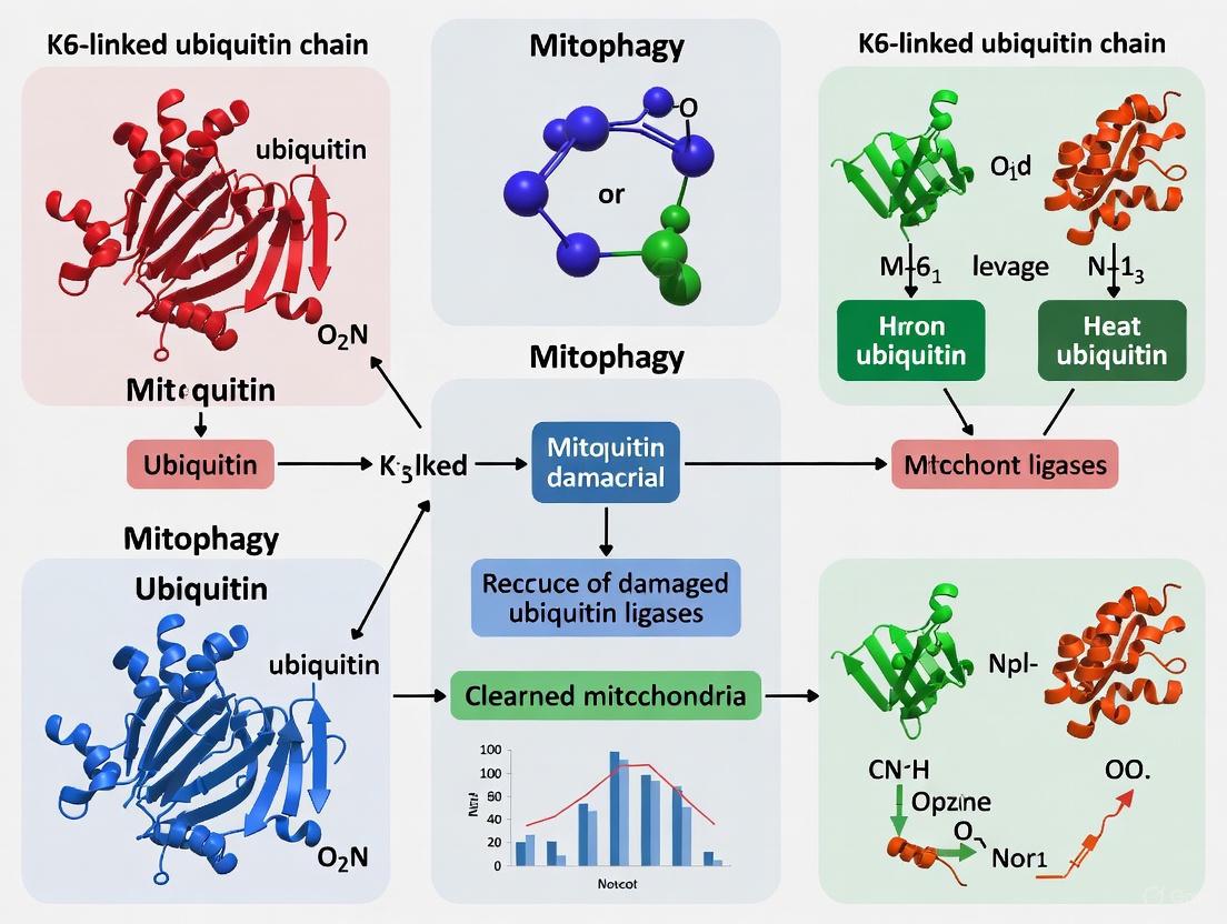
K6-linked ubiquitin chains represent a fascinating and understudied facet of the ubiquitin code. Although they account for a smaller fraction of cellular ubiquitin conjugates, emerging research has positioned them as critical regulators in essential quality control pathways, particularly in mitochondrial homeostasis and the pathogenesis of Parkinson's disease (PD) [2] [1]. Early studies linked K6 chains to the DNA damage response and the E3 ligase BRCA1 [1]. However, more recent work has uncovered their significant role in PINK1/Parkin-mediated mitophagy, where they contribute to the precise regulation of mitochondrial quality control [2] [3]. The structural architecture of K6-linked diubiquitin is distinct from other chain types, providing a molecular basis for its unique biological functions and recognition by specific enzymes [4].
This technical guide provides an in-depth exploration of K6-linked ubiquitin chains, focusing on their molecular mechanisms, detection methodologies, and functional implications in cellular quality control and human disease.
Molecular Mechanisms and Functional Roles in Mitophagy
The PINK1/Parkin Pathway: A Hub for K6-Linked Ubiquitination
The PINK1/Parkin pathway is a cornerstone of mitochondrial quality control, and K6-linked ubiquitin chains are intimately involved in its regulation. When mitochondria are damaged and lose their membrane potential, the kinase PINK1 accumulates on the outer mitochondrial membrane (OMM) [5] [6]. Here, it phosphorylates both the E3 ubiquitin ligase Parkin and ubiquitin itself at Ser65, activating Parkin and initiating a feed-forward amplification loop [5] [3].
Active Parkin ubiquitinates numerous proteins on the OMM, such as Mitofusins (Mfn1, Mfn2), VDAC1, and Miro1 [5] [7]. While Parkin assembles multiple chain types (including K6, K11, K48, and K63), research indicates it generates predominantly non-canonical K6-linked ubiquitin chains on itself and mitochondrial substrates during this process [2] [3]. These K6 chains on Parkin were initially thought to protect it from proteasomal degradation, but their precise function is complex and context-dependent [2].
The following diagram illustrates the key regulatory steps involving K6-linked ubiquitin in the PINK1/Parkin mitophagy pathway:
Deubiquitinating Enzymes (DUBs) as Specific Regulators of K6 Chains
The attachment of K6-linked ubiquitin is a reversible modification, tightly controlled by deubiquitinating enzymes (DUBs) with linkage specificity. The DUB USP8 has been identified as a key regulator that preferentially removes K6-linked ubiquitin chains from Parkin itself [2] [7]. This deubiquitination is critical for the efficient recruitment of Parkin to depolarized mitochondria and for the subsequent elimination of those mitochondria via mitophagy [2]. Mechanistically, USP8-mediated removal of K6 chains from Parkin is thought to release an auto-inhibited state, promoting Parkin's activity and turnover, which in turn facilitates mitophagy [2] [7].
Other DUBs also influence this pathway. For instance, USP30, another mitochondrial DUB, antagonizes Parkin-mediated ubiquitination—including K6 chains—and thereby inhibits mitophagy [1] [8]. The activity of these DUBs is itself regulated; PINK1-mediated phosphorylation of ubiquitin can impede certain DUBs, adding another layer of complexity to the system [3]. The bacterial DUB LotA from Legionella pneumophila has also been characterized for its remarkable specificity for K6-linked chains, making it a valuable tool for research [9].
Table 1: Deubiquitinating Enzymes (DUBs) Regulating K6-Linked Ubiquitination in Mitophagy
| DUB | Effect on Mitophagy | Mechanism of Action | Specificity |
|---|---|---|---|
| USP8 | Promotes [2] [6] | Removes K6-linked ubiquitin chains from Parkin, releasing auto-inhibition and promoting its recruitment to mitochondria. [2] [7] | Preferential for K6-linked chains on Parkin [2] |
| USP30 | Inhibits [1] [8] | Antagonizes Parkin-mediated ubiquitination on mitochondrial substrates, counteracting the ubiquitin signal for mitophagy. [1] [8] | Broad, but acts on Parkin targets [8] |
| LotA | N/A (Bacterial) | Used as a research tool due to its high specificity for cleaving K6-linked poly-ubiquitin chains. [9] | Highly specific for K6-linkages [9] |
Additional E3 Ligases Implicated in K6 Chain Assembly
While Parkin is a key E3 ligase for K6 chains in mitophagy, proteomic screens using K6-specific affinity reagents have identified other ligases capable of assembling K6-linked ubiquitin. The HECT-family E3 ligase HUWE1 has been identified as a major source of cellular K6 chains [1]. It can assemble K6-, K11-, and K48-linked chains in vitro, and cells lacking HUWE1 show significantly reduced global levels of K6 ubiquitination [1]. Furthermore, HUWE1 decorates the mitochondrial protein Mitofusin-2 (Mfn2) with K6-linked ubiquitin, highlighting its role in mitochondrial regulation [1]. The RBR-family E3 ligases RNF144A and RNF144B have also been shown to assemble K6-linked chains in vitro [1].
Experimental Methodologies for Studying K6 Linkages
Linkage-Specific Affinity Reagents
A significant hurdle in studying atypical ubiquitin chains has been the lack of high-quality, specific detection tools. Recent advances have led to the development of K6-linkage-specific affimers [1]. Affimers are small, stable protein scaffolds (based on the cystatin fold) with randomized loops that can be selected to bind specific epitopes with high affinity and specificity [1].
The characterized K6 affimer binds tightly to K6-diubiquitin with minimal cross-reactivity to other linkage types [1]. Crystallographic studies reveal that this specificity arises from a dimeric binding mode where the affimer dimer presents two binding surfaces that optimally engage the I44 patches of two ubiquitin moieties in a K6-linked diubiquitin, with a defined distance and orientation that other linkages cannot match [1]. These affimers, when biotinylated, are effective in western blotting, confocal microscopy, and pull-down assays, enabling the direct detection and enrichment of K6-ubiquitinated proteins from cellular lysates [1].
Table 2: Key Research Reagents for K6-Linked Ubiquitin Research
| Research Reagent / Tool | Type | Primary Function in Research | Key Application Examples |
|---|---|---|---|
| K6-Linkage Specific Affimer [1] | Non-antibody binding protein | High-affinity, linkage-specific recognition and detection of K6-linked polyubiquitin. | Western blotting, confocal fluorescence microscopy, immunoprecipitation/enrichment of K6-ubiquitinated proteins. [1] |
| K6-linked Diubiquitin [4] | Defined ubiquitin chain | Structural and biochemical standard; substrate for enzyme assays. | X-ray crystallography (e.g., PDB: 2XK5 [4]), in vitro DUB and E3 ligase activity assays. |
| LotA DUB Domain [9] | Deubiquitinating Enzyme | Highly specific cleavage of K6-linked poly-ubiquitin chains. | Tool for validating K6-linkage in samples, studying K6-specific signaling in cellular models. [9] |
| USP8 siRNA/shRNA [2] | Genetic Tool | Knockdown of USP8 to study its functional role in the PINK1/Parkin pathway. | Investigating the effects of persistent K6-ubiquitination on Parkin activity and mitophagy efficiency. [2] |
Structural Biology and Biophysical Techniques
Understanding the architecture and recognition of K6-linked chains is fundamental. The crystal structure of K6-linked diubiquitin (PDB: 2XK5) has been solved, revealing a conformation that is distinct from other linkage types such as K48 or K63 [4] [1]. This unique structure explains how specific DUBs and affinity reagents achieve linkage selectivity.
Techniques like isothermal titration calorimetry (ITC) and surface plasmon resonance (SPR) are used to quantitatively characterize the affinity and kinetics of interactions with K6 chains. For example, ITC confirmed the high-affinity binding of the K6 affimer to K6-diubiquitin and revealed a 2:1 stoichiometry (affimer:diUb), hinting at the dimeric binding mechanism later confirmed by crystallography [1]. SPR further demonstrated that specificity is achieved through very slow dissociation rates (off-rates) only for the cognate K6 chain [1].
The following workflow diagram outlines a key experimental strategy for identifying K6-specific interactors and biological functions:
Cellular Assays for Functional Validation
To validate the functional role of K6 ubiquitination in a physiological context, researchers employ several cellular models and assays:
- Parkin Translocation Assay: U2OS or HeLa cells stably expressing GFP-Parkin are treated with mitochondrial uncouplers like CCCP. The translocation of Parkin from the cytosol to mitochondria is monitored by live-cell or fixed-cell microscopy. Knockdown of USP8 via siRNA delays this translocation, implicating K6 deubiquitination in Parkin activation [2].
- Mitophagy Flux Assay: Cells are treated with CCCP for an extended period (e.g., 24 hours). The clearance of mitochondria is assessed by immunostaining for mitochondrial markers (e.g., TOM20, TIM23) or by using mitochondrial-targeted fluorescent probes (e.g., mt-Keima). Impairment of mitophagy upon USP8 knockdown demonstrates the pro-mitophagic role of K6 chain regulation [2].
- Detection of Endogenous K6 Chains: The improved K6 affimers enable the visualization of endogenous K6 chain dynamics by immunofluorescence and Western blotting under various stress conditions, such as DNA damage or mitochondrial depolarization [1].
Implications for Parkinson's Disease and Therapeutic Targeting
The precise regulation of K6-linked ubiquitination is critically important in neurons, and its dysregulation is strongly linked to Parkinson's disease. Recessive loss-of-function mutations in PARK2 (Parkin) and PINK1 are a well-established cause of early-onset familial PD [5] [6] [3]. As K6 chains are a major product of Parkin's E3 ligase activity in mitophagy, these mutations disrupt the entire mitochondrial quality control pathway [2] [3].
The discovery of DUBs like USP8 and USP30 as specific regulators of these pathways has opened new therapeutic avenues. Inhibiting a mitophagy-suppressing DUB like USP30 is being actively explored as a strategy to boost mitophagy and compensate for impaired PINK1/Parkin function in PD [8]. Conversely, the role of USP8 appears complex, as its inhibition can also be protective in some PD models, suggesting nuanced biology that requires further investigation [7]. The development of K6-linkage specific tools like affimers will accelerate the discovery of new therapeutic targets and biomarkers by allowing researchers to precisely monitor the dynamics of this ubiquitin linkage in healthy and diseased states [1].
K6-linked ubiquitin chains have evolved from a poorly understood "atypical" linkage to a key regulatory element in one of the most critical cellular quality control pathways. Their role in fine-tuning the PINK1/Parkin-mediated mitophagy pathway underscores their importance in maintaining neuronal health and preventing Parkinson's disease. The ongoing development and refinement of specific research tools—including affimers, defined structural standards, and specific DUBs—are finally allowing researchers to decipher the unique "syntax" of the K6 ubiquitin code. A deeper molecular understanding of how K6 chains are assembled, recognized, and disassembled will not only fill a significant gap in fundamental biology but also provide a robust platform for developing novel therapeutics for neurodegenerative diseases.
The PINK1/Parkin pathway stands as a central quality control mechanism, identifying and selectively targeting damaged mitochondria for autophagic clearance. At the heart of this pathway is the generation of a specific ubiquitin signal on the mitochondrial outer membrane. This technical guide delves into the molecular mechanics of how the serine/threonine kinase PINK1 and the RBR E3 ubiquitin ligase Parkin function in concert to produce phosphorylated polyubiquitin chains, with a specific emphasis on the non-canonical K6-linked ubiquitin chains that serve as a critical signal for mitophagy. We detail the experimental methodologies used to elucidate this pathway, provide quantitative data on its components, and frame these findings within the context of Parkinson's disease (PD) research, discussing the implications for therapeutic drug development.
Mitochondrial dysfunction is a well-established contributor to the pathogenesis of Parkinson's disease (PD). Evidence from studies on sporadic PD cases, toxin models (e.g., MPTP, rotenone), and the identification of causative genes for familial PD has consistently pointed to defects in mitochondrial health as a key driver of neurodegeneration [10] [11] [12]. Among the most common causes of autosomal recessive early-onset PD are loss-of-function mutations in the genes encoding the proteins PINK1 (PTEN-induced kinase 1) and Parkin [10] [5]. Genetic studies, particularly in Drosophila melanogaster, placed these two proteins in a common pathway, with PINK1 acting upstream of Parkin to regulate mitochondrial integrity [13] [11]. The seminal discovery that Parkin is recruited to depolarized mitochondria to promote their autophagic degradation (mitophagy) provided a cellular function for this pathway [13] [11]. Subsequent research has revealed that this process is governed by an intricate interplay of post-translational modifications (PTMs), wherein PINK1-generated phosphorylation events activate Parkin and trigger a feed-forward loop that decorates damaged mitochondria with ubiquitin chains, most notably including K6-linked polyubiquitin [13] [14] [15]. This whitepaper provides an in-depth technical analysis of this axis, with a focus on the generation and role of K6-linked ubiquitin signals.
Molecular Mechanisms of the PINK1/Parkin Pathway
PINK1: The Mitochondrial Damage Sensor
PINK1 functions as a sophisticated sensor of mitochondrial health through a unique import-retrotranslocation-degradation cycle [10] [5].
- In Healthy Mitochondria: PINK1 is synthesized in the cytosol and imported into mitochondria via the TOM/TIM complexes (Translocase of Outer/Inner Membrane). Its N-terminal Mitochondrial Targeting Sequence (MTS) is cleaved by the Mitochondrial Processing Peptidase (MPP). Subsequently, it is cleaved within its transmembrane domain by the inner membrane protease PARL (Presenilin-associated rhomboid-like protein). The cleaved fragment (∼52 kDa) is retro-translocated to the cytosol and rapidly degraded by the proteasome via the N-end rule pathway, maintaining low basal PINK1 levels [10] [13] [5].
- In Damaged/Depolarized Mitochondria: Loss of the mitochondrial membrane potential (ΔΨm) prevents PINK1 import through the TIM23 complex. Full-length PINK1 (∼64 kDa) accumulates on the outer mitochondrial membrane (OMM), where it forms a stable ∼720 kDa complex with the TOM complex [10] [5]. The interaction with TOM7 is critical for this stabilization [10] [5]. On the OMM, PINK1 undergoes autophosphorylation (e.g., at Ser228), which is essential for its activation [5].
The following diagram illustrates the distinct fates of PINK1 in healthy versus damaged mitochondria.
Parkin: The Signal Amplifier
Parkin is a multi-domain RBR (RING-Between-RING) E3 ubiquitin ligase that exists in an autoinhibited state in the cytosol under normal conditions [15]. Its domains include:
- Ubiquitin-like (Ubl) domain: Involved in regulation and proteasome interaction.
- RING0, RING1, IBR, and RING2 domains: Zinc-coordinating domains responsible for E2 binding and catalysis. The catalytic cysteine residue (Cys431 in human Parkin) is located in the RING2 domain [15].
The activation of Parkin is a multi-step process initiated by PINK1:
- Initial Recruitment and Phosphorylation: Activated PINK1 on the OMM phosphorylates ubiquitin molecules (at Ser65) that are either present in the cytosol or pre-existing on mitochondrial proteins. This phospho-ubiquitin (pUb) serves as the initial recruitment signal for cytosolic Parkin [13] [14].
- Conformational Activation: Binding of pUb to Parkin, coupled with direct phosphorylation of Parkin's Ubl domain at Ser65 by PINK1, triggers a dramatic conformational change. This releases the autoinhibitory interactions, exposing the E2-binding site on the RING1 domain and the catalytic Cys431 residue in the RING2 domain [5] [15].
- Feed-forward Amplification Loop: Activated Parkin ubiquitinates numerous OMM proteins (e.g., MFN1/2, Miro, VDAC1). These ubiquitin chains are, in turn, phosphorylated by PINK1, generating more pUb signals. This creates a powerful feed-forward loop that recruits and activates additional Parkin molecules, leading to the massive accumulation of ubiquitin chains on the damaged mitochondrion [13] [5] [14].
K6-Linked Ubiquitin Chains: The Key Effector Signal
Among the various types of polyubiquitin chains that Parkin assembles (including K48, K63, and K11), non-canonical K6-linked ubiquitin chains have been identified as a predominant signal in PINK1/Parkin-mediated mitophagy [13] [14] [15]. While the precise functional distinction between K6 and other chain types is still under investigation, they are thought to play a specific role in signaling for autophagic clearance. Parkin can also autoubiquitinate with K6-linked chains, which may regulate its own stability and activity [15].
Table 1: Key Ubiquitin Chain Types Generated by Parkin in Mitophagy
| Chain Type | Linkage | Proposed Role in PINK1/Parkin Pathway |
|---|---|---|
| K6-linked | Lys6 | Predominant mitophagy signal; role in autophagosome recruitment; Parkin autoubiquitination [13] [15] |
| K48-linked | Lys48 | Canonical signal for proteasomal degradation; may target specific OMM proteins for proteasomal processing to facilitate mitophagy [15] |
| K63-linked | Lys63 | Often involved in signaling and endocytosis; potential role in recruiting autophagy adapters [15] |
The following diagram synthesizes the entire PINK1/Parkin activation cascade and the role of K6-linked ubiquitin.
Experimental Protocols for Studying the Pathway
This section outlines key methodologies used to dissect the PINK1/Parkin axis and K6-linked ubiquitination.
Inducing and Monitoring Mitophagy in Cell Culture
Protocol: CCCP-Induced Mitophagy and Parkin Translocation Assay
- Principle: The protonophore Carbonyl cyanide m-chlorophenyl hydrazone (CCCP) collapses ΔΨm, inhibiting PINK1 import and triggering the pathway [10] [13].
- Procedure:
- Cell Line: Use HeLa, SH-SY5Y, or other cell lines stably or transiently expressing GFP-Parkin or similar fluorescently tagged Parkin.
- Treatment: Treat cells with 10-20 µM CCCP for 1-6 hours. A time course is recommended to capture Parkin translocation (early event) and mitochondrial clearance (later event).
- Monitoring:
- Imaging: Fix cells and immunostain for a mitochondrial marker (e.g., TOMM20) or use a mito-RFP. Parkin translocation is observed as a shift from diffuse cytosolic fluorescence to punctate structures co-localizing with mitochondria.
- Immunoblotting: Analyze whole-cell lysates for markers of mitophagy, such as a decrease in mitochondrial protein levels (e.g., TOMM20) and an increase in LC3-II.
Detecting Ubiquitin Chain Linkage Types
Protocol: Linkage-Specific Ubiquitin Immunoprecipitation and Immunoblotting
- Principle: To specifically identify K6-linked ubiquitin chains generated during mitophagy using linkage-specific antibodies.
- Procedure:
- Induction & Lysis: Induce mitophagy with CCCP in cells expressing Parkin. Lyse cells under denaturing conditions (e.g., 1% SDS lysis buffer) to preserve PTMs.
- Immunoprecipitation (IP): Dilute the lysate to reduce SDS concentration and perform IP using an antibody against a known mitochondrial ubiquitination target (e.g., anti-MFN1, anti-Miro) or an anti-ubiquitin antibody.
- Immunoblotting: Analyze the IP eluates and total lysates by SDS-PAGE and Western blotting.
- Probing: Probe the blots with K6-linkage specific ubiquitin antibodies (commercially available) alongside total ubiquitin and phosphorylation-specific (p-Ser65 ubiquitin) antibodies.
Analyzing PINK1 and Parkin Phosphorylation
Protocol: Phos-tag SDS-PAGE for Resolving Phosphorylated Species
- Principle: Phos-tag gels retard the migration of phosphorylated proteins, allowing separation from their non-phosphorylated counterparts.
- Procedure:
- Sample Preparation: Lyse control and CCCP-treated cells.
- Gel Electrophoresis: Resolve proteins using standard SDS-PAGE and Phos-tag SDS-PAGE in parallel.
- Immunoblotting: Transfer proteins to a membrane and immunoblot for PINK1 or Parkin.
- Interpretation: The appearance of higher molecular weight bands for PINK1 (full-length ~64 kDa) and Parkin (phosphorylated form) on the Phos-tag gel, but not the standard gel, confirms phosphorylation. This can be validated by treating lysates with lambda phosphatase.
Table 2: Key Reagents for Studying PINK1/Parkin-Mediated Mitophagy
| Reagent Category | Specific Examples | Function/Application |
|---|---|---|
| Inducers of Mitophagy | CCCP, Valinomycin, Oligomycin/Antimycin A | Dissipate ΔΨm to trigger PINK1 stabilization and Parkin activation [10] [13] |
| Chemical Inhibitors | MG132 (Proteasome), Bafilomycin A1 (Lysosome), 3-Methyladenine (Autophagy) | Block specific degradation steps to study pathway intermediates |
| Kinase Inhibitors | Kinase-dead PINK1 mutants, PINK1-specific small molecule inhibitors (e.g., KIN-001-138) | Validate PINK1-specific phosphorylation events [13] |
| Linkage-Specific Antibodies | Anti-K6-linkage Ubiquitin, Anti-K48-linkage Ubiquitin, Anti-K63-linkage Ubiquitin, Anti-p-Ser65-Ubiquitin | Detect specific ubiquitin chain types and PINK1 activity output [13] [15] |
| Cell Lines | Parkin/PINK1 knockout HeLa or HEK293T cells; Wild-type/mutant overexpression models | Provide isogenic backgrounds to study gene function and pathogenic mutations [13] [11] |
| Deubiquitinase (DUB) Tools | USP8, USP15, USP30 (overexpression or knockdown) | Study regulation and reversal of Parkin-mediated ubiquitination [13] [14] |
The Scientist's Toolkit: Essential Research Reagents
A curated table of critical reagents for investigating the PINK1/Parkin pathway and K6-linked ubiquitination.
Table 3: The Scientist's Toolkit for PINK1/Parkin and K6-Ubiquitin Research
| Reagent / Tool | Function / Role in Pathway | Example Use Case |
|---|---|---|
| CCCP | Protonophore; uncouples oxidative phosphorylation, collapses ΔΨm. | Standard tool to induce PINK1 stabilization and Parkin recruitment in vitro [10] [13]. |
| Anti-p-Ser65-Ubiquitin Antibody | Detects PINK1 kinase activity output. | Key readout for PINK1 activation; used in Western blot and immunofluorescence [13] [14]. |
| Anti-K6-linkage Ubiquitin Antibody | Specifically recognizes K6-linked polyubiquitin chains. | Critical for confirming the presence of the primary ubiquitin signal generated by Parkin in mitophagy [13] [15]. |
| Phos-tag Acrylamide | Binds phosphate groups, retarding migration of phosphorylated proteins in gels. | Essential for detecting the mobility shift of phosphorylated PINK1 and Parkin [5]. |
| Parkin RBR Domain Mutants (C431F) | Catalytically inactive Parkin; RING2 domain mutation. | Used as a negative control to distinguish Parkin-dependent ubiquitination from background [15]. |
| TUBE (Tandem Ubiquitin Binding Entity) | High-affinity ubiquitin-binding reagent. | Pulls down and enriches ubiquitinated proteins from cell lysates for proteomic or Western analysis. |
| Mito-QC Reporter Cell Line | pH-sensitive fluorescent reporter (mCherry-GFP) targeted to mitochondria. | Allows quantitative measurement of mitophagy via fluorescence imaging (GFP quenched in lysosomes, mCherry persists) [5]. |
Quantitative Data and Pathogenic Mutations
Quantitative Insights into the Pathway
The PINK1/Parkin pathway involves precise molecular stoichiometries and kinetics.
Table 4: Quantitative Data on PINK1, Parkin, and Associated Factors
| Parameter | Quantitative Measure / Detail | Context / Significance |
|---|---|---|
| PINK1 Molecular Complex | ~720 kDa complex with TOM | Indicates PINK1 forms a large, stable signaling complex on the OMM of damaged mitochondria [10]. |
| Zinc Ions per Parkin Molecule | 8 ions | Parkin's structural stability is dependent on coordinating 8 zinc ions across its RING domains [15]. |
| Parkin Pathogenic Mutations | >120 identified mutations | Highlights the critical importance of all Parkin domains; mutations are spread throughout Ubl, RING0, RING1, IBR, and RING2 domains [15]. |
| Key Parkin Phosphorylation Site | Ser65 (within Ubl domain) | Phosphorylation by PINK1 at this site is a critical step in relieving Parkin's autoinhibition [5] [15]. |
| Key Ubiquitin Phosphorylation Site | Ser65 | Phosphorylation by PINK1 transforms ubiquitin into a high-affinity receptor for Parkin [13] [14]. |
PINK1 and Parkin Mutations in Parkinson's Disease
Mutations in PINK1 and Parkin lead to a loss of the mitochondrial quality control function, resulting in the accumulation of damaged mitochondria, which contributes to neuronal toxicity and death, particularly in vulnerable populations like dopaminergic neurons of the substantia nigra [12] [16]. These neurons have high energetic demands and are especially susceptible to bioenergetic stress. The inability to clear damaged mitochondria via the PINK1/Parkin pathway is thus a key factor in the pathogenesis of autosomal recessive PD, underscoring the critical nature of the K6-linked ubiquitin signal in maintaining neuronal health [12].
The PINK1/Parkin axis represents a sophisticated molecular machinery for maintaining mitochondrial health, with the generation of K6-linked ubiquitin chains being a central event in the mitophagy signaling cascade. A deep understanding of the structural mechanisms of Parkin activation, the specificity of ubiquitin chain formation, and the regulatory roles of phosphorylation and deubiquitination is paramount. Future research should focus on:
- Defining the precise structural role of K6-linked chains in recruiting the autophagic machinery versus other chain types.
- Developing more specific agonists and antagonists of the pathway for therapeutic intervention in PD and other diseases linked to mitochondrial dysfunction.
- Exploring the pathway's activity and dynamics in human neurons and in vivo models to fully understand its role in neuronal vulnerability.
The continued elucidation of this pathway not only advances our fundamental knowledge of cellular quality control but also opens promising avenues for developing disease-modifying therapies for Parkinson's disease by targeting the root cause of mitochondrial dysfunction.
The post-translational modification of proteins with polyubiquitin chains is a fundamental mechanism for controlling cellular protein quality control. Among the eight possible linkage types, K6-linked ubiquitin chains have emerged as particularly important regulators of mitochondrial autophagy (mitophagy), a process critical for cellular health and neuronal survival. Unlike the well-characterized K48-linked chains that target substrates for proteasomal degradation or K63-linked chains involved in signaling and trafficking, K6-linked chains serve specialized functions in orchestrating autophagic responses to damaged organelles [2] [17] [18]. In the context of Parkinson's disease research, understanding K6-chain biology has become paramount, as these chains directly regulate the function of Parkin, an E3 ubiquitin ligase mutated in familial forms of the disease [2] [19] [7].
The process of selective autophagy requires precise recognition of specific cargo, such as damaged mitochondria, and their subsequent encapsulation within double-membraned autophagosomes for lysosomal degradation. K6-linked ubiquitin chains function as critical molecular signals in this process, operating at the interface between cargo recognition and autophagosome assembly. Through specialized receptor proteins, these chains facilitate the recruitment of core autophagy machinery to designated cargo, thereby ensuring spatial and temporal specificity in organelle turnover [20] [2]. This review examines the molecular mechanisms by which K6-linked ubiquitin chains orchestrate autophagosome encapsulation through adaptor proteins, with particular emphasis on implications for Parkinson's disease pathogenesis and therapeutic development.
Molecular Mechanisms of K6-Chain Signaling in Mitophagy
The PINK1-Parkin Axis and K6-Chain Dynamics
The PINK1-Parkin pathway represents the best-characterized mechanism for K6-linked ubiquitin signaling in mitophagy. Under conditions of mitochondrial damage, the kinase PINK1 accumulates on the outer mitochondrial membrane where it undergoes trans-autophosphorylation [20] [5]. This activation enables PINK1 to phosphorylate both ubiquitin and Parkin at Ser65, triggering a conformational change that relieves Parkin's autoinhibition and activates its E3 ligase function [20] [5]. Once activated, Parkin ubiquitinates numerous mitochondrial outer membrane proteins, generating a ubiquitin "coat" that serves as a platform for autophagosome assembly [20].
Within this cascade, Parkin undergoes autoubiquitination with K6-linked chains, which maintains the enzyme in an autoinhibited state [2] [19] [7]. The deubiquitinating enzyme USP8 specifically targets and removes these K6-linked chains from Parkin, thereby facilitating its recruitment to damaged mitochondria and promoting mitophagy [2] [19]. This regulatory mechanism ensures that Parkin activation occurs with appropriate timing and specificity, preventing premature engagement with healthy mitochondria. The precise removal of K6-linked ubiquitin conjugates by USP8 represents a critical regulatory checkpoint in the mitophagy cascade, with dysfunction in this process directly implicated in Parkinson's disease pathogenesis [2] [7].
Table 1: Key Proteins in K6-Linked Ubiquitin Signaling During Mitophagy
| Protein | Function | Role in K6 Signaling | Association with PD |
|---|---|---|---|
| Parkin | E3 ubiquitin ligase | Generates K6-autoubiquitination; regulated by USP8 | Mutated in autosomal recessive forms |
| PINK1 | Ser/Threonine kinase | Phosphorylates Parkin and ubiquitin to initiate pathway | Mutated in autosomal recessive forms |
| USP8 | Deubiquitinating enzyme | Removes K6-linked chains from Parkin to promote activation | Therapeutic target; inhibition protective in models |
| TOM Complex | Mitochondrial import | Regulates PINK1 stability on damaged mitochondria | Not directly linked |
| Autophagy adapters (OPTN, NDP52) | Link ubiquitin to LC3 | Recognize ubiquitinated mitochondria (various chains) | OPTN mutations associated with neurodegeneration |
Structural Basis of K6-Chain Recognition
The molecular recognition of K6-linked ubiquitin chains by interacting proteins represents a key determinant in their signaling specificity. Structural studies reveal that ubiquitin maintains a conserved β-grasp fold comprising a five-stranded β sheet cradling a central α helix, creating a stable platform for interactions [18]. While the three-dimensional structures of different ubiquitin linkage types exhibit distinct characteristics, K6-linked chains display unique surface properties that enable selective recognition by specific ubiquitin-binding domains.
The tandem ubiquitin-binding entities (TUBEs) technology has been particularly valuable for studying K6-chain recognition, as these engineered reagents contain multiple ubiquitin-associated domains that bind polyubiquitin chains with nanomolar affinities [17] [21]. Chain-specific TUBEs can differentiate between linkage types, allowing researchers to isolate and characterize K6-linked ubiquitination events in complex biological samples [17]. This approach has revealed that K6-chains create specific interaction surfaces that are recognized by a subset of autophagy receptors, particularly those involved in damaged organelle clearance. The structural basis for this selectivity involves complementary electrostatic interactions and hydrophobic surfaces that distinguish K6-linkages from other chain types [18].
Experimental Analysis of K6-Linked Ubiquitination
Methodologies for Detecting K6-Linked Ubiquitination
The analysis of K6-linked ubiquitination presents technical challenges due to the complexity of the ubiquitin system and the relative scarcity of K6-chains compared to major linkage types. Current methods for studying K6-ubiquitination employ multiple complementary approaches:
Chain-Specific TUBE-Based Affinity Enrichment: This high-throughput method uses K6-linkage-specific TUBEs coated on microplates or magnetic beads to selectively capture proteins modified with K6-linked chains from cell lysates [17] [21]. The protocol involves: (1) preparing cell lysates under nondenaturing conditions that preserve ubiquitination; (2) incubating lysates with chain-specific TUBEs; (3) extensive washing to remove nonspecific interactions; and (4) detection of captured proteins by immunoblotting or mass spectrometry. This approach enables quantitative analysis of K6-ubiquitination dynamics in response to cellular stimuli or pharmacological interventions [17].
Genetic Code Expansion and Chemical Synthesis: Novel methods for generating defined branched ubiquitin chains incorporate noncanonical amino acids through amber codon suppression in E. coli, allowing precise placement of modification sites [22]. The protocol involves: (1) site-specific incorporation of protected lysine analogs at K6 positions; (2) sequential chemical ligation of ubiquitin building blocks; (3) deprotection and refolding of assembled chains; and (4) purification of structurally defined K6-linked chains for biochemical studies [22]. These synthetically defined chains serve as critical tools for structural studies and enzyme characterization.
siRNA-Based Functional Screening: Genome-wide or DUB-focused siRNA screens identify regulators of K6-linked ubiquitin signaling [2] [7]. The methodology includes: (1) transfection of siRNA libraries targeting DUBs or ubiquitin-related proteins; (2) induction of mitophagy with CCCP or other depolarizing agents; (3) monitoring Parkin translocation by live-cell imaging; and (4) assessment of mitophagy efficiency by mitochondrial protein degradation assays [2]. This approach identified USP8 as a key regulator of K6-linked Parkin autoubiquitination [2] [19].
Table 2: Experimental Approaches for Studying K6-Linked Ubiquitination
| Method | Key Applications | Advantages | Limitations |
|---|---|---|---|
| Chain-specific TUBEs | Enrichment of K6-ubiquitinated proteins from cell lysates | High affinity and specificity; adaptable to HTS formats | May not distinguish branched vs homogenous chains |
| Genetic code expansion | Production of defined K6-linked chains | Precise control over chain architecture; incorporation of probes | Low yield; technically challenging |
| siRNA screening | Identification of K6-chain regulators | Unbiased discovery; functional assessment | Off-target effects; compensatory mechanisms |
| Mass spectrometry | Identification of K6-ubiquitination sites | Comprehensive mapping of modification sites | Requires enrichment; low sensitivity for rare sites |
| Drosophila models | In vivo functional validation | Whole-organism physiology; genetic manipulation | Evolutionary conservation limitations |
Analyzing K6-Chain Function in Cellular Models
The functional assessment of K6-linked ubiquitination in mitophagy employs well-established cellular models, primarily featuring Parkin overexpression in response to mitochondrial depolarization. The standard protocol for investigating K6-chain dynamics includes:
Cell Culture and Treatment: U2OS or HeLa cells stably expressing GFP-Parkin are maintained under standard conditions. Mitochondrial depolarization is induced with 10-20 μM carbonyl cyanide m-chlorophenyl hydrazone (CCCP) for 1-24 hours, depending on the specific readout [2] [19]. For USP8 inhibition studies, cells are pretreated with specific inhibitors or transfected with USP8-targeting siRNA 48-72 hours before CCCP treatment.
Parkin Translocation Assay: Following mitochondrial depolarization, Parkin translocation to mitochondria is monitored by live-cell imaging or fixed-cell immunofluorescence [2]. Cells are imaged at regular intervals (e.g., every 15 minutes) for 2-4 hours after CCCP addition. The percentage of cells with mitochondrial Parkin localization is quantified at each time point, with USP8 depletion typically delaying but not completely preventing Parkin translocation [2].
Mitophagy Assessment: Efficient mitophagy is evaluated after prolonged CCCP treatment (18-24 hours) by monitoring the degradation of mitochondrial markers such as TOM20, TIM23, or COX1 via immunoblotting or immunofluorescence [2] [19]. Alternatively, mitophagy reporter constructs (e.g., mt-Keima) provide quantitative assessment of mitochondrial delivery to lysosomes.
Ubiquitination Status Analysis: Parkin autoubiquitination is assessed by immunoprecipitation of Parkin from cell lysates followed by anti-ubiquitin immunoblotting [2] [19]. Linkage specificity is determined using chain-specific TUBEs or ubiquitin mutants that restrict chain formation to specific lysines.
Diagram 1: K6-linked ubiquitin signaling pathway in PINK1-Parkin mediated mitophagy. USP8-mediated removal of K6 chains from Parkin enhances its E3 ligase activity, promoting substrate ubiquitination and autophagosome recruitment.
The Scientist's Toolkit: Key Research Reagents
Advancing research on K6-linked ubiquitin chains requires specialized reagents and tools designed to probe the specificity and function of this unique modification.
Table 3: Essential Research Reagents for Studying K6-Linked Ubiquitination
| Reagent/Tool | Specific Example | Research Application | Commercial Source/Reference |
|---|---|---|---|
| K6-specific TUBEs | K6-TUBE coated microplates | Selective enrichment of K6-ubiquitinated proteins | LifeSensors [17] [21] |
| Linkage-specific antibodies | Anti-K6 ubiquitin antibodies | Detection of K6 chains by immunoblotting/IF | Multiple vendors |
| Ubiquitin mutants | UbK6R (K6-to-arg mutant) | Determining K6-chain specificity in cells | Commercial DNA constructs |
| DUB inhibitors | USP8-specific inhibitors | Probing K6-chain dynamics and function | In development [7] |
| Defined K6 chains | Chemically synthesized K6-diUb | Structural and biochemical studies | Custom synthesis [22] |
| Parkin activity sensors | Phospho-Parkin (Ser65) antibodies | Monitoring Parkin activation | Commercial antibodies |
| Mitophagy reporters | mt-Keima, mt-YFP | Quantifying mitophagy efficiency | Addgene plasmids |
K6-Chains in Parkinson's Disease and Therapeutic Implications
The regulation of K6-linked ubiquitination has profound implications for Parkinson's disease pathogenesis and treatment strategies. Mutations in Parkin represent a major cause of autosomal recessive early-onset PD, and these loss-of-function mutations disrupt the carefully orchestrated balance of ubiquitination and deubiquitination necessary for efficient mitophagy [2] [7]. The discovery that USP8 regulates Parkin through removal of K6-linked autoubiquitination has highlighted this specific linkage as a therapeutic target for restoring mitochondrial quality control in PD [19] [7].
In cellular and Drosophila models of PD, suppression of USP8 activity has demonstrated protective effects, including improved mitochondrial function, enhanced dopaminergic neuron survival, and rescue of locomotor deficits [7]. These benefits are attributed to facilitated Parkin activation and subsequent enhancement of mitophagy, enabling more efficient clearance of damaged mitochondria [2] [7]. Importantly, USP8 inhibition has shown efficacy not only in Parkin deficiency models but also in α-synuclein overexpression models, suggesting broader applicability across PD subtypes [7]. The development of specific USP8 inhibitors represents an promising therapeutic strategy for modulating K6-linked ubiquitination in Parkinson's disease and other conditions characterized by mitochondrial dysfunction.
Diagram 2: Experimental workflow for analyzing K6-linked ubiquitination using chain-specific TUBEs, with parallel validation methods to confirm functional outcomes.
K6-linked ubiquitin chains represent sophisticated regulatory elements in the complex coordination of autophagosome encapsulation, serving as both negative regulators of Parkin activity through autoubiquitination and as positive signals for adaptor protein recruitment. The precise manipulation of K6-chain dynamics, particularly through targeting specific DUBs like USP8, offers promising therapeutic avenues for Parkinson's disease and other conditions characterized by mitochondrial quality control defects. As research methodologies advance, particularly in the areas of chain-specific detection tools and chemical biology approaches for generating defined ubiquitin architectures, our understanding of K6-chain function will continue to deepen.
Future research directions should focus on elucidating the structural basis for K6-chain recognition by autophagy adaptors, developing more specific pharmacological modulators of K6-chain writers and erasers, and exploring potential cross-talk between K6-linkages and other ubiquitin chain types in forming branched architectures. The integration of K6-chain biology into broader cellular signaling networks will ultimately provide a more comprehensive understanding of how ubiquitin linkage specificity coordinates cellular homeostasis, potentially revealing novel therapeutic targets for neurodegenerative disease.
While the E3 ubiquitin ligase Parkin is a well-characterized regulator of mitochondrial quality control, its role in the formation of atypical K6-linked ubiquitin chains has remained less defined. Recent research has uncovered alternative E3 ligases with a more explicit capacity for synthesizing K6-linked ubiquitination. This whitepaper delves into the emerging role of K6-linked ubiquitin chains in cellular stress response, moving beyond the canonical Parkin-mediated mitophagy pathway. We explore the identification of the RING-in-between-RING (RBR) E3 ligase RNF14 as a specific builder of K6-linked chains and examine the machinery that recognizes this atypical linkage. Framed within the context of Parkinson's disease research, this review synthesizes current biochemical and proteomic evidence to present a revised model of K6-linked ubiquitination, providing methodologies and resources to advance this nascent field.
Ubiquitination is a crucial post-translational modification that regulates a myriad of eukaryotic functions, including protein degradation, DNA repair, and cell signaling. The diversity of ubiquitin signals—often termed the "ubiquitin code"—stems from the ability of ubiquitin to form polymers through its N-terminal methionine or seven lysine residues (K6, K11, K27, K29, K33, K48, K63), each potentially conferring distinct functional outcomes [23] [24]. Among these, K48-linked chains are the principal signal for proteasomal degradation, while K63-linked and M1-linked chains are heavily implicated in signaling pathways related to inflammation and immune responses [23]. In contrast, the biological functions of K6-linked ubiquitin chains have remained more elusive.
The interest in K6-linked chains has intensified within mitochondrial quality control and neurodegeneration research. Mutations in the PRKN gene, encoding the Parkin E3 ubiquitin ligase, are a leading cause of early-onset Parkinsonism [25] [2]. Parkin, a RING-InBetweenRING-Rcat (RBR) E3 ligase, plays a vital role in the clearance of damaged mitochondria via mitophagy by ubiquitylating numerous mitochondrial outer membrane proteins [25]. Early proteomic studies identified K6-linked ubiquitination among the chain types deposited by Parkin on mitochondria, suggesting a potential role in the mitophagy process [2]. However, the precise function of K6-linked chains in this context and the identity of other E3 ligases capable of building this linkage have only recently come to light.
Parkin and K6-Linked Ubiquitin: A Complex Relationship
Parkin is a primarily cytosolic E3 ubiquitin ligase that is recruited to damaged mitochondria to initiate their clearance. Under basal conditions, Parkin exists in an auto-inhibited state. Upon mitochondrial damage, the kinase PINK1 accumulates on the outer mitochondrial membrane and phosphorylates both ubiquitin and Parkin’s ubiquitin-like (Ubl) domain at Ser65. This phosphorylation event triggers a conformational change that activates Parkin, leading to the ubiquitylation of numerous mitochondrial substrates [25] [2].
The model of Parkin as a builder of K6-linked chains is nuanced. Parkin functions with E2 enzymes such as UbcH7 and UbcH8 to ubiquitylate substrates, and it can auto-ubiquitylate itself. Early work suggested that Parkin can generate K6-linked chains in vitro [2]. Furthermore, the deubiquitinating enzyme USP8 was found to be a critical regulator of Parkin by preferentially removing K6-linked ubiquitin chains from it. Silencing USP8 impairs the recruitment of Parkin to depolarized mitochondria and subsequent mitophagy, indicating that the dynamic regulation of K6-linked ubiquitination on Parkin itself is essential for its function in mitochondrial quality control [2]. This suggests that K6-linked auto-ubiquitination may serve as a regulatory mechanism, perhaps by protecting Parkin from proteasomal degradation and thereby fine-tuning its activity [2].
Table 1: Key Regulatory Post-Translational Modifications of Parkin
| Modification Type | Residue(s) | Enzyme | Proposed Function |
|---|---|---|---|
| Phosphorylation | S65 | PINK1 | Canonical activation; promotes translocation to mitochondria [25] |
| Phosphorylation | S9 | AMPK, CaMK2 | Potential fine-tuning of activity [25] |
| Phosphorylation | S131 | p38 MAPK, Dyrk1A, Cdk5 | Constitutively phosphorylated; potential alternative regulation [25] |
| K6-linked Ubiquitination | Multiple | Parkin (auto) / USP8 (removal) | Regulates parkin recruitment to mitochondria; potential auto-inhibition [2] |
However, the direct role of Parkin in synthesizing K6-linked chains on mitochondrial substrates during mitophagy is less clear. Much of the research has focused on its role in building K48, K63, and K11-linked chains on substrates like mitofusins. This ambiguity has prompted the search for other E3 ligases that more explicitly utilize the K6 linkage.
RNF14: An Alternative E3 Ligase for K6-Linked Ubiquitination
A significant breakthrough in the field came from recent research into the cellular response to aldehyde stress. In a landmark 2023 study, Rahmanto et al. identified the RBR E3 ligase RNF14 as a specific builder of K6-linked ubiquitin chains [26] [27].
Experimental Workflow for Identifying RNF14 Function
The discovery of RNF14's role involved a sophisticated multi-step workflow:
- Stress Induction: Human cells were treated with formaldehyde, a reactive aldehyde known to induce RNA-protein crosslinks (RPCs).
- Pulldown and Proteomics: RPCs were isolated, and the associated proteins were analyzed using quantitative proteomics.
- Ubiquitin Linkage Analysis: The ubiquitination status of crosslinked proteins was examined, revealing a specific enrichment of K6-linked ubiquitin chains.
- E3 Ligase Identification: Through siRNA screening and functional validation, RNF14 was identified as the E3 ligase responsible for depositing K6-linked ubiquitin on RPCs.
- Effector Recruitment: The K6-linked ubiquitin chains were shown to be recognized and resolved by the VCP/p97 segregase complex, leading to the clearance of crosslinked proteins and the restoration of translation [26].
Functional Significance of RNF14-Mediated K6-Ubiquitination
This pathway represents a clear, Parkin-independent function for K6-linked ubiquitination. The RNF14-K6-Ub-VCP axis acts as a dedicated quality control system to resolve transcription- and translation-blocking RPCs, protecting cells from aldehyde toxicity [26] [27]. This reveals a novel, evolutionarily conserved stress response pathway where K6-linked chains serve as a specific tag for VCP-dependent processing.
Diagram 1: The RNF14-K6-Ub-VCP pathway for resolving RNA-protein crosslinks.
Recognition of K6-Linked Chains: The Role of TAB2/NZF
The function of any ubiquitin chain is ultimately determined by the proteins that recognize it. For K6-linked chains, a key reader is the NZF domain of TAB2 (and its homolog TAB3) [23]. TAB2 is a component of the TAK1 complex, which is a critical regulator of the NF-κB and JNK signaling pathways.
Structural Basis for K6-Linked Ubiquitin Recognition
Structural biology has been instrumental in understanding how TAB2 recognizes K6 linkages. The crystal structure of the TAB2-NZF domain in complex with K6-linked diubiquitin (K6-Ub2) was resolved at 1.99-Å resolution [23]. The analysis revealed that:
- TAB2-NZF simultaneously interacts with both the distal and proximal ubiquitin moieties of K6-Ub2.
- The binding mechanism is remarkably similar to how TAB2-NZF recognizes K63-Ub2, with the key difference being the flexible C-terminal region of the distal ubiquitin.
- This structural plasticity allows the NZF domain of TAB2 to exhibit dual specificity for both K6- and K63-linked ubiquitin chains [23].
This finding is significant because it suggests that K6-linked chains could potentially modulate inflammatory and survival signaling pathways by competing with or supplementing K63-chain binding to the TAK1 complex.
Table 2: Proteins with Documented Roles in K6-Linked Ubiquitin Pathways
| Protein | Domain/Type | Function Related to K6-Ub | Experimental Evidence |
|---|---|---|---|
| RNF14 | RBR E3 Ligase | Synthesizes K6-linked chains on RNA-protein crosslinks [26] | siRNA, ubiquitination assays, proteomics |
| TAB2/TAB3 | NZF Domain | Binds and recognizes K6-linked chains [23] | X-ray crystallography, pull-down assays |
| USP8 | Deubiquitinase (DUB) | Preferentially removes K6-linked chains from Parkin [2] | siRNA, DUB activity assays, mitophagy assays |
| VCP/p97 | AAA+ ATPase | Unfoldase recruited by K6-linked chains to resolve RPCs [26] [27] | Functional genetics, co-immunoprecipitation |
Research Reagents and Methodologies for K6-Linked Ubiquitin Studies
Advancing research into K6-linked ubiquitination requires specialized reagents and well-established protocols. Below is a toolkit for investigators in this field.
Research Reagent Solutions
Table 3: Essential Research Reagents for Studying K6-Linked Ubiquitination
| Reagent / Material | Function/Application | Example Usage |
|---|---|---|
| K6-linked Diubiquitin (K6-Ub2) | Structural and binding studies; DUB specificity profiling. | Used in crystallography to determine TAB2-NZF complex structure [23] [4]. |
| Linkage-Specific K6-Ub Antibodies | Detect endogenous K6-linked chains via immunoblotting/immunofluorescence. | Critical for validating K6-Ub enrichment in pathways like RPC resolution [26]. |
| NleL (E3 Ligase) | In vitro synthesis of K6-linked ubiquitin chains. | Used in biochemical synthesis of K6-Ub2 for experiments [23]. |
| UbcH7 (E2 Enzyme) | Cooperates with E3 ligases (e.g., Parkin, RNF14) for ubiquitin transfer. | Used with NleL and Parkin for in vitro K6-Ub2 synthesis [23] [2]. |
| siRNA/shRNA vs. USP8, RNF14 | Functional genetics to probe the role of specific enzymes in K6-Ub pathways. | USP8 KD delays Parkin recruitment [2]; RNF14 KD blocks RPC resolution [26]. |
Key Experimental Protocols
A. In Vitro Synthesis of K6-Linked Diubiquitin [23] This protocol is fundamental for generating the pure, defined chains needed for biochemical and structural studies.
- Reaction Setup: Combine the following components in a ligation buffer (50 mM Tris-HCl pH 9.0, 10 mM ATP, 10 mM MgCl2, 0.6 mM DTT):
- Ubiquitin-activating enzyme E1 (0.3 μM)
- Ubiquitin-conjugating enzyme E2 (UbcH7, 8 μM)
- Bacterial E3 ligase NleL (2.5 μM)
- Deubiquitinase OTUB1 (10 μM) - to trim excess chains and promote diubiquitin formation.
- Ubiquitin (e.g., K6R mutant and/or D77 mutant, 800 μM each).
- Incubation: Incubate the reaction at 37°C for 15 hours.
- Completion: Add a second dose of OTUB1 (10 μM) and incubate for another 2 hours to ensure the reaction goes to completion.
- Purification: Purify the K6-Ub2 product using ion-exchange and size-exclusion chromatography.
B. Assessing Mitophagy via Mitochondrial Protein Degradation [28] This cellular assay is used to monitor the functional outcome of Parkin-mediated mitophagy.
- Cell Culture & Treatment: Use SH-SY5Y or U2OS cells stably expressing GFP-Parkin. Induce mitophagy with mitochondrial uncouplers like CCCP or FCCP (e.g., 10-20 μM) for 6-24 hours.
- Inhibition (Optional): Treat cells with lysosomotropic agents like hydroxychloroquine (HCQ) to block autophagosome-lysosome fusion, confirming an autophagic mechanism.
- Immunoblotting: Harvest cells and analyze lysates by Western blotting. Mitophagy is indicated by the degradation of mitochondrial proteins such as:
- TOM20 (outer membrane)
- TIM23 (inner membrane)
- COXIV (matrix)
- Validation: Monitor Parkin recruitment via GFP fluorescence and LC3-I to LC3-II conversion as a general autophagy marker.
Discussion and Implications for Parkinson's Disease Research
The discovery of specific builders and readers of K6-linked ubiquitin chains opens new avenues for understanding cellular stress response and its intersection with neurodegenerative disease. The emerging picture suggests that K6-linked ubiquitination is not merely a redundant or minor pathway but a specific signal mobilized in response to distinct proteotoxic stresses, such as damaged mitochondria and RNA-protein crosslinks.
Within the context of Parkinson's disease, this has several implications:
- Beyond Mitophagy: While Parkin's role in mitophagy is canonical, its regulation by K6-linked ubiquitination via USP8 adds a layer of complexity to its activation and turnover [2]. Dysregulation of this specific modification could contribute to the pathogenesis in patients with PRKN mutations or in sporadic cases.
- Integrated Stress Response: Neurons are particularly vulnerable to multiple forms of stress, including mitochondrial dysfunction and proteostatic stress. The existence of parallel pathways—Parkin for mitochondrial damage and RNF14 for RPC resolution—both utilizing atypical ubiquitin linkages, suggests a coordinated network for maintaining neuronal health. The failure of these quality control systems could synergistically contribute to the selective vulnerability of dopaminergic neurons in PD.
- Therapeutic Targeting: The enzymes in these pathways, such as RNF14, USP8, and the TAB2/TAB3 reader complex, represent potential novel therapeutic targets. Modulating the activity of these players could offer a strategy to bolster cellular resilience against the multiple stressors implicated in Parkinson's disease progression.
The landscape of K6-linked ubiquitination has expanded significantly, revealing a sophisticated system that extends far beyond Parkin. The identification of RNF14 as a dedicated E3 ligase for K6-chain synthesis and the elucidation of the TAB2-NZF domain as a specific reader provide a more concrete framework for investigating the functions of this atypical ubiquitin linkage. The methodologies and reagents outlined here will empower researchers to further dissect the roles of these pathways in mitochondrial quality control, DNA damage response, and neuronal survival. As we continue to move beyond Parkin, exploring the crosstalk and specificity within the family of K6-chain regulators will be crucial for unraveling their full potential in both fundamental biology and the development of new therapeutic strategies for complex diseases like Parkinson's.
Ubiquitination, a crucial post-translational modification, regulates virtually all cellular processes. Among the various ubiquitin linkage types, K6-linked ubiquitination has emerged as a significant yet understudied player in mitochondrial quality control and cellular homeostasis. This technical review examines the physiological impact of K6-linked ubiquitin chains in maintaining mitochondrial integrity through the regulation of mitophagy, with particular emphasis on the PINK1-Parkin pathway. We synthesize current research demonstrating how K6 ubiquitination modulates Parkin activity and its implications for Parkinson's disease pathogenesis. The comprehensive analysis presented herein integrates quantitative data, experimental methodologies, and molecular visualization to provide researchers with a foundational resource for investigating this atypical ubiquitin signaling pathway and its therapeutic potential in neurodegenerative disorders.
Ubiquitination involves the covalent attachment of ubiquitin molecules to target proteins, regulating their stability, function, and localization [29]. Unlike the well-characterized K48-linked polyubiquitination that typically targets proteins for proteasomal degradation, K6-linked ubiquitination represents one of the several "atypical" ubiquitin linkages that mediate diverse non-proteolytic functions [29]. K6-linked ubiquitin chains adopt compact conformations and participate in various cellular processes, including DNA damage repair, immune signaling, and mitochondrial quality control [29] [30].
The structural uniqueness of K6-linked chains confers specific recognition properties that distinguish them from other ubiquitin linkage types. Recent advances in affinity reagents have enabled more precise detection of K6-linked ubiquitination, revealing its significant presence in mitochondrial proteins and its functional importance in cellular homeostasis [30]. K6-linked ubiquitination is catalyzed by specific E3 ubiquitin ligases, including HUWE1, RNF144A, and RNF144B, which assemble K6-, K11-, and K48-linked polyubiquitin chains in vitro [30]. The identification of these enzymes has been instrumental in understanding the biosynthesis and functional diversity of K6-linked ubiquitination in cellular physiology.
K6-Ubiquitination in Mitochondrial Homeostasis
The PINK1-Parkin Mitophagy Pathway
Mitochondrial quality control is essential for cellular health, and the PINK1-Parkin pathway represents a crucial mechanism for eliminating damaged mitochondria via mitophagy [5] [3]. Under steady-state conditions, PINK1 is continuously imported into healthy mitochondria and degraded. However, upon mitochondrial depolarization or damage, PINK1 accumulates on the outer mitochondrial membrane where it phosphorylates both Parkin and ubiquitin at Ser65 [5] [3]. This phosphorylation activates Parkin, an E3 ubiquitin ligase that normally exists in an autoinhibited state in the cytosol [3]. Activated Parkin then ubiquitinates numerous mitochondrial outer membrane proteins, generating signals that recruit autophagy receptors and initiate mitophagy [5] [31].
The PINK1-Parkin pathway functions as a sophisticated damage sensing system where PINK1 serves as the mitochondrial damage sensor, Parkin acts as a signal amplifier, and ubiquitin chains function as signal effectors [5]. This coordinated system ensures that damaged mitochondria are promptly identified and selectively removed, thus maintaining mitochondrial homeostasis and preventing the accumulation of dysfunctional organelles that can generate excessive reactive oxygen species and trigger apoptotic pathways [32].
Regulatory Functions of K6-Linked Ubiquitination
K6-linked ubiquitination plays a multifaceted role in regulating Parkin-mediated mitophagy through several distinct mechanisms:
Parkin Auto-regulation: Parkin undergoes auto-ubiquitination with K6-linked chains, which appears to maintain the enzyme in an autoinhibited state under basal conditions [2] [7]. This autoinhibition prevents premature Parkin activation and translocation to mitochondria, thus serving as a critical regulatory checkpoint in the mitophagy cascade. The K6-linked auto-ubiquitination may function as a protective mechanism to prevent Parkin from inadvertently triggering mitophagy in response to transient, non-detrimental mitochondrial fluctuations.
USP8-Mediated Deubiquitination: The deubiquitinating enzyme USP8 (ubiquitin-specific protease 8) preferentially removes K6-linked ubiquitin conjugates from Parkin, facilitating its release from autoinhibition and promoting its recruitment to depolarized mitochondria [2] [7]. This deubiquitination step is essential for efficient Parkin translocation and subsequent mitophagy progression. The USP8-Parkin interaction represents a crucial regulatory node in mitochondrial quality control, integrating deubiquitination activities with mitophagy initiation.
Substrate Modification: Beyond regulating Parkin itself, K6-linked ubiquitination also targets specific mitochondrial substrates. For instance, mitofusin-2 (Mfn2), a mitochondrial fusion protein, is modified with K6-linked polyubiquitin in a HUWE1-dependent manner [30]. This modification may contribute to the segregation of damaged mitochondria from the healthy network, facilitating their selective removal. Additionally, STUB1, an E3 ubiquitin ligase, induces K6- and K48-linked polyubiquitination of GOT2 at K73, decreasing its stability and regulating mitochondrial aspartate synthesis [33].
Table 1: Key Proteins Regulating K6-Linked Ubiquitination in Mitophagy
| Protein | Function | Role in K6-Ubiquitination | Experimental Evidence |
|---|---|---|---|
| Parkin | E3 ubiquitin ligase | Auto-ubiquitinates with K6-linked chains; regulated by USP8-mediated deubiquitination | siRNA screens; immunoblotting; mass spectrometry [2] |
| USP8 | Deubiquitinating enzyme | Preferentially removes K6-linked ubiquitin from Parkin | DUB-specific RNAi screening; rescue experiments [2] [7] |
| HUWE1 | E3 ubiquitin ligase | Assembles K6-linked chains on mitochondrial substrates including Mfn2 | Affimer-based detection; ubiquitination assays [30] |
| STUB1 | E3 ubiquitin ligase | Mediates K6- and K48-linked ubiquitination of GOT2 | Immunoprecipitation; ubiquitination assays; metabolic profiling [33] |
Quantitative Analysis of K6-Ubiquitination in Mitophagy
The functional significance of K6-linked ubiquitination is supported by quantitative experimental data demonstrating its impact on Parkin dynamics and mitophagy efficiency. Studies using siRNA-mediated knockdown approaches have provided measurable outcomes highlighting the importance of this modification in mitochondrial quality control.
Table 2: Quantitative Effects of USP8 Manipulation on Parkin Dynamics and Mitophagy
| Parameter | Control Conditions | USP8 Knockdown | Rescue with USP8 Overexpression | Measurement Method |
|---|---|---|---|---|
| Parkin recruitment to depolarized mitochondria | Complete within 1 hour CCCP | Delayed recruitment (2+ hours); reduced efficiency | Restoration of timely recruitment | Time-lapse microscopy; immunofluorescence [2] |
| Parkin protein levels | Basal expression | Increased steady-state levels | Normalized parkin levels | Immunoblotting; fluorescence intensity [2] |
| Mitophagy efficiency (TOM20 loss) | >70% after 24h CCCP | Significantly impaired (<30%) | Partial restoration | Immunofluorescence; immunoblotting for mitochondrial markers [2] |
| Mitochondrial function | Normal | Impaired; increased ROS | Improved function | ROS assays; ATP production; oxygen consumption [7] |
The data presented in Table 2 demonstrate that USP8-mediated deubiquitination of K6-linked chains from Parkin is not merely an ancillary process but rather a critical regulatory mechanism governing Parkin stability, translocation efficiency, and ultimately mitophagic flux. The kinetic delays observed in Parkin recruitment following USP8 knockdown highlight the importance of timely deubiquitination for proper Parkin activation and function.
Experimental Methodologies for Studying K6-Ubiquitination
Key Experimental Protocols
Investigating K6-linked ubiquitination requires specialized methodologies designed to detect and manipulate this specific ubiquitin linkage type:
siRNA-Based Screening for DUBs: An unbiased siRNA screening approach targeting 87 putative deubiquitinating enzymes identified USP8 as a critical regulator of Parkin-mediated mitophagy [2]. The protocol involves:
- Transfecting U2OS cells stably expressing GFP-Parkin with individual siRNA oligonucleotides targeting specific DUBs
- Treating cells with mitochondrial uncoupler CCCP (10-20 μM) for 1-24 hours to induce mitophagy
- Monitoring Parkin translocation via live-cell imaging and immunofluorescence microscopy
- Validating hits through immunoblotting for protein levels and mitochondrial markers
- Performing rescue experiments with siRNA-resistant expression constructs [2]
Linkage-Specific Affimer Reagents: Novel affinity reagents (Affimers) specifically recognizing K6-linked ubiquitin chains enable precise detection and enrichment of K6-ubiquitinated proteins [30]. Experimental workflow includes:
- Generation and characterization of K6-linkage-specific Affimers through phage display
- Validation of specificity using ubiquitin chains of various linkages
- Crystal structure analysis of Affimer-ubiquitin complexes to determine recognition mechanisms
- Application in western blotting, confocal microscopy, and pull-down assays
- Proteomic identification of K6-ubiquitinated proteins from cellular extracts [30]
In Vitro Ubiquitination Assays: To identify E3 ligases responsible for K6-linked ubiquitination, in vitro reconstitution assays are employed:
- Purification of recombinant E3 ligases (e.g., HUWE1, RNF144A/B) and E2 enzymes
- Incubation with ubiquitin, ATP, and potential substrate proteins
- Analysis of ubiquitin chain linkage by mass spectrometry or linkage-specific antibodies
- Functional validation using siRNA knockdown in cellular models [30]
The Scientist's Toolkit: Essential Research Reagents
Table 3: Essential Research Reagents for Studying K6-Linked Ubiquitination
| Reagent/Category | Specific Examples | Function/Application | Key Considerations |
|---|---|---|---|
| Linkage-specific detection tools | K6-linkage-specific Affimers; linkage-specific antibodies | Detection and enrichment of K6-ubiquitinated proteins | Specificity validation against other linkage types required [30] |
| E3 ligase targeting reagents | HUWE1, RNF144A/B expression constructs; siRNA oligonucleotides | Manipulating K6-linked ubiquitination | Functional redundancy among E3 ligases may complicate interpretation [30] |
| DUB inhibitors | USP8-specific inhibitors; general DUB inhibitors | Probing deubiquitination effects on K6-linked chains | Potential off-target effects on other DUBs and cellular processes [2] [7] |
| Cell lines | U2OS-GFP-Parkin; HeLa cells; primary neurons | Mitophagy models with Parkin expression | Endogenous parkin levels vary significantly between cell lines [2] |
| Mitochondrial stress inducers | CCCP; valinomycin; antimycin A | Triggering PINK1-Parkin pathway activation | Concentration-dependent effects; potential cytotoxicity [2] [5] |
K6-Ubiquitination in Parkinson's Disease Pathogenesis
The critical role of K6-linked ubiquitination in mitochondrial quality control directly impacts our understanding of Parkinson's disease pathogenesis. Mutations in PRKN (encoding Parkin) and PINK1 are responsible for autosomal recessive forms of PD, and impaired mitophagy is believed to be central to disease development [2] [29]. Disruption of the delicate balance of K6-linked ubiquitination and deubiquitination may contribute to the accumulation of damaged mitochondria, particularly in energy-demanding dopaminergic neurons.
Multiple lines of evidence support the relevance of K6 ubiquitination in PD:
- K6-linked ubiquitination regulates Parkin availability and activity, with imbalances leading to defective mitophagy [2]
- USP8, which regulates Parkin through K6-chain removal, demonstrates protective effects in PD models; USP8 knockdown protects from α-synuclein-induced deficits in Drosophila models of PD [7]
- Other DUBs that counteract Parkin-mediated mitophagy, including USP30 and USP15, show linkage preferences that may include K6-chains [29]
- Beyond mitophagy, K6-linked polyubiquitination of alpha-synuclein and DJ-1, other PD-associated proteins, may influence their aggregation and insolubility [29]
The integration of these findings suggests that K6-linked ubiquitination serves as a crucial regulatory mechanism in multiple cellular processes relevant to PD pathogenesis, making it a potential target for therapeutic development.
Visualization of K6-Ubiquitination in Parkin Regulation
The molecular relationships and regulatory mechanisms governing K6-linked ubiquitination in Parkin-mediated mitophagy can be visualized through the following pathway diagram:
Diagram 1: USP8-K6 Ubiquitin Regulatory Axis in Parkin-Mediated Mitophagy. This diagram illustrates the sequential process whereby mitochondrial damage triggers PINK1-Parkin pathway activation, highlighting the critical regulatory step involving USP8-mediated removal of K6-linked ubiquitin chains from Parkin to facilitate mitophagy progression.
The experimental approaches for investigating K6-linked ubiquitination can be visualized through the following workflow:
Diagram 2: Experimental Workflow for K6-Ubiquitination Research. This diagram outlines the key methodological approaches for investigating K6-linked ubiquitination, from initial target identification through to validation of pathophysiological relevance in disease models.
K6-linked ubiquitination represents a critical regulatory mechanism in mitochondrial homeostasis through its multifaceted roles in controlling Parkin activity, facilitating mitochondrial protein turnover, and ensuring efficient mitophagy. The precise coordination between E3 ligases that install K6-linked chains and DUBs like USP8 that remove them creates a dynamic regulatory system that responds to mitochondrial stress and maintains cellular health. Disruption of this balance has significant implications for Parkinson's disease pathogenesis and potentially other neurodegenerative conditions.
Future research directions should focus on:
- Developing more specific tools for manipulating K6-linked ubiquitination without affecting other ubiquitin signaling pathways
- Elucidating the structural basis for K6-chain recognition by proteins like USP8
- Investigating the cross-talk between K6-linked ubiquitination and other post-translational modifications in mitochondrial proteins
- Exploring the therapeutic potential of modulating K6-linked ubiquitination in preclinical models of Parkinson's disease
The continued dissection of K6-linked ubiquitination pathways will undoubtedly enhance our understanding of mitochondrial quality control and provide new avenues for therapeutic intervention in neurodegenerative diseases characterized by mitochondrial dysfunction.
Advanced Techniques for Detecting K6 Linkages and Translating Findings into Therapeutics
The post-translational modification of proteins with polyubiquitin chains is a fundamental regulatory mechanism in cell biology. Among the eight possible ubiquitin linkage types, Lys6 (K6)-linked chains have emerged as a critical, though long-understudied, player in cellular quality control. Research over the past decade has established that K6-linked ubiquitination is particularly important in mitochondrial quality control, specifically in the process of mitophagy, the selective autophagy of damaged mitochondria. Given that mutations in mitophagy regulators like PINK1 and Parkin cause familial forms of Parkinson's disease, understanding the tools to study K6-linked ubiquitination has become paramount for both basic science and drug discovery. This technical guide details the specialized methodologies—linkage-specific affinity reagents, mass spectrometry, and structural biology—that researchers employ to detect, quantify, and understand K6-linked ubiquitin chains in the context of mitophagy and neurodegenerative disease.
K6-Linked Ubiquitin in Mitophagy and Parkinson's Disease Pathway
The PINK1-Parkin pathway represents the best-characterized mechanism involving K6-linked ubiquitin chains in mitophagy. The following diagram illustrates the key steps from mitochondrial damage to the recruitment of autophagy machinery, highlighting where specific research tools are critical for investigation.
Diagram Title: PINK1-Parkin Mitophagy Pathway with K6-Ubiquitin and Research Tools
This pathway initiates when mitochondrial damage causes PINK1 accumulation on the outer mitochondrial membrane (OMM), where it activates and phosphorylates ubiquitin. This phospho-ubiquitin signal recruits and activates the E3 ubiquitin ligase Parkin, which orchestrates the ubiquitination of numerous OMM proteins [5]. Parkin can build ubiquitin chains linked through various lysines, including K6-linked chains, which serve as a critical signal for mitophagy [2]. The deubiquitinating enzyme USP8 regulates this process by preferentially removing K6-linked ubiquitin from Parkin itself, a step that is paradoxically required for efficient mitophagy [2]. Defects in this pathway are directly linked to autosomal recessive early-onset Parkinson's disease, underscoring the biomedical importance of accurately detecting and manipulating K6-linked ubiquitination [5].
Linkage-Specific Affinity Reagents
The study of specific ubiquitin linkages was historically hampered by a lack of high-quality detection tools. Linkage-specific antibodies and affimers have revolutionized the field by enabling direct detection and localization of K6-linked chains.
Development and Mechanism of K6-Specific Affimers
Affimers are small (∼12 kDa), engineered non-antibody binding proteins derived from a cystatin scaffold. Their binding surfaces can be randomized to generate high-affinity, linkage-specific interactors for ubiquitin chains. Michel et al. (2017) characterized K6-linkage-specific affimers that achieve specificity through a unique dimerization mechanism [1]. The crystal structure of the K6-affimer in complex with K6-diubiquitin reveals that each affimer molecule binds one ubiquitin monomer, and the affimer dimerizes to simultaneously engage both ubiquitin moieties of a single diubiquitin molecule. This creates two binding sites for the I44 patches of ubiquitin with a defined distance and orientation that is exclusive to the K6 linkage [1].
Table 1: Characteristics of K6-Linkage-Specific Affimers
| Property | Specification | Experimental Validation |
|---|---|---|
| Scaffold | Cystatin-based (12 kDa) | N/A |
| Binding Specificity | High for K6-diUb; weak cross-reactivity with tetraUb of other types | Isothermal Titration Calorimetry (ITC), Western Blot [1] |
| Binding Stoichiometry | 2:1 (affimer:diUb complex) | ITC measurements (n = 0.46) [1] |
| Binding Kinetics | Very low off-rate for cognate diUb | Surface Plasmon Resonance (SPR) [1] |
| Key Applications | Western blotting, confocal microscopy, pull-down enrichment | Used to identify HUWE1 as a major cellular K6 ligase [1] |
Key Reagents and Experimental Protocols
Table 2: Key Research Reagent Solutions for K6-Ubiquitin Research
| Reagent / Tool | Function / Specificity | Key Application Examples |
|---|---|---|
| K6-Linkage-Specific Affimer | Recognizes K6-linked polyubiquitin chains with high specificity. | Western blotting, immunofluorescence, pull-down of K6-ubiquitinated proteins from cell lysates [1]. |
| K6-Linkage-Specific Antibodies | Polyclonal or monoclonal antibodies specific for K6 linkages. | Immunoprecipitation, Western blotting; used in early studies to reveal K6 chain dynamics [34]. |
| Recombinant E3 Ligases (HUWE1, Parkin, RNF144A/B) | Enzymes that assemble K6-linked chains in vitro. | In vitro ubiquitination assays to confirm direct K6 chain formation on substrates like Mitofusin-2 [1]. |
| Deubiquitinases (USP8, USP30) | Enzymes that selectively disassemble K6-linked chains. | Validation of chain linkage in vitro; probing regulatory mechanisms in cells [2] [1]. |
| Tandem Ubiquitin Binding Entities (TUBEs) | Multi-domain ubiquitin-associated (UBA) domains that protect polyubiquitin chains from DUBs. | General pulldown of ubiquitinated proteins; often combined with linkage-specific reagents. |
Protocol: Affimer-Based Pull-Down to Enrich K6-Ubiquitinated Proteins
- Cell Lysis: Lyse cells in a non-denaturing RIPA buffer supplemented with 10 mM N-Ethylmaleimide (NEM) to inhibit endogenous deubiquitinases and preserve ubiquitin chains.
- Incubation with Affimer: Incubate the clarified cell lysate with site-specifically biotinylated K6-affimer pre-bound to streptavidin-conjugated magnetic beads for 2-4 hours at 4°C.
- Washing: Wash beads stringently with lysis buffer to remove non-specifically bound proteins.
- Elution: Elute bound proteins using a low-pH buffer (e.g., 0.1 M glycine, pH 2.5) or by boiling in SDS-PAGE sample buffer.
- Downstream Analysis: Analyze the eluate by Western blotting with standard ubiquitin antibodies or by mass spectrometry for proteomic identification of K6-ubiquitinated proteins and their associated E3 ligases [1].
Mass Spectrometry for Ubiquitin Chain Analysis
Mass spectrometry (MS) is a powerful and versatile technology that detects molecules based on their mass-to-charge (m/z) ratio, enabling the identification and quantification of proteins and their post-translational modifications, including ubiquitination [35].
Methodology and Workflow for Ubiquitin Analysis
The typical MS workflow for analyzing ubiquitin linkages involves several key steps, from sample preparation to data analysis, as visualized below.
Diagram Title: Mass Spectrometry Workflow for Ubiquitin Analysis
Key Steps:
- Sample Preparation: Proteins are digested with trypsin, which cleaves after arginine and lysine. However, ubiquitination leaves a di-glycine (Gly-Gly) remnant on the modified lysine, which blocks tryptic cleavage at that site, generating a missed-cleavage peptide with a detectable +114.0429 Da mass shift [35].
- Enrichment: Due to the low stoichiometry of ubiquitination, the K-ε-GG-containing peptides are enriched using specific antibodies before LC-MS/MS analysis. This is a critical step for comprehensive coverage.
- Liquid Chromatography (LC): Peptides are separated by nano-flow high-performance liquid chromatography (HPLC) directly coupled to the mass spectrometer [35].
- Tandem Mass Spectrometry (MS/MS):
- MS1: The intact peptides are ionized (e.g., via Electrospray Ionization) and their m/z is measured.
- Fragmentation: Specific peptide ions are isolated and fragmented by methods like Higher Energy Collisional Dissociation (HCD).
- MS2: The m/z of the resulting fragment ions is measured. The fragmentation pattern contains the sequence information, which allows for the identification of the peptide and the specific site of ubiquitination [35].
- Data Analysis: Computational software matches the MS2 spectra to theoretical spectra from protein databases to identify the modified peptide and its modification site.
Distinguishing Ubiquitin Linkage Types by MS
A particular strength of MS is its ability to map the specific topology of polyubiquitin chains. This is achieved by exploiting the fact that trypsin cleaves ubiquitin after Arg-74, generating a signature "branch peptide" for each linkage type. For example, a K6-linked diubiquitin chain will, upon tryptic digestion, yield a peptide fragment that contains the C-terminal tail of the distal ubiquitin (after R74) still linked via an isopeptide bond to K6 of the proximal ubiquitin's remnant. The mass of this branch peptide is unique for each linkage type (K6, K11, K48, K63, etc.) and can be identified and quantified by MS, allowing researchers to determine the precise architecture of chains present in a sample.
Table 3: Mass Spectrometry-Based Techniques for Ubiquitin Research
| Technique | Principle | Application in K6-Chain Research |
|---|---|---|
| Liquid Chromatography-Tandem MS (LC-MS/MS) | Couples peptide separation with mass analysis and sequencing. | Primary workflow for identifying ubiquitination sites and linkage types from complex samples [35]. |
| DiGlycine (K-ε-GG) Enrichment | Immunoaffinity purification of peptides with the Gly-Gly remnant. | Enrichment of ubiquitinated peptides for sensitive detection of K6-chain substrates [35]. |
| Tandem Mass Tagging (TMT) | Isobaric labels for multiplexed quantitative comparison of different samples. | Quantifying changes in K6-ubiquitination in response to stimuli (e.g., mitochondrial depolarization) [35]. |
| Selected Reaction Monitoring (SRM)/PRM | Targeted MS for monitoring specific peptides with high sensitivity and reproducibility. | Validating and absolutely quantifying specific K6-ubiquitination events on proteins like Parkin or Mitofusin-2. |
Structural Biology Techniques
Structural biology provides atomic-level insights into how K6-linked ubiquitin chains are recognized, assembled, and disassembled, information that is crucial for rational drug design.
X-ray Crystallography for Mechanism and Specificity
X-ray crystallography has been instrumental in elucidating the mechanisms underlying the specificity of tools and enzymes that handle K6-linked chains. As noted in the affimer section, the crystal structure of the K6-affimer bound to K6-diubiquitin (resolved at 2.5 Å) revealed that specificity arises from the affimer's dimeric interface, which presents two ubiquitin-binding surfaces with a geometry complementary only to the K6 linkage [1]. Similarly, structural studies of DUBs like USP8 help explain their linkage preference. While a specific structure of USP8 with K6 chains is not available in the search results, its reported preference for removing K6-linked chains from Parkin suggests a specialized active site or binding domain that accommodates the unique geometry of K6 linkages [2].
Protocol: Crystallizing Protein-Ligand Complexes for Structural Studies
- Protein Complex Formation: Purify the recombinant proteins (e.g., K6-diubiquitin and the affimer) to homogeneity and mix them in a 1:1 molar ratio to form a stable complex.
- Crystallization Screening: Use robotic dispensers to set up thousands of crystallization trials using commercial sparse-matrix screens. These trials vary parameters like precipitant type, pH, and temperature in nanoliter-scale drops.
- Crystal Optimization: Once initial crystal "hits" are identified, systematically optimize conditions around those hits to improve crystal size and diffraction quality.
- Data Collection and Analysis: Flash-cool the crystal in liquid nitrogen and collect X-ray diffraction data at a synchrotron facility. The resulting diffraction pattern is used to calculate an electron density map into which the atomic model of the protein complex is built and refined [1].
Integrated Application: Discovering HUWE1 as a K6 Ligase
The power of these tools is best demonstrated by their integrated application in a key study. Michel et al. (2017) used K6-affimer-based pull-downs to enrich K6-ubiquitinated proteins from human cells, followed by quantitative mass spectrometry to identify the associated proteins [1]. This approach revealed the HECT-domain E3 ligase HUWE1 as a major interactor. Subsequent in vitro ubiquitination assays with purified HUWE1 confirmed its ability to directly assemble K6-linked chains. Furthermore, using the K6-affimer for Western blotting, they showed that genetic knockout of HUWE1 in cells led to a significant reduction in global K6 chain levels, functionally validating HUWE1 as a principal source of cellular K6-linked ubiquitin. Finally, they identified the mitochondrial protein Mitofusin-2 as a substrate modified by HUWE1 with K6-linked chains, thereby placing this ligase and linkage type into a key mitochondrial regulatory pathway [1]. This multi-technical pipeline—from affinity-based enrichment to functional validation—exemplifies the modern approach to deconvoluting the functions of atypical ubiquitin chains.
The selective autophagy of mitochondria, or mitophagy, is a critical quality control process whose dysfunction is strongly implicated in the pathogenesis of Parkinson's disease (PD). While the PINK1-Parkin pathway represents the most well-studied mitophagy mechanism, specific regulatory layers within this pathway, particularly involving atypical ubiquitin chain linkages, remain incompletely characterized. This technical guide focuses on the role of K6-linked ubiquitin chains in mitophagy and their regulation by the deubiquitinating enzyme USP8. We detail how contemporary cellular models, particularly those employing human induced pluripotent stem cells (iPSCs) and CRISPR-Cas9 genome engineering, are enabling the systematic dissection of this pathway. The document provides comprehensive experimental protocols, key reagent specifications, and visual workflow schematics to support researchers in investigating K6-linked ubiquitination and its translational relevance to PD drug discovery.
Mitophagy serves as a fundamental mitochondrial quality control mechanism, ensuring the removal of damaged or dysfunctional mitochondria via lysosomal degradation [5]. In both sporadic and familial Parkinson's disease, particularly cases linked to PARK2 (parkin) and PARK6 (PINK1) mutations, mitophagy efficiency is significantly impaired [36]. The E3 ubiquitin ligase parkin and the serine/threonine kinase PINK1 operate in a well-characterized pathway where PINK1 senses mitochondrial damage and activates parkin, leading to the ubiquitination of numerous outer mitochondrial membrane (OMM) proteins [5] [31].
Beyond the canonical K48- and K63-linked ubiquitin chains, atypical ubiquitin linkages including K6-linked chains have emerged as critical regulatory modifications in mitophagy [2]. Research has demonstrated that the deubiquitinating enzyme USP8 preferentially removes K6-linked ubiquitin conjugates from parkin itself, a process essential for the efficient recruitment of parkin to depolarized mitochondria and subsequent mitophagy completion [2]. This specific regulatory mechanism represents a promising therapeutic target, as USP8-mediated deubiquitination appears to promote parkin turnover and facilitate mitophagy progression [2].
Table 1: Key Genetic Links Between Mitophagy and Parkinson's Disease
| Gene | Protein | Function in Mitophagy | PD Association |
|---|---|---|---|
| PARK2 | Parkin | E3 ubiquitin ligase; ubiquitinates OMM proteins | Autosomal recessive early-onset PD [36] |
| PARK6 | PINK1 | Serine/threonine kinase; mitochondrial damage sensor | Autosomal recessive early-onset PD [36] |
| USP8 | USP8 | Deubiquitinating enzyme; removes K6-linked ubiquitin from parkin | Key regulator of parkin-mediated mitophagy [2] |
Molecular Mechanisms of K6-Linked Ubiquitination in Mitophagy
The PINK1-Parkin Signaling Axis
The PINK1-Parkin pathway initiates when mitochondrial damage, typically manifesting as membrane depolarization, impedes the import and cleavage of PINK1, leading to its stabilization on the OMM [5] [36]. Stabilized PINK1 undergoes autophosphorylation and recruits cytosolic parkin to damaged mitochondria [5]. PINK1 then phosphorylates both parkin's ubiquitin-like (Ubl) domain and ubiquitin molecules already present on OMM proteins at serine 65 [2] [5]. This phosphorylation event activates parkin's E3 ligase activity, triggering a feedforward amplification loop where parkin ubiquitinates additional OMM substrates—including mitofusins, VDAC1, and TOM20—generating more phosphorylation targets for PINK1 [5].
USP8 as a Regulator of K6-Linked Ubiquitination
Parkin undergoes extensive auto-ubiquitination, assembling polyubiquitin chains with various linkage topologies that dictate distinct biological outcomes [2]. Among these, K6-linked ubiquitin conjugates on parkin appear to protect it from proteasomal degradation while simultaneously impeding mitophagy progression [2]. The deubiquitinating enzyme USP8/UBPY plays a specialized role in this context by preferentially hydrolyzing K6-linked ubiquitin chains from parkin [2]. USP8-mediated deubiquitination does not significantly affect the ubiquitination status of other known parkin substrates on mitochondria but is specifically required for the efficient recruitment of parkin to depolarized mitochondria and their subsequent elimination [2]. This suggests a model where K6-linked ubiquitination maintains parkin in an inactive state, with USP8-mediated chain removal serving as a critical activation switch.
Diagram 1: USP8 regulates mitophagy by removing K6-linked ubiquitin from Parkin. The process initiates with PINK1 stabilization on damaged mitochondria, leading to Parkin recruitment and auto-ubiquitination with K6-linked chains. USP8-mediated removal of these chains activates Parkin, enabling efficient mitophagy.
Advanced Cellular Models for Studying K6-Linked Mitophagy
Human Induced Pluripotent Stem Cell (iPSC) Models
iPSC-derived neurons and cardiomyocytes provide physiologically relevant human models for investigating mitophagy pathways in disease-specific contexts [37]. These systems recapitulate key aspects of human cellular physiology that may not be fully captured in animal models or immortalized cell lines [37]. For mitophagy assessment, iPSCs can be engineered with fluorescent reporters such as mt-Keima, a pH-sensitive probe that enables quantitative tracking of mitochondrial delivery to lysosomes [37]. The mt-Keima protein exhibits a excitation shift from neutral to acidic pH, allowing differentiation between cytosolic mitochondria (pH ~8.0) and those within acidic autolysosomes (pH ~4.5) [37].
Protocol: Assessing Mitophagy in iPSC-Derived Neurons Using mt-Keima
- Generate mt-Keima iPSC Reporter Lines: Use CRISPR-Cas9 to integrate the mt-Keima transgene into a safe harbor locus (e.g., AAVS1) under a constitutive promoter (e.g., CAG) to ensure stable expression [37].
- Differentiate into Neurons: Employ established differentiation protocols with small molecules to generate cortical or dopaminergic neurons. Purity can be enhanced using metabolic selection with lactate-containing, glucose-depleted medium [37].
- Induce Mitochondrial Stress: Treat neurons with mitochondrial uncouplers (e.g., 10-20 µM CCCP or FCCP) for 4-24 hours to trigger mitophagy.
- Image Acquisition and Analysis: Capture confocal images using dual-excitation ratiometric imaging (excitation at 440 nm for neutral pH vs. 586 nm for acidic pH). Calculate the mitophagy index as the ratio of acidic (lysosomal) to total mitochondrial signal [37].
CRISPR-Based Functional Genomic Screens
CRISPR interference (CRISPRi) screens in iPSC-derived neurons enable genome-wide identification of genes regulating tau proteostasis and mitophagy [38]. A recent genome-wide CRISPRi screen identified oxidative phosphorylation and ubiquitin-proteasome system components, including CUL5, as key modifiers of tau oligomer levels, highlighting the interconnection between mitochondrial function and proteostasis [38].
Protocol: Genome-wide CRISPRi Screen for Mitophagy Modulators
- Engineer CRISPRi iPSC Line: Stably express dCas9-KRAB in a disease-relevant iPSC line (e.g., carrying PD-associated mutations) [38].
- Library Transduction: Infect iPSCs with a genome-wide sgRNA library (e.g., 5 sgRNAs/gene, ~104,535 sgRNAs total) at low MOI to ensure single integration events [38].
- Differentiate into Neurons: Differentiate transfected iPSCs into neurons using optimized protocols.
- Induce Mitophagy and Sort: Treat neurons with mitochondrial stressor, then fix and stain for mitophagy markers (e.g., T22 antibody for tau oligomers or mitochondrial markers). FACS-sort neurons based on high vs. low mitophagy signal [38].
- Sequencing and Hit Identification: Isolve genomic DNA, amplify sgRNA regions, and sequence to determine sgRNA enrichment/depletion in sorted populations. Compare to non-targeting controls using statistical tests (e.g., Mann-Whitney U) to identify significant modifiers [38].
Table 2: Quantitative Findings from Genetic Screens in Neuronal Models
| Screen Type | Model System | Key Pathways Identified | Effect on Protein Aggregation/Mitophagy | Citation |
|---|---|---|---|---|
| Genome-wide CRISPRi | iPSC-derived neurons (MAPT V337M) | Oxidative Phosphorylation | Knockdown increases tau oligomer levels [38] | |
| Genome-wide CRISPRi | iPSC-derived neurons (MAPT V337M) | Ubiquitin-Proteasome System (CUL5) | Knockdown modifies tau oligomer levels [38] | |
| siRNA DUB Screen | U2OS-GFP-parkin cells | USP8 | Knockdown delays parkin recruitment to mitochondria [2] | |
| Secondary Validation | iPSC-derived neurons | GPI-Anchor Biosynthesis | Knockdown decreases tau oligomer levels [38] |
Experimental Results and Key Findings
USP8 Regulation of Parkin-Mediated Mitophagy
Functional validation experiments demonstrate that USP8 knockdown significantly delays parkin translocation to depolarized mitochondria following treatment with uncouplers like CCCP, with recruitment ultimately occurring after approximately 2 hours instead of within 1 hour as in control cells [2]. This delay is not due to impaired endosomal function, as knockdown of other endosomal components (STAM1/2) does not affect parkin recruitment [2]. USP8 knockdown also increases steady-state parkin protein levels, suggesting that USP8-mediated deubiquitination normally promotes parkin turnover [2]. Critically, USP8 overexpression rescues both parkin levels and mitochondrial translocation defects caused by USP8 siRNA, confirming the specificity of this regulation [2].
Prolonged mitochondrial depolarization (24 hours with CCCP) normally leads to efficient clearance of mitochondrial proteins like TOM20, TIM23, and COX1 via mitophagy. However, USP8 silencing markedly impairs this mitochondrial clearance, demonstrating that the delayed parkin recruitment translates to functionally deficient mitophagy [2].
Interplay Between Mitochondrial Dysfunction and Proteostasis
CRISPR screens in iPSC-derived neurons revealed unexpected connections between mitochondrial function and protein aggregation. Knockdown of electron transport chain components consistently increased tau oligomer levels, suggesting that mitochondrial dysfunction directly impairs protein homeostasis in neurons [38]. Furthermore, acute oxidative stress in human neurons induces proteasome-mediated processing of tau, generating proteolytic fragments that are secreted and can accelerate tau fibril formation in vitro [38]. This intersection between mitochondrial health and proteostasis represents a potentially critical mechanism in neurodegenerative disease progression.
Diagram 2: Workflow for CRISPRi screens in iPSC models. The process begins with engineering an iPSC line with inducible CRISPRi machinery and mitophagy reporters, followed by sgRNA library transduction, neuronal differentiation, mitophagy induction, and finally FACS sorting and sequencing to identify genetic modifiers.
The Scientist's Toolkit: Essential Research Reagents
Table 3: Key Research Reagents for Investigating K6-Linked Mitophagy
| Reagent/Category | Specific Examples | Function/Application | Experimental Notes |
|---|---|---|---|
| Cell Models | U2OS-GFP-parkin; HeLa cells; iPSC-derived neurons | Parkin translocation assays; human disease modeling | Endogenous parkin low in U2OS cells [2] |
| IPSC Reporter Lines | mt-Keima iPSCs (AAVS1 safe harbor) | Quantitative mitophagy assessment via ratiometric imaging | Validate normal mitochondrial function after reporter integration [37] |
| CRISPR Tools | CRISPRi sgRNA libraries; dCas9-KRAB | Loss-of-function screens; gene knockdown | Include non-targeting controls for normalization [38] |
| Mitophagy Inducers | CCCP; FCCP (carbonyl cyanide p-trifluoromethoxyphenylhydrazone) | Mitochondrial depolarization; PINK1 stabilization | Optimize concentration (typically 10-20 µM) and duration [2] [37] |
| Antibodies | Anti-TOM20, TIM23, COX1; T22 (tau oligomers) | Mitochondrial clearance assessment; protein aggregation detection | Use multiple antibodies to confirm mitophagy [2] [38] |
| Gene Expression | siRNA against USP8; FLAG-USP8 plasmid | Loss- and gain-of-function studies for USP8 | Rescue experiments confirm specificity [2] |
Visualization of Mitophagy Signaling Pathways
Diagram 3: Integrated pathway of PINK1-Parkin mitophagy with K6-linkage regulation. PINK1 stabilization recruits Parkin, leading to ubiquitin chain formation. K6-linked ubiquitin chains on Parkin inhibit its activity; USP8 removes these inhibitory chains, enabling full Parkin activation, autophagy receptor recruitment, and ultimately mitophagosome formation.
The integration of advanced cellular models, particularly iPSC-derived neurons and CRISPR-based functional genomics, has dramatically accelerated our understanding of K6-linked ubiquitination in mitochondrial quality control. The discovery that USP8 specifically regulates parkin-mediated mitophagy through removal of K6-linked chains provides both a mechanistic insight and a potential therapeutic target for Parkinson's disease. Future research should focus on developing small molecule regulators of USP8 activity, exploring the potential crosstalk between K6-linked ubiquitination and other post-translational modifications in mitophagy, and validating these findings in more complex animal models and human tissue. The experimental frameworks and technical protocols outlined in this document provide a foundation for systematically investigating these questions and advancing toward therapeutic applications.
High-Throughput Screening Platforms for Identifying Modulators of K6-Specific Ubiquitination
The ubiquitin-proteasome system represents a sophisticated regulatory mechanism that controls diverse cellular functions through post-translational modification of proteins. Among the eight ubiquitin linkage types, lysine 6 (K6)-linked ubiquitin chains have emerged as crucial regulators in mitochondrial quality control and represent promising therapeutic targets for Parkinson's disease (PD). These atypical ubiquitin chains play a vital role in mitophagy, the selective autophagic clearance of damaged mitochondria, through their assembly by Parkin, an E3 ubiquitin ligase frequently mutated in early-onset PD [25] [1]. The strategic positioning of K6-linked ubiquitination within the mitophagy pathway, coupled with the discovery of K6-specific deubiquitinases such as USP30 that antagonize Parkin-mediated mitophagy, establishes this modification as a compelling node for therapeutic intervention [39]. This technical guide comprehensively outlines current high-throughput screening (HTS) platforms, experimental methodologies, and reagent solutions for identifying modulators of K6-specific ubiquitination, with particular emphasis on their application in PD research.
Technical Foundations of K6-Linked Ubiquitin Chain Biology
Structural and Functional Basis of K6 Ubiquitin Signaling
K6-linked ubiquitin chains exhibit distinct structural properties that enable specific recognition by binding domains and deubiquitinating enzymes. Structural analyses reveal that the Npl4 zinc-finger (NZF) domain of TAK1-binding protein 2 (TAB2) recognizes K6-linked diubiquitin (K6-Ub2) in a manner remarkably similar to its interaction with K63-linked chains, with differences primarily arising from the flexible C-terminal region of the distal ubiquitin moiety [23]. This dual specificity illustrates how K6 chains can integrate with established signaling pathways while potentially conferring unique functional outcomes.
The primary physiological function of K6 linkages in PD pathology centers on mitochondrial quality control. Parkin, a RING-InBetweenRING-Rcat (RBR) E3 ubiquitin ligase, synthesizes K6-linked chains on mitochondrial substrates such as mitofusin-2 (Mfn2) during the initiation of mitophagy [1]. This process is tightly regulated by the kinase PINK1, which phosphorylates both ubiquitin and Parkin to activate a feedforward mechanism that promotes widespread ubiquitination of damaged mitochondria [25]. Recent evidence also indicates that K6 linkages can form branched structures with K48-linked chains, potentially integrating degradative and non-degradative signaling within the same ubiquitin polymer [40].
The K6 Ubiquitin Landscape in Parkinson's Disease Pathogenesis
The central role of K6 ubiquitination in PD emerges from its regulation of mitophagy, a critical process for maintaining mitochondrial health in neurons. Mutations in PRKN, encoding Parkin, and PINK1 cause autosomal recessive forms of PD through disrupted clearance of damaged mitochondria, leading to neuronal vulnerability and death [25]. The K6-specific deubiquitinase USP30 acts as a key negative regulator of this pathway by removing K6-linked chains from mitochondrial substrates, thereby counteracting Parkin-mediated mitophagy [39]. This opposition creates a balanced system where increasing K6 ubiquitination through Parkin activation or USP30 inhibition represents a promising therapeutic strategy for restoring mitophagy in PD.
Table 1: Key Proteins Regulating K6-Linked Ubiquitination in Mitophagy
| Protein | Function | Role in K6 Ubiquitination | Association with PD |
|---|---|---|---|
| Parkin | RBR E3 Ubiquitin Ligase | Assemblies K6-linked chains on mitochondrial substrates | Mutated in early-onset PD |
| PINK1 | Serine/Threonine Kinase | Phosphorylates Ubiquitin (S65) and Parkin to activate K6 chain assembly | Mutated in early-onset PD |
| USP30 | Deubiquitinating Enzyme | Specifically cleaves K6-linked chains from mitochondrial substrates | Overexpression inhibits mitophagy; Potential therapeutic target |
| HUWE1 | HECT E3 Ubiquitin Ligase | Assemblies K6 chains on Mfn2 and other substrates | Potential modifier of PD progression |
| TAB2 | Adaptor Protein | Binds K6 chains via NZF domain; activates TAK1 complex in inflammation | Links K6 signaling to inflammatory pathways |
High-Throughput Screening Platforms for K6 Ubiquitination Modulators
Affimer-Based Detection and Screening Platforms
Affimer technology represents a breakthrough in K6 chain detection, providing high-affinity, linkage-specific binding reagents that overcome limitations of traditional antibodies. Affimers are small (12-kDa) non-antibody scaffolds based on the cystatin fold, with randomized surface loops that enable selection of highly specific binders from large libraries (~10^10 variants) [1]. Crystal structures of K6-specific affimers in complex with K6-diubiquitin reveal a unique dimerization-dependent recognition mechanism where each affimer molecule binds one ubiquitin moiety, creating a defined geometry that specifically accommodates the K6 linkage [1].
For HTS applications, K6-specific affimers can be deployed in multiple formats:
- Western blotting: Site-specifically biotinylated affimers detect endogenous K6 chains with minimal cross-reactivity against other linkage types
- Pull-down assays: Affimer-coated resins enrich K6-ubiquitinated proteins from cellular lysates for target identification
- Confocal microscopy: Affimers visualize subcellular localization of K6 chains in fixed cells
- Microplate assays: Immobilized affimers enable quantification of K6 ubiquitination levels in a high-throughput format [1]
The implementation of affimer-based screening identified HUWE1 as a major E3 ligase for cellular K6 chains and revealed that the RBR E3 ligases RNF144A and RNF144B also assemble K6-linked chains in vitro [1]. This platform enables screening for compounds that modulate K6 chain assembly by specific E3 ligases or recognition by downstream effectors.
Tandem Ubiquitin Binding Entity (TUBE) Technology
Tandem Ubiquitin Binding Entities (TUBEs) incorporate multiple ubiquitin-binding domains in series to achieve high-affinity interactions with polyubiquitin chains. While early TUBEs exhibited broad specificity across linkage types, recent advances have yielded linkage-specific TUBEs with nanomolar affinities for particular chain architectures [17]. K63- and K48-specific TUBEs have been successfully implemented in HTS formats, demonstrating the feasibility of developing K6-specific TUBEs for screening applications [17].
The standard HTS protocol using TUBE technology involves:
- Coating 96- or 384-well plates with linkage-specific TUBEs
- Incubating with cell lysates containing ubiquitinated proteins
- Washing to remove non-specifically bound material
- Detecting captured ubiquitinated proteins with target-specific antibodies [17]
This approach successfully differentiated between inflammatory stimulus-induced K63 ubiquitination and PROTAC-induced K48 ubiquitination of RIPK2, demonstrating the utility of linkage-specific TUBEs for characterizing context-dependent ubiquitination events [17]. Adaptation for K6 ubiquitination would enable similar profiling of mitophagy-associated signaling events.
Artificial Tandem Hybrid Ubiquitin-Binding Domain (ThUBD) Platforms
The artificial tandem hybrid Ubiquitin-Binding Domain (ThUBD) system represents an innovative HTS platform that achieves universal and highly sensitive detection of polyubiquitin chain modifications. This technology employs engineered ubiquitin-binding domains arranged in tandem to create a pan-ubiquitin capture reagent with enhanced affinity [41]. Evaluation of ThUBD-coated multi-well plates demonstrated strong universality and specificity in detecting ubiquitination signals across diverse biological samples, including cells, tissues, and urine [41].
The ThUBD platform offers several advantages for K6 ubiquitination screening:
- Capacity to detect ubiquitinated proteins across different mass ranges
- Accurate quantification of ubiquitination levels
- Compatibility with automated HTS systems
- Ability to analyze complex samples without genetic manipulation [41]
While not linkage-specific in its current implementation, the ThUBD platform could be adapted to incorporate K6-specific binding domains for targeted screening applications.
Table 2: Comparison of High-Throughput Screening Platforms for K6 Ubiquitination
| Platform | Detection Principle | Linkage Specificity | HTS Compatibility | Key Applications |
|---|---|---|---|---|
| Affimer Technology | Engineered cystatin scaffolds with specific ubiquitin-binding loops | High specificity for K6 linkages | Excellent in 96/384-well formats | Target identification, compound screening, cellular imaging |
| TUBE Technology | Tandem ubiquitin-binding domains | Moderate to high (linkage-specific variants) | Good in 96/384-well formats | Ubiquitination profiling, mechanism of action studies |
| ThUBD Platform | Artificial tandem hybrid UBDs | Broad specificity (pan-ubiquitin) | Excellent in multi-well formats | Global ubiquitination profiling, diagnostic applications |
| Linkage-Specific Antibodies | Immunoglobulin-based recognition | Variable (limited K6 antibodies available) | Moderate (depending on antibody quality) | Western blotting, immunohistochemistry |
Experimental Protocols for K6 Ubiquitination Analysis
Affimer-Based Pull-Down for K6-Ubiquitinated Protein Identification
Purpose: To identify novel substrates modified with K6-linked ubiquitin chains and characterize K6-dependent ubiquitination events in mitophagy.
Reagents and Materials:
- K6-linkage specific affimers (biotinylated)
- Streptavidin-conjugated magnetic beads
- Lysis buffer (50 mM Tris-HCl pH 7.5, 150 mM NaCl, 1% NP-40, 1 mM EDTA) supplemented with fresh protease inhibitors (10 μg/mL leupeptin, 10 μg/mL aprotinin, 1 mM PMSF) and 10 mM N-ethylmaleimide (NEM) to inhibit deubiquitinases
- Wash buffer (50 mM Tris-HCl pH 7.5, 150 mM NaCl, 0.1% NP-40)
- Elution buffer (100 mM Tris-HCl pH 6.8, 4% SDS, 10% glycerol, 0.02% bromophenol blue)
Procedure:
- Prepare cell lysates from Parkin-expressing cells treated with mitochondrial uncouplers (10 μM carbonyl cyanide m-chlorophenylhydrazone/CCCP) for 2-4 hours to induce mitophagy.
- Incubate 1-2 mg of total protein with 10 μg of biotinylated K6-affimer for 2 hours at 4°C with gentle rotation.
- Add 50 μL of streptavidin magnetic beads and incubate for an additional hour at 4°C.
- Collect beads using a magnetic separator and wash three times with 1 mL wash buffer.
- Elute bound proteins with 50 μL elution buffer by heating at 95°C for 5 minutes.
- Analyze eluates by western blotting with antibodies against mitochondrial proteins (TOMM20, TIMM23, Mfn2) or subject to mass spectrometry for identification.
Validation: Confirm specificity using isogenic USP30 knockout cells, which should show enhanced K6 ubiquitination signal, or Parkin knockout cells, which should show reduced signal [39] [1].
TUBE-Based HTS Assay for K6 Ubiquitination Modulators
Purpose: To screen compound libraries for modulators of K6-linked ubiquitination in a high-throughput format.
Reagents and Materials:
- K6-linkage specific TUBEs (commercially available or developed in-house)
- Black-walled 384-well microplates with high protein-binding capacity
- Assay buffer (20 mM HEPES pH 7.4, 150 mM NaCl, 0.1% Triton X-100, 1 mM DTT)
- Blocking buffer (assay buffer + 5% BSA)
- Detection antibodies: primary antibody against target protein (e.g., anti-Mfn2), Alexa Fluor-conjugated secondary antibody
- Positive control: USP30 inhibitor (e.g., MF-094 or FT3967385)
- Negative control: DMSO vehicle
Procedure:
- Coat 384-well plates with 10 μL of K6-TUBE solution (5 μg/mL in PBS) overnight at 4°C.
- Block plates with 50 μL blocking buffer for 2 hours at room temperature.
- Prepare cell lysates from Parkin-expressing HEK293T cells treated with 10 μM CCCP for 2 hours to induce mitophagy.
- Pre-incubate test compounds with cell lysates (10 μg/well) for 30 minutes at room temperature.
- Add compound-lystrate mixtures to TUBE-coated plates and incubate for 2 hours at room temperature.
- Wash plates 3 times with 50 μL wash buffer (assay buffer without BSA).
- Incubate with primary antibody (1:1000 dilution) for 1 hour, followed by fluorescent secondary antibody (1:2000 dilution) for 45 minutes in the dark.
- Measure fluorescence intensity using a plate reader with appropriate excitation/emission filters.
Data Analysis: Calculate Z-factor to confirm assay robustness. Normalize signals to DMSO controls and identify hits as compounds causing >3 standard deviation changes from mean [17].
Synthesis of K6-Linked Diubiquitin for Assay Development
Purpose: To generate defined K6-linked ubiquitin chains for standardization of screening assays and biochemical characterization.
Reagents and Materials:
- Ubiquitin-activating enzyme E1 (0.3 μM)
- Ubiquitin-conjugating enzyme UbcH7 (8 μM)
- Bacterial ligase NleL (2.5 μM)
- Deubiquitinating enzyme OTUB1 (10 μM)
- Ubiquitin (K6R; 800 μM) and ubiquitin (D77; 800 μM)
- Ligation buffer (50 mM Tris-HCl pH 9.0, 10 mM ATP, 10 mM MgCl2, 0.6 mM DTT)
Procedure:
- Combine all enzymes and ubiquitin variants in ligation buffer.
- Incubate at 37°C for 15 hours to allow chain formation.
- Add additional OTUB1 (10 μM) and incubate for another 2 hours to ensure reaction completion.
- Purify K6-Ub2 using ion-exchange chromatography (ResourceQ column) followed by size-exclusion chromatography (Superdex75).
- Verify chain linkage by mass spectrometry and affimer-based western blotting [23].
The Scientist's Toolkit: Essential Research Reagents
Table 3: Key Research Reagent Solutions for K6 Ubiquitination Studies
| Reagent Category | Specific Examples | Function/Application | Commercial Sources/References |
|---|---|---|---|
| K6 Detection Reagents | K6-specific affimers | Detection and enrichment of K6-linked chains in various assay formats | [1] |
| K6-linkage specific TUBEs | High-affinity capture of K6-ubiquitinated proteins from complex mixtures | [17] | |
| E3 Ligases | Parkin | Principal K6 chain assembler in mitophagy; tool for biochemical assays | [25] |
| HUWE1 | HECT E3 that assembles K6 chains on Mfn2; broader cellular K6 source | [1] | |
| RNF144A/B | RBR E3 ligases that assemble K6 chains in vitro | [1] | |
| DUBs | USP30 | K6-specific deubiquitinase; negative regulator of mitophagy | [39] |
| Activity Probes | K6-linked diubiquitin | Defined chains for assay standardization and biochemical studies | [23] |
| Cell Lines | Parkin-overexpressing HEK293 | Mitophagy models for screening assays | [25] |
| PINK1/Parkin knockout lines | Specificity controls for K6-dependent phenomena | [25] | |
| Small Molecule Inhibitors | MF-094, FT3967385 | USP30 inhibitors that enhance K6 ubiquitination; positive controls | [39] |
| Antibodies | Anti-Mfn2, Anti-TOMM20 | Readout antibodies for mitochondrial ubiquitination | Various commercial sources |
K6 Ubiquitination in Parkinson's Disease: Signaling Pathways and Screening Applications
The central role of K6-linked ubiquitination in Parkinson's disease pathogenesis revolves around the PINK1-Parkin axis, which orchestrates mitochondrial quality control. The following diagram illustrates this pathway and potential screening applications for K6 ubiquitination modulators:
Diagram 1: K6 Ubiquitination in Mitophagy and Screening Applications
This pathway illustrates the dynamic balance between K6 chain assembly by Parkin and HUWE1, and disassembly by USP30. High-throughput screening platforms can target multiple nodes in this pathway: Parkin activators that enhance K6 chain formation, USP30 inhibitors that prevent K6 chain removal, and compounds that modulate HUWE1 activity. The resulting increase in K6 ubiquitination on mitochondrial substrates promotes mitophagy, representing a potential therapeutic strategy for Parkinson's disease.
The development of robust high-throughput screening platforms for K6-specific ubiquitination represents a critical advancement in Parkinson's disease research and therapeutic development. Affimer technology, TUBE-based assays, and ThUBD platforms each offer distinct advantages for different screening applications, from target identification to compound library screening. The integration of these platforms with well-characterized experimental protocols and specialized research reagents creates a comprehensive toolbox for investigating this biologically significant but technically challenging ubiquitin linkage.
Future directions in this field will likely include the development of more sensitive K6-specific detection reagents, the implementation of phenotypic screening approaches that monitor mitophagy directly, and the creation of complex disease models that better recapitulate the neuronal environment of Parkinson's disease. As our understanding of K6 ubiquitination expands beyond mitophagy to include other cellular processes, these screening platforms will continue to provide valuable insights into the physiological and pathological functions of this atypical ubiquitin linkage, potentially revealing new therapeutic opportunities for neurodegenerative disorders and other human diseases.
Ubiquitination is a crucial post-translational modification that regulates protein stability and function, with deubiquitinating enzymes (DUBs) providing the counterbalancing mechanism to maintain cellular homeostasis. Among the various ubiquitin linkage types, the non-canonical K6-linked ubiquitin chains have emerged as critical regulators in mitochondrial quality control, particularly in the process of mitophagy—the selective autophagy of damaged mitochondria. This process is of paramount importance in Parkinson's disease (PD), as dysfunctional mitochondria accumulate and contribute to the degeneration of dopaminergic neurons. Two DUBs, USP30 and USP8, have been identified as key regulators of mitophagy through their specific actions on K6-linked ubiquitin chains. USP30, located on the mitochondrial outer membrane, preferentially cleaves K6-linked ubiquitin conjugates and acts as a negative regulator of PINK1/Parkin-mediated mitophagy [42] [43]. Meanwhile, USP8 regulates parkin recruitment to depolarized mitochondria by removing K6-linked ubiquitin conjugates from parkin itself [19] [44]. This technical guide comprehensively explores the development and optimization of inhibitors targeting these K6-regulating DUBs, framed within the context of advancing Parkinson's disease therapeutics.
Molecular Mechanisms of K6-Linked Ubiquitin Regulation in Mitophagy
Structural Basis of K6-Linkage Recognition
USP30 employs a unique catalytic triad consisting of Cys77, His452, and Ser477, which differs from the typical Cys-His-Asp/Asn triad found in most USP family members [42]. Structural studies of human USP30 complexed with Lys6-di-ubiquitin (di-Ub) reveal that the enzyme comprises three subdomains: thumb, palm, and fingers. The distal Ub (Ubdist) contacts the S1 site including all three subdomains, while the proximal Ub (Ubprox) interacts with the S1' site consisting of only the thumb and palm domains [42]. Three specific molecular features enable USP30's preference for K6-linked chains: (1) a hydrophobic region in the palm subdomain containing conserved residues His445, His452, and Trp475 that contact ubiquitin Phe4 patches; (2) ubiquitin β1 and β2 strands that form hydrogen bonds with USP30 loops from thumb and palm subdomains; and (3) optimal positioning of the scissile isopeptide bond of K6-di-Ub within the USP30 catalytic center [42].
USP8 demonstrates a different mechanism, preferentially cleaving K6-linked ubiquitin chains from parkin rather than from mitochondrial substrates. This activity facilitates parkin's translocation to depolarized mitochondria, a critical step in mitophagy initiation [19] [44]. USP8-mediated deubiquitination appears to maintain parkin in an active state competent for mitochondrial translocation, though the structural determinants of USP8's K6-linkage specificity require further characterization.
Signaling Pathways in PINK1/Parkin-Mediated Mitophagy
The PINK1/Parkin pathway represents the most well-characterized mechanism of ubiquitin-mediated mitophagy. Under conditions of mitochondrial depolarization, PINK1 stabilizes on the outer mitochondrial membrane and phosphorylates both ubiquitin and parkin at Ser65, activating parkin's E3 ubiquitin ligase activity. Activated parkin then ubiquitinates numerous mitochondrial outer membrane proteins, primarily forming K6, K11, and K63-linked ubiquitin chains. These ubiquitin chains serve as recognition signals for autophagic machinery, leading to the sequestration and lysosomal degradation of damaged mitochondria [45].
Table 1: Key Components in PINK1/Parkin-Mediated Mitophagy Pathway
| Component | Role in Mitophagy | Effect of Dysfunction |
|---|---|---|
| PINK1 | Ser/Thr kinase activated by mitochondrial depolarization; phosphorylates ubiquitin and parkin at Ser65 | Impaired mitophagy initiation; associated with early-onset PD |
| Parkin | E3 ubiquitin ligase activated by PINK1; ubiquitinates mitochondrial proteins | Reduced mitochondrial clearance; familial PD link |
| USP30 | Mitochondrial DUB that removes K6-linked ubiquitin chains; antagonizes parkin | Decreased mitophagy; implicated in PD pathogenesis |
| USP8 | Cytosolic DUB that removes K6-linked ubiquitin from parkin; regulates parkin recruitment | Altered parkin translocation to mitochondria |
The following diagram illustrates the core signaling pathway of PINK1/Parkin-mediated mitophagy and the regulatory roles of USP30 and USP8:
USP30 Inhibitor Development
Therapeutic Rationale for USP30 Inhibition
USP30 has emerged as a promising therapeutic target for Parkinson's disease based on its role as a negative regulator of PINK1/Parkin-mediated mitophagy. Under physiological conditions, USP30 localizes to the mitochondrial outer membrane through its N-terminal transmembrane domain (residues 36-56) and counters parkin-mediated ubiquitination by preferentially removing K6-linked ubiquitin chains from mitochondrial substrates [42] [46]. In PD models, this activity becomes detrimental as it impedes the clearance of damaged mitochondria, leading to neuronal stress and degeneration. Genetic studies demonstrate that USP30 knockdown enhances mitophagy in both Drosophila and mouse models, protecting against dopaminergic neuron loss and reducing α-synuclein aggregation [42] [45]. The hypothesis driving USP30 inhibitor development posits that pharmacological inhibition will accelerate removal of dysfunctional mitochondria, restore cellular energy balance, and ultimately slow PD progression.
Key USP30 Inhibitor Compounds and Development Status
Multiple pharmaceutical companies and research institutions are actively pursuing USP30 inhibitors, with several compounds advancing through preclinical development stages.
Table 2: USP30 Inhibitors in Development for Parkinson's Disease
| Compound | Developer | Chemical Class | Development Status | Key Characteristics |
|---|---|---|---|---|
| MTX325 | Mission Therapeutics | Not specified | Phase 1 clinical trials (initiated March 2024) | Demonstrated dose-dependent protection against dopaminergic neuron loss and α-synuclein aggregation in preclinical models [45] |
| Undisclosed lead program | Vincere Biosciences | Small molecule | Preclinical (IND-enabling studies) | Awarded $5M Michael J. Fox Foundation grant in 2024; Phase 1 trial planned for 2026 [47] |
| S3 | Academic compound | Phenylalanine derivative | Preclinical research | Enhances ubiquitination and reactivates mitophagy in cellular models [43] |
| MF-094 | Academic compound | Not specified | Preclinical research | Shows potential therapeutic benefits in preclinical PD models [43] |
| FT3967385 | Academic compound | Not specified | Preclinical research | Pharmacological inhibition demonstrates enhanced mitophagy [43] |
Mission Therapeutics' MTX325 represents the most advanced USP30 inhibitor, with Phase 1 trials enrolling up to 160 adults across the UK, including healthy volunteers and PD patients [45]. Early data from initial cohorts indicate favorable safety profiles, appropriate pharmacokinetics, and adequate CNS penetration. A 28-day dosing study in PD patients is expected to begin in early 2025, with the goal of identifying biomarkers responsive to USP30 inhibition [45].
Experimental Approaches for USP30 Inhibitor Validation
Cellular Models:
- Utilize iPSCs derived from PD patients and healthy controls differentiated into dopaminergic neurons (DAn) [48]
- Employ mitochondrial stress inducers (e.g., CCCP, antimycin A) to trigger mitophagy
- Measure ubiquitination status of mitochondrial proteins via immunoblotting
- Assess mitophagy rates using fluorescent reporters (e.g., mt-Keima, mito-QC)
- Evaluate mitochondrial function through ATP production, ROS generation, and membrane potential assays
In Vivo Models:
- Genetic knockdown models (USP30 KO mice) to validate target biology [42]
- Toxin-induced PD models (e.g., MPTP) to assess neuroprotection
- Transgenic α-synuclein models to evaluate impact on protein aggregation
- Behavioral assessments (motor function tests) to determine functional recovery
Biomarker Development:
- Phosphorylated ubiquitin at serine 65 (p-Ser65-Ub) in CSF and blood plasma as mitophagy activation marker [45]
- α-Synuclein seed amplification assays in skin or CSF samples [45]
- Dopamine metabolites and inflammatory markers
The following workflow diagram illustrates a standardized approach for evaluating USP30 inhibitors in translational PD models:
USP8 Inhibitor Development
Therapeutic Rationale for USP8 Inhibition
While USP8 has been extensively studied in cancer biology, recent evidence has illuminated its role in Parkinson's disease pathogenesis through regulation of parkin activity. USP8 preferentially removes K6-linked ubiquitin conjugates from parkin, a process required for efficient recruitment of parkin to depolarized mitochondria and subsequent mitophagy [19] [44]. A 2023 study identified a polymorphic USP8 allele (USP8D442G) significantly enriched in Chinese PD patients that enhances interaction with α-synuclein, increases K63-specific deubiquitination, and stabilizes α-synuclein in dopaminergic neurons [48]. This genetic evidence strongly supports USP8 as a promising therapeutic target for PD. The therapeutic hypothesis proposes that USP8 inhibition will enhance parkin translocation to damaged mitochondria, accelerate mitophagy, and reduce α-synuclein accumulation, ultimately providing neuroprotection.
USP8 Inhibitor Development Status
Unlike USP30 inhibitors, USP8-targeted therapies for Parkinson's disease remain in earlier stages of development. However, several small-molecule USP8 inhibitors already exist for cancer therapy, potentially accelerating repurposing efforts for neurodegenerative applications [49]. Current research includes:
- Highly specific small-molecule inhibitors being evaluated in PD models [49]
- Assessment of existing oncology USP8 inhibitors for neuroprotective properties
- Genetic validation using USP8 knockdown in cellular and animal models
The Michael J. Fox Foundation is funding research to determine whether inhibiting USP8 under physiological and PD-associated pathological conditions has neuroprotective effects, using both human neuron models and preclinical mouse models [49].
Experimental Approaches for USP8 Inhibitor Validation
Cellular Models:
- Patient-derived iPSCs with USP8D442G polymorphism [48]
- CRISPR/Cas9-generated USP8 knock-in and knock-out cell lines
- Parkin translocation assays using confocal microscopy
- Measurement of K6-linked ubiquitination on parkin
- α-Synuclein clearance and degradation assays
Molecular Techniques:
- Co-immunoprecipitation to assess USP8-α-synuclein interaction [48]
- Ubiquitination assays with linkage-specific antibodies
- Protein stability measurements via cycloheximide chase experiments
- Subcellular fractionation to monitor parkin distribution
In Vivo Models:
- USP8 transgenic mice to model the D442G polymorphism
- Assessment of neuroinflammation and dopaminergic neuron survival
- Motor function behavioral tests
Research Reagent Solutions for K6-DUB Studies
Table 3: Essential Research Reagents for Investigating K6-Regulating DUBs
| Reagent/Category | Specific Examples | Research Applications | Key Functions |
|---|---|---|---|
| Cell Lines | iPSCs from PD patients with USP8 polymorphisms [48]; USP30 KO cells [42] | Target validation, compound screening | Disease modeling, genetic validation |
| Antibodies | Phospho-Ser65-ubiquitin [45]; K6-linkage specific ubiquitin antibodies [19]; α-synuclein aggregation antibodies [45] | Immunoblotting, immunohistochemistry, immunofluorescence | Detection of mitophagy activation, ubiquitin chain typing, pathology assessment |
| Assay Kits | ATP detection kits, ROS detection probes, mitochondrial membrane potential assays (JC-1, TMRM) | Functional mitochondrial assessment | Quantification of mitochondrial health and function |
| Protein Tools | Recombinant USP30/USP8 proteins; K6-linked di-ubiquitin chains [42]; ubiquitin binding domains | Biochemical assays, structural studies, in vitro DUB assays | Enzymatic characterization, inhibitor screening |
| Animal Models | USP30 KO mice [42]; USP8 polymorphic mice [48]; MPTP-treated models [45] | In vivo target validation, efficacy studies | Preclinical assessment of therapeutic candidates |
Current Clinical Development Status and Future Directions
The field of K6-regulating DUB inhibitors is rapidly advancing toward clinical application. MTX325 (Mission Therapeutics) represents the first selective mitophagy enhancer to enter clinical trials for Parkinson's disease, with Phase 1 studies initiated in March 2024 [45]. Early data from healthy volunteer cohorts demonstrate favorable safety profiles and CNS penetration, supporting further development. Vincere Biosciences plans to initiate Phase 1 trials for their USP30 inhibitor in 2026, backed by a $5 million grant from The Michael J. Fox Foundation [47].
Critical challenges remain in the clinical development pathway, including:
- Identification and validation of reliable biomarkers for target engagement and efficacy assessment
- Patient stratification strategies based on mitophagy deficiency profiles
- Optimization of CNS delivery for small molecule inhibitors
- Understanding long-term effects of mitophagy enhancement
The recognition that mitochondrial dysfunction extends beyond Parkinson's disease suggests potential future applications for K6-DUB inhibitors in other conditions characterized by mitochondrial impairment, including age-related cognitive decline, cardiovascular diseases, and metabolic disorders [47] [45]. As these therapeutic candidates progress through clinical development, they will provide critical validation for the broader strategy of targeting K6-regulated mitophagy in human disease.
USP30 and USP8 represent compelling therapeutic targets for Parkinson's disease through their regulation of K6-linked ubiquitin chains in mitophagy. The development of selective inhibitors for these DUBs has progressed rapidly, with multiple candidates now entering clinical evaluation. The continuing optimization of these compounds requires sophisticated experimental approaches spanning structural biology, disease modeling, and translational biomarker development. Success in this endeavor would provide the first disease-modifying therapies targeting mitochondrial quality control for Parkinson's disease, potentially offering transformative benefits for patients facing this progressive neurodegenerative disorder.
The pursuit of novel therapeutic strategies for Parkinson's disease has brought mitophagy—the selective autophagy of damaged mitochondria—to the forefront of neurodegenerative research. While the PINK1-Parkin pathway represents the most extensively characterized mechanism, the specific role of K6-linked ubiquitin chains in mitophagy presents a compelling but scientifically nuanced target. This review critically assesses the druggability of mitophagy pathways, with a focused examination of the limited but evolving evidence for K6-linked ubiquitination. We provide a comprehensive technical framework for evaluating early preclinical efficacy of compounds targeting this system, integrating mechanistic enzymology, kinetic binding studies, and advanced pharmacokinetic-pharmacodynamic (PK/PD) modeling. This guide aims to equip researchers with the methodological rigor required to translate fundamental mitophagy research into viable therapeutic candidates for Parkinson's disease and related neurodegenerative conditions.
Mitochondrial quality control via mitophagy is essential for neuronal health, and its dysfunction is firmly implicated in the pathogenesis of Parkinson's disease (PD) [5] [50]. Autosomal recessive mutations in the genes encoding PINK1 (a serine/threonine kinase) and Parkin (an E3 ubiquitin ligase) are leading causes of early-onset Parkinsonism, underscoring the critical nature of this pathway [25] [31]. The canonical PINK1-Parkin pathway initiates when mitochondrial damage stabilizes PINK1 on the outer mitochondrial membrane (OMM). PINK1 then phosphorylates both ubiquitin and the Parkin protein itself, activating Parkin's E3 ligase activity and resulting in the extensive ubiquitination of OMM proteins [5] [31]. This ubiquitin coat acts as a signal for autophagic machinery to engulf and degrade the damaged organelle.
The ubiquitin system offers remarkable complexity, with at least eight distinct polyubiquitin chain linkage types determined by which of the seven lysine residues (K6, K11, K27, K29, K33, K48, K63) or the N-terminal methionine (M1) of one ubiquitin molecule connects to the C-terminus of the next [51]. This review focuses on the context for K6-linked chains. It is crucial to note that K6-linked ubiquitin chains are not a dominant feature of the well-established PINK1-Parkin mitophagy pathway. Current understanding, based on proteomic studies, indicates that the primary linkage types driving Parkin-mediated mitophagy are K6, K11, and K48 chains [5]. Therefore, the "K6-targeted compounds" in this title are best understood as targeting a specific, non-canonical facet of mitophagy regulation rather than its primary engine. This distinction is vital for designing appropriate assays and interpreting preclinical data, as the therapeutic potential of modulating K6 linkages may lie in fine-tuning mitophagy rather than on/off switching.
The Molecular Targets: Druggable Components of the Mitophagy Machinery
Targeting the ubiquitin system, including specific ubiquitin chain linkages, represents a frontier in drug discovery. The table below summarizes the key druggable targets within the mitophagy pathway.
Table 1: Druggable Targets in Mitophagy for Parkinson's Disease
| Target | Biological Function | Therapeutic Rationale | Development Stage |
|---|---|---|---|
| PINK1 Kinase | Sensor of mitochondrial damage; activates Parkin via phosphorylation. | Boost mitophagy in PINK1-deficient PD patients. | Preclinical (e.g., small molecule activators reported) [51]. |
| Parkin E3 Ligase | Amplifies mitophagy signal by ubiquitinating OMM proteins. | Restore mitophagy in Parkin-mutant PD patients. | Preclinical (e.g., small molecule activators under investigation) [51]. |
| Deubiquitinases (DUBs) e.g., USP30 | Removes ubiquitin signals from mitochondria, antagonizing Parkin. | Inhibit to enhance mitophagy across PD etiologies. | Preclinical (USP30 inhibition shows promising preclinical data) [52]. |
| Autophagy Receptors (OPTN, NDP52) | Bridge ubiquitin-tagged mitochondria to LC3-positive autophagosomes. | Potential target for enhancing specificity/rate of mitophagy. | Target validation; no known clinical compounds. |
| K6-Linked Ubiquitin Chains | Specific ubiquitin linkage; precise role in mitophagy under investigation. | Modulate specific downstream signaling outcomes of ubiquitination. | Early research; tool compounds needed for validation. |
The druggability of these targets varies significantly. E3 ligases like Parkin were long considered "undruggable," but the discovery of molecular glues like thalidomide and its analogs (IMiDs), which alter the substrate specificity of the E3 ligase CRL4^CRBN, provides a crucial proof-of-concept [51]. This demonstrates that small, orally available molecules can indeed modulate E3 ligase function, paving the way for similar approaches targeting Parkin or other mitophagy-related E3s.
Experimental Protocols for Assessing Target Engagement and Efficacy
Biochemical Assays for Binding Kinetics and Mechanism
Understanding the kinetics of drug-target interactions is critical, as affinity (Kd or IC50) alone is insufficient to predict in vivo efficacy [53] [54]. For a putative K6-chain targeting compound, the following assays are essential:
1. Surface Plasmon Resonance (SPR) or Bio-Layer Interferometry (BLI):
- Purpose: To determine the kinetic parameters of binding (
kon,koff) and the equilibrium dissociation constant (Kd) between the compound and its target (e.g., a protein domain that recognizes K6 chains). - Protocol Outline:
- Immobilize the target protein (e.g., a recombinant UBA domain or a synthetic K6-linked di-ubiquitin probe) on the sensor chip.
- Inject a concentration series of the test compound over the surface.
- Monitor the association phase in real-time.
- Monitor the dissociation phase by switching to buffer flow.
- Globally fit the sensorgram data to a 1:1 binding model to extract
kon(association rate constant) andkoff(dissociation rate constant). - Calculate
Kdaskoff/konand residence time as1/koff.
2. Mechanistic Enzymology for Inhibitor Profiling:
- Purpose: To characterize the mechanism of action (MOA) if the compound inhibits an enzyme like a DUB or E2 conjugating enzyme specific to K6 chains.
- Protocol Outline:
- Perform enzyme activity assays with varying substrate and inhibitor concentrations.
- Measure initial reaction velocities.
- Plot data on Lineweaver-Burk or Michaelis-Menten plots.
- Analyze how the pattern of line intersection changes to diagnose competitive, uncompetitive, or non-competitive inhibition [54].
Cell-Based Mitophagy Functional Assays
1. Mt-Keima Assay:
- Purpose: A robust, quantitative method to monitor mitophagy flux in live cells.
- Protocol Outline:
- Transduce cells with a mitochondrial-targeted Keima (mt-Keima), a pH-sensitive fluorescent protein.
- Keima has a dual excitation peak that shifts with pH. Use 488 nm excitation (neutral pH) for non-degraded mitochondria and 561 nm excitation (acidic pH) for mitochondria in lysosomes.
- Treat cells with the K6-targeted compound, with and without a mitophagy inducer (e.g., CCCP).
- Analyze by flow cytometry or confocal microscopy. The ratio of 561 nm/488 nm signal quantifies mitophagic flux [50].
2. Phospho-Ubiquitin (Ser65) Detection:
- Purpose: To directly measure the activity of the upstream PINK1 kinase, a key event in PINK1-Parkin mitophagy.
- Protocol Outline:
- Treat relevant cell models (e.g., primary neurons, SH-SY5Y) with the test compound.
- Lyse cells and analyze lysates by Western blot or ELISA using a phospho-specific antibody against Ubiquitin (Ser65).
- An increase in p-S65-Ub indicates PINK1 activation, which can be used to contextualize the compound's effect within the canonical pathway [50].
Pharmacokinetic-Pharmacodynamic (PK/PD) Modeling
Integrating the kinetics of drug-target binding into PK/PD models provides a more accurate prediction of in vivo efficacy than traditional, equilibrium-based models [53] [55]. For neurodegenerative diseases where drug exposure in the brain may be limited, optimizing for target residence time is particularly critical.
Mechanistic PK/PD Model Integration:
- Concept: Replace the standard Hill equation with a kinetic model that explicitly incorporates
konandkofffor the drug-target interaction, alongside classic PK parameters (absorption, distribution, metabolism, excretion) [55]. - Workflow:
- Obtain in vitro kinetic parameters (
kon,koff). - Determine the target vulnerability function (the relationship between target occupancy and pharmacological effect) using cellular efficacy data.
- Integrate these with in vivo PK data from animal studies.
- The model can then simulate time-dependent target occupancy and effect under fluctuating drug concentrations, guiding dose and regimen selection.
- Obtain in vitro kinetic parameters (
Table 2: Key Parameters for Kinetics-Driven PK/PD Modeling
| Parameter | Description | How it is Determined |
|---|---|---|
kon (M⁻¹s⁻¹) |
Second-order rate constant for drug-target complex formation. | In vitro binding assays (SPR, BLI). |
koff (s⁻¹) |
First-order rate constant for drug-target complex dissociation. | In vitro binding assays (SPR, BLI). |
Residence Time (1/koff) |
The lifetime of the drug-target complex. | Calculated from koff. |
| Target Turnover Rate | The synthesis and degradation rate of the protein target. | Pulse-chase experiments or metabolic labeling. |
| Target Vulnerability | The relationship between target occupancy and pharmacological effect (e.g., mitophagy flux). | In vitro and in vivo concentration-effect relationship studies. |
The following diagram illustrates the logical workflow and key relationships in transitioning from basic target identification to preclinical efficacy assessment.
The Scientist's Toolkit: Essential Research Reagents
Table 3: Key Research Reagents for Mitophagy and Ubiquitin Drug Discovery
| Reagent / Tool | Function / Application | Relevance to K6-Targeted Programs |
|---|---|---|
| Recombinant K6-linked di-/tri-ubiquitin | Biochemical tool for binding and enzymatic assays. | Essential for screening compounds for direct binding or inhibition of K6-specific interactions. |
| Phospho-Specific Antibodies (e.g., p-S65-Ub, p-S65-Parkin) | Detect key activation events in the PINK1-Parkin pathway by Western blot, IHC, or ELISA. | Contextualizes compound effects; distinguishes allosteric vs. direct pathway activation. |
| Tandem Ubiquitin Binding Entities (TUBEs) | Affinity matrices to enrich and analyze endogenous polyubiquitinated proteins. | Can be selected for K6-linkage specificity to probe changes in endogenous K6-chain profiles. |
| Mt-Keima / Mito-QC Reporter Systems | Quantitative measurement of mitophagy flux in live cells and fixed tissues. | Primary functional endpoint for assessing compound efficacy in cellular models. |
| PINK1/Parkin Knockout Cell Lines | Isogenic controls to determine on-target vs. off-target effects. | Crucial for deconvoluting a compound's mechanism and ensuring it does not rely solely on the canonical pathway. |
| USP30 Inhibitors | Tool compounds to inhibit a major mitochondrial deubiquitinase. | Positive control for enhancing mitophagy; allows comparison of efficacy against K6-targeting approach. |
The journey from bench to bedside for K6-targeted compounds is fraught with challenges but holds significant promise for addressing the unmet medical needs in Parkinson's disease. The scientific foundation for targeting the ubiquitin system is strengthening, with proof-of-concept molecules already in clinical development for other indications [51]. Success in this arena will depend on a deep mechanistic understanding of the role of K6-linked ubiquitination in mitochondrial quality control, moving beyond the dominant PINK1-Parkin paradigm.
Future work must prioritize the development of highly specific chemical probes to manipulate K6 chains in cells, the identification of the writers, erasers, and readers of this ubiquitin code in a mitochondrial context, and the application of sophisticated, kinetics-driven PK/PD models early in the drug discovery pipeline. By adhering to the rigorous experimental frameworks outlined in this review, researchers can systematically assess the druggability of this complex system and advance viable therapeutic candidates toward clinical evaluation.
Overcoming Challenges in K6-Chain Research and Optimizing Therapeutic Intervention
The ubiquitin code represents one of the most complex post-translational modification systems in eukaryotic cells, with at least eight distinct linkage types that direct diverse cellular outcomes. Among these, lysine 6 (K6)-linked ubiquitin chains have emerged as crucial regulators in fundamental processes, most notably in mitochondrial quality control through mitophagy—a selective autophagic pathway that eliminates damaged mitochondria. The precise detection and differentiation of K6-linked ubiquitin chains is not merely a technical challenge but a fundamental requirement for advancing our understanding of Parkinson's disease pathogenesis, where mutations in PINK1 and Parkin disrupt normal mitophagy, leading to neuronal dysfunction [56] [57].
The research community faces significant hurdles in studying K6 linkages due to their low cellular abundance relative to canonical K48 and K63 chains, the limited array of specific tools available for their detection, and their heterogeneous signaling functions across different biological contexts [1] [58]. This technical guide addresses these challenges by providing a comprehensive framework for differentiating K6-linked ubiquitin chains from other atypical linkages, with particular emphasis on methodological considerations relevant to Parkinson's disease and mitophagy research.
The Biological Significance of K6-Linked Ubiquitin Chains
Functional Roles in Key Cellular Processes
K6-linked ubiquitin chains serve distinct and critical functions in cellular homeostasis, with particularly important roles in:
Mitochondrial Quality Control: The E3 ubiquitin ligase Parkin assembles K6-linked ubiquitin chains on mitochondrial substrates during PINK1/Parkin-mediated mitophagy, a process critical for eliminating dysfunctional mitochondria [56]. These K6 linkages are preferentially removed by the deubiquitinating enzyme USP8, which regulates Parkin recruitment to depolarized mitochondria [56]. Additionally, USP30 acts as a mitochondrial DUB that antagonizes Parkin-mediated ubiquitination, including K6-linked chains, thereby functioning as a brake on mitophagy [1] [57].
DNA Damage Response: K6-linked chains accumulate following DNA damage and have been linked to the E3 ligase BRCA1, suggesting a role in DNA repair pathways [1] [58].
Stress Response Pathways: Recent research has identified that the RBR E3 ligase RNF14 catalyzes K6-linked ubiquitylation on RNA-protein crosslinks induced by formaldehyde, marking them for resolution by VCP/p97 [26]. The HECT E3 ligase HUWE1 has also been identified as a major source of cellular K6 chains, modifying mitofusin-2 (Mfn2) with K6-linked polyubiquitin [1].
K6 Linkages in Parkinson's Disease Pathology
In Parkinson's disease research, understanding K6-linked ubiquitination is particularly crucial due to its direct connection to pathogenic mechanisms. Parkin-mediated ubiquitination, including the formation of K6-linked conjugates, is essential for the efficient elimination of depolarized dysfunctional mitochondria via mitophagy [56]. As damaged mitochondria are a major source of toxic reactive oxygen species within cells, this pathway is highly relevant to PD pathogenesis [56] [59]. Mutations in the Park2 gene encoding Parkin, which impair its E3 ligase activity and ability to form K6 linkages, are responsible for a familial form of Parkinson's disease [56] [5].
Table 1: Key E3 Ligases and DUBs Regulating K6-Linked Ubiquitin Chains in Mitophagy
| Enzyme | Type | Function in K6 Signaling | Role in Mitophagy |
|---|---|---|---|
| Parkin | RBR E3 Ligase | Assemblies K6-linked chains on mitochondrial substrates | Promotes mitophagy; mutations cause early-onset PD |
| HUWE1 | HECT E3 Ligase | Major source of cellular K6 chains; modifies Mfn2 | Regulates mitochondrial dynamics |
| RNF144A/B | RBR E3 Ligase | Assemblies K6-, K11-, and K48-linked chains in vitro | Potential role in mitochondrial quality control |
| USP8 | Deubiquitinase | Preferentially removes K6-linked ubiquitin from Parkin | Enhances Parkin recruitment and mitophagy |
| USP30 | Deubiquitinase | Removes ubiquitin from mitochondrial substrates | Inhibits mitophagy; antagonizes Parkin |
Technical Challenges in K6-Linked Ubiquitin Chain Detection
Specificity and Cross-Reactivity Issues
The structural similarity between different ubiquitin linkage types presents a fundamental challenge for specific K6 chain detection. Ubiquitin contains eight primary amines that can form polyubiquitin chains (M1, K6, K11, K27, K29, K33, K48, K63), all sharing identical amino acid sequences outside the linkage region [1]. This high degree of structural conservation means that detection reagents must distinguish subtle differences in the spatial orientation and accessibility of the linkage region.
The development of linkage-specific antibodies has been hampered by the high identity of ubiquitin between species, which limits immunogenic response in animals [1]. Most available linkage-specific reagents have therefore been selected using phage display or alternative scaffold technologies rather than traditional immunization [1]. Even with advanced selection methods, many detection reagents exhibit varying degrees of cross-reactivity that must be carefully characterized. For instance, the K33-specific affimer characterized by Michel et al. demonstrated cross-reactivity with K11-linked chains in structural analyses [1].
Sensitivity Limitations and Abundance Constraints
K6-linked ubiquitin chains exist at significantly lower abundance in cells compared to canonical K48 and K63 linkages, creating substantial sensitivity challenges [1] [58]. This low abundance necessitates highly sensitive detection methods and often requires enrichment steps prior to analysis. The problem is compounded by the fact that conventional proteomic approaches typically favor the identification of more abundant chain types.
The difficulty is further exacerbated during mitophagy studies, where K6 linkages may be transient and spatially restricted to damaged mitochondria, creating a small signal against a high background of other ubiquitin species [56] [5]. This dynamic and compartmentalized nature of K6 signaling during mitophagy demands techniques with high spatial and temporal resolution.
Analytical Limitations in Current Methodologies
Current methodologies for ubiquitin linkage analysis face several technical limitations when applied to K6 chain detection:
Mass Spectrometry Challenges: Bottom-up proteomics approaches that digest proteins with trypsin obliterate the original linkage information because ubiquitin itself is cleaved into small peptides. Although di-glycine remnant profiling can identify ubiquitination sites, it cannot distinguish linkage types. Middle-down and top-down approaches that preserve linkage information require specialized instrumentation and expertise [1].
Linkage-Specific Reagent Limitations: As noted in research by Michel et al., "a main reason that less abundant chain types are still understudied is the current lack of tools to enable linkage-specific detection" [1]. While linkage-specific antibodies have been generated for five of the eight ubiquitin chain types, their availability for K6 linkages has been limited until recently.
Research Reagent Solutions for K6-Linked Ubiquitin Studies
Table 2: Key Research Reagents for K6-Linked Ubiquitin Chain Detection
| Reagent | Type | Specificity | Applications | Key Limitations |
|---|---|---|---|---|
| K6-Linkage Specific Affimers | Engineered protein scaffold | High specificity for K6-diUb | Western blot, confocal microscopy, pull-downs | Weak cross-reactivity with other chains in tetraUb |
| K6 Linkage-Specific Antibodies | Monoclonal antibodies | K6 linkage specificity | Immunoblotting, immunofluorescence | Limited commercial availability |
| TUBE (Tandem Ubiquitin Binding Entities) | Ubiquitin binding domains | Pan-ubiquitin affinity | Ubiquitin enrichment prior to linkage analysis | No inherent linkage specificity |
| Linkage-Specific DUBs | Deubiquitinating enzymes | Cleavage specificity | Biochemical analysis of chain composition | Require validation of linkage specificity |
Affimer Technology for K6 Linkage Detection
Michel et al. have characterized K6-linkage-specific affimers as high-affinity ubiquitin interactors that address some limitations of antibody-based detection [1]. These 12-kDa non-antibody scaffolds based on the cystatin fold utilize randomized surface loops to achieve linkage specificity. The structural mechanism of K6 affimer specificity involves:
- Dimeric Binding Mode: The affimer dimerizes to bind the two ubiquitin moieties of diUb in a linkage-specific manner, with variable loops responsible for both dimerization and ubiquitin recognition [1].
- I44 Patch Recognition: Specific dimerization provides two binding sites for ubiquitin I44 patches with a defined distance and relative orientation that is unique to K6 linkages [1].
- Structure-Guided Improvement: Crystal structures of affimer-diUb complexes enabled structure-guided improvements to generate superior affinity reagents [1].
These K6-specific affimers have been successfully used in western blotting, confocal fluorescence microscopy, and pull-down applications, demonstrating their versatility across multiple experimental platforms [1].
Tandem Ubiquitin Binding Entities (TUBEs) for Enrichment
TUBEs provide a powerful approach for enriching ubiquitinated proteins prior to linkage-specific analysis. While not linkage-specific themselves, TUBEs with pan-ubiquitin affinity can significantly enhance the detection of low-abundance K6-linked chains by concentrating ubiquitinated material from complex lysates. When combined with linkage-specific detection methods, TUBEs can substantially improve sensitivity for K6 chain analysis.
Experimental Workflows for K6-Linkage Analysis
Comprehensive Approach for K6-Linked Ubiquitin Detection in Mitophagy
The following diagram illustrates an integrated workflow for analyzing K6-linked ubiquitination during mitophagy, incorporating multiple validation steps to ensure specificity:
Detailed Methodological Protocols
K6-Linkage Detection via Western Blotting
Sample Preparation:
- Lyse cells in denaturing buffer (e.g., 1% SDS, 8M urea) to preserve ubiquitin linkages and prevent deubiquitination during processing
- Immediately boil samples at 95°C for 10 minutes to inactivate endogenous DUBs
- Dilute SDS concentration to 0.1% for compatibility with downstream immunoprecipitation
- Include DUB inhibitors (e.g., N-ethylmaleimide, PR-619) in all buffers
Affimer-Based Detection:
- Use site-specifically biotinylated K6 affimers for western blot detection
- Employ high-stringency washing conditions (e.g., 0.1% Tween-20 in TBS) to minimize cross-reactivity
- Validate signal specificity with competition experiments using recombinant K6-diUb
- Include controls with different linkage types (K11, K48, K63) to assess cross-reactivity
Microscopic Analysis of K6-Linkages During Mitophagy
Cell Culture and Treatment:
- Culture cells stably expressing GFP-Parkin (e.g., U2OS-GFP-Parkin, HeLa GFP-Parkin)
- Induce mitophagy with 10-20 μM CCCP or oligomycin/antimycin A for varying durations
- Include controls with PINK1 or Parkin knockdown to verify pathway specificity
Immunofluorescence Protocol:
- Fix cells with 4% formaldehyde for 15 minutes at room temperature
- Permeabilize with 0.1% Triton X-100 for 10 minutes
- Block with 5% BSA for 1 hour to reduce non-specific binding
- Incubate with K6-linkage specific reagents overnight at 4°C
- Use appropriate fluorescent secondary reagents for detection
- Counterstain with mitochondrial markers (TOM20, COX1) to visualize mitophagy progression
Validation and Troubleshooting Strategies
Specificity Controls for K6-Linkage Detection
Rigorous validation is essential for confident interpretation of K6 linkage data. The following approaches should be incorporated into experimental designs:
DUB-Based Validation:
- Treat samples with linkage-specific DUBs (e.g., USP8 for K6 linkages) to verify signal disappearance
- Compare DUB sensitivity profiles across different linkage types
- Use catalytically inactive DUB mutants as negative controls
Genetic Validation:
- Knockdown or knockout suspected E3 ligases (e.g., Parkin, HUWE1) to assess reduction in K6 signal
- Overexpress linkage-specific E3 ligases to enhance K6 signal
- Use siRNA screening approaches to identify novel regulators, as demonstrated in the identification of USP8's role in regulating K6-linked Parkin conjugates [56]
Competition Experiments:
- Pre-incubate detection reagents with recombinant K6-diUb to block specific binding
- Test cross-reactivity with other linkage types (K11, K48, K63 diUb)
- Use quantitative approaches (e.g., ITC, SPR) to determine binding affinity and specificity
Troubleshooting Common Experimental Issues
Table 3: Troubleshooting Guide for K6-Linked Ubiquitin Detection
| Problem | Potential Causes | Solutions |
|---|---|---|
| Weak or no signal | Low abundance of K6 chains; reagent sensitivity | Pre-enrich with TUBEs; optimize detection conditions |
| High background | Non-specific binding; cross-reactivity | Increase stringency washes; include competitive blockers |
| Inconsistent results | DUB activity during processing; sample degradation | Use fresh DUB inhibitors; standardize lysis protocols |
| Discrepancy between methods | Different sensitivity/specificity of techniques | Correlate multiple methods; use orthogonal validation |
Future Directions and Concluding Remarks
The field of atypical ubiquitin chain research, particularly focusing on K6 linkages, is rapidly evolving with several promising technological developments on the horizon. Improved linkage-specific reagents based on engineered protein scaffolds like affimers and monobodies offer enhanced specificity and affinity for K6 linkages. Advanced mass spectrometry approaches, including middle-down and top-down proteomics, preserve linkage information while enabling proteome-wide analysis. Optogenetic and chemogenetic tools allow spatial and temporal control of ubiquitination, facilitating precise analysis of K6 chain dynamics during mitophagy. Single-molecule imaging techniques provide unprecedented resolution for visualizing the formation and turnover of K6 linkages in live cells.
For the Parkinson's disease research community, addressing the technical challenges in K6 linkage analysis is not merely methodological but fundamentally connected to understanding disease pathogenesis. As noted by Durcan et al., "USP8 preferentially removes non-canonical K6-linked ubiquitin chains from parkin, a process required for the efficient recruitment of parkin to depolarized mitochondria and for their subsequent elimination by mitophagy" [56]. This molecular insight underscores the therapeutic potential of targeting K6-linked ubiquitination pathways in Parkinson's disease.
The continued development and refinement of techniques for differentiating K6 linkages from other atypical chains will undoubtedly yield new insights into mitochondrial quality control and its dysregulation in neurodegenerative diseases. By addressing the current technical pitfalls through rigorous validation, orthogonal methodologies, and appropriate controls, researchers can advance our understanding of this complex but biologically crucial post-translational modification.
K6-linked ubiquitin chains represent a fascinating paradox in ubiquitin signaling, functioning as potent mediators of proteasomal degradation in certain contexts while operating as non-degradative signals in others, particularly in mitochondrial quality control and mitophagy. This whitepaper synthesizes current research to resolve these apparent contradictions, highlighting how chain architecture, spatial organization, and specific enzyme systems dictate functional outcomes. Within the framework of Parkinson's disease research, we elucidate the precise molecular mechanisms governing K6-chain functionality, present quantitative analyses of their roles, and provide detailed experimental methodologies for their study. The emerging understanding of K6-linked ubiquitination offers significant promise for developing novel therapeutic strategies targeting mitochondrial dysfunction in neurodegenerative disorders.
Ubiquitination is a crucial post-translational modification that regulates diverse cellular processes, ranging from protein degradation to signal transduction. The versatility of ubiquitin signaling stems from its ability to form polyubiquitin chains through different lysine residues on the ubiquitin molecule itself. Among these, K6-linked chains have remained particularly enigmatic, displaying seemingly contradictory roles in cellular regulation [60]. Initially, K6-linked ubiquitination was largely associated with the DNA damage response, with pioneering studies reporting K6-linked auto-ubiquitination of the BRCA1-BARD1 complex, a major DNA damage response complex [60]. However, subsequent research has revealed its critical involvement in mitochondrial quality control, where it functions in non-proteolytic signaling.
The dual functionality of K6 chains presents a significant challenge for researchers and drug developers working in the field of neurodegeneration. On one hand, K6 linkages can serve as degradation signals, as demonstrated in the ubiquitin-proteasome system where they contribute to proteasomal targeting [61]. Conversely, in the context of mitophagy, K6-linked chains on parkin and mitochondrial substrates facilitate the selective autophagic clearance of damaged mitochondria without directing proteins to the proteasome [19] [60]. This whitepaper systematically dissects the contextual factors that determine these functional outcomes, with particular emphasis on the PINK1-Parkin pathway and its implications for Parkinson's disease therapeutics.
Molecular Mechanisms of K6-Linked Ubiquitination
Enzyme Systems in K6 Chain Assembly and Disassembly
The synthesis and removal of K6-linked ubiquitin chains are governed by specialized enzyme systems that exhibit precise linkage specificity. Understanding these enzymatic players is fundamental to deciphering the K6 ubiquitin code.
Table 1: Enzymes Regulating K6-Linked Ubiquitin Chains
| Enzyme | Type | Function in K6 Pathway | Specificity Notes |
|---|---|---|---|
| Parkin | RBR E3 Ligase | Assemblies K6 chains during mitophagy [60] | Also assembles K11, K48, K63 chains [60] |
| HUWE1 | HECT E3 Ligase | Major source of cellular K6 chains [1] [60] | Generates K6-, K11-, K48-linked chains [1] |
| RNF144A/B | RING E3 Ligase | Assemblies K6-linked chains in vitro [1] [60] | Produces K6-, K11-, K48-linked chains [1] |
| USP8 | Deubiquitinase | Removes K6-linked chains from parkin [19] | Prefers K6-linked ubiquitin conjugates [19] |
| USP30 | Deubiquitinase | Antagonizes parkin-mediated mitophagy [60] | Prefers K6-linked chains [60] |
The E3 ligase parkin demonstrates remarkable versatility in its chain-building capacity. As an RBR-type E3 ligase, parkin undergoes dramatic structural remodeling upon phosphorylation by PINK1, transitioning from an autoinhibited state to an active enzyme capable of ubiquitinating numerous mitochondrial substrates [5]. While early studies suggested parkin primarily generated K6-linked chains during mitophagy, more recent work has revealed that parkin can synthesize branched K6/K48 chains, adding complexity to its catalytic repertoire [40].
Beyond parkin, the HECT E3 ligase HUWE1 has been identified as a major source of cellular K6 chains. Proteomic analyses using K6-linkage specific affimers revealed that HUWE1 decorates the mitochondrial fusion protein mitofusin-2 (Mfn2) with K6-linked polyubiquitin, targeting it for proteasomal degradation in a manner distinct from parkin-mediated mitophagy [1]. This demonstrates that different E3 ligases can utilize K6 linkages for distinct cellular outcomes even within the mitochondrial compartment.
The deubiquitinating enzymes USP8 and USP30 provide counter-regulatory control over K6-linked ubiquitination. USP8 preferentially removes K6-linked ubiquitin conjugates from parkin itself, regulating parkin's recruitment to depolarized mitochondria and subsequent mitophagy activation [19]. Similarly, USP30, anchored in the outer mitochondrial membrane, antagonizes parkin-mediated ubiquitination by hydrolyzing K6-linked chains, thus serving as a brake on mitophagy initiation [60].
Structural and Biophysical Properties
The structural properties of K6-linked chains contribute significantly to their functional versatility. Crystallographic studies of K6-linkage specific affimers bound to K6-diubiquitin have revealed that recognition occurs through a unique dimerization mechanism where each affimer molecule binds one ubiquitin molecule, and the affimer dimerizes to bind the two ubiquitin moieties of diubiquitin in a linkage-specific manner [1]. This specific dimerization provides two binding sites for ubiquitin I44 patches with a defined distance and relative orientation, enabling selective recognition of K6 linkages over other chain types.
The conformation of K6-linked chains differs substantially from the compact structure of K48-linked chains and the extended conformation of K63-linked chains. This distinct structural presentation creates unique surfaces for interaction with specific ubiquitin-binding domains, allowing for specialized downstream signaling outcomes depending on the cellular context and binding partners involved.
Quantitative Analysis of K6 Chain Functions
To resolve the contradictory roles of K6-linked ubiquitination, we have compiled quantitative data from key studies examining their prevalence and functional outcomes across different biological contexts.
Table 2: Quantitative Analysis of K6-Linked Ubiquitin Chain Functions
| Experimental Context | K6 Chain abundance/Function | Detection Method | Reference |
|---|---|---|---|
| Global ubiquitin chain abundance | Unconventional linkages (K6, K11, K27, K29, K33) are abundant in vivo | Quantitative proteomics | [61] |
| Parkin-mediated ubiquitination | Parkin decorates damaged mitochondria with K6, K11, K48, K63-linked chains | Linkage-specific reagents | [60] |
| HUWE1-dependent ubiquitination | HUWE1−/− cells show significantly reduced K6 levels | K6-specific affimer pull-downs | [1] |
| Proteasomal targeting | Non-K63 linkages (including K6) target proteins for degradation | Proteomic profiling | [61] |
| USP8 activity | USP8 preferentially removes K6-linked chains from parkin | In vitro deubiquitination assays | [19] |
The quantitative data reveal several key insights regarding K6 chain functionality. First, K6-linked chains are not rare modifications but represent an abundant ubiquitin linkage type in cells [61]. Second, multiple E3 ligases, including parkin and HUWE1, contribute to the cellular pool of K6 linkages, with HUWE1 appearing to be a major source based on significant reductions in K6 levels observed in HUWE1 knockout cells [1]. Third, despite their involvement in non-proteolytic processes like mitophagy, K6-linked chains can indeed target proteins for proteasomal degradation, as demonstrated by global proteomic analyses [61] and specific examples like HUWE1-mediated degradation of Mfn2 [1].
Experimental Protocols for Studying K6-Linked Ubiquitination
Detection and Quantification of K6-Linked Chains
Protocol 1: K6-Linked Chain Detection Using Linkage-Specific Affimers
Purpose: To specifically detect and quantify K6-linked ubiquitin chains in cellular systems and in vitro assays.
Materials:
- K6-linkage specific affimers (commercially available from Avacta) [1]
- Site-specifically biotinylated affimers for western blotting
- Cell lysates from experimental conditions
- K6-linked diubiquitin and other linkage types for specificity controls
Method:
- Prepare cell lysates using RIPA buffer supplemented with proteasome inhibitors (e.g., MG132) and deubiquitinase inhibitors (e.g., N-ethylmaleimide) to preserve ubiquitin chains.
- For western blotting, separate proteins by SDS-PAGE and transfer to PVDF membranes.
- Incubate membranes with biotinylated K6-specific affimers (1:1000 dilution) in blocking buffer overnight at 4°C.
- Detect bound affimers using streptavidin-HRP conjugate (1:5000) and chemiluminescent substrate.
- Include controls with other linkage types (K11, K48, K63) to verify specificity.
- For pull-down experiments, immobilize biotinylated affimers on streptavidin beads and incubate with cell lysates for 2 hours at 4°C.
- Wash beads extensively and elute bound proteins for mass spectrometric analysis or western blotting.
Technical Notes: K6 affimers detect K6 diUb with high linkage specificity, though some cross-reactivity may occur with tetraUb of other linkage types [1]. Always include linkage-specific controls to verify results.
Protocol 2: Assessing K6 Chain Function in Mitophagy
Purpose: To evaluate the role of K6-linked ubiquitination in PINK1-Parkin-mediated mitophagy.
Materials:
- HeLa or HEK293T cells lacking endogenous parkin expression
- Plasmid constructs: wild-type parkin, parkin mutants, USP8, USP30
- Mitochondrial depolarization agents: CCCP or oligomycin/antimycin A
- K6 linkage-specific reagents (affimers or antibodies)
Method:
- Transiently transfect cells with parkin constructs along with relevant experimental plasmids (e.g., USP8, USP30, or empty vector controls).
- 24-48 hours post-transfection, induce mitochondrial depolarization by treating with 10-20 μM CCCP for 1-6 hours.
- Fix cells and immunostain for mitochondrial markers (e.g., TOM20) and ubiquitin using K6-linkage specific detection reagents.
- Analyze parkin recruitment to mitochondria by confocal microscopy and quantify mitophagy using mitophagy reporters (e.g., mt-Keima).
- For biochemical analysis, isolate mitochondrial fractions after CCCP treatment and assess ubiquitination patterns using K6-specific reagents.
- Modulate DUB activity using USP8 or USP30 overexpression, or with specific inhibitors for USP30.
Technical Notes: USP8-mediated deubiquitination of K6 chains on parkin regulates its mitochondrial recruitment [19]. USP30 antagonizes parkin-mediated ubiquitination by preferentially hydrolyzing K6 linkages [60].
Functional Validation of K6 Chain Roles
Protocol 3: Determining Proteasomal Targeting via K6 Linkages
Purpose: To assess whether specific K6-ubiquitinated substrates are targeted for proteasomal degradation.
Materials:
- Putative K6-ubiquitinated substrate (e.g., Mfn2 for HUWE1-mediated ubiquitination)
- Proteasome inhibitors (MG132, bortezomib)
- Protein synthesis inhibitors (cycloheximide)
- K6 linkage-specific detection reagents
Method:
- Treat cells with proteasome inhibitors (10 μM MG132) or vehicle control for 4-8 hours.
- For time-course experiments, treat with cycloheximide (100 μg/mL) to inhibit new protein synthesis and collect samples at various time points.
- Prepare cell lysates and immunoprecipitate the protein of interest.
- Detect K6-linked ubiquitination using specific affimers or antibodies via western blotting.
- Monitor protein stability by quantifying total protein levels over time in the presence of cycloheximide.
- Confirm direct ubiquitination using in vitro ubiquitination assays with purified E1, E2, E3 enzymes, and ubiquitin mutants (K6R or other lysine mutants).
Technical Notes: HUWE1 generates K6-linked chains on Mfn2 that target it for proteasomal degradation [1]. The ubiquitin K6R mutant can be used to confirm linkage specificity.
K6 Chains in Mitophagy and Parkinson's Disease
The PINK1-Parkin pathway represents the most thoroughly characterized context for K6-linked ubiquitination in neuronal homeostasis. In this quality control system, K6 chains play carefully orchestrated roles that defy simple categorization as solely degradative or non-degradative signals.
Diagram 1: K6-linked ubiquitination in the PINK1-Parkin mitophagy pathway. The diagram highlights regulatory steps where K6 linkages function and points of inhibition by deubiquitinating enzymes USP8 and USP30.
Following mitochondrial damage and PINK1 stabilization on the outer mitochondrial membrane (OMM), PINK1 phosphorylates both ubiquitin and parkin, triggering parkin's activation and recruitment to mitochondria [5] [62]. Once activated, parkin ubiquitinates numerous OMM proteins with various linkage types, including K6-linked chains [60]. These K6 linkages serve as critical signals in the mitophagy cascade, functioning in multiple capacities:
First, K6-linked autoubiquitination of parkin itself regulates its activity and mitochondrial retention. USP8 counteracts this by specifically removing K6-linked chains from parkin, thereby fine-tuning parkin's activity and mitophagy initiation [19]. Second, K6 linkages on mitochondrial substrates contribute to the recruitment of autophagy adaptors such as optineurin (OPTN) and NDP52, which bridge ubiquitinated mitochondria with the core autophagy machinery [5]. Third, K6 chains can undergo further modification to form branched structures with K48 linkages, potentially integrating degradative and non-degradative signals [40].
In Parkinson's disease pathology, mutations in PINK1 and parkin cause autosomal recessive early-onset forms of the disease [62]. Disruption of the carefully balanced ubiquitination-deubiquitination cycle involving K6 chains leads to defective mitophagy, accumulation of damaged mitochondria, and ultimately, degeneration of dopaminergic neurons in the substantia nigra [62]. The specific involvement of K6 linkages in this pathway highlights their physiological importance and suggests that therapeutic modulation of K6 chain dynamics - for instance through USP30 inhibition - may offer promising avenues for neuroprotection.
The Scientist's Toolkit: Essential Research Reagents
Table 3: Essential Research Reagents for Studying K6-Linked Ubiquitination
| Reagent | Type | Specific Function | Application Examples |
|---|---|---|---|
| K6-linkage specific affimers | Protein-based affinity reagents | High-affinity recognition of K6-linked ubiquitin chains [1] | Western blotting, immunofluorescence, pull-down assays [1] |
| Ubiquitin K6R mutant | Ubiquitin point mutant | Prevents K6-linked chain formation by eliminating acceptor lysine | Determining linkage specificity in in vitro assays |
| USP8 inhibitors | Chemical inhibitors or siRNA | Inhibit K6-chain removal from parkin | Enhancing parkin recruitment to mitochondria [19] |
| USP30 inhibitors | Chemical inhibitors | Block mitochondrial K6-chain hydrolysis | Promoting mitophagy in parkin-dependent pathways [60] |
| Recombinant HUWE1 | Recombinant protein | In vitro K6-linked chain assembly | Biochemical characterization of K6 chain formation [1] |
| TUBE reagents | Tandem Ubiquitin Binding Entities | Enrich polyubiquitinated proteins | Pre-enrichment for K6-specific detection |
The development of K6-linkage specific affimers represents a particularly significant advancement, as these reagents enable direct detection and purification of K6-linked chains without cross-reactivity with other linkage types [1]. These affimers have been successfully employed in western blotting, confocal microscopy, and pull-down applications, providing unprecedented insight into the dynamics and functions of K6-linked ubiquitination.
When designing experiments to study K6 chain functions, researchers should employ multiple complementary approaches, including linkage-specific detection, genetic manipulation of relevant enzymes (E3s and DUBs), and biochemical assessment of functional outcomes. The integration of these methodologies provides the most comprehensive approach to deciphering the complex roles of K6-linked ubiquitination in cellular regulation.
The apparent contradiction between the degradative and non-degradative functions of K6-linked ubiquitin chains resolves when viewed through the lens of contextual cellular signaling. The functional outcome of K6 ubiquitination depends on multiple factors, including the specific E3 ligase involved (parkin vs. HUWE1), the substrate being modified, chain architecture (homotypic vs. branched), subcellular location, and the complement of ubiquitin-binding proteins present in the microenvironment. In mitophagy, K6 chains function primarily as recruitment signals for autophagy machinery, while in other contexts, such as HUWE1-mediated Mfn2 regulation, they direct proteasomal degradation.
For researchers and drug development professionals working on Parkinson's disease and other neurodegenerative disorders, understanding these nuanced functions of K6 linkages provides exciting therapeutic opportunities. Targeting specific aspects of K6 chain regulation - such as inhibiting the counter-regulatory deubiquitinases USP30 or USP8 - may offer strategies to enhance mitochondrial quality control in neurons without disrupting global protein homeostasis. As our understanding of the ubiquitin code continues to evolve, the deliberate manipulation of specific ubiquitin linkage types represents a promising frontier for precision therapeutics in neurodegeneration.
The human genome encodes approximately 100 deubiquitinating enzymes (DUBs) that regulate critical cellular processes by cleaving ubiquitin chains from protein substrates [63] [64]. These cysteine and zinc metalloproteases have emerged as promising therapeutic targets for various diseases, including Parkinson's disease (PD), where dysfunctional mitophagy and protein aggregation contribute to pathogenesis [65] [66]. However, the development of selective DUB inhibitors faces substantial challenges due to structural conservation among DUB active sites and the multifunctional nature of ubiquitin signaling. Off-target effects represent a significant hurdle in DUB drug discovery, potentially leading to toxicity and diminished therapeutic efficacy [63] [64]. This technical guide examines current strategies to enhance DUB inhibitor specificity, with particular emphasis on their application in PD research involving K6-linked ubiquitin chains and mitochondrial quality control pathways.
The specificity challenge is particularly acute in PD research, where DUBs such as USP8 regulate mitophagy by modulating K6-linked ubiquitin chains on parkin, and USP30 negatively regulates PINK1/Parkin-mediated mitophagy [2] [65]. The delicate balance of ubiquitin signaling in neuronal survival necessitates highly precise therapeutic intervention. As the field moves toward clinical applications, understanding and optimizing DUB inhibitor specificity becomes paramount for developing effective treatments with acceptable safety profiles.
Structural Foundations of DUB Specificity
DUB Structural Classification and Active Site Diversity
DUBs are classified into six major subfamilies based on their catalytic domains: ubiquitin-specific proteases (USPs), ubiquitin C-terminal hydrolases (UCHs), ovarian tumor proteases (OTUs), Machado-Joseph disease proteases (MJDs), JAMM/MPN metalloproteases, and the recently characterized ZUFSP family [65]. Although all DUBs recognize and cleave the C-terminal glycine of ubiquitin, their active sites exhibit considerable structural variation that can be exploited for selective inhibitor design. The majority of DUBs are cysteine proteases featuring a characteristic catalytic triad, while JAMM/MPN family members are zinc-dependent metalloproteases [64].
Structural studies reveal that DUBs undergo significant conformational changes upon ubiquitin binding, realigning catalytic residues into active configurations [64]. For instance, ubiquitin binding induces a drastic conformational change in USP7's active site through movement of a "switching loop," activating the catalytic triad by bringing cysteine and histidine residues into proper orientation [64]. Similarly, UCHL1 requires a substrate-induced rearrangement that pushes His161 within 4 Å of Cys90 for catalytic activation [64]. These conformational dynamics create opportunities for designing inhibitors that target specific DUB conformational states, potentially enhancing selectivity.
Exploiting Structural Variations for Selective Inhibition
Beyond the catalytic core, DUBs possess specialized auxiliary domains that confer substrate specificity and regulate function, including zinc finger (ZnF) domains, ubiquitin-like (UBL) folds, ubiquitin-interacting motifs (UIMs), and coiled-coil (CC) motifs [65]. These structural elements create unique surface topographies and binding pockets that can be targeted for selective inhibition. Analysis of DUB-ubiquitin co-crystal structures reveals distinct interaction regions, including blocking loops 1 and 2 in the leucine-binding pocket S1, and a narrow channel leading to the catalytic cysteine that varies in architecture across DUB families [63].
Rational library design strategies capitalize on these structural variations by incorporating diverse chemical elements that interact with both conserved and unique regions around the catalytic site. Purpose-built libraries containing noncovalent building blocks, specialized linkers, and electrophilic warheads have demonstrated improved selectivity profiles compared to conventional screening collections [63]. The strategic incorporation of interaction moieties that engage less-conserved regions adjacent to the active site enables the development of inhibitors with enhanced specificity.
Strategic Approaches to Enhance Compound Specificity
Rational Structure-Based Design Strategies
Structure-based drug design (SBDD) leverages three-dimensional structural information from X-ray crystallography, NMR spectroscopy, and cryo-electron microscopy to guide the development of selective DUB inhibitors [67] [68]. This approach involves precise mapping of binding pockets and identification of unique subpockets that can be targeted for specificity. The key steps in SBDD include protein structure preparation, binding site identification, ligand preparation, and docking with scoring functions [68]. When applied to DUBs, SBDD focuses on regions with maximal structural variation, such as the ubiquitin-binding sites and allosteric regulatory domains.
Recent advances in computational methods have significantly enhanced SBDD for DUB inhibitors. Ultra-large virtual screening of chemical libraries containing billions of compounds allows identification of novel chemotypes with unique binding modes [67]. These approaches can be further refined through molecular dynamics simulations that account for protein flexibility and solvent effects—two significant challenges in traditional molecular docking [68]. The integration of machine learning algorithms with structural data enables prediction of binding affinities and selectivity profiles before chemical synthesis, accelerating the discovery of selective DUB inhibitors.
Covalent Library Design with Multi-Site Diversification
Covalent targeting of the catalytic cysteine represents a powerful strategy for DUB inhibition, with recent efforts focusing on strategic diversification to enhance specificity. Unlike traditional electrophile collections, modern covalent libraries incorporate diversified warheads connected to targeted recognition elements through optimized linkers [63]. This multi-site diversification approach engages multiple regions around the catalytic site, including less-conserved areas that contribute to selectivity.
The strategic combination of warhead categories, linker geometries, and noncovalent building blocks creates a multidimensional specificity filter. Warhead categories include cyano groups, α,β-unsaturated amides/sulfonamides, chloroacetamides, and halogenated aromatics, each with distinct reactivity profiles and steric requirements [63]. Linkers are designed to mimic the C-terminal residues of ubiquitin (GG) and traverse the narrow channel leading to the catalytic cysteine, with variations in length, flexibility, and hydrogen bonding capacity to capitalize on structural variations in this channel across DUBs [63]. Noncovalent building blocks incorporate diverse aromatic and heterocycle moieties to harness interactions with blocking loops and other less-conserved regions.
Table 1: Covalent Warhead Categories and Their Characteristics in DUB Inhibitor Design
| Warhead Category | Reactivity Profile | Selectivity Considerations | Example Applications |
|---|---|---|---|
| Cyano-based | Moderate | Tunable electrophilicity | N-cyanopyrrolidines for UCHL1 |
| α,β-unsaturated amides/sulfonamides | Variable | Conjugation kinetics influence selectivity | Vinyl sulfonamides for broad DUB screening |
| Chloroacetamides | High | Strategic positioning enhances specificity | USP7 inhibitors |
| Halogenated aromatics | Low to moderate | Relies on precise positioning | Targeted covalent inhibitors |
Advanced Screening Methodologies
Conventional high-throughput screening against isolated catalytic domains often yields promiscuous inhibitors with limited therapeutic utility. Advanced screening platforms that evaluate compound activity against endogenous, full-length DUBs in native cellular environments provide more relevant selectivity data [63]. Activity-based protein profiling (ABPP) has emerged as a particularly powerful approach, enabling simultaneous assessment of inhibitor potency and selectivity across multiple DUBs in a single experiment.
ABPP utilizes ubiquitin-based probes containing C-terminal electrophiles that covalently label the catalytic cysteine of active DUBs, coupled with quantitative mass spectrometry for detection [63]. When applied in a competitive format, this platform can screen compound libraries against dozens of endogenous DUBs simultaneously, providing a comprehensive selectivity profile at the earliest stages of drug discovery. The high-density primary screening of a purpose-built DUB-focused library against 65 cellular DUBs led to the identification of selective hits for 23 DUBs spanning four subfamilies, demonstrating the power of this approach for discovering specific inhibitors [63].
Table 2: Screening Platforms for DUB Inhibitor Discovery
| Screening Method | Key Features | Advantages for Specificity Optimization | Limitations |
|---|---|---|---|
| Biochemical (catalytic domain) | High throughput, cost-effective | Rapid initial assessment | May miss cellular context and regulatory domains |
| Cellular thermal shift assay (CETSA) | Measures target engagement in cells | Accounts for cellular environment | Limited to amenable targets |
| Activity-based protein profiling (ABPP) | Competitive binding in native proteome | Endogenous DUBs, full-length proteins, multiparameter assessment | Technical complexity, requires specialized expertise |
| DNA-encoded library screening | Ultra-high throughput | Massive chemical space exploration | Limited by library diversity and quality |
Experimental Validation of Specificity
Comprehensive Selectivity Profiling
Rigorous validation of inhibitor specificity requires multidimensional assessment across related enzymes and functional assays. Initial selectivity screening should encompass a broad panel of DUBs representing all major subfamilies, complemented by testing against other cysteine proteases and functionally related enzymes. This comprehensive profiling identifies potential off-target interactions that could lead to adverse effects [63]. For DUB inhibitors targeting PD-relevant pathways, additional specificity validation should include assessment against parkin and other E3 ubiquitin ligases involved in mitophagy regulation.
The dynamic nature of ubiquitin signaling necessitates evaluation of inhibitor effects in multiple cell types and under varying physiological conditions. As highlighted in PD research, DUBs such as USP8 exhibit cell-type-specific expression and regulation, potentially influencing inhibitor selectivity and efficacy [2]. Furthermore, pathological conditions may alter DUB expression and activity, requiring specificity validation in disease-relevant models.
Functional Validation in Disease-Relevant Models
For DUB inhibitors targeting PD pathways, functional validation should demonstrate specific modulation of intended biological processes without disrupting related cellular functions. In the context of K6-linked ubiquitin chains and mitophagy, this includes assessing effects on parkin recruitment to depolarized mitochondria, clearance of damaged mitochondria, and mitochondrial function [2] [5]. Effective inhibitors of negative regulators like USP30 should enhance mitophagy without completely abrogating ubiquitin signaling or impairing mitochondrial function.
Advanced functional validation incorporates multi-omics approaches to comprehensively evaluate on-target and off-target effects. Transcriptomic and proteomic analyses can identify unexpected pathway perturbations resulting from off-target inhibition, while ubiquitin proteomics specifically assesses changes in global ubiquitination patterns [63]. These comprehensive analyses provide systems-level understanding of inhibitor specificity and potential mechanistic liabilities before advancing to in vivo models.
Research Toolkit: Essential Reagents and Methodologies
Table 3: Research Reagent Solutions for DUB Inhibitor Development
| Research Tool | Function/Application | Specificity Considerations |
|---|---|---|
| Biotin-Ub-VME/PA probes | Activity-based profiling of DUBs in native proteome | Broad DUB family coverage; requires mass spectrometry detection |
| TMT multiplexed reagents | Quantitative proteomics for selectivity assessment | Enables simultaneous screening against multiple DUBs |
| Focused covalent libraries | Targeted inhibitor discovery | Purpose-built with multi-site diversification for enhanced specificity |
| DUB-deficient cell lines | Validation of on-target effects | CRISPR-generated knockouts confirm mechanism of action |
| Mitochondrial depolarization agents (CCCP) | Functional validation in mitophagy assays | Assess compound efficacy in disease-relevant pathways |
Visualization of Key Concepts
Strategic Framework for Specific DUB Inhibitor Design
Experimental Workflow for Specificity Optimization
The development of specific DUB inhibitors requires integrated strategies combining structural biology, rational library design, advanced screening methodologies, and comprehensive validation. The multi-site diversification approach, which strategically combines warheads, linkers, and recognition elements, has demonstrated considerable promise in generating selective compounds across DUB subfamilies [63]. When coupled with high-density screening platforms like ABPP, this approach enables systematic exploration of chemical space while maintaining focus on specificity from the earliest stages of discovery.
In PD research, where fine-tuned regulation of K6-linked ubiquitination is critical for mitochondrial quality control and neuronal survival, optimized DUB inhibitors offer potential disease-modifying therapies [2] [65]. The continued refinement of specificity optimization strategies will accelerate the development of chemical probes to decipher DUB biology and therapeutic candidates for PD and other neurodegenerative disorders. As structural and functional insights into DUB mechanisms deepen, and chemical design strategies become more sophisticated, the next generation of DUB inhibitors promises unprecedented specificity and therapeutic potential.
The ubiquitin system employs an intricate code of post-translational modifications to regulate diverse cellular processes, with lysine 6-linked (K6) ubiquitin chains representing one of the least understood forms. Once considered a minor linkage type, emerging evidence reveals that K6-linked ubiquitination plays critical roles in mitochondrial quality control and cellular stress adaptation. This whitepaper examines the sophisticated compensatory mechanisms that maintain cellular homeostasis through K6 ubiquitin signaling, with particular emphasis on mitophagy pathways relevant to Parkinson's disease pathogenesis. We synthesize recent advances in understanding molecular redundancy, highlight innovative tools for studying K6 chain dynamics, and explore therapeutic implications for neurodegenerative disorders. The emerging paradigm reveals that K6 ubiquitin signaling operates within a resilient network capable of remarkable adaptation to maintain mitochondrial function under stress conditions.
K6-linked ubiquitin chains belong to the group of "atypical" ubiquitin linkages that have historically been understudied due to technical challenges in detection and manipulation. Unlike the well-characterized K48-linked chains (targeting proteins for proteasomal degradation) and K63-linked chains (involved in non-proteolytic signaling), K6 linkages occupy a unique functional niche within cellular quality control pathways [1] [61]. Several key characteristics distinguish K6 ubiquitin chains:
- Structural Properties: K6 linkages connect ubiquitin molecules through lysine residue at position 6, creating unique structural features recognized by specific binding domains.
- Low Abundance: K6 chains constitute a minor fraction of cellular ubiquitin conjugates under basal conditions but are rapidly induced during specific stress conditions.
- Functional Diversity: K6 ubiquitination participates in both degradative and non-degradative signaling, depending on cellular context and chain architecture.
The relevance of K6-linked ubiquitination to human health is underscored by its emerging roles in mitochondrial quality control and neuronal survival, positioning this modification as a critical factor in Parkinson's disease pathogenesis [2] [5].
Compensatory Mechanisms in K6-Linked Ubiquitin Signaling
Enzyme Redundancy in K6 Chain Assembly and Disassembly
Cellular systems maintain robustness through redundant enzymatic pathways that ensure continuous K6 ubiquitin signaling despite fluctuations in individual components. Multiple E3 ubiquitin ligases demonstrate capability for K6 chain assembly, creating a buffer against single enzyme deficiency.
Table 1: E3 Ubiquitin Ligases with K6-Linked Chain Assembly Capability
| E3 Ligase | Family | K6 Chain Assembly Evidence | Cellular Context | Primary Substrates |
|---|---|---|---|---|
| HUWE1 | HECT | In vitro and cellular [1] | Steady-state and stress conditions | Mitofusin-2, others |
| Parkin | RBR | In vitro and cellular [2] | Mitochondrial stress | Mitochondrial proteins, itself |
| RNF144A/B | RBR | In vitro [1] | DNA damage response | Not fully characterized |
The deubiquitinating enzyme USP8 preferentially hydrolyzes K6-linked ubiquitin conjugates from parkin, enabling efficient mitophagy progression [2] [19]. This removal of K6 chains from parkin is essential for its proper recruitment to depolarized mitochondria, demonstrating that controlled chain disassembly is as crucial as assembly for pathway functionality. Additional DUBs likely contribute to K6 chain editing, creating a multi-layered regulatory system.
Pathway-Level Compensation in Mitophagy
When primary mitophagy pathways are compromised, secondary systems activate to maintain mitochondrial integrity. The PINK1-Parkin axis represents the best-characterized mitophagy pathway involving K6 ubiquitination, but multiple parallel routes exist:
- PINK1-Parkin Independent Pathways: Receptor-mediated mitophagy utilizing proteins like BNIP3, NIX, and FUNDC1 can partially compensate for Parkin dysfunction [5].
- Alternative Ubiquitin Ligases: When Parkin activity is impaired, other E3 ligases including HUWE1 can modify mitochondrial substrates with K6 chains to facilitate clearance [1].
- Cross-organellar Adaptation: Recent evidence indicates that peroxisomal import machinery upregulates in response to decreased ubiquitination capacity, suggesting inter-organelle communication as a compensatory mechanism [69].
The modular nature of mitophagy regulation creates inherent redundancy, allowing cells to maintain mitochondrial quality control despite challenges to specific pathway components.
Proteostatic Adaptation to Ubiquitination Capacity
Cells exhibit remarkable plasticity in adapting to fluctuations in ubiquitination capacity. When UBA1 (the primary E1 activating enzyme) is partially compromised, an adaptive stress response activates to sustain essential functions:
- Transcription-Independent Proteostasis: Moderate reduction in UBA1 activity triggers selective changes in protein abundance without transcriptional activation [69].
- Organelle-Specific Compensation: Peroxisomal protein import increases through upregulation of PEX proteins, maintaining organelle function despite reduced ubiquitination [69].
- Selective Sensitivity: Only specific protein subsets demonstrate sensitivity to moderate UBA1/E2 reduction, while most proteins remain stable, indicating targeted adaptation mechanisms.
This layered compensation explains why partial reduction in ubiquitination capacity is surprisingly well-tolerated across most cell types, with pathology emerging only in specific vulnerable tissues.
Experimental Approaches for Studying K6-Linked Ubiquitination
Detection and Quantification Methods
Advances in linkage-specific reagents have transformed our ability to study K6 ubiquitin chains. Traditional approaches relied on indirect methods, but new tools enable direct detection and manipulation:
Table 2: Research Reagent Solutions for K6-Linked Ubiquitin Studies
| Reagent/Technique | Type | Specificity | Applications | Key Features |
|---|---|---|---|---|
| K6-linkage affimers | Protein scaffold | High for K6-diUb | Western blot, microscopy, pull-downs | Non-antibody scaffold, crystal structures available |
| TMT Mass Spectrometry | Proteomic | Ubiquitin linkage | Proteome-wide quantification | Multiplexing capability, deep coverage |
| JUMPptm Pipeline | Computational | PTM analysis | Ubiquitination status from proteomics | Identifies linkage-specific modifications from TMT data |
| siRNA Screening | Functional tool | Gene-specific | Pathway component identification | Unbiased discovery of regulatory factors |
The development of K6-linkage-specific affimers represents a particular breakthrough, enabling researchers to detect endogenous K6 chains without relying on overexpression systems [1] [30]. These reagents have been validated across multiple applications including western blotting, confocal microscopy, and enrichment of K6-ubiquitinated proteins for proteomic analysis.
Detailed Experimental Protocol: siRNA Screening for K6 Ubiquitin Regulators
Based on the methodology that identified USP8 as a key regulator of parkin-mediated mitophagy [2], the following protocol provides a framework for identifying novel components of K6 ubiquitin signaling:
Step 1: Library Transfection
- Utilize a comprehensive siRNA library targeting known deubiquitinating enzymes (87 putative DUBs in the human genome).
- Transfert target cells (U2OS or HeLa cells work well) in 96-well format using appropriate transfection reagent.
- Include non-targeting siRNA controls and positive controls if available.
- Incubate for 48-72 hours to ensure sufficient protein knockdown.
Step 2: Mitophagy Induction and Parkin Translocation Assay
- Treat cells with mitochondrial uncoupler (CCCP, 10-20 μM) for 1-3 hours to depolarize mitochondria.
- For visualization, use cells stably expressing GFP-tagged parkin.
- Fix cells and immunostain for mitochondrial markers (TOM20, TIM23, or COX1).
- Image using high-content microscopy systems.
Step 3: Quantitative Analysis
- Quantify parkin recruitment to mitochondria by measuring GFP-parkin colocalization with mitochondrial markers.
- Assess mitophagy efficiency by measuring mitochondrial protein loss after prolonged CCCP treatment (24 hours).
- Validate hits through immunoblotting to confirm target protein reduction.
Step 4: Secondary Ubiquitination Analysis
- For confirmed hits, evaluate parkin ubiquitination status using linkage-specific reagents.
- Assess K6-linked ubiquitination specifically using affimer-based approaches.
- Measure steady-state parkin levels, as USP8 knockdown increases parkin accumulation.
This systematic approach can be adapted to identify regulators beyond DUBs, including E2 conjugating enzymes and E3 ligases involved in K6 chain dynamics.
Signaling Pathways and Molecular Interactions
The molecular architecture of K6 ubiquitin signaling in mitophagy involves coordinated interactions between sensors, amplifiers, and effectors. The following diagram illustrates the key pathways and compensatory mechanisms:
This pathway diagram illustrates the core PINK1-Parkin axis of mitophagy regulation and the compensatory mechanisms that activate when primary pathways are compromised. The dynamic interplay between K6 chain addition (by Parkin and HUWE1) and removal (by USP8) creates a tunable system capable of adaptation to various cellular conditions.
Implications for Parkinson's Disease Therapy
The compensatory mechanisms in K6 ubiquitin signaling present both challenges and opportunities for therapeutic development in Parkinson's disease. Several key considerations emerge:
Therapeutic Enhancement of Mitophagy: Augmenting endogenous compensatory pathways represents a promising strategy for PD treatment. Small molecules that enhance mitophagy through K6 ubiquitin-dependent or independent mechanisms could bypass genetic defects in familial PD [70].
Biomarker Development: Monitoring K6 ubiquitin chain dynamics in patient-derived cells may provide biomarkers for mitochondrial dysfunction in PD. Affimer-based detection systems enable quantitative assessment of K6 chain status in cellular models [1] [30].
Target Selection Considerations: The redundancy in E3 ligases suggests that targeting upstream regulators (such as DUBs like USP8) may offer more specific therapeutic effects than direct ligase manipulation. However, careful assessment of compensatory activation is essential during target validation.
Patient Stratification: Understanding individual variations in compensatory capacity may help identify patient subgroups most likely to respond to mitophagy-enhancing therapies. Those with intact alternative pathways may show better treatment responses than those with comprehensive mitophagy defects.
The resilience inherent in K6 ubiquitin signaling networks provides multiple entry points for therapeutic intervention while demanding sophisticated approaches to account for cellular adaptation capacity.
Future Directions and Unanswered Questions
Despite significant advances, key questions remain regarding compensatory mechanisms in K6 ubiquitin signaling:
- What molecular switches determine when compensatory pathways activate versus primary pathways?
- How do cells integrate signals from multiple ubiquitin linkage types to make fate decisions about mitochondrial quality control?
- To what extent do tissue-specific variations in compensatory capacity explain selective vulnerability in Parkinson's disease?
- Can we develop quantitative models predicting system behavior under various genetic and environmental challenges?
Addressing these questions will require continued development of specialized tools for studying K6 chains, particularly in vivo models capable of reporting chain dynamics in real time. The integration of chemical biology, proteomics, and systems biology approaches will be essential to unravel the complex logic of redundancy in ubiquitin signaling.
K6-linked ubiquitin signaling exemplifies the sophisticated redundancy inherent in cellular quality control systems. Through enzyme redundancy, pathway-level compensation, and proteostatic adaptation, cells maintain mitochondrial integrity despite challenges to individual pathway components. The continued development of specialized research tools, particularly linkage-specific affimers and proteomic methods, is rapidly advancing our understanding of these compensatory networks. For Parkinson's disease therapy, leveraging these innate compensatory mechanisms offers promising avenues for developing treatments that enhance mitochondrial quality control and neuronal survival. The future of this field lies in quantitative mapping of the redundancy landscape and developing strategies to manipulate these networks for therapeutic benefit.
Validating the Pathogenic Role of K6 Dysregulation and Comparative Ubiquitin Chain Biology
The PINK1-Parkin pathway constitutes a critical quality control mechanism that identifies and eliminates damaged mitochondria via mitophagy, a process essential for neuronal health. Mutations in these genes cause early-onset Parkinson's disease (PD), underscoring the pathway's importance. K6-linked ubiquitin chains have emerged as a key regulatory component in this process, serving as a reversible modification on Parkin that controls its recruitment to damaged mitochondria and subsequent mitophagic activity. This whitepaper synthesizes current evidence to establish a direct correlation between the dysregulation of K6-linked ubiquitination and impaired mitochondrial function in PD models. We provide a comprehensive technical guide detailing experimental methodologies for validating this pathway, complete with quantitative data summaries, visualized signaling pathways, and a catalog of essential research reagents. The findings frame K6-chain dynamics as a promising target for therapeutic intervention in Parkinson's disease.
Mitochondrial dysfunction is a long-established cornerstone of Parkinson's disease pathogenesis, supported by the inhibition of mitochondrial complex I in toxin-induced parkinsonism and the identification of PD-linked mutations in genes regulating mitochondrial health [71] [72]. Central to mitochondrial maintenance is mitophagy, the selective autophagic clearance of damaged mitochondria. The PINK1-Parkin pathway is a principal regulator of this process [5] [73]. Parkin, a cytosolic E3 ubiquitin ligase, is recruited to depolarized mitochondria where it ubiquitinates numerous outer membrane proteins, signaling for the autophagic engulfment of the damaged organelle [71] [5]. The discovery that the deubiquitinating enzyme USP8 preferentially removes non-canonical K6-linked ubiquitin chains from Parkin has revealed a novel layer of regulation within this pathway [2]. This specific ubiquitination topology appears to protect Parkin from proteasomal degradation and modulate its mitophagic activity. Dysregulation of this precise mechanism—whether through genetic mutation, altered USP8 expression, or pharmacological disruption—provides a compelling model for K6-chain dysregulation as a contributor to PD-associated mitochondrial dysfunction, offering a new axis for genetic and pharmacological validation.
Molecular Mechanisms of K6-Chain Regulation
The PINK1-Parkin mediated mitophagy pathway is a tightly orchestrated process where K6-linked ubiquitination plays a critical regulatory role. The diagram below illustrates the core signaling pathway and the specific point of K6-chain intervention.
The PINK1-Parkin Signaling Cascade
The pathway initiates upon mitochondrial depolarization, which leads to the stabilization and accumulation of PINK1 on the outer mitochondrial membrane (OMM) [5]. PINK1 then phosphorylates both ubiquitin and the Parkin protein itself at a conserved serine residue (Ser65). This phosphorylation event is the primary trigger that recruits cytosolic Parkin to the damaged mitochondrion and relieves its auto-inhibited state, activating its E3 ubiquitin ligase function [71] [5]. Activated Parkin proceeds to ubiquitinate numerous OMM proteins, such as mitofusins (MFN1/2) and voltage-dependent anion channel 1 (VDAC1), with various ubiquitin chain topologies. These ubiquitin tags serve as a platform for the recruitment of autophagy adapters like OPTN and NDP52, which in turn enlist the core autophagy machinery to engulf the mitochondrion within an autophagosome [5].
USP8 as a Key Regulator via K6-Linked Deubiquitination
A critical regulatory step within this cascade involves the deubiquitinating enzyme USP8 (UBPY). Research has demonstrated that USP8 is indispensable for the efficient recruitment of Parkin to depolarized mitochondria and the subsequent elimination of those mitochondria via mitophagy [2]. Mechanistically, USP8 preferentially targets and hydrolyzes K6-linked ubiquitin chains conjugated to Parkin itself. Under conditions where USP8 is silenced, Parkin accumulates K6-linked ubiquitin conjugates. This accumulation is associated with two key phenotypic outcomes: a significant delay in the translocation of Parkin to damaged mitochondria and a consequent impairment in mitophagy [2]. This indicates that the K6-linked ubiquitination status of Parkin acts as a reversible switch, where USP8-mediated deubiquitination is required to license Parkin for optimal mitophagic activity. The dysregulation of this specific enzymatic step is a direct mechanism through which K6-chain dysregulation can manifest in mitochondrial pathology.
The functional impact of USP8 and K6-linked ubiquitin chain regulation on Parkin-mediated mitophagy has been validated through multiple quantitative experimental approaches. The tables below summarize key phenotypic and molecular data from these studies.
Table 1: Impact of USP8 Knockdown on Parkin-Mediated Mitophagy
| Parameter Measured | Experimental System | Key Finding with USP8 Knockdown | Citation |
|---|---|---|---|
| Parkin Recruitment Delay | U2OS cells, HeLa cells | Significant delay in CCCP-induced Parkin translocation to mitochondria (recruitment ultimately occurs after ~2 hours) | [2] |
| Mitophagy Impairment | U2OS-GFP-Parkin cells | Fewer cells showed loss of mitochondrial marker TOM20 after 24h CCCP treatment | [2] |
| Parkin Protein Steady-State Level | Cell lines (U2OS, HeLa) and primary neurons | Increased levels of both transfected and endogenous Parkin protein | [2] |
Table 2: Biochemical Evidence for K6-Linkage Specificity
| Experimental Approach | Substrate | Key Finding | Citation |
|---|---|---|---|
| siRNA Screen of DUBs | Parkin | Silencing of USP8, but not 86 other DUBs, impaired Parkin recruitment | [2] |
| In Vitro Deubiquitination Assay | Parkin | USP8 preferentially removes K6-linked ubiquitin chains from Parkin over other linkage types | [2] |
| Genetic Rescue | Parkin | Ectopic expression of FLAG-USP8 rescued the recruitment delay and Parkin level changes caused by USP8 siRNA | [2] |
Experimental Protocols for Validation
To genetically and pharmacologically validate the role of K6-chain dysregulation in PD-associated mitochondrial dysfunction, researchers can employ the following detailed experimental workflows. The diagram below outlines the core process for validating the role of K6-linked ubiquitin chains.
Genetic Manipulation and Mitophagy Assays
1. Genetic Perturbation of USP8:
- Knockdown: Transfect cells (e.g., U2OS, HeLa, or primary neurons) with validated siRNA or shRNA constructs targeting USP8. A non-targeting siRNA should be used as a negative control. Efficacy of knockdown must be confirmed via western blotting 48-72 hours post-transfection [2].
- Overexpression: Transfert cells with a plasmid encoding wild-type FLAG-USP8. As controls, use an empty vector and/or a catalytically inactive USP8 mutant (e.g., C786A). This not only tests for rescue of siRNA phenotypes but also the enzyme's dependency on its catalytic activity [2].
2. Inducing Mitophagy and Measuring Parkin Translocation:
- Mitochondrial Depolarization: Treat cells with 10-20 µM Carbonyl cyanide m-chlorophenyl hydrazone (CCCP) for 1 to 3 hours to dissipate the mitochondrial membrane potential (ΔΨm) and activate the PINK1-Parkin pathway [2] [71].
- Quantifying Parkin Recruitment:
- Live-Cell Imaging: Use cells stably expressing GFP-Parkin. After CCCP treatment, image cells over time (e.g., every 15-30 minutes for 3 hours) using a fluorescence microscope. Parkin translocation is indicated by a shift from diffuse cytosolic fluorescence to punctate structures that co-localize with mitochondrial markers (e.g., MitoTracker) [2].
- Fixed-Cell Immunofluorescence: At defined time points post-CCCP, fix cells and immunostain for Parkin and a mitochondrial marker (e.g., TOM20, COX1). Quantify the percentage of cells with clear mitochondrial Parkin puncta across multiple fields of view [2].
3. Assessing Mitophagic Flux:
- Immunofluorescence-based Clearance: Treat cells with CCCP for an extended period (e.g., 24-48 hours). Fixed cells are then immunostained for integral mitochondrial proteins like TOM20, TIM23, or COX1. Efficient mitophagy is indicated by a significant loss of these markers, which can be quantified by measuring fluorescence intensity [2].
- Western Blot Analysis: Analyze whole-cell lysates from control and CCCP-treated cells for levels of mitochondrial proteins (e.g., COX1). A robust decrease in these proteins, coupled with an increase in autophagic markers like LC3-II, confirms mitochondrial degradation [5].
Biochemical Validation of K6-Linkage
1. Parkin Immunoprecipitation and Ubiquitin Analysis:
- Cell Lysis and IP: Lyse control and CCCP-treated cells in a mild lysis buffer (e.g., RIPA buffer). Immunoprecipitate Parkin using a specific antibody. Include USP8 knockdown and overexpression samples to assess the effect on Parkin ubiquitination [2].
- Ubiquitin Linkage Detection:
- Western Blot with Linkage-Specific Antibodies: Probe the Parkin immunoprecipitates with a well-validated antibody specific for K6-linked ubiquitin chains. An increase in K6-Ub signal on Parkin upon USP8 knockdown provides direct evidence for USP8's substrate preference [2].
- Mass Spectrometry: For an unbiased analysis, subject the immunoprecipitated Parkin to tryptic digest and liquid chromatography-tandem mass spectrometry (LC-MS/MS). Analyze the resulting peptides to identify the specific lysine residues on Parkin that are ubiquitinated and, crucially, the topology of the ubiquitin chains. This can definitively confirm the presence and relative abundance of K6-linkages [2].
The Scientist's Toolkit: Research Reagent Solutions
Table 3: Essential Reagents for Investigating K6-Linked Ubiquitin in Mitophagy
| Reagent / Tool | Function / Application | Example & Notes |
|---|---|---|
| siRNA/shRNA vs. USP8 | Genetic knockdown to inhibit USP8 function, leading to K6-chain accumulation on Parkin. | Validated siRNA pools from commercial suppliers (e.g., Dharmacon, Qiagen). Essential for establishing causality. |
| FLAG-USP8 (WT & Mutant) | Plasmid for overexpression to confirm phenotype specificity and test catalytic activity requirement. | Catalytically dead mutant (C786A) is a critical control for rescue experiments [2]. |
| K6-Linkage Specific Antibody | Detection of K6-linked ubiquitin chains in immunoprecipitation and western blot assays. | Available from several vendors (e.g., MilliporeSigma, Cell Signaling Technology). Requires rigorous validation for specificity. |
| CCCP | Mitochondrial uncoupler used to induce depolarization and activate the PINK1-Parkin pathway. | Standard concentration: 10-20 µM. Treatment duration (1-3h for recruitment, 24+h for flux) must be optimized [2] [71]. |
| GFP-Parkin Constructs | Live-cell imaging of Parkin translocation to damaged mitochondria. | U2OS and HeLa cell lines stably expressing GFP-Parkin are commonly used model systems [2]. |
| Mitochondrial Markers | Labeling mitochondria to assess morphology and colocalization with Parkin. | Antibodies: Anti-TOM20, Anti-COX1, Anti-TIM23. Dyes: MitoTracker Deep Red [2] [5]. |
| USP8 Inhibitors | Pharmacological inhibition to mimic genetic knockdown and explore therapeutic potential. | Research-grade inhibitors are available and can be used to acutely probe USP8 function. |
The genetic and pharmacological evidence firmly establishes the dysregulation of K6-linked ubiquitin chains on Parkin as a pathogenic mechanism impairing mitochondrial quality control, with direct relevance to Parkinson's disease. The specific role of USP8 in reversing this modification provides a novel nodal point for therapeutic intervention. Future research should prioritize the development of more specific and potent modulators of USP8 activity, the detailed characterization of K6-chain dysregulation in patient-derived neuronal models (e.g., iPSC-derived dopaminergic neurons), and the investigation of this pathway in the context of other PD-related genes and sporadic PD. Validating K6-chain homeostasis as a target will not only deepen our understanding of PD etiology but also may yield novel, disease-modifying therapeutic strategies aimed at restoring mitochondrial health in vulnerable neurons.
Ubiquitin chain topology serves as a critical molecular code that dictates the fate of modified proteins, with distinct chain types triggering diverse cellular outcomes. Within the specialized process of mitophagy, the selective autophagy of damaged mitochondria, different ubiquitin linkages play specialized and contrasting roles. This review provides a comprehensive comparative analysis of the functions of K6-linked ubiquitin chains alongside the well-characterized K48, K63, and M1 linkages in the regulation of mitophagy. We examine how K6 linkages on the E3 ubiquitin ligase Parkin are selectively removed by the deubiquitinating enzyme USP8 to promote Parkin activation and mitochondrial recruitment—a regulatory mechanism distinct from other chain types. The synthesis of detailed functional comparisons, experimental methodologies, and visualization of core pathways presented herein aims to provide researchers with a foundational resource for advancing therapeutic strategies targeting ubiquitin signaling in Parkinson's disease and related neurodegenerative disorders.
The ubiquitin system represents a sophisticated post-translational regulatory network that controls virtually every cellular process in eukaryotes. Ubiquitin can be conjugated to substrate proteins as a monomer or in the form of polymeric chains, with the specific topology of these chains determining the functional outcome—a concept known as the "ubiquitin code" [74]. The major types of ubiquitin chain linkages include K6, K11, K27, K29, K33, K48, K63, and M1 (linear), each possessing distinct structural characteristics and cellular functions [74] [40]. Mitochondrial quality control depends heavily on this ubiquitin code, particularly through the PINK1-Parkin pathway, mutations in which cause familial forms of Parkinson's disease [2] [5]. Within this pathway, different ubiquitin linkages serve non-redundant functions: K48-linked chains typically target proteins for proteasomal degradation, K63-linked chains facilitate signaling and trafficking events, M1-linear chains regulate inflammatory signaling, and the less-understood K6 linkages play specialized regulatory roles in controlling Parkin activity [74] [75]. This analysis systematically contrasts the functions, experimental evidence, and regulatory mechanisms of these key ubiquitin linkages in the context of mitophagy.
Functional Specialization of Ubiquitin Linkages in Mitophagy
Comparative Functions of Ubiquitin Chain Types
Table 1: Functional Comparison of Major Ubiquitin Linkages in Mitophagy
| Linkage Type | Primary Functions in Mitophagy | Key Regulatory Enzymes | Structural Features | Outcome for Mitochondria |
|---|---|---|---|---|
| K6-linked | Regulates Parkin autoinhibition and activation; removed by USP8 to promote mitophagy [2] | Parkin (synthesis); USP8 (removal) [2] [7] | Non-canonical, non-degradative signal [2] | Promotes Parkin recruitment to depolarized mitochondria [2] |
| K48-linked | Targets damaged mitochondrial proteins for proteasomal degradation; promotes mitofusin turnover [5] [75] | Multiple E3 ligases including Parkin [75] | Compact structure; canonical degradation signal [76] [74] | Proteasomal degradation of OMM proteins; mitochondrial priming for elimination [5] |
| K63-linked | Recruits autophagy adapters (OPTN, NDP52, p62) [5] | Parkin, TRAF6, others [40] | Extended structure; non-degradative scaffolding signal [76] | Engulfment by autophagosome; promotes bulk mitochondrial degradation [5] |
| M1-linked (Linear) | Regulates inflammatory signaling associated with mitophagy; NF-κB activation [74] | LUBAC complex [74] | Head-to-tail linear structure; non-degradative [74] | Modulates immune response to mitochondrial damage [74] |
Specialized Role of K6-Linkages in Parkin Regulation
K6-linked ubiquitin chains perform a unique regulatory function distinct from other linkage types in the mitophagy pathway. Rather than serving as a degradation signal or recruiting autophagy machinery, K6-linked ubiquitination on Parkin itself maintains the E3 ligase in an auto-inhibited state under basal conditions [2] [7]. Upon mitochondrial depolarization, the deubiquitinating enzyme USP8 specifically removes K6-linked ubiquitin conjugates from Parkin, alleviating this autoinhibition and enabling Parkin's translocation to damaged mitochondria [2]. This priming step is essential for subsequent Parkin activation and the ubiquitination of mitochondrial substrates that ultimately lead to mitophagy. The specificity of USP8 for K6-linkages highlights the specialized nature of this ubiquitin topology in regulating Parkin dynamics, with K6 chains acting as a reversible inhibitory modification rather than a degradative signal [2] [7].
Degradative and Scaffolding Functions of K48 and K63 Linkages
In contrast to the regulatory role of K6 linkages, K48- and K63-linked chains execute more direct effector functions in mitophagy. K48-linked ubiquitination primarily targets mitochondrial proteins such as mitofusins (MFN1/2) for proteasomal degradation, facilitating mitochondrial fragmentation and isolation from the network—a critical step in mitophagy initiation [5] [75]. This degradation signal represents the most abundant ubiquitin linkage in cells and serves as the canonical signal for proteasomal targeting [76]. Meanwhile, K63-linked chains function as scaffolding structures that recruit autophagy receptors including p62/SQSTM1, OPTN, and NDP52 through their ubiquitin-binding domains [5]. These receptors simultaneously bind LC3 on forming autophagosomal membranes, thereby tethering damaged mitochondria to the autophagy machinery [5]. The extended, flexible structure of K63 linkages makes them ideally suited for this scaffolding function, as they can project the ubiquitin signal for optimal recognition by multiple receptors [76].
Experimental Analysis of Ubiquitin Linkages in Mitophagy
Key Methodologies for Studying Ubiquitin Chain Functions
Table 2: Experimental Approaches for Ubiquitin Chain Analysis in Mitophagy Research
| Methodology | Application in Mitophagy Research | Key Reagents/Tools | Experimental Readout |
|---|---|---|---|
| Linkage-Specific Antibodies | Detection and localization of specific ubiquitin chain types [74] | Anti-K6-Ub, Anti-K48-Ub, Anti-K63-Ub, Anti-M1-Ub antibodies [74] | Immunofluorescence, immunoblotting for chain type distribution |
| Tandem Ubiquitin Binding Entities (TUBEs) | Enrichment of ubiquitinated proteins from cell lysates [74] | Recombinant TUBEs with linkage specificity | Mass spectrometry analysis of ubiquitinated mitochondrial proteins |
| DUB Profiling | Identify deubiquitinases with specificity for particular linkages [2] | Active DUB domains (e.g., USP8, USP30, USP15) [2] [62] | Deubiquitination assays with defined ubiquitin chain types |
| siRNA/CRISPR Screening | Systematic identification of regulators specific to different chain types [2] | siRNA libraries targeting DUBs or E3 ligases [2] | Parkin translocation assays; mitophagy flux measurements |
| Mass Spectrometry Proteomics | Comprehensive mapping of ubiquitination sites and chain types [40] | diGly antibody enrichment; linkage-specific spectral signatures | Identification of chain types on Parkin and mitochondrial substrates |
Key Research Reagents and Experimental Tools
Table 3: Essential Research Reagents for Studying Ubiquitin Linkages in Mitophagy
| Reagent Category | Specific Examples | Research Application | Function in Experiments |
|---|---|---|---|
| Ubiquitin Mutants | Ub(K6R), Ub(K48R), Ub(K63R), Ub(K0) [76] [74] | Define linkage-specific functions | Eliminate specific linkage types to study functional consequences |
| DUB Inhibitors | USP8 inhibitors, USP30 inhibitors [7] [62] | Probe DUB functions in mitophagy | Enhance ubiquitination on specific targets to stimulate mitophagy |
| Mitochondrial Depolarizers | CCCP, FCCP, Antimycin A/Oligomycin [2] [77] | Induce PINK1-Parkin mediated mitophagy | Trigger mitochondrial damage and initiate mitophagy cascade |
| Linkage-Specific Binders | M1-SUB, K6-SUB, K48-SUB, K63-SUB [74] | Detect and isolate specific chain types | Visualization and purification of specifically linked ubiquitin chains |
| Parkin Mutants | Parkin(C431F), Parkin(T240R) [2] | Dissect Parkin activation mechanism | Study structure-function relationships in Parkin regulation |
Experimental Evidence for K6-Linkage Function in Parkin Regulation
The foundational evidence for K6-linkages in Parkin regulation comes from a systematic siRNA screen targeting deubiquitinating enzymes (DUBs), which identified USP8 as a critical regulator of Parkin translocation [2]. Experimental approaches demonstrated that USP8 knockdown impaired Parkin recruitment to depolarized mitochondria and increased steady-state Parkin levels, suggesting impaired Parkin activation and turnover [2]. Crucially, in vitro deubiquitination assays with purified components showed that USP8 preferentially cleaves K6-linked ubiquitin chains from Parkin, with minimal activity toward other linkage types [2]. Rescue experiments with USP8 catalytic mutants confirmed that DUB activity is essential for promoting Parkin translocation and mitophagy [2]. These findings were further corroborated by the observation that USP8 knockdown impairs mitochondrial clearance after prolonged depolarization, connecting the biochemical function of K6-chain removal to the physiological outcome of mitophagy [2].
Molecular Visualization of Ubiquitin Signaling in Mitophagy
USP8-Mediated Regulation of Parkin via K6-Linked Ubiquitin
Diagram 1: USP8-mediated regulation of Parkin through K6-linked deubiquitination. Under basal conditions (top), Parkin remains autoinhibited with K6-linked ubiquitin chains. Upon mitochondrial damage (bottom), PINK1 accumulation recruits and activates USP8, which removes K6 chains from Parkin, enabling its activation and promotion of mitophagy.
Comparative Ubiquitin Linkage Signaling in Mitophagy
Diagram 2: Comparative signaling roles of different ubiquitin linkages in mitophagy. Activated Parkin generates distinct ubiquitin chain types that orchestrate mitophagy through complementary mechanisms: K6 linkages regulate Parkin itself, K48 linkages target mitochondrial proteins for degradation, K63 linkages recruit autophagic machinery, and M1 linkages modulate inflammatory signaling.
Discussion and Research Perspectives
The specialized functions of different ubiquitin linkages create a sophisticated regulatory network that ensures precise control over mitochondrial quality. The contrasting roles of K6 linkages as regulatory switches versus the degradative (K48) and scaffolding (K63) functions of other chain types highlight the complexity of the ubiquitin code in cellular homeostasis. The specific removal of K6-linked ubiquitin from Parkin by USP8 represents a critical priming event that licenses Parkin for mitochondrial quality control functions, positioning this regulatory step as a potential therapeutic target for Parkinson's disease [2] [7].
Future research directions should focus on elucidating the structural basis for USP8's specificity toward K6-linkages, developing more precise tools for monitoring K6-chain dynamics in live cells, and exploring the potential of small molecule modulators of USP8 activity as therapeutic agents. Additionally, the interplay between different ubiquitin chain types in forming branched heterotypic chains represents an emerging frontier in mitophagy research, with evidence that Parkin can synthesize branched chains containing both K6 and K48 linkages [40]. These complex ubiquitin architectures may enable fine-tuned regulation of Parkin activity and mitochondrial substrate fate, expanding the coding potential of the ubiquitin system beyond what is possible with homotypic chains alone.
From a therapeutic perspective, the specificity of USP8 for K6-linked ubiquitin on Parkin presents an attractive drug target for Parkinson's disease, where enhancing mitophagy could potentially alleviate pathological mitochondrial accumulation. However, the pleiotropic functions of USP8 in endosomal trafficking and growth factor signaling necessitate the development of highly specific inhibitors that can precisely modulate its activity toward Parkin without disrupting other essential cellular functions [7]. As our understanding of the specialized functions of different ubiquitin linkages continues to evolve, so too will opportunities for therapeutic intervention in neurodegenerative diseases characterized by mitochondrial dysfunction.
This comparative analysis reveals how the distinct functions of K6, K48, K63, and M1 ubiquitin linkages create a coordinated regulatory network controlling mitophagy. K6-linked chains serve a unique regulatory function through their reversible modification of Parkin, contrasting with the degradative signaling of K48 chains and the scaffolding role of K63 chains. The specific removal of K6 linkages by USP8 represents a critical checkpoint in Parkin activation, positioning this deubiquitination event as a key control point in mitochondrial quality control. The experimental methodologies, reagent tools, and molecular visualizations presented here provide researchers with comprehensive resources for advancing this biologically and therapeutically important field. As drug discovery efforts increasingly target ubiquitin system components in neurodegenerative disease, understanding these distinct ubiquitin linkage functions will be essential for developing specific and effective therapeutic strategies.
Ubiquitination is a crucial post-translational modification that regulates numerous cellular processes, with the type of ubiquitin chain linkage determining the functional outcome. Among the atypical ubiquitin linkages, K6-linked polyubiquitin chains have emerged as significant players in the pathogenesis of Parkinson's disease (PD). These chains are increasingly recognized for their roles in mitochondrial quality control, protein aggregation, and neuronal survival—processes that are fundamentally disrupted in PD [78]. The exploration of K6 chains in PD pathogenesis represents a frontier in understanding the molecular mechanisms underlying this neurodegenerative disorder, bridging the gap between protein homeostasis and mitochondrial dysfunction.
This review synthesizes current evidence from post-mortem brain tissue studies and animal models, focusing on the intricate involvement of K6-linked ubiquitination in PD-related processes. We examine how K6 chains contribute to mitophagy regulation through the PINK1-Parkin pathway, their association with pathological protein aggregates in Lewy bodies, and their modulation by deubiquitinating enzymes such as USP30. The findings summarized herein strengthen the premise that K6-linked ubiquitin chains serve as critical determinants in PD pathogenesis and offer promising therapeutic targets for intervention.
Molecular Mechanisms of K6-Linked Ubiquitination in Mitophagy
The PINK1-Parkin Pathway and K6 Chain Assembly
The PINK1-Parkin mediated mitophagy pathway represents a cornerstone of mitochondrial quality control, with K6-linked ubiquitin chains playing a specialized role in this process. Under conditions of mitochondrial stress, PTEN-induced putative kinase 1 (PINK1) accumulates on the outer mitochondrial membrane (OMM) where it phosphorylates both ubiquitin and the E3 ubiquitin ligase Parkin at Ser65 [5] [25]. This phosphorylation event activates Parkin, triggering its translocation to damaged mitochondria and initiating a ubiquitination cascade that marks these organelles for autophagic clearance [8].
Parkin demonstrates remarkable versatility in generating diverse ubiquitin chain topologies. K6-linked ubiquitination represents one of several atypical chain types produced during this process, alongside K11-, K27-, and K63-linked chains [78]. Structural studies reveal that Parkin's RING2 domain (Rcat) contains a catalytic cysteine residue (C431) that forms a thioester intermediate with ubiquitin before its transfer to substrate proteins [25]. This unique mechanism, combining elements of both RING and HECT E3 ligase activities, enables Parkin to generate the diverse ubiquitin chain linkages observed in mitophagy.
Table 1: Key Ubiquitin Linkages in Parkin-Mediated Mitophagy
| Linkage Type | Primary Function in Mitophagy | Proteasomal Targeting | Regulating DUBs |
|---|---|---|---|
| K6-linked chains | Mitophagy signaling; substrate modification | Limited evidence | USP30, USP15 |
| K11-linked chains | Proteasomal degradation; mitophagy regulation | Yes (heterotypic with K48) | USP30, USP8 |
| K27-linked chains | Unknown function in mitophagy | Not established | Not characterized |
| K48-linked chains | Substrate degradation | Yes | Multiple DUBs |
| K63-linked chains | Autophagic recognition; signal amplification | Context-dependent | USP30, USP15, USP8 |
K6 Chain Recognition and Downstream Signaling
The downstream effects of K6-linked ubiquitination depend on recognition by specific autophagy adapters. Proteins including sequestosome 1 (P62/SQSTM1), optineurin (OPTN), and nuclear dot protein 52 (NDP52/CALCOCO2) contain both ubiquitin-binding domains and LC3-interacting regions (LIR), enabling them to tether ubiquitinated mitochondria to the expanding phagophore [5]. While these adapters demonstrate preference for certain chain types, research indicates that K6-linked chains can be recognized, particularly when other ubiquitin linkages are present in mixed or branched chains [78].
The signaling outcome of K6 ubiquitination is further modulated by deubiquitinating enzymes (DUBs) that finely regulate the extent and duration of the mitophagy signal. USP30, an OMM-anchored deubiquitinase, has emerged as a key regulator of K6-linked chains on mitochondrial substrates [79] [39]. By removing these ubiquitin modifications, USP30 acts as a brake on Parkin-mediated mitophagy, providing a crucial checkpoint mechanism to prevent premature mitochondrial elimination [8].
Figure 1: PINK1-Parkin Mitophagy Pathway with K6-Linked Ubiquitination. Under mitochondrial stress conditions, PINK1 accumulation activates Parkin, initiating a ubiquitination cascade that includes K6-linked chains. These chains recruit autophagy adaptors, leading to mitophagic clearance. USP30 negatively regulates this process by removing K6 chains from mitochondrial substrates.
Evidence from Post-Mortem Human Brain Tissue
K6 Ubiquitin in Lewy Bodies and Protein Aggregates
Post-mortem analyses of human PD brain tissue have provided compelling evidence for the involvement of K6-linked ubiquitination in PD pathology. Lewy bodies, the hallmark proteinaceous inclusions of PD, consistently exhibit immunoreactivity for ubiquitin, with K6-linked chains identified among the heterogeneous ubiquitin linkages present in these structures [78]. The presence of K6 chains in Lewy bodies suggests their potential role in the aggregation process or the cellular response to accumulated proteins.
Mass spectrometry-based proteomic studies of purified Lewy bodies have revealed not only the predominant α-synuclein component but also numerous ubiquitinated proteins, with linkage-specific antibodies detecting K6 chains in these pathological inclusions [78]. This finding indicates that K6 ubiquitination may represent a cellular attempt to target misfolded proteins for degradation or to sequester them in a relatively inert state. The coexistence of K6 chains with other atypical ubiquitin linkages (K27, K29, K33) in Lewy bodies points to a complex ubiquitination landscape in PD pathology that extends beyond the classical K48-linked proteasomal targeting signal.
Alterations in K6-Regulating Enzymes in PD Brain
Comparative studies of post-mortem brain tissue from PD patients and healthy controls have revealed significant alterations in the expression and activity of enzymes that regulate K6-linked ubiquitination. Notably, immunohistochemical and biochemical analyses of substantia nigra samples show increased expression of USP30, the primary deubiquitinase responsible for removing K6 chains from mitochondrial proteins [79] [39]. This elevated USP30 expression potentially contributes to impaired mitophagy in PD by excessively removing the ubiquitin signals necessary for mitochondrial clearance.
Conversely, proteomic investigations have identified decreased K6-linked ubiquitination of specific mitochondrial substrates in PD brain tissue, consistent with heightened USP30 activity [8]. These findings collectively suggest that an imbalance in K6 chain regulation—whether through genetic mutations, oxidative stress, or age-related decline—may represent a contributing factor to the accumulation of dysfunctional mitochondria and protein aggregates that characterize PD pathogenesis.
Table 2: K6-Chain Related Alterations in Post-Mortem PD Brain Tissue
| Finding | Brain Region | Potential Pathogenic Mechanism | Detection Method |
|---|---|---|---|
| K6 chains in Lewy bodies | Substantia nigra, cortex | Altered aggregation or degradation of α-synuclein | Linkage-specific ubiquitin antibodies |
| Increased USP30 expression | Substantia nigra | Reduced mitophagy due to excessive deubiquitination | Immunohistochemistry, Western blot |
| Decreased mitochondrial K6 ubiquitination | Substantia nigra | Impaired targeting of damaged mitochondria | Ubiquitin proteomics with enrichment |
| Altered K6:K48 ubiquitin ratio | Temporal cortex | Shift in protein degradation balance | Mass spectrometry |
Evidence from Animal Models of Parkinson's Disease
Manipulating K6 Ubiquitination in PD Models
Animal models of PD have been instrumental in elucidating the functional consequences of altered K6-linked ubiquitination in disease pathogenesis. Genetic approaches targeting the enzymes that write, erase, or read K6 chains have provided particularly insightful data. Studies employing USP30 knockout mice have demonstrated that reducing K6 chain removal confers protection against dopaminergic neuron loss in neurotoxin models (MPTP, 6-OHDA) and α-synuclein overexpression models [8] [39]. These animals exhibit enhanced mitophagy, improved mitochondrial function, and reduced vulnerability to PD-related insults.
Complementary research using pharmacological inhibition of USP30 has replicated these neuroprotective effects, with compounds such as MF-094 and FT3967385 showing efficacy in preventing dopamine neuron degeneration across multiple PD models [79] [39]. The consistent findings from genetic and pharmacological approaches strongly support the pathogenic role of excessive K6 chain removal in PD and validate USP30 as a promising therapeutic target.
K6 Chains in Mitochondrial Quality Control In Vivo
In vivo imaging and biochemical studies in animal models have revealed the dynamic role of K6-linked ubiquitination in maintaining mitochondrial health in neurons. Using tissue-specific expression of ubiquitin mutants and biosensors, researchers have visualized the formation and turnover of K6 chains on mitochondria in response to neuronal stress [78]. These investigations demonstrate that K6 ubiquitination occurs as an early event in mitochondrial damage response, preceding and potentially facilitating the recruitment of Parkin and other mitophagy components.
In α-synuclein pre-formed fibril (PFF) models, which recapitulate the progressive spread of protein aggregation observed in PD, the manipulation of K6 ubiquitination pathways significantly influences disease progression [80]. Animals with enhanced K6 chain stability (through USP30 inhibition) show reduced α-synuclein pathology and associated neurodegeneration, suggesting interconnected mechanisms between mitochondrial quality control and protein aggregation pathways [8] [39].
Figure 2: Experimental Approaches for Studying K6 Chains in PD Animal Models. Genetic, pharmacological, and imaging approaches used to investigate K6-linked ubiquitination in PD models reveal consistent neuroprotective outcomes when K6 chain stability is enhanced through USP30 inhibition or deletion.
Research Reagents and Methodologies
Essential Research Tools for K6 Chain Investigation
The study of K6-linked ubiquitination in PD pathogenesis relies on specialized reagents and methodologies designed to detect, quantify, and manipulate this specific ubiquitin linkage in complex biological systems. Linkage-specific antibodies represent foundational tools that enable researchers to distinguish K6 chains from other ubiquitin modifications in techniques including immunohistochemistry, Western blotting, and immunoprecipitation. The development and validation of these reagents have been crucial for mapping the distribution and abundance of K6 chains in pathological specimens.
Ubiquitin activity-based probes (ABPs) that target DUBs regulating K6 chains provide another critical toolset. These chemical probes, such as ha-K6-diUb ABPs, allow profiling of DUB activity in cell lysates and tissue extracts, enabling researchers to assess the functional state of enzymes like USP30 without relying solely on expression measurements [39]. When combined with mass spectrometry, these approaches facilitate comprehensive characterization of the K6-linked ubiquitinome in response to pathological insults or therapeutic interventions.
Table 3: Essential Research Reagents for K6-Chain Studies in PD
| Reagent Category | Specific Examples | Primary Research Application | Key Features/Specifications |
|---|---|---|---|
| Linkage-specific antibodies | Anti-K6 ubiquitin monoclonal | IHC, WB, IP of PD tissue samples | Distinguishes K6 from other linkages |
| Activity-based probes | ha-K6-diUb ABP | DUB activity profiling | Targets USP30 and other K6-DUBs |
| USP30 inhibitors | MF-094, FT3967385 | Proof-of-concept studies | Enhances K6 chains in models |
| Ubiquitin mutants | K6R ubiquitin mutant | Mechanistic studies | Prevents K6 chain formation |
| Transgenic models | USP30 knockout mice | Pathogenesis studies | Enhanced basal mitophagy |
| Biosensors | MITO-QC with Ub[K6] | Live imaging of mitophagy | Visualizes K6 dynamics in neurons |
Experimental Workflows for K6 Chain Analysis
Standardized experimental workflows have emerged for comprehensive analysis of K6-linked ubiquitination in PD models. For assessment of K6 chains in pathological inclusions, the typical protocol involves tissue fixation, antigen retrieval, and sequential staining with anti-K6 ubiquitin and anti-α-synuclein antibodies, followed by high-resolution confocal microscopy and colocalization analysis [78]. For biochemical characterization, tissue homogenization under denaturing conditions, enrichment of ubiquitinated proteins using ubiquitin affinity matrices, and subsequent Western blotting with linkage-specific antibodies provides a robust approach to quantify K6 chain alterations.
More advanced proteomic approaches utilize diGly antibody enrichment following tryptic digestion to capture endogenous K6-linked ubiquitination sites, with tandem mass spectrometry enabling identification and quantification of modified peptides [78]. This methodology, when applied to PD models and post-mortem tissue, has revealed disease-specific alterations in the K6 ubiquitinome, particularly affecting mitochondrial proteins involved in energy production and quality control.
The accumulating evidence from post-mortem brain tissue and animal models solidly positions K6-linked ubiquitin chains as significant contributors to PD pathogenesis. Through their roles in regulating mitophagy, influencing protein aggregation, and maintaining mitochondrial function, K6 chains emerge as central players in the complex molecular network underlying dopaminergic neuron vulnerability. The consistent finding that potentiation of K6 ubiquitination—whether through genetic or pharmacological inhibition of USP30—confers neuroprotection across diverse PD models highlights the therapeutic promise of targeting this pathway.
Future research directions should focus on elucidating the precise molecular mechanisms that specify K6 chain formation by Parkin and other E3 ligases, and the detailed structural basis for recognition of K6 chains by autophagy receptors. The development of more specific research tools, including improved animal models that permit spatiotemporal control of K6 chain dynamics, will further advance our understanding of this ubiquitin linkage in PD pathogenesis. As the field progresses, therapeutic strategies aimed at modulating K6-linked ubiquitination hold substantial potential for addressing the fundamental mitochondrial and protein homeostasis defects that drive Parkinson's disease progression.
The K6-linked ubiquitin chain represents one of the least understood yet critically important post-translational modifications in cellular homeostasis. Emerging research has illuminated its pivotal role as a key integrator in mitochondrial stress response pathways, particularly in the context of mitophagy and Parkinson's disease pathogenesis. This in-depth technical review examines the complex interplay between K6-linked ubiquitination and other post-translational modifications, focusing on their collective regulation of mitochondrial quality control mechanisms. We synthesize current understanding of how K6-linkages interface with phosphorylation events in the PINK1-Parkin pathway, coordinate with diverse ubiquitin chain types, and are dynamically regulated by deubiquitinating enzymes. The comprehensive analysis presented herein provides researchers with detailed methodological frameworks for studying these complex interactions and offers drug development professionals insights into potential therapeutic targets for neurodegenerative conditions.
K6-linked ubiquitin chains belong to the category of "atypical" ubiquitin linkages that have historically been less characterized than their canonical counterparts (K48 and K63). However, significant advances in linkage-specific detection tools have revealed their critical importance in mitochondrial quality control and cellular stress response pathways. Unlike the well-established roles of K48-linked chains in proteasomal degradation and K63-linked chains in signaling processes, K6 linkages serve more specialized functions, particularly in the context of organelle quality control and stress adaptation [1].
The presence of K6-linked ubiquitin chains increases markedly during cellular stress conditions, including mitochondrial depolarization and DNA damage [1]. In mitochondrial quality control, these chains have been specifically implicated in the regulation of Parkin activity during mitophagy, the selective autophagic clearance of damaged mitochondria [2]. The E3 ubiquitin ligase Parkin assembles predominantly non-canonical K6-linked ubiquitin chains on mitochondrial substrates, creating a unique signaling platform that recruits autophagy machinery while being subject to complex regulatory control through deubiquitinating enzymes such as USP8 and USP30 [2] [1].
From a structural perspective, K6-linked chains possess distinct biophysical properties that influence their interaction with ubiquitin-binding domains. The formation of K6 linkages occurs through conjugation of the glycine 76 carboxyl group of one ubiquitin molecule to the ε-amino group of lysine 6 on the adjacent ubiquitin, creating a specific topology that is recognized by selective binding partners [1]. This structural uniqueness underpins the functional specificity of K6 linkages in mitochondrial stress response pathways and differentiates them from other ubiquitin chain types.
Molecular Mechanisms of K6-Linked Ubiquitination in Mitophagy
The PINK1-Parkin Axis: A Central Hub for PTM Integration
The PINK1-Parkin pathway represents the most extensively characterized mechanism for K6-linked ubiquitination in mitochondrial quality control. This pathway operates through a sophisticated sequence of post-translational modifications that ultimately culminate in the decoration of damaged mitochondria with ubiquitin chains, predominantly through K6 linkages [2] [3].
Table 1: Key Post-Translational Modifications in the PINK1-Parkin Pathway
| Protein | Modification Type | Residue | Enzyme | Functional Consequence |
|---|---|---|---|---|
| PINK1 | Phosphorylation | Ser228 | PINK1 (autophosphorylation) | Stabilization at OMM, kinase activation [5] |
| Parkin | Phosphorylation | Ser65 | PINK1 | Partial activation, structural remodeling [5] [3] |
| Ubiquitin | Phosphorylation | Ser65 | PINK1 | Parkin activation, feed-forward amplification [3] |
| Parkin | K6-linked ubiquitination | Multiple lysines | Parkin (auto-ubiquitination) | Regulation of Parkin activity and stability [2] |
| Mfn1/2 | K6-linked ubiquitination | Multiple lysines | Parkin | Mitochondrial targeting for degradation [5] |
Under steady-state conditions with healthy mitochondria, PINK1 is continuously imported into mitochondria through the TOM/TIM complexes, where it undergoes proteolytic processing and degradation, maintaining low cellular levels [5] [3]. When mitochondrial damage occurs, particularly depolarization of the mitochondrial membrane potential, PINK1 import is stalled, leading to its accumulation on the outer mitochondrial membrane (OMM). Here, PINK1 forms a complex with the TOM machinery and undergoes autophosphorylation at Ser228, which stabilizes its active conformation [5].
The activation of PINK1 initiates a phosphorylation cascade that represents the first critical layer of post-translational modification in this pathway. PINK1 phosphorylates both Parkin at Ser65 within its ubiquitin-like (Ubl) domain and ubiquitin itself at Ser65 [3]. For Parkin, which normally exists in an autoinhibited state in the cytosol, this phosphorylation triggers substantial conformational changes that release its inhibitory elements and expose its catalytic core [5] [3]. The phosphorylation of ubiquitin creates a feed-forward amplification loop where phospho-ubiquitin acts as an additional allosteric activator of Parkin, dramatically enhancing its E3 ligase activity [3].
Once fully activated, Parkin mediates the attachment of ubiquitin chains to numerous OMM proteins, including mitofusins (Mfn1 and Mfn2), VDAC1, and Miro1 [5]. Structural and biochemical studies have revealed that Parkin preferentially assembles K6-linked ubiquitin chains on these substrates, although it also generates other linkage types including K11 and K48 [2] [1]. The K6-linked ubiquitination serves as a critical recognition signal for autophagy receptors while also participating in the regulation of Parkin's own activity through auto-ubiquitination.
The following diagram illustrates the core signaling pathway of PINK1-Parkin mediated mitophagy, highlighting the key post-translational modifications:
While Parkin represents a significant source of K6-linked ubiquitination in mitophagy, recent research has identified additional E3 ligases capable of generating these atypical chains. HUWE1, a HECT-domain E3 ubiquitin ligase, has been identified as a major generator of K6-linked ubiquitin chains in cells [1]. Pull-down experiments using K6-linkage specific affimers followed by mass spectrometry analysis revealed that HUWE1 contributes substantially to the cellular pool of K6 linkages. Furthermore, HUWE1 decorates the mitochondrial protein mitofusin-2 (Mfn2) with K6-linked chains, suggesting its involvement in mitochondrial dynamics and quality control independent of the PINK1-Parkin pathway [1].
The RING-in-between-RING (RBR) E3 ligases RNF144A and RNF144B have also been demonstrated to assemble K6-linked ubiquitin chains in vitro, along with K11- and K48-linked chains [1]. More recently, the RBR E3 ligase RNF14 was shown to catalyze K6-linked ubiquitylation of RNA-protein crosslinks induced by formaldehyde stress, indicating that the role of K6 linkages extends beyond mitochondrial quality control to other cellular stress response pathways [81].
These findings suggest the existence of a more extensive network of K6-linked ubiquitination in cellular homeostasis than previously appreciated, with multiple E3 ligases contributing to the generation of these chains in a context-dependent manner.
Cross-Talk with Other Post-Translational Modifications
Phospho-Ubiquitin: The Master Regulator
The discovery that ubiquitin can be phosphorylated at Ser65 by PINK1 represents a paradigm shift in understanding ubiquitin signaling and its integration with kinase-based signaling pathways [3]. Phospho-ubiquitin (pS65-Ub) serves as a critical integrator between phosphorylation and ubiquitination in the mitochondrial stress response, creating a sophisticated regulatory circuit with multiple functions.
First, pS65-Ub acts as a powerful allosteric activator of Parkin, binding to the RING1 domain and promoting the release of its autoinhibited conformation. This activation mechanism exemplifies classic cross-talk between phosphorylation and ubiquitination, where one modification directly regulates the functionality of the other [3]. Second, pS65-Ub creates a positive feedback loop that amplifies the mitophagy signal. As Parkin becomes activated and generates more ubiquitin chains on mitochondrial substrates, these chains serve as additional substrates for PINK1, generating more pS65-Ub that further activates Parkin [3]. This feed-forward mechanism ensures rapid and robust response to mitochondrial damage.
Third, pS65-Ub appears to influence deubiquitination dynamics. Evidence suggests that phosphorylation of ubiquitin chains can impede the activity of certain deubiquitinating enzymes (DUBs), including USP8, USP15, and USP30, thereby protecting the ubiquitin signal on damaged mitochondria and facilitating mitophagy progression [3]. This represents an additional layer of regulation where phosphorylation modulates the stability of ubiquitin modifications.
Deubiquitinating Enzymes: Dynamic Regulation of K6 Signaling
The ubiquitination process is counterbalanced by deubiquitinating enzymes (DUBs) that remove ubiquitin chains, providing dynamic control over ubiquitin signaling. Several DUBs have been identified that specifically regulate K6-linked ubiquitination in mitochondrial quality control, creating an intricate balance that determines mitophagy efficiency.
Table 2: Deubiquitinating Enzymes Regulating K6-Linked Ubiquitination in Mitophagy
| DUB | Specificity | Function in Mitophagy | Cellular Process | Therapeutic Potential |
|---|---|---|---|---|
| USP8 | Preferential removal of K6-linked chains from Parkin | Promotes Parkin activation and mitochondrial recruitment [2] [7] | Endosomal trafficking, EGFR signaling | Inhibition protects in PD models [7] |
| USP30 | Mitochondrial DUB, antagonizes Parkin | Removes ubiquitin chains from mitochondrial substrates [1] | Mitochondrial quality control | Inhibition enhances mitophagy [1] |
| USP15 | Regulates Parkin and ubiquitin chains | Opposes Parkin-mediated mitophagy [3] | TGF-β signaling, NF-κB pathway | Potential target for PD therapy |
| Ataxin-3 | Interacts with Parkin | Regulates Parkin ubiquitination state [2] | Protein quality control | Mutation causes Machado-Joseph disease |
USP8 (Ubiquitin Specific Protease 8) represents one of the best-characterized DUBs regulating K6-linked ubiquitination. USP8 preferentially removes K6-linked ubiquitin conjugates from Parkin itself, a process required for efficient recruitment of Parkin to depolarized mitochondria and subsequent mitophagy [2]. Interestingly, while USP8-mediated deubiquitination of K6 chains from Parkin promotes Parkin activation and mitophagy, it also appears to promote Parkin turnover, suggesting complex regulation of Parkin stability [2]. The knockdown of USP8 leads to increased steady-state Parkin levels and delays Parkin recruitment to mitochondria, ultimately impairing mitophagy [2] [7].
USP30 represents another important regulator of mitochondrial ubiquitination. As a mitochondrial-localized DUB, USP30 antagonizes Parkin-mediated mitophagy by removing ubiquitin chains from mitochondrial substrates [1]. Inhibition of USP30 has been shown to enhance mitophagy, suggesting its potential as a therapeutic target for Parkinson's disease where boosting mitochondrial clearance may be beneficial.
The interplay between these DUBs creates a sophisticated system for fine-tuning the ubiquitin signal on mitochondria, with different DUBs acting at distinct stages of the process and on different substrates. The balance between ubiquitination by Parkin and other E3 ligases and deubiquitination by these DUBs ultimately determines the efficiency of mitochondrial quality control and the cellular response to stress.
Research Tools and Methodologies
Advanced Reagents for Studying K6 Linkages
The study of atypical ubiquitin linkages like K6 has been historically challenging due to the lack of specific detection tools. Recent advances in affinity reagent development have significantly improved our ability to detect and characterize K6-linked ubiquitin chains in various experimental contexts.
Table 3: Key Research Reagents for K6-Linked Ubiquitination Studies
| Reagent/Tool | Type | Specificity | Applications | Key Features |
|---|---|---|---|---|
| K6-linkage specific affimers | Engineered binding proteins | K6-diUb (high specificity) | Western blot, confocal microscopy, pull-downs [1] | Non-antibody scaffold based on cystatin fold |
| TUBE2 (Tandem Ubiquitin Binding Entities) | Ubiquitin binding domains | Pan-ubiquitin | Ubiquitin enrichment, proteomics | Protection from DUBs, affinity purification |
| Linkage-specific antibodies | Antibodies | Various linkages | Immunoblotting, immunofluorescence | Commercial availability varies by linkage type |
| DiUb mutants | Recombinant ubiquitin | Specific linkage types | In vitro assays, structural studies | Defined linkage specificity for mechanistic studies |
| CRISPR base-editing screens | Genetic screening | Functional lysines | Genome-wide identification | Identifies critical lysine residues [82] |
K6-linkage specific affimers represent a particularly significant advancement. These reagents are based on a 12-kDa non-antibody scaffold derived from the cystatin fold, with randomized surface loops that enable selection of high-affinity binders against specific ubiquitin linkages [1]. Structural studies of K6-affimers bound to K6-diUb have revealed that these affimers dimerize to create two binding sites for ubiquitin molecules with precise spacing and orientation that matches the K6-linked diUb structure, explaining their high specificity [1]. These affimers have been successfully used in western blotting, confocal microscopy, and pull-down applications, enabling researchers to probe the dynamics and functions of K6 linkages in cellular contexts.
Alongside these specialized tools, more general ubiquitin proteomics approaches remain valuable for comprehensive analysis of ubiquitination. Tandem ubiquitin binding entities (TUBEs) provide a means for enriching ubiquitinated proteins while protecting them from deubiquitinating enzymes during extraction. When combined with mass spectrometry, these approaches allow system-wide identification of ubiquitination sites and, when using linkage-specific diUb analogs, can provide information about chain linkage preferences.
Experimental Workflows for Mechanistic Studies
The following diagram outlines a comprehensive experimental workflow for investigating K6-linked ubiquitination in mitochondrial stress response:
For in vitro ubiquitination assays, researchers can employ recombinant E3 ligases (such as Parkin, HUWE1, or RNF144A/B) with specific E2 enzymes in combination with ubiquitin mutants that restrict chain formation to specific linkages. These assays help establish the inherent linkage specificity of particular E3-E2 pairs and can be complemented with structural approaches like X-ray crystallography or cryo-EM to understand the molecular basis for linkage specificity [1].
Cell-based assays for monitoring mitophagy typically involve treatment with mitochondrial uncouplers like CCCP to induce damage, followed by assessment of Parkin translocation, ubiquitin chain accumulation using linkage-specific reagents, and ultimately mitochondrial degradation. The mt-Keima assay represents a particularly powerful approach, as the Keima fluorescent protein targeted to mitochondria exhibits a pH-dependent excitation shift that allows specific quantification of mitochondria delivered to acidic lysosomes [5].
Proteomic approaches using K6-linkage specific affimers for pull-down experiments have proven successful in identifying novel regulators and substrates of K6-linked ubiquitination. For example, this approach identified HUWE1 as a major source of K6 chains in cells and revealed Mfn2 as a specific substrate modified with K6-linked ubiquitin in a HUWE1-dependent manner [1].
Implications for Parkinson's Disease and Therapeutic Development
The critical role of K6-linked ubiquitination in mitochondrial quality control has profound implications for understanding Parkinson's disease pathogenesis and developing novel therapeutic strategies. Mutations in the Park2 gene encoding Parkin are responsible for a familial form of Parkinson's disease, and these loss-of-function mutations impair Parkin's ability to generate K6-linked ubiquitin chains on mitochondrial substrates, ultimately compromising mitophagy [2] [5]. This failure in mitochondrial quality control allows damaged, ROS-producing mitochondria to accumulate, particularly in vulnerable dopaminergic neurons, creating a cellular environment conducive to neurodegeneration.
The integration of K6-linked ubiquitination with phosphorylation in the PINK1-Parkin pathway further explains the genetic relationship between PINK1 and Parkin mutations in Parkinson's disease. As both proteins function in the same pathway with PINK1 acting upstream of Parkin, mutations in either gene disrupt the carefully orchestrated sequence of post-translational modifications necessary for efficient mitochondrial clearance [3]. The discovery of phospho-ubiquitin as a key signaling molecule in this pathway provides additional insight into how these two PD-associated proteins cooperate to maintain mitochondrial health.
From a therapeutic perspective, several nodes in this pathway offer potential intervention points. Enhancing mitophagy through inhibition of specific DUBs that antagonize Parkin function represents a promising strategy. For instance, inhibition of USP30, which removes ubiquitin chains from mitochondrial substrates, has been shown to enhance mitophagy and may compensate for reduced Parkin activity in PD [1]. Similarly, modulation of USP8 activity might provide a means to fine-tune Parkin function, though the effects of USP8 manipulation appear complex and context-dependent [2] [7].
The development of small molecules that directly modulate the activity of Parkin or other E3 ligases that generate K6 linkages offers another therapeutic approach. Such compounds could potentially boost mitophagy in sporadic PD cases where pathway activity may be suboptimal without genetic mutations. Additionally, strategies to stabilize the interaction between Parkin and phospho-ubiquitin or to enhance PINK1 kinase activity might provide means to amplify the endogenous mitophagy response in vulnerable neurons.
The expanding understanding of K6-linked ubiquitination in mitochondrial stress response continues to reveal new layers of complexity in both basic cell biology and disease pathogenesis. As research tools become increasingly sophisticated, particularly with the development of better linkage-specific reagents and more sensitive detection methods, our ability to probe the subtleties of these signaling networks will continue to improve. This progress promises not only fundamental advances in understanding cellular quality control mechanisms but also practical therapeutic innovations for Parkinson's disease and other conditions linked to mitochondrial dysfunction.
The ubiquitin system represents a complex post-translational modification code that regulates virtually all cellular processes. While the functions of canonical ubiquitin linkages like K48 and K63 are well-established, the biological roles of atypical ubiquitin chains (K6, K11, K27, K29, K33) remain less understood. This technical review examines the specificity and functional redundancy among these atypical linkages, with particular emphasis on K6-linked chains in mitochondrial quality control and Parkinson's disease pathogenesis. We synthesize current understanding of the enzymes that assemble, recognize, and disassemble atypical chains, and provide detailed experimental methodologies for their study. The emerging paradigm suggests that atypical ubiquitin chains exhibit both specialized functions and compensatory relationships that maintain cellular homeostasis, with significant implications for neurodegenerative disease therapeutics.
Ubiquitin signaling represents one of the most versatile post-translational modification systems in eukaryotic cells, capable of generating remarkable diversity through different chain topologies. The eight possible ubiquitin linkage types (M1, K6, K11, K27, K29, K33, K48, K63) create a complex regulatory language that controls protein stability, activity, and interactions [40]. While K48-linked chains typically target substrates for proteasomal degradation and K63-linked chains function in DNA repair, kinase signaling, and trafficking, the so-called "atypical" linkages (K6, K11, K27, K29, K33) have remained less characterized due to their lower abundance and the historical lack of research tools [1].
Recent advances in linkage-specific reagents have revealed that atypical ubiquitin chains play specialized roles in critical cellular processes. K6-linked chains have been implicated in DNA damage response, mitochondrial quality control, and mitophagy [1]. K11-linked chains function in cell cycle regulation and endoplasmic reticulum-associated degradation, while K27 and K29-linked chains have been associated with the DNA damage response and protein aggregation diseases [40]. The K33 linkage has been linked to protein trafficking and kinase modulation. Despite these specialized roles, substantial functional redundancy exists between different linkage types, creating a robust signaling network that maintains cellular homeostasis even when individual components are compromised.
This technical review examines the current understanding of both specific and overlapping functions among atypical ubiquitin linkages, with particular emphasis on K6-linked chains in the context of Parkinson's disease. We provide detailed experimental methodologies for studying these modifications and offer a comprehensive toolkit for researchers investigating this complex regulatory system.
The K6-Linked Ubiquitin Chain: Architecture and Functions
Among the atypical ubiquitin linkages, K6-linked chains have emerged as particularly important in quality control pathways, especially in the removal of damaged mitochondria via mitophagy. The structural architecture of K6-linked chains creates unique surfaces that are recognized by specific effector proteins, allowing these chains to function as precise signals in cellular regulation.
Biosynthesis and Structural Characteristics
K6-linked ubiquitin chains are assembled by a limited set of E3 ubiquitin ligases with specialized catalytic activities. Recent research has identified several E3 ligases capable of synthesizing K6-linked chains:
HUWE1: A HECT-domain E3 ligase identified as a major source of cellular K6-linked ubiquitin chains. Proteomic studies using K6-specific affimers revealed that HUWE1 knockdown significantly reduces cellular K6 chain levels, establishing its importance in K6 chain biosynthesis [1]. HUWE1 assembles K6-, K11-, and K48-linked polyubiquitin in vitro and modifies its substrate mitofusin-2 (Mfn2) with K6-linked chains.
Parkin: This RBR family E3 ligase, mutated in familial Parkinson's disease, assembles K6-linked chains during mitophagy. Parkin can synthesize branched K6/K48 chains, adding complexity to its signaling output [40]. Parkin-mediated K6 ubiquitination is critical for the efficient elimination of depolarized dysfunctional mitochondria.
BRCA1: This RING-type E3 ligase has been historically linked to K6 chain formation in DNA damage repair, though its activity toward K6 linkages appears more limited compared to HUWE1 and Parkin [1].
RNF144A and RNF144B: These RBR E3 ligases have been shown to assemble K6-, K11-, and K48-linked polyubiquitin in vitro, though their relative contribution to cellular K6 chain levels appears less significant than HUWE1 [1].
The structural features of K6-linked chains create distinct recognition surfaces that are selectively bound by proteins containing specialized ubiquitin-binding domains (UBDs). Crystallographic studies of K6-linked diubiquitin reveal unique conformational properties that distinguish it from other linkage types. These structural differences enable specific recognition by K6-selective deubiquitinases and ubiquitin-binding proteins, allowing for precise decoding of this ubiquitin signal.
K6 Linkages in Mitophagy and Parkinson's Disease
K6-linked ubiquitin chains play a particularly important role in mitochondrial quality control, with direct implications for Parkinson's disease pathogenesis. The PINK1-Parkin pathway represents the best-characterized system involving K6 ubiquitination [19] [83] [84].
In this pathway, the kinase PINK1 accumulates on damaged mitochondria and phosphorylates both Parkin and ubiquitin at Ser65. This phosphorylation activates Parkin's E3 ligase activity and promotes its recruitment to damaged mitochondria. Once activated, Parkin ubiquitinates numerous mitochondrial outer membrane proteins, including mitofusins (Mfn1 and Mfn2), VDAC, and MIRO, with various ubiquitin chain types including K6, K11, and K48 linkages [83] [84].
The K6-linked ubiquitin conjugates assembled by Parkin serve multiple functions in mitophagy. First, they create recognition sites for autophagy receptors that contain ubiquitin-binding domains. Second, they recruit additional regulatory proteins to mitochondria, including deubiquitinases that fine-tune the ubiquitin signal. USP8 preferentially removes K6-linked ubiquitin conjugates from Parkin itself, a process required for efficient Parkin recruitment to depolarized mitochondria and their subsequent elimination [19]. This creates a regulatory cycle where K6 chain addition and removal must be carefully balanced for optimal mitophagy progression.
Dysregulation of K6-linked ubiquitination in mitophagy has profound implications for Parkinson's disease. Mutations in Parkin represent a common cause of autosomal recessive juvenile Parkinsonism, and these mutations typically impair Parkin's ability to assemble ubiquitin chains including K6 linkages [19] [83]. The resulting defect in mitochondrial quality control leads to accumulation of damaged mitochondria, increased reactive oxygen species, and ultimately degeneration of dopaminergic neurons. Beyond Parkin mutations, altered regulation of K6 chains by deubiquitinases like USP8 and USP30 has also been implicated in Parkinson's disease pathogenesis, highlighting the importance of balanced K6 ubiquitination in neuronal health [83].
Table 1: E3 Ubiquitin Ligases Involved in Atypical Ubiquitin Chain Assembly
| E3 Ligase | Family | Linkages Formed | Biological Context | Substrates |
|---|---|---|---|---|
| HUWE1 | HECT | K6, K11, K48 | Mitochondrial quality control | Mfn2, others |
| Parkin | RBR | K6, K11, K48, branched K6/K48 | Mitophagy, Parkinson's disease | Mitofusins, VDAC, MIRO |
| RNF144A/B | RBR | K6, K11, K48 | DNA damage response? | Not fully characterized |
| BRCA1 | RING | K6 | DNA damage repair | Histone H2A, others |
| UBR5 | HECT | K48/K63 branched | Apoptosis, NF-κB signaling | TXNIP |
Specificity Versus Redundancy in Atypical Ubiquitin Signaling
The ubiquitin system exhibits both remarkable specificity and substantial redundancy, creating a robust regulatory network that maintains cellular function despite environmental challenges and genetic variation. Understanding the balance between specialized functions and overlapping activities is particularly important for the atypical ubiquitin linkages.
Specialized Functions of Atypical Linkages
Each atypical ubiquitin linkage has evolved to control specific cellular processes through unique structural features and recognition by specialized effector proteins:
K6 linkages are particularly important for mitochondrial quality control and mitophagy, as discussed previously. Their specialized role in this process is demonstrated by the specific recruitment of Parkin to damaged mitochondria and the selective removal of K6 chains by USP8 to regulate Parkin activity [19]. The non-canonical nature of K6 linkages may make them particularly suitable for signaling processes that require distinction from degradative K48-linked chains.
K11 linkages have been extensively studied in cell cycle regulation, particularly in the spindle assembly checkpoint where the anaphase-promising complex/cyclosome (APC/C) assembles branched K11/K48 chains on substrates to target them for degradation [40]. K11 chains are also important for endoplasmic reticulum-associated degradation (ERAD), where they facilitate the clearance of misfolded proteins from the ER.
K29 and K33 linkages have been implicated in protein aggregation diseases and cellular stress responses. K29 linkages are often found on proteins that form aggregates in neurodegenerative diseases, while K33 linkages have been linked to kinase regulation and protein trafficking [1].
Branched chains containing atypical linkages represent an additional layer of complexity in ubiquitin signaling. For example, branched K6/K48 chains assembled by Parkin may combine signaling functions (through K6) with degradative functions (through K48), creating a hybrid signal that coordinates different aspects of mitochondrial quality control [40].
The functional specialization of different linkage types is enforced at multiple levels: through the specificity of E2 enzymes and E3 ligases that assemble the chains; through linkage-selective ubiquitin-binding domains that recognize the chains; and through specialized deubiquitinases that disassemble them.
Redundancy and Overlap in Atypical Chain Functions
Despite the specialized roles described above, substantial functional redundancy exists between different atypical ubiquitin linkages, creating a robust system that can compensate for the loss of individual components:
Compensatory chain formation: When specific E3 ligases are inhibited or depleted, other ligases with overlapping linkage specificity can often partially compensate. For example, multiple E3 ligases (HUWE1, Parkin, RNF144A/B) can assemble K6-linked chains, creating redundancy in K6 chain biosynthesis [1].
Overlapping effector recognition: Some ubiquitin-binding domains can recognize multiple linkage types, though often with differing affinities. This promiscuity allows different chain types to activate common downstream pathways when one linkage type is compromised.
Enzyme promiscuity: Many deubiquitinases display broad linkage specificity, allowing them to regulate multiple chain types. A systematic analysis of 30 deubiquitinases revealed that while some DUBs show strong linkage preferences, many can cleave multiple ubiquitin chain types [85]. This enzymatic promiscuity creates redundancy in chain disassembly.
The balance between specificity and redundancy varies across different biological contexts. In the PINK1-Parkin pathway, K6 linkages appear to have a non-redundant role in regulating Parkin recruitment and activity, as demonstrated by the specific effects of USP8-mediated deubiquitination of K6 chains on Parkin [19]. In contrast, for proteasomal targeting, multiple atypical linkages (K11, K29) can function alongside K48 linkages to target substrates for degradation, creating substantial redundancy in degradative ubiquitin signaling.
Table 2: Specificity and Redundancy in Atypical Ubiquitin Linkage Functions
| Linkage Type | Specialized Functions | Redundant/Overlapping Functions | Key Regulatory Enzymes |
|---|---|---|---|
| K6 | Parkin activation in mitophagy, DNA damage response | Mitochondrial protein ubiquitination (with K11, K48) | HUWE1, Parkin, USP8, USP30 |
| K11 | Cell cycle regulation, ERAD | Proteasomal targeting (with K48) | APC/C, UBE2S |
| K27 | DNA damage response, immune signaling | Not well characterized | HOIP, others |
| K29 | Protein aggregation, UFD pathway | Proteasomal targeting (with K48) | UFD4, UBE3C |
| K33 | Kinase modulation, protein trafficking | DNA damage response (with K6) | Unknown |
Experimental Approaches for Studying Atypical Ubiquitin Linkages
Investigating the functions of atypical ubiquitin linkages requires specialized methodologies that can distinguish between different chain types in complex biological samples. This section details key experimental protocols and technical approaches for studying K6-linked chains and other atypical ubiquitin modifications.
Linkage-Specific Affimer Reagents
The development of linkage-specific affinity reagents represents a major advance in studying atypical ubiquitin chains. Affimers are small (12-kDa) non-antibody scaffolds based on the cystatin fold, in which randomization of surface loops enables generation of high-affinity binders against specific epitopes [1].
Protocol for K6 Linkage Detection Using Affimers:
Affimer Production: Generate K6-linkage specific affimers through phage display selection against K6-linked diubiquitin. The initial affimer binders are typically obtained from commercial providers (e.g., Avacta) and can be further optimized through structure-guided improvements.
Validation of Specificity: Validate affimer specificity using isothermal titration calorimetry (ITC) and surface plasmon resonance (SPR) against a panel of different ubiquitin linkage types. The K6 affimer should show tight binding to K6 diUb (KD in nanomolar range) with no detectable binding to K33 diUb and minimal cross-reactivity with other chain types.
Western Blotting Applications:
- Separate proteins by SDS-PAGE and transfer to PVDF membrane
- Block membrane with 5% BSA in TBST
- Incubate with site-specifically biotinylated K6 affimer (1:1000 dilution) overnight at 4°C
- Detect with streptavidin-HRP conjugate and chemiluminescent substrate
- Validate specificity using knockdown of known K6 chain producers (HUWE1) or linkage-specific DUBs
Immunofluorescence and Confocal Microscopy:
- Culture cells on glass coverslips and treat with appropriate stimuli (e.g., mitochondrial uncouplers for mitophagy)
- Fix with 4% formaldehyde and permeabilize with 0.1% Triton X-100
- Block with 5% normal goat serum
- Incubate with biotinylated K6 affimer (1:500) for 2 hours at room temperature
- Detect with streptavidin-conjugated fluorophores and image by confocal microscopy
Pull-Down and Proteomic Applications:
- Incubate cell lysates with immobilized K6 affimer beads for 2 hours at 4°C
- Wash extensively with lysis buffer
- Elute bound proteins with SDS sample buffer or specific competing ligands
- Identify enriched proteins by mass spectrometry
The crystal structures of K6 affimers bound to diUb reveal that these reagents achieve linkage specificity through dimerization to create two binding sites that recognize the I44 patches of two adjacent ubiquitin moieties with defined geometry [1]. This structural insight guides optimal use of these reagents in different applications.
Systematic Analysis of DUB Specificity
Understanding the regulation of atypical ubiquitin linkages requires comprehensive characterization of deubiquitinase specificity. The following protocol enables systematic comparison of DUB activities against diverse ubiquitinated substrates:
Quantitative Proteomic Analysis of DUB Specificity [85]:
Sample Preparation:
- Prepare Xenopus egg extract or mammalian cell lysate as a source of diverse ubiquitinated proteins
- Treat with ubiquitin vinyl sulfone (UbVS) to broadly inhibit cysteine protease DUBs
- After UbVS consumption, add back single recombinant DUBs (30 tested in parallel) along with HA-tagged ubiquitin
Substrate Isolation:
- Immunopurify HA-ubiquitin conjugates using anti-HA agarose beads
- Wash extensively with buffer containing 1% Triton X-100 and 500mM NaCl
- Elute with HA peptide or SDS sample buffer
Quantitative Proteomics:
- Digest purified proteins with trypsin
- Label with TMT isobaric tags
- Analyze by LC-MS/MS with synchronous precursor selection
- Quantify abundance changes for each protein across DUB treatments
Data Analysis:
- Calculate "Impact" for each DUB as the percentage of proteins reduced in abundance after DUB addition
- Determine "Effect" as the average fold reduction for affected substrates
- Identify candidate substrates as proteins significantly reduced (log2 fold change < -0.5, p < 0.05) with specific DUBs
- Cluster DUBs based on substrate overlap and functional enrichment
This approach revealed that a small set of "high impact" DUBs (including USP7, USP9X, USP36, USP15, and USP24) each reduced ubiquitylation of over 10% of isolated proteins, while other DUBs showed more restricted substrate specificity [85]. The method provides a comprehensive view of DUB activity against physiological substrates rather than artificial diubiquitin probes.
Ubiquiton System for Inducible Linkage-Specific Ubiquitylation
The recently developed Ubiquiton system enables inducible, linkage-specific polyubiquitylation of target proteins in living cells, providing a powerful tool for studying the functional consequences of specific ubiquitin chain types [86].
Implementation of K6-Ubiquiton System:
Component Design:
- Engineer a custom E3 ligase module with specificity for K6-linked chain synthesis
- Design matching ubiquitin acceptor tags for fusion to proteins of interest
- Incorporate rapamycin-inducible dimerization domains to control ubiquitylation
System Validation:
- Express Ubiquiton components in yeast or mammalian cells
- Induce with rapamycin and monitor target protein modification by western blotting with linkage-specific reagents
- Verify chain linkage specificity by mass spectrometry
- Assess functional consequences for target protein localization, stability, or activity
Application to Mitophagy Research:
- Fuse K6-Ubiquiton acceptor tag to mitochondrial outer membrane proteins (e.g., TOM20)
- Co-express with K6-specific E3 ligase module
- Induce K6-linked ubiquitylation with rapamycin
- Monitor mitophagy induction using mitochondrial markers and autophagy reporters
The Ubiquiton system has been successfully applied to soluble cytoplasmic, nuclear, chromatin-associated, and integral membrane proteins, demonstrating its broad utility for exploring linkage-specific ubiquitin signaling [86].
Research Reagent Solutions for Studying Atypical Ubiquitin Linkages
Table 3: Essential Research Reagents for Studying Atypical Ubiquitin Linkages
| Reagent Type | Specific Examples | Applications | Key Features | Commercial Sources |
|---|---|---|---|---|
| Linkage-specific affimers | K6-specific affimer, K33/K11-specific affimer | Western blotting, immunofluorescence, pull-downs | High linkage specificity, non-antibody scaffold | Avacta, custom generation |
| Linkage-specific DUBs | USP8 (K6-specific), USP30 (mitochondrial K6) | In vitro deubiquitination, pathway regulation | Selective cleavage of specific linkages | Recombinant expression |
| E3 ligase constructs | HUWE1, Parkin, RNF144A/B | In vitro ubiquitylation, cellular overexpression | Linkage-specific chain assembly | Addgene, commercial clones |
| Branched chain reagents | K6/K48 diUb, K11/K48 diUb | Structural studies, in vitro assays | Defined branched ubiquitin topology | Custom synthesis |
| Activity-based probes | Ubiquitin vinyl sulfone (UbVS) | DUB profiling, substrate identification | Broad DUB inhibition, covalent modification | Boston Biochem, UBPBio |
| Inducible ubiquitylation systems | Ubiquiton (M1, K48, K63 versions) | Controlled chain formation in cells | Rapamycin-inducible, linkage-specific | Academic labs |
The study of atypical ubiquitin linkages has progressed significantly from initial biochemical characterization to functional analysis in physiological contexts. The emerging picture reveals a sophisticated regulatory system in which different linkage types exhibit both specialized functions and substantial redundancy. K6-linked chains stand out for their critical role in mitochondrial quality control and Parkinson's disease pathogenesis, with specific regulatory mechanisms involving dedicated E3 ligases (HUWE1, Parkin) and deubiquitinases (USP8, USP30).
Future research directions should focus on several key areas. First, the development of additional linkage-specific reagents, particularly for the least-characterized linkages (K27, K29, K33), will enable more comprehensive analysis of the ubiquitin code. Second, understanding the functions of branched ubiquitin chains containing atypical linkages represents a frontier in the field, with emerging evidence that these hybrid signals integrate multiple regulatory inputs. Third, translating basic knowledge of atypical ubiquitin linkages into therapeutic approaches for neurodegenerative diseases offers promising clinical applications.
The experimental methodologies detailed in this review provide a foundation for addressing these challenges. As research tools continue to improve, our understanding of specificity and redundancy in atypical ubiquitin signaling will undoubtedly expand, revealing new insights into cellular regulation and disease pathogenesis.
Diagrams
Conclusion
The burgeoning field of K6-linked ubiquitination firmly establishes these atypical chains as critical, non-redundant regulators of PINK1/Parkin-mediated mitophagy. Their precise manipulation by enzymes like USP30 and USP8 presents a compelling therapeutic avenue for restoring mitochondrial quality control in Parkinson's disease. Future research must prioritize the development of even more specific chemical probes and high-resolution structural studies to fully elucidate the K6 ubiquitin code. Translating these insights into clinical applications will require robust biomarker development and carefully designed clinical trials that target this pathway, offering a promising path toward disease-modifying treatments for PD and other neurodegenerative disorders linked to mitochondrial dysfunction.
References
- 1. Ubiquitin Linkage-Specific Affimers Reveal Insights into K6 ... [https://pmc.ncbi.nlm.nih.gov/articles/PMC5640506/]
- 2. USP8 regulates mitophagy by removing K6-linked ubiquitin ... [https://pmc.ncbi.nlm.nih.gov/articles/PMC4283406/]
- 3. PARKIN, PINK1, and post-translational modifications [https://genesdev.cshlp.org/content/29/10/989.full]
- 4. 2XK5: Crystal structure of K6-linked diubiquitin [https://www.rcsb.org/structure/2xk5]
- 5. The mitophagy pathway and its implications in human ... [https://www.nature.com/articles/s41392-023-01503-7]
- 6. Precise control of mitophagy through ubiquitin proteasome ... [https://www.bmbreports.org/journal/view.html?uid=1627]
- 7. Deubiquitinating Enzymes in Parkinson's Disease [https://www.frontiersin.org/journals/physiology/articles/10.3389/fphys.2020.00535/full]
- 8. Molecular Symphony of Mitophagy: Ubiquitin‐Specific ... [https://pmc.ncbi.nlm.nih.gov/articles/PMC11751875/]
- 9. Mechanism of Lys6 poly-ubiquitin specificity by the L. ... [https://www.sciencedirect.com/science/article/pii/S1097276522011352]
- 10. Mechanisms of PINK1, ubiquitin and Parkin interactions in ... [https://pmc.ncbi.nlm.nih.gov/articles/PMC11105328/]
- 11. Mitochondrial Quality Control Mediated by PINK1 and Parkin [https://pmc.ncbi.nlm.nih.gov/articles/PMC3536340/]
- 12. PINK1 and Parkin mitochondrial quality control [https://molecularneurodegeneration.biomedcentral.com/articles/10.1186/s13024-020-00367-7]
- 13. The three 'P's of mitophagy: PARKIN, PINK1, and post ... [https://pmc.ncbi.nlm.nih.gov/articles/PMC4441056/]
- 14. PARKIN, PINK1, and post-translational modifications [https://genesdev.cshlp.org/content/29/10/989]
- 15. Parkin structure and function - PMC - PubMed Central - NIH [https://pmc.ncbi.nlm.nih.gov/articles/PMC4672691/]
- 16. Mitochondrial Quality Control and Dynamics in Parkinson's ... [https://pmc.ncbi.nlm.nih.gov/articles/PMC3292751/]
- 17. Unraveling chain specific ubiquitination in cells using ... [https://www.nature.com/articles/s41598-025-07242-9]
- 18. Review Ubiquitin—A structural perspective [https://www.sciencedirect.com/science/article/abs/pii/S1097276524010104]
- 19. USP8 regulates mitophagy by removing K6-linked ubiquitin ... [https://pubmed.ncbi.nlm.nih.gov/25216678/]
- 20. Mechanisms Underlying Ubiquitin-driven Selective ... [https://pmc.ncbi.nlm.nih.gov/articles/PMC9254164/]
- 21. Unraveling Chain specific Ubiquitination in Cells Using ... [https://lifesensors.com/unraveling-chain-specific-ubiquitination-in-cells-using-tandem-ubiquitin-binding-entities-in-a-high-throughput-assay/]
- 22. Emerging tools and methods to study cell signalling ... [https://pmc.ncbi.nlm.nih.gov/articles/PMC12224918/]
- 23. Structural basis for specific recognition of K6-linked ... [https://pmc.ncbi.nlm.nih.gov/articles/PMC8391078/]
- 24. Mitochondrial Quality Control Governed by Ubiquitin [https://www.frontiersin.org/journals/cell-and-developmental-biology/articles/10.3389/fcell.2020.00270/full]
- 25. Canonical and alternate mechanisms that regulate ... [https://pmc.ncbi.nlm.nih.gov/articles/PMC12493190/]
- 26. K6-linked ubiquitylation marks formaldehyde-induced RNA ... [https://www.sciencedirect.com/science/article/pii/S1097276523008481]
- 27. Atypical K6-ubiquitin chains mobilize p97/VCP and the ... [https://www.sciencedirect.com/science/article/pii/S1097276523009541]
- 28. MicroRNA-181a suppresses parkin-mediated mitophagy ... [https://pmc.ncbi.nlm.nih.gov/articles/PMC5173134/]
- 29. Atypical Ubiquitination and Parkinson's Disease - PMC [https://pmc.ncbi.nlm.nih.gov/articles/PMC8998352/]
- 30. Linkage-Specific Affimers Reveal Insights into Ubiquitin - K ... 6 Linked [https://pubmed.ncbi.nlm.nih.gov/28943312/]
- 31. Ubiquitin and Receptor Dependent Mitophagy Pathways and ... [https://pmc.ncbi.nlm.nih.gov/articles/PMC7195237/]
- 32. Mitophagy, Mitochondrial Homeostasis, and Cell Fate [https://www.frontiersin.org/journals/cell-and-developmental-biology/articles/10.3389/fcell.2020.00467/full]
- 33. Glucose-induced STUB1-GOT2 axis promotes aspartate ... [https://www.nature.com/articles/s41419-025-07840-5]
- 34. Ubiquitin Chain Editing Revealed by Polyubiquitin Linkage ... [https://www.sciencedirect.com/science/article/pii/S0092867408009598]
- 35. Overview of Mass Spectrometry for Protein Analysis [https://www.thermofisher.com/us/en/home/life-science/protein-biology/protein-biology-learning-center/protein-biology-resource-library/pierce-protein-methods/overview-mass-spectrometry.html]
- 36. Targeting mitophagy in Parkinson's disease - PMC [https://pmc.ncbi.nlm.nih.gov/articles/PMC7948953/]
- 37. Assessment of mitophagy in human iPSC-derived ... [https://pmc.ncbi.nlm.nih.gov/articles/PMC9542630/]
- 38. CRISPR screens in iPSC-derived neurons reveal ... [https://pmc.ncbi.nlm.nih.gov/articles/PMC10312804/]
- 39. Spotlight on USP30: structure, function, disease and target ... [https://www.frontiersin.org/journals/pharmacology/articles/10.3389/fphar.2025.1629709/full]
- 40. Emerging functions of branched ubiquitin chains [https://www.nature.com/articles/s41421-020-00237-y]
- 41. [Evaluation of high-throughput detection technology for ... [https://pubmed.ncbi.nlm.nih.gov/40873330/]
- 42. USP30: Structure, Emerging Physiological Role, and Target ... [https://pmc.ncbi.nlm.nih.gov/articles/PMC8927814/]
- 43. Spotlight on USP30: structure, function, disease and target ... [https://pubmed.ncbi.nlm.nih.gov/40918504/]
- 44. USP8 regulates mitophagy by removing K - 6 ... linked ubiquitin [https://www.sigmaaldrich.com/CA/en/tech-docs/paper/503029]
- 45. Enhancing Mitophagy to Treat Parkinson's Disease [https://medrac.web.unc.edu/2025/04/enhancing-mitophagy-to-treat-parkinsons-disease/]
- 46. USP30 Neurodegeneration/Parkinson's Disease Program [https://missiontherapeutics.com/pipeline/usp30-neurodegenerationparkinsons-disease-program/]
- 47. Mitophagy-promoting Parkinson's drug funded into clinical ... [https://longevity.technology/news/mitophagy-promoting-parkinsons-drug-funded-into-clinical-trials/]
- 48. Polymorphic USP8 allele promotes Parkinson's disease by ... [https://pmc.ncbi.nlm.nih.gov/articles/PMC11072815/]
- 49. Enhancing Removal of Damaged Mitochondria in Neurons ... [https://www.michaeljfox.org/grant/enhancing-removal-damaged-mitochondria-neurons-inhibiting-enzyme-usp8]
- 50. Understanding Mitophagy: Pathways, Disease Links ... [https://blog.cellsignal.com/understanding-mitophagy]
- 51. Targeted Chemical Libraries: The keys to unlock ... [https://www.ddw-online.com/targeted-chemical-libraries-the-keys-to-unlock-the-ubiquitin-system-858-201408/]
- 52. Diverse routes to mitophagy governed by ubiquitylation ... [https://www.sciencedirect.com/science/article/pii/S0962892425000030]
- 53. Drug–Target Kinetics in Drug Discovery - PubMed Central - NIH [https://pmc.ncbi.nlm.nih.gov/articles/PMC5767540/]
- 54. Mechanistic enzymology in drug discovery: a fresh ... [https://www.nature.com/articles/nrd.2017.219]
- 55. Pharmacokinetic-Pharmacodynamic Models that Incorporate ... [https://pmc.ncbi.nlm.nih.gov/articles/PMC7050630/]
- 56. USP8 regulates mitophagy by removing K6-linked ubiquitin conjugates from parkin - PMC [https://www.ncbi.nlm.nih.gov/pmc/articles/PMC4283406/]
- 57. Mechanisms of mitophagy: PINK1, Parkin, USP30 and ... [https://pubmed.ncbi.nlm.nih.gov/27094585/]
- 58. The Role of Atypical Ubiquitin Chains in the Regulation ... [https://www.frontiersin.org/journals/cell-and-developmental-biology/articles/10.3389/fcell.2019.00392/full]
- 59. Therapeutic targeting of mitophagy in Parkinson's disease - PubMed [https://pubmed.ncbi.nlm.nih.gov/35311891/]
- 60. Beyond K48 and K63: non-canonical protein ubiquitination [https://cmbl.biomedcentral.com/articles/10.1186/s11658-020-00245-6]
- 61. Article Quantitative Proteomics Reveals the Function of ... [https://www.sciencedirect.com/science/article/pii/S0092867409000890]
- 62. The Role of the PINK1-Parkin Pathway in Mitophagy and ... [https://www.jebms.org/full-text/190]
- 63. Accelerating inhibitor discovery for deubiquitinating enzymes [https://www.nature.com/articles/s41467-023-36246-0]
- 64. Recent Advances in the Discovery of Deubiquitinating ... [https://pmc.ncbi.nlm.nih.gov/articles/PMC7112275/]
- 65. Deubiquitinating enzymes in parkinson's disease [https://pmc.ncbi.nlm.nih.gov/articles/PMC12593799/]
- 66. Signaling pathways in Parkinson's disease: molecular ... [https://www.nature.com/articles/s41392-023-01353-3]
- 67. Computational approaches streamlining drug discovery [https://www.nature.com/articles/s41586-023-05905-z]
- 68. Fundamental considerations in drug design - PMC [https://pmc.ncbi.nlm.nih.gov/articles/PMC9212230/]
- 69. An adaptive stress response that confers cellular resilience to ... [https://pmc.ncbi.nlm.nih.gov/articles/PMC10646096/]
- 70. Targeting mitophagy in Parkinson's disease [https://www.sciencedirect.com/science/article/pii/S0021925820002057]
- 71. PINK1, Parkin, and Mitochondrial Quality Control [https://pmc.ncbi.nlm.nih.gov/articles/PMC5302033/]
- 72. Understanding Parkinson's disease: current trends and its ... [https://pmc.ncbi.nlm.nih.gov/articles/PMC12488584/]
- 73. PTEN-induced kinase 1 (PINK1) and Parkin [https://www.sciencedirect.com/science/article/pii/S0959438821001069]
- 74. Visualizing ubiquitination in mammalian cells - PMC [https://pmc.ncbi.nlm.nih.gov/articles/PMC6362358/]
- 75. Exploring the Role of Ubiquitin-Proteasome System in the ... [https://pmc.ncbi.nlm.nih.gov/articles/PMC11207014/]
- 76. Mixed-linkage ubiquitin chains send mixed messages - PMC [https://pmc.ncbi.nlm.nih.gov/articles/PMC3654000/]
- 77. Mitophagy: Molecular Mechanisms, New Concepts on Parkin ... [https://pmc.ncbi.nlm.nih.gov/articles/PMC8750607/]
- 78. Atypical Ubiquitination and Parkinson's Disease [https://www.mdpi.com/1422-0067/23/7/3705]
- 79. Deubiquitinating enzymes in parkinson's disease [https://molmed.biomedcentral.com/articles/10.1186/s10020-025-01389-x]
- 80. Neurodegeneration models in Parkinson's disease: cellular ... [https://pmc.ncbi.nlm.nih.gov/articles/PMC12125839/]
- 81. K6-linked ubiquitylation marks formaldehyde-induced RNA ... [https://pubmed.ncbi.nlm.nih.gov/37951215/]
- 82. A genome-wide base-editing screen uncovers a pivotal ... [https://www.sciencedirect.com/science/article/pii/S2211124725005194]
- 83. The Role of Ubiquitin –Proteasome System and Mitophagy in the... [https://link.springer.com/article/10.1007/s12017-023-08755-0]
- 84. Building and decoding ubiquitin for chains mitophagy [https://www.nature.com/articles/nrm.2017.129?error=cookies_not_supported&code=eea48207-4afe-4b58-92a6-2084c887e4af]
- 85. Specificity profiling of deubiquitylases against ... [https://pmc.ncbi.nlm.nih.gov/articles/PMC11260241/]
- 86. Ubiquiton—An inducible, linkage-specific ... [https://www.sciencedirect.com/science/article/pii/S1097276523009619]