K-ε-GG Antibody Enrichment Protocol: A Complete Guide to Mass Spectrometry-Based Ubiquitinome Profiling
This comprehensive guide details the K-ε-GG antibody enrichment protocol, the gold-standard method for isolating and identifying protein ubiquitination sites via mass spectrometry.
K-ε-GG Antibody Enrichment Protocol: A Complete Guide to Mass Spectrometry-Based Ubiquitinome Profiling
Abstract
This comprehensive guide details the K-ε-GG antibody enrichment protocol, the gold-standard method for isolating and identifying protein ubiquitination sites via mass spectrometry. We explore the foundational biology of ubiquitin signaling, provide a step-by-step methodological workflow from cell lysis to LC-MS/MS analysis, and address common troubleshooting scenarios. Furthermore, we compare this method to alternative enrichment strategies and discuss validation techniques to ensure data reliability. Designed for proteomics researchers and drug discovery scientists, this article serves as a practical resource for deciphering the ubiquitin code in health and disease.
Decoding the Ubiquitin Code: Why K-ε-GG Enrichment is Fundamental for Ubiquitinome Analysis
Ubiquitination is a critical post-translational modification (PTM) regulating virtually all cellular processes. It involves the covalent attachment of ubiquitin, a 76-amino acid protein, to lysine (K) residues on substrate proteins via an isopeptide bond. This modification is orchestrated by a cascade of enzymes: E1 (activating), E2 (conjugating), and E3 (ligating). Ubiquitination is highly versatile, with monoubiquitination or polyubiquitin chains linked through different ubiquitin lysines (e.g., K48, K63) dictating distinct fates—most notably proteasomal degradation (K48) or altered signaling/trafficking (K63, K11). Research into ubiquitination sites is pivotal for understanding disease mechanisms, particularly in cancer and neurodegeneration, and for developing targeted therapies. The K-ε-GG antibody enrichment protocol is a cornerstone methodology for the large-scale identification and quantification of ubiquitination sites, forming the core of modern ubiquitinomics.
Application Notes
Role in Cellular Homeostasis and Disease
Ubiquitination is the primary signal for the regulated degradation of proteins by the 26S proteasome, controlling the levels of key regulators like cyclins and tumor suppressors (e.g., p53). Dysregulation is directly linked to oncogenesis, with E3 ligases (e.g., MDM2) and deubiquitinases (DUBs) being prominent drug targets.
Signaling and Endocytosis
Beyond degradation, ubiquitination modulates kinase activation (e.g., NF-κB pathway) and receptor endocytosis. Monoubiquitination serves as a signal for histone regulation and DNA repair.
Utility of K-ε-GG Enrichment in Drug Discovery
Pharmaceutical research utilizes ubiquitination site mapping to identify novel drug targets, measure drug efficacy (e.g., proteasome inhibitors), and understand mechanisms of resistance. Profiling changes in the ubiquitinome in response to therapy provides critical pharmacodynamic biomarkers.
Table 1: Ubiquitin Linkage Types and Primary Functions
| Ubiquitin Linkage Type | Primary Cellular Function | Associated Antibody for Enrichment |
|---|---|---|
| K48-linked polyUb | Targeting to 26S proteasome for degradation | K-ε-GG (pan-ubiquitin remnant) |
| K63-linked polyUb | DNA repair, inflammatory signaling, endocytosis | Linkage-specific antibodies (e.g., α-K63) |
| K11-linked polyUb | Cell cycle regulation, ER-associated degradation (ERAD) | Linkage-specific antibodies (e.g., α-K11) |
| K27-linked polyUb | DNA damage response, autophagy | Linkage-specific antibodies |
| Monoubiquitination | Histone regulation, endocytosis, vesicular trafficking | K-ε-GG |
Protocols
Protocol 1: Sample Preparation for Ubiquitinome Analysis
Objective: Generate tryptic peptides with K-ε-GG remnant motif from cell or tissue lysates.
- Lysis: Homogenize cells in a denaturing lysis buffer (e.g., 8M Urea, 50mM Tris-HCl pH 8.0, 75mM NaCl) supplemented with protease and phosphatase inhibitors. Add 5-10mM N-ethylmaleimide (NEM) to inhibit DUBs.
- Protein Quantification: Use a BCA assay. Typically, start with 5-10 mg of total protein for deep ubiquitinome profiling.
- Reduction and Alkylation: Reduce with 5mM DTT (30 min, 25°C). Alkylate with 15mM iodoacetamide (30 min, 25°C in the dark).
- Digestion: Dilute urea to <2M. Digest with trypsin (1:50 w/w) overnight at 37°C.
- Desalting: Acidify peptides with 1% trifluoroacetic acid (TFA) and desalt using C18 solid-phase extraction columns. Lyophilize and store at -80°C.
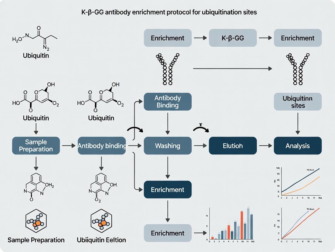
Protocol 2: K-ε-GG Peptide Immunoaffinity Enrichment (Core Thesis Protocol)
Objective: Enrich ubiquitinated peptides from complex tryptic digests.
- Resuspension: Resuspend desalted peptides in 1.4 mL immunoaffinity purification (IAP) buffer (50mM MOPS-NaOH pH 7.2, 10mM Na₂HPO₄, 50mM NaCl).
- Antibody Coupling: Use 10-20 µg of anti-K-ε-GG monoclonal antibody (e.g., clone PTM-1101) conjugated to 40 µL of Protein A/G agarose beads per sample.
- Enrichment: Incubate peptide solution with antibody-conjugated beads for 2 hours at 4°C with gentle agitation.
- Washing: Wash beads 3x with 1 mL IAP buffer, then 2x with 1 mL HPLC-grade water.
- Elution: Elute peptides with 50 µL of 0.15% TFA (2 x 5 min). Combine eluates, dry in a vacuum concentrator, and clean with C18 StageTips.
Protocol 3: LC-MS/MS Analysis and Data Processing
Objective: Identify and quantify K-ε-GG sites.
- Chromatography: Reconstitute peptides in 0.1% formic acid. Separate on a 25-cm C18 column using a 90-180 min gradient of 5-30% acetonitrile in 0.1% formic acid.
- Mass Spectrometry: Acquire data on a high-resolution tandem mass spectrometer (e.g., Orbitrap). Use a data-dependent acquisition (DDA) method with MS1 scans at 120k resolution and HCD MS2 scans at 15k resolution.
- Database Search: Process raw files using search engines (e.g., MaxQuant, Proteome Discoverer) against the appropriate protein database. Set variable modifications: K-ε-GG (+114.04293 Da) on lysine, carbamidomethylation on cysteine (fixed), and methionine oxidation (variable).
- Filtering: Apply a 1% false discovery rate (FDR) at the peptide level. Remove reverse hits and common contaminants.
Diagrams
Diagram Title: Ubiquitin Enzyme Cascade and Fate Determination
Diagram Title: K-ε-GG Enrichment and MS Workflow
The Scientist's Toolkit: Key Research Reagent Solutions
Table 2: Essential Materials for K-ε-GG Ubiquitinome Research
| Item | Function/Benefit | Example Product/Catalog # |
|---|---|---|
| Anti-K-ε-GG Monoclonal Antibody | Specifically immunoaffinity-purifies tryptic peptides containing the diglycine remnant on lysine. Core reagent. | PTM-1101 (Cell Signaling Tech #5562) |
| Protease & Phosphatase Inhibitor Cocktail | Preserves the native ubiquitination state during lysis by inhibiting proteases and DUBs. | EDTA-free tablets (Roche) |
| N-Ethylmaleimide (NEM) | Alkylating agent that irreversibly inhibits deubiquitinating enzymes (DUBs), preventing artifactually loss of Ub-signal. | Sigma-Aldrich E3876 |
| Sequencing Grade Modified Trypsin | High-purity enzyme for reproducible digestion; minimizes missed cleavages to ensure consistent K-ε-GG motif generation. | Promega V5113 |
| Protein A/G Agarose/Sepharose Beads | High-binding-capacity beads for conjugating the anti-K-ε-GG antibody for immunoprecipitation. | Pierce #20423 |
| C18 Solid-Phase Extraction Tips/Columns | For desalting and cleaning peptide samples pre- and post-enrichment to improve MS sensitivity. | Empore C18 disks, StageTips |
| Urea (Ultra-Pure) | Denaturing agent for effective cell lysis and protein unfolding while keeping cysteines reduced for alkylation. | Invitrogen 15505-035 |
| MOPS Buffer | Provides stable pH 7.2 for the IAP step, critical for antibody-antigen binding specificity and efficiency. | Thermo Fisher Scientific 28390 |
The Ubiquitin Proteasome System (UPS) and Its Role in Cellular Regulation & Disease
Introduction Within the context of proteomics research focused on post-translational modifications (PTMs), the Ubiquitin Proteasome System (UPS) stands as a critical regulator of protein homeostasis. The targeted degradation of proteins via ubiquitination is a central mechanism governing cell cycle progression, signal transduction, DNA repair, and immune responses. Dysregulation of the UPS is implicated in numerous diseases, including cancer, neurodegenerative disorders, and inflammatory conditions. Research into ubiquitination dynamics, particularly through the enrichment and identification of ubiquitination sites using K-ε-GG antibody-based protocols, provides essential insights into disease mechanisms and therapeutic targets. These Application Notes detail the experimental context and methodologies for studying the UPS.
Research Reagent Solutions The following table catalogs key reagents and materials essential for K-ε-GG enrichment-based ubiquitinomics.
| Reagent/Material | Function & Explanation |
|---|---|
| K-ε-GG Remnant Motif-Specific Antibody (Monoclonal) | Primary antibody that specifically recognizes the diglycine (GG) lysine remnant left on trypsinized peptides following ubiquitination. Crucial for immunoaffinity enrichment. |
| Protein A/G Magnetic Beads | Solid-phase support for antibody immobilization, enabling efficient pulldown and washing of enriched ubiquitinated peptides. |
| Trypsin (MS-Grade) | Protease used to digest proteins into peptides, generating the K-ε-GG remnant motif for antibody recognition. |
| Tandem Mass Tag (TMT) Reagents | Isobaric labeling reagents for multiplexed quantitative proteomics, allowing comparison of ubiquitination levels across multiple samples (e.g., time points, treatments). |
| LC-MS/MS System (e.g., Q-Exactive HF) | High-resolution mass spectrometry platform for the identification and quantification of enriched ubiquitinated peptides. |
| Deubiquitinase (DUB) Inhibitor Cocktail (e.g., N-ethylmaleimide) | Added to cell lysis buffer to preserve the native ubiquitin conjugates by inhibiting ubiquitin chain removal. |
| Urea Lysis Buffer (8M) | Efficiently denatures proteins to expose ubiquitination sites and inactivate endogenous proteases/deubiquitinases. |
Detailed Protocol: K-ε-GG Antibody Enrichment for Ubiquitinome Analysis
1. Sample Preparation & Protein Digestion
- Cell Lysis: Harvest cells, wash with cold PBS, and lyse in 8M urea lysis buffer supplemented with 10mM N-ethylmaleimide (DUB inhibitor), 1x protease inhibitor cocktail, and 1x phosphatase inhibitor. Sonicate on ice and clarify by centrifugation (16,000 x g, 15 min, 4°C).
- Protein Quantification: Determine protein concentration using a BCA assay.
- Reduction and Alkylation: Reduce proteins with 5mM dithiothreitol (DTT, 30 min, room temp). Alkylate with 15mM iodoacetamide (IAM, 20 min, room temp, in dark).
- Trypsin Digestion: Dilute urea concentration to 2M with 50mM ammonium bicarbonate. Digest with trypsin (1:50 w/w) overnight at 37°C. Quench with 1% trifluoroacetic acid (TFA).
2. Peptide Desalting & TMT Labeling (Optional for Quantification)
- Desalt peptides using C18 solid-phase extraction columns per manufacturer's instructions.
- Reconstitute peptides in 50mM HEPES pH 8.5. Label with TMT reagents (e.g., 0.8mg reagent per 100µg peptide, 1 hour, room temp). Quench with 5% hydroxylamine.
- Pool labeled samples, dry in a vacuum concentrator.
3. Immunoaffinity Enrichment of K-ε-GG Peptides
- Antibody-Bead Conjugation: Wash 500 µg of Protein A/G magnetic beads per sample with PBS+0.5% Triton X-100 (PBST). Incubate beads with 10µg of anti-K-ε-GG antibody in PBST for 2 hours at room temperature with rotation.
- Peptide Binding: Reconstitute digested/desalted peptides in IAP buffer (50mM MOPS pH 7.2, 10mM Na₂HPO₄, 50mM NaCl). Incubate with antibody-conjugated beads overnight at 4°C with rotation.
- Stringent Washes: Wash beads sequentially with:
- IAP buffer (3x)
- PBS (3x)
- LC-MS grade water (2x)
- Peptide Elution: Elute bound K-ε-GG peptides twice with 0.15% TFA (15 min each, room temperature). Combine eluates, dry, and desalt using C18 StageTips.
4. LC-MS/MS Analysis & Data Processing
- Reconstitute peptides in 2% acetonitrile/0.1% formic acid.
- Load onto a nano-flow LC system coupled to a high-resolution tandem mass spectrometer.
- MS Parameters: Full MS scan (300-1600 m/z, 70,000 resolution). Data-dependent MS/MS on top 20 ions using HCD fragmentation (NCE 32-35).
- Data Analysis: Search RAW files against the appropriate proteome database using software (e.g., MaxQuant, Proteome Discoverer) with the following variable modifications: K-ε-GG (GlyGly, +114.0429 Da) on lysine, oxidation (M), and fixed carbamidomethylation (C). Use a 1% FDR threshold.
Quantitative Data Summary Table 1: Typical Yield Metrics from a K-ε-GG Enrichment Experiment (HeLa cells, 5mg peptide input)
| Metric | Typical Result Range | Notes |
|---|---|---|
| Total Identified Peptides | 80,000 - 120,000 | Post-enrichment, MS/MS spectra |
| K-ε-GG Modified Peptides | 15,000 - 25,000 | Unique ubiquitination sites |
| Unique Ubiquitinated Proteins | 4,000 - 6,000 | Proteins with ≥1 identified K-ε-GG site |
| Enrichment Specificity (% K-ε-GG Peptides) | 70% - 90% | Percentage of total identified peptides carrying the modification |
| Quantitative Precision (CV across replicates) | < 15% | Median coefficient of variation for TMT reporter ion intensities |
Visualization
Ubiquitin-Proteasome Degradation Cascade
K-ε-GG Enrichment & MS Workflow
Ubiquitinomics, the global study of protein ubiquitination, is central to understanding cellular regulation, protein degradation, and disease mechanisms. The primary challenges are the substoichiometric modification levels (typically <1% of a target protein pool) and the rapid, stimulus-dependent dynamics of ubiquitylation. The K-ε-GG antibody, which recognizes the diglycine remnant left on lysine residues after tryptic digestion of ubiquitylated proteins, is the cornerstone of enrichment strategies for mass spectrometry-based ubiquitinomics.
Table 1: Quantitative Landscape of Ubiquitination Site Challenges
| Metric | Typical Range / Value | Implication for Enrichment |
|---|---|---|
| Stoichiometry of Modification | 0.01% - 1% of target protein | Requires 100-10,000x enrichment for detection. |
| Dynamic Range in Cell Lysate | >6 orders of magnitude (vs. abundant proteins) | Co-enrichment of abundant non-modified peptides must be minimized. |
| K-ε-GG Peptide Abundance | ~0.1-1% of total peptide mixture post-digestion | High-specificity affinity capture is critical. |
| Common Enrichment Yield | ~70-90% (with optimized protocol) | Losses must be controlled to preserve low-abundance sites. |
| MS Identification Rate | 10,000 - 20,000+ unique sites per experiment (state-of-the-art) | Dependent on starting material, depth of fractionation, and instrument sensitivity. |
Detailed Application Notes & Protocols
Application Note: Optimized K-ε-GG Enrichment for Dynamic Ubiquitination Studies
- Thesis Context: This protocol is designed to maximize the capture efficiency and specificity of K-ε-GG peptides from complex digests, directly addressing the challenges of low abundance and temporal dynamics central to the broader thesis.
- Key Principle: Utilize a two-step cleanup and a carefully titrated amount of antibody-conjugated beads to reduce non-specific binding while maintaining high yield.
- Critical Controls: Always include a negative control sample (e.g., treated with a deubiquitinase prior to digestion) processed in parallel to identify background binders.
Protocol: K-ε-GG Peptide Immunoaffinity Enrichment (IAP)
A. Materials & Sample Preparation
- Lyse cells/tissues in a denaturing buffer (e.g., 8M Urea, 50mM Tris-HCl pH 8.0) supplemented with protease inhibitors and 10mM N-Ethylmaleimide (NEM) to block cysteine residues and preserve ubiquitination.
- Reduce disulfides with 5mM DTT (30 min, 25°C) and alkylate with 15mM Iodoacetamide (30 min, 25°C in dark).
- Dilute urea concentration to <2M with 50mM Tris-HCl pH 8.0. Digest sequentially with Lys-C (1:100 w/w, 4h) and Trypsin (1:50 w/w, overnight) at 25°C.
- Acidify digest to pH ~2 with trifluoroacetic acid (TFA). Desalt peptides using C18 solid-phase extraction (SPE) cartridges. Dry completely in a vacuum concentrator.
B. Peptide Cleanup Pre-Enrichment (Critical Step)
- Resuspend dried peptide pellets in 1.5mL IAP Buffer (50mM MOPS-NaOH pH 7.2, 10mM Na₂HPO₄, 50mM NaCl).
- Use a strong cation exchange (SCX) StageTip or spin column to remove detergents, nucleic acids, and highly charged species. Elute peptides in IAP buffer. This step dramatically reduces non-specific binding to beads.
C. K-ε-GG Peptide Immunoaffinity Enrichment
- Bead Preparation: For 1-10mg of total peptide input, use 10-15µg of K-ε-GG monoclonal antibody (e.g., Cell Signaling Technology #5562) conjugated to 40µL of Protein A/G agarose beads. Wash beads 3x with IAP Buffer.
- Incubation: Incubate the cleaned-up peptide solution with the prepared beads for 2 hours at 4°C with gentle end-over-end mixing.
- Washing: Pellet beads and transfer to a micro-spin column. Wash sequentially with:
- 3x 1mL IAP Buffer.
- 3x 1mL Ice-cold PBS.
- 3x 1mL Ice-cold HPLC-grade H₂O. Perform washes quickly to minimize non-specific elution.
- Elution: Elute bound K-ε-GG peptides with 2 x 55µL of 0.15% TFA. Combine eluates and dry completely.
D. Mass Spectrometry Analysis
- Resuspend peptides in 2% acetonitrile/0.1% formic acid for LC-MS/MS.
- Use a long (e.g., 120-180 min) gradient on a C18 column coupled to a high-resolution tandem mass spectrometer.
- Data Analysis: Search data against an appropriate database using search engines (e.g., MaxQuant, Spectronaut) with the following variable modifications: GlyGly (K, +114.0429 Da), Carbamidomethyl (C, fixed), Oxidation (M), and Acetyl (Protein N-term).
Visualizations
Ubiquitinomics K-ε-GG Enrichment Workflow
The Ubiquitinomics Challenge & MS Consequence
The Scientist's Toolkit: Key Research Reagent Solutions
Table 2: Essential Materials for K-ε-GG Ubiquitinomics
| Item | Function & Rationale |
|---|---|
| K-ε-GG Motif-Specific Antibody (Clone: e.g., PTMScan Ubiquitin Remnant Motif Kit) | High-affinity monoclonal antibody for immunoaffinity purification of diglycine-modified lysine peptides. The core enrichment reagent. |
| N-Ethylmaleimide (NEM) | Thiol-alkylating agent used during lysis to inhibit deubiquitinating enzymes (DUBs), preserving the native ubiquitome. |
| Iodoacetamide (IAA) | Alkylates cysteine thiols post-reduction to prevent disulfide reformation and alkylation artifacts. |
| Sequencing-Grade Trypsin/Lys-C | High-purity enzymes ensure complete, specific digestion, generating consistent K-ε-GG remnant peptides. |
| Protein A/G Agarose Beads | Robust, high-binding-capacity support for conjugating the IgG antibody for pull-downs. Magnetic alternatives available. |
| Strong Cation Exchange (SCX) Material (e.g., spin columns, StageTips) | Pre-enrichment cleanup to remove compounds that cause non-specific binding, drastically improving specificity. |
| IAP Buffer | Optimized buffer system (MOPS/phosphate/NaCl) that maintains antibody-peptide interaction while minimizing ionic non-specific binding. |
| C18 Solid-Phase Extraction (SPE) Cartridges | For rapid desalting and cleanup of peptides post-digestion and pre-enrichment. |
The K-ε-GG motif, formed by the isopeptide linkage of ubiquitin's C-terminal glycine to a substrate lysine's epsilon-amino group, is the defining mass-spectrometric remnant following tryptic digestion. This ~114.0429 Da mass shift serves as the critical handle for the proteomic identification of ubiquitination sites, enabling system-wide studies of this essential post-translational modification. This Application Note details integrated protocols for antibody-based enrichment of K-ε-GG-containing peptides, central to a thesis on ubiquitinome profiling.
The K-ε-GG Motif: Core Concept and Detection Principle
Upon trypsin digestion of ubiquitinated proteins, the ubiquitin moiety is cleaved, leaving a diglycine (GG) adduct covalently attached via an isopeptide bond to the modified lysine residue of the substrate peptide. This creates the K-ε-GG motif, introducing a precise molecular signature detectable by LC-MS/MS.
Table 1: Key Mass Spectrometric Signature of Ubiquitination
| Parameter | Specification |
|---|---|
| Modification | Ubiquitin Remnant (GlyGly) |
| Site | Lysine (K) |
| Mass Shift (Da) | +114.0429 |
| Chemical Formula | C4H6N2O2 |
| Primary Detection Method | LC-MS/MS with High-Resolution Precursor Scanning |
Core Protocol: K-ε-GG Peptide Immunoaffinity Enrichment for Ubiquitinome Analysis
This detailed protocol is designed for the enrichment of K-ε-GG-containing peptides from complex tryptic digests prior to LC-MS/MS analysis.
Materials & Reagents
- Cell or tissue lysate
- Lysis Buffer: 8 M Urea, 50 mM Tris-HCl (pH 8.0), 75 mM NaCl, plus protease and deubiquitinase inhibitors.
- Trypsin (sequencing grade)
- Anti-K-ε-GG Antibody (e.g., monoclonal clone)
- Protein A/G or Antibody-Specific Magnetic Beads
- Immunoaffinity Purification (IAP) Buffer: 50 mM MOPS-NaOH (pH 7.2), 10 mM Na2HPO4, 50 mM NaCl.
- Elution Buffer: 0.15% Trifluoroacetic Acid (TFA)
- C18 StageTips or Columns for desalting
Procedure
A. Sample Preparation & Digestion
- Lysis: Homogenize cells/tissue in ice-cold lysis buffer. Clarify by centrifugation (16,000 x g, 15 min, 4°C).
- Reduction/Alkylation: Reduce disulfides with 5 mM DTT (30 min, 25°C). Alkylate with 15 mM iodoacetamide (30 min, 25°C in dark).
- Digestion: Dilute urea to <2M with 50 mM Tris-HCl. Digest with trypsin (1:50 w/w) overnight at 37°C. Quench with TFA to pH ~2.
- Desalt: Desalt peptides using C18 solid-phase extraction. Dry via vacuum centrifugation.
B. K-ε-GG Peptide Immunoaffinity Enrichment
- Bead Preparation: Wash magnetic Protein A/G beads (50 µL slurry per sample) twice with IAP buffer.
- Antibody Coupling: Incubate beads with 5-10 µg of anti-K-ε-GG antibody per sample in IAP buffer for 2 hours at 4°C with gentle rotation.
- Peptide Binding: Resuspend dried peptide digest in IAP buffer. Incubate with antibody-coupled beads for 2 hours at 4°C with rotation.
- Washing: Wash beads sequentially with:
- IAP Buffer (2x)
- HPLC-grade H2O (2x)
- Ice-cold 10% Acetonitrile (quick wash)
- Elution: Elute bound K-ε-GG peptides with 2 x 50 µL of 0.15% TFA. Combine eluates.
C. Post-Enrichment Processing & MS Analysis
- Desalt: Desalt eluted peptides using C18 StageTips.
- LC-MS/MS Analysis: Analyze by nanoflow LC-MS/MS on a high-resolution instrument (e.g., Q-Exactive, timsTOF).
- Data Analysis: Search MS/MS data against appropriate database using search engines (e.g., MaxQuant, Proteome Discoverer) specifying +114.0429 Da on Lys as a variable modification.
The Scientist's Toolkit: Key Research Reagents
Table 2: Essential Reagents for K-ε-GG Enrichment Studies
| Reagent | Function & Rationale |
|---|---|
| Anti-K-ε-GG Monoclonal Antibody | High-affinity, specific recognition of the tryptic ubiquitin remnant motif for selective enrichment. |
| Crosslinked Protein A/G Magnetic Beads | Solid-phase support for antibody immobilization; reduces antibody co-elution. |
| Deubiquitinase (DUB) Inhibitors (e.g., N-Ethylmaleimide, PR-619) | Preserve endogenous ubiquitination state during cell lysis and initial processing. |
| TFA (Trifluoroacetic Acid) | Low-pH elution disrupts antibody-antigen interaction for efficient peptide recovery. |
| Iodoacetamide | Alkylates cysteine thiols to prevent disulfide bond reformation and unwanted side reactions. |
Visualization of Workflows and Pathways
K-ε-GG Ubiquitinome Profiling Workflow
Formation of the K-ε-GG Motif After Digestion
MS Detection and Identification Logic
Within the broader thesis on K-ε-GG antibody enrichment protocols for ubiquitination site research, understanding the core principle of antibody specificity is foundational. Anti-K-ε-GG antibodies are monoclonal or polyclonal antibodies raised against the diglycine (GG) remnant left on a lysine (K) residue following tryptic digestion of ubiquitinated proteins. This ε-amino group linkage (K-ε-GG) is a unique, stable signature of ubiquitination. The antibody's antigen-binding site is engineered or selected for high-affinity, specific recognition of this precise peptide motif, enabling the immunopurification of ubiquitinated peptides from complex protein digests amidst a vast background of non-modified peptides.
Application Notes
Principle of Specific Enrichment
The anti-K-ε-GG antibody does not recognize free ubiquitin or intact ubiquitinated proteins. Its specificity is conferred post-proteolytic digestion, where trypsin cleaves C-terminal to arginine and lysine, but the isopeptide bond between the target lysine and ubiquitin's C-terminal glycine remains. This cleavage leaves a diglycine moiety (approximately 114.0429 Da mass shift) covalently attached via an isopeptide bond to the ε-amino group of the modified lysine. The antibody's paratope binds this K-ε-GG structure with high selectivity, discriminating against non-modified lysines and other common post-translational modifications.
Key Performance Metrics
The efficacy of enrichment is measured by specificity (percentage of K-ε-GG peptides in the final eluate) and depth of coverage (number of unique ubiquitination sites identified). Performance varies by vendor, antibody clone (e.g., PTMScan Ubiquitin Remnant Motif (K-ε-GG) Kit from Cell Signaling Technology uses a proprietary monoclonal antibody), and protocol details.
Table 1: Typical Performance Metrics of Anti-K-ε-GG Enrichment
| Metric | Typical Range | Notes |
|---|---|---|
| Enrichment Specificity | 70% - 95% | Percentage of K-ε-GG peptides in final LC-MS/MS sample. Lower purity may indicate insufficient washing or antibody cross-reactivity. |
| Ubiquitin Sites ID'd per Experiment | 10,000 - 20,000+ | From mammalian cell lysates. Dependent on sample amount, MS instrument sensitivity, and fractionation. |
| Enrichment Factor | >500-fold | Estimated increase in relative abundance of K-ε-GG peptides post-enrichment. |
| Antibody Capacity | 1-5 μg peptide per mg bead | Vendor-specific. Critical for determining scale. |
Detailed Protocol: Enrichment of K-ε-GG Peptides for Mass Spectrometry
Materials & Reagent Solutions
Table 2: Research Reagent Solutions Toolkit
| Reagent/Material | Function & Explanation |
|---|---|
| Anti-K-ε-GG Motif Antibody | Core reagent. Monoclonal antibody conjugated to agarose/protein A beads for immunoaffinity purification. |
| IAP Buffer (Immunoaffinity Purification) | Provides optimal pH and ionic strength for antibody-antigen binding. Typically 50 mM MOPS/NaOH, pH 7.2, 10 mM Na₂HPO₄, 50 mM NaCl. |
| Urea Lysis Buffer | For initial protein denaturation and extraction (e.g., 8 M Urea, 50 mM Tris-HCl, pH 8.0). Inactivates deubiquitinases. |
| Tris(2-carboxyethyl)phosphine (TCEP) | Reducing agent for disulfide bonds. |
| Iodoacetamide (IAA) | Alkylating agent for cysteine residues to prevent reformation of disulfides. |
| Sequencing-grade Trypsin | Protease that cleaves after Lys/Arg, generating the K-ε-GG remnant motif. |
| C18 Desalting Columns | For peptide cleanup and buffer exchange post-digestion, pre-enrichment. |
| Trifluoroacetic Acid (TFA) | Used for acidifying peptides for C18 binding and in MS sample loading. |
| Ammonium Bicarbonate | Buffer for tryptic digestion at pH ~8. |
| Acetonitrile (ACN), HPLC-grade | Organic solvent for peptide elution from C18 and LC-MS gradients. |
| FA (Formic Acid), LC-MS-grade | Ion-pairing agent for LC-MS peptide separation. |
Step-by-Step Methodology
Part A: Sample Preparation & Tryptic Digestion
- Lysate Preparation: Lyse cells or tissue in Urea Lysis Buffer supplemented with protease and phosphatase inhibitors. Sonicate to shear DNA and clarify by centrifugation.
- Protein Quantification: Determine protein concentration using a compatible assay (e.g., BCA).
- Reduction & Alkylation: Dilute lysate to 1-2 mg/mL. Add TCEP to 5 mM, incubate 30 min at room temp. Add IAA to 15 mM, incubate 20 min in the dark.
- Digestion: Dilute urea concentration to <2 M with 50 mM ammonium bicarbonate. Add trypsin at a 1:50 (w/w) enzyme-to-protein ratio. Digest overnight at 37°C.
- Digestion Quench & Acidification: Acidify peptides to pH <3 with TFA (final ~1% v/v). Confirm pH with pH paper.
- Desalting: Desalt peptides using C18 solid-phase extraction columns or cartridges per manufacturer's instructions. Elute in 30-50% ACN/0.1% TFA. Lyophilize or vacuum concentrate to dryness.
Part B: Immunoaffinity Enrichment (IAP)
- Peptide Reconstitution: Resuspend dried peptide pellet in 1.4 mL of cold IAP Buffer. Vortex and sonicate to ensure full solubilization. Centrifuge to clarify.
- Antibody-Bead Preparation: Gently vortex the vial of anti-K-ε-GG antibody-conjugated beads. Transfer the appropriate volume (e.g., 25 μL bead slurry per 1-10 mg peptide input) to a microcentrifuge tube.
- Bead Washing: Wash beads twice with 1 mL of cold IAP Buffer. Centrifuge at 2,000 x g for 30 seconds between washes. Remove supernatant completely.
- Peptide Incubation: Add the solubilized peptide solution to the washed beads. Incubate with gentle rotation or agitation for 2 hours at 4°C.
- Washing: Centrifuge bead-peptide mixture at 2,000 x g for 30 sec. Carefully remove supernatant (non-bound fraction). Perform sequential stringent washes:
- Wash 1: 1 mL cold IAP Buffer.
- Wash 2: 1 mL cold PBS.
- Wash 3: 1 mL cold LC-MS grade water. Centrifuge and remove supernatant completely after each wash.
- Elution: Add 50 μL of 0.15% TFA to the beads. Gently vortex and incubate for 10 minutes at room temperature. Centrifuge and carefully transfer the eluate (containing enriched K-ε-GG peptides) to a clean vial. Repeat elution once and pool.
- Peptide Cleanup: Desalt the combined eluate using StageTips or micro C18 columns. Elute in 50% ACN/0.1% FA. Concentrate to near-dryness and reconstitute in 10-20 μL of 0.1% FA for LC-MS/MS analysis.
Part C: LC-MS/MS Analysis & Data Processing
- Chromatography: Use a nano-flow HPLC system with a C18 column. Inject 1-5 μL. Separate peptides with a 60-180 min gradient from 2% to 35% ACN in 0.1% FA.
- Mass Spectrometry: Operate the mass spectrometer in data-dependent acquisition (DDA) mode. Survey MS1 scans (e.g., 350-1400 m/z) followed by MS2 fragmentation of the most intense ions. Set inclusion lists for the K-ε-GG remnant mass shift (+114.0429 Da on lysine).
- Database Search: Process raw files with search engines (e.g., MaxQuant, Proteome Discoverer). Specify variable modifications: GlyGly (K) (+114.0429 Da), Oxidation (M), and Acetyl (Protein N-term). Set Carbamidomethyl (C) as fixed.
Workflow for Anti-K-ε-GG Enrichment and Ubiquitination Site Identification
Specific Isolation of K-ε-GG Peptides from a Complex Mixture
Evolution and Development of High-Affinity K-ε-GG Antibodies for Proteomics
Application Notes: The Central Role of K-ε-GG Antibodies in Ubiquitinomics
The systematic study of protein ubiquitination (Ubiquitinomics) is fundamental to understanding cellular regulation, protein degradation, and disease mechanisms. The development and continuous refinement of high-affinity antibodies specific for the diglycine (GG) remnant left on lysine (K) residues following tryptic digestion of ubiquitinated proteins—the K-ε-GG motif—has been the cornerstone of this field. These antibodies enable the immunoenrichment of modified peptides from complex biological samples for subsequent identification and quantification by mass spectrometry (MS).
Evolutionary Milestones:
- First Generation (c. 2003-2005): Polyclonal sera demonstrated proof-of-concept but suffered from low specificity, high batch-to-batch variability, and cross-reactivity with other diGly motifs (e.g., from NEDD8).
- Second Generation (c. 2006-2010): Monoclonal antibodies (e.g., Cell Signaling Technology #5562) offered improved consistency. However, affinity was moderate, requiring high antibody input and leading to incomplete enrichment of lower-abundance ubiquitination events.
- Third Generation (c. 2011-Present): Engineered monoclonal and affinity-matured clones (e.g., CST #5564, Thermo Fisher PTMScan) provide superior affinity (K_d in low nM range) and exquisite specificity. This allows for efficient capture from smaller sample amounts (≤1 mg protein lysate), deeper proteome coverage, and compatibility with stringent wash buffers to reduce non-specific binding.
Impact on Drug Development: High-affinity K-ε-GG antibodies are critical tools for mapping ubiquitination sites altered by disease states (e.g., cancer, neurodegeneration) and for profiling the efficacy and mechanisms of novel therapeutics, particularly deubiquitinase (DUB) inhibitors and PROTACs (Proteolysis Targeting Chimeras).
Detailed Protocols
Protocol 1: Immunoaffinity Enrichment of K-ε-GG Peptides for LC-MS/MS
Objective: To isolate ubiquitinated peptides from a complex tryptic digest for site-specific identification.
Materials:
- Sample: 1-2 mg of peptides from cell or tissue lysate, digested with trypsin, and desalted.
- Key Reagent: High-affinity monoclonal K-ε-GG antibody (e.g., Cell Signaling Technology #5564) covalently conjugated to protein A/G agarose beads.
- Buffers: IAP Buffer (50 mM MOPS/NaOH pH 7.2, 10 mM Na₂HPO₄, 50 mM NaCl), PBS, ELUTION BUFFER (0.15% TFA), Neutralization Buffer (1% NH₄HCO₃, 30% ACN).
Procedure:
- Antibody-Bead Preparation: Wash 20-30 µL of antibody-conjugated bead slurry twice with 1 mL IAP buffer. Pellet beads by gentle centrifugation (2,000 x g, 30 sec).
- Peptide Binding: Resuspend beads in 500 µL IAP buffer. Add the 1-2 mg peptide sample. Incubate with end-over-end rotation for 2 hours at 4°C.
- Washing: Pellet beads and carefully remove supernatant.
- Wash 1: 1 mL IAP Buffer. Rotate 5 min, centrifuge, discard supernatant.
- Wash 2: 1 mL PBS. Rotate 5 min, centrifuge, discard supernatant.
- Wash 3: 1 mL HPLC-grade H₂O. Rotate 1 min, centrifuge, discard supernatant.
- Elution: Elute bound peptides from beads with 2 x 50 µL of 0.15% TFA by vortexing for 10 minutes at room temperature. Combine eluates.
- Peptide Cleanup: Immediately neutralize eluate with ~100 µL of Neutralization Buffer. Desalt using C18 StageTips or micro-columns. Dry peptides in a vacuum concentrator and reconstitute in 10 µL of 0.1% FA for LC-MS/MS analysis.
Protocol 2: Validation of Antibody Specificity by Peptide Array
Objective: To confirm minimal cross-reactivity of the antibody with related motifs.
Materials:
- Membranes spotted with a library of synthetic peptides (K-ε-GG, NEDDyl-ε-GG, SUMOyl-ε-GG, ISGyl-ε-GG, and unmodified sequences).
- K-ε-GG antibody.
- Standard Western blotting reagents.
Procedure:
- Block the peptide array membrane in 5% BSA/TBST for 1 hour.
- Incubate with primary K-ε-GG antibody (1:1000 in blocking buffer) overnight at 4°C.
- Wash membrane 3 x 10 min with TBST.
- Incubate with HRP-conjugated secondary antibody (1:2000) for 1 hour.
- Wash 3 x 10 min with TBST.
- Develop using chemiluminescent substrate and image. Signal should be predominantly at the K-ε-GG peptide spots.
Data Presentation
Table 1: Performance Comparison of K-ε-GG Antibody Generations
| Generation & Example Clone | Approx. K_d (nM) | Recommended Peptide Input | Typical Sites ID'd (from HEK293T) | Key Improvement | Limitation |
|---|---|---|---|---|---|
| 1st (Polyclonal) | 100 - 1000 | > 5 mg | 100 - 500 | Proof-of-concept | Low specificity, high variability |
| 2nd (mAb #5562) | 10 - 50 | 2 - 5 mg | 500 - 5,000 | Consistency, commercial availability | Moderate affinity, high background |
| 3rd (mAb #5564) | 0.1 - 5.0 | 0.5 - 2 mg | 5,000 - 20,000+ | High affinity/ specificity, low input | Higher cost, potential over-enrichment of abundant sites |
Table 2: Essential Research Reagent Solutions for K-ε-GG Enrichment
| Reagent | Function / Purpose | Example Product / Composition |
|---|---|---|
| High-Affinity K-ε-GG mAb | Specific immunoaffinity capture of ubiquitinated peptides. | Cell Signaling Technology #5564; Thermo Fisher PTMScan Ubiquitin Remnant Motif Kit |
| IAP Buffer | Optimal binding buffer; maintains pH and ionic strength for specific antibody-antigen interaction. | 50 mM MOPS pH 7.2, 10 mM Na₂HPO₄, 50 mM NaCl |
| Acidified Elution Buffer | Disrupts antibody-peptide interaction for peptide recovery. | 0.15% Trifluoroacetic Acid (TFA) in H₂O |
| C18 Desalting Tips | Desalting and concentration of eluted peptides prior to MS. | Thermo Scientific StageTips; Millipore ZipTip C18 |
| Trypsin (MS-grade) | Protein digestion to generate K-ε-GG containing peptides. | Promega Sequencing Grade Modified Trypsin |
| Protease/Phosphatase Inhibitors | Preserves the native ubiquitinome during lysis. | Cocktail tablets in lysis buffer (e.g., EDTA-free) |
| Deubiquitinase (DUB) Inhibitors | Prevents loss of ubiquitin chains during sample prep. | N-Ethylmaleimide (NEM) or Iodoacetamide (IAA) in lysis buffer |
Visualizations
Title: K-ε-GG Ubiquitinomics Experimental Workflow
Title: Evolution Path of K-ε-GG Antibody Generations
Application Notes
The enrichment and analysis of ubiquitinated proteins, specifically via the K-ε-GG antibody enrichment protocol, has become a cornerstone in modern signaling research. This methodology directly supports a thesis focused on optimizing this protocol to enhance the identification and quantification of ubiquitination sites, providing a critical tool for understanding disease mechanisms.
From Basic Signaling to Disease Pathogenesis: In basic research, ubiquitination is a key regulator of protein stability, localization, and activity, controlling fundamental processes like the cell cycle, DNA repair, and signal transduction (e.g., NF-κB, Wnt pathways). Dysregulation of ubiquitin signaling is a hallmark of numerous diseases. In cancer, oncoproteins may be stabilized (e.g., Myc, β-catenin) or tumor suppressors degraded (e.g., p53) due to altered ubiquitination. In neurodegenerative diseases like Alzheimer's and Parkinson's, aberrant ubiquitination is linked to the accumulation of toxic protein aggregates (e.g., tau, α-synuclein). The K-ε-GG enrichment protocol allows for the system-wide mapping of these modifications, transitioning from mechanistic discovery to target identification.
Quantitative Data from Recent Studies: The following table summarizes key quantitative findings from recent proteomic studies utilizing K-ε-GG enrichment, highlighting its application in disease contexts.
Table 1: Quantitative Insights from K-ε-GG Enrichment Studies in Disease Models
| Disease Context | Key Finding (Quantitative) | Implication for Drug Targets | Reference (Type) |
|---|---|---|---|
| Glioblastoma | Inhibition of USP8 deubiquitinase increased K-ε-GG sites on 1,244 proteins, including 218 receptor tyrosine kinases (RTKs) & substrates. | Identifies USP8 as a target to dysgrade multiple oncogenic drivers. | 2023, Cell Reports |
| Alzheimer's Disease (AD) | >2,000 unique ubiquitination sites significantly altered in AD brain vs. control; distinct patterns in amyloid vs. tau pathology. | Pinpoints ubiquitination pathways specific to different proteinopathies for targeted intervention. | 2022, Nature Aging |
| Parkinson's Disease (PD) | Parkin (E3 ligase) activation led to ubiquitination of 1,856 sites on 556 mitochondrial proteins within 3 hours. | Maps the global mitochondrial substrate landscape for Parkin, relevant to mitochondrial quality control therapies. | 2023, Molecular Cell |
| Colorectal Cancer | USP28 stabilizes HIF1-α; its inhibition reduces HIF1-α K-ε-GG signal >70% and impairs tumor growth in vivo. | Validates USP28 as a target to disrupt cancer cell adaptation to hypoxia. | 2024, Science Signaling |
Experimental Protocols
Protocol 1: TMT-based Quantitative Ubiquitinome Profiling Using K-ε-GG Enrichment
Objective: To identify and quantify changes in protein ubiquitination sites across different experimental conditions (e.g., drug treatment, disease state).
Materials:
- Cell or tissue lysate.
- Protease/Phosphatase inhibitors.
- Lysis Buffer: 8M Urea, 50mM Tris-HCl pH 8.0.
- Dithiothreitol (DTT), Iodoacetamide (IAA).
- Lys-C and Trypsin proteases.
- Tandem Mass Tag (TMT) reagents (e.g., 10-plex).
- Anti-K-ε-GG antibody beads.
- C18 Solid-Phase Extraction (SPE) cartridges.
- StageTips (for desalting).
- LC-MS/MS system.
Methodology:
- Lysis & Digestion: Lyse cells/tissue in urea buffer. Reduce proteins with 5mM DTT (30min, RT) and alkylate with 15mM IAA (30min, RT in dark). Dilute urea to 2M. Digest first with Lys-C (3h, RT), then with trypsin (overnight, 37°C).
- Peptide Labeling: Desalt peptides. Reconstitute in 100mM TEAB buffer. Label peptides from each condition with a unique TMT reagent (1h, RT). Quench reaction with hydroxylamine. Combine all TMT-labeled samples into one pool.
- K-ε-GG Enrichment: Dilute the pooled peptide sample in immunoaffinity purification (IAP) buffer (50mM MOPS pH 7.2, 10mM Na₂HPO₄, 50mM NaCl). Incubate with pre-washed anti-K-ε-GG antibody-conjugated beads for 2h at 4°C with gentle agitation.
- Wash & Elution: Wash beads 3x with IAP buffer and 2x with HPLC-grade water. Elute ubiquitinated peptides with 0.15% trifluoroacetic acid (TFA) for 10min. Dry eluate in a vacuum concentrator.
- LC-MS/MS Analysis: Reconstitute peptides in 0.1% formic acid. Separate via nano-flow HPLC and analyze by tandem MS on an Orbitrap mass spectrometer. Use higher-energy collisional dissociation (HCD) for TMT quantification.
- Data Analysis: Process raw files using search engines (e.g., MaxQuant, Proteome Discoverer) against a relevant protein database. Quantify TMT reporter ion intensities. Normalize and statistically analyze to identify differentially ubiquitinated sites.
Protocol 2: Validation of Ubiquitination for a Candidate Drug Target
Objective: To biochemically validate the ubiquitination status of a protein of interest (POI) identified via proteomic screening.
Materials:
- Plasmids: POI-HA/FLAG, Ubiquitin-Myc.
- Transfection reagent.
- Proteasome inhibitor (MG132).
- Lysis Buffer (RIPA).
- Anti-HA/FLAG antibody, Anti-Myc antibody, Anti-K-ε-GG antibody.
- Protein A/G agarose beads.
- Western blotting reagents.
Methodology:
- Co-immunoprecipitation (Co-IP): Co-transfect cells with POI and Ubiquitin plasmids. Treat cells with MG132 (10µM, 4-6h) prior to harvest to enrich for ubiquitinated forms.
- Cell Lysis: Lyse cells in RIPA buffer with inhibitors. Centrifuge to clear lysate.
- Immunoprecipitation: Incubate lysate with antibody against the tag on the POI (e.g., anti-FLAG) overnight at 4°C. Add Protein A/G beads for 2h.
- Western Blot Analysis: Wash beads, elute proteins in Laemmli buffer, and separate by SDS-PAGE. Probe western blots with:
- Anti-K-ε-GG antibody to detect ubiquitinated POI (smear pattern).
- Anti-Myc (for Ub) to confirm co-precipitation.
- Anti-tag for POI input control.
Visualizations
Title: Ubiquitin's Role in Disease-Relevant Signaling Pathways
Title: Ubiquitinome Profiling Workflow for Target Discovery
The Scientist's Toolkit
Table 2: Essential Research Reagent Solutions for K-ε-GG Ubiquitinome Analysis
| Reagent / Material | Function / Purpose | Key Consideration |
|---|---|---|
| Anti-K-ε-GG Monoclonal Antibody (Agarose Conjugate) | Immunoaffinity enrichment of tryptic peptides containing the K-ε-GG remnant. | Clone number (e.g., PTMScan) is critical for specificity. Use magnetic or agarose beads. |
| Tandem Mass Tag (TMT) Reagents | Enables multiplexed, quantitative comparison of up to 18 samples in a single MS run. | Choose plex level (6, 10, 16, 18) based on experimental design. Requires high-resolution MS for quantitation. |
| Urea Lysis Buffer (8M) | Efficient denaturation and solubilization of proteins while inhibiting proteases/deubiquitinases. | Fresh preparation is key; avoid heating to prevent protein carbamylation. |
| Trypsin / Lys-C Protease | Specific cleavage after Lys/Arg residues to generate peptides with C-terminal K-ε-GG remnant. | Use sequencing grade. Lys-C first in high urea enhances efficiency. |
| Proteasome Inhibitor (MG132) | Blocks degradation of polyubiquitinated proteins, increasing yield for validation experiments (Co-IP/WB). | Use during cell treatment pre-harvest. Toxic; handle with care. |
| Deubiquitinase (DUB) Inhibitors (e.g., PR-619, NEM) | Preserve ubiquitin chains during lysis by inhibiting endogenous DUB activity. | Add directly to lysis buffer immediately before use. |
| Strong Cation Exchange (SCX) or High-pH Reverse-Phase Cartridges | Fractionate complex peptide mixtures pre-enrichment to increase depth of ubiquitinome coverage. | Optional but recommended for deep profiling. |
| StageTips (C18 Material) | Desalting and cleaning of peptide samples prior to LC-MS/MS. | Low-cost, efficient alternative to HPLC columns or SPE cartridges. |
Step-by-Step K-ε-GG Enrichment Protocol: From Cell Lysate to LC-MS/MS Ready Peptides
The reliable identification of protein ubiquitination sites via K-ε-GG antibody enrichment is critically dependent on upstream sample preparation. Efficient cell lysis, complete protein extraction, and controlled, reproducible digestion are prerequisites for generating peptides suitable for enrichment and subsequent LC-MS/MS analysis. This protocol details optimized methods for Stage 1, establishing a robust foundation for the broader ubiquitinomics workflow central to our thesis on profiling ubiquitination dynamics in drug response studies.
Key Research Reagent Solutions
The following table lists essential materials and their functions for the optimized Stage 1 workflow.
Table 1: Essential Research Reagents for Sample Preparation
| Reagent/Material | Function & Rationale |
|---|---|
| Modified RIPA Lysis Buffer (8M Urea, 50mM Tris-HCl pH 8.0, 150mM NaCl, 1% NP-40, 0.1% SDS, 1x EDTA-free protease inhibitor, 5mM N-ethylmaleimide, 10mM Chloroacetamide, 1x Deubiquitinase Inhibitor) | Provides robust denaturation (Urea), detergent-based membrane disruption, and comprehensive inhibition of proteases and deubiquitinases to preserve the ubiquitinome. Dual cysteine alkylators (NEM/CA) prevent disulfide scrambling. |
| Benzonase Nuclease | Degrades nucleic acids to reduce sample viscosity, improving protein yield and handling, and preventing co-precipitation with proteins. |
| Bicinchoninic Acid (BCA) Assay Kit | For accurate, detergent-compatible quantification of extracted protein concentration to ensure consistent input for digestion. |
| Tris(2-carboxyethyl)phosphine (TCEP) | A potent, odorless reducing agent effective at neutral-alkaline pH to break disulfide bonds. |
| Iodoacetamide (IAA) | Alkylates free cysteine thiols to prevent reformation of disulfides and block unwanted side reactions. |
| Sequencing-Grade Modified Trypsin/Lys-C Mix | A highly purified, specific protease blend. Lys-C cleaves at Lys residues under denaturing conditions, facilitating subsequent trypsin digestion for highly efficient and complete proteolysis. |
| Trifluoroacetic Acid (TFA), HPLC Grade | Used to acidify and halt digestion, and for subsequent peptide desalting steps. |
Detailed Protocols
Optimized Cell Lysis and Protein Extraction Protocol
Objective: To maximize protein yield while preserving ubiquitination states and minimizing artifacts.
- Cell Harvesting: Wash adherent cells (e.g., HEK293T, HeLa) twice with ice-cold PBS. Scrape cells in PBS and pellet by centrifugation at 500 x g for 5 min at 4°C.
- Lysis: Resuspend cell pellet in 5-10 volumes of ice-cold Modified RIPA Lysis Buffer.
- Sonication: Sonicate on ice using a probe sonicator (3 pulses of 10 sec each at 30% amplitude, with 30 sec cooling intervals).
- Nuclease Treatment: Add Benzonase to a final concentration of 25 U/mL. Incubate on a rotator for 15 min at 4°C.
- Clarification: Centrifuge lysate at 20,000 x g for 15 min at 4°C. Carefully transfer supernatant (soluble protein fraction) to a new tube.
- Quantification: Perform BCA assay according to manufacturer's instructions. Typical yields: 2-5 µg/µL from ~1x10⁶ cells.
- Aliquot and Store: Aliquot clarified lysates and store at -80°C if not proceeding immediately to digestion.
Optimized In-Solution Digestion Protocol (Trypsin/Lys-C)
Objective: To achieve complete, specific digestion with minimal missed cleavages, generating peptides ideal for K-ε-GG enrichment. Table 2: Optimized Digestion Parameters vs. Conventional Method
| Parameter | Conventional Trypsin Digestion | Optimized Trypsin/Lys-C Digestion |
|---|---|---|
| Denaturant | 2M Urea or 0.1% RapiGest | 8M Urea (diluted post-alkylation) |
| Reduction | 5mM DTT, 30 min, 56°C | 5mM TCEP, 10 min, Room Temp |
| Alkylation | 15mM IAA, 20 min, Dark, RT | 15mM IAA, 10 min, Dark, RT |
| Primary Protease | Trypsin alone | Lys-C (1:50 w/w), 3h, RT |
| Dilution & Secondary Protease | Dilute Urea to <1M, Trypsin (1:50) | Dilute Urea to 1.6M, Trypsin (1:50 w/w) |
| Digestion Time | Overnight (~16h), 37°C | Overnight (~16h), 37°C |
| Quenching | Acidification with TFA | Acidification with TFA to pH <2 |
| Typical Efficiency | ~85-90% completeness, 10-15% missed cleavages | >95% completeness, <5% missed cleavages |
Procedure:
- Denaturation & Reduction: Adjust 100 µg of protein lysate to a final volume of 50 µL with lysis buffer. Add TCEP from a fresh 200mM stock to a final concentration of 5mM. Incubate for 10 min at room temperature.
- Alkylation: Add IAA from a fresh 300mM stock to a final concentration of 15mM. Incubate in the dark for 10 min at room temperature.
- Primary Digestion (Lys-C): Add sequencing-grade Lys-C at a 1:50 (enzyme:protein) ratio. Incubate for 3 hours at room temperature.
- Dilution & Secondary Digestion (Trypsin): Dilute the sample with 50mM Tris-HCl (pH 8.0) to reduce urea concentration to ~1.6M. Add sequencing-grade trypsin at a 1:50 (enzyme:protein) ratio.
- Overnight Digestion: Incubate at 37°C for 16 hours with gentle agitation.
- Quenching: Acidify the digest by adding TFA to a final concentration of 0.5% (v/v, pH < 2). Vortex and centrifuge briefly.
- Desalting: Proceed immediately to desalting (e.g., using C18 solid-phase extraction tips or columns) prior to K-ε-GG antibody enrichment.
Diagrams
Within the broader K-ε-GG antibody enrichment protocol for identifying ubiquitination sites, Stage 2 is a critical juncture. Following proteolytic digestion (Stage 1), the resulting peptide mixture contains salts, detergents, lipids, and other interfering substances from cell lysis and digestion buffers. These contaminants severely compromise the efficiency and specificity of the subsequent immunoaffinity purification (Stage 3) using K-ε-GG monoclonal antibodies. This stage focuses on desalting and peptide cleanup to exchange the peptide milieu into a biocompatible buffer, concentrate the sample, and remove contaminants that cause high background and antibody degradation. The success of this step directly impacts the depth of ubiquitome coverage and the reliability of downstream mass spectrometry analysis.
Optimal desalting achieves near-complete removal of interfering agents while maximizing peptide recovery. The choice of method depends on sample scale, starting volume, and equipment availability.
Table 1: Comparison of Common Peptide Cleanup Methods
| Method | Optimal Sample Amount | Recovery Efficiency | Key Advantage | Key Limitation |
|---|---|---|---|---|
| StageTip (C18) | 0.1 - 10 µg | 60-80% | Low cost, high flexibility, no specialized equipment. | Manual, less consistent for very complex samples. |
| Spin Columns (C18) | 1 - 100 µg | 70-90% | Rapid (10-15 min), consistent, minimal hands-on time. | Limited binding capacity; sample may be diluted. |
| Solid-Phase Extraction (SPE) Cartridges | 10 µg - 1 mg | 80-95% | High capacity, excellent for large volumes, scalable. | Requires vacuum manifold; more solvent use. |
| Precipitation (e.g., Methanol/Chloroform) | Any amount | 50-70% | Removes detergents and lipids effectively. | Harsh; may lose hydrophilic peptides; not ideal for low mass. |
Table 2: Critical Buffer Compositions for Desalting
| Buffer Name | Standard Composition | Purpose in Workflow |
|---|---|---|
| Equilibration & Wash Buffer | 0.1% Trifluoroacetic Acid (TFA) in HPLC-grade water. | Acidifies peptides to protonate carboxyl groups, promoting binding to hydrophobic C18 resin. |
| Elution Buffer | 0.1% TFA in 60-80% Acetonitrile (ACN). | Reduces polarity, eluting peptides from the C18 resin. |
| Reconstitution Buffer | 0.1% Formic Acid (FA) in HPLC-grade water OR 1x PBS (pH ~7.4). | Prepares peptides for IAP. FA is for direct MS; PBS is for antibody-based enrichment. |
Detailed Protocol: C18 StageTip Desalting
This protocol is adapted for processing up to 10 µg of peptides, suitable for most cell line or tissue digests prior to K-ε-GG enrichment.
Materials & Equipment:
- C18 StageTip disks (e.g., Empore)
- Piper tips (200 µL) or specialized StageTip barrels
- Microcentrifuge tubes (1.5 mL, low-binding)
- Centrifuge with rotor for 1.5 mL tubes
- HPLC-grade water, ACN, TFA
- Vacuum concentrator (SpeedVac)
Procedure:
- StageTip Preparation: Punch out a small disk of C18 material and place it securely in the constricted end of a 200 µL pipette tip. Activate the disk by pushing through 50 µL of methanol (100%) using a syringe or low-speed centrifugation (2 min at 1,000 x g). Condition with 50 µL of elution buffer (80% ACN, 0.1% TFA), followed by equilibration with 100 µL of wash buffer (0.1% TFA).
- Sample Loading: Acidify the digested peptide sample with TFA to a final concentration of 0.1-1%. Load the sample onto the prepared StageTip slowly by gentle centrifugation (1,500 x g, 3-5 min). Pass the flow-through back over the tip once to maximize binding.
- Washing: Wash the bound peptides twice with 100 µL of wash buffer (0.1% TFA). Centrifuge at 2,000 x g for 2 min after each wash to remove all salts and contaminants.
- Elution: Elute peptides into a fresh low-binding tube using 30-50 µL of elution buffer (60% ACN, 0.1% TFA). Centrifuge at 1,500 x g for 3 min.
- Sample Reconstitution: Concentrate the eluate in a vacuum concentrator (~30-45 min) to remove ACN completely. Do not dry the pellet completely if proceeding to immunoaffinity purification. Reconstitute the peptide pellet in 20-30 µL of 1x PBS, pH 7.4, with 0.1% Tween-20 (optional, to reduce non-specific binding). Vortex and sonicate briefly. The sample is now ready for Stage 3: Immunoaffinity Purification with K-ε-GG antibody.
The Scientist's Toolkit: Research Reagent Solutions
Table 3: Essential Materials for Peptide Desalting and Cleanup
| Item | Function & Rationale |
|---|---|
| C18 Reverse-Phase Material | Hydrophobic stationary phase that binds peptide backbones in aqueous/low-pH conditions, allowing salts (hydrophilic) to pass through. |
| Trifluoroacetic Acid (TFA) | Ion-pairing agent that acidifies the solution, ensuring peptides are positively charged and bind efficiently to C18. |
| Acetonitrile (ACN), HPLC-grade | Organic solvent that disrupts hydrophobic interactions between peptides and C18 resin, enabling elution. |
| Formic Acid (FA) | A volatile, MS-compatible acid used in reconstitution buffers for samples destined directly for LC-MS/MS. |
| Phosphate-Buffered Saline (PBS) | Biocompatible, isotonic buffer used to reconstitute peptides for antibody-based workflows, maintaining antibody structure and function. |
| Low-Binding Microcentrifuge Tubes | Minimizes adsorptive loss of low-abundance peptides to plastic surfaces. |
| SpeedVac Concentrator | Rapidly removes organic solvents from eluted samples without excessive heat, preparing peptides for reconstitution. |
Visual Workflow: From Digest to Enrichment
Diagram Title: Peptide Desalting Workflow for IAP Preparation
Diagram Title: Desalting Role in Ubiquitin Enrichment Thesis
This protocol details the core enrichment stage for the isolation of ubiquitinated peptides, a critical step in profiling ubiquitination sites via mass spectrometry. It is framed within a broader thesis aiming to standardize and optimize the K-ε-GG antibody enrichment workflow for increased reproducibility in ubiquitinomics research. Successful execution is paramount for the identification and quantification of endogenous ubiquitination sites, with direct applications in understanding disease mechanisms and drug target discovery.
Application Notes
The anti-K-ε-GG antibody specifically recognizes the diglycine remnant (Gly-Gly; ε-GG) left on lysine residues after tryptic digestion of ubiquitinated proteins. This antibody-based enrichment is the most widely used method for large-scale ubiquitinome profiling. Key considerations include:
- Specificity: The monoclonal antibody offers high specificity for the K-ε-GG motif, minimizing non-specific binding.
- Sample Input: Recommended starting amounts are 5-10 mg of total peptide digest for deep profiling from complex cell or tissue lysates.
- Buffer Compatibility: The enrichment must be performed in an IP (Immunoprecipitation) buffer system that maintains antibody affinity while reducing non-specific interactions.
- Downstream Compatibility: Eluted peptides are directly compatible with LC-MS/MS analysis after desalting and concentration.
Detailed Protocol for Antibody Incubation and Bead Capture
Materials & Reagents
Research Reagent Solutions:
| Item | Function in Protocol |
|---|---|
| Anti-K-ε-GG Monoclonal Antibody | Primary immunocapture reagent. Binds specifically to the tryptic diglycine remnant on modified lysines. |
| Protein A or G Agarose/Linked Magnetic Beads | Solid-phase support for antibody capture. Facilitates separation of antibody-peptide complexes from solution. |
| IAP Buffer (50mM MOPS, 10mM Na₂HPO₄, 50mM NaCl, pH 7.2) | Standard Immunoaffinity Purification buffer. Optimal pH and ionic strength for antibody-antigen binding. |
| Urea Lysis Buffer (8M Urea, 50mM Tris-HCl, 75mM NaCl, pH 8.0) | Used in initial protein extraction (prior stage). Included for context of starting material. |
| ABC Buffer (50mM Ammonium Bicarbonate, pH 8.0) | Buffer for tryptic digestion (prior stage). Included for context. |
| TFA (Trifluoroacetic Acid), 0.1% in Water | Used for acidification and peptide elution from C18 desalting columns. |
| LC-MS Grade Water & Acetonitrile | For sample preparation and chromatography. |
Method
Part A: Antibody-Peptide Incubation
- Prepare Peptide Solution: Resuspend the dried, trypsin-digested peptide sample (from Stage 2) in 1.4 mL of cold IAP Buffer. Vortex and briefly centrifuge to ensure full dissolution.
- pH Check: Verify that the pH of the solution is between 7.0 and 7.4 using pH paper. Adjust with dilute NaOH or HCl if necessary.
- Add Antibody: Add 10-20 µg of anti-K-ε-GG monoclonal antibody to the peptide solution. The exact amount may be optimized per antibody lot.
- Incubate: Rotate the mixture gently at 4°C for 2 hours to allow formation of the antibody-K-ε-GG peptide complex.
Part B: Bead Capture and Wash
- Prepare Beads: For each sample, aliquot 100 µL of 50% Protein A bead slurry (equivalent to 50 µL bead volume) into a low-binding microcentrifuge tube.
- Wash Beads: Wash the beads twice with 1 mL of IAP Buffer. Use a magnetic rack for magnetic beads or brief centrifugation for agarose beads. Discard the supernatant.
- Capture Complexes: Transfer the entire antibody-peptide incubation mixture to the tube containing the washed beads.
- Bind: Rotate the bead mixture gently at 4°C for 1.5 hours.
- Wash: Sequentially wash the beads to remove non-specifically bound peptides. Perform all washes with 1 mL of ice-cold buffer and ensure brief centrifugation or magnetic separation between steps:
- Wash 1: IAP Buffer (x2)
- Wash 2: LC-MS Grade Water (x1)
- Remove Residual Liquid: After the final wash, use a fine-gauge syringe or pipette tip to carefully remove all residual wash buffer without disturbing the bead pellet.
Part C: Peptide Elution
- Elute: Add 55 µL of 0.15% trifluoroacetic acid (TFA) in water directly to the beads. Vortex briefly to mix.
- Incubate: Agitate at room temperature for 10 minutes.
- Separate: Place the tube in a magnetic rack or centrifuge briefly. Carefully transfer the acidic supernatant (containing the eluted peptides) to a new low-bind tube.
- Repeat Elution: Perform a second elution with 55 µL of 0.15% TFA and pool it with the first eluate.
- Desalt: Desalt the pooled eluate (~110 µL) using a C18 StageTip or micro-column according to standard procedures. Elute desalted peptides with 60 µL of 50% acetonitrile/0.1% formic acid.
- Concentrate & Analyze: Reduce the volume to near-dryness in a vacuum concentrator. Reconstitute in 10-20 µL of 0.1% formic acid for LC-MS/MS analysis.
Table 1: Typical Yield and Efficiency Metrics for K-ε-GG Enrichment
| Parameter | Typical Range/Value | Notes |
|---|---|---|
| Peptide Input Mass | 5 - 10 mg | From HEK293 or similar cell line lysate. |
| Antibody Amount | 10 - 20 µg | Per enrichment reaction. |
| Incubation Time (Antibody+Peptide) | 2 hours | At 4°C with gentle rotation. |
| Bead Capture Time | 1.5 hours | At 4°C with gentle rotation. |
| Elution Efficiency | >85% | With double elution using 0.15% TFA. |
| Expected K-ε-GG Peptide Yield | 1 - 5 µg | Total mass after enrichment. |
| Expected Unique Sites Identified | 10,000 - 20,000 | Using a high-resolution tandem mass spectrometer. |
Protocol Visualizations
Diagram 1: Core enrichment workflow from peptides to elution.
Diagram 2: Molecular binding interactions during capture.
Within the K-ε-GG antibody enrichment protocol for ubiquitination site mapping, Stage 4 represents the critical point where specificity is secured. Following immunoaffinity capture of ubiquitinated peptides, rigorous washing is required to remove non-specifically bound peptides, contaminants, and residual reagents that contribute to high background. This stage directly impacts signal-to-noise ratios, mass spectrometry dynamic range, and the overall reliability of ubiquitinomics data for downstream drug target validation.
Quantifying the Impact of Wash Stringency
Comparative studies of wash buffer composition, volume, and repetition reveal significant quantitative effects on proteomic outcomes.
Table 1: Impact of Wash Buffer Composition on Enrichment Specificity
| Wash Buffer Component | Typical Concentration | Primary Function | Effect on K-ε-GG Peptide Recovery (%) | Reduction in Non-Specific Background (%) |
|---|---|---|---|---|
| PBS (Baseline) | 1X | Ionic strength maintenance | 100 (Reference) | 0 (Reference) |
| Urea | 2M | Chaotropic agent, disrupts weak hydrophobic interactions | 95 ± 3 | 65 ± 8 |
| SDS | 0.1% (w/v) | Anionic detergent, solubilizes proteins | 85 ± 5 | 75 ± 6 |
| NaCl (High Salt) | 500 mM | Disrupts ionic interactions | 92 ± 4 | 50 ± 10 |
| Organic Solvent (ACN) | 25% (v/v) | Reduces hydrophobic binding | 98 ± 2 | 40 ± 12 |
| Tween-20 | 0.1% (v/v) | Non-ionic detergent, blocks surfaces | 99 ± 1 | 30 ± 9 |
| Formic Acid | 0.1% (v/v) | Lowers pH, protonates carboxyl groups | 90 ± 4 | 70 ± 7 |
Table 2: Optimization of Wash Volume and Repetition
| Protocol Step | Wash Buffer | Volume per Wash (µL) | Number of Washes | Median K-ε-GG Sites Identified | Median Non-Specific Peptides Post-Enrichment |
|---|---|---|---|---|---|
| A | IAP Buffer* | 200 | 3 | 1,450 | 850 |
| B | IAP Buffer | 200 | 5 | 1,430 | 420 |
| C | IAP Buffer | 500 | 3 | 1,460 | 310 |
| D | 25% ACN / 0.1% FA | 200 | 3 | 1,520 | 185 |
| E (Optimal) | IAP Buffer then 25% ACN / 0.1% FA | 200 each | 3 + 3 | 1,550 | <100 |
*IAP Buffer: Proprietary commercial immunoaffinity purification buffer, typically a PBS-based formulation with mild detergents.
Detailed Experimental Protocols
Protocol 4.1: Standardized Post-Enrichment Wash for K-ε-GG Beads
Objective: To remove non-specifically adsorbed peptides from antibody-conjugated beads after enrichment. Materials: Magnetic protein A/G beads conjugated to K-ε-GG monoclonal antibody, post-enrichment bead complex, wash buffers (see Table 1), magnetic rack, low-protein-binding microcentrifuge tubes. Procedure:
- Initial Salt Wash: Following incubation with the digested peptide sample, place the tube on a magnetic rack for 2 minutes or until the supernatant is clear. Aspirate and discard the supernatant. Resuspend beads in 200 µL of IAP buffer (or PBS with 0.1% Tween-20). Rotate at room temperature for 5 minutes. Place on magnetic rack, aspirate supernatant. Repeat for a total of three washes.
- Chaotropic Wash: Resuspend beads in 200 µL of 2M urea in 20 mM Tris-HCl, pH 8.0. Rotate for 3 minutes. Magnetize and aspirate. Perform one wash.
- Organic Solvent Wash: Resuspend beads in 200 µL of 25% acetonitrile (ACN) in water with 0.1% formic acid. Rotate for 3 minutes. Magnetize and aspirate. Perform two washes.
- Final Volatile Buffer Wash: Resuspend beads in 200 µL of 0.1% formic acid in water. Quickly magnetize and aspirate. This low-salt, low-pH wash prepares beads for peptide elution while minimizing carryover of non-volatile salts to the LC-MS/MS system. Critical Note: Maintain beads in a suspended state during wash steps. Complete removal of supernatant is crucial, but avoid drying the bead pellet.
Protocol 4.2: On-Bead Tryptic Digest Clean-Up Wash
Objective: To remove residual enzymes, detergents, and contaminants following any on-bead digestion steps prior to enrichment. Materials: Beads with bound proteins/peptides, ammonium bicarbonate (ABC) buffer, water, ACN. Procedure:
- After on-bead digestion, magnetize and transfer the peptide-containing supernatant (digestate) to a new tube.
- Bead Back-Extraction: To recover peptides retained on beads, add 50 µL of 5% ACN / 0.1% FA to the beads. Sonicate in a water bath for 5 minutes. Magnetize and pool this supernatant with the initial digestate.
- Peptide Clean-Up (StageTip): Prepare a C18 StageTip by wetting with 100 µL methanol, equilibrating with 100 µL 0.1% FA. Load the pooled digestate. Wash the tip with 100 µL of 0.1% FA / 5% ACN to remove salts and polar contaminants. Wash with 100 µL of 0.1% FA / 25% ACN to remove remaining detergents and less hydrophobic contaminants.
- Elute peptides with 60 µL of 0.1% FA / 60% ACN into a clean LC-MS vial for analysis.
Visualization of Workflows and Relationships
Title: Sequential Wash Strategy for K-ε-GG Beads
Title: Background Sources and Wash Counteractions
The Scientist's Toolkit: Key Research Reagent Solutions
Table 3: Essential Materials for Rigorous Washing Protocols
| Item | Function in Wash Protocol | Key Consideration for Ubiquitin Enrichment |
|---|---|---|
| K-ε-GG Monoclonal Antibody (Clone PTM-1106) | Immunoaffinity capture reagent for diglycine remnant on lysine. | Clone specificity is critical; must be conjugated to beads at optimal density to balance capacity and accessibility. |
| Magnetic Protein A/G Beads | Solid support for antibody immobilization. | Superior magnetic separation minimizes bead loss during high-stringency, multi-step washes compared to agarose. |
| IAP Buffer (Commercial) | Proprietary buffer designed for immunoaffinity purification. Typically contains PBS, pH 7.4, with mild non-ionic detergents. | Provides initial gentle wash to remove unbound sample matrix without stripping true positives. |
| Ultra-Pure Urea (Powder) | Chaotropic agent for preparing 2M urea wash. Disrupts hydrogen bonding and hydrophobic interactions. | Must be freshly prepared to avoid isocyanate formation, which can artifactually modify peptides. |
| Mass Spectrometry Grade Water & Acetonitrile (ACN) | Solvents for organic wash steps (e.g., 25% ACN). Reduces hydrophobic non-specific binding. | High purity is mandatory to prevent polymer contamination that causes high background in LC-MS. |
| Optima Grade Formic Acid (FA) | Acidifying agent for low-pH, volatile wash buffers (0.1% FA). Protonates acidic residues, disrupting ionic bonds. | Volatility ensures it does not interfere with downstream LC-MS ionization. |
| Low-Protein-Binding Microcentrifuge Tubes | Reaction vessels for all wash steps. | Minimizes adsorptive loss of low-abundance ubiquitinated peptides during buffer transfers. |
| C18 StageTips (or Commercial Spin Columns) | For post-digestion clean-up washes prior to enrichment. Remove detergents, salts, and enzymes. | Empowers a versatile "wash-and-go" strategy, crucial for processing multiple samples in parallel. |
| pH Meter with Micro Electrode | Verification of wash buffer pH. | Stringent pH control in wash buffers (especially ~pH 8 for urea wash) is vital for reproducible binding behavior. |
In the context of a thesis focused on optimizing a K-ε-GG antibody enrichment protocol for ubiquitination site profiling, the elution stage is critical for efficient and specific recovery of modified peptides. This application note evaluates two predominant methods: Acidic Elution (low-pH buffers) and Competitive Elution (using a soluble analog of the epitope). The choice of elution directly impacts peptide yield, specificity, and downstream mass spectrometry analysis quality.
Methodologies and Data Comparison
Detailed Protocol: Acidic Elution
- Wash: After enrichment with immobilized anti-K-ε-GG antibody beads, wash the beads thoroughly with 1 mL of cold PBS, pH 7.4, three times.
- Elution Buffer Preparation: Prepare 0.1% (v/v) Trifluoroacetic Acid (TFA) or 0.15% Formic Acid (FA) in HPLC-grade water. Keep on ice.
- Elution: Add 50-100 µL of the acidic elution buffer to the bead pellet. Vortex gently to mix.
- Incubation: Incubate the bead-buffer mixture for 5 minutes at room temperature with constant, gentle agitation.
- Separation: Centrifuge at 2,000 x g for 1 minute. Carefully transfer the supernatant (containing eluted peptides) to a fresh low-binding microcentrifuge tube.
- Repeat: Perform a second elution with a fresh 50 µL of acidic buffer and pool with the first eluate.
- Desalting/Cleanup: Immediately proceed to desalting using C18 StageTips or micro-columns. Lyophilize and reconstitute in 0.1% FA for LC-MS/MS analysis.
Detailed Protocol: Competitive Elution
- Wash: Wash enriched beads with 1 mL of cold PBS, pH 7.4, three times.
- Competitor Solution: Prepare a 1 mM solution of synthetic, unlabeled K-ε-GG peptide (“diGly remnant” peptide) in PBS.
- Elution: Add 100 µL of the competitor solution to the bead pellet.
- Incubation: Incubate for 30 minutes at 4°C with gentle rotation. This extended, cold incubation favors competitive displacement.
- Separation: Centrifuge at 2,000 x g for 1 minute. Collect the supernatant.
- Repeat Elution: Perform a second competitive elution with fresh solution. Pool eluates.
- Buffer Exchange: The eluate is in a high-salt buffer (PBS). Use a stringent buffer exchange (e.g., multiple rounds of centrifugation with 0.1% FA in a 10-kDa MWCO filter) or immediate StageTip cleanup to remove salts and the competing peptide before MS.
Table 1: Quantitative Comparison of Elution Methods
| Parameter | Acidic Elution | Competitive Elution |
|---|---|---|
| Primary Mechanism | Disruption of antibody-antigen ionic/hydrogen bonds. | Displacement by soluble epitope analog. |
| Typical Elution Buffer | 0.1% TFA or 0.15% FA (pH ~2). | 1 mM K-ε-GG peptide in PBS (pH 7.4). |
| Incubation Time | 5-10 min at RT. | 30-60 min at 4°C. |
| Average Peptide Yield | High (80-95% recovery). | Moderate to High (60-85% recovery). |
| Specificity (Background) | Moderate; co-elutes non-specifically bound peptides. | High; primarily elutes specifically bound ubiquitinated peptides. |
| Compatibility with MS | Direct, after acid cleanup. | Requires buffer exchange to remove competitor peptide and salts. |
| Antibody Bead Reusability | No; antibodies are denatured. | Yes; antibody activity is preserved for multiple uses. |
| Relative Cost | Low. | High (cost of synthetic competitor peptide). |
Table 2: Impact on Ubiquitinome Profiling Data (Representative LC-MS/MS Outcomes)
| Data Metric | Acidic Elution | Competitive Elution |
|---|---|---|
| Total K-ε-GG Sites Identified | ~8,000-10,000 from HeLa lysate. | ~7,000-9,000 from HeLa lysate. |
| Median Spectral Counts per Site | 3.5 | 4.1 |
| Non-Specific Bindings (e.g., keratin) | Higher incidence. | Reduced by ~40%. |
| Reproducibility (CV across replicates) | 15-20% | 10-15% |
The Scientist's Toolkit: Research Reagent Solutions
Table 3: Essential Materials for K-ε-GG Peptide Elution
| Item | Function & Rationale |
|---|---|
| Immobilized anti-K-ε-GG Antibody | Enrichment matrix for ubiquitinated peptides containing the diglycine remnant. |
| Trifluoroacetic Acid (TFA), 0.1% | Low-ppH eluent for acidic method; efficiently protonates carboxyl groups, disrupting binding. |
| Synthetic K-ε-GG Peptide | Soluble competitor for gentle, specific elution while preserving antibody integrity. |
| C18 StageTips / Micro-Columns | For desalting and concentrating eluted peptides prior to LC-MS/MS. |
| Low-Binding Microcentrifuge Tubes | Minimizes peptide adhesion to tube walls, maximizing recovery. |
| pH Test Strips (pH 1-4) | Quick verification of acidic elution buffer pH. |
| 10-kDa MWCO Filters | For buffer exchange of competitively eluted samples to remove the high-mass competitor peptide. |
Visualized Workflows and Pathways
Title: Acidic Elution Workflow for K-ε-GG Peptides
Title: Competitive Elution Workflow for K-ε-GG Peptides
Title: Decision Logic for Elution Method Selection
Within the broader thesis workflow for profiling ubiquitination sites using a K-ε-GG antibody enrichment protocol, Stage 6 is critical for ensuring high-quality mass spectrometry (MS) analysis. Following immunoaffinity enrichment of modified peptides, the eluate contains salts, detergents, and other contaminants from prior stages (cell lysis, digestion, enrichment) that suppress ionization and interfere with LC-MS/MS. This stage focuses on removing these interferents, concentrating the target peptides, and preparing the sample in an MS-compatible solvent to maximize sensitivity and reproducibility for identifying and quantifying ubiquitination sites.
Core Principles and Objectives
The primary objective is to desalt and concentrate the peptide sample while maximizing recovery. Key considerations include:
- Removal of MS-Incompatible Substances: Sodium dodecyl sulfate (SDS), salts (e.g., NaCl, phosphate buffers), glycerol, and primary amines.
- Compatibility with LC-MS/MS: Final reconstitution in a low-concentration aqueous acid (e.g., 0.1% formic acid or trifluoroacetic acid).
- Minimizing Sample Loss: Employing techniques and materials that minimize non-specific binding of low-abundance enriched peptides.
- Trace Sample Handling: Using low-binding tubes and tips throughout the process.
Detailed Experimental Protocols
Protocol 3.1: C18 StageTip Desalting and Concentration
This method uses homemade or commercial C18 StageTips for robust, low-cost sample cleanup.
Materials:
- C18 solid phase extraction material (e.g., Empore C18 disks)
- P200 pipette tips
- Microcentrifuge
- Solvent A: 0.1% Formic Acid in water
- Solvent B: 0.1% Formic Acid in acetonitrile
- Low-binding 1.5 mL microcentrifuge tubes
- Vacuum concentrator (e.g., SpeedVac)
Procedure:
- StageTip Preparation: Punch a small disk of C18 material and pack it into the end of a P200 pipette tip using a blunt-ended wire. Condition the tip by centrifuging (1,000 x g, 2 min) sequentially with 50 µL of Solvent B, 50 µL of Solvent A. Do not let the disk dry out.
- Sample Binding: Acidify the enriched peptide eluate (from Stage 5) with formic acid to a final concentration of ~1%. Load the sample onto the conditioned StageTip via centrifugation (1,000 x g, 5-10 min, repeat until all sample is passed through).
- Washing: Wash the disk with 100 µL of Solvent A via centrifugation (1,000 x g, 2 min) to remove salts.
- Elution: Elute peptides into a fresh low-binding tube with 40 µL of Solvent B via centrifugation (1,000 x g, 3 min).
- Concentration: Reduce the volume of the eluate to ~2-5 µL in a vacuum concentrator. Avoid complete dryness.
- Reconstitution: Reconstitute peptides in 10-15 µL of Solvent A for MS injection. Vortex and centrifuge briefly.
Protocol 3.2: Commercial Spin Column Cleanup
Suitable for higher sample volumes or when standardized kits are preferred.
Materials:
- Commercial C18 spin column desalting kit (e.g., Pierce C18 Tips, ZipTip)
- Centrifuge with microcentrifuge tube rotor
- Buffers as specified by the kit (typically equilibration, wash, and elution buffers)
Procedure:
- Column Conditioning: Follow manufacturer instructions. Typically involves centrifuging equilibration buffer through the column.
- Sample Binding: Acidify the peptide eluate. Load the sample onto the column bed by centrifugation.
- Washing: Pass wash buffer (typically 5% acetonitrile, 0.1% FA) through the column by centrifugation.
- Elution: Elute peptides with a small volume (20-40 µL) of elution buffer (typically 50-80% acetonitrile, 0.1% FA) into a fresh collection tube.
- Concentration & Reconstitution: Concentrate and reconstitute as in Protocol 3.1, Steps 5-6.
Data Presentation: Protocol Comparison and Performance Metrics
Table 1: Comparison of Post-Enrichment Cleanup Methods
| Parameter | C18 StageTip | Commercial Spin Column | In-Line Trap Column |
|---|---|---|---|
| Approx. Cost per Sample | Very Low ($1-2) | Moderate to High ($5-15) | Integrated into LC system |
| Typical Peptide Recovery* | 70-85% | 75-90% | >95% (no transfer loss) |
| Hands-on Time | Moderate (15-20 min) | Low (10 min) | Low (setup only) |
| Scalability | Excellent for multiplexing | Good | Limited |
| Best For | High-plex experiments, labs with budget constraints | Routine processing, standardized workflows | Automated LC-MS setups, minimal sample handling |
| Risk of Contamination | Low (single-use tip) | Low (single-use column) | Low (requires rigorous cleaning) |
*Recovery can vary based on peptide hydrophobicity and sample amount. Data derived from published method comparisons.
Table 2: Impact of Cleanup on MS Signal Quality
| Sample Condition | Average MS1 Intensity* (x10⁶) | # of K-ε-GG Peptides Identified (p<0.01) | Median Peak Width (sec) |
|---|---|---|---|
| No Cleanup (Crude Eluate) | 2.1 ± 0.8 | 125 ± 35 | 22.5 ± 3.1 |
| After StageTip Cleanup | 8.5 ± 1.2 | 410 ± 55 | 16.8 ± 1.5 |
| After Spin Column Cleanup | 9.1 ± 1.0 | 435 ± 48 | 16.2 ± 1.2 |
*Hypothetical data based on typical results from 1 mg HEK293T input analyzed on a Q-Exactive HF platform. Cleanup significantly improves signal-to-noise, identifications, and chromatographic quality.
Visualization of Workflows
Post-Enrichment Cleanup and Concentration Workflow
Stage 6 Context in Ubiquitinome Profiling Thesis
The Scientist's Toolkit: Research Reagent Solutions
Table 3: Essential Materials for Post-Enrichment Cleanup
| Item | Function & Rationale | Example Product/Brand |
|---|---|---|
| C18 Solid Phase Material | Reversible hydrophobic binding of peptides for separation from hydrophilic contaminants (salts). | Empore C18 Disks, OASIS HLB |
| Low-Binding Microcentrifuge Tubes | Minimizes adsorptive loss of low-abundance enriched peptides to plastic surfaces. | Eppendorf Protein LoBind, Axygen Maxymum Recovery |
| Mass Spectrometry-Grade Acids | Provides protons for peptide ionization (FA) and improves chromatography; purity prevents chemical noise. | Formic Acid (FA), Trifluoroacetic Acid (TFA) |
| Mass Spectrometry-Grade Solvents | High-purity water and acetonitrile prevent background ions that interfere with detection. | Water (LC-MS grade), Acetonitrile (LC-MS grade) |
| Vacuum Concentrator | Gently removes organic elution solvent to concentrate the peptide sample prior to MS injection. | Thermo Scientific SpeedVac, Labconco CentriVap |
| Commercial Desalting Kit | Provides a standardized, user-friendly format for reliable peptide cleanup and recovery. | Pierce C18 Spin Columns, Millipore ZipTip with C18 |
This protocol details the liquid chromatography-tandem mass spectrometry (LC-MS/MS) analysis stage following the immunoaffinity enrichment of ubiquitinated peptides using a K-ε-GG remnant motif antibody. This step is critical for the identification and quantification of ubiquitination sites within the broader thesis on post-translational modification profiling for drug target discovery.
Core Instrument Configuration
Optimal analysis of ubiquitinated peptides, which are typically longer and more hydrophilic than standard tryptic peptides, requires adjustments to standard LC and MS parameters to improve detection.
Nanoflow Liquid Chromatography System
A detailed methodology for chromatographic separation is as follows:
- Column: Use a reversed-phase analytical column (e.g., 75 µm i.d. x 25 cm, packed with 1.9 µm C18 beads).
- Loading: Inject the enriched peptide sample via an autosampler onto a trap column for desalting.
- Gradient Elution: Employ a nanoliter-per-minute flow rate with a multi-step linear gradient. A representative 120-minute method is:
- Mobile Phase A: 0.1% Formic acid in water.
- Mobile Phase B: 0.1% Formic acid in 80% acetonitrile.
- Gradient: 2-6% B over 1 min, 6-25% B over 100 min, 25-35% B over 19 min, 35-95% B over 1 min, hold at 95% B for 9 min, then re-equilibrate.
- Column Oven: Maintain at 50°C to reduce backpressure and improve peak shape.
Tandem Mass Spectrometer Settings
Data-dependent acquisition (DDA) on a high-resolution Q-TOF or Orbitrap instrument is standard. A detailed MS protocol:
- MS1 Survey Scan: Acquire at a resolution of 120,000 (at 200 m/z) over a scan range of 375-1500 m/z. Use an automatic gain control (AGC) target of
3e6and a maximum injection time of 50 ms. - Peptide Selection: Isolate the top 20 most intense precursor ions with charge states 2-7. Use a dynamic exclusion window of 30 seconds.
- MS2 Fragmentation: Fragment precursors with a normalized collision energy (HCD) set to 28-32%. Acquire MS2 spectra at a resolution of 30,000. Set the AGC target to
1e5and maximum injection time to 54 ms. Use an isolation window of 1.4-1.6 m/z.
Quantitative Data Acquisition Parameters
For relative quantification, data-independent acquisition (DIA or SWATH-MS) is increasingly employed. Key parameters are summarized below.
Table 1: Comparison of DDA and DIA Parameters for Ubiquitinome Analysis
| Parameter | Data-Dependent Acquisition (DDA) | Data-Independent Acquisition (DIA / SWATH) |
|---|---|---|
| Primary Use | Discovery, site identification | Quantification across multiple samples |
| MS1 Resolution | 120,000 | 60,000 - 120,000 |
| MS2 Resolution | 15,000 - 30,000 | 30,000 |
| Isolation Scheme | Discrete precursor ions | Consecutive m/z windows (e.g., 26 x 25 Da) |
| Collision Energy | Fixed (e.g., 28%) or Stepped | Stepped (e.g., 22, 28, 35%) per window |
| Cycle Time | ~1-3 seconds | Adjusted to cover precursor range |
| Key Advantage | High-quality library spectra | Comprehensive, reproducible quantitation |
| Key Challenge | Stochastic sampling | Complex data deconvolution |
Table 2: Optimized LC-MS/MS Parameters for K-ε-GG Peptide Analysis
| Component | Setting | Rationale |
|---|---|---|
| LC Gradient Duration | 90 - 180 min | Ensures separation of complex ubiquitinated peptide mixtures |
| MS1 Scan Range | 375 - 1500 m/z | Avoids low-mass chemical noise; includes longer peptides |
| MS1 Resolution | ≥ 60,000 | Enables charge state determination and accurate quantitation |
| Inclusion Charge State | 2 - 7 | K-ε-GG peptides often have higher charge states |
| Normalized HCD Energy | 28 - 32% | Optimized for cleavage of the isopeptide bond and backbone |
| Dynamic Exclusion | 20 - 45 s | Prevents repeated sequencing of abundant peptides |
The Scientist's Toolkit: Key Research Reagents & Materials
Table 3: Essential Materials for LC-MS/MS Analysis of Ubiquitinated Peptides
| Item | Function & Specification |
|---|---|
| K-ε-GG Enriched Peptide Sample | The final product from the immunoaffinity enrichment stage, dried and ready for LC-MS/MS reconstitution. |
| LC-MS Grade Solvents | 0.1% Formic Acid in Water (Solvent A) and 0.1% Formic Acid in Acetonitrile (Solvent B). Minimizes ion suppression and system contamination. |
| NanoLC Column | Fused silica capillary packed with C18 reversed-phase material (1.9 µm, 120Å). Critical for high-resolution peptide separation. |
| Calibration Solution | ESI Positive Ion Calibration Mix for the specific MS instrument. Ensures mass accuracy across the detection range. |
| Data Analysis Software Suite | Software for DDA (e.g., MaxQuant, Proteome Discoverer) and DIA (e.g., Spectronaut, DIA-NN). Essential for database searching, site localization, and quantification. |
Workflow & Pathway Diagrams
LC-MS/MS Acquisition Pathways for Ubiquitinomics
MS/MS Signature of K-ε-GG Peptides
Within the broader thesis on the K-ε-GG antibody enrichment protocol for ubiquitination site research, this application note details its critical role in interrogating the dynamic ubiquitin landscape under two key perturbations: proteasome inhibition and E3 ligase activation. These experiments are fundamental for understanding protein homeostasis, drug mechanisms, and targeted protein degradation therapies.
Key Quantitative Findings
Recent studies utilizing K-ε-GG enrichment coupled with Tandem Mass Tag (TMT) proteomics reveal distinct quantitative shifts in the ubiquitinome.
Table 1: Ubiquitinome Dynamics Under Perturbations
| Perturbation Agent (Example) | Treatment Duration | Avg. Increase in K-ε-GG Sites | Notable Pathway Enrichment | Key Downstream Effect |
|---|---|---|---|---|
| Bortezomib (Proteasome Inhibitor) | 4 - 6 hours | 2.5 to 4-fold | ERAD, p53, Cell Cycle | Accumulation of polyubiquitinated proteasome substrates |
| MG-132 (Proteasome Inhibitor) | 2 hours | ~3-fold | NF-κB, DNA Repair | Stress response induction, apoptosis |
| MLN4924 (NEDD8-Activating Enzyme Inhibitor) | 6 hours | Variable decrease (30-70% for CRL substrates) | Cullin-RING Ligase (CRL) pathways | Stabilization of CRL-specific substrates |
| TNF-α (Induces NF-κB / activates TRAF6 E3 complex) | 15 - 30 minutes | Site-specific spikes (>5-fold on IκBα, RIP1) | NF-κB Signaling, Innate Immunity | Signal transduction, inflammatory response |
Detailed Experimental Protocols
Protocol 1: Mapping Ubiquitination Dynamics Post-Proteasome Inhibition
Objective: To capture the accumulation of polyubiquitinated substrates. Materials: HeLa or HEK293T cells, DMSO (vehicle), 10µM MG-132 or 100nM Bortezomib, Lysis Buffer (8M Urea, 50mM Tris-HCl pH 8.0, 75mM NaCl, protease/phosphatase/deubiquitinase inhibitors). Procedure:
- Treatment: Culture cells to 80% confluency. Treat with inhibitor or DMSO for 2-6 hours.
- Lysis & Protein Quantification: Harvest cells in ice-cold lysis buffer. Sonicate and centrifuge at 20,000g for 15 min at 15°C. Determine protein concentration via BCA assay.
- Digestion: Reduce with 5mM DTT (30min, 25°C), alkylate with 15mM iodoacetamide (30min, dark), quench with 5mM DTT. Dilute urea to 2M with 50mM Tris pH 8.0. Digest with Lys-C (1:100) for 2h, then trypsin (1:50) overnight at 25°C.
- K-ε-GG Peptide Enrichment: Acidify digest to pH ~2 with TFA. Desalt via C18 cartridge. Resuspend peptides in IAP buffer (50mM MOPS pH 7.2, 10mM Na2HPO4, 50mM NaCl). Incubate with pre-washed anti-K-ε-GG antibody-conjugated beads for 2 hours at 4°C.
- Wash & Elution: Wash beads sequentially with IAP buffer and water. Elute peptides twice with 0.15% TFA.
- LC-MS/MS Analysis: Desalt eluates, analyze on a Q-Exactive HF or Orbitrap Fusion Lumos coupled to a nanoLC system. Use a data-dependent acquisition method with HCD fragmentation.
Protocol 2: Mapping Ubiquitination Upon E3 Ligase Activation
Objective: To identify immediate, substrate-specific ubiquitination events. Materials: HEK293 cells stably expressing inducible construct (e.g., Doxycycline-inducible E3 ligase or receptor for TNF-α), 100 ng/mL TNF-α or Doxycycline. Procedure:
- Stimulation: Serum-starve cells for 4 hours. Stimulate with TNF-α (5-30 min) or induce E3 expression with Doxycycline (time-course: 15min to 4h). Include unstimulated controls.
- Rapid Lysis & Quenching: Immediately place cells on ice, wash with ice-cold PBS, and lyse using the urea buffer (see Protocol 1) supplemented with 10mM N-Ethylmaleimide to inhibit deubiquitinases.
- Sample Processing & Enrichment: Follow steps 2-6 from Protocol 1. For time-course experiments, use TMT or label-free quantification.
- Data Analysis: Filter for >5-fold increase in K-ε-GG site abundance at early time points relative to control. Validate using siRNA against the activated E3 ligase.
Pathway & Workflow Visualizations
Title: Experimental Workflow for Mapping Ubiquitination Dynamics
Title: TNF-α/NF-κB Pathway & Inhibitor Effect
The Scientist's Toolkit: Key Research Reagent Solutions
Table 2: Essential Materials for K-ε-GG Ubiquitinome Mapping
| Item | Function & Importance |
|---|---|
| Anti-K-ε-GG Remnant Antibody (Clone: Agar) | Gold-standard for immunoaffinity purification (IAP) of tryptic peptides containing the Gly-Gly remnant on lysine. |
| Cell-Permeable Proteasome Inhibitors (MG-132, Bortezomib) | Induce global accumulation of polyubiquitinated proteins by blocking the 26S proteasome. |
| Deubiquitinase (DUB) Inhibitors (N-Ethylmaleimide, PR-619) | Preserve the ubiquitinome by inhibiting endogenous DUB activity during cell lysis and processing. |
| Tandem Mass Tag (TMT) Kits | Enable multiplexed, quantitative comparison of ubiquitination sites across multiple conditions (e.g., time courses). |
| C18 StageTips or Spin Columns | For rapid desalting and cleanup of peptide samples pre- and post-enrichment. |
| NanoLC System coupled to High-Resolution Mass Spectrometer | Essential for separating complex peptide mixtures and achieving high-confidence identification of K-ε-GG sites. |
| Urea-based Lysis Buffer (8M Urea) | Efficiently denatures proteins to inactivate proteases/DUBs while being compatible with downstream digestion. |
| Sequence-Grade Trypsin/Lys-C | High-purity enzymes ensure complete digestion, generating the canonical diGly remnant for antibody recognition. |
Troubleshooting Your K-ε-GG Enrichment: Solving Common Problems and Boosting Yield
Within the broader thesis on optimizing the K-ε-GG antibody enrichment protocol for ubiquitination site mapping, a critical bottleneck is consistently low yield of enriched ubiquitinated peptides. This directly compromises the depth and statistical power of subsequent mass spectrometry analysis. Low enrichment yield can stem from inefficiencies or failures at three core stages: tissue/cell lysis, protein digestion, and the immunoaffinity enrichment itself. This application note provides a systematic diagnostic framework, experimental protocols, and reagent solutions to identify and rectify these issues.
Diagnostic Framework and Quantitative Benchmarks
Successful enrichment depends on meeting performance benchmarks at each preparatory stage. The following table summarizes key quantitative checkpoints.
Table 1: Diagnostic Checkpoints and Benchmarks for Enrichment Yield
| Stage | Checkpoint Metric | Target Benchmark | Implication of Low Value |
|---|---|---|---|
| Lysis & Clarification | Protein Concentration (Bradford/BCA) | >5 mg/mL from cultured cells | Insufficient starting material; inefficient lysis |
| Solubilized Ubiquitin (Western Blot) | Strong high-MW smear | Poor lysis or ubiquitin chain deconstruction | |
| Digestion & Peptide Prep | Peptide Yield | >60% of input protein mass | Incomplete digestion or peptide loss |
| Peptide Length (MS QC) | Majority 8-20 amino acids | Over- or under-digestion | |
| Digestion Efficiency (K-ε-GG) | >95% conversion (see Protocol 1) | Residual urea, poor enzyme activity | |
| Enrichment & Binding | Antibody Bead Capacity | 1-2 μg peptide/mg bead | Bead saturation or degradation |
| Flow-Through Ubiquitin Peptides (MS) | Minimal K-ε-GG peptides | Inefficient antibody binding | |
| Final Enriched Yield | 1-3% of total peptide mass | Cumulative failure at one or more stages |
Detailed Experimental Protocols
Protocol 1: Diagnosing Digestion Efficiency via Missed Cleavage and DiGlycine Remnant Analysis
Purpose: To determine if trypsin digestion has proceeded to completion, specifically ensuring cleavage C-terminal to arginine/lysine and the generation of the canonical Gly-Gly remnant on modified lysines. Materials: Pre-digestion peptide sample, LC-MS/MS system. Procedure:
- Sample Preparation: Inject a small aliquot (~1 μg) of the post-digestion, pre-enrichment peptide mixture for LC-MS/MS analysis on a short gradient (30 min).
- Data Acquisition: Run a standard DDA (Data-Dependent Acquisition) method.
- Data Analysis:
- Search Parameters: Search the raw data against the appropriate species database using software (e.g., MaxQuant, Proteome Discoverer).
- Variable Modifications: Set: GlyGly (K) [+114.0429 Da]; Oxidation (M); Acetyl (Protein N-term).
- Cleavage Specificity: Set enzyme to Trypsin/P with a maximum of 2 missed cleavages.
- Diagnostic Output:
- Calculate the % of K-ε-GG peptides containing a missed cleavage (K or R C-terminal to the modified site).
- Calculate the % of identified K-ε-GG sites with the correct +114.0429 Da modification vs. other adducts.
- Interpretation: A high rate (>5%) of missed cleavages at K-ε-GG sites indicates suboptimal digestion. The presence of non-canonical remnants suggests chemical interference.
Protocol 2: Evaluating Antibody Bead Binding Capacity and Specificity
Purpose: To empirically test the binding capacity and specificity of the anti-K-ε-GG antibody-conjugated beads. Materials: Anti-K-ε-GG antibody beads (commercial or custom), synthetic K-ε-GG peptide standard, non-modified peptide standard, binding/wash buffer (50 mM MOPS-NaOH, pH 7.3, 10 mM Na₂HPO₄, 50 mM NaCl), elution buffer (0.15% TFA). Procedure:
- Bead Aliquot: Prepare 5 aliquots of 10 μL bead slurry (assume ~1 mg beads).
- Spike-In Sample Preparation: Create a dilution series of the synthetic K-ε-GG peptide (e.g., 0.5, 1, 2, 4, 8 μg) in 1 mg of a complex tryptic background (e.g., HEK293 cell digest).
- Binding Test: Incubate each peptide mixture with a bead aliquot for 2h at 4°C with rotation.
- Wash & Elution: Wash beads 3x with 1 mL cold binding buffer. Elute peptides with 2x 50 μL of 0.15% TFA.
- Analysis: Analyze eluates via targeted LC-MS/MS (SRM/MRM) or quantitative Western blot for the synthetic standard.
- Interpretation: Plot amount input vs. amount recovered. Saturation indicates bead capacity. Low recovery at all levels indicates poor antibody affinity or bead malfunction.
Visualization of Diagnostic Workflow
Diagnostic Decision Tree for Low Yield
The Scientist's Toolkit: Key Research Reagent Solutions
Table 2: Essential Reagents for K-ε-GG Enrichment Optimization
| Reagent / Material | Function & Critical Role in Diagnosis |
|---|---|
| Urea-free Lysis Buffer (e.g., NP-40-based) | For re-testing lysis efficiency; eliminates urea carryover that inhibits trypsin. |
| Deubiquitinase (DUB) Inhibitors (e.g., PR-619, N-Ethylmaleimide) | Preserves ubiquitin chains during lysis; their omission causes low yield. |
| Sequencing Grade Modified Trypsin | High-purity, stabilized enzyme essential for complete, reproducible digestion. |
| Synthetic K-ε-GG Peptide Standard | Absolute critical for Protocol 2 to test antibody bead capacity and specificity. |
| Anti-K-ε-GG Monoclonal Antibody (e.g., PTMScan) | High-affinity, motif-specific antibody conjugated to agarose/beads. |
| MOPS-based Binding Buffer (pH 7.3) | Optimal buffer for anti-K-ε-GG antibody-antigen interaction; pH is critical. |
| C18 StageTips / Spin Columns | For efficient desalting and concentration of peptide samples pre- and post-enrichment. |
| HeLa Cell Digest Standard | Complex, ubiquitin-positive standard sample for inter-experiment benchmarking. |
This application note addresses a critical technical challenge encountered during the K-ε-GG antibody-based enrichment of ubiquitinated peptides for mass spectrometry analysis. High background and non-specific binding to solid-phase supports (e.g., magnetic beads) compromise the specificity and depth of ubiquitinome profiling, a core methodology in the broader thesis investigating signaling pathway modulation via ubiquitination. Optimizing wash buffer stringency and implementing effective bead blocking are therefore essential steps to maximize signal-to-noise ratios and ensure the identification of genuine ubiquitination sites.
Research Reagent Solutions Toolkit
| Reagent / Material | Function in Protocol |
|---|---|
| Magnetic Protein A/G Beads | Solid-phase support for immobilizing K-ε-GG monoclonal antibody. |
| K-ε-GG Monoclonal Antibody | Immunoaffinity reagent that specifically recognizes diglycine (GG) remnant on lysine after tryptic digestion. |
| Synthetic K-ε-GG Peptide | Used as a blocking agent to pre-saturate non-specific binding sites on beads and antibody. |
| BSA (Bovine Serum Albumin) | A common protein-based blocking agent for coating bead surfaces. |
| Tween-20 / NP-40 | Non-ionic detergents used in wash buffers to reduce hydrophobic interactions. |
| Urea / Guanidine HCl | Chaotropic agents used in high-stringency washes to disrupt non-covalent binding. |
| Tris-HCl / HEPES | Buffering agents to maintain stable pH during incubation and washing steps. |
| Phosphatase & Protease Inhibitors | Preserve post-translational modification states and prevent sample degradation. |
| LC-MS Grade Water & Solvents | Ensure minimal background contamination for downstream mass spectrometry. |
Quantitative Comparison of Wash Buffer Compositions
The effectiveness of various wash buffer formulations was evaluated by measuring the percentage of non-K-ε-GG peptides (background) and the total number of high-confidence K-ε-GG sites identified post-enrichment.
Table 1: Efficacy of Wash Buffer Regimens for Background Reduction
| Wash Buffer Step | Composition | pH | Avg. Background Peptides (%)* | Avg. K-ε-GG IDs* | Recommended Use |
|---|---|---|---|---|---|
| Low Stringency | 50 mM Tris-HCl, 150 mM NaCl, 0.1% Tween-20 | 7.5 | 45-55% | 1,200 | Initial gentle rinse. |
| High Stringency (Standard) | 50 mM Tris-HCl, 500 mM NaCl, 0.5% NP-40 | 7.5 | 20-30% | 2,800 | Primary wash for moderate stringency. |
| High Stringency (Chaotropic) | 50 mM HEPES, 1 M Urea, 0.2% SDS | 8.0 | 10-15% | 3,500 | Critical wash for maximal background removal. |
| MS-Compatible Final Wash | 50 mM Ammonium Bicarbonate, LC-MS H₂O | 8.0 | <5% | 3,400 | Final rinse to remove detergents/salts before elution. |
*Representative data from triplicate experiments using HeLa cell lysate digests.
Table 2: Comparison of Bead Blocking Agents
| Blocking Agent | Concentration | Incubation Time | Resultant Background (vs. Unblocked) | Impact on K-ε-GG Recovery |
|---|---|---|---|---|
| None (Unblocked) | - | - | 100% (Baseline) | Baseline |
| BSA (1% w/v) | 1% | 1 hr, RT | ~40% Reduction | Minimal Loss (<5%) |
| Synthetic K-ε-GG Peptide | 0.1 mg/mL | 30 min, RT | ~65% Reduction | No Loss |
| Casein (2% w/v) | 2% | 2 hr, RT | ~50% Reduction | Slight Loss (~10%) |
| BSA + K-ε-GG Peptide | 1% + 0.05 mg/mL | 1 hr, RT | ~75% Reduction | No Significant Loss |
Detailed Optimized Protocols
Protocol 4.1: Bead Preparation and Blocking
Objective: To minimize non-specific adsorption of peptides to magnetic beads and the antibody itself.
- Wash: Resuspend 50 µL of magnetic Protein A/G bead slurry per sample in 1 mL of PBS + 0.1% Tween-20 (PBS-T). Place on magnet for 1 min, discard supernatant. Repeat twice.
- Blocking: Resuspend washed beads in 500 µL of blocking solution (1% BSA + 0.05 mg/mL synthetic K-ε-GG peptide in PBS-T).
- Incubate: Rotate for 1 hour at room temperature (22-25°C).
- Coupling: Add 2-5 µg of K-ε-GG monoclonal antibody per sample. Rotate for 2 hours at RT.
- Wash & Store: Wash beads 3x with 1 mL PBS-T. Resuspend in original volume of PBS-T. Use immediately or store at 4°C for up to 24 hours.
Protocol 4.2: Immunoaffinity Enrichment with Optimized Wash Steps
Objective: To specifically isolate K-ε-GG-modified peptides from a complex tryptic digest.
- Incubation with Sample: Combine the blocked antibody-coupled beads with the tryptic peptide digest (up to 2 mg in 1 mL of IAP buffer: 50 mM MOPS/HEPES pH 7.3, 50 mM NaCl, 0.1% Tween-20). Rotate overnight at 4°C.
- Wash 1 (Low Stringency): Place on magnet, discard supernatant. Add 1 mL of IAP buffer. Rotate for 5 min at RT. Magnetize and discard supernatant. Repeat once.
- Wash 2 (High Stringency - Salt/Detergent): Add 1 mL of High Salt Wash Buffer (50 mM Tris-HCl pH 7.5, 500 mM NaCl, 0.5% NP-40). Rotate for 8 min at RT. Magnetize and discard supernatant.
- Wash 3 (High Stringency - Chaotropic): Add 1 mL of Chaotropic Wash Buffer (50 mM HEPES pH 8.0, 1 M Urea, 0.2% SDS). Rotate for 5 min at RT. Magnetize and discard supernatant. CAUTION: Do not carry SDS over into subsequent steps.
- Wash 4 (MS-Compatible Rinse): Wash beads twice with 1 mL of 50 mM Ammonium Bicarbonate (pH 8.0). Wash once with 1 mL of LC-MS grade water.
- Elution: Elute peptides from beads with 50 µL of 0.2% TFA (Trifluoroacetic acid) or 0.5% acetic acid by incubating for 10 min at RT with gentle agitation. Magnetize and carefully transfer the supernatant (containing enriched peptides) to a clean LC-MS vial.
- Desalting: Desalt peptides using C18 StageTips or micro-columns prior to LC-MS/MS analysis.
Visualizations
Diagram Title: Workflow for Specific K-ε-GG Peptide Enrichment
Diagram Title: Non-Specific Binding Forces and Counteractive Buffer Components
This application note details a critical technical variable within the broader workflow for ubiquitination site profiling using K-ε-GG antibody enrichment. The thesis of the overarching research posits that the sensitivity and accuracy of ubiquitinomics studies are fundamentally constrained by the efficiency of sample preparation prior to immunoaffinity enrichment. Specifically, incomplete trypsin digestion generates peptides lacking the canonical K-ε-GG motif or containing missed cleavages, which are not efficiently captured by the anti-K-ε-GG antibody. This leads to substantial under-representation of ubiquitination sites, increased background, and compromised quantitative accuracy. Optimizing trypsin digestion is therefore not merely a preparatory step, but a pivotal determinant of experimental success.
Quantitative Impact of Digestion Efficiency
The following tables summarize key data from recent studies on the effects of digestion parameters on K-ε-GG peptide yield.
Table 1: Effect of Trypsin-to-Protein Ratio on K-ε-GG Identifications
| Trypsin:Protein Ratio | Digestion Time (hrs) | Unique K-ε-GG Sites Identified | % Peptides with Missed Cleavages | Recommended Application Context |
|---|---|---|---|---|
| 1:50 | 12 | 5,120 | 18% | High-complexity samples (e.g., whole cell lysate) |
| 1:25 | 12 | 8,745 | 8% | Standard discovery ubiquitinomics |
| 1:25 | 4 | 6,230 | 22% | Rapid profiling, lower coverage |
| 1:10 | 12 | 9,100 | 5% | Maximum coverage for precious samples |
| 1:100 | 18 | 3,450 | 35% | Extended digestion for hard-to-digest complexes |
Table 2: Impact of Chaotrope and Denaturant on Digestion Efficiency
| Denaturation Condition | Trypsin Efficiency (Relative) | K-ε-GG Recovery (vs. Urea) | Key Advantage/Disadvantage |
|---|---|---|---|
| 8M Urea | 1.0 (Reference) | 100% | Strong denaturation; must be diluted for trypsin. |
| 2M Urea + 0.1% RapiGest | 1.4 | 135% | Superior solubility; acid-cleavable. |
| 1% SDS (with subsequent removal) | 1.7 | 125% | Most powerful denaturation; requires clean-up. |
| 5M Guanidine-HCl | 0.9 | 90% | Strong chaotrope; can inhibit trypsin if not diluted. |
| No denaturant (Native) | 0.2 | 25% | Poor efficiency; not recommended. |
Detailed Protocols
Protocol 3.1: Optimized In-Solution Trypsin Digestion for K-ε-GG Enrichment
Objective: To generate complete tryptic digests with maximal yield of canonical K-ε-GG remnant peptides. Materials: Protein sample, Urea, Tris-HCl, DTT, IAA, Lys-C, Trypsin (sequencing grade), RapiGest SF, HCl.
- Denaturation & Reduction: Resuspend protein pellet in 100 µL of 8M urea, 50mM Tris-HCl, pH 8.0. Add DTT to 5mM final concentration. Incubate at 37°C for 30 min.
- Alkylation: Add iodoacetamide (IAA) to 15mM final concentration. Incubate in the dark at room temperature for 20 min.
- Dilution & Primary Digestion: Dilute the sample 8-fold with 50mM Tris-HCl, pH 8.0, to reduce urea concentration to <1M. Add Lys-C at a 1:100 (w/w) enzyme-to-protein ratio. Incubate at 37°C for 2 hours.
- Secondary Digestion: Add trypsin at a 1:25 (w/w) enzyme-to-protein ratio. Incubate at 37°C for 12-16 hours.
- Quenching & Clean-up: Acidify the digest with 1% trifluoroacetic acid (TFA) to pH <3. Desalt using C18 solid-phase extraction columns. Lyophilize and store at -80°C prior to K-ε-GG enrichment.
Protocol 3.2: Digestion Efficiency QC via LC-MS/MS Pre-Enrichment
Objective: To assess trypsin completeness before committing to antibody enrichment. Materials: Tryptic digest, C18 StageTips, LC-MS/MS system.
- Sample Fraction: Remove a 1% aliquot of the digest (post-clean-up).
- LC-MS/MS Analysis: Reconstitute the aliquot in 0.1% formic acid. Analyze using a short (30-min) LC gradient coupled to a mass spectrometer.
- Data Analysis: Search data against the relevant proteome database. Key Metrics:
- Missed Cleavage Rate: Calculate the percentage of all identified peptides containing one or more missed cleavage sites. Target <10%.
- K-ε-GG Motif Presence: Manually inspect spectra of putative ubiquitin-derived peptides (e.g., TITLE[K-ε-GG]ESTLHLVLR) for diagnostic GG remnant ions (m/z 114.0429).
- Decision Point: If missed cleavage rate >15% or GG remnant ions are absent, repeat digestion with optimized conditions (e.g., increased enzyme ratio, addition of Lys-C, or use of alternative denaturant like RapiGest).
Visualizations
Diagram 1: Impact of Digestion on K-ε-GG Enrichment
Diagram 2: Optimized Ubiquitinomics Workflow with QC
The Scientist's Toolkit: Research Reagent Solutions
Table 3: Essential Materials for Optimized K-ε-GG Sample Preparation
| Item | Function | Critical Consideration for Digestion |
|---|---|---|
| Sequencing Grade Modified Trypsin | Primary protease cleaving C-terminal to Lys/Arg. Low autolysis rate is critical. | Use a 1:25 to 1:10 (w/w) ratio to protein for complex samples to ensure completeness. |
| Lys-C (Endoproteinase) | Cleaves C-terminal to Lysine. Complementary to trypsin. | Use prior to trypsin (1-2 hr) in low urea to reduce missed cleavages and improve digestion efficiency. |
| RapiGest SF Surfactant | Acid-cleavable detergent for protein solubilization. | Superior to urea for membrane proteins. Must be cleaved with acid post-digestion before MS. |
| Urea (Ultra-Pure) | Chaotropic agent for protein denaturation. | Concentration must be <2M during trypsin digestion. Contains cyanate ions that can carbamylate amines over time; use fresh. |
| Iodoacetamide (IAA) | Alkylates cysteine thiols to prevent reformation of disulfides. | Freshly prepared. Over-alkylation can modify lysines, inhibiting trypsin cleavage. |
| Anti-K-ε-GG Antibody (Agarose Beads) | Immunoaffinity enrichment of diglycine remnant peptides. | Incomplete digestion reduces effective peptide pool for enrichment, lowering yield. |
| C18 Solid Phase Extraction Tips/Columns | Desalting and concentrating peptide digests pre-enrichment and pre-MS. | Removes detergents, salts, and other interferents that inhibit trypsin or antibody binding. |
Within the context of a broader thesis focused on K-ε-GG antibody enrichment for ubiquitination site research, optimizing the amount of input protein or peptide is a critical determinant of success. This protocol details a systematic approach to determine the ideal input material for ubiquitin remnant immunoaffinity purification, balancing depth of proteome coverage, specificity, and cost-efficiency for researchers and drug development professionals.
Table 1: Recommended Input Mass for K-ε-GG Enrichment Protocols
| Platform / Scale | Recommended Protein Input (µg) | Recommended Peptide Input (µg) | Typical Ubiquitination Sites Identified | Key Considerations |
|---|---|---|---|---|
| High-Sensitivity (nanoLC-MS/MS) | 1 - 5 µg | 50 - 200 µg | 1,000 - 5,000+ | Limited sample availability, requires high-specificity antibodies. |
| Standard Discovery (nanoLC-MS/MS) | 500 - 2,000 µg | 2,000 - 5,000 µg | 10,000 - 20,000+ | Balance between coverage, reproducibility, and reagent cost. |
| Large-Scale / Bioreactor | 5,000 - 10,000+ µg | 10,000+ µg | 20,000+ | Maximizes site identification; risk of column overloading. |
| TMT Multiplexed (10-plex) | 100 - 200 µg per channel | 50 - 100 µg per channel | Varies with pooling | Total peptide load post-enrichment must match MS column capacity. |
Table 2: Impact of Input Material on Enrichment Performance
| Input Peptide (µg) | Antibody Bead Volume (µL) | Non-Specific Binding | % K-ε-GG Peptides (Post-Enrichment) | Saturation Risk |
|---|---|---|---|---|
| 50 | 20 | Low | 70-90% | Low |
| 500 | 20 | Moderate | 50-70% | Medium |
| 5,000 | 20 | High | 20-40% | High (Overloaded) |
| 5,000 | 100 | Moderate | 50-70% | Medium |
Detailed Experimental Protocol: Titration for Optimal Input
Objective
To empirically determine the ideal amount of tryptic peptide input for K-ε-GG immunoaffinity enrichment that maximizes ubiquitination site identifications while maintaining high specificity.
Materials & Reagents
- Cell lysate or tissue sample of interest.
- Protease and Phosphatase Inhibitors.
- Lysis Buffer (e.g., 8M Urea, 50mM Tris-HCl, pH 8.0).
- Dithiothreitol (DTT) and Iodoacetamide (IAA).
- Trypsin/Lys-C protease mix.
- C18 Solid-Phase Extraction (SPE) columns or StageTips.
- K-ε-GG Remnant Motif (Gly-Gly-lysine) Antibody-conjugated Agarose Beads.
- IP Buffer: 50mM MOPS-NaOH, pH 7.2, 10mM Na₂HPO₄, 50mM NaCl.
- Low-pH Elution Buffer: 0.15% Trifluoroacetic Acid (TFA).
- Vacuum concentrator.
- LC-MS/MS system (nanoflow configuration recommended).
Protocol
Part 1: Sample Preparation and Digestion
- Lysis: Homogenize cells/tissue in ice-cold lysis buffer with inhibitors. Sonicate on ice and centrifuge at 16,000 x g for 15 min at 4°C. Collect supernatant.
- Protein Quantification: Accurately determine protein concentration using a colorimetric assay (e.g., BCA).
- Aliquot and Reduce/Alkylate: Aliquot protein lysate into four amounts (e.g., 0.5 mg, 1 mg, 2 mg, 5 mg). Dilute to 6M urea. Reduce with 5mM DTT (30 min, RT), then alkylate with 15mM IAA (30 min, RT in dark).
- Digestion: Dilute to 1M urea with 50mM Tris-HCl (pH 8.0). Add trypsin/Lys-C at 1:50 (w/w) enzyme:protein. Incubate overnight (~16h) at 37°C.
- Peptide Cleanup: Acidify peptides with TFA to pH <3. Desalt using C18 SPE columns. Dry peptides in a vacuum concentrator.
Part 2: Input Titration and K-ε-GG Enrichment
- Reconstitution: Reconstitute each dried peptide sample in 1 mL of IP Buffer. Determine peptide concentration via A280 measurement.
- Input Aliquots: From the 5 mg digestion, prepare four equal-mass peptide aliquots for enrichment (e.g., 0.5 mg, 1 mg, 2 mg, 5 mg). Adjust all volumes to 1 mL with IP Buffer.
- Immunoaffinity Purification:
- Pre-wash 40 µL of K-ε-GG antibody bead slurry per sample with 1 mL IP Buffer x3.
- Incubate each peptide aliquot with the pre-washed beads for 2 hours at 4°C with end-over-end rotation.
- Washing: Pellet beads (2,000 x g, 1 min). Wash sequentially with:
- 1 mL IP Buffer (x3)
- 1 mL HPLC-grade H₂O (x2)
- Elution: Elute bound peptides with 2 x 100 µL of 0.15% TFA. Pool eluates for each sample.
- Post-Enrichment Cleanup: Desalt eluted peptides using C18 StageTips. Dry and store at -20°C until MS analysis.
Part 3: LC-MS/MS Analysis and Data Processing
- Reconstitution: Reconstitute peptides in 2% acetonitrile/0.1% formic acid for LC-MS/MS.
- LC-MS/MS: Inject equal volumes (or 80% of sample) per run on a nanoLC-MS/MS system using a 120-min gradient. Use data-dependent acquisition (DDA) with higher-energy collisional dissociation (HCD).
- Database Search: Search data against the appropriate proteome database using search engines (e.g., MaxQuant, Proteome Discoverer). Set variable modifications: Gly-Gly-Lys (+114.0429 Da), Oxidation (M), Acetyl (Protein N-term). Set fixed modification: Carbamidomethyl (C).
- Analysis: Plot the number of unique K-ε-GG sites identified versus the input peptide mass. The optimal input is at the inflection point before the curve plateaus, indicating bead saturation.
Visualization of Protocols and Pathways
Diagram 1: K-ε-GG Enrichment & Analysis Workflow
Diagram 2: Key Factors in Input Optimization
The Scientist's Toolkit: Research Reagent Solutions
Table 3: Essential Materials for K-ε-GG Enrichment Optimization
| Item | Function & Relevance to Input Optimization |
|---|---|
| K-ε-GG Motif-Specific Antibody (Covalently Conjugated to Beads) | The core reagent for immunoaffinity purification. Bead binding capacity (µg peptide/µL beads) directly defines the upper limit of effective input. |
| MS-Grade Trypsin/Lys-C Mix | Ensures complete digestion to generate C-terminal arginine/lysine residues, which is required for the K-ε-GG remnant motif generation after trypsin cleavage. |
| C18 Desalting Cartridges/StageTips | Critical for removing detergents, salts, and urea before enrichment and for cleaning eluted peptides before MS to prevent ion suppression. |
| IP Buffer (50mM MOPS, pH 7.2) | Optimized buffer maintains antibody-antigen binding affinity while minimizing non-specific electrostatic interactions with other peptides. |
| Trifluoroacetic Acid (TFA), 0.15% | Low-pH elution disrupts antibody-peptide binding, releasing enriched K-ε-GG peptides for collection. Must be MS-grade. |
| Pierce Quantitative Colorimetric Peptide Assay | Allows accurate measurement of peptide concentration post-digestion and cleanup, which is essential for preparing precise input aliquots for the titration. |
| BCA Protein Assay Kit | Accurately quantifies protein lysate before digestion to ensure starting amounts are known for normalization across titration points. |
Within the critical context of ubiquitination sites research, particularly when employing the K-ε-GG antibody enrichment protocol, consistent performance is paramount. Two of the most significant, yet often overlooked, sources of variability are commercial antibody lot-to-lot differences and the choice of solid-phase affinity beads. This application note provides a detailed analysis of these factors and offers standardized protocols to mitigate variability, ensuring reproducible and reliable quantification of ubiquitin remnants.
Quantifying Antibody Lot Variability
Commercial K-ε-GG antibodies, essential for enriching tryptic peptides containing diglycine (GG) remnants on lysine residues, exhibit considerable variability between lots. This variability directly impacts enrichment efficiency, background binding, and ultimately, site identification and quantification.
Table 1: Comparative Analysis of Three Consecutive K-ε-GG Antibody Lots
| Parameter | Lot #A1123 | Lot #A1124 | Lot #A1125 | Assay Method |
|---|---|---|---|---|
| Antibody Concentration (mg/mL) | 1.0 | 0.9 | 1.1 | A280 Absorbance |
| Endotoxin Level (EU/mg) | < 1.0 | < 1.0 | < 1.0 | LAL Chromogenic |
| Enrichment Efficiency (%) | 78 ± 5 | 65 ± 8 | 82 ± 4 | Spike-in Heavy Standard Peptide LC-MS/MS |
| Non-Specific Binding (% of total) | 12 ± 3 | 25 ± 6 | 8 ± 2 | Flow-Through Analysis |
| Median CV (Triplicate Enrichments) | 8% | 18% | 7% | 200 Ubiquitinated Peptides |
Protocol 1.1: Pre-Qualification of New Antibody Lots Objective: To evaluate the performance of a new K-ε-GG antibody lot prior to full-scale experimental use.
- Prepare Test Sample: Generate a tryptic digest from a well-characterized cell lysate (e.g., HEK293). Spike in a known quantity (e.g., 0.5 pmol) of synthetic, stable isotope-labeled K-ε-GG reference peptides.
- Aliquot Antibody: Reconstitute or dilute the new antibody lot and a currently validated control lot per manufacturer instructions. Prepare 10 µg of each antibody for coupling.
- Couple to Beads: Follow Protocol 2.1 below to couple each antibody aliquot to 30 µL of magnetic Protein A beads separately.
- Perform Parallel Enrichment: Using 100 µg of the test sample digest, perform the enrichment protocol (Protocol 2.2) in triplicate for both the new and control antibody-bead complexes.
- LC-MS/MS Analysis: Analyze all eluates on the same LC-MS/MS system using a standardized 90-minute gradient.
- Data Analysis:
- Calculate the recovery efficiency of the spiked heavy reference peptides.
- Compare the number of unique endogenous K-ε-GG peptide identifications between lots.
- Assess the coefficient of variation (CV) for high-abundance ubiquitinated peptides across triplicates.
- A new lot should be within ±15% of the control lot's performance metrics.
Bead Selection and Coupling Optimization
The solid support for antibody immobilization significantly influences antibody orientation, accessibility, and non-specific binding. Magnetic beads are preferred for ease of handling.
The Scientist's Toolkit: Key Research Reagent Solutions
| Item | Function & Rationale |
|---|---|
| K-ε-GG Motif Antibody (Rabbit Monoclonal) | Specifically recognizes the diglycine remnant left on lysine after tryptic digestion of ubiquitinated proteins. Critical for IP. |
| Magnetic Protein A/G Beads | Protein A has high affinity for rabbit IgG Fc regions. Magnetic cores allow for rapid buffer changes and minimal sample loss. |
| Crosslinker (e.g., DSS, BS³) | Stabilizes the antibody-bead linkage, preventing antibody leaching and co-elution with target peptides, which reduces MS interference. |
| Diglycine-Lysine (GG-K) Peptide Library | A set of non-labeled peptides for competitive elution, offering a cleaner, MS-compatible alternative to acidic elution. |
| Stable Isotope-Labeled Standard (SIS) K-ε-GG Peptides | Absolute quantification standards spiked in before digestion to correct for variability in enrichment efficiency and MS ionization. |
| Low-Bind Microcentrifuge Tubes | Minimizes adsorptive loss of low-abundance peptides during processing. |
Protocol 2.1: Crosslinked Antibody-Bead Complex Preparation Objective: To generate a stable, reusable immunocapture matrix with consistent antibody orientation.
- Wash Beads: Resuspend 1 mL of magnetic Protein A bead slurry. Place on magnet, discard supernatant. Wash twice with 1 mL of 1X PBS, pH 7.4.
- Antibody Binding: Add 100 µg of K-ε-GG antibody to the washed beads in 1 mL PBS. Rotate for 1 hour at room temperature.
- Crosslinking: Wash antibody-bound beads twice with 1 mL PBS. Resuspend in 1 mL PBS. Add freshly prepared disuccinimidyl suberate (DSS) to a final concentration of 5 mM. Rotate for 30 minutes at RT.
- Quenching: Add Tris-HCl, pH 8.0, to a final concentration of 100 mM. Rotate for 15 minutes to quench unreacted crosslinker.
- Storage: Wash beads twice with storage buffer (PBS + 0.05% sodium azide). Resuspend in 1 mL storage buffer. Store at 4°C. The complex is stable for at least 3 months.
Table 2: Performance of Different Bead Types with K-ε-GG Antibody
| Bead Type | Base Material | Binding Capacity (µg IgG/µL bead) | Non-Specific Binding | Crosslinking Efficiency | Recommended Use |
|---|---|---|---|---|---|
| Magnetic Protein A | Polystyrene | 15-20 | Low | High | Standard high-throughput enrichment |
| Magnetic Protein G | Silica | 10-15 | Very Low | Moderate | If antibody has weak Protein A binding |
| Agarose Protein A | Agarose | 5-10 | Moderate | High | For large-scale preparative enrichment |
| Streptavidin-Biotin | Polystyrene | N/A | High | N/A | Not recommended for peptide IP |
Protocol 2.2: Standardized K-ε-GG Peptide Immunoenrichment Workflow Objective: To consistently enrich ubiquitinated peptides from complex tryptic digests.
- Input Preparation: Desalt 1-2 mg of tryptic peptide digest. Dry completely and reconstitute in 1.4 mL of Immunoaffinity Purification (IAP) Buffer (50 mM MOPS/NaOH, pH 7.2, 10 mM Na₂HPO₄, 50 mM NaCl).
- Pre-Clear: Add 50 µL of control beads (crosslinked with non-specific IgG) to the sample. Rotate for 1 hour at 4°C. Magnetize and transfer supernatant to a new tube.
- Immunocapture: Add 50 µL of prepared K-ε-GG antibody-bead complex (Protocol 2.1) to the pre-cleared supernatant. Rotate for 2 hours at 4°C.
- Washing: Place tube on magnet, discard supernatant. Wash beads sequentially with:
- 1 mL IAP Buffer (1x)
- 1 mL HPLC-grade H₂O (2x)
- Elution: Elute peptides with 2 x 50 µL of 0.15% trifluoroacetic acid (TFA) for 10 minutes each with agitation. Combine eluates.
- Clean-up: Desalt eluted peptides using C18 StageTips. Dry and store at -80°C until LC-MS/MS analysis.
Integrated Pathway and Workflow Visualization
Diagram Title: K-ε-GG Ubiquitin Peptide Enrichment and MS Workflow
Diagram Title: Factors and Controls for Enrichment Consistency
Achieving high reproducibility in ubiquitination site mapping requires a proactive approach to managing reagent variability. Implementing mandatory pre-qualification of new K-ε-GG antibody lots, standardizing on a crosslinked magnetic bead system, and adhering to detailed, consistent protocols are non-negotiable practices. By integrating these tips and the provided protocols into the broader K-ε-GG enrichment workflow, researchers can significantly reduce technical noise, enabling more accurate biological insights and more robust drug development target validation.
Addressing Keratin and Common Lab Contaminants During the Enrichment Process
In the context of ubiquitination site research utilizing K-ε-GG antibody enrichment protocols, sample purity is paramount. Keratins from skin, hair, and dust, alongside common laboratory contaminants like polymeric materials and albumin, are a significant source of interference. They cause high background noise, suppress ionization efficiency during LC-MS/MS, and lead to false-negative identifications or misassignment of ubiquitination sites. This application note details strategies to identify, mitigate, and monitor these contaminants during the enrichment workflow for ubiquitinated peptides.
The table below summarizes common contaminants, their sources, and their impact on K-ε-GG enrichment studies.
Table 1: Common Contaminants in Ubiquitin Enrichment Workflows
| Contaminant Class | Primary Source | Typical Mass (kDa) | Impact on K-ε-GG Enrichment/MS | Estimated Abundance in Contaminated Samples* |
|---|---|---|---|---|
| Keratins (e.g., K1, K10, K2e) | Skin flakes, dust, hair, clothing | 40-70 | Major source of spectral interference; co-elutes with target peptides. | Can constitute >50% of total protein in severe cases. |
| Albumin (BSA/HSA) | Serum, FBS, lab reagents, wear from equipment | 66.5 | Suppresses ionization of low-abundance ubiquitinated peptides. | Variable; common in cell culture-derived samples. |
| Immunoglobulin G (IgG) | Antibody reagent carryover from lysis/immunoprecipitation | 150 | Can fragment, producing peptides that confound database search. | High if antibody cleanup steps are omitted. |
| Trypsin/Autolysis Products | Protease self-digestion | 20-24 | Creates non-specific peptides that increase sample complexity. | ~5-10% of total peptide yield without protease control. |
| Polymers (PEG, detergents) | Plasticware, buffers, surfactants | Variable | Ion suppression, adduct formation, column fouling. | Trace amounts sufficient to disrupt MS signals. |
*Estimates based on published contaminant studies in proteomics (2022-2024).
Integrated Protocol for Contaminant-Aware K-ε-GG Enrichment
This protocol augments standard K-ε-GG enrichment with critical contaminant mitigation steps.
Materials & Reagents
- Lysis/Buffers: Urea-free or mass spec-grade detergent lysis buffers (e.g., SDC). High-purity Tris-HCl, NaCl, PBS.
- Protease Inhibitors: Mass spectrometry-compatible cocktails (e.g., AEBSF, E-64).
- Reduction/Alkylation: High-purity DTT or TCEP, and IAA or CAA.
- Proteolysis: Sequencing-grade, Lys-C/Trypsin mixture. Use low-adsorption tubes.
- Enrichment: Anti-K-ε-GG monoclonal antibody-conjugated beads. Strictly avoid keratin-containing slurries.
- Cleanup: StageTips with C18 or SDB-RPS material, or low-binding spin columns.
- LC-MS/MS: Pre-column filter, nano-flow HPLC system, high-resolution tandem mass spectrometer.
Detailed Procedure
Part A: Pre-Enrichment Sample Preparation & Contaminant Mitigation
- Controlled Environment: Perform all pre-MS steps in a laminar flow hood or dedicated "clean bench." Wear a lab coat, gloves, and a non-fibrous sleeve covering. Use a full-face mask if handling dry samples.
- Protein Extraction: Lyse cells/tissues in a denaturing buffer containing 1% SDC. Avoid urea to prevent carbamylation and cyanate formation. Heat at 95°C for 5 min.
- Protein Quantification: Use a fluorometric assay (e.g., Qubit) over colorimetric assays to avoid polymer interference.
- Reduction & Alkylation: Reduce with 5 mM TCEP (15 min, 55°C). Alkylate with 10 mM CAA (15 min, RT in dark). Use fresh stock solutions.
- Digestion: Dilute SDC to 0.5%. Digest with Lys-C (1:100, 3h, RT) followed by Trypsin (1:50, overnight, 37°C). Use low-protein-binding tubes.
- Acidification & Cleanup: Acidify with TFA to 1% final pH < 2. SDC will precipitate. Centrifuge and transfer supernatant. Perform a double StageTip cleanup (SDB-RPS followed by C18) to remove detergents, polymers, and salts.
Part B: K-ε-GG Immunoaffinity Enrichment
- Bead Preparation: Wash anti-K-ε-GG antibody beads (e.g., PTMScan or equivalent) 3x with IAP buffer (50 mM MOPS/NaOH pH 7.2, 10 mM Na₂HPO₄, 50 mM NaCl).
- Peptide Binding: Resuscleaned peptides in 1.4 mL IAP buffer. Incubate with prepared bead slurry for 2h at 4°C with gentle rotation.
- Stringent Washes:
- Wash 1: 3x with IAP buffer.
- Wash 2: 3x with HPLC-grade H₂O.
- Perform all washes in a cold room, using chilled buffers.
- Peptide Elution: Elute peptides with 2x 50 µL of 0.15% TFA. Combine eluates and dry completely in a vacuum concentrator.
Part C: Post-Enrichment Cleanup & MS Analysis
- Desalting: Reconstitute dried peptides in 1% FA and desalt using a C18 StageTip.
- LC-MS/MS Analysis: Reconstitute in 2% ACN/0.1% FA. Analyze by LC-MS/MS using a 90-min gradient. Include a blank run (0.1% FA) between samples to monitor for carryover.
- Data Analysis: Search data against a concatenated target-decoy database that includes sequences for common contaminants (keratins, albumin, IgG). Set a stringent 1% FDR at the peptide level. Manually inspect putative ubiquitination sites for diagnostic GG remnant ions (m/z 114.0429).
The Scientist's Toolkit: Key Reagent Solutions
Table 2: Essential Reagents for Contaminant-Minimized Ubiquitin Enrichment
| Reagent/Material | Function & Critical Feature | Rationale for Contaminant Control |
|---|---|---|
| Anti-K-ε-GG Beads (Ceramic/Magnetic) | Immunoaffinity capture of diGly-modified peptides. Keratin-free manufacture. | Eliminates the introduction of keratins from animal-sourced agarose/sera. |
| Mass-Spec Grade Water/LC-MS Solvents | Preparation of all buffers and sample reconstitution. Ultrapure ( | Eliminates background from solvent impurities and polymers. |
| Low-Protein-Binding Tubes & Tips (e.g., polypropylene) | Sample handling during digestion and enrichment. | Minimizes adsorption of target peptides and leachates from plastics. |
| Sodium Deoxycholate (SDC) | MS-compatible, efficient detergent for lysis and digestion. Acid-precipitable. | Superior to urea for avoiding modifications; easily removed pre-MS. |
| StageTips (C18 & SDB-RPS) | Microscale desalting and detergent removal. | Two-step cleanup removes polymers, lipids, and residual SDC more effectively than single-stage. |
| Pre-column Filter (2µm) | Placed inline before the analytical column. | Traps any particulates or aggregated material, protecting the expensive nano-column. |
Visualization of Workflows and Concepts
Diagram 1: Integrated workflow for K-ε-GG enrichment with contaminant control points.
Diagram 2: Conceptual impact of contaminants on MS detection of target peptides.
Within the broader thesis on optimizing the K-ε-GG antibody enrichment protocol for ubiquitination site discovery, a critical advancement lies in its integration with orthogonal proteomic techniques. This application note details two powerful, multiplexed strategies: combining K-ε-GG enrichment with Tandem Mass Tag (TMT) labeling for deep, quantitative ubiquitylome profiling across conditions, and performing sequential phosphopeptide and ubiquitin remnant enrichment to study the complex interplay between phosphorylation and ubiquitination (the "phospho-ubiquitinome"). These integrated workflows maximize data breadth and depth from precious samples, crucial for researchers and drug development professionals investigating signaling networks and target engagement.
Table 1: Comparative Performance of Integrated K-ε-GG Strategies
| Parameter | K-ε-GG Enrichment + TMT Multiplexing | Sequential p-enrich / K-ε-GG Enrichment | Standard K-ε-GG Enrichment (Singleplex) |
|---|---|---|---|
| Primary Goal | High-throughput quantification across multiple conditions (e.g., 10-16 samples) | Uncover phospho-ubiquitin crosstalk from a single sample condition | Ubiquitination site discovery from a single sample |
| Typical Sample Throughput per MS Run | 10-16 samples (TMTpro 16plex) | 1 sample (deep profiling) | 1 sample |
| Avg. Identified K-ε-GG Sites | 8,000 - 15,000+ (aggregate across channels) | 1,500 - 3,000 (from the phospho-depleted flow-through) | 10,000 - 20,000 |
| Key Quantitative Metric | Reporter Ion Intensity Ratio (TMT) | Spectral Count / Label-free Quantification (LFQ) | Spectral Count |
| Major Advantage | Reduced MS instrument time, improved quantification precision across samples | Direct insight into dual-post-translational modification regulation | Simplicity, maximum depth for one condition |
| Key Challenge | Ratio compression due to co-isolated ions | Potential sample loss during sequential enrichment; data complexity | Limited throughput |
Detailed Experimental Protocols
Protocol A: K-ε-GG Enrichment with TMTpro Multiplexing
Objective: To quantitatively compare ubiquitination site abundances across up to 16 different experimental conditions (e.g., time-course, drug doses, genetic perturbations).
Materials & Reagents:
- Cell or tissue lysates from conditions to be compared.
- TMTpro 16plex Label Reagent Set.
- K-ε-GG Remnant Motif (K-ε-GG) Immunoaffinity Beads.
- High-Select Fe-NTA Phosphopeptide Enrichment Kit (optional, for parallel phospho-profiling).
- StageTips (C18 material) for desalting.
- LC-MS/MS system (e.g., Orbitrap Eclipse) with high-field asymmetric waveform ion mobility spectrometry (FAIMS) Pro.
Procedure:
- Sample Preparation & Digestion: Independently process each condition. Lyse cells, reduce, alkylate, and digest proteins with Lys-C/Trypsin.
- TMT Labeling: Desalt each sample. Reconstitute peptides in 100 mM TEAB buffer. React each sample with a unique TMTpro channel reagent for 1 hour. Quench reaction with hydroxylamine. Pool all TMT-labeled samples in equal amounts.
- Peptide Clean-up: Desalt the pooled sample using C18 StageTips.
- K-ε-GG Enrichment: Incubate the pooled, labeled peptide mixture with K-ε-GG antibody beads for 2 hours at 4°C with gentle rotation. Wash beads sequentially with ice-cold IAP buffer (Cell Signaling) and water. Elute ubiquitinated peptides with 0.15% trifluoroacetic acid.
- LC-MS/MS Analysis: Desalt eluate. Analyze via nano-LC-MS/MS using a 2-hour gradient. Employ a synchronous precursor selection (SPS)-MS3 method on a tribrid instrument to minimize TMT ratio compression. FAIMS Pro is recommended with 3 CVs (e.g., -45V, -60V, -75V).
Protocol B: Sequential Phosphopeptide and K-ε-GG Enrichment
Objective: To comprehensively map ubiquitination and phosphorylation events, and specifically identify proteins harboring both modifications, from a single biological sample.
Materials & Reagents:
- Digested peptide sample.
- High-Select Fe-NTA Phosphopeptide Enrichment Kit or TiO2 beads.
- K-ε-GG Immunoaffinity Beads.
- Loading/Wash Buffers for both enrichments.
- C18 StageTips.
Procedure:
- Initial Phosphopeptide Enrichment: Subject the total digested peptide sample to phosphopeptide enrichment using Fe-NTA or TiO2 beads per manufacturer's protocol. Collect the phospho-enriched fraction (eluent 1).
- Flow-Through Processing: Carefully collect the phospho-depleted flow-through. Acidify and desalt this fraction using C18 StageTips. This fraction is enriched for non-phosphorylated peptides, including the majority of ubiquitinated peptides.
- K-ε-GG Enrichment: Perform standard K-ε-GG immunoaffinity enrichment on the desalted, phospho-depleted flow-through as described in Protocol A, step 4. Collect the ubiquitin-enriched fraction (eluent 2).
- LC-MS/MS Analysis: Analyze three fractions separately on the LC-MS/MS:
- Fraction 1: Phospho-enriched fraction (for phosphoproteome).
- Fraction 2: Ubiquitin-enriched fraction (for ubiquitylome).
- Fraction 3 (Optional): A small aliquot of the original digest (for total proteome).
Visualized Workflows & Pathways
Title: Two Advanced K-ε-GG Integration Workflows
Title: Phosphorylation-Guided Ubiquitination Pathway
The Scientist's Toolkit: Research Reagent Solutions
Table 2: Essential Materials for Integrated Ubiquitylome Studies
| Item | Function & Role in Workflow |
|---|---|
| K-ε-GG Remnant Motif Antibody Beads | Immunoaffinity resin for specific enrichment of tryptic diglycine remnant left on ubiquitinated lysines. Core reagent for ubiquitylome. |
| TMTpro 16plex Label Reagent Set | Isobaric chemical tags for multiplexing, allowing simultaneous quantification of up to 16 samples post-enrichment. |
| High-Select Fe-NTA Phosphopeptide Enrichment Kit | Immobilized metal affinity chromatography resin for global phosphopeptide isolation. Used prior to K-ε-GG enrichment. |
| C18 StageTips / Desalting Columns | For sample clean-up, buffer exchange, and peptide concentration before and after enrichment steps. |
| FAIMS Pro Device | High-field asymmetric waveform ion mobility spectrometry interface for LC-MS that reduces sample complexity in real-time, improving PTM identification. |
| Trypsin/Lys-C, MS Grade | Proteases for generating peptides with consistent C-termini (Arg, Lys) and the K-ε-GG remnant. |
| Tribrid Mass Spectrometer (e.g., Orbitrap Eclipse) | Enables advanced quantitative methods like SPS-MS3 to ensure accurate TMT quantification and deep PTM profiling. |
Validating Ubiquitination Sites: How K-ε-GG Enrichment Compares to Other Ubiquitinomics Methods
Within the broader thesis investigating K-ε-GG antibody enrichment protocols for ubiquitination site discovery, this document details the essential validation steps required following mass spectrometry (MS) identification. Initial MS-based proteomics, particularly after immunoaffinity purification with K-ε-GG antibodies, generates large datasets of putative ubiquitination sites. However, these identifications can include false positives due to antibody cross-reactivity, sample preparation artifacts, or database search ambiguities. Therefore, orthogonal validation using Western blot analysis and site-directed mutagenesis (specifically creating lysine-to-arginine, K-to-R, mutants) is a critical standard in the field. This protocol provides a structured workflow to confirm specific ubiquitination events on target proteins, thereby strengthening the findings of the primary K-ε-GG enrichment screen.
Application Notes
The Validation Imperative
K-ε-GG antibody enrichment is a powerful but indirect method. Validation confirms:
- Specificity: That the detected K-ε-GG peptide maps to the correct protein and site.
- Biological Relevance: That the modification occurs in a relevant biological context and is not an artifact of overexpression or cell stress during lysis.
- Functional Potential: That the specific lysine residue is critical for the protein's regulation, often assessed by mutagenesis.
Strategic Use of K-to-R Mutagenesis
Lysine-to-arginine (K-to-R) mutagenesis is the gold-standard genetic validation.
- Rationale: Arginine (R) is positively charged like lysine (K), often preserving protein structure and function, but cannot form the isopeptide bond for ubiquitin attachment, thus blocking ubiquitination at that specific residue.
- Outcome: A loss of ubiquitin signal at the protein's molecular weight in a K-to-R mutant, compared to the wild-type (WT) or a control lysine mutant, provides strong evidence for the specificity of the MS-identified site.
Detailed Protocols
Protocol 1: Western Blot Validation Following K-ε-GG Enrichment
Objective: To confirm ubiquitination of a candidate protein and observe shift patterns.
Materials:
- Cell lysates (treatment vs. control).
- Primary antibodies: Anti-target protein, anti-ubiquitin (linkage-specific if applicable, e.g., K48-, K63-), K-ε-GG antibody (for validating enrichment).
- Secondary antibodies: HRP-conjugated.
- Lysis Buffer: 50 mM Tris-HCl pH 7.5, 150 mM NaCl, 1% NP-40, 1 mM EDTA, supplemented with 10 mM N-Ethylmaleimide (NEM, ubiquitin protease inhibitor) and complete protease inhibitor cocktail.
- SDS-PAGE and Western blotting system.
Method:
- Sample Preparation: Lyse cells directly in 2X Laemmli buffer containing 50 mM NEM. Boil samples at 95°C for 10 minutes.
- Gel Electrophoresis: Load 20-40 µg of total protein per lane on a 4-12% Bis-Tris gradient gel. Include a pre-stained protein ladder.
- Transfer: Perform wet or semi-dry transfer to a PVDF membrane.
- Blocking: Block membrane with 5% non-fat milk in TBST for 1 hour at RT.
- Primary Antibody Incubation: Incubate with primary antibody diluted in 5% BSA/TBST overnight at 4°C.
- Probe the membrane first for the target protein to assess total levels.
- Re-probe for ubiquitin or K-ε-GG to detect ubiquitinated species (appearing as higher molecular weight smears or discrete bands).
- Secondary Antibody & Detection: Incubate with appropriate HRP-conjugated secondary antibody (1:5000) for 1 hour at RT. Develop using enhanced chemiluminescence (ECL) substrate.
Protocol 2: Site-Directed Mutagenesis and Validation (K-to-R Mutant)
Objective: To confirm the specific lysine residue identified by MS is ubiquitinated.
Materials:
- Plasmid containing cDNA of the target protein.
- Site-directed mutagenesis kit (e.g., Q5 Site-Directed Mutagenesis Kit).
- Sequencing primers.
- HEK293T or other relevant cell line for transient transfection.
- Transfection reagent.
- Proteasome inhibitor (MG132, 10 µM) to stabilize ubiquitinated forms.
Method: Part A: Generating the Mutant
- Primer Design: Design primers to change the target lysine (AAA or AAG) codon to arginine (AGA or CGG/CGT/CGC). Include 10-15 bp homology on each side.
- PCR Mutagenesis: Perform PCR using the mutagenesis kit according to manufacturer's instructions. Use high-fidelity DNA polymerase.
- Transformation & Sequencing: Transform the reaction product into competent E. coli. Isolate plasmid DNA and Sanger sequence the entire open reading frame to confirm the mutation and rule off-target errors.
Part B: Cellular Validation
- Transfection: Co-transfect HEK293T cells in 6-well plates with plasmids for:
- Wild-Type (WT) target protein.
- K-to-R Mutant target protein.
- Vector Control. Optionally, co-transfect with a plasmid expressing HA- or FLAG-tagged ubiquitin.
- Treatment: 24 hours post-transfection, treat cells with 10 µM MG132 for 4-6 hours before harvest.
- Analysis: Harvest cells and prepare lysates as in Protocol 1.
- Perform Western blot probing for the target protein (to confirm equal expression of WT vs. mutant).
- Probe for ubiquitin (or the tag on ubiquitin) to visualize ubiquitination. The K-to-R mutant should show a reduction or elimination of the ubiquitin smear/bands compared to WT.
Data Presentation
Table 1: Example MS Data Requiring Validation
| Protein | Peptide Sequence (K-ε-GG) | MS Score | Localization Probability | Spectrum Count | Fold-Change (Treatment/Ctrl) |
|---|---|---|---|---|---|
| TP53 | K^ε-GGSTSR | 45.2 | 0.99 | 5 | 3.5 |
| MYC | AAK^ε-GGVEQL | 32.8 | 0.87 | 2 | 8.1 |
| AKT1 | RTRK^ε-GGSF | 50.1 | 0.99 | 7 | 1.2 |
Table 2: Validation Summary for Candidate Sites
| Protein (Residue) | WB: Ub-Smear? | WB: Linkage-Specific? | K-to-R Mutant: Ub Loss? | Functional Assay Impact | Validated? |
|---|---|---|---|---|---|
| TP53 (K382) | Yes (Shift) | K48-positive | Yes (Complete) | Stabilized protein | Yes |
| MYC (K148) | Faint smear | Inconclusive | Partial (≥50%) | Reduced turnover | Partial |
| AKT1 (K8) | No | N/A | No | None | No |
Diagrams
Title: Workflow for Validating MS-Identified Ubiquitination Sites
Title: Ubiquitin Cascade and K-to-R Mutant Block
The Scientist's Toolkit
Table 3: Key Research Reagent Solutions
| Reagent / Material | Function / Purpose in Validation | Critical Note |
|---|---|---|
| K-ε-GG Remnant Motif Antibody | Immunoaffinity enrichment of tryptic digests containing GlyGly remnant on lysine. Primary tool for discovery. | Cross-reactivity can occur; validation is non-negotiable. |
| N-Ethylmaleimide (NEM) | Alkylating agent that inhibits deubiquitinating enzymes (DUBs). Preserves the ubiquitome during lysis. | Must be added fresh to lysis buffer. Can be toxic. |
| MG132 / Bortezomib | Proteasome inhibitors. Stabilize polyubiquitinated proteins by preventing their degradation. | Essential for clear detection of ubiquitin smears by WB. |
| HA-Ubiquitin / FLAG-Ubiquitin Plasmids | Epitope-tagged ubiquitin for overexpression. Simplifies WB detection (anti-HA/FLAG) and reduces background. | Use in conjunction with endogenous ubiquitin inhibitors. |
| Site-Directed Mutagenesis Kit | Enables precise codon change (K-to-R) at the MS-identified site in the expression plasmid. | High-fidelity polymerase is critical to avoid secondary mutations. |
| Linkage-Specific Ubiquitin Antibodies | Antibodies specific for K48, K63, etc., linkages. Provide mechanistic insight into ubiquitin signaling. | Specificity varies by vendor; require careful validation. |
| Control Lysine Mutant (K-to-R) | Mutating a non-identified lysine on the same protein. Controls for potential structural effects of mutagenesis. | Important to distinguish specific site effects from global protein changes. |
Within the broader scope of developing a robust K-ε-GG antibody enrichment protocol for ubiquitination site research, the definitive validation of modified residues is paramount. High-throughput mass spectrometry (MS) generates numerous candidate sites, but distinguishing true ubiquitination events from false positives is a critical challenge. This document details the application of two key confidence metrics—A-Score and Localization Probability—for the site-specific validation of ubiquitin remnants (GlyGly lysine modifications) following immunoaffinity purification. These computational tools quantitatively assess the probability that a detected GlyGly modification is correctly localized to a specific lysine residue within a peptide sequence, thereby increasing confidence in downstream biological interpretation and drug target validation.
Key Confidence Metrics: Definitions and Calculations
A-Score
The A-Score (Algorithmic Score) is a probability-based metric developed for phosphorylation site localization but widely adapted for ubiquitination. It uses the presence and intensity of site-determining ions (SDIs)—fragment ions that differ depending on the modification site—in the tandem MS (MS/MS) spectrum. The calculation involves a binomial probability test.
Calculation: For each potential modification site on a peptide, the algorithm identifies SDIs. The A-Score is calculated as:
A-Score = -10 * log10(P)
where P is the probability that the observed distribution of SDIs is due to chance. A higher A-Score indicates greater confidence. Typically, an A-Score ≥ 19 (p < 0.01) or ≥ 13 (p < 0.05) indicates confident localization.
Localization Probability
Localization Probability, often computed by tools like MaxQuant or PTMProphet, represents the posterior probability that a modification is correctly placed on a specific residue. It integrates the intensities of all relevant fragment ions.
Calculation: It is commonly derived using Bayesian inference or machine learning models that consider fragment ion matches, mass errors, and ion series completeness. A probability ≥ 0.99 (99%) is considered strong evidence, while ≥ 0.75 (75%) is often used as a common threshold for inclusion.
Quantitative Comparison of Metrics
The following table summarizes the core characteristics and typical thresholds for these two metrics.
Table 1: Comparison of A-Score and Localization Probability Metrics
| Feature | A-Score | Localization Probability |
|---|---|---|
| Core Principle | Binomial probability on site-determining ions (SDIs) | Bayesian posterior probability from all fragment evidence |
| Output Range | 0 to >100 (theoretical, typically 0-60) | 0.0 to 1.0 (or 0% to 100%) |
| Typical Threshold | ≥ 19 (99% confidence, p<0.01) | ≥ 0.99 (99% confidence) |
| Common Software | Proteome Discoverer, AScore algorithm | MaxQuant, PTMProphet (FragPipe), MSFragger |
| Strengths | Direct, interpretable statistical test; good for lower-quality spectra | Integrates more evidence from the full MS/MS spectrum; intuitive probability scale |
| Considerations | Relies heavily on clear SDIs; can be sensitive to spectrum noise | Model-dependent; requires careful calibration of the underlying algorithm |
Integrated Protocol for Site Validation in Ubiquitinomics
This protocol follows the K-ε-GG antibody enrichment of tryptic peptides and LC-MS/MS analysis.
Step 1: Data Processing with Site Localization Tools
- Software Setup: Process raw MS files using a search engine (e.g., Andromeda in MaxQuant, Comet or MSFragger in FragPipe) against the appropriate protein database.
- Parameter Configuration:
- Fixed modifications: Carbamidomethylation (C).
- Variable modifications: GlyGly (K) for ubiquitin remnant, Oxidation (M), Acetyl (Protein N-term).
- Digestion: Trypsin/P with a maximum of 2 missed cleavages.
- Localization Settings: Enable the "Localization Probability" output in MaxQuant or run the PTMProphet node in FragPipe. For A-Score analysis in Proteome Discoverer, enable the "PhosphoRS" or "AScore" node.
Step 2: Threshold Application and Data Filtering
- Apply initial filters: Peptide-spectrum match (PSM) FDR ≤ 0.01, protein FDR ≤ 0.01.
- Filter the ubiquitination site table:
- Primary Filter: Retain sites with Localization Probability ≥ 0.75.
- Stringent Filter: For high-confidence validation (e.g., mechanistic studies or biomarker candidates), apply dual criteria: Localization Probability ≥ 0.99 AND A-Score ≥ 19.
- Manual Validation (Critical Targets): For a subset of biologically crucial sites, manually inspect the MS/MS spectra. Verify the presence of a contiguous series of b- and y-ions and confirm that the dominant site-determining ions support the localized lysine.
Step 3: Downstream Analysis Integration
- Use the confidently localized site list (from Step 2) for quantitative differential analysis, pathway mapping, and motif analysis.
- Report confidence metrics (both probability and A-Score) alongside fold-changes in all results tables.
The Scientist's Toolkit
Table 2: Essential Research Reagent Solutions for K-ε-GG Enrichment & Validation Workflow
| Item | Function in Protocol |
|---|---|
| K-ε-GG Motif Antibody (Rabbit Monoclonal) | Immunoaffinity enrichment of tryptic peptides containing the diglycine remnant on lysine. |
| Protein A/G Magnetic Beads | Solid-phase support for antibody immobilization during enrichment. |
| Trypsin, MS-Grade | Proteolytic enzyme for digesting proteins into peptides for MS analysis. |
| Iodoacetamide | Alkylating agent for cysteine blocking, preventing disulfide bond formation. |
| LC-MS Grade Solvents (Water, Acetonitrile) | Mobile phase preparation for HPLC separation; minimizes MS background noise. |
| Formic Acid, MS Grade | Acidifier for peptide solutions and mobile phase to promote protonation in ESI-MS. |
| Tris(2-carboxyethyl)phosphine (TCEP) | Reducing agent for breaking protein disulfide bonds prior to alkylation. |
| Ubiquitin Branch Motif Peptide (K-ε-GG Standard) | Positive control peptide for monitoring enrichment efficiency and MS detection. |
Visualized Workflows and Relationships
Title: Computational Validation Workflow for Ubiquitination Sites
Title: Role of Confidence Metrics in Ubiquitinomics Thesis
Both the DiGly (or di-glycine remnant) and K-ε-GG methodologies are cornerstone techniques for the enrichment and mass spectrometry-based identification of ubiquitination sites. They operate on the same core principle: the immunoaffinity purification of peptides containing the signature motif left after tryptic digestion of ubiquitinated proteins. This motif is the isopeptide-linked diglycine adduct (Gly-Gly, -GG) that remains attached to the ε-amino group of a modified lysine (K-ε-GG) following trypsin cleavage.
- DiGly: This term refers specifically to the peptide remnant itself (the diglycine moiety).
- K-ε-GG: This term refers to the antigenic motif recognized by the monoclonal antibody—the lysine residue modified with the diglycine adduct.
The critical advancement enabling both methods was the development of a highly specific monoclonal antibody that recognizes the K-ε-GG motif with high affinity, irrespective of the surrounding peptide sequence. This allows for the selective enrichment of ubiquitinated peptides from complex proteolytic mixtures, dramatically improving the depth of ubiquitinome analysis.
Application Notes & Comparative Data
The choice between methodologies is often semantic, as the underlying workflow is identical. However, differences arise in antibody source, bead coupling chemistry, and protocol optimization. The following table summarizes key quantitative benchmarks from recent literature.
Table 1: Comparative Performance of DiGly/K-ε-GG Enrichment Workflows
| Parameter | Typical Performance Range (Current) | Notes & Impact on Research |
|---|---|---|
| Enrichment Specificity | 85% - 98% (K-ε-GG peptides in eluate) | High specificity is critical to reduce false-positive identifications. Depends on antibody clone and stringency of washes. |
| Depth of Identification | 10,000 - 20,000+ unique K-ε-GG sites from mammalian cell lysates | Enabled by high-resolution mass spectrometers and improved protocols. Depth varies with sample amount and complexity. |
| Antibody Clone | Cell Signaling Technology #5562 (Original/Premium), PTMScan Ubiquitin Remnant Motif Kit | The original clone (CST #5562) is the most widely validated. Alternatives exist but require re-validation. |
| Sample Input | 1 - 10 mg of peptide digest per mg of antibody-conjugated beads | Higher input increases identifications but may require scaling. Lower input (1mg) is common for limited samples. |
| Enrichment Yield | ~1-3% of total input peptides (K-ε-GG enriched fraction) | Highlights the efficiency of enrichment from the background of unmodified peptides. |
| Key Limitation | Cannot distinguish mono-ubiquitination from poly-ubiquitin chain linkages | The tryptic digestion removes chain topology information. Complementary methods (e.g., UbiCRest) are needed. |
Detailed Experimental Protocol: K-ε-GG Antibody Enrichment
This protocol is designed for the enrichment of K-ε-GG peptides from trypsin-digested mammalian cell or tissue lysates, framed within the context of ubiquitination site discovery for drug target profiling.
A. Materials & Reagent Preparation
- Lysis Buffer: 8M Urea, 50mM Tris-HCl (pH 8.0), 75mM NaCl, supplemented with protease inhibitors (e.g., 1x EDTA-free cocktail) and 10mM N-Ethylmaleimide (NEM) to inhibit deubiquitinating enzymes.
- Reduction/Alkylation: 10mM Tris(2-carboxyethyl)phosphine (TCEP), 40mM Chloroacetamide (CAA).
- Digestion: Sequencing-grade modified trypsin (Promega), Lys-C (optional).
- Desalting: C18 Solid Phase Extraction (SPE) cartridges or StageTips.
- Immunoaffinity Enrichment:
- K-ε-GG monoclonal antibody (e.g., CST #5562).
- Protein A or Protein G Agarose/Sepharose beads (crosslinked recommended).
- IAP Buffer: 50mM MOPS-NaOH (pH 7.2), 10mM Na₂HPO₄, 50mM NaCl.
- Low Salt Wash Buffer: IAP Buffer + 0.1% NP-40.
- High Salt Wash Buffer: 50mM HEPES (pH 7.5), 1M NaCl, 0.1% NP-40.
- Final Wash: 25mM Tris-HCl (pH 7.5).
- Elution Buffer: 0.15% Trifluoroacetic Acid (TFA) in 30% Acetonitrile (ACN).
- LC-MS/MS Analysis: Solvent A: 0.1% Formic Acid in water; Solvent B: 0.1% Formic Acid in ACN.
B. Step-by-Step Workflow
Sample Preparation & Digestion:
- Lyse cells/tissue in ice-cold lysis buffer. Sonicate and clear by centrifugation (16,000 x g, 10 min, 4°C).
- Measure protein concentration. Reduce with 10mM TCEP (30 min, RT) and alkylate with 40mM CAA (30 min, RT in dark).
- Dilute urea concentration to <2M using 50mM Tris-HCl (pH 8.0). Digest with Lys-C (1:100 w/w, 3h, RT) followed by trypsin (1:50 w/w, overnight, RT or 37°C).
- Acidify digest to pH ~2-3 with TFA. Desalt peptides using C18 SPE. Dry peptides in a vacuum concentrator.
Antibody-Bead Coupling (Pre-enrichment):
- Wash 40 µL of Protein A/G bead slurry per sample 3x with IAP Buffer.
- Incubate beads with 10 µg of K-ε-GG monoclonal antibody per mg of peptide input in IAP Buffer for 2h at 4°C with end-over-end mixing.
- Wash beads 3x with IAP Buffer to remove unbound antibody. Proceed immediately to enrichment.
Immunoaffinity Enrichment:
- Resuspend dried peptide pellets in 1 mL of IAP Buffer.
- Incubate the peptide solution with the antibody-conjugated beads for 2h at 4°C with end-over-end mixing.
- Pellet beads and carefully collect the supernatant (unbound flow-through).
- Perform sequential washes: 3x with 1 mL Low Salt Wash Buffer, 3x with 1 mL High Salt Wash Buffer, 3x with 1 mL LC-MS Grade Water.
- Transfer beads to a fresh microcentrifuge tube with a final water wash.
Peptide Elution & Clean-up:
- Elute bound K-ε-GG peptides from the beads by adding 100 µL of Elution Buffer. Vortex for 10 min at room temperature.
- Pellet beads and transfer the acidic eluate to a fresh tube. Repeat elution once and combine.
- SpeedVac the eluate to near-dryness (~5-10 µL). Desalt using C18 StageTips or micro-columns.
- Elute from StageTip with 60% ACN / 0.1% FA. Dry completely and reconstitute in 3% ACN / 0.1% FA for LC-MS/MS.
LC-MS/MS Analysis:
- Analyze enriched peptides on a high-resolution Q-Exactive Orbitrap or similar instrument.
- Use a 25-50 cm C18 column with a 90-120 min gradient.
- Data-Dependent Acquisition (DDA): Full MS scan (350-1400 m/z, R=70k) followed by MS/MS of top N ions (R=17.5k, HCD collision energy ~28-32).
- Database Search: Use search engines (SequestHT, Andromeda) against the appropriate proteome database. Specify fixed modification: Carbamidomethyl (C); variable modifications: K-ε-GG (GlyGly, +114.0429 Da), Oxidation (M), Protein N-term Acetylation.
The Scientist's Toolkit: Key Research Reagents
Table 2: Essential Reagents for K-ε-GG Enrichment Studies
| Reagent | Function & Rationale |
|---|---|
| K-ε-GG Motif Antibody (CST #5562) | High-specificity monoclonal antibody for immunoaffinity capture of ubiquitinated peptides. The cornerstone reagent. |
| Crosslinked Protein A/G Beads | Solid support for antibody immobilization. Crosslinking prevents antibody co-elution, reducing background. |
| N-Ethylmaleimide (NEM) | Cysteine protease/deubiquitinase (DUB) inhibitor. Preserves the ubiquitination state during cell lysis. |
| Chloroacetamide (CAA) | Alkylating agent. More compatible with ubiquitination studies than iodoacetamide, reduces artifacts. |
| Sequencing-Grade Trypsin | High-purity protease that generates the diagnostic K-ε-GG remnant. Essential for consistent digestion. |
| MOPS/IAP Buffer | Provides optimal ionic strength and pH (7.2) for antibody-antigen (K-ε-GG) interaction during enrichment. |
| Trifluoroacetic Acid (TFA) / ACN Elution | Low-ppH organic solvent disrupts antibody-peptide binding for efficient recovery of enriched peptides. |
| C18 Desalting Media | For sample clean-up before and after enrichment to remove salts, detergents, and other MS-interfering compounds. |
Visualized Workflows & Pathways
Diagram 1: K-ε-GG Enrichment & MS Workflow
Diagram 2: Core Principle of DiGly Remnant Generation & Capture
Application Notes
Tandem Ubiquitin Binding Entities (TUBEs) provide a powerful alternative to K-ε-GG antibody enrichment for ubiquitin-proteomics research. While K-ε-GG antibodies specifically isolate peptides containing the diglycine remnant of ubiquitin, TUBEs are engineered multivalent ubiquitin-binding proteins that capture entire polyubiquitinated proteins, preserving the native polyubiquitin chain architecture. This allows for downstream analysis of chain topology (e.g., Lys48 vs. Lys63 linkages), the identity of the ubiquitinated substrate, and the dynamics of chain elongation and editing.
Within the broader thesis on K-ε-GG antibody enrichment, TUBEs represent a complementary, substrate-centric approach. K-ε-GG profiling offers a global, site-specific census of ubiquitination events, whereas TUBEs enable functional and mechanistic studies of ubiquitin signaling on specific proteins or pathways. The integrated use of both methods provides a comprehensive view of the ubiquitinome.
Key Advantages:
- Protection from Deubiquitinases (DUBs): TUBEs shield polyubiquitin chains from degradation by endogenous DUBs during cell lysis.
- Chain Topology Analysis: Enables characterization of chain linkage types via Western blot with linkage-specific antibodies or mass spectrometry.
- Substrate Co-purification: Pulls down the ubiquitinated substrate protein for identification.
- Study of Ubiquitin Dynamics: Useful for monitoring changes in polyubiquitination status in response to stimuli or inhibitors.
Protocol: Affinity Purification of Polyubiquitinated Proteins Using Agarose-Conjugated TUBEs
I. Materials and Reagents
- Cells: Treated or untreated cultured cells.
- Lysis Buffer: 50 mM Tris-HCl (pH 7.5), 150 mM NaCl, 1% NP-40, 10% glycerol. Freshly add: 1 mM DTT, 1.5 mM Na₃VO₄, 10 mM NaF, 1x EDTA-free protease inhibitor cocktail, and 10 μM DUB inhibitor (e.g., PR-619 or N-ethylmaleimide).
- Wash Buffer: Same as lysis buffer but with 0.1% NP-40.
- Elution Buffer: 1x SDS-PAGE sample buffer (with DTT) or 100 mM Glycine-HCl (pH 2.5) for neutralization and reprecipitation.
- TUBE Agarose: Commercial agarose beads conjugated to TUBEs (e.g., TUBE1, TUBE2).
- Control Agarose: Beads conjugated to an irrelevant protein.
- Equipment: Microcentrifuge, rotator, gel electrophoresis system, Western blot apparatus.
II. Procedure
- Cell Lysis: Harvest cells and lyse in ice-cold lysis buffer (e.g., 1 mL per 10⁷ cells). Incubate on ice for 15-30 min.
- Clarification: Centrifuge lysates at 16,000 x g for 15 min at 4°C. Transfer supernatant to a new tube. Determine protein concentration.
- Pre-clearing (Optional): Incubate lysate with control agarose beads for 30 min at 4°C with rotation. Pellet beads and retain supernatant.
- TUBE Capture: Incubate 500-1000 μg of lysate protein with 20-50 μL of TUBE-agarose slurry overnight at 4°C with rotation.
- Washing: Pellet beads and carefully aspirate supernatant. Wash beads 4 times with 1 mL of Wash Buffer, rotating for 5 min per wash.
- Elution: Elute bound proteins by boiling beads in 40-60 μL of 1x SDS-PAGE sample buffer at 95°C for 5-10 min. Alternatively, elute with Glycine-HCl buffer (pH 2.5) and neutralize with 1M Tris-HCl (pH 8.0).
- Analysis: Analyze eluates by SDS-PAGE followed by:
- Western Blot: Probe for ubiquitin, specific chain linkages (K48, K63), or the suspected substrate protein.
- Mass Spectrometry: For substrate identification, run eluates on a gel, stain, and perform in-gel digestion.
Research Reagent Solutions Toolkit
| Reagent | Function/Application |
|---|---|
| Agarose-Conjugated TUBEs | Core reagent for affinity capture of polyubiquitinated proteins. Different TUBE variants (e.g., TUBE1, TUBE2) have preferences for chain types. |
| Linkage-Specific Ub Antibodies | Antibodies specific for Lys48-, Lys63-, or other linkage types. Used in Western blot to analyze topology of TUBE-captured material. |
| Pan-Selective DUB Inhibitors | e.g., PR-619 or N-Ethylmaleimide (NEM). Added to lysis buffers to preserve polyubiquitin chains during sample preparation. |
| Protease Inhibitor Cocktail | EDTA-free formulation prevents interference with ubiquitin binding domains. Essential for maintaining protein integrity. |
| Ubiquitin-Aldehyde | A potent DUB inhibitor used in specific protocols to maximize chain preservation. |
| K-ε-GG Remnant Antibody | For comparative analysis; used to immuno-enrich ubiquitinated peptides for MS-based site mapping. |
| Recombinant Ubiquitin Variants | e.g., K48-only or K63-only Ub₇ chains. Used as standards in blotting or binding assays to validate TUBE specificity. |
Quantitative Data Summary
Table 1: Comparison of TUBE Enrichment vs. K-ε-GG Antibody Methods
| Parameter | TUBE-Based Enrichment | K-ε-GG Antibody Enrichment |
|---|---|---|
| Target | Full polyubiquitinated protein | Diglycine remnant on ubiquitinated peptide |
| Chain Information | Preserved and analyzable | Lost during digestion |
| Primary Application | Substrate identification, chain topology, dynamics | Global site-specific ubiquitinome profiling |
| Typical Enrichment Yield | High (nM affinity range) | Moderate, dependent on antibody efficacy |
| Compatibility with DUBs | High (protective) | Low (requires DUB inhibition post-lysis) |
| Downstream MS Analysis | Gel-based, substrate-centric | Peptide-centric, high-throughput |
Table 2: Common TUBE Types and Their Reported Binding Preferences
| TUBE Type | Derived From | Reported Binding Preference |
|---|---|---|
| TUBE1 | hHR23a UBA domains | Binds K48- and K63-linked chains with ~nM affinity. |
| TUBE2 | Ubiquilin UBA domain | Strong preference for K63-linked chains. |
| K48-TUBE | Engineered specificity | Designed for selective K48 linkage recognition. |
| K63-TUBE | Engineered specificity | Designed for selective K63 linkage recognition. |
Visualizations
TUBE-based Polyubiquitin Enrichment Workflow
Thesis Framework: Integrating TUBE and K-ε-GG Methods
Ubiquitin Cascade and TUBE Analysis Point
Within the context of a broader thesis on the K-ε-GG antibody enrichment protocol for ubiquitination site research, two primary methodological paradigms exist for the large-scale study of the ubiquitin-proteasome system: Ubiquitin Remnant Profiling (also known as diGly proteomics) and methods employing Full-Length Ubiquitin Affinity Tools. The former captures the signature "K-ε-GG" remnant left on substrate lysines after proteasomal cleavage, while the latter isolates proteins conjugated to intact ubiquitin. This application note provides a detailed comparison of these approaches, including current protocols, data, and reagent toolkits for researchers and drug development professionals.
Table 1: Core Methodological Comparison
| Feature | Ubiquitin Remnant Profiling (K-ε-GG Enrichment) | Full-Length Ubiquitin Affinity Tools |
|---|---|---|
| Target | Diglycine (GG) remnant on modified lysine | Intact ubiquitin moiety or ubiquitin chain |
| Primary Tool | Anti-K-ε-GG monoclonal antibody | Tandem Ubiquitin-Binding Entities (TUBEs), Ubiquitin-Associated (UBA) domains, anti-ubiquitin antibodies |
| Information Gained | Site-specific ubiquitination events (precise lysine residue) | Protein-level ubiquitination status; potential for chain topology analysis |
| Throughput & Scale | Proteome-wide; routinely identifies >10,000 sites from cells, >20,000 from tissues (current benchmarks) | Targeted; identifies hundreds to low thousands of ubiquitinated proteins |
| Quantification | Excellent for SILAC, TMT, or label-free quantification of site occupancy | Suitable for protein-level quantification; can be combined with crosslinking for stability |
| Key Limitation | Cannot discern polyubiquitin chain linkage type; requires trypsin digestion | Does not provide site-specific information unless coupled with MS/MS; may miss low-abundance or transient events |
| Best For | Global mapping of ubiquitin sites for signaling studies, biomarker discovery | Studying ubiquitin chain dynamics, protein complex ubiquitination, pull-down of ubiquitinated substrates for functional assays |
Table 2: Typical Experimental Output Metrics (Recent Data)
| Metric | Ubiquitin Remnant Profiling | Full-Length Ubiquitin Affinity (TUBE-based) |
|---|---|---|
| Typical IDs per Experiment | 15,000 - 35,000 unique ubiquitination sites | 500 - 2,000 ubiquitinated proteins |
| Median Log2 Fold-Change Precision | ~0.3 (for label-free replicates) | ~0.5 (for label-free replicates) |
| Required Protein Input | 5 - 20 mg cell lysate | 1 - 5 mg cell lysate |
| MS Instrument Time | High (deep fractionation required) | Moderate (often 1-4 fractions) |
| Ability to Detect Chain Linkage | No | Yes (when using linkage-specific tools like K48- or K63-TUBEs) |
Detailed Protocols
Protocol 3.1: Ubiquitin Remnant Profiling (K-ε-GG Enrichment)
Title: Global Ubiquitin Site Mapping by K-ε-GG Immunoaffinity Enrichment and LC-MS/MS
Principle: Following proteasomal cleavage of ubiquitinated proteins, a diglycine remnant (K-ε-GG) remains on the modified lysine. This modification is enriched using a specific monoclonal antibody and identified by high-resolution tandem mass spectrometry.
Materials: Cell or tissue lysate, UBL-specific protease (e.g., USP2), DTT, IAA, Trypsin/Lys-C, anti-K-ε-GG antibody (agarose/conjugated beads), C18 StageTips, LC-MS/MS system.
Procedure:
- Lysate Preparation: Lyse cells/tissue in a denaturing buffer (e.g., 6 M Guanidine-HCl, 100 mM Tris, pH 8.5) with protease and deubiquitinase (DUB) inhibitors. Reduce with 5 mM DTT (30 min, RT) and alkylate with 10 mM Iodoacetamide (20 min, RT in dark).
- Protein Digestion: Dilute lysate to ~1 M Guanidine-HCl with 100 mM Tris (pH 8.5). Digest with Trypsin/Lys-C mix (1:50 w/w) overnight at 37°C. Quench with 1% TFA.
- Desalting: Desalt peptides using reversed-phase C18 solid-phase extraction. Dry via vacuum centrifugation.
- Immunoaffinity Enrichment: a. Resuspend peptide pellet in IAP buffer (50 mM MOPS/NaOH pH 7.2, 10 mM Na₂HPO₄, 50 mM NaCl). b. Incubate with anti-K-ε-GG antibody-conjugated beads (typically 10-20 µl bead slurry per mg peptide input) for 2 hours at 4°C with gentle agitation. c. Wash beads sequentially with: i) IAP buffer, ii) HPLC-grade H₂O, iii) 50 mM Tris-HCl (pH 7.5). Perform washes in a spin-column format. d. Elute bound peptides with 0.15% TFA (2 x 50 µl).
- LC-MS/MS Analysis: Concentrate eluate on C18 StageTips. Analyze by nanoflow LC-MS/MS using a 2-hour to 4-hour gradient coupled to a high-resolution tandem mass spectrometer (e.g., Orbitrap Eclipse). Use data-dependent acquisition with inclusion lists for known ubiquitination sites if desired.
- Data Analysis: Search raw files against appropriate protein database (e.g., UniProt Human) using search engines (Sequest HT, MS Amanda, etc.) with "GlyGly" (K, +114.0429 Da) as a variable modification. Control false discovery rate (FDR) at <1% using target-decoy strategy.
Protocol 3.2: Full-Length Ubiquitin Affinity Enrichment Using TUBEs
Title: Enrichment of Polyubiquitinated Proteins Using Tandem Ubiquitin-Binding Entities (TUBEs)
Principle: Recombinant TUBEs, comprising multiple ubiquitin-associated (UBA) domains in tandem, bind with high affinity to polyubiquitin chains, protecting them from deubiquitinases and enabling isolation of ubiquitinated protein complexes.
Materials: Cell lysate (native conditions), TUBE reagent (agarose/magnetic beads), Lysis buffer (e.g., 50 mM Tris pH 7.5, 150 mM NaCl, 1% NP-40, 1 mM EDTA), DUB inhibitors (N-ethylmaleimide, PR-619), Elution buffer (8 M Urea or 2x SDS-PAGE buffer), Wash buffer.
Procedure:
- Native Cell Lysis: Lyse cells in ice-cold lysis buffer supplemented with DUB inhibitors, protease inhibitors, and 1 mM PMSF. Clarify by centrifugation (16,000 x g, 15 min, 4°C).
- Pre-Clear Lysate: Incubate lysate with control agarose beads for 30 min at 4°C to reduce non-specific binding.
- TUBE Affinity Capture: Incubate pre-cleared supernatant with TUBE-conjugated beads (recommended amount per mg protein as per manufacturer) for 2-4 hours at 4°C with gentle rotation.
- Washing: Pellet beads and wash 4-5 times with ice-cold lysis buffer.
- Elution and Analysis: a. For Western Blot: Elute bound proteins directly in 2x Laemmli SDS-PAGE sample buffer by boiling for 10 min. Probe with anti-ubiquitin or target-specific antibodies. b. For Mass Spectrometry: Elute with 8 M urea buffer or directly digest on-bead with Trypsin/Lys-C. Process peptides for LC-MS/MS analysis to identify ubiquitinated proteins.
Visualizations
Diagram 1: Ubiquitin remnant profiling workflow.
Diagram 2: Full-length ubiquitin affinity enrichment workflow.
Diagram 3: Method relationship within the thesis context.
The Scientist's Toolkit: Research Reagent Solutions
Table 3: Essential Reagents for Ubiquitin Proteomics
| Reagent | Primary Function | Key Consideration & Example |
|---|---|---|
| Anti-K-ε-GG Antibody | Immunoaffinity enrichment of tryptic peptides containing the diglycine remnant on lysine. | Clone specificity and affinity are critical. Commercial options: Cell Signaling Technology #5562 (PTMScan); MS-compatible magnetic bead conjugates available. |
| Tandem Ubiquitin-Binding Entities (TUBEs) | High-affinity capture of polyubiquitinated proteins/complexes from native lysates; protects from DUBs. | Choose agarose or magnetic bead conjugates. Linkage-specific TUBEs (K48, K63) are available for topology studies. |
| Deubiquitinase (DUB) Inhibitors | Preserve the endogenous ubiquitin conjugate profile during cell lysis and processing. | Use broad-spectrum inhibitors like N-ethylmaleimide (NEM) or PR-619. Include in all lysis buffers for full-length methods. |
| Trypsin/Lys-C Mix | Proteolytic digestion of proteins to generate peptides for MS. Essential for generating the K-ε-GG epitope. | High-purity, MS-grade. Sequencing-grade trypsin ensures efficient cleavage after Lys/Arg. |
| Ubiquitin Active-Site Probes | Chemical tools to profile DUB activity or to label ubiquitin C-terminus for chemoselective capture. | Useful for orthogonal validation (e.g., HA-Ub-VS). |
| Linkage-Specific Ubiquitin Antibodies | Detect specific polyubiquitin chain types (K48, K63) by Western blot after TUBE enrichment. | Validate chain topology after TUBE pull-down. Not suitable for proteome-wide MS. |
| Stable Isotope Labeling Reagents (SILAC/TMT) | Enable quantitative comparison of ubiquitination changes across conditions. | SILAC metabolic labeling or TMT isobaric tagging post-enrichment. |
| Protein A/G Magnetic Beads | For custom conjugation of antibodies (e.g., anti-ubiquitin) for affinity purifications. | Provide flexibility for researcher-developed protocols. |
Application Notes
The K-ε-GG antibody enrichment protocol is the cornerstone of modern ubiquitin proteomics, enabling the systematic identification and quantification of ubiquitination sites. Its integration into a research thesis provides a robust methodological framework for investigating post-translational modification (PTM) dynamics in signaling, protein degradation, and disease mechanisms.
Key Advantages:
- Site-Specific Resolution: The K-ε-GG antibody specifically recognizes the diglycine (GG) remnant left on lysine residues after tryptic digestion of ubiquitinated proteins, allowing precise mapping of the modified site.
- Broad Compatibility: The protocol is compatible with standard mass spectrometry (MS) platforms and can be integrated with other PTM enrichment strategies (e.g., phosphorylation) for multi-omics studies.
- Established Workflow: As the most widely adopted method, it benefits from extensive community optimization, well-characterized reagents, and rich public datasets for comparative analysis.
Quantitative Performance Metrics: The following table summarizes typical performance data from recent studies using advanced LC-MS/MS setups.
Table 1: Typical Performance Metrics of K-ε-GG Enrichment Protocols
| Metric | Typical Range | Notes & Conditions |
|---|---|---|
| Identified Ubiquitination Sites | 10,000 - 20,000+ | From human cell lines (e.g., HEK293T, HeLa) using a TMT 16-plex or DIA approach. |
| Enrichment Specificity | >95% | Percentage of spectra containing the K-ε-GG motif. |
| Reproducibility (CV) | <15% | Median coefficient of variation for quantified sites across replicates. |
| Dynamic Range | 3-4 orders of magnitude | Enabled by pre-fractionation or high-resolution MS. |
| Starting Material | 1-10 mg | Total protein lysate per enrichment for deep profiling. |
Detailed Experimental Protocol
This protocol details the core enrichment procedure for the thesis context.
Title: K-ε-GG Ubiquitin Remnant Enrichment for Mass Spectrometry
Principle: Ubiquitinated proteins are digested with trypsin, generating peptides with a di-glycine (GG) modification on the modified lysine (K-ε-GG). These peptides are immunoaffinity-purified using a monoclonal anti-K-ε-GG antibody conjugated to beads, followed by LC-MS/MS analysis.
Materials & Reagents:
- Lysis Buffer: 8M Urea, 50mM Tris-HCl (pH 8.0), 75mM NaCl, 1x Protease Inhibitor Cocktail, 10mM N-Ethylmaleimide (NEM), 1mM PMSF.
- Trypsin (Sequencing Grade).
- Anti-K-ε-GG Antibody (Monoclonal).
- Protein A/G or Anti-Mouse IgG Magnetic Beads.
- Binding/Wash Buffer: 50mM MOPS (pH 7.2), 10mM Na₂HPO₄, 50mM NaCl.
- Elution Buffer: 0.1-0.2% TFA or 0.15% TFA/30% ACN.
- C18 StageTips for desalting.
Procedure:
- Cell Lysis & Protein Extraction: Lyse cells/tissue in ice-cold lysis buffer. Sonicate and clarify by centrifugation (16,000 x g, 10 min, 4°C). Determine protein concentration.
- Protein Digestion: Reduce and alkylate proteins. Dilute urea concentration to <2M. Digest with trypsin (1:50 w/w) overnight at 37°C. Acidify digest with TFA to pH <3.
- Peptide Clean-up: Desalt peptides using C18 solid-phase extraction. Dry peptides in a vacuum concentrator.
- Immunoaffinity Enrichment: a. Antibody-Bead Conjugation: Incubate anti-K-ε-GG antibody with washed magnetic beads for 1-2 hours at room temperature with rotation. b. Peptide Binding: Resuspend dried peptide sample in binding/wash buffer. Incubate with antibody-conjugated beads overnight at 4°C with rotation. c. Washing: Wash beads 3-4 times with 1 mL of ice-cold binding/wash buffer, then 2 times with ice-cold HPLC-grade water.
- Peptide Elution: Elute bound K-ε-GG peptides from beads twice with 50-100 µL of elution buffer. Combine eluates and dry completely.
- LC-MS/MS Analysis: Reconstitute peptides in MS loading buffer. Analyze by nano-flow LC-MS/MS using a 60-120 min gradient on a C18 column coupled to a high-resolution tandem mass spectrometer (e.g., Orbitrap Eclipse, timsTOF Pro).
- Data Analysis: Search MS/MS data against appropriate protein databases using search engines (e.g., MaxQuant, Proteome Discoverer) with K-ε-GG (GlyGly on Lys, +114.0429 Da) as a variable modification.
The Scientist's Toolkit
Table 2: Essential Research Reagent Solutions for K-ε-GG Enrichment
| Item | Function & Importance |
|---|---|
| Monoclonal Anti-K-ε-GG Antibody | Core reagent for specific immunoaffinity capture of ubiquitin remnant peptides. Critical for sensitivity and specificity. |
| Magnetic Protein A/G Beads | Solid support for antibody immobilization, enabling efficient washing and elution. |
| Trypsin, Sequencing Grade | High-purity protease for generating consistent K-ε-GG-containing peptides with low autolysis. |
| Deubiquitinase (DUB) Inhibitors (NEM, IAA, PR-619) | Preserve the ubiquitinome by inhibiting endogenous DUB activity during lysis. |
| Protease Inhibitor Cocktail | Prevents general protein degradation during sample preparation. |
| TMTpro 16-plex / TMT 11-plex | Isobaric tags for multiplexed quantification of up to 16 samples, increasing throughput. |
| C18 StageTips / Spin Columns | For micro-scale desalting and clean-up of peptide samples pre- and post-enrichment. |
| High-purity Urea & Buffers | Minimize artifacts like carbamylation that interfere with MS analysis. |
Visualizations
Title: K-ε-GG Enrichment Workflow for Ubiquitin Site Mapping
Title: K-ε-GG Protocol Integration in a Research Thesis
Within the context of research utilizing K-ε-GG antibody enrichment protocols for ubiquitination site mapping, a significant methodological limitation exists. The standard proteomic workflow, while powerful for identifying thousands of ubiquitination sites, is intrinsically unable to differentiate between monoubiquitination and the various topological forms of polyubiquitin chains (e.g., K48, K63, M1). This caveat is critical as these different modifications dictate vastly different biological fates for the substrate protein, such as proteasomal degradation (K48 chains) or activation of signaling pathways (K63 chains, M1 linear chains).
Core Limitation: What the K-ε-GG Enrichment Protocol Cannot Reveal
The di-glycine remnant (K-ε-GG) left after tryptic digestion is identical regardless of the original ubiquitin architecture. The enrichment using anti-K-ε-GG antibodies captures all modified peptides equally.
Table 1: Ubiquitin Forms Indistinguishable by Standard K-ε-GG Proteomics
| Ubiquitin Form | Biological Consequence | Detectable by K-ε-GG? | Distinguishable by K-ε-GG? |
|---|---|---|---|
| Monoubiquitination | Signaling, endocytosis | Yes | No |
| K48-linked Polyubiquitin | Proteasomal degradation | Yes | No |
| K63-linked Polyubiquitin | NF-κB signaling, DNA repair | Yes | No |
| M1-linked (Linear) Polyubiquitin | NF-κB signaling, inflammation | Yes | No |
| Other linkages (K6, K11, K27, K29, K33) | Diverse cellular processes | Yes | No |
Experimental Protocols to Address the Caveat
Protocol 3.1: Ubiquitin Chain Restriction Analysis (UCRA) Prior to Digestion
This protocol uses linkage-specific deubiquitinases (DUBs) to interrogate chain topology on enriched proteins before tryptic digestion and K-ε-GG peptide enrichment.
Detailed Methodology:
- Post-Lysis Protein Enrichment: Perform immunoprecipitation (IP) of your protein of interest from cell lysates under denaturing conditions (e.g., 1% SDS, boiled) to halt endogenous DUB activity.
- DUB Treatment: Divide the IP sample into multiple aliquots. Treat each aliquot with:
- Control: Reaction buffer only.
- OTUB1: K48-linkage specific DUB (cleaves K48 chains).
- AMSH: K63-linkage specific DUB (cleaves K63 chains).
- OTULIN: M1-linkage specific DUB (cleaves linear chains).
- USP2: Pan-specific DUB (cleaves all linkages).
- Incubate at 37°C for 2 hours.
- Western Blot Analysis: Probe the treated samples with an anti-ubiquitin antibody. The disappearance of high-molecular-weight smears in specific DUB-treated samples indicates the predominant chain linkage present on the protein.
- Downstream Proteomics: The remaining material can be subjected to standard K-ε-GG enrichment and LC-MS/MS to identify the modified sites, but now with contextual linkage information from the parallel DUB assay.
Protocol 3.2: Tandem Ubiquitin Binding Entity (TUBE) - K-ε-GG Sequential Enrichment
This protocol uses TUBEs, which are engineered proteins with high affinity for polyubiquitin chains, to first isolate polyubiquitinated proteins, followed by standard proteomic processing.
Detailed Methodology:
- Cell Lysis: Lyse cells in a buffer containing 1% NP-40, 50mM Tris-HCl (pH 7.5), 150mM NaCl, 1mM EDTA, supplemented with 10mM N-Ethylmaleimide (NEM) and protease inhibitors.
- TUBE Affinity Purification: Incubate clarified lysate with agarose-conjugated TUBEs (e.g., bind to K48/K63 di-ubiquitin) for 2 hours at 4°C.
- Wash & Elution: Wash beads extensively with lysis buffer. Elute bound polyubiquitinated proteins with 2x Laemmli buffer containing 100mM DTT at 95°C for 10 minutes.
- Proteomic Processing: Pool the eluates. Perform in-solution tryptic digestion.
- K-ε-GG Peptide Enrichment: Subject the resulting peptides to standard anti-K-ε-GG antibody enrichment on magnetic beads.
- LC-MS/MS Analysis: Analyze the enriched peptides. Sites identified in this workflow are more likely to originate from polyubiquitinated proteins, though monoubiquitination is not fully excluded.
Visualizing the Limitations and Solutions
Diagram 1: Standard K-ε-GG Workflow and Its Core Limitation
Diagram 2: Complementary Strategies to Decipher Ubiquitin Topology
The Scientist's Toolkit: Essential Research Reagents
Table 2: Key Reagents for Addressing Ubiquitin Topology Caveats
| Reagent | Function / Specificity | Example Supplier / Catalog | Critical Notes |
|---|---|---|---|
| Anti-K-ε-GG Antibody | Enriches for tryptic peptides containing the di-glycine remnant. | Cell Signaling #5562; PTMScan | Core reagent. Does not distinguish linkage. |
| Linkage-Specific Anti-Ub Antibodies | Detect specific polyubiquitin linkages via Western Blot (e.g., K48, K63, M1). | MilliporeSigma (APU2, APU3); CST (#8081, #5621) | For validation, not MS enrichment. |
| Recombinant DUBs (OTUB1, AMSH, OTULIN) | Enzymatically cleave specific ubiquitin linkages for topological analysis. | R&D Systems; Boston Biochem; Enzo Life Sciences | Use with DUB inhibitors in lysis buffer. |
| Tandem Ubiquitin Binding Entities (TUBEs) | High-affinity binders for polyubiquitin chains; used for protein-level enrichment. | LifeSensors (UM series); | Can be linkage-preferential (e.g., K48/K63 di-Ub). |
| DUB Inhibitors (NEM, IAA, PR-619) | Preserve ubiquitin landscape during cell lysis by inhibiting endogenous DUBs. | Sigma-Aldrich; LifeSensors | Essential for all protocols to prevent artifact. |
| Ubiquitin Active-Site Probes (e.g., HA-Ub-VS) | Chemically tag active E1/E2/E3 enzymes in profiling studies. | Boston Biochem; UbiQ | Useful for complementary activity assays. |
Within the broader thesis investigating the K-ε-GG antibody enrichment protocol for ubiquitination site profiling, this application note details methodologies for integrating the resulting ubiquitinome data with complementary transcriptomic and proteomic datasets. The dynamic and reversible nature of ubiquitination, a key post-translational modification (PTM) regulating protein stability, localization, and activity, necessitates a multi-omics correlation approach to unravel its functional consequences. By correlating ubiquitination site occupancy (from K-ε-GG enrichment proteomics) with changes in mRNA expression (transcriptomics) and total protein abundance (proteomics), researchers can distinguish transcriptional from post-translational regulatory events, identify key substrates of ubiquitin ligases/deubiquitinases, and prioritize therapeutic targets in diseases like cancer and neurodegeneration.
Key Research Reagent Solutions
| Reagent/Material | Function in Integration Studies |
|---|---|
| K-ε-GG Motif-Specific Antibody | Core reagent for immunoaffinity enrichment of tryptic peptides containing diglycine (GG) remnants on ubiquitinated lysines (K-ε-GG). Essential for generating the primary ubiquitinome dataset. |
| Tandem Mass Tag (TMT) Proplex Reagents | Isobaric labels for multiplexed quantitative proteomics. Enables simultaneous quantification of protein abundance across multiple conditions (e.g., control vs. treated) in a single LC-MS/MS run, reducing technical variation for correlation analyses. |
| Poly(A) mRNA Magnetic Beads | For isolation of high-quality mRNA from the same cell lysate used for proteomic/ubiquitinomic analysis, ensuring biological consistency for transcriptome-ubiquitinome correlations. |
| Lys-C/Trypsin Protease | Enzymes for generating peptides compatible with MS analysis and K-ε-GG antibody recognition. Specific digestion is critical for consistent quantification across omics layers. |
| Phosphatase/Deubiquitinase Inhibitor Cocktails | Preserve the native ubiquitination state of proteins during cell lysis and sample preparation, preventing artefactual changes. |
| Stable Isotope Labeling by Amino Acids in Cell Culture (SILAC) Media | Metabolic labeling alternative to TMT for quantitative proteomics and ubiquitinomics, allowing for direct mixing of samples pre-enrichment. |
Experimental Protocols
Protocol 3.1: Integrated Sample Preparation for Tri-omics Analysis
Objective: To generate matched ubiquitinome, proteome, and transcriptome samples from the same biological source.
- Cell Culture & Treatment: Culture cells in biological triplicates. Apply experimental perturbation (e.g., proteasome inhibitor MG132, E3 ligase overexpression).
- Lysis & Split: Harvest cells. Lyse in a modified RIPA buffer (1% NP-40, 0.1% SDS, 50mM Tris pH 8.0) containing protease, phosphatase, and deubiquitinase inhibitors.
- Aliquot 1 (90% of lysate): For Ubiquitinome and Proteome analysis. Centrifuge at 16,000 x g, 15 min, 4°C. Transfer supernatant.
- Protein Quantification: Use BCA assay.
- Proteome Sample: Remove 10% (by protein mass) as the "total proteome" input.
- Ubiquitinome Sample: Use the remaining 90% for K-ε-GG peptide enrichment (see Protocol 3.2).
- Aliquot 2 (10% of lysate): For Transcriptome analysis. Immediately add 5 volumes of QIAzol Lysis Reagent and store at -80°C until RNA extraction via standard miRNeasy kit protocol.
- Aliquot 1 (90% of lysate): For Ubiquitinome and Proteome analysis. Centrifuge at 16,000 x g, 15 min, 4°C. Transfer supernatant.
- Digestion: Reduce and alkylate the proteome/ubiquitinome lysate. Pre-digest with Lys-C (1:100 w/w, 3h), then dilute and digest with trypsin (1:50 w/w, overnight) at 37°C. Desalt peptides using C18 solid-phase extraction.
Protocol 3.2: K-ε-GG Peptide Immunoaffinity Enrichment
Objective: To isolate ubiquitinated peptides for LC-MS/MS analysis.
- Peptide Labeling (Optional): Label desalted peptides from proteome and ubiquitinome samples with TMTpro 16-plex reagents according to manufacturer's instructions. Pool channels post-labeling for co-enrichment.
- Immunoaffinity Enrichment:
- Reconstitute dried peptides in IAP buffer (50 mM MOPS pH 7.2, 10 mM Na₂HPO₄, 50 mM NaCl).
- Incubate with pre-washed K-ε-GG antibody-conjugated beads (e.g., PTMScan) for 2h at 4°C with gentle rotation.
- Wash beads 3x with IAP buffer and 2x with HPLC-grade H₂O.
- Elute bound peptides with 0.15% trifluoroacetic acid (TFA). Desalt using C18 StageTips.
Protocol 3.3: LC-MS/MS Data Acquisition & Quantification
Objective: To generate quantitative data for ubiquitinated peptides and total proteins.
- Chromatography: Separate peptides on a 50cm C18 column using a 90-180 min gradient of 2-30% acetonitrile in 0.1% formic acid.
- Mass Spectrometry: Analyze on a high-resolution tandem mass spectrometer (e.g., Orbitrap Exploris 480).
- Ubiquitinome: Use a data-dependent acquisition (DDA) method with MS2 (or MS3 for TMT) scanning for TMT quantification and peptide identification.
- Proteome: Analyze the "total proteome" input separately using similar DDA methods.
- Data Processing: Search RAW files against the appropriate UniProt database using search engines (MaxQuant, Proteome Discoverer). Quantify TMT reporter ion intensities. Filter ubiquitinome data at 1% FDR for sites and proteins.
Protocol 3.4: Transcriptomic Profiling (RNA-Seq)
Objective: To quantify gene expression changes from the matched samples.
- Library Preparation: Using the isolated mRNA, prepare sequencing libraries with a stranded mRNA library prep kit (e.g., Illumina TruSeq).
- Sequencing: Pool libraries and sequence on a platform like Illumina NovaSeq to a depth of ~30-40 million paired-end reads per sample.
- Bioinformatics: Align reads to the reference genome (STAR aligner). Quantify gene-level counts (featureCounts). Perform differential expression analysis (DESeq2).
Data Integration & Correlation Analysis
The core challenge is to integrate three distinct data types: Ubiquitin Site Intensity (log2 fold-change), Protein Abundance (log2 fold-change), and mRNA Expression (log2 fold-change).
- Data Normalization & Merging: Normalize each dataset (e.g., median-centering, variance stabilization). Create a unified data table where each row is a gene/protein, and columns contain fold-changes (and p-values) from the three omics layers. Ubiquitin sites are mapped to their parent proteins.
- Correlation & Clustering: Calculate pairwise correlation coefficients (e.g., Pearson's r) between omics layers across all significantly changing entities. Use clustering algorithms (e.g., k-means, hierarchical) on the multi-omics fold-change matrix to group proteins/genes with similar regulatory patterns.
- Interpretation Frameworks:
- Transcriptional Regulation: Significant change in mRNA and corresponding protein, but no change in ubiquitination of that protein.
- Post-Translational Regulation (via Ubiquitination): Significant change in ubiquitination site occupancy without a corresponding change in total protein abundance (suggesting altered activity/trafficking) OR with an inverse change in protein level (suggesting regulation of stability).
- Feedback/Feedforward Loops: Complex patterns, e.g., increased mRNA but decreased protein with increased ubiquitination, suggesting a compensatory degradation loop.
Summarized Quantitative Data from a Model Study (Proteasome Inhibition)
Table 1: Summary of significantly altered entities (FDR < 0.05, \|log2FC\| > 0.5) in a tri-omics study of HeLa cells treated with 10µM MG132 for 6 hours.
| Omics Layer | Total Entities Measured | Significantly Increased | Significantly Decreased | Key Pathway Enrichment (KEGG) |
|---|---|---|---|---|
| Ubiquitinome | 12,543 sites (on 3,450 proteins) | 1,845 sites (14.7%) | 322 sites (2.6%) | Proteasome, Spliceosome, RNA Transport |
| Proteome | 6,200 proteins | 850 proteins (13.7%) | 420 proteins (6.8%) | Proteasome, Ubiquitin Mediated Proteolysis, Autophagy |
| Transcriptome | 18,500 genes | 1,150 genes (6.2%) | 980 genes (5.3%) | p53 signaling pathway, Cell cycle, Proteasome |
Table 2: Correlation Matrix of Log2 Fold-Changes across significantly changing proteins/genes (n=2,800).
| Correlation Pair | Pearson's r | Interpretation |
|---|---|---|
| mRNA vs. Protein | 0.68 | Strong positive correlation, indicating substantial transcriptional control. |
| Protein vs. Ubiquitin (Site on same protein) | -0.42 | Moderate negative correlation, consistent with ubiquitin's role in targeting proteins for degradation. |
| mRNA vs. Ubiquitin | -0.18 | Weak negative correlation, suggesting ubiquitination is largely independent of transcriptional changes. |
Visualizations
Title: Integrated Workflow for Ubiquitinome, Proteome & Transcriptome Analysis
Title: Logical Relationships Between Omics Layers & Phenotype
Conclusion
The K-ε-GG antibody enrichment protocol remains an indispensable, robust, and highly specific method for the system-wide mapping of ubiquitination sites, forming the backbone of modern ubiquitinomics. By understanding its foundational principles, meticulously executing the step-by-step workflow, applying targeted troubleshooting, and critically validating results against complementary methods, researchers can generate high-quality data to unravel complex ubiquitin signaling networks. The continued evolution of antibody quality, enrichment workflows, and integrative multi-omics approaches will further empower the discovery of disease-associated ubiquitination events, paving the way for the development of novel diagnostics and therapeutic strategies targeting the ubiquitin-proteasome system in cancer, neurodegenerative disorders, and beyond.