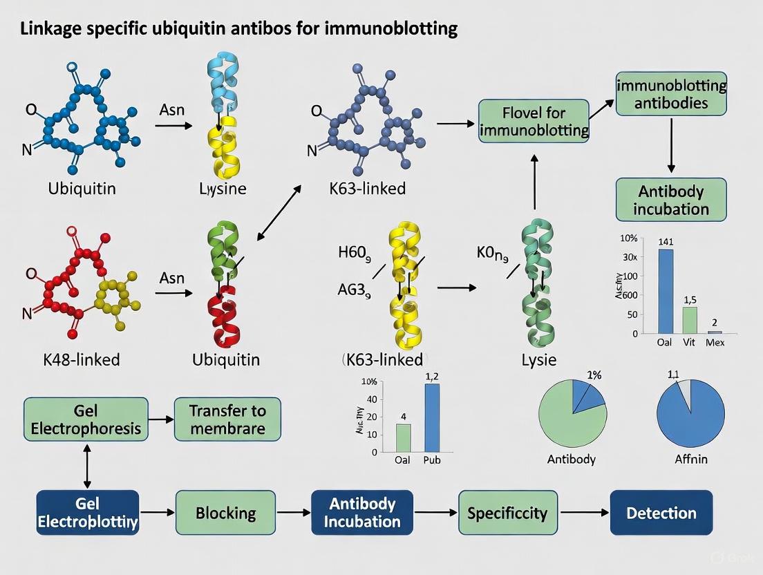
Linkage-Specific Ubiquitin Antibodies: A Comprehensive Guide for Immunoblotting Applications in Research and Drug Development
This article provides a comprehensive resource for researchers and drug development professionals utilizing linkage-specific ubiquitin antibodies in immunoblotting.
Linkage-Specific Ubiquitin Antibodies: A Comprehensive Guide for Immunoblotting Applications in Research and Drug Development
Abstract
This article provides a comprehensive resource for researchers and drug development professionals utilizing linkage-specific ubiquitin antibodies in immunoblotting. It covers the foundational principles of ubiquitin signaling and the critical need for linkage-specific reagents. The content details methodological approaches for detecting specific ubiquitin chain types, offers troubleshooting strategies for common pitfalls such as deubiquitination and antibody specificity, and establishes a framework for rigorous antibody validation. By synthesizing current methodologies and validation standards, this guide aims to enhance the accuracy and reproducibility of ubiquitin research, thereby supporting advancements in understanding disease mechanisms and developing targeted therapies.
Ubiquitin Code Deciphered: Understanding Linkage-Specific Signaling and Antibody Development
Ubiquitination is a crucial post-translational modification that regulates diverse cellular functions, including protein degradation, cell cycle progression, DNA repair, and immune signaling [1] [2]. This modification involves the covalent attachment of ubiquitin—a highly conserved 76-amino acid protein—to substrate proteins. The process is catalyzed by a sequential enzymatic cascade involving E1 (activating), E2 (conjugating), and E3 (ligating) enzymes [2]. Ubiquitin itself contains seven lysine residues (K6, K11, K27, K29, K33, K48, K63) and an N-terminal methionine (M1), all of which can serve as linkage sites for further ubiquitination, enabling the formation of complex polyubiquitin chains [3].
The biological outcome of ubiquitination is determined by the architecture of the ubiquitin chain, creating a "ubiquitin code" that is read by cellular machinery. K48-linked chains primarily target substrates for proteasomal degradation, while K63-linked chains typically mediate non-proteolytic functions such as signal transduction and DNA repair [2]. Less common linkage types (K6, K11, K27, K29, K33, M1) regulate specialized processes including endoplasmic reticulum-associated degradation (ERAD), immune signaling, and mitotic progression [4] [2]. Deciphering this complex code requires specific research tools, particularly linkage-specific antibodies that can distinguish between these structurally distinct ubiquitin chain architectures in immunoblotting applications.
The Ubiquitin Toolkit: Antibodies and Binding Reagents
Researchers investigating the ubiquitin landscape have developed various affinity reagents to capture and detect ubiquitinated proteins. These tools can be broadly categorized into three classes: pan-specific ubiquitin antibodies, linkage-specific antibodies, and ubiquitin-binding domains (UBDs).
Table 1: Key Reagent Categories for Ubiquitin Immunoblotting
| Reagent Category | Key Examples | Recognized Epitope/Feature | Primary Applications |
|---|---|---|---|
| Pan-specific Ubiquitin Antibodies | Ubi-1 (Clone 13-1600) [5], CST #3933 [1] | Conjugated and unconjugated ubiquitin; both mono- and polyubiquitin | General detection of ubiquitinated proteins; immunoprecipitation |
| Linkage-specific Ubiquitin Antibodies | K48-specific [2], K63-specific [2], M1-specific (linear) [3] | Specific ubiquitin chain linkages (K48, K63, M1, etc.) | Determining chain topology and functional consequences |
| Ubiquitin-Binding Entities (UBDs) | Tandem-repeated UBDs (TUBEs) [2] [3] | Multiple ubiquitin chain linkages with high affinity | Preservation and pull-down of ubiquitinated proteins; DUB inhibition |
| Specialized Recognition Tools | Anti-GGX antibodies [6] | N-terminal diglycine remnant on tryptic peptides | Mass spectrometry-based identification of ubiquitination sites |
Pan-specific ubiquitin antibodies, such as the monoclonal Ubi-1 antibody (Clone 13-1600), recognize both conjugated and unconjugated ubiquitin without linkage preference, making them valuable for initial detection of ubiquitination events [5]. These antibodies are particularly useful for observing the characteristic ubiquitin smears or ladders in Western blots that indicate heterogeneous ubiquitination. In contrast, linkage-specific antibodies target unique structural epitopes present in particular ubiquitin chain linkages, enabling researchers to decipher the functional consequences of specific ubiquitin signals [2]. For example, Nakayama et al. employed a K48-linkage specific antibody to demonstrate abnormal accumulation of K48-linked polyubiquitinated tau proteins in Alzheimer's disease [2].
Beyond conventional antibodies, tandem-repeated ubiquitin-binding entities (TUBEs) have emerged as powerful tools with significantly higher affinity for ubiquitinated proteins compared to single UBDs [2] [3]. TUBEs not only facilitate the enrichment of ubiquitinated proteins but also protect ubiquitin chains from deubiquitinating enzymes (DUBs) during cell lysis and processing, thereby preserving the native ubiquitination state [3].
Methodological Considerations for Ubiquitin Immunoblotting
Sample Preparation: Preserving the Ubiquitination State
Accurate detection of ubiquitinated proteins requires careful sample preparation to preserve the labile ubiquitin conjugates. Two critical considerations include inhibition of deubiquitinating enzymes (DUBs) and proteasome activity:
DUB Inhibition: Ubiquitination is rapidly reversed by DUBs during cell lysis. Effective DUB inhibition requires both chelating agents (EDTA or EGTA) to remove heavy metal ions essential for metalloproteinase DUBs, and alkylating agents (N-ethylmaleimide [NEM] or iodoacetamide [IAA]) to target cysteine proteinase DUBs [3]. While traditional protocols use 5-10 mM of these inhibitors, recent findings indicate that up to 10-fold higher concentrations may be necessary to fully preserve certain ubiquitination events, such as K63- and M1-linked chains [3]. NEM is generally preferred over IAA for mass spectrometry applications, as IAA creates a 114 Da adduct identical to the tryptic ubiquitin remnant, potentially interfering with ubiquitination site identification [3].
Proteasome Inhibition: Proteins modified with certain ubiquitin linkages (particularly K48-linked chains) are rapidly degraded by the proteasome. Treatment with proteasome inhibitors like MG132 prevents this degradation, allowing accumulation and detection of ubiquitinated species [3]. However, prolonged inhibitor treatment (12-24 hours) can induce cellular stress responses, potentially confounding results.
Table 2: Critical Components for Sample Preparation in Ubiquitin Studies
| Component | Purpose | Recommended Concentration | Important Considerations |
|---|---|---|---|
| NEM | DUB inhibition (alkylates active site cysteine) | 5-100 mM [3] | Preferred for MS applications; better for K63/M1 chains |
| IAA | DUB inhibition (alkylates active site cysteine) | 5-100 mM [3] | Light-sensitive; may interfere with MS identification of ubiquitylation sites |
| EDTA/EGTA | DUB inhibition (chelates metal ions) | 1-10 mM [3] | Targets metalloproteinase DUBs |
| MG132 | Proteasome inhibition | 10-50 µM [3] | Prevents degradation of ubiquitinated proteins; avoid prolonged treatment |
Electrophoresis and Transfer Optimization
The significant size heterogeneity of ubiquitinated proteins—with polyubiquitin chains adding over 200 kDa to substrate molecular weight—requires careful optimization of electrophoretic conditions [3]. The choice of gel system and running buffer impacts resolution of different ubiquitin chain lengths:
- MES Buffer: Optimal for resolving small ubiquitin oligomers (2-5 ubiquitins)
- MOPS Buffer: Superior for longer polyubiquitin chains (8+ ubiquitins)
- Tris-Acetate Buffer: Best for proteins in the 40-400 kDa range
- Tris-Glycine Buffer with 8% acrylamide: Can separate chains up to 20 ubiquitins long [3]
For comprehensive analysis, researchers often use multiple gel systems to capture both short and long ubiquitin chains. Higher percentage acrylamide gels (~12%) improve resolution of monoubiquitin and short chains but reduce separation of longer polyubiquitin species [3].
Advanced Applications: Ubi-Tagging for Site-Specific Conjugation
Beyond analytical applications, ubiquitin biochemistry has been harnessed for protein engineering through "ubi-tagging"—a novel technique that enables site-directed multivalent conjugation of antibodies to various payloads [7]. This method addresses limitations of traditional antibody conjugation strategies that rely on stochastic modification of lysine or cysteine residues, often resulting in heterogeneous products with compromised functionality [7].
The ubi-tagging approach utilizes the enzymatic ubiquitination cascade to create defined antibody conjugates through three key components: (1) specific ubiquitination enzymes (E1, E2, E3) for the desired ubiquitin linkage type; (2) a donor ubi-tag (Ubdon) with a free C-terminal glycine and a mutated conjugating lysine (e.g., K48R) to prevent homodimer formation; and (3) an acceptor ubi-tag (Ubacc) containing the corresponding conjugation lysine residue but with an unreactive C-terminus [7].
This system enables rapid (30-minute) conjugation of various molecular payloads—including fluorescent dyes, peptides, and proteins—to antibodies, antibody fragments, or nanobodies with high efficiency (93-96% conversion) and without compromising antigen binding capability or protein stability [7]. Applications include generating bispecific T-cell engagers and dendritic-cell-targeted antigens that potently activate T-cell responses, demonstrating the therapeutic potential of this technology [7].
Diagram 1: Ubi-tagging Conjugation Workflow. This site-specific conjugation method uses ubiquitination enzymes to link donor and acceptor ubi-tags, creating homogeneous antibody conjugates [7].
Experimental Protocol: Linkage-Specific Analysis of Protein Ubiquitination
Sample Preparation for Ubiquitin Immunoblotting
Materials:
- Lysis Buffer: 50 mM Tris-HCl (pH 7.5), 150 mM NaCl, 1% NP-40, 1 mM EDTA
- DUB Inhibitors: 50-100 mM N-ethylmaleimide (NEM) prepared fresh in ethanol
- Proteasome Inhibitor: 25 µM MG132 in DMSO
- Protein Assay Kit (e.g., BCA assay)
- 4× SDS Sample Buffer: 250 mM Tris-HCl (pH 6.8), 8% SDS, 40% glycerol, 0.02% bromophenol blue
Procedure:
- Pre-treat cells with MG132 for 4-6 hours before harvesting to stabilize ubiquitinated proteins.
- Prepare ice-cold lysis buffer supplemented with 50 mM NEM immediately before use.
- Lyse cells on ice for 15-30 minutes with occasional vortexing.
- Clarify lysates by centrifugation at 14,000 × g for 15 minutes at 4°C.
- Determine protein concentration using BCA assay.
- Mix 20-40 µg of protein with 4× SDS sample buffer (final 1×).
- Denature samples at 95°C for 5-10 minutes. Avoid extended boiling to prevent ubiquitin chain disruption.
Western Blotting for Ubiquitin Detection
Materials:
- Pre-cast gradient gels (4-20% or 8-16% acrylamide)
- MES or MOPS running buffer
- PVDF or nitrocellulose membrane
- Transfer buffer
- Blocking buffer: 5% non-fat milk in TBST
- Primary antibodies: Pan-ubiquitin antibody (e.g., Ubi-1, 1:1000) and linkage-specific antibodies (1:1000)
- Secondary antibodies: HRP-conjugated anti-mouse or anti-rabbit (1:5000)
- ECL detection reagent
Procedure:
- Separate proteins by SDS-PAGE using appropriate buffer system:
- For chains ≤5 ubiquitins: MES buffer with 12% gel
- For chains ≥8 ubiquitins: MOPS buffer with 8% gel
- Transfer to membrane using standard wet transfer protocol.
- Block membrane with 5% milk in TBST for 1 hour at room temperature.
- Incubate with primary antibody diluted in blocking buffer overnight at 4°C.
- Wash membrane 3× with TBST for 10 minutes each.
- Incubate with HRP-conjugated secondary antibody for 1 hour at room temperature.
- Wash membrane 3× with TBST for 10 minutes each.
- Develop with ECL reagent and image.
Validation and Troubleshooting
Specificity Controls:
- Include linkage-specific deubiquitinases (DUBs) to selectively remove particular ubiquitin chains
- Use ubiquitin binding domains (e.g., TUBEs) as competitive inhibitors to confirm specificity
- Combine multiple linkage-specific antibodies to map complex ubiquitin chain architectures
Common Issues and Solutions:
- Smearing pattern: Reduce protein loading amount; optimize gel percentage
- Weak or no signal: Increase DUB inhibitor concentration; verify antibody specificity
- Non-specific bands: Include peptide competition controls; optimize blocking conditions
Diagram 2: Ubiquitin Immunoblotting Workflow. This optimized protocol preserves ubiquitin conjugates through DUB inhibition and uses electrophoresis conditions tailored to ubiquitin chain length [4] [3].
Research Reagent Solutions
Table 3: Essential Reagents for Ubiquitin Immunoblotting Research
| Reagent | Supplier Examples | Catalog Number Examples | Application Notes |
|---|---|---|---|
| Pan-Ubiquitin Antibody | Thermo Fisher Scientific [5] | 13-1600 (Ubi-1) [5] | Mouse monoclonal; recognizes conjugated and unconjugated ubiquitin; works in WB, IHC, IP |
| Linkage-specific Antibodies | Cell Signaling Technology [1] | Various [2] | K48-, K63-, M1-linear specific antibodies available; verify specificity with DUB treatment |
| DUB Inhibitors | Various chemical suppliers | NEM, IAA [3] | Prepare fresh stock solutions; use higher concentrations (up to 100 mM) for challenging targets |
| Proteasome Inhibitors | Various chemical suppliers | MG132, MG274 [3] | Use 10-50 µM for 4-6 hours; avoid prolonged treatment to prevent stress responses |
| TUBE Reagents | Available commercially | Various [2] [3] | Tandem ubiquitin-binding entities for enhanced ubiquitin affinity and DUB protection |
| GGX Antibodies | Custom discovery [6] | 1C7, 2B12, 2E9, 2H2 [6] | Specialized antibodies for N-terminal ubiquitination site identification via mass spectrometry |
The complexity of the ubiquitin landscape—from monoubiquitination to diverse chain architectures—requires sophisticated experimental approaches for accurate characterization. Linkage-specific ubiquitin antibodies provide powerful tools for deciphering the biological functions of distinct ubiquitin signals in health and disease. When combined with optimized sample preparation methods, appropriate electrophoretic conditions, and rigorous validation controls, these reagents enable researchers to obtain reliable and interpretable data from immunoblotting experiments. The continued development of novel tools, such as ubi-tagging for therapeutic antibody engineering and advanced mass spectrometry reagents, promises to further expand our understanding of this crucial regulatory system.
Ubiquitination is a crucial post-translational modification that regulates virtually all aspects of eukaryotic cell biology [8]. This process involves the covalent attachment of ubiquitin, a 76-amino acid protein, to substrate proteins via a three-step enzymatic cascade involving E1 (activating), E2 (conjugating), and E3 (ligating) enzymes [9] [8]. The functional outcome of ubiquitination depends critically on the specific linkage type between ubiquitin moieties in polyubiquitin chains. The seven lysine residues (K6, K11, K27, K29, K33, K48, K63) and N-terminal methionine (M1) of ubiquitin serve as linkage points, creating structurally and functionally distinct signals often referred to as the "ubiquitin code" [8]. This application note examines the functional consequences of different ubiquitin linkages, focusing on the dichotomy between degradative and non-degradative signaling, and provides detailed methodologies for studying these processes using linkage-specific antibodies in immunoblotting applications.
The Ubiquitin Code: Linkage-Specific Functions
Different polyubiquitin chain types vary significantly in their abundance and primary functions within cells. The table below summarizes the key characteristics of major ubiquitin linkages:
Table 1: Characteristics and Functions of Major Ubiquitin Linkage Types
| Linkage Type | Relative Abundance | Primary Functions | Cellular Processes |
|---|---|---|---|
| K48-linked | ~40% (most abundant) [8] | Proteasomal degradation [10] | Protein turnover, cell cycle regulation, stress response [9] [10] |
| K63-linked | ~30% (second most abundant) [8] | Non-degradative signaling [8] [11] | DNA damage response, endocytic trafficking, inflammation, kinase activation [9] [8] [11] |
| K11-linked | Not specified | Proteasomal degradation [8] | Cell cycle regulation, ER-associated degradation [8] |
| M1-linear | Not specified | Non-degradative signaling [8] [11] | NF-κB activation, inflammation [11] |
| K6, K27, K29, K33-linked | Lower abundance | Varied, less characterized [8] | DNA repair, mitophagy, immune signaling [9] [8] |
Structural Basis for Linkage-Specific Functions
The linkage type determines the three-dimensional architecture of polyubiquitin chains, which in turn dictates their functional specificity [8]. K48-linked chains adopt compact conformations that facilitate recognition by the proteasome, while K63-linked chains form more open, extended structures suitable for signaling complex assembly [8]. These structural differences enable specific recognition by ubiquitin-binding domains (UBDs) present in proteins that determine the ultimate fate of the modified substrate [8].
Degradative Ubiquitin Signaling
K48-Linked Polyubiquitination
K48-linked polyubiquitination serves as the primary signal for proteasomal degradation [10]. Proteins modified with K48-linked chains containing at least four ubiquitin moieties are recognized by the 26S proteasome, leading to their ATP-dependent unfolding and degradation [9] [10]. This process is essential for maintaining cellular proteostasis by eliminating damaged, misfolded, or regulatory proteins.
Key proteins degraded via K48-linked ubiquitination include:
- IκB: Degradation activates NF-κB signaling [10]
- p53: Regulates cell cycle and DNA damage response [10]
- Cell cycle regulators: Including cyclins and CDK inhibitors [10]
Protocol: Detecting K48-Linked Polyubiquitination by Immunoblotting
Purpose: To specifically identify proteins modified with K48-linked ubiquitin chains.
Materials:
- K48-linkage specific polyubiquitin antibody (e.g., Cell Signaling Technology #4289) [10]
- RIPA lysis buffer with protease inhibitors
- Protein quantification assay (e.g., BCA)
- SDS-PAGE gel system
- Western blot transfer apparatus
- HRP-conjugated secondary antibody
- Enhanced chemiluminescence (ECL) detection reagents
Procedure:
- Cell Lysis: Lyse cells in RIPA buffer containing protease inhibitors (including 10 μM MG132 to prevent proteasomal degradation during processing)
- Protein Quantification: Determine protein concentration using BCA assay and normalize samples
- SDS-PAGE: Load 20-30 μg protein per lane and separate on 4-12% Bis-Tris gradient gel
- Protein Transfer: Transfer to PVDF membrane using wet or semi-dry transfer system
- Blocking: Incubate membrane in 5% non-fat dry milk/TBST for 1 hour at room temperature
- Primary Antibody Incubation: Incubate with anti-K48-linkage specific ubiquitin antibody (1:1000 dilution in 5% BSA/TBST) overnight at 4°C
- Washing: Wash membrane 3×10 minutes with TBST
- Secondary Antibody Incubation: Incubate with HRP-conjugated anti-rabbit IgG (1:2000-1:5000 in 5% milk/TBST) for 1 hour at room temperature
- Detection: Develop with ECL reagent and image using chemiluminescence detection system
Technical Notes:
- The K48-specific antibody (clone #4289) demonstrates slight cross-reactivity with linear polyubiquitin chains but not with other linkage types [10]
- Include controls: proteasome inhibitor (MG132) treatment should increase K48-linked ubiquitin conjugates
- For specific substrate analysis, immunoprecipitation may be required before immunoblotting
Non-Degradative Ubiquitin Signaling
K63-Linked Polyubiquitination
K63-linked chains represent the best-characterized non-degradative ubiquitin signal and function as scaffolds for protein complex assembly in multiple signaling pathways [8] [11]. Unlike K48 linkages, K63-linked ubiquitination regulates:
- Kinase activation: In NF-κB and MAPK pathways [11]
- DNA damage response: Recruitment of repair factors [8]
- Protein trafficking: Endocytosis and lysosomal targeting [12]
- Inflammatory signaling: TLR and TNF receptor pathways [11]
Other Non-Degradative Linkages
- M1-linear ubiquitination: Regulates NF-κB signaling through modification of NEMO/IKKγ [11]
- K6-linked chains: Implicated in mitophagy and DNA damage response [9]
- K11-linked chains: Function in both degradative and non-degradative contexts [8]
Protocol: Detecting K63-Linked Polyubiquitination by Immunoblotting
Purpose: To specifically identify proteins modified with K63-linked ubiquitin chains.
Materials:
- Anti-ubiquitin (linkage-specific K63) antibody (e.g., Abcam ab179434) [13]
- Complete lysis buffer (as above)
- Electrophoresis and transfer systems
- Detection reagents
Procedure:
- Sample Preparation: Prepare cell lysates as described for K48 detection
- SDS-PAGE: Separate proteins using 4-12% gradient gels (K63-linked chains often appear as high molecular weight smears)
- Transfer: Transfer to PVDF membrane
- Blocking: Block with 5% non-fat dry milk for 1 hour
- Primary Antibody Incubation: Incubate with anti-K63-linkage specific antibody (1:1000 dilution) overnight at 4°C [13]
- Washing and Detection: Follow standard western blotting procedure as above
Technical Notes:
- The K63-specific antibody (clone EPR8590-448) shows minimal cross-reactivity with other linkage types [13]
- For intracellular flow cytometry, use 1:210 dilution after methanol permeabilization [13]
- K63-linked ubiquitination increases in response to DNA damage and inflammatory stimuli
Research Reagent Solutions
Table 2: Essential Research Reagents for Linkage-Specific Ubiquitin Research
| Reagent Category | Specific Examples | Key Features & Applications | Commercial Sources |
|---|---|---|---|
| Linkage-Specific Antibodies | Anti-K48 (CST #4289) [10] | Rabbit polyclonal; detects endogenous K48-linked chains; WB (1:1000) | Cell Signaling Technology |
| Anti-K63 (Abcam ab179434) [13] | Rabbit monoclonal (EPR8590-448); WB, IHC-P, Flow Cyt; species: human, mouse, rat | Abcam | |
| Enzyme Antibody Kits | Ubiquitin Activation (E1, E2) Antibody Sampler Kit [14] | Includes antibodies against UBE1, UBC3, UbcH5C, UBE2L3/UBCH7, UBE2N/Ubc13 | Cell Signaling Technology |
| Ubiquitin Conjugates | K63-linked di-ubiquitin to hepta-ubiquitin [13] | Recombinant proteins for standardization and competition assays | Various suppliers |
| Activity-Based Probes | Ubiquitin vinyl sulfones | For deubiquitinase (DUB) activity profiling | Multiple suppliers |
Advanced Applications: Ubi-Tagging Technology
Recent biotechnology advances have exploited the specificity of ubiquitin conjugation for protein engineering. The "ubi-tagging" technique enables site-directed multivalent conjugation of antibodies to ubiquitinated payloads within 30 minutes [7]. This methodology utilizes:
- Donor ubi-tag (Ubdon): Contains free C-terminal glycine with mutated conjugating lysine (e.g., K48R) to prevent homodimer formation
- Acceptor ubi-tag (Ubacc): Contains conjugating lysine residue (e.g., K48) with unreactive C-terminus
- Specific E1 and K48-specific E2-E3 fusion enzyme (gp78RING-Ube2g2) [7]
This platform achieves >93% conjugation efficiency and maintains antibody functionality while enabling generation of bispecific T-cell engagers and other multimeric protein constructs [7].
The functional consequences of ubiquitin linkages extend far beyond protein degradation to encompass sophisticated regulatory mechanisms controlling virtually all cellular processes. The dichotomy between K48-mediated degradation and K63-mediated signaling represents just one aspect of the complex ubiquitin code that continues to be elucidated. Linkage-specific antibodies provide powerful tools for deciphering this code through immunoblotting and other applications. As research progresses, particularly in understanding atypical ubiquitin linkages and developing ubiquitin-based biotechnologies, our ability to manipulate these pathways for therapeutic intervention will continue to advance, offering new opportunities for targeting ubiquitin-related processes in cancer, neurodegeneration, and inflammatory diseases.
Protein ubiquitination is a pivotal post-translational modification that regulates virtually all aspects of eukaryotic cell biology, from protein degradation to DNA repair and immune signaling [8]. The complexity of ubiquitin signaling arises from the ability of ubiquitin to form various chain architectures through different linkage types between its amino acid residues. Ubiquitin can be conjugated to substrate proteins as a single molecule (monoubiquitination) or as polyubiquitin chains connected through one of seven lysine residues (K6, K11, K27, K29, K33, K48, K63) or the N-terminal methionine (M1) [8]. Recently, ester-linked ubiquitination via serine and threonine residues has been identified, bringing the total number of known ubiquitin linkages in cells to twelve [8]. Each linkage type confers a distinct three-dimensional structure to the ubiquitin chain, enabling specific functions and outcomes within the cell [8]. This vast array of modifications constitutes what is known as the "Ubiquitin Code" [8].
The critical importance of linkage-specific signaling is exemplified by the distinct cellular functions of different chain types. While K48-linked chains predominantly target proteins for proteasomal degradation, K63-linked chains are primarily involved in non-proteolytic signaling pathways such as DNA damage response and immune signaling [8]. The "atypical" linkage types (M1, K6, K11, K27, K29, K33) play important but less characterized roles in processes including cell cycle regulation and proteotoxic stress [8]. Deciphering this complex code requires highly specific tools capable of distinguishing between these structurally similar yet functionally distinct ubiquitin modifications.
The Specificity Challenge in Ubiquitin Antibody Development
Fundamental Obstacles to Specificity
Generating antibodies with the requisite specificity for individual ubiquitin linkage types presents unique challenges not encountered with other post-translational modifications. The primary hurdles include:
- Epitope Size and Complexity: Unlike smaller modifications such as phosphorylation or acetylation, ubiquitin is a full-sized protein of 76 amino acids [15]. The epitope for a site-specific ubiquitin antibody encompasses not only the modified lysine but also substantial portions of both the ubiquitin molecule and the target protein, creating a large, complex antigenic structure.
- Instability of Native Linkage: The isopeptide bond between ubiquitin and substrate lysines is highly susceptible to cleavage by deubiquitinating enzymes (DUBs) present in biological systems, including during the immunization process itself [15]. This instability compromises antigen integrity and immune recognition.
- Structural Similarity Between Linkages: Different ubiquitin linkage types share significant structural homology, making it difficult to generate antibodies that can discriminate between them with high fidelity. The linkage points are often buried within the ubiquitin structure or present similar surface features that challenge selective antibody binding.
Limitations of Conventional Approaches
Traditional immunization strategies using short peptides or ubiquitin fragments have proven largely unsuccessful for generating high-quality site-specific ubiquitin antibodies [15]. These approaches fail to present the complete conformational epitope necessary for the immune system to produce antibodies with the required specificity. Consequently, the field has suffered from a scarcity of reliable reagents for monitoring specific ubiquitination events, significantly hampering progress in understanding ubiquitin-dependent regulatory mechanisms [15].
Strategic Solutions: Advanced Antigen Design
Proteolytically Stable Antigen Synthesis
To overcome the challenges of antigen instability, researchers have developed sophisticated chemical biology approaches for creating proteolytically stable ubiquitin conjugates. The strategy involves synthesizing well-defined Ub-modified polypeptides using advanced ligation technologies, primarily through two approaches:
- Native Isopeptide Mimetics: Using thiolysine-mediated ligation to generate antigens with native isopeptide linkages, though these remain susceptible to DUB cleavage.
- Stabilized Isostere Replacement: Creating proteolytically stable antigens by replacing the native isopeptide bond with an amide triazole isostere via click chemistry [15]. This modification preserves the overall structure of the ubiquitin-lysine environment while conferring resistance to enzymatic cleavage during immunization.
These synthetic antigens incorporate the full ubiquitin protein, increasing the likelihood of exposing a complete site-specific epitope to the immune system [15]. The successful generation of a monoclonal antibody specific for ubiquitin on lysine 123 of yeast histone H2B (yH2B-K123ub) using this approach demonstrates its effectiveness [15].
Immunization and Screening Workflow
The process for developing site-specific ubiquitin antibodies follows a systematic approach:
- Design and synthesis of non-hydrolyzable Ub-peptide conjugates for immunization
- Design and synthesis of extended native isopeptide-linked Ub-peptide conjugates for screening
- Immunization and generation/screening of hybridomas
- Clone selection and antibody validation in native contexts [15]
This workflow emphasizes the critical importance of using different antigen designs for immunization versus screening, optimizing each for their respective purposes while maintaining epitope integrity throughout the process.
Figure 1: Development workflow for site-specific ubiquitin antibodies, highlighting the critical stages from antigen design to final validation.
The Molecular Toolbox for Ubiquitin Analysis
Beyond traditional antibodies, researchers have developed a diverse array of molecular tools for studying linkage-specific ubiquitin signaling. These reagents offer complementary advantages for different experimental applications.
Table 1: Molecular Tools for Linkage-Specific Ubiquitin Analysis
| Tool Category | Key Examples | Mechanism of Action | Applications | Advantages/Limitations |
|---|---|---|---|---|
| Traditional Antibodies | Polyclonal and monoclonal anti-ubiquitin [16] | Recognize specific ubiquitin epitopes | Western blot, IHC, ICC, ELISA, Flow Cytometry [16] | Well-established protocols; may lack linkage specificity |
| Linkage-Specific Antibodies | Anti-Ubiquitin (K63-linkage specific) [EPR8590-448] [13] | Specifically bind to K63-linked polyubiquitin chains | Western blot, Flow Cytometry (Intra), IHC-P [13] | High linkage specificity; limited availability for atypical linkages |
| Engineered Ubiquitin-Binding Domains (UBDs) | OtUBD [17] | High-affinity nanomolar binding to ubiquitin | Enrichment of ubiquitinated proteins, proteomics [17] | Versatile for various ubiquitin conjugates; requires protein engineering |
| Tandem Ubiquitin-Binding Entities (TUBEs) | Not specified in sources | Multiple linked UBDs for avidity effect | Protection from DUBs, purification of polyubiquitinated proteins | Excellent for polyubiquitin; poor for monoubiquitination [17] |
| Catalytically Inactive DUBs | Not specified in sources | High-affinity binding without cleavage | Detection, enrichment, structural studies [8] | Naturally evolved specificity; requires inactivation mutagenesis |
| Affimers and Macrocyclic Peptides | Not specified in sources | Synthetic binding scaffolds | Similar to antibodies; customization possible [8] | Potential for high specificity; emerging technology |
Experimental Protocols for Validation
Specificity Validation for Linkage-Specific Antibodies
Rigorous validation is essential to confirm linkage specificity. The following protocol, adapted from commercial antibody validation data, provides a comprehensive approach:
Materials:
- Test antibody (e.g., Anti-Ubiquitin K63-linkage specific [EPR8590-448]) [13]
- Control cell lysates (HEK-293, HeLa, mouse/rat brain tissue) [13]
- Recombinant di-ubiquitin proteins of various linkages (K6, K11, K27, K29, K33, K48, K63) [13]
- Western blot equipment and reagents
- Blocking buffer (5% non-fat dry milk/TBST) [13]
- HRP-conjugated secondary antibodies
Procedure:
- Prepare Samples: Load 20 µg of cell lysates or 0.02-20 µg of recombinant di-ubiquitin proteins on SDS-PAGE gels [13].
- Transfer and Block: Transfer proteins to PVDF membrane and block with 5% non-fat dry milk in TBST.
- Primary Antibody Incubation: Incubate with linkage-specific antibody at optimized dilution (e.g., 1/1000 for K63-specific antibody [EPR8590-448] in 5% non-fat dry milk/TBST) [13].
- Detection: Incubate with appropriate HRP-conjugated secondary antibody (e.g., 1/1000 dilution) and develop with ECL reagent [13].
- Specificity Assessment: Confirm that the antibody reacts only with the intended linkage type (e.g., K63-linked chains) and shows no cross-reactivity with other linkage types [13].
OtUBD-Based Enrichment of Ubiquitinated Proteins
The OtUBD protocol provides an alternative method for enriching ubiquitinated proteins using a high-affinity ubiquitin-binding domain:
Materials:
- pET21a-cys-His6-OtUBD plasmid (Addgene #190091) [17]
- SulfoLink coupling resin [17]
- Lysis buffers (native: 50 mM Tris pH 7.5, 150 mM NaCl, 1% Triton X-100; denaturing: 6 M Guanidine HCl) [17]
- Protease inhibitors (PMSF, Complete EDTA-free) [17]
- DUB inhibitors (N-ethylmaleimide)
- Elution buffer (50 mM Tris pH 7.5, 2% SDS, 10 mM DTT) [17]
Procedure:
- OtUBD Purification: Express and purify recombinant OtUBD from E. coli using nickel-affinity chromatography [17].
- Resin Preparation: Couple purified OtUBD to SulfoLink resin via cysteine residue [17].
- Sample Preparation: Lyse cells in appropriate buffer (native for interactome studies, denaturing for direct ubiquitome analysis) with protease and DUB inhibitors [17].
- Enrichment: Incubate lysates with OtUBD resin for 2 hours at 4°C with gentle rotation.
- Washing: Wash resin extensively with respective lysis buffer.
- Elution: Elute bound proteins with SDS-PAGE sample buffer or specific elution buffers [17].
- Downstream Analysis: Analyze eluates by immunoblotting or mass spectrometry.
Figure 2: Workflow for OtUBD-based enrichment of ubiquitinated proteins, showing both native and denaturing condition pathways.
Research Reagent Solutions
Table 2: Essential Research Reagents for Site-Specific Ubiquitin Studies
| Reagent Category | Specific Examples | Supplier/Source | Primary Applications | Key Considerations |
|---|---|---|---|---|
| Linkage-Specific Antibodies | Anti-Ubiquitin (K63-linkage specific) [EPR8590-448] (ab179434) [13] | Abcam | Western blot, IHC-P, Flow Cytometry (Intra) [13] | Validate for specific applications; check species reactivity |
| General Ubiquitin Antibodies | Monoclonal anti-ubiquitin (P4D1) [17] | Multiple suppliers | Western blot, IP, general ubiquitin detection | Lacks linkage specificity; good for total ubiquitin detection |
| Plasmids for Tool Development | pRT498-OtUBD (Addgene #190089) [17] | Addgene | Recombinant OtUBD production | Enables in-house reagent production |
| Enrichment Resins | SulfoLink coupling resin [17] | Thermo Scientific | Immobilization of Ub-binding domains | Compatible with cysteine-containing proteins |
| Inhibitors | N-ethylmaleimide (NEM) [17] | Multiple suppliers | DUB inhibition in lysates | Essential for preserving ubiquitination states |
| Recombinant Ubiquitin Proteins | K63-linked-Ub2-7 recombinant protein [13] | Multiple suppliers | Antibody validation, controls | Critical for specificity testing |
Discussion and Future Perspectives
The field of ubiquitin research continues to evolve with emerging technologies offering new approaches to overcome the specificity challenge. Advanced antigen design strategies using proteolytically stable mimics have demonstrated feasibility for generating high-quality site-specific antibodies, as evidenced by the successful development of antibodies against yeast H2B-K123ub [15]. However, the limited commercial availability of well-validated linkage-specific antibodies, particularly for atypical ubiquitin linkages, remains a significant constraint in the field.
Future directions will likely include the increased application of non-antibody affinity reagents such as affimers and engineered ubiquitin-binding domains, which offer alternative paths to achieving the required specificity [8]. Additionally, the development of standardized validation protocols and reference materials will be essential for ensuring reproducibility across studies. As our understanding of the ubiquitin code expands to include non-canonical linkages and non-protein substrates, the demand for increasingly specific detection tools will continue to grow, driving innovation in this challenging yet critical area of research.
For researchers embarking on studies of linkage-specific ubiquitination, a multimodal approach combining multiple tools and validation methods is recommended to ensure robust and interpretable results. The strategic selection of reagents from the growing molecular toolbox, coupled with rigorous validation using the protocols outlined herein, will advance our ability to decipher the complex language of ubiquitin signaling in health and disease.
The study of ubiquitin signaling is fundamental to understanding diverse cellular processes, from protein degradation to DNA repair and immune signaling. A significant challenge in this field is the development of high-quality linkage-specific ubiquitin antibodies, a process entirely dependent on the strategic design of antigen used for immunization. The inherent structural complexity and lability of the native isopeptide bond, which covalently connects ubiquitin to substrate proteins or other ubiquitin molecules, presents a unique set of obstacles. This application note details two primary chemical strategies for antigen design: the use of native isopeptide bonds and the implementation of proteolytically stable mimics. We will provide a comparative analysis, supported by quantitative data, and deliver detailed protocols tailored for researchers, scientists, and drug development professionals focused on advancing immunoblotting applications for ubiquitin research.
The isopeptide bond, a defining feature of protein ubiquitination, is formed between the C-terminal glycine of ubiquitin and the ε-amino group of a lysine residue on a substrate protein or another ubiquitin molecule [8]. This linkage is a target for deubiquitinating enzymes (DUBs), which are highly active in biological systems. When designing antigens for antibody production, this lability is a major impediment, as DUBs present in vivo can cleave the antigen before a robust immune response is mounted [15]. Consequently, the choice of antigen design strategy directly influences the specificity, affinity, and ultimate success of the resulting antibodies.
Comparative Analysis of Antigen Design Strategies
The core challenge in generating site-specific ubiquitin antibodies is presenting the immune system with a stable, authentic epitope. The following table summarizes the key characteristics of the two main strategies.
Table 1: Comparison of Antigen Design Strategies for Ubiquitin Antibodies
| Feature | Native Isopeptide Antigens | Proteolytically Stable Mimics |
|---|---|---|
| Chemical Linkage | Native Lys-ε-NH-Gly isopeptide bond [15] | Non-hydrolyzable triazole isostere or single-atom substitutions (O/S, O/Se) [18] [15] |
| Epitope Fidelity | High, identical to native target | High; designed to closely mimic native structure and presentation [18] [15] |
| Stability to DUBs | Low; susceptible to cleavage during immunization [15] | Very high; resistant to enzymatic cleavage [15] |
| Synthetic Complexity | High; requires advanced chemical ligation or native protein expression [15] | Moderate to High; requires specialized organic synthesis [18] [15] |
| Primary Application | Ideal for screening assays to identify clones that recognize the native structure [15] | Recommended for the initial immunization to elicit a robust, specific immune response [15] |
| Reported Success | Used in successful antibody development workflows [15] | Critical for generating antibodies against yeast H2B-K123ub; successful in designing potent tumor-associated antigen mimics [18] [15] |
Strategic Workflow for Antigen Selection and Antibody Development
The decision between these strategies is not mutually exclusive. A synergistic approach, leveraging the strengths of both methods, often yields the best results. The following diagram outlines a recommended workflow for antigen design and antibody development.
Diagram 1: Integrated workflow for antibody development using both stable mimics for immunization and native antigens for screening.
Detailed Protocols for Antigen Synthesis and Evaluation
Protocol 1: Synthesis of Proteolytically Stable Ubiquitin-Peptide Conjugates
This protocol describes the creation of a stable antigen using "click chemistry" to form a triazole isostere, a proven method for generating site-specific ubiquitin antibodies [15].
Materials:
- Resins and Reagents: Fmoc-Lys(Dde)-OH, Fmoc-protected amino acids, peptide synthesis resin, synthesizer.
- Ubiquitin Modifiers: Recombinant ubiquitin modified with an azide-containing handle at its C-terminus.
- Click Chemistry Reagents: Copper(II) sulfate pentahydrate (CuSO₄), Sodium ascorbate, TBTA (Tris[(1-benzyl-1H-1,2,3-triazol-4-yl)methyl]amine).
- Purification: HPLC system with C18 column.
Procedure:
- Peptide Synthesis: Synthesize the target substrate peptide (e.g., derived from histone H2B or PCNA) using standard Fmoc-solid phase peptide synthesis. Incorporate a propargyl-glycine residue at the position corresponding to the target lysine. This alkyne group will serve as the reaction partner for click chemistry.
- Ubiquitin Activation: Prepare recombinant ubiquitin with an azide moiety at its C-terminal glycine. This can be achieved through protein semi-synthesis or enzymatic ligation.
- Copper-Catalyzed Azide-Alkyne Cycloaddition (CuAAC):
- Combine the alkyne-containing peptide (from Step 1) and azide-functionalized ubiquitin (from Step 2) in a degassed buffer (e.g., phosphate-buffered saline with 1% SDS).
- Add the catalyst system: 1 mM CuSO₄, 2 mM sodium ascorbate, and 100 µM TBTA.
- React the mixture for 2-4 hours at room temperature with gentle agitation.
- Purification and Characterization:
- Terminate the reaction by acidification with trifluoroacetic acid (TFA).
- Purify the conjugate using reverse-phase HPLC.
- Verify the identity and mass of the final product using mass spectrometry (MS). The resulting ubiquitin-peptide conjugate will be linked by a stable triazole bond, mimicking the structure of the native isopeptide linkage.
Protocol 2: Generating Native Isopeptide-Linked Antigens for Screening
This protocol outlines the production of antigens containing the labile native isopeptide bond, which are best used for screening hybridoma clones.
Materials:
- Enzymatic Machinery: Purified E1 activating enzyme, specific E2 conjugating enzyme, and E3 ligase relevant to the target ubiquitination site.
- Buffers: ATP-containing reaction buffer (50 mM Tris-HCl, pH 7.5, 50 mM NaCl, 10 mM MgCl₂, 1 mM DTT, 2 mM ATP).
- Substrates: Recombinant substrate protein or a long peptide fragment (> 20 amino acids) encompassing the target site.
- Purification: Affinity tags (e.g., His-tag on ubiquitin or substrate), Ubiquitin-Binding Entities (TUBEs) to enrich for ubiquitylated proteins [3].
Procedure:
- In Vitro Ubiquitination Reaction:
- Combine the substrate protein/peptide, ubiquitin, E1 enzyme, E2 enzyme, and E3 ligase in the ATP-containing reaction buffer.
- Incubate at 30°C for 1-3 hours.
- Reaction Termination and Preservation:
- To prevent deubiquitination, stop the reaction by adding denaturing buffer containing 1% SDS and 20-50 mM N-Ethylmaleimide (NEM) or Iodoacetamide (IAA) to alkylate and inhibit DUBs [3].
- Immediately heat the sample to 95°C for 5 minutes.
- Purification of Ubiquitinated Product:
- Use affinity chromatography (e.g., Ni-NTA if ubiquitin is His-tagged) or immunoprecipitation with TUBEs to isolate the ubiquitylated substrate from the reaction mixture [3].
- Confirm the modification and assess yield via SDS-PAGE and immunoblotting with a pan-ubiquitin antibody.
Protocol 3: Sample Preparation for Immunoblotting Analysis
Proper sample preparation is critical for accurately assessing ubiquitination in immunoblotting applications.
Materials:
- Lysis Buffer: RIPA buffer or similar, supplemented fresh with:
- DUB Inhibitors: 20-50 mM N-Ethylmaleimide (NEM) or Iodoacetamide (IAA).
- Protease Inhibitors: Complete EDTA-free protease inhibitor cocktail.
- Proteasome Inhibitor (optional): 10-20 µM MG132 (to stabilize proteasome-targeted ubiquitinated species) [3].
- Gel Electrophoresis: Pre-casted gradient gels (e.g., 4-12% or 4-20%), MES or MOPS running buffer for optimal resolution of polyubiquitin chains [3].
Procedure:
- Cell Lysis: Lyse cultured cells or tissue directly in pre-heated (95°C) 1% SDS lysis buffer to instantly denature all proteins and inactivate DUBs. Alternatively, for non-denaturing lysis, use RIPA buffer containing 50 mM NEM.
- Sample Denaturation: Boil lysates for 5-10 minutes. Shear genomic DNA by sonication or pass through a fine-gauge needle to reduce viscosity.
- SDS-PAGE and Immunoblotting:
- Resolve proteins by SDS-PAGE. For optimal separation of polyubiquitin chains, use a MES-based buffer system for shorter chains (2-5 ubiquitins) and a MOPS-based system for longer chains [3].
- Transfer to a PVDF or nitrocellulose membrane.
- Probe the membrane with the linkage-specific ubiquitin antibody generated through the above strategies.
The Scientist's Toolkit: Essential Research Reagents
The following table catalogues critical reagents and their functions for research in this field, as derived from the cited literature.
Table 2: Key Research Reagent Solutions for Ubiquitin Antigen Design and Immunoblotting
| Reagent / Tool | Function / Application | Key Characteristics |
|---|---|---|
| Tandem-repeated Ubiquitin-Binding Entities (TUBEs) | Affinity enrichment of ubiquitylated proteins from cell lysates [3]. | Protects ubiquitin chains from DUBs and proteasomal degradation during purification; binds all linkage types. |
| Linkage-Specific Deubiquitylases (DUBs) | Analytical tool for confirming ubiquitin chain topology [3]. | Enzymes that selectively cleave a specific ubiquitin linkage (e.g., OTUB1 for K48). |
| N-Ethylmaleimide (NEM) / Iodoacetamide (IAA) | Covalent DUB inhibitors in lysis and assay buffers [3]. | Alkylating agents that irreversibly modify the active site cysteine of most DUBs; crucial for preserving the ubiquitination state. |
| Proteasome Inhibitor (MG132) | Stabilizes K48- and other proteasome-targeted ubiquitin conjugates [3]. | Reduces degradation of ubiquitylated proteins of interest, enhancing their detection. |
| Computational Antibody Design Tools (e.g., Rosetta) | In silico prediction and optimization of antibody structures and antigen-antibody interactions [19]. | Enables homology modeling and de novo design of antibody CDR loops, streamlining development. |
| Stabilizing Isopeptide Bonds (Engineered) | Protein engineering strategy to enhance the stability of antigen formulations [20]. | Intramolecular Lys-Asn bonds that form autocatalytically, conferring extreme thermal and proteolytic resistance. |
Concluding Remarks
The strategic choice between native isopeptides and proteolytically stable mimics is pivotal for the successful development of linkage-specific ubiquitin antibodies. A combined approach—using stable mimics for immunization to ensure a robust and specific immune response, and native isopeptide antigens for screening to select clones that recognize the physiological target—has proven highly effective [15]. Adherence to the detailed protocols for antigen synthesis and sample preparation, coupled with the use of specialized reagents like TUBEs and DUB inhibitors, will significantly enhance the reliability and quality of data obtained in immunoblotting applications. As the field advances, the integration of computational design [19] and novel protein engineering strategies, such as the incorporation of stabilizing isopeptide bonds [20], will further empower researchers to decode the complex language of ubiquitin signaling.
Practical Immunoblotting Protocols for Linkage-Specific Ubiquitin Detection
The ubiquitin (Ub) code, comprising monomeric Ub and diverse polyubiquitin chains, regulates virtually all aspects of eukaryotic cell biology, from protein degradation to DNA repair and signal transduction [8]. A central challenge in deciphering this code is the dynamic and reversible nature of ubiquitination, primarily countered by the activity of deubiquitinating enzymes (DUBs). During cell lysis, the loss of compartmentalization and changing conditions trigger DUB activity, leading to the rapid erasure of ubiquitin signals and compromising experimental reproducibility. This application note details robust protocols for sample preparation that leverage DUB inhibitors and novel affinity tools to preserve the native ubiquitylation state, providing a critical foundation for accurate analysis using linkage-specific ubiquitin antibodies in immunoblotting applications.
Section 1: Core Principles and Reagent Solutions
The Necessity of Stabilizing the Ubiquitinome
The ubiquitin system is characterized by its extraordinary complexity and dynamism. With more than 100 putative human DUBs constantly shaping the cellular ubiquitin landscape, the steady-state level of any ubiquitination event is a delicate balance between conjugation and deconjugation [21]. When cells are lysed, this equilibrium is disrupted. DUBs, many of which are cysteine proteases, remain active in cell extracts and can rapidly strip ubiquitin modifications from substrates unless explicitly inhibited [22]. For research relying on linkage-specific antibodies, which often detect subtle changes in specific chain types like K48 or K63 linkages, this deubiquitination activity poses a significant threat to data validity.
Research Reagent Solutions for Ubiquitin Preservation
A combination of pharmacological and affinity-based tools is essential for effective ubiquitin preservation.
Table 1: Key Reagents for Preserving Native Ubiquitylation States
| Reagent Name | Type | Primary Function | Key Characteristics |
|---|---|---|---|
| PR-619 | Broad-spectrum DUB Inhibitor | Inhibits cysteine protease DUBs | Used at 10-50 µM in lysis buffer; enables snapshot of ubiquitome [23]. |
| Tandem Ubiquitin Binding Entities (TUBEs) | Recombinant Affinity Tools | Protect & purify polyubiquitylated proteins | High-affinity ubiquitin binding; shields chains from DUBs/proteasomes [22]. |
| N-Ethylmaleimide (NEM) | Cysteine Alkylating Agent | Irreversibly inhibits cysteine protease DUBs | Common component in lysis buffers; note potential for side reactions [22]. |
| Linkage-Specific Ubiquitin Antibodies | Detection Antibodies | Detect specific polyubiquitin linkages | e.g., anti-K48 (Cell Signaling Tech #4289) & anti-K63 (Abcam ab179434); require preserved antigen [10] [13]. |
Section 2: Experimental Protocols for Sample Preparation
Protocol 1: Cell Lysis with DUB-Inhibiting Buffers for Immunoblotting
This protocol is optimized for preparing samples for subsequent immunoblotting with linkage-specific antibodies.
Materials:
- Freshly prepared Urea Lysis Buffer (8 M urea, 50 mM Tris HCl pH 8.0, 150 mM NaCl, 1 mM EDTA)
- Protease and DUB Inhibitor Cocktail: 2 µg/mL Aprotinin, 10 µg/mL Leupeptin, 50 µM PR-619, 1 mM PMSF, 1 mM Chloroacetamide (or Iodoacetamide) [23]
- Phosphate-Buffered Saline (PBS), ice-cold
- Cell scraper (for adherent cells) or centrifuge (for suspension cells)
- Sonicator or needle and syringe for mechanical shearing
Procedure:
- Pre-cool Equipment: Ensure centrifuges, rotors, and tubes are chilled.
- Wash Cells: Rapidly wash cell monoleries or pellets with ice-cold PBS to remove serum and dead cells.
- Add Lysis Buffer: Aspirate PBS completely and immediately add Urea Lysis Buffer containing the complete inhibitor cocktail (e.g., 1 mL per 10⁷ cells). The denaturing conditions of urea help inactivate enzymes and disrupt non-covalent interactions.
- Harvest Cells: For adherent cells, scrape them into the lysis buffer on ice. For suspension cells, vortex briefly.
- Lyse Cells: Perform brief sonication on ice (3x 5-second pulses with 15-second rests) or pass the lysate 10-15 times through a 21-gauge needle to shear DNA and reduce viscosity.
- Clarify Lysate: Centrifuge the lysate at 16,000 × g for 15 minutes at 4°C to pellet insoluble debris.
- Collect Supernatant: Transfer the clarified supernatant to a fresh, pre-chilled tube.
- Protein Quantification & Storage: Determine protein concentration using a compatible assay (e.g., BCA). Aliquot and snap-freeze samples at -80°C if not used immediately.
Protocol 2: Native Lysis Combined with TUBE Enrichment
This protocol uses TUBEs for the purification and protection of polyubiquitylated proteins under native conditions, ideal for functional studies or when analyzing weak ubiquitination signals [22].
Materials:
- Native Lysis Buffer (e.g., 50 mM Tris pH 7.5, 150 mM NaCl, 1% NP-40)
- DUB Inhibitors: PR-619 (10-50 µM) or NEM (5-10 mM)
- GST- or His-tagged TUBE protein
- Glutathione or Ni-NTA Agarose Beads
- Appropriate washing and elution buffers
Procedure:
- Lysate Preparation: Lyse cells in Native Lysis Buffer containing DUB inhibitors. Avoid strong denaturants like urea or SDS.
- Clarify Lysate: Centrifuge at high speed to remove insoluble material.
- Incubate with TUBEs: Add the tagged TUBE protein directly to the clarified lysate and incubate for 1-2 hours at 4°C with gentle rotation. Critical Step: Adding TUBEs during lysis provides immediate protection from DUBs.
- Capture Complexes: Add the appropriate affinity beads (e.g., Glutathione Agarose for GST-TUBEs) and incubate for an additional 1 hour.
- Wash Beads: Pellet beads and wash 3-4 times with native lysis buffer to remove non-specifically bound proteins.
- Elution: Elute bound ubiquitylated proteins using a competitive agent like reduced glutathione (for GST-TUBEs) or by boiling in SDS-PAGE sample buffer for direct immunoblot analysis.
The following diagram illustrates the core strategic decision-making process for selecting the appropriate sample preparation method based on research goals.
Section 3: Data, Validation, and Application
Quantitative Impact of DUB and Proteasome Inhibition
Mass spectrometry-based ubiquitinome analyses provide a system-wide view of how inhibition strategies reshape the ubiquitin landscape. The data below summarizes the profound effects of targeting DUBs versus the proteasome.
Table 2: Quantitative Ubiquitinome Dynamics Following Inhibition Data derived from UbiSite mass spectrometry analysis of U2OS cells treated for 3 hours [21].
| Treatment | Target | Total Ubiquitination Sites Identified | Significantly Changed Sites (vs. DMSO) | Key Functional Examples of Regulated Proteins |
|---|---|---|---|---|
| PR-619 | Cysteine Protease DUBs | 55,355 sites on 9,267 proteins | 77% of sites | PARP1 (Hyperubiquitination, increased activity) [21] |
| MG-132 | Proteasome | 55,355 sites on 9,267 proteins | 77% of sites | Accumulation of K48-linked chains [21] |
| TAK-243 | Ubiquitin E1 Activating Enzyme | 55,355 sites on 9,267 proteins | Not Specified | Global depletion of ubiquitin conjugates [21] |
Verification with Linkage-Specific Antibodies
After sample preparation using the described protocols, linkage-specific antibodies are used for detection. It is critical to validate the specificity of these reagents.
- Anti-K63-linkage (ab179434): This rabbit monoclonal antibody shows specificity for K63-linked chains over other linkage types (K6, K11, K27, K29, K33, K48) in western blotting [13]. It is suitable for use with human, mouse, and rat samples.
- Anti-K48-linkage (#4289): This antibody specifically detects polyubiquitin chains linked via K48 and is the primary signal for targeting proteins to the proteasome. It demonstrates minimal cross-reactivity with linear chains or chains formed through other lysine residues [10].
The following workflow diagram integrates these verification steps and shows the downstream consequences of successful ubiquitin preservation, leading to meaningful biological insights.
The integrity of research on linkage-specific ubiquitination hinges on the initial steps of sample preparation. The combination of rapid, cold lysis with potent, broad-spectrum DUB inhibitors like PR-619 is the minimum requirement for capturing a reliable snapshot of the cellular ubiquitinome. For more specialized applications, particularly those involving the study of labile modifications or the functional characterization of ubiquitinated proteins, the use of TUBEs represents a superior strategy by actively shielding ubiquitin chains from degradation and deconjugation.
Best Practice Summary:
- Always use inhibitors: Never lyse cells without DUB inhibitors in the buffer.
- Prepare fresh: Always prepare lysis buffers with inhibitors fresh immediately before use.
- Work quickly: Keep samples on ice and process them rapidly to minimize enzymatic activity.
- Validate antibodies: Confirm the specificity of linkage-specific antibodies using available recombinant ubiquitin ladders.
By adhering to these detailed protocols and understanding the underlying principles, researchers can significantly enhance the accuracy and reproducibility of their data in the complex field of ubiquitin signaling.
Ubiquitination is a pivotal post-translational modification that regulates virtually all aspects of eukaryotic cell biology, governing processes from proteasomal degradation to DNA repair, kinase activation, and immune signaling [8]. The complexity of ubiquitin signaling stems from the ability of ubiquitin itself to be conjugated through any of its seven lysine residues (K6, K11, K27, K29, K33, K48, K63) or its N-terminal methionine (M1), forming structurally and functionally distinct polyubiquitin chains [8] [3]. This vast array of modifications constitutes what is known as the "Ubiquitin Code," where each linkage type can encode specific cellular outcomes [8]. For instance, K48-linked chains predominantly target proteins for proteasomal degradation, while K63-linked chains mainly facilitate non-proteolytic signaling in pathways such as DNA damage response and inflammation [10] [24]. The ability to detect and characterize these specific ubiquitin linkages is therefore fundamental to advancing our understanding of cellular regulation and developing targeted therapeutic interventions.
The development of linkage-specific ubiquitin antibodies represents a critical technological advancement for researchers seeking to decipher this complex signaling system. These reagents enable the specific detection of individual ubiquitin chain types amidst a background of highly similar structures, providing insights into the dynamic and heterogeneous landscape of ubiquitin signaling [8]. This application note provides a comprehensive overview of commercially available linkage-specific ubiquitin antibodies, with a specific focus on their application in immunoblotting, and offers detailed protocols to optimize experimental outcomes for researchers and drug development professionals.
The Ubiquitin Signaling System: Complexity and Function
The ubiquitination process involves a sophisticated enzymatic cascade comprising E1 (activating), E2 (conjugating), and E3 (ligating) enzymes that work in concert to attach ubiquitin to substrate proteins [8]. A hallmark of this system is that ubiquitin itself can be ubiquitinated, giving rise to various polyubiquitin chain architectures, including homotypic chains (single linkage type), mixed chains (multiple linkage types in sequence), and branched chains (multiple ubiquitins attached to a single ubiquitin moiety) [8]. The linkage between ubiquitin molecules determines the overall architecture and topology of the chain, which in turn dictates its function by creating distinct surfaces for recognition by ubiquitin-binding domains (UBDs) [8].
Among the different linkage types, K48-linked and K63-linked chains are the most abundant and well-characterized, constituting approximately 40% and 30% of cellular ubiquitin linkages, respectively [8]. The remaining "atypical" linkages (M1, K6, K11, K27, K29, K33) and the recently discovered non-canonical ester-linked chains (via serine and threonine residues) are less understood but play important roles in specific cellular contexts [8]. The functional diversity of ubiquitin chains necessitates specific detection tools, as traditional pan-ubiquitin antibodies cannot distinguish between these functionally distinct signals.
Commercially Available Linkage-Specific Antibodies
The specificity of linkage-specific ubiquitin antibodies is achieved through immunization with synthetic antigens that mimic the unique structural features around specific ubiquitin linkage sites. The following table summarizes key commercially available antibodies for the detection of major ubiquitin linkages via immunoblotting.
Table 1: Commercially Available Linkage-Specific Ubiquitin Antibodies for Immunoblotting
| Target Linkage | Product Name | Supplier | Clonality | Recommended Dilution | Reactivity | Key Specificity Notes |
|---|---|---|---|---|---|---|
| K48-linked | K48-linkage Specific Polyubiquitin Antibody #4289 | Cell Signaling Technology | Polyclonal | 1:1000 | All Species | Slight cross-reactivity with linear (M1) chains [10] |
| K48-linked | Anti-Ubiquitin (linkage-specific K48) [EP8589] (ab140601) | Abcam | Monoclonal (Rabbit) | 1:1000 | Human, Mouse, Rat | Specific for K48 linkages; validated in multiple applications [25] |
| K63-linked | Anti-Ubiquitin (linkage-specific K63) [EPR8590-448] (ab179434) | Abcam | Monoclonal (Rabbit) | 1:1000 | Human, Mouse, Rat | Specific for K63 linkages; no cross-reactivity with other linkage types [13] |
These antibodies have been rigorously validated for specificity against various ubiquitin linkage types. For example, the Anti-Ubiquitin (linkage-specific K63) antibody (ab179434) shows no cross-reactivity when tested against K6-, K11-, K29-, K33-, or K48-linked diubiquitin, demonstrating excellent linkage specificity [13]. Similarly, the K48-linkage Specific Polyubiquitin Antibody (#4289) shows no cross-reactivity with monoubiquitin or polyubiquitin chains formed by linkage to different lysine residues, though it demonstrates slight cross-reactivity with linear polyubiquitin chains [10].
Critical Methodological Considerations for Immunoblotting
Preservation of Ubiquitination States
The dynamic nature of ubiquitination, with a median modification half-life of only approximately 12 minutes, presents significant challenges for experimental detection [8]. Protein ubiquitination is rapidly reversed by deubiquitinases (DUBs), making preservation of the ubiquitination state at the time of cell lysis paramount for accurate analysis.
- Deubiquitinase Inhibition: The inclusion of potent DUB inhibitors in cell lysis buffers is essential. Both N-ethylmaleimide (NEM) and iodoacetamide (IAA) are cysteine protease inhibitors that alkylate the active site cysteine residues of DUBs. While typically used at concentrations of 5-10 mM, studies have shown that up to 10-fold higher concentrations (50-100 mM) may be required to fully preserve the ubiquitination status of some proteins, such as IRAK1, and specific ubiquitin chains [3]. NEM is generally more effective than IAA at preserving K63-linked and M1-linked ubiquitin chains and is preferred for mass spectrometry applications, as IAA creates an adduct identical in mass to the Gly-Gly dipeptide remnant that marks ubiquitination sites [3].
- Proteasome Inhibition: For proteins modified with proteasome-targeting ubiquitin chains (particularly K48-linked), inhibition of the proteasome with compounds such as MG132 is necessary to prevent degradation of the ubiquitinated protein of interest and facilitate its detection [3]. However, prolonged treatment (12-24 hours) with proteasome inhibitors can have cytotoxic effects and induce stress responses that may alter ubiquitination patterns [3].
Optimization of Electrophoresis and Detection
The large molecular weight range of polyubiquitinated proteins, which can exceed 200 kDa, requires careful optimization of electrophoresis conditions for proper resolution.
- Gel and Buffer Selection: The choice of gel system and running buffer significantly impacts the resolution of ubiquitin chains. Pre-poured gradient gels with MES buffer provide improved resolution of smaller ubiquitin oligomers (2-5 ubiquitins), while MOPS buffer is superior for resolving longer chains (8+ ubiquitins) [3]. Tris-acetate (TA) buffers offer excellent resolution in the 40-400 kDa range, making them suitable for many ubiquitinated proteins. For single-percentage gels, 8% acrylamide with Tris-glycine (TG) buffer can separate ubiquitin chains containing up to 20 ubiquitins, while 12% acrylamide is needed to resolve monoubiquitin and short oligomers, albeit with reduced resolution of longer chains [3].
Detailed Experimental Protocol for Linkage-Specific Ubiquitin Immunoblotting
Sample Preparation
Cell Treatment and Lysis:
- Treat cells with appropriate inhibitors (e.g., 10-20 μM MG132 for proteasome inhibition) for the desired duration.
- Place culture dishes on ice and aspirate media.
- Wash cells twice with ice-cold phosphate-buffered saline (PBS) containing 10-20 mM NEM.
- Add pre-heated (95°C) lysis buffer (1% SDS, 50 mM Tris-HCl pH 7.5, 150 mM NaCl, 10 mM NEM, 50 mM IAA, 10 mM EDTA, and protease inhibitors) directly to cells.
- Immediately scrape cells and transfer the lysate to a microcentrifuge tube.
- Boil samples for 10 minutes to fully denature proteins and inactivate enzymes.
Sample Clarification and Quantification:
- Centrifuge lysates at 16,000 × g for 15 minutes at 4°C to remove insoluble material.
- Transfer the supernatant to a new tube.
- Quantify protein concentration using a compatible assay (e.g., BCA assay).
- Add Laemmli sample buffer containing β-mercaptoethanol and boil for 5 minutes.
Immunoblotting Procedure
Gel Electrophoresis:
- Load 20-50 μg of total protein per lane on an appropriate gel system (e.g., 4-12% Bis-Tris gradient gel for broad molecular weight range).
- Run electrophoresis using MOPS or MES running buffer according to the protein size range of interest.
- Transfer proteins to PVDF or nitrocellulose membrane using standard protocols.
Immunodetection:
- Block membranes with 5% non-fat dry milk in TBST for 1 hour at room temperature.
- Incubate with primary linkage-specific ubiquitin antibody diluted in blocking buffer (typically 1:1000) overnight at 4°C with gentle agitation.
- Wash membrane three times for 10 minutes each with TBST.
- Incubate with appropriate HRP-conjugated secondary antibody (1:5000-1:10000) for 1 hour at room temperature.
- Wash membrane three times for 10 minutes each with TBST.
- Develop using enhanced chemiluminescence (ECL) substrate and visualize.
The following diagram illustrates the complete experimental workflow for linkage-specific detection of ubiquitinated proteins:
Alternative Enrichment Strategies and Validation Approaches
While linkage-specific antibodies are powerful tools, alternative enrichment strategies can be employed to complement immunoblotting approaches.
- Ubiquitin-Binding Domains (UBDs): Engineered UBDs, such as the high-affinity OtUBD from Orientia tsutsugamushi, can enrich ubiquitinated proteins from complex lysates [17]. OtUBD exhibits low nanomolar affinity for ubiquitin and can capture both mono- and polyubiquitinated proteins, providing a versatile tool for ubiquitin enrichment before linkage-specific detection [17].
- Tandem-Repeated Ubiquitin-Binding Entities (TUBEs): TUBEs concatenate multiple UBDs in a single polypeptide, creating high-avidity reagents that efficiently capture polyubiquitinated proteins but work less effectively for monoubiquitinated proteins [3] [17].
- Deubiquitinases (DUBs) for Linkage Validation: Catalytically inactive DUBs can be used as linkage-specific binding reagents, while active DUBs with known linkage specificity can be employed to validate immunoblotting results through enzymatic digestion of specific ubiquitin chain types [8] [3].
The following diagram illustrates the role of ubiquitin linkages in a specific signaling pathway, highlighting potential detection points:
The Scientist's Toolkit: Essential Research Reagents
Table 2: Essential Reagents for Linkage-Specific Ubiquitin Research
| Reagent Category | Specific Examples | Function and Application |
|---|---|---|
| Linkage-Specific Antibodies | Anti-K48 (CST #4289, Abcam ab140601), Anti-K63 (Abcam ab179434) | Detection of specific ubiquitin linkages in immunoblotting and other applications [10] [13] [25] |
| DUB Inhibitors | N-Ethylmaleimide (NEM), Iodoacetamide (IAA) | Preserve ubiquitination state by inhibiting deubiquitinases during cell lysis [3] |
| Proteasome Inhibitors | MG132, Bortezomib | Prevent degradation of ubiquitinated proteins, enhancing detection [3] |
| Ubiquitin Enrichment Tools | OtUBD, Tandem UBDs (TUBEs) | Affinity purification of ubiquitinated proteins prior to linkage-specific analysis [17] |
| Positive Control Antigens | Recombinant linkage-specific diubiquitin proteins | Validate antibody specificity and optimize experimental conditions [13] [25] |
Linkage-specific ubiquitin antibodies represent indispensable tools for deciphering the complex language of ubiquitin signaling in cellular regulation and disease pathogenesis. The successful application of these reagents requires careful attention to sample preparation, including robust inhibition of DUBs and proteasomal activity, as well as optimization of electrophoretic conditions to resolve ubiquitinated proteins across a broad molecular weight range. When implemented according to the detailed protocols outlined in this application note, these antibodies provide researchers with the specific detection capabilities needed to advance our understanding of ubiquitin linkage-specific functions in health and disease. As the ubiquitin field continues to evolve, further development of reagents targeting atypical ubiquitin linkages will undoubtedly expand our capacity to fully decipher the ubiquitin code.
Within the framework of research utilizing linkage-specific ubiquitin antibodies, the accurate resolution and detection of ubiquitin conjugates by immunoblotting is a foundational technique. The inherent complexity of the ubiquitin code, comprising multiple chain linkage types and a wide spectrum of conjugate sizes, presents unique analytical challenges [3] [8]. Unlike many proteins, which appear as discrete bands, polyubiquitylated proteins can form high-molecular-weight smears extending beyond 200 kDa [3]. This application note provides detailed methodologies to optimize gel electrophoresis and protein transfer protocols, ensuring the high-resolution separation and efficient membrane transfer necessary for reliable detection with linkage-specific reagents.
Preserving the Ubiquitylation State
Inhibition of Deubiquitylases (DUBs) and Proteasomes
A critical first step in any ubiquitin immunoblotting experiment is the preservation of the ubiquitin conjugates as they existed in the cell. The dynamic nature of ubiquitination, which is rapidly reversed by deubiquitylases (DUBs), makes this preservation paramount.
- DUB Inhibition: To prevent the hydrolysis of ubiquitin chains after cell lysis, it is essential to include DUB inhibitors in the lysis buffer. As cysteine proteases, DUBs require active site cysteine residues for their function. While concentrations of 5–10 mM N-ethylmaleimide (NEM) or iodoacetamide (IAA) are commonly used, research indicates that for some substrates, such as IRAK1, concentrations up to 50-100 mM may be necessary to fully preserve the ubiquitylation state [3]. NEM is generally preferred over IAA for its greater stability, and it is particularly recommended if subsequent mass spectrometry analysis is planned, as IAA-derived adducts can interfere with the identification of ubiquitylation sites [3].
- Proteasome Inhibition: Since several ubiquitin linkage types (e.g., K48, K11) target proteins for degradation by the 26S proteasome, inhibiting the proteasome is often required to stabilize these conjugates for detection. MG132 is a widely used proteasome inhibitor that blocks degradation and allows for the accumulation of ubiquitylated forms of a protein of interest [3]. It is important to note that prolonged treatment (12-24 hours) with MG132 can induce cytotoxic stress responses, which may themselves affect ubiquitylation patterns.
Table 1: Key Reagents for Preserving Ubiquitin Conjugates
| Reagent | Function | Recommended Concentration/Usage |
|---|---|---|
| N-Ethylmaleimide (NEM) | Alkylates active site cysteine residues of DUBs | 5-100 mM in lysis buffer [3] |
| Iodoacetamide (IAA) | Alkylates active site cysteine residues of DUBs | 5-100 mM in lysis buffer (light-sensitive) [3] |
| MG132 | Inhibits the 26S proteasome | Treat cells prior to lysis (e.g., 10-20 µM for 4-8 hours); optimize to minimize cytotoxicity [3] |
| EDTA/EGTA | Chelates metal ions required for metallo-proteinase family DUBs | 1-10 mM in lysis buffer [3] |
Optimizing Gel Electrophoresis for Ubiquitin Conjugates
The choice of gel system and running buffer is a major determinant in the successful resolution of ubiquitin conjugates, which can range from a single ubiquitin modification (∼8.5 kDa) to massive polyubiquitylated complexes.
Gel and Buffer Selection for Optimal Resolution
Different gel and buffer systems offer distinct advantages for resolving ubiquitin chains of varying lengths. The table below summarizes the optimal conditions for resolving different molecular weight ranges.
Table 2: Gel and Buffer Systems for Resolving Ubiquitin Conjugates
| Separation Goal | Recommended Gel Type | Recommended Running Buffer | Key Advantages |
|---|---|---|---|
| Short ubiquitin oligomers (2-5 ubiquitins) | Pre-cast gradient gel | MES (2-(N-morpholino)ethanesulfonic acid) | Improved resolution of smaller ubiquitin oligomers [3] |
| Long polyubiquitin chains (8+ ubiquitins) | Pre-cast gradient gel | MOPS (3-(N-morpholino)propanesulfonic acid) | Superior resolution of longer polyubiquitin chains [3] |
| Broad-range proteins (40-400 kDa) | Pre-cast gradient gel | Tris-Acetate (TA) | Excellent for high-molecular-weight ubiquitylated proteins [3] |
| Comprehensive separation (up to 20 ubiquitins) | "Homemade" 8% single-percentage gel | Tris-Glycine (TG) | Cost-effective; capable of resolving very long chains [3] |
| Mono-ubiquitin & short chains | "Homemade" 12% single-percentage gel | Tris-Glycine (TG) | High resolution for lower molecular weight species [3] |
Accelerated Electrophoresis Protocol
For laboratories requiring higher throughput, the electrophoresis time can be significantly reduced without sacrificing resolution. A modified running buffer formula enables faster runs at higher voltages [26].
- Modified Running Buffer Recipe:
- Tris: 38.1 mM
- Glycine: 266.7 mM
- HEPES: 21.0 mM
- SDS: 3.5 mM
- pH: 8.3
- Protocol:
- Prepare samples in 1X SDS sample buffer, boil for 5 minutes, and load 20-30 µg of total protein per lane [27].
- Fill the electrophoresis tank with the modified running buffer.
- Run the gel at 200 V for 35 minutes at room temperature. If using voltages up to 300 V, an ice-water bath is recommended to dissipate heat [26].
- Proceed to transfer.
This optimized protocol completes the electrophoresis in 35 minutes, compared to 90 minutes or more with traditional methods [26].
Optimizing Protein Transfer to Membrane
Efficient and complete transfer of proteins from the gel to a membrane is crucial, particularly for high-molecular-weight ubiquitylated species which can transfer inefficiently.
Membrane Selection
The choice of membrane impacts the retention of proteins, especially low-molecular-weight ubiquitin chains.
- PVDF vs. Nitrocellulose (NC): For general ubiquitin work, both 0.45 µm PVDF and 0.45 µm NC membranes are suitable. However, studies show that a 0.22 µm PVDF membrane is superior for retaining small-molecular-weight proteins and ubiquitin monomers, significantly reducing the risk of "over-transfer" [26]. While 0.45 µm NC membranes effectively intercept protein marker dyes, they can still allow small proteins to pass through.
Transfer Buffer and Conditions
- Reduced-Toxicity Transfer Buffer: Methanol, a common component of wet transfer buffers, can be replaced with ethanol to reduce toxicity without compromising transfer efficiency. A recommended semi-dry transfer buffer is: 36.9 mM Tris, 39.1 mM Glycine, 1.3 mM SDS, and 20% Ethanol [26]. The inclusion of SDS enhances the transfer efficiency of higher molecular weight proteins.
- Optimized Transfer Times: The optimal transfer time varies with the molecular weight of the target protein. The following parameters are recommended for semi-dry transfer at 25 V [26]:
- 10-25 kDa: 15 minutes
- 25-55 kDa: 20 minutes
- 55-70 kDa: 25 minutes
- 70-130 kDa: 30-35 minutes
For very high-molecular-weight ubiquitin smears (>150 kDa), slightly longer transfer times may be beneficial to ensure complete movement out of the gel, though this should be empirically verified.
Detection and Normalization
Blocking and Antibody Incubation
After transfer, the membrane should be blocked to prevent non-specific antibody binding. A 5% non-fat dry milk solution in TBST is a standard blocking agent, though other blockers like polyvinylpyrrolidone-40 (PVP-40) can reduce blocking time to 10 minutes [26]. Incubation with a linkage-specific primary antibody should be performed according to the manufacturer's instructions, typically overnight at 4°C [27].
Normalization for Quantitative Analysis
For quantitative ubiquitin immunoblotting, proper normalization is critical. The traditional use of housekeeping proteins (HKPs) like GAPDH or β-actin is falling out of favor with major journals due to the variable expression of HKPs under different experimental conditions [28]. Total Protein Normalization (TPN) is now considered the gold standard.
- Advantages of TPN: TPN is not affected by experimental manipulations, provides a larger dynamic range for detection, and offers information about the quality of electrophoresis and transfer. It involves normalizing the signal of the target ubiquitin conjugate to the total amount of protein loaded in each lane, which can be determined using a fluorescent total protein stain [28].
The Scientist's Toolkit: Key Research Reagents
Table 3: Essential Reagents for Ubiquitin Immunoblotting
| Reagent / Tool | Function / Application |
|---|---|
| Linkage-Specific Ubiquitin Antibodies | Immunodetection of specific ubiquitin chain topologies (e.g., K48, K63, M1) [8]. |
| Tandem-repeated Ubiquitin-Binding Entities (TUBEs) | Affinity reagents used to enrich and preserve polyubiquitylated proteins from cell lysates, protecting them from DUBs and the proteasome [3]. |
| Deubiquitylase (DUB) Inhibitors (NEM, IAA) | Preserve the native ubiquitylation state of proteins during cell lysis and immunoprecipitation [3]. |
| Proteasome Inhibitor (MG132) | Stabilizes proteasome-targeted ubiquitin conjugates by blocking their degradation [3]. |
| Ubiquitin-Binding Domains (UBDs) | Engineered tools (e.g., UBAN, NZF) used in combination with DUBs to analyze ubiquitin chain topology [3]. |
| Fluorescent Total Protein Stain | Enables accurate Total Protein Normalization (TPN) for quantitative western blotting [28]. |
Workflow and Ubiquitin Signaling Pathway
The following diagram illustrates the integrated workflow for sample preparation, electrophoresis, and transfer, culminating in the detection of ubiquitin conjugates.
Diagram 1: Ubiquitin Immunoblotting Workflow (Width: 760px)
The core process of ubiquitin conjugation, which these techniques aim to detect, is governed by a well-defined enzymatic cascade, as shown below.
Diagram 2: Ubiquitin Conjugation Cascade (Width: 760px)
The successful resolution and detection of ubiquitin conjugates demand a tailored approach to immunoblotting. By implementing the optimized protocols outlined here—including robust preservation of ubiquitylation, strategic selection of gel and buffer systems, accelerated electrophoresis, and optimized membrane transfer—researchers can significantly enhance the quality and reliability of their data. These methods provide a solid foundation for exploiting linkage-specific ubiquitin antibodies to decipher the complex language of ubiquitin signaling in health and disease.
Ubiquitination is one of the most pervasive and dynamic post-translational modifications in eukaryotic cells, regulating virtually all aspects of cell biology through an intricate "ubiquitin code" [8]. This code consists of diverse ubiquitin architectures—including monoubiquitination and various polyubiquitin chains connected through different linkage types (M1, K6, K11, K27, K29, K33, K48, and K63)—that determine specific cellular outcomes for modified substrates [8] [3]. The median half-life of ubiquitination is approximately 12 minutes, making it both highly dynamic and challenging to study [8]. While traditional immunoblotting with linkage-specific antibodies has significantly advanced our understanding of ubiquitin signaling, this approach faces limitations due to the low abundance of certain chain types, the dynamic nature of ubiquitination, and potential antibody cross-reactivity [8] [29]. These challenges have driven the development of complementary enrichment tools, particularly Tandem Ubiquitin-Binding Entities (TUBEs) and engineered Ubiquitin-Binding Domains (UBDs), which enable researchers to preserve, enrich, and characterize ubiquitinated proteins with unprecedented sensitivity and specificity [8] [30].
The Limitations of Traditional Approaches
Traditional immunoblotting for ubiquitin research faces several significant challenges that can compromise data quality and interpretation. The reversible nature of ubiquitination presents a fundamental technical hurdle, as deubiquitinases (DUBs) remain active during cell lysis and can rapidly remove ubiquitin modifications unless properly inhibited [3]. Standard concentrations of DUB inhibitors (5-10 mM N-ethylmaleimide or iodoacetamide) often prove insufficient, with studies showing that up to 10-fold higher concentrations may be required to effectively preserve certain ubiquitin chains like K63 and M1 linkages [3]. Additionally, the proteasomal degradation of proteins modified with specific chain types (particularly K48-linked chains) necessitates the use of proteasome inhibitors such as MG132 to stabilize these modifications for detection [3].
The structural diversity of ubiquitin chains further complicates their analysis. Proteins modified with long polyubiquitin chains can exhibit molecular weights exceeding 200 kDa, creating a "smear" effect that requires optimized electrophoretic conditions for proper resolution [3]. Different buffer systems offer varying advantages: MES buffer provides superior resolution for shorter ubiquitin oligomers (2-5 ubiquitins), MOPS buffer better resolves longer chains (8+ ubiquitins), while Tris-acetate buffers are ideal for the 40-400 kDa range where many ubiquitinated proteins migrate [3]. These technical requirements, combined with the limited availability of high-quality linkage-specific antibodies for atypical ubiquitin chains, highlight the need for complementary approaches that can overcome these limitations [29].
The Molecular Toolbox: TUBEs and UBDs
Tandem Ubiquitin-Binding Entities (TUBEs)
TUBEs represent a breakthrough technology engineered to address the challenges of ubiquitin research. These reagents consist of multiple ubiquitin-associated (UBA) domains connected in tandem, creating high-affinity ubiquitin-binding molecules with dissociation constants (Kds) in the nanomolar range [30]. The strategic arrangement of multiple UBDs provides TUBEs with significantly enhanced avidity for polyubiquitin chains compared to single-domain approaches, effectively competing with cellular DUBs to protect ubiquitinated substrates from deubiquitination during sample preparation [30].
LifeSensors offers both pan-selective and chain-selective TUBEs, providing researchers with versatile tools for different applications. Pan-selective TUBEs (TUBE1 and TUBE2) bind all ubiquitin linkage types with high affinity, making them ideal for comprehensive studies of the ubiquitinome or when investigating unknown ubiquitin modifications [30]. For research focused on specific biological processes, linkage-specific TUBEs offer remarkable selectivity: the K48-selective TUBE exhibits enhanced selectivity for K48-linked chains involved in proteasomal degradation, while the K63-selective TUBE shows a 1,000 to 10,000-fold preference for K63-linked chains crucial for autophagy, DNA repair, and various signaling pathways [30]. The emerging Phospho-TUBE technology specifically targets Ser65-phosphorylated ubiquitin chains, which play critical roles in PINK1-Parkin-mediated mitophagy and mitochondrial quality control [30].
Ubiquitin-Binding Domains (UBDs) and Affimer Reagents
Beyond TUBEs, the researcher's toolbox includes various engineered UBDs and alternative binding scaffolds that provide additional options for ubiquitin enrichment and detection. The OtUBD domain, derived from Orientia tsutsugamushi, exemplifies this category with its exceptionally high affinity for ubiquitin (Kd in the low nanomolar range) [17]. Unlike TUBEs that preferentially bind polyubiquitin chains, OtUBD effectively enriches both monoubiquitinated and polyubiquitinated proteins, making it particularly valuable for studying the substantial fraction of monoubiquitinated proteins present in mammalian cells [17]. The versatility of OtUBD is demonstrated through its adaptable protocol that includes both native and denaturing workflows, enabling researchers to specifically enrich either covalently ubiquitinated proteins or both directly modified proteins and their noncovalent interaction partners [17].
For challenging atypical ubiquitin linkages, affimer reagents provide an innovative solution. Affimers are small (12-kDa) non-antibody scaffolds based on the cystatin fold, where randomization of surface loops enables generation of high-affinity binders against specific targets [29]. Structural studies reveal that linkage-specific affimers achieve their remarkable specificity through a unique dimerization mechanism that creates two binding sites precisely positioned to engage ubiquitin moieties in a linkage-dependent manner [29]. The development of K6-linked ubiquitin chain affimers has enabled critical discoveries about this poorly understood chain type, including the identification of HUWE1 as a major E3 ligase for K6 chains and the role of K6 ubiquitination in regulating mitofusin-2 [29].
Table 1: Comparison of Ubiquitin Enrichment Tools
| Tool | Mechanism | Advantages | Common Applications |
|---|---|---|---|
| Pan-Selective TUBEs | Multiple UBA domains in tandem | Binds all linkage types; protects from DUBs | Ubiquitome analysis; stabilizing labile modifications [30] |
| Linkage-Specific TUBEs | Engineered UBA domains with linkage preference | 1,000-10,000-fold selectivity for specific chains | Studying K48-mediated degradation; K63-linked signaling [30] |
| OtUBD | Single high-affinity UBD from bacteria | Enriches mono- and polyubiquitin; nanomolar affinity | Native and denaturing purifications; interactome studies [17] |
| Affimers | Engineered cystatin scaffold | Specific for poorly studied linkages (K6, K33) | Western blotting, microscopy, pull-downs for atypical chains [29] |
Experimental Protocols
Protocol 1: TUBE-Based Enrichment of Ubiquitinated Proteins
This protocol details the use of TUBEs for the affinity purification of ubiquitinated proteins from cell lysates, enabling downstream applications including immunoblotting and mass spectrometry analysis.
Reagents and Materials:
- Appropriate TUBE type (pan-selective or linkage-specific) coupled to agarose beads
- Cell lysis buffer: 50 mM Tris-HCl (pH 7.5), 150 mM NaCl, 1% NP-40, 10% glycerol
- DUB inhibitors: 50-100 mM N-ethylmaleimide (NEM) or 10-20 mM iodoacetamide (IAA)
- Protease inhibitor cocktail (EDTA-free)
- Proteasome inhibitor: MG132 (10-20 µM for cell pretreatment)
- Wash buffer: 50 mM Tris-HCl (pH 7.5), 150 mM NaCl, 0.1% NP-40
- Elution buffer: 1X SDS-PAGE sample buffer or 100 mM glycine (pH 2.5)
Procedure:
- Cell Pretreatment and Lysis: Treat cells with MG132 (10-20 µM) for 4-6 hours before harvesting to stabilize ubiquitinated proteins. Prepare ice-cold lysis buffer supplemented with fresh DUB inhibitors (50-100 mM NEM recommended for K63 and M1 chains) and protease inhibitors. Lyse cells using your preferred method (e.g., syringe passage, sonication) and clarify lysates by centrifugation at 16,000 × g for 15 minutes at 4°C [3].
Affinity Purification: Incubate clarified lysate (500-1000 µg total protein) with TUBE-coupled beads (25-50 µL bead slurry) for 2 hours at 4°C with gentle rotation. The extended incubation is possible due to the DUB-protective function of TUBEs [30].
Washing: Pellet beads by brief centrifugation (1000 × g, 1 minute) and carefully remove supernatant. Wash beads three times with 1 mL wash buffer supplemented with DUB inhibitors (1-5 mM NEM) to maintain ubiquitin chain integrity [3].
Elution: Elute bound proteins by adding 2X SDS-PAGE sample buffer and heating at 95°C for 5-10 minutes, or alternatively, use low-pH elution (100 mM glycine, pH 2.5) followed by neutralization for functional studies [30].
Downstream Analysis: Analyze eluates by immunoblotting with linkage-specific antibodies or process for mass spectrometry-based proteomic analysis to identify ubiquitinated proteins and modification sites.
Protocol 2: OtUBD-Based Enrichment Under Denaturing Conditions
This protocol employs the high-affinity OtUBD domain under denaturing conditions to specifically isolate covalently ubiquitinated proteins while eliminating noncovalent interactions.
Reagents and Materials:
- Recombinant OtUBD coupled to SulfoLink resin
- Denaturing lysis buffer: 6 M guanidine-HCl, 100 mM NaH₂PO₄, 10 mM Tris-HCl (pH 8.0)
- DUB inhibitors: 50 mM NEM or 20 mM IAA
- Wash buffer 1: 8 M urea, 100 mM NaH₂PO₄, 10 mM Tris-HCl (pH 8.0)
- Wash buffer 2: 8 M urea, 100 mM NaH₂PO₄, 10 mM Tris-HCl (pH 6.3)
- Elution buffer: 100 mM glycine (pH 2.5) or 1X SDS-PAGE sample buffer
Procedure:
- Denaturing Cell Lysis: Lyse cells directly in denaturing lysis buffer (preheated to 95°C) supplemented with 50 mM NEM. Immediately vortex and heat at 95°C for 5-10 minutes to ensure complete denaturation and DUB inactivation [17].
Clarification and Binding: Dilute lysates 1:5 with cold PBS to reduce denaturant concentration. Incubate with OtUBD resin (50 μL bed volume) for 2 hours at room temperature with gentle rotation [17].
Stringent Washing: Pellet beads and wash sequentially with 1 mL of each wash buffer (pH 8.0 followed by pH 6.3) to remove nonspecifically bound proteins while maintaining denaturing conditions.
Elution and Analysis: Elute with glycine buffer (pH 2.5) or SDS-PAGE sample buffer. Process eluates for immunoblotting with linkage-specific antibodies such as anti-K63 (ab179434) at 1:1000 dilution or anti-K48 (#4289) at 1:1000 dilution [13] [10].
The following workflow diagram illustrates the key decision points in selecting and applying these complementary tools:
Research Reagent Solutions
Table 2: Key Research Reagents for Ubiquitin Enrichment and Detection
| Reagent | Supplier/Reference | Primary Function | Key Features/Specifications |
|---|---|---|---|
| Pan-Selective TUBEs | LifeSensors [30] | Broad ubiquitin enrichment | Binds all linkage types; protects from DUBs; Kd in nanomolar range |
| K48-Selective TUBE | LifeSensors [30] | K48-linked chain enrichment | Enhanced selectivity for proteasomal degradation signals |
| K63-Selective TUBE | LifeSensors [30] | K63-linked chain enrichment | 1,000-10,000-fold preference for DNA repair/signaling chains |
| OtUBD Affinity Resin | Bio-protocol [17] | Ubiquitinated protein pull-down | High affinity (low nM Kd); works with mono- and polyUb; denaturing/native protocols |
| K6-Linkage Affimer | Michel et al. [29] | K6-linked chain detection | Western blotting, microscopy, pull-downs; identifies HUWE1 substrates |
| Anti-K63 Ubiquitin | Abcam [13] | K63 linkage immunodetection | Rabbit monoclonal [EPR8590-448]; WB (1:1000), Flow Cytometry (1:210) |
| Anti-K48 Ubiquitin | Cell Signaling [10] | K48 linkage immunodetection | Rabbit polyclonal #4289; WB (1:1000); detects endogenous proteins |
| N-Ethylmaleimide (NEM) | Sigma-Aldrich [3] | DUB inhibition | Alkylates active site cysteine; use at 50-100 mM for optimal protection |
| MG132 | Various suppliers [3] | Proteasome inhibition | Stabilizes degradation-targeted ubiquitinated proteins; use 10-20 µM |
Integrated Applications and Case Studies
The power of combining traditional immunoblotting with TUBE and UBD enrichment is exemplified by several key applications that have advanced our understanding of ubiquitin signaling. The identification of HUWE1 as a major E3 ligase for K6-linked ubiquitin chains demonstrates this integrated approach [29]. In this study, researchers used K6 linkage-specific affimers to enrich this poorly characterized chain type from cellular extracts, followed by mass spectrometry analysis to identify associated proteins. This enrichment-based discovery was validated through immunoblotting with linkage-specific reagents, revealing that HUWE1 knockdown significantly reduces cellular K6 chain levels and that the mitochondrial protein mitofusin-2 undergoes HUWE1-dependent K6-linked polyubiquitination [29].
The concept of ubiquitin chain editing, first revealed through linkage-specific antibodies, further illustrates the importance of complementary approaches [31]. Studies of signaling adaptors RIP1 and IRAK1 during innate immune responses showed that these proteins initially acquire K63-linked chains to activate signaling pathways, but are later modified with K48-linked chains that target them for proteasomal degradation [31]. This dynamic modification switch was detected using K63- and K48-linkage specific antibodies, but required careful preservation of ubiquitination states during sample preparation—a challenge ideally addressed by incorporating TUBEs in future studies to enhance detection sensitivity and stability of these transient modifications [31].
For the functional characterization of deubiquitinases (DUBs), TUBE-based enrichment provides a superior alternative to traditional methods. The UbiTEST assay platform utilizes TUBEs to capture ubiquitinated substrates which are then presented to DUBs in controlled enzymatic assays [30]. This approach maintains the native structure of ubiquitin chains and enables quantitative assessment of DUB activity and linkage specificity without being compromised by the DUB-resistant properties of TUBEs themselves [30]. Similarly, the development of Phospho-TUBEs specifically designed to recognize phosphorylated ubiquitin chains (particularly Ser65-phosphorylated ubiquitin involved in PINK1-Parkin signaling) provides researchers with tools to investigate this specialized ubiquitin modification that would be challenging to study with conventional antibodies alone [30].
The evolving complexity of ubiquitin signaling demands methodological approaches that extend beyond traditional immunoblotting. TUBEs, engineered UBDs, and affimer reagents represent powerful complementary tools that address fundamental challenges in ubiquitin research—preserving labile modifications, enhancing detection sensitivity for low-abundance chains, and providing specificity for atypical ubiquitin linkages. When strategically integrated with well-validated linkage-specific antibodies, these enrichment technologies create a robust methodological framework that enables researchers to decipher the ubiquitin code with unprecedented precision. As the ubiquitin field continues to expand, particularly in the areas of neurodegenerative disease, cancer biology, and targeted protein degradation therapeutics, these complementary approaches will play an increasingly vital role in translating ubiquitin signatures into mechanistic understanding and therapeutic opportunities.
Solving Common Problems: Optimizing Signal and Specificity in Ubiquitin Immunoblots
Within the rapidly expanding field of ubiquitin research, the preservation of native ubiquitination states is paramount for accurate data interpretation, particularly when using linkage-specific ubiquitin antibodies. This application note details optimized protocols for using N-Ethylmaleimide (NEM) and Iodoacetamide (IAA) to inhibit Deubiquitinating Enzymes (DUBs) during sample preparation. By preventing artifactual deubiquitination, these methods ensure the reliable detection and analysis of linkage-specific ubiquitin signaling, a cornerstone for advancing research in targeted protein degradation and drug development.
Protein ubiquitination is a dynamic and reversible post-translational modification regulating virtually all eukaryotic cellular processes, from proteasomal degradation to DNA repair and immune signaling [8] [32]. This reversibility is mediated by Deubiquitinating Enzymes (DUBs), which hydrolyze the isopeptide bonds between ubiquitin and its substrates. During cell lysis and subsequent immunoprecipitation steps, the activities of these DUBs, if left unchecked, can rapidly strip ubiquitin chains from proteins, leading to significant underestimation of ubiquitination levels and potentially erroneous conclusions [33] [34].
The integrity of the ubiquitinome is especially critical when employing linkage-specific ubiquitin antibodies for immunoblotting. These reagents are engineered to detect unique structural epitopes presented by specific polyubiquitin chain linkages (e.g., K48, K63). Artifactual chain hydrolysis by DUBs can destroy these epitopes, compromising the specificity and sensitivity of the detection method [8] [13]. Therefore, effective and well-optimized DUB inhibition is not merely a procedural step but a foundational requirement for generating robust, reproducible data in ubiquitin signaling research.
The Scientist's Toolkit: Key Reagents for DUB Inhibition
The following reagents are essential for the effective preservation of ubiquitin conjugates.
Table 1: Essential Research Reagents for DUB Inhibition and Ubiquitin Preservation
| Reagent | Function & Mechanism | Key Considerations |
|---|---|---|
| N-Ethylmaleimide (NEM) | Irreversibly alkylates active-site cysteine residues in cysteine protease DUBs [33]. | More stable than IAA; preferred for mass spectrometry to avoid adducts that mimic diGly remnants [33]. |
| Iodoacetamide (IAA) | Irreversibly alkylates active-site cysteine residues in cysteine protease DUBs [33]. | Light-sensitive and degrades rapidly; its 114 Da adduct can interfere with MS-based ubiquitin site identification [33]. |
| EDTA/EGTA | Chelates heavy metal ions (Zn²⁺), inhibiting metalloprotease DUBs [33]. | Should be used in conjunction with NEM or IAA for comprehensive DUB family inhibition. |
| MG132 | Inhibits the 26S proteasome, preventing degradation of ubiquitylated proteins and facilitating detection [33] [21]. | Cytotoxic with prolonged incubation (>12-24h), which may induce stress-related ubiquitylation [33]. |
| TAK243 | Inhibits the ubiquitin-activating enzyme (E1), halting all new ubiquitination [21]. | Useful for studying ubiquitin turnover dynamics in combination with DUB/proteasome inhibitors. |
| Tandem-repeated Ubiquitin-Binding Entities (TUBEs) | Multivalent ubiquitin-binding domains that protect polyubiquitylated proteins from DUBs and proteasomal degradation in situ [22]. | Can be used in lysis buffers instead of, or in addition to, chemical DUB inhibitors to safeguard the ubiquitinome [22]. |
Optimized Protocols for DUB Inhibition
Cell Lysis with Optimized DUB Inhibitor Concentrations
The following protocol is optimized to preserve ubiquitin conjugates during cell lysis for subsequent immunoblotting with linkage-specific antibodies.
Materials:
- Lysis Buffer (e.g., RIPA or NP-40 based)
- N-Ethylmaleimide (NEM) stock solution (e.g., 500 mM in ethanol) or Iodoacetamide (IAA) stock solution (e.g., 500 mM in water)
- EDTA (0.5 M stock, pH 8.0)
- Protease Inhibitor Cocktail (without DUB inhibitors)
- Proteasome inhibitor (e.g., MG132, 10 mM stock in DMSO)
Procedure:
- Pre-treatment (Optional but Recommended): Treat cells with a proteasome inhibitor like MG132 (e.g., 10-20 µM) for 2-6 hours before harvesting to stabilize proteasome-targeted ubiquitin conjugates [33] [21].
- Inhibitor Preparation: Prepare fresh lysis buffer supplemented with:
- 50-100 mM NEM or 50-100 mM IAA [33]
- 1-5 mM EDTA
- Protease inhibitor cocktail
Critical: These concentrations are significantly higher than those used in standard protease inhibition cocktails and are based on empirical optimization for preserving labile ubiquitin chains [33].
- Cell Lysis:
- Place culture dishes on ice and aspirate media.
- Wash cells once with ice-cold phosphate-buffered saline (PBS).
- Add the freshly prepared, inhibitor-supplemented lysis buffer directly to the cells.
- Immediately scrape the cells and transfer the lysate to a pre-cooled microcentrifuge tube.
- Clarification: Incubate lysates on ice for 10-30 minutes, then centrifuge at >16,000 × g for 15 minutes at 4°C to remove insoluble debris. Transfer the supernatant to a new tube for immediate use or storage at -80°C.
Validation of Inhibition Efficiency via Immunoblotting
The efficacy of DUB inhibition should be validated by monitoring the accumulation of high molecular weight ubiquitin smears and specific chain types.
Table 2: Quantitative Analysis of DUB Inhibitor Efficacy on Ubiquitin Chain Preservation
| Experimental Condition | Impact on Total Ubiquitin Signal | Impact on K63-Ub Chains | Impact on K48-Ub Chains |
|---|---|---|---|
| No Inhibitor (Control) | Low/absent high MW smear | Low signal | Low signal |
| Standard Inhibitor (5-10 mM NEM/IAA) | Moderate smear, potential for loss of labile chains | Moderate accumulation | Moderate accumulation |
| Optimized Inhibitor (50-100 mM NEM) | Strong, intense high MW smear [33] | Significant accumulation [33] [21] | Significant accumulation [21] |
| Proteasome Inhibition (e.g., MG132) | Strong smear, primarily K48-linked and other degradative chains | Minor accumulation | Very strong accumulation [21] |
Validation Workflow:
Immunoblot Validation Workflow for DUB Inhibition
Integration with Linkage-Specific Ubiquitin Antibody Applications
The precise application of NEM and IAA is critically enabling for linkage-specific ubiquitin research. Different polyubiquitin chain linkages adopt distinct architectures, and the antibodies that recognize them are highly dependent on the integrity of these structures [8]. For instance, K63-linked and M1-linked (linear) chains are notably labile and require robust DUB inhibition for preservation, a feat achieved more effectively by high concentrations of NEM than IAA in experimental settings [33].
Furthermore, the use of linkage-specific antibodies, such as anti-K63-linkage specific monoclonal antibodies (e.g., ab179434), must be paired with verified sample integrity [13]. Artifactual deubiquitylation not only reduces signal intensity but can also lead to incorrect biological interpretations—for example, misjudging the role of K63-linked chains in a NF-κB signaling pathway if these chains are degraded during sample processing [33] [32]. Therefore, the protocols described herein establish the foundational sample quality required for these sophisticated detection tools to yield meaningful data.
Troubleshooting Common Artifacts and Pitfalls
Even with DUB inhibition, researchers must be aware of potential artifacts.
Troubleshooting Common DUB Inhibition Artifacts
The rigorous application of optimized DUB inhibition protocols using NEM and IAA is a critical determinant for success in ubiquitin research. By implementing the high-concentration inhibitor formulations and validation workflows detailed in this application note, researchers can confidently preserve the native ubiquitinome. This, in turn, ensures that subsequent analyses with linkage-specific antibodies are accurate, reproducible, and truly reflective of the complex biological signaling events under investigation.
In the study of ubiquitin signaling, the ability to accurately detect specific polyubiquitin linkages via immunoblotting is paramount. Linkage-specific ubiquitin antibodies are indispensable tools for deciphering the ubiquitin code, yet their effectiveness is frequently compromised by high background staining and non-specific binding. These artifacts can obscure true biological signals, leading to misinterpretation of ubiquitin chain topology and function. The structural diversity of ubiquitin modifications—including homotypic chains, mixed linkages, and branched architectures—demands exceptional antibody specificity [8]. This application note provides detailed protocols and evidence-based strategies to optimize blocking conditions and antibody titration, thereby enhancing signal-to-noise ratio and data reliability in linkage-specific ubiquitin research. Implementing these procedures is essential for researchers investigating proteasomal targeting, DNA damage responses, and other ubiquitin-dependent processes where precise linkage determination is critical.
Common Causes of Non-Specific Binding
Non-specific binding manifests as undesirable background signal that is not attributable to the target epitope. In the context of linkage-specific ubiquitin immunoblotting, where distinguishing between structurally similar linkages (e.g., K48 vs. K63) is required, controlling these artifacts becomes particularly crucial. The primary sources include:
- Excess Antibody Concentration: Oversaturation of antibodies beyond their specific binding capacity promotes binding to lower-affinity off-target sites, a significant concern when working with linkage-specific reagents that must discriminate between highly similar epitopes [35] [36].
- Fc Receptor Interactions: Fc regions of antibodies can bind to Fc receptors present on certain cell types used in lysate preparation, or to proteins in sample buffers, generating false-positive signals [35].
- Non-viable Cells in Lysates: Compromised cellular membranes in poorly prepared lysates expose internal components that bind antibodies non-specifically, with DNA from dead cells being a particularly sticky substrate [35].
- Insufficient Protein in Buffers: Inadequate protein content in washing and staining solutions can promote non-specific antibody adsorption to surfaces and sample components [35].
- Endogenous Enzyme Activity: In immunohistochemical applications, endogenous peroxidases or phosphatases can interact with detection substrates, generating signal independent of antibody binding [36].
Impact on Linkage-Specific Ubiquitin Research
The functional implications of non-specific binding are particularly profound in ubiquitin research. For example, a K48-linkage specific antibody demonstrating slight cross-reactivity with linear polyubiquitin chains could lead to incorrect conclusions about proteasomal targeting pathways [10]. Similarly, background interference might obscure the detection of less abundant atypical linkages (K6, K11, K27, K29, K33) that play important roles in cell cycle regulation, immune signaling, and proteotoxic stress responses [8]. Since K48-linked and K63-linked chains constitute approximately 40% and 30% of cellular ubiquitin linkages respectively, while atypical linkages are less abundant, optimization becomes essential for detecting these lower-signal targets against potentially high background [8].
Table 1: Troubleshooting Guide for Background and Non-Specific Binding
| Problem | Primary Cause | Optimal Solution | Expected Outcome |
|---|---|---|---|
| High background across entire membrane | Insufficient blocking [36] | Optimize blocking buffer composition and duration; include 1-5% BSA or normal serum | Clean background with specific bands evident |
| Irregular speckled background | Inadequate washing [36] | Increase wash volume, frequency, and duration; add 0.05% Tween-20 to buffers | Uniform background throughout membrane |
| Non-specific bands | Antibody concentration too high [35] [36] | Perform primary antibody titration to determine optimal dilution | Elimination of non-target bands while retaining specific signal |
| Fc receptor-mediated binding | Fc region interactions [35] | Include Fc blocking reagent or secondary antibody-specific F(ab')₂ fragments | Reduced background in specific cell types |
| Dead cell-induced background | Non-viable cells in samples [35] | Use viability dyes (7-AAD, PI) during cell preparation; improve lysis conditions | Reduced non-specific staining in lysates |
Table 2: Antibody Titration Results for Linkage-Specific Ubiquitin Detection
| Antibody Dilution | Signal Intensity | Background Level | Signal-to-Noise Ratio | Recommended Application |
|---|---|---|---|---|
| 1:500 | Very Strong | High | Low | Not recommended; excessive background |
| 1:1000 | Strong | Moderate | Good | Detection of low-abundance linkages |
| 1:2000 | Moderate | Low | Optimal | Standard applications; most linkage types |
| 1:5000 | Weak | Very Low | Acceptable | High-abundance linkages (K48, K63) only |
| 1:10000 | Very Weak | Minimal | Poor | Not recommended; insufficient signal |
Experimental Protocols
Protocol 1: Primary Antibody Titration for Linkage-Specific Ubiquitin Antibodies
Purpose: To determine the optimal dilution of linkage-specific ubiquitin antibodies that provides maximum specific signal with minimal background.
Materials:
- Linkage-specific ubiquitin antibody (e.g., K48-linkage specific polyubiquitin antibody [10])
- Relevant cell lysates (positive and negative controls)
- Nitrocellulose or PVDF membrane
- Blocking buffer (5% BSA in TBST)
- TBST washing buffer (Tris-buffered saline with 0.1% Tween-20)
- Enhanced chemiluminescence (ECL) detection reagents
Procedure:
- Prepare a serial dilution of the primary antibody in blocking buffer: 1:500, 1:1000, 1:2000, 1:5000, and 1:10000.
- Separate ubiquitinated proteins from cell lysates by SDS-PAGE and transfer to membrane.
- Block the membrane with 5% BSA in TBST for 1 hour at room temperature with gentle agitation.
- Cut the membrane into strips, each containing all necessary control and experimental lanes.
- Incubate each strip with a different antibody dilution overnight at 4°C with gentle agitation.
- Wash membranes 4 times for 5 minutes each with TBST.
- Incubate with appropriate HRP-conjugated secondary antibody (1:2000-1:5000) for 1 hour at room temperature.
- Wash membranes 4 times for 5 minutes each with TBST.
- Develop with ECL reagent and image.
- Analyze results to identify the dilution providing the best signal-to-noise ratio.
Troubleshooting: If background remains high even at higher dilutions, increase the number and duration of washes, or try different blocking agents [36].
Protocol 2: Advanced Blocking and Washing Strategies
Purpose: To minimize non-specific binding through optimized blocking and washing procedures.
Materials:
- Blocking agents (BSA, non-fat dry milk, normal serum)
- Fc receptor blocking reagent (for specific applications)
- Washing buffers (TBST, PBST)
- Automated immunoblotting system (optional)
Procedure:
- Blocking Optimization:
- Test different blocking agents: 5% BSA, 5% non-fat dry milk, or a combination.
- For challenging applications, include 1-5% normal serum from the species of the secondary antibody.
- For Fc receptor-mediated background, include specific Fc blocking reagents [35].
- Extend blocking time to 2 hours or perform overnight blocking at 4°C for difficult targets.
Washing Optimization:
- Increase wash buffer volume to ensure complete membrane immersion.
- Implement graduated stringency washes: initial brief washes followed by longer washes (10-15 minutes) with agitation.
- Consider adding additional detergent (up to 0.1% Tween-20) or mild chaotropes to washing buffers.
- For consistent results, consider using automated systems that provide standardized washing [36].
Validation:
- Always include appropriate controls: no primary antibody, isotype control, and positive/negative cell lines.
- Validate specificity using ubiquitin binding domains or linkage-specific deubiquitinases where available [8].
Signaling Pathways and Workflows
Diagram 1: Troubleshooting workflow for high background and non-specific binding
Diagram 2: Antibody titration experimental workflow
The Scientist's Toolkit: Research Reagent Solutions
Table 3: Essential Reagents for Optimized Linkage-Specific Ubiquitin Detection
| Reagent Category | Specific Examples | Function & Application | Optimization Tips |
|---|---|---|---|
| Blocking Agents | BSA (1-5%), non-fat dry milk (5%), normal serum | Reduce non-specific binding by saturating unused protein-binding sites | Test different agents; BSA generally preferred for phospho-specific antibodies |
| Washing Buffers | TBST, PBST with 0.05-0.1% Tween-20 | Remove unbound antibodies and reagents; reduce background | Increase stringency with higher detergent concentrations for difficult applications |
| Fc Blocking Reagents | Species-specific FcR blocking buffers, F(ab')₂ fragments | Prevent Fc-mediated non-specific binding | Essential for immune cell lysates and certain tissue types |
| Viability Dyes | 7-AAD, propidium iodide (PI) | Identify and exclude non-viable cells during sample preparation | Critical for samples with significant cell death; use during cell harvesting |
| Linkage-Specific Antibodies | K48-specific, K63-specific, M1-linear specific antibodies [8] [10] | Detect specific ubiquitin linkage types with minimal cross-reactivity | Always titrate for each new lot; verify specificity with appropriate controls |
| Detection Systems | HRP-conjugated secondaries with ECL, fluorescent secondaries | Signal generation and amplification | Match secondary specificity to primary antibody host species |
The analysis of protein ubiquitylation by immunoblotting frequently presents a challenging pattern of smears and ladders, which correspond to distinct polyubiquitin chain lengths and linkages. Proper resolution of these patterns is fundamental for accurate interpretation of ubiquitin signaling. This application note provides detailed methodologies for optimizing electrophoretic separation of ubiquitylated proteins through strategic selection of gel and running buffer systems, specifically MES, MOPS, and Tris-Acetate. Framed within the context of linkage-specific ubiquitin antibody research, we present standardized protocols and reagent solutions to enhance data quality, minimize artifacts, and improve the reliability of ubiquitin immunoblotting applications in drug development and basic research.
Protein ubiquitylation represents one of the most complex post-translational modifications, with proteins being modified by mono-ubiquitin or polyubiquitin chains of various lengths and linkages. Each ubiquitin moiety adds approximately 8.5 kDa to the molecular mass of a substrate protein, potentially creating a spectrum of modified species that can differ by hundreds of kilodaltons [3] [37]. When separated by SDS-PAGE, these modified proteins frequently appear as characteristic smears or discrete ladders that can be challenging to resolve. The choice of electrophoresis buffer system—whether MES, MOPS, or Tris-Acetate—significantly impacts the resolution of these patterns [3]. For researchers employing linkage-specific ubiquitin antibodies, optimal separation is not merely a matter of visual appeal but is crucial for accurate identification and interpretation of specific ubiquitin chain topologies that play distinct roles in cellular signaling, protein degradation, and DNA repair pathways [37].
Buffer System Selection Guide
Quantitative Comparison of Electrophoresis Buffer Systems
The table below summarizes the key characteristics and optimal applications of the three primary buffer systems used for resolving ubiquitylated proteins.
Table 1: Buffer Systems for Separation of Ubiquitylated Proteins
| Buffer System | Optimal Separation Range | Optimal Ubiquitin Chain Length | Key Advantages | Compatible Gel Chemistry |
|---|---|---|---|---|
| MES | 1-200 kDa [38] | Short chains (2-5 ubiquitins) [3] | Superior resolution of small ubiquitin oligomers [3] | Bis-Tris discontinuous system [38] [39] |
| MOPS | 1-200 kDa [38] | Long chains (8+ ubiquitins) [3] | Improved resolution of longer polyubiquitin chains [3] [40] | Bis-Tris discontinuous system [38] [39] |
| Tris-Acetate | 36-400 kDa [38] | Broad range, particularly 40-400 kDa proteins [3] | Superior for high molecular weight proteins; sharper bands for monoclonal antibodies [3] [41] | Tris-Acetate discontinuous system [38] |
| Tris-Glycine | Broad range | Up to 20 ubiquitins [3] [40] | Widely available; good overall separation [39] [40] | Traditional Laemmli system [38] [39] |
Decision Framework for Buffer Selection
The following workflow diagram outlines a systematic approach for selecting the appropriate buffer system based on experimental goals:
Experimental Protocols
Sample Preparation for Ubiquitin Preservation
Critical Step: Preservation of the native ubiquitylation state requires inhibition of deubiquitylating enzymes (DUBs) and proteasomal degradation [3] [40].
Reagents Required:
- Cell Lysis Buffer (e.g., RIPA or NP-40 based)
- N-Ethylmaleimide (NEM): Prepare fresh 500 mM stock in ethanol or DMSO
- Iodoacetamide (IAA): Prepare fresh 500 mM stock in water (light-sensitive)
- EDTA or EGTA: 0.5 M stock solution, pH 8.0
- Proteasome Inhibitor (MG132): 10 mM stock in DMSO
- SDS Sample Buffer (4X concentration)
Detailed Protocol:
- Prepare Inhibitor-Enhanced Lysis Buffer:
- Add NEM to final concentration of 50-100 mM (10× higher than traditional protocols) for optimal preservation of K63 and M1 linkages [3] [40]
- Add EDTA or EGTA to final concentration of 5-10 mM to chelate metal ions required by metalloproteinase DUBs [3]
- Add MG132 to final concentration of 10-20 µM to prevent proteasomal degradation of ubiquitylated proteins [3]
- Optional: For mass spectrometry applications, use NEM instead of IAA to avoid interference with ubiquitylation site identification [3]
Cell Lysis and Protein Extraction:
- Lyse cells directly in pre-warmed inhibitor-enhanced lysis buffer
- For critical applications, consider direct lysis in boiling 1% SDS buffer to immediately denature and inactivate DUBs [3]
- Incubate samples at room temperature for 10 minutes with occasional vortexing
Sample Denaturation:
- Mix cell lysate with 4X SDS Sample Buffer
- For Bis-Tris gels: Heat at 70°C for 10 minutes using LDS sample buffer [38] [39]
- For Tris-Glycine gels: Traditional Laemmli buffer may require heating at 100°C, but this can promote Asp-Pro bond cleavage [39]
- Avoid excessive heating that may promote protein degradation or modification
Electrophoresis Protocol for Ubiquitin Chain Separation
Reagents Required:
- Pre-cast Bis-Tris gels (8-12% acrylamide) or Tris-Acetate gels (3-8% gradient)
- Appropriate running buffer (20X MES, MOPS, or Tris-Acetate)
- Antioxidant (for reduced conditions)
- Protein Molecular Weight Standard (high range: 10-300 kDa)
Detailed Protocol:
- Gel and Buffer Selection:
- For short ubiquitin chains (2-5 ubiquitins): Use Bis-Tris gel with MES running buffer [3] [38]
- For long ubiquitin chains (8+ ubiquitins): Use Bis-Tris gel with MOPS running buffer [3] [38]
- For high molecular weight complexes (>200 kDa): Use Tris-Acetate gradient gels [3] [38] [41]
- For general ubiquitin analysis: 8% Tris-Glycine gels can resolve chains up to 20 ubiquitins [3] [40]
Electrophoresis Conditions:
- Assemble gel apparatus according to manufacturer instructions
- Add antioxidant to the running buffer if analyzing reduced samples [38]
- Load pre-stained molecular weight standards and samples
- Run at constant voltage: 150-200 V for mini-gel systems
- Run until dye front reaches the bottom of the gel (approximately 45-60 minutes)
Post-Electrophoresis Processing:
Linkage-Specific Immunoblotting
Reagents Required:
- PVDF membrane (0.2 µm pore size)
- Linkage-specific ubiquitin antibodies (K6, K11, K33, K48, K63 available commercially)
- Blocking solution (5% BSA or non-fat dry milk)
- Enhanced chemiluminescence substrate
Detailed Protocol:
- Membrane Activation and Blocking:
- Activate PVDF membrane in methanol for 1 minute
- Block with 5% BSA in TBST for 1 hour at room temperature
- For antibodies raised against denatured ubiquitin: Consider denaturing treatment of blot [40]
Antibody Incubation:
- Incubate with primary linkage-specific antibody diluted in blocking buffer
- Use appropriate positive and negative controls for linkage specificity
- Incubate overnight at 4°C with gentle agitation
- Wash membrane 3×10 minutes with TBST
Signal Detection:
- Incubate with HRP-conjugated secondary antibody for 1 hour at room temperature
- Wash membrane 3×10 minutes with TBST
- Develop with enhanced chemiluminescence substrate
- Ensure multiple exposure times to capture both weak and strong signals
Research Reagent Solutions
Table 2: Essential Reagents for Ubiquitin Immunoblotting
| Reagent Category | Specific Products | Function & Application Notes |
|---|---|---|
| DUB Inhibitors | N-Ethylmaleimide (NEM), Iodoacetamide (IAA) | Preserve ubiquitin chains by alkylating active site cysteine residues of deubiquitylases; use 50-100 mM NEM for K63 linkages [3] [40] |
| Proteasome Inhibitors | MG132, Lactacystin | Prevent degradation of ubiquitylated proteins; use 10-20 µM MG132 in cell culture pre-lysis [3] |
| Gel Systems | NuPAGE Bis-Tris, Tris-Acetate Pre-cast Gels | Provide neutral pH environment for superior protein stability and band resolution [38] [39] |
| Running Buffers | MES SDS, MOPS SDS, Tris-Acetate SDS | Determine separation range; MES for small chains, MOPS for large chains, Tris-Acetate for high MW complexes [3] [38] |
| Sample Buffers | LDS Sample Buffer, Laemmli Buffer | Denature proteins; LDS buffer with Bis-Tris gels avoids Asp-Pro bond cleavage [38] [39] |
| Linkage-Specific Antibodies | Anti-K48, Anti-K63, Anti-K11 Ubiquitin | Identify specific ubiquitin chain topologies; note variable recognition of M1 linkages by different antibody clones [37] [40] |
| Ubiquitin Binding Reagents | Tandem-repeated Ubiquitin-Binding Entities (TUBEs) | Enrich ubiquitylated proteins from lysates; capture all ubiquitin chain types [3] |
Troubleshooting Common Issues
Problem: Poor Resolution of Ubiquitin Ladders
Potential Causes and Solutions:
- Incorrect buffer system: Switch to MES for short chains (<5 ubiquitins) or MOPS for long chains (>8 ubiquitins) [3]
- Inadequate DUB inhibition: Increase NEM concentration to 50-100 mM, particularly for K63 and M1 linkages [3] [40]
- Improper gel percentage: Use lower percentage gels (6-10%) for longer chains and higher percentage (12-15%) for shorter chains [3] [40]
Problem: Loss of Signal for Specific Linkages
Potential Causes and Solutions:
- Antibody specificity issues: Validate antibodies with known positive controls; some commercial antibodies poorly recognize M1 linkages [40]
- Transfer-induced unfolding: Use longer transfer times (2.5 hours at 30V) instead of rapid transfer to maintain ubiquitin conformation [40]
- Incomplete denaturation: For antibodies raised against denatured ubiquitin, use membrane denaturation treatment [40]
The resolution of ubiquitin smears and ladders in immunoblotting applications requires careful consideration of electrophoretic conditions, with buffer selection playing a pivotal role in data quality. Through strategic implementation of MES, MOPS, and Tris-Acetate buffer systems tailored to specific experimental goals, researchers can significantly enhance the resolution of ubiquitin chains and improve the reliability of linkage-specific ubiquitin antibody applications. The protocols and guidelines presented here provide a standardized approach for obtaining publication-quality data in ubiquitin signaling research, ultimately supporting advances in both basic science and drug development targeting the ubiquitin-proteasome system.
The ubiquitin system represents one of the most complex post-translational modification networks in eukaryotic cells, regulating virtually all aspects of cell biology through a sophisticated code of ubiquitin modifications [8]. Ubiquitin can be conjugated to substrate proteins as a single molecule (monoubiquitination) or as polyubiquitin chains connected through specific linkage types, each capable of encoding distinct functional outcomes within the cell [8]. This complexity presents significant challenges for researchers using immunoblotting techniques to study ubiquitin signaling, particularly when interpreting multiple bands that may represent either biologically relevant information or technical artifacts. The fundamental challenge lies in distinguishing true ubiquitin linkage-specific signals from non-specific banding patterns that can arise from various methodological pitfalls.
Immunoblotting remains a cornerstone technique for detecting ubiquitin modifications, offering information about protein abundance, apparent molecular mass, post-translational modifications, and splice variants [42]. However, the accurate interpretation of banding patterns is complicated by the fact that ubiquitin itself can form at least 12 distinct linkage types—eight canonical amide linkages (K6, K11, K27, K29, K33, K48, K63, and M1) and four non-canonical ester linkages—which can further assemble into homotypic, mixed, or branched chains with distinct architectures and functions [8]. This vast combinatorial diversity, combined with technical variables in immunoblotting methodology, creates a challenging analytical landscape where distinguishing specific signal from artifact becomes paramount for generating reliable scientific insights into ubiquitin-dependent cellular processes.
Understanding Ubiquitin Linkage Types and Their Functions
The Ubiquitin Code and Linkage-Specific Functions
The concept of a "ubiquitin code" [8] emerges from the structural and functional diversity of polyubiquitin chains, where the specific linkage type between ubiquitin moieties determines the cellular outcome of the modification. The linkage point on the proximal ubiquitin molecule dictates the relative orientation of the conjugated distal ubiquitin, resulting in distinct chain architectures that present unique surfaces for recognition by ubiquitin-binding domains (UBDs) and receptor proteins [8]. This molecular recognition system enables the translation of specific ubiquitin modifications into precise cellular responses, ranging from protein degradation to activation of signaling pathways. Among the canonical ubiquitin chains, K48-linked and K63-linked chains represent the best-characterized polyubiquitin modifications, with K48-linked chains predominantly targeting substrates for proteasomal degradation and K63-linked chains primarily facilitating non-proteolytic signaling functions in DNA damage response, immune signaling, and protein trafficking [8].
The atypical linkage types (M1, K6, K11, K27, K29, and K33) are less well understood but play crucial roles in processes such as cell cycle regulation, proteotoxic stress, and immune signaling [8]. The relative abundance of different chain types varies significantly, with K48-linked chains constituting approximately 40% of cellular ubiquitin linkages and K63-linked chains comprising approximately 30%, while the atypical linkages are generally less abundant [8]. This distribution reflects the central importance of proteasomal degradation and key signaling pathways in cellular homeostasis, while also indicating the specialized roles of less common linkage types in regulating specific biological processes under particular conditions.
Linkage-Specific Antibodies as Analytical Tools
The development of linkage-specific ubiquitin antibodies has revolutionized the study of ubiquitin signaling by enabling researchers to distinguish between different polyubiquitin chain types in immunoblotting experiments. These specialized reagents are engineered to recognize unique structural features associated with specific ubiquitin linkages, allowing for the selective detection of particular chain types amidst the complex background of cellular ubiquitination. For example, the K48-linkage Specific Polyubiquitin Antibody (#4289) detects polyubiquitin chains formed by Lys48 residue linkage while demonstrating minimal cross-reactivity with other linkage types, though it may show slight cross-reactivity with linear polyubiquitin chains [10]. Similarly, the Anti-Ubiquitin (linkage-specific K63) antibody [EPR8590-448] (ab179434) specifically recognizes K63-linked polyubiquitin chains with demonstrated specificity against other linkage types in Western blot applications [13].
The molecular toolbox for linkage-type specific analysis of ubiquitin signaling continues to expand, now encompassing a range of affinity reagents including antibodies and antibody-like molecules, affimers, engineered ubiquitin-binding domains, catalytically inactive deubiquitinases, and macrocyclic peptides [8]. Each class of reagent offers unique characteristics and binding modes that influence their performance in different applications. The engineering of these ubiquitin-binding molecules makes them valuable tools that can be coupled with various analytical methods, including immunoblotting, fluorescence microscopy, mass spectrometry-based proteomics, and enzymatic analyses, to decipher the expanding complexity of ubiquitin modifications [8]. When selecting linkage-specific antibodies, researchers must consider factors such as specificity validation, application suitability, and potential cross-reactivity to ensure appropriate interpretation of experimental results.
Table 1: Major Ubiquitin Linkage Types and Their Primary Cellular Functions
| Linkage Type | Relative Abundance | Primary Cellular Functions | Structural Features |
|---|---|---|---|
| K48-linked | ~40% [8] | Proteasomal degradation [8] | Compact structure [8] |
| K63-linked | ~30% [8] | DNA repair, signaling, trafficking [8] | Extended structure [8] |
| K11-linked | Low [8] | Cell cycle regulation, ERAD [8] | Mixed compact/extended [8] |
| K27-linked | Low [8] | Immune signaling [8] | Not well characterized |
| M1-linked (Linear) | Low [8] | NF-κB signaling [8] | Extended rigid structure [8] |
| K6-linked | Low [8] | DNA damage response, mitophagy [8] | Not well characterized |
| K29-linked | Low [8] | Proteotoxic stress [8] | Not well characterized |
| K33-linked | Low [8] | Kinase regulation [8] | Not well characterized |
Analysis of Common Banding Patterns and Their Interpretation
Scientifically Relevant vs. Artifact Banding Patterns
In linkage-specific ubiquitin immunoblotting, banding patterns can be broadly categorized as either scientifically relevant or technical artifacts, with distinct characteristics differentiating these two classes. Scientifically relevant bands typically represent biologically significant forms of the target protein, including post-translationally modified variants, splice isoforms, or cleavage products that are specifically recognized by the primary antibody [42]. These bands demonstrate specificity in blocking experiments, show consistency across biological replicates, and often align with predicted molecular weights based on known protein modifications. In contrast, technical artifacts arise from methodological issues and typically display irregular patterns, vary between technical replicates, and fail to demonstrate specificity in validation experiments [42].
Higher molecular weight bands often indicate post-translational modifications such as ubiquitination, phosphorylation, glycosylation, acetylation, or the formation of protein complexes and multimers [42]. For ubiquitin-specific immunoblots, the appearance of high molecular weight smears or discrete bands above the expected size may represent polyubiquitinated species, with the specific pattern reflecting the chain type and length distribution. Lower molecular weight bands frequently correspond to proteolytic fragments, alternative splice variants, or cleavage products generated during protein maturation or activation cascades [42]. The specific pattern of lower molecular weight bands can provide valuable information about protein processing, though careful validation is required to distinguish true cleavage products from degradation artifacts.
Table 2: Interpretation of Common Banding Patterns in Ubiquitin Immunoblots
| Banding Pattern | Potential Biological Significance | Common Artifact Causes | Validation Approaches |
|---|---|---|---|
| High MW smears | Polyubiquitinated species [42] | Incomplete protein reduction [42] | Compare reducing vs. non-reducing conditions [42] |
| Discrete higher MW bands | Specific ubiquitin chain types [8] | Non-specific antibody binding [42] | Peptide blocking experiments [42] |
| Multiple lower MW bands | Protein degradation fragments [42] | Protease activity in samples [42] | Add protease inhibitors [42] |
| Single unexpected band | Alternative splicing [42] | Dirty antibodies [42] | Use affinity-purified antibodies [42] |
| Vertical streaking | Overloaded protein [42] | Excessive lysate loaded [42] | Titrate protein load [42] |
| No signal | Absence of modification | Improper transfer | Include positive controls |
Case Studies in Banding Pattern Analysis
The diagnostic power of carefully analyzed banding patterns is exemplified by research on porcine cysticercosis, where specific antibody banding patterns on enzyme-linked immunoelectrotransfer blot (EITB) clearly discriminate viable infections [43]. In this model, four distinct pattern classes were identified based on reactivity against seven Taenia solium larval antigens across three protein families (GP50, T24/42, and 8-kDa) [43]. Class 1 patterns (negative or positive only for GP50 family) and Class 2 patterns (positive for GP50 and T24/42 families but negative for 8-kDa antigens) showed low rates of viable infection (0.7% and 27.6% respectively), while Class 3 and Class 4 patterns (both positive for GP50, T24/42, and specific 8-kDa antigens) demonstrated high rates of viable cysts (72.6% and 96.4% respectively) [43]. This clear correlation between specific banding patterns and biological outcomes highlights the importance of pattern-based analysis rather than simple presence/absence interpretation.
In ubiquitin immunoblotting, similar principles apply when distinguishing between different ubiquitin linkage types. For example, the K48-linkage Specific Polyubiquitin Antibody demonstrates slight cross-reactivity with linear polyubiquitin chains but shows no cross-reactivity with monoubiquitin or polyubiquitin chains formed by linkage to different lysine residues [10]. This specificity profile produces characteristic banding patterns that must be understood for accurate interpretation. Similarly, the Anti-Ubiquitin (linkage-specific K63) antibody (ab179434) shows distinct reactivity patterns across different linkage types, with strong signal for K63-linked chains and minimal detection of K6, K11, K29, K33, and K48-linked chains in validation experiments [13]. Understanding these specificity profiles is essential for correctly interpreting complex banding patterns in linkage-specific ubiquitin immunoblots.
Experimental Protocols for Banding Pattern Validation
Protocol 1: Specificity Validation for Linkage-Specific Antibodies
Purpose: To confirm that observed bands represent specific recognition of the target ubiquitin linkage type rather than non-specific binding or cross-reactivity with other ubiquitin forms.
Materials:
- Linkage-specific ubiquitin antibody (e.g., K48-specific #4289 [10] or K63-specific ab179434 [13])
- Appropriate positive control lysates or recombinant proteins
- Blocking peptides (if available)
- Standard Western blot equipment and reagents
Procedure:
- Prepare Samples: Include control samples that contain known linkage types, such as recombinant diubiquitins of specific linkages (K6, K11, K27, K29, K33, K48, K63, M1) [13].
- Electrophoresis and Transfer: Perform standard SDS-PAGE and transfer to membrane according to your established protocol.
- Blocking: Block membrane with 5% non-fat dry milk/TBST or appropriate blocking buffer [13].
- Antibody Incubation: Incubate with linkage-specific antibody at optimized dilution (typically 1:1000 for many linkage-specific antibodies [10] [13]) in blocking buffer.
- Peptide Blocking Control: Pre-incubate separate aliquots of primary antibody with increasing concentrations of blocking peptides (epitope-matching peptides) for 30-60 minutes before applying to membrane [42].
- Detection: Proceed with appropriate secondary antibody and detection method.
- Interpretation: Specifically bound bands will disappear or show significantly reduced intensity in peptide-blocked lanes, while non-specific bands will remain [42].
Troubleshooting: If specificity is not demonstrated, consider titrating antibody concentration, optimizing blocking conditions, or trying alternative linkage-specific antibodies with different epitopes.
Protocol 2: Distinguishing True Ubiquitin Modifications from Artifacts
Purpose: To systematically determine whether multiple bands represent true ubiquitin-modified species or technical artifacts.
Materials:
- Cell lysates with and without target protein expression
- Protease inhibitor cocktails
- Reducing agents (DTT, β-mercaptoethanol)
- Standard Western blot equipment
Procedure:
- Sample Preparation Controls:
Protein Load Titration:
Antibody Titration:
- Perform checkerboard titration of primary and secondary antibodies using dot blot or Western format [42]
- Non-specific binding often decreases more rapidly than specific signal with antibody dilution
Secondary Antibody Control:
- Perform Western blot without primary antibody to identify bands caused by secondary antibody non-specificity [42]
Comparative Analysis:
- Compare banding patterns across multiple biological replicates
- Analyze samples with and without proteasomal inhibition (e.g., MG132) for degradation-related ubiquitin signals
- Examine time courses or treatment responses for consistent pattern changes
Validation: True ubiquitin-dependent bands should be reproducible across replicates, responsive to biological perturbations, and demonstrate specificity in validation experiments.
Technical Artifacts: Causes and Solutions
Common Technical Issues in Ubiquitin Immunoblotting
Technical artifacts in ubiquitin immunoblotting can arise from multiple sources throughout the experimental workflow, with specific patterns often indicating particular issues. Non-specific binding represents a frequent challenge, manifesting as bands at higher or lower molecular weights than the expected target and often resulting from excessive antibody concentrations, insufficient blocking, or overloading of protein lysate [42]. This type of artifact can be identified through antibody titration experiments and controlled through optimization of antibody concentrations and blocking conditions. Protein degradation presents another common problem, typically appearing as multiple lower molecular weight bands or smearing below the main band and indicating proteolytic cleavage of target proteins during sample preparation or handling [42]. This artifact can be minimized through strict temperature control, use of comprehensive protease inhibitor cocktails, and limitation of freeze-thaw cycles.
Higher molecular weight artifacts frequently stem from incomplete protein reduction or the formation of protein complexes and aggregates that persist despite denaturing conditions [42]. These artifacts can be addressed through extended boiling in SDS-PAGE buffer (up to 10 minutes rather than 5 minutes), increased concentrations of reducing agents, and comparison between reducing and non-reducing conditions to identify disulfide-dependent multimers. Vertical streaking and smearing artifacts often indicate overloading of protein lysate or issues with electrophoresis conditions, such as improper gel polymerization, buffer depletion, or uneven transfer efficiency [42]. These patterns can be resolved through titration of protein load, verification of gel quality, and ensuring fresh electrophoresis buffers.
Table 3: Troubleshooting Guide for Common Ubiquitin Immunoblotting Artifacts
| Artifact Type | Primary Causes | Immediate Solutions | Preventive Measures |
|---|---|---|---|
| Non-specific bands | High antibody concentration [42] | Titrate antibodies [42] | Use affinity-purified antibodies [42] |
| Protein degradation | Protease activity [42] | Add protease inhibitors [42] | Keep samples cold; limit freeze-thaw [42] |
| Higher MW aggregates | Incomplete reduction [42] | Boil 10 min in SDS-PAGE buffer [42] | Fresh reducing agents; compare reduced/non-reduced [42] |
| Smearing/streaking | Too much lysate loaded [42] | Decrease protein load [42] | Titrate protein concentration; optimize transfer |
| No signal | Low abundance | Increase protein load | Concentration methods; enhanced detection |
| High background | Insufficient blocking | Increase wash stringency | Optimize blocking buffer; filter antibodies |
Optimization Strategies for Linkage-Specific Detection
Achieving specific and sensitive detection of ubiquitin linkages requires systematic optimization of multiple parameters throughout the immunoblotting workflow. For linkage-specific ubiquitin antibodies, begin with manufacturer-recommended conditions as a starting point, then empirically optimize for your specific experimental system. Key parameters requiring optimization include protein loading amount, antibody concentration, blocking conditions, and detection method. The appropriate amount of lysate must be determined through load titration experiments, balancing the need for sufficient signal from potentially low-abundance ubiquitin linkages against the risk of non-specific binding at excessive protein concentrations [42]. For linkage-specific detection, typical protein loads range from 20-50 μg of total cellular protein, though this may vary based on ubiquitination abundance in your system.
Antibody concentration represents a critical optimization parameter, with both primary and secondary antibodies requiring titration to achieve specific signal without background [42]. Many linkage-specific ubiquitin antibodies work well at dilutions of 1:1000 [10] [13], but optimal concentration should be determined empirically for each application. Blocking conditions must be optimized to minimize non-specific binding while maintaining antibody accessibility to target epitopes; 5% non-fat dry milk in TBST represents a standard starting point [13], but alternative blocking reagents such as BSA or commercial blocking buffers may yield superior results for specific antibodies. Enhanced washing stringency, including increased wash number, duration, and detergent concentration, can significantly reduce non-specific binding without adversely affecting specific signal, particularly for low-abundance targets where higher protein loads are necessary [42].
Research Reagent Solutions for Ubiquitin Immunoblotting
The expanding molecular toolbox for studying ubiquitin signaling includes numerous specialized reagents designed to address the unique challenges of linkage-specific detection. These reagents range from well-characterized commercial antibodies to engineered binding domains and enzymatic tools, each offering distinct advantages for specific applications. Selection of appropriate reagents requires careful consideration of specificity validation, application compatibility, and experimental requirements.
Table 4: Essential Research Reagents for Linkage-Specific Ubiquitin Analysis
| Reagent Category | Specific Examples | Key Features | Applications |
|---|---|---|---|
| Linkage-specific antibodies | K48-specific #4289 [10] | Rabbit polyclonal; detects K48-linked chains [10] | Western blot [10] |
| Linkage-specific antibodies | K63-specific ab179434 [13] | Rabbit monoclonal; minimal cross-reactivity [13] | WB, IHC-P, Flow Cyt [13] |
| Specificity controls | Recombinant diubiquitins | Defined linkage types [13] | Antibody validation [13] |
| Detection reagents | HRP-conjugated secondaries | High sensitivity | Enhanced chemiluminescence |
| Affinity tools | TUBE (Tandem Ubiquitin Binding Entities) | High affinity; pan-ubiquitin recognition | Ubiquitin enrichment |
| Enzymatic tools | Linkage-specific DUBs | Cleave specific ubiquitin linkages | Pattern verification |
Data Presentation and Visualization in Ubiquitin Research
Effective presentation of ubiquitin immunoblotting data requires careful consideration of visualization methods to accurately convey complex banding patterns and experimental results. Quantitative data presentation should follow established principles for scientific communication, utilizing appropriate graphical representations that maintain data integrity while facilitating interpretation [44]. For ubiquitin immunoblots, this includes clear labeling of molecular weight standards, indication of expected band positions, and consistent exposure levels across comparable samples. Data presentation should emphasize clarity and avoid misleading manipulations that could distort band intensity or pattern interpretation.
Statistical representation of ubiquitin band quantification should utilize appropriate data visualization formats based on the nature of the data and the intended communication goal. Bar graphs effectively compare band intensity measurements across discrete experimental conditions, while line graphs or frequency polygons better illustrate time-dependent changes in ubiquitination patterns [45] [46]. For complex datasets comparing multiple ubiquitin linkages across different conditions, heat maps can provide enhanced visualization by applying color saturation to indicate abundance levels, allowing rapid identification of pattern changes [44]. Regardless of the specific visualization method, all data presentations should include clear labels, appropriate statistical annotations, and explicit indication of experimental replicates to ensure accurate interpretation.
The interpretation of complex banding patterns in linkage-specific ubiquitin immunoblotting represents a significant challenge requiring integrated methodological rigor and biological insight. The expanding complexity of the ubiquitin code, with its diverse linkage types and functional specializations, demands sophisticated analytical approaches that carefully distinguish specific signals from technical artifacts. Through systematic validation protocols, optimization of technical parameters, and appropriate use of specialized reagents, researchers can reliably extract meaningful biological information from complex immunoblotting patterns. The continued development of increasingly specific detection reagents and methodological refinements will further enhance our ability to decipher the sophisticated language of ubiquitin signaling, advancing our understanding of its crucial roles in cellular regulation and disease pathogenesis.
Ensuring Data Rigor: Validation Strategies and Comparative Analysis of Ubiquitin Reagents
In the study of ubiquitin signaling, the concept of linkage specificity is paramount. Ubiquitin, a highly conserved eukaryotic protein, can be conjugated to substrate proteins as a monomer or as an array of polyubiquitin chains with defined linkages between ubiquitin moieties [8]. The human proteome contains more than 110,000 documented ubiquitination sites across over 12,000 proteins, making ubiquitination one of the most pervasive and dynamic post-translational modifications [8]. A hallmark of the ubiquitin system is that ubiquitin itself can be ubiquitinated, giving rise to structurally and functionally distinct types of polyubiquitin chains that should be considered distinct post-translational modifications [8]. Canonically, polyubiquitin chains can form via amide bonds at one of seven lysine residues (K6, K11, K27, K29, K33, K48, or K63) or the N-terminal methionine (M1), with recent research identifying additional ester linkages through serine and threonine residues, bringing the total number of ubiquitin linkages identified in cells to twelve [8].
The critical importance of linkage specificity stems from the distinct structures and functions associated with each chain type. For instance, K48-linked polyubiquitin chains primarily target proteins for proteasomal degradation, whereas K63-linked chains are mainly involved in non-degradative signaling in DNA damage response, immune signaling, and protein trafficking [8] [10]. The "atypical" linkage types (M1, K6, K11, K27, K29, and K33) play important but less understood roles in processes such as cell cycle regulation, proteotoxic stress, and immune signaling [8]. Given that different ubiquitin chain types mediate distinct cellular outcomes and are present at vastly different abundances—with K48-linked chains constituting approximately 40% and K63-linked chains approximately 30% of cellular ubiquitin linkages—precise experimental tools are required to distinguish between them [8]. This application note addresses the essential role of positive and negative controls in establishing antibody specificity for linkage-specific ubiquitin research, providing detailed protocols and analytical frameworks to ensure experimental rigor.
The Molecular Toolbox for Linkage-Specific Analysis
The dynamics, heterogeneity, and low abundance of some ubiquitin chain types make analysis of linkage-specific ubiquitin signaling particularly challenging [8]. A molecular "toolbox" has been developed to address these challenges, consisting of affinity reagents with unique characteristics and binding modes. These include linkage-specific antibodies, antibody-like molecules, engineered ubiquitin-binding domains, catalytically inactive deubiquitinases, and macrocyclic peptides [8]. When selecting antibodies for linkage-specific ubiquitin research, rigorous validation is paramount. The gold standard for demonstrating primary antibody specificity involves testing against wild-type and knockout samples, which confirms the target signal is present only when the target epitope is available [47].
Table 1: Research Reagent Solutions for Linkage-Specific Ubiquitin Immunoblotting
| Reagent Type | Specific Example | Function in Experiment | Key Characteristics |
|---|---|---|---|
| Linkage-Specific Antibody | K48-linkage Specific Polyubiquitin Antibody [10] | Detects polyubiquitin chains formed by Lys48 residue linkage | • Slight cross-reactivity with linear polyubiquitin chain• No cross-reactivity with monoubiquitin or other lysine-linked chains |
| Positive Control Lysate | Lysate from cells expressing known linkage type [48] | Verifies antibody detects target linkage under experimental conditions | • Should express target ubiquitin linkage type• Enables protocol validation |
| Negative Control Lysate | Knockout/knockdown cell lines [48] [47] | Confirms absence of non-specific binding | • Lacks target ubiquitin linkage• Essential for specificity verification |
| Loading Control Antibodies | Antibodies to housekeeping proteins (e.g., GAPDH, β-actin) [48] [28] | Normalizes for protein loading and transfer variations | • Confirms equal protein loading across lanes• Must be highly expressed in sample type |
| Secondary Antibodies | Species-specific conjugated antibodies [47] | Enables detection of primary antibody | • Should be tested without primary to establish baseline signal• Minimal cross-reactivity |
Experimental Design and Control Strategies
Fundamental Control Concepts
Scientific controls serve to minimize the effects of variables other than the independent variable, providing reference points for treatment groups to compare data against [48]. In linkage-specific ubiquitin research, two main types of controls are essential: positive controls produce the expected result to assess test validity, while negative controls characterized by the absence of reagents or components necessary for successful analyte detection establish a baseline and check for non-specific signals [48]. The interpretation of experimental results depends heavily on the outcomes from these control groups, as summarized in Table 2.
Table 2: Control Group Outcome Interpretation Framework
| Positive Control | Negative Control | Treatment Group | Outcome Interpretation |
|---|---|---|---|
| + | + | - | False-positive; possible causes: use of inappropriately high antibody concentration, non-specific antibody-antigen binding, buffer components |
| - | + | - | False-negative; protocol requires optimization |
| + | - | - | Treatment had no effect; procedure is working and optimized; negative results are valid (true negative) |
| + | - | + | Treatment produced an effect; procedure is working and optimized; positive results are valid (true positive) |
| + | + | + | Positive result may be due to false-positive or non-specific signal; confounding variable involved; results not solely due to treatment |
Control Selection for Linkage-Specific Ubiquitin Studies
For researchers investigating linkage-specific ubiquitination, several specialized controls are essential:
Linkage-Specific Positive Controls: Cell lines or tissues known to express the specific ubiquitin linkage type of interest, ideally under conditions where that linkage is enriched [8] [48]. For K48-linkage studies, this might include cells treated with proteasome inhibitors to accumulate K48-linked chains [10].
Linkage-Specific Negative Controls: Knockout cell lines for specific E2 enzymes or E3 ligases required for formation of particular ubiquitin linkages, or cells expressing ubiquitin mutants where critical lysine residues are mutated to arginine [8] [47].
Antibody Specificity Controls: In addition to standard negative controls, for linkage-specific ubiquitin antibodies, pre-absorption controls with the immunogen peptide are particularly valuable to demonstrate binding specificity [49].
Loading Controls: Housekeeping proteins such as GAPDH, β-actin, or β-tubulin confirm equal protein loading across lanes, though total protein normalization (TPN) is increasingly recommended as a superior alternative [28].
Diagram 1: Control Strategy for Ubiquitin Immunoblotting. This workflow illustrates the integration of positive and negative controls at both lysate selection and antibody validation stages to establish experimental specificity.
Detailed Methodologies and Protocols
Sample Preparation for Linkage-Specific Ubiquitin Detection
Efficient sample preparation is critical for successful ubiquitin immunoblotting, as interpretation is heavily influenced by protein preparation methods [50]. For ubiquitin studies, lysis buffers must be selected to preserve ubiquitin conjugates while effectively disrupting cellular structures.
Protocol: Protein Extraction for Ubiquitin Immunoblotting
Cell Lysis: Use RIPA buffer supplemented with:
- 50 mM Tris-HCl (pH 7.4)
- 150 mM NaCl
- 1% NP-40 or Triton X-100
- 0.5% sodium deoxycholate
- 0.1% SDS
- Protease inhibitors (including 10 μM MG-132 proteasome inhibitor to prevent deubiquitination)
- 10 mM N-ethylmaleimide (NEM) to inhibit deubiquitinating enzymes
Homogenization: For tissue samples, use mechanical homogenization with a Dounce homogenizer or sonication on ice with 3-5 pulses of 10 seconds each with 30-second rest intervals [50].
Centrifugation: Centrifuge lysates at 14,000 × g for 15 minutes at 4°C to remove insoluble material.
Protein Quantification: Determine protein concentration using a colorimetric assay such as BCA or Bradford assay, preparing dilutions in the same lysis buffer to maintain consistency.
Sample Preparation for Electrophoresis: Mix protein samples with 4× Laemmli buffer to final 1× concentration, containing:
- 62.5 mM Tris-HCl (pH 6.8)
- 2% SDS
- 10% glycerol
- 0.01% bromophenol blue
- 50 mM DTT (freshly added)
Denaturation: Heat samples at 95°C for 5 minutes, then immediately place on ice. Avoid extended heating as it may promote ubiquitin chain disassembly.
Electrophoresis and Transfer
Protocol: SDS-PAGE and Western Blotting
Gel Selection: Based on target protein molecular weight:
- For unmodified ubiquitin (8.6 kDa): 15% polyacrylamide gel
- For ubiquitinated proteins: 8-12% gradient gels to capture broad molecular weight range
Electrophoresis: Load 20-50 μg of total protein per lane alongside prestained molecular weight markers. Run gels at constant voltage (100-120V) until the dye front reaches the bottom of the gel.
Transfer: Use wet or semi-dry transfer systems to move proteins from gel to PVDF membrane. For ubiquitin chains, which can be large (>100 kDa), use:
- Constant current: 300 mA for 90 minutes at 4°C (wet transfer)
- Or 25V for 30 minutes (semi-dry transfer)
Transfer Verification: Stain membrane with Ponceau S to visualize total protein and confirm even transfer across all lanes [50].
Immunoblotting with Controls
Protocol: Membrane Blocking and Antibody Incubation
Blocking: Block membrane with 5% non-fat dry milk or BSA in TBST (Tris-buffered saline with 0.1% Tween-20) for 1 hour at room temperature with gentle agitation.
Primary Antibody Incubation:
- Prepare primary antibody dilutions in blocking solution or antibody dilution buffer
- For linkage-specific ubiquitin antibodies (e.g., K48-linkage specific antibody #4289), use manufacturer-recommended dilution (typically 1:1000) [10]
- Incubate membrane with primary antibody overnight at 4°C with gentle agitation
Washing: Wash membrane 3 times for 10 minutes each with TBST.
Secondary Antibody Incubation:
- Prepare HRP-conjugated secondary antibody in blocking solution (typically 1:2000-1:5000)
- Incubate for 1 hour at room temperature with gentle agitation
Washing: Repeat washing step as above.
Detection: Use enhanced chemiluminescence (ECL) substrate according to manufacturer instructions and image with digital imaging system.
Control Implementation
Parallel Control Experiments:
Positive Control Lane: Load lysate from cells known to express the target ubiquitin linkage type:
- For K48-linkage: Cells treated with proteasome inhibitor (e.g., MG-132) for 4-6 hours
- For K63-linkage: Cells stimulated with TNF-α or other inflammatory cytokines
Negative Control Lane: Load lysate from:
- Knockout cells lacking the specific ubiquitin linkage
- OR cells expressing ubiquitin mutants (K-to-R mutations)
- OR untransfected control cells
No Primary Antibody Control: Process one membrane strip identical to experimental samples but omit primary antibody during incubation step to detect non-specific secondary antibody binding [48] [49].
Isotype Control: For monoclonal primary antibodies, include a control using a non-immune antibody of the same isotype at the same concentration as the primary antibody [49].
Data Analysis and Normalization Strategies
Normalization Methods for Quantitative Western Blotting
Accurate quantification of western blot data requires appropriate normalization to account for variations in protein loading and transfer efficiency. Two primary normalization strategies are employed:
Housekeeping Protein (HKP) Normalization: This traditional approach uses antibodies to constitutively expressed proteins like GAPDH, β-actin, or β-tubulin as loading controls [28]. However, HKP expression can vary with cell type, developmental stage, tissue pathology, and experimental conditions, limiting reliability [28]. HKPs are also typically much more abundant than target proteins, causing band intensities to saturate easily and leading to misinterpretation [28].
Total Protein Normalization (TPN): This superior method normalizes target protein signal to the total amount of protein in each lane rather than a single loading control [28]. TPN is not affected by experimental manipulations, provides a larger dynamic range for detection, and offers information about electrophoresis and transfer quality. TPN can be achieved with total protein stains or fluorescent labeling technologies such as the No-Stain Protein Labeling Reagent, which allows visualization of strong, uniform signal with low background [28].
Table 3: Quantitative Data Analysis Framework for Linkage-Specific Ubiquitin Blots
| Analysis Step | Procedure | Purpose | Acceptance Criteria |
|---|---|---|---|
| Signal Acquisition | Digital imaging with appropriate exposure to avoid saturation | Capture band intensity within linear dynamic range | No pixel saturation in bands of interest |
| Background Subtraction | Subtract local background from each band | Eliminate non-specific background signal | Consistent background across lanes |
| Normalization | Divide target band intensity by loading control or total protein signal | Account for loading and transfer variations | CV < 15% across replicate samples |
| Statistical Analysis | Appropriate tests (t-test, ANOVA) based on experimental design | Determine significance of observed changes | p < 0.05 considered statistically significant |
| Data Transparency | Provide uncropped blots in supplemental materials | Enable full evaluation of blot quality | Molecular weight markers visible in source data |
Data Presentation and Publication Standards
Recent analyses of publication practices reveal significant shortcomings in western blot data presentation. A systematic examination of 551 articles found that over 90% published only cropped blots, and more than 95% lacked visible molecular weight markers in all western blot images [51]. These practices omit essential information needed to evaluate antibody specificity and band identity.
For publication-ready western blots, follow these journal-preferred guidelines:
- Provide uncropped blot source data in supplements with visible molecular weight markers [51]
- Include molecular weight labels for all relevant bands, not just the target band [51]
- Use total protein normalization instead of housekeeping proteins when possible [28]
- Report detailed antibody information including company, catalog number, and RRID [51]
- Disclose protein amount loaded, blocking conditions, and antibody incubation protocols [51]
Diagram 2: Data Analysis and Validation Workflow. This diagram outlines the sequential process for analyzing ubiquitin immunoblot data, highlighting critical validation steps against control expectations.
Troubleshooting and Validation Protocols
Common Pitfalls in Linkage-Specific Ubiquitin Detection
Non-specific Banding Patterns: Linkage-specific ubiquitin antibodies may detect multiple bands due to:
- Non-specific binding to unrelated proteins
- Cross-reactivity with similar epitopes in other proteins
- Detection of other ubiquitin linkage types
Solution: Implement comprehensive controls including:
- Knockout/knockdown validation for antibody specificity [47]
- Peptide competition assays (pre-absorption controls) [49]
- Comparison with linkage-non-specific ubiquitin antibodies
High Background Signal: Excessive non-specific signal can obscure results.
Solution:
- Optimize blocking conditions (try different blocking agents: BSA, non-fat milk, or commercial blockers)
- Increase wash stringency (increase salt concentration or add detergents)
- Titrate primary and secondary antibody concentrations to optimal levels
Weak or No Signal: Failure to detect target ubiquitin linkages.
Solution:
- Verify antibody specificity with positive control lysates
- Enrich for ubiquitinated proteins using ubiquitin enrichment protocols
- Check protein transfer efficiency with Ponceau S staining
- Ensure proper antibody storage and handling
Specificity Validation for Linkage-Specific Antibodies
Comprehensive Validation Protocol:
Knockout/Knockdown Validation:
- Obtain or generate cell lines lacking the target ubiquitin linkage
- Process knockout and wild-type cells in parallel
- Confirm absence of signal in knockout samples
Pre-absorption Control:
- Incubate antibody with 10-fold molar excess of immunogen peptide overnight at 4°C [49]
- Use pre-absorbed antibody for western blotting alongside untreated antibody
- Signal should be significantly reduced or eliminated with pre-absorbed antibody
Cross-Reactivity Testing:
- Test antibody against other ubiquitin linkage types
- For K48-linkage specific antibodies, verify minimal cross-reactivity with linear polyubiquitin chains [10]
Dilution Series:
- Perform antibody titrations to determine optimal concentration
- Identify concentration that provides strong specific signal with minimal background
The implementation of robust positive and negative controls is not merely a technical formality but a fundamental requirement for generating reliable, interpretable, and reproducible data in linkage-specific ubiquitin research. As the ubiquitin code continues to reveal its complexity, with twelve identified linkage types and expanding functions in cellular regulation, the tools and methodologies for their study must maintain equivalent sophistication. By adhering to the detailed protocols and control strategies outlined in this application note, researchers can navigate the challenges of ubiquitin immunoblotting with greater confidence, contributing to the advancement of this crucial field with rigorously validated findings.
Utilizing Ubiquitin Mutants (Point-Lys and Single-Lys) for Antibody Validation
The specificity of linkage-specific ubiquitin antibodies is paramount for accurate research in ubiquitin signaling. This application note details a robust immunoblotting protocol using ubiquitin point-lysine (K-to-R) and single-lysine (K-Only) mutants to rigorously validate antibody linkage specificity. Framed within the context of a broader thesis on ubiquitin antibody characterization, we provide a definitive methodology to identify antibody cross-reactivity, confirm intended specificity, and ensure reliable interpretation of experimental data for researchers and drug development professionals.
Ubiquitination is a fundamental post-translational modification that regulates virtually all aspects of eukaryotic cell biology. The versatility of ubiquitin signaling arises from its ability to form polyubiquitin chains through eight distinct linkage sites: M1 (linear) and via seven lysine residues (K6, K11, K27, K29, K33, K48, K63) [8]. Each linkage type adopts a unique structure, enabling specific functional outcomes, from targeting proteins for proteasomal degradation (e.g., K48) to non-proteolytic signaling in DNA repair and immune pathways (e.g., K63) [32] [52]. The dysregulation of linkage-specific ubiquitination is implicated in numerous diseases, including cancers and neurodegenerative disorders, making it an attractive target for therapeutic intervention [32].
Linkage-specific ubiquitin antibodies are indispensable tools for deciphering this complex code. However, a significant challenge in the field is that commercial antibodies can exhibit variable degrees of cross-reactivity, potentially leading to erroneous conclusions [8]. Engineered ubiquitin mutants provide a powerful genetic solution to this problem. This protocol leverages two complementary sets of mutants—Lysine-to-Arginine (K-to-R) and Single-Lysine (K-Only)—in controlled in vitro ubiquitination reactions to provide an unequivocal assessment of antibody specificity [53]. This validation is a critical prerequisite for any subsequent research employing these reagents for immunoblotting applications.
Core Concepts: The Ubiquitin Mutant Toolbox
The experimental strategy is based on two primary classes of ubiquitin mutants, each serving a distinct purpose in the validation workflow.
- Point-Lysine (K-to-R) Mutants: In these mutants, a single lysine residue is changed to arginine (e.g., K48R), while the other six lysines remain wild-type. This mutation prevents the formation of chains via the targeted lysine. In a validation assay, the antibody should fail to detect chains formed by a ubiquitin mutant if the mutated lysine is the one to which the antibody is specific.
- Single-Lysine (K-Only) Mutants: These mutants retain only one lysine residue, with the remaining six mutated to arginine (e.g., K48-Only). This forces all polyubiquitin chains to be formed exclusively through that single linkage type. An antibody with perfect specificity should only detect chains formed by its cognate K-Only mutant.
Table 1: Ubiquitin Mutant Classes for Antibody Validation
| Mutant Class | Description | Utility in Validation | Expected Result with Specific Antibody |
|---|---|---|---|
| K-to-R (Point-Lys) | Single lysine mutated to arginine; other lysines wild-type. | Identifies the lysine required for antibody recognition. | Loss of signal with the mutant corresponding to the antibody's target linkage. |
| K-Only (Single-Lys) | Only one lysine remains; all other lysines are arginine. | Confirms specificity by demonstrating detection is limited to one linkage. | Signal only with the mutant matching the antibody's target linkage. |
The following diagram illustrates the logical workflow for employing these mutants in a sequential validation strategy.
Experimental Protocols
This section provides a detailed step-by-step protocol for the in vitro ubiquitination assay, which serves as the foundation for antibody validation.
In Vitro Ubiquitination Assay for Linkage Determination
The following protocol is adapted from established methodologies [53] and is designed to test an antibody against a panel of ubiquitin mutants.
Materials and Reagents
Table 2: Key Reagents and Solutions
| Reagent / Solution | Stock Concentration | Working Concentration | Function in Assay |
|---|---|---|---|
| E1 Activating Enzyme | 5 µM | 100 nM | Activates ubiquitin in an ATP-dependent manner. |
| E2 Conjugating Enzyme | 25 µM | 1 µM | Accepts ubiquitin from E1 and cooperates with E3. |
| E3 Ligase | 10 µM | 1 µM | Catalyzes the transfer of ubiquitin to the substrate. |
| 10X E3 Reaction Buffer | 10X (500 mM HEPES pH 8.0, 500 mM NaCl, 10 mM TCEP) | 1X | Provides optimal pH, ionic strength, and reducing conditions. |
| Wild-type and Mutant Ubiquitin | 1.17 mM (10 mg/mL) | ~100 µM | Substrate for polyubiquitin chain formation (the analyte). |
| MgATP Solution | 100 mM | 10 mM | Energy source for the E1-mediated activation step. |
Step-by-Step Procedure
Reaction Setup: For both the K-to-R and K-Only mutant panels, set up nine separate 25 µL reactions in microcentrifuge tubes. Each set should include:
- Reaction 1: Wild-type Ubiquitin
- Reactions 2-8: Individual Ubiquitin Mutants (K6R, K11R, K27R, K29R, K33R, K48R, K63R for the K-to-R panel; K6-Only, K11-Only, etc., for the K-Only panel)
- Reaction 9: Negative Control (replace MgATP with dH₂O)
Assembly: To each tube, add the components in the following order to achieve the specified working concentrations:
- dH₂O (to a final volume of 25 µL)
- 2.5 µL of 10X E3 Reaction Buffer
- 1 µL of the respective Ubiquitin or Ubiquitin Mutant (~100 µM final)
- 2.5 µL of MgATP Solution (10 mM final)
- Substrate protein (optional, 5-10 µM final)
- 0.5 µL of E1 Enzyme (100 nM final)
- 1 µL of E2 Enzyme (1 µM final)
- X µL of E3 Ligase (1 µM final)
Incubation: Incubate all reaction tubes in a 37°C water bath for 30-60 minutes.
Reaction Termination: Terminate the reactions based on your downstream application.
- For immunoblotting: Add 25 µL of 2X SDS-PAGE sample buffer.
- For other enzymatic applications: Add 0.5 µL of 500 mM EDTA (20 mM final) or 1 µL of 1 M DTT (100 mM final).
Analysis: Resolve the reaction products by SDS-PAGE and transfer to a PVDF or nitrocellulose membrane. Perform a western blot using the linkage-specific antibody under validation.
Data Interpretation and Validation Criteria
The expected outcomes for a perfectly specific antibody, such as one targeting K63-linked chains, are summarized below.
Table 3: Expected Immunoblot Results for a K63-linkage Specific Antibody
| Ubiquitin Species | K-to-R Mutant Panel Result | K-Only Mutant Panel Result | Interpretation |
|---|---|---|---|
| Wild-type | Strong signal | Strong signal | Positive control. |
| K63R Mutant | Loss of signal | Not Applicable (N/A) | Confirms K63 linkage is essential for recognition. |
| All other K-to-R Mutants | Signal maintained | N/A | Confirms no dependence on other lysines. |
| K63-Only Mutant | N/A | Signal maintained | Confirms antibody can detect homotypic K63 chains. |
| All other K-Only Mutants | N/A | No signal | Confirms lack of cross-reactivity with other linkages. |
The Scientist's Toolkit: Essential Research Reagents
A successful validation experiment requires a suite of specialized reagents. The following table details the key components of the ubiquitin researcher's toolbox.
Table 4: Essential Research Reagent Solutions
| Research Reagent | Function / Utility | Application in Validation |
|---|---|---|
| Linkage-Specific DUBs (UbiCRest) | Enzymes that cleave specific ubiquitin linkages (e.g., OTUB1 for K48, AMSH for K63) [54]. | Orthogonal method to confirm chain linkage and architecture after antibody-based detection. |
| Tandem Ubiquitin Binding Entities (TUBEs) | Engineered protein domains with high affinity for polyubiquitin chains, offering protection from DUBs and enabling enrichment [55]. | Can be used to enrich endogenous ubiquitinated proteins for downstream validation by immunoblotting. |
| Mass Spectrometry (Ubiquitinomics) | High-sensitivity method to identify ubiquitination sites and linkage types by detecting diGlycine (K-ε-GG) remnant peptides [56]. | Gold-standard for global profiling of ubiquitination; can be used to verify findings from antibody-based methods. |
| Linkage-Specific Affimers | Small, non-antibody binding proteins (~12 kDa) selected for linkage specificity, particularly for K6 and K33 chains [8] [55]. | Provide an alternative recognition reagent where high-quality antibodies are not available. |
Orthogonal Validation Methods
While the mutant-based assay is a powerful primary validation tool, incorporating orthogonal methods strengthens the conclusions.
- UbiCRest (Deubiquitinase-based Restriction): This method uses a panel of linkage-specific deubiquitinases (DUBs) to digest ubiquitin chains on a substrate of interest, followed by gel shift analysis [54]. For example, pre-treatment of a sample with a K63-specific DUB like AMSH should abolish the signal from a validated K63-specific antibody, providing functional confirmation of the results obtained with the ubiquitin mutants.
- Mass Spectrometry: Modern ubiquitinomics workflows, utilizing optimized lysis protocols (e.g., with sodium deoxycholate) and data-independent acquisition mass spectrometry (DIA-MS), can identify and quantify tens of thousands of ubiquitination sites [56]. This approach can be used to independently verify the linkage types present in a sample used for antibody characterization.
The rigorous validation of linkage-specific ubiquitin antibodies using point-lysine and single-lysine ubiquitin mutants is a critical, non-negotiable step in ensuring the integrity of ubiquitin signaling research. The detailed immunoblotting protocol outlined herein provides a clear, reliable framework to definitively confirm antibody specificity and identify potential cross-reactivities. By employing this strategy within a broader quality control framework that may include orthogonal methods like UbiCRest, researchers and drug developers can generate highly reliable, interpretable data, thereby advancing our understanding of the complex ubiquitin code and its therapeutic implications.
Orthogonal Validation with Linkage-Specific DUBs and Mass Spectrometry
Ubiquitination is a dynamic and complex post-translational modification that regulates virtually all aspects of eukaryotic cell biology [8]. The complexity arises from the ability of ubiquitin to form various chain architectures through different linkage types between ubiquitin moieties. Ubiquitin itself contains seven lysine residues (K6, K11, K27, K29, K33, K48, and K63) and an N-terminal methionine (M1) that can serve as linkage points for polyubiquitin chain formation [8] [3]. The specific linkage type determines the three-dimensional structure of the ubiquitin chain and consequently its cellular function, creating a "ubiquitin code" that must be deciphered to understand fundamental cellular processes [8].
The challenge for researchers lies in accurately detecting and validating these specific ubiquitin linkages. Antibody-based methods, particularly immunoblotting, remain widely used due to their accessibility and sensitivity but require rigorous validation to ensure linkage specificity [3]. Orthogonal validation strategies that combine linkage-specific deubiquitinases (DUBs) with mass spectrometry provide a powerful approach to confirm antibody specificity and generate reliable data in ubiquitin research [57] [58].
Principles of Orthogonal Validation
Orthogonal validation involves cross-referencing results from an antibody-dependent method with data obtained using antibody-independent techniques [58]. This approach is particularly valuable because it controls for methodological biases and provides independent verification of experimental findings. In the context of ubiquitin research, orthogonal validation typically combines immunoblotting using linkage-specific antibodies with two independent methods: (1) enzymatic digestion by linkage-specific DUBs, and (2) mass spectrometric analysis of ubiquitin chain composition [57] [3].
The foundation of this approach rests on the specificities of deubiquitinases, which are enzymes that cleave ubiquitin chains with defined linkage preferences [57]. When used in conjunction with mass spectrometry—which provides unbiased identification and quantification of ubiquitin linkages—researchers can create a robust framework for validating linkage-specific antibodies [59] [58].
Key Research Reagents and Tools
Table 1: Essential Research Reagents for Ubiquitin Linkage Analysis
| Reagent Category | Specific Examples | Function and Application |
|---|---|---|
| Linkage-Specific Antibodies | Anti-K63-linkage (e.g., ab179434 [13]), Anti-K48-linkage (e.g., #4289 [10]) | Detection of specific polyubiquitin chain types by immunoblotting, IHC, and flow cytometry |
| DUB Inhibitors | N-ethylmaleimide (NEM), Iodoacetamide (IAA) | Preserve ubiquitination state during cell lysis by inhibiting deubiquitinase activity [3] |
| Proteasome Inhibitors | MG132 | Prevent degradation of ubiquitinated proteins, facilitating detection of proteasome-targeted substrates [3] |
| Linkage-Specific DUBs | Yeast Ubp2 (K63-specific [57]), OTU family DUBs with various linkage preferences | Enzymatic tools for linkage-specific chain cleavage and validation [57] |
| Mass Spectrometry Platforms | LC-MS/MS, SRM (Selected Reaction Monitoring) | Antibody-independent identification and quantification of ubiquitin linkages [57] [58] |
| Ubiquitin Binding Entities | Tandem-repeated Ubiquitin-Binding Entities (TUBEs) | Enrich ubiquitinated proteins from complex lysates while protecting from deubiquitination [3] |
Experimental Protocols
Sample Preparation for Ubiquitin Analysis
Critical Step: Preservation of Ubiquitination State
- Prepare lysis buffer containing 50-100 mM N-ethylmaleimide (NEM) or iodoacetamide (IAA) to inhibit deubiquitinases [3]. NEM is preferred over IAA for mass spectrometry applications as it doesn't interfere with Gly-Gly remnant identification [3].
- Include proteasome inhibitors (e.g., 10-20 μM MG132) if studying proteasomal substrates, though note that prolonged treatment (>12 hours) may induce cellular stress responses [3].
- For cell culture experiments, aspirate media and immediately lyse cells in pre-heated SDS lysis buffer (1% SDS, 50 mM Tris-HCl pH 7.5, 150 mM NaCl) containing fresh DUB inhibitors. Boil samples for 5-10 minutes to rapidly inactivate DUBs [3].
- For tissue samples, flash-freeze in liquid nitrogen and pulverize while frozen before adding lysis buffer with DUB inhibitors.
Optimal Gel Electrophoresis Conditions
- For resolving ubiquitin chains of 2-5 ubiquitins: Use MES buffer with 10-12% acrylamide gels [3].
- For longer ubiquitin chains (8+ ubiquitins): Use MOPS buffer with 4-12% gradient gels [3].
- For high molecular weight ubiquitinated proteins (>40 kDa): Tris-acetate buffers with 3-8% gradient gels provide superior resolution [3].
DUB-Mediated Validation of Linkage Specificity
Protocol: DUB Specificity Profiling Using Quantitative Proteomics This protocol is adapted from the DUB-mediated identification of linkage-specific ubiquitinated substrates (DILUS) method [57].
- Prepare Cell Lysates: Generate wild-type and DUB-deletion yeast strains (20 strains total) [57]. Culture cells to mid-log phase and harvest equivalent cell numbers for each strain.
- Inhibit Endogenous DUBs: Lyse cells in buffer containing 50-100 mM NEM to preserve endogenous ubiquitination states.
- Fractionate Ubiquitin Conjugates: Purify ubiquitin conjugates and separate into three molecular weight fractions: <10 kDa (monoubiquitin and free chains), 10-50 kDa (free ubiquitin chains), and >50 kDa (ubiquitinated proteins) [57].
- Analyze Linkage Accumulation: Use selective reaction monitoring (SRM) mass spectrometry to quantify the abundance of each ubiquitin linkage type in DUB deletion strains compared to wild-type [57]. Significant accumulation of a specific linkage in a deletion strain indicates the deleted DUB normally cleaves that linkage type in vivo.
- Validate with Recombinant DUBs: Express and purify recombinant DUBs with established linkage specificities. Incubate ubiquitinated samples with these DUBs (0.1-1 μg/μL) for 1-2 hours at 37°C in appropriate reaction buffers before immunoblotting. Loss of signal with specific DUBs confirms linkage specificity.
Table 2: Example DUB Linkage Specificities Identified Through Proteomic Profiling
| DUB | Family | Primary Linkage Specificities | Functional Notes |
|---|---|---|---|
| Ubp2 | USP | K63-linked chains [57] | Regulates K63-linked chains on specific substrates like Cpr1 |
| Ubp3 | USP | K48-linked chains [57] | Controls proteasomal degradation of specific substrates |
| Ubp14 (USP5 in humans) | USP | K29- and K48-linked free chains [57] | Hydrolyzes unanchored ubiquitin chains with strong linkage preference |
| Otu1 | OTU | Multiple linkage types | Displays broad linkage specificity in vitro |
| Miy1 | MINDY | Preferentially cleaves K48-linked tetra-ubiquitin [57] | Specifically regulated K48-linked chains in profiling study |
Mass Spectrometric Analysis of Ubiquitin Linkages
Protocol: SRM-Based Quantification of Ubiquitin Linkages
- Sample Digestion: Digest purified ubiquitin conjugates with trypsin (1:50 enzyme-to-substrate ratio) in 50 mM ammonium bicarbonate pH 8.0 overnight at 37°C. Trypsin cleaves after lysine and arginine residues but leaves Gly-Gly remnants on modified lysines [57].
- Peptide Selection: Identify proteotypic peptides unique to each ubiquitin linkage type. For absolute quantification, synthesize stable isotope-labeled versions of these peptides as internal standards [57].
- SRM Method Development: Optimize collision energies for each transition (precursor ion → fragment ion). Typically, 3-5 transitions per peptide provide sufficient specificity for accurate quantification [57].
- LC-SRM Analysis: Use reverse-phase nanoflow chromatography coupled to a triple quadrupole mass spectrometer. Program the instrument to monitor specific transitions at precisely timed retention windows.
- Data Analysis: Integrate peak areas for each transition and normalize to internal standards. Calculate the abundance of each linkage type relative to total ubiquitin or as absolute quantities [57].
Integrated Workflow for Orthogonal Validation
The following workflow diagram illustrates the comprehensive approach to orthogonal validation of ubiquitin linkage-specific antibodies:
Data Interpretation and Analysis
Quantitative Assessment of Linkage Specificity
Table 3: Quantitative Proteomics Data from DUB Deletion Strains Showing Linkage-Specific Accumulation
| DUB Deletion Strain | K48-Linked Chains (Fold Change vs WT) | K63-Linked Chains (Fold Change vs WT) | K11-Linked Chains (Fold Change vs WT) | Other Linkages | Biological Significance |
|---|---|---|---|---|---|
| ubp2Δ | No significant change [57] | ~2.5-fold increase [57] | No significant change | Variable | Implicated in DNA repair pathways and regulation of substrates like Cpr1 [57] |
| ubp3Δ | ~3.1-fold increase [57] | No significant change | ~1.8-fold increase | K29: ~2.2-fold increase | Controls proteasomal degradation of specific substrates [57] |
| ubp14Δ | ~30-fold increase (free chains) [57] | No significant change | ~15-fold increase (free chains) | K29: ~30-fold increase | Specific for unanchored ubiquitin chains with strong preference [57] |
| otu1Δ | ~2.1-fold increase | ~1.9-fold increase | ~2.3-fold increase | Multiple linkages increased | Broad specificity DUB affecting multiple pathways |
Correlation Analysis Between Methods
When performing orthogonal validation, researchers should calculate correlation coefficients between antibody-based detection and mass spectrometry results across multiple biological replicates. A Pearson correlation coefficient of ≥0.5 is typically considered acceptable, though higher stringency (≥0.7) is preferred for quantitative applications [59]. The analysis should include samples with varying expression levels of the target ubiquitin linkage to ensure the dynamic range of detection is comparable between methods [58].
Troubleshooting and Technical Considerations
Common Pitfalls and Solutions:
- Incomplete DUB Inhibition: If ubiquitin signals appear smeared or weaker than expected, increase NEM concentration to 50-100 mM and verify freshness of inhibitor stocks [3].
- Non-Specific Antibody Binding: Include linkage-specific DUB treatment controls; true specific signal should be eliminated by DUBs with matching linkage specificity [57].
- Poor Mass Spectrometry Sensitivity: Use TUBEs (tandem-repeated ubiquitin-binding entities) to enrich for ubiquitinated proteins prior to analysis, increasing detection of low-abundance ubiquitination events [3].
- Discrepancies Between Methods: Consider that immunoblotting and mass spectrometry may detect different subpools of ubiquitinated proteins (e.g., membrane-bound vs. soluble fractions). Fractionate samples to resolve these differences [3].
The orthogonal validation approach combining linkage-specific DUBs and mass spectrometry provides a robust framework for verifying the specificity of ubiquitin linkage-specific antibodies. This multi-layered methodology addresses the growing concern about antibody specificity in biomedical research while providing researchers with confidence in their ubiquitination data. As the ubiquitin field continues to expand—with recent discoveries of non-canonical ester-linked ubiquitin chains adding further complexity [8]—implementing rigorous validation strategies becomes increasingly critical for generating reliable, reproducible scientific findings.
Comparative Performance of Antibody-Based vs. Affinity-Based (TUBE/OtUBD) Enrichment Methods
Within the context of research on linkage-specific ubiquitin antibodies for immunoblotting applications, the selection of an appropriate enrichment method is paramount. Ubiquitination, one of the most pervasive post-translational modifications, regulates virtually all aspects of eukaryotic cell biology [8]. The complexity of the "ubiquitin code" - comprising monoubiquitination and various polyubiquitin chain linkage types - means that each chain type can adopt a distinct structure and mediate specific cellular functions [8]. This application note provides a detailed comparative analysis of two fundamental enrichment strategies: antibody-based immunoprecipitation and affinity-based methods utilizing tools like TUBEs (Tandem Ubiquitin Binding Entities) and the high-affinity ubiquitin-binding domain OtUBD. We include standardized protocols to guide researchers in applying these techniques to decipher linkage-specific ubiquitin signaling in their immunoblotting workflows.
Principles of Enrichment Methodologies
Antibody-Based Enrichment
Antibody-based enrichment, primarily immunoprecipitation (IP), relies on the specific binding affinity between an antibody and its target ubiquitin epitope. The basic principle involves a specific antibody binding to a target protein or ubiquitin linkage within a complex mixture, followed by capture of this antibody-protein complex using a solid support matrix like protein A/G agarose or magnetic beads. Subsequent washing removes non-specifically bound proteins, and the target is eluted for analysis [60] [61]. A key advantage of this approach is the commercial availability of linkage-specific antibodies, such as anti-Ubiquitin (linkage-specific K63) antibodies, which can distinguish between different polyubiquitin chain architectures in immunoblotting applications [13].
The success of IP hinges on antibody affinity (the strength of a single bond) and avidity (the combined strength of all binding sites). While affinity represents the strength of interaction between one paratope and one epitope, avidity represents the total functional binding strength of a multivalent antibody molecule. This is particularly relevant for IgM antibodies, which possess ten binding sites compared to the two found on IgG antibodies [62].
Affinity-Based Enrichment (TUBE/OtUBD)
Affinity-based enrichment utilizes engineered protein domains with high inherent affinity for ubiquitin. OtUBD is a high-affinity ubiquitin-binding domain derived from Orientia tsutsugamushi. Unlike antibodies, it binds ubiquitin independently of the linkage type, enabling broad enrichment of mono- and polyubiquitinated proteins from complex lysates [63]. TUBEs (Tandem Ubiquitin Binding Entities) represent another class of affinity reagents, typically comprising multiple ubiquitin-associated domains (UBA) linked in tandem to enhance avidity for ubiquitin chains and protect them from deubiquitinase (DUB) activity.
These reagents function through native or denaturing pulldown assays, where the bait protein (e.g., OtUBD) is immobilized on a solid support to capture interacting proteins or ubiquitin conjugates from a biological sample [60] [63]. The OtUBD tool, for instance, can be used in different buffer formulations to specifically enrich for proteins covalently modified by ubiquitin, with or without their noncovalently associated proteins [63].
Comparative Performance Analysis
The table below summarizes the key characteristics of antibody-based and affinity-based enrichment methods, highlighting their performance differences for ubiquitin research.
Table 1: Comparative Performance of Ubiquitin Enrichment Methods
| Characteristic | Antibody-Based Enrichment (IP/Co-IP) | Affinity-Based Enrichment (TUBE/OtUBD) |
|---|---|---|
| Basis of Recognition | Antigen-antibody interaction; epitope-specific [61] | Ubiquitin-binding domain interaction; structure-specific [63] |
| Linkage Specificity | High (with linkage-specific antibodies) [13] | Low to none (pan-selective for ubiquitin) [63] |
| Primary Application | Enrichment of specific ubiquitin linkage types or ubiquitinated proteins [13] | Global ubiquitome analysis; co-enrichment of ubiquitinated proteins and interactors [63] |
| Ability to Preserve Complexes | Yes, especially in Co-IP [60] | Yes, with native workflow conditions [63] |
| DUB Protection | Limited | High (particularly for TUBEs) |
| Typical Yield | Variable; depends on antibody affinity and abundance | High, due to strong avidity for diverse ubiquitin conjugates [63] |
| Key Advantage | High specificity for defined targets | Versatility and comprehensiveness; DUB inhibition |
Detailed Experimental Protocols
Protocol for Linkage-Specific Immunoprecipitation
This protocol is optimized for the enrichment of a specific ubiquitin linkage type, such as K63-linked chains, using a linkage-specific antibody, suitable for subsequent immunoblotting analysis [61] [13].
Reagents and Materials:
- Lysis Buffer (e.g., RIPA buffer supplemented with protease and deubiquitinase inhibitors)
- Linkage-specific primary antibody (e.g., Anti-Ubiquitin K63-linkage specific [EPR8590-448]) [13]
- Protein A/G Magnetic Beads or Agarose Resin
- Wash Buffer (e.g., Tris-buffered saline with 0.1% Tween-20)
- Elution Buffer (e.g., Low-pH glycine buffer or 1X SDS-PAGE loading buffer)
- Cell or tissue sample of interest
Procedure:
- Sample Preparation:
- Lyse cells or tissue in an appropriate volume of ice-cold Lysis Buffer.
- Clarify the lysate by centrifugation at >10,000 × g for 10 minutes at 4°C. Transfer the supernatant to a new tube and determine protein concentration.
Pre-clearing (Optional):
- Incubate the lysate with protein A/G beads for 30-60 minutes at 4°C to remove proteins that bind nonspecifically to the beads. Pellet the beads and transfer the supernatant to a new tube.
Antibody Incubation:
- Add the linkage-specific primary antibody to the pre-cleared lysate. The dilution should be optimized (e.g., a starting point of 1-10 µg of antibody per 500 µg of total protein is recommended) [13].
- Incubate with end-over-end mixing for 2 hours to overnight at 4°C.
Capture with Solid Support:
- Add washed Protein A/G beads to the lysate-antibody mixture.
- Incubate with mixing for 1-2 hours at 4°C.
Washing:
- Pellet the beads magnetically or by gentle centrifugation and carefully remove the supernatant.
- Wash the beads 3-4 times with 1 mL of Wash Buffer to remove non-specifically bound proteins thoroughly.
Elution:
- Elute the bound ubiquitinated proteins by adding 1X SDS-PAGE loading buffer and heating at 95°C for 5-10 minutes. Alternatively, for low-pH elution, incubate beads with 0.1 M glycine (pH 2.5-3.0) for 5 minutes, then neutralize the eluate with 1 M Tris-HCl (pH 8.0).
- The eluted proteins are now ready for separation by SDS-PAGE and immunoblotting analysis.
Protocol for OtUBD Affinity Purification
This protocol describes the use of the OtUBD domain for the non-specific enrichment of the global ubiquitome from cell lysates, under either native or denaturing conditions [63].
Reagents and Materials:
- OtUBD affinity resin
- Lysis Buffer (Native: e.g., 50 mM Tris-HCl pH 7.5, 150 mM NaCl, 1% NP-40. Denaturing: e.g., with 1% SDS)
- Wash Buffers (e.g., with varying salt concentrations from 150 mM to 1 M NaCl)
- Elution Buffer: Competitor-based (e.g., free ubiquitin) or SDS-PAGE loading buffer
Procedure:
- Resin Preparation: Equilibrate the OtUBD affinity resin in the appropriate Lysis Buffer.
Sample Preparation:
- For Native Enrichment (to preserve protein interactions): Lyse cells in native lysis buffer. Clarify by centrifugation.
- For Denaturing Enrichment (to analyze covalent ubiquitination only): Lyse cells in a denaturing buffer (e.g., containing 1% SDS). Dilute the lysate 10-fold with a non-denaturing buffer to reduce the SDS concentration before enrichment.
Pulldown Incubation:
- Incubate the prepared lysate with the equilibrated OtUBD resin for 1-2 hours at 4°C with gentle mixing.
Washing:
- Pellet the resin and remove the flow-through.
- Wash the resin stringently with 3-4 volumes of Wash Buffer. To reduce non-specific binding, a high-salt wash (e.g., with 1 M NaCl) can be included.
Elution:
- Competitor Elution: Elute bound proteins using a buffer containing free ubiquitin (e.g., 1-5 mg/mL) to competitively displace bound ubiquitinated proteins from the OtUBD resin. This method is compatible with downstream enzymatic assays or mass spectrometry.
- Denaturing Elution: For direct immunoblotting analysis, elute the bound proteins by adding 1X SDS-PAGE loading buffer and heating at 95°C for 5-10 minutes.
The following workflow diagrams illustrate the key procedural steps and strategic application of these two methods.
Diagram 1: Experimental workflows for antibody-based IP and OtUBD affinity purification.
Diagram 2: A decision tree to guide the selection of the appropriate enrichment method based on research objectives.
Research Reagent Solutions
The following table catalogues essential reagents and tools utilized in the featured enrichment protocols.
Table 2: Key Research Reagent Solutions for Ubiquitin Enrichment
| Reagent / Tool | Type/Example | Primary Function in Enrichment |
|---|---|---|
| Linkage-Specific Antibodies | Anti-K63 Ubiquitin [EPR8590-448] (ab179434) [13] | Highly specific recognition and pulldown of a single ubiquitin linkage type (e.g., K63) for immunoblotting. |
| High-Affinity Ubiquitin Binders | OtUBD affinity resin [63] | Pan-selective capture of diverse mono- and polyubiquitinated proteins from complex lysates. |
| Solid Support Matrix | Protein A/G Agarose or Magnetic Beads [60] [61] | Immobilizes the capture agent (antibody or OtUBD) to isolate target complexes from solution. |
| Deubiquitinase (DUB) Inhibitors | Specific small-molecule inhibitors or inclusion in lysis buffers | Preserves the native ubiquitin signal by preventing chain cleavage by endogenous DUBs during processing. |
| Engineered Binding Domains | TUBEs (Tandem Ubiquitin Binding Entities) | High-avidity capture of polyubiquitin chains with enhanced protection against DUBs. |
| Epitope-Tag System | His-tag, GST-tag on OtUBD [60] [63] | Facilitates immobilization of affinity reagents and elution under mild, non-denaturing conditions. |
Both antibody-based and affinity-based enrichment methods are indispensable tools in the molecular toolbox for studying ubiquitin signaling. The choice between them is not a matter of superiority but of strategic application. Antibody-based immunoprecipitation is the unequivocal method when the research question demands linkage-specific resolution, such as validating the presence of K63-linked chains on a substrate of interest via immunoblotting. In contrast, affinity-based methods like OtUBD and TUBEs excel in providing a comprehensive view of the ubiquitome, offering robust yield, protection from DUBs, and the flexibility to study ubiquitin interactomes under native conditions. By leveraging the protocols and decision framework provided herein, researchers can effectively select and implement the optimal enrichment strategy to advance their investigations into the complex world of ubiquitin biology.
Conclusion
Linkage-specific ubiquitin antibodies are indispensable tools for deciphering the complex language of ubiquitin signaling in health and disease. Their effective application in immunoblotting hinges on a solid foundational understanding of ubiquitin biology, meticulous methodological execution, proactive troubleshooting, and, most critically, rigorous validation. As research continues to uncover the roles of atypical and mixed ubiquitin chains in pathologies like cancer and neurodegeneration, the demand for highly specific and well-characterized reagents will only intensify. Future directions will likely see increased adoption of recombinant antibodies for improved reproducibility, the development of antibodies for branched chain topologies, and the integration of these specific detection tools with proteomic workflows. By adhering to the principles outlined in this guide, researchers can generate robust, reproducible data that accelerates our understanding of ubiquitin biology and fosters the development of novel therapeutics targeting the ubiquitin-proteasome system.
References
- 1. | Cell Signaling Technology Ubiquitin Antibody [https://www.cellsignal.com/products/primary-antibodies/ubiquitin-antibody/3933]
- 2. Current methodologies in protein ubiquitination characterization: from ubiquitinated protein to ubiquitin chain architecture | Cell & Bioscience | Full Text [https://cellandbioscience.biomedcentral.com/articles/10.1186/s13578-022-00870-y]
- 3. Optimising methods for the preservation, capture and identification of ubiquitin chains and ubiquitylated proteins by immunoblotting - PMC [https://pmc.ncbi.nlm.nih.gov/articles/PMC4709362/]
- 4. Optimising methods for the preservation, capture and identification of ubiquitin chains and ubiquitylated proteins by immunoblotting - PubMed [https://pubmed.ncbi.nlm.nih.gov/26325464/]
- 5. Monoclonal Ubiquitin (Ubi-1) (13-1600) Antibody [https://www.thermofisher.com/antibody/product/Ubiquitin-Antibody-clone-Ubi-1-Monoclonal/13-1600]
- 6. toolkit reveals N-terminally... | Nature Communications Antibody [https://www.nature.com/articles/s41467-021-24669-6?error=cookies_not_supported&code=412324e5-08b1-4b16-ad67-035e7ad3a9fd]
- 7. Site-directed multivalent conjugation of antibodies to ubiquitinated payloads | Nature Biomedical Engineering [https://www.nature.com/articles/s41551-024-01342-z]
- 8. The Molecular Toolbox for Linkage Type‐ Specific Analysis of... [https://pmc.ncbi.nlm.nih.gov/articles/PMC12118340/]
- 9. in neurodegeneration: mechanisms and... Ubiquitin signalling [https://pmc.ncbi.nlm.nih.gov/articles/PMC7862249/]
- 10. K48- linkage Polyubiquitin Specific Antibody [https://www.cellsignal.com/products/primary-antibodies/k48-linkage-specific-polyubiquitin-antibody/4289]
- 11. What is Non - Degradative ? - Creative Proteomics Ubiquitination [https://www.creative-proteomics.com/resource/non-degradative-ubiquitination-cellular-signaling.htm]
- 12. Ubiquitin binding and conjugation regulate the recruitment of Rabex-5 to early endosomes - PubMed [https://pubmed.ncbi.nlm.nih.gov/18772883/]
- 13. Anti- Ubiquitin ( linkage - specific K63) antibody ... | Abcam [https://www.abcam.com/en-us/products/primary-antibodies/ubiquitin-linkage-specific-k63-antibody-epr8590-448-ab179434]
- 14. Ubiquitin Activation (E1, E2 Enzymes) Antibody Sampler Kit | Cell Signaling Technology [https://www.cellsignal.com/products/primary-antibodies/ubiquitin-activation-e1-e2-enzymes-antibody-sampler-kit/8645]
- 15. Frontiers | Strategy for Development of Site - Specific ... Ubiquitin [https://www.frontiersin.org/journals/chemistry/articles/10.3389/fchem.2020.00111/full]
- 16. Anti- Ubiquitin | Invitrogen Antibodies [https://www.thermofisher.com/antibody/primary/target/ubiquitin]
- 17. Use of a High-Affinity Ubiquitin -Binding Domain to Detect and Purify... [https://bio-protocol.org/en/bpdetail?id=5426&type=0]
- 18. Structure-Based Design of Potent Tumor-Associated Antigens ... [https://pubmed.ncbi.nlm.nih.gov/30726084/]
- 19. Computational Methods in Immunology and Vaccinology: Design and Development of Antibodies and Immunogens - PMC [https://pmc.ncbi.nlm.nih.gov/articles/PMC10448727/]
- 20. Engineering a Lys-Asn isopeptide bond into an immunoglobulin-like... [https://www.nature.com/articles/srep42753?error=cookies_not_supported&code=3725cf45-7460-46e9-bab4-6c7db9299e28]
- 21. Deubiquitinating enzymes and the... | Nature Communications [https://www.nature.com/articles/s41467-022-30376-7?error=cookies_not_supported&code=61f8a004-e32c-47e0-9c1b-671d0697a587]
- 22. Efficient protection and isolation of ubiquitylated proteins using tandem... [https://pmc.ncbi.nlm.nih.gov/articles/PMC2775171/]
- 23. Large-Scale Identification of Ubiquitination Sites by Mass Spectrometry - PMC [https://pmc.ncbi.nlm.nih.gov/articles/PMC4725055/]
- 24. The E3 ubiquitin ligase MARCH2 controls TNF-α mediated... [https://biosignaling.biomedcentral.com/articles/10.1186/s12964-025-02260-6]
- 25. Anti-Ubiquitin (linkage-specific K48) antibody [EP8589] (ab140601) | Abcam [https://www.abcam.com/en-us/products/primary-antibodies/ubiquitin-linkage-specific-k48-antibody-ep8589-ab140601]
- 26. Comprehensive Optimization of Western Blotting - PMC [https://pmc.ncbi.nlm.nih.gov/articles/PMC10453944/]
- 27. Western Blot Protocol | Sino Biological [https://www.sinobiological.com/category/wb-protocol]
- 28. 2024 Western Blot Publication Guide - Life in the Lab [https://www.thermofisher.com/blog/life-in-the-lab/2023-guide-to-quantitative-western-blot-publication/]
- 29. Ubiquitin Linkage-Specific Affimers Reveal Insights into K6-Linked Ubiquitin Signaling - PMC [https://pmc.ncbi.nlm.nih.gov/articles/PMC5640506/]
- 30. Unveiling the Power of TUBEs: Revolutionizing Ubiquitin Research - LifeSensors [https://lifesensors.com/unveiling-the-power-of-tubes-revolutionizing-ubiquitin-research/]
- 31. Ubiquitin chain editing revealed by polyubiquitin linkage-specific antibodies - PubMed [https://pubmed.ncbi.nlm.nih.gov/18724939/]
- 32. Ubiquitination detection techniques - PMC [https://pmc.ncbi.nlm.nih.gov/articles/PMC10625345/]
- 33. Optimising methods for the preservation, capture and identification of ubiquitin chains and ubiquitylated proteins by immunoblotting - PMC [https://www.ncbi.nlm.nih.gov/pmc/articles/PMC4709362/]
- 34. Detection of Protein Ubiquitination - PMC [https://pmc.ncbi.nlm.nih.gov/articles/PMC3149903/]
- 35. International Clinical Cytometry Society [https://www.cytometry.org/web/q_view.php?id=153&filter=Analysis%20Techniques]
- 36. How to Troubleshoot Background Staining: Primary Antibody ... [https://www.celnovte.com/news/how-to-troubleshoot-background-staining-primary-antibody-titration-washing-tricks/]
- 37. Identification of Ubiquitinated Proteins [https://www.labome.com/method/Identification-of-Ubiquitinated-Proteins.html]
- 38. One-Dimensional SDS Gel ... | Thermo Fisher Scientific - ES [https://www.thermofisher.com/es/es/home/references/protocols/proteins-expression-isolation-and-analysis/sds-page-protocol/one-dimensional-sds-gel-electrophoresis-of-proteins-pre-cast-gels-.html]
- 39. Bis Tris Polyacrylamide Gel Electrophoresis Technology [https://www.sigmaaldrich.com/CH/de/technical-documents/technical-article/protein-biology/gel-electrophoresis/bis-tris-polyacrylamide-gel-electrophoresis-technology]
- 40. Extracting Better Ubiquitin Data from your Samples [https://bitesizebio.com/31971/extracting-better-ubiquitin-data-samples/]
- 41. Improving SDS-PAGE method for monoclonal antibodies: The... [https://pubmed.ncbi.nlm.nih.gov/33596474/]
- 42. Multiple Bands in Western Blots – Causes and Solutions - Advansta Inc. [https://advansta.com/wikis/multiple-bands-in-western-blots-causes-and-solutions-2/]
- 43. Antibody Banding on the Enzyme-Linked... Patterns [https://pubmed.ncbi.nlm.nih.gov/38251323/]
- 44. Statistical data presentation - PMC [https://pmc.ncbi.nlm.nih.gov/articles/PMC5453888/]
- 45. Presenting Quantitative Data Graphically | Mathematics for the Liberal Arts [https://courses.lumenlearning.com/waymakermath4libarts/chapter/presenting-quantitative-data-graphically/]
- 46. Presentation of Quantitative Data | PSM Made Easy [https://ihatepsm.com/blog/presentation-quantitative-data]
- 47. A Control for All Seasons [https://blog.addgene.org/a-control-for-all-seasons]
- 48. How to Design Positive for IHC... | Boster Bio and Negative Controls [https://www.bosterbio.com/how-to-design-positive-negative-controls-ihc-western-blot-elisa]
- 49. IHC | Abcam Controls in [https://www.abcam.com/en-us/technical-resources/guides/ihc-guide/controls-in-ihc]
- 50. Western blot - Wikipedia [https://en.wikipedia.org/wiki/Western_blot]
- 51. Blind spots on western blots: Assessment of common problems in western blot figures and methods reporting with recommendations to improve them - PMC [https://pmc.ncbi.nlm.nih.gov/articles/PMC9518894/]
- 52. A ubiquitin with specific defects in DNA repair and... mutant [https://pubmed.ncbi.nlm.nih.gov/7862120/]
- 53. Protocol for Determining Ubiquitin Chain Linkage: R&D Systems [https://www.rndsystems.com/resources/protocols/determine-ubiquitin-chain-linkage]
- 54. Deubiquitinase–based analysis of ubiquitin chain architecture using Ubiquitin Chain Restriction (UbiCRest) - PMC [https://pmc.ncbi.nlm.nih.gov/articles/PMC5011418/]
- 55. Ubiquitination Quantitative Proteomic Analysis Service | MtoZ Biolabs [https://www.mtoz-biolabs.com/ubiquitination-quantitative-proteomics-analysis.html]
- 56. Time-resolved in vivo ubiquitinome profiling by DIA-MS reveals USP7 targets on a proteome-wide scale | Nature Communications [https://www.nature.com/articles/s41467-021-25454-1]
- 57. Ubiquitin Linkage Specificity of Deubiquitinases Determines Cyclophilin Nuclear Localization and Degradation - PMC [https://pmc.ncbi.nlm.nih.gov/articles/PMC7115106/]
- 58. Orthogonal Antibody Validation | Antibody Essentials [https://blog.cellsignal.com/hallmarks-of-validation-orthogonal-strategy]
- 59. Enhanced validation of antibodies for research applications - PMC [https://pmc.ncbi.nlm.nih.gov/articles/PMC6175901/]
- 60. Protein Enrichment : IP vs ... - Creative Proteomics Affinity [https://www.creative-proteomics.com/resource/comparative-analysis-protein-enrichment-techniques.htm]
- 61. Immunoprecipitation: Techniques, Applications, and Future Challenges - Creative Proteomics [https://www.creative-proteomics.com/resource/immunoprecipitation-techniques-applications-future-challenges.htm]
- 62. Avidity: Understanding Affinity –Antigen Binding vs Antibody [https://www.technologynetworks.com/immunology/articles/affinity-vs-avidity-333559]
- 63. Use of a High-Affinity Ubiquitin-Binding Domain to Detect and Purify Ubiquitinated Substrates and Their Interacting Proteins - PubMed [https://pubmed.ncbi.nlm.nih.gov/40948893/]