Mass Spectrometry-Based Ubiquitinomics: Advanced Proteomic Approaches for Biomarker Discovery and Drug Development
This article provides a comprehensive overview of mass spectrometry (MS)-based ubiquitinomics, a specialized field of proteomics dedicated to profiling protein ubiquitination.
Mass Spectrometry-Based Ubiquitinomics: Advanced Proteomic Approaches for Biomarker Discovery and Drug Development
Abstract
This article provides a comprehensive overview of mass spectrometry (MS)-based ubiquitinomics, a specialized field of proteomics dedicated to profiling protein ubiquitination. We explore the fundamental role of the ubiquitin-proteasome system in cellular regulation and disease, detail cutting-edge methodological workflows from sample preparation to data acquisition, and present practical troubleshooting guidance. A strong emphasis is placed on the application of these approaches in drug discovery, particularly for targeting deubiquitinases (DUBs) and E3 ligases, and on the critical process of biomarker validation. This resource is tailored for researchers, scientists, and drug development professionals seeking to implement robust ubiquitinomics strategies in translational and clinical research.
The Ubiquitin Signaling Landscape: From Fundamental Biology to Disease Pathways
Introduction The Ubiquitin-Proteasome System (UPS) is the primary pathway for targeted protein degradation in eukaryotic cells, playing a critical role in maintaining cellular homeostasis by regulating the stability, activity, and localization of a vast array of proteins [1] [2] [3]. This system governs essential processes, including the cell cycle, apoptosis, DNA repair, and immune response [1] [4]. The UPS operates through a coordinated enzymatic cascade that conjugates the small protein ubiquitin to substrate proteins, marking them for specific fates. Central to this process are ubiquitin-activating enzymes (E1), ubiquitin-conjugating enzymes (E2), ubiquitin ligases (E3), and deubiquitinating enzymes (DUBs) [1] [3] [4]. The dysregulation of UPS components is implicated in numerous diseases, particularly cancer, making it a focal point for therapeutic development and basic research [1] [3]. Within this context, mass spectrometry (MS)-based ubiquitinomics has emerged as an indispensable proteomics approach for system-wide profiling of ubiquitination events, enabling the discovery of substrates, the characterization of ubiquitin chain linkages, and the evaluation of drug effects on the ubiquitin landscape [5] [6] [7].
1. The Core Enzymatic Machinery of the UPS The process of ubiquitination is a sequential ATP-dependent enzymatic cascade that culminates in the covalent attachment of ubiquitin to a substrate protein.
1.1 The Ubiquitination Cascade The pathway is initiated by an E1 activating enzyme, which activates ubiquitin in an ATP-dependent manner and forms a high-energy thioester bond with it [1] [4]. The ubiquitin is then transferred to the active-site cysteine of an E2 conjugating enzyme, also via a thioester bond [1] [3]. Finally, an E3 ubiquitin ligase facilitates the transfer of ubiquitin from the E2 enzyme to a lysine residue on the substrate protein, forming an isopeptide bond [1] [3]. A critical feature of the UPS is its extensive diversification at the E2 and E3 levels, with approximately 40 E2s and over 600 E3s encoded in the human genome, which allows for exquisite substrate specificity [1] [3]. E3 ligases are primarily classified into three major groups based on their mechanism and structure: RING (Really Interesting New Gene), HECT (Homologous to the E6AP C-Terminus), and RBR (RING-Between-RING) types [1] [3].
The following diagram illustrates this enzymatic cascade and the key enzymes involved:
1.2 Deubiquitinases (DUBs) and Reversibility Ubiquitination is a dynamic and reversible process. Deubiquitinases (DUBs) are proteases that cleave ubiquitin from substrate proteins, thereby reversing ubiquitin signals [1] [2]. This family of over 90 enzymes in humans is responsible for processing ubiquitin precursors, rescuing substrates from degradation, editing ubiquitin chains, and recycling ubiquitin to maintain a free ubiquitin pool [1] [3] [4]. DUBs are classified into five major families: ubiquitin-specific proteases (USP), ubiquitin C-terminal hydrolases (UCH), Machado-Joseph domain (MJD), ovarian tumor (OTU) proteases, and JAB1/MPN/MOV34 (JAMM) metalloenzymes [3]. The balance between the activities of E3 ligases and DUBs tightly regulates cellular levels of key proteins like the tumor suppressor p53 [2].
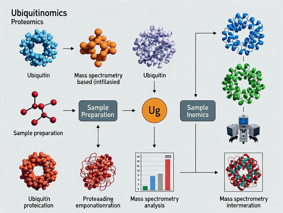
2. Mass Spectrometry-Based Ubiquitinomics: Principles and Workflows MS-based ubiquitinomics enables the global identification and quantification of ubiquitination sites, providing a system-level understanding of ubiquitin signaling [5] [6] [7].
2.1 Fundamental Principles of Ubiquitinomics The core strategy relies on the specific enrichment of peptides derived from ubiquitinated proteins, followed by LC-MS/MS analysis. During tryptic digestion, a ubiquitin-modified lysine residue retains a di-glycine (Gly-Gly, K-ε-GG) remnant with a mass shift of 114.0429 Da, which serves as a mass spectrometry-detectable signature for the ubiquitination site [6] [7] [8]. The primary challenge in ubiquitinomics is the typically low stoichiometry of ubiquitinated species, making affinity enrichment a critical step [7]. Common enrichment methods include:
- Immunoaffinity Purification: Using antibodies specific for the K-ε-GG remnant motif [6] [8].
- DiGly Antibody Enrichment is a cornerstone technique for ubiquitinomics, enabling the specific isolation of tryptic peptides containing the K-ε-GG remnant. This is often performed using commercially available kits like the PTMScan Ubiquitin Remnant Motif Kit [8].
- Ubiquitin-Binding Domains (UBDs): Utilizing domains that non-covalently bind to ubiquitin or ubiquitin chains [7].
- Epitope-Tagged Ubiquitin: Expressing ubiquitin with an N-terminal tag (e.g., His, FLAG, HA, biotin) in cells to allow purification via the tag [5] [7].
2.2 Advanced Ubiquitinomics Workflow (DIA-MS) Recent advances have led to highly robust and deep quantitative ubiquitinomics workflows. The following diagram and protocol detail a state-of-the-art approach using Data-Independent Acquisition (DIA) mass spectrometry.
- Protocol: Deep Ubiquitinome Profiling Using DIA-MS [6]
- Cell Lysis: Lyse cells in a lysis buffer containing Sodium Deoxycholate (SDC) supplemented with Chloroacetamide (CAA) for immediate and effective alkylation of cysteine residues. Immediate boiling post-lysis is recommended to rapidly inactivate DUBs and preserve the native ubiquitinome.
- Protein Digestion: Digest proteins using trypsin. The SDC is efficiently removed by acidification after digestion.
- K-ε-GG Peptide Enrichment: Enrich for ubiquitinated peptides using anti-K-ε-GG remnant motif antibody beads. Optimize incubation times and peptide-to-bead ratios for maximum efficiency.
- Mass Spectrometry Analysis: Analyze enriched peptides using liquid chromatography coupled to a mass spectrometer operating in Data-Independent Acquisition (DIA) mode. DIA fragments all ions within sequential isolation windows, providing comprehensive and reproducible data.
- Data Analysis: Process the raw DIA data using specialized software like DIA-NN. This deep neural network-based tool can operate in a "library-free" mode, directly querying the data against a protein sequence database, and is optimized for the confident identification and precise quantification of K-ε-GG peptides.
This optimized DIA-MS workflow has been shown to quantify over 70,000 unique ubiquitinated peptides in a single MS run, significantly outperforming traditional Data-Dependent Acquisition (DDA) methods in coverage, reproducibility, and quantitative precision [6].
3. Quantitative Profiling and Data Interpretation Quantitative proteomic strategies are essential for comparing ubiquitinomes across different biological conditions, such as drug treatments or genetic perturbations.
3.1 Quantitative MS Strategies Two primary methods are widely used:
- Stable Isotope Labeling with Amino Acids in Cell Culture (SILAC): This metabolic labeling method involves culturing cells in "light" (normal) or "heavy" (isotope-labeled) amino acids. Cells from different conditions are combined after culture but before lysis, allowing for accurate relative quantification early in the workflow and minimizing technical variability [7] [8].
- Label-Free Quantification (LFQ): This method quantifies peptides based on their chromatographic peak areas or MS signal intensities across multiple runs without isotopic labeling. It is advantageous for analyzing samples that cannot be metabolically labeled, such as clinical tissues [6].
3.2 Functional Interpretation of Ubiquitinomics Data A key application of quantitative ubiquitinomics is distinguishing between ubiquitination signals that lead to protein degradation and those that mediate non-proteolytic functions. The following table summarizes a computational approach that infers functionality based on changes in ubiquitin occupancy and protein abundance in response to proteasome inhibition [8].
Table 1: Interpreting Ubiquitin Signaling from Proteomic Data Following Proteasome Inhibition
| Observed Change | Proteasome Inhibitor Treatment (e.g., MG-132) | Inferred Function of Ubiquitination | Biological Interpretation |
|---|---|---|---|
| Ubiquitin Occupancy | Protein Abundance | ||
| Increases | No Change or Decreases | Non-Degradative Signaling | Ubiquitination at this site regulates processes like activity, interactions, or trafficking, not proteasomal degradation. |
| Increases | Increases | Degradative Signaling | The substrate is normally targeted by the proteasome; inhibition halts its degradation, causing accumulation of both the protein and its ubiquitinated form. |
| No Change | No Change | Constitutive/Stable Modification | Ubiquitination may have a minor, regulatory, or non-essential role under the conditions studied. |
Application Example: This strategy was successfully applied to profile targets of the deubiquitinase USP7. Upon USP7 inhibition, hundreds of proteins showed increased ubiquitination within minutes. However, by correlating these changes with protein abundance over time, researchers could distinguish the small subset of proteins that were subsequently degraded from the majority that were subject to non-degradative regulatory ubiquitination [6].
4. The Scientist's Toolkit: Key Research Reagent Solutions The following table catalogues essential reagents and tools for conducting MS-based ubiquitinomics research.
Table 2: Essential Reagents and Tools for Ubiquitinomics Research
| Tool / Reagent | Function / Application | Examples & Notes |
|---|---|---|
| K-ε-GG Motif Antibodies | Immunoaffinity purification of ubiquitinated tryptic peptides for MS analysis. | PTMScan Ubiquitin Remnant Motif Kit [8]; critical for enriching low-stoichiometry ubiquitinated peptides. |
| Epitope-Tagged Ubiquitin | Enables purification of ubiquitin conjugates under denaturing conditions via the tag. | His-, HA-, FLAG-, or biotin-tagged ubiquitin expressed in cells [5] [7]. |
| Proteasome Inhibitors | Block degradation of ubiquitinated proteins, increasing their abundance for detection. | Bortezomib (FDA-approved), MG-132, Carfilzomib [2] [3] [8]. |
| DUB/USP Inhibitors | Pharmacologically probe DUB function and identify DUB substrates. | Selective USP7 inhibitors (e.g., for mode-of-action studies) [6]. |
| SDC Lysis Buffer | Efficient protein extraction with simultaneous inactivation of DUBs. | Sodium Deoxycholate buffer with Chloroacetamide (CAA); improves ubiquitin site coverage vs. urea [6]. |
| Stable Isotope Labels (SILAC) | For accurate relative quantification of ubiquitinated peptides across conditions. | "Heavy" amino acids (e.g., 13C6-lysine, 13C6-15N4-arginine) [7] [8]. |
| DIA-NN Software | Computational analysis of DIA-MS data for deep, precise ubiquitinome quantification. | Specialized software that boosts identification numbers and quantitative accuracy [6]. |
5. Therapeutic Targeting and Application in Drug Development Components of the UPS are well-validated therapeutic targets in human disease, particularly in oncology [1] [3] [4].
- Proteasome Inhibitors: Bortezomib was the first FDA-approved proteasome inhibitor in 2003 for the treatment of relapsed multiple myeloma. Its success validated the UPS as a target for cancer therapy and spurred drug development efforts focused on other UPS components [1] [2] [4].
- E1 Enzyme Inhibitors: Small-molecule inhibitors of the NEDD8-activating enzyme (NAE) have been developed. NEDDylation is crucial for the activity of Cullin-RING ligases (CRLs), a major class of E3s, making NAE inhibition a strategy to indirectly dampen the activity of multiple E3s [4].
- E3 Ligase Modulators: E3 ligases are attractive targets due to their substrate specificity. Thalidomide and its analogs (IMiDs) are a prime example, which modulate the E3 ligase CRL4CRBN to promote the selective degradation of specific oncogenic proteins like IKZF1/3 in multiple myeloma [6]. This has given rise to the field of Proteolysis-Targeting Chimeras (PROTACs), which are heterobifunctional molecules that recruit E3 ligases to neo-substrates to induce their degradation [6].
- DUB Inhibitors: DUBs are emerging as promising drug targets. For instance, USP7 is an oncology target that regulates the stability of p53 and other proteins. Selective USP7 inhibitors are in development, and ubiquitinomics provides a powerful tool for profiling their on-target effects and understanding their mechanism of action on a proteome-wide scale [6].
Conclusion The Ubiquitin-Proteasome System, with its intricate E1-E2-E3-DUB enzymatic network, is a cornerstone of cellular regulation. Mass spectrometry-based ubiquitinomics has revolutionized our ability to study this system at a global level, providing unprecedented insights into the scope and dynamics of protein ubiquitination. The continued refinement of protocols—such as optimized SDC lysis, DIA-MS acquisition, and sophisticated computational data analysis—is driving the field toward deeper, more precise, and higher-throughput analyses. As therapeutic interventions targeting the UPS continue to expand beyond proteasome inhibitors to include E3 ligases and DUBs, ubiquitinomics will remain an indispensable platform for target discovery, drug validation, and mechanistic elucidation in both basic research and clinical drug development.
Ubiquitination, once primarily recognized as a degradation signal for the proteasome, is now understood to regulate a diverse array of cellular processes through distinct mechanisms. This post-translational modification involves the covalent attachment of ubiquitin to target proteins through a sequential enzymatic cascade involving E1 (activating), E2 (conjugating), and E3 (ligase) enzymes [9]. The functional diversity of ubiquitination stems from variations in ubiquitin chain topology—including monoubiquitination and different polyubiquitin linkages—that determine specific biological outcomes [10]. While K48-linked polyubiquitin chains predominantly target substrates for proteasomal degradation, other chain types, particularly K63-linked polyubiquitination, serve crucial non-proteolytic functions in signaling pathways and membrane trafficking [10] [11]. Additionally, monoubiquitination has emerged as an important regulatory mechanism in DNA repair and protein trafficking [10]. These non-degradative ubiquitination forms fundamentally regulate key cellular processes, including innate immune signaling, DNA damage repair, gene expression, and synaptic plasticity in neurons [10] [11].
Advances in mass spectrometry (MS)-based proteomics, particularly ubiquitinomics approaches, have revolutionized our understanding of these diverse ubiquitin functions by enabling system-wide profiling of ubiquitination events [12] [13]. This application note details experimental frameworks for investigating non-proteolytic ubiquitination, providing researchers with robust methodologies to explore the full functional spectrum of ubiquitin signaling in health and disease.
Key Non-Proteolytic Functions of Ubiquitination
Signaling Regulation Through K63-Linked Ubiquitination
K63-linked polyubiquitin chains serve as critical regulatory scaffolds in multiple signaling pathways, modulating protein-protein interactions and complex formation without triggering degradation [10]. A seminal example is the activation of IκB kinase (IKK) in the NF-κB signaling pathway, where K63-linked ubiquitination creates platforms for recruiting essential signaling components [10]. Similarly, in DNA damage response pathways, K63-linked chains facilitate the assembly of repair complexes at sites of DNA lesions, coordinating critical repair processes [10]. These signaling roles demonstrate how ubiquitin can function analogously to other post-translational modifications like phosphorylation, dynamically regulating protein activity and intermolecular interactions.
Monoubiquitination in Membrane Trafficking and DNA Repair
Monoubiquitination, involving attachment of a single ubiquitin molecule, serves as a key signal for endocytic trafficking and protein sorting decisions [9]. This modification regulates the internalization of cell surface receptors and their subsequent sorting to lysosomes for degradation or recycling. In DNA repair, monoubiquitination of histone variants such as H2A and H2B coordinates the recruitment of repair machinery to damaged chromatin, illustrating how this modification type controls protein localization and complex assembly [10]. The functional outcomes of monoubiquitination contrast sharply with degradative K48-linked ubiquitination, highlighting the remarkable functional diversity encoded within the ubiquitin system.
Ubiquitination in Neuronal Function and Dysfunction
The unique morphology and functional requirements of neurons make them particularly dependent on precise ubiquitin-mediated regulation. At presynaptic terminals, ubiquitination regulates the size of recycling vesicle pools and vesicle release properties through modification of proteins like RIM1, a calcium-dependent priming factor [11]. Postsynaptically, ubiquitination controls the abundance of glutamate receptors and organizers of the postsynaptic density, thereby modulating synaptic strength and plasticity [11]. These mechanisms contribute to learning and memory processes, with proteasome inhibition experiments demonstrating impaired long-term potentiation and memory retention [11]. Notably, dysfunction in ubiquitin-mediated processes is implicated in numerous neurodegenerative diseases, including Alzheimer's disease, Parkinson's disease, and Huntington's disease, characterized by accumulation of ubiquitin-positive protein aggregates [11].
Table 1: Diverse Functions of Ubiquitination in Cellular Regulation
| Ubiquitination Type | Primary Functions | Key Examples | Biological Outcomes |
|---|---|---|---|
| K48-linked chains | Proteasomal targeting | Regulation of cell cycle proteins | Protein degradation, homeostasis |
| K63-linked chains | Signaling scaffold | IKK complex activation, DNA repair | Kinase activation, complex assembly |
| Monoubiquitination | Trafficking, DNA repair | Histone modification, receptor endocytosis | Chromatin remodeling, membrane transport |
| Mixed/linked chains | Regulatory functions | Proteasome, translation regulation | Complex signaling integration |
Mass Spectrometry-Based Ubiquitinomics Approaches
Enrichment Strategies for Ubiquitinated Peptides
Comprehensive ubiquitinome profiling requires specialized enrichment techniques to isolate low-abundance ubiquitinated peptides from complex proteomic backgrounds. The most widely adopted method leverages antibodies specific for di-glycine (K-GG) remnants left after tryptic digestion, which recognize the signature Gly-Gly modification on lysine residues where ubiquitin was attached [9] [13]. This approach has been significantly enhanced through optimized sample preparation protocols, including sodium deoxycholate (SDC)-based lysis buffers supplemented with chloroacetamide (CAA) for immediate cysteine protease inactivation, improving ubiquitin site coverage by approximately 38% compared to conventional urea-based methods [13]. Alternative strategies include poly-His tagged ubiquitin systems coupled with nickel-NTA purification and utilization of ubiquitin-binding domains for enrichment, though these methods show varying efficacy in proteome-wide applications [9].
Advanced MS Acquisition and Data Analysis Methods
Data-independent acquisition (DIA) mass spectrometry has emerged as a transformative technology for ubiquitinomics, overcoming limitations of traditional data-dependent acquisition (DDA) methods. When coupled with deep neural network-based processing using tools like DIA-NN, DIA-MS more than triples identification numbers to approximately 70,000 ubiquitinated peptides in single runs while significantly improving quantitative precision and reproducibility [13]. This approach enables highly robust ubiquitinome profiling even in large sample series, with median coefficients of variation below 10% for quantified K-GG peptides [13]. The method's power is further enhanced through "library-free" analysis modes that eliminate the need for extensive spectral library generation while maintaining high identification confidence through rigorous false discovery rate control specifically optimized for ubiquitin remnant peptides [13].
Table 2: Comparison of MS Methods for Ubiquitinome Profiling
| Parameter | Data-Dependent Acquisition (DDA) | Data-Independent Acquisition (DIA) |
|---|---|---|
| Typical K-GG peptide IDs | ~21,000-30,000 | ~68,000-70,000 |
| Quantitative precision (median CV) | 15-20% | ~10% |
| Missing values in replicates | ~50% | Minimal (<5%) |
| Required protein input | 500 μg - 4 mg | 31 μg - 2 mg |
| Throughput | Moderate | High |
| Optimal processing software | MaxQuant | DIA-NN |
Functional Applications in Drug Discovery and Target Validation
Ubiquitinomics approaches have proven particularly valuable in drug discovery, especially for characterizing targeted protein degradation platforms such as molecular glue degraders (MGDs) and proteolysis-targeting chimeras (PROTACs). High-throughput proteomic screening of cereblon (CRBN)-recruiting MGDs has revealed an extensive neosubstrate landscape, identifying highly selective degraders for targets including KDM4B, G3BP2, and VCL [14]. Integrated proteomics and ubiquitinomics profiling enables comprehensive mode-of-action studies by simultaneously tracking ubiquitination changes and consequent protein abundance shifts following treatment with deubiquitinase (DUB) inhibitors or E3 ligase modulators [13]. This approach was powerfully demonstrated in studies of USP7 inhibition, where time-resolved analysis revealed that while ubiquitination of hundreds of proteins increased within minutes, only a small fraction underwent degradation, effectively distinguishing degradative from non-degradative ubiquitination events [13].
Experimental Protocols for Ubiquitinomics
Optimized Sample Preparation for Ubiquitinome Profiling
Protocol: SDC-Based Lysis and Digestion for Ubiquitinomics
Cell Lysis: Lyse cells in SDC buffer (4% SDC, 100 mM Tris-HCl pH 8.5, 40 mM chloroacetamide) followed by immediate boiling at 95°C for 10 minutes to inactivate ubiquitin proteases [13].
Protein Digestion: Digest proteins using trypsin (1:50 enzyme-to-protein ratio) overnight at 37°C after diluting SDC concentration to 1% to prevent interference [13].
Peptide Cleanup: Acidify digests with trifluoroacetic acid (final 1%), precipitate SDC by centrifugation, and desalt supernatants using C18 solid-phase extraction cartridges [13].
K-GG Peptide Enrichment: Incubate purified peptides with anti-K-GG antibody-conjugated beads for 4-16 hours at 4°C with gentle agitation. Wash beads extensively with ice-cold PBS before eluting with 0.1% TFA [13].
This protocol typically yields 38% more K-GG identifications compared to urea-based methods with excellent enrichment specificity and reproducibility [13].
DIA-MS Acquisition Parameters for Ubiquitinomics
Method: DIA-MS for Comprehensive Ubiquitinome Profiling
LC Conditions: 75-125 min nanoLC gradients using C18 columns (75 μm × 25 cm) with acetonitrile gradients from 2-30% in 0.1% formic acid [13].
MS Instrument Setup: High-resolution tandem mass spectrometer (Q-Exactive Orbitrap or similar) operated in DIA mode [13].
DIA Settings: 30-60 variable-width windows covering 400-1000 m/z range; MS1 resolution: 120,000; MS2 resolution: 30,000; normalized collision energy: 25-30% [13].
Data Processing: Analyze raw files using DIA-NN in "library-free" mode against appropriate protein sequence databases with K-GG modification specified as variable modification [13].
This method typically identifies >68,000 ubiquitination sites with median CV <10% across replicates, enabling robust quantitative comparisons across experimental conditions [13].
Visualization of Ubiquitin Signaling and Experimental Workflows
Non-Degradative Ubiquitin Signaling Pathways
Non-Degradative Ubiquitin Signaling Pathways
Ubiquitinomics Experimental Workflow
Ubiquitinomics Experimental Workflow
Research Reagent Solutions for Ubiquitinomics
Table 3: Essential Research Reagents for Ubiquitinomics Studies
| Reagent/Category | Specific Examples | Function/Application |
|---|---|---|
| Lysis Buffers | SDC buffer (4% SDC, 100 mM Tris-HCl, 40 mM CAA) | Optimal protein extraction for ubiquitinomics [13] |
| Protease Inhibitors | MG-132, Bortezomib | Proteasome inhibition to preserve ubiquitinated proteins [13] |
| Enrichment Reagents | Anti-K-GG antibody beads | Immunoaffinity purification of ubiquitin remnant peptides [13] |
| MS Instruments | Q-Exactive Orbitrap, timsTOF | High-resolution mass spectrometry for ubiquitinome profiling [14] [13] |
| Data Processing | DIA-NN, MaxQuant | Software for identification/quantification of ubiquitin sites [13] |
| E3 Ligase Modulators | Molecular glue degraders, PROTACs | Tools to manipulate cellular ubiquitination [14] |
| DUB Inhibitors | USP7 inhibitors | Investigate deubiquitination effects on ubiquitinome [13] |
Ubiquitination is a fundamental post-translational modification (PTM) that regulates virtually all cellular processes in eukaryotic cells, including protein degradation, DNA repair, cell signaling, and immune responses [15] [16]. This modification involves the covalent attachment of ubiquitin—a small 76-amino acid protein—to substrate proteins via a sequential enzymatic cascade involving E1 (activating), E2 (conjugating), and E3 (ligating) enzymes [15] [17]. The system is dynamically reversed by deubiquitinating enzymes (DUBs), creating a reversible regulatory mechanism comparable to other PTMs like phosphorylation [16] [18]. The human genome encodes approximately 2 E1 enzymes, 60 E2 enzymes, over 600 E3 ligases, and more than 100 DUBs, enabling exquisite specificity in substrate selection and regulatory control [16] [18].
The complexity of ubiquitin signaling extends beyond simple monoubiquitination. Ubiquitin itself contains seven lysine residues (K6, K11, K27, K29, K33, K48, K63) and an N-terminus that can serve as attachment points for additional ubiquitin molecules, forming polyubiquitin chains of various architectures and lengths [16] [19]. These chains can be homotypic (single linkage type), heterotypic (mixed linkages), or branched (multiple modifications on a single ubiquitin molecule), creating a sophisticated "ubiquitin code" that determines functional outcomes [16] [19]. The versatility of ubiquitin signaling is further expanded by non-canonical ubiquitination on non-lysine residues and the existence of unanchored polyubiquitin chains not attached to substrates [15] [16]. Mass spectrometry-based proteomics has emerged as a powerful technology for deciphering this complex ubiquitin code, enabling system-wide profiling of ubiquitination events under physiological and pathological conditions [16] [13].
The Expanding Landscape of Ubiquitin Modifications
Canonical Ubiquitination
Canonical ubiquitination involves the formation of an isopeptide bond between the C-terminal glycine of ubiquitin and the ε-amino group of a lysine residue on the substrate protein [15] [17]. This process can result in either monoubiquitination (single ubiquitin modification) or multi-monoubiquitination (multiple single ubiquitins on different lysines) [16]. When additional ubiquitin molecules are conjugated to one of the seven lysine residues or the N-terminal methionine of the previously attached ubiquitin, polyubiquitin chains are formed [16] [19].
The specific linkage type within polyubiquitin chains determines the functional consequence for the modified substrate. For instance, K48-linked chains primarily target proteins for proteasomal degradation, while K63-linked chains typically mediate non-proteolytic signaling in processes such as DNA repair, inflammation, and endocytosis [19] [20]. Other linkages, including K6, K11, K27, K29, and K33, have been associated with diverse cellular functions, although their roles are less characterized [16]. The combinatorial complexity increases further with the formation of mixed linkage chains and branched ubiquitin chains, where a single ubiquitin molecule is modified at multiple sites [16] [19].
Table 1: Major Ubiquitin Chain Linkages and Their Primary Functions
| Linkage Type | Primary Cellular Functions | Structural Features |
|---|---|---|
| K48 | Proteasomal degradation, cell cycle progression | Compact structure targeting to proteasome |
| K63 | DNA repair, NF-κB signaling, endocytosis, kinase activation | Extended conformation facilitating signaling |
| K11 | Proteasomal degradation, cell cycle regulation (mitosis) | Mixed features of K48 and K63 |
| K29 | Proteasomal degradation (with K48 in branched chains) | Less characterized, implicated in proteolysis |
| K33 | Kinase regulation, intracellular trafficking | Role in endosomal sorting |
| K6 | DNA damage response, mitophagy | Implicated in Parkinson's disease pathway |
| K27 | Immune signaling, inflammatory response | Key regulator of innate immunity |
| M1 (linear) | NF-κB activation, inflammatory signaling | Generated by LUBAC complex |
Non-Canonical Ubiquitination
Beyond canonical lysine ubiquitination, emerging research has established the existence and functional significance of non-canonical ubiquitination on non-lysine residues [15]. These modifications expand the ubiquitin code by employing chemical bonds distinct from the conventional isopeptide linkage:
N-terminal ubiquitination: Ubiquitin is conjugated to the α-amino group of a protein's N-terminus via a peptide bond [15]. This modification has been demonstrated to target proteins such as Ngn2, p14ARF, and p21 for degradation and can distinctly alter the catalytic activity of deubiquitinating enzymes like UCHL1 and UCHL5 [15]. The E2 enzyme UBE2W has been specifically implicated in mediating N-terminal ubiquitination due to its flexible C-terminus that enables selective targeting of α-amino groups [15].
Cysteine, Serine, and Threonine ubiquitination: These modifications occur through thioester (cysteine) or oxyester (serine/threonine) bonds [15] [16]. While less common than lysine ubiquitination, they play significant non-proteolytic signaling roles. For example, cysteine ubiquitination of MHC-I by viral E3 ligases can mediate immune evasion [16].
Pathogen-mediated unconventional ubiquitination: The bacterium Legionella pneumophila secretes effector proteins (SdeA, SdeB, SdeC, SidE) that catalyze phosphoribosyl (PR)-linked serine ubiquitination, completely bypassing the host E1-E2-E3 enzymatic cascade [15]. This unique form of ubiquitination involves conjugation of ubiquitin's Arg42 to substrate serine residues via a phosphoribosyl linker and remodels host cellular processes to promote bacterial infectivity [15].
Table 2: Types of Non-Canonical Ubiquitination and Their Characteristics
| Modification Type | Bond Formation | Functional Examples | Enzymatic Requirements |
|---|---|---|---|
| N-terminal ubiquitination | Peptide bond to α-amino group | Targets p21, p14ARF for degradation; modulates DUB activity | UBE2W E2 enzyme specialized for N-termini |
| Cysteine ubiquitination | Thioester bond | Non-proteolytic signaling; MHC-I regulation by viral ligases | Conventional E1-E2-E3 cascade |
| Serine/Threonine ubiquitination | Oxyester bond | Downregulation of BST-2/tetherin by HIV-1 Vpu | Conventional E1-E2-E3 cascade |
| PR-serine ubiquitination | Phosphoribosyl linker | Host protein modification by Legionella pneumophila | SidE family effectors (single enzyme) |
Branched Ubiquitin Chains and Chain Plasticity
Branched ubiquitin chains, wherein a single ubiquitin molecule is modified at multiple sites, represent an additional layer of complexity in the ubiquitin code [19]. These architectures can include bifurcations at consecutive lysines (K6/K11, K27/K29, K29/K33) or more complex branching patterns (K11/K48, K48/K63) [16] [19]. Initially, branched chains were thought to be less effective at promoting protein degradation compared to homotypic chains, but recent evidence challenges this view [19]. For instance, K11/K48 branched chains produced by the anaphase-promoting complex/cyclosome (APC/C) are particularly efficient at triggering proteasomal degradation of cell cycle regulators [19].
The plasticity of ubiquitin chain architecture enables dynamic reprogramming of ubiquitin signaling in response to cellular stimuli. For example, cancer cells can strategically manipulate K63-linked chains to stabilize DNA repair factors while concurrently inhibiting K48-mediated degradation of survival proteins, creating adaptive resistance mechanisms to therapies like radiotherapy [20]. This rewiring of ubiquitin signaling manifests through multiple mechanisms, including DNA repair manipulation and metabolic adaptation, highlighting the functional significance of understanding chain topology in pathological contexts [20].
Analytical Challenges in Ubiquitinomics
The structural diversity of ubiquitin modifications presents significant challenges for comprehensive analysis. Conventional bottom-up proteomics approaches, which involve tryptic digestion of proteins followed by liquid chromatography-tandem mass spectrometry (LC-MS/MS), have proven highly successful for identifying ubiquitination sites but inevitably collapse information about polyubiquitin chain topology [16] [19]. During trypsin digestion, the ubiquitin molecule is trimmed to a diglycine (Gly-Gly) remnant that remains attached to the modified lysine, adding a monoisotopic mass of 114.043 Da—a signature used for site identification [17] [13]. While this approach enables large-scale mapping of ubiquitination sites, it eliminates the connectivity information between ubiquitin molecules in chains.
Middle-down and top-down strategies have been developed to preserve information about ubiquitin chain architecture [19]. The Ubiquitin Chain Enrichment Middle-down Mass Spectrometry (UbiChEM-MS) approach employs minimal trypsinolysis under nondenaturing conditions, which cleaves ubiquitin only between Arg74 and Gly75, yielding an intact Ub1-74 fragment [19]. When a branch point is present within the ubiquitin chain, the ubiquitin moiety bearing the branch point will be modified with two Gly-Gly modifications (2xGG-Ub1-74), enabling detection and characterization of multiple modifications on a single ubiquitin molecule [19]. This method has been successfully applied to detect branched ubiquitin chains in human cells, revealing that approximately 1% of chains isolated with tandem ubiquitin binding entities (TUBEs) contain branch points, increasing to ~4% after proteasome inhibition [19].
Additional challenges in ubiquitinomics include the typically low stoichiometry of ubiquitination sites, the lability of certain non-canonical linkages (particularly oxyester bonds), and the need to distinguish ubiquitin modification sites from other lysine modifications such as acetylation or SUMOylation [15] [16]. Furthermore, the dynamic nature of ubiquitination, with continuous attachment and removal by DUBs, necessitates careful experimental design including the use of proteasome inhibitors or DUB inhibitors to capture transient ubiquitination events [13].
Advanced Mass Spectrometry Approaches for Ubiquitinomics
Sample Preparation and Enrichment Strategies
Effective ubiquitinomics relies on specialized sample preparation methods to enrich low-abundance ubiquitinated peptides from complex proteomic backgrounds. The most common approach involves immunoaffinity purification using antibodies specific for the diglycine remnant left on modified lysines after tryptic digestion [13] [21]. Recent methodological improvements have significantly enhanced the sensitivity and specificity of this enrichment process.
A key advancement is the implementation of sodium deoxycholate (SDC)-based lysis protocols supplemented with chloroacetamide (CAA) for immediate cysteine alkylation [13]. Compared to conventional urea-based buffers, SDC lysis increases K-GG peptide identification by approximately 38% (26,756 vs. 19,403 peptides from HCT116 cells) while maintaining high enrichment specificity [13]. This protocol rapidly inactivates cysteine-dependent DUBs through immediate boiling and alkylation, better preserving the native ubiquitinome landscape [13]. Additionally, CAA prevents unspecific di-carbamidomethylation of lysine residues that can mimic K-GG peptides—an artifact observed with iodoacetamide that complicates data interpretation [13].
For specialized applications focusing on specific ubiquitin chain types, ubiquitin-binding domains (UBDs) such as tandem ubiquitin binding entities (TUBEs) or linkage-selective UBDs like the K29-selective NZF1 domain from TRABID can be employed [19]. These tools enable enrichment of particular chain architectures before middle-down MS analysis, facilitating characterization of branched ubiquitin chains that are inaccessible to conventional bottom-up approaches [19].
Data-Independent Acquisition Mass Spectrometry
Data-independent acquisition (DIA) mass spectrometry has emerged as a transformative technology for ubiquitinomics, offering significant advantages over traditional data-dependent acquisition (DDA) methods [13] [14]. While DDA typically identifies 20,000-30,000 K-GG peptides with substantial missing values across replicates, DIA-MS more than triples identification to approximately 70,000 ubiquitinated peptides in single MS runs while dramatically improving quantitative precision and reproducibility [13].
The power of DIA-MS is further enhanced when coupled with deep neural network-based data processing tools like DIA-NN [13]. This combination achieves a median coefficient of variation (CV) of ~10% for quantified K-GG peptides, with over 68,000 peptides consistently quantified across replicates [13]. The method demonstrates excellent quantitative accuracy across a wide dynamic range, as validated by spike-in experiments with synthetic K-GG peptides at different concentrations [13]. This level of performance enables complex time-resolved studies, such as monitoring ubiquitination dynamics following USP7 inhibition at multiple time points, capturing both immediate ubiquitination changes and subsequent protein abundance alterations [13].
The high throughput and reproducibility of DIA-MS make it particularly suitable for large-scale screening applications, such as profiling molecular glue degrader libraries against hundreds of compounds [14]. In such studies, DIA-MS can quantify over 10,000 protein groups with median CVs of ~6% across replicates, enabling robust statistical identification of neosubstrates based on both ubiquitination increases and protein degradation [14].
Integrated Proteomics and Ubiquitinomics Profiling
A powerful application of modern ubiquitinomics is the simultaneous profiling of ubiquitination changes and corresponding protein abundance alterations [13] [14]. This integrated approach distinguishes regulatory ubiquitination events that lead to protein degradation from non-degradative ubiquitination that modulates protein function, localization, or interactions.
In practice, this involves parallel processing of samples for global proteomics (measuring protein abundance) and ubiquitinomics (measuring ubiquitination sites) [14]. Following treatment with perturbations such as DUB inhibitors or molecular glue degraders, proteins showing increased ubiquitination without corresponding degradation likely undergo non-proteolytic regulatory ubiquitination [13]. Conversely, proteins displaying both increased ubiquitination and decreased abundance represent candidates for degradative ubiquitination [14]. This distinction is critical for understanding the mechanistic consequences of ubiquitin signaling perturbations and for validating putative drug targets.
For example, in studies of USP7 inhibition, hundreds of proteins show increased ubiquitination within minutes, but only a small fraction subsequently undergo degradation, highlighting that most ubiquitination events serve non-proteolytic functions [13]. Similarly, in molecular glue degrader screens, this integrated approach can identify bona fide neosubstrates based on rapid ubiquitination following treatment, followed by CRL-dependent protein degradation that is rescued by NEDD8-activating enzyme inhibition [14].
Experimental Protocols for Comprehensive Ubiquitinome Analysis
Protocol 1: SDC-Based Sample Preparation for Deep Ubiquitinome Profiling
This protocol describes an optimized workflow for preparing samples for ubiquitinomics analysis, achieving identification of >30,000 K-GG peptides from 2 mg of protein input with high reproducibility [13].
Reagents and Materials
- Lysis Buffer: 1% SDC in 100 mM Tris-HCl (pH 8.5), 100 mM chloroacetamide (CAA), 40 mM 2-chloroacetamide
- PBS (phosphate-buffered saline)
- Protease inhibitor cocktail
- Phosphatase inhibitor cocktail
- Deubiquitinase inhibitor (optional)
- BCA protein assay kit
- sequencing-grade modified trypsin
- PreOmics iST binding buffer
- anti-K-ε-GG antibody-conjugated beads
- Trifluoroacetic acid (TFA)
- StageTips with C18 material
Procedure
- Cell Lysis: Aspirate culture medium and wash cells twice with ice-cold PBS. Add SDC lysis buffer (1-2 mL per 10⁷ cells) supplemented with protease and phosphatase inhibitors. Immediately scrape cells and transfer to preheated (95°C) microcentrifuge tubes. Incubate at 95°C for 10 minutes with shaking at 1000 rpm [13].
- Protein Quantification: Cool samples to room temperature and determine protein concentration using BCA assay. Use 2 mg protein per condition for optimal results, with minimum 500 µg for limited samples [13].
- Protein Digestion: Adjust protein concentration to 1 mg/mL with lysis buffer. Add trypsin at 1:50 (w/w) enzyme-to-protein ratio and incubate overnight at 37°C with shaking [13].
- Peptide Cleanup: Acidify samples to pH < 3 with TFA (final concentration 1%). Centrifuge at 15,000 × g for 10 minutes to pellet SDC. Transfer supernatant to new tubes and desalt using StageTips with C18 material according to manufacturer's instructions [13].
- K-ε-GG Peptide Enrichment: Resuspend dried peptides in PreOmics iST binding buffer. Incubate with anti-K-ε-GG antibody-conjugated beads for 2 hours at room temperature with gentle rotation. Wash beads three times with binding buffer and twice with water [13].
- Peptide Elution: Elute K-ε-GG peptides with 0.2% TFA. Dry peptides in a speedvac and store at -20°C until MS analysis [13].
Protocol 2: UbiChEM-MS for Branched Ubiquitin Chain Analysis
This protocol enables detection and characterization of branched ubiquitin chains that are inaccessible to conventional bottom-up proteomics [19].
Reagents and Materials
- TUBEs (tandem ubiquitin-binding entities) or linkage-selective UBDs (e.g., NZF1 for K29 chains)
- HaloLink resin (Promega)
- Binding buffer: 50 mM Tris, 150 mM NaCl, 10% glycerol, 0.05% IGEPAL CA-630, pH 7.5
- Minimal buffer: 50 mM Tris, 150 mM NaCl, pH 7.5
- Sequencing-grade modified trypsin
- Acetic acid
- C18 Sep-Pak columns (Waters)
Procedure
- UBD Resin Preparation: Couple HaloTag-UBD fusion protein to HaloLink resin according to manufacturer's instructions. Use 100 µL TUBE resin or 200 µL NZF1 resin per sample [19].
- Ubiquitin Chain Enrichment: Incubate 45 mg cell lysate with UBD resin overnight at 4°C with rotation. Pellet resin at 800 × g for 2 minutes and discard supernatant. Wash resin three times with 2 mL binding buffer and twice with 2 mL minimal buffer [19].
- On-Resin Minimal Trypsinolysis: Resuspend resin in 100 µL minimal buffer. Add trypsin at empirically determined ratio (typically 1:20 to 1:100 enzyme-to-substrate ratio) and incubate at room temperature for 16 hours [19].
- Reaction Termination and Peptide Extraction: Acidify samples to pH 2 with acetic acid to deactivate trypsin. Incubate at 4°C for 15-30 minutes, then centrifuge at 13,000 × g for 5 minutes. Transfer supernatant to new tubes [19].
- Sample Desalting: Concentrate supernatant 5-7-fold using a speedvac. Load onto C18 Sep-Pak column pre-equilibrated with 0.1% TFA. Wash with 3 mL equilibration solution, then elute with step gradients of 10-60% acetonitrile in 0.1% TFA. Lyophilize fractions containing ubiquitin species [19].
- Middle-Down MS Analysis: Resuspend samples in water/acetonitrile/acetic acid (45:45:10) and analyze by high-resolution mass spectrometry (e.g., Orbitrap Fusion) with electron-transfer dissociation (ETD) capability for branched chain characterization [19].
The Scientist's Toolkit: Essential Research Reagents
Table 3: Key Research Reagents for Ubiquitinomics Studies
| Reagent Category | Specific Examples | Function and Application |
|---|---|---|
| Ubiquitin Enrichment Tools | anti-K-ε-GG antibodies | Immunoaffinity purification of ubiquitinated peptides for bottom-up ubiquitinomics |
| TUBEs (Tandem Ubiquitin Binding Entities) | Enrichment of polyubiquitinated proteins under native conditions for functional studies | |
| Linkage-selective UBDs (e.g., NZF1, UBAN) | Selective isolation of specific ubiquitin chain types (K29, K63, M1) | |
| Mass Spectrometry Standards | Stable isotope-labeled ubiquitin | Internal standard for quantitative ubiquitinomics |
| Synthetic K-GG peptides | Spike-in standards for quantification accuracy assessment | |
| Enzyme Inhibitors | Proteasome inhibitors (MG-132, bortezomib) | Stabilize ubiquitinated proteins by blocking degradation |
| DUB inhibitors (PR-619, USP7 inhibitors) | Capture transient ubiquitination by preventing deubiquitination | |
| NEDD8-activating enzyme inhibitor (MLN4924) | Validate CRL-dependent ubiquitination mechanisms | |
| Specialized MS Reagents | HaloTag fusion vectors for UBD expression | Customizable ubiquitin chain enrichment platforms |
| Chloroacetamide (CAA) | Alkylating agent that prevents artifacts in ubiquitinomics |
Data Visualization and Analysis
The following diagrams illustrate key ubiquitin signaling pathways and experimental workflows described in this application note.
Diagram 1: The Expanding Ubiquitin Code. This diagram illustrates the structural complexity of ubiquitin modifications, including canonical lysine ubiquitination with major linkage types, non-canonical ubiquitination on various residues, and diverse chain architectures.
Diagram 2: Ubiquitinomics Workflow from Sample to Insight. This diagram outlines the complete experimental workflow for comprehensive ubiquitinome analysis, highlighting critical steps from sample preparation through data integration and biological application.
The complexity of the ubiquitin code—encompassing canonical and non-canonical modifications alongside diverse polyubiquitin chain topologies—represents a sophisticated regulatory system governing virtually all cellular processes. Mass spectrometry-based ubiquitinomics has evolved into a powerful technology for deciphering this complexity, enabling researchers to map ubiquitination events on a proteome-wide scale with unprecedented depth and precision. The methodologies and applications described in this document provide a framework for leveraging these advances to accelerate drug discovery, identify novel therapeutic targets, and elucidate mechanisms of disease pathogenesis. As ubiquitinomics technologies continue to mature, particularly with the adoption of DIA-MS and integrated multi-omics approaches, our ability to interpret the ubiquitin code and harness its therapeutic potential will undoubtedly expand, opening new frontiers in biomedical research and precision medicine.
Mass Spectrometry as a Discovery Engine for System-Wide Ubiquitinome Profiling
Mass spectrometry (MS)-based ubiquitinomics provides a system-level understanding of ubiquitin signaling, a process vital for regulating intracellular events like cell cycle progression, selective autophagy, and response to growth factors [22]. The ubiquitin-proteasome system (UPS) comprises approximately 750 enzymes that mediate the attachment and cleavage of ubiquitin from target proteins [22]. Dysregulation of this system can contribute to carcinogenesis, making various UPS components, such as deubiquitinases (DUBs) and E3 ligases, active targets for anticancer drug development [22]. Modern ubiquitinomics relies on immunoaffinity purification and MS-based detection of diglycine-modified peptides (K-GG), which are generated by tryptic digestion of ubiquitin-modified proteins, enabling global profiling of ubiquitination events [22].
Optimized Protocols for Ubiquitinome Profiling
Sample Preparation and Lysis
Robust sample preparation is foundational for deep ubiquitinome coverage. An improved sodium deoxycholate (SDC)-based lysis protocol has been demonstrated to significantly boost the yield of K-GG peptides compared to conventional urea-based methods [22].
- Cell Lysis: Lyse cell pellets or tissue in an ice-cold buffer containing 50 mM Tris HCl and 0.5% SDC. For tissue, a buffer with 100 mM Tris HCl, 12 mM SDC, and 12 mM sodium N-lauroylsarcosinate is effective.
- Immediate Protease Inactivation: Supplement the SDC buffer with chloroacetamide (CAA) and immediately boil samples post-lysis. CAA rapidly alkylates and inactivates cysteine ubiquitin proteases without causing non-specific di-carbamidomethylation of lysine residues, which can mimic K-GG peptides.
- Protein Processing: Reduce proteins with 5 mM DTT (30 min, 50°C), alkylate with 10 mM iodoacetamide (15 min, in the dark), and digest using Lys-C (4 hours) followed by trypsin (overnight at 30°C).
- Peptide Clean-up: Precipitate detergents with 0.5% TFA and centrifugate before collecting the peptide-containing supernatant.
This optimized SDC protocol yielded 38% more K-GG peptides than urea buffer and significantly improved reproducibility and quantitative precision [22].
Peptide Enrichment and Fractionation
To isolate low-abundance ubiquitin remnants, immunoaffinity purification (IP) is employed using ubiquitin remnant motif antibodies [23].
Key Protocol Steps [23]:
- Crude Fractionation: Prior to IP, fractionate tryptic peptides using high-pH reverse-phase C18 chromatography. For ~10 mg protein digest, use a column with 0.5 g of C18 material. Elute peptides into three distinct fractions using 10 mM ammonium formate with 7%, 13.5%, and 50% acetonitrile, respectively.
- Immunoprecipitation: Incubate peptide fractions with antibody-conjugated beads for two hours at 4°C. A sequential incubation with two fresh batches of beads can increase yield.
- Wash and Elute: Wash beads thoroughly with ice-cold IAP buffer and water. Elute bound peptides with 0.15% TFA, desalt using C18 stage tips, and dry via vacuum centrifugation.
This fractionation approach, combined with sequential IP, helps manage sample complexity and enables the identification of over 23,000 unique diGly peptides from a single sample of HeLa cells [23].
Mass Spectrometry Acquisition and Data Analysis
Data-Independent Acquisition (DIA) coupled with advanced data processing has emerged as a superior method for ubiquitinomics, offering greater depth, robustness, and quantitative precision compared to traditional Data-Dependent Acquisition (DDA) [22].
Key Protocol Parameters [22]:
- MS Acquisition: Utilize a sensitive nanoflow LC-MS/MS system. For DIA, specific MS methods are optimized for ubiquitinomics (see Supplementary Data 4 in [22]).
- Data Processing: Process DIA data using DIA-NN software in "library-free" mode, which uses a deep neural network-based algorithm specifically optimized for the confident identification of modified peptides like K-GG peptides. This approach can quantify over 68,000 K-GG peptides in a single run, more than tripling the numbers achievable by DDA [22].
- Confidence Assessment: DIA-NN employs a rigorous false discovery rate (FDR) determination for K-GG peptides, with validation confirming comparable confidence to DDA workflows [22].
Table 1: Comparison of Lysis Buffer Performance for Ubiquitinomics
| Parameter | SDC-Based Lysis Buffer | Conventional Urea-Based Buffer |
|---|---|---|
| Average K-GG Peptide Yield | 26,756 peptides [22] | 19,403 peptides [22] |
| Key Additive | Chloroacetamide (CAA) [22] | Iodoacetamide [22] |
| Reproducibility | Significantly improved robustness and precision [22] | Lower reproducibility and precision [22] |
| Specificity Concern | No unspecific di-carbamidomethylation [22] | Potential for di-carbamidomethylation mimicking K-GG [22] |
Quantitative Performance and Data Quality
The integration of SDC-based lysis with DIA-MS and DIA-NN processing represents a state-of-the-art workflow that dramatically enhances the scope and reliability of ubiquitinome profiling.
Table 2: Performance Benchmarking of MS Acquisition Methods in Ubiquitinomics
| Performance Metric | Data-Dependent Acquisition (DDA) | Data-Independent Acquisition (DIA) |
|---|---|---|
| Average K-GG Peptides ID (Single Run) | 21,434 peptides [22] | 68,429 peptides [22] |
| Quantitative Precision (Median CV) | Higher variability [22] | ~10% [22] |
| Missing Values in Replicates | ~50% of IDs have missing values [22] | 68,057 peptides quantified in ≥3 of 4 replicates [22] |
| Throughput & Robustness | Suitable for smaller studies; susceptible to run-to-run variability [22] | Ideal for large sample series; high robustness [22] |
This optimized workflow enables the simultaneous recording of ubiquitination changes and consequent abundance changes for more than 8,000 proteins at high temporal resolution, providing a powerful tool for dynamic studies [22].
Application in Drug Discovery and Systems Biology
The power of deep, time-resolved ubiquitinome profiling is exemplified in the functional investigation of deubiquitinases (DUBs), such as the oncology target USP7 [22].
Application Protocol:
- Perturbation: Treat cells (e.g., HCT116) with a selective USP7 inhibitor.
- Time-Resolved Profiling: Conduct simultaneous ubiquitinome and proteome profiling at multiple time points (e.g., minutes to hours) post-inhibition using the described DIA-MS workflow.
- Data Integration: Combine the profiles of ubiquitinated peptides with the corresponding protein abundance data to distinguish ubiquitination events that lead to protein degradation from those that are non-degradative.
This approach revealed that upon USP7 inhibition, hundreds of proteins showed increased ubiquitination within minutes, but only a small fraction of those were subsequently degraded [22]. This dissects the scope of USP7 action, demonstrating that its primary role may be in non-proteolytic signaling for many substrates, a critical insight for drug mechanism-of-action studies [22]. This method enables rapid and precise mode-of-action profiling for candidate drugs targeting DUBs or ubiquitin ligases [22].
The Scientist's Toolkit: Essential Research Reagents
Successful execution of a system-wide ubiquitinome study requires a suite of specific reagents and tools.
Table 3: Key Research Reagent Solutions for Ubiquitinomics
| Reagent / Tool | Function / Application | Example / Key Feature |
|---|---|---|
| Ubiquitin Remnant Motif Antibody | Immunoaffinity purification of K-GG peptides from tryptic digests [22] [23] | Conjugated to protein A agarose beads [23] |
| Sodium Deoxycholate (SDC) | Powerful detergent for efficient protein extraction during cell lysis [22] | Used in optimized lysis buffer with Tris HCl [22] |
| Chloroacetamide (CAA) | Cysteine alkylating agent to rapidly inactivate DUBs during lysis [22] | Preferred over iodoacetamide to avoid lysine artifacts [22] |
| Tandem Mass Tag (TMT) | Isobaric label for multiplexed quantitative proteomics [24] | Enables parallel analysis of 10 samples in a single MS run [24] |
| DIA-NN Software | Deep neural network-based data processing for DIA-MS data [22] | Features a specialized scoring module for confident K-GG peptide ID [22] |
Workflow and Signaling Visualizations
The following diagrams, generated with DOT language, summarize the core experimental workflow and the biological context of ubiquitin signaling. The color palette adheres to the specified guidelines, ensuring sufficient contrast for readability.
Ubiquitinome Profiling Workflow
Ubiquitin Signaling Pathway
Linking Dysregulated Ubiquitin Signaling to Cancer and Neurodegenerative Diseases
The ubiquitin-proteasome system (UPS) serves as a critical post-translational regulatory mechanism that governs nearly every cellular process in eukaryotes by controlling protein stability, localization, and functional activity [25] [26]. This sophisticated system involves a coordinated enzymatic cascade whereby ubiquitin—a highly conserved 76-amino acid polypeptide—is covalently attached to substrate proteins via a series of E1 (activating), E2 (conjugating), and E3 (ligating) enzymes [27] [25]. The resulting ubiquitin modifications can range from single ubiquitin molecules (monoubiquitination) to complex polyubiquitin chains formed through different lysine linkages within ubiquitin itself, with each topology encoding distinct functional consequences for the modified substrate [26] [28].
Dysregulation of ubiquitin signaling has emerged as a fundamental pathogenic mechanism in diverse human diseases, particularly in cancer and neurodegenerative disorders [27] [26] [29]. In cancer cells, altered ubiquitination patterns can drive oncogenic signaling, cell cycle progression, and metastatic behavior [30] [29]. Conversely, in neurodegenerative diseases, impaired ubiquitin-mediated protein clearance contributes to the accumulation of toxic protein aggregates that characterize conditions such as Alzheimer's disease, Parkinson's disease, and amyotrophic lateral sclerosis [27] [26]. Mass spectrometry-based ubiquitinomics has revolutionized our ability to systematically profile these alterations, providing unprecedented insights into disease mechanisms and revealing potential therapeutic targets [13] [25] [28].
Analytical Framework: Mass Spectrometry-Based Ubiquitinomics
Core Methodological Principles
Modern ubiquitinomics relies primarily on anti-diglycine remnant immunoaffinity enrichment techniques, which exploit the characteristic diglycine (K-ε-GG) signature left on trypsinized peptides following ubiquitination [29] [31]. When combined with high-resolution mass spectrometry, this approach enables system-wide identification and quantification of ubiquitination sites with exceptional precision and depth [13] [29]. The field has progressively evolved from data-dependent acquisition (DDA) methods to more advanced data-independent acquisition (DIA) approaches, which significantly enhance reproducibility, quantification accuracy, and coverage of the ubiquitinome [13].
Recent methodological innovations have substantially improved the sensitivity and scope of ubiquitinomics analyses. The introduction of sodium deoxycholate (SDC)-based lysis protocols with immediate boiling and chloroacetamide alkylation has demonstrated a 38% increase in identified K-ε-GG peptides compared to conventional urea-based methods, while simultaneously improving quantitative reproducibility [13]. When coupled with deep neural network-based data processing tools like DIA-NN, researchers can now routinely identify >70,000 ubiquitinated peptides in single LC-MS runs—more than triple the coverage achievable with DDA methods—while maintaining excellent quantitative precision (median CV ≈10%) [13].
Advanced Enrichment and Quantification Strategies
For specialized applications, tandem ubiquitin binding entities (TUBEs) offer significant advantages for studying labile ubiquitination events. TUBEs provide up to 1,000-fold increased affinity for polyubiquitin chains compared to single ubiquitin-binding domains and protect ubiquitinated proteins from both proteasomal degradation and deubiquitinating enzyme activity [31]. This approach is particularly valuable for investigating ubiquitination in neurodegenerative contexts, where protein aggregates often impede conventional analysis methods [27] [31].
Quantitative ubiquitinomics has been transformed by stable isotope labeling and label-free quantification approaches that enable precise measurement of ubiquitination dynamics across multiple cellular conditions [25] [29]. These techniques are especially powerful when combined with temporal resolution studies, allowing researchers to distinguish regulatory ubiquitination events that lead to protein degradation from those mediating non-proteolytic functions [13]. The resulting datasets provide unprecedented insights into the kinetics and functional consequences of ubiquitin signaling in pathological states.
Ubiquitin Signaling Dysregulation in Cancer
Global Ubiquitinome Alterations in Cancer Pathogenesis
Comprehensive ubiquitinome profiling has revealed extensive reprogramming of ubiquitination networks in human cancers. A system-wide analysis of protein acetylation and ubiquitination in human cancer cells identified more than 5,000 ubiquitination sites and 1,600 acetylation sites, with 236 lysine residues across 141 proteins subject to both modifications—suggesting complex cross-regulatory mechanisms in oncogenesis [30]. Notably, motif analysis revealed glutamic acid (E) highly enriched at the -1 position for acetylation sites, while ubiquitination sites displayed more dispersed amino acid distributions, indicating distinct sequence constraints for these modifications [30].
Pathway analysis of cancer ubiquitinomes has consistently identified protein translational control pathways as key hubs of dysregulated ubiquitination. Specifically, the eukaryotic initiation factor 2 (EIF2) signaling pathway and ubiquitin-mediated proteolysis pathway emerge as significantly enriched in cancer cells, highlighting the central role of UPS dysfunction in malignant transformation [30]. These global alterations represent potentially targetable vulnerabilities for cancer therapy.
Metastasis-Associated Ubiquitination Events
Comparative ubiquitinome profiling of human primary and metastatic colon adenocarcinoma tissues has identified distinct ubiquitination patterns associated with disease progression [29]. This research quantified 375 differentially regulated ubiquitination sites across 341 proteins between primary and metastatic tissues, with 132 sites (127 proteins) upregulated and 243 sites (214 proteins) downregulated in metastases [29]. Bioinformatic analysis revealed pronounced enrichment of metastasis-associated ubiquitination events in RNA transport and cell cycle regulation pathways, suggesting fundamental reprogramming of these processes during cancer dissemination.
Table 1: Ubiquitination Changes in Human Primary vs. Metastatic Colon Adenocarcinoma Tissues
| Regulation | Number of Ubiquitination Sites | Number of Proteins | Key Enriched Pathways |
|---|---|---|---|
| Upregulated | 132 | 127 | RNA transport, Cell cycle |
| Downregulated | 243 | 214 | Metabolic pathways, Proteolysis |
Among the most significantly altered targets, cyclin-dependent kinase 1 (CDK1) exhibited disease-stage-dependent ubiquitination patterns, suggesting that modified ubiquitination of this central cell cycle regulator may serve as a pro-metastatic factor in colon adenocarcinoma [29]. These findings illustrate how ubiquitinomics can identify functionally important regulatory nodes in cancer progression.
Therapeutic Targeting of Ubiquitin Signaling in Cancer
The druggability of the ubiquitin system is exemplified by ongoing efforts to target deubiquitinating enzymes (DUBs) such as USP7, which regulates the stability of key oncoproteins and tumor suppressors including p53 [13]. Time-resolved ubiquitinome profiling following USP7 inhibition has enabled comprehensive mapping of USP7 substrates, revealing that hundreds of proteins show increased ubiquitination within minutes of inhibitor treatment [13]. Importantly, simultaneous monitoring of protein abundance demonstrated that only a fraction of these ubiquitination events lead to degradation, highlighting the dual role of ubiquitination in proteolytic and non-proteolytic signaling [13].
These findings underscore the potential of ubiquitinomics for mode-of-action studies of DUB and ubiquitin ligase inhibitors, facilitating rapid characterization of candidate therapeutics at unprecedented precision and throughput [13]. The ability to distinguish degradative from non-degradative ubiquitination events provides critical insights for drug development, particularly for targeted protein degradation approaches that are transforming oncology therapeutics.
Ubiquitin Signaling Dysregulation in Neurodegenerative Diseases
Impaired Protein Quality Control in Neurodegeneration
In neurodegenerative diseases, ubiquitin signaling is primarily associated with protein quality control mechanisms that eliminate aberrant proteins which form aggregates fatal to post-mitotic neurons [27]. The two main catabolic pathways—the ubiquitin-proteasome system (UPS) and autophagy-lysosomal pathway (ALP)—are interconnected systems that rely on ubiquitin signaling to target proteins for degradation [27]. The UPS predominantly degrades soluble ubiquitinated proteins through the 26S proteasome, while autophagy efficiently clears larger protein aggregates through a selective process called aggrephagy [27]. Both systems typically decline with aging, creating a permissive environment for toxic protein accumulation in age-associated neurodegenerative disorders [27].
The pathological hallmark of many neurodegenerative diseases is the presence of ubiquitin-positive inclusions, such as Lewy bodies in Parkinson's disease (primarily composed of α-synuclein) and various tau-containing aggregates in Alzheimer's disease [27] [26]. These deposits represent failed cellular quality control and reflect either overwhelming production of aggregation-prone proteins or progressive failure of ubiquitin-mediated clearance mechanisms—most likely a combination of both factors [27] [26].
Genetic Evidence Linking UPS Components to Neurodegeneration
Strong genetic evidence supports the central role of ubiquitin signaling dysfunction in neurodegenerative pathogenesis. Mutations in several E3 ubiquitin ligases, including Parkin (associated with autosomal recessive juvenile Parkinson's disease), directly impair ubiquitin signaling and protein quality control [27] [26]. Similarly, mutations in genes encoding ubiquitin-binding proteins such as ubiquilin have been linked to neurodegenerative processes, though the exact mechanisms remain under investigation [26].
Interestingly, the functional consequences of these mutations extend beyond proteasomal degradation defects. For example, Parkin also regulates mitochondrial quality control through mitophagy and may facilitate the sequestration of toxic proteins into inclusion bodies via K63-linked ubiquitin chains [26]. This complexity highlights the diverse functions of ubiquitin signaling in neuronal homeostasis and the multiple mechanisms through which its disruption can contribute to neurodegeneration.
Compensatory Mechanisms and Therapeutic Opportunities
Contrary to the initial hypothesis that global UPS impairment is a primary driver of neurodegeneration, accumulating evidence suggests that the UPS remains largely operative in many disease models [26]. Instead, neurodegenerative pathologies may involve more selective defects in handling specific aggregation-prone proteins, combined with age-related declines in proteostatic capacity that create vulnerability to protein misfolding [26]. This paradigm shift repositioned the UPS from a dysfunctional system in neurodegeneration to a potentially harnessable therapeutic target for accelerating clearance of disease-linked proteins [26].
Emerging evidence suggests that adaptive cellular responses help alleviate the burden of aggregation-prone proteins to maintain ubiquitin-dependent proteolysis [26]. These compensatory mechanisms represent promising therapeutic avenues, potentially including enhancement of UPS function, stimulation of ubiquitin ligase activity, or modulation of specific DUBs to rebalance ubiquitin signaling in affected neurons [27] [26].
Experimental Protocols for Ubiquitinomics
Ubiquitin Remnant Profiling from Tissues
This protocol outlines the comprehensive ubiquitinome analysis of human primary and metastatic colon adenocarcinoma tissues [29], providing a framework for tissue-based ubiquitinomics studies.
Sample Preparation:
- Tissue Lysis: Homogenize frozen tissues (30 mg) in urea-based lysis buffer (8 M urea, 10 mM EDTA, 10 mM DTT, 1% protease inhibitor cocktail) using a high-intensity ultrasonic processor on ice [29].
- Protein Extraction: Remove debris by centrifugation at 12,000 × g for 10 min at 4°C. Collect supernatant and determine protein concentration using a 2D Quant kit [29].
- Trypsin Digestion: Reduce proteins with 10 mM DTT (1 h, 56°C) and alkylate with 30 mM iodoacetamide (45 min, room temperature in darkness). Dilute samples with 100 mM NH₄HCO₃ to reduce urea concentration below 2 M. Digest with trypsin (1:50 w/w ratio overnight, followed by 1:100 ratio for 4 h) [29].
- Peptide Desalting: Load peptides onto Strata-X C18 columns, wash with 0.1% formic acid (FA) + 5% acetonitrile (ACN), and elute with 0.1% FA + 80% ACN. Dry eluates by vacuum centrifugation [29].
Ubiquitinated Peptide Enrichment:
- Immunoaffinity Purification: Dissolve tryptic peptides in NETN buffer (100 mM NaCl, 1 mM EDTA, 50 mM Tris-HCl, 0.5% NP-40, pH 8.0) and incubate with anti-K-ε-GG remnant antibody beads (PTMScan Ubiquitin Remnant Motif Kit, Cell Signaling Technology) at 4°C overnight with gentle shaking [29].
- Bead Washing: Wash beads four times with NETN buffer and twice with H₂O [29].
- Peptide Elution: Elute bound peptides with 0.1% trifluoroacetic acid, combine eluates, and dry by vacuum centrifugation [29].
- Sample Cleanup: Desalt peptides using C18 ZipTips per manufacturer's instructions [29].
LC-MS/MS Analysis:
- Chromatographic Separation: Resuspend peptides in 0.1% FA and load onto a Thermo Scientific UltiMate 3000 UHPLC system with a trap column and analytical column (homemade nanocapillary C18, 75 μm × 25 cm, 3 μm particles) using a 250 nL/min flow rate [29].
- Gradient Elution: Employ a 40-min linear gradient from 5% to 25% buffer B (98% ACN, 0.1% FA), followed by 5 min to 35% B, 2 min to 80% B, 2 min hold at 80% B, and 1 min return to 5% B with 6 min re-equilibration [29].
- Mass Spectrometry Analysis: Ionize peptides using nanoESI and analyze on a Q-Exactive HF X mass spectrometer in DDA mode with the following parameters: MS1 resolution: 60,000; MS2 resolution: 30,000; HCD fragmentation; dynamic exclusion: 30 s [29].
Data Processing:
- Database Searching: Process MS/MS data using MaxQuant (v1.5.2.8) against the SwissProt Human database with trypsin/P specificity allowing two missed cleavages [29].
- PTM Identification: Set carbamidomethylation of cysteine as fixed modification and Gly-Gly modification of lysine plus methionine oxidation as variable modifications [29].
- Quantification: Apply label-free quantification (LFQ) with FDR adjustment to <1% at peptide and protein levels [29].
Advanced DIA-Ubiquitinomics for Dynamic Signaling Studies
This protocol describes a high-resolution, time-resolved ubiquitinomics workflow for capturing ubiquitination dynamics, such as following DUB inhibition [13].
Optimized Sample Preparation:
- SDC-Based Lysis: Lyse cells in SDC buffer (2% sodium deoxycholate, 40 mM chloroacetamide, 100 mM Tris-HCl, pH 8.5) with immediate incubation at 95°C for 5 min to inactivate ubiquitin proteases [13].
- Protein Digestion: Digest proteins directly in SDC buffer using trypsin (1:50 w/w ratio) after diluting SDC concentration below 0.5% to prevent precipitation [13].
- Peptide Cleanup: Acidify digests to pH <2 with trifluoroacetic acid to precipitate SDC, followed by centrifugation and collection of supernatant containing peptides [13].
DIA-MS Analysis:
- Chromatography: Utilize a 75-min nanoLC gradient on a reversed-phase column with 1.9 μm C18 particles for high-resolution peptide separation [13].
- Data Acquisition: Acquire MS data in DIA mode with optimized isolation windows (typically 4-8 m/z) covering the full m/z range (350-1800) [13].
- Spectral Libraries: Generate comprehensive spectral libraries either in silico or through fractionated DDA runs for maximum identifications [13].
Data Processing with DIA-NN:
- Library-Free Analysis: Process DIA data using DIA-NN in "library-free" mode against appropriate protein sequence databases [13].
- False Discovery Control: Apply stringent filters to maintain FDR <1% at both peptide and protein levels [13].
- Quantitative Analysis: Leverage DIA-NN's neural network-based quantification for precise measurement of ubiquitination changes across time courses [13].
Essential Research Tools for Ubiquitinomics
Table 2: Key Research Reagents and Resources for Ubiquitinomics Studies
| Reagent/Resource | Type | Key Function | Application Examples |
|---|---|---|---|
| Anti-K-ε-GG Antibody | Immunoaffinity reagent | Enrichment of ubiquitinated peptides from tryptic digests | Global ubiquitinome profiling from tissues and cells [29] |
| TUBEs (Tandem Ubiquitin Binding Entities) | Affinity capture reagent | Protection and enrichment of polyubiquitinated proteins | Study of labile ubiquitination in neurodegenerative models [31] |
| USP7 Inhibitors | Small molecule probes | Selective inhibition of deubiquitinating enzyme USP7 | Mode-of-action studies and target identification [13] |
| SDC Lysis Buffer | Lysis reagent | Enhanced protein extraction with protease inactivation | Improved ubiquitin site coverage and reproducibility [13] |
| DIA-NN Software | Computational tool | Deep neural network-based DIA data processing | High-sensitivity ubiquitinome identification and quantification [13] |
Visualizing Ubiquitin Signaling Pathways and Methodologies
Ubiquitin Signaling in Health and Disease: This diagram illustrates the ubiquitin conjugation cascade and the divergent functional outcomes of different ubiquitin modifications, highlighting how specific chain topologies direct substrates toward proteasomal degradation, autophagy, or non-degradative signaling—processes dysregulated in cancer and neurodegenerative diseases.
Ubiquitinomics Experimental Workflow: This diagram outlines the core steps in mass spectrometry-based ubiquitinomics, from sample preparation to data analysis, highlighting critical methodological options such as SDC lysis, TUBE enrichment, DIA-MS acquisition, and DIA-NN processing that enhance experimental outcomes.
Mass spectrometry-based ubiquitinomics has fundamentally transformed our understanding of ubiquitin signaling dysregulation in human diseases, providing unprecedented system-level insights into the complex role of ubiquitination in both cancer and neurodegenerative disorders. The continuing evolution of methodological approaches—including improved lysis protocols, advanced DIA acquisition strategies, and sophisticated computational tools—promises even deeper characterization of the ubiquitinome in coming years.
For therapeutic development, ubiquitinomics offers powerful capabilities for target identification, mechanism-of-action studies, and biomarker discovery. As the resolution and throughput of these methods continue to improve, we anticipate increasingly comprehensive maps of ubiquitin signaling networks that will illuminate new therapeutic opportunities for modulating the ubiquitin system in disease contexts. The integration of ubiquitinomics with other omics technologies will further enhance our ability to decipher the complex language of ubiquitin signaling and develop targeted interventions for diseases characterized by ubiquitin system dysregulation.
Cutting-Edge Ubiquitinomics Workflows: From Sample to Insight
Mass spectrometry (MS)-based ubiquitinomics has become an indispensable tool for system-level understanding of ubiquitin signaling, a post-translational modification crucial for regulating nearly every cellular process in eukaryotes, from protein degradation to DNA repair and cell signaling [16] [5]. The ubiquitin-proteasome system (UPS) consists of approximately 750 enzymes that mediate ubiquitin attachment and cleavage, regulating a myriad of intracellular processes [22]. Consequently, dysregulation of the UPS contributes to carcinogenesis, making various UPS components attractive targets for anticancer drugs [22]. However, the comprehensive analysis of ubiquitinated proteins presents significant technical challenges, primarily due to the low stoichiometry of ubiquitination and the complexity of ubiquitin chain architectures [16] [18]. This application note details an optimized workflow using sodium deoxycholate (SDC)-based lysis protocols that significantly enhance ubiquitinated peptide recovery for deep and precise in vivo ubiquitinome profiling.
Results and Discussion
Enhanced Performance of SDC-Based Lysis
Table 1: Comparative Performance of SDC vs. Urea Lysis Buffer
| Parameter | SDC-Based Lysis | Conventional Urea Lysis | Improvement |
|---|---|---|---|
| Average K-GG Peptide Identifications | 26,756 | 19,403 | 38% increase |
| Enrichment Specificity | Maintained high | Reference standard | No negative effect |
| Quantitative Precision (CV < 20%) | Significantly increased | Baseline | Improved reproducibility |
| Protein Input Requirement | 2 mg for ~30,000 IDs | Higher inputs typically needed | More efficient |
Our benchmarking experiments demonstrated that the SDC-based lysis protocol substantially outperforms conventional urea-based methods [22]. When processing HCT116 cells treated with the proteasome inhibitor MG-132, SDC-based lysis yielded on average 38% more K-GG remnant peptides compared to urea buffer (26,756 vs. 19,403, n = 4 workflow replicates), without compromising enrichment specificity [22]. This improved protocol also increased both the number of precisely quantified K-GG peptides (those with coefficient of variation < 20%) and overall reproducibility, critical factors for reliable quantitative ubiquitinomics [22].
The SDC protocol was further benchmarked against the UbiSite method, which relies on urea lysis and immunoaffinity purification of K-GGRLRLVLHLTSE remnant peptides from Lys-C digested proteins [22]. While UbiSite quantified approximately 30% more K-GG peptides in biological replicate samples, our single-shot SDC workflow yielded a higher number of precisely quantified peptides and demonstrated superior enrichment specificity [22]. Importantly, the SDC protocol required 20-times less protein input and only 1/10th of the MS acquisition time per sample, making it particularly advantageous for most applications where sample material or instrument time is limited [22].
DIA-MS and Neural Network Processing
Table 2: DIA-MS vs. DDA Performance for Ubiquitinomics
| Acquisition Method | K-GG Peptides Identified | Quantitative Precision (Median CV) | Peptides in ≥3 Replicates |
|---|---|---|---|
| Data-Dependent Acquisition (DDA) | 21,434 | Not reported | ~50% without missing values |
| Data-Independent Acquisition (DIA) | 68,429 | ~10% | 68,057 |
| Improvement | >3x increase | Significant enhancement | Major robustness improvement |
The combination of SDC-based sample preparation with data-independent acquisition mass spectrometry (DIA-MS) and neural network-based data processing represents a breakthrough in ubiquitinome coverage [22]. When benchmarked against state-of-the-art label-free DDA, our DIA workflow more than tripled identification numbers to 68,429 K-GG peptides in single MS runs from proteasome inhibitor-treated HCT116 cells, compared to 21,434 peptides with DDA [22].
Besides dramatically increased coverage, DIA showed excellent quantitative precision and reproducibility, with a median CV for all quantified K-GG peptides of approximately 10% [22]. Notably, 68,057 peptides were quantified in at least three replicates, demonstrating significantly improved robustness compared to DDA, where only about 50% of identifications were without missing values in replicate samples [22]. The DIA-NN software, expanded with an additional scoring module for confident identification of modified peptides including K-GG peptides, identified on average 40% more K-GG peptides than alternative DIA processing software [22].
Application to USP7 Target Mapping
The power of this optimized workflow was demonstrated through comprehensive mapping of substrates of the deubiquitinase USP7, an actively investigated anticancer drug target [22]. Following inhibition with selective inhibitors, the dynamics of both the proteome and ubiquitinome were profiled at high temporal resolution [22]. Combining ubiquitinated peptide profiles with corresponding protein abundances enabled confident identification of putative USP7 targets and, importantly, allowed distinction between regulatory ubiquitination leading to protein degradation versus non-degradative events [22].
This application revealed that while ubiquitination of hundreds of proteins increased within minutes of USP7 inhibition, only a small fraction of those were subsequently degraded, thereby precisely dissecting the scope of USP7 action and demonstrating the method's capability for rapid mode-of-action profiling of candidate drugs targeting DUBs or ubiquitin ligases [22].
Experimental Protocol
Materials and Reagents
Research Reagent Solutions
| Reagent | Function/Application | Specifications |
|---|---|---|
| Sodium Deoxycholate (SDC) | Lysis buffer component for efficient protein extraction | Ice-cold, 0.5% in 50 mM Tris HCl |
| Chloroacetamide (CAA) | Cysteine alkylation, rapid DUB inactivation | Supplemented in SDC buffer |
| DTT (Dithiothreitol) | Protein reduction | 5 mM, 30 min at 50°C |
| Iodoacetamide | Alkylation agent | 10 mM, 15 min in dark |
| Lys-C & Trypsin | Proteolytic digestion | Sequential digestion |
| TFA (Trifluoroacetic acid) | Peptide precipitation and acidification | Final concentration 0.5% |
| Ubiquitin Remnant Motif Antibodies | K-GG peptide immunoprecipitation | Conjugated to protein A agarose |
| C18 Stage Tips | Peptide desalting | Prior to LC-MS/MS analysis |
Step-by-Step Procedure
Cell Lysis and Protein Extraction
- Cell Preparation: Begin with a cell pellet from one 150 cm² culture plate or approximately 2 mg of mouse brain tissue [32].
- SDC Lysis: Lyse the cell pellet in 2 mL of ice-cold lysis buffer containing 50 mM Tris HCl with 0.5% sodium deoxycholate [32]. For tissue samples, use ice-cold buffer containing 100 mM Tris HCl, 12 mM sodium deoxycholate, and 12 mM sodium N-lauroylsarcosinate [32].
- Heat Denaturation: Immediately boil the lysate at 95°C for 5 minutes to denature proteins and inactivate deubiquitinases [22] [32].
- Sonication: Sonicate the lysate at 4°C for 10 minutes to ensure complete disruption and homogenization [32].
- Protein Quantification: Determine total protein concentration using a colorimetric BCA protein assay kit. A minimum of several milligrams is required for successful diGly peptide immunoprecipitation [32].
Protein Digestion and Peptide Preparation
- Reduction: Add DTT to a final concentration of 5 mM and incubate at 50°C for 30 minutes [32].
- Alkylation: Add iodoacetamide to 10 mM final concentration and incubate for 15 minutes in the dark [32]. Note that chloroacetamide (CAA) is preferred in the SDC buffer as it rapidly inactivates cysteine ubiquitin proteases by alkylation without causing di-carbamidomethylation of lysine residues that can mimic ubiquitin remnant K-GG peptides [22].
- Enzymatic Digestion:
- Peptide Cleanup: Add TFA to the digested sample to a final concentration of 0.5% [32].
- Detergent Removal: Centrifuge at 10,000 × g for 10 minutes to precipitate and remove detergent. Collect the peptide-containing supernatant for subsequent fractionation [32].
Peptide Fractionation and Enrichment
- High-pH Reverse Phase Fractionation: Fractionate tryptic peptides using high pH reverse phase C18 chromatography. For approximately 10 mg of protein digest, prepare an empty 6 mL column cartridge filled with 0.5 g of stationary phase material [32].
- Column Processing:
- Load peptides onto the prepared column.
- Wash with approximately 10 volumes of 0.1% TFA, followed by 10 volumes of water.
- Elute peptides into three fractions using 10 column volumes of 10 mM ammonium formate with 7%, 13.5%, and 50% acetonitrile, respectively [32].
- Immunoaffinity Purification:
- Split one batch of ubiquitin remnant motif antibodies conjugated to protein A agarose bead slurry into six equal fractions [32].
- Add the three peptide fractions to the bead slurry and incubate for two hours at 4°C on a rotator unit [32].
- Spin down the beads and transfer the supernatant to a fresh batch of beads, repeating the incubation with the remaining three bead slurry fractions [32].
- Bead Washing and Peptide Elution:
- Transfer beads to 200 μL pipette tips equipped with a GFF filter plug.
- Wash beads three times with 200 μL of ice-cold IAP buffer, followed by three washes with ice-cold purified water.
- Spin down columns at 200 × g for two minutes between washes, ensuring the column does not run dry.
- Elute peptides with two cycles of 50 μL of 0.15% TFA [32].
- Sample Preparation for MS: Desalt the peptides with a C18 stage tip and dry them using vacuum centrifugation [32].
Mass Spectrometry Analysis
- LC-MS/MS Configuration: Perform LC-MS/MS experiments on a sensitive mass spectrometer coupled to a nanoflow LC system. Maintain the column at 50°C throughout the analysis [32].
- Data Acquisition:
- Operate the mass spectrometer in data-dependent acquisition (DDA) mode for standard analyses or data-independent acquisition (DIA) mode for enhanced coverage [22].
- For DDA, collect MS1 mass spectra at high resolution with an automated gain controlled target setting of 4E5 and a maximum injection time of 50 milliseconds [32].
- Perform MS analysis in "most intense first" mode using the top speed method with a total cycle time of three seconds [32].
- Conduct a second round of DDA MS analysis in "least intense first" mode to ensure optimal detection of low abundance peptides [32].
- Data Processing:
- Analyze mass spectrometry raw files using search engines such as MaxQuant or DIA-NN specifically optimized for ubiquitinomics [22] [32].
- For database searching, select digestion parameters allowing for three missed cleavages, and add diGly to the variable modifications [32].
- For quantitative analysis of SILAC experiments, set the multiplicity to two and select the appropriate heavy amino acid labels [32].
Critical Steps and Troubleshooting
- Rapid Processing: After cell lysis, immediate boiling is essential to preserve ubiquitination states by inactivating deubiquitinases [22].
- Alkylation Considerations: Chloroacetamide (CAA) is preferred over iodoacetamide in the SDC buffer as it does not induce unspecific di-carbamidomethylation of lysine residues, even when incubated at high temperatures [22].
- Fractionation Strategy: The crude fractionation into three fractions prior to immunoprecipitation significantly improves peptide identification, with one fraction typically containing ubiquitin's own K48 modified tryptic diGly peptide characterized by a broad peak in the LC chromatogram [32].
- MS Acquisition Optimization: Combining LC-MS runs with both "highest first" and "lowest first" fragmentation regimes in the data analysis procedure can yield thousands of additional unique diGly peptides [32].
Visualizations
Ubiquitin-Proteasome System Signaling Pathway
SDC-Based Ubiquitinomics Workflow
The optimized SDC-based lysis and digestion protocol detailed in this application note represents a significant advancement in MS-based ubiquitinomics, enabling deeper, more precise, and more robust ubiquitinome profiling. By coupling improved sample preparation with DIA-MS and neural network-based data processing specifically optimized for ubiquitinomics, researchers can achieve unprecedented coverage of over 70,000 ubiquitinated peptides in single MS runs while maintaining excellent quantitative precision. This workflow provides a scalable platform for rapid mode-of-action profiling of candidate drugs targeting DUBs or ubiquitin ligases, with demonstrated application in mapping USP7 targets on a proteome-wide scale. The enhanced recovery and reproducibility offered by this protocol will accelerate research into the complex landscape of ubiquitin signaling in both basic biology and drug development contexts.
Enrichment Strategies for K-ε-GG Remnant Peptides and Other Ubiquitin Signatures
Ubiquitination is a crucial post-translational modification (PTM) that regulates diverse cellular processes, including protein degradation, cell signaling, DNA repair, and immune responses [33] [18]. This modification involves the covalent attachment of ubiquitin, a small 8.6 kDa protein, to target substrate proteins via a three-enzyme cascade [33]. The versatility of ubiquitin signaling arises from the ability to form polyubiquitin chains through different linkage types, each capable of encoding distinct functional outcomes [34].
Mass spectrometry (MS)-based proteomics has emerged as a powerful technology for the global profiling of ubiquitination events. A cornerstone of ubiquitinomics is the specific enrichment of peptides containing the diglycine (K-ε-GG) remnant, which remains attached to modified lysine residues after tryptic digestion of ubiquitinated proteins [35] [36]. This application note details established and emerging methodologies for the enrichment of K-ε-GG remnant peptides and other ubiquitin signatures, providing researchers with detailed protocols for implementation within drug discovery and basic research programs.
The Ubiquitin Landscape and Analytical Challenges
Protein ubiquitination exhibits remarkable complexity. Beyond single monoubiquitination events, substrates can be modified with polyubiquitin chains connected through one of eight different linkage types (Lys6, Lys11, Lys27, Lys29, Lys33, Lys48, Lys63, or Met1) [33] [34]. These chain architectures form a "ubiquitin code" that is interpreted by specific effector proteins to determine the substrate's fate [34]. The table below summarizes the primary ubiquitin linkage types and their known biological functions.
Table 1: Ubiquitin Chain Linkage Types and Their Primary Functions
| Linkage Type | Known Primary Functions |
|---|---|
| K48-linked | Major signal for proteasomal degradation [33] |
| K63-linked | Regulation of protein-protein interactions, NF-κB pathway, autophagy [33] |
| M1-linked (Linear) | Immune signaling, cell death regulation, NF-κB activation [34] |
| K11-linked | Proteasomal degradation, cell cycle regulation [18] |
| K6, K27, K29, K33-linked | Less well-defined functions; involved in DNA damage response, endocytosis, and kinase modification [33] |
Analytical challenges in ubiquitinomics include the low stoichiometry of modified proteins, the lability of the modification, and the need to distinguish ubiquitination from other ubiquitin-like modifications (e.g., NEDD8, ISG15) that leave an identical K-ε-GG remnant upon tryptic digestion [36] [33]. Consequently, robust enrichment strategies are essential for deep ubiquitinome coverage.
Core Enrichment Methodologies
K-ε-GG Remnant Antibody Enrichment
The most widely used method for ubiquitinome analysis is immunoaffinity enrichment using antibodies specifically raised against the K-ε-GG remnant. This approach enables the identification and quantification of thousands of ubiquitination sites from cells, tissues, or other biological materials [35] [36].
Table 2: Comparison of Ubiquitinated Peptide Enrichment Approaches
| Enrichment Strategy | Principle | Advantages | Limitations |
|---|---|---|---|
| K-ε-GG Antibody | Immunoaffinity purification of tryptic peptides containing the diglycine remnant [35] [36] | High specificity; applicable to any eukaryotic organism or tissue; identifies exact modification sites [36] | Cannot distinguish ubiquitination from NEDDylation/ISGylation; collapses information on chain architecture [36] [18] |
| Tagged Ubiquitin (e.g., His, Strep) | Overexpression of epitope-tagged ubiquitin; enrichment of ubiquitinated proteins [33] | Relatively low cost; easy implementation [33] | Potential artifacts from tagged ubiquitin expression; infeasible for clinical tissues; high background [33] |
| Ubiquitin Binding Domains (UBDs/TUBEs) | Enrichment using proteins/domains that bind ubiquitin [33] | Can preserve polyubiquitin chains; studies endogenous ubiquitination [33] | Lower affinity of single UBDs; may not identify modification sites [33] |
| Linkage-Specific Antibodies | Antibodies specific to a particular polyubiquitin linkage [33] | Provides linkage-type information [33] | High cost; does not typically provide site-level information on substrates [33] |
The following workflow diagram illustrates the standard protocol for global ubiquitination analysis using K-ε-GG remnant antibody enrichment.
The Scientist's Toolkit: Key Research Reagent Solutions
Successful implementation of ubiquitinomics workflows depends on critical reagents and materials. The following table details essential components and their functions.
Table 3: Essential Research Reagents for K-ε-GG Remnant Enrichment Studies
| Reagent / Material | Function / Purpose | Example Specifications / Notes |
|---|---|---|
| K-ε-GG Remnant Motif Antibody | Immunoaffinity enrichment of ubiquitinated peptides; core of the PTMScan methodology [35] [36] | Specific for the diglycine remnant left after trypsin digestion; available as part of commercial kits [36] |
| Lysis Buffer | Protein extraction while preserving ubiquitination signatures and inhibiting deubiquitinases (DUBs) [36] [37] | 8M Urea or 5% SDC; must include DUB inhibitors like N-Ethylmaleimide (NEM) or Chloroacetamide (CAA) [36] [37] |
| * Protease Inhibitors* | Prevent general proteolysis during sample preparation [36] | Complete Protease Inhibitor Cocktails (e.g., Roche) [36] |
| N-Ethylmaleimide (NEM) | Alkylating agent that inhibits cysteine-based DUBs by modifying active site cysteines [36] | Typically used at 5-20 mM; prepare fresh in ethanol [36] |
| Chloroacetamide (CAA) | Alternative alkylating agent; avoids di-carbamidomethylation artifacts that can mimic K-ε-GG [37] | Used in SDC protocols at high concentration (e.g., 40 mM) with immediate boiling [37] |
| Sep-Pak tC18 Cartridges | For peptide desalting and clean-up prior to immunoaffinity enrichment [36] | 500 mg sorbent for 30 mg protein digest; requires activation with ACN and equilibration [36] |
| Stable Isotope Labels (SILAC) | For quantitative ubiquitinomics; metabolic labeling of cells for comparative studies [36] | Heavy Lysine (K8) and Arginine (R10) in culture media [36] |
Advanced Protocols and Recent Technological Advances
Detailed Protocol: K-ε-GG Remnant Peptide Enrichment
Cell Lysis and Protein Digestion
- Lysis: Resuspend cell pellets or ground tissue in SDC lysis buffer (5% SDC, 50 mM Tris-HCl, pH 8.0, 150 mM NaCl) supplemented with 40 mM chloroacetamide (CAA) and protease inhibitors. Immediately boil samples for 10 minutes to inactivate DUBs [37]. As an alternative, urea lysis buffer (8M Urea, 50 mM Tris-HCl, pH 8.0, 150 mM NaCl) with 5-10 mM NEM can be used [36].
- Protein Digestion: Reduce and alkylate proteins. For SDC buffers, dilute the lysate with 50 mM Tris-HCl, pH 8.0, and digest first with LysC (1:100 enzyme-to-protein ratio) for 3 hours, followed by trypsin digestion (1:50 ratio) overnight at 37°C [36] [37]. Precipitate and remove SDC by acidification before desalting.
- Peptide Desalting: Desalt digested peptides using reversed-phase C18 cartridges (e.g., Waters Sep-Pak tC18). Activate cartridge with 100% acetonitrile (ACN), equilibrate with 0.1% trifluoroacetic acid (TFA), load sample, wash with 0.1% TFA, and elute with 50% ACN/0.5% acetic acid. Lyophilize eluted peptides for enrichment [36].
Immunoaffinity Enrichment and MS Analysis
- K-ε-GG Peptide Enrichment: Reconstitute lyophilized peptides in IAP buffer (50 mM MOPS/NaOH, pH 7.2, 10 mM Na₂HPO₄, 50 mM NaCl). Incubate with anti-K-ε-GG antibody (cross-linked to protein A/G beads) for 90 minutes at 4°C with gentle agitation. Wash beads sequentially with IAP buffer and HPLC-grade H₂O. Elute remnant peptides with 0.15% TFA [35] [36].
- Mass Spectrometry Analysis: Analyze enriched peptides by LC-MS/MS. For maximum depth and quantitative precision, use Data-Independent Acquisition (DIA) with a 75-125 min LC gradient. DIA has been shown to quantify >65,000 K-ε-GG peptides in a single run, significantly outperforming Data-Dependent Acquisition (DDA) in coverage and reproducibility [37]. Process DIA data with specialized software (e.g., DIA-NN) for ubiquitinomics.
Emerging Strategies and Optimizations
Recent advances focus on improving sensitivity, throughput, and information content. The schematic below illustrates key improvements in the MS acquisition workflow that have dramatically enhanced ubiquitinome profiling.
Key Optimizations:
- SDC Lysis with CAA: Replacing urea with SDC lysis buffer, supplemented with CAA and immediate boiling, increases K-ε-GG peptide identifications by approximately 38% while minimizing artifacts [37].
- Data-Independent Acquisition (DIA): DIA-MS coupled with neural network-based data processing (DIA-NN) more than triples ubiquitinated peptide identifications compared to DDA, quantifying over 68,000 K-ε-GG peptides per single LC-MS run with a median coefficient of variation below 10% [37].
- Quantitative Approaches: Both label-free and isobaric labeling (e.g., TMT) methods can be applied. Stable Isotope Labeling by Amino Acids in Cell Culture (SILAC) is also widely used for quantitative ubiquitinomics to compare different cellular states [36] [38].
Enrichment of K-ε-GG remnant peptides using specific antibodies provides a powerful and widely accessible method for system-wide analysis of protein ubiquitination. The protocols detailed herein, incorporating recent optimizations such as SDC-based lysis and DIA-MS, enable researchers to achieve unprecedented depth and quantitative accuracy in profiling the ubiquitinome. These methodologies are indispensable tools for deciphering the complex ubiquitin code in basic research and for identifying novel therapeutic targets and biomarkers in drug development.
In mass spectrometry-based proteomics, the choice of data acquisition mode fundamentally shapes experimental outcomes, determining the depth, reproducibility, and quantitative accuracy of results. This is particularly critical in the challenging field of ubiquitinomics, where researchers aim to comprehensively analyze protein ubiquitination—a complex post-translational modification (PTM) governing virtually all cellular processes [39]. The dynamic nature and low stoichiometry of ubiquitination sites demand highly sensitive and reproducible methods. While conventional data-dependent acquisition (DDA) has long been the workhorse for discovery proteomics, data-independent acquisition (DIA) and specifically SWATH-MS have emerged as powerful next-generation alternatives that generate permanent, reproducible digital proteome maps [40] [41]. This application note provides a detailed comparison of these core acquisition modes, framed within the specific context of ubiquitinomics research, to guide researchers in selecting and implementing the optimal approach for their experimental goals.
Fundamental Principles: DDA vs. DIA
Data-Dependent Acquisition (DDA)
Core Mechanism: DDA operates in a targeted yet stochastic manner. The instrument first performs a full scan (MS1) to detect all intact peptide ions within a specified mass range. In real-time, it then selects the most abundant precursor ions (typically the "top N") from this MS1 scan for immediate isolation and fragmentation, collecting MS2 spectra sequentially for each selected precursor [42] [43]. This precursor-dependent selection means the instrument's data acquisition logic decides "on the fly" which peptides to fragment based on their intensity.
Inherent Characteristics: A key consequence of this design is its bias toward high-abundance ions. While this simplifies data interpretation by generating relatively clean MS2 spectra, it often results in under-sampling of low-abundance peptides and gaps in data across sample replicates, a phenomenon known as "missing values" [42] [41].
Data-Independent Acquisition (DIA/SWATH-MS)
Core Mechanism: DIA takes a systematic and unbiased approach. Instead of selecting individual precursors, the entire mass range is divided into consecutive, wide precursor isolation windows (e.g., 20-25 m/z). The instrument then cycles through these windows, isolating and collectively fragmenting all ions within each window before moving to the next [44] [45]. A specific, widely adopted variant of DIA is Sequential Windowed Acquisition of All Theoretical Fragment Ion Mass Spectra (SWATH-MS) [44]. This method generates highly multiplexed fragment ion maps where MS2 spectra contain signal from multiple co-eluting peptides.
Inherent Characteristics: By decoupling the fragmentation step from precursor intensity, DIA ensures all detectable peptides, regardless of abundance, are fragmented and recorded. This leads to vastly improved reproducibility and greatly reduced missing values across sample runs, making it superior for large-scale quantitative studies [41].
Table 1: Core Operational Principles of DDA and DIA
| Feature | Data-Dependent Acquisition (DDA) | Data-Independent Acquisition (DIA/SWATH-MS) |
|---|---|---|
| Selection Logic | Intensity-based selection of "top N" precursors from MS1 scan [42] | Systematic, sequential isolation of all precursors in pre-defined m/z windows [44] |
| Fragmentation | Sequential fragmentation of selected intense ions [43] | Parallel fragmentation of all ions within a given window [45] |
| MS2 Spectra | Relatively "clean," typically from a single peptide [43] | Highly multiplexed, containing fragments from multiple co-eluting peptides [44] |
| Data Analysis | Direct database search (spectrum-centric scoring) [42] | Peptide-centric scoring against spectral libraries; requires deconvolution [44] |
Comparative Analysis: Performance in Ubiquitinomics
The unique challenges of ubiquitinomics—such as the low stoichiometry of modified peptides and the complexity of the ubiquitin code—make the choice between DDA and DIA particularly consequential.
Reproducibility and Data Completeness
A primary advantage of DIA in ubiquitinomics is its superior reproducibility. The stochastic precursor selection in DDA leads to a well-documented issue: a significant inability to consistently identify the same set of peptides across technical replicates. In a notable comparison, DIA resulted in only 1.6% missing values across 24 samples, whereas DDA exhibited a staggering 51% missing values under the same conditions [41]. This data completeness is vital for ubiquitinomics, where reliable quantification across many samples is necessary to decipher complex regulatory patterns.
Sensitivity and Dynamic Range
DIA demonstrates a significant advantage in detecting low-abundance peptides. In a systematic evaluation of phosphoproteomics and ubiquitinomics DIA data, library-free strategies like Spectronaut's directDIA and DIA-NN's in silico-predicted libraries showed high sensitivity [46]. For ubiquitinomics diaPASEF data (an advanced DIA method combining ion mobility), the in silico-predicted library based on DIA-NN detected approximately 50% more K-GG peptides than a project-specific DDA spectral library [46]. This enhanced sensitivity is crucial for mapping the ubiquitinome, as ubiquitination sites are often sub-stoichiometric.
Suitability for Ubiquitinomics
For ubiquitin site profiling, the K-GG remnant peptide enrichment is the most common method [39]. The quantitative consistency and depth of coverage offered by DIA are ideally suited to this workflow. Recent advancements are pushing the boundaries further; the application of DIA to ubiquitinomics has enabled the identification of an astonishing >90,000 ubiquitination sites in a single experiment [39]. This demonstrates the powerful synergy between robust enrichment protocols and comprehensive DIA acquisition.
Table 2: Performance Comparison for Ubiquitinomics Applications
| Performance Metric | Data-Dependent Acquisition (DDA) | Data-Independent Acquisition (DIA/SWATH-MS) |
|---|---|---|
| Typical Ubiquitinome Depth | Fewer K-GG peptide identifications; limited by dynamic range [39] | >90,000 sites reported with DIA-MS; detects ~50% more K-GG peptides than DDA libraries [46] [39] |
| Inter-Replicate Reproducibility | Lower; stochastic sampling leads to inconsistent identification [41] | High; median Pearson correlation of 0.94 for 4,077 proteins across labs [41] |
| Data Completeness | High rate of missing values (e.g., 51% across 24 samples) [41] | Near-complete data matrices (e.g., 1.6% missing values) [41] |
| Quantitative Precision | Lower precision and reproducibility [42] | Higher precision and quantitative accuracy [42] [44] |
| Optimal Use Case | Initial discovery, generating spectral libraries [46] | Large-scale cohort studies, longitudinal profiling, biomarker verification [46] [40] |
Advanced DIA Modifications: Scanning SWATH and diaPASEF
Recent technological innovations have further enhanced the capabilities of DIA. Scanning SWATH represents a significant evolution of the traditional method. Instead of using static, stepped isolation windows, it employs a continuously scanning quadrupole, which creates a new data dimension by allowing precursor mass assignment to MS/MS traces [47]. This innovation results in a ~70% increase in precursor identifications compared to conventional SWATH on short gradients and improves the distinction of true targets from interferences [47].
Another major advancement is diaPASEF, which integrates parallel accumulation–serial fragmentation (PASEF) with DIA on trapped ion mobility spectrometry (TIMS) instruments. diaPASEF leverages ion mobility separation to better align precursor and fragment ions, significantly increasing the sensitivity and depth of proteome coverage, which is particularly beneficial for analyzing complex PTM samples like the ubiquitinome [48].
Experimental Protocols for Ubiquitinomics
Protocol 1: DDA-Based Ubiquitin Site Profiling
This protocol is suitable for initial discovery and generating project-specific spectral libraries.
- Sample Preparation: Extract proteins from cells or tissue. Digest proteins to peptides using trypsin. During trypsinization, the ubiquitin modification is cleaved, leaving a characteristic diGlycine (K-GG) remnant on the modified lysine [39].
- Peptide Enrichment: Use anti-K-GG remnant motif antibodies (e.g., from Cell Signaling Technology) to immunoaffinity enrich for ubiquitinated peptides. This step is critical due to the low stoichiometry of ubiquitination [39].
- Liquid Chromatography: Desalt and separate peptides using a nano-flow or micro-flow LC system with a reversed-phase C18 column and a typical gradient of 60-120 minutes.
- DDA Mass Spectrometry:
- Instrument Setup: Use a high-resolution tandem MS instrument (e.g., Q-TOF or Orbitrap).
- MS1: Acquire a full scan survey spectrum (e.g., 350-1400 m/z).
- MS2: Select the top 10-20 most intense precursors from the MS1 scan for fragmentation. Use dynamic exclusion to improve coverage of less abundant ions.
- Data Analysis: Search the resulting MS2 spectra against a protein sequence database using software like MaxQuant or PEAKS. The results can be used to construct a project-specific spectral library for subsequent DIA analysis.
Protocol 2: DIA/SWATH-MS for Large-Scale Ubiquitinomics
This protocol is designed for high-reproducibility quantification across many samples.
- Sample Preparation & Enrichment: Perform steps 1 and 2 as in the DDA protocol. For large cohorts, use multiplexing strategies like TMT labeling combined with on-bead labeling (UbiFast) to reduce sample requirement and MS run time [39].
- Liquid Chromatography: For ultra-high throughput, employ high-flow chromatography (e.g., 800 µl/min flow rate) with short gradients (0.5-5 minutes). This allows processing of hundreds of samples per day with minimal carryover and high quantitative precision [47].
- DIA Mass Spectrometry:
- Instrument Setup: Configure for DIA (e.g., SWATH-MS or Scanning SWATH) on a fast-scanning instrument.
- MS1: Acquire a high-resolution survey scan.
- MS2: Define the mass range (e.g., 400-1000 m/z) and divide it into consecutive windows. For Scanning SWATH, use a single, continuously scanning window (e.g., 10 m/z window size stepped across the mass range) [47]. For diaPASEF, define mobility-informed isolation windows [48].
- Data Analysis:
- Spectral Library: Use a comprehensive spectral library. This can be a project-specific library generated from DDA data (Protocol 1) or a publicly available reference library (e.g., from SWATHAtlas).
- Peptide-Centric Analysis: Use specialized software (e.g., DIA-NN, Spectronaut, or PEAKS) to query the complex DIA data against the spectral library [46] [47] [48]. The software extracts fragment ion chromatograms for library peptides and scores their detection.
- Quantification: Perform label-free quantification based on the extracted peak areas. DIA-NN has been shown to provide excellent performance for deep ubiquitinome analysis [46] [39].
The Scientist's Toolkit: Essential Reagents and Software
Successful ubiquitinomics studies rely on a suite of specialized reagents and bioinformatics tools.
Table 3: Essential Research Reagents and Software Solutions
| Item | Function/Application | Key Characteristics |
|---|---|---|
| Anti-K-GG Antibody | Immunoaffinity enrichment of ubiquitinated peptides after trypsin digestion [39] | Recognizes the diGlycine remnant left on lysine; crucial for sensitivity. |
| Tandem Mass Tag (TMT) | Multiplexing for comparing ubiquitinomes across multiple conditions (e.g., UbiFast) [39] | Reduces sample requirement and MS run time; enables complex experimental designs. |
| Spectral Library (e.g., SWATHAtlas) | Reference database for peptide-centric analysis of DIA data [41] | Contains mass spectrometric & chromatographic parameters for peptides; eliminates need for project-specific library. |
| DIA-NN Software | Computational analysis of DIA ubiquitinomics data [46] [47] [39] | Open-source; uses deep neural networks; excels with in silico-predicted libraries. |
| PEAKS DIA Workflow | Integrated software for DIA data analysis [48] | Supports library/database search and de novo sequencing; offers all-inclusive PTM support. |
The evolution of mass spectrometry acquisition modes has profoundly impacted the field of ubiquitinomics. While DDA remains a valid approach for initial, targeted investigations, DIA/SWATH-MS is the unequivocal method of choice for large-scale, quantitative ubiquitinome profiling that demands high reproducibility, sensitivity, and data completeness. The demonstrated ability of DIA to quantify tens of thousands of ubiquitination sites across large clinical cohorts positions it as a cornerstone technology for translational research, including biomarker discovery and drug mechanism-of-action studies in oncology and other diseases [40] [41].
The future of ubiquitinomics will be shaped by continued advancements in DIA methodology, such as Scanning SWATH and diaPASEF, coupled with more sophisticated machine learning-based algorithms for data deconvolution [47] [48]. Furthermore, the growing ability to perform multi-omic PTM analysis—simultaneously probing the ubiquitinome, phosphoproteome, and acetylome from a single sample—will provide unprecedented insights into the complex cross-talk that regulates cellular signaling networks [39]. As these technologies mature and become more accessible, DIA-based ubiquitinomics is poised to become an indispensable tool for unraveling the complexities of the ubiquitin code and its role in health and disease.
Deep Neural Network-Based Data Processing with Tools like DIA-NN for Maximal Coverage
Ubiquitinomics, the system-wide study of protein ubiquitination, plays a crucial role in understanding critical cellular processes such as cell cycle progression, selective autophagy, and response to growth factors [22]. This post-translational modification, where ubiquitin is attached to lysine residues and protein N-termini, creates a complex regulatory signal that can be monoubiquitinated or form polymeric chains with distinct functions—for instance, K48-linked chains often target proteins for proteasomal degradation, whereas K63-linked chains modulate protein-protein interactions [22]. Mass spectrometry (MS)-based proteomics has revolutionized this field by enabling global profiling of ubiquitin signaling, moving beyond single-target studies to system-level analyses. The primary methodological approach relies on immunoaffinity purification and MS-based detection of diglycine-modified peptides (K-ε-GG), which are generated during tryptic digestion of ubiquitinated proteins [22]. However, the conventional data-dependent acquisition (DDA) methods used in MS have been limited by semi-stochastic sampling, resulting in significant missing values across replicate samples and reduced quantitative robustness. To address these limitations, data-independent acquisition (DIA) modes coupled with deep neural network-based processing tools like DIA-NN have emerged, substantially enhancing the depth, precision, and throughput of ubiquitinome analyses [22].
Table 1: Key Challenges in Ubiquitinomics and DIA-NN Solutions
| Challenge in Ubiquitinomics | Conventional Approach | DIA-NN Enabled Solution |
|---|---|---|
| Coverage Depth | DDA typically identifies ~21,000 K-GG peptides [22] | DIA identifies >68,000 K-GG peptides, tripling coverage [22] |
| Quantitative Robustness | ~50% peptides without missing values in replicates [22] | >68,000 peptides quantified in ≥3 replicates with median CV of ~10% [22] |
| Throughput | Lengthy fractionation required for deep coverage (e.g., 16 fractions) [22] | Single-shot analysis with minimal protein input (2 mg) achieves deep coverage [22] |
| Analysis Flexibility | Dependent on experimental spectral libraries | Library-free mode generates in silico predicted libraries, enabling rapid method deployment [49] |
DIA-NN (Data-Independent Acquisition by Neural Networks) is an integrated software suite specifically designed to process DIA proteomics data by leveraging deep neural networks and innovative quantification strategies [50]. First published in Nature Methods in 2020, this tool was developed to overcome the limitations of traditional DDA approaches, particularly for high-throughput applications where speed, depth of coverage, and quantitative accuracy are paramount [50] [51]. The underlying architecture of DIA-NN incorporates specialized modules for confident identification of modified peptides, including the K-GG remnant peptides central to ubiquitinomics studies, and employs interference correction strategies to enhance signal quantification [22] [50]. A key advantage of DIA-NN is its flexibility in operation—it can function in both library-based and library-free modes, with the latter utilizing in silico predicted spectral libraries generated from sequence databases, thus eliminating the dependency on project-specific DDA libraries and accelerating experimental workflows [49] [51].
For ubiquitinomics applications, DIA-NN has demonstrated remarkable performance improvements. When benchmarked against state-of-the-art label-free DDA, DIA-NN more than tripled identification numbers from 21,434 to 68,429 K-GG peptides in single MS runs while significantly improving quantitative precision, with median coefficients of variation (CV) of approximately 10% for all quantified ubiquitinated peptides [22]. This exceptional reproducibility means that 68,057 peptides could be consistently quantified across at least three replicates, dramatically increasing the reliability of downstream biological interpretations [22]. Furthermore, DIA-NN achieves this performance with substantially less protein input (as low as 2 mg) compared to alternative methods like UbiSite, which requires 20-times more material, making it particularly valuable for precious clinical samples or limited cellular material [22].
Diagram 1: DIA-NN Workflow for Comprehensive Ubiquitinome Analysis. This illustrates the processing pathway from raw data input through deep learning-based analysis to comprehensive ubiquitinome and proteome output.
Optimized Sample Preparation Protocol for Ubiquitinomics
SDC-Based Lysis and Protein Extraction
The foundation for successful ubiquitinome profiling begins with optimized sample preparation that preserves the ubiquitination state while enabling efficient extraction and digestion. Traditional urea-based lysis buffers have been superseded by a more effective protocol utilizing sodium deoxycholate (SDC) [22]. This improved method involves supplementing the SDC buffer with chloroacetamide (CAA) for immediate and effective alkylation of cysteine residues. The critical innovation lies in immediate sample boiling after lysis combined with high concentrations of CAA, which rapidly inactivates cysteine ubiquitin proteases that might otherwise deubiquitinate targets during processing [22]. Chloroacetamide is preferred over iodoacetamide because it does not cause di-carbamidomethylation of lysine residues, which can artificially generate modifications mimicking K-GG remnant peptides with identical mass tags (both 114.0249 Da) [22]. When directly compared to conventional urea-based buffers, the SDC protocol yielded 38% more K-GG peptides (26,756 vs. 19,403) without compromising enrichment specificity, while simultaneously improving reproducibility and the number of precisely quantified peptides (CV < 20%) [22].
Digestion, K-GG Peptide Enrichment, and Quality Control
Following protein extraction, samples undergo tryptic digestion to generate peptides including the characteristic K-GG remnants. Different protein inputs should be evaluated based on experimental needs, with identification numbers dropping below 20,000 K-GG peptides for inputs of 500 μg or less, while approximately 30,000 peptides can be quantified from 2 mg of protein input [22]. After digestion, K-GG peptides are enriched using immunoaffinity purification with specific antibodies targeting the diglycine remnant. The efficiency of this enrichment step significantly impacts final data quality, with the SDC-based protocol demonstrating superior specificity compared to alternative methods [22]. For quality control, it is essential to verify that no unspecific di-carbamidomethylation of lysine residues has occurred, which could be misinterpreted as ubiquitination sites [22]. Additionally, benchmarking against established methodologies like UbiSite confirms that the SDC workflow achieves higher numbers of precisely quantified peptides with better enrichment specificity while requiring only 1/10th of the MS acquisition time per sample and substantially less protein input [22].
Table 2: Step-by-Step Ubiquitinomics Sample Preparation Protocol
| Step | Procedure | Critical Parameters | Purpose |
|---|---|---|---|
| Cell Lysis | SDC buffer with CAA, immediate boiling | 95-100°C for 5-10 min; High CAA concentration (≥40 mM) | Rapid protease inactivation, preserve ubiquitination states |
| Protein Quantification | BCA or similar assay | Adjust concentration to 2-4 mg/mL | Standardize input material across samples |
| Reduction & Alkylation | DTT followed by CAA | 10 mM DTT (30 min, RT), 40 mM CAA (30 min, dark) | Prevent disulfide bridge formation, alkylate cysteines |
| Trypsin Digestion | 1:50 enzyme:protein ratio | 37°C overnight with agitation | Generate K-GG remnant peptides |
| K-GG Peptide Enrichment | Immunoaffinity purification | Antibody cross-linked to beads; Multiple washes | Selective isolation of ubiquitinated peptides |
| Desalting | C18 solid-phase extraction | Condition with ACN; Equilibrate with 0.1% FA | Remove detergents, concentrate peptides |
| MS Sample Preparation | Reconstitute in 0.1% formic acid | Peptide concentration ~1 μg/μL | Optimal for LC-MS/MS injection |
Experimental Design and DIA-MS Configuration for Ubiquitinome Profiling
Mass Spectrometry Parameter Configuration
For optimal DIA ubiquitinome profiling, specific mass spectrometry parameters must be carefully configured based on the instrument platform. On timsTOF instruments, both MS/MS and MS1 mass tolerances should be set to 15.0 ppm, while for Orbitrap Astral systems, mass accuracy is typically set to 10.0 ppm with MS1 accuracy at 4.0 ppm [49]. TripleTOF 6600 or ZenoTOF instruments perform best with mass accuracy set to 20.0 ppm and MS1 accuracy to 12.0 ppm [49]. The "scan window" parameter, which defines the approximate number of DIA cycles during the average peptide elution time, should be optimized for each LC-MS setup. For a medium-length nanoLC gradient of 75 minutes, specific DIA methods can be implemented with optimized isolation windows to maximize coverage while maintaining quantitative accuracy [22]. When establishing new methods, it is recommended to initially run DIA-NN with the "Unrelated runs" option enabled on several representative samples to determine the optimal mass accuracy and scan window parameters specific to the experimental setup [49].
DIA-NN Software Configuration and Data Processing
The DIA-NN software suite offers both graphical user interface (GUI) for interactive use and command-line interface for high-throughput batch processing and integration into automated pipelines [49]. For ubiquitinomics applications, the software should be configured with the "Deep learning" checkbox enabled to leverage neural networks for improved spectrum prediction and quantification [49]. The "Generate spectral library" option can be selected to create empirical spectral libraries for future experiments, while match-between-runs (MBR) functionality enhances identification transfer across samples [49]. For library-free analysis, which is particularly valuable when project-specific spectral libraries are unavailable, DIA-NN can generate predicted spectral libraries directly from FASTA sequence databases [22] [49]. This approach involves adding the relevant UniProt-formatted sequence databases, checking the "FASTA digest" option, and running the prediction module, which typically takes less than 2 minutes per million precursors on a modern 16-core desktop CPU [49]. For data processing, the "library-free" mode searching against a sequence database without an experimentally-generated spectral library has been shown to perform equivalently to using ultra-deep fractionated libraries while offering greater flexibility [22].
Application Note: System-Wide Target Mapping of USP7 Inhibition
Experimental Setup and Temporal Dynamics
The power of DIA-NN-driven ubiquitinomics is exemplified by its application to map system-wide targets of the deubiquitinase USP7, an actively investigated anticancer drug target known to regulate the tumor suppressor p53 [22]. In this experimental design, HCT116 cells are treated with specific USP7 inhibitors, and samples are collected at multiple time points (e.g., 0, 15, 30, 60, 120 minutes) to capture rapid dynamics of ubiquitination changes following target inhibition [22]. Following the optimized SDC-based lysis protocol, proteins are extracted, digested, and K-GG peptides are enriched prior to DIA-MS analysis using the specified parameters. The DIA-NN software then processes the raw data, simultaneously quantifying both ubiquitination changes and consequent abundance alterations of more than 8,000 proteins at high temporal resolution [22]. This integrated approach enables not only the identification of putative USP7 substrates through increased ubiquitination following inhibition but also the distinction between regulatory ubiquitination events that lead to protein degradation versus those mediating non-degradative functions.
Data Interpretation and Biological Insights
The application of this methodology to USP7 inhibition revealed that while ubiquitination of hundreds of proteins increased within minutes of inhibitor treatment, only a small fraction of these targets subsequently underwent degradation [22]. This critical finding dissects the scope of USP7 action, separating its role in stabilizing proteins against degradation from its function in modulating non-proteolytic ubiquitin signaling. The high quantitative precision of the method (median CV of ~10% for ubiquitinated peptides) enabled confident detection of these rapid dynamics, which would be challenging with conventional DDA approaches [22]. The experimental design, combining ubiquitinome with proteome profiling, provides a comprehensive picture of USP7's mechanism of action, offering valuable insights for drug development efforts targeting this oncology-relevant deubiquitinase. This dual profiling approach represents a powerful strategy for mode-of-action studies of candidate drugs targeting DUBs or ubiquitin ligases, delivering high-precision data at throughput levels compatible with drug discovery pipelines [22].
Diagram 2: USP7 Inhibition Signaling Pathway. This diagram outlines the cellular response to USP7 deubiquitinase inhibition, from initial treatment through distinct degradative and non-degradative ubiquitination outcomes.
Research Reagent Solutions for Ubiquitinomics
Table 3: Essential Research Reagents for DIA-NN Ubiquitinomics
| Reagent/Material | Specification | Function in Workflow |
|---|---|---|
| SDC Lysis Buffer | Sodium deoxycholate with 40 mM chloroacetamide | Efficient protein extraction with simultaneous protease inactivation during cell lysis |
| K-GG Enrichment Antibodies | Anti-K-ε-GG monoclonal antibody, cross-linked to beads | Immunoaffinity purification of ubiquitinated peptides post-digestion |
| Protease Inhibitors | Broad-spectrum cocktails without ubiquitin protease bias | Prevent protein degradation during sample preparation |
| Trypsin | Sequencing grade, modified | Highly specific digestion to generate K-GG remnant peptides |
| USP7 Inhibitors | Selective compounds (e.g., HBX 41,108) | Pharmacological perturbation to study deubiquitinase function |
| LC-MS Columns | C18 reversed-phase, 75 μm × 25 cm, 1.6-1.9 μm beads | High-resolution peptide separation prior to MS analysis |
| DIA-NN Software | Version 2.3.0 or later (academic) | Neural network-based processing of DIA ubiquitinomics data |
| FASTA Databases | UniProt-formatted organism-specific proteomes | Reference sequences for library generation and protein identification |
Profiling DUB Inhibitors like USP7 with High Temporal Resolution
Mass spectrometry (MS)-based ubiquitinomics has emerged as a powerful methodology for system-level understanding of ubiquitin signaling, particularly in the context of drug development. This application note details a scalable workflow for profiling deubiquitinating enzyme (DUB) inhibitors, with specific emphasis on the oncology target USP7, using high-temporal-resolution proteomic and ubiquitinomic approaches. By coupling optimized sample preparation with advanced data-independent acquisition mass spectrometry (DIA-MS) and neural network-based data processing, researchers can simultaneously monitor ubiquitination events and consequent protein abundance changes for thousands of proteins at physiological timescales. This methodology enables rapid mode-of-action profiling of candidate DUB-targeted therapeutics, distinguishing degradative from non-degradative ubiquitination events and overcoming challenges posed by DUB redundancy, thereby accelerating translational development of DUB-targeted agents.
Deubiquitinating enzymes (DUBs) represent a family of approximately 100 proteases that counter-regulate ubiquitination by removing ubiquitin conjugates from protein substrates, thereby controlling protein stability, activity, and interaction networks [52]. As central components of the ubiquitin-proteasome system (UPS), DUBs have emerged as attractive therapeutic targets for cancer and other diseases due to their regulatory roles in critical cellular processes including DNA damage repair, cell cycle progression, and oncogene stabilization [52] [53]. Despite growing interest in DUBs as drug targets, comprehensive understanding of their cellular functions and specific substrates remains limited, impeding rational drug development efforts.
The challenge of DUB redundancy has historically complicated substrate identification, as multiple DUBs can act on the same substrate, thwarting conventional genetic approaches [54]. Additionally, the transient nature of DUB-substrate interactions and the dynamic regulation of ubiquitin signaling networks necessitate analytical approaches with high temporal resolution to distinguish primary substrates from secondary effects [55]. Recent advances in MS-based proteomics, particularly in ubiquitinome profiling, now enable researchers to overcome these challenges through selective DUB inhibition coupled with sophisticated quantitative proteomic methodologies.
Methodological Advancements in Ubiquitinomics
Optimized Sample Preparation for Ubiquitinome Profiling
SDC-Based Lysis Protocol: A critical advancement in ubiquitinome profiling involves the implementation of a sodium deoxycholate (SDC)-based protein extraction buffer supplemented with chloroacetamide (CAA) for immediate cysteine protease inactivation [13]. This approach significantly improves ubiquitin site coverage compared to conventional urea-based methods, yielding approximately 38% more K-GG remnant peptides while maintaining enrichment specificity [13]. The protocol involves immediate sample boiling after lysis with high CAA concentrations (typically 40 mM) to rapidly alkylate and inactivate DUBs, thereby preserving endogenous ubiquitination states.
Table 1: Comparison of Lysis Buffer Performance for Ubiquitinomics
| Parameter | SDC-Based Buffer | Conventional Urea Buffer |
|---|---|---|
| K-GG Peptides Identified | 26,756 (average) | 19,403 (average) |
| Reproducibility (CV < 20%) | Significantly improved | Moderate |
| Enrichment Specificity | High | High |
| Protein Input Requirement | 2 mg (optimal) | Higher requirements |
| DUB Inactivation Efficiency | Superior (immediate boiling with CAA) | Standard |
Advanced MS Acquisition and Data Processing
DIA-MS with Neural Network Processing: The implementation of data-independent acquisition mass spectrometry (DIA-MS) coupled with deep neural network-based data processing (DIA-NN) represents a transformative advancement for ubiquitinomics [56] [13]. This approach more than triples identification numbers to approximately 70,000 ubiquitinated peptides in single MS runs compared to traditional data-dependent acquisition (DDA), while significantly improving quantitative precision and reproducibility [13]. The median coefficient of variation for all quantified K-GG peptides is approximately 10%, with over 68,000 peptides typically quantified across replicate samples [13].
Library-Free Analysis: DIA-NN's library-free mode, which searches against sequence databases without requiring experimentally generated spectral libraries, enables comprehensive ubiquitinome profiling while maintaining high reproducibility—88% of ubiquitinated peptides detected by DDA are consistently identified by DIA [13].
Experimental Protocol: Profiling USP7 Inhibitors
Cell Treatment and Sample Collection
Inhibitor Treatment:
- Utilize potent and selective USP7 inhibitors (e.g., XL177A covalent inhibitor, IC50 = 0.34 nM; XL188 non-covalent inhibitor, IC50 = 90 nM) alongside enantiomeric control compounds (XL177B, IC50 = 340 nM; XL203C, IC50 = 7 μM) to establish specificity [55].
- Treat MM.1S myeloma cells or HCT116 colon carcinoma cells with inhibitors at working concentrations (1 μM for covalent inhibitors, 10 μM for non-covalent inhibitors) for time courses ranging from 2 to 24 hours [55].
- Include DMSO vehicle controls (0.1%) and proteasome inhibitor controls (MG-132, 10 μM) where appropriate.
- For high-temporal-resolution studies, collect samples at early time points (2, 6, 12, 24 hours) to distinguish primary ubiquitination events from secondary effects [55] [56].
Sample Processing:
- Lyse cells immediately using SDC-based lysis buffer (2% SDC, 40 mM chloroacetamide, 100 mM Tris-HCl pH 8.5) with immediate boiling at 95°C for 10 minutes [13].
- Digest proteins with trypsin (1:50 enzyme-to-substrate ratio) overnight at 37°C following protein reduction and alkylation.
- Acidify samples to precipitate SDC, then clarify by centrifugation.
Ubiquitinated Peptide Enrichment and MS Analysis
K-GG Remnant Peptide Enrichment:
- Use anti-diglycine remnant motif antibodies for immunoaffinity purification of ubiquitinated peptides [13].
- Perform enrichments following manufacturer protocols with modified wash conditions to maintain peptide diversity.
- Elute peptides with 0.1% trifluoroacetic acid and desalt using C18 stage tips prior to MS analysis.
DIA-MS Parameters:
- Utilize 75-125 minute nanoLC gradients coupled to high-resolution tandem mass spectrometers.
- Implement optimized DIA methods with variable window sizes to maximize peptide identification [13].
- For comprehensive profiling, set MS1 resolution to 120,000 with MS2 resolution at 30,000.
- Process raw data using DIA-NN in library-free mode against appropriate protein sequence databases.
Data Integration and Analysis
Multi-layered Data Integration:
- Combine ubiquitinome data with parallel proteomic measurements to distinguish ubiquitination events that lead to protein degradation from regulatory ubiquitination [56].
- Implement temporal clustering analysis to group proteins with similar ubiquitination kinetics, identifying direct substrates based on early response patterns.
- Utilize correlation analysis between ubiquitination changes and protein abundance alterations to establish functional consequences.
Substrate Validation:
- Apply orthogonal validation using CRISPR-Cas9 knockout or RNA interference approaches to confirm substrate specificity [52].
- Employ linkage-specific ubiquitin antibodies or UbiCRest analysis to characterize chain topology on candidate substrates [57].
Key Findings and Data Interpretation
Temporal Regulation Patterns
Rapid Ubiquitinome Remodeling: USP7 inhibition triggers increased ubiquitination of hundreds of proteins within minutes, with distinct temporal patterns emerging across substrate classes [56]. However, only a small fraction of these ubiquitination events ultimately lead to protein degradation, highlighting USP7's predominant role in non-proteolytic ubiquitin signaling [56].
Table 2: Temporal Regulation of Selected USP7 Substrates Following Inhibition
| Substrate | Ubiquitination Change (2h) | Protein Abundance Change (6h) | Functional Category |
|---|---|---|---|
| MDM2 | Increased | Decreased (2h), then Recovery | E3 Ubiquitin Ligase |
| TRIM27 | Increased | Decreased | DNA Repair |
| RNF220 | Increased | Decreased | E3 Ubiquitin Ligase |
| TOPORS | Increased | Decreased | DNA Repair |
| p53 | Not Significant | Increased | Tumor Suppressor |
| RAD18 | Increased | Decreased | DNA Repair |
| HLTF | Increased | Decreased | DNA Repair |
Functional Substrate Classes
USP7 substrates are significantly enriched for specific functional categories, providing insight into its physiological roles and therapeutic potential:
DNA Repair Enzymes: Multiple DNA repair proteins, including RAD18, HLTF, and others, demonstrate USP7-dependent stabilization, positioning USP7 as a key regulator of genome integrity [55].
E3 Ubiquitin Ligases: USP7 regulates numerous E3 ligases (e.g., TRIM27, RNF220, TOPORS), creating a hierarchical regulatory network within the ubiquitin system [55].
Cell Cycle and Apoptosis Regulators: The well-characterized USP7 substrates MDM2 and p53 illustrate USP7's central role in cell fate decisions, with USP7 inhibition stabilizing p53 while initially destabilizing its negative regulator MDM2 [55].
The Scientist's Toolkit
Table 3: Essential Research Reagents for DUB Inhibitor Profiling
| Reagent/Category | Specific Examples | Function/Application |
|---|---|---|
| Selective USP7 Inhibitors | XL177A (covalent), XL188 (non-covalent) | Selective pharmacological inhibition of USP7 catalytic activity |
| Control Compounds | XL177B, XL203C (enantiomers) | Control for off-target effects of inhibitor chemistry |
| Proteasome Inhibitors | MG-132, Bortezomib, Carfilzomib | Block protein degradation to amplify ubiquitination signals |
| DUB Activity Probes | HAUbVME, HAUbBr2 | Activity-based profiling of DUB inhibition in cellular contexts |
| Ubiquitin Enrichment Reagents | Anti-K-GG remnant antibodies, UbiSite antibody | Specific isolation of ubiquitinated peptides for MS analysis |
| Lysis Buffers | SDC-based buffer with CAA | Optimal protein extraction with immediate DUB inactivation |
| MS Acquisition Software | DIA-NN with ubiquitinomics optimization | Neural network-based data processing for enhanced ubiquitinome coverage |
Data Interpretation Framework
The interpretation of temporal ubiquitinome profiling data requires careful consideration of kinetic patterns and correlation with proteomic changes:
Direct Substrate Identification: Proteins exhibiting rapid ubiquitination increases (within 2-6 hours) without prior protein abundance changes represent high-confidence direct USP7 substrates [55] [56]. This pattern is observed for known USP7 targets including TRIM27, RNF220, and multiple DNA repair enzymes.
Functional Classification: Integration of ubiquitination kinetics with protein abundance measurements enables classification of USP7 substrates into degradative versus non-degradative regulatory categories. Most USP7 substrates demonstrate minimal abundance changes despite significant ubiquitination increases, emphasizing its predominant role in non-proteolytic signaling [56].
The integration of high-temporal-resolution ubiquitinome profiling with advanced DIA-MS represents a powerful framework for elucidating the mechanisms of action of DUB inhibitors in drug development. The methodology outlined herein enables researchers to comprehensively map DUB substrates, distinguish degradative from non-degradative ubiquitination events, and overcome the challenges posed by DUB redundancy and dynamic regulation. For USP7, this approach has revealed its extensive regulatory network encompassing DNA repair enzymes, E3 ubiquitin ligases, and key signaling nodes, providing critical insights for targeted therapeutic development. As DUB-targeted therapies continue to advance in clinical development, these proteomics-driven MoA studies will play an increasingly vital role in validating target engagement, understanding pathway dependencies, and identifying predictive biomarkers for patient stratification.
Optimizing for Depth and Reproducibility: A Practical Guide to Robust Ubiquitinomics
Addressing Challenges of Dynamic Range and Low Abundance in Complex Samples
Mass spectrometry (MS)-based ubiquitinomics provides a system-level understanding of ubiquitin signaling, a crucial post-translational modification (PTM) that regulates protein stability, localization, and activity, thereby maintaining cellular homeostasis [22] [21]. This finely tuned system governs numerous essential cellular processes, including cell cycle progression, apoptosis, DNA damage repair, and immune responses [21]. Dysregulation of ubiquitination has been linked to a wide range of diseases, including cancer, neurodegenerative disorders, and cardiovascular conditions, underscoring its importance in biomedical research and drug development [21].
However, the analysis of ubiquitinated peptides in complex biological samples presents significant analytical challenges. The extreme dynamic range of protein concentrations in biological samples such as plasma, where a few highly abundant proteins constitute ~90% of the total protein mass, means that low-abundance ubiquitinated peptides are often masked [58]. Additionally, the substoichiometric nature of ubiquitination further reduces the effective concentration of target analytes [22]. This application note details optimized protocols and methodologies to overcome these challenges, enabling robust, reproducible, and quantitative ubiquitinome profiling.
Optimized Sample Preparation for Enhanced Ubiquitinome Coverage
Improved Lysis and Digestion Protocols
Effective sample preparation is critical for comprehensive ubiquitinome analysis. Traditional urea-based lysis buffers have been superseded by more effective alternatives:
- SDC-Based Lysis Buffer: Supplementing sodium deoxycholate (SDC) buffer with chloroacetamide (CAA) and immediate boiling after lysis significantly improves ubiquitin site coverage. This approach rapidly inactivates cysteine ubiquitin proteases through alkylation, preserving ubiquitination states. Compared to conventional urea-based buffers, SDC lysis increases K-ε-GG peptide identifications by approximately 38% without compromising enrichment specificity [22].
- Chloroacetamide over Iodoacetamide: The use of CAA is preferred as it avoids di-carbamidomethylation of lysine residues, which can mimic ubiquitin remnant K-ε-GG peptides in terms of mass tag added (both 114.0249 Da) [22].
- Protein Input Requirements: For deep ubiquitinome profiling, 2 mg of protein input is recommended, with identification numbers dropping significantly below 500 µg inputs [22].
Specific Enrichment of Ubiquitinated Peptides
Following tryptic digestion, ubiquitinated peptides are specifically enriched using high-affinity anti-K-ε-GG antibodies that recognize the diglycine (Gly-Gly) remnant left on lysine residues after trypsin digestion [21]. This enrichment is essential for reducing sample complexity and enabling detection of low-abundance ubiquitinated peptides.
Table 1: Comparison of Sample Preparation Methods for Ubiquitinomics
| Method | Key Features | Advantages | Limitations | Typical K-ε-GG Peptide Yield |
|---|---|---|---|---|
| SDC-Based Lysis | SDC buffer with CAA, immediate boiling | 38% more identifications vs urea, improved reproducibility | Requires optimization of SDC concentration | ~26,756 peptides (HCT116 cells) [22] |
| Urea-Based Lysis | Conventional urea buffer | Well-established protocol | Lower identification rates | ~19,403 peptides (HCT116 cells) [22] |
| UbiSite Method | Urea lysis, immunoaffinity of K-ε-GGRLRLVLHLTSE (Lys-C peptides) | Higher total identifications with fractionation | Requires 20x more protein input, 10x more MS time | ~30% more than single-shot SDC [22] |
Advanced Chromatographic and MS Acquisition Strategies
Micro-Flow LC-MS/MS for Enhanced Robustness
While nano-flow LC has been the mainstay in proteomics due to its excellent sensitivity, it often comes at the expense of robustness, particularly in large-scale studies [59]. Micro-flow LC-MS/MS using a 1 × 150 mm column provides an excellent balance between sensitivity and robustness:
- Improved Reproducibility: Demonstrates exceptional chromatographic retention time stability (<0.3% coefficient of variation) and protein quantification precision (<7.5% CV) across thousands of samples [59].
- High Throughput: The system enables direct sample injection at 100 µL/min, drastically reducing overhead times and supporting analysis of up to 96 samples per day [59].
- Deep Proteome Coverage: Identifies >9,000 proteins and >120,000 peptides in 16 hours, comparable to nano-flow LC performance [59].
- Extended Column Lifespan: The same column can analyze >7,500 samples without apparent performance loss [59].
Data-Independent Acquisition for Comprehensive Ubiquitinome Profiling
Data-independent acquisition (DIA) coupled with neural network-based data processing significantly enhances ubiquitinome coverage compared to traditional data-dependent acquisition (DDA):
- Tripled Identifications: DIA more than triples identification numbers to approximately 70,000 ubiquitinated peptides in single MS runs compared to DDA [22].
- Enhanced Reproducibility: DIA demonstrates excellent quantitative precision with median CV <10% for quantified K-ε-GG peptides, and 68,057 peptides quantified in at least three replicates [22].
- Library-Free Analysis: DIA-NN software enables library-free analysis against sequence databases, performing comparably to analyses using ultra-deep spectral libraries [22].
Diagram 1: Optimized ubiquitinomics workflow integrating improved sample preparation with advanced LC-MS acquisition and data processing.
Addressing Dynamic Range Through Depletion and Enrichment Strategies
Depletion of High-Abundance Proteins
In complex samples like plasma or serum, the vast dynamic range of protein concentrations necessitates depletion strategies to reveal low-abundance ubiquitinated proteins:
- Immunoaffinity Depletion: Commercial columns (e.g., Agilent MARS, Sigma Seppro) with immobilized antibodies selectively remove specific high-abundance proteins (e.g., albumin, IgG). Depleting the top 20 abundant proteins eliminates ~97% of total plasma protein mass, significantly reducing dynamic range [58].
- Chemical Precipitation: Methods using perchloric acid or organic solvents (methanol, ethanol, acetone) denature and precipitate major proteins. Methanol precipitation has demonstrated detection of 700+ proteins, including low-abundance candidates [58].
- Combinatorial Ligand Libraries: Bio-Rad's ProteoMiner kit uses a solid-phase library of hexapeptides to bind and "equalize" protein concentrations by saturating binding sites for abundant proteins while concentrating rare proteins [58].
Enrichment of Low-Abundance Proteins
- Nanoparticle-Based Enrichment: Engineered nanoparticles with various surface chemistries form protein coronas that selectively bind distinct subsets of plasma proteins. This broadly unbiased approach partitions the proteome based on physicochemical traits, effectively addressing plasma's dynamic range and enabling detection of over 4,000 proteins in human plasma [58].
- Ultrafiltration: Size-based separation using molecular-weight cutoff filters enriches low-molecular-weight proteins and peptides, potentially accessing biomarker-rich regions of the proteome [58].
Table 2: Performance Comparison of Depletion and Enrichment Methods
| Method | Principle | Proteins Identified | Advantages | Limitations |
|---|---|---|---|---|
| Immunoaffinity Depletion (Top-20) | Antibody-based removal of high-abundance proteins | ~25% more than no depletion | Highly specific, reproducible | Expensive, potential loss of bound biomarkers |
| Nanoparticle Enrichment | Protein corona formation on engineered nanoparticles | >4,000 proteins in plasma | Broad and unbiased, addresses dynamic range | Binding preferences for certain protein classes |
| Combinatorial Ligand Library (ProteoMiner) | Equalization through limited binding capacity | Proteins at ~10 pg/mL levels | Cost-effective, handles large volumes | Less depth than immunodepletion, variable reproducibility |
| Chemical Precipitation (Methanol) | Solvent-induced protein aggregation | 700+ proteins, some at low abundance | Low cost, simple, high-throughput | Non-specific, potential loss of interesting proteins |
Quantitative Profiling of Ubiquitination Dynamics
Temporal Monitoring of Ubiquitination Events
The optimized workflows enable time-resolved monitoring of ubiquitination dynamics in response to perturbations:
- USP7 Inhibition Study: Upon inhibition of the deubiquitinase USP7, simultaneous recording of ubiquitination and consequent changes in abundance of more than 8,000 proteins at high temporal resolution revealed that while ubiquitination of hundreds of proteins increased within minutes, only a small fraction of those were subsequently degraded [22].
- Differentiating Regulatory Outcomes: Combining ubiquitinated peptide profiles with corresponding protein abundance measurements allows distinction between regulatory ubiquitination leading to protein degradation versus non-degradative events [22].
Quantitative Approaches for Ubiquitinomics
- Label-Free Quantification: Provides simplicity and cost-effectiveness for large-scale studies, though with potential limitations in reproducibility and sensitivity [60].
- Isobaric Labeling (TMT, iTRAQ): Enables multiplexed analysis of multiple samples (up to 16-plex for TMT), enhancing throughput for comparative studies of ubiquitination dynamics across different conditions [59] [60].
- Targeted Methods (PRM, SRM): Offer superior sensitivity and quantitative precision for validation of candidate ubiquitination sites [59].
Diagram 2: Ubiquitin signaling pathway showing the enzymatic cascade and different functional outcomes based on ubiquitin chain linkage types.
Research Reagent Solutions for Ubiquitinomics
Table 3: Essential Research Reagents for Ubiquitinomics Studies
| Reagent/Category | Function | Examples/Specifications |
|---|---|---|
| Anti-K-ε-GG Antibodies | Immunoaffinity enrichment of ubiquitinated peptides | High-affinity antibodies recognizing diglycine remnant on lysine [21] |
| Protein Extraction Reagents | Cell lysis while preserving ubiquitination states | SDC-based buffers with CAA; avoid iodoacetamide [22] |
| Depletion Kits | Removal of high-abundance proteins | Agilent MARS (Top-6, Top-14), Sigma Seppro (Top-20) [58] |
| Enrichment Kits | Concentration of low-abundance proteins | ProteoMiner combinatorial ligand library [58] |
| LC Columns | Peptide separation prior to MS analysis | 1 × 150 mm reversed-phase for micro-flow LC [59] |
| Proteasome Inhibitors | Stabilization of ubiquitinated proteins | MG-132 (increases ubiquitin signal) [22] |
| DUB Inhibitors | Perturbation of ubiquitination dynamics | USP7 inhibitors for functional studies [22] |
| Isotopic Labels | Multiplexed quantitative analysis | TMT (16-plex), iTRAQ (8-plex), SILAC [59] [60] |
The integration of optimized sample preparation methods, advanced chromatographic separations, sophisticated MS acquisition strategies, and specialized data processing algorithms has significantly advanced our ability to address the challenges of dynamic range and low abundance in ubiquitinomics. These methodologies enable comprehensive profiling of ubiquitination events across a wide dynamic range, providing unprecedented insights into the complex landscape of ubiquitin signaling in health and disease. As these technologies continue to evolve, they will undoubtedly accelerate biomarker discovery and therapeutic development in ubiquitin-related pathologies.
In the field of mass spectrometry (MS)-based ubiquitinomics, the accurate identification and quantification of ubiquitination events are paramount. However, two significant technical challenges can compromise data integrity: in vitro artifacts introduced during sample preparation, such as di-carbamidomethylation, and endogenous enzymatic activity, specifically from deubiquitinases (DUBs), which can dismantle the ubiquitin signatures researchers seek to capture [61] [62] [16]. This document outlines detailed protocols and strategies to mitigate these issues, ensuring the reliability of ubiquitinomics data for research and drug development.
Di-carbamidomethylation is a carbamylation artifact that can occur when urea, a common denaturant, degrades into isocyanate, which reacts with primary amines on protein N-termini and lysine side chains [61]. This non-enzymatic post-translational modification (PTM) can hamper tryptic digestion, peptide identification by MS, and interfere with stable isotope-labeling techniques. Concurrently, DUBs, which are cysteine proteases, can remain active during the initial stages of lysis, rapidly deubiquitinating substrates and leading to an underestimation of ubiquitination levels [62]. Therefore, a robust sample preparation workflow must incorporate specific measures to address both these pitfalls.
Understanding and Preventing Di-carbamidomethylation Artifacts
Mechanism and Impact of Carbamylation
Carbamylation is a non-enzymatic, covalent modification where isocyanate, a breakdown product of urea, reacts with the primary amines of protein N-termini and the ε-amino groups of lysine residues. In MS-based proteomics, this manifests as a +43.0058 Da mass shift on affected peptides, which can be misassigned or obscure other PTMs. Critically, carbamylation compromises proteomic analysis by reducing the efficiency of tryptic digestion, as trypsin cannot cleave at carbamylated lysine residues, and complicates peptide fragmentation and database searching [61].
The extent of carbamylation is influenced by urea concentration, temperature, and incubation time. One study found that carbamidomethylation in the presence of 8.0 M urea led to the carbamylation of 17% of protein N-termini and 4% of lysine residues [61]. The table below summarizes key factors and data on urea-induced carbamylation.
Table 1: Quantitative Data and Factors Influencing Protein Carbamylation
| Factor | Experimental Condition | Effect on Carbamylation | Key Quantitative Finding |
|---|---|---|---|
| Urea Concentration | 8.0 M urea | Significant carbamylation | 17% of N-termini, 4% of Lys residues modified [61] |
| Temperature | Elevated temperature (e.g., >25°C) | Accelerates urea decomposition and carbamylation rate | Varies; prolonged incubation at 37°C strongly discouraged |
| Incubation Time | Prolonged exposure (hours) | Increases cumulative level of modification | A high degree of carbamylation found in large-scale datasets [61] |
| pH | Alkaline conditions (e.g., >pH 8.0) | Favors the reaction of isocyanate with amines | Varies; recommends using urea solutions at neutral pH where possible |
Protocol: Minimizing Carbamylation During Sample Preparation
The following protocol is designed to minimize the introduction of carbamylation artifacts during protein extraction and denaturation.
Materials:
- Lysis Buffer (see Reagent Table in Section 5.1)
- 1 M Tris(2-carboxyethyl)phosphine (TCEP), pH 7.0
- 500 mM Iodoacetamide (IAA)
- Urea-free Lysis Buffer (e.g., based on SDS or Guanidine HCl)
Procedure:
- Fresh Urea Solution Preparation: Prepare urea-containing buffers immediately before use. Do not heat urea solutions to dissolve them, as this accelerates decomposition. Instead, use room temperature or cold buffer to solubilize high-purity urea.
- Controlled Lysis and Denaturation:
- Add chilled, fresh urea-based lysis buffer directly to cell pellets or tissue.
- Incubate on ice or at 4°C for the shortest time necessary for complete lysis and denaturation (typically 30 minutes). Avoid incubation at room temperature or 37°C.
- Centrifuge at 16,000 × g for 15 minutes at 4°C to clarify the lysate.
- Rapid Processing and Urea Elimination:
- Proceed immediately to reduction with TCEP (5-10 mM, 30°C for 30-60 minutes) and alkylation with IAA (10-20 mM, room temperature for 30 minutes in the dark).
- Following alkylation, dilute the urea concentration below 2 M or, preferably, exchange the sample into a urea-free, MS-compatible buffer (e.g., 50 mM Tris-HCl, pH 8.0) using protein precipitation or buffer exchange columns before adding trypsin.
- Alternative Denaturants: For critical ubiquitinomics workflows where even minimal carbamylation is a concern, consider replacing urea with 1% SDS or 4-6 M guanidine hydrochloride. If SDS is used, it must be compatible with downstream steps, often requiring precipitation or detergent removal columns prior to digestion.
Diagram: Workflow for Preventing Carbamylation Artifacts
Understanding and Inactivating Deubiquitinases (DUBs)
The Challenge of DUB Activity in Ubiquitinomics
DUBs are cysteine proteases that catalyze the removal of ubiquitin from substrate proteins. During cell lysis, the carefully regulated cellular environment is disrupted, but DUBs can retain their activity, leading to the loss of ubiquitin signals before they can be stabilized by denaturation or protease inhibition [62]. This results in a distorted view of the cellular ubiquitinome. A study screening 34 human DUBs found that many are reversibly regulated by reactive oxygen species (ROS), which oxidize the catalytic cysteine, abrogating their activity [62]. This intrinsic regulatory mechanism can be harnessed experimentally.
Protocol: Inactivating DUBs via Oxidative Inhibition
This protocol uses controlled oxidative stress to rapidly and reversibly inactivate DUBs at the point of cell lysis.
Materials:
- Lysis Buffer with ROS (see Reagent Table)
- 1 M N-Ethylmaleimide (NEM)
- 1 M Dithiothreitol (DTT)
Procedure:
- Preparation of Oxidative Lysis Buffer: Supplement a standard urea- or SDS-based lysis buffer with 1-5 mM hydrogen peroxide (H₂O₂) or 250-500 µM diamide immediately before use. Keep the buffer on ice.
- Rapid Lysis under Oxidizing Conditions:
- Add the oxidative lysis buffer directly to the cell pellet.
- Vortex immediately and thoroughly to ensure rapid and uniform exposure of cellular contents to the oxidant.
- Incubate on ice for 5-10 minutes. This brief exposure is sufficient to oxidize the catalytic cysteine of most DUBs, forming a reversible sulphenyl-amide intermediate that inactivates the enzyme [62].
- Irreversible Alkylation of Reduced Cysteines:
- After oxidative inactivation, add NEM to a final concentration of 10-20 mM to alkylate any remaining reduced cysteine residues, including those on other proteins, preventing post-lysis disulfide scrambling or DUB reactivation.
- Incubate for 15-30 minutes at room temperature in the dark.
- Reduction for Downstream Analysis (Optional): For downstream steps that require reduced cysteines (e.g., tryptic digestion after urea removal), the oxidized DUBs can be reduced by adding DTT to 5-10 mM. However, by this point, the proteins are largely denatured, and DUBs will not refold and regain activity.
Diagram: Strategy for DUB Inactivation via Oxidation
Protocol: Inactivating DUBs via Thermal Denaturation and Alkylation
For situations where oxidative stress is undesirable, thermal denaturation provides an effective alternative.
Materials:
- Lysis Buffer with SDS
- 1 M NEM
Procedure:
- Boiling Lysis:
- Resuspend the cell pellet directly in a lysis buffer containing 1-2% SDS.
- Immediately heat the sample at 95°C for 5-10 minutes with vigorous shaking.
- Alkylation:
- Cool the sample to room temperature.
- Add NEM to a final concentration of 20 mM and incubate for 30 minutes in the dark. The combination of heat denaturation and alkylation effectively and irreversibly inactivates DUBs.
Integrated Workflow for Ubiquitinomics Sample Preparation
The following integrated protocol combines the strategies for preventing both carbamylation and DUB activity.
Diagram: Integrated Ubiquitinomics Sample Preparation Workflow
Procedure:
- Simultaneous Lysis and DUB Inactivation: Add a pre-warmed (95°C) lysis buffer containing 1% SDS and 1 mM H₂O₂ to the cell pellet. Immediately vortex and incubate at 95°C for 5 minutes with shaking.
- Alkylation: Cool the lysate to room temperature. Add NEM to a final concentration of 20 mM and incubate for 30 minutes in the dark.
- Preparation for Digestion: Dilute the SDS to a concentration below 0.1% (or use a detergent removal column) to make it compatible with trypsin. Alternatively, precipitate proteins and resuspend in a urea-free buffer.
- Trypsin Digestion and Ubiquitinome Enrichment: Perform tryptic digestion following standard protocols. The resulting peptides, containing K-ε-GG (diGly) remnants from ubiquitinated lysines, can then be enriched using specific anti-diGly antibodies for subsequent LC-MS/MS analysis [16] [63].
The Scientist's Toolkit
Key Research Reagent Solutions
Table 2: Essential Reagents for Artifact Prevention in Ubiquitinomics
| Reagent | Function/Application | Key Consideration |
|---|---|---|
| High-Purity Urea | Protein denaturation in lysis buffers. | Prepare fresh; avoid heating to prevent isocyanate formation and carbamylation [61]. |
| Hydrogen Peroxide (H₂O₂) | Oxidative inhibitor of DUBs. | Use at 1-5 mM in lysis buffer for rapid, reversible inactivation of catalytic cysteine [62]. |
| N-Ethylmaleimide (NEM) | Irreversible cysteine alkylator. | Use after oxidation or heat denaturation to block DUB active sites permanently (10-20 mM). |
| SDS (Sodium Dodecyl Sulfate) | Ionic detergent for protein denaturation. | Effective for heat-based DUB inactivation; requires dilution/removal before digestion. |
| Anti-diGly (K-ε-GG) Antibody | Immunoaffinity enrichment of ubiquitinated peptides. | Critical for enriching low-stoichiometry ubiquitin remnants after tryptic digest [16] [63]. |
| DTT (Dithiothreitol) | Reducing agent. | Can reverse oxidative DUB inhibition; use only after full denaturation and alkylation. |
Concluding Remarks
The integrity of MS-based ubiquitinomics data is highly dependent on the quality of sample preparation. By understanding the sources of artifacts—namely, urea-induced carbamylation and residual DUB activity—researchers can implement the strategies outlined here to mitigate them effectively. The integrated use of fresh, cold urea solutions or alternative denaturants, coupled with rapid oxidative or thermal inactivation of DUBs, provides a robust foundation for capturing an accurate snapshot of the cellular ubiquitinome. Adhering to these protocols will enhance the reliability of research findings in fundamental biology and drug development.
Mass spectrometry (MS)-based ubiquitinomics enables the system-level exploration of ubiquitin signaling, a crucial post-translational modification regulating protein stability, activity, and degradation [22] [21]. However, the semi-stochastic nature of data-dependent acquisition (DDA) and the low abundance of ubiquitinated peptides often lead to high rates of missing values and elevated coefficients of variation (CVs), which compromise data quality and statistical power [22] [64]. This application note outlines integrated best practices—spanning sample preparation, data acquisition, and bioinformatic processing—to significantly enhance quantitative precision, depth, and robustness in ubiquitinomics studies for drug development research.
Optimized Sample Preparation for Enhanced Reproducibility
The initial steps of sample preparation are foundational for maximizing the yield and reproducibility of ubiquitinated peptides. An optimized lysis and digestion protocol directly increases the number of quantifiable ubiquitination sites and reduces technical variation.
SDC-Based Lysis with Immediate Alkylation
- Procedure: Resuspend cell pellets in a lysis buffer containing 1-2% sodium deoxycholate (SDC), 40 mM chloroacetamide (CAA), and 50 mM Tris-HCl (pH 8.5) [22].
- Critical Step: Immediately after resuspension, boil samples at 95°C for 5 minutes to rapidly denature proteins and inactivate endogenous deubiquitinases (DUBs) [22].
- Rationale: Compared to conventional urea-based buffers, this SDC-based lysis protocol increases the yield of K-ε-GG remnant peptides by approximately 38% and significantly improves enrichment specificity and quantitative reproducibility (CV < 20%) [22]. The use of CAA, instead of iodoacetamide, prevents non-specific di-carbamidomethylation of lysine residues, which can mimic the K-ε-GG mass tag and lead to false positives [22].
Specific Enrichment of Ubiquitinated Peptides
- Procedure: Following tryptic digestion, which generates peptides with a characteristic diglycine (K-ε-GG) remnant on ubiquitinated lysines, use high-specificity anti-K-ε-GG antibodies for immunoaffinity purification [22] [21].
- Application: This enrichment is critical for detecting low-abundance ubiquitinated peptides from complex biological samples and is a cornerstone of most ubiquitinomics workflows [63] [21].
Advanced LC-MS Acquisition to Minimize Missing Values
The choice of mass spectrometry acquisition method is perhaps the most significant factor in combating missing values and improving quantitative precision in large-scale studies.
Data-Independent Acquisition (DIA)
- Implementation: Instead of isolating a few top-intensity precursors as in DDA, DIA cycles through sequential, wide isolation windows that cover the entire mass range, fragmenting all ions within each window [22].
- Performance: In a direct comparison, DIA more than tripled the number of identified ubiquitinated peptides compared to state-of-the-art DDA (from ~21,434 to ~68,429 peptides) [22]. Furthermore, DIA demonstrated a median CV of about 10% for all quantified K-ε-GG peptides, with over 68,000 peptides quantified across at least three replicates, showcasing superior quantitative precision and completeness [22].
High-Field Asymmetric Waveform Ion Mobility Spectrometry (FAIMS)
- Implementation: Integrate a FAIMS device between the nanoLC and the Orbitrap mass spectrometer. FAIMS acts as an additional separation dimension, filtering chemical noise and reducing background interference by separating ions based on their differential mobility in high and low electric fields [65].
- Performance: In affinity purification studies, the use of FAIMS resulted in a 92% increase in unique protein identifications and an 88% increase in unique peptide identifications. Critically, it reduced the percentage of missing values in datasets to less than 15% [65].
Table 1: Comparative Performance of Advanced MS Acquisition Methods for Ubiquitinomics
| Method | Total Ubiquitinated Peptides Identified | Median Quantitative CV | Key Advantage |
|---|---|---|---|
| Data-Dependent Acquisition (DDA) | ~21,434 [22] | >20% [22] | Standard method; well-established libraries |
| Data-Independent Acquisition (DIA) | ~68,429 [22] | ~10% [22] | Near-complete sampling; greatly reduced missing values |
| FAIMS-Enhanced DDA | 44-88% increase vs. no FAIMS [65] | Not Reported | Reduced chemical noise; fewer missing values |
Bioinformatic Processing for Optimal Data Completeness and Precision
Even with optimal wet-lab methods, sophisticated bioinformatic strategies are required to handle residual missing data and ensure precise quantification.
Neural Network-Based DIA Data Processing
- Tool: Utilize specialized software like DIA-NN, which incorporates deep neural networks and an additional scoring module optimized for the confident identification of modified peptides like K-ε-GG remnants [22].
- Benefit: This approach boosts ubiquitinome coverage, quantitative accuracy, and reproducibility when processing DIA data. It can be run in a "library-free" mode against a sequence database or with a project-specific, deeply fractionated spectral library for maximum depth [22].
Deep Learning for Intelligent Imputation
- Tool: For persistent missing values not addressed at the acquisition level, consider deep learning imputation models such as Proteomics Imputation Modeling Mass Spectrometry (PIMMS) [64]. PIMMS employs collaborative filtering (CF), denoising autoencoders (DAE), and variational autoencoders (VAE) to learn the underlying structure of the proteomics dataset and impute missing values in a context-aware manner.
- Performance: In an evaluation on a large HeLa cell line dataset, these models effectively recovered artificially removed data. When applied to a clinical cohort dataset from patients with alcohol-related liver disease, PIMMS-VAE imputation enabled the identification of 30 additional significantly differentially abundant proteins (+13.2%) that were missed without imputation [64].
Table 2: Key Research Reagent Solutions for Ubiquitinomics
| Reagent / Tool | Function in Workflow | Key characteristic / Alternative |
|---|---|---|
| Anti-K-ε-GG Antibody | Immunoaffinity enrichment of ubiquitinated peptides from tryptic digests | Essential for detecting low-abundance ubiquitinated peptides; defines specificity [22] [21] |
| Chloroacetamide (CAA) | Alkylating agent for cysteine residues | Prevents artifactual di-carbamidomethylation of lysines that mimics K-ε-GG [22] |
| Sodium Deoxycholate (SDC) | Ionic detergent for protein extraction and solubilization | Superior to urea for ubiquitinome depth; requires boiling for DUB inactivation [22] |
| Data-Independent Acquisition (DIA) | MS acquisition method that fragments all ions in sequential windows | Drastically reduces missing values vs. standard DDA [22] [63] |
| DIA-NN Software | Neural network-based data processing for DIA-MS data | Optimized for modified peptides; increases coverage and quantitative precision [22] |
Integrated Experimental Workflow
The following diagram summarizes the optimized end-to-end workflow, from sample preparation to data analysis, highlighting the key steps that contribute to reducing missing values and CVs.
Data Processing and Analysis Logic
After data acquisition, a structured bioinformatic pipeline is crucial for extracting robust, quantitative information.
Achieving high quantitative precision in ubiquitinomics requires a holistic strategy that addresses the primary sources of missing values and high variability. The synergistic combination of an SDC-based lysis protocol, DIA mass spectrometry—potentially enhanced by FAIMS—and modern bioinformatic tools like DIA-NN and deep learning imputation represents a powerful pipeline. This integrated approach enables researchers and drug development professionals to obtain deeper, more reproducible, and more complete ubiquitinome maps, thereby providing a firmer foundation for discovering novel biomarkers and therapeutic targets.
Balancing Protein Input, Fractionation, and Throughput for Your Experimental Goals
In mass spectrometry (MS)-based ubiquitinomics, the depth and precision with which researchers can profile the cellular ubiquitinome are heavily influenced by key experimental design choices. Protein input amount, the decision to fractionate samples, and the selected chromatographic throughput form a critical triangle that determines the success of a study. This application note provides structured guidelines and detailed protocols to help researchers navigate these trade-offs, enabling the design of robust ubiquitinomics workflows tailored to specific research objectives, from high-throughput screens to deep, discovery-level analyses.
Key Experimental Parameters in Ubiquitinomics
The table below summarizes the core parameters to balance when designing a ubiquitinomics experiment, with data-driven recommendations:
| Experimental Parameter | Low-Throughput / High-Depth Profile | Balanced Approach | High-Throughput Profile |
|---|---|---|---|
| Protein Input | 2–4 mg [13] | 0.5–2 mg [13] | < 0.5 mg [13] |
| Fractionation | High-pH reversed-phase (16+ fractions) [13] | Mild or no fractionation | No fractionation (Single-shot) [13] |
| LC-MS Gradient | Long (>120 min) | Medium (75–90 min) [13] | Short (<60 min) |
| MS Acquisition | Data-Dependent Acquisition (DDA) with fractionation | Data-Independent Acquisition (DIA) [13] | Data-Independent Acquisition (DIA) [13] |
| Expected K-$\epsilon$-GG Peptide IDs | ~30,000+ (with fractionation) [13] | ~20,000–30,000 (DDA); ~70,000 (DIA) [13] | < 20,000 [13] |
| Best Use Cases | De novo discovery, linkage-type profiling, system-wide mapping | Quantitative time-course experiments, mode-of-action studies [13] | Rapid screening, translational research, clinical cohorts |
The relationship between protein input, identification depth, and throughput can be visualized as a strategic decision workflow:
Detailed Experimental Protocols
Protocol 1: Scalable Ubiquitinome Profiling Using SDC Lysis and Single-Shot DIA-MS
This protocol is optimized for robust, high-coverage ubiquitinome profiling from mammalian cells and is adaptable to various throughput needs [13].
I. Cell Lysis and Protein Extraction (SDC-Based Method)
- Reagents: SDC Lysis Buffer (1-2% Sodium Deoxycholate, 100 mM Tris-HCl pH 8.5, 40 mM Chloroacetamide (CAA), 10 mM TCEP), Protease Inhibitors, Phosphatase Inhibitors, NEDD8-activating enzyme inhibitor (e.g., MLN4924, optional), Deubiquitinase (DUB) Inhibitors (optional).
- Procedure:
- Prepare SDC lysis buffer supplemented with 40 mM CAA and 10 mM TCEP. Keep at room temperature to prevent precipitation.
- Aspirate culture media from cell pellets and wash with ice-cold PBS.
- Add an appropriate volume of SDC lysis buffer to the cell pellet (e.g., 1 mL per 100 mg pellet). Vortex immediately and thoroughly.
- Immediately boil the lysate at 95–100°C for 5–10 minutes to rapidly inactivate ubiquitin proteases.
- Sonicate the lysate on ice (10-15 cycles of 30 seconds on/30 seconds off) to reduce viscosity and ensure complete lysis.
- Centrifuge at 16,000–20,000 × g for 10 minutes to clarify the lysate. Transfer the supernatant to a new tube.
- Notes: Immediate boiling and the use of CAA instead of iodoacetamide are critical to preserve the ubiquitinome by inactivating DUBs and avoiding di-carbamidomethylation artifacts that mimic K-ε-GG remnants [13].
II. Protein Digestion and Peptide Cleanup
- Reagents: Trypsin/Lys-C mix, Endoproteinase Lys-C, Formic Acid (FA), Ethyl Acetate, C18 Solid-Phase Extraction (SPE) cartridges.
- Procedure:
- Measure protein concentration using a compatible assay (e.g., BCA).
- For a typical 2 mg input, dilute the lysate with 100 mM Tris-HCl to reduce SDC concentration to ~1%.
- Add Lys-C at a 1:100 (w/w) enzyme-to-protein ratio and incubate for 2–4 hours at 30–37°C with shaking.
- Add Trypsin at a 1:50 (w/w) ratio and incubate overnight (~16 hours) at 30–37°C.
- Terminate digestion by acidifying with FA to a final concentration of 1-2%. A precipitate will form.
- Centrifuge to pellet the SDC precipitate. Transfer the acidified supernatant to a new tube.
- Perform peptide cleanup using C18 SPE or similar method. Dry peptides completely in a vacuum concentrator.
III. Immunoaffinity Enrichment of K-ε-GG Peptides
- Reagents: Anti-K-ε-GG Remnant Motif Antibody (e.g., PTMScan Ubiquitin Remnant Motif Kit), Protein A or G Agarose/Sepharose Beads, IAP Buffer (50 mM MOPS/NaOH pH 7.2, 10 mM Na2HPO4, 50 mM NaCl), HPLC-grade Water.
- Procedure:
- Resolve dried peptide pellets in 1 mL of IAP Buffer.
- Centrifuge to clarify and transfer the supernatant to a low-protein-binding tube.
- Add the anti-K-ε-GG antibody beads and incubate with rotation for 90 minutes to 2 hours at 4°C.
- Wash beads 3–4 times with 1 mL IAP Buffer, followed by 2–3 washes with 1 mL HPLC-grade water.
- Elute peptides with 50–100 µL of 0.1–0.2% FA. Acidify the eluate if necessary and dry for LC-MS analysis.
IV. Liquid Chromatography and DIA-MS Analysis
- LC Conditions: Nano-flow HPLC system with a C18 column (e.g., 75 µm x 25 cm, 1.6–2 µm beads). Use a 75–90 minute organic gradient (e.g., 2–30% acetonitrile in 0.1% FA) for balanced throughput and depth [13].
- MS Acquisition:
- Utilize a high-resolution tandem mass spectrometer capable of DIA.
- Set up a DIA method with variable-width windows tuned to the instrument's specific mass range to optimize coverage.
- A typical 75-min method can include ~60 variable windows.
- Data Processing: Process raw DIA files using specialized software (e.g., DIA-NN or Spectronaut) in "library-free" mode against a human proteome database, enabling the identification and quantification of over 70,000 K-ε-GG peptides in a single run [13].
Protocol 2: Tandem Enrichment of Ubiquitinated Peptides with SCASP-PTM
This protocol allows for the sequential enrichment of ubiquitinated, phosphorylated, and glycosylated peptides from a single sample, maximizing the information gained from precious samples [66].
I. Protein Extraction and Digestion using SCASP
- Reagents: SDS-Cyclodextrin-Assisted Sample Preparation (SCASP) Buffer, Trypsin, C18 Cartridges.
- Procedure:
- Lyse cells or tissue in SCASP buffer.
- Reduce, alkylate, and digest proteins using trypsin.
- Acidify the digest and desalt peptides using a C18 cartridge. Do not dry the peptides completely if proceeding directly to enrichment.
II. Serial Peptide Enrichment without Intermediate Desalting
- Reagents: Anti-K-ε-GG Antibody Beads, IMAC or TiO2 for Phosphopeptides, Hydrazide Chemistry for Glycopeptides.
- Procedure:
- First, subject the peptide mixture to K-ε-GG immunoaffinity enrichment as described in Protocol 1.
- Collect the flow-through from the K-ε-GG enrichment.
- Use this flow-through for the subsequent enrichment of phosphorylated peptides using IMAC or TiO2 chromatography.
- Collect the flow-through from the phosphopeptide enrichment for the final enrichment of glycosylated peptides via hydrazide chemistry.
- Finally, desalt each pool of enriched peptides separately before LC-MS analysis [66].
The Scientist's Toolkit: Key Research Reagent Solutions
The following table details essential reagents and materials for a successful ubiquitinomics study.
| Reagent / Material | Function / Application | Key Considerations |
|---|---|---|
| Anti-K-ε-GG Remnant Motif Antibody | Immunoaffinity enrichment of tryptic ubiquitinated peptides from complex digests [33]. | The cornerstone of most ubiquitinomics workflows; available in monoclonal and polyclonal forms. |
| Sodium Deoxycholate (SDC) | Powerful anionic detergent for cell lysis and protein solubilization [13]. | Superior to urea for ubiquitinomics, yielding ~38% more K-ε-GG identifications; must be removed by precipitation after digestion [13]. |
| Chloroacetamide (CAA) | Cysteine alkylating agent [13]. | Preferred over iodoacetamide as it prevents di-carbamidomethylation of lysines, which can mimic K-ε-GG mass shifts [13]. |
| His- or Strep-Tagged Ubiquitin | Expression in cells to affinity-purify ubiquitinated proteins under denaturing conditions [33]. | Useful for proteome-wide substrate identification; may not perfectly mimic endogenous ubiquitin dynamics. |
| Tandem Ubiquitin-Binding Entities (TUBEs) | High-affinity domains to enrich endogenous ubiquitinated proteins from lysates without genetic manipulation [33]. | Useful for studying endogenous ubiquitination and protecting ubiquitin chains from DUBs during purification. |
| Linkage-Specific Ub Antibodies | Enrichment of polyubiquitin chains with a specific linkage (e.g., K48, K63) [33]. | Essential for studying the biology of distinct ubiquitin signals. |
| Proteasome Inhibitors (e.g., Bortezomib, MG-132) | Increase global ubiquitin conjugate levels by blocking degradation [13]. | Boosts signal for ubiquitinated substrate identification but perturbs cellular physiology. |
| DUB Inhibitors | Broad-spectrum inhibition of deubiquitinating enzymes during lysis. | Helps preserve the native ubiquitinome by preventing chain disassembly post-lysis. |
| Data-Independent Acquisition (DIA) Workflows | MS acquisition method for highly reproducible and deep ubiquitinome quantification [13]. | More than triples peptide identifications compared to standard DDA and significantly improves quantitative precision [13]. |
Strategic balancing of protein input, fractionation, and LC-MS throughput is fundamental to effective ubiquitinomics research. For high-throughput, high-precision studies of cellular signaling, a single-shot DIA-MS workflow with 0.5-2 mg protein input is recommended. When maximal depth of coverage is the priority, higher protein inputs combined with extensive fractionation remain the gold standard. The protocols and guidelines provided here offer a actionable framework for researchers to optimize their experimental designs for specific goals in drug development and basic research.
Critical Steps for Confident PTM Localization and False Discovery Rate (FDR) Control
Mass spectrometry (MS)-based proteomics has become an indispensable tool for the system-level understanding of ubiquitin signaling, a post-translational modification (PTM) critical for regulating myriad intracellular processes, including cell cycle progression, selective autophagy, and response to growth factors [22]. The ubiquitin-proteasome system (UPS), with its approximately 750 enzymes, represents a rich target area for therapeutic intervention, particularly in oncology [22]. However, confident characterization of PTMs like ubiquitination requires rigorous methodological approaches to ensure accurate site localization and control of false discoveries. This application note details critical steps and protocols for achieving high-confidence PTM localization and robust false discovery rate (FDR) control within the context of MS-based ubiquitinomics research, providing drug development professionals with frameworks for validating new therapeutic targets.
Methodological Foundations for Confident PTM Analysis
Optimized Sample Preparation for Ubiquitinomics
Robust PTM analysis begins with optimized sample preparation to preserve labile modifications. For ubiquitinomics, a sodium deoxycholate (SDC)-based lysis protocol supplemented with chloroacetamide (CAA) has demonstrated significant advantages over conventional urea-based methods [22]. Immediate sample boiling after lysis inactivates cysteine ubiquitin proteases, while CAA rapidly alkylates cysteine residues without causing di-carbamidomethylation of lysines, which can mimic ubiquitin remnant K-Ɛ-GG peptides [22].
- Key Protocol Steps:
- Lyse cells in SDC buffer containing high-concentration CAA.
- Immediately boil samples to denature proteins and inactivate enzymes.
- Digest proteins with trypsin to generate peptides.
- Enrich ubiquitinated peptides using immunoaffinity purification with K-Ɛ-GG antibodies.
- Analyze enriched peptides by liquid chromatography-mass spectrometry (LC-MS).
This SDC-based workflow increases ubiquitin site coverage by an average of 38%, improves reproducibility, and significantly boosts the number of precisely quantified K-Ɛ-GG peptides compared to urea-based methods [22].
Mass Spectrometry Acquisition: DDA vs. DIA
The choice of mass spectrometry acquisition strategy profoundly impacts PTM identification depth and quantitative accuracy.
Data-Dependent Acquisition (DDA): Traditionally used for PTM analysis, DDA selects the most abundant precursor ions for fragmentation. However, its semi-stochastic sampling leads to significant missing values across replicate samples, reducing the number of robustly quantified PTM sites in large sample series [22].
Data-Independent Acquisition (DIA): This method fragments all ions within predetermined isolation windows, recording fragment ion data for all analytes. When coupled with neural network-based data processing tools like DIA-NN, DIA more than triples identification numbers for ubiquitinated peptides (e.g., quantifying 68,429 K-Ɛ-GG peptides versus 21,434 with DDA) while significantly improving quantitative precision and reproducibility, with median coefficients of variation (CV) of approximately 10% [22].
Table 1: Comparison of DDA and DIA Performance in Ubiquitinomics
| Parameter | Data-Dependent Acquisition (DDA) | Data-Independent Acquisition (DIA) |
|---|---|---|
| Identification Depth | ~21,000 K-Ɛ-GG peptides (single run) [22] | ~68,000 K-Ɛ-GG peptides (single run) [22] |
| Quantitative Precision | Lower; many missing values in replicates [22] | High; median CV ~10% for K-Ɛ-GG peptides [22] |
| Stochastic Sampling | Yes, leading to run-to-run variability [22] | No, more comprehensive data recording [22] |
| Recommended Software | MaxQuant [22] | DIA-NN [22] |
Critical Steps for False Discovery Rate Control
Controlling the false discovery rate is a critical challenge in proteomics, especially for DIA analyses and PTM studies. Recent research indicates that many DIA search tools do not consistently control the FDR at the peptide level, with performance worsening at the protein level and in single-cell datasets [67] [68]. Proper FDR control is essential not only for ensuring valid scientific conclusions but also for fair comparison of instrument platforms and proteomics workflows [68].
Validation Using Entrapment Experiments
The entrapment method provides a rigorous framework for empirically evaluating a tool's FDR control. This involves expanding the search database with peptides from proteomes not expected in the sample (e.g., from a different species). Any reported entrapment peptide is a verifiable false discovery [68].
A critical review of literature reveals three prevalent methods for estimating the False Discovery Proportion (FDP) from entrapment data, with one being invalid and another providing only a lower bound [68]. The valid combined method for estimating the FDP among the combined target and entrapment discoveries is:
[ \widehat{\text{FDP}}{\mathcal{T}\cup \mathcal{E}{\mathcal{T}}} = \frac{N{\mathcal{E}} (1 + 1/r)}{N{\mathcal{T}} + N_{\mathcal{E}}} ]
Where:
- (N_{\mathcal{T}}) = number of original target discoveries
- (N_{\mathcal{E}}) = number of entrapment discoveries
- (r) = effective ratio of entrapment to original target database size [68]
This method provides an estimated upper bound on the FDP. If this upper bound falls below the line y=x when plotted against the tool's reported FDR, it suggests successful FDR control. Conversely, an invalid simplified method ((\widehat{\text{FDP}} = N{\mathcal{E}} / (N{\mathcal{T}} + N_{\mathcal{E}}))) provides only a lower bound and cannot validate FDR control [68].
FDR Validation via Entrapment
Advanced Postprocessing with FDR Control
Postprocessing tools that use machine learning to rerank peptide-spectrum matches can improve sensitivity but may compromise FDR control. Percolator-RESET is an adaptation that integrates the RESET meta-procedure with Percolator's iterative SVM training, providing valid FDR control while maintaining high statistical power [69]. It operates in both standard single-decoy and more powerful two-decoy modes, with the latter showing less variability and marginally better performance [69].
Experimental Protocol for Time-Resolved Ubiquitinome Profiling
This protocol outlines a complete workflow for deep and precise in vivo ubiquitinome profiling, incorporating optimized sample preparation, DIA-MS, and rigorous data processing.
Materials and Reagents
Table 2: Essential Research Reagent Solutions for Ubiquitinomics
| Reagent / Solution | Function / Purpose | Key Considerations |
|---|---|---|
| SDC Lysis Buffer | Protein extraction and solubilization | Superior to urea for ubiquitinomics; yields 38% more K-Ɛ-GG peptides [22] |
| Chloroacetamide (CAA) | Alkylating agent | Prevents di-carbamidomethylation artifacts that mimic K-Ɛ-GG mass tags [22] |
| K-Ɛ-GG Antibody Beads | Immunoaffinity enrichment | Critical for specific isolation of ubiquitin remnant peptides from complex digests |
| Trypsin | Proteolytic enzyme | Generates K-Ɛ-GG remnant peptides for LC-MS analysis |
| DIA-NN Software | Data processing and analysis | Deep neural network-based software optimized for DIA ubiquitinomics [22] |
Step-by-Step Procedure
Cell Lysis and Digestion:
- Aspirate culture medium and wash cells with ice-cold PBS.
- Lyse cells directly in SDC lysis buffer (e.g., 1% SDC, 100 mM Tris-HCl pH 8.5) containing 40 mM chloroacetamide.
- Immediately boil samples at 95°C for 10 minutes to denature proteins and inactivate enzymes.
- Sonicate samples to reduce viscosity and shear DNA.
- Digest proteins with trypsin (1:50 enzyme-to-protein ratio) overnight at 37°C.
- Acidify samples with trifluoroacetic acid (TFA) to precipitate SDC. Centrifuge and collect supernatant.
Peptide Enrichment:
- Desalt peptides using C18 solid-phase extraction cartridges.
- Resuspend dried peptides in immunoaffinity purification (IAP) buffer.
- Incubate peptides with anti-K-Ɛ-GG antibody-conjugated beads for 2 hours at 4°C.
- Wash beads extensively with IAP buffer followed by water.
- Elute ubiquitinated peptides with 0.15% TFA.
Mass Spectrometry Analysis:
- Analyze enriched peptides using a nanoflow LC system coupled to a high-resolution mass spectrometer.
- Employ a DIA method with optimized isolation windows (see reference for specific parameters) [22].
- Use a medium-length (75-125 min) nanoLC gradient for deep profiling.
Data Processing and FDR Control:
- Process raw DIA data using DIA-NN in "library-free" mode against an appropriate protein sequence database.
- Utilize the software's specialized scoring module for confident K-Ɛ-GG peptide identification [22].
- For critical applications, validate FDR control using an entrapment experiment as described in Section 3.1.
- Apply postprocessing tools like Percolator-RESET for improved power while maintaining FDR control [69].
Ubiquitinomics Experimental Workflow
Data Visualization and Interpretation
Effective visualization is crucial for interpreting complex PTM datasets. Tools like BatMass provide fast, interactive visualization of mass spectrometry data, allowing synchronized views of MS1 and MS2 data, detected LC/MS features, and peptide identification tables [70]. Key visualization approaches include:
- Chromatograms: Total Ion Chromatogram (TIC) and Base Peak Intensity (BPI) provide quality control overviews of LC-MS runs [71].
- MS2 Spectra: Display fragment ions for peptide sequencing, with b-ions and y-ions color-coded for interpretation [71].
- Coverage Plots: Visualize peptide coverage across a protein sequence, highlighting modified sites and sequence regions detected with high confidence [71].
Application in Drug Discovery: USP7 Inhibition Case Study
The power of this integrated approach was demonstrated in a study profiling the deubiquitinase USP7, an oncology target [22]. Following USP7 inhibition:
- The DIA-based workflow simultaneously recorded ubiquitination changes and abundance changes for over 8,000 proteins at high temporal resolution.
- While ubiquitination of hundreds of proteins increased within minutes of inhibition, only a small fraction of those proteins were subsequently degraded.
- This simultaneous profiling allowed researchers to distinguish regulatory ubiquitination (leading to degradation) from non-degradative ubiquitination events, thereby precisely dissecting the scope of USP7 action and identifying potential mechanistic biomarkers for drug development [22].
Table 3: Quantitative Results from USP7 Inhibition Time-Course Experiment
| Measurement Type | Proteins/Peptides Quantified | Key Finding | Biological Implication |
|---|---|---|---|
| Ubiquitinome Dynamics | Increased ubiquitination on hundreds of proteins within minutes [22] | Rapid response to DUB inhibition | Identifies direct and indirect USP7 targets |
| Proteome Abundance | >8,000 proteins tracked for abundance changes [22] | Only a small subset of ubiquitinated proteins were degraded [22] | Distinguishes degradative from non-degradative ubiquitination |
| Temporal Resolution | Multiple time points post-inhibition | Enables kinetic analysis of signaling cascades | Reveals order of events in ubiquitin signaling |
Confident PTM localization and rigorous FDR control are foundational to generating reliable data in MS-based ubiquitinomics. The protocols outlined here—featuring optimized SDC-based sample preparation, DIA-MS acquisition, neural network-enhanced data processing, and rigorous entrapment-based FDR validation—provide a robust framework for drug development researchers. This comprehensive approach enables rapid mode-of-action profiling for candidate drugs targeting DUBs or ubiquitin ligases with high precision and throughput, ultimately supporting the identification and validation of novel therapeutic targets in oncology and beyond.
From Candidate Lists to Clinical Utility: Validation and Translational Applications
In the field of mass spectrometry-based proteomics, Selected Reaction Monitoring (SRM) and Multiple Reaction Monitoring (MRM) represent highly specific and sensitive targeted mass spectrometry approaches for the precise quantification of proteins and peptides in complex biological samples [72]. These techniques are particularly valuable in biomarker verification pipelines, where they serve as an essential bridge between initial biomarker discovery and clinical validation [73]. Unlike discovery proteomics approaches that aim to identify as many proteins as possible, SRM/MRM focuses on the accurate measurement of a predefined set of target proteins, making it ideal for validating candidate biomarkers from ubiquitinomics studies [74].
The fundamental principle of SRM/MRM involves using triple quadrupole mass spectrometers to monitor specific precursor-product ion pairs (transitions) unique to the peptides of interest [74] [75]. This two-stage mass filtering provides exceptional selectivity, while the fast scanning capabilities enable high sensitivity—often detecting peptides at concentrations as low as the femtomole range in complex biological fluids [76]. For ubiquitinomics research, this targeted approach allows researchers to verify the regulation of specific ubiquitination sites identified in global profiling studies, providing a crucial validation step before investing in costly clinical studies [77] [6].
Principles and Advantages of SRM/MRM
Fundamental Principles
The SRM/MRM methodology employs a triple quadrupole instrument configuration where each quadrupole serves a distinct function [74]. The first quadrupole (Q1) filters and selects specific precursor ions based on their mass-to-charge ratio (m/z). These selected ions are then fragmented in the second quadrupole (Q2), which serves as a collision cell. The third quadrupole (Q3) then filters specific product ions derived from the fragmentation of the precursor ions. The specific pair of m/z values for the precursor and product ions is referred to as a "transition" [74]. When this technique is applied to monitor multiple product ions from one or more precursor ions, it is termed Multiple Reaction Monitoring (MRM) [75].
Key Advantages for Biomarker Verification
SRM/MRM offers several distinct advantages that make it particularly suitable for biomarker verification in ubiquitinomics research:
- High Specificity and Selectivity: The two-stage mass filtering significantly reduces background interference, enabling accurate detection of target peptides even in complex matrices like plasma, serum, or urine [73] [72].
- Excellent Sensitivity: SRM enhances the lower detection limit for peptides by up to 100-fold compared to untargeted full scan MS/MS analyses, allowing quantification of low-abundance biomarkers [74].
- Multiplexing Capability: The technology enables simultaneous quantification of dozens of candidate biomarkers in a single analytical run, significantly accelerating verification workflows [73].
- Absolute Quantification: When combined with stable isotope-labeled internal standards, SRM/MRM can provide absolute quantification of target proteins, delivering copy number per cell measurements essential for clinical assay development [75].
- Reproducibility: The targeted nature of SRM/MRM results in high precision, with coefficients of variation typically below 15-20% across replicate measurements [74].
SRM/MRM Experimental Workflow
The implementation of SRM/MRM assays follows a systematic workflow that can be divided into several critical phases. The diagram below illustrates the complete process from assay development to data analysis:
Proteotypic Peptide Selection
The foundation of a robust SRM/MRM assay lies in selecting appropriate proteotypic peptides that uniquely represent the target protein and exhibit favorable mass spectrometric properties [74]. Key considerations for peptide selection include:
- Uniqueness: Peptides must be unique to the target protein to avoid false positives from homologous proteins.
- Length: Optimal peptide length is typically 7-25 amino acids [74].
- Avoidance of Problematic Sequences: Peptides with missed cleavage sites, ragged ends, or frequently modified amino acids (e.g., methionine oxidation, asparagine deamidation) should be avoided [74].
- Optimal Physicochemical Properties: Peptides should have good ionization efficiency and chromatographic behavior.
For ubiquitinomics applications, researchers must specifically select peptides containing the lysine residues modified by ubiquitin. The tryptic digestion of ubiquitinated proteins generates diGly-modified peptides (K-ε-GG) containing the remnant of ubiquitin, which serves as the analytical target for monitoring ubiquitination sites [77] [6].
Transition Optimization and Validation
After selecting target peptides, the next critical step is optimizing and validating the SRM/MRM transitions:
- Fragment Ion Selection: Typically, 3-4 specific fragment ions are monitored per peptide, with preference for y-ions with higher m/z values as they tend to be more specific and less susceptible to interference [74].
- Collision Energy Optimization: The collision energy for each transition must be optimized to maximize signal intensity. Instrument-specific equations are available to calculate optimal collision energies based on peptide m/z values [74].
- Retention Time Determination: The chromatographic elution time for each peptide is determined to enable scheduled SRM, which improves monitoring capacity and sensitivity by focusing acquisition around expected elution windows [74].
Software tools like Skyline are widely used for transition selection, optimization, and data analysis, providing an integrated environment for SRM/MRM assay development [74].
Sample Preparation for Ubiquitinomics
For ubiquitinomics applications, sample preparation requires specific considerations to preserve the ubiquitination status and enable detection of low-abundance ubiquitinated peptides:
- Lysis Conditions: Recent advances demonstrate that sodium deoxycholate (SDC)-based lysis buffers supplemented with chloroacetamide (CAA) provide superior performance for ubiquitinomics compared to traditional urea-based buffers, yielding up to 38% more K-ε-GG peptide identifications [6].
- Rapid Protease Inactivation: Immediate sample boiling after lysis with high concentrations of CAA rapidly inactivates deubiquitinases, preserving the native ubiquitination state [6].
- Digestion and Enrichment: Following tryptic digestion, K-ε-GG peptides are enriched using immunoaffinity purification with specific antibodies before LC-SRM/MRM analysis [6].
Analytical Performance Characteristics
SRM/MRM assays exhibit well-characterized performance metrics that make them suitable for biomarker verification. The table below summarizes key analytical characteristics based on published data:
Table 1: Analytical Performance Characteristics of SRM/MRM Assays
| Performance Parameter | Typical Performance Range | Experimental Context |
|---|---|---|
| Limit of Detection (LOD) | Femtomole range for peptides [76] | Standard solutions |
| Limit of Quantification (LOQ) | ~0.3-1 μg/mL in undepleted human plasma [74] | Complex biological matrices |
| Precision (CV) | <15-20% [74] | Across replicate measurements |
| Linear Dynamic Range | 3-4 orders of magnitude [72] | With isotope-labeled standards |
| Multiplexing Capacity | ~100 peptides/run [72] | Without fractionation |
| Analysis Time | ~100 peptides/hour [74] | Scheduled SRM mode |
The sensitivity of SRM/MRM can be further enhanced through various enrichment strategies. For urinary biomarkers, direct quantification without extensive sample preprocessing has been demonstrated, highlighting the method's robustness for clinical applications [73].
Research Reagent Solutions
Successful implementation of SRM/MRM assays requires specific reagents and materials optimized for targeted proteomics applications. The following table outlines essential research reagent solutions:
Table 2: Essential Research Reagents for SRM/MRM Assays
| Reagent/Material | Function | Application Notes |
|---|---|---|
| Stable Isotope-Labeled Peptide Standards | Absolute quantification; internal standards | Heavy-labeled (13C, 15N) versions of target peptides [72] |
| Anti-K-ε-GG Antibody | Immunoaffinity enrichment of ubiquitin remnant peptides | Essential for ubiquitinomics workflows [6] |
| Modified Trypsin | Protein digestion | High purity, sequencing grade recommended [74] |
| Sodium Deoxycholate (SDC) | Protein extraction and solubilization | Superior to urea for ubiquitinomics [6] |
| Chloroacetamide (CAA) | Cysteine alkylation | Preferred over iodoacetamide to avoid di-carbamidomethylation artifacts [6] |
| Solid-Phase Extraction Cartridges | Sample cleanup and desalting | C18-based materials for peptide purification |
Applications in Ubiquitinomics and Biomarker Verification
SRM/MRM has proven particularly valuable in ubiquitinomics research, where it enables the verification and quantification of specific ubiquitination events identified in discovery studies. Key applications include:
- Verification of Ubiquitination Dynamics: Following the inhibition of deubiquitinating enzymes (DUBs) such as USP7, SRM/MRM can monitor changes in ubiquitination status of specific substrates at high temporal resolution [6].
- Distinguishing Degradative vs Regulatory Ubiquitination: By simultaneously monitoring ubiquitination levels and protein abundance, SRM/MRM can differentiate ubiquitination events leading to degradation from those mediating non-proteolytic functions [6].
- Clinical Biomarker Verification: The technology has been successfully applied to verify candidate protein biomarkers in various biological fluids, including urine, plasma, and serum [73] [72].
The implementation of SAFE-SRM, an enhanced SRM pipeline, has demonstrated dramatically improved diagnostic performance compared to traditional antibody-based methods like ELISA, highlighting the potential of targeted mass spectrometry for clinical applications [75].
SRM/MRM targeted proteomics represents a powerful methodology for the verification of candidate biomarkers in ubiquitinomics research. The technology combines high specificity, sensitivity, and multiplexing capability with the potential for absolute quantification, addressing critical needs in the biomarker development pipeline. As mass spectrometry instrumentation and computational tools continue to advance, SRM/MRM is poised to play an increasingly important role in translating ubiquitinomics discoveries into clinically applicable biomarkers.
Integrating Ubiquitinome and Proteome Data to Distinguish Degradative from Signaling Events
Protein ubiquitination is a fundamental post-translational modification (PTM) that regulates a vast array of cellular processes, including proteasomal degradation, DNA repair, immune signaling, and protein trafficking [78] [79]. The Ubiquitin-Proteasome System (UPS) comprises approximately 750 enzymes in humans, including E3 ubiquitin ligases and deubiquitinases (DUBs), which orchestrate the precise addition and removal of ubiquitin tags [22]. Dysregulation of this system is implicated in numerous diseases, particularly cancer and neurodegenerative disorders, making it a critical area for therapeutic intervention [80].
A single ubiquitin modification can lead to divergent functional outcomes. While K48- and K11-linked polyubiquitin chains often target substrates for proteasomal degradation, monoubiquitination or K63-linked chains typically mediate non-degradative signaling functions [22] [78]. Traditional ubiquitinomics approaches have excelled at cataloging ubiquitination sites but have often struggled to differentiate between these functionally distinct states. This protocol details an integrated mass spectrometry-based method that simultaneously quantifies changes in the ubiquitinome and the global proteome, enabling researchers to directly correlate ubiquitination events with protein turnover and thus distinguish degradative from non-degradative ubiquitination [22].
Key Principles and Experimental Design
Conceptual Framework for Differentiating Ubiquitination Types
The core principle of this integrated approach lies in the temporal monitoring of two key molecular metrics following a perturbation (e.g., DUB inhibition): 1) changes in ubiquitination site abundance (the ubiquitinome), and 2) changes in total protein abundance (the proteome). The relationship between these two datasets reveals the functional consequence of the ubiquitination event [22].
- Degradative Ubiquitination: An increase in site-specific ubiquitination is followed by a subsequent decrease in the total abundance of the corresponding protein. This indicates the ubiquitin tag has led to proteasomal degradation.
- Signaling Ubiquitination: An increase in site-specific ubiquitination occurs without a corresponding decrease in the protein's total abundance. This suggests the modification regulates a non-degradative process, such as protein-protein interactions, activity, or subcellular localization.
The following diagram illustrates the comprehensive integrated workflow for ubiquitinome and proteome analysis, from sample preparation to data interpretation.
Materials and Reagents
Research Reagent Solutions
The following table lists the essential reagents and materials required for the successful execution of this integrated ubiquitinomics protocol.
Table 1: Key Research Reagents and Their Functions
| Reagent/Material | Function/Application | Key Considerations |
|---|---|---|
| Anti-K-ε-GG Antibody | Immunoaffinity enrichment of ubiquitin remnant peptides after tryptic digestion. | Critical for specificity; ensures isolation of ubiquitinated peptides from complex digests [22] [79]. |
| Sodium Deoxycholate (SDC) | Lysis buffer detergent for efficient protein extraction and solubilization. | Superior to urea for ubiquitinomics, yielding higher peptide identifications and reproducibility [22]. |
| Chloroacetamide (CAA) | Cysteine alkylating agent. | Preferred over iodoacetamide; rapidly inactivates DUBs during lysis and prevents artifactual di-carbamidomethylation of lysines [22]. |
| Data-Independent Acquisition (DIA) Kit | Predefined MS/MS acquisition methods for label-free quantification. | Enables comprehensive, reproducible peptide sampling. Methods should be optimized for ubiquitinomics [22]. |
| Stable Isotope-labeled Amino Acids (SILAC) | For metabolic labeling in quantitative proteomics (e.g., Arg +10, Lys +8). | Alternative to label-free DIA; allows for precise multiplexed quantification of proteome and ubiquitinome dynamics [80] [78]. |
| USP7 Inhibitor | Selective deubiquitinase inhibitor used as a model perturbation. | Induces rapid, widespread changes in ubiquitination, ideal for method validation and mode-of-action studies [22]. |
| Ni-NTA Agarose | For enrichment of His-tagged ubiquitin conjugates. | Used as an alternative to immunoaffinity purification when working with epitope-tagged ubiquitin systems [78]. |
Buffers and Solutions
- SDC Lysis Buffer: 10 mM Tris-HCl (pH 8.0), 0.1 M NaH₂PO₄, 8 M Urea, 10 mM β-mercaptoethanol. Supplement with 40-50 mM Chloroacetamide immediately before use [22] [78].
- SILAC Media: Prepare light and heavy media using synthetic media base, amino acid cocktail (without Lys/Arg), and respective forms of L-arginine and L-lysine (light: natural; heavy: ¹³C₆¹⁵N₄ Arg and ¹³C₆¹⁵N₂ Lys) [78].
- Washing Buffers (for Ni-NTA): Buffer A: 10 mM Tris-HCl (pH 8.0), 0.1 M NaH₂PO₄, 8 M Urea, 10 mM IAA. Buffer B: 10 mM Tris-HCl (pH 6.3), 0.1 M NaH₂PO₄, 8 M Urea, 10 mM IAA [78].
Step-by-Step Protocol
Sample Preparation and Lysis
- Cell Culture and Perturbation: Culture cells (e.g., HCT116, Jurkat) under standard conditions. Perform experiments in biological triplicate at a minimum. Apply the chosen perturbation—such as treatment with a proteasome inhibitor (e.g., MG-132) or a DUB inhibitor (e.g., USP7 inhibitor)—for a defined time course (e.g., 0, 15, 30, 60, 120 minutes) to capture dynamics [22] [80].
- Rapid SDC-based Lysis: Aspirate culture media and immediately lyse cells in pre-warmed SDC lysis buffer. A critical step is to boil the samples immediately (e.g., 95°C for 5-10 minutes) after adding the CAA-supplemented lysis buffer to instantaneously denature proteins and inactivate endogenous DUBs, thereby preserving the native ubiquitinome [22].
- Protein Quantification and Normalization: Cool lysates on ice, sonicate to reduce viscosity and shear DNA, and then clarify by centrifugation. Determine protein concentration using a compatible assay (e.g., BCA assay). Normalize samples to the same protein concentration across all conditions.
Protein Digestion and Ubiquitinated Peptide Enrichment
- Trypsin Digestion: Dilute the SDC lysate with 50-100 mM Tris-HCl (pH 8.0) to reduce the SDC concentration below its critical micelle concentration (typically to ≤1%). Digest the proteins with sequencing-grade trypsin (e.g., 1:50 w/w enzyme-to-protein ratio) overnight at 37°C [22].
- Peptide Clean-up: Acidify the digest with trifluoroacetic acid (TFA) to a final concentration of 1%. SDC will precipitate and can be removed by centrifugation. Desalt the resulting peptide supernatant using C18 solid-phase extraction cartridges or StageTips.
- K-ε-GG Peptide Immunoaffinity Purification (IAP): Resuspend the desalted peptides in IAP buffer (e.g., 50 mM MOPS pH 7.2, 10 mM Na₂HPO₄, 50 mM NaCl). Incubate the peptide mixture with anti-K-ε-GG antibody-conjugated beads for 1-2 hours at 4°C with gentle agitation. This step is crucial for enriching the low-stoichiometry ubiquitinated peptides [22] [79].
- Wash and Elute: Wash the beads extensively with IAP buffer followed by water to remove non-specifically bound peptides. Elute the bound K-ε-GG peptides with 0.1-0.2% TFA. Dry down the eluate in a vacuum concentrator for MS analysis.
- Proteome Sample Preparation: The flow-through from the IAP step, which contains the non-ubiquitinated peptides, can be collected and cleaned up via C18 desalting. This fraction represents the global proteome and will be analyzed in parallel to determine protein abundance changes [22].
Mass Spectrometric Data Acquisition
- Chromatography: Reconstitute both the enriched ubiquitinome and the global proteome samples in MS loading buffer. Separate peptides using nanoflow liquid chromatography (nanoLC) with a medium-length (e.g., 75-125 min) reverse-phase gradient [22].
- Data-Independent Acquisition (DIA): Acquire data on a high-resolution mass spectrometer (e.g., Orbitrap series) operated in DIA mode. The use of DIA is strongly recommended over Data-Dependent Acquisition (DDA) due to its superior quantitative precision, reproducibility, and significantly higher identification rates in complex ubiquitinome samples [22].
- Recommended DIA Settings: Full MS scans at high resolution (e.g., 120,000), followed by sequential isolation windows (e.g., 2-4 m/z steps) covering the entire precursor mass range (e.g., 400-1000 m/z). Use optimized collision energies for HCD fragmentation [22].
Data Processing and Integration
- Library-Free DIA Processing: Process the raw DIA data using the DIA-NN software suite in its "library-free" mode. This approach searches the DIA data directly against a protein sequence database and is specifically optimized for the identification and quantification of modified peptides like K-ε-GG, eliminating the need for a project-specific spectral library [22].
- False Discovery Rate (FDR) Control: DIA-NN applies a robust neural network-based scoring system to control the FDR at both the peptide and protein levels (e.g., <1%). The software's integrated scoring module for modified peptides ensures high confidence in K-ε-GG identifications [22].
- Data Integration and Normalization: Generate two quantitative matrices: one for all quantified K-ε-GG peptides (Ubiquitinome) and one for all quantified proteins (Proteome). Normalize the data for technical variation. The ubiquitinome data should ideally be normalized relative to the proteome data to correct for underlying changes in protein abundance.
Data Analysis and Interpretation
Quantitative Performance Benchmarks
The integrated DIA-MS workflow delivers exceptional depth and precision for ubiquitinome profiling, as summarized in the table below.
Table 2: Quantitative Performance of the Integrated DIA-MS Ubiquitinomics Workflow
| Performance Metric | DDA-MS (Conventional) | Integrated DIA-MS Workflow |
|---|---|---|
| K-ε-GG Peptides Identified (per run) | ~21,400 [22] | ~68,400 (>3x increase) [22] |
| Median Quantitative CV (K-ε-GG) | >20% [22] | ~10% [22] |
| Robustly Quantified Peptides (in 3/3 reps) | ~50% of IDs [22] | >99% of IDs [22] |
| Simultaneous Proteome Coverage | Limited | >8,000 proteins [22] |
| Required Protein Input | High (e.g., 2 mg) [22] | Scalable down to ~500 µg [22] |
Classifying Functional Ubiquitination Outcomes
The core of the data interpretation involves correlating the temporal profiles of ubiquitinated peptides with the abundances of their source proteins. The following conceptual diagram illustrates the decision logic for classifying ubiquitination events.
Application Example: Upon inhibition of the deubiquitinase USP7, this workflow revealed that while hundreds of proteins showed increased ubiquitination within minutes, only a small fraction of these were subsequently degraded. This finding allowed the researchers to dissect the primary scope of USP7 action, identifying its key substrates and separating degradative from regulatory outcomes [22]. This level of insight is crucial for understanding the mechanism of DUB-targeting drugs.
Troubleshooting and Technical Notes
- Low Ubiquitinome Coverage: Ensure rapid inactivation of DUBs during lysis by using the SDC/CAA buffer and immediate boiling. Check the efficiency of the anti-K-ε-GG antibody enrichment and consider increasing the protein input amount (up to 2 mg is robust) [22].
- High Quantitative Variability: This is most often mitigated by using the DIA-MS workflow over DDA. Within the DIA workflow, ensure consistent sample preparation and LC-MS performance. The median CV for K-ε-GG peptides should be around 10% [22].
- Differentiating Ubiquitin Chain Types: The standard K-ε-GG enrichment does not distinguish polyubiquitin chain linkages. To investigate chain topology, specific antibodies or Ub mutants can be used in conjunction with this workflow [78] [79].
- Data Integration Challenges: When a protein has multiple ubiquitination sites, it is essential to analyze the dynamics of each site individually, as different sites on the same protein can be regulated independently and have distinct functional consequences (e.g., one leading to degradation and another mediating signaling) [80].
Ubiquitinomics, the large-scale study of protein ubiquitination, is crucial for understanding diverse cellular processes including protein degradation, DNA repair, and cell signaling [18] [16]. Unlike smaller post-translational modifications, ubiquitination involves the covalent attachment of an 8 kDa ubiquitin protein to target proteins, creating tremendous diversity through monoubiquitination, multiubiquitination, and polyubiquitin chains with various linkage types [18] [16]. Mass spectrometry (MS) has emerged as the primary technology for system-wide ubiquitinome profiling, with data-dependent acquisition (DDA) and data-independent acquisition (DIA) representing the two principal methodologies for LC-MS/MS-based analysis [22] [42].
The fundamental challenge in ubiquitinomics lies in the low stoichiometry of ubiquitination and the complexity of ubiquitin signaling. As with other PTMs, the relative stoichiometry of ubiquitination on substrate proteins varies widely but is almost always well below 100% for any given protein [18] [16]. This necessitates specialized biochemical enrichment strategies prior to MS analysis, typically through immunoaffinity purification of diglycine (K-GG)-modified peptides resulting from tryptic digestion of ubiquitinated proteins [22] [16]. The choice between DDA and DIA significantly impacts the depth, reproducibility, and quantitative accuracy of ubiquitinome studies, making methodological selection critical for research outcomes.
Fundamental Principles of DDA and DIA
Data-Dependent Acquisition (DDA)
DDA operates on a targeted selection principle where the mass spectrometer performs real-time selection of precursor ions based on signal intensity [81] [42]. Following full MS1 scans, the instrument isolates the most abundant ions (typically the "top N" precursors, where N is usually 10-15) for fragmentation and MS2 analysis [82] [42]. This process cycles iteratively between MS1 and MS2 scans throughout the chromatographic separation. While this targeted approach generates cleaner, more interpretable MS2 spectra, it introduces substantial bias toward high-abundance precursors and suffers from stochastic sampling that limits reproducibility across multiple samples [83] [42]. In ubiquitinomics, this is particularly problematic as many ubiquitinated peptides are of low abundance and may be consistently overlooked in favor of more intense non-modified peptides [22].
Data-Independent Acquisition (DIA)
DIA takes a comprehensive approach by systematically fragmenting all ions within pre-defined m/z windows without prior selection [81] [84]. Instead of selecting individual precursors, the mass spectrometer cycles through consecutive isolation windows (typically 5-25 Da wide) covering the entire m/z range of interest, fragmentating and analyzing all precursors within each window simultaneously [84] [42]. This non-discriminatory acquisition generates highly complex, multiplexed MS2 spectra where fragment ions from multiple precursors are recorded together [42]. While this requires advanced computational approaches for deconvolution and data analysis, it eliminates the sampling bias and missing data issues inherent to DDA [22] [83]. For ubiquitinomics, this translates to more consistent detection of low-abundance ubiquitinated peptides across multiple samples and experimental runs [22].
Performance Benchmarking in Ubiquitinomics
Direct Performance Comparison
Recent advancements in DIA methodology and data analysis have demonstrated significant performance advantages for ubiquitinome profiling. A landmark 2021 study published in Nature Communications directly compared optimized DIA against state-of-the-art label-free DDA for ubiquitinomics [22]. The researchers employed an improved sample preparation protocol featuring sodium deoxycholate (SDC)-based lysis supplemented with chloroacetamide (CAA) for rapid cysteine protease inactivation, followed by immunoaffinity purification of K-GG peptides and analysis using either DDA or DIA with neural network-based data processing [22].
Table 1: Direct Performance Comparison of DIA vs. DDA in Ubiquitinomics
| Performance Metric | DDA | DIA | Improvement |
|---|---|---|---|
| K-GG Peptides Identified | 21,434 | 68,429 | ~3.2x increase |
| Quantitative Precision (Median CV) | >20% | ~10% | 2x improvement |
| Run-to-Run Reproducibility | ~50% peptides without missing values | 68,057 peptides in ≥3 replicates | Substantial improvement |
| Methodology | MaxQuant processing | DIA-NN library-free analysis | Neural network-based |
The results demonstrated that DIA more than tripled the number of identified ubiquitinated peptides compared to DDA (68,429 vs. 21,434 K-GG peptides) while significantly improving quantitative precision, with median coefficients of variation (CV) of approximately 10% for DIA versus over 20% for DDA [22]. Additionally, the DIA workflow quantified 68,057 ubiquitinated peptides in at least three replicates, dramatically improving data completeness compared to DDA, where only about 50% of identifications were without missing values across replicate samples [22].
Comprehensive Method Comparison
When evaluating the overall strengths and limitations of each approach for ubiquitinomics applications, several key factors emerge beyond identification numbers:
Table 2: Comprehensive Method Comparison for Ubiquitinomics
| Feature | DDA | DIA |
|---|---|---|
| Acquisition Principle | Selective; intensity-driven | Comprehensive; systematic |
| Coverage Bias | Favors high-abundance peptides | Unbiased; covers full dynamic range |
| Reproducibility | Moderate; stochastic sampling | High; consistent across runs |
| Low-Abundance Peptide Detection | Limited | Enhanced |
| Data Completeness | Frequent missing values | Near-complete |
| Spectral Quality | Clean, interpretable MS2 spectra | Complex, multiplexed spectra |
| Data Analysis Complexity | Moderate; established workflows | High; requires advanced bioinformatics |
| Spectral Library Requirement | Not required | Beneficial but not mandatory |
| Ideal Use Cases | Novel site discovery, small-scale studies | Large cohorts, quantitative studies |
DIA's key advantages include its unparalleled quantitative reproducibility in large-scale analyses, enhanced detection of low-abundance ubiquitinated peptides, and consistent performance across extended study timelines [22] [82]. DDA maintains utility for exploratory investigations targeting novel ubiquitination site discovery and resource-constrained settings where DIA infrastructure is unavailable [82].
Experimental Protocols for Ubiquitinomics
Optimized Sample Preparation Protocol
The following protocol, adapted from the Nature Communications DIA ubiquitinomics study, details an optimized workflow for ubiquitinome profiling [22]:
Cell Lysis and Protein Extraction
- Aspirate culture medium and wash cells with ice-cold PBS
- Lyse cells directly in culture dishes using SDC lysis buffer (1% sodium deoxycholate, 100 mM Tris-HCl pH 8.5, 40 mM chloroacetamide, 10 mM TCEP)
- Immediately boil samples at 95°C for 5 minutes to inactivate enzymes
- Sonicate samples to complete cell lysis and reduce viscosity
- Centrifuge at 16,000 × g for 10 minutes to clarify lysate
- Determine protein concentration using BCA assay
Protein Digestion
- Dilute protein extracts with 100 mM Tris-HCl to reduce SDC concentration to <0.5%
- Add Lys-C protease (1:100 w/w) and incubate at 30°C for 3 hours
- Further dilute with 100 mM Tris-HCl to <0.2% SDC
- Add trypsin (1:50 w/w) and incubate overnight at 37°C
- Acidify with trifluoroacetic acid to pH <3 to precipitate SDC
- Centrifuge at 16,000 × g for 10 minutes and collect supernatant
K-GG Peptide Enrichment
- Condition anti-K-GG antibody-coupled beads with IAP buffer (50 mM MOPS/NaOH pH 7.2, 10 mM Na₂HPO₄, 50 mM NaCl)
- Incubate digested peptides with antibody beads for 2 hours at 4°C with gentle agitation
- Wash beads sequentially with:
- IAP buffer (3 times)
- High-salt wash buffer (IAP buffer + 350 mM NaCl)
- Water
- Elute K-GG peptides with 0.2% trifluoroacetic acid (3 times)
- Dry eluted peptides using vacuum centrifugation and store at -80°C until MS analysis
Mass Spectrometry Acquisition Methods
DDA Method Parameters
- LC System: Nanoflow liquid chromatography system
- Column: C18 reversed-phase column (75 μm × 25 cm)
- Gradient: 125-150 min linear gradient from 2-30% acetonitrile in 0.1% formic acid
- Mass Spectrometer: Q-Exactive HF-X or similar high-resolution instrument
- MS1 Settings: Resolution 120,000, scan range 350-1400 m/z, AGC target 3e6
- MS2 Settings: Resolution 30,000, top 20 precursors, isolation window 1.4 m/z, NCE 28, dynamic exclusion 30s
DIA Method Parameters
- LC System: Nanoflow liquid chromatography system
- Column: C18 reversed-phase column (75 μm × 25 cm)
- Gradient: 75-120 min linear gradient from 2-30% acetonitrile in 0.1% formic acid
- Mass Spectrometer: Q-Exactive HF-X or timsTOF SCP
- MS1 Settings: Resolution 120,000, scan range 350-1400 m/z
- DIA Settings: 20-40 variable windows covering 400-1000 m/z, resolution 30,000, NCE 28
Figure 1: Experimental workflow for ubiquitinomics comparing DDA and DIA pathways
Data Analysis Workflows
DDA Data Processing
- Raw file conversion to open format (e.g., mzML)
- Database search using MaxQuant or similar software
- Search parameters: Trypsin specificity, up to 2 missed cleavages, carbamidomethylation (C) as fixed modification, oxidation (M) and GlyGly (K) as variable modifications
- FDR threshold: 1% at peptide and protein levels
- Label-free quantification using MS1 intensity
DIA Data Processing
- Library-free analysis using DIA-NN software
- Search against appropriate sequence database (e.g., UniProt)
- Same modification parameters as DDA search
- Neural network-based scoring for modified peptides
- Retention time prediction and alignment
- Cross-run normalization and protein inference
Application to Drug Target Profiling
The significantly improved performance of DIA for ubiquitinomics enables new applications in drug discovery, particularly for profiling compounds targeting deubiquitinases (DUBs) and ubiquitin ligases. The aforementioned Nature Communications study demonstrated this capability through time-resolved profiling of USP7 inhibition [22]. USP7 is a prominent oncology target that regulates the stability of key tumor suppressors including p53 [22].
Experimental Design for DUB Inhibitor Profiling
- Treat cells with USP7 inhibitor for various durations (minutes to hours)
- Process samples using optimized SDC lysis protocol
- Perform simultaneous ubiquitinome and proteome profiling using DIA-MS
- Analyze data using DIA-NN with specialized scoring for K-GG peptides
- Integrate ubiquitination changes with protein abundance data
This approach identified hundreds of proteins with increased ubiquitination within minutes of USP7 inhibition, while simultaneous proteome analysis revealed that only a subset of these targets underwent degradation, thus distinguishing regulatory ubiquitination events from degradative ubiquitination [22]. The method enabled rapid mode-of-action profiling of a DUB-targeting drug candidate at unprecedented scale and temporal resolution, highlighting the practical utility of DIA ubiquitinomics for pharmaceutical development.
Figure 2: USP7 inhibition analysis differentiating degradative and non-degradative ubiquitination
The Scientist's Toolkit: Essential Research Reagents and Materials
Table 3: Essential Research Reagents and Materials for Ubiquitinomics
| Item | Function | Application Notes |
|---|---|---|
| Anti-K-GG Antibody Beads | Immunoaffinity enrichment of ubiquitinated peptides | Critical for reducing sample complexity; multiple commercial sources available |
| Sodium Deoxycholate (SDC) | Powerful detergent for cell lysis and protein extraction | Superior to urea for ubiquitinomics; improves yield by ~38% [22] |
| Chloroacetamide (CAA) | Cysteine alkylating agent | Preferred over iodoacetamide; prevents di-carbamidomethylation artifacts [22] |
| High-Purity Trypsin/Lys-C | Proteolytic digestion of protein extracts | Sequential digestion improves protein coverage and digestion efficiency |
| DIA-NN Software | Computational analysis of DIA data | Implements neural networks for improved K-GG peptide identification [22] |
| High-pH Reversed-Phase Fractions | Fractionation for comprehensive spectral libraries | Enables deep library generation (>140,000 K-GG peptides) [22] |
| TMT or SILAC Reagents | Multiplexed quantification | Alternative to label-free quantification for specific experimental designs |
The benchmarking data clearly establishes DIA as the superior methodology for large-scale ubiquitinomics studies, particularly those requiring high quantitative precision across multiple samples. The dramatic improvement in identification numbers (approximately 3.2× increase), quantitative reproducibility (median CV of 10% versus >20%), and data completeness make DIA the recommended approach for cohort studies, temporal analyses, and drug mechanism studies [22].
Implementation Recommendations:
- Choose DIA for studies involving >10 samples, clinical cohorts, therapeutic monitoring, and any application requiring high quantitative precision across multiple conditions
- Choose DDA for initial discovery studies with limited sample numbers, resource-constrained environments, or when targeting novel ubiquitination site discovery without existing spectral libraries
- Adopt the Integrated DDA-DIA Approach for comprehensive studies: use deep-coverage DDA to build project-specific spectral libraries, then implement DIA for large-scale quantification across all experimental samples
As computational tools for DIA data analysis continue to advance, particularly library-free approaches such as directDIA and DIA-NN's neural network-based algorithms, the performance advantages of DIA for ubiquitinomics are likely to become even more pronounced [46] [22]. The optimized protocols and benchmarking data presented here provide a foundation for implementing these powerful methods in ubiquitin-related research and drug development programs.
Mass spectrometry-based ubiquitinomics, the system-wide study of protein ubiquitination, provides profound insights into cellular regulation and disease mechanisms, particularly in cancer. This post-translational modification regulates critical processes including protein degradation, cell signaling, and inflammation through the ubiquitin-proteasome system (UPS). Research has revealed that ubiquitinated proteins offer exceptional potential as biomarkers for diseases such as lung squamous cell carcinoma (LSCC), where specific ubiquitination patterns correlate with disease progression and patient prognosis [85]. However, the path from discovering these promising biomarkers to implementing them in clinical practice is complex and challenging. The translational pipeline requires rigorous experimental design, standardized workflows, and adherence to regulatory pathways to ensure that biomarkers are not only scientifically valid but also clinically useful. This document outlines best practices and detailed protocols for advancing ubiquitinomics-based biomarkers through qualification and standardization processes, providing researchers and drug development professionals with a framework for successful clinical translation.
Regulatory and Strategic Framework for Biomarker Qualification
FDA Biomarker Qualification Program (BQP)
The FDA Biomarker Qualification Program provides a critical pathway for formal regulatory acceptance of biomarkers for use in drug development. This collaborative process involves interaction with the FDA throughout three progressive stages of evidence generation: Letter of Intent (LOI), Qualification Plan (QP), and Full Qualification Package (FQP) [86]. The program encourages early engagement through Pre-LOI meetings, which serve as opportunities for requestors to receive non-binding guidance from FDA regarding their biomarker programs. These 30-45 minute teleconferences allow for discussion of requirements for the CDER biomarker qualification program, intended use of the biomarker, and various pathways to engage with FDA [86]. Stakeholders across the research ecosystem participate in this program, with academic organizations (70.0%) being the most common applicants, followed by pharmaceuticals-related industries (55%), government entities (51.25%), and pharmaceutical firms (50%), frequently working through multi-party consortia [87].
Biomarker Development Pipeline
Successful biomarker translation follows a structured pipeline with distinct phases, each with specific objectives and technical requirements:
Table 1: Phases of Biomarker Development and Validation
| Phase | Objective | Sample Scale | Key Technologies | Primary Output |
|---|---|---|---|---|
| Discovery | Identify potential biomarkers | Small number of samples | Untargeted LC-MS/MS, DIA-MS, Ubiquitin remnant enrichment | Large number of candidate proteins (hundreds) |
| Verification | Confirm differential abundance | Dozens to hundreds of samples | Targeted MS (SRM/MRM), Stable isotope-labeled peptides | Confirmed differential abundance of targeted peptides |
| Analytical Validation | Confirm assay utility | Hundreds to thousands of samples | Targeted MS or immunoassays | Clinically relevant protein concentrations |
| Clinical Validation | Establish clinical utility | 500-1000+ samples | Validated assays | FDA-qualified biomarkers for specific context of use |
The transition through these phases requires a strategic reduction in the number of candidate biomarkers as the number of samples increases, creating a funnel approach that focuses resources on the most promising candidates [88] [89]. Adherence to established guidelines, such as those from the National Academy of Medicine, helps mitigate risks including overfitting and ensures that biomarkers perform robustly across different populations and study designs [90].
Experimental Workflows for Ubiquitinomics Biomarker Development
Optimized Sample Preparation for Ubiquitinomics
Robust sample preparation is fundamental for reproducible ubiquitinomics. The following protocol, optimized from established methodologies, significantly improves ubiquitin site coverage and reproducibility compared to conventional approaches [91]:
SDC-Based Lysis and Protein Extraction Protocol
- Cell Lysis: Extract proteins using SDS lysis buffer supplemented with 40mM chloroacetamide (CAA). Immediate boiling of samples after lysis with high concentrations of CAA rapidly inactivates cysteine ubiquitin proteases by alkylation.
- Protein Digestion: Digest proteins with trypsin (1:50 enzyme-to-protein ratio) at 37°C for 16 hours.
- Ubiquitinated Peptide Enrichment: Enrich K-GG remnant peptides using immunoaffinity purification with anti-diGly antibodies.
- Peptide Cleanup: Desalt peptides using C18 solid-phase extraction cartridges.
- LC-MS/MS Analysis: Analyze using data-independent acquisition (DIA) mass spectrometry.
This SDC-based protocol has been shown to yield approximately 38% more K-GG peptides than traditional urea buffer (26,756 vs 19,403, n = 4 workflow replicates) while maintaining high enrichment specificity [91]. The use of CAA instead of iodoacetamide prevents di-carbamidomethylation of lysine residues, which can mimic ubiquitin remnant K-GG peptides and lead to false identifications.
Mass Spectrometry Acquisition Methods
The transition from data-dependent acquisition (DDA) to data-independent acquisition (DIA) represents a significant advancement for ubiquitinomics, particularly for large-scale biomarker studies:
DIA-MS Ubiquitinomics Workflow
- Chromatographic Separation: Use medium-length (75 min) nanoLC gradients for peptide separation.
- MS Data Acquisition: Implement DIA methods with optimized window schemes (detailed in Supplementary Data 4 of [91]).
- Data Processing: Process data using DIA-NN software in "library-free" mode, searching against a sequence database without an experimentally-generated spectral library, with an additional scoring module for confident identification of K-GG modified peptides.
This DIA-based approach more than triples identification numbers compared to DDA (from 21,434 to 68,429 K-GG peptides on average per sample) while significantly improving quantitative precision and reproducibility [91]. The median coefficient of variation (CV) for all quantified K-GG peptides is approximately 10%, with 68,057 peptides quantifiable in at least three replicates, making it particularly suitable for large cohort studies requiring high consistency.
Targeted Assay Development for Verification
The verification phase requires transition from discovery-grade platforms to targeted, quantitative methods:
SRM/MRM Assay Development Protocol
- Proteotypic Peptide Selection: Select surrogate peptides that uniquely represent the protein of interest, prioritizing peptides with favorable ionization characteristics and absence of polymorphic sites.
- Stable Isotope-Labeled Standards: Synthesize stable isotope-labeled (SIL) versions of target peptides as internal standards.
- Assay Optimization: Optimize collision energies and fragment ion selection for each target peptide.
- Normalization Strategy: Implement signal normalization using internal standard peptides or exogenous normalization proteins.
This targeted approach forms the basis for clinically implemented tests such as Xpresys Lung, which measures the relative expression of eleven proteins (five diagnostic and six for normalization) to generate a probability estimate that a lung nodule is benign [90].
Ubiquitinomics Data Analysis and Pathway Mapping
Data Processing and Statistical Analysis
Robust data processing and statistical analysis are critical for deriving biological insights from ubiquitinomics data. The integration of neural network-based processing tools like DIA-NN has significantly improved the depth and accuracy of ubiquitinomics analyses [91]. Following data acquisition, the processing workflow should include:
- False Discovery Rate (FDR) Control: Implement rigorous FDR determination specifically optimized for K-GG peptide identification.
- Quantitative Normalization: Apply normalization strategies that address various sources of analytic and pre-analytic variation, including batch effects and sample handling variability.
- Statistical Analysis for Biomarker Identification: Employ appropriate statistical methods that account for multiple testing and biological variability.
For ubiquitinomics studies specifically, researchers should prioritize the identification of "cooperative" proteins that perform well in panels rather than focusing solely on individual performers. Computational methods can identify these cooperative proteins by sampling combinatorial possibilities across millions of potential panels to find the best "team players" [90].
Ubiquitin-Proteasome System Signaling Pathway
The ubiquitin-proteasome system represents a complex network of enzymes and regulatory proteins that control protein fate within cells. The following diagram illustrates the key components and regulatory relationships in USP7-mediated ubiquitination, a pathway frequently dysregulated in cancer:
Diagram 1: USP7-Mediated Ubiquitination Pathway. This pathway illustrates the enzymatic cascade of protein ubiquitination, highlighting the role of the deubiquitinase USP7 as a regulator of substrate fate. Ubiquitin is activated by E1 enzymes, transferred to E2 conjugating enzymes, and finally attached to target proteins via E3 ligases. USP7 removes ubiquitin from specific substrates, while K48-linked polyubiquitination targets proteins for degradation by the proteasome. Inhibition of USP7 leads to increased ubiquitination of its substrates, affecting their stability and function [91].
In lung squamous cell carcinoma, ubiquitinomics analyses have revealed that specific motifs (A-X(1/2/3)-K*) are prone to be ubiquitinated, and differentially ubiquitinated proteins are involved in multiple molecular network systems including the ubiquitin-proteasome system, cell metabolism, cell adhesion, and signal transduction [85]. Integration of ubiquitinomics data with protein-protein interaction networks and survival analysis from databases like TCGA can identify prognosis-related biomarkers such as VIM and ABCC1, which show altered ubiquitination patterns in LSCC [85].
Standardization and Quality Control
Standardized Reference Materials and Protocols
Standardization is essential for unlocking the full potential of ubiquitinomics data and enabling cross-study comparisons. Recent initiatives propose practical and scalable solutions using standardized reference materials, including donor-derived or synthetic plasma samples spiked with isotopically labeled proteins that serve as universal benchmarks [92]. By incorporating these references into every experimental batch, researchers can normalize data across platforms and time points, enabling robust meta-analyses and reproducible findings. Companies like Evosep are addressing standardization challenges by developing integrated workflows that include:
- Evotip-based standardized kits designed for reproducibility and minimal manual handling
- Reference protocols harmonized for consistent results across labs
- Sample preparation systems designed for efficient and robust standardized sample handling [93]
These standardization efforts transform the entire technology pipeline, enabling data integration across studies, improving reproducibility, and facilitating the transition of biomarkers from research to clinical applications [92].
Quality Assurance and Control Measures
Comprehensive quality control measures must be implemented throughout the ubiquitinomics workflow to ensure data reliability:
Table 2: Quality Control Checkpoints in Ubiquitinomics Workflow
| Stage | QC Checkpoint | Acceptance Criteria | Corrective Action |
|---|---|---|---|
| Sample Preparation | Protein quantification | CV < 15% across replicates | Repeat quantification or adjust volumes |
| Digestion Efficiency | Tryptic peptide pattern | Consistent chromatographic profile | Extend digestion time or optimize enzyme ratio |
| Enrichment Specificity | K-GG peptide abundance | >70% enrichment specificity | Optimize antibody ratio or washing conditions |
| LC-MS Performance | QC standard retention time | RT shift < 0.5 min | Recalibrate LC system or replace column |
| MS Signal Stability | Base peak intensity | CV < 20% across runs | Clean ion source or recalibrate mass spectrometer |
These quality control measures help maintain analytical validity throughout the biomarker development process, ensuring that tests remain robust over time and reproducible over hundreds of thousands of tests [90]. Parameters including precision, specificity, sensitivity, recovery, and stability must be established for any assay intended for clinical use [88].
The Scientist's Toolkit: Essential Research Reagent Solutions
Successful implementation of ubiquitinomics workflows requires specific reagents and materials designed to address the unique challenges of ubiquitin remnant analysis:
Table 3: Essential Research Reagents for Ubiquitinomics Studies
| Reagent/Material | Function | Application Example | Considerations |
|---|---|---|---|
| Anti-diGly Antibodies | Immunoaffinity enrichment of K-GG remnant peptides | Ubiquitinated peptide purification from complex lysates | Specificity, lot-to-lot variability |
| Chloroacetamide (CAA) | Cysteine alkylation while preventing lysine di-carbamidomethylation | SDC-based lysis protocol for ubiquitinomics | Preferred over iodoacetamide to avoid artifacts |
| Sodium Deoxycholate (SDC) | Efficient protein extraction with improved ubiquitin site coverage | Cell lysis for ubiquitinomics | Compatibility with MS analysis after cleanup |
| Stable Isotope-Labeled Peptides | Internal standards for absolute quantification | Targeted MS verification of biomarker candidates | Purity, correct heavy isotope incorporation |
| Reference Plasma Materials | Inter-laboratory standardization and normalization | Batch-to-batch calibration in large studies | Commutability with patient samples |
| USP7 Inhibitors | Probe for deubiquitinase substrate identification | Mode-of-action studies in ubiquitin signaling | Selectivity, cellular potency |
These specialized reagents address critical challenges in ubiquitinomics, including the specific enrichment of ubiquitinated peptides, preservation of ubiquitination states during sample processing, and accurate quantification across multiple samples and experimental batches [91] [85].
The successful translation of ubiquitinomics biomarkers to clinical practice requires an integrated strategy that spans technological innovation, rigorous validation, regulatory engagement, and standardization. The examples of Xpresys Lung and PreTRM, the first multiplexed SRM-MS tests in clinical practice, demonstrate that following a structured pathway from discovery through validation can yield clinically impactful biomarkers [90]. For ubiquitinomics specifically, the development of optimized workflows incorporating SDC-based lysis, DIA-MS acquisition, and neural network-based data processing has dramatically improved the depth, reproducibility, and quantitative accuracy of ubiquitination analyses [91]. As standardization initiatives gain traction across the proteomics community, the field moves closer to realizing the promise of ubiquitinomics for precision medicine—transforming our understanding of disease mechanisms and providing clinicians with powerful tools for prediction, diagnosis, and monitoring of disease.
Mass spectrometry (MS)-based proteomics has become an indispensable tool in cancer research, offering high-throughput, quantitative insights into protein expression, post-translational modifications (PTMs), and protein-protein interactions [94]. The proteome reflects the phenotypic consequence of the cancer genome and links genetic information with the dynamic molecular landscape within the cell, positioning proteomic analysis as a crucial methodology for understanding tumor biology [41]. Within this field, ubiquitinomics—the large-scale study of protein ubiquitination—has emerged as a critical area for investigating cancer mechanisms, as ubiquitination regulates key cellular processes including protein degradation, cell signaling, and DNA repair [94].
This application note presents detailed case studies and protocols demonstrating how MS-based ubiquitinomics and proteomic approaches are successfully applied in oncology for the discovery and validation of prognostic biomarkers. We focus specifically on data-independent acquisition mass spectrometry (DIA-MS) and targeted proteomics methods that have shown exceptional reproducibility and sensitivity in clinical cancer studies [40] [41]. The content is structured to provide practicing researchers and drug development professionals with actionable methodologies and analytical frameworks for implementing these approaches in their own biomarker discovery pipelines.
Comparative Analysis of Acquisition Methods
Modern MS-based proteomics employs several acquisition strategies, each with distinct strengths for biomarker discovery. The table below summarizes the key technical parameters of predominant methods:
Table 1: Comparison of MS-Based Proteomic Acquisition Methods
| Method | Principle | Throughput | Reproducibility | Quantitative Precision | Ideal Application in Biomarker Discovery |
|---|---|---|---|---|---|
| Data-Dependent Acquisition (DDA) | Selects top N most abundant precursors from MS1 survey scan for fragmentation [41] | Moderate | Low-moderate (missing values problem) [41] | Variable | Preliminary biomarker screening |
| Data-Independent Acquisition (DIA/SWATH-MS) | Fragments all precursors in sequential isolation windows across full m/z range [41] | High | Exceptional (<2% missing values) [41] | High (4-5 orders of magnitude dynamic range) [41] | Large-scale biomarker verification studies |
| Multiple Reaction Monitoring (MRM) | Monitors predefined precursor/fragment ion pairs for specific targets [95] | High for targeted panels | High | Excellent (broad dynamic range) [95] | Validation of candidate biomarkers in clinical cohorts |
DIA-MS represents a particular advancement for biomarker discovery due to its combination of comprehensive data acquisition with high reproducibility. This method generates permanent digital proteome maps that enable retrospective analysis of cellular and tissue specimens without requiring new MS data acquisition [40] [41]. The exceptional reproducibility of DIA-MS—with less than 2% missing values across sample sets compared to 51% in DDA-MS—makes it especially valuable for clinical studies where consistency across large sample cohorts is essential [41].
Ubiquitinomics Workflow for Prognostic Biomarker Discovery
The following diagram illustrates the integrated workflow for MS-based ubiquitinomics in oncology biomarker research:
Diagram 1: Ubiquitinomics Workflow for Biomarker Discovery
This workflow highlights the critical enrichment step specific to ubiquitinomics, where ubiquitinated peptides are isolated prior to MS analysis, enabling comprehensive profiling of ubiquitination events that may serve as prognostic indicators in cancer.
Case Studies in Oncology
Prognostic Biomarker Discovery in Non-Small Cell Lung Cancer (NSCLC)
Background: Prognostic biomarkers provide information about overall expected clinical outcomes for a patient, regardless of therapy or treatment selection [96]. In NSCLC, identification of such biomarkers enables improved risk stratification and treatment planning.
Experimental Protocol:
- Sample Cohort: Tissue samples collected from a consecutive series of patients with non-squamous NSCLC who underwent curative-intent surgical resection [96]
- Sample Preparation: Protein extraction from FFPE tissue sections using specialized protocols to reverse cross-linking [95]
- Ubiquitin Enrichment: Immunoaffinity purification of ubiquitinated peptides using anti-diGly remnant antibodies following tryptic digestion
- LC-MS/MS Analysis: Single-shot analysis using DIA-MS on a quadrupole time-of-flight (QTOF) instrument
- Data Processing: Spectral library search against a comprehensive human reference library [41]
- Statistical Analysis: Main effect test of association between biomarker and outcome in a statistical model with appropriate multiple comparisons control [96]
Key Findings: The STK11 mutation was identified as a prognostic biomarker associated with poorer outcome in non-squamous NSCLC [96]. This discovery was validated in two external datasets, strengthening the validity of the finding and demonstrating the reproducibility of the MS-based approach.
Proteomic Stratification of Soft Tissue Sarcomas
Background: Soft tissue sarcomas represent a diverse group of malignancies with heterogeneous clinical outcomes. Molecular classification using proteomic profiling offers opportunities for improved prognostic stratification.
Experimental Protocol:
- Sample Preparation: Protein extraction from frozen tissue specimens, enzymatic digestion with trypsin
- LC-MS/MS Analysis: DIA-MS using 32 variable windows covering 400-1000 m/z range
- Spectral Library: Mouse reference spectral library for proteomic applications of SWATH/DIA-MS [41]
- Data Analysis: Unsupervised clustering of protein abundance patterns to identify molecular subtypes
- Validation: Correlation of proteomic subtypes with clinical outcomes including overall survival and metastasis-free survival
Key Findings: DIA-MS analysis enabled reproducible quantification of over 4,000 proteins across the sarcoma cohort [41]. The proteomic stratification identified distinct subtypes with significant differences in clinical outcomes, providing a prognostic classification system that could inform clinical decision-making.
Biomarker Panels for Colorectal Cancer Prognosis
Background: Single biomarkers often lack sufficient sensitivity and specificity for robust prognosis, necessitating the development of biomarker panels [96].
Experimental Protocol:
- Sample Preparation: Plasma samples processed using immunoaffinity depletion to remove high-abundance proteins
- Digestion: Trypsin digestion with stable isotope-labeled standard peptides for quantification
- LC-MS/MS Analysis: Targeted proteomics using MRM on a triple-quadrupole mass spectrometer [95]
- Data Analysis: Multiplexed quantification of 187 candidate marker proteins for colorectal cancer [95]
- Statistical Analysis: Panel development using variable selection methods such as shrinkage to minimize overfitting [96]
Key Findings: The MRM-based approach demonstrated high repeatability, reproducibility, and broad dynamic range, enabling excellent absolute and relative protein quantification across multiple biological samples [95]. The resulting biomarker panel showed improved prognostic performance compared to individual biomarkers, highlighting the power of MS-based targeted proteomics for clinical assay development.
The application of MS-based proteomics in oncology has generated substantial quantitative data on biomarker performance. The table below summarizes key findings from published studies:
Table 2: Quantitative Performance of MS-Based Proteomic Methods in Biomarker Studies
| Cancer Type | MS Method | Proteins Quantified | Analytical Precision (CV%) | Dynamic Range | Limit of Detection | Reference |
|---|---|---|---|---|---|---|
| Multiple Cancers | Aptamer-based | 813 proteins | 5% median | 7 logs (~100 fM-1 µM) | 1 pM median | [97] |
| HEK293 Cell Lysate | DIA-MS | 4,077 proteins | High (Pearson r=0.94) | 4-5 orders of magnitude | ~100 amol | [41] |
| Colorectal Cancer | MRM | 187 candidates | High reproducibility | Broad dynamic range | Not specified | [95] |
| General Proteomics | DIA-MS | 3,000-5,000/sample | Exceptional (<2% missing values) | 4-5 orders of magnitude | ~100 amol | [41] |
These quantitative metrics demonstrate the robust performance characteristics of modern MS platforms for biomarker research. The high reproducibility of DIA-MS, evidenced by a median inter-laboratory Pearson correlation coefficient of 0.94 for 4,077 proteins in HEK293 cell lysates, is particularly noteworthy for multi-center studies requiring consistent data generation [41].
Essential Research Reagent Solutions
Successful implementation of ubiquitinomics workflows requires specific research reagents and materials. The following table details key solutions for MS-based biomarker discovery:
Table 3: Essential Research Reagent Solutions for Ubiquitinomics
| Reagent/Material | Function | Application Notes |
|---|---|---|
| Anti-diGly Remnant Antibodies | Immunoaffinity enrichment of ubiquitinated peptides | Critical for ubiquitinomics; enables isolation of tryptic peptides containing Gly-Gly remnant on lysine |
| Trypsin, Sequencing Grade | Protein digestion to peptides | Generates peptides with C-terminal lysine or arginine; creates diGly remnant on ubiquitinated lysines |
| Stable Isotope-Labeled Standard Peptides | Absolute quantification of target proteins | Essential for MRM assays; enables precise measurement of biomarker concentrations [95] |
| Spectral Libraries | Peptide identification in DIA-MS data | Comprehensive human reference libraries available publicly (e.g., SWATHAtlas.org) [41] |
| Formalin-Fixed Paraffin-Embedded (FFPE) Tissue Kits | Protein extraction from archived clinical samples | Specialized protocols reverse cross-linking; enable utilization of extensive tissue banks [95] |
Analytical Considerations for Biomarker Validation
Statistical Framework
The journey from biomarker discovery to clinical application requires rigorous statistical validation [96]. Key considerations include:
- Prognostic vs. Predictive Biomarkers: A prognostic biomarker is identified through a main effect test of association between the biomarker and the outcome in a statistical model, while a predictive biomarker requires an interaction test between treatment and biomarker [96]
- Multiple Comparisons Control: False discovery rate (FDR) control is essential when evaluating multiple biomarkers in high-dimensional data [96]
- Model Development: Using each biomarker in its continuous state instead of a dichotomized version retains maximal information for model development [96]
Validation Metrics
The table below summarizes key metrics for evaluating biomarker performance:
Table 4: Statistical Metrics for Biomarker Evaluation
| Metric | Description | Application in Biomarker Development |
|---|---|---|
| Sensitivity | Proportion of cases that test positive | Diagnostic performance for disease detection |
| Specificity | Proportion of controls that test negative | Ability to correctly identify non-disease states |
| Discrimination | How well the marker distinguishes cases from controls | Measured by area under the ROC curve; ranges from 0.5 (coin flip) to 1 (perfect) [96] |
| Calibration | How well a marker estimates the risk of disease or event | Critical for risk stratification biomarkers [96] |
MS-based ubiquitinomics and proteomic approaches have transformed prognostic biomarker discovery in oncology, enabling comprehensive profiling of protein expression and post-translational modifications at unprecedented scale and reproducibility. The case studies presented demonstrate the successful application of these technologies across diverse cancer types, with DIA-MS emerging as a particularly powerful method due to its exceptional reproducibility and ability to generate permanent digital proteome maps.
As these technologies continue to evolve and become more accessible, they hold significant promise for advancing precision cancer medicine. The integration of ubiquitinomics with other omics technologies through multi-omics approaches will further enhance our understanding of tumor heterogeneity and treatment response, ultimately leading to improved patient outcomes through more accurate prognosis and personalized treatment strategies.
Conclusion
Mass spectrometry-based ubiquitinomics has matured into a powerful and indispensable tool for proteomic research, enabling the system-wide, quantitative dissection of ubiquitin signaling with unprecedented depth and precision. The integration of optimized sample preparation, robust DIA-MS acquisition, and sophisticated data analysis now allows researchers to not only catalog ubiquitination events but also to dynamically track them in response to perturbations, such as DUB inhibition, thereby directly facilitating drug mode-of-action studies. As these methodologies continue to become more accessible and standardized, their impact is poised to grow significantly. Future directions will likely see deeper integration with other omics data in proteogenomic frameworks, the development of even more sensitive methods for analyzing limited clinical samples, and the successful translation of ubiquitin-based biomarkers into clinical diagnostics and personalized therapeutic strategies, ultimately bridging the gap between basic molecular biology and clinical application.
References
- 1. Targeting the Ubiquitin – Proteasome and Recent Advances... System [https://pmc.ncbi.nlm.nih.gov/articles/PMC10778545/]
- 2. Measuring activity in the ubiquitin - proteasome : From large... system [https://pmc.ncbi.nlm.nih.gov/articles/PMC3758771/]
- 3. Drug Development Targeting the Ubiquitin–Proteasome System (UPS) for the Treatment of Human Cancers - PMC [https://pmc.ncbi.nlm.nih.gov/articles/PMC7226376/]
- 4. Ubiquitin-like protein conjugation and the ubiquitin–proteasome system as drug targets | Nature Reviews Drug Discovery [https://www.nature.com/articles/nrd3321]
- 5. Weighing in on Ubiquitin: The Expanding Role of Mass Spectrometry-based Proteomics - PMC [https://pmc.ncbi.nlm.nih.gov/articles/PMC1224607/]
- 6. Time-resolved in vivo ubiquitinome profiling by DIA- MS reveals... [https://www.nature.com/articles/s41467-021-25454-1?error=cookies_not_supported&code=7be4a0cf-6c32-4654-805c-89de5a39454b]
- 7. Analysis of Ubiquitinated Proteome by Quantitative Mass Spectrometry - PMC [https://pmc.ncbi.nlm.nih.gov/articles/PMC3467951/]
- 8. Proteomic analysis of degradation ubiquitin signaling by ubiquitin occupancy changes responding to 26S proteasome inhibition | Clinical Proteomics | Full Text [https://clinicalproteomicsjournal.biomedcentral.com/articles/10.1186/s12014-020-9265-x]
- 9. - MS for Ubiquitin Characterization based Approaches [https://www.labx.com/resources/ms-based-approaches-for-ubiquitin-characterization/124]
- 10. The novel functions of ubiquitination in signaling [https://pubmed.ncbi.nlm.nih.gov/15196553/]
- 11. , the proteasome and protein Ubiquitin in neuronal... degradation [https://www.nature.com/articles/nrn2499?error=cookies_not_supported&code=94be4525-993c-4afa-9089-c72344334498]
- 12. Ubiquitinomics: History, methods, and applications in basic research and drug discovery - PubMed [https://pubmed.ncbi.nlm.nih.gov/35353442/]
- 13. Time-resolved in vivo ubiquitinome profiling by DIA-MS reveals USP7 targets on a proteome-wide scale | Nature Communications [https://www.nature.com/articles/s41467-021-25454-1]
- 14. Unbiased mapping of cereblon neosubstrate... | Nature Communications [https://www.nature.com/articles/s41467-025-62829-0?error=cookies_not_supported&code=a210e1d7-dbe7-48c2-9c31-1fdadaf33349]
- 15. Deciphering non - canonical signaling: biology and... ubiquitin [https://pmc.ncbi.nlm.nih.gov/articles/PMC10897730/]
- 16. to study Proteomic - PMC approaches ubiquitinomics [https://pmc.ncbi.nlm.nih.gov/articles/PMC10612121/]
- 17. Dissecting the ubiquitin pathway by mass spectrometry - PMC [https://pmc.ncbi.nlm.nih.gov/articles/PMC1828906/]
- 18. sciencedirect.com/science/article/abs/pii/S1874939923000354 [https://www.sciencedirect.com/science/article/abs/pii/S1874939923000354]
- 19. Ubiquitin Chain Enrichment Middle-Down Mass Spectrometry Enables Characterization of Branched Ubiquitin Chains in Cellulo - PMC [https://pmc.ncbi.nlm.nih.gov/articles/PMC5541364/]
- 20. Decoding the ubiquitin network: molecular mechanisms and... [https://molecular-cancer.biomedcentral.com/articles/10.1186/s12943-025-02433-4]
- 21. Ubiquitin Proteomics Service | Quantitative Ubiquitination Site Profiling - MetwareBio [https://www.metwarebio.com/ubiquitin-proteomics-services/]
- 22. Time-resolved in vivo ubiquitinome by DIA-MS reveals... profiling [https://www.nature.com/articles/s41467-021-25454-1?error=cookies_not_supported&code=636437d9-8baa-4ed3-a190-62aa3bba229c]
- 23. Video: Detection of Protein Ubiquitination Sites by Peptide Enrichment... [https://app.jove.com/v/59079/detection-protein-ubiquitination-sites-peptide-enrichment-mass]
- 24. A Quantitative Map of the Human Body - PMC Proteome [https://pmc.ncbi.nlm.nih.gov/articles/PMC7575058/]
- 25. Weighing in on Ubiquitin: The Expanding Role of Mass Spectrometry-based Proteomics - PMC [https://www.ncbi.nlm.nih.gov/pmc/articles/PMC1224607/]
- 26. Frontiers | The ubiquitin -proteasome system in neurodegenerative ... [https://www.frontiersin.org/journals/molecular-neuroscience/articles/10.3389/fnmol.2014.00070/full]
- 27. in Ubiquitin : an autophagy... signaling neurodegenerative diseases [https://pmc.ncbi.nlm.nih.gov/articles/PMC7862246/]
- 28. Mass spectrometry techniques for studying the ubiquitin system - PubMed [https://pubmed.ncbi.nlm.nih.gov/28939693/]
- 29. Frontiers | Proteomic Analysis of Protein Ubiquitination Events in... [https://www.frontiersin.org/journals/oncology/articles/10.3389/fonc.2020.01684/full]
- 30. System-Wide Analysis of Protein Acetylation and Ubiquitination ... [https://pubmed.ncbi.nlm.nih.gov/32155916/]
- 31. of protein post-translational modifications implicated in... Proteomics [https://translationalneurodegeneration.biomedcentral.com/articles/10.1186/2047-9158-3-23]
- 32. Video: Detection of Protein Ubiquitination Sites by Peptide Enrichment... [https://www.jove.com/v/59079/detection-protein-ubiquitination-sites-peptide-enrichment-mass]
- 33. Current methodologies in protein ubiquitination characterization: from ubiquitinated protein to ubiquitin chain architecture | Cell & Bioscience | Full Text [https://cellandbioscience.biomedcentral.com/articles/10.1186/s13578-022-00870-y]
- 34. Frontiers | Linear Ubiquitin Chains: Cellular Functions and Strategies... [https://www.frontiersin.org/journals/chemistry/articles/10.3389/fchem.2019.00915/full]
- 35. Global Mass -Based Analysis of Protein Ubiquitination... Spectrometry [https://pubmed.ncbi.nlm.nih.gov/34432246/]
- 36. diGLY Ubiquitin as an approach to identify and proteomics ... quantify [https://pmc.ncbi.nlm.nih.gov/articles/PMC6791129/]
- 37. Time-resolved in vivo ubiquitinome profiling... | Nature Communications [https://www.nature.com/articles/s41467-021-25454-1?error=cookies_not_supported&code=c1897eea-2aee-4ff5-9b89-2887e86a8419]
- 38. Quantitative Ubiquitylomics Analysis Service - Creative Proteomics [https://www.creative-proteomics.com/ptms-proteomics/quantitative-ubiquitylomics-analysis-service.htm]
- 39. Ubiquitomics: An Overview and Future - PMC [https://pmc.ncbi.nlm.nih.gov/articles/PMC7603029/]
- 40. - Data mass spectrometry ( independent - acquisition ) for... DIA MS [https://pubmed.ncbi.nlm.nih.gov/33034323/]
- 41. - Data mass spectrometry ( independent - acquisition ) for... DIA MS [https://pubs.rsc.org/en/content/articlehtml/2021/mo/d0mo00072h]
- 42. - Data vs. Data-independent... | Technology Networks dependent [https://www.technologynetworks.com/proteomics/lists/data-dependent-vs-data-independent-proteomic-analysis-331712]
- 43. Mass Spectrometry Acquisition Mode Showdown: DDA ... - MetwareBio [https://www.metwarebio.com/mass-spectrometry-acquisition-modes-dda-vs-dia-vs-mrm-vs-prm/]
- 44. Data‐independent acquisition‐based SWATH‐MS for quantitative proteomics: a tutorial - PMC [https://pmc.ncbi.nlm.nih.gov/articles/PMC6088389/]
- 45. vs DDA Mass Spectrometry: DIA Proteomics Comparison [https://www.creative-proteomics.com/blog/data-dependent-acquisition-data.htm]
- 46. Evaluation of DDA Library-Free Strategies for Phosphoproteomics and... [https://pubmed.ncbi.nlm.nih.gov/37256709/]
- 47. Ultra-fast proteomics with Scanning SWATH | Nature Biotechnology [https://www.nature.com/articles/s41587-021-00860-4]
- 48. PEAKS DIA - Bioinformatics Solutions Inc Workflow [https://www.bioinfor.com/peaks-dia-workflow/]
- 49. GitHub - vdemichev/DiaNN: DIA - NN - a universal automated software... [https://github.com/vdemichev/DiaNN]
- 50. - DIA : NN and interference correction enable neural ... networks deep [https://pubmed.ncbi.nlm.nih.gov/31768060/]
- 51. Maximizing Proteomic Potential: Top Software Solutions... - MetwareBio [https://www.metwarebio.com/guide-ms-proteomics-software-solutions/]
- 52. Integrating multi-omics data reveals function and therapeutic potential of deubiquitinating enzymes | eLife [https://elifesciences.org/articles/72879]
- 53. Interest-Prof. Benedikt Kessler — Target Discovery Institute Research [https://www.tdi.ox.ac.uk/research/research/tdi-mass-spectrometry-laboratory/research-overview/research-interest-prof-benedikt-kessler]
- 54. of broad deubiquitylase Proteomics unmasks redundant... inhibition [https://pubmed.ncbi.nlm.nih.gov/33417828/]
- 55. Proteomics-Based Identification of DUB Substrates Using Selective Inhibitors - PMC [https://pmc.ncbi.nlm.nih.gov/articles/PMC7855594/]
- 56. Time-resolved in vivo ubiquitinome profiling by DIA-MS reveals USP7 targets on a proteome-wide scale - PubMed [https://pubmed.ncbi.nlm.nih.gov/34518535/]
- 57. Deubiquitinase–based analysis of ubiquitin chain architecture using Ubiquitin Chain Restriction (UbiCRest) - PMC [https://pmc.ncbi.nlm.nih.gov/articles/PMC5011418/]
- 58. Overcoming Dynamic Range in MS - Based Blood Proteomics ... [https://www.metwarebio.com/blood-proteomics-depletion-enrichment-ms/]
- 59. Robust, reproducible and quantitative analysis of thousands of proteomes by micro-flow LC–MS/MS | Nature Communications [https://www.nature.com/articles/s41467-019-13973-x]
- 60. LC– MS - Based for Protein Biomarker Quantification for... Proteomics [https://www.chromatographyonline.com/view/lc-ms-based-proteomics-for-protein-biomarker-quantification-for-both-prognosis-and-diagnosis-in-the-clinical-setting]
- 61. Protein carbamylation: in vivo modification or in vitro artefact ? [https://pubmed.ncbi.nlm.nih.gov/23335428/]
- 62. Reversible inactivation of deubiquitinases... | Nature Communications [https://www.nature.com/articles/ncomms2532?error=cookies_not_supported]
- 63. DIA-MS Ubiquitinome Analysis Services - Creative Proteomics [https://www.creative-proteomics.com/ngpro/dia-ms-ubiquitinome-analysis-services.html]
- 64. Imputation of label-free quantitative -based... mass spectrometry [https://www.nature.com/articles/s41467-024-48711-5?error=cookies_not_supported&code=1ff8e2b5-c0d4-4dc0-a090-0addfac8959c]
- 65. Analysis of FAIMS for the Study of Affinity-Purified Protein Complexes... [https://pmc.ncbi.nlm.nih.gov/articles/PMC11643029/]
- 66. Protocol for tandem enrichment of ubiquitinated, ... [https://www.sciencedirect.com/science/article/pii/S2666166725003508]
- 67. Assessment of false in tandem mass... discovery rate control [https://pubmed.ncbi.nlm.nih.gov/40524023/]
- 68. Assessment of false in... | Nature discovery rate control Methods [https://www.nature.com/articles/s41592-025-02719-x?error=cookies_not_supported&code=5ec74136-4112-487a-b96b-238dbca244c6]
- 69. How to train a postprocessor for tandem mass spectrometry... [https://researchers.mq.edu.au/en/publications/how-to-train-a-postprocessor-for-tandem-mass-spectrometry-proteom]
- 70. BatMass [https://batmass.org/]
- 71. A Practical Guide to Interpreting Proteomics Data Visualization - aiinbioinformatics [https://aiinbioinformatics.com/a-practical-guide-to-interpreting-proteomics-data-visualization/]
- 72. Selected Reaction Monitoring - an overview | ScienceDirect Topics [https://www.sciencedirect.com/topics/medicine-and-dentistry/selected-reaction-monitoring]
- 73. / SRM as a tool for MRM validation... targeted proteomics biomarker [https://pubmed.ncbi.nlm.nih.gov/26472065/]
- 74. SRM [https://proteomicsresource.washington.edu/protocols05/MRM.php]
- 75. Selected reaction monitoring - Wikipedia [https://en.wikipedia.org/wiki/Selected_reaction_monitoring]
- 76. Discovery and Validation Biomarker [https://www.proteomics.com.au/biomarker-discovery-and-validation]
- 77. to study Proteomic approaches ubiquitinomics [https://pubmed.ncbi.nlm.nih.gov/37121501/]
- 78. Analysis of Ubiquitinated Proteome by Quantitative Mass Spectrometry [https://link.springer.com/protocol/10.1007/978-1-61779-885-6_26]
- 79. Unraveling the ubiquitin-regulated signaling networks by mass... [https://pubmed.ncbi.nlm.nih.gov/23019148/]
- 80. Reveals Extensive Changes in the... Quantitative Proteomics [https://pubmed.ncbi.nlm.nih.gov/28665616/]
- 81. vs DIA : A Comprehensive Proteomics DDA Proteomics Comparison [https://www.metwarebio.com/dia-vs-dda-proteomics/]
- 82. in Phosphoproteomics: Which One... - Creative Proteomics DIA vs DDA [https://www.creative-proteomics.com/resource/phosphoproteomics-dia-vs-dda.htm]
- 83. vs DIA : Key... - Creative DDA Mass Spectrometry Proteomics [https://www.creative-proteomics.com/ngpro/resource-dia-vs-dda-mass-spectrometry-a-comprehensive-comparison.html]
- 84. Data-independent acquisition (DIA): an emerging proteomics technology for analysis of drug-metabolizing enzymes and transporters - PMC [https://pmc.ncbi.nlm.nih.gov/articles/PMC8674493/]
- 85. Label-free quantitative identification of abnormally ubiquitinated... [https://pubmed.ncbi.nlm.nih.gov/32140187/]
- 86. Resources for Biomarker Requestors | FDA [https://www.fda.gov/drugs/biomarker-qualification-program/resources-biomarker-requestors]
- 87. Participants in the FDA's Biomarker Program Qualification [https://pubmed.ncbi.nlm.nih.gov/40202162/]
- 88. Tutorial: best practices and considerations for mass-spectrometry-based protein biomarker discovery and validation | Nature Protocols [https://www.nature.com/articles/s41596-021-00566-6]
- 89. Mass spectrometry based biomarker discovery, verification, and validation — Quality assurance and control of protein biomarker assays - PMC [https://pmc.ncbi.nlm.nih.gov/articles/PMC5528535/]
- 90. The building blocks of successful translation of proteomics to the clinic - PMC [https://www.ncbi.nlm.nih.gov/pmc/articles/PMC6091638/]
- 91. Time-resolved in vivo ubiquitinome profiling by DIA- MS reveals... [https://www.nature.com/articles/s41467-021-25454-1?error=cookies_not_supported&code=8ae09e40-9247-4aee-9a83-6afd1b0d7471]
- 92. Blog post I A shared language for blood proteomics [https://www.linkedin.com/pulse/how-standardization-blood-proteomics-accelerates-precision-1smwe]
- 93. Evosep unveils open innovation initiative to expand standardization in... [https://www.news-medical.net/news/20251113/Evosep-unveils-open-innovation-initiative-to-expand-standardization-in-proteomics.aspx]
- 94. -Based Mass Approaches in... | SpringerLink Spectrometry Proteomic [https://link.springer.com/collections/hdgjcfaadf]
- 95. Mass spectrometry- based as an emerging tool in clinical... proteomics [https://clinicalproteomicsjournal.biomedcentral.com/articles/10.1186/s12014-023-09424-x]
- 96. and Validation: Statistical Considerations - PMC Biomarker Discovery [https://pmc.ncbi.nlm.nih.gov/articles/PMC8012218/]
- 97. Aptamer- based multiplexed proteomic technology for biomarker ... [https://pubmed.ncbi.nlm.nih.gov/21165148/]