Mastering Linkage-Specific Ubiquitin IHC on FFPE Tissue: A Complete Protocol for Research & Drug Development
This comprehensive guide details an optimized immunohistochemistry (IHC) protocol for detecting specific ubiquitin chain linkages (e.g., K48, K63, M1) in formalin-fixed paraffin-embedded (FFPE) tissues.
Mastering Linkage-Specific Ubiquitin IHC on FFPE Tissue: A Complete Protocol for Research & Drug Development
Abstract
This comprehensive guide details an optimized immunohistochemistry (IHC) protocol for detecting specific ubiquitin chain linkages (e.g., K48, K63, M1) in formalin-fixed paraffin-embedded (FFPE) tissues. Targeted at researchers and drug development scientists, it covers foundational principles of the ubiquitin-proteasome system and linkage biology, a step-by-step methodological workflow from antigen retrieval to imaging, robust troubleshooting strategies for common pitfalls, and validation approaches comparing IHC to other ubiquitin detection methods. The article aims to empower users to reliably visualize and quantify differential ubiquitin signaling in pathological contexts, supporting target discovery and biomarker development.
Ubiquitin Code Deciphered: Why Linkage-Specificity Matters in Disease Pathology
Application Notes
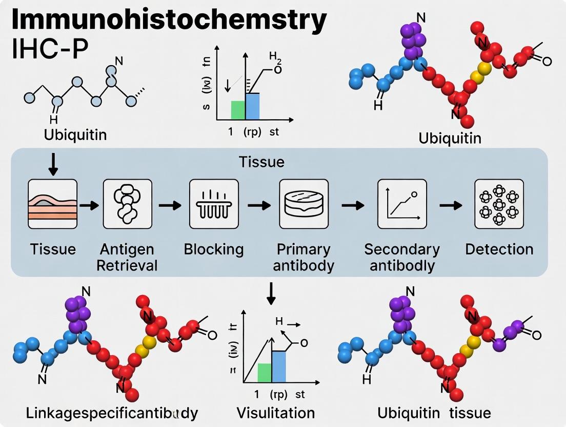
The ubiquitin-proteasome system (UPS) is a master regulator of cellular homeostasis, traditionally recognized for its role in targeted protein degradation. However, contemporary research underscores its integral function in orchestrating complex signaling networks, including DNA damage repair, immune response (NF-κB), Wnt/β-catenin, and cell cycle progression. These non-degradative functions are mediated through specific ubiquitin linkages (e.g., K63, K11, K48, M1) that act as molecular scaffolds to recruit signaling complexes. Investigating these linkages in paraffin-embedded clinical tissues via immunohistochemistry (IHC) provides critical spatial context for understanding disease mechanisms and evaluating therapeutic targets in cancer and neurodegeneration. The following notes and protocols are framed within a thesis focused on optimizing IHC-P for ubiquitin linkage-specific antibodies in FFPE tissue.
Note 1: Specificity Validation is Paramount. Linkage-specific antibodies (e.g., for K63- or K48-linked chains) are prone to cross-reactivity. Validation in FFPE tissue must include:
- Knockdown/knockout of key E2/E3 enzymes (e.g., TRAF6 for K63, APC/C for K11) to reduce specific chain types.
- Competition assays with linkage-specific recombinant ubiquitin chains.
- Correlation with mass spectrometry-based ubiquitin proteomics from adjacent tissue sections when possible.
Note 2: Antigen Retrieval Optimization. The compact nature of ubiquitin chains requires tailored antigen retrieval. While a high-pH Tris-EDTA buffer (pH 9.0) is often effective for K48 and K63 linkages, linear (M1) chains may require low-pH citrate-based retrieval. Empirical testing for each antibody is required.
Note 3: Signal Interpretation in IHC-P. Distinct subcellular patterns indicate different functions:
- Nuclear K48-polyUb: Often associated with proteasomal degradation of transcription factors.
- Cytoplasmic K63-polyUb: Frequently linked to kinase activation (e.g., in NF-κB signaling).
- Membrane-associated M1-linear Ub: Key in immune signaling complexes.
Quantitative Data Summary: Common Ubiquitin Linkages & Their Primary Functions
| Ubiquitin Linkage Type | Primary E2/E3 Enzymes (Examples) | Dominant Functional Role | Associated Pathway Examples | Common Readout in IHC-P (FFPE) |
|---|---|---|---|---|
| K48-linked chains | CDC34, UBE2R2 / SCF Complex, HUWE1 | Proteasomal Degradation | Cell Cycle (p27 degradation), HIF-1α regulation | Nuclear or diffuse cytoplasmic staining. Correlates with high proliferation indices. |
| K63-linked chains | UBC13/UEV1A, TRAF6, RNF8 | Signal Activation & Trafficking | NF-κB, DNA Damage Repair, Endocytosis | Punctate cytoplasmic aggregates, membrane staining. |
| K11-linked chains | UBE2S, UBE2C / APC/C | Proteasomal Degradation & Regulation | Mitotic Progression, ERAD | Diffuse cytoplasmic during interphase; strong nuclear/centrosomal in mitosis. |
| M1-linked (linear) chains | HOIP, SHARPIN / LUBAC Complex | Signal Activation & Complex Assembly | TNFα/NF-κB, Inflammation | Distinct punctate or rosette-like patterns at signaling complexes. |
| K27/K29-linked chains | UBE2W, RNF26 / Trim28 | Autophagy, Signaling | Selective Autophagy, Innate Immunity | Punctate perinuclear or autophagosome-associated staining. |
Detailed Protocols
Protocol 1: IHC-P for Ubiquitin Linkage-Specific Antibodies in FFPE Tissue Sections
Objective: To detect and localize specific ubiquitin chain linkages (e.g., K63 or K48) in formalin-fixed, paraffin-embedded (FFPE) tissue sections.
Materials:
- FFPE tissue sections (4-5 µm) on charged slides
- Xylene and ethanol series (100%, 95%, 70%)
- Target antigen retrieval buffer (e.g., 10 mM Tris, 1 mM EDTA, pH 9.0, or 10 mM Citrate, pH 6.0)
- Hydrogen peroxide (3% in methanol)
- Blocking solution: 5% normal serum (from host species of secondary antibody) in PBS
- Primary antibody: Validated linkage-specific ubiquitin antibody (e.g., anti-K63-Ub [clone Apu3], anti-K48-Ub [clone Apu2])
- Species-appropriate HRP-polymer secondary detection system
- DAB or other chromogen substrate kit
- Hematoxylin counterstain
- Mounting medium
Methodology:
- Deparaffinization & Rehydration: Bake slides at 60°C for 20 min. Immerse in xylene (3 x 5 min), then 100% ethanol (2 x 3 min), 95% ethanol (2 min), 70% ethanol (2 min), and finally distilled water.
- Antigen Retrieval: Place slides in pre-heated target retrieval buffer in a decloaking chamber or pressure cooker. Heat for 20-30 min at 95-100°C. Cool for 30 min at room temperature (RT). Wash in PBS (pH 7.4).
- Peroxidase Quenching: Incubate slides in 3% H₂O₂ in methanol for 15 min at RT to block endogenous peroxidase activity. Wash in PBS.
- Blocking: Apply 200-300 µL of blocking solution to cover tissue. Incubate in a humidified chamber for 1 hour at RT.
- Primary Antibody Incubation: Tap off blocking solution. Apply primary antibody at optimized dilution in blocking solution. Incubate overnight at 4°C in a humidified chamber.
- Detection: Wash slides in PBS (3 x 5 min). Apply polymer-HRP secondary antibody as per manufacturer's instructions. Incubate for 30-60 min at RT. Wash in PBS (3 x 5 min).
- Visualization: Apply DAB chromogen solution. Monitor development under a microscope (typically 30 sec to 5 min). Stop reaction by immersing in distilled water.
- Counterstaining & Mounting: Counterstain with hematoxylin for 30 sec. Rinse in tap water, then dehydrate through ethanol series (70%, 95%, 100%) and xylene. Coverslip with permanent mounting medium.
Protocol 2: Validation via siRNA Knockdown in Cell Pellet FFPE Blocks
Objective: To validate antibody specificity by generating positive/negative control FFPE cell pellets with modulated ubiquitin chain levels.
Methodology:
- Culture HEK293 or HeLa cells in 6-well plates.
- Transfect with siRNA targeting a relevant E2 enzyme (e.g., UBE2N for K63 chains) or a non-targeting control using standard transfection reagent. Incubate for 72 hours.
- Cell Pellet Formation: Trypsinize cells, wash with PBS, and centrifuge at 300 x g for 5 min. Resuspend pellet in 4% PFA for 20 min at RT for fixation. Centrifuge again to form a tight pellet. Aspirate supernatant, leaving ~50 µL.
- FFPE Processing: Resuspend pellet in warm (50°C) 2% agarose in PBS. Let solidify. Process the agarose-embedded pellet through a standard ethanol dehydration series, xylene clearing, and paraffin embedding protocol.
- Section the cell block and perform IHC-P (as in Protocol 1) alongside test tissue sections. Specific antibody signal should be markedly reduced in the knockdown pellet compared to control.
Diagrams
The Scientist's Toolkit: Key Research Reagent Solutions
| Reagent / Material | Vendor Examples (Illustrative) | Key Function in UPS/FFPE Research |
|---|---|---|
| Linkage-Specific Ubiquitin Antibodies | MilliporeSigma (Apu2, Apu3), Cell Signaling Technology, Abcam | Core Detection: Selective recognition of K48, K63, M1, etc., ubiquitin chains for IHC-P and Western blot. Specificity is critical. |
| Recombinant Ubiquitin Chains (K48, K63, M1) | R&D Systems, Ubiquigent, Boston Biochem | Validation & Competition: Used as blocking reagents in antibody validation experiments to confirm linkage specificity. |
| Deubiquitinase (DUB) Inhibitors (PR-619, G5) | Cayman Chemical, Selleckchem | Pathway Modulation: Stabilize ubiquitinated proteins in cell-based studies prior to FFPE fixation to enhance signal. |
| Proteasome Inhibitors (MG-132, Bortezomib) | MedChemExpress, Sigma-Aldrich | Positive Control Induction: Induce accumulation of K48-polyubiquitinated proteins, creating a positive control for IHC. |
| siRNA Libraries (E1/E2/E3 Enzymes) | Horizon Discovery, Sigma-Aldrich | Functional Validation: Knockdown specific UPS components to validate antibody specificity or study pathway function in cell pellet models. |
| Polymer-HRP IHC Detection Kits | Agilent Dako, Abcam, Vector Laboratories | Amplified Detection: Provide high sensitivity and low background for detecting ubiquitin conjugates in FFPE tissue. |
| Antigen Retrieval Buffers (pH 6.0 & 9.0) | Vector Laboratories, Thermo Fisher Scientific | Epitope Unmasking: Critical for exposing buried ubiquitin chain epitopes fixed by formalin. pH must be optimized per antibody. |
| FFPE Cell Pellet Preparation Kits | Thermo Fisher Scientific, Cell Signaling Technology | Control Generation: Standardized kits for creating controlled positive/negative cell line samples for IHC assay validation. |
Ubiquitin chains, linked via specific lysine residues or methionine-1, form a complex post-translational code that dictates diverse cellular fates. In the context of immunohistochemistry on paraffin-embedded (IHC-P) tissue, decoding this code provides critical insights into disease mechanisms, proteinopathies, and therapeutic responses. This application note details protocols and reagents for the specific detection of clinically relevant ubiquitin linkages in FFPE tissue sections, a cornerstone for translational research in oncology, neurodegeneration, and inflammation.
Clinically Relevant Ubiquitin Linkages: Functions & Disease Associations
The table below summarizes key ubiquitin linkage types, their primary functions, and associated pathological contexts relevant to IHC-P research.
Table 1: Key Ubiquitin Linkages: Characteristics and Clinical Relevance
| Linkage Type | Primary Cellular Function | Key Reader/Effector Proteins | Associated Pathologies (IHC-P Context) | Common IHC Biomarker/Co-localization |
|---|---|---|---|---|
| K48-linked | Proteasomal degradation | Proteasome 19S cap, Ubiquilins | Neurodegenerative aggregates (Tau, α-synuclein), some cancers | p62/SQSTM1, proteasome subunits |
| K63-linked | DNA repair, inflammation, endocytosis | TAB2/3, RNF168, ESCRT components | Inflammatory diseases, solid tumors, DNA damage response | p62, ATM/ATR markers, inflammatory cytokines |
| M1-linked (Linear) | NF-κB activation, inflammation | NEMO, ABIN-1, HOIP | Autoimmune disorders, chronic inflammation | p65/RelA (NF-κB), inflammatory infiltrates |
| K11-linked | Cell cycle regulation, ERAD | CDC20, APC/C | Carcinogenesis (breast, colon) | Cyclin B1, Ki-67 |
| K6-linked | Mitophagy, DNA damage | Parkin, BRCA1 | Parkinson's disease, breast cancer | LC3, Mitochondrial markers (TOMM20) |
| K27-linked | Kinase activation, immune signaling | TAB2/3 | Glioblastoma, autoimmune conditions | Kinase targets (e.g., mTOR pathway) |
| K29-linked | Proteotoxic stress response | Hul5, UBR4/5 | Proteostasis-related disorders | Autophagy markers (LC3) |
Detailed IHC-P Protocol for Ubiquitin Linkage-Specific Staining
This protocol is optimized for visualizing specific ubiquitin chains in formalin-fixed, paraffin-embedded (FFPE) tissue sections using validated linkage-specific antibodies.
Protocol 1: Antigen Retrieval and Staining for K48 & K63 Linkages
- Tissue Sectioning: Cut 4-5 μm FFPE sections onto positively charged slides. Dry at 60°C for 1 hour.
- Deparaffinization & Rehydration: Xylene (3 x 5 min) → 100% Ethanol (2 x 2 min) → 95% Ethanol (2 min) → 70% Ethanol (2 min) → dH₂O rinse.
- Antigen Retrieval (Critical): Use pressure cooker or decloaking chamber with pH 9.0 Tris-EDTA buffer for K63 linkage antibodies or pH 6.0 Sodium Citrate buffer for K48 linkage antibodies. Boil for 15-20 min, cool for 30 min at room temperature (RT). Rinse in PBS + 0.025% Triton X-100 (PBS-T).
- Peroxidase Blocking: Incubate with 3% H₂O₂ in methanol for 15 min at RT. Rinse in PBS-T.
- Protein Block: Apply 2.5% normal horse serum (Vector Labs) for 30 min at RT.
- Primary Antibody Incubation: Apply linkage-specific monoclonal antibody (see Reagent Table). Dilute in antibody diluent. Incubate overnight at 4°C in a humidified chamber.
- Detection: Use polymer-based detection systems (e.g., HRP-polymer). Incubate with appropriate secondary polymer for 30 min at RT. Visualize with DAB (brown) or Vector Red (pink/red) chromogen for 5-10 min. Counterstain with Hematoxylin. Dehydrate, clear, and mount.
Protocol 2: Sequential Immunofluorescence for Co-localization Studies
- Follow steps 1-5 from Protocol 1.
- Primary Antibody 1: Apply first linkage-specific antibody (e.g., anti-K63). Incubate overnight at 4°C.
- Detection 1: Apply species-specific fluorophore-conjugated secondary (e.g., Alexa Fluor 488, 1:500) for 1 hr at RT. Protect from light.
- Antibody Elution (Optional for same-host species): To stain with a second primary from the same host, perform mild antigen retrieval again or use antibody elution buffer (pH 2.0 glycine) for 20 min at 60°C.
- Primary Antibody 2: Apply second antibody (e.g., anti-p62). Incubate overnight at 4°C.
- Detection 2: Apply a different species-specific fluorophore-conjugated secondary (e.g., Alexa Fluor 594).
- Mounting: Apply anti-fade mounting medium with DAPI. Seal edges.
Key Signaling Pathways Involving Ubiquitin Linkages
Diagram Title: M1-Linked Ubiquitin in NF-κB Activation
Diagram Title: K48-Linked Ubiquitin in Proteasomal Degradation
Experimental Workflow for Ubiquitin Linkage Analysis in FFPE Tissue
Diagram Title: IHC-P Workflow for Ubiquitin Linkages
The Scientist's Toolkit: Research Reagent Solutions
Table 2: Essential Reagents for Ubiquitin Linkage-Specific IHC-P
| Reagent/Category | Specific Example/Product Code | Function in Protocol |
|---|---|---|
| Linkage-specific mAbs | Anti-K48 (clone Apu2, Millipore), Anti-K63 (clone Apu3), Anti-M1 (clone 1E3) | Highly specific recognition of defined ubiquitin linkage topology. Critical for signal specificity. |
| Polymer-based Detection | ImmPRESS HRP Polymer systems (Vector Labs) | Amplifies signal with low background. Species-specific polymers reduce cross-reactivity. |
| Chromogens | DAB (Brown), Vector Red (Alkaline Phosphatase), AEC (Red) | Provides visible precipitate for microscopy. Choice impacts contrast and compatibility with counterstains. |
| Antigen Retrieval Buffers | Tris-EDTA (pH 9.0), Sodium Citrate (pH 6.0) | Unmasks epitopes cross-linked by formalin. Optimal pH is antibody-dependent. |
| Blocking Serum | Normal Horse/Goat Serum (Vector Labs) | Reduces non-specific binding of secondary antibodies to tissue. |
| Mounting Media | VECTASHIELD (with/without DAPI), Permount | Preserves stain, provides fluorescence anti-fade (for IF), or permanent mounting (for DAB). |
| Positive Control Tissue | Alzheimer's brain (K48), Hodgkin's lymphoma (K63), Rheumatoid synovium (M1) | Validates antibody performance and protocol optimization for each linkage. |
| Protein Block (Optional) | Casein, BSA, or proprietary blockers (e.g., Background Sniper) | Further reduces non-specific hydrophobic/ionic interactions, lowering background. |
Table 3: Representative IHC-P Findings of Ubiquitin Linkages in Human Pathologies
| Pathology | Tissue Type | Predominant Linkage | Quantitative Finding (vs. Control) | Method & Antibody Cited |
|---|---|---|---|---|
| Alzheimer's Disease | Temporal Cortex | K48 | >70% of plaques co-localize with K48 | IHC, Anti-K48 (Apu2) |
| Colorectal Carcinoma | Tumor Core | K63 | 3.5-fold increase in staining intensity | Digital IHC, Anti-K63 (Apu3) |
| Rheumatoid Arthritis | Synovium | M1 | 89% of patients show strong linear/M1 staining | IHC, Anti-M1 (1E3) |
| Glioblastoma Multiforme | Tumor Margin | K27 | High K27 correlates with poor survival (HR=2.1) | Multiplex IHC, Anti-K27 (ABM-0005) |
| Parkinson's Disease | Substantia Nigra | K63 & K6 | K63 increase in Lewy bodies; K6 in mitochondria | Sequential IF, K63 (Apu3) & K6 (Abcam) |
Application Notes: Ubiquitin Linkage-Specific Pathology in Human Diseases
Ubiquitin chain topology determines the fate of modified proteins. The development of linkage-specific antibodies has enabled the direct assessment of these post-translational modifications in archival formalin-fixed, paraffin-embedded (FFPE) tissues, linking specific chain types to disease mechanisms and patient outcomes.
Table 1: Quantitative Association of Specific Ubiquitin Chains with Human Disease Pathogenesis
| Ubiquitin Linkage | Primary Disease Association | Key Target Proteins/Pathways | Reported Effect in Patient Tissues (IHC) | Correlation with Clinical Metrics |
|---|---|---|---|---|
| K48-linked | Neurodegeneration (Alzheimer’s, PD) | Tau, α-synuclein, misfolded proteins | Elevated in neuronal inclusions vs. healthy tissue. | Positively correlates with disease stage & cognitive decline. |
| K63-linked | Inflammation (RA, IBD), Solid Tumors | RIPK1, TRAF6, NF-κB pathway | High in inflamed synovium/tumor stroma macrophages. | Correlates with inflammatory cytokine levels & poor prognosis. |
| K11-linked | Cancer (Breast, Glioblastoma) | APC/C substrates, cell cycle regulators | Elevated in high-grade tumor nuclei/cytoplasm. | Associated with mitotic index, genomic instability, and reduced survival. |
| Linear/M1-linked | Inflammation, Immune Cell Activation | NEMO, RIPK1, NF-κB pathway | Detected in activated immune infiltrates in autoimmunity. | Correlates with disease activity scores in autoimmune disorders. |
Protocol: Immunohistochemistry on FFPE Tissue Using Ubiquitin Linkage-Specific Antibodies
I. Sample Preparation & Antigen Retrieval
- Cut 4-5 μm sections from FFPE tissue blocks and mount on charged slides.
- Bake slides at 60°C for 60 minutes.
- Deparaffinize and rehydrate: Xylene (3 x 5 min) → 100% Ethanol (2 x 3 min) → 95% Ethanol (2 x 3 min) → 70% Ethanol (2 x 3 min) → dH₂O rinse.
- Critical: Perform heat-induced epitope retrieval (HIER) in a pressure cooker for 20 min using pH 6.0 Citrate Buffer for K48- and K63-linkage antibodies or pH 9.0 Tris-EDTA Buffer for K11- and M1-linkage antibodies.
- Cool slides for 30 min in retrieval buffer at room temperature, then rinse in PBS + 0.025% Triton X-100 (PBS-T).
II. Primary & Secondary Antibody Staining
- Block endogenous peroxidases with 3% H₂O₂ in methanol for 15 min. Rinse in PBS-T.
- Apply protein block (e.g., 5% normal goat serum/2% BSA in PBS) for 1 hour at RT.
- Incubate with primary linkage-specific antibody diluted in blocking buffer overnight at 4°C in a humidified chamber.
- Recommended dilutions (optimize per lot): Anti-K48 (1:200), Anti-K63 (1:250), Anti-K11 (1:150), Anti-M1 (1:100).
- Wash slides in PBS-T (3 x 5 min).
- Apply HRP-conjugated polymer secondary antibody (e.g., anti-rabbit EnVision+ system) for 60 min at RT. Wash in PBS-T (3 x 5 min).
III. Detection, Counterstaining & Analysis
- Develop with DAB chromogen for 3-10 min, monitoring under a microscope.
- Rinse in dH₂O. Counterstain with Hematoxylin for 30-60 sec. Rinse, dehydrate, and mount.
- Analysis: Score using a semi-quantitative H-score (H-Score = Σ (pi × i), where pi = percentage of cells stained at intensity i (0-3)). Analyze minimum of 5 high-power fields.
Pathway & Workflow Visualizations
Diagram 1: K63/Linear Ubiquitin in NF-κB Inflammation
Diagram 2: K48 Ubiquitin in Proteinopathies
Diagram 3: IHC-P Workflow for Ubiquitin Chains
The Scientist's Toolkit: Key Research Reagents
Table 2: Essential Reagents for Ubiquitin Linkage-Specific IHC Research
| Reagent / Solution | Function & Role in Protocol | Critical Specification / Note |
|---|---|---|
| FFPE Tissue Sections | Archival patient or disease model samples for spatial pathology analysis. | Optimal fixation (24h in 10% NBF) is critical for epitope preservation. |
| Linkage-Specific Ub Antibodies | Primary antibodies discriminating specific ubiquitin chain linkages. | Must be validated for IHC-P (e.g., K48: clone Apu2; K63: clone Apu3). |
| pH-specific Antigen Retrieval Buffers | Unmask cross-linked epitopes. Most critical step for specificity. | Use pH 6.0 Citrate for K48/K63; pH 9.0 Tris-EDTA for K11/M1 chains. |
| HRP-labeled Polymer Secondary | Amplifies signal with high sensitivity and low background. | Species-matched polymer system (e.g., EnVision+, ImmPRESS). |
| DAB Chromogen Substrate | Produces brown, stable precipitate at site of antigen-antibody binding. | Use same developing time across compared slides for consistency. |
| Hematoxylin Counterstain | Provides nuclear contrast for histological context. | Differentiate carefully to avoid masking low-intensity DAB signal. |
| Automated Slide Stainer | Enables standardized, high-throughput processing of sample batches. | Program must include appropriate retrieval and cooling steps. |
Why FFPE Tissue? Advantages and Challenges for Ubiquitin Biomarker Research
Formalin-fixed, paraffin-embedded (FFPE) tissue represents the cornerstone of clinical histopathology and a critical resource for retrospective biomarker research. For the study of ubiquitination—a complex, multi-faceted post-translational modification—FFPE tissues offer unparalleled access to vast, clinically annotated archives spanning decades. This application note details the advantages, inherent challenges, and validated protocols for utilizing FFPE specimens in ubiquitin linkage-specific immunohistochemistry (IHC-P) research, framed within a thesis on advancing IHC-P protocols for ubiquitin system biomarkers.
The ubiquitin-proteasome system regulates protein degradation, signaling, and localization through diverse polyubiquitin chain linkages (e.g., K48, K63, M1). Dysregulation is implicated in cancer, neurodegeneration, and inflammatory diseases. Linkage-specific antibodies enable the spatial mapping of these modifications in tissue architecture, providing critical functional insights.
Advantages of FFPE Tissues for Ubiquitin Research
| Advantage | Rationale & Impact on Ubiquitin Research |
|---|---|
| Archival Abundance | Enables large-scale retrospective cohort studies correlating ubiquitin signatures with long-term clinical outcomes. |
| Preserved Morphology | Allows precise subcellular (e.g., cytoplasmic aggregates, nuclear foci) localization of ubiquitin signals within the tissue context. |
| Clinical Annotation | Ubiquitin biomarker discovery is directly linked to patient metadata (diagnosis, treatment response, survival). |
| Molecular Stability | When properly fixed, ubiquitin epitopes and associated proteins are stabilized for long-term room-temperature storage. |
Key Challenges & Mitigation Strategies
| Challenge | Impact on Ubiquitin IHC-P | Mitigation Protocol |
|---|---|---|
| Cross-linking Artifacts | Formaldehyde cross-linking masks epitopes, especially critical for discerning subtle linkage-specific signals. | Heat-induced epitope retrieval (HIER) is essential. Optimize pH and time (see Protocol 1). |
| Variable Fixation | Pre-analytical variability (ischemia time, fixation delay/duration) alters ubiquitin patterns. | Implement strict tissue handling SOPs and use control tissue microarrays (TMAs) with fixation controls. |
| Antibody Specificity | High risk of false positives; many antibodies show cross-reactivity or detect free ubiquitin. | Validate antibodies with FFPE-compatible positive/negative controls (e.g., transfected cell pellets, siRNA knockdown). |
| Signal Quantification | Ubiquitin staining is often granular/heterogeneous, challenging standard scoring. | Employ digital pathology & image analysis for objective, quantitative scoring of stain intensity and distribution. |
Protocols
Protocol 1: Optimized IHC-P for Ubiquitin Linkage-Specific Antibodies on FFPE Tissue
Objective: Reliable detection of specific polyubiquitin linkages (K48, K63, M1) in FFPE sections.
Materials: Research Reagent Solutions Toolkit
| Item | Function & Critical Notes |
|---|---|
| Linkage-Specific Anti-Ubiquitin Primary Antibodies | Clone must be validated for IHC-P on FFPE. Test multiple clones (e.g., Apu2 for K48, Apu3 for K63). |
| pH 6 or pH 9 Epitope Retrieval Buffer | K48 often requires high-pH retrieval; K63 may require low-pH. Must be optimized per antibody. |
| Validated IHC Detection System | Polymer-based HRP or AP systems recommended for high sensitivity and low background. |
| FFPE Cell Pellet Controls | Cells overexpressing specific ubiquitin linkages or treated with proteasome inhibitors (MG132) as positive controls. |
| Isotype Control & Knockdown Controls | Essential for confirming signal specificity. Use siRNA-treated cell pellets or tissue with known low expression. |
| Automated Stainers | Recommended for consistency, especially in multi-cohort studies. |
Methodology:
- Sectioning: Cut 4-5 µm FFPE sections onto charged slides. Dry at 60°C for 1 hour.
- Deparaffinization & Rehydration: Standard xylene/ethanol series.
- Epitope Retrieval (HIER): Place slides in retrieval buffer (e.g., Tris-EDTA, pH 9.0) and heat in a pressure cooker (121°C, 15 min) or water bath (96-98°C, 20-40 min). Cool for 30 min at room temp.
- Peroxidase Block: 3% H₂O₂ in methanol, 15 min.
- Blocking: Incubate with protein block (e.g., 5% normal serum/BSA) for 30 min.
- Primary Antibody: Apply optimized dilution of linkage-specific antibody in blocking buffer. Incubate overnight at 4°C. (Include isotype control slide).
- Detection: Apply labeled polymer-HRP secondary antibody (30 min at RT). Visualize with DAB+ chromogen (5-10 min).
- Counterstaining & Mounting: Hematoxylin counterstain, dehydrate, clear, and mount.
Validation: Staining must be abolished by pre-incubation of the antibody with its cognate ubiquitin peptide antigen and show expected modulation in controls (e.g., proteasome inhibitor treatment increases K48 signal).
Protocol 2: Multiplex Immunofluorescence (mIF) for Ubiquitin & Cell Markers
Objective: Co-localize specific ubiquitin linkages with cell lineage or signaling markers (e.g., p62, phospho-proteins).
Methodology: Adapt Protocol 1 using sequential IHC staining with antibody stripping or, preferably, using tyramide signal amplification (TSA) multiplex kits with different fluorophores. After first-round IHC (e.g., anti-K48), slides are heated in retrieval buffer to remove antibodies while leaving the deposited fluorophore intact. Process is repeated for second marker. Spectral imaging is used for deconvolution and analysis.
Data Analysis & Interpretation
Quantitative digital pathology analysis is non-negotiable. Use whole-slide imaging and software to quantify:
- H-Score: Combines intensity (0-3+) and percentage of positive cells.
- Signal Distribution: Analyze within specific compartments (nuclear vs. cytoplasmic) or cell populations (tumor vs. stroma).
Table: Representative Quantitative Data from FFPE Ubiquitin IHC Studies
| Disease Context | Linkage Studied | Key Finding (vs. Normal) | Assay Used | Reference (Example) |
|---|---|---|---|---|
| Alzheimer's Disease | K48, K63 | K63-linked ubiquitin increased in neurofibrillary tangles. | IHC-P, mIF | Acta Neuropathol, 2021 |
| Colorectal Carcinoma | K48 | Low tumor K48 correlated with worse prognosis (HR=2.1). | IHC-P on TMA | Mod Pathol, 2022 |
| Lung Adenocarcinoma | M1 (Linear) | Strong linear ubiquitin in immune cells associated with better response to immunotherapy. | IHC-P, digital scoring | J Immunother Cancer, 2023 |
FFPE tissues are an indispensable but demanding resource for translational ubiquitin research. Success hinges on rigorous pre-analytical control, meticulous antibody validation, optimized epitope retrieval, and quantitative digital analysis. The protocols outlined provide a foundation for robust, reproducible investigation of ubiquitin pathway dynamics directly in human disease contexts, accelerating biomarker discovery and therapeutic development.
Diagrams
Within the context of advancing IHC-P protocol development for paraffin-embedded tissue research, the evolution of ubiquitin detection reagents represents a paradigm shift. The transition from pan-ubiquitin antibodies, which recognize all ubiquitinated proteins irrespective of linkage type, to linkage-specific antibodies that discern the topology of the ubiquitin chain (e.g., K48, K63, M1) has been transformative. This allows researchers to infer specific cellular signals, as different linkages dictate distinct functional outcomes such as proteasomal degradation (K48) or NF-κB activation (K63, M1).
The Scientist's Toolkit: Key Research Reagent Solutions
| Reagent / Material | Function in IHC-P for Ubiquitin Research |
|---|---|
| Pan-Ubiquitin Antibody (e.g., clone P4D1) | Broad-spectrum detection of ubiquitin conjugates; useful for initial assessment of global ubiquitination levels but lacks functional specificity. |
| Linkage-Specific Ubiquitin Antibodies (K48, K63, M1) | Discriminate specific polyubiquitin chain linkages, enabling functional interpretation of ubiquitin signaling pathways in disease contexts. |
| Formalin-Fixed, Paraffin-Embedded (FFPE) Tissue Sections | Archival clinical samples; the primary substrate for retrospective translational research using IHC-P. |
| High-Temperature Antigen Retrieval Buffer (pH 9.0) | Essential for breaking protein cross-links formed during fixation to expose ubiquitin epitopes in FFPE tissues. |
| HRP-Conjugated Polymer Detection System | Provides high sensitivity and low background amplification of the primary antibody signal for visualization. |
| DAB Chromogen | Forms a stable, brown precipitate at the site of antibody binding, enabling microscopic analysis. |
Quantitative Comparison: Pan-Ubiquitin vs. Linkage-Specific Detection
Table 1: Performance Metrics in FFPE IHC-P
| Parameter | Pan-Ubiquitin Antibody | Linkage-Specific (K48) Antibody | Linkage-Specific (K63) Antibody |
|---|---|---|---|
| Primary Clones/Sources | P4D1, FK1, FK2 | Clone D9D5, Clone A19656 | Clone D7A11, Clone D2614 |
| Typical Dilution for IHC-P | 1:100 - 1:500 | 1:50 - 1:200 | 1:50 - 1:200 |
| Optimal Antigen Retrieval | Citrate pH 6.0 or Tris-EDTA pH 9.0 | Tris-EDTA pH 9.0 (high-temp) | Tris-EDTA pH 9.0 (high-temp) |
| Key Biological Insight | Total protein load tagged for degradation/regulation | Marks proteins for proteasomal degradation | Involved in DNA repair, kinase activation, signaling |
| Common IHC Pattern in Cancer | Diffuse cytoplasmic/nuclear staining | Focal cytoplasmic aggregates (proteasome foci) | Strong membranous/cytoplasmic in invasive regions |
Table 2: Published Data from Recent Studies (Representative)
| Study Focus (Disease) | Antibody Target | % of Cases Positive (n) | H-Score / Staining Intensity Correlation | Clinical Correlation |
|---|---|---|---|---|
| Glioblastoma (2023) | Pan-Ubiquitin | 100% (n=45) | High score in >80% | Poor specificity for outcome |
| Glioblastoma (2023) | K63-linkage | 62% (n=45) | High score in 40% | Strong correlation with tumor grade & invasion |
| Colorectal Cancer (2024) | K48-linkage | 58% (n=60) | Variable intensity | Associated with response to proteasome inhibitors |
| Breast Cancer (2024) | M1-linkage (linear) | 31% (n=75) | Low/Moderate intensity | Linked to inflammatory tumor microenvironment |
Detailed Protocol: IHC-P for Linkage-Specific Ubiquitin in FFPE Tissue
Protocol Title: Immunohistochemical Detection of K63-Linked Polyubiquitin in Archival FFPE Tissue Sections
Objective: To specifically visualize proteins modified with K63-linked polyubiquitin chains, a marker for active signal transduction and DNA damage response, in paraffin-embedded human tissue.
Materials:
- FFPE tissue sections (4-5 µm) on charged slides
- Xylene and ethanol series (100%, 95%, 70%)
- High-temperature antigen retrieval solution (Tris-EDTA, pH 9.0)
- Hydrogen peroxide block (3% H₂O₂ in methanol)
- Protein block (serum-free)
- Primary antibody: Anti-K63-linkage specific ubiquitin (e.g., Rabbit mAb D7A11)
- HRP-labeled polymer anti-rabbit detection system
- DAB chromogen substrate kit
- Hematoxylin counterstain
- Mounting medium
Methodology:
- Deparaffinization & Rehydration: Bake slides at 60°C for 20 min. Immerse in xylene (3 changes, 5 min each). Rehydrate through graded ethanol (100%, 95%, 70%, 2 min each). Rinse in distilled water.
- Antigen Retrieval (Critical Step): Place slides in pre-heated Tris-EDTA buffer (pH 9.0) in a decloaking chamber or pressure cooker. Heat at 95-100°C for 20 min. Cool at room temperature for 30 min. Rinse in PBS (pH 7.4).
- Peroxidase Blocking: Apply 3% H₂O₂ solution for 10 min to quench endogenous peroxidase activity. Rinse with PBS.
- Protein Block: Apply serum-free protein block for 10 min at room temperature. Do not rinse; gently tap off excess.
- Primary Antibody Incubation: Apply optimally diluted anti-K63 ubiquitin antibody (e.g., 1:100 in antibody diluent). Incubate overnight at 4°C in a humidified chamber. The following day, rinse slides thoroughly with PBS-Tween 20 (0.05%).
- Polymer Detection: Apply HRP-conjugated secondary polymer for 30 min at room temperature. Rinse with PBS.
- Chromogen Development: Apply prepared DAB solution (per manufacturer's instructions) for 3-5 minutes. Monitor development under a microscope. Stop reaction by immersing in distilled water.
- Counterstaining & Mounting: Counterstain with hematoxylin for 30-60 seconds. Differentiate in bluing reagent, dehydrate through graded alcohols and xylene. Mount with permanent mounting medium.
Interpretation: Positive K63-linked ubiquitin signal appears as a distinct brown, granular or diffuse cytoplasmic/membranous precipitate. Nuclear staining may be present in cases of DNA damage. Compare with pan-ubiquitin and negative control (omission of primary antibody) slides.
Visualizing Ubiquitin Signaling Pathways and Workflows
Ubiquitin Chain Type Determines Protein Fate
IHC-P Workflow for Linkage-Specific Ubiquitin
Step-by-Step Protocol: Optimized IHC for K48, K63, and M1 Ubiquitin in FFPE Samples
This application note details the critical pre-analytical steps for preparing paraffin-embedded tissue specimens for immunohistochemistry (IHC) targeting ubiquitin linkage-specific antibodies. The integrity of ubiquitin signal localization is exquisitely sensitive to variations in fixation, processing, and sectioning. Optimal pre-protocol procedures are essential for generating reproducible and biologically relevant data in drug development research.
Tissue Fixation: Principles and Protocols
Fixation is the most critical determinant of IHC success for labile post-translational modifications like ubiquitin linkages. The goal is to rapidly terminate enzymatic activity and preserve antigenicity while maintaining tissue morphology.
Protocol 1.1: Optimal Fixation for Ubiquitin Preservation
- Reagent: 10% Neutral Buffered Formalin (NBF).
- Procedure:
- Immerse tissue specimen in a volume of 10% NBF that is at least 10 times the tissue volume.
- Fixation time is tissue thickness-dependent. For most tissues, a guideline of 1 hour per 1 mm of thickness is used, with a minimum of 6 hours and a maximum of 24-48 hours for standard specimens.
- For large organs, perfuse-fixation in situ is superior. Alternatively, slice into <5 mm slabs before immersion.
- After fixation, rinse tissue thoroughly in phosphate-buffered saline (PBS) or directly transfer to 70% ethanol for storage or the start of processing.
Key Quantitative Data on Fixation Variables
Table 1: Impact of Fixation Variables on Ubiquitin Linkage Detection
| Variable | Optimal Condition | Suboptimal Condition | Effect on K48/K63 Linkage Signal |
|---|---|---|---|
| Fixative | 10% NBF, pH 7.2-7.4 | Unbuffered Formalin, Bouin’s, Alcohol-based | High background & loss of specificity; altered epitope presentation. |
| Fixation Duration | 18-24 hrs (for 3-5mm block) | <6 hrs (under-fixation) >72 hrs (over-fixation) | Under-fixation: Diffusion/ loss. Over-fixation: Masking & cross-linking. |
| Tissue Thickness | ≤ 5 mm | > 10 mm | Incomplete fixation core leads to uneven staining and false negatives. |
| Delay to Fixation | < 30 minutes (Cold Ischemia Time) | > 60 minutes | Rapid degradation of ubiquitin conjugates; increased background. |
| Temperature | Room Temperature (20-25°C) | > 40°C or < 4°C | Artifactual aggregation or poor preservation of morphology. |
Tissue Processing: Dehydration, Clearing, and Infiltration
Processing prepares fixed tissue for embedding by removing water and replacing it with paraffin wax. Inconsistent processing creates artifacts that hinder sectioning and antibody penetration.
Protocol 1.2: Standardized Processing for Consistent Paraffin Infiltration
- Equipment: Automated Tissue Processor.
Reagent Sequence & Timing (Standard Protocol):
- 70% Ethanol: 60 minutes
- 80% Ethanol: 60 minutes
- 95% Ethanol: 60 minutes
- 100% Ethanol I: 60 minutes
- 100% Ethanol II: 60 minutes
- Xylene (or Clearing Substitute) I: 60 minutes
- Xylene (or Clearing Substitute) II: 60 minutes
- Paraffin Wax I (58-60°C): 60 minutes
- Paraffin Wax II (58-60°C): 60 minutes
Notes: Tissues can be held in 70% ethanol at 4°C prior to processing. For delicate tissues (e.g., brain), extend ethanol steps gently. Vacuum impregnation can be used during wax steps to improve infiltration of dense tissues.
Table 2: Troubleshooting Common Processing Artifacts
| Artifact | Primary Cause | Consequence for IHC | Correction |
|---|---|---|---|
| Incomplete Infiltration | Insufficient time in wax; cold wax. | Tissue crumbles on sectioning; uneven antibody penetration. | Increase wax time/temp; use vacuum; ensure reagent freshness. |
| Excessive Hardness | Prolonged dehydration in high-grade alcohols. | Brittle tissue; chattering sections; high background. | Shorten 100% ethanol steps; use lower alcohol grades for delicate tissue. |
| Tissue Shrinkage | Aggressive dehydration/clearing. | Morphological distortion; altered subcellular localization. | Use graded ethanol series; consider ethanol-based clearing agents. |
| White Chalky Areas | Incomplete clearing; water contamination in ethanol/xylene. | Poor sectioning; non-specific staining. | Ensure absolute alcohol and clearant are anhydrous; increase clearing time. |
Microtomy and Section Mounting
High-quality, consistent sections are non-negotiable for quantitative IHC analysis. Section thickness and mounting technique directly impact antigen accessibility and staining uniformity.
Protocol 1.3: Sectioning and Mounting for Optimal Antigen Retrieval
- Materials: Water bath (40-45°C), positively charged or adhesive glass slides, forceps, fine brush, oven (37-60°C).
- Procedure:
- Block Trimming: Cool paraffin block on ice. Trim excess wax until the full tissue face is exposed.
- Sectioning: Set microtome for 3-5 μm thickness. Cut sections using a smooth, steady motion. Use a cold block for harder tissues.
- Ribbon Handling: Float the ribbon on the surface of a warm water bath (40-45°C) to gently spread wrinkles.
- Mounting: Submerge a charged slide at an angle, guide it under a floating section, and lift smoothly to capture the section onto the slide.
- Drying: Drain excess water and dry slides upright in a 37°C oven for 30-60 minutes, then transfer to a 56-60°C oven for minimum 1 hour to overnight. This step is critical for optimal adhesion during subsequent rigorous antigen retrieval steps.
Key Quantitative Data on Sectioning
Table 3: Sectioning Parameters for Ubiquitin IHC
| Parameter | Recommended Specification | Rationale |
|---|---|---|
| Section Thickness | 3-5 μm | Balances morphological detail with antibody penetration. Thicker sections (>5μm) risk uneven staining. |
| Water Bath Temperature | 40-45°C | Minimizes section expansion artifacts and prevents melting of paraffin. |
| Slide Drying Temperature | 56-60°C | Ensures complete section adhesion. Lower temps (<37°C) lead to section loss during retrieval. |
| Drying Duration | ≥ 1 hour (Overnight ideal) | Prevents detachment during high-temperature antigen retrieval. |
| Slide Type | Positively Charged/Adhesive | Electrostatic binding prevents tissue loss, crucial for proteolytic or harsh retrieval methods. |
The Scientist's Toolkit: Research Reagent Solutions
Table 4: Essential Materials for Pre-IHC Tissue Preparation
| Item | Function & Rationale |
|---|---|
| 10% Neutral Buffered Formalin | Gold-standard fixative. Buffering prevents acid-induced artifact and preserves antigenicity for a wide range of epitopes, including ubiquitin linkages. |
| Phosphate-Buffered Saline (PBS) | For rinsing fixed tissue to remove excess fixative before processing, preventing carryover. |
| Graduated Ethanol Series (70%, 95%, 100%) | Dehydrates tissue gradually to prevent excessive shrinkage and distortion. |
| Xylene or Xylene-Substitute Clearing Agent | Removes alcohol, making tissue miscible with molten paraffin wax. |
| High-Purity Paraffin Wax (58-60°C melting point) | Infiltrates tissue to provide a supportive matrix for thin sectioning. |
| Positively Charged Microscope Slides | Provides electrostatic adhesion for tissue sections, preventing detachment during antigen retrieval. |
| RNase/DNase-Free Water (for water bath) | Prevents nucleic acid contamination of sections, which is critical if subsequent in situ hybridization is planned. |
| Antigen Retrieval Buffer (e.g., Tris-EDTA, pH 9.0 or Citrate, pH 6.0) | Critical for breaking protein cross-links formed during fixation to expose hidden epitopes for antibody binding. Choice depends on target antigen. |
Visualizing the Pre-Analytical Workflow and Its Impact
Title: Pre-IHC Workflow with Critical Failure Points
Title: Fixation Impact on Ubiquitin Linkage-Specific Antibody Binding
Within the framework of a thesis investigating ubiquitin linkage-specific antibodies (e.g., K48, K63, M1) in paraffin-embedded tissues, optimal antigen retrieval (AR) is paramount. These antibodies often target epitopes masked by formalin-induced cross-links and the ubiquitin-proteasome structure itself. This application note compares pressure cooker and microwave heating methods, evaluates buffer pH (6.0 vs. 9.0), and recommends protocols for maximizing signal specificity and intensity in ubiquitin IHC-P.
Table 1: Comparison of AR Methods for Ubiquitin Linkage-Specific IHC
| AR Method | Buffer pH | Avg. Signal Intensity (K48) | Avg. Signal Intensity (K63) | Non-Specific Background | Epitope Preservation Score (1-5) |
|---|---|---|---|---|---|
| Pressure Cooker | 6.0 (Citrate) | 3.2 | 2.8 | Low | 4 |
| Pressure Cooker | 9.0 (Tris-EDTA) | 4.1 | 3.9 | Moderate | 5 |
| Microwave | 6.0 (Citrate) | 2.5 | 2.1 | Low | 3 |
| Microwave | 9.0 (Tris-EDTA) | 3.4 | 3.2 | High | 4 |
Table 2: Buffer Composition and Function
| Buffer | pH | Key Components | Primary Function in Ubiquitin IHC |
|---|---|---|---|
| Sodium Citrate | 6.0 | Citric acid, Sodium citrate | Cleaves protein cross-links; suitable for some ubiquitin folds. |
| Tris-EDTA | 9.0 | Tris base, EDTA | Chelates divalent cations; superior for unmasking phosphorylated ubiquitin epitopes. |
Experimental Protocols
Protocol 1: Pressure Cooker-Based Antigen Retrieval (Recommended for K48/K63)
- Deparaffinize and rehydrate tissue sections (4 µm) through xylene and graded ethanol series to distilled water.
- Fill a decloaking chamber or household pressure cooker with 1.5L of pre-heated Tris-EDTA buffer (10mM Tris Base, 1mM EDTA, 0.05% Tween 20, pH 9.0). Alternatively, use Citrate buffer (10mM, pH 6.0).
- Place slides in a metal rack and submerge in the pre-heated buffer.
- Seal the lid and heat until full pressure is achieved (≈ 15 mins).
- Process at high pressure (15 psi, ≈ 120°C) for 10 minutes.
- Remove from heat and allow the pressure to drop naturally (≈ 20 mins).
- Cool slides in the buffer at room temperature for 20 minutes.
- Rinse in distilled water and proceed to IHC staining (blocking, primary antibody incubation, etc.).
Protocol 2: Microwave-Based Antigen Retrieval
- Deparaffinize and rehydrate tissue sections as in Protocol 1.
- Place slides in a plastic coplin jar filled with 250ml of chosen AR buffer.
- Microwave at 800-1000W until the buffer boils (≈ 3-5 mins).
- Reduce power to 150-200W (or use power cycling: 20 sec on, 40 sec off) to maintain a gentle boil for 15 minutes. Monitor to avoid buffer evaporation.
- Carefully remove the jar and cool at room temperature for 30 minutes.
- Rinse in distilled water and proceed to IHC staining.
Visualizations
Diagram 1: AR Optimization Decision Path
Diagram 2: AR Unmasks Ubiquitin Epitopes
The Scientist's Toolkit
Table 3: Essential Research Reagent Solutions for Ubiquitin IHC-P
| Item | Function in Ubiquitin IHC-P Research |
|---|---|
| Linkage-Specific Ubiquitin Antibodies (K48, K63, M1) | Primary antibodies that distinguish polyubiquitin chain topology, crucial for decoding signaling outcomes. |
| pH 9.0 Tris-EDTA AR Buffer | High-pH buffer with chelating agent (EDTA) optimal for breaking cross-links masking phosphorylated ubiquitin epitopes. |
| Decloaking Chamber / Pressure Cooker | Provides consistent high-temperature/high-pressure AR, superior for difficult ubiquitin epitopes. |
| Protein Block (e.g., Casein or BSA) | Reduces non-specific binding of ubiquitin antibodies, improving signal-to-noise ratio. |
| HIER-Compatible Epitope Tags | Validated positive control tissues/cell lines expressing tagged ubiquitin constructs. |
| Polymer-based HRP Detection System | High-sensitivity detection for often low-abundance ubiquitin conjugates. |
| Digital Slide Scanner & Quantification Software | Enables precise, reproducible quantification of ubiquitin signal localization and intensity. |
Within our broader thesis on utilizing ubiquitin linkage-specific antibodies (e.g., for K48, K63, M1 chains) in immunohistochemistry on paraffin-embedded (IHC-P) tissue, effective blocking is paramount. The high sensitivity required to detect specific ubiquitin linkages is easily compromised by endogenous peroxidase activity (EP) and non-specific antibody binding. This application note details validated protocols to suppress these confounding factors, ensuring signal fidelity for precise localization of ubiquitin post-translational modifications in pathological and drug response research.
Table 1: Efficacy of Endogenous Peroxidase Blockers
| Blocking Agent | Standard Concentration/Incubation | Reported Efficacy Reduction | Key Considerations for Ubiquitin IHC-P |
|---|---|---|---|
| 3% Hydrogen Peroxide (H₂O₂) | 10-15 minutes, RT | >95% | Compatible with most epitope retrieval methods. Can oxidize sensitive epitopes; test with linkage-specific Abs. |
| 0.3% H₂O₂ in Methanol | 30 minutes, RT | >99% | Harsher. Excellent for blood-rich tissues (spleen, liver). Methanol may alter tissue morphology. |
| 3% H₂O₂ in PBS | 10-15 minutes, RT | >95% | Standard aqueous method. Less aggressive than methanol-based. |
| Glucose Oxidase-Based | 1-2 hours, 37°C | ~90-95% | Gentle, enzymatic. Ideal for labile epitopes but longer protocol. |
Table 2: Strategies for Reducing Non-Specific Binding
| Blocking Component | Typical Solution | Primary Function | Critical Application Note |
|---|---|---|---|
| Normal Serum | 2-5% in buffer | Occupies charged, non-specific sites. Matched to secondary antibody host. | Essential: Use serum from species of secondary Ab host. Incubate 30-60 min RT. |
| Protein Blockers | 1-5% BSA or Casein | Provides inert protein background, reduces hydrophobic/ionic interactions. | BSA is universal; casein can offer lower background for phospho-specific and linkage-specific Abs. |
| Detergent-Based | 0.1-0.5% Triton X-100, Tween-20 | Reduces hydrophobic interactions, permeabilizes membranes. | Concentration is critical; high levels may disrupt tissue architecture. |
| Avidin/Biotin Block | Sequential application | Pre-emptively saturates endogenous biotin, biotin-binding proteins. | Mandatory if using ABC or streptavidin-based detection systems, especially in kidney, liver, brain. |
| Commercial Blocking Buffers | e.g., Background Sniper, Protein Block Serum-Free | Proprietary mixtures of proteins, polymers, and detergents. | Often highly effective, consistent, and reduce total protocol time. |
Experimental Protocols
Protocol A: Comprehensive Blocking for Ubiquitin Linkage-Specific IHC-P
This integrated protocol follows heat-induced epitope retrieval (HIER).
Materials:
- Phosphate-Buffered Saline (PBS), pH 7.4
- 3% Aqueous Hydrogen Peroxide (30% H₂O₂ diluted in PBS)
- Blocking Buffer: 2.5% Normal Serum (from secondary Ab host) + 1% Bovine Serum Albumin (BSA) in PBS.
- Avidin/Biotin Blocking Kit (if using biotin-streptavidin detection)
- Humidified chamber
Procedure:
- Deparaffinization & Rehydration: Follow standard IHC-P steps (xylene, graded ethanol to water).
- Epitope Retrieval: Perform HIER using appropriate citrate or EDTA-based buffer for your ubiquitin linkage-specific antibody.
- Cool & Rinse: Cool slides to RT. Rinse in distilled water, then wash in PBS (2 x 5 min).
- Endogenous Peroxidase Block: Incubate sections with 3% H₂O₂ in PBS for 15 minutes at RT in the dark.
- Wash: Wash slides in PBS (3 x 5 min).
- Avidin/Biotin Block (if applicable): Apply avidin block solution for 15 minutes, rinse briefly in PBS, then apply biotin block solution for 15 minutes. Wash in PBS (2 x 5 min).
- Protein/Serum Block: Apply prepared Blocking Buffer to completely cover the tissue section. Incubate in a humidified chamber for 1 hour at RT.
- Primary Antibody Application: Tap off blocking buffer. Do not wash. Immediately apply the ubiquitin linkage-specific primary antibody diluted in an appropriate antibody diluent. Proceed with incubation (overnight at 4°C recommended for specificity).
Protocol B: Validating Blocking Efficacy (Control Experiments)
1. No Primary Antibody Control (Background Control):
- Follow Protocol A, but substitute the primary antibody with plain antibody diluent or PBS.
- Proceed with full detection protocol. Any staining indicates non-specific binding of detection components (secondary Ab, streptavidin-HRP) or insufficient protein/serum block.
2. Endogenous Peroxidase Activity Control:
- After deparaffinization and rehydration, apply chromogen/substrate (e.g., DAB) directly to a tissue section for 5-10 minutes.
- Omit all antibody steps. Any development indicates residual EP activity, requiring optimization of the H₂O₂ block (increase concentration or time).
3. Endogenous Biotin Control (for biotin-based detection):
- Perform IHC on tissues with high endogenous biotin (liver) with and without the Avidin/Biotin blocking step (Protocol A, Step 6).
- Compare staining patterns. Non-nuclear staining in the unblocked sample indicates interference from endogenous biotin.
Visualizations
Title: IHC-P Blocking Workflow for Ubiquitin Studies
Title: Non-Specific Binding Causes and Blocking Solutions
The Scientist's Toolkit: Research Reagent Solutions
Table 3: Essential Materials for Effective Blocking in IHC-P
| Item | Function & Rationale | Example/Note |
|---|---|---|
| Hydrogen Peroxide (30% stock) | Source for making aqueous peroxidase blocking solutions. Allows flexible concentration adjustment. | Dilute to 3% in PBS or methanol for EP blocking. Store at 4°C, dark. |
| Normal Serum (Goat, Donkey, Horse) | Provides a mixture of proteins to adsorb to non-specific sites, preventing secondary antibody cross-reactivity. | Must be from the same species as the host of the secondary antibody. Aliquot and store at -20°C. |
| Bovine Serum Albumin (BSA), Fraction V | Inert protein additive that reduces background by occupying sticky sites on tissue and slides. | Use at 1-5% in PBS or Tris buffer. A common component of antibody diluents. |
| Avidin/Biotin Blocking Kit | Prevents high background in tissues rich in endogenous biotin (liver, kidney) when using biotin-based detection. | Typically includes separate vials of avidin and biotin solutions for sequential application. |
| Triton X-100 or Tween-20 | Mild non-ionic detergents. Reduce hydrophobic interactions and aid in tissue permeabilization. | Use at low concentration (0.1-0.3%) in wash buffers or blocking solutions. |
| Commercial Serum-Free Protein Block | Optimized, ready-to-use formulations designed to minimize background across diverse tissue types. | Saves preparation time and can offer superior consistency (e.g., Background Sniper, DAKO Protein Block). |
| Humidified Chamber | Prevents evaporation of reagents during incubation periods, which can cause high edge artifact and inconsistent staining. | Simple chambers with a sealed lid and moist paper towels are sufficient. |
1. Introduction Within a comprehensive thesis on IHC-P protocol development for ubiquitin linkage-specific antibodies in paraffin-embedded tissue research, the primary antibody incubation step is the critical determinant of specificity and signal-to-noise ratio. Unlike conventional antibodies, linkage-specific clones (e.g., for K48, K63, M1 linear ubiquitin chains) require stringent validation to distinguish target ubiquitin architectures from mono-ubiquitin or other chain types. This application note details a systematic approach to primary antibody titration, specificity validation, and optimal dilution establishment to ensure reproducible and interpretable data in drug development and pathology research.
2. Key Research Reagent Solutions
| Item | Function in Linkage-Specific IHC |
|---|---|
| Linkage-Specific Monoclonal Antibodies (e.g., anti-K48, anti-K63) | Clone-derived reagents that selectively bind to a specific topology of ubiquitin chains, enabling the detection of distinct cellular signaling events. |
| Competing Antigen Peptides (Linkage-specific) | Synthetic di- or tri-ubiquitin chains of defined linkage. Used in blocking experiments to validate antibody specificity by pre-adsorption. |
| Isotype Control Antibodies | Matched immunoglobulin subclasses at the same concentration as the primary antibody. Control for non-specific Fc receptor or protein binding. |
| Ubiquitin Modification Cell Lysates (e.g., Treated, KO) | Lysates from cells subjected to proteasomal/autophagy stimuli or with CRISPR knockout of specific E3 ligases/DUBs. Used for parallel western blot validation. |
| ER/UPR, NF-κB, or DNA Damage Inducers | Pharmacological agents (e.g., Thapsigargin, TNF-α, Camptothecin) to upregulate specific ubiquitination pathways in control tissue/cells. |
| High-PH Epitope Retrieval Buffer (pH 9-10) | Often required for unmasking of ubiquitin epitopes in formalin-fixed, paraffin-embedded (FFPE) tissues, which are highly cross-linked. |
3. Quantitative Data Summary: Titration & Validation Parameters
Table 1: Example Titration Grid for a Hypothetical Anti-K48 Ubiquitin Clone (Clone A10)
| Antibody Dilution | Staining Intensity (0-3+) | Background | Specificity Score (0-5) | Recommended For |
|---|---|---|---|---|
| 1:50 | 3+ | High (3+) | 1 | Initial screening only |
| 1:100 | 3+ | Moderate (2+) | 2 | Not optimal |
| 1:200 | 2+ | Low (1+) | 4 | Optimal for high-expressing targets |
| 1:400 | 1+ | Very Low (0) | 5 | Optimal for abundant tissue |
| 1:800 | 0/+ | None | N/A | Insufficient signal |
Table 2: Specificity Validation Controls for Linkage-Specific Clones
| Control Experiment | Protocol Summary | Expected Outcome for Specific Antibody |
|---|---|---|
| Pre-Adsorption with Homologous Antigen | Pre-incubate Ab with 10x molar excess of K48-diUb for 1h at RT before IHC. | >95% signal loss. |
| Pre-Adsorption with Heterologous Antigen | Pre-incubate Ab with 10x molar excess of K63-diUb or monoUb. | <10% signal reduction. |
| Knockout/Knockdown Tissue Lysate (WB) | Perform WB on lysates from cells with impaired K48 formation (e.g., E1 inhibitor). | Band disappearance on WB, correlating IHC signal loss. |
| Isotype Control | Apply matched IgG at same protein concentration as optimal dilution. | No specific staining. |
4. Experimental Protocols
Protocol 4.1: Checkerboard Titration for Linkage-Specific Antibodies on FFPE Tissue
- Section & Retrieve: Cut 5μm FFPE sections from a high-expressing control tissue (e.g., stressed liver). Perform heat-induced epitope retrieval (HIER) using Tris-EDTA, pH 9.0, at 95°C for 20 min.
- Block: Block endogenous peroxidases and apply protein block (e.g., 2.5% normal serum/1% BSA) for 30 min at RT.
- Titration: Apply the primary linkage-specific antibody in a checkerboard dilution series (e.g., 1:50, 1:100, 1:200, 1:400, 1:800) in antibody diluent. Incubate overnight at 4°C in a humidified chamber.
- Detect & Visualize: Use a standardized detection system (e.g., polymer-based HRP) with DAB chromogen. Hematoxylin counterstain.
- Score: Two independent pathologists/researchers score staining intensity (0-3+), background (0-3+), and localization. The optimal dilution yields maximum specific signal with minimal background.
Protocol 4.2: Specificity Validation via Competitive Peptide Blocking
- Prepare Solutions: Aliquot the optimal antibody dilution (from Protocol 4.1). Prepare two separate tubes: (A) Antibody + 10-fold molar excess of homologous linkage-specific peptide (e.g., K48-diUb). (B) Antibody + 10-fold molar excess of heterologous control peptide (e.g., K63-diUb).
- Pre-adsorb: Incubate both tubes for 1 hour at room temperature with gentle agitation.
- Apply: Treat consecutive FFPE tissue sections with: (i) Unblocked antibody, (ii) Homologous peptide-blocked antibody (A), (iii) Heterologous peptide-blocked antibody (B). Follow standard IHC from retrieval to visualization.
- Analyze: Specificity is confirmed if staining in condition (ii) is abolished (>95%) while staining in conditions (i) and (iii) remains comparable.
5. Visualized Workflows and Pathways
Title: IHC-P Workflow for Linkage-Specific Antibody Optimization
Title: Ubiquitination Pathways & Specific Antibody Detection
This application note provides detailed protocols for the selection and optimization of horseradish peroxidase (HRP) and alkaline phosphatase (AP) detection systems, with a focus on signal enhancement for low-abundance targets. The content is framed within a broader thesis research project utilizing ubiquitin linkage-specific antibodies (e.g., K48-, K63-specific) on formalin-fixed, paraffin-embedded (FFPE) tissue sections. The goal is to enable precise, sensitive, and reproducible detection of specific ubiquitin modifications in pathological samples for drug discovery and biomarker validation.
Core Detection Systems: HRP vs. AP
Table 1: Comparison of HRP and AP Enzymatic Detection Systems
| Property | Horseradish Peroxidase (HRP) | Alkaline Phosphatase (AP) |
|---|---|---|
| Enzyme Source | Horseradish root | Calf intestinal tissue or bacterial |
| Common Chromogens | DAB (brown), AEC (red), TMB (blue) | BCIP/NBT (purple/blue), Fast Red, Vector Red |
| Reaction Type | Oxidative, requires H₂O₂ | Hydrolytic, requires phosphate substrate |
| Optimal pH | ~5.0-7.0 | ~9.0-9.5 |
| Endogenous Activity | Common in tissues (e.g., RBCs, myeloid cells); requires quenching | Less common; inhibited by levamisole |
| Inactivation Methods | Sodium azide, methanol/H₂O₂ | EDTA, heat, low pH |
| Sensitivity | Very high with amplification | High |
| Reaction Speed | Fast | Slower |
| Signal Stability | DAB is permanent and alcohol-resistant. AEC is alcohol-soluble. | Most are alcohol-soluble; some newer precipitating forms available. |
| Best For | High-sensitivity work, multiplexing (with sequential development), FFPE tissues | Tissues with high endogenous peroxidase, alkaline environments, multiplexing with HRP |
| Key Limitation | Inhibited by cyanides, azides, and sulfides. Susceptible to drying. | Inhibited by phosphate buffers and chelators. |
Recommendation for Ubiquitin IHC-P: For FFPE tissues, HRP/DAB is generally recommended due to its high sensitivity, permanent signal, and compatibility with automated stainers. If endogenous peroxidase is problematic (e.g., in spleen or bone marrow), AP/Vector Red is an excellent alternative, as it provides a crisp, contrasting signal.
Signal Enhancement Strategies
Table 2: Signal Amplification Methods for Low-Abundance Ubiquitin Modifications
| Method | Principle | Typical Gain | Key Consideration |
|---|---|---|---|
| Tyramide Signal Amplification (TSA) | HRP catalyzes deposition of labeled tyramide, creating a localized precipitate. | 10-1000x | Requires careful optimization of primary antibody and tyramide concentration to avoid high background. |
| Polymer/Micropolymer Systems | Multiple enzyme molecules linked to a dextran or polymer backbone, increasing label density. | 5-50x | Standard on most automated platforms. Low background. Best first-choice enhancement. |
| Biotin-Streptavidin (B-SA) Amplification | Multi-layered binding of biotinylated secondary antibody and enzyme-labeled streptavidin. | 5-20x | Endogenous biotin in tissues (e.g., liver, kidney) can cause background; requires blocking. |
| Multi-Step Labeled Streptavidin-Biotin (LSAB) | Sequential application of biotinylated secondary Ab and enzyme-conjugated streptavidin. | 5-20x | Sensitive and robust. Common in manual protocols. |
| Pre-Treatment Antigen Retrieval | Heat-induced epitope retrieval (HIER) or protease-induced. | Variable, critical | Essential for ubiquitin epitopes in FFPE. pH and buffer choice (citrate vs. EDTA) significantly impact linkage-specific antibody binding. |
Detailed Protocol: IHC-P for K48-Ubiquitin with HRP Polymer & TSA Enhancement
A. Materials & Reagents (The Scientist's Toolkit)
Table 3: Essential Research Reagent Solutions
| Item | Function & Rationale |
|---|---|
| FFPE Tissue Sections | 4-5 µm sections on positively charged slides. Paraffin embedding preserves tissue architecture and ubiquitin modifications. |
| Linkage-Specific Anti-Ubiquitin Ab | Primary antibody (e.g., anti-K48-Ubiquitin, rabbit mAb). Specificity must be validated for IHC-P. |
| HIER Buffer (pH 9.0, 10 mM EDTA) | High-pH retrieval is often superior for ubiquitin epitopes and phospho-epitopes. EDTA chelates metals, improving retrieval. |
| HRP Polymer Conjugate | Secondary antibody polymer coupled with numerous HRP enzymes. Provides baseline amplification with low noise. |
| Tyramide Signal Amplification Kit | Contains tyramide reagent (e.g., tyramide-Cy3 or tyramide-biotin), amplification buffer, and H₂O₂. Critical for low-copy targets. |
| Chromogen (DAB) | Yields an insoluble, permanent brown precipitate upon oxidation by HRP. |
| Hematoxylin Counterstain | Provides nuclear contrast for histological assessment. |
| Endogenous Peroxidase Block | 3% H₂O₂ in methanol or commercial blocker. Eliminates background from tissue peroxidases. |
| Protein Block (Serum or BSA) | Reduces non-specific binding of antibodies to charged sites on tissue. |
B. Step-by-Step Protocol
Day 1: Deparaffinization, Retrieval, and Primary Antibody
Deparaffinization & Hydration:
- Bake slides at 60°C for 20 min.
- Immerse in fresh xylene (or substitute), 3 changes, 5 min each.
- Hydrate through graded ethanol: 100% (2x), 95%, 70% (3 min each).
- Rinse in distilled water (dH₂O) for 5 min.
Antigen Retrieval (HIER):
- Place slides in pre-heated (95-100°C) EDTA-based retrieval buffer (pH 9.0) in a decloaking chamber or water bath.
- Incubate for 20 minutes at 95°C+.
- Cool slides in buffer at room temperature for 30 minutes.
- Rinse in dH₂O, then transfer to wash buffer (1X PBS or TBS).
Endogenous Peroxidase Block:
- Apply 3% H₂O₂ in methanol for 10 minutes at RT.
- Wash in buffer (3 x 2 min).
Protein Block:
- Apply 5% normal serum (from species of secondary antibody) or 3% BSA in buffer for 30 minutes at RT.
- Tap off excess (do not wash).
Primary Antibody Incubation:
- Apply optimized dilution of anti-K48-Ubiquitin antibody in antibody diluent.
- Incubate overnight at 4°C in a humidified chamber.
Day 2: Amplification and Detection
Wash & HRP Polymer Incubation:
- Wash slides in buffer (3 x 5 min).
- Apply HRP-labeled polymer secondary antibody (e.g., anti-rabbit EnVision+ system) for 30-60 minutes at RT.
- Wash in buffer (3 x 5 min).
Tyramide Signal Amplification (Optional, for weak signals):
- Prepare tyramide working solution per kit instructions (typically 1:50 to 1:100 dilution in amplification buffer).
- Apply tyramide reagent to sections for 2-10 minutes at RT. CRITICAL: Optimize time and concentration to prevent over-amplification.
- Wash thoroughly in buffer (3 x 5 min).
Chromogen Development:
- Prepare DAB substrate solution immediately before use.
- Apply DAB to sections and monitor development under a microscope (typically 30 seconds to 5 minutes).
- Stop reaction by immersing slides in dH₂O.
Counterstaining & Mounting:
- Counterstain with hematoxylin for 30-60 seconds.
- "Blue" in Scott's tap water or running tap water for 5 min.
- Dehydrate through graded alcohols (70%, 95%, 100% x2) and clear in xylene (3 changes).
- Mount with permanent mounting medium.
Visualization of Pathways and Workflows
Diagram Titles:
- IHC-P Signal Generation & Amplification Workflow
- HRP-Tyramide Amplification Chemical Pathway
Counterstaining, Mounting, and Slide Preservation for Quantitative Analysis
Within the broader thesis on IHC-P protocol optimization for ubiquitin linkage-specific antibodies (e.g., K48- vs. K63-specific) in paraffin-embedded tissue, the final steps of counterstaining, mounting, and preservation are critical for quantitative fidelity. Suboptimal practices here can introduce signal-to-noise artifacts, quenching, or physical degradation that bias densitometric and morphometric analyses. These protocols are tailored for high-precision quantitative imaging, including whole-slide scanning and automated analysis pipelines.
Application Notes for Quantitative IHC
- Counterstain Selection: For ubiquitin signal quantification (typically DAB, brown), a hematoxylin counterstain is standard. The intensity and differentiation must be tightly controlled to avoid nuclear masking of cytoplasmic or perinuclear ubiquitin aggregates. Alternative nuclear stains (e.g., Methyl Green) may offer better spectral separation for multiplex fluorescent detection of different ubiquitin linkages.
- Mounting Medium Criticality: Aqueous mounting media can cause signal fading within days. For quantitative studies requiring repeated scanning or archival over the thesis timeline, permanent mounting with a synthetic, non-aqueous, low-fluorescence medium is mandatory. The refractive index must be matched to the microscope's objective specifications.
- Preservation for Longitudinal Analysis: Slides must be protected from photobleaching (for fluorescence) and oxidization (DAB). Storage in slide boxes, under inert gas (argon), and at -20°C is recommended for long-term preservation of quantifiable signal.
Table 1: Impact of Mounting Media on Signal Preservation in Quantitative IHC
| Mounting Medium Type | Refractive Index | Signal Retention (DAB, 6 months) | Fluorescence Quenching (FITC, 1 week) | Suitability for Ubiquitin Linkage Quant |
|---|---|---|---|---|
| Aqueous, Polyvinyl-based | ~1.42 | 65-75% | Severe (>50% loss) | Poor - High signal decay risk |
| Glycerol-based | ~1.47 | 70-80% | Moderate (30% loss) | Conditional - For short-term analysis only |
| Hard-set, Synthetic (e.g., DPX) | ~1.52 | >95% | Minimal (<5% loss) | Excellent - Optimal for archival quant |
| Nail Polish Sealed Aqueous | ~1.42 | 80-85% | Variable | Poor - Inconsistent, not recommended |
Table 2: Counterstain Protocols for Spectral Separation
| Counterstain | Target | Incubation Time (Quantitative IHC) | Differentiation (Critical Step) | Optimal for Ubiquitin Linkage Detection |
|---|---|---|---|---|
| Harris Hematoxylin | Nuclei | 30-45 seconds | 1-2 dips in 0.5% acid alcohol | Excellent for brightfield DAB (K48/K63) |
| Methyl Green | Nuclei | 3-5 minutes | Rinse in distilled water | Superior for multiplex fluorescence |
| DAPI (Fluorescence) | Nuclei | 5 min, 1:5000 | Not required | Essential for fluorescent multiplex panels |
Detailed Experimental Protocols
Protocol 1: Optimized H&E Counterstaining for DAB-Based Ubiquitin Quantification This protocol follows IHC staining with a ubiquitin linkage-specific primary antibody and polymer-HRP/DAB development.
- Rinse: Following DAB development and water wash, immerse slides in fresh distilled water for 2 min.
- Counterstain: Place slides in filtered Harris Hematoxylin for 30 seconds precisely.
- Rinse: Rinse in running lukewarm tap water for 1 minute.
- Differentiate: Dip slides 1-2 times in 0.5% Acid Alcohol (0.5% HCl in 70% ethanol). Monitor under microscope. Nuclei should be crisp blue with minimal background.
- Bluing: Immediately immerse in 0.1% Ammonia Water (or Scott's Tap Water) for 30 seconds until nuclei turn blue.
- Rinse: Rinse in running tap water for 5 minutes.
- Dehydrate: Process through an ethanol series: 70% (2 min), 95% (2 min), 100% (2 x 2 min).
- Clear: Immerse in xylene or xylene-substitute (3 x 3 min).
Protocol 2: Hard-Set Mounting for Permanent Slide Preservation
- Clearing: Ensure slides are in the final xylene bath for at least 3 minutes.
- Mounting: Remove one slide, wipe the back and around the specimen, but leave the tissue section wet.
- Apply Medium: Using a glass rod, place a small drop of hard-set mounting medium (e.g., DPX, Entellan) directly onto the tissue.
- Lower Coverslip: Gently lower a #1.5 thickness (0.17mm) glass coverslip at a 45-degree angle to avoid bubbles.
- Press: Apply gentle pressure with fine forceps to spread medium and eliminate bubbles.
- Curing: Lay slides flat in a dust-free fume hood. Allow to cure for 24-48 hours before quantitative imaging.
- Storage: Store cured slides in slide boxes, in the dark, at room temperature or -20°C for long-term archival.
Visualization
Diagram 1: Quantitative IHC Slide Prep Workflow
Diagram 2: Signal Preservation Factors for Analysis
The Scientist's Toolkit
Table 3: Essential Reagents for Quantitative Slide Preparation
| Item | Function & Rationale |
|---|---|
| #1.5 (0.17mm) Coverslips | Optimal thickness for high-resolution oil-immersion objectives critical for quantifying subcellular ubiquitin puncta. |
| Hard-Set Mounting Medium (e.g., DPX) | Permanent, non-aqueous resin. Prevents fading, minimizes fluorescence quenching, and provides optimal refractive index (~1.52) for imaging. |
| Filtered Harris Hematoxylin | Provides consistent, particulate-free nuclear counterstain. Allows precise timing for controlled intensity. |
| 0.5% Acid Alcohol | Critical for differentiation. Removes excess hematoxylin from cytoplasm to prevent masking of ubiquitin-specific signal. |
| Xylene or Xylene-Substitute | Essential clearing agent. Removes alcohol for complete medium infiltration; incomplete clearing causes haze and quantification artifacts. |
| Glass Coplin Jars | For consistent, uniform processing during dehydration and clearing steps. Plastic can be degraded by solvents. |
| Slide Storage Box (Archival Quality) | Light-proof, airtight boxes for protecting mounted slides from photobleaching and environmental oxidation. |
| #1 Microscope Slides, Frosted | Pre-cleaned, charged slides ensure optimal tissue adhesion throughout rigorous IHC and quantitative analysis protocols. |
Within the broader thesis on IHC-P protocol ubiquitin linkage-specific antibody research, this application note details a standardized methodology for profiling ubiquitin post-translational modifications in formalin-fixed, paraffin-embedded (FFPE) tumor microarrays (TMAs) and patient cohorts. This protocol enables the spatial mapping of ubiquitin chain linkages (e.g., K48, K63, M1) within the tumor microenvironment, correlating specific ubiquitin signatures with clinical outcomes.
Table 1: Ubiquitin Linkage Expression in Common Carcinoma TMAs
| Ubiquitin Linkage | High-Grade Tumors (%) (n=150) | Low-Grade Tumors (%) (n=150) | Adjacent Normal Tissue (%) (n=100) | p-value (High vs. Low) |
|---|---|---|---|---|
| K48-polyUb | 87.3 | 45.2 | 12.1 | <0.001 |
| K63-polyUb | 65.4 | 78.9 | 25.4 | 0.003 |
| M1-linear Ub | 23.1 | 55.6 | 8.7 | <0.001 |
| K11-polyUb | 34.5 | 28.8 | 5.2 | 0.182 |
Table 2: Correlation of K48 Ubiquitin H-Score with Patient Survival
| Cancer Type | Cohort Size | Median H-Score (High) | Median H-Score (Low) | HR for High K48 (95% CI) |
|---|---|---|---|---|
| Triple-Negative Breast | 120 | 185 | 75 | 2.45 (1.65-3.62) |
| Colorectal | 95 | 167 | 82 | 1.89 (1.22-2.94) |
| Non-Small Cell Lung | 110 | 205 | 90 | 2.10 (1.40-3.15) |
Detailed Experimental Protocols
Protocol 1: IHC-P for Ubiquitin Linkage-Specific Antibodies on FFPE TMA Sections
Materials & Reagents:
- FFPE TMA blocks (4-5 µm sections)
- Linkage-specific Ub antibodies (e.g., anti-K48, anti-K63, anti-M1)
- Citrate-based (pH 6.0) or EDTA-based (pH 9.0) antigen retrieval buffer
- HRP-polymer detection system
- DAB chromogen and hematoxylin counterstain
Methodology:
- Deparaffinization & Rehydration: Bake slides at 60°C for 30 min. Deparaffinize in xylene (3 x 5 min) and rehydrate through graded ethanol series (100%, 95%, 70%) to distilled water.
- Antigen Retrieval: Perform heat-induced epitope retrieval (HIER) in appropriate buffer using a pressure cooker (120°C, 10 min) or water bath (96°C, 40 min). Cool for 30 min.
- Peroxidase Blocking: Incubate with 3% H₂O₂ in methanol for 15 min at RT to quench endogenous peroxidase activity.
- Protein Block: Apply 5% normal goat serum/2.5% BSA in PBS for 1 hour at RT.
- Primary Antibody Incubation: Apply linkage-specific ubiquitin antibody at optimized dilution (typically 1:100-1:500 in blocking buffer). Incubate overnight at 4°C in a humidified chamber.
- Detection: Apply polymer-HRP secondary antibody for 30 min at RT. Visualize with DAB chromogen for 3-10 min, monitoring under a microscope.
- Counterstaining & Mounting: Counterstain with hematoxylin for 30 sec, dehydrate, clear, and mount with a permanent mounting medium.
Protocol 2: Multiplex Immunofluorescence (mIF) for Ubiquitin and Cell Phenotype Markers
Methodology:
- Follow steps 1-4 from Protocol 1.
- Sequential Staining: Incubate with first primary antibody (e.g., anti-K48 Ub), then appropriate fluorophore-conjugated tyramide (TSA) signal amplification reagent. Perform another round of HIER to strip antibodies before applying the next primary antibody (e.g., anti-CD8 for T-cells, anti-αSMA for CAFs).
- Nuclear Stain & Mounting: After the final cycle, stain nuclei with DAPI (1 µg/mL) for 5 min and mount with anti-fade mounting medium.
- Image Acquisition & Analysis: Use a multispectral imaging system to capture fluorescent signals. Employ spectral unmixing and quantitative image analysis software to calculate co-localization and density metrics.
Protocol 3: Digital Image Analysis & H-Score Calculation
Methodology:
- Scanning: Digitize stained TMA slides at 20x magnification using a whole-slide scanner.
- Annotation: Annotate viable tumor regions, stroma, and normal tissue cores.
- Scoring Algorithm: Use image analysis software (e.g., QuPath, HALO) to apply a scoring algorithm:
- Segment cells based on nuclear detection (hematoxylin).
- Quantify DAB intensity in the cytoplasmic/perinuclear compartment on a per-cell basis (0: none, 1+: weak, 2+: moderate, 3+: strong).
- Calculate H-Score: H-Score = Σ (1 * % of 1+ cells) + (2 * % of 2+ cells) + (3 * % of 3+ cells). Range = 0-300.
- Statistical Integration: Export H-scores for each core and link to patient clinical data for survival and correlation analysis.
The Scientist's Toolkit: Key Research Reagent Solutions
Table 3: Essential Reagents for Ubiquitin Landscape Profiling
| Item | Function in Experiment | Example Product/Specification |
|---|---|---|
| Linkage-Specific Ubiquitin Antibodies | Specifically detect polyUb chains linked via Lys48, Lys63, Met1, etc., in IHC. | Rabbit monoclonal anti-K48 (clone Apu2), anti-K63 (clone Apu3). Validated for IHC-P. |
| FFPE-Compatible Antigen Retrieval Buffers | Unmask cross-linked epitopes critical for antibody binding in fixed tissue. | Citrate Buffer (pH 6.0) or Tris-EDTA Buffer (pH 9.0). |
| Polymer-HRP Detection System | Amplify signal with high sensitivity and low background for chromogenic IHC. | ImmPRESS HRP Polymer Kits. |
| Tyramide Signal Amplification (TSA) Kits | Enable highly sensitive multiplex fluorescent detection from standard antibodies. | Opal Polychromatic IHC Kits (7-plex). |
| Chromogenic Substrate | Produce stable, insoluble brown precipitate at antigen site for brightfield imaging. | DAB (3,3'-Diaminobenzidine) substrate kit. |
| Multispectral Whole Slide Scanner | Capture high-resolution images for quantitative, reproducible analysis. | Akoya Vectra POLARIS or PhenoImager HT. |
| Quantitative Pathology Software | Digitally analyze staining intensity, cellular localization, and co-expression. | Indica Labs HALO, QuPath (open-source). |
Visualizations
IHC-P Workflow for Ubiquitin Profiling
Key Ubiquitin Signaling Pathways in Cancer
Solving the Puzzle: Troubleshooting Poor Signal, Background, and Specificity in Ubiquitin IHC
Within the specialized domain of IHC-P for detecting specific ubiquitin linkages (e.g., K48, K63, M1) in paraffin-embedded tissue, a weak or absent signal is a critical failure point. This directly compromises data validity in research focused on ubiquitin-driven pathologies (e.g., neurodegenerative diseases, cancer) and the development of targeted ubiquitin-system therapeutics. This Application Note systematically details the causes and evidence-based protocols to resolve this pitfall.
Table 1: Ranked Causes and Their Relative Frequency in Ubiquitin-Linkage IHC-P Failure
| Cause Category | Specific Factor | Estimated Frequency in Failures* | Key Impact on Signal |
|---|---|---|---|
| Pre-Analytical | Prolonged Cold Ischemia Time (>1 hr) | 30-40% | Rapid deubiquitination, epitope degradation |
| Pre-Analytical | Inadequate Fixation (Under/Over) | 25-35% | Epitope masking or destruction |
| Antibody & Epitope | Ineffective Antigen Retrieval | 60-70% | Failure to expose linkage-specific epitope |
| Antibody & Epitope | Antibody Clone/Specificity Issue | 20-30% | Poor affinity or cross-reactivity |
| Antibody & Epitope | Low Abundance of Target Linkage | 15-25% | Physiological or pathological low expression |
| Detection System | Insufficient Amplification | 10-20% | Inadequate sensitivity for low-abundance targets |
*Frequency estimates synthesized from current literature and reagent troubleshooting guides.
Detailed Experimental Protocols for Diagnosis & Resolution
Protocol 3.1: Systematic Diagnostic Workflow for Signal Failure
Objective: To identify the root cause of weak/no signal in a logical sequence. Materials: Positive control tissue (e.g., tumor with known high ubiquitin load), linkage-specific antibody (e.g., anti-K48), pan-ubiquitin antibody, detection kit.
- Run a Pan-Ubiquitin Control: Process the test tissue with a validated pan-ubiquitin antibody (e.g., Clone Ubi-1). A strong signal confirms general IHC protocol integrity. A weak signal indicates pre-analytical or general detection issues (Proceed to Step 2). A strong pan signal but weak linkage-specific signal indicates an issue with the linkage-specific antibody or retrieval (Proceed to Step 3).
- Assess Pre-Analytical Variables:
- Review tissue fixation records. Optimize by testing a freshly cut section with a shortened, controlled fixation time (e.g., 18-24h in 10% NBF).
- Use a positive control tissue block processed under ideal conditions.
- Titrate Antigen Retrieval: Perform a retrieval matrix experiment on the test tissue and positive control.
- Methods: EDTA-based (pH 9.0) and Citrate-based (pH 6.0) buffers.
- Variables: Time (10 min, 20 min, 30 min) in a pressure cooker or steamer.
- Process slides with the linkage-specific antibody and compare signal intensity.
Protocol 3.2: Optimized IHC-P Protocol for Ubiquitin Linkage-Specific Antibodies
Objective: A standardized, high-sensitivity protocol for K48, K63, or M1 linkage detection. Reagents: See "The Scientist's Toolkit" below. Procedure:
- Sectioning: Cut 4µm formalin-fixed, paraffin-embedded (FFPE) sections onto charged slides. Dry at 60°C for 1 hour.
- Deparaffinization & Rehydration: Xylene (2 x 10 min) → 100% Ethanol (2 x 5 min) → 95% Ethanol (2 x 5 min) → 70% Ethanol (5 min) → DI water (5 min).
- Antigen Retrieval (Critical Step): Use high-temperature, high-pressure retrieval in 1mM EDTA buffer, pH 9.0, for 20 minutes. Cool slides for 30 min in buffer at room temperature.
- Peroxidase Blocking: Incubate with 3% H₂O₂ in methanol for 15 min. Wash in PBS + 0.025% Triton X-100 (PBS-T).
- Protein Block: Apply 2.5% normal horse serum in PBS for 30 min at RT.
- Primary Antibody: Apply linkage-specific monoclonal antibody (e.g., anti-ubiquitin K48, clone Apu2) at optimized dilution (typically 1:100-1:500 in blocking serum) overnight at 4°C.
- Secondary Detection: Use a polymer-based HRP detection system (e.g., ImmPRESS HRP) for 30 min at RT. Wash in PBS-T.
- Visualization: Apply DAB chromogen for 2-10 minutes, monitoring under a microscope. Counterstain with hematoxylin, dehydrate, clear, and mount.
Visualization: Pathways and Workflows
Diagnosis of Weak Signal in Linkage-Specific IHC
Optimized IHC-P Workflow for Ubiquitin Linkages
The Scientist's Toolkit: Key Research Reagent Solutions
Table 2: Essential Materials for Ubiquitin Linkage-Specific IHC-P
| Item | Specific Example/Type | Function & Rationale |
|---|---|---|
| Linkage-Specific mAbs | Anti-Ubiquitin (K48-linkage specific, clone Apu2) | Monoclonal antibody with high specificity for K48-linked polyUb chains, minimizing cross-reactivity. |
| Positive Control Tissue | FFPE blocks of human glioblastoma or Alzheimer's brain | Tissues known to contain high levels of specific ubiquitin linkages (K48, K63). Essential for protocol validation. |
| Antigen Retrieval Buffer | Tris-EDTA Buffer, pH 9.0 (10mM Tris Base, 1mM EDTA) | High-pH, metal-ion chelating buffer optimal for unmasking compacted, cross-linked ubiquitin epitopes. |
| Detection System | Polymer-based HRP system (e.g., ImmPRESS VR) | Provides high amplification with low background. Crucial for detecting low-abundance, specific linkages. |
| Ubiquitin Standard | Purified K48-linked or K63-linked tetra-ubiquitin | Essential for western blot validation of antibody specificity prior to IHC use. |
| Proteasome Inhibitor | MG-132 (for live tissue models) | Used in pre-clinical models to artificially accumulate poly-ubiquitinated proteins, creating a strong positive control. |
Application Notes
In the context of a thesis focused on validating ubiquitin linkage-specific antibodies for immunohistochemistry on paraffin-embedded (IHC-P) tissue, addressing high background and non-specific staining is paramount. These artifacts can obscure the specific signal of polyubiquitin chains (e.g., K48, K63, M1), leading to erroneous interpretation of proteasomal degradation, DNA repair, or inflammatory signaling pathways. Refinement techniques target antibody-epitope specificity and block endogenous interferences prevalent in formalin-fixed, paraffin-embedded (FFPE) matrices.
| Challenge | Common Cause in Ubiquitin IHC-P | Typical Impact on Signal-to-Noise Ratio | Refinement Target |
|---|---|---|---|
| Endogenous Enzyme Activity | Peroxidase/Alkaline Phosphatase in RBCs, Liver, Kidney | Reduction up to 60% | Blocking with 3% H₂O₂, Levamisol |
| Non-Specific Protein Binding | Hydrophobic/Charge interactions with tissue | Increase background by 2-5 fold | Protein Block (BSA, Casein, Normal Serum) |
| Cross-Reactive Epitopes | Shared motifs in ubiquitin family proteins | False positive rate up to 30% | Antigen Retrieval Optimization, Antibody Dilution |
| Hydrophobic Interactions | Paraffin residues, exposed hydrophobic regions | Patchy, granular background | Use of Detergents (Tween-20, Triton X-100) |
| Endogenous Biotin | Liver, kidney, brain tissues | High DAB precipitation | Sequential Avidin/Biotin Block |
| Antigen Retrieval Over-/Under-fixation | Masking of linkage-specific epitopes | Complete loss of specific signal | pH & Time Titration (Citrate vs. EDTA buffer) |
Detailed Experimental Protocols
Protocol 1: Optimized Antigen Retrieval for Linkage-Specific Ubiquitin Antibodies
Objective: To unmask specific polyubiquitin chain epitopes while minimizing exposure of non-specific cross-reactive sites.
- Cut 4-5 µm FFPE sections onto charged slides. Bake at 60°C for 1 hour.
- Deparaffinize in xylene (3 changes, 5 min each). Rehydrate through graded ethanol (100%, 95%, 70%) to distilled water.
- Critical Step: Choose retrieval buffer based on preliminary titration.
- For K48-linkage (Proteasomal target): EDTA buffer (pH 9.0).
- For K63-linkage (Signaling complexes): Citrate buffer (pH 6.0).
- Place slides in retrieval buffer in a heat-resistant container. Perform heat-induced epitope retrieval (HIER) in a decloaking chamber or pressure cooker: 95°C for 20 min (citrate) or 110°C for 15 min (EDTA).
- Cool slides in buffer for 30 min at room temperature.
- Rinse in PBS (pH 7.4). Proceed to blocking.
Protocol 2: Comprehensive Blocking for High-Background Tissues
Objective: To sequester endogenous interfering substances and prevent non-specific antibody binding.
- After retrieval, incubate slides in 3% hydrogen peroxide in methanol for 15 min to quench endogenous peroxidases.
- Wash in PBS, 3 x 2 min.
- For tissues with high endogenous biotin (e.g., liver), apply an avidin block (Vector Labs) for 15 min, wash, then apply a biotin block for 15 min. Wash.
- Apply a protein block solution: 5% normal serum (from species of secondary antibody) + 1% Bovine Serum Albumin (BSA) in PBS. Incubate for 30 min at RT. Do not rinse.
- Tip: For stubborn background, add 0.1% Triton X-100 to the protein block.
Protocol 3: Antibody Titration and Validation with Isotype Control
Objective: To determine the optimal primary antibody concentration that maximizes specific signal and minimizes background.
- Following protein block, apply linkage-specific primary antibody (e.g., anti-Ubiquitin K48, clone Apu2) at a range of dilutions (e.g., 1:50, 1:100, 1:250, 1:500, 1:1000) in antibody diluent (1% BSA in PBS).
- Incubate overnight at 4°C in a humidified chamber.
- Include a negative control slide: Replace primary antibody with an isotype-matched IgG at the same protein concentration.
- Include a tissue control: A tissue known to express the target linkage (e.g., neurodegenerative brain for K48) and one negative.
- Wash in PBS + 0.025% Tween-20 (PBST), 3 x 5 min.
- Apply appropriate HRP-conjugated secondary antibody (e.g., anti-rabbit). Incubate 1 hour at RT. Wash.
- Detect with DAB. Counterstain, dehydrate, and mount.
- Analysis: The optimal dilution is the highest dilution that yields strong specific staining with no signal in the isotype control.
Visualization: Pathways and Workflows
Title: IHC-P Workflow with Pitfalls and Refinement Points
Title: Ubiquitin Linkage Specificity and Staining Artifacts
The Scientist's Toolkit: Key Research Reagent Solutions
| Item | Function in Ubiquitin IHC-P Refinement |
|---|---|
| Linkage-Specific mAb (e.g., Apu2, Apu3) | Monoclonal antibody selectively recognizing K48 or K63 polyubiquitin chains. Critical for pathway-specific interpretation. |
| HIER Buffer, pH 6.0 (Citrate) & pH 9.0 (EDTA) | Buffers for heat-induced epitope retrieval. pH must be titrated for optimal exposure of specific linkage epitopes. |
| Normal Serum (from Secondary Host) | Provides non-specific protein block to reduce background from secondary antibody. Must match secondary species. |
| Endogenous Biotin Blocking Kit | Sequential application of avidin and biotin to saturate endogenous biotin in tissues like liver, preventing false-positive DAB signal. |
| Antibody Diluent with Carrier Protein | Stabilizes primary antibody during incubation. 1% BSA in PBS is standard; casein-based diluents can further reduce non-specific binding. |
| Class-Specific IgG Isotype Control | Used at the same concentration as the primary antibody to differentiate specific signal from background/non-specific Fc binding. |
| Polymer-based HRP Secondary Detection System | Highly sensitive and low-background alternative to traditional avidin-biotin complex (ABC) systems. Reduces non-specific staining. |
| Stable Chromogen (DAB/NovaRED) | Forms an insoluble precipitate at the antigen site. Must be freshly prepared and reaction time controlled to prevent background deposition. |
Application Notes
Within the broader thesis on IHC-P protocol ubiquitin linkage-specific antibody research, a primary challenge is establishing that antibodies designed to detect specific polyubiquitin linkages (e.g., K48, K63, M1) do not cross-react with other ubiquitin topologies or unrelated epitopes in FFPE tissue. This validation is critical for accurate biological interpretation in disease research and drug development. The following notes and protocols outline a systematic approach.
Key Validation Parameters and Data Summary:
Table 1: Primary Validation Controls for Linkage-Specific Ubiquitin Antibodies
| Validation Control | Purpose | Expected Outcome | Typical Assay |
|---|---|---|---|
| Recombinant Antigen Blocking | Confirm antibody-epitope specificity. | >70% reduction in IHC signal. | Pre-absorption with linkage-specific polyUb chains. |
| Ubiquitin Knockout/Knockdown | Confirm signal dependency on ubiquitin. | Significant signal loss in modified cells/tissues. | siRNA/shRNA in cell pellets processed to FFPE. |
| Linkage-Specific DUB Treatment | Confirm specificity for ubiquitin linkage. | Selective signal loss for target linkage. | Pre-treatment of FFPE sections with OTUB1 (K48-specific) or AMSH (K63-specific). |
| Isotype Control | Identify non-specific Fc binding. | Absence of specific staining pattern. | Parallel IHC with same host species isotype. |
| Tissue Microarray (TMA) Screening | Assess staining patterns across multiple tissues and pathologies. | Pattern consistency with known biology; no aberrant staining. | IHC on FFPE TMA containing normal and disease tissues. |
Table 2: Common Cross-Reactivity Pitfalls and Solutions
| Pitfall | Potential Cause | Solution/Alternative Test |
|---|---|---|
| Staining in Ubiquitin-KO tissue | Non-specific protein interaction, e.g., with keratins. | Use Fab fragment antibodies; validate with KO FFPE cell pellets. |
| Staining not blocked by recombinant antigen | Antibody recognizes non-intended post-translational modification (PTM) or folded protein context. | Test blocking with linear vs. folded ubiquitin motifs; use alternative antibody clone. |
| Identical staining pattern with multiple linkage antibodies | Antibodies recognize common mono-ubiquitin or protein backbone. | Employ linkage-forming enzymes (E2/E3) in cell-based assays; use mass spectrometry validation. |
Experimental Protocols
Protocol 1: FFPE-IHC with Competitive Blocking Using Recombinant Ubiquitin Chains Objective: To verify epitope specificity of a linkage-specific ubiquitin antibody. Materials: FFPE tissue sections, linkage-specific antibody (e.g., anti-K48-Ub), recombinant K48-linked and K63-linked tetra-ubiquitin chains, standard IHC detection kit. Method:
- Dilute primary antibody to working concentration in IHC diluent.
- Prepare two blocking solutions: (A) antibody + 10x molar excess of target antigen (K48-Ub chain), (B) antibody + 10x molar excess of non-target antigen (K63-Ub chain). Incubate at 4°C for 2 hours with agitation.
- Perform deparaffinization and antigen retrieval on three consecutive tissue sections.
- Apply blocked antibody solutions: Section 1: Antibody only (no block). Section 2: Antibody + target antigen block (A). Section 3: Antibody + non-target antigen block (B).
- Complete IHC protocol (peroxide block, secondary antibody, chromogen, counterstain, mounting).
- Analysis: Quantify staining intensity (e.g., H-score or % positive cells) via digital pathology software. Specific antibody requires >70% signal reduction in Section 2 compared to Sections 1 and 3.
Protocol 2: Deubiquitinase (DUB) Treatment of FFPE Tissue Sections Objective: To enzymatically validate linkage specificity on tissue. Materials: FFPE sections, recombinant DUBs (e.g., OTUB1 for K48, AMSH for K63), appropriate DUB reaction buffers, humidified chamber. Method:
- Deparaffinize and perform standard antigen retrieval on sections.
- Prepare DUB reaction buffer (e.g., 50 mM Tris-HCl, pH 7.5, 50 mM NaCl, 1 mM DTT).
- Apply buffer alone (control) or buffer containing 1-5 µM active DUB to tissue sections. Incubate in a humidified chamber at 37°C for 2 hours.
- Wash sections thoroughly in TBST.
- Proceed with standard IHC staining for the target linkage and a control linkage.
- Analysis: Compare staining intensity between DUB-treated and buffer-treated sections. Validated K48-specific antibody signal should be abolished by OTUB1 but unaffected by AMSH.
Visualizations
Title: FFPE-IHC Antibody Validation Workflow
Title: Ubiquitin Linkage Formation & Antibody Target
The Scientist's Toolkit: Research Reagent Solutions
Table 3: Essential Materials for Linkage-Specific Ubiquitin IHC Validation
| Item | Function & Importance in Validation |
|---|---|
| Recombinant Linkage-Specific Polyubiquitin Chains | Pure antigen for competitive blocking experiments; gold standard for testing direct antibody-epitope interaction. |
| Active Recombinant Deubiquitinases (DUBs) | Enzymatic tools to selectively remove specific ubiquitin linkages on tissue, providing functional validation of antibody specificity. |
| FFPE-Embedded Ubiquitin-Knockout Cell Pellets | Critical negative control tissue to identify non-specific antibody binding independent of the ubiquitin epitope. |
| Validated Linkage-Specific Primary Antibodies | Clones with published validation data (e.g., knockdown/knockout, mass spectrometry). Avoid "in-house" antibodies without independent verification. |
| Multiplex IHC/IF Detection Systems | Enable co-staining of multiple ubiquitin linkages or targets in one section, assessing localization and exclusivity. |
| Tissue Microarray (TMA) of Relevant Disease States | High-throughput platform to assess antibody staining patterns across diverse biological contexts and identify anomalous cross-reactivity. |
| Digital Pathology Image Analysis Software | Enables objective, quantitative comparison of staining intensity across validation experiments (e.g., pre- vs. post-block). |
Thesis Context: This work supports a broader thesis investigating the spatiotemporal dynamics of ubiquitin signaling in cancer biopsies via linkage-specific antibodies (e.g., K48, K63, M1) on formalin-fixed, paraffin-embedded (FFPE) tissue. Reproducible, high-throughput immunohistochemical (IHC) validation is critical for translating findings into drug development pipelines targeting ubiquitin pathways.
1. Introduction Automated IHC platforms enhance reproducibility, throughput, and standardization, essential for preclinical and diagnostic validation. Adapting manual ubiquitin linkage-specific IHC protocols for automation requires optimization of antibody dilution, epitope retrieval (ER), incubation times, and reagent dispensing to maintain specificity while minimizing reagent use and variability.
2. Key Optimization Parameters & Quantitative Data Summary Table 1: Optimization Parameters for Ubiquitin Linkage-Specific Antibodies on FFPE Tissue Using the Ventana Benchmark Ultra Platform
| Parameter | Manual Protocol Baseline | Automated Protocol (Optimized) | Rationale for Change | Impact on Signal-to-Noise Ratio (Quantified vs. Manual) |
|---|---|---|---|---|
| Epitope Retrieval | Citrate pH 6.0, 95°C, 20 min | CC1 (Tris-EDTA pH 8.0), 95°C, 32 min | Platform-standard, superior for many ubiquitin epitopes. | K63: +15%; K48: +10%; M1: +5% (H-score, n=10 cores) |
| Primary Antibody Incubation | 60 min, RT, humid chamber | 32 min, 37°C (pre-diluted) | Leverages heated chamber for accelerated kinetics. | Equivalent staining intensity achieved with 25% less antibody. |
| Detection System | Polymeric HRP, 30 min | OptiView DAB IHC Detection Kit | Integrated, low-volume, standardized amplification. | Background reduced by 40% (pixel intensity analysis). |
| Wash Steps | Manual agitation in PBS-T | Programmed, high-stringency washes (Surfactant-based) | Consistent, thorough removal of unbound reagents. | Inter-slide CV improved from 18% to <8%. |
| Total Hands-On Time | ~145 minutes | ~25 minutes (loading only) | Dramatic increase in researcher throughput. | Enables batch processing of >120 slides per run. |
3. Detailed Automated Protocol for K48-Specific Ubiquitin Staining Protocol Title: Automated IHC-P for K48-Ubiquitin Chains on FFPE Tissue Using the Ventana Benchmark Ultra
Materials:
- FFPE tissue sections (4 µm) on positively charged slides.
- Ubiquitin (K48-linkage specific) monoclonal antibody (e.g., clone Apu2).
- Ventana Benchmark Ultra automated stainer.
- Ventana Reagents: EZ Prep solution, CC1 retrieval buffer, OptiView DAB IHC Detection Kit, Hematoxylin II, Bluing Reagent.
- Coverslipping medium.
Methodology:
- Baking & Deparaffinization: Bake slides at 60°C for 30 min. Load onto instrument. Run "Bake and Deparaffinization" program (EZ Prep, 72–80°C).
- Epitope Retrieval: Apply Cell Conditioning 1 (CC1) solution. Perform standard retrieval at 95–100°C for 32 minutes.
- Primary Antibody Incubation: Apply anti-K48-Ubiquitin antibody (optimized dilution 1:100 in antibody diluent) via instrument. Incubate at 37°C for 32 minutes.
- Detection: Apply OptiView HQ Universal Linker for 8 min, then OptiView HRP Multimer for 8 min at 37°C.
- Visualization: Apply OptiView DAB Chromogen and H2O2 mixture for 8 minutes.
- Counterstaining & Washing: Apply Hematoxylin II for 8 minutes, followed by Bluing Reagent for 4 minutes.
- Removal & Coverslipping: Manually remove slides, wash in warm soapy water, dehydrate, clear, and mount with permanent medium.
4. Visualizing the Experimental Workflow
Title: Automated IHC-P Workflow for Ubiquitin Staining
5. The Scientist's Toolkit: Key Research Reagent Solutions Table 2: Essential Reagents for Automated Ubiquitin-Linkage IHC
| Reagent / Solution | Function in Protocol | Critical for Automation? |
|---|---|---|
| Linkage-Specific Ubiquitin mAbs (K48, K63, M1) | Highly specific detection of polyubiquitin chain topology. | Yes. Must be validated for automated diluent and retrieval. |
| Ventana CC1 Buffer (Tris-EDTA pH 8.0) | Standardized, high-pH epitope retrieval solution. | Yes. Platform-specific, ensures consistent heating and reagent delivery. |
| OptiView DAB Detection Kit | Ultra-sensitive, low-background polymer-based detection system. | Yes. Formulated for automated, low-volume dispensing. |
| Ventana Antibody Diluent | Stabilizing solution for pre-diluted primary antibodies. | Yes. Prevents evaporation/plate-out in capillary gap. |
| Permanent Aqueous Mounting Medium | Retains chromogen intensity for digital pathology scanning. | No, but critical for downstream high-throughput analysis. |
6. Visualization of Ubiquitin Signaling Pathway Context
Title: Ubiquitin Linkage Fate Determines Signaling Output
The accurate interpretation of immunohistochemical (IHC) staining for ubiquitin linkage-specific antibodies in paraffin-embedded tissue is critically dependent on the implementation of a comprehensive control strategy. The specificity of antibodies targeting modifications like K48- or K63-linked polyubiquitin chains is easily confounded by cross-reactivity and non-specific binding. This application note provides detailed protocols for establishing rigorous experimental controls, framed within a broader research thesis on ubiquitin signaling dynamics in disease pathogenesis, to ensure data validity for research and drug development.
The Critical Role of Controls in Ubiquitin-Linkage Specific IHC
Ubiquitin linkage-specific IHC presents unique challenges. The ubiquitin monomer is identical across all chains, and the epitope recognized by linkage-specific antibodies is often a conformational surface created by the specific isopeptide linkage. Proper controls are the only way to verify that observed staining represents the target linkage and not:
- Non-specific antibody binding to tissue components.
- Detection of other ubiquitin linkages (cross-reactivity).
- Endogenous biotin or Fc receptor interactions.
- Artefacts of antigen retrieval or detection systems.
Control Design: Protocols & Application
Positive Control Protocol
Purpose: To confirm that the entire IHC protocol (antigen retrieval, staining, detection) is functioning correctly. Recommended Tissue: A tissue microarray (TMA) containing cell lines or xenografts with genetically validated, high expression of the target ubiquitin linkage. For example:
- K48-linkage: Cells treated with proteasome inhibitor (e.g., MG-132 for 4-6 hours) to accumulate K48-linked polyubiquitinated proteins.
- K63-linkage: Tissue from a disease model with known activated NF-κB or DNA repair pathways.
Detailed Protocol:
- TMA Construction: Embed formalin-fixed, paraffin-embedded (FFPE) pellets of control cell lines (e.g., HEK293T with overexpressed mutant ubiquitin or relevant pathway activation) alongside experimental tissues.
- Sectioning: Cut 4 µm sections onto charged slides.
- Co-staining: Perform IHC with the linkage-specific antibody and a correlative marker (e.g., p62 for proteasome inhibition in K48 controls) on serial sections.
- Validation: Signal in the positive control core must be strong, localized, and diminished upon peptide blockade (see Negative Control 3.2.2).
Negative Control Protocols
Primary Antibody Omission Control
Protocol: For each experimental batch, run one slide where the primary linkage-specific antibody is replaced with antibody diluent or PBS. All other steps (retrieval, blocking, detection) remain identical. Interpretation: Any remaining staining indicates non-specific signal from the detection system or endogenous enzymes (e.g., alkaline phosphatase, peroxidases). This signal must be absent for the experiment to be valid.
Peptide Competition (Neutralization) Control
Protocol: This is the gold standard for verifying antibody specificity.
- Peptide Preparation: Obtain the specific ubiquitin linkage peptide used to generate the antibody (e.g., K48-linked di-ubiquitin).
- Incubation: Pre-incubate the primary antibody at working concentration with a 5-10 molar excess of the peptide in a small volume for 1-2 hours at room temperature.
- Centrifugation: Centrifuge at 14,000 x g for 10 minutes to pellet any aggregates.
- Application: Use the supernatant as the primary antibody on a serial section from the experimental block. Interpretation: A significant reduction (>70-90%) in staining intensity confirms specificity. Persistent staining suggests cross-reactivity.
Genetic/Knockdown Control
Protocol: Use siRNA or CRISPR-Cas9 to knock down key enzymes responsible for forming the target linkage in a cell line model.
- Example for K63: Knockdown of UBE2N (Ubc13), the E2 enzyme for K63 linkage.
- Example for K48: Knockdown of UBE2K (E2-25K), a K48-specific E2.
- Procedure: Generate FFPE pellets from control (scramble siRNA) and knockdown cells. Process for IHC in parallel. Interpretation: Specific staining should be markedly reduced in the knockdown sample.
Isotype Control
Purpose: To assess non-specific binding mediated by the Fc region of the antibody or by the immunoglobulin class. Protocol: Use an immunoglobulin of the same species, class (e.g., IgG1, IgG2a), and concentration as the primary antibody, but with irrelevant specificity (e.g., anti-beta-galactosidase). Interpretation: Staining with the isotype control reveals background from Fc receptor binding or hydrophobic interactions. This is particularly important for tissues with high immune cell infiltration.
Table 1: Expected Outcomes for Control Strategies in Ubiquitin-Linkage Specific IHC
| Control Type | Specific Protocol | Expected Result (Valid Experiment) | Acceptable Threshold |
|---|---|---|---|
| Positive | TMA with activated pathway | Strong, specific signal in known cores | Signal Intensity ≥ 3+ (on 0-4 scale) |
| Negative: Omission | No primary antibody | No cellular staining | Intensity = 0 |
| Negative: Peptide | Antibody + target peptide | >70% reduction vs. standard IHC | Residual Intensity ≤ 1+ |
| Negative: Genetic | Knockdown of forming enzyme | >80% reduction vs. wild-type | Residual Intensity ≤ 1+ |
| Isotype | Irrelevant same-class IgG | No specific cellular staining | Intensity = 0 |
Table 2: Common Linkage-Specific Antibodies and Corresponding Control Reagents
| Target Linkage | Example Antibody (Clone) | Recommended Positive Control Tissue | Required Peptide for Competition |
|---|---|---|---|
| K48-linked | Rabbit mAb (Apu2) | MG-132 treated cell FFPE pellet | K48-linked di-ubiquitin |
| K63-linked | Rabbit mAb (Apu3) | Breast cancer (known NF-κB activation) | K63-linked di-ubiquitin |
| M1-linked (Linear) | Rabbit mAb (ABIN308852) | TNFα-stimulated endothelial cells | Linear di-ubiquitin |
Experimental Workflow Diagram
Diagram 1: IHC Control Experimental Workflow
Ubiquitin Linkage Signaling Pathways in IHC Context
Diagram 2: K48 vs K63 Ubiquitin Pathways & Antibody Targets
The Scientist's Toolkit: Essential Research Reagent Solutions
Table 3: Key Reagents for Controlled Ubiquitin-Linkage IHC
| Item | Function & Importance | Example Product/Specification |
|---|---|---|
| Linkage-Specific Di-Ubiquitin Peptides | Critical for peptide competition controls. Must match antibody epitope. | K48- or K63-linked di-ubiquitin (lyophilized, >95% purity). |
| Validated Positive Control FFPE Blocks | Provides consistent benchmark for protocol performance. | TMA slides with characterized cell line pellets (e.g., ProSci, AMSBIO). |
| Species/Isotype-Matched Control IgG | Identifies Fc-mediated non-specific binding. | Irrelevant primary antibody from same host and subclass. |
| High-Stringency Antibody Diluent | Reduces background hydrophobic/ionic interactions. | Diluent with carrier proteins and mild detergent (e.g., Da Vinci Green). |
| Biotin Blocking System | Eliminates endogenous biotin signal, crucial for ABC methods. | Sequential Avidin/Biotin blocking solutions. |
| Epitope Retrieval Buffer (pH-specific) | Optimal pH is critical for exposing linkage-specific epitopes. | Tris-EDTA pH 9.0 or Citrate pH 6.0, must be empirically determined. |
| Polymer-Based Detection System | High sensitivity with low background; eliminates endogenous biotin issues. | HRP-labeled polymer conjugated to secondary antibody (e.g., EnVision). |
| Digital Slide Scanning & Analysis Software | Enables quantitative, unbiased comparison of staining across controls and experiments. | Slide scanner with 20x objective and analysis suite (e.g., HALO, QuPath). |
The accurate detection of specific ubiquitin linkages (e.g., K48, K63, M1) in paraffin-embedded tissues via immunohistochemistry (IHC-P) is critically dependent on tissue integrity at the molecular level. Ubiquitin chains are rapid, dynamic post-translational modifications that can be degraded or altered by cellular proteases and deubiquitinases (DUBs) immediately after tissue excision. Therefore, pre-analytical variables—the conditions tissue undergoes from devitalization to fixation—directly govern epitope preservation, antibody specificity, and assay reproducibility. This document provides application notes and standardized protocols to control for fixation time, ischemia, and storage, framed within a thesis focused on optimizing IHC-P for ubiquitin linkage-specific antibodies.
Table 1: Impact of Ischemia Time on Ubiquitin Signal Intensity in Rodent Models
| Ischemia Time (Minutes) | K48-PolyUb Signal (H-Score) | K63-PolyUb Signal (H-Score) | M1-PolyUb Signal (H-Score) | Key Proteomic Change (Mass Spec) |
|---|---|---|---|---|
| 0 (Snapshot) | 185 ± 12 | 210 ± 18 | 95 ± 8 | Baseline profile |
| 15 | 165 ± 15 | 195 ± 20 | 70 ± 10 | ↑ Free Ubiquitin; ↓ K48 chains |
| 30 | 120 ± 20* | 180 ± 22 | 45 ± 12* | ↑ DUB activity; chain truncation |
| 60 | 80 ± 25* | 155 ± 25* | 20 ± 15* | Significant global degradation |
| 120 | 50 ± 30* | 110 ± 30* | 10 ± 10* | Near-total loss of specific epitopes |
*p < 0.01 compared to 0-minute control. H-Score range 0-300. Data synthesized from recent studies on murine cardiac and renal tissue.
Table 2: Effect of Formalin Fixation Time on Epitope Retrieval Efficiency
| Fixation Time in 10% NBF | Optimal AR Method (pH) | K48 Ab Staining Intensity (0-3+) | Nucleus/Cytoplasm Artifact |
|---|---|---|---|
| 6-12 hours | pH 6 (Citrate) | 3+ | None |
| 12-24 hours | pH 9 (Tris/EDTA) | 3+ | Minimal |
| 24-48 hours | pH 9 (Tris/EDTA) | 2+ | Moderate |
| 72 hours | pH 9 + Heat Extended | 1+ | Significant (high background) |
| 1 week | Multiple AR steps | 0/+ | Severe |
Table 3: Long-Term Storage of FFPE Blocks on Antigen Stability
| Storage Condition (Years) | Storage Temperature | K63 Signal Retention (%) | Recommended Re-optimization Step |
|---|---|---|---|
| < 2 | Room Temp (15-25°C) | 95-100% | None |
| 2-5 | Room Temp | 85-90% | AR time +20% |
| 5-10 | Room Temp | 70-80% | Titrate primary antibody |
| >10 | Room Temp | 50-70% | Full protocol re-validation |
| >5 (4°C) | 4°C | 90-95% | Minimal |
Detailed Experimental Protocols
Protocol 3.1: Controlled Ischemia Induction & Tissue Harvesting for Ubiquitin Studies
Objective: To standardize cold and warm ischemia times in animal models prior to fixation. Materials: See "Scientist's Toolkit" Table 4. Procedure:
- Ethical Approval & Planning: Secure IACUC approval. Pre-label cassettes and vials of 10% Neutral Buffered Formalin (NBF).
- Euthanasia & Start Timer: Euthanize animal via approved method (e.g., CO2, pentobarbital). Record time as T=0.
- Simulate Warm Ischemia: Leave organ in situ for the designated period (0, 15, 30, 60, 120 min) at ambient temperature.
- Rapid Dissection: At precise time point, surgically remove target tissue.
- Simulate Cold Ischemia: Immediately place tissue on ice-cold saline-moistened gauze for a defined cold ischemia period (e.g., 0, 30 min).
- Sectioning: Using a sterile blade, slice tissue into slices no thicker than 5 mm.
- Immediate Fixation: Immerse slices in >10x volume of 10% NBF immediately. Record fixation start time.
- Fixation: Fix for 24 hours at room temperature with gentle agitation.
- Processing: Process to paraffin using a standard automated protocol (e.g., 70% EtOH → 95% EtOH → 100% EtOH → Xylene → Paraffin).
Protocol 3.2: Optimized Fixation & Processing for Ubiquitin Epitope Preservation
Objective: To achieve consistent fixation for linkage-specific ubiquitin IHC. Procedure:
- Fixative Preparation: Prepare fresh 10% NBF (pH 7.2-7.4) weekly.
- Tissue Dimension: Ensure tissue thickness ≤ 5mm.
- Fixation Time: Fix for 6-24 hours (optimal window). For large cohorts, aim for a consistent time (e.g., 18 hours) across all samples.
- Post-Fixation Wash: After fixation, rinse tissue cassettes in 70% ethanol for 1 hour to stop fixation and prevent over-fixation artifacts.
- Dehydration & Clearing: Use a graded ethanol series (70%, 95%, 100% x2) and xylene clearing in an automated tissue processor. Total processing time should not exceed 14 hours.
- Embedding: Use low-melt-temperature paraffin (56-58°C). Orient tissue for desired cross-section.
- Sectioning & Slide Preparation: Cut 4-5 μm sections. Float on a 42°C water bath containing nuclease-free, distilled water. Mount on positively charged or poly-L-lysine slides.
- Slide Drying: Dry slides overnight at 37°C. Store slides at 4°C in a desiccated box if not used immediately.
Protocol 3.3: IHC-P for Ubiquitin Linkage-Specific Antibodies with Enhanced AR
Objective: Detect K48, K63, or M1 polyubiquitin chains in FFPE tissue sections. Procedure:
- Deparaffinization & Rehydration:
- Xylene: 3 x 5 minutes.
- 100% Ethanol: 2 x 3 minutes.
- 95% Ethanol: 2 x 3 minutes.
- dH2O: Rinse.
- Antigen Retrieval (Critical Step):
- For fixation times <24h: Use pH 6.0, 10mM sodium citrate buffer.
- For fixation times ≥24h or archived tissue: Use high-pH (pH 9.0) Tris-EDTA buffer.
- Heat in a pressure cooker or decloaking chamber for 15-20 minutes at >95°C. Cool for 30 minutes at room temperature.
- Rinse in dH2O, then PBS.
- Peroxidase Blocking: Incubate with 3% H2O2 in PBS for 10 minutes. Rinse in PBS.
- Protein Block: Apply 5% normal serum (from secondary antibody host species) / 2.5% BSA in PBS for 30 minutes.
- Primary Antibody Incubation:
- Apply validated, linkage-specific ubiquitin antibody (e.g., anti-K48, clone Apu2; anti-K63, clone Apu3).
- Dilute in antibody diluent with background reducing components.
- Incubate overnight at 4°C in a humidified chamber.
- Detection:
- Rinse in PBS-T (0.025% Tween-20).
- Apply HRP-polymer-conjugated secondary antibody for 30-60 minutes at RT.
- Rinse in PBS-T.
- Develop with DAB+ chromogen for 1-10 minutes. Monitor under microscope.
- Rinse in dH2O.
- Counterstaining & Mounting:
- Counterstain with Hematoxylin for 30 seconds. Rinse in tap water.
- Dehydrate (70% EtOH → 95% EtOH → 100% EtOH → Xylene).
- Mount with permanent mounting medium.
Visualizations
Title: Pre-Analytical Tissue Journey & Impact on Ubiquitin
Title: Variable Impact on Molecular State & IHC Result
Title: AR Decision Flow Based on Fixation History
The Scientist's Toolkit
Table 4: Essential Research Reagent Solutions for Pre-Analytical Control
| Item / Reagent | Specific Function in Ubiquitin IHC-P Research | Recommended Product / Specification |
|---|---|---|
| Linkage-Specific Ubiquitin Antibodies | Primary antibodies that distinguish K48, K63, M1, etc., polyubiquitin chains. Validate for IHC-P. | e.g., MilliporeSigma (Apu2, Apu3), Cell Signaling Technology, Abcam. |
| pH 6.0 Citrate Antigen Retrieval Buffer | Standard AR buffer for optimally fixed tissues, preserves epitope conformation. | 10mM Sodium Citrate, 0.05% Tween-20, pH 6.0. |
| pH 9.0 Tris-EDTA Antigen Retrieval Buffer | High-pH AR buffer required for over-fixed or archived tissues to break additional cross-links. | 10mM Tris Base, 1mM EDTA, 0.05% Tween-20, pH 9.0. |
| Neutral Buffered Formalin (10% NBF) | Gold-standard fixative. Must be fresh (<1 week old) and neutral pH to prevent acid hydrolysis of epitopes. | 4% Formaldehyde in PBS, pH 7.2-7.4. |
| RNA/DNase-Free Water Bath for Section Flotation | Prevents nucleic acid contamination and ensures clean sections for potential downstream molecular analysis. | Temperature controlled (40-42°C). |
| Positively Charged or Poly-L-Lysine Slides | Maximizes tissue section adhesion, preventing wash-off during stringent AR and IHC steps. | e.g., Superfrost Plus, Fisherbrand Colorfrost. |
| Humidified Chamber for 4°C Incubation | Allows for consistent, non-evaporative overnight primary antibody incubation, improving sensitivity. | Sealed container with moist paper towel. |
| H-Score Validation Software | Enables semi-quantitative, reproducible scoring of IHC staining intensity and distribution. | e.g., QuPath, ImageJ with IHC profiler plugins, HALO. |
| Tissue Control Microarray (TMA) | Contains cores with known ubiquitin modification status and pre-analytical conditions for batch validation. | Commercially available or custom-made from validated lab blocks. |
Beyond IHC: Validating Linkage-Specific Staining with Orthogonal Methods
Within the context of a thesis focusing on the validation and application of ubiquitin linkage-specific antibodies in IHC-P (Immunohistochemistry on Paraffin-embedded tissue) protocols, correlation with established biochemical methods is paramount. Western Blot (WB) and Immunoprecipitation (IP) serve as essential orthogonal techniques to verify antibody specificity, quantify target ubiquitin chain types (e.g., K48, K63, M1), and assess the biochemical state of proteins in tissue lysates. These methods bridge the gap between spatial localization (IHC-P) and molecular specificity, providing a robust framework for target validation in drug development research.
Key Applications:
- Specificity Validation: Confirming that a linkage-specific antibody used in IHC-P recognizes the intended ubiquitin chain topology in denatured (WB) and native (IP) conditions.
- Quantitative Correlation: Measuring changes in specific ubiquitin linkages in diseased versus normal tissue lysates, correlating with IHC-P staining intensity scores.
- Target Identification: Isolating and identifying proteins modified with specific ubiquitin chains from complex paraffin-embedded tissue lysates via IP-MS (Mass Spectrometry) workflows.
Experimental Protocols
Protocol 2.1: Sequential Protein Extraction from Formalin-Fixed, Paraffin-Embedded (FFPE) Tissue for Biochemical Analysis
Principle: To generate lysates suitable for WB and IP from FFPE tissue blocks, reversing cross-links and recovering proteins.
Materials:
- FFPE tissue scrolls or core biopsies (10–20 μm thickness, equivalent to ~10–20 mg tissue).
- Xylene or xylene substitute.
- Ethanol (100%, 95%, 70%).
- Protein Extraction Buffer: 20 mM Tris-HCl pH 8.8, 2% SDS, 200 mM DTT. Add fresh protease inhibitors (e.g., 1 mM PMSF, 10 μg/mL leupeptin) and 20 μM PR-619 (deubiquitinase inhibitor).
- Heat block or water bath capable of 100°C.
- Sonicator with microtip.
Procedure:
- Dewaxing: Place FFPE scrolls in a microcentrifuge tube. Add 1 mL xylene, vortex, incubate 10 min at RT. Centrifuge at 16,000 × g for 5 min. Carefully remove supernatant.
- Rehydration: Wash pellet sequentially with 1 mL of: 100% ethanol (twice), 95% ethanol, 70% ethanol. Centrifuge and remove supernatant after each step.
- Protein Extraction: Air-dry pellet briefly (2-3 min). Add 100–200 μL of pre-heated (100°C) Protein Extraction Buffer per 10 mg tissue.
- Heat-Induced Antigen Retrieval & Extraction: Incubate at 100°C for 90 min, vortexing briefly every 20 min.
- Sonication: Cool sample, sonicate on ice with 3 pulses of 10 seconds at 20% amplitude to shear DNA and reduce viscosity.
- Clarification: Centrifuge at 16,000 × g for 20 min at 4°C. Transfer supernatant (soluble protein fraction) to a new tube. Quantify protein using a detergent-compatible assay (e.g., BCA assay).
- Storage: Store aliquots at -80°C. Avoid repeated freeze-thaw cycles.
Protocol 2.2: Validation of Linkage-Specific Ubiquitin Antibodies by Western Blot
Principle: To assess the specificity of antibodies against K48-, K63-, or other ubiquitin linkages using FFPE-derived lysates and chain-specific controls.
Materials:
- FFPE protein lysates (from Protocol 2.1).
- Recombinant ubiquitin ladders (K48- or K63-linked tetra-ubiquitin) or cell lysates from cells treated with proteasome inhibitor (MG132) for poly-ubiquitin enrichment.
- Linkage-specific ubiquitin antibodies (e.g., anti-K48-Ub, anti-K63-Ub).
- Pan-ubiquitin antibody (control).
- Standard SDS-PAGE and Western Blot equipment.
Procedure:
- Gel Electrophoresis: Load 20–40 μg of FFPE lysate alongside recombinant ubiquitin controls on a 4–12% Bis-Tris gradient gel. Run at constant voltage (150V) for ~60 min.
- Transfer: Transfer proteins to a PVDF membrane using standard wet or semi-dry transfer.
- Blocking: Block membrane with 5% non-fat milk in TBST for 1 hour at RT.
- Primary Antibody Incubation: Incubate with linkage-specific ubiquitin antibody (dilution as per manufacturer's recommendation, typically 1:1000) in blocking buffer overnight at 4°C.
- Washing: Wash membrane 3 times for 10 min each with TBST.
- Secondary Antibody Incubation: Incubate with HRP-conjugated appropriate secondary antibody (1:5000) for 1 hour at RT.
- Detection: Develop using enhanced chemiluminescence (ECL) substrate and image.
Protocol 2.3: Immunoprecipitation of Ubiquitinated Proteins from FFPE Lysates
Principle: To isolate proteins modified with specific ubiquitin linkages or total ubiquitin for downstream analysis (WB or MS).
Materials:
- FFPE protein lysates (from Protocol 2.1). Note: Dilute SDS concentration to <0.1% for IP.
- Protein A/G Magnetic Beads.
- IP-Compatible Lysis/Wash Buffer: 50 mM Tris-HCl pH 7.5, 150 mM NaCl, 1% NP-40, 1 mM EDTA. Add fresh protease and deubiquitinase inhibitors.
- Target-specific antibody or linkage-specific ubiquitin antibody.
- Control IgG (species-matched).
Procedure:
- Lysate Preparation: Dilute 100–200 μg of FFPE lysate in 500 μL IP-Compatible Lysis/Wash Buffer. Pre-clear by incubating with 20 μL Protein A/G beads for 30 min at 4°C. Discard beads.
- Bead Preparation: Wash 40 μL of Protein A/G beads twice with IP buffer. Conjugate beads with 2–5 μg of target antibody or control IgG for 1 hour at RT.
- Immunoprecipitation: Incubate pre-cleared lysate with antibody-conjugated beads overnight at 4°C with gentle rotation.
- Washing: Pellet beads magnetically. Wash 4 times with 500 μL of cold IP Buffer.
- Elution: Elute proteins by boiling beads in 40 μL 2X Laemmli SDS sample buffer for 10 min at 95°C.
- Analysis: Analyze eluate by Western Blot (Protocol 2.2) using antibodies against the protein of interest, pan-ubiquitin, or a specific ubiquitin linkage.
Data Presentation
Table 1: Correlation Analysis Between IHC-P H-Score and Western Blot Densitometry for K48-Ubiquitin in Colorectal Cancer FFPE Samples
| Sample ID (Tumor Grade) | IHC-P H-Score (K48-Ub) | WB: Total K48-Ub Signal (Integrated Density) | WB: K48-Ub Signal Normalized to β-Actin | Pearson Correlation (IHC vs Normalized WB) |
|---|---|---|---|---|
| CRC_001 (Grade II) | 185 | 2,450,123 | 1.85 | r = 0.89 |
| CRC_002 (Grade III) | 210 | 3,120,551 | 2.41 | (p < 0.0001) |
| CRC_003 (Grade II) | 155 | 1,950,487 | 1.42 | |
| CRC_004 (Grade III) | 230 | 3,450,998 | 2.68 | |
| CRC_005 (Grade I) | 120 | 1,230,449 | 0.95 | |
| NormalMucosa01 | 45 | 450,112 | 0.35 |
Table 2: Efficiency of Ubiquitinated Protein Immunoprecipitation from FFPE vs. Fresh Frozen Lung Tissue Lysates
| Tissue Type & Treatment | Input Total Protein (μg) | IP Antibody Target | Eluate Total Protein (μg) [Mean ± SD] | Enrichment Factor (Ubiquitin Signal in Eluate vs. Input) |
|---|---|---|---|---|
| FFPE, NSCLC (MG132) | 200 | Anti-K63-Ubiquitin | 1.8 ± 0.3 | 22x |
| Fresh Frozen, NSCLC (MG132) | 200 | Anti-K63-Ubiquitin | 2.5 ± 0.4 | 30x |
| FFPE, NSCLC (MG132) | 200 | Control IgG | 0.2 ± 0.1 | 1x |
| FFPE, Adjacent Normal | 200 | Anti-K63-Ubiquitin | 0.9 ± 0.2 | 8x |
The Scientist's Toolkit: Research Reagent Solutions
Table 3: Essential Reagents for Ubiquitin Research in FFPE Tissue
| Reagent / Material | Function / Purpose | Key Consideration for FFPE Work |
|---|---|---|
| Linkage-Specific Ubiquitin Antibodies (e.g., anti-K48, anti-K63, anti-M1) | Detects specific polyubiquitin chain topologies in WB, IP, and IHC-P. | Validate specificity on FFPE lysates using recombinant ubiquitin ladders. High cross-reactivity risk. |
| Deubiquitinase (DUB) Inhibitors (e.g., PR-619, N-Ethylmaleimide) | Preserves ubiquitin signals during tissue lysis and protein extraction by inhibiting endogenous DUBs. | Critical for FFPE workflows due to prolonged exposure to active DUBs during rehydration. |
| Recombinant Ubiquitin Protein Ladders (K48-, K63-linked) | Essential positive controls for antibody validation in Western Blot. | Confirms antibody recognizes the correct linkage-specific epitope in denatured conditions. |
| Protein A/G Magnetic Beads | Solid-phase matrix for efficient immunoprecipitation of antigen-antibody complexes. | Preferred over agarose beads for handling viscous FFPE lysates and minimizing non-specific binding. |
| SDS-Based Protein Extraction Buffer with DTT | Reverses formalin-induced cross-links and efficiently extracts proteins from FFPE tissue for biochemical assays. | Optimal temperature (100°C) and time (90-120 min) are tissue-dependent and require optimization. |
| Pan-Ubiquitin Antibody (FK2, P4D1) | Recognizes mono- and poly-ubiquitinated proteins regardless of linkage type. Useful for total ubiquitin assessment. | Some clones (e.g., FK2) do not recognize free ubiquitin; confirm clone compatibility with your assay. |
| Proteasome & Protease Inhibitors (e.g., MG132, PMSF, Leupeptin) | Prevents degradation of ubiquitinated proteins during processing. | Standard cocktails are insufficient; must be supplemented with specific DUB inhibitors. |
Visualizations
Diagram 1: FFPE Tissue Biochemical Analysis Workflow for Ubiquitin Studies
Diagram 2: Ubiquitin Conjugation Pathways and Linkage-Specific Antibody Targets
This application note details protocols for integrating immunohistochemistry (IHC) with immunofluorescence (IF) and mass spectrometry imaging (MSI) to achieve multiplexed spatial phenotyping and molecular profiling. Within the broader thesis on IHC-P protocol ubiquitin linkage-specific antibody paraffin-embedded tissue research, these integrative techniques are critical for validating antibody specificity, understanding the spatial distribution of ubiquitin chain types (e.g., K48, K63, M1), and correlating these patterns with downstream proteomic and metabolic states in complex tissue architectures. The goal is to move beyond single-plex IHC to a multi-modal spatial biology framework.
Key Applications & Rationale
- Validation of Ubiquitin-Linkage Specific Antibodies: Sequential IHC-IF-MSI allows for orthogonal validation of antibody staining patterns in FFPE tissue using fluorescence colocalization and direct molecular detection.
- Spatial Correlation of Pathway Activity: Enables mapping of specific ubiquitination events (e.g., K48-polyUb for degradation) alongside key pathway markers (phosphoproteins, metabolic enzymes) in the same tissue region.
- Unbiased Discovery: MSI can identify novel metabolites or peptides associated with regions of interest (ROIs) defined by specific ubiquitin IHC staining, generating hypotheses for further mechanistic study.
- Pharmacodynamic Biomarker Development: In drug development, this integration can visualize target engagement (via ubiquitin marks) and simultaneous downstream effects in the spatial context of a tumor microenvironment.
Experimental Protocols
Protocol 3.1: Sequential IHC (Chromogenic) and Immunofluorescence (IF) on Serial FFPE Sections
Objective: To correlate chromogenic IHC staining for a specific ubiquitin linkage (e.g., K48) with multiplex IF protein markers on adjacent sections.
Materials:
- Consecutive FFPE tissue sections (4-5 µm)
- Ubiquitin linkage-specific primary antibody (e.g., anti-K48-polyUb)
- HRP-conjugated secondary antibody & DAB chromogen kit
- Target Retrieval Buffer (pH 6 or pH 9)
- Blocking buffer (e.g., 2.5% normal horse serum)
- Primary antibodies for IF (species must differ from IHC primary)
- Fluorophore-conjugated secondary antibodies (e.g., Alexa Fluor 488, 555, 647)
- Autofluorescence quenching reagent (e.g., Vector TrueVIEW)
- ProLong Diamond Antifade Mountant with DAPI
Method:
- Section 1: Chromogenic IHC
- Deparaffinize and rehydrate sections.
- Perform heat-induced epitope retrieval (HIER) in appropriate buffer.
- Block endogenous peroxidase (3% H₂O₂).
- Apply protein block for 10 min.
- Incubate with anti-ubiquitin linkage primary antibody overnight at 4°C.
- Apply HRP-polymer secondary for 30 min at RT.
- Develop with DAB for 3-10 min, monitor microscopically.
- Counterstain with hematoxylin, dehydrate, and mount with non-fluorescent mounting medium.
- Scan slide using a brightfield whole-slide scanner.
- Section 2: Multiplex Immunofluorescence
- On the consecutive section, perform deparaffinization, rehydration, and identical HIER.
- Apply protein block for 30 min.
- Apply primary antibody cocktail for IF targets (e.g., CD8, CK, Ki67) overnight at 4°C.
- Apply fluorophore-conjugated secondary antibody cocktail for 1 hr at RT in the dark.
- Optional: Apply autofluorescence quencher for 5 min.
- Apply DAPI for nuclei staining (5 min).
- Mount with ProLong Diamond Antifade.
- Image using a multispectral or confocal fluorescence microscope.
Analysis: Co-register the IHC and IF whole-slide images using anatomical landmarks or DAPI/hematoxylin alignment features in image analysis software (e.g., HALO, QuPath, InForm). Define ROIs based on strong/weak ubiquitin staining and quantify IF marker expression within those same ROIs on the adjacent section.
Protocol 3.2: IHC-Guided Region Selection for Subsequent MALDI Mass Spectrometry Imaging
Objective: To perform matrix-assisted laser desorption/ionization (MALDI-MSI) on an FFPE section previously stained with IHC, targeting regions defined by ubiquitin staining patterns.
Materials:
- FFPE tissue section on conductive ITO-coated glass slide
- Low-profile microtome
- Ethanol, xylene, chloroform
- Trypsin or other enzyme for on-tissue digestion (for proteomics)
- α-Cyano-4-hydroxycinnamic acid (CHCA) or 1,5-Diaminonaphthalene (DAN) matrix
- Automated matrix sprayer (e.g., HTX TM-Sprayer)
- MALDI mass spectrometer equipped with an imaging source (e.g., timsTOF fleX, rapifleX)
- Optical scanner
Method (Post-IHC On-Tissue Digestion for Proteomics):
- Perform Light IHC: Follow Protocol 3.1, Steps 1-7, but use a shortened DAB development time to achieve a light, non-saturating stain. Do not counterstain with hematoxylin or use a light nuclear fast red counterstain. Air-dry slide.
- Scan and Define ROIs: Digitally scan the IHC-stained slide. Annotate ROIs (e.g., K48-polyUb high vs. low regions).
- Matrix Removal & Tissue Processing: Immerse slide in 100% ethanol for 2 min to remove mounting medium (if used). Process through xylene and graded ethanols to water.
- On-Tissue Digestion: Apply trypsin solution (e.g., 0.1 µg/µL in 50 mM ammonium bicarbonate) uniformly using a sprayer. Incubate in a humidified chamber at 37°C for 2-4 hours.
- Matrix Application: Apply MALDI matrix (e.g., CHCA at 7 mg/mL in 50% ACN/0.1% TFA) uniformly using an automated sprayer with precise temperature and flow control.
- MALDI-MSI Acquisition: Load slide into the mass spectrometer. Import the optical scan with ROI annotations. Set acquisition parameters (e.g., m/z range 600-4000, spatial resolution 20-50 µm). Acquire mass spectra pixel-by-pixel across the entire tissue or specific ROIs.
- Data Analysis: Use MSI software (e.g., SCiLS Lab, Bruker SCiLS, MSiReader) to co-register the IHC optical image with the MSI ion images. Perform statistical analysis (e.g., t-test, ROC) to find m/z species differentially abundant in the annotated ubiquitin ROIs.
Data Presentation
Table 1: Comparison of Integrated Spatial Imaging Modalities
| Feature | IHC (Chromogenic) | Immunofluorescence (IF) | MALDI Mass Spectrometry Imaging (MSI) |
|---|---|---|---|
| Multiplexing Capability | Low (1-2 markers) | High (4-8+ with cycles) | Very High (1000s of m/z features) |
| Target Type | Proteins (known) | Proteins (known) | Proteins, Peptides, Metabolites, Lipids (known/unknown) |
| Throughput | High | Medium | Low-Medium |
| Spatial Resolution | ~0.25 µm | ~0.25 µm | 5-100 µm |
| Quantification | Semi-quantitative (density) | Quantitative (fluorescence intensity) | Semi-quantitative (ion intensity) |
| Primary Role in Integration | Definitive ROI selection for ubiquitin linkages | High-plex protein context in adjacent section | Unbiased molecular profiling within IHC-defined ROIs |
Table 2: Example Research Reagent Solutions for Integrated Ubiquitin Spatial Analysis
| Item | Function & Relevance | Example Product/Catalog # |
|---|---|---|
| Ubiquitin Linkage-Specific mAb (K48) | Specifically detects K48-linked polyubiquitin chains in FFPE tissue; cornerstone of the primary thesis research. | MilliporeSigma, clone Apu2, 05-1307 |
| MS-Grade Trypsin | Enzyme for on-tissue digestion of proteins into peptides for MALDI-MSI proteomic analysis. | Promega, Sequencing Grade, V5280 |
| CHCA MALDI Matrix | Organic matrix for co-crystallization with analytes, enabling desorption/ionization of peptides in MSI. | Bruker, 70900 |
| Multiplex IF Antibody Cocktail | Pre-validated panel of antibodies for key tumor microenvironment markers (e.g., CD8, PD-L1, Pan-CK). | Akoya Biosciences, PhenoCycler-Flex panels |
| Antigen Retrieval Buffer (pH 9) | Critical for unmasking ubiquitin and other epitopes in FFPE tissue; pH optimization is essential. | Abcam, ab93684 (EDTA buffer) |
| ITO-Coated Glass Slides | Conductive slides required to dissipate charge during MALDI-MSI acquisition. | Bruker, 8237001 |
Visualization Diagrams
Diagram Title: Integrated Spatial Multi-Omics Workflow for FFPE Tissue
Diagram Title: Ubiquitin Linkage-Specific Signaling to MSI/IF Detectable Phenotypes
Application Notes: Integration of Linkage-Specific Ubiquitin IHC in Digital Quantitative Pathology
The transition to digital pathology has enabled robust, reproducible quantification of immunohistochemical (IHC) staining for ubiquitin linkage-specific antibodies (e.g., K48-, K63-, M1-specific) in paraffin-embedded tissues. This is critical for thesis research aiming to correlate specific ubiquitin signaling dysregulation with disease phenotype and therapy response.
Table 1: Comparison of Major Digital Image Analysis Platforms for Ubiquitin IHC Quantification
| Platform Name | Primary Analysis Type | Suitability for Cytoplasmic/Nuclear Ubiquitin Signals | H-Score Automation | Key Strengths for Ubiquitin Research |
|---|---|---|---|---|
| QuPath (Open Source) | Pixel & Object-based | High (Customizable classifiers) | Full via scripting | Cost-effective, highly customizable for novel linkage antibodies. |
| HALO (Indica Labs) | Object-based Multiplex | Excellent (AI-based segmentation) | Full module available | Superior for co-localization analysis of multiple ubiquitin linkages. |
| Visiopharm | AI-Driven Object | Excellent (TOP AI modules) | Full module available | Powerful pre-trained AI for subcellular phenotyping. |
| Aperio ImageScope (Leica) | Pixel-based & Nuclear | Moderate (Color deconvolution) | Semi-automated (Macros) | Widely available, good for initial assay validation. |
Table 2: Quantitative Scoring Methodologies for Ubiquitin Linkage-Specific Staining
| Methodology | Formula / Calculation | Application Context | Advantages | Limitations |
|---|---|---|---|---|
| H-Score | H = Σ (PI * i) where i=1-3, PI=% cells at intensity i. Range: 0-300. | Heterogeneous tissue staining; correlative studies. | Incorporates both intensity and prevalence; standard in drug development. | Semi-quantitative; observer variance in intensity bins. |
| Digital H-Score | H = (1%Weak + 2%Moderate + 3*%Strong) via AI classification. | High-throughput analysis of TMAs or whole slides. | Reproducible, high-throughput, removes observer bias. | Dependent on algorithm training accuracy. |
| Allred Score (Modified) | Proportion Score (PS 0-5) + Intensity Score (IS 0-3); Total 0-8. | Rapid assessment of target ubiquitin signal presence. | Simple, quick for screening. | Less granular; can overlook subtle differences. |
| Continuous Intensity Metrics | Mean Optical Density (OD) or Positive Pixel Count. | Homogeneous staining patterns; cellular compartments. | Purely quantitative, continuous data. | Sensitive to artifacts and tissue folds. |
Experimental Protocols
Protocol 1: Digital H-Score Quantification for K48-Ubiquitin in FFPE Tumor Sections
Objective: To quantitatively assess proteasome-targeting K48-polyubiquitin load in tumor cell cytoplasm.
Materials: See "The Scientist's Toolkit" below.
Workflow:
- Slide Digitization: Scan stained FFPE slides at 40x magnification (0.25 µm/pixel) using a calibrated whole slide scanner (e.g., Leica Aperio AT2).
- Digital Region of Interest (ROI) Annotation:
- Load digital slide into analysis platform (e.g., QuPath).
- Annotate viable tumor regions, excluding necrosis, stroma, and artifacts, based on H&E serial section guidance.
- Cell Segmentation & Classification:
- Apply a suitable cell detection algorithm (e.g., StarDist in QuPath) using DAPI channel.
- Train a pixel classifier to differentiate tumor cytoplasm, nucleus, and background using representative samples.
- Intensity Thresholding & Scoring:
- Measure the mean optical density (OD) of the K48-signal within the cytoplasmic compartment of each cell.
- Define intensity bins: Weak (OD: 0.1-0.3), Moderate (OD: 0.3-0.5), Strong (OD > 0.5). Calibrate bins using isotype controls and negative regions.
- Classify each detected cell into an intensity bin (0, 1+, 2+, 3+).
- Automated H-Score Calculation:
- For the annotated tumor ROI, calculate: H-Score = (% 1+ cells * 1) + (% 2+ cells * 2) + (% 3+ cells * 3).
- Export data for statistical analysis.
Protocol 2: Multiplex Analysis of Ubiquitin Linkages with Co-localization Metrics
Objective: To analyze spatial relationships between K63- and M1-linked ubiquitin in immune cell infiltrates.
Workflow:
- Perform multiplex IHC/IF (sequential staining) for K63-Ub (Cy3) and M1-Ub (Cy5) on serial FFPE sections.
- Scan slides using a multispectral imaging system (e.g., Vectra Polaris).
- Use a multiplex analysis platform (e.g., HALO) to unmix spectra and create separate signal layers.
- Segment individual immune cells (CD45+ channel).
- Co-localization Analysis: Calculate Mander's Overlap Coefficient (MOC) for K63 and M1 signals within each cell segment.
- Spatial Analysis: Calculate the proximity of high K63/M1 co-localizing cells to tumor cell membranes.
Visualizations
Digital H-Score Analysis Workflow
Ubiquitin Linkage Fate in Proteostasis & Disease
The Scientist's Toolkit: Key Research Reagent Solutions
| Item | Function in Ubiquitin IHC Research |
|---|---|
| Linkage-Specific Ubiquitin Antibodies (e.g., anti-K48, anti-K63, anti-M1) | Primary antibodies specifically recognizing the ubiquitin chain topology of interest, crucial for mechanistic insight. |
| Validated FFPE-Compatible IHC/IF Detection Kit | Ensures sensitive and specific amplification of signal from often low-abundance ubiquitin linkage epitopes. |
| Multiplex Fluorescence Opal / Tyramide Signal Amplification (TSA) Kits | Enable simultaneous detection of multiple ubiquitin linkages and cell markers on one FFPE section. |
| Tissue Microarray (TMA) of Disease Cohort | High-throughput platform for screening ubiquitin linkage profiles across hundreds of patient samples. |
| Digital Slide Scanner (40x, Fluorescence capable) | Converts physical slides into high-resolution digital images for quantitative analysis. |
| AI-Based Image Analysis Software License (e.g., HALO, QuPath) | Essential for performing reproducible cell segmentation, intensity measurement, and automated scoring. |
| Isotype & Absorption Controls | Critical for validating antibody specificity for linkage-determinants in IHC on FFPE tissue. |
Within the context of advancing IHC-P protocol development for ubiquitin signaling in paraffin-embedded tissue (FFPE) research, the availability and performance of ubiquitin linkage-specific antibodies are critical. These reagents enable the precise detection of polyubiquitin chain topologies (e.g., K48, K63, M1), which dictate cellular outcomes like proteasomal degradation or NF-κB activation. This application note provides a comparative performance review of commercially available clones, detailing key protocols and presenting quantitative data to guide reagent selection for drug development and basic research.
Table 1: Comparative Performance of Key K48-Linkage Specific Antibodies in IHC-P (FFPE Tissue)
| Vendor | Clone/Catalog # | Recommended Dilution (IHC-P) | Signal Intensity (0-5) | Background (0-5, 5=clean) | Consistency (Lot-to-Lot) | Key Application Note |
|---|---|---|---|---|---|---|
| Cell Signaling Technology | D9D5 (Apu2) #8081 | 1:100 | 4.5 | 4.0 | High | Requires stringent HIER (pH 9). Best for nuclear/cytoplasmic K48-polyUb. |
| MilliporeSigma | 05-1307 (Clone FK2) | 1:250 | 3.5 (K48-preferential) | 3.0 | Moderate | Binds K48/K63; specificity requires validation with deubiquitinase controls. |
| Abcam | ab140601 | 1:50 | 4.0 | 4.5 | High | High specificity validated by chain competition assays. Optimal with citrate pH 6.0 HIER. |
| Enzo Life Sciences | BML-PW0600-0025 | 1:200 | 3.0 | 4.0 | High | Good for immunoblot; IHC-P signal can be variable in dense tissues. |
Table 2: Comparative Performance of Key K63-Linkage Specific Antibodies in IHC-P (FFPE Tissue)
| Vendor | Clone/Catalog # | Recommended Dilution (IHC-P) | Signal Intensity (0-5) | Background (0-5, 5=clean) | Consistency (Lot-to-Lot) | Key Application Note |
|---|---|---|---|---|---|---|
| Cell Signaling Technology | D7A11 #5621 | 1:50 | 4.0 | 3.5 | High | Robust for DNA damage foci. Use with EDTA-based HIER (pH 8.0). |
| MilliporeSigma | 05-1308 (Clone HWA4C4) | 1:100 | 4.5 | 4.0 | Moderate | Highly specific; optimal for membranous/cytoplasmic staining in cancer sections. |
| Abcam | ab179434 | 1:200 | 3.5 | 4.5 | High | Exceptionally low background. Ideal for neuronal tissue. |
| CST (Alternative) | D5A7 #12930 | 1:100 | 4.0 | 4.0 | High | Recommended for parallel IHC-P and immunofluorescence workflows. |
Detailed Experimental Protocols
Protocol 1: Standardized IHC-P for Ubiquitin Linkage-Specific Antibodies on FFPE Tissue Objective: To ensure specific, reproducible detection of K48 or K63-linked polyubiquitin chains in archival FFPE tissue sections.
- Sectioning & Baking: Cut 4-5 µm sections onto positively charged slides. Bake at 60°C for 1 hour.
- Deparaffinization & Rehydration: Perform through xylene (3 x 5 min) and graded ethanol series (100%, 95%, 70% - 2 min each) to distilled water.
- Heat-Induced Epitope Retrieval (HIER):
- For K48 (Clone D9D5): Use Tris-EDTA buffer (pH 9.0). Bring to a boil in a pressure cooker, incubate slides for 15 min under pressure. Cool for 30 min.
- For K63 (Clone D7A11): Use EDTA buffer (pH 8.0). Microwave at high power for 2 min, then at 20% power for 15 min to maintain sub-boiling temperature. Cool for 30 min.
- Peroxidase Blocking: Quench endogenous peroxidase with 3% H₂O₂ in methanol for 15 min at RT.
- Blocking: Apply 2.5% normal horse serum (Vector Labs) in PBS for 30 min at RT.
- Primary Antibody Incubation: Apply linkage-specific antibody at optimized dilution (see Table 1/2) in blocking buffer. Incubate overnight at 4°C in a humidified chamber.
- Detection: Use a polymer-based detection system (e.g., Vector ImmPRESS HRP). Apply anti-rabbit or anti-mouse IgG polymer for 30 min at RT.
- Visualization: Develop with DAB substrate (e.g., Vector DAB) for 1-5 min, monitor under microscope.
- Counterstaining & Mounting: Counterstain with hematoxylin, dehydrate, clear in xylene, and mount with permanent mounting medium.
Protocol 2: Specificity Validation via In-Situ Deubiquitinase (DUB) Treatment Objective: To confirm linkage specificity of antibody staining by enzymatic chain removal.
- Prepare FFPE sections as per Protocol 1 through the HIER and cooling steps.
- DUB Treatment: Following HIER and PBS wash, treat slides with:
- Control Buffer: 50 mM Tris-HCl (pH 7.5), 5 mM DTT.
- K48-specific DUB: 0.5 µM Otulin in control buffer.
- K63-specific DUB: 0.5 µM AMSH in control buffer. Incubate in a humidified chamber at 37°C for 2 hours.
- Wash: Rinse slides thoroughly with PBS (3 x 5 min).
- Proceed with IHC-P: Continue from Step 4 (Peroxidase Blocking) of Protocol 1. Specific antibody signal should be ablated only by its cognate DUB.
Pathway and Workflow Visualizations
Title: Ubiquitin Linkage-Specific Pathways Determine Protein Fate
Title: IHC-P Workflow for Ubiquitin Linkage-Specific Staining
The Scientist's Toolkit: Research Reagent Solutions
Table 3: Essential Materials for Ubiquitin Linkage-Specific IHC-P Research
| Item (Example Vendor) | Function & Application Note |
|---|---|
| Linkage-Specific Primary Antibodies (CST, MilliporeSigma, Abcam) | Core reagent for detecting specific polyUb chain topologies (K48, K63, M1). Critical to validate specificity via DUB treatment. |
| Polymer-HRP Detection Kit (Vector Labs ImmPRESS) | High-sensitivity, low-background detection system. Avoids endogenous biotin issues common in FFPE. |
| HIER Buffers (pH 6.0 Citrate, pH 8.0 EDTA, pH 9.0 Tris-EDTA) (Vector Labs, Dako) | Optimal epitope retrieval is linkage- and clone-dependent. Must be empirically determined for each antibody. |
| Recombinant Deubiquitinases (DUBs) (Otulin, AMSH) (R&D Systems, Enzo) | Essential for antibody validation. Pre-treatment cleaves specific chains, confirming staining specificity. |
| Charged Microscope Slides (Fisherbrand Superfrost Plus) | Ensures tissue adhesion during stringent HIER and washing steps. |
| DAB Peroxidase Substrate Kit (Vector Labs, Dako) | Chromogen for HRP, producing a brown precipitate. Must be prepared fresh and monitored to prevent over-development. |
| Hydrated Autoclave or Decloaking Chamber (NxGen, Biocare) | Provides consistent, high-temperature HIER, crucial for unlocking ubiquitin epitopes in FFPE tissue. |
| Humidified Staining Chamber | Prevents evaporation and drying of sections during long primary antibody incubations. |
Application Notes
The development of ubiquitin linkage-specific antibodies for immunohistochemistry (IHC) on paraffin-embedded (FFPE) tissue represents a transformative advance in the broader thesis of ubiquitin system research. These tools enable the spatial mapping of specific ubiquitin chain types (e.g., K48, K63, M1) within the pathological architecture of diseased tissues. This document synthesizes current evidence on the concordance between IHC readouts, functional biochemical assays, and ultimate clinical endpoints, providing a framework for validation in translational research and drug development.
Core Quantitative Data Summary
Table 1: Concordance Analysis from Selected Case Studies in Oncology and Neurodegeneration
| Disease Context | Ubiquitin Linkage (IHC Target) | Functional Assay Correlate | Clinical Endpoint Correlation | Concordance Strength (Cohen's κ / Correlation Coefficient) | Key Implication |
|---|---|---|---|---|---|
| Triple-Negative Breast Cancer | K63-polyUb | In vitro Proteasome Inhibition Assay | Reduced Overall Survival | κ = 0.72 (Strong Agreement) | K63 signal marks proteasome stress, aggressive phenotype. |
| Alzheimer's Disease (Tauopathy) | K48-polyUb | Biochemical Ubiquitin Profiling (Tandem Ubiquitin Binding Entities - TUBEs) | Cognitive Decline (MMSE Score) | r = -0.81 (Strong Negative) | K48 burden correlates with tau degradation failure. |
| Colorectal Cancer | M1-linear Ub (Anti-Linear Ubiquitin Assembly Complex - LUBAC) | NF-κB Luciferase Reporter Assay | Resistance to 5-FU Chemotherapy | κ = 0.65 (Moderate-Strong) | Linear Ub IHC predicts activated NF-κB signaling. |
| Parkinson's Disease (Lewy Bodies) | K48-polyUb & p62 (SQSTM1) | Autophagic Flux Assay (LC3-II turnover) | Disease Progression Rate (UPDRS) | r = 0.78 (K48), r = 0.75 (p62) | Co-localization indicates impaired aggrephagy. |
Detailed Experimental Protocols
Protocol 1: IHC-P for Ubiquitin Linkage-Specific Antibodies on FFPE Tissue
- Deparaffinization & Rehydration: Bake slides at 60°C for 1 hr. Process through xylene (3 x 5 min) and graded ethanol (100%, 100%, 95%, 70% - 2 min each). Rinse in distilled water.
- Antigen Retrieval: Use pressure cooker with 10 mM Tris/1 mM EDTA buffer, pH 9.0, for 20 min. Cool slides for 30 min in buffer at room temperature. Wash in PBS + 0.025% Triton X-100 (PBS-T).
- Peroxidase Blocking: Incubate with 3% H₂O₂ in methanol for 15 min. Wash in PBS-T.
- Protein Block: Apply 2.5% normal horse serum in PBS for 30 min at room temperature.
- Primary Antibody Incubation: Apply linkage-specific monoclonal antibody (e.g., anti-K48, clone Apu2; anti-K63, clone Apu3) at optimized dilution (typically 1:100-1:500 in blocking serum) overnight at 4°C. Wash in PBS-T.
- Detection: Apply polymer-based HRP-conjugated secondary antibody (e.g., ImmPRESS system) for 30 min at room temperature. Wash. Develop with DAB chromogen for 3-5 min. Monitor microscopically.
- Counterstaining & Mounting: Counterstain with hematoxylin for 30 sec. Dehydrate, clear, and mount with permanent mounting medium.
Protocol 2: Tandem Ubiquitin Binding Entity (TUBE) Pulldown for Biochemical Concordance
- Tissue Lysate Preparation: Homogenize frozen tissue or scraped FFPE sections in TUBE lysis buffer (50 mM Tris-HCl pH 7.5, 150 mM NaCl, 1% NP-40, 1 mM EDTA) supplemented with 10 mM N-Ethylmaleimide (NEM), protease inhibitors, and 1,10-Phenanthroline to inhibit deubiquitinases (DUBs).
- Clarification: Centrifuge at 16,000 x g for 20 min at 4°C. Collect supernatant and quantify protein.
- Affinity Purification: Incubate 500 µg of lysate with 10 µg of agarose-conjugated TUBE (specific for K48/K63/M1 linkages or pan-selective) for 2 hours at 4°C with gentle rotation.
- Washing: Pellet beads and wash 4x with ice-cold lysis buffer without inhibitors.
- Elution & Analysis: Elute bound polyubiquitinated proteins by boiling in 2X Laemmli sample buffer containing 100 mM DTT for 10 min. Analyze by Western blot using antibodies against the protein of interest (e.g., Tau, α-synuclein) or ubiquitin.
Protocol 3: Cellular Autophagic Flux Assay (Correlative Functional Assay)
- Cell Culture & Treatment: Culture relevant cell line (e.g., SH-SY5Y for neurodegeneration models). Seed in 12-well plates. Treat with lysosomal inhibitors (Bafilomycin A1, 100 nM or chloroquine, 50 µM) for 4-6 hours to block autophagosome degradation.
- Lysate Preparation: Wash cells with PBS and lyse in RIPA buffer with protease inhibitors. Centrifuge and collect supernatant.
- Western Blot Analysis: Separate 20 µg protein via SDS-PAGE. Transfer to PVDF membrane. Probe for LC3-I/II (a marker of autophagosomes). Increased LC3-II in inhibitor-treated vs. untreated cells indicates basal autophagic flux. Correlate flux efficiency with IHC scores for K48/p62.
Signaling Pathways and Workflows
Title: Workflow for Validating Ubiquitin IHC-Clinical Concordance
Title: M1-Linear Ubiquitin in NF-κB Signaling & IHC Detection
The Scientist's Toolkit: Research Reagent Solutions
Table 2: Essential Materials for Ubiquitin IHC-Clinical Concordance Studies
| Item | Function & Rationale |
|---|---|
| Linkage-Specific Ubiquitin mAbs (e.g., anti-K48, anti-K63, anti-M1) | Core detection tool. Monoclonal antibodies validated for IHC-P specificity are critical to avoid cross-reactivity. |
| Polymer-based HRP Detection System (e.g., ImmPRESS, EnVision) | Amplifies signal from low-abundance ubiquitin conjugates in FFPE tissue with low background. |
| Tandem Ubiquitin Binding Entities (TUBEs) | Agarose or magnetic bead conjugates for affinity purification of polyubiquitinated proteins from tissue lysates, preserving labile linkages. |
| Deubiquitinase (DUB) Inhibitors (N-Ethylmaleimide, 1,10-Phenanthroline) | Added to lysis buffers to prevent artifactual deubiquitination during sample processing for functional assays. |
| Lysosomal Inhibitors (Bafilomycin A1, Chloroquine) | Essential for measuring autophagic flux in functional assays correlating with p62/K48 IHC. |
| Digital Pathology & Image Analysis Software (e.g., QuPath, Halo) | Enables objective, quantitative scoring of IHC staining intensity (H-score, % positivity) for robust correlation statistics. |
| FFPE RNA/DNA Extraction Kits | Allows for multi-omic correlation (e.g., linking ubiquitin IHC with transcriptomic profiles from the same block). |
Immunohistochemistry (IHC) on paraffin-embedded (FFPE) tissue is a cornerstone technique for visualizing ubiquitin localization in pathological contexts. However, when framed within a thesis on ubiquitin linkage-specific antibody research, it is critical to understand the inherent limitations of standard IHC protocols. This application note details what a standard ubiquitin IHC protocol cannot reveal, emphasizing the necessity for complementary techniques.
Key Limitations of Standard Ubiquitin IHC
A standard IHC protocol using pan-ubiquitin antibodies (e.g., clone P4D1) provides spatial localization but fails to deliver critical molecular specifics. The following table summarizes these limitations.
Table 1: Limitations of Standard Ubiquitin IHC and Unanswered Questions
| Limitation Category | What Standard IHC Shows | What It Cannot Tell You | Impact on Linkage-Specific Research |
|---|---|---|---|
| Linkage Specificity | Total ubiquitin signal (all linkages) | Type of polyubiquitin chain (K48, K63, M1, etc.) | Cannot distinguish proteasomal targeting (K48) from NF-κB signaling (M1) or DNA repair (K63). |
| Topology | Aggregate cellular localization | Molecular target of ubiquitination | Signal may represent ubiquitin on protein aggregates, but the underlying substrate(s) are unknown. |
| Chain Dynamics | Static snapshot of ubiquitin at fixation | Rate of ubiquitination or deubiquitination | Cannot determine if signal is from accumulated (pathological) or transient (signaling) ubiquitination. |
| Modification State | Presence of ubiquitin-like domains | Monoubiquitination vs. polyubiquitination | Pan-antibodies may not differentiate single ubiquitin modifications from chains. |
| Artifact Sensitivity | Apparent ubiquitin-positive inclusions | Specific epitope masking due to fixation/embedding | Over-fixation can cross-link and hide epitopes, leading to false negatives for some linkages. |
Complementary Experimental Protocols
To address the gaps in Table 1, researchers must employ supplementary techniques. Below are detailed methodologies for key experiments.
Protocol 1: Sequential Immunoprecipitation and Linkage-Specific Immunoblotting
This protocol is used to identify specific polyubiquitin chain types present in tissue lysates.
- Tissue Lysate Preparation: Homogenize 30 mg of FFPE tissue section curls or frozen tissue in 300 µL of RIPA buffer containing 1% SDS, protease inhibitors, and 10 mM N-ethylmaleimide (NEM) to inhibit deubiquitinases.
- Heat-Mediated Antigen Retrieval (for FFPE): For FFPE lysates, heat samples to 95°C for 45 minutes to reverse cross-links, then dilute SDS concentration to 0.1%.
- Primary Immunoprecipitation (IP): Incubate lysate with 2 µg of a protein-specific antibody (e.g., anti-TDP-43) conjugated to magnetic beads overnight at 4°C.
- Bead Washing: Wash beads 3x with cold PBS-Tween.
- Elution: Elute bound proteins using 50 µL of 1X Laemmli buffer with 5% β-mercaptoethanol at 95°C for 10 min.
- Secondary IP (for Ubiquitin): Dilute eluate 1:10 in RIPA (no SDS) and perform a second IP with 2 µg of a pan-ubiquitin antibody (e.g., P4D1).
- Immunoblotting: Resolve the final eluate by SDS-PAGE, transfer to PVDF membrane, and probe with linkage-specific antibodies (e.g., anti-K48, anti-K63, anti-M1). Use HRP-conjugated secondary antibodies and chemiluminescent detection.
Protocol 2: In-Situ Proximity Ligation Assay (PLA) for Ubiquitin-Substrate Proximity
This protocol visualizes close proximity (<40 nm) between a specific protein substrate and ubiquitin in tissue sections, suggesting direct modification.
- Slide Preparation: Perform standard deparaffinization and antigen retrieval on FFPE tissue sections.
- Blocking: Block with serum-free protein block for 1 hour at RT.
- Primary Antibody Incubation: Incubate sections overnight at 4°C with TWO primary antibodies from different host species: e.g., mouse anti-target protein (e.g., α-synuclein) and rabbit anti-ubiquitin.
- PLA Probe Incubation: Incubate with species-specific PLA probes (MINUS and PLUS) for 1 hour at 37°C.
- Ligation & Amplification: Perform ligation of circular DNA templates followed by rolling-circle amplification with fluorescently labeled nucleotides as per manufacturer's instructions.
- Counterstaining and Mounting: Counterstain nuclei with DAPI and mount with anti-fade medium.
- Imaging: Acquire images using a fluorescence microscope. Each fluorescent dot represents a close-proximity interaction event.
Visualization of Concepts and Workflows
Title: IHC Limitations & Required Complementary Assays
Title: In-Situ PLA Workflow for Ubiquitin-Substrate Proximity
The Scientist's Toolkit: Research Reagent Solutions
Table 2: Essential Reagents for Advanced Ubiquitin IHC Research
| Reagent / Material | Function & Explanation |
|---|---|
| Linkage-Specific Ubiquitin Antibodies (e.g., anti-K48, anti-K63, anti-M1 linear) | Rabbit monoclonal antibodies that specifically recognize the unique epitopes formed by distinct ubiquitin linkage types. Essential for determining chain topology via immunoblotting. |
| FFPE Tissue Lysate Kit | Commercial kit optimized for protein extraction from FFPE tissue curls, often involving heat, detergent, and protease inhibitors. Enables biochemical analysis (IP, WB) from archived samples. |
| Duolink In-Situ PLA Kit | Complete kit containing secondary PLA probes, ligation, and amplification reagents. Enables visualization of protein-protein proximity (<40 nm) in situ, suggesting ubiquitin-substrate relationships. |
| Magnetic Protein A/G Beads | Beads for efficient immunoprecipitation of target proteins or ubiquitinated complexes from tissue lysates with low non-specific binding. |
| Deubiquitinase (DUB) Inhibitors (e.g., N-ethylmaleimide (NEM), PR-619) | Added fresh to lysis buffers to prevent the cleavage of ubiquitin chains by endogenous DUBs during sample processing, preserving the native ubiquitination state. |
| SDS Laemmli Sample Buffer (with β-Me) | Standard buffer for eluting proteins after IP and preparing samples for SDS-PAGE. The reducing agent breaks non-covalent bonds to ensure proper separation. |
| Polyvinylidene Difluoride (PVDF) Membrane | Preferred membrane for immunoblotting ubiquitinated proteins due to superior protein retention, especially for high molecular weight complexes. |
Conclusion
Successful detection of linkage-specific ubiquitin in FFPE tissue via IHC requires a meticulous, validated protocol grounded in an understanding of ubiquitin biology. By integrating robust foundational knowledge, a precise methodological workflow, systematic troubleshooting, and rigorous validation with orthogonal techniques, researchers can transform ubiquitin IHC from a challenging assay into a powerful, reproducible tool. This enables the spatial profiling of ubiquitin signaling networks directly in archival clinical specimens, opening new avenues for discovering disease mechanisms, identifying prognostic and predictive biomarkers, and developing novel therapeutics targeting the ubiquitin-proteasome system. Future directions will focus on multiplexed ubiquitin chain detection, single-cell resolution, and AI-driven analysis to fully decode the ubiquitinome's role in pathology.