Optimized Sample Preparation for Ubiquitination Site Mapping from Tissue: A Comprehensive Guide for Proteomics Research
This article provides a detailed guide for researchers and drug development professionals on sample preparation strategies for mapping ubiquitination sites from tissue samples.
Optimized Sample Preparation for Ubiquitination Site Mapping from Tissue: A Comprehensive Guide for Proteomics Research
Abstract
This article provides a detailed guide for researchers and drug development professionals on sample preparation strategies for mapping ubiquitination sites from tissue samples. Covering foundational principles to advanced applications, it explores the critical challenges of working with complex tissue proteomes, including low ubiquitination stoichiometry and tissue-specific sample handling. The content outlines robust methodological workflows for enrichment and mass spectrometry analysis, offers troubleshooting and optimization strategies for common pitfalls, and discusses validation techniques to ensure data accuracy. With a focus on practical, actionable protocols and the latest technological advancements, this resource aims to empower scientists to generate high-quality ubiquitinome data from clinically relevant tissue specimens, thereby accelerating discoveries in disease mechanisms and therapeutic development.
Understanding Ubiquitination and Tissue-Specific Analysis Challenges
Ubiquitination is a critical post-translational modification that regulates a vast array of cellular processes, including protein degradation, DNA damage repair, and signal transduction [1]. This reversible process involves a sequential enzymatic cascade comprising ubiquitin-activating enzymes (E1), ubiquitin-conjugating enzymes (E2), and ubiquitin ligases (E3), which work in concert to attach ubiquitin molecules to substrate proteins. The specificity and outcome of ubiquitination are further refined by deubiquitinating enzymes (DUBs), which remove ubiquitin modifications, providing a dynamic regulatory mechanism [1]. Understanding this cascade is fundamental for research in genomic integrity, disease mechanisms, and drug development.
The Core Enzymatic Machinery
The E1-E2-E3 Cascade
The ubiquitination pathway initiates with the E1 enzyme, which activates ubiquitin in an ATP-dependent reaction. Research using phage display has revealed that while the arginine at position 72 (Arg72) of ubiquitin is absolutely essential for E1 recognition, other C-terminal residues exhibit considerable flexibility [2]. For instance, ubiquitin residues at positions 71, 73, and 74 can be replaced with bulky aromatic side chains, and Gly75 can be mutated to Ser, Asp, or Asn while still permitting efficient E1 activation [2]. This promiscuity suggests potential for engineering ubiquitin variants.
Following activation, ubiquitin is transferred to an E2 enzyme, forming an E2~Ub thioester intermediate. The E2 then collaborates with an E3 ligase to facilitate the final transfer of ubiquitin to a lysine residue on the target protein. Notably, certain ubiquitin variants that are efficiently activated by E1 and transferred to E2 enzymes are blocked from further transfer to E3 enzymes, indicating that the C-terminal sequence of ubiquitin is critical for its discharge from E2 and subsequent transfer to E3 [2].
Deubiquitinating Enzymes (DUBs)
Deubiquitinating enzymes (DUBs) perform the reverse reaction, cleaving ubiquitin from substrate proteins and thereby opposing the action of the E1-E2-E3 cascade. The human genome encodes approximately 100 DUBs, which are classified into six families: ubiquitin-specific proteases (USPs), ubiquitin COOH-terminal hydrolases (UCHs), ovarian tumor proteases (OTUs), Josephins, the JAB1/MPN/MOV34 family (JAMMs), and the motif interacting with Ub-containing novel DUB family (MINDYs) [1]. DUBs play a crucial role in maintaining ubiquitin homeostasis, proofreading ubiquitin signals, and regulating key cellular processes such as the DNA damage response.
Table 1: Major Families of Deubiquitinating Enzymes (DUBs)
| DUB Family | Catalytic Type | Representative Members | Key Functions |
|---|---|---|---|
| USPs | Thiol proteases | USP7, USP10 | Large family with diverse substrate specificity; regulates p53 pathway, DNA damage response [1]. |
| UCHs | Thiol proteases | UCH-L1, UCH-L3 | Processes ubiquitin precursors; involved in neuronal function. |
| OTUs | Thiol proteases | OTUB1 | Regulates E2 enzymes; inhibits Ubc13 and UbcH5 non-catalytically [1]. |
| Josephins | Thiol proteases | Ataxin-3 | Modulates E2 (UbcH7) and E3 (CHIP) activity; associated with Machado-Joseph disease [1]. |
| JAMMs | Zn²⁺ metalloproteases | RPN11/PSMD14 | Proteasome-associated DUB; cleaves ubiquitin chains during substrate degradation. |
| MINDYs | Thiol proteases | — | Preferentially cleave lysine-48-linked polyubiquitin chains. |
Regulatory Specificity and Cross-Talk
DUBs employ sophisticated mechanisms to achieve specificity and regulate the ubiquitination cascade. A key mechanism involves the direct modulation of E2 and E3 enzymes. For example:
- E2 Inhibition: OTUB1 binds to and inhibits "charged" E2~Ub intermediates like Ubc13~Ub and UbcH5b~Ub, preventing Ub transfer to an E3 or substrate without using its catalytic activity [1].
- E3 Counteraction: Many DUBs form specific pairs or complexes with E3 ligases to fine-tune substrate ubiquitination. USP7 deubiquitinates the E3 ligase Mdm2, thereby regulating the stability of the tumor suppressor p53. Similarly, USP10 cooperates with the E3 ligase Huwe1 to maintain homeostasis of proteins like TATA-binding protein (TBP) [1].
Application Notes: Ubiquitination in Disease and Therapeutics
Dysregulation of the ubiquitination cascade is increasingly implicated in tumorigenesis. Genomic instability resulting from faulty ubiquitination or deubiquitination can drive cancer development [1]. Recent pan-cancer analyses have identified key nodes within the ubiquitination modification network, revealing that a conserved ubiquitination-related prognostic signature (URPS) can effectively stratify patients into high-risk and low-risk groups across multiple cancer types, including lung, esophageal, and cervical cancers [3]. This signature holds promise as a novel biomarker for predicting patient prognosis and response to immunotherapy.
A specific example involves the OTUB1-TRIM28 ubiquitination axis, which has been shown to modulate the MYC pathway and influence patient prognosis [3]. Furthermore, ubiquitination scores are positively correlated with squamous or neuroendocrine transdifferentiation in adenocarcinoma, impacting histological fate and therapy resistance [3]. These insights open new avenues for drug development by targeting ubiquitination regulators of traditionally "undruggable" targets like MYC.
Protocols for Preparation of Ubiquitinated Protein Samples
The following protocols are adapted from established methodologies for the enrichment and purification of ubiquitinated proteins, critical for downstream analyses such as ubiquitination site mapping by mass spectrometry [4].
Protocol 1: Enrichment of Polyubiquitinated Proteins using Affinity Resin
This method uses polyubiquitin affinity resin to enrich for ubiquitinated proteins from complex samples [4].
- Sample Preparation: Lyse tissue or cells in an appropriate lysis buffer containing protease inhibitors (e.g., 1 mmol/L PMSF, 1 mmol/L EDTA, 0.7 μg/mL Pepstatin, 0.5 μg/mL Leupeptin) and 5 mmol/L N-Ethylmaleimide (NEM) to inhibit deubiquitinating enzymes [4].
- Clarification: Centrifuge the lysate at 14,000 × g for 15 minutes at 4°C to remove insoluble debris.
- Incubation with Resin: Add the clarified lysate to a suspension of polyubiquitin affinity resin in a centrifuge column. Incubate on a vertical shaker at 4°C for 2 hours or overnight for maximum binding [4].
- Washing: Centrifuge the column briefly to drain the flow-through. Wash the resin three times with 300 μL of an appropriate washing buffer (e.g., a mixture of lysis buffer and TBS in a 1:9 volume ratio) to remove non-specifically bound proteins [4].
- Elution: Elute the bound ubiquitinated proteins by adding 50-75 μL of SDS-PAGE loading buffer (containing 4% SDS and 0.2 mol/L DTT) to the resin. Vortex briefly and incubate in a metal bath or boiling water bath for 5-10 minutes before centrifugation to collect the eluate. The enriched fraction is now ready for analysis by Western blot or isoelectric focusing [4].
Protocol 2: Affinity Purification of Ubiquitinated Proteins from Mammalian Cells Expressing His₆-Ub
This protocol utilizes nickel chelate chromatography to purify ubiquitinated proteins from cells expressing histidine-tagged ubiquitin (His₆-Ub) [4].
- Cell Lysis: Lyse cultured mammalian cells expressing His₆-Ub and control cells in a denaturing guanidine hydrochloride lysis solution (e.g., 6 M guanidine hydrochloride, 100 mmol/L sodium phosphate buffer pH 8.0, 5 mmol/L imidazole). Perform light sonication to reduce viscosity [4].
- Clarification: Centrifuge the lysate at 14,000 × g for 15 minutes at 4°C.
- Ni²⁺-NTA Binding: Incubate the clarified supernatant with 75 μL of Ni²⁺-NTA-agarose beads for 4 hours at 4°C on a vertical shaker.
- Washing: Transfer the bead mixture to a disposable column and drain. Wash the beads sequentially with the following buffers to remove non-specifically bound proteins [4]:
- 1 mL of 6 M guanidine hydrochloride/100 mmol/L sodium phosphate buffer (pH 8.0), without imidazole.
- 2 mL of 6 M guanidine hydrochloride/100 mmol/L sodium phosphate buffer (pH 5.8).
- 1 mL of 6 M guanidine hydrochloride/100 mmol/L sodium phosphate buffer (pH 8.0), without imidazole.
- 2 mL of a 1:1 (v/v) mixture of 6 M guanidine hydrochloride/100 mmol/L sodium phosphate buffer (pH 8.0) and protein buffer (without imidazole).
- 2 mL of a 1:3 (v/v) mixture of the same buffers.
- 2 mL of protein buffer without imidazole.
- 1 mL of protein buffer containing 10 mmol/L imidazole.
- Elution: Elute the purified ubiquitinated proteins with 1 mL of protein buffer containing 200 mmol/L imidazole.
- Precipitation and Analysis: Precipitate the eluted proteins using 10% (v/v) trichloroacetic acid (TCA). Resuspend the precipitate in 2× SDS-PAGE loading buffer, boil for 5 minutes, and analyze by SDS-PAGE and Western blotting or mass spectrometry [4].
Table 2: Key Research Reagent Solutions for Ubiquitination Studies
| Reagent / Material | Function / Application | Example Composition / Notes |
|---|---|---|
| Polyubiquitin Affinity Resin | Selective enrichment of polyubiquitinated proteins from complex lysates. | Commercial resin (e.g., from PIERCE); binds ubiquitin chains. |
| Ni²⁺-NTA-Agarose Beads | Affinity purification of polyhistidine-tagged proteins (e.g., His₆-Ubiquitin). | Binds to the 6xHis tag; used under native or denaturing conditions. |
| Protease Inhibitor Cocktail | Prevents proteolytic degradation of samples during preparation. | Typically includes PMSF (35 μg/mL), EDTA (0.3 mg/mL), Pepstatin (0.7 μg/mL), Leupeptin (0.5 μg/mL) [4]. |
| N-Ethylmaleimide (NEM) | Irreversible inhibitor of deubiquitinating enzymes (DUBs). | Preserves ubiquitin conjugates by preventing deubiquitination; use at 5 mmol/L [4]. |
| Guanidine Hydrochloride Lysis Buffer | Denaturing lysis buffer for complete cell disruption and protein denaturation. | 6 M guanidine hydrochloride, 100 mmol/L sodium phosphate buffer (pH 8.0), 5 mmol/L imidazole [4]. |
| SDS-PAGE Loading Buffer | Denatures proteins and prepares them for gel electrophoresis. | 4% SDS, 20% glycerol, 0.125 mol/L Tris-Cl (pH 6.8), 0.2 mol/L DTT, 0.01% Bromophenol Blue [4]. |
Quantitative Data and Functional Insights
Table 3: Ubiquitin C-Terminal Sequence Tolerance in the E1-E2-E3 Cascade [2]
| Ubiquitin Residue | Wild-Type Amino Acid | Permissible Mutations (from Phage Display) | Functional Consequence |
|---|---|---|---|
| 71 | Leucine (L) | Bulky aromatic side chains | E1 activation remains efficient. |
| 72 | Arginine (R) | None (absolute requirement) | Essential for E1 recognition; mutation blocks cascade initiation. |
| 73 | Leucine (L) | Bulky aromatic side chains (Phe, Tyr) | E1 activation remains efficient; Leu73Tyr/Phe mutants confer resistance to cleavage by some DUBs [2]. |
| 74 | Arginine (R) | Bulky aromatic side chains | E1 activation remains efficient. |
| 75 | Glycine (G) | Serine (S), Aspartic Acid (D), Asparagine (N) | E1 activation remains efficient; critical for E2 to E3 transfer. |
| 76 | Glycine (G) | — | C-terminal residue after processing. |
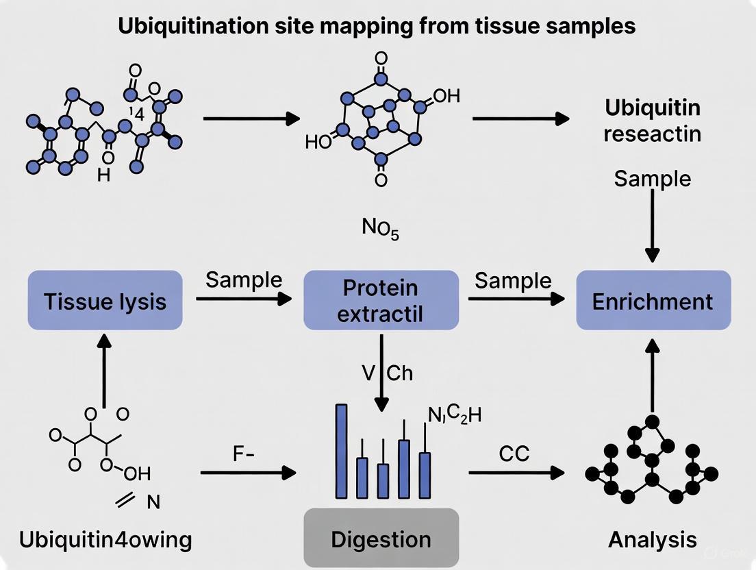
Visualizing the Ubiquitination Cascade and Workflows
The Ubiquitination and Deubiquitination Cycle
Ubiquitinated Protein Sample Preparation Workflow
Ubiquitin is a small, 76-amino acid regulatory protein that is ubiquitously expressed in eukaryotic cells and serves as a critical post-translational modification (PTM) signal [5]. The covalent attachment of ubiquitin to substrate proteins—a process known as ubiquitination—regulates diverse fundamental cellular functions including protein degradation, activity, localization, and interaction networks [6] [7]. This modification versatility stems from the remarkable complexity of ubiquitin conjugates, which can range from a single ubiquitin monomer to polymers of varying length, linkage types, and architectures [6]. The ubiquitin system employs a hierarchical enzymatic cascade consisting of E1 activating enzymes, E2 conjugating enzymes, and E3 ligases to orchestrate the specific attachment of ubiquitin to target proteins [5] [8]. This system is counterbalanced by deubiquitinases (DUBs) that remove ubiquitin modifications, allowing for dynamic regulation of protein fate and function [6] [7].
Understanding the complexity of ubiquitin modifications is particularly crucial when working with tissue samples, where preserving the native ubiquitination landscape presents unique challenges compared to cell culture models. The stoichiometry of protein ubiquitination is typically low under normal physiological conditions, and the dynamic nature of these modifications requires careful sample handling to prevent artifactual changes during preparation [6]. Furthermore, the substantial heterogeneity of tissue samples, containing multiple cell types with distinct ubiquitination profiles, adds another layer of complexity to the analysis of ubiquitination sites from clinical specimens.
The Ubiquitin Code: Types and Functions
Monoubiquitination and Multi-Monoubiquitination
Monoubiquitination occurs when a single ubiquitin molecule is covalently attached to a substrate protein, typically on a lysine residue [6] [5]. This modification can alter protein localization, activity, and interactions without targeting the substrate for proteasomal degradation [9]. Multi-monoubiquitination describes the attachment of single ubiquitin molecules to multiple lysine residues on the same substrate protein, creating a ubiquitination pattern that can initiate specific signaling outcomes distinct from polyubiquitin chains [6].
Polyubiquitin Chains: Linkages and Functions
Polyubiquitin chains form when the C-terminus of additional ubiquitin molecules conjugates to specific lysine residues or the N-terminal methionine of the previously attached ubiquitin [6] [5]. Ubiquitin contains seven lysine residues (K6, K11, K27, K29, K33, K48, K63) and one N-terminal methionine (M1) that can serve as linkage sites, generating eight possible homotypic chain types [6] [9]. Each linkage type creates a distinct structural topology that is recognized by specific effector proteins, leading to different functional consequences for the modified substrate [9].
Table 1: Ubiquitin Chain Linkages and Their Primary Functions
| Linkage Type | Known Functions | Structural Features |
|---|---|---|
| K48-linked | Major signal for proteasomal degradation [6] [5] | Most abundant linkage in cells [6] |
| K63-linked | Non-proteolytic signaling (NF-κB pathway, kinase activation, DNA repair) [6] | Distinguished from K48 linkages [9] |
| M1-linked (Linear) | Inflammatory signaling, NF-κB activation [7] | Unique N-terminal linkage [9] |
| K6-linked | DNA damage response, mitochondrial homeostasis [6] | Atypical chain, less characterized [6] |
| K11-linked | Cell cycle regulation, ER-associated degradation [9] | Atypical chain with specialized functions [6] |
| K27-linked | Mitophagy, innate immune signaling [6] | Atypical chain [6] |
| K29-linked | Proteasomal degradation (non-canonical), Wnt signaling [6] [9] | Atypical chain [6] |
| K33-linked | Kinase regulation, endosomal sorting [6] | Atypical chain [6] |
Beyond homotypic chains, ubiquitin can form heterotypic chains (mixed linkages) and branched chains (multiple linkages on a single ubiquitin molecule), further expanding the coding potential of ubiquitin signaling [6] [9]. This complex "ubiquitin code" allows for precise regulation of cellular processes through specialized "writer" (E3 ligases), "editor" (DUBs), and "reader" (ubiquitin-binding domains) proteins that create, modify, and interpret these modifications, respectively [8].
Methodological Approaches for Ubiquitination Analysis in Tissues
Enrichment Strategies for Ubiquitinated Proteins
The low stoichiometry of ubiquitination necessitates efficient enrichment strategies prior to mass spectrometry analysis, particularly for tissue samples where material may be limited [6]. Several approaches have been developed to isolate ubiquitinated proteins or peptides from complex mixtures.
Ubiquitin Tagging-Based Approaches utilize genetically engineered ubiquitin containing affinity tags (e.g., His, Strep, HA) for purification of ubiquitinated substrates [6]. After expressing tagged ubiquitin in biological systems, ubiquitinated proteins can be enriched using affinity resins such as Ni-NTA for His tags or Strep-Tactin for Strep tags [6]. While this approach is relatively low-cost and straightforward, it has limitations for tissue research as it requires genetic manipulation and may not fully replicate endogenous ubiquitin behavior [6].
Antibody-Based Enrichment employs anti-ubiquitin antibodies to isolate ubiquitinated proteins or peptides from native tissue samples without genetic manipulation [6]. Pan-specific ubiquitin antibodies (e.g., P4D1, FK1/FK2) recognize all ubiquitin linkages, while linkage-specific antibodies selectively enrich for particular chain types (M1-, K11-, K27-, K48-, K63-linkage specific antibodies) [6]. This approach preserves endogenous ubiquitination patterns but can be limited by antibody cost, availability, and potential non-specific binding [6].
Ubiquitin-Binding Domain (UBD)-Based Approaches exploit natural ubiquitin receptors containing UBDs to capture ubiquitinated proteins [6]. Single UBDs typically have low affinity for ubiquitin, so tandem-repeated UBDs are often used to enhance binding avidity [6]. This method can provide linkage selectivity based on the inherent preferences of specific UBDs and maintains endogenous modification patterns.
Table 2: Comparison of Ubiquitin Enrichment Methods for Tissue Research
| Method | Principle | Advantages | Limitations for Tissue Research |
|---|---|---|---|
| Ubiquitin Tagging | Affinity-tagged ubiquitin expression | High purity, relatively low cost | Requires genetic manipulation, may not mimic endogenous ubiquitin |
| Antibody-Based | Immunoaffinity with anti-ubiquitin antibodies | Preserves endogenous patterns, works on native tissue | High cost, potential non-specific binding, batch variability |
| UBD-Based | Affinity capture with ubiquitin-binding domains | Linkage selectivity possible, preserves endogenous patterns | Optimization required for specificity and affinity |
| DiGly Immunoprecipitation | Anti-K-ε-GG antibody capture of tryptic peptides | Site-specific identification, high sensitivity | Requires efficient digestion, misses non-lysine ubiquitination |
Mass Spectrometry-Based Ubiquitinomics
Advanced mass spectrometry techniques have revolutionized the study of ubiquitination, enabling system-wide identification of ubiquitination sites and linkage types [6] [10]. Data-independent acquisition (DIA) mass spectrometry has emerged as a powerful approach for comprehensive ubiquitinome profiling, as demonstrated in recent high-throughput studies of ubiquitin ligase function [10]. This method provides highly reproducible quantification across many samples and deep proteome coverage—quantifying over 10,000 protein groups from limited material with median coefficients of variation below 6% in recent applications [10].
For ubiquitination site identification, trypsin digestion of ubiquitinated proteins generates a characteristic di-glycine remnant on modified lysine residues, which produces a 114.04 Da mass shift detectable by MS and allows discrimination from unmodified peptides [6] [5]. This "di-glycine signature" enables specific identification of ubiquitination sites when combined with anti-K-ε-GG antibody enrichment [5].
Global ubiquitinomics workflows can capture dynamic ubiquitination events by employing short treatment times (as brief as 30 minutes) without proteasome inhibition, allowing observation of ubiquitination dynamics under near-physiological conditions [10]. This approach has confirmed degrader-induced ubiquitination of both known and novel substrates in tissue-relevant models [10].
Protocols for Ubiquitination Site Mapping from Tissue Samples
Tissue Collection and Lysis for Ubiquitination Studies
Proper tissue collection and lysis are critical for preserving the native ubiquitination state, as the ubiquitin system remains active post-collection. Rapid processing is essential—flash-freeze tissue specimens in liquid nitrogen within minutes of excision to prevent artifactual changes in ubiquitination [10]. For lysis, use denaturing conditions (e.g., 8 M urea, 2% SDS) in the presence of protease inhibitors and N-ethylmaleimide (NEM) to irreversibly inhibit DUBs and preserve ubiquitin conjugates [6] [10]. Maintain samples at low temperatures (4°C or below) during all processing steps. For tissue heterogeneity concerns, consider laser capture microdissection to isolate specific cell populations before lysis, particularly when studying tumor microenvironments where different cell types may exhibit distinct ubiquitination profiles [3].
Enrichment of Ubiquitinated Proteins from Tissue Lysates
Protocol for Antibody-Based Enrichment of Ubiquitinated Proteins:
- Protein Extraction and Digestion: Extract proteins under denaturing conditions. For ubiquitination site identification, digest proteins with trypsin to generate peptides with di-glycine remnants on ubiquitinated lysines [6] [5].
- Peptide Clean-up: Desalt peptides using C18 solid-phase extraction columns.
- Immunoaffinity Enrichment: Incubate peptides with anti-K-ε-GG antibody-conjugated beads for 2 hours at room temperature with gentle agitation [10].
- Washing: Wash beads extensively with ice-cold PBS to remove non-specifically bound peptides.
- Elution: Elute bound peptides with 0.1% trifluoroacetic acid.
- Clean-up for MS: Desalt eluted peptides using StageTips or similar micro-solid-phase extraction methods.
Protocol for Ubiquitin-Binding Domain Enrichment:
- Immobilization of Tandem UBDs: Couple recombinant tandem UBD proteins (e.g., tandem ubiquitin-interacting motifs) to affinity resin.
- Binding: Incubate tissue lysates with UBD-resin for 1-2 hours at 4°C.
- Washing: Wash with mild detergent-containing buffer to reduce non-specific binding.
- Elution: Elute with SDS-PAGE loading buffer or competitive elution with free ubiquitin.
Mass Spectrometry Analysis and Data Processing
Liquid Chromatography and Mass Spectrometry Parameters:
- Use nano-flow liquid chromatography systems coupled to high-resolution mass spectrometers (Q-Exactive, Orbitrap Fusion series, or timsTOF platforms) [10].
- For DIA methods, implement 2-4 m/z precursor isolation windows covering 400-1000 m/z range [10].
- Employ stepped collision energy (25-35 eV) for improved fragmentation.
- Use 90-120 minute linear gradients for deep proteome coverage.
Data Analysis Workflow:
- Database Search: Search MS data against appropriate protein databases using software such as MaxQuant, Spectronaut, or DIA-NN, enabling the "di-glycine (K)" modification for ubiquitination site identification [10].
- False Discovery Control: Apply 1% false discovery rate (FDR) thresholds at both protein and peptide levels.
- Quantification: Use label-free quantification algorithms for relative quantification across samples.
- Site Localization: Apply localization probability thresholds (>0.75) for confident ubiquitination site assignment.
The Scientist's Toolkit: Essential Reagents and Materials
Table 3: Key Research Reagents for Ubiquitination Studies in Tissues
| Reagent/Material | Function | Application Notes |
|---|---|---|
| Anti-K-ε-GG Antibody | Immunoaffinity enrichment of ubiquitinated peptides | Critical for site identification; validate lot-to-lot consistency |
| Linkage-Specific Ub Antibodies | Selective enrichment of specific polyubiquitin chains | K48, K63, M1 antibodies most characterized; check linkage specificity |
| N-Ethylmaleimide (NEM) | Deubiquitinase inhibitor | Essential in lysis buffers to preserve ubiquitin conjugates |
| Ubiquitin-Activating Enzyme (E1) Inhibitor | Blocks ubiquitination cascade | Prevents post-lysis ubiquitination artifacts |
| MLN4924 | NEDD8-activating enzyme inhibitor | Blocks cullin-RING ligase activity; validates CRL-dependent ubiquitination [10] |
| Recombinant Tandem-UBD Proteins | Affinity capture of ubiquitinated proteins | Can provide linkage selectivity; requires optimization |
| DiGly Standard Peptides | Mass spectrometry quantification standards | AQUA peptides for absolute quantification of ubiquitination |
| Deubiquitinase Inhibitors | Broad-spectrum DUB inhibition | Cocktails recommended to target multiple DUB families |
The complexity of ubiquitin modifications—from monoubiquitination to diverse polyubiquitin chains with distinct functions—presents both challenges and opportunities for researchers studying tissue samples. Successful mapping of ubiquitination sites from tissue specimens requires careful attention to sample preservation, appropriate enrichment strategies, and advanced mass spectrometry techniques. The continued development of improved affinity reagents, mass spectrometry methods, and bioinformatic tools will further enhance our ability to decipher the ubiquitin code in physiological and pathological contexts. As research in this field advances, understanding tissue-specific ubiquitination patterns promises to reveal new insights into disease mechanisms and potential therapeutic interventions, particularly in cancer and neurodegenerative disorders where ubiquitin signaling is frequently disrupted [3] [7].
Why Tissue Samples Present Unique Challenges for Ubiquitinome Analysis
Ubiquitinome analysis, the large-scale study of protein ubiquitination, is a powerful tool for understanding cellular regulation, protein degradation, and signaling pathways in physiological and disease contexts. While cell lines provide valuable model systems, tissue samples offer unparalleled biological relevance by preserving the native tissue architecture, cellular heterogeneity, and pathophysiological environment of disease states. However, this biological complexity introduces substantial technical challenges for ubiquitination site mapping that are less pronounced in cultured cell models. This application note examines the unique obstacles presented by tissue samples in ubiquitinome analysis and provides detailed methodologies to address these challenges within the broader context of sample preparation for ubiquitination research.
Unique Challenges in Tissue-Based Ubiquitinome Analysis
Tissue samples present a constellation of challenges that differentiate them from cell culture models and complicate every stage of ubiquitinome analysis, from sample preparation to data interpretation. The table below summarizes these key challenges and their specific impacts on ubiquitination analysis.
Table 1: Key Challenges of Ubiquitinome Analysis in Tissue Samples
| Challenge Category | Specific Issues in Tissues | Impact on Ubiquitination Analysis |
|---|---|---|
| Cellular Heterogeneity | Mixed cell types with different ubiquitination profiles; variable tumor/stroma/immune cell ratios [11] | Masks cell-type-specific ubiquitination events; averages signaling patterns across distinct cellular compartments |
| Sample Availability & Quality | Limited quantities from biopsies; post-surgical ischemia; variable degradation rates during collection [12] | Reduces ubiquitinated peptide yield for MS detection; introduces artifactual ubiquitination changes from hypoxia/stress |
| Analytical Sensitivity | Low abundance of ubiquitinated proteins amidst complex tissue proteome; ~0.1-1% of total cellular proteins | Requires highly efficient enrichment to detect low-abundance ubiquitination events against high background |
| Protein Extraction Complexity | Abundant structural proteins (collagens); lipid-rich membranes; extensive protein-protein interactions [13] | Incomplete protein solubilization biases against certain ubiquitinated proteins; co-precipitation of non-targeted proteins |
| Pathway Interpretation | Convoluted signaling inputs from multiple cell types; diverse metabolic states within tissue microenvironments | Difficult to attribute ubiquitination changes to specific pathways or cell types without additional validation |
The post-surgical ischemia inherent to tissue collection presents a particularly critical challenge. The rapid hypoxia and metabolic stress following resection can trigger substantial changes in ubiquitination patterns within minutes, potentially obscuring the physiological ubiquitinome with stress-induced artifacts [12]. Furthermore, the cellular heterogeneity of tissues means that ubiquitination signatures obtained from bulk analysis represent averaged patterns across multiple cell types, potentially masking cell-specific regulatory events that could be crucial for understanding disease mechanisms [11].
Essential Methodologies for Tissue Ubiquitinome Analysis
Tissue-Specific Protein Extraction and Digestion
Effective protein extraction from tissues requires more aggressive methods than those used for cell lines. The SDS-cyclodextrin-assisted sample preparation (SCASP) protocol has been adapted for tissues to enhance protein recovery while maintaining ubiquitination integrity [13].
Detailed Protocol: Tissue Protein Extraction and Digestion using SCASP-PTM
Tissue Homogenization:
- Flash-freeze tissue samples in liquid nitrogen immediately after resection.
- Cryopulverize tissue using a pre-cooled mortar and pestle or a specialized tissue pulverizer under continuous liquid nitrogen cooling.
- Suspend powdered tissue in SDS-containing lysis buffer (e.g., 2% SDS, 50 mM Tris-HCl pH 8.0, 10 mM TCEP, 40 mM CAA) supplemented with 1% cyclodextrin.
- Homogenize using a high-power probe sonicator with 3-5 cycles of 15-second pulses at 30% amplitude, with 30-second cooling intervals on ice.
Protein Clean-up and Digestion:
- Add 5 volumes of cold acetone and incubate at -20°C for 4 hours to precipitate proteins.
- Centrifuge at 15,000 × g for 15 minutes at 4°C and discard supernatant.
- Wash pellet twice with cold 90% acetone and air-dry for 5 minutes.
- Resuspend protein pellet in 50 mM ammonium bicarbonate buffer containing 0.1% SDS and 2% cyclodextrin.
- Digest with trypsin (1:50 enzyme-to-protein ratio) overnight at 37°C with agitation.
Peptide Clean-up:
- Acidify digested peptides with 1% trifluoroacetic acid (TFA) to pH < 3.
- Desalt using C18 solid-phase extraction columns according to manufacturer's instructions.
- Lyophilize and store at -80°C until enrichment.
Enrichment of Ubiquitinated Peptides
The critical step for ubiquitinome analysis is the specific enrichment of ubiquitinated peptides from complex tissue digests. The two primary methods are antibody-based enrichment and affinity-based approaches.
A. Anti-K-ε-GG Antibody Enrichment
This method uses antibodies specifically recognizing the di-glycine (GG) remnant left on lysine residues after tryptic digestion of ubiquitinated proteins [11].
- Procedure: Reconstitute desalted tissue peptides in immunoaffinity purification (IAP) buffer (50 mM MOPS pH 7.2, 10 mM Na₂HPO₄, 50 mM NaCl). Incubate with anti-K-ε-GG antibody-coupled beads for 2 hours at 4°C with gentle rotation. Wash beads sequentially with IAP buffer and then with water. Elute ubiquitinated peptides with 0.1% TFA [11].
B. Tandem Ubiquitin Binding Entities (TUBEs) for Tissue Applications
TUBEs, which are engineered proteins with high affinity for polyubiquitin chains, can be applied to tissue lysates before digestion to protect ubiquitinated proteins from deubiquitinating enzymes (DUBs) and proteasomal degradation during extraction [14].
- Procedure: Add chain-specific or pan-specific TUBEs (e.g., K48-TUBE, K63-TUBE) directly to tissue homogenization buffer. Incubate for 30 minutes at 4°C. Capture TUBE-ubiquitin complexes using TUBE-binding magnetic beads. Wash complexes and then elute ubiquitinated proteins using SDS-PAGE loading buffer or directly digest bead-bound proteins [14].
Diagram 1: Tissue ubiquitinome analysis workflow.
Mass Spectrometry Analysis and Data Interpretation
For tissue-derived ubiquitinated peptides, Data-Independent Acquisition (DIA) mass spectrometry is particularly advantageous as it provides comprehensive recording of all fragment ions, reducing missing data across multiple tissue samples [10].
LC-MS/MS Parameters: Use a nano-flow UHPLC system coupled to a high-resolution tandem mass spectrometer. Peptides are separated on a C18 column (75 µm × 25 cm) with a 90-minute gradient from 2% to 30% acetonitrile in 0.1% formic acid. DIA methods should include a survey scan followed by 20-40 variable-width DIA windows covering the m/z range 400-1000.
Data Analysis: Process DIA data using spectral library-based tools (DIA-NN, Spectronaut) against a protein sequence database. Ubiquitination sites are identified by searching for the GG remnant (K-ε-GG, +114.042 Da mass shift) on lysine residues. Site localization should be validated using a localization probability score (> 0.75) [10].
The Scientist's Toolkit: Key Research Reagents
Successful ubiquitinome analysis from tissues requires a specialized set of reagents to address the unique challenges outlined. The table below details essential materials and their specific functions in the experimental workflow.
Table 2: Essential Research Reagents for Tissue Ubiquitinome Analysis
| Reagent/Category | Specific Examples | Function in Tissue Ubiquitinome Analysis |
|---|---|---|
| Lysis & Stabilization | SDS-cyclodextrin buffer [13]; DUB inhibitors (N-ethylmaleimide); Proteasome inhibitor (MG-132) [12] | Efficient tissue disruption and protein solubilization; prevents loss of ubiquitination during sample preparation |
| Enrichment Reagents | Anti-K-ε-GG antibody [11]; Chain-specific TUBEs (K48, K63) [14]; Pan-selective TUBEs | Selective isolation of ubiquitinated peptides or proteins; enables linkage-specific ubiquitination analysis |
| Digestion & Clean-up | Sequencing-grade trypsin; C18 solid-phase extraction cartridges; Cyclodextrin additives [13] | Efficient protein digestion; removal of detergents and contaminants that interfere with MS analysis |
| Mass Spectrometry | Data-Independent Acquisition (DIA) platforms [10]; TMT/Isobaric tags for multiplexing | Comprehensive, reproducible quantification of ubiquitinated peptides across multiple tissue samples |
| Validation Reagents | Linkage-specific ubiquitin antibodies; siRNA for candidate targets; Immunoprecipitation-grade antibodies | Confirmation of ubiquitination status and biological relevance of identified targets |
Signaling Pathways and Functional Implications in Tissue Environments
Ubiquitination regulates critical signaling pathways that are often altered in disease states studied using tissue samples, such as cancer and inflammatory conditions. Mapping these pathways in tissues reveals how ubiquitination controls cellular processes within their native context.
Diagram 2: Ubiquitin linkage-specific signaling pathways.
The K63-linked ubiquitination of RIPK2, induced by inflammatory stimuli like L18-MDP, serves as a critical signaling scaffold that activates the NF-κB pathway and promotes inflammatory cytokine production [14]. In contrast, K48-linked ubiquitination, such as that induced by PROTAC degraders, targets proteins for proteasomal degradation, resulting in signaling ablation [14]. These distinct functional outcomes underscore the importance of linkage-specific analysis in understanding ubiquitin signaling in tissue environments.
Tissue samples present a unique set of challenges for ubiquitinome analysis, stemming primarily from their cellular heterogeneity, sample stability issues, and analytical complexity. However, through implementation of robust tissue-specific protocols—including rapid stabilization, efficient protein extraction using methods like SCASP, and highly specific enrichment techniques—researchers can successfully overcome these hurdles. The ability to accurately map ubiquitination sites in tissue environments provides crucial insights into disease mechanisms and enables the development of targeted therapies that exploit the ubiquitin-proteasome system, particularly through emerging modalities like PROTACs and molecular glue degraders. As mass spectrometry technologies continue to advance, tissue-based ubiquitinome analysis will play an increasingly vital role in translating our understanding of ubiquitin biology into clinical applications.
Protein ubiquitination, the covalent attachment of ubiquitin to lysine residues on target proteins, represents a crucial regulatory mechanism governing protein stability, activity, and localization [15]. Mapping ubiquitination sites from tissue samples presents unique analytical challenges that must be addressed through optimized sample preparation protocols. The dynamic nature of this modification, combined with its characteristically low stoichiometry and inherent heterogeneity of modification sites, demands stringent preservation and enrichment strategies to ensure reliable detection [16] [17] [15]. This application note details standardized protocols designed to address these challenges specifically for tissue-based research, enabling researchers to obtain high-quality data for both discovery-phase and targeted ubiquitination analyses.
The foundation of any successful ubiquitination mapping experiment lies in the initial sample handling phases. Inadequate preservation can lead to rapid erasure of native ubiquitination states through the action of endogenous deubiquitinating enzymes (DUBs), while suboptimal processing can introduce artifacts that compromise data validity [16]. The following sections provide detailed methodologies for maintaining ubiquitin modification integrity from tissue collection through to mass spectrometric analysis.
Critical Challenges in Ubiquitination Analysis
Low Stoichiometry of Modification
The low stoichiometry of individual ubiquitinated species presents a fundamental detection challenge. Modified variants often constitute merely 1–5% of the total protein population, requiring significant enrichment to detect against background signals [16]. This issue is particularly acute in tissue samples, where starting material may be limited and cellular heterogeneity further dilutes modification signals. Without appropriate enrichment strategies, low-abundance ubiquitination events are easily obscured by more abundant unmodified peptides during mass spectrometric analysis [16] [15].
Dynamic Nature and Sample Preservation
Ubiquitination states are highly dynamic and can change rapidly in response to cellular conditions, including the ischemia that inevitably occurs during tissue collection [16] [18]. Enzymes such as deubiquitinases and isopeptidases remain active post-tissue excision and can rapidly erase modification signatures if not promptly inactivated [16]. The structural integrity of tissue biomolecules is also vulnerable; prolonged post-mortem intervals can lead to breakdown of biomolecular networks, reducing their density and detectability [18]. These preservation challenges are compounded in tissue research by practical constraints of surgical collection or post-mortem intervals.
Heterogeneity of Modification Sites
Ubiquitination exhibits complexity at multiple levels: a single protein may be modified at multiple lysine residues simultaneously, and ubiquitin itself can form polymers with different linkage types (K48, K63, etc.) that dictate functional outcomes [17] [15]. This heterogeneity creates analytical challenges in distinguishing between biologically relevant patterns and stochastic modification events. Furthermore, tissue samples inherently contain multiple cell types, each with potentially distinct ubiquitination profiles, adding another layer of complexity to data interpretation [15].
Table 1: Key Challenges in Tissue Ubiquitination Analysis
| Challenge | Impact on Analysis | Tissue-Specific Considerations |
|---|---|---|
| Low Stoichiometry | Modified species diluted by unmodified counterparts; detection sensitivity limited | Tissue heterogeneity further dilutes signal; material often limited |
| Rapid Demodification | Native ubiquitination state altered before fixation | Post-mortem intervals or surgical ischemia activate DUBs |
| Structural Heterogeneity | Multiple modification sites and chain types complicate analysis | Cellular diversity in tissues creates complex modification patterns |
| Sample Complexity | Ubiquitinated peptides masked by abundant unmodified proteins | Tissue extracts contain high concentrations of structural proteins |
Sample Collection and Preservation Protocols
Rapid Tissue Processing Guidelines
Immediate stabilization of ubiquitination states is critical upon tissue collection. The following protocol is optimized to preserve in vivo ubiquitination patterns:
- Collection: Use pre-chilled instruments to excise tissue and immediately rinse with ice-cold, neutral pH buffer (e.g., PBS) to remove contaminants [16].
- Preservation: Flash-freeze tissue fragments in liquid nitrogen within minutes of excision. For larger specimens (<5 mm thickness), subdivide to ensure rapid penetration of cold [16] [19].
- Storage: Maintain continuous storage at -80°C. Avoid -20°C storage, which permits ongoing enzymatic degradation [16].
- Documentation: Record post-mortem interval or ischemia time precisely, as this critically impacts preservation quality [18].
Tissue samples intended for ubiquitination analysis require greater mass than standard proteomic preparations due to low modification abundance. Recommended starting amounts are >500 mg of animal tissue to ensure sufficient material for subsequent enrichment steps [16].
Inhibition of Demodifying Enzymes
Cellular lysis during extraction liberates endogenous deubiquitinating enzymes (DUBs) that must be immediately inactivated to preserve ubiquitination signatures:
- Protease Inhibition: Add broad-spectrum protease inhibitor cocktail to all lysis buffers immediately before use [16].
- DUB Inhibition: Incorporate 5–10 mM N-ethylmaleimide (NEM) or iodoacetamide (IAA) along with EDTA/EGTA to inhibit deubiquitinating enzymes [16].
- Operational Conditions: Perform all extraction steps on ice or at 4°C to minimize enzymatic activity [16].
- Mechanical Processing: Avoid vigorous homogenization that causes excessive heating or shearing; use controlled mechanical disruptors with cooling [16].
Histone and Protein Extraction Methods
Acid Extraction Protocol for Histones
Histone extraction from tissue requires special consideration for nuclear isolation prior to acid extraction. This protocol is adapted for ubiquitination analysis:
- Tissue Disruption: Cryogenically grind flash-frozen tissue under liquid nitrogen using mortar and pestle or cryomill [16].
- Cell Lysis: Resuspend powdered tissue in NETN lysis buffer (20 mM Tris pH 8.0, 500 mM NaCl, 0.5% NP-40, 1 mM EDTA) supplemented with fresh protease inhibitors and DUB inhibitors. Perform lysis on ice for 15 minutes with gentle agitation [16].
- Nuclear Isolation: Centrifuge lysate at 1,500 × g, 4°C for 10 minutes. Discard supernatant and wash insoluble pellet (containing nuclei) 1–2 times with NETN buffer [16].
- Acid Extraction: Add 0.2 M HCl to nuclear pellet (approximately 5× pellet volume). Lyse nuclei by vigorous vortexing and incubate in ice-water bath for 30 minutes with occasional mixing [16].
- Clarification and Neutralization: Centrifuge at 12,000 × g, 4°C for 15 minutes. Transfer supernatant to new tube and neutralize with 1 M Tris (pH 8.0) using approximately 1:5 ratio of Tris to acid extract. Check pH indicator color change from yellow to blue [16].
- Concentration Determination: Quantify histone concentration using Bradford assay (UV absorption is unreliable due to histone absence of tryptophan). Aliquot and store at -80°C [16].
Comparison of Extraction Methods
Table 2: Comparison of Protein Extraction Methods for Ubiquitination Studies
| Method | Principle | Advantages | Disadvantages | PTM Preservation |
|---|---|---|---|---|
| Acid Extraction | Exploits high histone solubility in strong acid | High purity; excellent PTM preservation | Multiple steps; time-consuming | Excellent |
| High-Ionic-Strength Salt Extraction | Disrupts electrostatic interactions between histones and DNA | Straightforward protocol; avoids strong acids | Requires desalting; lower purity; salt interference | Good |
| Commercial Kit | Optimized proprietary buffer systems | Standardized; high consistency; user-friendly | Higher cost; proprietary formulations | Excellent |
| RIPA Lysis (Total Protein) | Detergent-based total protein extraction | Rapid and simple | Very low histone purity; detergents interfere | Poor |
Selection Criteria for Extraction Method
Choose extraction method based on research objectives:
- For PTM-focused studies: Acid extraction or high-quality commercial kits provide optimal ubiquitination preservation [16].
- For downstream mass spectrometry: High purity with minimal contaminants is essential; acid extraction is preferred [16] [20].
- When analyzing multiple PTMs: Commercial kits often provide balanced performance for various modifications [16].
- With limited tissue material: Scale-compatible methods (typically acid extraction or kits) should be selected [16].
Enrichment Strategies for Ubiquitinated Peptides
Immunoaffinity Enrichment Using K-ε-GG Antibodies
Peptide-level immunoaffinity enrichment specifically targets the diglycine (K-ε-GG) remnant left on ubiquitinated lysine residues after tryptic digestion. This method significantly enhances detection sensitivity for ubiquitination sites:
- Protein Digestion: Following extraction, digest proteins to peptides using trypsin. This cleaves ubiquitin, leaving the characteristic +114.0429 Da mass signature on modified lysines [21] [22].
- Antibody Immobilization: Chemically cross-link anti-K-ε-GG antibody to beads to create an immobilized enrichment resin [20].
- Peptide Enrichment: Incubate digested peptides with antibody-conjugated beads for 2–4 hours with gentle rotation [20] [21].
- Washing: Remove non-specifically bound peptides with multiple washes using ice-cold PBS or specialized wash buffers [20].
- Elution: Release enriched ubiquitinated peptides using low-pH elution conditions or competitive elution [20].
This approach has demonstrated greater than fourfold higher levels of modified peptide recovery compared to protein-level enrichment methods, making it particularly valuable for detecting low-stoichiometry ubiquitination events in complex tissue samples [21].
Alternative Enrichment Methods
- Ubiquitin-Binding Domains (UBDs): Tandem-repeated Ub-binding entities (TUBEs) exhibit enhanced affinity for ubiquitinated proteins and can protect ubiquitin conjugates from degradation and deubiquitination during extraction [15].
- Tagged Ubiquitin Systems: While primarily for cell-based studies, tagged ubiquitin systems (e.g., His-tagged Ub) enable purification under denaturing conditions, reducing co-purification of non-specifically bound proteins [15].
- Linkage-Specific Antibodies: For investigating specific ubiquitin chain types, linkage-specific antibodies (K48-, K63-specific, etc.) enable isolation of particular ubiquitin topological structures [15].
Mass Spectrometric Analysis and Data Interpretation
LC-MS/MS Configuration for Ubiquitination Site Mapping
Optimal mass spectrometry parameters for ubiquitinated peptide detection:
- Chromatography: Use reversed-phase nanoflow chromatography with extended gradients (120–180 minutes) for sufficient separation complexity [20] [22].
- MS Acquisition: Implement data-dependent acquisition with dynamic exclusion for comprehensive peptide sampling [20].
- Fragmentation: Employ higher-energy collisional dissociation (HCD) which preserves the K-ε-GG signature and enables localization of modification sites [22].
- Resolution: Utilize high-resolution mass analyzers (Orbitrap platforms) for accurate mass measurements and reliable identification [20] [22].
Quantitative Ubiquitination Profiling
For comparative studies investigating ubiquitination dynamics under different conditions:
- SILAC Labeling: Incorporate stable isotopes through metabolic labeling during cell culture before tissue collection [20] [22].
- Isobaric Tagging: Employ TMT or iTRAQ reagents for multiplexed analysis of multiple samples [22].
- Label-Free Quantification: Use spectral counting or extracted ion currents for studies where isotopic labeling is impractical [22].
Data Analysis and Validation
- Database Search: Process raw files through search engines (MaxQuant, Proteome Discoverer) configured to include ubiquitination (+114.0429 Da) as a variable modification on lysine [20] [22].
- Site Localization: Apply localization probability scoring (e.g., PTM-score) to confidently assign modification sites [22].
- False Discovery Control: Use target-decoy approaches with FDR threshold of <1% for site identifications [20].
- Manual Validation: Inspect MS/MS spectra for characteristic fragmentation patterns, including GG-immonium ion (m/z 112.0865) [21].
The Scientist's Toolkit: Essential Research Reagents
Table 3: Key Research Reagents for Ubiquitination Site Mapping
| Reagent/Category | Specific Examples | Function and Application |
|---|---|---|
| DUB Inhibitors | N-ethylmaleimide (NEM), Iodoacetamide | Preserve ubiquitination state by inhibiting deubiquitinating enzymes |
| Protease Inhibitors | PMSF, Aprotinin, Leupeptin, Pepstatin A | Prevent general protein degradation during extraction |
| Enrichment Antibodies | Anti-K-ε-GG, Linkage-specific anti-Ub antibodies | Immunoaffinity enrichment of ubiquitinated peptides |
| Tagged Ubiquitin Systems | His-Ub, Strep-Ub, HA-Ub | Expression systems for affinity-based purification |
| Affinity Resins | Ni-NTA (His-tag), Strep-Tactin (Strep-tag) | Purification of tagged ubiquitin conjugates |
| Activity-Based Probes | Ubiquitin-based chemical probes | Detection and enrichment of active deubiquitinating enzymes |
| Mass Spec Standards | Stable isotope-labeled ubiquitinated peptides | Quantification and instrument calibration |
Workflow Visualization
Tissue Ubiquitination Analysis Workflow
Ubiquitination Complexity and Analysis Challenge
Concluding Remarks
Successful ubiquitination site mapping from tissue samples requires meticulous attention to each step of the workflow, with particular emphasis on the intersecting challenges of low stoichiometry, sample preservation, and modification heterogeneity. The protocols detailed in this application note provide a standardized framework for maintaining ubiquitination integrity throughout processing, significantly enhancing detection sensitivity and reliability. By implementing these methods—from rapid tissue preservation to targeted enrichment strategies—researchers can overcome the inherent analytical hurdles and generate high-quality ubiquitination data from complex tissue samples. These approaches enable more accurate profiling of ubiquitination dynamics in physiological and pathological contexts, supporting advancements in both basic research and drug development initiatives targeting the ubiquitin-proteasome system.
Practical Workflows for Ubiquitinated Peptide Enrichment from Tissue Lysates
Tissue Lysis and Protein Extraction Under Denaturing Conditions with DUB Inhibitors
The accurate mapping of ubiquitination sites from tissue samples is a cornerstone of proteomic research, directly influencing our understanding of cellular regulation, protein degradation, and signaling pathways. The success of these analyses is critically dependent on the initial sample preparation steps, particularly tissue lysis and protein extraction. The labile nature of ubiquitin modifications necessitates the use of stringent denaturing conditions and potent deubiquitinase (DUB) inhibitors during this phase to preserve the native ubiquitinome. This application note provides a detailed, optimized protocol for these critical steps, framed within the broader context of a thesis on sample preparation for ubiquitination site mapping from tissue research. The methodologies outlined are designed to ensure the integrity of post-translational modifications (PTMs) for subsequent enrichment and mass spectrometric analysis, such as the SCASP-PTM approach designed for tandem PTM enrichment [13].
The Critical Role of DUB Inhibition in Ubiquitinome Preservation
Deubiquitinating enzymes are a large family of proteases that rapidly remove ubiquitin from modified proteins, thereby dynamically opposing the action of E3 ubiquitin ligases [23]. During the process of tissue disruption and lysis, cellular compartmentalization is lost, releasing active DUBs that can artificially erase ubiquitin signals before they can be captured for analysis. Members of the Ubiquitin-Specific Peptidase (USP) family, such as USP17LA, are significantly upregulated during cellular stimulation and play pivotal roles in regulatory pathways, underscoring the abundance and activity of these enzymes in biological systems [24]. Therefore, the inclusion of broad-spectrum DUB inhibitors in the lysis buffer is not optional but mandatory for faithful ubiquitinome analysis. Failure to do so results in significant and irreversible loss of ubiquitination events, compromising all downstream experiments.
Comprehensive Reagents and Equipment
Research Reagent Solutions
The following table details the essential reagents and materials required for the successful execution of this protocol.
Table 1: Essential Research Reagents and Materials
| Item Name | Function/Explanation |
|---|---|
| Broad-Spectrum DUB Inhibitor (e.g., PR-619) | A cell-permeable, broad-spectrum DUB inhibitor that targets a wide range of cysteine-dependent DUBs. It is crucial for stabilizing ubiquitin conjugates during and after cell lysis by preventing deubiquitination. |
| Ubi-Tagging Enzymes (E1, E2–E3) | Recombinant enzymes (E1, E2–E3 fusion proteins) that facilitate site-directed multivalent conjugation of antibodies to ubiquitinated payloads. This modular technique, "ubi-tagging," allows for efficient generation of defined conjugates [23]. |
| Denaturing Lysis Buffer | A buffer containing strong denaturants (e.g., 1-2% SDS) that instantly inactivates proteases and DUBs by disrupting protein tertiary structure. This is the primary mechanism for preserving the native state of ubiquitinated proteins. |
| SCASP-PTM Reagents | Reagents for SDS-cyclodextrin-assisted sample preparation, which is compatible with downstream tandem enrichment of ubiquitinated, phosphorylated, and glycosylated peptides from a single sample [13]. |
| Protein G Affinity Resin | Used for the purification of antibody conjugates, such as ubi-tagged Fab fragments, post-conjugation reaction to isolate specific ubiquitinated proteins of interest [23]. |
The following table consolidates key quantitative parameters from relevant literature to guide the optimization of experimental conditions.
Table 2: Key Quantitative Parameters for Ubiquitination Workflows
| Parameter | Value / Condition | Context / Purpose |
|---|---|---|
| Ubi-tagging Reaction Time | 30 minutes | Complete consumption of starting material (e.g., Fab-Ub(K48R)don) and formation of fluorescently labelled Fab' conjugate is observed within this short timeframe [23]. |
| Ubi-tagging Conversion Efficiency | 93 - 96% | Average efficiency for reactions involving ubi-tagged antibodies, demonstrating the high yield of the conjugation process [23]. |
| Protein Stability (Tm) | ~75°C | The thermal unfolding profile of both conjugated and unconjugated Fab-Ub(K48R)don, indicating that the ubi-tagging process does not compromise protein thermostability [23]. |
| Ubiquitinated Peptide Enrichment | Serial, without intermediate desalting | The SCASP-PTM protocol allows for the tandem enrichment of ubiquitinated, phosphorylated, and glycosylated peptides from one sample in a serial manner, streamlining the workflow [13]. |
Detailed Experimental Protocol
Tissue Lysis and Protein Extraction Under Denaturing Conditions
This protocol is designed for ~50 mg of snap-frozen tissue.
- Pre-cool Equipment: Pre-cool a mechanical homogenizer (e.g., bead mill or rotor-stator) and a microcentrifuge to 4°C.
- Prepare Denaturing Lysis Buffer: Prepare the following buffer fresh and pre-warm it to 95°C to prevent SDS precipitation.
- 1-2% (w/v) Sodium Dodecyl Sulfate (SDS)
- 50 mM Tris-HCl, pH 7.5
- 150 mM NaCl
- 10 mM EDTA (chelates metal ions, inhibiting some DUBs)
- 5 mM N-Ethylmaleimide (NEM) or 10 mM Iodoacetamide (IAA) - alkylating agents for cysteine DUBs
- 1x Commercial Broad-Spectrum DUB Inhibitor Cocktail (e.g., PR-619)
- Note: Avoid urea-based buffers at this stage, as they do not instantly denature proteins like SDS and allow time for DUB activity.
- Rapid Tissue Homogenization:
- Transfer the frozen tissue to a pre-cooled tube containing lysis buffer and homogenization beads (e.g., ceramic beads).
- Add 500 µL of pre-warmed (95°C) denaturing lysis buffer per 50 mg of tissue.
- Immediately homogenize using the bead mill for 2 cycles of 45 seconds each, ensuring the sample is kept hot. Alternatively, for non-bead methods, homogenize directly in the pre-warmed buffer.
- Immediately after homogenization, incubate the lysate at 95°C for 10 minutes to ensure complete denaturation.
- Clear the Lysate:
- Centrifuge the heated lysate at 16,000 × g for 15 minutes at room temperature.
- Carefully transfer the supernatant (containing the solubilized proteins) to a new tube, avoiding the insoluble pellet.
- Protein Quantification and Alkylation:
- Quantify protein concentration using a compatible assay (e.g., BCA assay adapted for SDS).
- If not already included in the lysis buffer, alkylate cysteine residues by adding IAA to 10 mM and incubating in the dark for 30 minutes at room temperature. This step can be skipped if NEM/IAA was in the lysis buffer.
- Sample Preparation for MS:
- The sample is now ready for downstream processing, such as digestion and enrichment. For tandem PTM enrichment, follow protocols like SCASP-PTM, which is designed to handle SDS-lysed samples and enables serial enrichment of ubiquitinated peptides without intermediate desalting [13].
Validation Experiment: Confirming Ubiquitin Conjugate Preservation
To validate the efficacy of the lysis protocol, a conjugation reaction can be performed using ubi-tagging technology [23].
- Reaction Setup: In a final volume of 50 µL, combine:
- Purified protein or antibody fragment of interest (e.g., 10 µM Fab-Ub(K48R)don).
- Five-fold excess of acceptor ubi-tag (e.g., 50 µM Rho-Ubacc-ΔGG).
- Ubiquitination enzymes (0.25 µM E1, 20 µM E2–E3 fusion protein, e.g., gp78RING-Ube2g2 for K48 linkage).
- Appropriate reaction buffer.
- Incubation: Incubate the reaction mixture at 30°C for 30 minutes.
- Analysis:
- SDS-PAGE: Analyze the reaction products by SDS-PAGE. A successful conjugation will show a complete shift of the starting material to a higher molecular weight band, visible by Coomassie staining or fluorescence imaging.
- Purification: Purify the conjugate using Protein G affinity purification [23].
- Mass Spectrometry: Confirm the exact mass of the conjugate using ESI-TOF mass spectrometry.
Signaling Pathways and Experimental Workflows
DUB-Mediated Regulation of T-Cell Signaling
This diagram illustrates the role of a specific DUB, USP17LA, in regulating T-cell activation, highlighting the importance of DUBs in key signaling pathways relevant to disease and drug development [24].
Workflow for Ubiquitination Site Mapping from Tissue
This diagram outlines the complete end-to-end workflow for processing tissue samples to map ubiquitination sites, emphasizing the critical initial steps detailed in this protocol.
Concluding Remarks
The meticulous application of this protocol for tissue lysis and protein extraction under denaturing conditions with DUB inhibitors provides a solid foundation for reliable ubiquitinome mapping. The rapid and complete inactivation of DUBs is the single most critical factor in preserving the true biological state of ubiquitination. By integrating these robust initial steps with advanced downstream techniques like ubi-tagging for validation [23] and SCASP-PTM for tandem PTM enrichment [13], researchers can achieve a comprehensive and accurate picture of ubiquitin signaling in complex tissue samples, thereby directly supporting drug development and basic research in proteomics.
Protein ubiquitination is a fundamental post-translational modification (PTM) that regulates a vast array of cellular processes, including protein degradation, kinase activation, and DNA repair [6] [25]. The versatility of ubiquitin signaling arises from the complexity of ubiquitin conjugates, which can range from a single ubiquitin monomer to polymers of different lengths and linkage types [6]. Dysregulation of ubiquitination is implicated in numerous pathologies, such as cancer and neurodegenerative diseases, making its precise characterization a critical objective in biomedical research [6] [26].
A significant challenge in ubiquitin research is the low stoichiometry of modified proteins under physiological conditions, necessitating highly efficient enrichment strategies prior to mass spectrometry (MS) analysis [6]. This article provides detailed application notes and protocols for the three core enrichment methodologies—anti-diGly antibodies, tandem ubiquitin-binding entities (TUBEs), and affinity tags—with a specific focus on their application in mapping ubiquitination sites from tissue samples, a context particularly relevant for drug development professionals studying disease mechanisms.
Methodological Comparison and Selection Guide
The table below summarizes the key characteristics, advantages, and limitations of the three primary enrichment strategies, providing a basis for informed methodological selection.
Table 1: Core Ubiquitin Enrichment Strategies at a Glance
| Strategy | Principle | Best For | Throughput | Key Advantages | Key Limitations |
|---|---|---|---|---|---|
| Anti-diGly Antibodies [26] [27] | Immunoaffinity enrichment of tryptic peptides containing a diGly (GG) remnant on modified lysines. | High-throughput, site-specific mapping from complex tissues; large sample cohorts. | High (e.g., 96 samples/day with automation) [26] | High sensitivity and specificity for site identification; amenable to multiplexing (e.g., TMT) [26]. | Cannot detect non-lysine ubiquitination; cross-reacts with NEDD8/ISG15; expensive antibodies [28] [27]. |
| TUBEs (Tandem Ubiquitin-Binding Entities) [28] [27] | Recombinant proteins with multiple ubiquitin-binding domains (UBDs) for affinity purification of polyubiquitinated proteins. | Enriching endogenous polyubiquitinated proteins and studying ubiquitin chain topology. | Medium | Protects ubiquitin chains from deubiquitinases (DUBs); enriches endogenous proteins; linkage-specific TUBEs available [27]. | Poor affinity for monoubiquitinated proteins; may co-purify strong ubiquitin interactors [28] [29]. |
| Affinity Tags (e.g., His, Strep) [6] [27] | Expression of epitope-tagged ubiquitin in cells, followed by purification of conjugated proteins under denaturing conditions. | Controlled cell culture systems where genetic manipulation is feasible. | Low to Medium | Economical; gentle elution; works with all conjugate types [28] [29]. | Not suitable for native tissues; potential for artifactual ubiquitination [6] [27]. |
Protocol 1: Anti-diGly Antibody-Based Enrichment
The UbiFast method, which uses antibodies against the K-ε-GG motif, represents a highly sensitive and automatable approach for ubiquitin site mapping and is particularly suited for tissue-derived samples [26].
The following diagram illustrates the automated UbiFast protocol for high-throughput ubiquitinomics.
Detailed Experimental Procedure
Key Reagent Solutions:
- HS mag anti-K-ε-GG Antibody: Magnetic bead-conjugated monoclonal antibody for high-sensitivity enrichment [26].
- Tandem Mass Tag (TMT) Reagents: Isobaric labels for multiplexed quantitative analysis [26].
- Lysis Buffer: 8 M Urea, 50 mM Tris-HCl (pH 8.0), 150 mM NaCl, 1 mM EDTA, supplemented with protease inhibitors (e.g., 50 μM PR-619) and 1 mM chloroacetamide (CAA) to preserve ubiquitination [26].
Step-by-Step Protocol:
- Tissue Lysis and Protein Digestion:
- Homogenize ~50 mg of flash-frozen tissue in 1 mL of ice-cold lysis buffer.
- Centrifuge the lysate at 20,000 × g for 10 min at 4°C.
- Determine protein concentration using a BCA assay. Use 500 μg of protein lysate as input.
- Reduce proteins with 5 mM dithiothreitol (DTT) for 45 min at room temperature (RT).
- Alkylate with 10 mM iodoacetamide (IAA) for 30 min at RT in the dark.
- Dilute the lysate 1:4 with 50 mM Tris-HCl (pH 8.0) and digest first with Lys-C (1:50 enzyme-to-substrate ratio) for 2 h at RT, followed by trypsin (1:50 ratio) overnight at RT [26].
Peptide Clean-up:
- Acidify digests to 1% trifluoroacetic acid (TFA) and desalt peptides using a C18 solid-phase extraction cartridge (e.g., Sep-Pak tC18). Elute peptides with 50% acetonitrile (ACN)/0.1% formic acid (FA) and dry completely in vacuo [26].
Automated K-ε-GG Peptide Enrichment (on a magnetic particle processor):
- Reconstitute peptides in 1.4 mL of IAP Buffer (50 mM MOPS/NaOH, pH 7.4, 10 mM Na₂HPO₄, 50 mM NaCl).
- Add 50 μL of HS mag anti-K-ε-GG bead slurry per sample and incubate with agitation for 2 h at RT.
- Perform all subsequent washes on the magnetic processor: twice with IAP Buffer and twice with HPLC-grade water.
- While peptides are bound, perform on-bead labeling with TMT reagents for 1 h at RT. Quench the reaction with 0.3% hydroxylamine for 15 min.
- Combine TMT-labeled samples into a single tube, wash once more with water, and elute peptides with 0.2% TFA [26].
LC-MS/MS Analysis:
- Analyze the eluted peptides using a liquid chromatography-tandem mass spectrometry (LC-MS/MS) system. A 2-hour data-independent acquisition (DIA) method like diaPASEF is recommended for deep and reproducible ubiquitinome profiling from complex tissue samples [10].
Protocol 2: TUBE-Based Affinity Purification
TUBEs are ideal for studying endogenous protein ubiquitination and the architecture of ubiquitin chains, without requiring genetic modification of the sample [28] [27].
The OtUBD strategy provides a versatile and high-affinity TUBE-based method for enriching ubiquitinated proteins from tissue lysates.
Detailed Experimental Procedure
Key Reagent Solutions:
- OtUBD Affinity Resin: High-affinity ubiquitin-binding domain from O. tsutsugamushi coupled to a resin [28] [29].
- Native Lysis Buffer: 50 mM Tris-HCl (pH 7.5), 150 mM NaCl, 1% NP-40, 10% glycerol, 1 mM EDTA, plus 10 mM N-ethylmaleimide (NEM) and protease inhibitors to inhibit DUBs [28].
- Denaturing Lysis Buffer: 6 M Guanidine-HCl, 100 mM NaH₂PO₄, 10 mM Tris-HCl (pH 8.0), 20 mM Imidazole, 10 mM NEM [28].
Step-by-Step Protocol:
- Tissue Lysis (Choose one):
- Native Lysis (for Ubiquitin Interactome): Homogenize tissue in Native Lysis Buffer. Centrifuge at 20,000 × g for 15 min. Use the supernatant for enrichment, which will capture both covalently ubiquitinated proteins and their non-covalent interactors [28].
- Denaturing Lysis (for Covalent Ubiquitinome): Homogenize tissue in Denaturing Lysis Buffer. Boil for 10 min, sonicate, and centrifuge. This method denatures proteins, disrupting non-covalent interactions to isolate only covalently ubiquitinated proteins [28].
OtUBD Affinity Enrichment:
- For every 1 mg of total protein from the native lysate (or equivalent volume from denatured lysate), add 50 μL of pre-equilibrated OtUBD affinity resin.
- Incubate the mixture with end-over-end rotation for 2 h at 4°C.
- Pellet the resin by gentle centrifugation and wash sequentially with:
- Wash Buffer 1: Native Lysis Buffer (or Denaturing Lysis Buffer if used).
- Wash Buffer 2: 50 mM Tris-HCl (pH 7.5), 500 mM NaCl, 1% NP-40, 10% glycerol.
- Wash Buffer 3: 50 mM Tris-HCl (pH 7.5), 150 mM NaCl [28].
Elution and Digestion:
- Elute bound proteins by incubating the resin with 50-100 μL of 1× LDS Sample Buffer containing 50 mM DTT for 10 min at 95°C.
- Separate proteins by SDS-PAGE. Excise the entire lane, and perform in-gel tryptic digestion to generate peptides for LC-MS/MS analysis [28].
Protocol 3: Affinity-Tagged Ubiquitin Method
This method involves the stable expression of affinity-tagged ubiquitin (e.g., 6xHis) in cells, which are then used to generate tissue samples, such as patient-derived xenograft (PDX) models [26] [27].
The StUbEx PLUS strategy refines traditional affinity tag approaches for more specific ubiquitination site identification.
Detailed Experimental Procedure
Key Reagent Solutions:
- StUbEx Cell Line: U2OS cells stably expressing 6xHis-tagged ubiquitin with the tag inserted between S65 and T66 to minimize interference [27].
- Denaturing Lysis Buffer: 6 M Guanidine-HCl, 100 mM NaH₂PO₄, 10 mM Tris-HCl (pH 8.0), 20 mM Imidazole, 5 mM β-mercaptoethanol [27].
- Wash Buffer 1: 8 M Urea, 100 mM NaH₂PO₄, 10 mM Tris-HCl (pH 8.0), 20 mM Imidazole.
- Wash Buffer 2: 8 M Urea, 100 mM NaH₂PO₄, 10 mM Tris-HCl (pH 6.3), 20 mM Imidazole.
Step-by-Step Protocol:
- Tissue Generation and Lysis:
Immobilized Metal Affinity Chromatography (IMAC):
- Incubate the clarified lysate with Ni-NTA agarose resin for 2-3 h at RT with rotation.
- Pellet the resin and wash sequentially with:
- Wash Buffer 1 (pH 8.0)
- Wash Buffer 2 (pH 6.3)
- Wash Buffer 2 supplemented with 1% Triton X-100
- A final wash with Wash Buffer 2 [27].
On-Bead Proteolysis and GG-Peptide Elution:
- On the resin, perform a two-step digestion. First, digest with Lys-C in 4 M Urea, 100 mM NaH₂PO₄, 10 mM Tris-HCl (pH 8.0) for 4 h at RT. This cleaves the substrate proteins but leaves the C-terminal fragment of ubiquitin (with the His-tag) attached to the modified lysine.
- Then, add trypsin and calcium chloride and incubate overnight at RT. This second digestion releases the GG-modified peptides into the supernatant.
- Collect the supernatant containing the enriched GG-peptides, acidify, and clean up with a C18 StageTip before LC-MS/MS analysis [27].
The Scientist's Toolkit: Essential Research Reagents
Table 2: Key Reagents for Ubiquitin Enrichment
| Reagent / Tool | Function / Feature | Example & Specification |
|---|---|---|
| HS mag anti-K-ε-GG Ab [26] | Magnetic bead-conjugated antibody for high-sensitivity, automated enrichment of GG-peptides. | Cell Signaling Technology; Used in automated UbiFast protocol. |
| OtUBD Affinity Resin [28] [29] | High-affinity resin for purifying mono- and polyubiquitinated proteins under native or denaturing conditions. | Recombinantly expressed; Kd ~5 nM; Binds I44 patch on ubiquitin. |
| TUBEs (4xUBA) [27] | Tandem UBDs for high-avidity binding to polyubiquitin chains, offering DUB protection. | Available with HaloTag for covalent bead coupling; linkage-specific versions exist. |
| StUbEx Cell Line [27] | Engineered cell line with endogenous ubiquitin replaced by 6xHis-tagged Ub for clean enrichment. | U2OS cells with His-tag inserted at S65/T66 to minimize steric effects. |
| Linkage-Specific Affimers [27] | Non-antibody binders for enriching rare ubiquitin chain types (e.g., K6, K33). | Cystatin-based scaffold (12 kDa); provides high linkage specificity. |
| Deubiquitinase Inhibitors | Preserve the native ubiquitinome during sample preparation by inhibiting DUB activity. | N-Ethylmaleimide (NEM), PR-619; added fresh to lysis buffers [26] [28]. |
In the field of proteomics, particularly for mapping ubiquitination sites from complex tissue samples, comprehensive Post-Translational Modification (PTM) profiling has been hampered by limited sample availability and the technical challenges of sequential enrichment procedures. Traditional methods require separate sample processing for each PTM type, consuming valuable tissue material and introducing quantitative variability [13]. The SCASP-PTM (SDS-cyclodextrin-assisted sample preparation-post-translational modification) protocol addresses these limitations by enabling the tandem enrichment of ubiquitinated, phosphorylated, and glycosylated peptides from a single sample without intermediate desalting steps [13] [30]. This streamlined approach is especially valuable for tissue-based research where sample amount is often restricted, as it maximizes the molecular information obtained from minimal starting material while maintaining compatibility with downstream mass spectrometric analysis.
Key Advantages of the SCASP-PTM Workflow
Table 1: Comparative Advantages of SCASP-PTM Workflow
| Feature | SCASP-PTM Protocol | Conventional Sequential Methods |
|---|---|---|
| Sample Requirement | Single sample for multiple PTMs | Separate samples for each PTM type |
| Intermediate Desalting | Not required between enrichment steps | Often required between steps |
| Processing Time | Reduced due to streamlined workflow | Extended due to multiple procedures |
| Data Consistency | High (minimizes technical variation) | Variable between separate processing runs |
| Material Loss | Minimized through tandem approach | Cumulative loss with each processing step |
This protocol is framed within a broader thesis on sample preparation for ubiquitination site mapping from tissue research, offering significant improvements for researchers investigating cross-talk between different PTM pathways in disease mechanisms, including cancer and signal transduction [13] [30]. The method's efficiency in handling limited samples makes it particularly suitable for precious tissue specimens where comprehensive PTM profiling was previously challenging.
Materials and Reagents
Table 2: Essential Research Reagent Solutions for SCASP-PTM
| Reagent/Category | Specific Examples | Function in Protocol |
|---|---|---|
| Lysis Buffer Components | SDS, Cyclodextrin | Efficient protein extraction and solubilization from tissue samples |
| Digestion Enzymes | Trypsin | Specific proteolytic cleavage to generate peptides for analysis |
| Enrichment Materials | Ubiquitin remnant antibodies, TiO₂ beads, HILIC materials | Selective isolation of ubiquitinated, phosphorylated, and glycosylated peptides |
| Desalting Materials | C18 stationary phase | Cleanup of enriched peptides prior to mass spectrometry |
| Mass Spectrometry Standards | iRT peptides | Retention time calibration for accurate quantitative analysis |
Experimental Methodology
Protein Extraction and Digestion Using SCASP
The initial stage of the protocol focuses on efficient protein recovery from tissue samples while maintaining PTM integrity. The SCASP methodology utilizes SDS-containing buffer for complete protein solubilization, with cyclodextrin serving to facilitate detergent removal without compromising PTM preservation [13]. Following extraction, proteins undergo enzymatic digestion, typically using trypsin, to generate peptides suitable for downstream enrichment and mass spectrometric analysis. This step is critical for tissue samples where efficient lysis and complete digestion can be challenging due to structural complexity.
Tandem Enrichment of PTMs Without Intermediate Desalting
The core innovation of the SCASP-PTM approach lies in its serial enrichment strategy that eliminates the need for desalting between PTM isolation steps:
- Ubiquitinated Peptide Enrichment: The initial step involves enrichment of ubiquitinated peptides directly from the protein digest using specific antibodies targeting ubiquitin remnant motifs. This first enrichment captures one of the more challenging PTMs for analysis [13].
- Phosphorylated Peptide Enrichment: The flowthrough from the ubiquitin enrichment is directly applied to phosphopeptide enrichment materials, typically titanium dioxide (TiO₂) beads, which selectively bind phosphorylated peptides through coordination chemistry [30].
- Glycosylated Peptide Enrichment: The subsequent flowthrough is then utilized for glycosylated peptide enrichment, often through hydrophilic interaction liquid chromatography (HILIC)-based methods that capture glycopeptides based on their hydrophilic properties [13].
This sequential approach maximizes yield by minimizing sample handling and adsorption losses that typically occur with multiple clean-up steps. The protocol specifically notes that desalting is only required after the complete enrichment process, immediately prior to mass spectrometric analysis [13] [30].
Mass Spectrometric Analysis and Data Processing
Following enrichment and final desalting, peptides are analyzed by liquid chromatography-tandem mass spectrometry (LC-MS/MS), with data-independent acquisition (DIA) methods being particularly suitable for comprehensive PTM quantification [13]. Specialized data processing pipelines are then employed to identify and quantify PTM sites, with particular attention to ubiquitination site mapping from the complex tissue-derived peptide mixtures.
Workflow and Signaling Pathways
The following workflow diagram illustrates the complete SCASP-PTM procedure from sample preparation to data analysis:
SCASP-PTM Workflow Diagram
The streamlined nature of this workflow demonstrates the efficiency gains compared to conventional approaches, particularly through the serial use of flowthrough without intermediate clean-up steps, enabling researchers to extract comprehensive PTM information from limited tissue samples.
The comprehensive analysis of protein ubiquitination in tissue samples presents significant challenges, including the dynamic nature of the modification and the low stoichiometry of ubiquitinated peptides. Data-Independent Acquisition (DIA) mass spectrometry has emerged as a powerful solution to these challenges, offering deeper proteome coverage, improved quantification accuracy, and enhanced reproducibility compared to traditional data-dependent acquisition (DDA) methods. When framed within the critical context of sample preparation for tissue research, DIA enables researchers to capture a more complete picture of the ubiquitin landscape, which is crucial for understanding cellular regulation, protein degradation, and signaling pathways in physiological and disease states. This application note provides a detailed framework for implementing DIA-based ubiquitination site mapping from tissue samples, with optimized protocols and analytical workflows specifically designed for drug development researchers and scientists.
Sample Preparation for Tissue Ubiquitination Analysis
Tissue-Specific Protein Extraction
Efficient and reproducible sample preparation is the foundational step for successful ubiquitination site mapping. The adapted SPEED (Sample Preparation by Easy Extraction and Digestion) protocol provides a simplified, detergent-free approach that has been specifically tailored for various biological matrices, including lysis-resistant tissue samples [31]. This protocol refines protein extraction and denaturation steps for eight different biological matrices, enabling standardized, cost-effective, and scalable proteomics analysis on 96-well plates.
For tissue samples requiring downstream applications like Western blotting, a low-detergent RIPA buffer can be employed as an alternative [31]. The protocol demonstrates remarkable down-scalability, enabling robust proteomics measurements from as few as 3000 cells per sample for preparation, and even down to 300 cells per LC-MS/MS analysis [31]. Below is the detailed workflow for tissue processing:
Ubiquitinated Peptide Enrichment Strategies
Following protein extraction and digestion, specific enrichment of ubiquitinated peptides is essential due to their low abundance. The UbiSite antibody-based enrichment approach recognizes a 13-amino-acid remnant specific to ubiquitin left on ubiquitinated proteins after digestion with the protease LysC [32]. This method demonstrates specificity over approaches that use antibodies targeting diglycine remnants, which can show bias toward certain sequences and cannot distinguish ubiquitination from other ubiquitin-like modifications [32].
In application, this UbiSite-based enrichment combined with proteasomal inhibitors has enabled identification of more than 63,000 ubiquitination sites on more than 9,000 proteins in human cell lines, revealing that ubiquitination affects proteins involved in all cellular processes and locations [32]. This enrichment strategy can be effectively integrated with the tissue protein extraction protocol described above to create a comprehensive sample preparation pipeline for tissue ubiquitination analysis.
DIA Acquisition Schemes and Instrument Methods
DIA Acquisition Fundamentals
Data-Independent Acquisition represents a paradigm shift from traditional DDA methods by systematically fragmenting all ions within predetermined isolation windows across the full m/z range, regardless of precursor intensity [33]. This approach eliminates the stochastic sampling limitations of DDA, providing more comprehensive and reproducible data collection—particularly crucial for capturing low-abundance ubiquitinated peptides.
DIA acquisition schemes can be categorized based on the design of precursor isolation windows [33]. The table below summarizes the primary DIA acquisition methods relevant to ubiquitination analysis:
Table 1: DIA Acquisition Methods for Ubiquitination Site Mapping
| Method Category | Window Scheme | Key Characteristics | Optimal Use Cases |
|---|---|---|---|
| Wide-Window DIA | 4-10-20 m/z windows | Faster cycle times, reduced complexity | Global proteome screening |
| Narrow-Window DIA | 1-4 m/z windows | Higher specificity, improved selectivity | Complex ubiquitin digests |
| Overlapping-Window DIA | 1-2 m/z windows with overlaps | Reduced chimericity, enhanced coverage | Deep ubiquitinome mapping |
| diaPASEF | Ion mobility separation + narrow windows | Increased sensitivity, reduced interference | Low-abundance ubiquitinated peptides |
diaPASEF Method Optimization
The diaPASEF (data-independent acquisition parallel accumulation-serial fragmentation) method leverages ion mobility separation to enhance DIA performance [31]. This approach is particularly beneficial for ubiquitination studies as it increases sensitivity and reduces spectral complexity, addressing the key challenges of detecting low-abundance modified peptides.
Key advancements include a 30-minute nanoLC-MS/MS run, achieving a 15-20 samples-per-day throughput, and leveraging thoroughly optimized DIA windows to enhance proteome coverage [31]. The power of diaPASEF is further enhanced when using timsTOF Pro series instruments, which combine trapped ion mobility spectrometry with time-of-flight mass analysis for superior separation and detection capabilities [34].
Data Analysis and Bioinformatics Pipeline
DIA Data Analysis Strategies
The analysis of DIA data requires specialized computational approaches to deconvolve complex fragment ion spectra. Major analysis strategies can be classified into several categories [33]:
- Library-based search: Utilizes pre-acquired or project-specific spectral libraries to identify peptides
- Sequence-based search: Directly matches DIA spectra to theoretical spectra from protein sequences
- Spectrum reconstruction: Reconstructs pseudo-MS/MS spectra from DIA data
- De novo sequencing: Infers peptide sequences directly from fragment patterns without database reliance
- Sequencing-independent approaches: Use untargeted or profile-based analysis methods
For ubiquitination site mapping specifically, the UbqTop computational platform provides a custom solution that predicts ubiquitin chain topology from tandem MS (MS2) fragmentation data by utilizing a Bayesian-like scoring algorithm [35]. This integrated strategy enables simultaneous determination of ubiquitin site and chain architecture using top-down mass spectrometry (TD-MS), addressing a significant limitation in current ubiquitination analysis methods.
Machine Learning for Ubiquitination Prediction
Advanced computational methods have been developed to enhance ubiquitination site identification. Ubigo-X represents a novel protein ubiquitination prediction tool that uses ensemble learning with image-based feature representation and weighted voting [36]. This tool integrates three sub-models:
- Single-Type sequence-based features (Single-Type SBF) using amino acid composition (AAC), amino acid index (AAindex), and one-hot encoding
- k-mer sequence-based features (Co-Type SBF) using Single-Type SBF via k-mer encoding
- Structure-based and function-based features (S-FBF) using secondary structure, relative solvent accessibility (RSA)/absolute solvent-accessible area (ASA), and signal peptide cleavage sites
In independent testing using PhosphoSitePlus data, Ubigo-X achieved an area under the curve (AUC) of 0.85, accuracy (ACC) of 0.79, and Matthews correlation coefficient (MCC) of 0.58 with balanced data, demonstrating superior performance compared to existing tools [36].
Quality Control and Method Validation
DIA-Specific Quality Control Metrics
Robust quality control is essential for reliable ubiquitination analysis. The iDIA-QC platform provides an AI-empowered solution specifically designed for DIA-based quality control [34]. This approach prioritizes 15 key metrics for evaluating DIA files, categorized across five characteristics of the LC-MS system:
- Chromatography metrics: Retention time stability, peak width, precursor ion chromatogram (PIC)
- Ion source metrics: MS1 signal intensity, ion injection time
- MS1 scan metrics: Mass accuracy, peak intensity distribution
- MS2 scan metrics: Fragment ion quality, spectral continuity
- Identification and quantification metrics: Protein/peptide IDs, quantitative precision
Research demonstrates that DIA-based quality control exhibits higher sensitivity compared to DDA-based QC metrics in detecting changes in LC-MS status [34]. This enhanced sensitivity is particularly valuable for longitudinal studies of ubiquitination dynamics in tissue samples, where instrument performance stability is critical for reliable quantification.
Table 2: Key Quality Control Metrics for DIA-Based Ubiquitination Analysis
| QC Category | Key Metrics | Target Values | Monitoring Frequency |
|---|---|---|---|
| Chromatography | Retention time stability, Peak width, Peak symmetry | <1% RSD, <30s peak width | Every injection |
| MS1 Performance | Mass accuracy, MS1 intensity, Total MS1 area | <3 ppm, >1e8 intensity | Daily |
| MS2 Performance | Fragment ion intensity, Spectral continuity, Identification rate | Consistent library matching | Each sample batch |
| Quantification | CV of high-abundance proteins, Missing values | <15% CV, <20% missing data | Project summary |
Performance Benchmarking
The iDIA-QC AI model achieves impressive performance with AUCs of 0.91 (LC) and 0.97 (MS) in the first validation dataset (n = 528), and 0.78 (LC) and 0.94 (MS) in an independent validation dataset (n = 116) [34]. This robust performance enables researchers to quickly identify and troubleshoot issues in their DIA workflows, ensuring high-quality data for ubiquitination site mapping.
The Scientist's Toolkit: Essential Research Reagents and Materials
Table 3: Essential Research Reagent Solutions for DIA-Based Ubiquitination Analysis
| Reagent/Material | Function | Application Notes |
|---|---|---|
| SPEED Lysis Buffer | Detergent-free protein extraction | Optimal for tissue samples; compatible with downstream ubiquitin enrichment [31] |
| UbiSite Antibody | Ubiquitinated peptide enrichment | Recognizes 13-aa ubiquitin remnant after LysC digestion; specific for ubiquitination [32] |
| Modified Trypsin | Protein digestion | High-purity, proteomics-grade; enables efficient ubiquitinated peptide release |
| C18 Stage Tips | Peptide desalting and cleanup | Efficient recovery of hydrophobic ubiquitinated peptides |
| iRT Kit | Retention time calibration | Essential for precise alignment in DIA analysis |
| DIA Spectral Library | Peptide identification reference | Project-specific or public libraries (e.g., PlasmoDIA) |
| LC-MS Grade Solvents | Mobile phase preparation | Minimize background interference and ion suppression |
The integration of optimized sample preparation methods with advanced DIA mass spectrometry represents a powerful approach for comprehensive ubiquitination site mapping from tissue samples. The SPEED protocol provides a robust foundation for protein extraction from multiple biological matrices, while DIA acquisition methods—particularly diaPASEF—deliver the depth, reproducibility, and quantitative accuracy required for confident ubiquitination analysis. Coupled with specialized computational tools like UbiSite antibody enrichment, Ubigo-X prediction, and iDIA-QC quality control, researchers now have an end-to-end workflow for deep exploration of the tissue ubiquitinome. This comprehensive framework enables drug development professionals to uncover novel ubiquitination-dependent regulatory mechanisms and potentially identify new therapeutic targets across a range of disease areas.
Solving Common Problems and Enhancing Enrichment Specificity
In the analysis of ubiquitination sites from tissue samples, the preservation of the native ubiquitinome is a fundamental prerequisite for obtaining biologically relevant data. The dynamic and reversible nature of ubiquitination presents a significant technical challenge, as deubiquitinating enzymes (DUBs) remain active during sample preparation and can rapidly erase ubiquitination signatures before analysis. Within the broader context of sample preparation for ubiquitination site mapping from tissue research, the optimization of lysis conditions represents a critical first step that determines the success of all downstream applications. This protocol details the systematic incorporation of N-ethylmaleimide (NEM) and broad-spectrum protease inhibitors into lysis buffers to effectively prevent deubiquitination, thereby capturing an accurate snapshot of the ubiquitin landscape in tissue samples.
The Scientific Basis for Deubiquitination Inhibition
The Challenge of DUB Activity During Lysis
Upon tissue disruption, the compartmentalization that normally regulates DUB activity is lost, allowing these enzymes unrestricted access to their ubiquitinated substrates. Mass spectrometry-based ubiquitinomics studies have revealed that DUBs regulate a vast network of cellular proteins via at least 40,000 unique ubiquitination sites, involved in critical processes including autophagy, apoptosis, genome integrity, and signal transduction [37]. The inhibition of DUB activity results in rapid accumulation of ubiquitinated substrates, demonstrating their potent activity even after cell rupture [37].
Mechanism of Action for Key Inhibitors
N-Ethylmaleimide (NEM) functions as a cysteine protease inhibitor that irreversibly alkylates the catalytic cysteine residue present in the active sites of most DUB families, including ubiquitin-specific proteases (USPs), ubiquitin C-terminal hydrolases (UCHs), and ovarian tumor proteases (OTUs) [38] [4]. This covalent modification permanently inactivates DUB activity, preventing the removal of ubiquitin chains from substrates during tissue processing.
Protease inhibitor cocktails provide complementary protection by inhibiting a broad spectrum of serine, aspartic, and metallo-proteases that could otherwise degrade ubiquitinated proteins or the ubiquitin chains themselves during sample preparation [39] [4]. The inclusion of chloroacetamide (CAA) has emerged as a particularly effective alternative to NEM in recent ubiquitinomics workflows, as it rapidly inactivates cysteine DUBs by alkylation while avoiding the di-carbamidomethylation artifacts that can occur with iodoacetamide [40].
Table 1: Key Inhibitors for Preventing Deubiquitination During Lysis
| Inhibitor | Target Enzymes | Mechanism of Action | Working Concentration | Considerations |
|---|---|---|---|---|
| N-Ethylmaleimide (NEM) | Cysteine-dependent DUBs | Irreversible alkylation of active site cysteine | 5-20 mM | Light-sensitive; must be added fresh |
| Chloroacetamide (CAA) | Cysteine-dependent DUBs | Alkylation without di-carbamidomethylation artifacts | 10-40 mM | Compatible with SDC-based lysis and boiling |
| Protease Inhibitor Cocktail | Serine, aspartic, metallo-proteases | Mixed inhibition based on specificities | 1X concentration | Broad-spectrum protection against protein degradation |
| Phenylmethylsulfonyl fluoride (PMSF) | Serine proteases | Irreversible sulfonylation of serine residue | 0.1-1 mM | Short half-life in aqueous solutions |
Optimized Lysis Buffer Formulations for Tissue Research
Denaturing Lysis Buffer with SDS (Recommended for Ubiquitinomics)
The 1% SDS hot lysis buffer provides immediate denaturation of enzymes, offering superior protection against deubiquitination:
- 10 mM Tris-HCl (pH 8.0)
- 1% SDS (strong denaturant)
- 1.0 mM Na-Orthovanadate (phosphatase inhibitor)
- 5-20 mM NEM (DUB inhibitor)
- 1X protease inhibitor cocktail [39]
This formulation is particularly effective when combined with immediate sample boiling at 90-95°C for 10-20 minutes after tissue homogenization, ensuring rapid and complete inactivation of DUBs [39].
SDC-Based Lysis Buffer (Advanced MS-Compatible Protocol)
Recent advancements in ubiquitinomics have demonstrated that sodium deoxycholate (SDC)-based lysis provides excellent denaturation while maintaining compatibility with mass spectrometry analysis:
- 5% SDC in aqueous solution
- 10-40 mM chloroacetamide (CAA, DUB inhibitor)
- 1X protease inhibitor cocktail [40]
This SDC-based approach has been shown to increase ubiquitin site identification by 38% compared to traditional urea buffers when analyzing HCT116 cells, while significantly improving reproducibility and quantitative accuracy [40].
Modified RIPA Buffer (For Interaction Studies)
For researchers requiring preservation of protein complexes while still inhibiting DUB activity:
- 150 mM NaCl
- 50 mM Tris-HCl (pH 8.0)
- 1% NP-40 (non-ionic detergent)
- 0.5% sodium deoxycholate
- 0.1% SDS (mild denaturant)
- 5 mM NEM
- 1X protease inhibitor cocktail [41]
Step-by-Step Tissue Processing Protocol
Stage 1: Tissue Collection and Pre-Homogenization
Rapid harvesting: Immediately flash-freeze tissue samples in liquid nitrogen following dissection to preserve in vivo ubiquitination states.
Cryogenic pulverization:
- Pre-cool mortar and pestle with liquid nitrogen
- Shatter frozen tissue with pre-cooled scissors
- Grind tissue into fine powder under continuous liquid nitrogen cooling [39]
Inhibitor preparation: Prepare fresh lysis buffer with all inhibitors immediately before use. Keep on ice throughout the procedure.
Stage 2: Tissue Lysis with Deubiquitination Prevention
Rapid transfer: Weigh appropriate amount of tissue powder (typically 20-100 mg) and immediately transfer to pre-warmed (95°C) denaturing lysis buffer.
Immediate denaturation:
- For SDS buffer: Boil samples at 90-95°C for 10-20 minutes with periodic mixing
- For SDC buffer: Incubate at 95°C for 5 minutes followed by cooling [40]
Complete homogenization:
- Use an ultrasonic cell disruptor with microtip probe
- Settings: 3-second pulses, 10-second intervals, 5-15 cycles at 40% amplitude
- Keep samples on ice between pulses to prevent overheating [39]
Clarification:
- Centrifuge at 15,000-17,000 × g for 10-15 minutes at 4°C
- Carefully collect supernatant without disturbing the insoluble pellet [39]
Stage 3: Post-Lysis Processing and Quality Assessment
Protein quantification: Use compatible assays (BCA or Bradford) with appropriate standards and dilutions to account for detergent interference.
Aliquoting and storage: Divide lysates into single-use aliquots and store at -80°C to avoid freeze-thaw cycles that can reactivate residual enzyme activity.
Quality control: Assess ubiquitin preservation by:
- Western blotting for total ubiquitin conjugates
- Monitoring high-molecular-weight smearing characteristic of polyubiquitinated proteins
- Testing specific substrates of interest when possible
Experimental Workflow and Inhibitor Mechanism
The following diagram illustrates the complete tissue processing workflow and the molecular mechanism of N-Ethylmaleimide (NEM) action:
The Scientist's Toolkit: Essential Research Reagents
Table 2: Key Reagents for Ubiquitin-Preserving Lysis Buffer Preparation
| Reagent | Function | Example Products | Critical Considerations |
|---|---|---|---|
| N-Ethylmaleimide (NEM) | Irreversible cysteine protease inhibitor | Sigma-Aldrich E3876 | Light-sensitive; prepare fresh stock solutions |
| Protease Inhibitor Cocktail | Broad-spectrum protease inhibition | Thermo Scientific #78442 | Use EDTA-free for metal-dependent enzymes |
| Chloroacetamide (CAA) | Alternative DUB inhibitor for MS workflows | Sigma-Aldrich C0267 | Avoids artifacts associated with iodoacetamide |
| SDS (Sodium Dodecyl Sulfate) | Strong denaturing detergent | GFS Chemicals #2288 | May interfere with some protein assays |
| Sodium Deoxycholate (SDC) | MS-compatible denaturant | Various suppliers | Superior performance in ubiquitinomics studies |
| Tris(2-carboxyethyl)phosphine (TCEP) | Reducing agent | Sigma-Aldrich C4706 | More stable than DTT at room temperature |
Applications and Limitations in Tissue Research
Applications
This optimized lysis protocol enables:
- Comprehensive ubiquitin site mapping from complex tissue samples
- Accurate quantification of ubiquitination dynamics in disease states
- Preservation of labile ubiquitination events that would otherwise be lost
- Integration with downstream ubiquitin enrichment methods including OtUBD-based approaches and diGly remnant immunopurification [38]
Limitations and Troubleshooting
- SDS incompatibility: SDS-based lysis buffers are not suitable for co-immunoprecipitation studies requiring native protein conformation
- NEM stability: NEM has limited stability in aqueous solutions and must be added immediately before use
- Tissue heterogeneity: Different tissue types may require optimization of buffer-to-tissue ratios
- Ultrasonication requirements: Dense connective tissues may require extended homogenization
For challenging tissue types with high endogenous protease and DUB activity, consider:
- Increasing NEM concentration to 20 mM
- Implementing a combination of NEM and CAA (5-10 mM each)
- Reducing processing time between tissue dissection and complete lysis
- Verifying inhibitor efficacy through comparison with untreated controls
The preservation of ubiquitination states during tissue sample preparation requires immediate and irreversible inhibition of DUB activity through optimized lysis conditions. The strategic implementation of NEM and protease inhibitors in denaturing buffers, combined with rapid tissue processing and complete homogenization, provides a robust foundation for accurate ubiquitinome mapping. As ubiquitination continues to emerge as a critical regulatory mechanism in physiology and disease, these refined sample preparation methodologies will enable researchers to capture the true complexity of ubiquitin signaling in tissue contexts, ultimately advancing our understanding of ubiquitin-mediated processes in health and disease.
Managing High-Abundance Proteins and Non-Specific Binding
In the pursuit of mapping ubiquitination sites from tissue samples, researchers face a formidable analytical challenge: the dynamic range of the proteome. Tissue lysates are dominated by a small number of highly abundant proteins, particularly in biological fluids like plasma or serum, where human serum albumin (HSA) and immunoglobulin G (IgG) alone constitute 50-70% and 8-26% of total protein content, respectively [42]. These high-abundance proteins mask the detection of lower-abundance proteins, including ubiquitinated species, which are not only scarce but also exist in a complex landscape of modification states. Ubiquitination itself is a highly dynamic post-translational modification regulating diverse cellular functions, and its stoichiometry is typically low under physiological conditions [6]. Effective management of high-abundance proteins and minimization of non-specific binding are therefore critical prerequisites for successful ubiquitination site mapping from tissue, enabling researchers to delve deeper into the ubiquitin-modified proteome (ubiquitinome) for biomarker discovery and therapeutic target identification.
The Impact of High-Abundance Proteins on Ubiquitination Analysis
The wide dynamic concentration range of proteins in biological fluids—spanning over 10 orders of magnitude—presents a significant analytical challenge [42]. Without depletion, the mass spectrometry (MS) analysis of ubiquitination sites is severely compromised. The ionization suppression effects caused by abundant proteins like albumin obscure the signal from less abundant, ubiquitinated peptides. Furthermore, the chromatographic separation in liquid chromatography-mass spectrometry (LC-MS) workflows is impaired, leading to poorly resolved peaks and reduced sensitivity [42]. This is particularly problematic when analyzing tissue samples, where the starting material may be limited, and the target ubiquitinated peptides are of inherently low abundance. Depletion of high-abundance proteins thus becomes essential not only for improving the detection of low-abundance proteins but also for enhancing the overall quality and reproducibility of the ubiquitination site mapping data.
Strategies for Depleting High-Abundance Proteins
Several strategies exist for depleting high-abundance proteins from complex samples. The ideal depletion technique should be highly selective, removing 100% of the targeted proteins without binding non-targeted proteins, and should be compatible with downstream processing, including MS analysis [42].
Table 1: Comparison of Common Protein Depletion Methods
| Method | Principle | Targets | Advantages | Limitations |
|---|---|---|---|---|
| Dye Affinity Chromatography | Uses immobilized Cibacron Blue dye to bind HSA [42]. | Primarily HSA [42]. | Low cost; commercially available in various formats [42]. | Incomplete depletion; non-specific binding of other proteins ("albumin sponge effect") [42]. |
| Immunoaffinity Depletion | Uses immobilized antibodies specific for high-abundance proteins [42]. | Multiple proteins (e.g., 2, 6, 12, or up to 20) [42]. | High specificity and efficiency; simultaneous removal of multiple proteins [42]. | Higher cost; potential for antibody leakage; limited to species with available antibodies [42]. |
| Protein A/G/L Depletion | Uses bacterial proteins (A, G) that bind the Fc region of IgG, or protein L that binds kappa light chains [42]. | Immunoglobulins (IgG) [42]. | High specificity for immunoglobulins; useful in combination with other methods [42]. | Targets only a specific class of proteins; may require a separate column [42]. |
Selecting a Depletion Method for Tissue Research
For ubiquitination site mapping from tissue, multiple immunoaffinity removal systems offer significant advantages. These columns can simultaneously deplete several high-abundance proteins, dramatically reducing sample complexity. A comparative study of depletion methods found that the Seppro IgY system and Multiple Affinity Removal Column (MARC) showed superior performance in terms of depletion efficiency, minimal non-specific binding, and the number of protein spots detected post-depletion in 2D gel electrophoresis [43]. When selecting a method, consider the sample loading capacity, which determines how much tissue lysate can be processed to enrich for low-abundance ubiquitinated proteins. The format (e.g., spin column vs. cartridge) should also fit the laboratory workflow, especially if automation is desired [42].
Minimizing Non-Specific Binding in Sample Preparation
Non-specific binding (NSB) occurs when biomolecules interact with surfaces (e.g., tubes, resin beads, tips) through hydrophobic, ionic, or other non-targeted forces. This leads to the loss of proteins and peptides of interest, including ubiquitinated species. To minimize NSB:
- Use Low-Binding Labware: Employ polypropylene tubes and tips instead of polystyrene.
- Optimize Buffer Conditions: Include detergents (e.g., 0.1% RapiGest) or chaotropes (e.g., urea) in lysis and binding buffers to reduce hydrophobic interactions. Include protease inhibitors and N-ethylmaleimide (NEM) to inhibit deubiquitinases (DUBs) and preserve ubiquitination signals [44].
- Include Competitor Proteins: Adding a small amount of a non-relevant protein (e.g., bovine serum albumin) can block non-specific sites on surfaces and resins.
- Control Washing Stringency: Implement rigorous but specific wash steps. For immunoaffinity procedures, washes with high-salt buffers (e.g., 420 mM NaCl) can disrupt ionic interactions without eluting specifically bound proteins [21].
Integrated Protocol for Ubiquitination Site Mapping from Tissue
This protocol integrates high-abundance protein depletion with a state-of-the-art peptide-level immunoaffinity enrichment strategy to maximize the identification of ubiquitination sites from mammalian tissue.
Materials and Reagents
Table 2: Research Reagent Solutions for Ubiquitination Site Mapping
| Reagent/Material | Function/Application | Example/Note |
|---|---|---|
| Tissue Lysis Buffer | Protein extraction and solubilization from tissue. | Modified RIPA buffer (1% NP-40, 0.1% sodium deoxycholate, 150 mM NaCl, 1 mM EDTA, 50 mM Tris-HCl, pH 7.5) [44]. |
| Protease Inhibitors | Prevent protein degradation during lysis. | Commercial EDTA-free mixtures are recommended [44]. |
| N-Ethylmaleimide (NEM) | Inhibits deubiquitinases (DUBs) to preserve ubiquitination [44]. | Typically used at 5-20 mM concentration in lysis buffer [44]. |
| Multi-Protein Immunodepletion Column | Removal of high-abundance proteins (e.g., albumin, IgG). | Seppro IgY system or MARC have shown good performance [43]. |
| Trypsin/Lys-C Mix | Proteolytic digestion of proteins into peptides. | Generates the K-ε-GG remnant for enrichment and MS identification [20]. |
| anti-K-ε-GG Antibody | Immunoaffinity enrichment of ubiquitinated peptides. | Monoclonal antibody cross-linked to protein A/G beads [20] [45]. |
| C18 StageTips | Desalting and concentration of peptides prior to MS. |
Workflow Procedure
Step 1: Tissue Lysis and Protein Extraction Homogenize ~50 mg of frozen tissue in 1 mL of ice-cold lysis buffer supplemented with protease inhibitors and 10 mM NEM. Incubate on ice for 15-30 minutes, then centrifuge at 16,000 × g for 15 minutes at 4°C to clear the lysate. Determine the protein concentration using a BCA assay [44].
Step 2: Depletion of High-Abundance Proteins Following the manufacturer's instructions, dilute the tissue lysate with the recommended binding buffer and load it onto the selected immunoaffinity depletion column (e.g., Seppro IgY). Collect the flow-through fraction, which contains the depleted proteome. A single pass may be sufficient, but for deeper depletion, a second pass can be performed. The depleted protein fraction can be concentrated if necessary using centrifugal filters with a 10-kDa cutoff [42] [43].
Step 3: Protein Precipitation, Denaturation, and Digestion Precipitate proteins from the depleted flow-through using a fourfold volume of ice-cold acetone overnight at -20°C. Re-dissolve the protein pellet in denaturation buffer (6 M urea, 2 M thiourea in 10 mM HEPES, pH 8.0). Reduce disulfide bonds with 1 mM dithiothreitol (37°C, 30 min) and alkylate with 5.5 mM chloroacetamide (room temperature, 30 min in the dark). Dilute the sample fourfold with deionized water to reduce urea concentration, and digest first with Lys-C (3 hours, room temperature), then with trypsin (overnight, room temperature). Quit the digestion by acidifying with trifluoroacetic acid (TFA) to a final concentration of 1% [44].
Step 4: Peptide Clean-up and Fractionation Desalt the resulting peptides using C18 Sep-Pak cartridges or StageTips. For deep coverage, off-line high-pH reversed-phase fractionation is highly recommended. Fractionate the peptides into 8-12 fractions using a C18 column and a gradient of increasing acetonitrile in a volatile high-pH buffer (e.g., ammonium bicarbonate, pH 10). Pool fractions concatenatively to reduce the number of MS runs [20].
Step 5: Immunoaffinity Enrichment of K-ε-GG Peptides Reconstitute each peptide fraction in immunoaffinity buffer (10 mM sodium phosphate, 50 mM NaCl, 50 mM MOPS, pH 7.2). For each mg of starting peptide material, use ~5 μg of anti-K-ε-GG antibody that has been chemically cross-linked to protein A/G beads. Incubate the peptides with the antibody beads for 12 hours at 4°C with gentle rotation [44] [20]. Wash the beads stringently to remove non-specifically bound peptides. A suggested wash regimen includes one high-salt wash (e.g., 20 mM HEPES, pH 7.9, 420 mM NaCl) followed by three low-salt washes (e.g., 20 mM Tris-HCl, pH 7.4, 300 mM NaCl, 0.1% NP-40) [21]. Elute the enriched K-ε-GG peptides with two washes of 0.15% TFA.
Step 6: Mass Spectrometric Analysis Desalt the enriched peptides using C18 StageTips and analyze them on a high-resolution mass spectrometer (e.g., LTQ-Orbitrap) coupled to a nanoflow HPLC system. Use a data-dependent acquisition method with higher-energy C-trap dissociation (HCD) as the fragmentation method, as it optimally preserves the diGly modification on the peptide backbone for confident site localization [44] [20].
Diagram 1: Integrated workflow for ubiquitination site mapping from tissue.
Troubleshooting and Quality Control
- Low Ubiquitination Site Identifications: Ensure DUB activity is effectively inhibited by using fresh NEM in the lysis buffer. Increase the amount of starting tissue material and optimize the antibody-to-peptide ratio during enrichment. Check the cross-linking efficiency of the antibody to the beads to prevent antibody co-elution.
- High Non-Specific Binding: Increase the stringency of washes during the immunoaffinity enrichment step. Include a high-salt wash and ensure detergents are thoroughly removed before MS analysis. Use competitor proteins like bovine serum albumin (BSA) in the incubation buffer (e.g., 0.1-0.5 mg/mL) to block non-specific sites on the beads [21].
- Verification of Depletion Efficiency: Analyze a small aliquot of the pre- and post-depletion sample by SDS-PAGE and Coomassie staining. Successful depletion is indicated by the dramatic reduction or disappearance of bands corresponding to albumin (~66 kDa) and IgG heavy (~50 kDa) and light (~25 kDa) chains [42] [43].
The successful mapping of ubiquitination sites from tissue research hinges on rigorous sample preparation. The combined strategy of immunoaffinity-based depletion of high-abundance proteins and subsequent peptide-level immunoaffinity enrichment using anti-K-ε-GG antibodies provides a powerful and effective pipeline. This integrated protocol directly addresses the core challenges of dynamic range and specificity, enabling researchers to achieve comprehensive and quantitative analysis of the tissue ubiquitinome. This approach opens new avenues for discovering novel ubiquitination-dependent regulatory mechanisms in physiology and disease.
Low peptide yield following enrichment is a critical bottleneck in mass spectrometry-based ubiquitination site mapping, often leading to insufficient data depth and unreliable quantification. This challenge is particularly pronounced when working with complex tissue samples, where the dynamic range of protein expression and the substoichiometric nature of ubiquitination can severely limit the recovery of modified peptides. Success in these experiments depends on a thorough understanding of how to optimize the amount of starting material and systematically scale the enrichment protocol to match the input. This Application Note provides a structured framework and detailed protocols to address the issue of low peptide yield, ensuring robust and reproducible ubiquitinome profiling from tissue research.
Quantitative Foundations for Scaling
The relationship between input material and identified peptides is not always linear, and understanding the typical yields from established protocols is the first step in planning a successful scale-up. The following table summarizes key quantitative benchmarks from recent literature, providing a baseline for evaluating your own experimental outcomes.
Table 1: Quantitative Benchmarks in Ubiquitinated Peptide Analysis from Tissues
| Tissue Type / System | Starting Protein Input | Enrichment Method | Identified Ubiquitinated Peptides | Key Parameters |
|---|---|---|---|---|
| PDX Breast Cancer Tumors (Basal-like & Luminal) [46] | 1 mg (per replicate in 96-well plate) | Antibody-based Magnetic Beads (PTMScan HS Kit) | >14,000 | DIA-MS analysis; Spearman correlation between replicates ≥0.98 |
| Rice Young Panicles (O. sativa ssp. indica) [47] | Not Specified | Anti-diGly-Lysine Antibody | 1,612 peptides (1,638 sites on 916 proteins) | LC-MS/MS; 98.2% of peptides contained a single ubiquitination site |
| AUTO-SP Platform (PDX Tumors) [46] | Automated processing of 1 mg protein | AUTO-SP for digestion & enrichment | >14,000 ubiquitinated peptides; >25,000 phosphopeptides | Automated BCA, digestion, and bead-based enrichment; CV for BCA <5.5% |
The data in Table 1 illustrates that achieving a deep ubiquitinome coverage, exemplified by the identification of over 14,000 ubiquitinated peptides, is feasible with a standardized input of 1 mg of protein when paired with a robust, reproducible enrichment workflow [46]. Furthermore, the high correlation between replicates underscores the importance of consistency in sample preparation for reliable results.
Core Experimental Protocol for Scalable Ubiquitinated Peptide Enrichment
This section provides a detailed, step-by-step protocol for the preparation and enrichment of ubiquitinated peptides from tissue samples, with a focus on steps that are critical for maximizing yield. The workflow is also summarized in the diagram below.
Protein Extraction and Digestion
- Tissue Lysis: Add 400 µL of urea lysis buffer (8 M urea, 75 mM NaCl, 50 mM Tris, pH 8.0, 1 mM EDTA) supplemented with protease and phosphatase inhibitors (e.g., 2 µg/mL aprotinin, 10 µg/mL leupeptin, 1 mM PMSF, Phosphatase Inhibitor Cocktails 2 & 3) to 100 mg of cryopulverized tissue [46]. Vortex repeatedly until the tissue is fully homogenized.
- Clarification: Centrifuge the lysate at 20,000× g for 10 minutes at 4°C. Transfer the supernatant to a new tube.
- Protein Quantification: Determine the protein concentration of the clarified lysate using the BCA assay. Automated systems like the AUTO-SP platform can achieve a coefficient of variation (CV) below 5.5%, ensuring high reproducibility [46].
- Reduction and Alkylation: For a 1 mg protein aliquot, reduce with 5 mM dithiothreitol (DTT) at room temperature for 30 minutes, then alkylate with 10 mM iodoacetamide (IAA) in the dark for 30 minutes.
- Digestion: Dilute the sample 1:3 with 50 mM Tris-HCl (pH 8.0) to reduce urea concentration. Digest first with Lys-C (1 mAU:50 µg enzyme-to-substrate ratio) for 2-4 hours, followed by overnight digestion with sequencing-grade modified trypsin (1:50 enzyme-to-substrate ratio) at 37°C [46].
Peptide Desalting and Enrichment
- Acidification and Desalting: Acidify the digested peptide sample with 50% formic acid (FA) to approximately pH 2.0. Desalt the peptides using a C18 Solid-Phase Extraction (SPE) plate or cartridge [46].
- Ubiquitinated Peptide Enrichment: The scale of the enrichment reaction is a primary factor in determining yield. The following table provides a scaling guide based on starting protein input.
Table 2: Enrichment Scale-Up Guide Based on Protein Input
| Starting Protein Input | Recommended Enrichment Method | Suggested Bead/Antibody Amount | Elution Volume | Expected Outcome |
|---|---|---|---|---|
| 1 - 5 mg | Antibody-based Magnetic Beads (e.g., PTMScan HS) [46] | Scale according to mfg. guidelines for >1 mg | 20 - 40 µL | Deep coverage; suitable for discovery studies |
| 0.5 - 1 mg | Antibody-based Magnetic Beads [46] | Standard mfg. recommendation (e.g., for 1 mg) | 15 - 20 µL | Robust identification for targeted projects |
| < 0.5 mg | Tandem Enrichment (SCASP-PTM) [13] | As per protocol; allows multi-PTM from low input | 10 - 15 µL | Maximizes information from limited material |
- Execute Enrichment: For a standard 1 mg input, use the PTMScan HS Ubiquitin/SUMO remnant motif (K-ε-GG) kit. Resuspend the desalted peptides in the provided immunoaffinity purification buffer. Incubate the peptide mixture with the pre-washed antibody-conjugated magnetic beads for 2 hours at 4°C with gentle rotation [46].
- Washing and Elution: Wash the beads thoroughly with ice-cold PBS (at least 3 times) to remove non-specifically bound peptides. Elute the ubiquitinated peptides from the beads using 0.1% trifluoroacetic acid (TFA) or a mild acid solution recommended by the kit manufacturer.
The Scientist's Toolkit: Essential Research Reagents
A successful experiment relies on high-quality, specialized reagents. The following table lists key materials and their critical functions in the ubiquitination site mapping workflow.
Table 3: Essential Reagents for Ubiquitinated Peptide Enrichment
| Reagent / Kit | Function / Role in Workflow | Example & Notes |
|---|---|---|
| PTMScan HS Ubiquitin/SUMO Kit [46] | Immunoaffinity enrichment of peptides containing the K-ε-GG remnant (diGly signature). | Contains antibody-conjugated magnetic beads; critical for specific pull-down. |
| Ubiquitinated-Lysine Motif Antibody [47] | Enrichment of lysine-ubiquitinated peptides for mass spectrometry. | Used in non-kit formats; specificity is paramount for low-background results. |
| Immobilized Metal Affinity Chromatography (IMAC) Beads | Can be used for phosphopeptide enrichment; part of multi-PTM workflows. | Often used in parallel or sequential with ubiquitination enrichment [46]. |
| C18 Desalting Plates/Cartridges [46] | Cleanup of digested peptides prior to enrichment, removing salts and detergents. | 100 mg Sep-Pak C18 SPE plates are commonly used for 1 mg scale input. |
| SDS-cyclodextrin-assisted sample preparation (SCASP-PTM) [13] | Protocol for tandem enrichment of multiple PTMs (Ub, Phospho, Glyco) from one sample. | Ideal for limited samples, avoiding intermediate desalting steps. |
Optimizing peptide yield for ubiquitination site mapping is a systematic process that hinges on two pillars: employing adequate starting material, typically in the 1 mg protein range, and meticulously scaling the enrichment reaction to match this input. The protocols and data benchmarks provided here serve as a guide for researchers to troubleshoot low-yield issues and design robust experiments. By adhering to these detailed methodologies—from controlled tissue lysis and automated digestion to scaled immunoaffinity enrichment—scientists can significantly enhance the depth and reliability of their ubiquitinome data, thereby unlocking deeper insights into this critical post-translational regulatory mechanism.
Critical Steps for Conserving Scarce Tissue Sample Material
The integrity of your initial tissue sample is the foundational determinant for successful ubiquitination site mapping. For researchers and drug development professionals, the challenge is twofold: obtaining sufficient material and preserving the labile ubiquitination signature throughout the pre-analytical workflow. Ubiquitination, a critical reversible post-translational modification, regulates vast cellular processes, including protein degradation, metabolism, and signal transduction [3]. Its dysregulation is implicated in numerous oncogenic pathways, making its accurate profiling essential [48] [3]. However, ubiquitin modifications can be rapidly removed by deubiquitinating enzymes (DUBs) following tissue collection, leading to a loss of critical biological information. This application note provides a detailed protocol designed to mitigate these risks, focusing on robust methodologies to conserve precious tissue material from the moment of excision through to analysis, thereby ensuring the reliability of your ubiquitination data.
Effective conservation requires strategic planning for the use of limited sample material. The following table summarizes the recommended allocation of a standard 25 mg scarce tissue sample for key analyses, ensuring maximal information return while preserving material for future studies.
Table 1: Recommended Allocation of a 25 mg Scarce Tissue Sample
| Analysis Type | Mass Allocated | Primary Conservation Method | Downstream Application |
|---|---|---|---|
| Ubiquitin Enrichment & Proteomics | 15 mg (60%) | Snap-freezing in liquid N₂ | LC-MS/MS for ubiquitination site mapping |
| Histological Validation | 5 mg (20%) | Optimal Cutting Temperature (OCT) compound embedding | Immunofluorescence for ubiquitin and target proteins |
| Biochemical Analysis (Western Blot) | 3 mg (12%) | Snap-freezing in liquid N₂ | Validation of ubiquitinated protein levels |
| Long-term Biobanking | 2 mg (8%) | Cryopreservation at <-150°C | Future, undiscovered assays |
Adhering to this allocation strategy prevents the exhaustive use of material on a single assay and facilitates a multi-modal approach to validation and discovery. The cornerstone of this strategy is the partnership with a repository like the Ambrose Monell Cryo Collection (AMCC), which provides cryogenic storage in liquid nitrogen-cooled freezers at temperatures below -150°C for long-term preservation of sample viability and molecular integrity [49].
Experimental Protocol for Sample Collection and Stabilization
Materials and Reagents
Table 2: Essential Research Reagent Solutions for Tissue Conservation
| Reagent/Material | Function | Example/Note |
|---|---|---|
| Liquid Nitrogen | Rapid snap-freezing to halt enzymatic activity | Preserves post-translational modifications including ubiquitination. |
| Protease Inhibitor Cocktails | Inhibits proteolytic degradation of proteins. | Must include specific DUB inhibitors (e.g., N-ethylmaleimide). |
| Phosphate-Buffered Saline (PBS) | Washing tissue to remove contaminants. | Use ice-cold, nuclease-free PBS. |
| Lysis Buffer (RIPA variant) | Protein extraction for downstream analysis. | Must be supplemented with 5-10 mM N-ethylmaleimide and 1-2 µM PR-619. |
| DiGYLly-Lysine (diGly) Antibody | Immuno-enrichment of ubiquitinated peptides. | For mass spectrometry-based ubiquitinome mapping. |
| Cryogenic Vials | Secure long-term storage of samples. | Pre-cool in liquid nitrogen vapor before use. |
Detailed Step-by-Step Methodology
Step 1: Rapid Tissue Excision and Washing Immediately upon dissection, submerge the tissue sample (e.g., 25 mg) in ice-cold, nuclease-free PBS supplemented with a broad-spectrum protease inhibitor cocktail. Gently agitate for 5-10 seconds to remove blood and cellular debris. Pat-dry briefly on sterile filter paper. Critical Point: The ischemia time—the period between blood supply interruption and freezing—must be minimized and documented, as it directly impacts ubiquitination landscape stability.
Step 2: Snap-Freezing and Cryopreservation Using pre-cooled forceps, place the tissue into a labeled cryogenic vial and submerge it directly into liquid nitrogen. Hold for 30 seconds to ensure complete vitrification. For long-term storage, transfer the vial to a cryogenic storage system maintained at or below -150°C, such as the liquid nitrogen-cooled freezers used by the AMCC [49]. Note: Slow freezing can lead to ice crystal formation, which compromises cellular architecture and molecular integrity.
Step 3: Protein Extraction Under Denaturing Conditions To accurately capture the ubiquitination state, denaturing conditions are required to inactivate DUBs. Homogenize the frozen tissue in a pre-heated (95°C) lysis buffer containing 1% SDS and 5 mM N-ethylmaleimide. Vortex vigorously and incubate at 95°C for 10 minutes. Cool the lysate and clarify by centrifugation at 14,000 x g for 15 minutes. The resulting supernatant is now stable for protein quantification and downstream processing.
Step 4: Ubiquitinated Peptide Enrichment for Mass Spectrometry Following tryptic digestion, ubiquitinated peptides, which contain a characteristic diGly remnant on lysine residues, are immuno-enriched. Incubate the digested peptide mixture with anti-diGYLly-Lysine antibody-conjugated beads for 2 hours at 4°C with gentle rotation. Wash the beads stringently to remove non-specifically bound peptides. Elute the ubiquitinated peptides using a low-pH buffer for subsequent LC-MS/MS analysis.
Workflow and Pathway Visualization
The following diagram illustrates the complete experimental workflow, from tissue collection to data analysis, ensuring sample integrity is maintained at every stage.
Diagram 1: Workflow for Conserving Tissue Samples for Ubiquitination Analysis
The conservation process is designed to stabilize the ubiquitination pathway, a key regulatory mechanism. The diagram below outlines the core ubiquitination cascade and how proper sample preservation prevents its de-regulation.
Diagram 2: Ubiquitination Pathway and Stabilization Target
As shown in Diagram 2, E3 ligases like NEDD4L are crucial for targeting specific proteins (e.g., GSDMD and GSDME) for ubiquitination, which controls their stability and function [48]. Failure to stabilize samples immediately after collection allows DUBs to reverse this process, leading to inaccurate representation of protein ubiquitination states. The prescribed protocol specifically blocks DUB activity, thereby preserving the native ubiquitination signature.
The fidelity of ubiquitination site mapping data is inextricably linked to the pre-analytical handling of tissue samples. By implementing the detailed conservation strategies outlined in this application note—emphasizing rapid excision, immediate snap-freezing, cryopreservation at below -150°C, and the use of DUB-inhibiting buffers—researchers can significantly enhance the reliability and reproducibility of their findings. Adhering to a planned sample allocation strategy further maximizes the value of every milligram of scarce tissue, empowering robust discovery and validation in the field of ubiquitination research and therapeutic development.
Ensuring Data Accuracy and Selecting the Right Workflow
Ubiquitination is a critical post-translational modification (PTM) that regulates diverse cellular processes, including protein degradation, signal transduction, and immune responses [3] [50]. The accurate identification of ubiquitination sites is essential for understanding cellular homeostasis and developing targeted therapies for diseases such as cancer [3]. However, the highly dynamic and reversible nature of ubiquitination makes experimental detection challenging, time-consuming, and costly [51] [50]. This application note outlines an integrated framework combining state-of-the-art computational prediction tools with robust experimental validation protocols to reliably map ubiquitination sites, specifically within the context of tissue research sample preparation.
Computational Prediction Tools
Computational approaches provide a rapid and cost-effective means to predict potential ubiquitination sites, guiding subsequent experimental design. The following tools represent the current state-of-the-art.
Table 1: Comparison of Ubiquitination Site Prediction Tools
| Tool Name | Underlying Technology | Key Features | Performance Highlights | Access |
|---|---|---|---|---|
| EUP [51] [52] | Conditional Variational Autoencoder based on ESM2 | Cross-species prediction; Identifies evolutionarily conserved features | Superior performance across animals, plants, and microbes | Web server: https://eup.aibtit.com/ |
| DeepMVP [53] | Ensemble Deep Learning (CNN & Bidirectional GRU) | Trained on high-quality PTMAtlas data; Predicts 6 PTM types including ubiquitination | Substantially outperforms existing tools | http://deepmvp.ptmax.org |
| Hybrid Feature-Based DNN [50] | Deep Neural Network combining sequence and hand-crafted features | Uses both raw amino acid sequences and physicochemical properties | F1-score: 0.902; Accuracy: 0.8198 | - |
The workflow for computational prediction typically involves submitting a protein sequence of interest. The models analyze sequence motifs and structural features surrounding lysine residues to output a probability score for each potential ubiquitination site. These predictions serve as a priority list for experimental validation.
Experimental Validation Protocols
Computational predictions require confirmation through rigorous experimental methods. The following protocols are standard for enriching and identifying ubiquitinated peptides from complex biological samples like tissue lysates.
Protocol 1: Tandem Enrichment of Ubiquitinated Peptides using SCASP-PTM
This protocol allows for the serial enrichment of ubiquitinated, phosphorylated, and glycosylated peptides from a single sample without intermediate desalting [13].
Protein Extraction and Digestion:
- Homogenize tissue samples in a suitable lysis buffer. The buffer should contain protease inhibitors (e.g., 1 mmol/L PMSF) and 5 mmol/L N-Ethylmaleimide (NEM) to deactivate deubiquitinating enzymes and preserve ubiquitination signatures [4].
- Perform protein extraction and digestion using the SDS-cyclodextrin-assisted sample preparation (SCASP) method as detailed by Lin et al. [13].
Enrichment of Ubiquitinated Peptides:
- Subject the protein digest directly to enrichment for ubiquitinated peptides using Ubiquitin Binding Entities (UBEs) without a prior desalting step.
- Following ubiquitin enrichment, the flowthrough can be sequentially used for phosphorylated or glycosylated peptide enrichment.
Cleanup and Mass Spectrometric Analysis:
- Desalt the enriched ubiquitinated peptides.
- Analyze via Liquid Chromatography coupled to Tandem Mass Spectrometry (LC-MS/MS), preferably using Data-Independent Acquisition (DIA) for comprehensive data collection [13].
Protocol 2: Affinity Purification of Ubiquitinated Proteins using His6-Ubiquitin Tag
This method relies on affinity purification of ubiquitinated proteins from cells or tissues expressing His6-tagged ubiquitin [4].
Cell Lysis:
- Lyse tissue samples expressing His6-Ubiquitin in a denaturing guanidine hydrochloride lysis buffer (6 M guanidine hydrochloride, 100 mM sodium phosphate buffer pH 8.0, 5 mM imidazole) to inactivate enzymes and ensure efficient binding.
Immobilized Metal Affinity Chromatography (IMAC):
- Incubate the clarified lysate with Ni2+-NTA-agarose beads for several hours at 4°C to capture His6-tagged ubiquitinated proteins.
- Pack the mixture into a disposable column and wash extensively with a series of buffers, starting with high-stringency guanidine hydrochloride washes at varying pH levels, followed by washes with a non-denaturing protein buffer.
Elution and Analysis:
- Elute the bound ubiquitinated proteins using a protein buffer containing 200 mM imidazole.
- Precipitate the eluate with trichloroacetic acid (TCA), resuspend in SDS-PAGE loading buffer, and analyze by Western blot or process for MS identification.
Protocol 3: Linkage-Specific Ubiquitination Analysis using TUBEs
Tandem Ubiquitin Binding Entities (TUBEs) are high-affinity tools used to capture and study linkage-specific polyubiquitination, such as K48- or K63-linked chains, on endogenous proteins [14].
Capture of Polyubiquitinated Proteins:
- Lyse tissue samples in a non-denaturing lysis buffer optimized to preserve polyubiquitin chains.
- Incubate the cell lysate with chain-specific TUBEs (e.g., K48-TUBE or K63-TUBE) conjugated to magnetic beads for 2 hours or overnight at 4°C.
Washing and Elution:
- Wash the beads with an appropriate washing buffer to remove non-specifically bound proteins.
- Elute the captured polyubiquitinated proteins by boiling in SDS-PAGE loading buffer.
Detection:
- The eluate can be analyzed by Western blotting using a target-specific antibody to investigate the context-dependent ubiquitination of a protein of interest [14].
Integrated Ubiquitination Site Validation Workflow
The Scientist's Toolkit: Key Research Reagents
Successful validation of ubiquitination sites relies on specific reagents and materials.
Table 2: Essential Reagents for Ubiquitination Site Mapping
| Reagent / Material | Function / Application | Example / Key Components |
|---|---|---|
| TUBEs (Tandem Ubiquitin Binding Entities) [14] | High-affinity capture of polyubiquitinated proteins; can be linkage-specific (K48, K63) or pan-selective. | K48-TUBE, K63-TUBE, Pan-TUBE (e.g., from LifeSensors Inc.) |
| Affinity Resins [4] | Solid-phase matrix for purifying tagged ubiquitinated proteins. | Ni2+-NTA-agarose (for His6-tag purification), Anti-ubiquitin antibody-conjugated beads. |
| Lysis/Wash Buffers [4] | Extract proteins while preserving ubiquitination; remove non-specific binders during enrichment. | Guanidine HCl lysis buffer, Urea wash buffer, NP-40 containing buffer. |
| Protease Inhibitors [4] | Prevent protein degradation and deubiquitination during sample preparation. | Broad-spectrum mixture: PMSF (35 μg/mL), EDTA (0.3 mg/mL), Pepstatin (0.7 μg/mL), Leupeptin (0.5 μg/mL). |
| Enzymes for Digestion | Digest proteins into peptides suitable for MS analysis. | Trypsin, Lys-C. |
| Data-Independent Acquisition (DIA) Mass Spectrometry [13] [10] | Provides comprehensive, reproducible, and quantitative profiling of ubiquitinated peptides. | - |
The integration of robust computational predictions from tools like EUP and DeepMVP with rigorous experimental protocols such as SCASP-PTM and TUBE-based enrichment provides a powerful framework for the accurate validation of ubiquitination sites. This cross-checking strategy is particularly vital in tissue research, where sample amount and quality are often limiting. By leveraging this synergistic approach, researchers can efficiently map ubiquitination sites, unravel their functional roles in signaling pathways, and accelerate drug discovery efforts targeting the ubiquitin-proteasome system.
Ubiquitination Chain Linkages and Functional Consequences
Within the framework of a broader thesis on sample preparation for ubiquitination site mapping from tissue research, the selection of an appropriate enrichment methodology is paramount. The characterization of the ubiquitinome in complex tissue samples presents significant challenges, including low stoichiometry of modified proteins and the vast complexity of ubiquitin chain architectures [6]. This application note provides a comparative analysis of three core enrichment techniques—antibody-based methods, Tandem Ubiquitin Binding Entities (TUBEs), and affinity tags—focusing on their application in tissue research for ubiquitination site mapping. We present structured quantitative data, detailed experimental protocols, and analytical workflows to guide researchers in selecting the optimal strategy for their specific research context in drug development and basic research.
Technical Comparison of Enrichment Methods
The following table summarizes the key characteristics, advantages, and limitations of each ubiquitin enrichment method, providing researchers with a concise overview to inform methodological selection.
Table 1: Comparative Analysis of Ubiquitin Enrichment Methodologies
| Parameter | Antibody-Based Methods | TUBEs (Tandem Ubiquitin Binding Entities) | Affinity Tags (e.g., His, Strep) |
|---|---|---|---|
| Basis of Enrichment | Immunoaffinity using ubiquitin-specific antibodies [6] | High-affinity binding from engineered tandem ubiquitin-binding domains [14] | Affinity chromatography of epitope-tagged ubiquitin (e.g., His, HA, Flag) [6] |
| Key Feature | Linkage-specific antibodies available (e.g., K48, K63) [6] | Pan-specific or linkage-specific variants available; protect ubiquitin chains from DUBs [14] | Requires genetic manipulation to express tagged ubiquitin [6] |
| Tissue Compatibility | High - works directly on native tissue samples [6] [54] | High - compatible with endogenous ubiquitin in tissues [14] | Low - not feasible for most human or animal tissues without genetic modification [6] |
| Throughput Potential | Medium | High (adaptable to 96-well format) [14] | Medium |
| Relative Cost | High (antibody cost) | Medium | Low |
| Major Advantage | Detects endogenous ubiquitination; linkage-specificity | High affinity and protection from deubiquitinases; preserves labile chains | Relatively low-cost and easy implementation in cell culture |
| Major Limitation | Potential for non-specific binding; high cost [6] | Limited availability of linkage-specific TUBEs | Not applicable to clinical or most tissue samples; may not mimic endogenous ubiquitin [6] |
Workflow and Signaling Pathways
The following diagram illustrates the core experimental workflows for each enrichment method, highlighting the critical differences in their application, particularly at the initial sample handling stage which is crucial for tissue research.
Diagram 1: Comparative Workflows for Ubiquitin Enrichment Methods. The workflow diverges at the initial sample stage, with affinity tagging requiring genetic engineering not feasible for most tissue samples.
Detailed Experimental Protocols
Protocol 1: Enrichment Using Ubiquitin-Specific Antibodies
This protocol is optimized for mapping endogenous ubiquitination sites from tissue lysates, such as the sugar beet M14 line used in salt-stress studies [54].
Reagents & Materials:
- Lysis Buffer: 50 mM Tris-HCl (pH 7.5), 150 mM NaCl, 1% NP-40, 1 mM EDTA, supplemented with 1x Complete Protease Inhibitor Cocktail and 10 mM N-Ethylmaleimide (NEM) to inhibit deubiquitinases [6] [54].
- Protein A/G Magnetic Beads
- Ubiquitin Pan-Specific Antibody (e.g., P4D1, FK1/FK2) or Linkage-Specific Antibody (e.g., K48-, K63-specific) [6]
- Wash Buffer: 50 mM Tris-HCl (pH 7.5), 150 mM NaCl, 0.1% NP-40
- Elution Buffer: 0.1 M Glycine-HCl (pH 2.5) or 1x Laemmli buffer for direct denaturation
Procedure:
- Tissue Homogenization: Homogenize 50-100 mg of flash-frozen tissue in 500 µL to 1 mL of ice-cold lysis buffer using a mechanical homogenizer. Maintain samples at 4°C throughout.
- Clarification: Centrifuge the homogenate at 16,000 × g for 15 minutes at 4°C. Transfer the supernatant (whole tissue lysate) to a new tube and determine protein concentration.
- Pre-clearing: Incubate 1 mg of total protein lysate with 20 µL of Protein A/G magnetic beads for 30 minutes at 4°C with end-over-end mixing. Discard the beads.
- Immunoprecipitation: Incubate the pre-cleared lysate with 1-5 µg of the desired ubiquitin antibody overnight at 4°C with gentle rotation.
- Capture: Add 40 µL of pre-washed Protein A/G magnetic beads and incubate for 2 hours at 4°C.
- Washing: Pellet the beads and wash three times with 500 µL of Wash Buffer.
- Elution: Elute ubiquitinated proteins with 50 µL of Elution Buffer (for neutralization and downstream MS) or directly with 1x Laemmli buffer for Western blot analysis.
Protocol 2: Enrichment Using Tandem Ubiquitin Binding Entities (TUBEs)
This protocol leverages TUBEs for high-affinity capture, ideal for preserving labile ubiquitin linkages, as demonstrated in studies of RIPK2 ubiquitination [14].
Reagents & Materials:
- TUBE Lysis Buffer: 50 mM Tris-HCl (pH 7.5), 150 mM NaCl, 1% Triton X-100, 1 mM DTT, 1x Protease Inhibitor Cocktail, 10 mM NEM.
- Pan-Specific or Linkage-Specific TUBEs (e.g., K48-TUBE, K63-TUBE) coupled to magnetic beads [14]
- TUBE Wash Buffer: 50 mM Tris-HCl (pH 7.5), 150 mM NaCl, 0.5% Triton X-100
Procedure:
- Tissue Lysate Preparation: Prepare tissue lysate as described in Step 1 of Protocol 1, but using TUBE Lysis Buffer.
- Direct Capture: Incubate 1 mg of total protein lysate with 20-50 µL of TUBE-coupled magnetic beads for 2-4 hours at 4°C with gentle rotation.
- Washing: Pellet the beads magnetically and wash three times with 500 µL of TUBE Wash Buffer.
- On-Bead Digestion or Elution: For mass spectrometry, proceed directly to on-bead tryptic digestion. For Western blotting, elute proteins with 40 µL of 1x Laemmli buffer by heating at 95°C for 5-10 minutes.
Protocol for Cell Culture: Affinity-Tag-Based Enrichment
Note: This protocol is included for completeness but is not applicable to native tissue samples.
Reagents & Materials:
- Lysis Buffer: 50 mM Tris-HCl (pH 8.0), 150 mM NaCl, 1% NP-40, 10 mM Imidazole, 1x Protease Inhibitor Cocktail (for His-tag purification)
- Denaturing Lysis Buffer: 6 M Guanidine-HCl, 0.1 M Na₂HPO₄/NaH₂PO₄, 10 mM Tris-HCl (pH 8.0)
- Ni-NTA Agarose or Strep-Tactin Resin
- Wash Buffer 1 (Denaturing): 8 M Urea, 0.1 M Na₂HPO₄/NaH₂PO₄, 10 mM Tris-HCl (pH 8.0)
- Wash Buffer 2 (Denaturing): Same as Wash Buffer 1 but pH 6.3
- Elution Buffer: Denaturing lysis buffer + 250 mM Imidazole, or 50 mM Biotin for Strep-tag
Procedure:
- Cell Lysis: Lyse cells expressing tagged ubiquitin in 1-2 mL of Denaturing Lysis Buffer per 15 cm plate.
- Enrichment: Incubate the lysate with 50 µL of Ni-NTA resin for 2-3 hours at room temperature.
- Washing: Wash resin twice with Wash Buffer 1 (pH 8.0) and twice with Wash Buffer 2 (pH 6.3).
- Elution: Elute with Elution Buffer. The eluate can be dialyzed or precipitated for subsequent analysis [6].
The Scientist's Toolkit: Essential Research Reagents
The following table lists key reagents essential for successful ubiquitin enrichment experiments, drawing from the methodologies cited in the search results.
Table 2: Essential Research Reagents for Ubiquitin Enrichment Studies
| Reagent Category | Specific Examples | Function & Application Notes |
|---|---|---|
| Ubiquitin Antibodies | Pan-specific: P4D1, FK1/FK2 [6]Linkage-specific: K48-linkage, K63-linkage [6] | Immunoprecipitation and Western blot detection of endogenous ubiquitin conjugates. Linkage-specific antibodies enable study of chain topology. |
| TUBE Reagents | Pan-TUBE, K48-TUBE, K63-TUBE [14] | High-affinity capture of polyubiquitinated proteins from lysates. Protects ubiquitin chains from deubiquitinases and proteasomal degradation. |
| Deubiquitinase (DUB) Inhibitors | N-Ethylmaleimide (NEM) [54] | Critical for preserving ubiquitin signals during sample preparation by inhibiting DUB activity present in lysates. |
| Affinity Resins | Protein A/G Magnetic Beads, Ni-NTA Agarose, Strep-Tactin Resin [6] | Solid support for immobilizing antibodies (Protein A/G) or capturing tagged proteins (Ni-NTA for His-tags, Strep-Tactin for Strep-tag). |
| Lysis Buffers | NP-40-based (non-denaturing) [14], Guanidine-HCl (denaturing) | Extraction of proteins from tissue or cells. Denaturing buffers more effectively inactivate DUBs but disrupt protein complexes. |
| Protease Inhibitors | Commercial EDTA-free Protease Inhibitor Cocktails | Prevent non-specific proteolytic degradation of target proteins during the enrichment process. |
The choice between antibody-based enrichment, TUBEs, and affinity tags for ubiquitination site mapping is dictated by the experimental context, particularly the source of the biological sample. For research conducted on native human or animal tissues, where genetic manipulation is not feasible, antibody-based methods and TUBEs represent the only viable options. Antibodies offer the distinct advantage of linkage-specificity, while TUBEs provide superior affinity and protection of labile ubiquitin chains. Affinity tags, though powerful and cost-effective in cell culture models, have limited applicability in tissue research. Researchers must therefore align their enrichment strategy with their sample type and specific research questions to successfully decode the complex ubiquitin code in physiological and pathological contexts.
Within the context of sample preparation for ubiquitination site mapping from tissue research, benchmarking the performance of experimental workflows is paramount for generating reliable, biologically meaningful data. The dynamic nature and low stoichiometry of ubiquitination, particularly in complex tissue lysates, present significant challenges that necessitate rigorous evaluation of sensitivity, specificity, and reproducibility [6]. This document outlines established metrics and detailed protocols for benchmarking these critical parameters, providing a framework for researchers to validate and optimize their ubiquitinomics pipelines, thereby supporting robust drug discovery and basic research.
Key Performance Metrics and Quantitative Benchmarks
The performance of ubiquitination site mapping workflows is quantitatively assessed using several key metrics. The table below summarizes typical benchmarks achieved by state-of-the-art methodologies, providing targets for experimental design and validation.
Table 1: Key Performance Metrics for Ubiquitination Site Mapping
| Metric | Description | Benchmark Performance | Reference Methodology |
|---|---|---|---|
| Sensitivity | Number of unique ubiquitination sites (K-GG peptides) identified. | >70,000 sites in a single MS run; ~30,000 sites from 2 mg of protein input. [55] | DIA-MS with SDC lysis and K-GG immunoaffinity enrichment. |
| Quantitative Precision | Reproducibility of peptide quantification across replicates, measured by Coefficient of Variation (CV). | Median CV <10% for ubiquitinated peptides. [55] | DIA-MS acquisition and neural network-based data processing (DIA-NN). |
| Data Completeness | Proportion of targets quantified across all replicate measurements without missing values. | >98% of quantified protein groups retained after filtering for missing values in large-scale screens. [10] | High-throughput DIA-MS proteomic screening platform. |
| Specificity | Efficiency of enriching ubiquitinated peptides versus non-modified peptides. | Significant improvement over urea-based methods; superior to fractionation-based UbiSite approach with 1/10th MS time. [55] | Improved SDC-based lysis protocol with immediate cysteine protease inactivation. |
Experimental Protocols for Benchmarking
Protocol A: Benchmarking via Deep Ubiquitinome Profiling with DIA-MS
This protocol, adapted from a high-performance workflow, is designed for achieving deep ubiquitinome coverage with high reproducibility [55].
I. Sample Preparation and Lysis
- Lysis Buffer Preparation: Prepare a sodium deoxycholate (SDC)-based lysis buffer. Supplement it with 40 mM chloroacetamide (CAA) to rapidly alkylate and inactivate cysteine-dependent deubiquitinases (DUBs) immediately upon cell or tissue disruption.
- Tissue Lysis: Homogenize tissue samples in the prepared SDC lysis buffer.
- Heat Denaturation: Immediately boil the lysates at 95°C for 10 minutes to ensure complete protein denaturation and further inhibit protease and DUB activity.
II. Protein Digestion and Peptide Enrichment
- Digestion: Digest the extracted proteins to peptides using trypsin. This cleaves proteins after arginine and lysine residues, and also cleaves after arginine-74 in ubiquitin, generating the characteristic K-ε-GG remnant on modified lysines.
- Immunoaffinity Purification (IAP): Enrich for K-GG-modified peptides using anti-K-GG remnant motif antibodies conjugated to beads. After incubation, wash the beads thoroughly to remove non-specifically bound peptides.
- Peptide Elution: Elute the purified K-GG peptides from the antibodies using a mild acid solution.
III. Mass Spectrometric Analysis and Data Processing
- LC-MS/MS Analysis: Analyze the enriched peptides using nano-liquid chromatography coupled to a mass spectrometer.
- Data Acquisition: Employ a Data-Independent Acquisition (DIA) method. A typical setup uses a 75-minute LC gradient with optimized MS methods for ubiquitinomics [55].
- Data Processing: Process the raw DIA data using specialized software (e.g., DIA-NN) in "library-free" mode, searching directly against a protein sequence database. The software should include a scoring module optimized for confident identification of K-GG peptides.
Diagram: Workflow for Deep Ubiquitinome Profiling
Protocol B: Targeted Validation of Putative Ubiquitination Sites
This protocol is crucial for confirming putative substrates identified in global analyses, such as in molecular glue degrader studies [10] [6].
I. Validation of CRL-Dependent Degradation
- Compound Treatment: Treat cells or tissue explants with the compound of interest (e.g., a molecular glue degrader) for a short duration (e.g., 6 hours).
- CRL Inhibition Co-treatment: In a parallel experiment, co-treat samples with the compound and MLN4924, a NEDD8-activating enzyme inhibitor that blocks cullin-RING ligase (CRL) activity.
- Global Proteomics Analysis: Lyse the samples and analyze global protein abundance changes using label-free DIA-MS.
- Data Interpretation: Classify a protein as a bona fide CRL-dependent neosubstrate if its levels decrease significantly upon compound treatment and this decrease is rescued by MLN4924 co-treatment.
II. Confirmation of Direct Ubiquitination
- Acute Treatment: Treat cells or tissue explants with the compound for a very short period (e.g., 30 minutes) to capture early ubiquitination events without a proteasome inhibitor.
- Global Ubiquitinomics: Follow Protocol A (steps I-III) to enrich for K-GG peptides and perform DIA-MS analysis.
- Validation: Confirm a protein as a direct substrate by identifying a significant increase in site-specific ubiquitination following acute treatment, prior to observable degradation.
The Scientist's Toolkit: Essential Research Reagents
Successful ubiquitination site mapping relies on a suite of specific reagents and tools. The following table details key solutions for the featured workflows.
Table 2: Research Reagent Solutions for Ubiquitination Site Mapping
| Reagent / Tool | Function | Application in Workflow |
|---|---|---|
| SDC Lysis Buffer with CAA | A highly efficient and reproducible protein extraction method that minimizes post-lysis deubiquitination by rapid alkylation. [55] | Sample preparation for deep ubiquitinome profiling. |
| Anti-K-GG Remnant Motif Antibody | Immunoaffinity reagent that specifically binds the diglycine lysine remnant, enabling enrichment of ubiquitinated peptides from complex digests. [55] [56] | Peptide-level enrichment for mass spectrometry. |
| Data-Independent Acquisition (DIA) MS | A mass spectrometry acquisition technique that fragments all ions in pre-defined m/z windows, drastically improving reproducibility and quantitative precision compared to data-dependent methods. [55] [10] | LC-MS/MS analysis for comprehensive and robust peptide quantification. |
| DIA-NN Software | Deep neural network-based data processing software specifically optimized for analyzing DIA ubiquitinomics data, boosting identification numbers and accuracy. [55] | Data processing and ubiquitination site identification/quantification. |
| MLN4924 (NEDD8 Inhibitor) | A small molecule inhibitor of the NEDD8-activating enzyme that blocks the activity of cullin-RING E3 ubiquitin ligases (CRLs). [10] | Validation of CRL-dependent substrate degradation. |
| Linkage-Specific Ub Antibodies | Antibodies that recognize polyubiquitin chains with specific linkages (e.g., K48, K63), allowing for the study of chain topology. [6] | Immunoblotting or enrichment to probe specific ubiquitin signaling functions. |
Rigorous benchmarking using the metrics and protocols described herein is fundamental for advancing ubiquitination research in tissue contexts. The adoption of high-performance workflows featuring SDC-based lysis, DIA-MS, and neural network-based data analysis sets a new standard for sensitivity, specificity, and reproducibility. These validated approaches empower researchers to decode the ubiquitin code with greater confidence, accelerating both fundamental biological discovery and the development of novel therapeutics targeting the ubiquitin-proteasome system.
Integrating DIA and Spectral Libraries for Confident Site Identification
Ubiquitination, a crucial post-translational modification, regulates virtually all cellular processes, including protein degradation, signal transduction, and DNA repair [57]. Mass spectrometry (MS)-based ubiquitinomics has enabled system-level investigation of ubiquitin signaling, yet confident site identification remains challenging due to the low stoichiometry of ubiquitination and sample complexity [57] [58]. Data-independent acquisition (DIA) has emerged as a powerful alternative to data-dependent acquisition (DDA) for MS-based proteomics, offering improved reproducibility, quantitative accuracy, and sensitivity [59] [57]. However, effective integration of DIA with spectral libraries is critical for maximizing ubiquitination site coverage and identification confidence. This application note details optimized protocols for integrating DIA-MS with spectral library strategies to achieve comprehensive and confident ubiquitination site mapping, with particular relevance for tissue research in drug development contexts.
Performance Comparison: DIA vs. DDA for Ubiquitinomics
Table 1: Quantitative Performance Comparison Between DDA and DIA Ubiquitinomics Workflows
| Performance Metric | DDA (Label-Free) | DIA (Library-Free) | DIA (with Spectral Library) |
|---|---|---|---|
| Average K-GG Peptides Identified (Single Run) | 21,434 [58] | 26,780 ± 59 [57] | 35,111 ± 682 [57] |
| Peptides Quantified in ≥3 Replicates | ~50% [58] | Not Reported | 68,057 [58] |
| Median Quantitative CV (Coefficient of Variation) | >20% [58] | Not Reported | ~10% [58] |
| Quantitative Accuracy (CV < 20%) | 15% of peptides [57] | Not Reported | 45% of peptides [57] |
DIA-MS demonstrates superior performance for ubiquitinome analysis, more than tripling identification numbers compared to DDA in single measurements—from approximately 20,000 to over 70,000 ubiquitinated peptides in some studies [58]. This enhanced coverage is complemented by significantly improved quantitative precision, with median coefficients of variation around 10% for DIA compared to often >20% for DDA [58]. The combination of deeper coverage and better reproducibility makes DIA particularly advantageous for large-scale studies and time-resolved experiments where detection consistency across samples is critical [57].
Spectral Library Generation and Integration Strategies
Table 2: Spectral Library Generation Methods for DIA Ubiquitinomics
| Library Generation Method | Key Features | Advantages | Limitations |
|---|---|---|---|
| DDA-Based Empirical Libraries | Generated from fractionated DDA data; contains empirical RT and fragment ion patterns [60] | High quality spectra for identified peptides; well-established workflow [60] | Limited to previously observed peptides; resource-intensive to create [59] [61] |
| In Silico Prediction from DDA | Deep learning models (e.g., Prosit) predict fragment intensities and RT from sequences [61] | Comprehensive coverage without experimental effort; includes novel peptides [61] | Systematic intensity differences between DDA and DIA data [61] |
| DIA-Derived Libraries (DIA-MS2pep) | Library-free framework using data-driven spectrum demultiplexing [59] | Identifies modified peptides without library; works with interfered precursors [59] | Requires optimization of demultiplexing parameters |
| Carafe | Trains deep learning models directly on DIA data; masks interfered peaks [61] | Experiment-specific libraries; addresses DDA-DIA intensity mismatch [61] | Complex installation; requires DIA data for training |
Spectral libraries provide the reference data needed to interpret complex DIA spectra, containing peptide sequences with associated fragment ion intensities, retention times (RT), and other distinguishing features [60]. Traditional library generation relies on DDA data from fractionated samples, requiring significant instrument time and sample material [60]. However, emerging approaches like Carafe now enable generation of experiment-specific spectral libraries by training deep learning models directly on DIA data, effectively addressing the systematic intensity differences between DDA and DIA fragmentation [61]. For tissue research where sample amount may be limited, library-free approaches like DIA-MS2pep offer valuable alternatives by identifying peptides directly from DIA data without predefined libraries [59].
Experimental Protocols
Optimized Sample Preparation for Ubiquitinome Analysis
Protocol: SDC-Based Lysis for Deep Ubiquitinome Coverage
- Reagents: Sodium deoxycholate (SDC), chloroacetamide (CAA), Tris(2-carboxyethyl)phosphine (TCEP), trypsin, anti-K-GG antibody beads.
- Procedure:
- Cell Lysis: Lyse tissue samples or cells with SDC lysis buffer (5% SDC, 50 mM CAA, 100 mM Tris pH 8.5) [58]. Immediately boil samples at 95°C for 10 minutes to inactivate deubiquitinases.
- Protein Digestion: Dilute SDC to 1.5% with 50 mM ammonium bicarbonate. Digest proteins with trypsin (1:50 enzyme-to-protein ratio) overnight at 37°C [58].
- Peptide Desalting: Acidify peptides with trifluoroacetic acid (TFA) to pH <3, precipitating SDC. Centrifuge at 20,000 × g for 10 minutes and desalt supernatant using C18 solid-phase extraction cartridges.
- K-GG Peptide Enrichment: Use anti-K-GG antibody beads for immunoaffinity purification. Incubate 1-2 mg of peptide material with 31.25 μg antibody for 2 hours at room temperature [57].
- Wash and Elution: Wash beads three times with ice-cold PBS, then elute K-GG peptides with 0.2% TFA.
- Sample Cleanup: Desalt enriched peptides using C18 StageTips prior to LC-MS analysis.
This SDC-based protocol increases ubiquitinated peptide identifications by approximately 38% compared to conventional urea-based methods while improving enrichment specificity and quantitative reproducibility [58]. The immediate boiling with high concentrations of CAA rapidly alkylates and inactivates cysteine deubiquitinases, preserving the ubiquitinome landscape.
DIA-MS Data Acquisition for Ubiquitinomics
Protocol: Optimized DIA Method for Ubiquitinated Peptides
- LC Conditions: Medium-length nanoLC gradient (75-125 min); C18 column with 1.9 μm particles; 35°C column temperature [58].
- MS Settings:
- Method Optimization: DiGly precursors often generate longer peptides with higher charge states due to impeded C-terminal cleavage of modified lysines. Window widths and numbers should be optimized for these unique characteristics, which can improve identifications by 13% compared to standard proteome methods [57].
Data Processing and Analysis Workflow
Protocol: DIA-NN Processing for Confident Ubiquitination Site Identification
- Software: DIA-NN (version 1.8+ recommended) with specialized scoring module for modified peptides [58].
- Library Options:
- Library-Free Mode: Search against sequence database (e.g., SwissProt) without experimental spectral library [58].
- Library-Based Mode: Use comprehensive spectral library (DDA-based, in silico, or DIA-derived).
- Hybrid Approach: Combine DDA library with direct DIA search results for maximal coverage [57].
- Search Parameters:
- Enable "deep learning-based spectra and RT prediction" [58].
- Set precursor FDR to 1% and protein-level FDR to 1%.
- Use "robust LC (high precision)" algorithm for quantification.
- Specify "K-GG" as variable modification (+114.042928 Da on lysine).
- Validation: For critical targets, validate identifications by manual inspection of fragment ion matches and elution profiles.
Visualizing DIA-Ubiquitinomics Workflows
Comparative DIA vs. DDA Ubiquitinomics Workflow
Carafe Spectral Library Generation from DIA Data
Research Reagent Solutions
Table 3: Essential Research Reagents for DIA Ubiquitinomics
| Reagent/Resource | Function in Workflow | Specification Notes |
|---|---|---|
| Anti-K-GG Antibody | Immunoaffinity enrichment of ubiquitinated peptides | Use 31.25 μg antibody per 1 mg peptide input for optimal yield [57] |
| Sodium Deoxycholate (SDC) | Lysis and protein extraction buffer | 5% SDC with 50 mM chloroacetamide; increases identifications by 38% vs urea [58] |
| Chloroacetamide (CAA) | Cysteine alkylation | Preferred over iodoacetamide to avoid di-carbamidomethylation artifacts [58] |
| Trypsin | Protein digestion | 1:50 enzyme-to-protein ratio; overnight digestion at 37°C [58] |
| DIA-NN Software | Data processing and analysis | Includes specialized scoring for modified peptides; library-free capability [58] |
| Carafe | Spectral library generation | Generates experiment-specific libraries from DIA data; integrated with Skyline [61] |
| Spectral Libraries | Reference for peptide identification | Use DIA-derived libraries (e.g., DIA-MS2pep) for improved accuracy [59] |
The integration of optimized sample preparation, DIA-MS acquisition, and advanced spectral library strategies enables confident identification of thousands to tens of thousands of ubiquitination sites from complex samples. The SDC-based lysis protocol significantly improves ubiquitinated peptide recovery, while DIA-MS provides superior coverage and quantitative reproducibility compared to DDA. Emerging approaches like Carafe and DIA-MS2pep address critical limitations of traditional spectral libraries by generating experiment-specific references directly from DIA data, further enhancing identification confidence. For tissue-based ubiquitination mapping in drug development contexts, these integrated workflows provide the depth, precision, and robustness required for comprehensive ubiquitin signaling analysis and mode-of-action studies for targets such as deubiquitinases.
Conclusion
Successful ubiquitination site mapping from tissue samples hinges on a meticulously optimized sample preparation pipeline, from immediate and appropriate tissue preservation to the selection of a specific enrichment strategy. The integration of robust methods like diGly antibody-based enrichment with advanced mass spectrometry techniques, particularly DIA, now allows for unprecedented depth and quantitative accuracy in profiling the tissue ubiquitinome. As these protocols become more refined and accessible, they will undoubtedly unlock new insights into the role of ubiquitination in disease pathophysiology directly from clinical specimens. Future directions will likely involve greater automation, the development of even more specific binders, and the seamless integration of ubiquitinome data with other omics layers, paving the way for discovering novel biomarkers and therapeutic targets in areas such as cancer, neurodegeneration, and inflammatory disorders.
References
- 1. The emerging role of deubiquitinating enzymes in genomic integrity... [https://cellandbioscience.biomedcentral.com/articles/10.1186/s13578-016-0127-1]
- 2. Specificity of the E - 1 - E for 2 ... E 3 enzymatic cascade ubiquitin [https://pubmed.ncbi.nlm.nih.gov/23003343/]
- 3. Constructing a pancancer ubiquitination regulatory network to ... [https://pmc.ncbi.nlm.nih.gov/articles/PMC12382526/]
- 4. Protocol of Preparation of Ubiquitinated Protein Samples [https://www.creativebiomart.net/resource/principle-protocol-protocol-of-preparation-of-ubiquitinated-protein-samples-428.htm]
- 5. Ubiquitin [https://en.wikipedia.org/wiki/Ubiquitin]
- 6. Current methodologies in protein ubiquitination characterization: from... [https://pmc.ncbi.nlm.nih.gov/articles/PMC9373315/]
- 7. The ubiquitin system: from cell signalling to disease ... [https://www.nature.com/articles/s41418-020-00703-w]
- 8. Frontiers | Hybrid Chains : A Collaboration of Ubiquitin and... [https://www.frontiersin.org/journals/chemistry/articles/10.3389/fchem.2019.00931/full]
- 9. Ubiquitin modifications - PMC [https://pmc.ncbi.nlm.nih.gov/articles/PMC4822133/]
- 10. Unbiased mapping of cereblon neosubstrate landscape by ... [https://www.nature.com/articles/s41467-025-62829-0]
- 11. Quantitative Analysis of Ubiquitinated Proteins in Human ... [https://www.frontiersin.org/journals/endocrinology/articles/10.3389/fendo.2019.00328/full]
- 12. Proteome and Ubiquitinome Analyses Reveal the ... [https://pmc.ncbi.nlm.nih.gov/articles/PMC12413316/]
- 13. Protocol for tandem enrichment of ubiquitinated, ... [https://www.sciencedirect.com/science/article/pii/S2666166725003508]
- 14. Unraveling chain specific ubiquitination in cells using ... [https://www.nature.com/articles/s41598-025-07242-9]
- 15. Current methodologies in protein ubiquitination characterization: from... [https://cellandbioscience.biomedcentral.com/articles/10.1186/s13578-022-00870-y]
- 16. Preparation Protocols for Histone PTM... - Creative Proteomics Sample [https://www.creative-proteomics.com/resource/sample-preparation-histone-ptm-analysis.htm]
- 17. Targeted mass spectrometric strategy for global mapping of... [https://pubmed.ncbi.nlm.nih.gov/17883243/]
- 18. Evaluating ultrastructural preservation quality in banked brain tissue ... [https://pmc.ncbi.nlm.nih.gov/articles/PMC12132437/]
- 19. Collection | The Society for the Tissue of... Sample Preservation [https://spnhc.org/tissue-sample-collection/]
- 20. Large-scale identification of ubiquitination by... | Nature sites Protocols [https://www.nature.com/articles/nprot.2013.120?error=cookies_not_supported&code=d4e91ab1-4b58-46a9-b52a-5e5fd1eb1fbc]
- 21. Peptide Level Immunoaffinity Enrichment Enhances Ubiquitination ... [https://pmc.ncbi.nlm.nih.gov/articles/PMC3879610/]
- 22. Protein Ubiquitination : Assays, Sites & Proteomics Methods [https://www.creative-proteomics.com/resource/protein-ubiquitination-detection-assays-site-identification-proteomics.htm]
- 23. Site-directed multivalent conjugation of antibodies to ... [https://www.nature.com/articles/s41551-024-01342-z]
- 24. The deubiquitinase USP17LA negatively regulates T-cell ... [https://pmc.ncbi.nlm.nih.gov/articles/PMC12592519/]
- 25. Deciphering non-canonical ubiquitin signaling: biology and ... [https://www.frontiersin.org/journals/molecular-biosciences/articles/10.3389/fmolb.2023.1332872/full]
- 26. Automating UbiFast for High-throughput and Multiplexed ... [https://pmc.ncbi.nlm.nih.gov/articles/PMC9357436/]
- 27. Ubiquitination Quantitative Proteomic Analysis Service [https://www.mtoz-biolabs.com/ubiquitination-quantitative-proteomics-analysis.html]
- 28. Use of a High-Affinity Ubiquitin-Binding Domain to Detect and ... [https://pmc.ncbi.nlm.nih.gov/articles/PMC12423275/]
- 29. A versatile new tool derived from a bacterial deubiquitylase to ... [https://pmc.ncbi.nlm.nih.gov/articles/PMC9278747/]
- 30. Protocol for tandem enrichment of ubiquitinated, ... [https://pubmed.ncbi.nlm.nih.gov/40650901/]
- 31. Easy Proteomics Sample Preparation [https://pubmed.ncbi.nlm.nih.gov/41132037/]
- 32. Comprehensive mapping of ubiquitination | Nature Methods [https://www.nature.com/articles/s41592-018-0122-z]
- 33. Acquisition and Analysis of DIA-Based Proteomic Data [https://www.sciencedirect.com/science/article/pii/S1535947624000021]
- 34. iDIA-QC: AI-empowered data-independent acquisition ... [https://www.nature.com/articles/s41467-024-54871-1]
- 35. Computationally Driven Top-Down Mass Spectrometry of ... [https://pubmed.ncbi.nlm.nih.gov/40766653/]
- 36. Ubigo-X: Protein ubiquitination site prediction using ... [https://www.sciencedirect.com/science/article/pii/S2001037025002910]
- 37. Deubiquitinating enzymes and the... | Nature Communications [https://www.nature.com/articles/s41467-022-30376-7?error=cookies_not_supported&code=61f8a004-e32c-47e0-9c1b-671d0697a587]
- 38. Use of a High-Affinity Ubiquitin -Binding Domain to Detect and Purify... [https://bio-protocol.org/en/bpdetail?id=5426&type=0]
- 39. Lysate preparation protocol for western blotting | Abcam [https://www.abcam.com/en-us/technical-resources/protocols/lysate-preparation-for-western-blot]
- 40. Time-resolved in vivo ubiquitinome profiling by DIA-MS ... [https://www.nature.com/articles/s41467-021-25454-1]
- 41. Cell Lysate Preparation - RIPA Buffer Method | Fortis Life Sciences [https://www.fortislife.com/protocols/antibody-protocols/cell-lysate-preparation-ripa-method]
- 42. Removal and Depletion of High-Abundance Proteins ... [https://www.chromatographyonline.com/view/removal-and-depletion-high-abundance-proteins-biological-fluids]
- 43. High-abundance protein depletion: Comparison of ... [https://researchers.mq.edu.au/en/publications/high-abundance-protein-depletion-comparison-of-methods-for-human-/]
- 44. A Proteome-wide, Quantitative Survey of In Vivo ... [https://pmc.ncbi.nlm.nih.gov/articles/PMC3205876/]
- 45. Systematic and Quantitative Assessment of the Ubiquitin- ... [https://www.sciencedirect.com/science/article/pii/S1097276511006757]
- 46. AUTO-SP: automated sample preparation for analyzing ... [https://pmc.ncbi.nlm.nih.gov/articles/PMC12355475/]
- 47. Ubiquitinome Profiling Reveals the Landscape of ... [https://pmc.ncbi.nlm.nih.gov/articles/PMC7801245/]
- 48. NEDD4L-mediated Gasdermin D and E ubiquitination ... [https://www.nature.com/articles/s41418-025-01598-1]
- 49. Guidance for Collection and Deposit of NPS Animal Tissue ... [https://www.nps.gov/subjects/science/collection-guide.htm]
- 50. Machine learning-based approaches for ubiquitination site ... [https://bmcbioinformatics.biomedcentral.com/articles/10.1186/s12859-023-05581-w]
- 51. EUP: Enhanced cross-species prediction of ubiquitination ... [https://journals.plos.org/ploscompbiol/article?id=10.1371/journal.pcbi.1013268]
- 52. EUP: Enhanced cross-species prediction of ubiquitination ... [https://pubmed.ncbi.nlm.nih.gov/40668800/]
- 53. DeepMVP: deep learning models trained on high-quality ... [https://www.nature.com/articles/s41592-025-02797-x]
- 54. Comparative Ubiquitination Proteomics Revealed the Salt ... [https://www.mdpi.com/1422-0067/23/24/16088]
- 55. Time-resolved in vivo ubiquitinome profiling... | Nature Communications [https://www.nature.com/articles/s41467-021-25454-1?error=cookies_not_supported&code=636437d9-8baa-4ed3-a190-62aa3bba229c]
- 56. Characterizing Ubiquitination Sites by Peptide-based ... [https://pmc.ncbi.nlm.nih.gov/articles/PMC3518114/]
- 57. Data-independent acquisition method for ubiquitinome ... [https://www.nature.com/articles/s41467-020-20509-1]
- 58. Time-resolved in vivo ubiquitinome profiling by DIA-MS ... [https://pmc.ncbi.nlm.nih.gov/articles/PMC8438043/]
- 59. DIA-MS2pep: a library-free framework for comprehensive ... [https://pmc.ncbi.nlm.nih.gov/articles/PMC10166510/]
- 60. Spectral Library Search [https://www.bioinfor.com/spectral-library-search/]
- 61. Carafe enables high quality in silico spectral library ... [https://www.nature.com/articles/s41467-025-64928-4]