Optimizing Cell Lysis: A Guide to Using NEM and IAA for Preserving Ubiquitination Signals
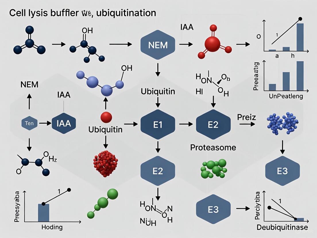
Accurate analysis of protein ubiquitylation relies critically on the effective preservation of this labile modification during cell lysis.
Optimizing Cell Lysis: A Guide to Using NEM and IAA for Preserving Ubiquitination Signals
Abstract
Accurate analysis of protein ubiquitylation relies critically on the effective preservation of this labile modification during cell lysis. This article provides a comprehensive guide for researchers and drug development professionals on the use of N-ethylmaleimide (NEM) and iodoacetamide (IAA) as deubiquitylase (DUB) inhibitors. We cover the foundational mechanisms of DUB inhibition, detail optimized methodological protocols for lysis buffer preparation, address common troubleshooting and optimization challenges, and outline validation strategies to confirm data reliability. By integrating current best practices and addressing key pitfalls, this resource aims to empower scientists to generate more reproducible and high-quality data in ubiquitin signaling research.
Understanding the Challenge: Why Ubiquitination is Lost During Lysis and How to Stop It
Deubiquitinating enzymes (DUBs) constitute a family of approximately 100 proteases responsible for cleaving ubiquitin moieties from substrate proteins, thereby reversing the process of ubiquitination [1] [2]. This dynamic balance between ubiquitination by E3 ligases and deubiquitination by DUBs is essential for maintaining cellular homeostasis, governing protein stability, localization, and functional activity [2] [3]. The majority of DUB families—including ubiquitin-specific proteases (USPs), ubiquitin C-terminal hydrolases (UCHs), ovarian tumor proteases (OTUs), Machado-Joseph disease proteases (MJDs), MINDY, and ZUFSP—are cysteine proteases that rely on an active-site cysteine residue for catalytic activity [3] [4]. This cysteine performs a nucleophilic attack on the isopeptide bond linking ubiquitin to its substrate, forming a transient thioester intermediate before hydrolysis releases free ubiquitin and the deubiquitinated protein [4].
In experimental settings focused on studying protein ubiquitination, this DUB activity presents a significant challenge. During cell lysis, the compartmentalization that naturally regulates DUB activity is lost, allowing these enzymes to artificially remove ubiquitin chains from proteins of interest, potentially leading to misinterpretation of results [5]. Consequently, the use of cysteine-targeting inhibitors such as N-ethylmaleimide (NEM) and iodoacetamide (IAA) in cell lysis buffers is a critical strategy for preserving the native ubiquitination state of proteins by irreversibly inactivating cysteine-dependent DUBs before they can alter the ubiquitin landscape [5].
Chemical Mechanisms of Cysteine-Targeting Inhibitors
N-Ethylmaleimide (NEM)
NEM is an alkylating agent that functions through a Michael addition reaction. Its maleimide ring contains an electron-deficient alkene that is highly susceptible to nucleophilic attack by the thiolate anion (S⁻) of a cysteine residue in a DUB's active site [4]. This reaction results in the formation of a stable carbon-sulfur (C-S) thioether bond, thereby irreversibly alkylating the catalytic cysteine and rendering the enzyme inactive.
Chemical Reaction: DUB-S⁻ + C₂H₅N(CO)₂CH=CH₂ → DUB-S-CH₂-CH₂-N(CO)₂C₂H₅
Iodoacetamide (IAA)
IAA operates via a nucleophilic substitution (S_N2) mechanism. The iodine atom in IAA is an excellent leaving group, facilitating a direct substitution by the sulfur atom of the deprotonated cysteine thiol. This reaction covalently attaches a carbamidomethyl group to the cysteine sulfur atom, creating a stable thioether linkage and causing irreversible inhibition of the DUB.
Chemical Reaction: DUB-S⁻ + I-CH₂-C(=O)-NH₂ → DUB-S-CH₂-C(=O)-NH₂ + I⁻
The following diagram illustrates the irreversible inhibition of cysteine-dependent DUBs by NEM and IAA, which is crucial for preserving ubiquitin signals during cell lysis.
Comparative Analysis of NEM and IAA
The selection between NEM and IAA for a specific experiment depends on their distinct biochemical properties and practical considerations. The table below provides a detailed comparison to guide this decision-making process.
Table 1: Comparative Properties of NEM and IAA as DUB Inhibitors
| Property | N-Ethylmaleimide (NEM) | Iodoacetamide (IAA) |
|---|---|---|
| Chemical Mechanism | Michael addition | Nucleophilic substitution (S_N2) |
| Reactivity | High (targets thiolate anions) | Moderate |
| Specificity | Lower (can react with other nucleophiles) | Higher for cysteine thiols |
| Reversibility | Irreversible | Irreversible |
| Membrane Permeability | Cell-permeable | Cell-permeable |
| Stability in Buffer | Stable at neutral to basic pH | Stable, but light-sensitive |
| Common Working Concentration | 5-25 mM | 10-50 mM |
| Key Consideration | Must be freshly prepared or stored aliquoted at -20°C; can modify primary amines at high concentrations | Must be protected from light; generally considered more specific than NEM |
| Quenching Agent | Dithiothreitol (DTT) | Dithiothreitol (DTT) or β-mercaptoethanol |
Application Notes for Ubiquitination Preservation Research
Integration in Cell Lysis Protocols
The primary application of NEM and IAA in ubiquitination research is their incorporation into cell lysis buffers to preserve the cellular ubiquitin landscape at the moment of lysis. As emphasized in methodological optimizations for ubiquitin chain analysis, the inclusion of these inhibitors is essential for preventing the post-lysis deubiquitination that can lead to erroneous conclusions [5]. The recommended workflow involves:
- Preparation of Lysis Buffer: Add NEM or IAA to the lysis buffer immediately before use. For IAA, protect the solution from light.
- Rapid Lysis: Lyse cells directly in pre-chilled buffer containing the inhibitor. Vortex briefly and ensure complete lysis.
- Incubation: Incubate the lysate on ice for 5-15 minutes to allow the inhibitor to fully inactivate all accessible DUBs.
- Centrifugation: Clarify the lysate by centrifugation at 4°C.
- Quenching (If Necessary): If downstream applications require functional thiol groups (e.g., for certain assays or further processing), the alkylating agent can be quenched with an excess of DTT (e.g., 10-20 mM).
The Scientist's Toolkit: Essential Research Reagents
Successful experimentation with NEM and IAA requires a set of key reagents. The following table lists essential materials and their specific functions in protocols aimed at preserving ubiquitination.
Table 2: Research Reagent Solutions for Ubiquitination Preservation Studies
| Reagent | Function/Description | Application Note |
|---|---|---|
| N-Ethylmaleimide (NEM) | Irreversible cysteine alkylator; inhibits cysteine-dependent DUBs during lysis. | Prepare a fresh stock solution in ethanol or water; final working concentration typically 10-25 mM in lysis buffer. |
| Iodoacetamide (IAA) | Irreversible cysteine alkylator; more specific than NEM for cysteine thiols. | Prepare a stock solution in water protected from light; final working concentration typically 20-50 mM. |
| Lysis Buffer (RIPA or NP-40 based) | Provides the ionic and detergent environment for efficient cell disruption and protein extraction. | Must be kept ice-cold and supplemented with a broad-spectrum protease inhibitor cocktail in addition to NEM/IAA. |
| Dithiothreitol (DTT) | Reducing agent used to quench excess alkylating agent post-lysis. | Add to a final concentration of 10-20 mM after the initial inhibition period to restore thiol groups for downstream steps. |
| Protease Inhibitor Cocktail (without DTT) | Inhibits serine, cysteine, aspartic, and metallo-proteases to prevent general protein degradation. | Essential to use a formulation that does not contain reducing agents which would compete with and inactivate NEM/IAA. |
| Activity-Based Probes (e.g., Ub-VS) | Covalent probes that label active DUBs; used to validate inhibitor efficacy. | Can be used in a control experiment to confirm that DUB activity in the lysate has been successfully abolished by the pretreatment with NEM or IAA [6] [4]. |
NEM and IAA are cornerstone reagents in the molecular toolkit for ubiquitination research. Their role as irreversible, cysteine-targeting inhibitors is critical for arresting the activity of the majority of DUBs during the critical window of cell lysis and sample preparation. Understanding their distinct mechanisms—Michael addition for NEM versus nucleophilic substitution for IAA—allows researchers to make an informed choice based on the requirements for reactivity, specificity, and compatibility with downstream assays. The consistent and correct application of these inhibitors, as part of a comprehensive lysis buffer strategy, is a fundamental prerequisite for obtaining reliable and interpretable data on protein ubiquitination, thereby forming the foundation for accurate insights into the complex biology regulated by the ubiquitin-proteasome system.
The ubiquitin-proteasome system (UPS) has long been recognized as the primary mechanism for targeted protein degradation in eukaryotic cells. However, research over the past decade has revealed that ubiquitin signaling extends far beyond its canonical role in directing substrates to proteasomal destruction. Ubiquitin, a 76-amino acid protein, can be conjugated to target proteins through a sequential enzymatic cascade involving E1 activating, E2 conjugating, and E3 ligase enzymes [7]. The complexity of ubiquitin signaling arises from the ability of ubiquitin itself to be modified at any of its seven lysine residues (K6, K11, K27, K29, K33, K48, K63) or its N-terminal methionine (M1), enabling formation of at least eight distinct homotypic polyubiquitin chains, plus heterotypic and branched chains with unique structures and functions [8] [7]. This diverse "ubiquitin code" allows for precise regulation of virtually all cellular processes, from inflammatory signaling to DNA repair mechanisms [7].
The preservation of this complex ubiquitin landscape during experimental procedures presents significant technical challenges. The dynamic nature of ubiquitination, with constant opposition by deubiquitinating enzymes (DUBs), necessitates careful methodological consideration, particularly during the critical initial step of cell lysis and sample preparation [5]. This Application Note provides both the theoretical framework and practical methodologies for investigating non-proteolytic ubiquitin signaling, with emphasis on maintaining the native ubiquitination state through appropriate buffer composition and inhibitor selection.
The Expanding Functional Landscape of Ubiquitin Signaling
Non-Proteolytic Functions of Ubiquitin Chains
Different ubiquitin chain linkages create distinct molecular architectures that are recognized as specific signals by cellular machinery. While K48-linked chains remain the principal signal for proteasomal degradation, numerous other linkage types mediate diverse non-proteolytic functions:
Table 1: Non-Proteolytic Functions of Ubiquitin Chain Linkages
| Ubiquitin Linkage Type | Primary Cellular Functions | Key Signaling Pathways | Experimental Tools for Study |
|---|---|---|---|
| K63-linked chains | DNA damage repair, endocytosis, inflammatory signaling, kinase activation | NF-κB activation, TLR signaling, mTOR pathway | Linkage-specific antibodies, UbiREAD assay [8] |
| M1-linear chains | Innate immunity, inflammation, cell death regulation | TNF signaling, NF-κB pathway, necroptosis | OTULIN DUB analysis, LUBAC inhibition [7] |
| K11-linked chains | Cell cycle regulation, endoplasmic reticulum-associated degradation | Mitotic progression, ERAD quality control | CC0651 E2 inhibitor, K11-linkage specific binders |
| K6-linked chains | DNA damage response, mitochondrial homeostasis | DNA repair pathways, mitophagy | BRCA1-BARD1 E3 ligase studies |
| K27 & K29-linked chains | Wnt signaling, innate immunity, protein aggregation | Wnt/β-catenin pathway, immune response | Linkage-specific DUBs, mass spectrometry |
| Branched/mixed chains | Complex signaling integration, hierarchical degradation signals | Cellular stress responses, quality control | UbiREAD, TUBE-based purification [8] |
Recent research using advanced tools like the UbiREAD platform has revealed surprising complexity in how ubiquitin chains determine substrate fate. For instance, K48-linked ubiquitin chains induce GFP degradation with a half-life of approximately 1 minute, but these chains must consist of at least three ubiquitin molecules to be effective, as dimers remain stable intracellularly due to DUB activity [8]. Conversely, K63 chains are rapidly deubiquitinated and generally do not affect substrate stability. Perhaps most intriguingly, branched ubiquitin chains containing both K48 and K63 linkages display a clear hierarchy, with the chain directly conjugated to the substrate protein overriding the influence of the branching chain in determining degradation fate [8].
Ubiquitin in Membrane Protein Extraction and Organelle Homeostasis
The p97/VCP ATPase complex (also known as Cdc48 in yeast) represents a crucial system that exemplifies the non-degradative functions of ubiquitin signaling. This complex, particularly in conjunction with the UFD1L-NPLOC4 heterodimer, unfolds ubiquitinated proteins to facilitate their extraction from cellular compartments and large macromolecular assemblies [9]. Recent research has identified a conserved ubiquitin-binding helix (UBH) in many UBX-containing p97 adapters, including FAF2, that substantially enhances the engagement of ubiquitinated substrates with p97-UFD1L-NPLOC4 [9].
This UBH-UBX module amplifies p97's mechanical output power by approximately two-fold, as measured by both working ATPase activity and unfolding capacity, enabling the extraction of challenging substrates from lipid bilayers during ER-associated degradation (ERAD) and mitochondria-associated degradation (MAD) [9]. The functional significance of this enhanced unfolding power is particularly evident in membrane protein extraction, where the energy barrier for removing transmembrane domains from lipid bilayers is substantially higher than for soluble protein unfolding.
The diagram above illustrates the coordinated process of membrane protein extraction mediated by the p97 complex and enhanced by the FAF2 UBH-UBX module. This process highlights how ubiquitin signals can direct proteins toward either degradative or non-degradative fates, depending on cellular context and the nature of the ubiquitin chain involved.
Methodological Considerations for Ubiquitination Studies
Preservation of Ubiquitin Signals During Cell Lysis
The accurate analysis of ubiquitination events depends critically on the preservation of these labile modifications during sample preparation. The dynamic equilibrium between ubiquitination by E3 ligases and deubiquitination by DUBs must be rapidly arrested at the moment of cell lysis to maintain the native ubiquitination state [5]. Failure to do so can result in significant experimental artifacts and misinterpretation of results.
Essential Inhibitors for Ubiquitin Preservation:
N-Ethylmaleimide (NEM): This cell-permeable, irreversible cysteine protease inhibitor effectively inhibits most deubiquitinating enzymes (DUBs) by covalently modifying their active site cysteine residues. Working concentrations typically range from 5-25 mM in lysis buffer [5].
Iodoacetamide (IAA): An alternative alkylating agent that similarly targets cysteine residues in DUB active sites. While effective, it is generally less permeable to intact cells and is often used at 10-50 mM concentrations, primarily during protein extraction rather than in pre-lysis treatments [5].
Comprehensive Lysis Buffer Composition: A recommended buffer for ubiquitination preservation includes:
- 50 mM Tris-HCl (pH 7.5)
- 150 mM NaCl
- 1% NP-40 or Triton X-100
- 0.1% SDS (for efficient membrane protein solubilization)
- 5-25 mM NEM (freshly prepared)
- 10-50 mM IAA (when used)
- 10 mM EDTA (to chelate metal cofactors required by some DUBs)
- Protease inhibitor cocktail (without EDTA where possible)
- 5 mM Sodium Fluoride and 1 mM Sodium Orthovanadate (phosphatase inhibitors to preserve signaling context)
Practical Considerations:
- Fresh Preparation: NEM and IAA solutions should be prepared immediately before use as they hydrolyze in aqueous solution.
- Sequential Application: Some protocols recommend sequential use of membrane-permeable NEM before lysis followed by IAA during extraction to ensure comprehensive DUB inhibition.
- Temperature Control: All procedures should be performed on ice or at 4°C to slow enzymatic activity.
- Time Optimization: Minimize the interval between lysis and complete denaturation of samples by heating in SDS-PAGE sample buffer.
Advanced Techniques for Ubiquitin Chain Characterization
Linkage-Specific Analysis: The development of linkage-specific ubiquitin-binding domains (UBDs) and antibodies has revolutionized the study of ubiquitin chain topology. Tandem-repeated ubiquitin-binding entities (TUBEs) can be employed to protect ubiquitin chains from DUB activity during purification, while linkage-specific antibodies enable immunoblotting detection of particular chain types [5].
Deubiquitinase-Based Mapping: The combined use of linkage-specific deubiquitylases (DUBs) provides a powerful method for ubiquitin chain identification. Treatment of samples with DUBs such as OTUB1 (K48-specific), AMSH (K63-specific), or OTULIN (M1-specific) followed by immunoblotting can confirm the presence of specific linkage types through characteristic band shift patterns [5].
Mass Spectrometry Approaches: Di-glycine remnant immunopurification coupled with mass spectrometry enables proteome-wide identification of ubiquitination sites. Recent advancements have revealed extensive changes in the ubiquitin landscape during aging, with 29% of quantified ubiquitylation sites in mouse brain being altered independently of protein abundance changes [10]. This approach can distinguish between different ubiquitin chain linkages when combined with specific enrichment strategies.
Research Reagent Solutions for Ubiquitination Studies
Table 2: Essential Research Reagents for Ubiquitin Signaling Studies
| Reagent Category | Specific Examples | Function & Application | Considerations for Use |
|---|---|---|---|
| DUB Inhibitors | N-Ethylmaleimide (NEM), Iodoacetamide (IAA), PR-619 | Preserve ubiquitin conjugates during cell lysis by inhibiting deubiquitinating enzymes | NEM is cell-permeable; use fresh solutions; IAA less permeable but effective during extraction |
| Proteasome Inhibitors | MG132, Bortezomib, Carfilzomib | Block degradation of ubiquitinated proteins, allowing accumulation for detection | Can indirectly affect ubiquitination patterns by altering cellular homeostasis |
| E1 Inhibitors | TAK-243, PYR-41 | Block ubiquitin activation, preventing all ubiquitination events | Useful for determining dependence on ubiquitination; highly toxic to cells |
| Linkage-Specific Binders | TUBEs (Tandem-repeated Ubiquitin-Binding Entities), Linkage-specific UBDs | Affinity purification of ubiquitinated proteins with protection from DUBs | Some TUBEs show preference for certain chain types; K48 and K63 specific variants available |
| Linkage-Specific Antibodies | K48-linkage specific, K63-linkage specific, M1-linear specific antibodies | Immunoblotting detection of specific ubiquitin chain types | Varying specificity between commercial sources; require validation with linkage standards |
| DUB Enzymes | OTUB1 (K48-specific), AMSH (K63-specific), OTULIN (M1-specific) | Mapping ubiquitin chain topology through characteristic cleavage patterns | Specificity should be verified; controlled reaction conditions essential |
| Activity-Based Probes | HA-Ub-VS, HA-Ub-Br2 | Label active DUBs and some E3 ligases for detection and enrichment | Can identify active enzymes in complex mixtures; useful for inhibitor screening |
Experimental Protocol: Analysis of Endogenous Ubiquitination in Cell Culture Models
Sample Preparation with Ubiquitin Preservation
Materials:
- Cell culture of interest
- Complete culture medium
- PBS, ice-cold
- Ubiquitin-preserving lysis buffer (as described in Section 3.1)
- BCA or Bradford protein assay kit
- 4× SDS-PAGE sample buffer (with 400 mM DTT)
- Heating block or water bath
Procedure:
- Pre-treatment (if applicable): Apply experimental treatments to cells according to experimental design. Include appropriate controls (e.g., DMSO vehicle, stimulation conditions).
Inhibition Phase: Prior to lysis, replace culture medium with pre-warmed medium containing 10 mM NEM. Incubate for 5 minutes at 37°C to allow inhibitor penetration.
Rapid Washing: Remove NEM-containing medium and immediately wash cells twice with ice-cold PBS containing 5 mM NEM.
Cell Lysis: Add ubiquitin-preserving lysis buffer (approximately 100-200 μL per 10⁶ cells) directly to culture dishes on ice. Scrape cells thoroughly and transfer lysates to pre-cooled microcentrifuge tubes.
Extraction: Rotate lysates at 4°C for 30 minutes to ensure complete extraction.
Clarification: Centrifuge at 16,000 × g for 15 minutes at 4°C. Transfer supernatant to fresh tubes.
Protein Quantification: Perform protein assay using BCA or Bradford method. Include appropriate standards and blanks.
Denaturation: Mix lysates with 4× SDS-PAGE sample buffer to final 1× concentration. Heat at 95°C for 5-10 minutes.
Storage: Aliquot and store at -80°C if not used immediately. Avoid repeated freeze-thaw cycles.
Immunoblotting for Ubiquitinated Proteins
Materials:
- Prepared protein samples
- SDS-PAGE gel system (8-12% gradient gels recommended)
- Transfer apparatus
- Nitrocellulose or PVDF membranes
- TBST buffer (Tris-buffered saline with 0.1% Tween-20)
- Blocking buffer (5% non-fat dry milk or BSA in TBST)
- Primary antibodies: pan-ubiquitin, linkage-specific ubiquitin, protein of interest, loading control
- HRP-conjugated secondary antibodies
- Enhanced chemiluminescence (ECL) substrate
- Imaging system
Procedure:
- Electrophoresis: Load equal protein amounts (20-50 μg recommended) onto SDS-PAGE gel. Include molecular weight markers and appropriate controls. Run gel at constant voltage until adequate separation.
Transfer: Transfer proteins to membrane using wet or semi-dry transfer system according to manufacturer's recommendations.
Blocking: Incubate membrane in blocking buffer for 1 hour at room temperature with gentle agitation.
Primary Antibody Incubation: Dilute primary antibody in blocking buffer or antibody dilution buffer according to manufacturer's recommendations. Incubate with membrane overnight at 4°C with gentle agitation.
Washing: Wash membrane 3× for 10 minutes each with TBST.
Secondary Antibody Incubation: Incubate with appropriate HRP-conjugated secondary antibody in blocking buffer for 1 hour at room temperature.
Washing: Repeat washing as in step 5.
Detection: Apply ECL substrate according to manufacturer's instructions and image using appropriate detection system.
Troubleshooting Notes:
- High background may indicate insufficient blocking or antibody concentration too high.
- Multiple non-specific bands common with pan-ubiquitin antibodies; include linkage-specific antibodies for confirmation.
- For high molecular weight smearing, reduce transfer time or use gradient gels for better separation.
- Always include control for protein loading (e.g., actin, GAPDH).
The experimental workflow above outlines the critical steps for preserving and analyzing ubiquitinated proteins, emphasizing the importance of DUB inhibition at multiple stages to maintain the native ubiquitination state.
The expanding understanding of non-proteolytic ubiquitin signaling reveals an intricate regulatory system that coordinates virtually all cellular processes. From membrane protein extraction to inflammatory signaling and DNA repair, ubiquitin modifications serve as versatile molecular signals that extend far beyond their classical degradation targeting function. The methodological approaches outlined in this Application Note provide researchers with robust tools for investigating this complex signaling system, with particular emphasis on preserving the native ubiquitination state through appropriate use of DUB inhibitors like NEM and IAA during sample preparation. As research continues to decipher the complexities of the ubiquitin code, maintaining methodological rigor in ubiquitination studies remains paramount for generating accurate, biologically relevant data.
Within the broader research on cell lysis buffers with N-ethylmaleimide (NEM) or iodoacetamide (IAA) for ubiquitination preservation, a critical and often overlooked factor is the efficacy of deubiquitylase (DUB) inhibition. DUBs are proteases that rapidly reverse protein ubiquitylation, a dynamic post-translational modification regulating diverse cellular processes from protein degradation to signal transduction [11] [12]. Inadequate inhibition during cell lysis and subsequent procedures leads to the loss of ubiquitin signals, directly causing the misinterpretation of experimental data and the drawing of erroneous conclusions regarding the ubiquitylation status, dynamics, and function of proteins of interest [13]. This application note details the consequences of insufficient DUB inhibition and provides optimized protocols to preserve the native ubiquitylation state of proteins for accurate analysis.
Quantitative Analysis of DUB Inhibitor Efficacy
The concentration of DUB inhibitors in lysis buffers is a primary determinant of ubiquitylation preservation. Conventional protocols often recommend insufficient concentrations, leading to significant signal loss.
Table 1: Impact of DUB Inhibitor Concentration on Ubiquitin Chain Preservation
| DUB Inhibor | Conventional Concentration | Optimized Concentration | Effect on K63/M1-Ub Chains | Compatibility with Mass Spectrometry |
|---|---|---|---|---|
| N-Ethylmaleimide (NEM) | 5-10 mM | Up to 50 mM | Superior preservation of K63-linked and M1-linked chains [13] | Recommended; adduct does not interfere with Gly-Gly remnant identification [13] |
| Iodoacetamide (IAA) | 5-10 mM | Up to 50 mM | Moderate preservation; less effective than NEM for some chains [13] | Not recommended; forms a 114 Da adduct identical to the tryptic Gly-Gly signature [13] |
Table 2: Consequences of Inadequate DUB Inhibition on Experimental Outcomes
| Experimental Context | Common Erroneous Conclusion | Actual Consequence of Poor Inhibition |
|---|---|---|
| Ubiquitylation State Analysis | "The protein is not ubiquitylated." | Failure to detect genuine, often labile, ubiquitylation due to enzymatic deconjugation during lysis [13]. |
| Ubiquitin Chain Topology | "The protein is modified primarily by K48-linked chains." | Preferential cleavage of specific chain types (e.g., K63, M1) by active DUBs, skewing linkage analysis [13]. |
| Drug Mechanism of Action | "The DUB inhibitor does not affect target ubiquitylation." | Inadequate preservation masks the true extent of drug-induced ubiquitylation, leading to false negatives [12]. |
The Scientist's Toolkit: Essential Research Reagents
Table 3: Key Reagents for Preserving Protein Ubiquitylation
| Reagent / Solution | Function & Mechanism | Application Notes |
|---|---|---|
| N-Ethylmaleimide (NEM) | Cysteine protease DUB inhibitor; alkylates the active site cysteine residue of most DUBs [13]. | Use at 20-50 mM in lysis buffer. Stable and recommended for mass spectrometry workflows [13]. |
| Iodoacetamide (IAA) | Cysteine protease DUB inhibitor; alkylates active site cysteine. Light-sensitive [13]. | Use at 20-50 mM. Prepare fresh and protect from light. Avoid for MS-ubiquitomics due to adduct interference [13]. |
| EDTA/EGTA | Metalloprotease DUB inhibitor; chelates zinc ions, inactivating JAMM/MPN+ family metalloproteases [13]. | Include at 1-10 mM in lysis buffers to provide comprehensive DUB inhibition alongside NEM/IAA. |
| Proteasome Inhibitors (e.g., MG132) | Inhibits the 26S proteasome, preventing degradation of ubiquitylated proteins and facilitating detection [13]. | Use for 4-8 hours prior to lysis. Prolonged incubation (>12h) can induce cellular stress responses [13]. |
| SDS Lysis Buffer | Denatures and instantly inactivates DUBs; preserves the ubiquitylation state at the moment of lysis [13]. | For direct immunoblotting, lyse cells in 1% SDS buffer and boil immediately. Not suitable for native IP. |
Experimental Protocol: Optimized Workflow for Ubiquitin Preservation
Cell Lysis with Guaranteed DUB Inhibition
Materials:
- Pre-chilled PBS
- Optimized Lysis Buffer (see Table 3): 50 mM Tris-HCl (pH 7.5), 150 mM NaCl, 1% NP-40, 0.5% Sodium deoxycholate, 50 mM NEM, 10 mM EDTA, and protease inhibitors.
- SDS Lysis Buffer (alternative): 50 mM Tris-HCl (pH 7.5), 1% SDS, 50 mM NEM, 10 mM EDTA.
Procedure:
- Pre-treatment: Treat cells with 10-20 µM MG132 or an equivalent proteasome inhibitor for 4-6 hours before lysis if studying proteasomal targets [13].
- Lysis:
- For Native Co-immunoprecipitation (Co-IP): Wash cells with PBS and lyse with Optimized Lysis Buffer (500 µL per 10 cm dish) for 30 minutes on a rotator at 4°C.
- For Direct Immunoblotting: Wash cells with PBS. Scrape cells directly into 1-2 mL of pre-heated (95°C) SDS Lysis Buffer and immediately vortex and boil for 10 minutes [13].
- Clarification: Centrifuge lysates at 16,000 × g for 15 minutes at 4°C. Transfer the supernatant to a new tube.
- Post-Lysis Processing: For Co-IP, proceed immediately to the immunoprecipitation step, ensuring the lysis buffer is supplemented with NEM (20 mM). Avoid prolonged incubations.
Validation of Ubiquitin Chain Integrity
To confirm that the ubiquitylation profile is accurately preserved, use a combination of ubiquitin-binding proteins (e.g., Tandem-repeated Ubiquitin-Binding Entities - TUBEs) and linkage-specific DUBs in validation experiments [13].
- TUBE Pull-down: Use immobilized TUBEs to capture the full complement of ubiquitin chains from your lysate. Compare the profile to a standard lysate prepared with sub-optimal DUB inhibition.
- Linkage-Specific DUB Treatment: After capture, treat the ubiquitin conjugates with purified, linkage-specific DUBs (e.g., an OTU family DUB for K63 linkages) to confirm the identity of the chains. This validates that the observed smears or bands are specific ubiquitin modifications [13].
Signaling Pathway and Experimental Workflow Diagrams
Diagram 1: Impact of DUB Inhibition on Experimental Outcomes
Diagram 2: Optimized Workflow for Ubiquitin Preservation
Practical Protocols: Formulating Lysis Buffers with NEM and IAA for Robust Ubiquitin Preservation
The preservation of cellular ubiquitination states during cell lysis presents a significant methodological challenge for researchers studying protein regulation. The ubiquitin-proteasome system involves dynamic, reversible modifications that can be rapidly erased by endogenous deubiquitinase (DUB) enzymes during sample preparation. N-Ethylmaleimide (NEM) and iodoacetamide (IAA) serve as critical cysteine alkylators that inhibit DUB activity, thereby maintaining the native ubiquitin landscape for accurate analysis. While standard laboratory protocols frequently recommend concentrations of 5-10 mM for these inhibitors, emerging evidence demonstrates that this range proves insufficient for preserving specific ubiquitin chain architectures and challenging biological contexts. This application note establishes an optimized framework for employing elevated inhibitor concentrations (50-100 mM) to address stubborn targets in ubiquitination research, providing researchers with detailed protocols and empirical support for moving beyond conventional approaches.
Table 1: Common DUB Inhibitors and Their Applications
| Inhibitor | Standard Concentration | High Concentration Range | Primary Mechanism | Key Considerations |
|---|---|---|---|---|
| N-Ethylmaleimide (NEM) | 5-10 mM | 50-100 mM | Irreversible cysteine alkylation | K63-linked chains require higher concentrations; stock solutions in ethanol [14] [15] |
| Iodoacetamide (IAA) | 5-10 mM | 50-100 mM | Irreversible cysteine alkylation | Use fresh solutions protected from light [15] |
| EDTA/EGTA | 1-5 mM | 5-10 mM | Metalloprotease inhibition | Chelates Zn²⁺ required by certain DUB families [16] [14] |
| MG132 | 10-20 µM | 25-50 µM | Proteasome inhibition | Prevents degradation of extracted proteins; extended use may induce stress response [14] |
The Scientific Basis for High-Concentration DUB Inhibition
Limitations of Standard Inhibitor Concentrations
Conventional DUB inhibitor concentrations (5-10 mM) fail to provide complete protection against the diverse family of deubiquitinating enzymes, particularly for labile ubiquitin linkages. The fundamental issue stems from the heterogeneity of DUB enzymes and their varying sensitivity to cysteine alkylating agents. Research has demonstrated that K63-linked ubiquitin chains exhibit particular sensitivity to DUB activity and require significantly higher NEM concentrations for effective preservation compared to other chain types [14]. At standard concentrations, residual DUB activity continues to dismantle ubiquitin chains during the critical window between cell lysis and complete protein denaturation, leading to substantial loss of biological signal.
Furthermore, the cellular context significantly influences DUB inhibition requirements. Proteins such as IRAK1 (Interleukin-1 receptor-associated kinase 1) demonstrate exceptional susceptibility to deubiquitination, necessitating up to tenfold higher concentrations of NEM or IAA (50-100 mM) for adequate stabilization [15]. This suggests that either specific DUBs with lower inhibitor sensitivity target these proteins or their structural presentation makes the ubiquitin-chain interface particularly accessible to DUB activity even in the presence of standard inhibitor concentrations.
Empirical Evidence Supporting Elevated Concentrations
Recent investigations into ubiquitin chain interactomes have provided additional support for optimized inhibition strategies. Comparative studies of DUB inhibitors revealed that NEM and chloroacetamide (CAA) produce significantly different interactor profiles in ubiquitin pulldown experiments, indicating inhibitor-specific effects on ubiquitin-binding proteins beyond DUB inhibition itself [17]. This underscores the importance of not only using adequate concentrations but also selecting appropriate inhibitors based on the specific research goals.
The development of tandem-repeated ubiquitin-binding entities (TUBEs) has further highlighted the necessity of robust DUB inhibition. These tools designed to protect ubiquitin chains from deubiquitination during purification rely on complementary use of high-grade DUB inhibitors to function effectively [5]. The integration of structural biology findings with biochemical methodologies has created a compelling case for re-evaluating standard laboratory protocols for ubiquitination studies.
Optimized Reagent Formulations and Recipes
High-Strength Lysis Buffer with Enhanced DUB Inhibition
The following formulation builds upon traditional RIPA buffer by incorporating elevated DUB inhibitor concentrations while maintaining compatibility with downstream applications:
Table 2: High-Strength RIPA Lysis Buffer for Stubborn Ubiquitinated Targets
| Component | Final Concentration | Stock Solution | Volume for 10 mL | Function |
|---|---|---|---|---|
| Tris-HCl, pH 8.0 | 50 mM | 1 M | 500 µL | Buffer capacity |
| NaCl | 150 mM | 5 M | 300 µL | Ionic strength |
| NP-40 | 1% | 100% | 100 µL | Non-ionic detergent |
| Sodium deoxycholate | 0.5% | 10% | 500 µL | Ionic detergent |
| SDS | 0.1% | 10% | 100 µL | Denaturing detergent |
| NEM | 50-100 mM | 1 M (in ethanol) | 500-1000 µL | DUB inhibition |
| EDTA | 5 mM | 0.5 M | 100 µL | Metalloprotease inhibition |
| MG132 | 25 µM | 10 mM (in DMSO) | 25 µL | Proteasome inhibition |
| Halt Protease & Phosphatase Inhibitor Cocktail | 1X | 100X | 100 µL | Broad-spectrum inhibition |
| dH₂O | - | - | to 10 mL | Solvent |
Preparation Notes:
- Prepare NEM stock solution fresh in absolute ethanol immediately before use
- Add all inhibitors to the base buffer immediately before cell lysis
- For IAA-based formulations, protect from light during preparation and use
- Final DMSO concentration from inhibitor stocks should not exceed 0.5% to maintain protein integrity
Specialized Buffer Systems for Specific Applications
Certain experimental contexts require customized buffer formulations:
Native Immunoprecipitation Buffer: For ubiquitin interactome studies requiring native protein interactions, utilize 50 mM NEM in NP-40-based lysis buffer (50 mM Tris pH 7.4, 150 mM NaCl, 1% NP-40) without SDS or sodium deoxycholate. This formulation preserves protein-protein interactions while still providing robust DUB inhibition [17].
Membrane Protein Extraction Buffer: When studying ubiquitinated membrane proteins, incorporate 75-100 mM NEM into specialized extraction buffers containing digitonin or n-dodecyl-β-D-maltoside to maintain inhibition during the extended extraction period often required for these challenging targets [9].
Experimental Protocols for Validation and Application
Cell Lysis Procedure with Enhanced DUB Inhibition
The following protocol details the optimized procedure for cell lysis with elevated DUB inhibitor concentrations:
Critical Steps and Troubleshooting:
- Temperature Control: Maintain samples at 0-4°C throughout the procedure to slow enzymatic activity not fully inhibited by chemical agents
- Inhibitor Freshness: NEM solutions degrade in aqueous environments; add to lysis buffer immediately before use
- Sonication Optimization: Adjust sonication power empirically for different cell types to ensure complete lysis without excessive foam formation
- Viscosity Reduction: If lysate remains viscous after sonication, repeat pulse cycle or extend incubation time to digest genomic DNA
- Concentration Determination: Use BCA assay for protein quantification as it demonstrates greater compatibility with detergents than Bradford assays [18]
Validation Experiments for Inhibition Efficiency
Ubiquitin Chain Stability Assay
To empirically validate the efficiency of DUB inhibition at different NEM concentrations:
- Prepare identical cell samples lysed with either standard (10 mM) or high-concentration (50 mM and 100 mM) NEM
- Process lysates according to the standard protocol
- Incubate lysates at 4°C for timed intervals (0, 30, 60, 120 minutes) before adding Laemmli buffer
- Perform western blotting for both total ubiquitin and specific chain linkages (K48, K63)
- Quantify signal retention using densitometry and compare degradation kinetics across conditions
Table 3: Expected Signal Retention at Different NEM Concentrations
| Incubation Time | 10 mM NEM | 50 mM NEM | 100 mM NEM |
|---|---|---|---|
| 0 minutes | 100% | 100% | 100% |
| 30 minutes | 65-75% | 85-92% | 95-98% |
| 60 minutes | 45-55% | 75-85% | 90-95% |
| 120 minutes | 25-35% | 65-75% | 85-90% |
Linkage-Specific Preservation Assessment
For researchers focusing on specific ubiquitin chain linkages:
- Transfer PVDF membrane following SDS-PAGE and denature by incubating in 6 M guanidine-HCl for 30 minutes at 4°C to enhance antibody accessibility to ubiquitin epitopes [14]
- Probe with linkage-specific antibodies (K48, K63, K11, etc.)
- Compare signal intensity and background across inhibition conditions
- Validate antibody specificity using linkage-specific deubiquitinases (DUBs) in control experiments [5]
The Researcher's Toolkit: Essential Reagents and Materials
Table 4: Key Research Reagent Solutions for Advanced Ubiquitination Studies
| Reagent/Category | Specific Examples | Function/Application | Usage Notes |
|---|---|---|---|
| Cysteine Alkylators | N-Ethylmaleimide (NEM), Iodoacetamide (IAA) | Irreversible DUB inhibition | Fresh preparation critical; ethanol stocks for NEM |
| Proteasome Inhibitors | MG132, Bortezomib, Carfilzomib | Prevent proteasomal degradation | Limited exposure time to avoid stress response |
| Metalloprotease Inhibitors | EDTA, EGTA | Inhibition of metal-dependent DUBs | Include in all buffer formulations |
| Commercial Inhibitor Cocktails | Halt Protease & Phosphatase Inhibitor Cocktail, cOmplete ULTRA Tablets | Broad-spectrum protection | Convenient but may require supplementation with high-dose NEM |
| Ubiquitin Binding Reagents | TUBEs (Tandem-repeated Ubiquitin Binding Entities) | Protection and pulldown of ubiquitinated proteins | Combine with chemical inhibition for maximum protection |
| Linkage-Specific Antibodies | Anti-K48, Anti-K63, Anti-K11 ubiquitin | Detection of specific chain architectures | Validate with appropriate controls for each application |
| Deubiquitinase Enzymes | OTUB1 (K48-specific), AMSH (K63-specific) | Linkage specificity controls | Essential for validating antibody specificity and chain architecture |
Implementation Workflow and Decision Framework
Application-Specific Recommendations:
Standard Systems (10 mM NEM sufficient):
- Steady-state analysis of abundant ubiquitinated proteins
- K48-linked ubiquitin chains in overexpression systems
- Preliminary screens where relative comparisons are sufficient
Stubborn Targets (50-100 mM NEM required):
- K63-linked ubiquitin chains [14]
- IRAK1 and similarly sensitive targets [15]
- Endogenous ubiquitination with low stoichiometry
- Quantitative studies requiring maximum signal preservation
- Membrane-associated ubiquitination events
- Time-course experiments with multiple handling steps
Concluding Remarks
The strategic implementation of elevated DUB inhibitor concentrations represents a critical methodological advancement for researchers investigating challenging ubiquitination events. By moving beyond the conventional 5-10 mM range to 50-100 mM NEM for appropriate targets, scientists can significantly improve signal retention, data quality, and biological relevance in their ubiquitination studies. The protocols and formulations presented herein provide a validated framework for implementing these enhanced methods while maintaining experimental practicality. As the ubiquitin field continues to evolve with increasing focus on subtle regulatory events and endogenous protein modifications, such optimized methodological approaches will become increasingly essential for generating reliable, reproducible data that accurately reflects cellular biology.
The study of protein ubiquitylation is fundamental to understanding diverse cellular processes, including proteasomal degradation, cell signaling, and DNA repair. However, the dynamic and reversible nature of this post-translational modification presents a significant technical challenge. The preservation of a protein's native ubiquitylation state during cell lysis is paramount, as the hydrolysis of ubiquitin chains by deubiquitylating enzymes (DUBs) can rapidly lead to artifactual results and erroneous conclusions [5] [13]. The strategic integration of EDTA or EGTA with cysteine-targeting alkylating agents, most commonly N-ethylmaleimide (NEM) or iodoacetamide (IAA), forms the cornerstone of an effective lysis strategy for ubiquitin research. This application note details the rationale, optimization, and implementation of these critical inhibitors to ensure the reliable analysis of the ubiquitome.
Scientific Rationale: Synergistic Inhibition of Deubiquitylating Enzymes
Protein ubiquitylation is reversed by the activity of deubiquitylases (DUBs), which are classified into five families—four of which are cysteine proteases, and one a metallo-protease [13]. Therefore, a successful inhibition strategy must target both enzymatic classes simultaneously.
- EDTA/EGTA Function: These chelating agents serve to inactivate metallo-DUBs by sequestering essential heavy metal ions from the lysis buffer. The inclusion of 1-10 mM EDTA or EGTA is recommended to achieve this [13] [14].
- NEM/IAA Function: As cysteine protease inhibitors, NEM and IAA alkylate the active-site cysteine residues of the majority of DUBs, irreversibly blocking their activity. The standard concentration of 5-10 mM is often insufficient, with research indicating that certain substrates, such as IRAK1 or K63-linked ubiquitin chains, require concentrations up to 50-100 mM for complete preservation [13] [14].
The synergy between these agents is critical; the removal of metal ions alone does not affect cysteine DUBs, and alkylating agents alone do not inhibit metal-dependent DUBs. Only their combined use ensures comprehensive DUB inhibition.
Table 1: Key Inhibitors for Preserving Protein Ubiquitylation
| Inhibitor | Primary Target | Mechanism of Action | Common Working Concentration | Considerations |
|---|---|---|---|---|
| NEM (N-ethylmaleimide) | Cysteine Protease DUBs | Alkylates active-site cysteine residues [13] | 5 - 100 mM [13] [14] | More effective than IAA for preserving K63- and M1-linked chains; stable in aqueous solution [13]. |
| IAA (Iodoacetamide) | Cysteine Protease DUBs | Alkylates active-site cysteine residues [13] | 5 - 100 mM [13] [14] | Light-sensitive and degraded within minutes; can form adducts that interfere with MS analysis [13] [19]. |
| EDTA / EGTA | Metallo-Protease DUBs | Chelates divalent metal ions (e.g., Zn²⁺) [13] | 1 - 10 mM [13] [20] | A fundamental component that must be used in conjunction with NEM or IAA. |
Optimized Reagent Selection and Formulation
Choosing Between NEM and IAA
The choice between NEM and IAA is a critical decision point that depends on the downstream applications and specific research goals.
- For Mass Spectrometry (MS) Analysis: NEM is strongly recommended over IAA. The covalent adduct formed by IAA with cysteine residues has a mass of 114 Da, which is isobaric to the Gly-Gly dipeptide remnant that is used to identify ubiquitylation sites on trypsin-digested proteins. This can cause misinterpretation of MS data, whereas the NEM adduct does not share this mass and thus avoids this interference [13].
- For Immunoblotting and General Use: Both NEM and IAA are considered compatible. However, NEM is often preferred for its greater stability in solution and observed superior efficacy in preserving certain ubiquitin chain linkages, such as K63 and M1 [13].
- Handling and Stability: IAA is highly light-sensitive and must be prepared fresh from powder immediately before adding to the lysis buffer. NEM is more stable in aqueous solution, making it less prone to rapid degradation [13] [14].
Complete Lysis Buffer Composition for Ubiquitination Studies
A robust lysis buffer must do more than just inhibit DUBs. The following table provides a representative recipe for a denaturing lysis buffer, suitable for most ubiquitylation studies by immunoblotting.
Table 2: Example Composition of a Denaturing Lysis Buffer for Ubiquitin Research
| Component | Function | Final Concentration | Notes |
|---|---|---|---|
| Tris-HCl, pH 7.5 | Buffering Agent | 20 - 50 mM | Maintains physiological pH. |
| NaCl | Ionic Strength | 100 - 150 mM | Prevents non-specific protein precipitation. |
| SDS | Denaturant / Solubilizer | 1% | Denatures proteins and inactivates enzymes rapidly; must be removed for MS [13] [21]. |
| Glycerol | Stabilizer | 10% (v/v) | Optional; can help stabilize some protein complexes. |
| NEM | Cysteine DUB Inhibitor | 50 - 100 mM | Add from a fresh 1M stock in ethanol or water. |
| EDTA or EGTA | Metallo-DUB Inhibitor | 5 - 10 mM | Add from a 0.5M stock, pH 8.0. |
| Protease Inhibitor Cocktail | Serine/Threonine Proteases | 1X | Broad-spectrum inhibition of non-cysteine proteases. |
| MG132 / Proteasome Inhibitor | Proteasome | 10 - 20 µM | Prevents degradation of ubiquitylated proteins captured by the proteasome [13] [14]. |
Preparation Note: NEM, EDTA, and protease inhibitors should be added to the lysis buffer immediately before use. For non-denaturing lysis (e.g., for co-immunoprecipitation), SDS can be replaced with a non-ionic detergent like 1% Triton X-100 or NP-40, but the inclusion of DUB inhibitors becomes even more critical due to the longer incubation times in native conditions [13] [19].
Detailed Experimental Protocols
Protocol 1: Cell Lysis for Immunoblotting Analysis of Ubiquitylation
This protocol is designed for the direct detection of ubiquitylated proteins via SDS-PAGE and western blot.
- Inhibitor Preparation: Prepare a 1 M stock of NEM in ethanol or DMSO. Prepare a 0.5 M stock of EDTA, pH 8.0.
- Lysis Buffer Assembly: Combine all lysis buffer components (from Table 2) except NEM. Add the appropriate volume of NEM stock solution to achieve a final concentration of 50-100 mM immediately before lysing cells.
- Cell Harvest and Lysis:
- Aspirate media from cultured cells (e.g., a 10 cm dish).
- Rinse cells once with ice-cold PBS.
- Add 150 - 300 µL of freshly prepared, complete lysis buffer directly to the plate.
- Scrape cells and transfer the lysate to a pre-cooled microcentrifuge tube.
- Vortex briefly and incubate on ice for 10-15 minutes.
- Clarification: Centrifuge the lysate at >12,000 × g for 15 minutes at 4°C to pellet insoluble debris.
- Sample Analysis: Transfer the clarified supernatant to a new tube. Determine protein concentration, add Laemmli sample buffer, and proceed with SDS-PAGE and immunoblotting.
Protocol 2: Pre-treatment and Native Lysis for Subcellular Fractionation
For experiments where preserving subcellular localization is critical (e.g., to prevent the leakage of nuclear proteins during fractionation), a pre-treatment method can be employed [22].
- NEM Pre-treatment: After applying an apoptotic stimulus or other treatment, add NEM directly to the cell culture medium to a final concentration of 10 mM. Incubate for 10-15 minutes at 37°C. This step alkylates DUBs and other cysteine-dependent enzymes in vivo before membrane disruption.
- Cell Harvest: Wash cells briefly with PBS containing 10 mM NEM to maintain inhibition during washing.
- Lysis: Proceed with lysis using a non-ionic, native lysis buffer (e.g., containing 1% Triton X-100) that also contains 10 mM NEM and 5 mM EDTA. This method has been shown to effectively prevent the artifactual redistribution of proteins like caspase-2 and estrogen receptor-α from the nucleus upon cell lysis [22].
The Scientist's Toolkit: Essential Reagents for Ubiquitin Research
Table 3: Key Research Reagent Solutions for Ubiquitination Studies
| Reagent / Tool | Function / Application | Key Feature |
|---|---|---|
| NEM (N-ethylmaleimide) | Broad-spectrum cysteine DUB inhibitor [13] | Preferred for MS compatibility; stable in solution. |
| TUBEs (Tandem-repeated Ubiquitin-Binding Entities) | High-affinity capture and protection of poly-ubiquitylated proteins from native lysates [19] | Protect ubiquitin chains from DUBs and proteasomal degradation even in sub-optimal lysis conditions. |
| MG132 | Proteasome inhibitor [13] | Prevents degradation of K48-linked and other proteasomal-targeted ubiquitylated proteins. |
| Linkage-Specific Ubiquitin Antibodies | Detection of specific ubiquitin chain topologies (e.g., K48, K63) by immunoblotting [14] | Allows for functional interpretation of ubiquitin signals. |
| DUB Inhibitor Cocktails | Pharmaceutical-grade, broad-spectrum DUB inhibition. | Useful for specific pharmacological studies, though often proprietary in formulation. |
Workflow and Data Analysis
The diagram below summarizes the critical decision points and workflow for preparing samples to analyze ubiquitylated proteins.
Troubleshooting and Concluding Remarks
Despite careful optimization, researchers may encounter issues. A common problem is a weak or absent ubiquitin signal in western blots.
- Potential Cause 1: Insufficient DUB Inhibition. The most likely cause is the degradation of ubiquitin chains during lysis. Solution: Confirm that NEM/IAA stocks are fresh and that the final concentration in the lysis buffer is sufficiently high (≥50 mM). Ensure EDTA is included.
- Potential Cause 2: Incomplete Proteasome Inhibition. If studying proteasomal substrates, the ubiquitylated forms may be degraded. Solution: Optimize the concentration and pre-treatment time with MG132 (typically 10-20 µM for 4-6 hours), noting that prolonged treatment (>12-24 h) can induce cellular stress and alter ubiquitylation patterns [13] [14].
- Potential Cause 3: Suboptimal Immunodetection. Ubiquitin is a small, globular protein that can be difficult to denature. Solution: For antibodies raised against denatured ubiquitin, try post-transfer denaturation of the PVDF membrane by incubating in 6 M guanidine-HCl for 30 minutes at 4°C before blocking and antibody incubation [14].
In conclusion, the integrity of ubiquitin research data is critically dependent on the initial sample preparation. The synergistic use of EDTA/EGTA with high concentrations of alkylating agents like NEM provides a robust biochemical foundation for preserving the native ubiquitome. By adhering to the optimized protocols and reagent selections outlined in this document, researchers can significantly enhance the reliability and quality of their data, thereby enabling more accurate insights into the complex world of ubiquitin signaling.
Synergistic Use of Proteasome Inhibitors (e.g., MG132) with DUB Inhibitors
The Ubiquitin-Proteasome System (UPS) serves as a critical regulatory pathway for intracellular protein degradation and signaling, with its dysfunction implicated in various pathologies, most notably cancer [23]. The 26S proteasome complex consists of the 20S core particle (CP), which carries out proteolytic activity, and the 19S regulatory particle (RP) that recognizes, deubiquitinates, and unfolds substrate proteins [23]. A key regulatory component of the UPS involves deubiquitinating enzymes (DUBs), which catalyze the removal of ubiquitin from substrate proteins, thereby reversing ubiquitin signaling and preventing protein degradation [23]. Three primary DUBs are associated with the 19S RP: USP14, UCHL5 (cysteine proteases), and RPN11 (a metalloprotease) [23]. The synergistic application of proteasome inhibitors alongside DUB inhibitors presents a powerful strategy to enhance cytotoxic effects against malignant cells, particularly those resistant to conventional 20S proteasome inhibitors [23]. This protocol details methodologies to exploit this synergy, with specific emphasis on preserving the cellular ubiquitinome during sample preparation through optimized lysis buffers containing N-ethylmaleimide (NEM) or iodoacetamide (IAA).
Key Biological Principles and Signaling Pathways
Functional Roles of 19S-Associated Deubiquitinating Enzymes
The regulatory 19S particle hosts three major DUBs that coordinate substrate processing through distinct mechanisms and kinetics, offering multiple targets for pharmacological intervention [23].
- USP14: This cysteine protease binds to the RPN1 subunit and trims ubiquitin chains from the distal end, acting as a potent inhibitor of proteasomal degradation. Its activity governs the rate of degradation for many ubiquitin conjugates and helps maintain cellular ubiquitin pools. USP14 also regulates the gate opening of the 20S proteasome [23].
- UCHL5: Another cysteine protease, UCHL5 is activated through conformational change upon binding to the RPN13/ADRM1 ubiquitin receptor. It also disassembles distal polyubiquitin moieties and can catalyze the selective degradation of specific substrates like nitric oxide synthase and IkB. Its activity is 19S proteasome-binding dependent [23].
- RPN11: A JAMM metalloprotease positioned at the base of the 19S RP, RPN11 cleaves entire ubiquitin chains en bloc from the substrate base, committing it to degradation. Its activity is delayed until the substrate is engaged and committed to degradation, preventing premature disassembly that would inhibit proteolysis [23].
Pathway Logic: Substrate Fate at the Proteasome
The decision between substrate degradation and rescue is determined by the balance between ubiquitination and deubiquitination. The following diagram illustrates the logical flow of substrate processing at the 26S proteasome and the points of intervention for DUB and proteasome inhibitors.
Diagram 1: Logic of substrate fate determination at the 26S proteasome and inhibitor intervention points.
Essential Reagents and Experimental Toolkit
Successful investigation of UPS inhibition requires a carefully selected set of pharmacological tools and reagents designed to preserve and detect the ubiquitin signature. The following table catalogues the essential components.
Table 1: Research Reagent Solutions for UPS Inhibition Studies
| Reagent Category | Specific Examples | Primary Function & Mechanism |
|---|---|---|
| Proteasome Inhibitors | MG132, Bortezomib, Carfilzomib [24] | Reversibly or irreversibly inhibit chymotryptic-like activity of the 20S core particle (β5 subunit), preventing substrate hydrolysis and stabilizing ubiquitinated proteins. |
| Broad-Spectrum DUB Inhibitors | PR619 [24] | Cell-permeable inhibitor of cysteine proteases (USPs, UCHs, etc.), stabilizing ubiquitin chains on substrates by preventing their cleavage. |
| DUB Active-Site Probes | HAUbVME, HAUbVS [25] | Activity-based probes that covalently bind active site cysteine of DUBs; used for profiling active DUB populations and inhibitor validation. |
| Deubiquitinase Blockers | N-Ethylmaleimide (NEM), Iodoacetamide (IAA) [13] [14] | Alkylating agents that irreversibly modify the active-site cysteine of cysteine protease DUBs, inactivating them during cell lysis to preserve ubiquitination. |
| Metal Chelators | EDTA, EGTA [13] [14] | Chelate heavy metal ions (Zn²⁺), inactivating metalloprotease DUBs (e.g., JAMM family) in cell lysis buffers. |
| Linkage-Specific Ub Antibodies | Anti-K48, Anti-K63, Anti-K11 [14] | Immunoblotting reagents that detect specific polyubiquitin chain linkages to decipher ubiquitin signaling codes. |
| Ubiquitin-Binding Entities | Tandem-repeated Ubiquitin-Binding Entities (TUBEs) [13] | Affinity matrices used to enrich low-abundance ubiquitinated proteins from complex lysates, protecting captured ubiquitin chains from DUBs. |
Quantitative Profiling of Inhibitor Effects
System-wide ubiquitinome analyses reveal distinct yet complementary roles for the proteasome and DUBs in ubiquitin dynamics. The following quantitative data, derived from large-scale mass spectrometry studies, illustrates the scope and specificity of regulation.
Table 2: System-wide Quantitative Impact of Proteasome and DUB Inhibition on the Ubiquitinome
| Inhibitor Treatment | Proteins with Significantly Altered Ubiquitination | Representative Biological Processes Regulated | Key Findings from Ubiquitinome Profiling |
|---|---|---|---|
| MG132 (Proteasome Inhibitor) | Accumulation of proteins targeted for degradation [24] | Cell cycle progression, Transcription, DNA damage response, Mitochondrial function [24] | Preferentially stabilizes a subset of ubiquitinated substrates committed to proteasomal degradation (e.g., K48-linked chains). |
| PR619 (DUB Inhibitor) | >40,000 unique ubiquitin sites on thousands of proteins [24] | Autophagy, Apoptosis, Genome integrity, Signal transduction, Pre-mRNA splicing [24] | Uncover vast degradation-independent ubiquitin signaling networks; PARP1 hyperubiquitination increases its enzymatic activity. |
| TAK243 (E1 Inhibitor) | Depletion of virtually all ubiquitin conjugates [24] | N/A | Serves as a negative control; depletes ubiquitin conjugates, confirming UPS-specificity of observed effects. |
Core Experimental Protocols
Optimized Cell Lysis for Ubiquitination Preservation
The inherent activity of DUBs during sample preparation can rapidly erase ubiquitin signals, making lysis buffer formulation the most critical step.
Materials:
- Cell culture of interest
- Optimized Cell Lysis Buffer (see formulation below)
- DUB Inhibitors: 50-100 mM NEM or 50-100 mM IAA (freshly prepared) [13] [14]
- Proteasome Inhibitor: 10-20 µM MG132 (from a concentrated stock in DMSO) [13]
- Phosphatase Inhibitor Cocktail
- Complete EDTA-free Protease Inhibitor Cocktail
Lysis Buffer Formulation:
- 50 mM Tris-HCl, pH 7.5
- 150 mM NaCl
- 1% NP-40 or Triton X-100
- 50-100 mM NEM (for superior preservation of K63- and M1-linked chains) or 50-100 mM IAA (Note: IAA is light-sensitive) [13] [14]
- 10 mM EDTA or EGTA (to chelate metals and inhibit metallo-DUBs like RPN11) [13] [14]
- 10-20 µM MG132 (to prevent degradation of captured ubiquitinated proteins) [13]
- 1x Protease and Phosphatase Inhibitor Cocktails
Procedure:
- Pre-treatment: Treat cells with desired inhibitors (e.g., DUB or proteasome inhibitors) for the required time in vivo. Note: Prolonged MG132 treatment (>12 hours) can induce cellular stress responses. [14]
- Preparation: Pre-chill lysis buffer on ice. Add NEM/IAA and MG132 immediately before use.
- Lysis: Place culture dish on ice. Aspirate medium and wash cells once with ice-cold PBS.
- Inactivation: Lyse cells directly by adding the pre-cooled, inhibitor-supplemented lysis buffer.
- Harvest: Scrape adherent cells and transfer the lysate to a pre-cooled microcentrifuge tube.
- Clarification: Vortex briefly and incubate on ice for 10-30 minutes. Centrifuge at >16,000 × g for 15 minutes at 4°C to pellet insoluble material.
- Storage: Transfer the clarified supernatant to a new tube. Keep on ice for immediate use or store at -80°C.
Workflow for Evaluating Synergistic Inhibition
The following diagram outlines an integrated experimental workflow, from cell treatment to data analysis, for assessing the combined effects of proteasome and DUB inhibitors.
Diagram 2: Integrated experimental workflow for synergistic inhibition studies.
Immunoblotting for Ubiquitinated Proteins
Proper electrophoretic separation and transfer are crucial for resolving diverse ubiquitin conjugates.
Materials:
- Pre-cast gradient gels (4-12% or 4-15%)
- MES or MOPS Running Buffer
- PVDF membrane (0.2 µm pore size)
- Transfer buffer
Procedure:
- Sample Preparation: Mix clarified cell lysates with 4x Laemmli sample buffer. Do not boil excessively; heat at 70-95°C for 5-10 minutes is sufficient.
- Gel Electrophoresis:
- Load equal protein amounts onto the gel.
- For resolving small ubiquitin oligomers (2-5 ubiquitins), use a high-percentage gel (12%) with MES buffer [13] [14].
- For resolving long polyubiquitin chains (>8 ubiquitins), use a lower-percentage gel (8%) with MOPS buffer [13] [14].
- For a balanced view of both mono-ubiquitination and long chains, a gradient gel (4-12%) with Tris-Glycine buffer is a good all-rounder [13].
- Western Transfer:
- Immunodetection:
- Probe with appropriate antibodies: pan-ubiquitin, linkage-specific (K48, K63), or against your protein of interest to detect its ubiquitinated forms.
- Optional Denaturation: For antibodies raised against denatured ubiquitin, enhance signal by denaturing the membrane after transfer with a 30-minute incubation in 6 M guanidine-HCl [14].
Troubleshooting and Technical Considerations
- NEM vs. IAA Selection: Use NEM for mass spectrometry workflows, as its adduct does not interfere with the identification of ubiquitylation sites. IAA's adduct is isobaric to the Gly-Gly dipeptide remnant, complicating MS analysis [13]. NEM is also more effective at preserving K63- and M1-linked chains [13].
- Antibody Specificity: Be aware that many commercial "polyubiquitin" antibodies do not recognize all linkage types equally. For instance, some show poor reactivity against M1-linked linear chains [14]. Always consult the manufacturer's data.
- Inhibitor Cytotoxicity: Long-term treatment with MG132 (12-24 hours) can activate cellular stress responses, potentially leading to ubiquitination events that are secondary to stress rather than a direct effect of the intended pathway inhibition [13] [14].
- Activity-Based Validation: Confirm target engagement of DUB inhibitors using activity-based probes (ABPs) like ubiquitin-vinyl sulfone (UbVS) or HAUbVME. These covalent probes label active DUBs, and their reduced labeling in inhibitor-treated samples confirms on-target activity [25].
The preservation of a protein's native ubiquitylation state during cell lysis is a fundamental prerequisite for obtaining biologically relevant data. This process is critically threatened by deubiquitylases (DUBs), enzymes that rapidly remove ubiquitin modifications upon cell disruption. The choice of DUB inhibitor—typically N-ethylmaleimide (NEM) or iodoacetamide (IAA)—becomes a pivotal experimental decision that carries distinct consequences for downstream applications, particularly when comparing immunoblotting and mass spectrometry (MS) workflows. While both alkylating agents inactivate cysteine-dependent DUBs by modifying their active-site thiol groups, their differing chemical properties and downstream compatibilities necessitate careful selection based on the ultimate analytical goal. This application note delineates the specific considerations for inhibitor choice, providing structured protocols and data to guide researchers in optimizing their experimental designs for either immunoblotting or MS-based detection of protein ubiquitylation.
Comparative Analysis of DUB Inhibitors: NEM vs. IAA
Table 1: Key Characteristics of Deubiquitylase (DUB) Inhibitors
| Characteristic | N-Ethylmaleimide (NEM) | Iodoacetamide (IAA) |
|---|---|---|
| Primary Recommendation | Mass Spectrometry (MS) | Immunoblotting |
| Chemical Mechanism | Alkylation of DUB active-site cysteine residues | Alkylation of DUB active-site cysteine residues |
| Key Advantage for MS | Does not create a 114 Da adduct that interferes with -GG remnant detection [13] | Creates a 114 Da adduct identical to the tryptic Gly-Gly dipeptide, complicating MS spectrum interpretation [13] |
| Key Advantage for Immunoblotting | Superior preservation of K63- and M1-linked ubiquitin chains at high concentrations [13] | Light-sensitive; degradation in minutes prevents continued alkylation, offering a control point [13] |
| Typical Concentration Range | 5–20 mM (up to 50-100 mM may be required for some chains) [13] [14] | 5–20 mM (up to 50-100 mM may be required for some chains) [13] |
| Stability | Stable | Light-sensitive, degrades within minutes [13] |
Recommended Protocols for Ubiquitination Preservation and Analysis
General Cell Lysis Buffer for Ubiquitination Studies
A robust lysis buffer must inactivate DUBs and proteasomes to preserve the native ubiquitylation state.
- Lysis Buffer Formulation:
- 50 mM Tris-HCl, pH 7.5
- 150 mM NaCl
- 1% NP-40 or SDS (for denaturing lysis)
- DUB Inhibitors: Add either 10-20 mM NEM or 10-20 mM IAA immediately before use. For particularly sensitive chains like K63, concentrations up to 50-100 mM may be necessary [13] [14].
- Proteasome Inhibitor: 10-20 µM MG132 to prevent degradation of ubiquitylated substrates [13] [26].
- Chelating Agent: 5-10 mM EDTA or EGTA to inhibit metalloproteinase-family DUBs [13] [14].
- Procedure:
- Pre-chill buffer on ice.
- Add fresh NEM or IAA from a concentrated stock solution.
- Lyse cells directly in pre-warmed Laemmli buffer (for direct immunoblotting) or in the prepared lysis buffer.
- Incubate lysates on ice for 15-30 minutes, then centrifuge to clarify.
- Proceed immediately to immunoprecipitation, sample preparation for MS, or SDS-PAGE.
Protocol 1: Sample Preparation for Immunoblotting
This protocol is optimized for the subsequent detection of ubiquitylated proteins via western blot.
- Cell Treatment: Treat cells with 10 µM MG132 for 4-6 hours prior to lysis to enrich for ubiquitylated proteins [13].
- Lysis: Lyse cells in the general lysis buffer described in section 3.1. IAA is often suitable for this application, but NEM can be used, especially if preserving K63 linkages is a priority [13].
- SDS-PAGE:
- Membrane Transfer: Use a PVDF membrane (0.2 µm pore size for smaller chains) and transfer at 30V for 2.5 hours for optimal signal [14].
Protocol 2: Sample Preparation for Mass Spectrometry (Ubiquitin-AQUA)
This protocol is tailored for the identification and quantification of ubiquitin modifications via MS, with a stated preference for NEM.
- Lysis: Lyse cells in the general lysis buffer. It is strongly recommended to use NEM (10-20 mM) instead of IAA to avoid the 114 Da adduct that confounds the detection of the diagnostic -GG remnant peptide [13].
- Enrichment (Optional): Enrich for ubiquitylated proteins using antibodies, TUBEs (Tandem-repeated Ubiquitin-Binding Entities), or other affinity methods [27].
- Digestion: Digest samples with trypsin. This generates a characteristic di-glycine (-GG) remnant on the modified lysine, with a mass shift of 114.04 Da, which serves as the signature for ubiquitination sites [27] [10].
- MS Analysis and Quantification:
- Utilize synthetic, isotopically labeled internal standard peptides (AQUA peptides) for absolute quantification.
- Monitor the branched -GG signature peptides alongside peptides from the N-terminus of ubiquitin and other loci to comprehensively determine the total ubiquitin content and linkage types [28].
Experimental Workflow Visualization
The following diagram illustrates the critical decision points in the sample preparation workflow, highlighting the divergent paths for immunoblotting and mass spectrometry applications.
Research Reagent Solutions
Table 2: Essential Reagents for Ubiquitination Studies
| Reagent / Tool | Function / Description | Application Notes |
|---|---|---|
| N-Ethylmaleimide (NEM) | Alkylating agent; inhibits cysteine-dependent DUBs. | Preferred for MS workflows. Use at 10-20 mM; higher concentrations (up to 50-100 mM) may be needed for K63/M1 chains [13]. |
| Iodoacetamide (IAA) | Alkylating agent; inhibits cysteine-dependent DUBs. | Use for immunoblotting. Light-sensitive. Avoid for MS as its 114 Da adduct interferes with -GG remnant detection [13]. |
| MG132 | Proteasome inhibitor. | Prevents degradation of ubiquitylated proteins. Use at 10-20 µM for 4-6 hours pre-lysis. Prolonged use (12-24h) may induce stress responses [13]. |
| EDTA/EGTA | Chelating agents. | Inhibit metalloproteinase-family DUBs. Include at 5-10 mM in lysis buffer [13] [14]. |
| Linkage-Specific Ub Antibodies | Immunodetection of specific polyubiquitin chain types. | Available for K6, K11, K33, K48, K63 linkages. Varying recognition efficiency for different linkages must be considered [27] [14]. |
| Tandem-repeated Ubiquitin-Binding Entities (TUBEs) | High-affinity ubiquitin-binding domains for enrichment. | Used to pull down and enrich ubiquitylated proteins from lysates under denaturing conditions, preserving the ubiquitin signal [13] [27]. |
| AQUA Peptides | Synthetic, isotopically labeled internal standard peptides for MS. | Enable absolute quantification of ubiquitin and its linkage types in mass spectrometry analyses [28]. |
The accurate characterization of protein ubiquitylation is critically dependent on the initial steps of sample preparation. The choice between NEM and IAA as a DUB inhibitor is not trivial and should be dictated by the final analytical readout. For immunoblotting, IAA is often suitable, though NEM shows superior performance for preserving certain chain linkages. For mass spectrometry, NEM is unequivocally the recommended inhibitor due to its lack of interference with the key diagnostic signature of ubiquitination. By adhering to the specified protocols, buffer formulations, and reagent selections outlined in this application note, researchers can significantly enhance the reliability and quality of their data in ubiquitination research.
Solving Common Problems: Optimizing NEM and IAA for Your Specific Experimental System
The preservation of labile post-translational modifications during protein extraction is a fundamental challenge in biochemical research, particularly in the study of ubiquitination. The integrity of these signals is paramount for generating reliable and interpretable data. A common manifestation of compromised ubiquitin preservation is the appearance of "persistent smears" on western blots—a diffuse pattern that obscures specific bands and complicates analysis. This application note, framed within a broader thesis on ubiquitination preservation, delineates a structured methodology for diagnosing the causes of these artifacts and systematically optimizing inhibitor concentrations in cell lysis buffers, specifically focusing on N-ethylmaleimide (NEM) and iodoacetamide (IAA).
The Critical Role of Deubiquitylase (DUB) Inhibition
Ubiquitin conjugates are highly susceptible to hydrolysis by a family of enzymes known as deubiquitylases (DUBs). During cell lysis, the disruption of cellular compartments releases these proteases, which can rapidly remove ubiquitin chains from substrate proteins if not properly controlled [5] [15]. This enzymatic activity is a primary contributor to the diffuse smearing observed in immunoblot analysis, as it leads to a heterogeneous mixture of partially degraded ubiquitin conjugates.
The core strategy for preserving ubiquitination is the inclusion of cysteine-directed alkylating agents in the lysis buffer. These compounds, most notably NEM and IAA, act as irreversible inhibitors of many DUBs by covalently modifying the catalytic cysteine residue within their active sites [15]. The standard practice is to use these inhibitors at concentrations of 5-10 mM. However, empirical evidence demonstrates that this concentration is insufficient for certain proteins and experimental contexts, necessitating a methodical approach to optimization [5].
Quantitative Guidelines for Inhibitor Optimization
The decision to increase inhibitor concentration should be guided by the specific protein of interest and the initial results from standard protocols. The following table summarizes key quantitative data and recommendations from the literature.
Table 1: Optimization Guidelines for DUB Inhibitors in Lysis Buffer
| Inhibitor | Commonly Used Concentration | Recommended High Concentration | Target Proteins/Context | Key Considerations |
|---|---|---|---|---|
| N-Ethylmaleimide (NEM) | 5-10 mM [15] | Up to 50-100 mM [5] | Critical for proteins like IRAK1 [5] | Competes efficiently with VTT for cellular uptake; effective in in-vitro ubiquitination cascades [29] [26]. |
| Iodoacetamide (IAA) | 5-10 mM [15] | Up to 50-100 mM [5] | Standard alkylating reagent; used in proteomics and redox studies [30] [31] | Alkylates active site cysteines of E1/E2 enzymes to freeze ubiquitination states [26]. |
Beyond increasing the concentration of a single inhibitor, another effective strategy is to use NEM and IAA in combination. This approach can help ensure broad-spectrum inhibition of DUBs with varying susceptibilities to these alkylating agents [15].
A Step-by-Step Experimental Protocol for Troubleshooting
This protocol provides a detailed workflow for diagnosing the cause of persistent smears and determining the optimal concentration of DUB inhibitors for your specific experimental system.
Materials and Reagents
- Lysis Buffer Base: (e.g., RIPA or Tris-HCl buffer)
- Protease Inhibitor Cocktail (EDTA-free recommended)
- Proteasome Inhibitor: MG132 (e.g., 10-20 µM) [15]
- DUB Inhibitors:
- N-Ethylmaleimide (NEM): Prepare a 1 M stock solution in ethanol or water.
- Iodoacetamide (IAA): Prepare a 1 M stock solution in water. Prepare fresh.
- Phosphate-Buffered Saline (PBS), ice-cold
- Cell Culture or Tissue Samples
Optimized Lysis Buffer Formulation
A recommended, high-stringency lysis buffer formulation is as follows:
- Lysis Buffer Base (e.g., RIPA): 1 mL
- Protease Inhibitor Cocktail: 1X final concentration
- NEM: 10-100 mM (start with 10 mM and titrate up)
- IAA: 10-100 mM (start with 10 mM and titrate up)
- MG132: 10-20 µM
Note: The buffer should be prepared fresh and kept on ice. IAA is light-sensitive and should be protected from light.
Step-by-Step Procedure
Sample Preparation and Pre-treatment:
- For adherent cells, quickly rinse the culture dish with ice-cold PBS.
- Aspirate PBS and immediately add the pre-chilled, inhibitor-supplemented lysis buffer directly to the cells.
- For tissues, rapidly harvest and flash-freeze in liquid nitrogen. Pulverize the frozen tissue using a mortar and pestle cooled with liquid nitrogen, then transfer the powder to lysis buffer.
Inhibitor Titration Experiment:
- Prepare a series of lysis buffers with varying concentrations of NEM and/or IAA. A suggested range is 0 mM, 5 mM, 10 mM, 25 mM, 50 mM, and 100 mM for each inhibitor, testing them both individually and in combination.
- Divide your sample and lyse each aliquot with a different buffer variant.
- Incubate the lysates on ice for 15-30 minutes with occasional vortexing.
Post-Lysis Processing:
- Clarify the lysates by centrifugation at >12,000 x g for 15 minutes at 4°C.
- Transfer the supernatant to a fresh tube.
- Proceed immediately to protein quantification and western blot analysis.
Analysis:
- Perform western blotting for ubiquitin, your protein of interest, and a loading control.
- Assess the blot for a reduction in smearing and a clearer, more discrete banding pattern for your target protein and its ubiquitinated forms at the different inhibitor concentrations.
The logical workflow for this troubleshooting and optimization process is outlined in the diagram below.
Diagram 1: A logical workflow for troubleshooting persistent smears on western blots by optimizing DUB inhibitor concentrations.
The Scientist's Toolkit: Key Research Reagents
Table 2: Essential Reagents for Ubiquitination Preservation Studies
| Reagent | Function | Key Considerations |
|---|---|---|
| N-Ethylmaleimide (NEM) | Irreversible cysteine alkylator; inhibits many DUBs [15] | Cell-permeable; can compete with other cysteine-reactive probes like VTT [29]. Avoid if subsequent enzymatic activity in lysate is needed. |
| Iodoacetamide (IAA) | Irreversible cysteine alkylator; inhibits DUBs and E1/E2 enzymes [15] [26] | Standard for proteomics; alkylates free thiols to prevent disulfide scrambling [30]. Light-sensitive; prepare fresh. |
| MG132 | Proteasome inhibitor; prevents degradation of ubiquitylated proteins [15] | Helps accumulate poly-ubiquitylated species destined for degradation. |
| EDTA/EGTA | Chelating agents; inhibit metal-dependent DUBs [15] | Broad-spectrum addition to lysis buffer to enhance ubiquitin preservation. |
| Tandem-repeated Ubiquitin-Binding Entities (TUBEs) | Affinity matrices to capture and protect ubiquitylated proteins [5] | Can be used during extraction to shield ubiquitin chains from DUBs. |
Persistent smearing on western blots is a solvable problem. A systematic approach that involves titrating the concentrations of DUB inhibitors like NEM and IAA beyond their standard 5-10 mM range can successfully preserve ubiquitin signals and yield high-quality, interpretable data. The integration of these optimized conditions with appropriate controls and complementary techniques, such as the use of proteasome inhibitors and TUBEs, provides a robust framework for successful ubiquitination research.
The preservation of endogenous protein ubiquitination during cell lysis is a fundamental prerequisite for accurate analysis. This technical note provides a detailed investigation into the linkage-specific sensitivity of ubiquitin chains to deubiquitinases (DUBs), with a particular focus on the heightened susceptibility of K63- and M1-linked chains. We demonstrate that effective preservation requires significantly higher concentrations of deubiquitinase inhibitors, specifically N-ethylmaleimide (NEM), than commonly used in standard protocols. Supported by quantitative data and structured protocols, this application note provides optimized methodologies for researchers investigating non-proteolytic ubiquitination in signaling pathways, cancer, and inflammatory diseases.
Ubiquitination is a crucial post-translational modification that regulates diverse cellular processes, ranging from protein degradation to signal transduction and immune responses [32]. The functional diversity of ubiquitination is largely determined by the topology of polyubiquitin chains. Among these, K63-linked and linear M1-linked ubiquitin chains play particularly important non-proteolytic roles in cellular signaling, notably in the activation of the NF-κB pathway and immune responses [32] [33] [34].
A significant technical challenge in ubiquitination research is the preservation of these modifications during cell lysis and protein extraction. The labile nature of certain ubiquitin linkages necessitates careful optimization of lysis conditions, particularly regarding the use of DUB inhibitors. This note establishes that K63- and M1-linked chains exhibit exceptional sensitivity to DUB activity and require specialized preservation strategies for accurate detection and analysis.
Biological Significance of K63 and M1 Ubiquitin Chains
Non-Proteolytic Signaling Functions
K63-linked Ubiquitination: This chain type does not target proteins for proteasomal degradation but instead functions as a scaffold in multiple signaling pathways. It regulates signal transduction in innate and adaptive immunity, DNA damage repair, and protein-protein interactions [32] [35]. K63-linked chains are essential for T and B cell receptor signaling, Toll-like receptor pathways, and the activation of key immune kinases.
M1-linked (Linear) Ubiquitination: Assembled by the Linear Ubiquitin Chain Assembly Complex (LUBAC), M1 chains are critical regulators of inflammatory signaling and cell death pathways. They promote the oligomerization and activation of NEMO (NF-κB Essential Modulator), the core regulatory component of the IκB kinase (IKK) complex [36] [33]. This modification induces liquid-liquid phase separation of NEMO, facilitating efficient NF-κB pathway activation.
Sensitivity to Deubiquitinating Enzymes
Both K63 and M1-linked ubiquitin chains are regulated by specific deubiquitinases (DUBs) that efficiently dismantle these structures. The DUBs CYLD and OTULIN specifically target K63 and M1 linkages, respectively [32] [33]. Their high activity, even post-cell lysis, necessitates robust inhibition during sample preparation to preserve these labile modifications for accurate analysis.
Quantitative Analysis of DUB Inhibition
Optimization of N-Ethylmaleimide (NEM) Concentration
Comprehensive experiments analyzing the ubiquitination status of proteins such as IRAK1 (Interleukin-1 Receptor-Associated Kinase 1) and free ubiquitin chains reveal that concentrations of NEM between 50-100 mM are substantially more effective at preserving K63- and M1-linked ubiquitin chains compared to the conventional 5-10 mM range [13]. The efficacy of different NEM concentrations is quantified in the table below.
Table 1: Efficacy of N-Ethylmaleimide (NEM) Concentrations in Preserving Ubiquitin Chains
| NEM Concentration | K63-Ub Chain Preservation | M1-Ub Chain Preservation | General Ubiquitination Preservation | Recommended Use Cases |
|---|---|---|---|---|
| 5-10 mM | Inadequate | Inadequate | Moderate | General ubiquitination studies where linkage type is not a focus |
| 50 mM | Good | Good | Good | Standard studies focusing on K63 and M1 linkages |
| 100 mM | Excellent | Excellent | Excellent | Critical applications requiring maximum preservation of K63/M1 chains |
Comparison of Alkylating Reagents
While both NEM and iodoacetamide (IAA) alkylate the catalytic cysteine residue of DUBs, NEM demonstrates superior performance in preserving K63 and M1 ubiquitin chains, likely due to its greater stability in solution [13]. IAA is light-sensitive and degrades rapidly, potentially compromising its effectiveness during prolonged procedures such as immunoprecipitation.
Table 2: Comparison of Deubiquitinase Inhibitors for Ubiquitination Preservation
| Inhibitor | Mechanism of Action | Stability | Efficacy on K63/M1 Chains | Compatibility with Mass Spectrometry | Key Considerations |
|---|---|---|---|---|---|
| NEM (N-Ethylmaleimide) | Alkylates active site cysteine | High; stable in solution | Excellent (at 50-100 mM) | Not compatible (adds 125 Da adduct) | Recommended for immunoblotting |
| IAA (Iodoacetamide) | Alkylates active site cysteine | Low; light-sensitive and degrades | Moderate (at 50-100 mM) | Compatible (adds 114 Da, mimics Gly-Gly) | Can be used if MS is the final readout |
Diagram 1: Impact of NEM concentration on preserving K63 and M1-linked ubiquitin chains during cell lysis.
Recommended Protocols for Ubiquitin Preservation
Optimized Cell Lysis Buffer for K63/M1 Ubiquitination Studies
This protocol is designed for the preservation of K63- and M1-linked ubiquitination in mammalian cells, with specific adaptations for downstream applications.
Reagents and Equipment:
- Lysis Buffer Base (e.g., RIPA, NP-40, or SDS-based)
- 1M NEM stock solution in ethanol or DMSO (prepare fresh)
- 0.5M EDTA, pH 8.0
- Protease Inhibitor Cocktail (without EDTA)
- Phosphate-Buffered Saline (PBS), cold
- Pre-chilled microcentrifuge tubes and cell scrapers
Procedure:
- Preparation of Complete Lysis Buffer:
- To 920 µL of lysis buffer base, add:
- 50 µL of 1M NEM (final concentration 50 mM)
- 20 µL of 0.5M EDTA (final concentration 10 mM)
- 10 µL of 100X protease inhibitor cocktail
- Mix thoroughly and keep on ice. Prepare immediately before use.
- To 920 µL of lysis buffer base, add:
Cell Harvesting:
- Aspirate culture medium and wash cells once with 10 mL cold PBS.
- For adherent cells: Add 5 mL cold PBS containing 20 mM NEM and 5 mM IAA. Scrape cells immediately and transfer to a pre-chilled centrifuge tube [37].
- For suspension cells: Pellet cells and resuspend in PBS with NEM/IAA.
Cell Lysis:
- Pellet cells by centrifugation at 500 × g for 5 min at 4°C.
- Thoroughly resuspend cell pellet in 5-10 volumes of complete lysis buffer.
- Vortex vigorously and incubate on ice for 15-30 min with occasional mixing.
Clarification and Storage:
- Centrifuge lysates at 16,000 × g for 15 min at 4°C to remove insoluble material.
- Transfer supernatant to a new pre-chilled tube.
- Use immediately or store at -80°C.
Critical Considerations:
- NEM Stock Stability: Always prepare NEM stock fresh as it hydrolyzes in aqueous solution.
- Downstream Applications: For mass spectrometry, substitute NEM with IAA (50-100 mM) to avoid interference with Gly-Gly remnant identification [13].
- SDS Lysis: For maximum DUB inhibition, lyse cells directly in 1% SDS lysis buffer containing 50-100 mM NEM, followed by dilution with non-ionic detergent buffer.
Validation Workflow for Ubiquitination Preservation
Objective: To confirm the effective preservation of K63- and M1-linked ubiquitin chains using the optimized protocol.
Procedure:
- Prepare parallel cell samples using:
- Standard lysis buffer (5-10 mM NEM)
- Optimized lysis buffer (50 mM NEM)
- Optimized lysis buffer (100 mM NEM)
Resolve lysates by SDS-PAGE using 4-12% Bis-Tris gradient gels with MOPS or MES running buffer for optimal resolution of ubiquitin chains [13].
Transfer to PVDF or nitrocellulose membranes using standard protocols.
Probe membranes with:
- K63-linkage specific ubiquitin antibodies
- M1-linkage specific ubiquitin antibodies (e.g., AF4306)
- Total ubiquitin antibody (control)
- Loading control (e.g., GAPDH, actin)
Compare signal intensity and smearing patterns across conditions to assess preservation efficacy.
The Scientist's Toolkit: Essential Research Reagents
Table 3: Key Research Reagents for Studying K63 and M1 Ubiquitination
| Reagent Category | Specific Examples | Function & Application |
|---|---|---|
| DUB Inhibitors | N-Ethylmaleimide (NEM), Iodoacetamide (IAA) | Preserves ubiquitination state during cell lysis by alkylating active site cysteines of DUBs |
| Linkage-Specific Binders | GST-NEMO-UBAN domain [33] [34] | Pull-down assay tool for specifically isolating M1-linked ubiquitin chains |
| Linkage-Specific Antibodies | Anti-K63-Ubiquitin, Anti-M1-Ubiquitin (e.g., AF4306) | Immunoblot detection of specific ubiquitin chain types |
| Deubiquitinases | Recombinant OTULIN, vOTU, CYLD | Specific cleavage of ubiquitin chains (OTULIN for M1, vOTU for most except M1) to validate linkage type |
| Proteasome Inhibitor | MG132 | Prevents degradation of proteasome-targeted proteins, allowing accumulation of ubiquitylated species |
| E3 Ligase Tools | Recombinant LUBAC, HUWE1, Ubc13-Uev1a complex [32] [38] [33] | In vitro ubiquitination assays to study chain assembly mechanisms |
The preservation of K63- and M1-linked ubiquitin chains presents unique challenges that demand specialized methodologies. The heightened sensitivity of these linkages to DUB activity necessitates the use of NEM concentrations (50-100 mM) that significantly exceed conventional practices. Implementation of the optimized protocols and reagents described in this application note will enable researchers to more accurately capture and analyze these critical non-proteolytic ubiquitination events, advancing our understanding of their roles in signaling, disease mechanisms, and therapeutic development.
The preservation of post-translational modifications such as ubiquitination during cell lysis is a fundamental requirement for obtaining biologically accurate data in signaling research. The integrity of these modifications is entirely dependent on the rapid and effective inhibition of a family of enzymes known as deubiquitylases (DUBs). N-ethylmaleimide (NEM) and Iodoacetamide (IAA) are two critical, widely used cysteine-alkylating agents that achieve this inhibition. However, their effectiveness is critically dependent on proper handling and preparation, as their chemical instability can lead to rapid loss of activity and compromised experimental results. This application note provides detailed, actionable protocols for the stabilization and preparation of NEM and IAA within the context of ubiquitination preservation, forming an essential component of a robust cell lysis strategy.
Chemical Characteristics and Research Significance
NEM and IAA function as irreversible cysteine protease inhibitors by alkylating the catalytically active cysteine residue in DUBs. This activity is crucial because upon cell lysis, liberated DUBs can rapidly remove ubiquitin chains from their protein substrates, erasing the very signaling events researchers aim to capture [5] [15]. The effectiveness of this process is governed by the reagents' intrinsic chemical properties, which also dictate their handling requirements.
Table 1: Key Characteristics of DUB Inhibitors
| Characteristic | N-Ethylmaleimide (NEM) | Iodoacetamide (IAA) |
|---|---|---|
| Primary Role | Deubiquitylase (DUB) inhibitor | Deubiquitylase (DUB) inhibitor |
| Mechanism | Alkylation of cysteine thiol groups | Alkylation of cysteine thiol groups |
| Stability in Aqueous Solution | Low; hydrolyzes rapidly | Poor in light; light-sensitive |
| Key Handling Consideration | Must be prepared fresh for each use | Requires protection from light |
| Common Working Concentration | 5–10 mM [15] | 5–10 mM [15] |
| High-Concentration Use Cases | Up to 50-100 mM for challenging substrates like IRAK1 [5] | Up to 50-100 mM for challenging substrates like IRAK1 [5] |
Research Reagent Solutions
A successful experiment relies on more than just NEM and IAA. The table below lists essential reagents and materials required for the preparation of cell lysis buffers aimed at ubiquitination studies.
Table 2: Essential Research Reagents for Ubiquitination Preservation
| Reagent/Material | Function/Explanation |
|---|---|
| N-Ethylmaleimide (NEM) | A cysteine-alkylating agent that inhibits Deubiquitylases (DUBs) by covalently modifying their active site cysteine, preventing the removal of ubiquitin chains [15]. |
| Iodoacetamide (IAA) | A light-sensitive cysteine-alkylating agent that also inhibits DUBs; requires handling in amber tubes or foil-wrapped vessels to prevent degradation [15]. |
| Proteasome Inhibitor (e.g., MG132) | Blocks the 26S proteasome, preventing the degradation of ubiquitylated proteins and leading to their accumulation for easier detection [15] [39]. |
| Protease Inhibitor Cocktail | A broad-spectrum mixture that inhibits serine, cysteine, aspartic, and metallo proteases to prevent general protein degradation during and after lysis [15]. |
| EDTA or EGTA | Chelates metal ions (Mg²⁺, Zn²⁺) that are essential cofactors for the activity of many DUBs and other metalloproteases [15]. |
| Strong Denaturants (e.g., Urea) | Can be added to the buffer to denature proteases and DUBs, minimizing their activity, though this must be compatible with downstream applications like immunoprecipitation [15]. |
Detailed Experimental Protocols
Protocol 1: Fresh Preparation of NEM Stock Solution
NEM is highly susceptible to hydrolysis in aqueous solutions, making fresh preparation non-negotiable for reliable DUB inhibition.
Materials:
- NEM crystalline powder
- Anhydrous ethanol or DMSO (high purity)
- Microcentrifuge tubes (sterile)
- Piperettes and tips
Procedure:
- Calculate Mass: Calculate the mass of NEM powder required to make a 100-500 mM concentrated stock solution. For example, to prepare 1 mL of a 500 mM stock in ethanol, weigh approximately 62.6 mg of NEM.
- Weigh: Quickly but accurately weigh the required mass of NEM into a sterile microcentrifuge tube. Reseal the container of NEM powder immediately to prevent moisture absorption.
- Dissolve: Add the appropriate volume of anhydrous solvent (ethanol or DMSO) to the tube to achieve the desired final concentration. Vortex vigorously until the crystals are completely dissolved.
- Immediate Use: Add the freshly prepared NEM stock directly to your pre-chilled lysis buffer to achieve the final working concentration (typically 5-10 mM). The lysis buffer should be used immediately for cell or tissue lysis.
Protocol 2: Handling and Stabilization of Light-Sensitive IAA
IAA degrades when exposed to light, forming iodine, which can lead to reduced efficacy and increased experimental background.
Materials:
- IAA crystalline powder
- Appropriate solvent (e.g., water, buffer)
- Amber microcentrifuge tubes or aluminum foil
- Piperettes and tips
Procedure:
- Light-Safe Environment: Perform all weighing and dissolution steps in a low-light environment or use amber-colored tubes. If clear tubes are used, they must be completely wrapped in aluminum foil.
- Prepare Stock Solution: Weigh the IAA powder and dissolve it in the chosen solvent to create a concentrated stock solution (e.g., 100-500 mM).
- Storage: Store the IAA stock solution in a light-safe tube at -20°C for short-term use (e.g., up to 2 weeks). However, for the most consistent and reliable results, preparing small aliquots fresh for critical experiments is highly recommended.
- Lysis Buffer Supplementation: When adding the IAA stock to your lysis buffer, ensure the buffer container is also protected from light by wrapping it in foil.
Protocol 3: Formulation of a Complete Ubiquitin-Preserving Lysis Buffer
A recommended lysis buffer formulation for ubiquitination studies is provided below. Prepare all components on ice.
- Base Buffer: RIPA buffer or another suitable lysis buffer.
- Essential Additives (add fresh):
Lysis Workflow:
- Harvest cells or tissue and place immediately on ice.
- Wash with ice-cold PBS to remove contaminants.
- Lyse the samples using the freshly prepared, supplemented lysis buffer.
- Incubate the lysate on ice for 15-30 minutes with occasional vortexing.
- Clarify the lysate by centrifugation at >12,000 × g for 15 minutes at 4°C.
- Transfer the supernatant (cleared lysate) to a new pre-chilled tube and proceed immediately to protein quantification and downstream analysis like SDS-PAGE and immunoblotting.
Diagram 1: Experimental workflow for sample lysis with ubiquitin preservation, highlighting critical steps for reagent handling.
Concluding Recommendations
The pursuit of high-quality data in ubiquitination research demands meticulous attention to sample preparation. The following points are critical for success:
- Freshness is Paramount: The single most important factor for effective DUB inhibition is the use of a freshly prepared NEM stock solution for every experiment.
- Consistent Light Protection: IAA must be shielded from light from the moment the bottle is opened until the lysate is denatured for gel electrophoresis.
- Combined Inhibitor Use: Using NEM and IAA in conjunction with proteasome and general protease inhibitors provides a robust defense against the loss of ubiquitin signals.
- Empirical Optimization: While 5-10 mM is a standard starting point, researchers should be prepared to titrate NEM and IAA concentrations up to 50-100 mM for particularly labile ubiquitin modifications, as demonstrated for proteins like IRAK1 [5].
Adherence to these detailed protocols for handling NEM and IAA will significantly enhance the reliability and reproducibility of your research into the complex world of ubiquitin signaling.
In the study of cellular signaling and protein degradation, the ubiquitin-proteasome system (UPS) serves as a critical regulatory mechanism. Post-translational modifications, particularly ubiquitination, control numerous cellular processes, from protein stability to signal transduction [40]. However, the analysis of ubiquitination events presents significant technical challenges due to the rapid activity of deubiquitinating enzymes (DUBs) that remove ubiquitin tags during cell lysis [5] [14].
Alkylating agents such as N-ethylmaleimide (NEM) and iodoacetamide (IAA) are essential components of lysis buffers for ubiquitination studies because they inhibit DUBs and thereby preserve the ubiquitin landscape [5] [14]. These compounds work by covalently modifying cysteine residues in the active sites of DUBs, irreversibly inactivating them [14]. However, a significant challenge emerges: excessive or non-specific alkylation can disrupt protein function, interfere with protein-protein interactions, and generate artifacts in downstream analyses [41] [22]. This application note provides detailed methodologies for achieving the crucial balance between effective DUB inhibition and preservation of native protein function.
The Alkylation Balancing Act: Mechanism and Consequences
Biochemical Mechanisms of Alkylating Agents
NEM and IAA function as electrophilic reagents that form stable thioether bonds with the sulfhydryl groups of cysteine residues. This modification is fundamental for inhibiting cysteine-dependent DUBs, which constitute the majority of deubiquitinating enzymes [14]. NEM is particularly valued for its cell permeability, which allows for pre-lysis treatment of cells to prevent protein redistribution artifacts during sample preparation [22]. IAA, while less cell-permeable, is often used in concentrated lysis buffers for its specificity.
The principal risk of these reagents lies in their potential to promiscuously alkylate cysteine residues beyond DUB active sites. This non-specific modification can alter protein conformation, disrupt functional complexes, and lead to loss of biological activity [41]. Evidence suggests that certain protein-acrolein adducts formed through Michael addition can be reversed by cellular redox systems, highlighting the delicate equilibrium of thiol modifications in biological systems [41].
Consequences of Excessive Alkylation
- Disrupted Protein Function: Essential cysteine residues in non-target proteins may be modified, affecting enzymatic activity and binding capabilities [41].
- Altered Subcellular Localization: Artifactual redistribution of nuclear proteins to extra-nuclear fractions can occur during lysis without proper alkylation control [22].
- Masked Epitopes: Antibody recognition sites may be obscured, compromising western blot and immunoprecipitation results [14].
- Interference with Downstream Applications: Excessive alkylation can affect tryptic digestion efficiency and mass spectrometry analysis for proteomic studies.
Table 1: Comparison of Common Alkylating Agents for Ubiquitination Studies
| Agent | Mechanism | Cell Permeability | Optimal Concentration | Key Advantages | Primary Limitations |
|---|---|---|---|---|---|
| NEM | Irreversible cysteine alkylation | High | 5-100 mM (context-dependent) [14] [22] | Rapid action; penetrates intact cells; prevents protein redistribution [22] | Can be less specific at higher concentrations; requires careful optimization |
| IAA | Irreversible cysteine alkylation | Low | Typically 10-50 mM | More controlled reaction during lysis; suitable for post-lysis inhibition | Cannot treat intact cells; may miss early DUB activity |
Optimized Alkylation Protocol for Ubiquitin Preservation
This section details a optimized protocol for cell lysis with controlled alkylation to preserve ubiquitination states while maintaining protein integrity.
Reagent Preparation
NEM Stock Solution:
- Prepare a 1M stock solution of NEM in pure ethanol or DMSO
- Aliquot and store at -20°C for up to 3 months
- Avoid repeated freeze-thaw cycles
Complete Lysis Buffer Formulation:
- 50 mM Tris-HCl, pH 7.5
- 150 mM NaCl
- 1% NP-40 or Triton X-100
- 5-100 mM NEM (optimized for specific application) [14] [22]
- 10 mM N-ethylmaleimide (NEM) for standard applications [14]
- Up to 100 mM NEM for K63 linkage preservation [14]
- 5-10 mM EDTA or EGTA
- Protease inhibitor cocktail (without EDTA)
- 10-20 μM MG132 (proteasome inhibitor) [14]
Step-by-Step Procedure
Pre-lysis Cell Treatment (Optional but Recommended):
- Grow cells to 70-80% confluence under standard conditions
- For adherent cells, gently wash with ice-cold PBS
- Add pre-warmed culture media containing 5-10 mM NEM
- Incubate for 10 minutes at 37°C [22]
- Remove media and wash quickly with ice-cold PBS
Cell Lysis with Controlled Alkylation:
- Place culture dishes on ice and aspirate any remaining PBS
- Add appropriate volume of complete lysis buffer with freshly added NEM
- 100-200 μL for 24-well plate
- 500-1000 μL for 100 mm plate [42]
- Incubate on ice for 30 minutes with gentle agitation
- For difficult-to-lyse samples, perform brief sonication on ice (3-5 pulses of 5 seconds each) [43]
- Centrifuge at 14,000 × g for 10 minutes at 4°C
- Immediately transfer supernatant to fresh pre-chilled tubes
- Proceed to protein quantification and downstream applications
Critical Control Points:
- Always include a no-alkylation control to assess DUB activity
- Test different NEM concentrations (10-100 mM) to establish ideal conditions [14]
- For temperature-sensitive processes, maintain samples at 4°C throughout
- Process samples quickly to minimize residual DUB activity
Experimental Workflow for Alkylation Optimization
The following diagram illustrates the complete experimental workflow for optimizing alkylation conditions to preserve ubiquitin signals while maintaining protein function:
Validation and Troubleshooting
Assessing Protocol Effectiveness
Ubiquitin Preservation Metrics:
- Western Blot Analysis: Monitor high-molecular-weight smears characteristic of polyubiquitinated proteins [14]. Use 8% Tris-glycine gels for optimal separation of large ubiquitin chains.
- Linkage-Specific Analysis: Employ ubiquitin linkage-specific antibodies (K48, K63) to verify preservation of specific chain types [14].
- Functional Assays: Validate maintained protein function through enzymatic assays or interaction studies where applicable.
Troubleshooting Common Issues:
Table 2: Troubleshooting Guide for Alkylation-Related Problems
| Problem | Potential Causes | Solutions |
|---|---|---|
| Persistent DUB activity | Insufficient NEM concentration; outdated NEM stock; inadequate incubation time | Titrate NEM concentration (10-100 mM); prepare fresh NEM stock; extend incubation time to 30 min [14] |
| Protein function loss | Excessive alkylation; too high NEM concentration | Reduce NEM to minimum effective concentration; test IAA as alternative; include functional controls |
| Poor protein yield | Over-alkylation leading to precipitation; incomplete lysis | Optimize detergent concentration; implement multi-step solubilization protocol [43] |
| Inconsistent results | Variable cell density; uneven inhibitor distribution | Standardize cell culture conditions; ensure homogeneous reagent mixing |
Quantitative Optimization Data
Table 3: NEM Concentration Effects on Ubiquitin Chain Preservation and Protein Function
| NEM Concentration | K48 Ubiquitin Signal | K63 Ubiquitin Signal | Protein Function Preservation | Recommended Applications |
|---|---|---|---|---|
| 5-10 mM | Moderate | Poor | High | General protein studies where DUB inhibition is secondary |
| 10-25 mM | Good | Moderate | Good | Balanced approach for most ubiquitination studies |
| 25-50 mM | Excellent | Good | Moderate | Focused ubiquitin analysis where some functional loss is acceptable |
| 50-100 mM | Excellent | Excellent [14] | Low | Specialized preservation of sensitive ubiquitin linkages (e.g., K63) |
Research Reagent Solutions
The following table outlines essential reagents for implementing optimized alkylation protocols in ubiquitination studies:
Table 4: Essential Research Reagents for Alkylation Optimization Studies
| Reagent | Function | Application Notes | Key Considerations |
|---|---|---|---|
| NEM (N-Ethylmaleimide) | Cysteine-directed alkylating agent; inhibits DUBs [14] [22] | Cell pre-treatment or lysis buffer addition | Concentration-critical (5-100 mM); cell-permeable; prepare fresh solutions [14] [22] |
| IAA (Iodoacetamide) | Alternative alkylating agent; modifies cysteine residues | Lysis buffer component | Less cell-permeable than NEM; typically used at 10-50 mM |
| MG132 | Proteasome inhibitor [14] | Prevents degradation of ubiquitinated proteins | Use at 10-20 μM; extended use may induce cellular stress responses [14] |
| EDTA/EGTA | Chelating agents; inhibit metalloproteases [14] | Standard lysis buffer components | Help preserve ubiquitin chains by inhibiting metal-dependent DUBs |
| Protease Inhibitor Cocktail | Broad-spectrum protease inhibition | Essential supplement to lysis buffer | Use versions without EDTA to allow separate optimization of chelator concentration |
| Triton X-100/NP-40 | Non-ionic detergents | Membrane protein solubilization | Effective for most cellular compartments; alternative to RIPA for functional studies [42] |
Achieving the precise balance between effective DUB inhibition and preservation of protein function requires careful optimization of alkylation conditions. The protocols presented herein provide a systematic approach to navigating this critical methodological challenge in ubiquitination research. By implementing concentration titration, appropriate controls, and rigorous validation methods, researchers can significantly enhance the quality and reliability of their ubiquitination data while maintaining biological relevance. The optimal alkylation strategy must be determined empirically for each experimental system, considering the specific research questions and downstream applications.
The accurate detection of protein ubiquitylation by western blotting is a cornerstone of research into post-translational modifications. However, the labile nature of this modification makes it exceptionally prone to loss during sample preparation, often leading to degraded samples and poor blot signals. Within the context of optimizing cell lysis buffers with N-ethylmaleimide (NEM) or iodoacetamide (IAA) for ubiquitination preservation, this guide provides a systematic approach to troubleshooting. The reversible nature of protein ubiquitylation necessitates the use of deubiquitylase (DUB) inhibitors in lysis buffers to preserve the in vivo ubiquitylation state of proteins from the moment of cell lysis [13]. Failure to do so can result in the rapid hydrolysis of ubiquitin chains, leading to misinterpreted data and erroneous conclusions. This application note details the common pitfalls from sample degradation to final detection and provides proven methodologies to ensure the reliability of your ubiquitination data.
Preserving the Ubiquitylation State: Sample Preparation Fundamentals
The single most critical step in studying ubiquitylation occurs at the very beginning: sample preparation. Without proper preservation, the ubiquitin signal can be lost before detection even begins.
Inhibition of Deubiquitylases (DUBs)
Protein ubiquitylation is rapidly reversed by DUBs, which are cysteine proteases that become activated upon cell lysis. To preserve the native ubiquitylation state, it is essential to include broad-spectrum DUB inhibitors in the lysis buffer [13].
- Key Inhibitors: The standard lysis buffer should include both EDTA or EGTA (to chelate heavy metal ions required by metallo-DUBs) and a cysteine alkylating agent such as NEM or IAA (to inhibit cysteine-based DUBs) [13] [14].
- Optimizing Concentrations: While many protocols use 5-10 mM NEM or IAA, research shows that certain ubiquitin linkages, particularly K63-linked and M1-linked chains, are exceptionally sensitive and may require concentrations up to 10-fold higher (50-100 mM) for complete preservation [13]. The choice between NEM and IAA is also important; NEM is often more effective at preserving K63-Ub and M1-Ub chains and is preferred for subsequent mass spectrometry analysis because its adduct does not interfere with the identification of ubiquitylation sites [13].
Inhibition of the Proteasome
To prevent the degradation of ubiquitylated proteins and facilitate their detection, proteasome inhibition is essential. MG132 is the most widely used inhibitor [13]. However, prolonged treatment (12-24 hours) can induce cellular stress and secondary ubiquitylation, so treatment times should be carefully optimized for each experimental system [13] [14].
Table 1: Essential Components for Ubiquitin Preservation in Lysis Buffer
| Reagent | Function | Recommended Concentration | Special Considerations |
|---|---|---|---|
| NEM (N-Ethylmaleimide) | Alkylates active site cysteine of DUBs | 5 - 100 mM [13] | More effective for K63/M1 chains; preferred for MS. |
| IAA (Iodoacetamide) | Alkylates active site cysteine of DUBs | 5 - 100 mM [13] | Light-sensitive; its adduct can interfere with MS. |
| EDTA/EGTA | Chelates metal ions; inhibits metallo-DUBs | Standard concentrations (e.g., 1-10 mM) | Use in conjunction with NEM or IAA. |
| MG132 | Proteasome inhibitor | Varies (e.g., 10-50 µM) | Avoid prolonged use (>12-24h) to prevent stress responses [13]. |
Troubleshooting Poor Western Blot Signals
Even with perfect sample preservation, the western blotting process itself is fraught with potential issues. The following table organizes common problems, their causes, and solutions specific to ubiquitination studies.
Table 2: Troubleshooting Guide for Western Blotting of Ubiquitylated Proteins
| Problem | Possible Cause | Recommended Solution |
|---|---|---|
| Weak or No Signal | Incomplete transfer of high molecular weight ubiquitin conjugates [44]. | Increase transfer time; stain gel/membrane post-transfer to assess efficiency; for high MW antigens, add 0.01–0.05% SDS to transfer buffer [44]. |
| Antigen masked by blocking buffer [44]. | Decrease concentration of protein in blocking buffer; try an alternative buffer (e.g., BSA in TBS for phosphoproteins) [44]. | |
| Inefficient antibody binding to ubiquitin chains. | For antibodies raised against denatured ubiquitin, pre-treat PVDF membrane with denaturants (e.g., 6 M guanidine-HCl) [14]. | |
| High Background | Antibody concentration too high [44] [45]. | Titrate down primary and/or secondary antibody concentrations. |
| Insufficient blocking or washing [44] [46]. | Increase blocking time (≥1 hr at RT); increase number and volume of washes; add 0.05% Tween 20 to wash buffers [44]. | |
| Incompatible blocking buffer [44]. | Avoid milk with biotin-avidin systems or phosphoprotein detection; use BSA in TBS instead [44]. | |
| Non-specific or Diffuse Bands | Excess protein loaded per lane [44] [45]. | Reduce the total amount of protein loaded. For mini-gels, do not exceed 10–15 μg of cell lysate per lane [44]. |
| Antibody cross-reactivity or poor specificity [44]. | Use antibodies validated for western blot; reduce primary antibody concentration. | |
| DNA contamination causing sample viscosity and smearing [44]. | Shear genomic DNA by sonication or add DNase to the lysis buffer [44] [46]. | |
| Smeared Lanes Upward | Loss of ubiquitin chains during electrophoresis due to inappropriate gel/running buffer [13]. | Use pre-poured gradient gels with MES buffer (2-5 ubiquitins) or MOPS buffer (>8 ubiquitins); for a wide range, use 8% gels with Tris-Glycine buffer [13] [14]. |
Optimized Experimental Protocols for Ubiquitin Detection
Protocol: Sample Preparation for Ubiquitination Studies
This protocol is designed to maximize the preservation of ubiquitin conjugates from cultured cells.
- Pre-treatment: Incubate cells with 10-50 µM MG132 for 4-6 hours prior to lysis to inhibit the proteasome [13].
- Lysis Buffer Preparation: Prepare fresh lysis buffer (e.g., RIPA) supplemented with:
- 5-100 mM NEM (start with 20 mM and titrate up if needed) [13].
- 1-10 mM EDTA or EGTA.
- 1x protease inhibitor cocktail (without EDTA).
- Cell Lysis: Place culture dish on ice. Aspirate media and wash cells with ice-cold PBS. Add an appropriate volume of lysis buffer to the cells and incubate on ice for 15-30 minutes with gentle agitation.
- Clarification: Scrape the cells and transfer the lysate to a microcentrifuge tube. Centrifuge at >12,000 x g for 15 minutes at 4°C.
- Protein Quantification: Transfer the supernatant to a new tube. Determine protein concentration using a compatible assay (e.g., Pierce BCA Protein Assay). Critical Note: Do not boil samples in SDS sample buffer, as this can promote deubiquitylation and aggregation. Instead, heat at 70°C for 10 minutes [44].
Protocol: SDS-PAGE and Transfer for Ubiquitin Chains
The large molecular weight and complex nature of polyubiquitin chains require specific electrophoretic conditions for optimal resolution.
- Gel Electrophoresis:
- Gel Type: For resolving a wide range of ubiquitin chains (mono-ubiquitin to chains >20 ubiquitins), use an 8% polyacrylamide gel with a Tris-Glycine (TG) running buffer [13] [14].
- Alternative Buffers: For improved resolution of small oligomers (2-5 ubiquitins), use a MES buffer. For very long chains (>8 ubiquitins), use a MOPS buffer [13].
- Load: Do not exceed 10-15 μg of total protein per lane on a mini-gel to prevent overloading and smearing [44].
- Protein Transfer:
- Membrane: Use PVDF membrane for its high binding capacity and strong signal [14]. For low molecular weight antigens, use a 0.2 µm pore size [14].
- Conditions: For standard wet transfer, use 30 V for 2.5 hours [14]. Transferring too fast can cause ubiquitin chains to unfold, impairing antibody recognition.
- Optimization: For low MW antigens (<20 kDa), add 20% methanol to the transfer buffer to aid binding. For high MW antigens (>150 kDa), add 0.01–0.05% SDS to help elute proteins from the gel [44].
Protocol: Immunodetection of Ubiquitylated Proteins
- Blocking: Block the membrane in a suitable blocking buffer (e.g., 5% BSA in TBST) for at least 1 hour at room temperature with agitation. Avoid milk when detecting phosphoproteins or using biotin-based systems [44].
- Primary Antibody Incubation:
- Antibody Selection: Be aware that most anti-ubiquitin antibodies recognize both mono- and poly-ubiquitin, and their affinity can vary for different linkage types (e.g., M1 vs K48) [14]. For linkage-specific detection, use validated linkage-specific antibodies (e.g., anti-K48, anti-K63).
- Incubation: Dilute the primary antibody in blocking buffer. Incubate with the membrane for 1 hour at room temperature or overnight at 4°C with agitation. The optimal dilution must be determined by titration.
- Washing: Wash the membrane three times for 5-10 minutes each with a large volume of TBST (with 0.05% Tween 20) [44].
- Secondary Antibody Incubation: Incubate with an HRP-conjugated secondary antibody, diluted in blocking buffer, for 1 hour at room temperature. Avoid excess secondary antibody, a common cause of high background [47].
- Detection: Use a chemiluminescent substrate with a wide dynamic range (e.g., SuperSignal West Dura) for quantitative applications [47]. Image the blot with a system capable of detecting a linear signal without saturation.
The Scientist's Toolkit: Essential Reagents for Ubiquitin Research
Table 3: Key Research Reagent Solutions
| Item | Function | Example & Note |
|---|---|---|
| DUB Inhibitors (NEM/IAA) | Preserves ubiquitin chains during lysis by alkylating DUBs. | NEM is preferred for stability and MS compatibility. Concentrations up to 100 mM may be needed [13]. |
| Proteasome Inhibitor (MG132) | Prevents degradation of ubiquitylated proteins, aiding their accumulation and detection. | Short-term treatment (4-6h) is recommended to minimize stress responses [13]. |
| Tandem-repeated Ubiquitin-Binding Entities (TUBEs) | Capture all types of ubiquitin chains from lysates; protect chains from DUBs and proteasomal degradation during IP. | Useful for enriching low-abundance ubiquitylated proteins [13]. |
| Linkage-specific Ub Antibodies | Detect specific ubiquitin chain topologies (e.g., K48 vs K63). | Available for K6, K11, K33, K48, K63. Affinity for different linkages can vary [14]. |
| Wide Dynamic Range HRP Substrate | Provides sensitive, linear detection for quantitative analysis of both high- and low-abundance targets. | Substrates like SuperSignal West Dura are ideal for quantitation as they are less prone to oversaturation [47]. |
| Total Protein Normalization Reagent | Provides superior loading control compared to traditional housekeeping proteins, which can saturate. | Reagents like No-Stain Protein Labeling Reagent offer a linear response over a wide load range [47]. |
Visualizing the Workflow and Complexity of Ubiquitin Signaling
The following diagrams outline the optimized experimental workflow and the complexity of ubiquitin modifications that these methods are designed to capture.
Diagram 1: Optimized Western Blot Workflow for Ubiquitin.
Diagram 2: Complexity of Ubiquitin Modifications.
Ensuring Data Quality: How to Validate Your Ubiquitination Preservation Strategy
The preservation of native ubiquitination states during cell lysis is a fundamental prerequisite for accurate analysis of ubiquitin signaling. This application note provides a systematic, evidence-based comparison of N-ethylmaleimide (NEM) and iodoacetamide (IAA), the two most commonly used cysteine alkylators for deubiquitylase (DUB) inhibition in ubiquitination research. Within the broader context of optimizing cell lysis buffers for ubiquitination preservation, we present quantitative data on the performance of these inhibitors across different ubiquitin chain types, detailed experimental protocols for their implementation, and practical guidance for researcher reagent selection based on specific experimental objectives.
Mechanistic Action and Key Considerations
Both NEM and IAA function by alkylating the catalytic cysteine residues of DUBs, thereby inhibiting their hydrolytic activity and preventing the loss of ubiquitin signals during sample preparation. However, their distinct biochemical properties lead to differential applications and limitations.
Table 1: Fundamental Properties of NEM and IAA
| Property | N-Ethylmaleimide (NEM) | Iodoacetamide (IAA) |
|---|---|---|
| Primary Mechanism | Alkylation of DUB catalytic cysteine residues | Alkylation of DUB catalytic cysteine residues |
| Optimal Concentration | 5-100 mM (context-dependent) [13] | 5-100 mM (context-dependent) [13] |
| Stability in Solution | Relatively stable | Light-sensitive; degraded within minutes [13] |
| Mass of Cysteine Adduct | 125 Da [17] | 114 Da [13] |
| Compatibility with Mass Spectrometry | Recommended; adduct mass distinct from Gly-Gly remnant [13] | Not recommended; adduct mass mimics tryptic Gly-Gly remnant (114 Da) [13] [19] |
| Reported Impact on Specific Ubiquitin Binding | Can perturb NEMO binding to K63 chains in vitro (when combined with IAA) [17] | Can perturb NEMO binding to K63 chains in vitro (when combined with NEM) [17] |
Performance Comparison Across Ubiquitin Chain Types
Quantitative Efficacy in Chain Preservation
Research indicates that the efficacy of DUB inhibitors is not universal and can vary significantly depending on the ubiquitin chain linkage type being studied.
Table 2: Chain Type-Specific Inhibitor Performance
| Ubiquitin Chain Type | NEM Performance | IAA Performance | Experimental Context |
|---|---|---|---|
| K63-Linked Ubiquitin Chains | Superior preservation at high concentrations [13] | Inferior preservation compared to NEM [13] | Analysis of endogenous ubiquitin chains [13] |
| M1-Linked (Linear) Ubiquitin Chains | Superior preservation at high concentrations [13] | Inferior preservation compared to NEM [13] | Analysis of endogenous ubiquitin chains [13] |
| General Polyubiquitinated Proteins (e.g., IRAK1) | Effective at high concentrations (up to 50-100 mM) [13] | Effective at high concentrations (up to 50-100 mM) [13] | Immunoblotting analysis [13] |
| In Vitro Ubiquitination Reactions (APC/C) | Inhibits APC/C activity at pH 7.5 [26] | Compatible (10 mM); does not inhibit APC/C [26] | E2~dID assay in anaphase extracts [26] |
Concentration Optimization for Effective Preservation
A critical finding from recent studies is that conventional concentrations (5-10 mM) of alkylating agents may be insufficient for complete DUB inhibition. For challenging targets like interleukin receptor-associated kinase 1 (IRAK1), concentrations up to 50-100 mM are necessary to effectively preserve the ubiquitinated species [13]. This underscores the importance of dose-response experiments when establishing new protocols.
Detailed Experimental Protocols
Protocol 1: Cell Lysis with DUB Inhibition for Immunoblotting
This protocol is optimized for the preservation of ubiquitinated proteins for subsequent detection by immunoblotting.
Workflow: Cell Lysis for Immunoblotting
Reagents and Solutions
- Lysis Buffer Base: 50 mM Tris-HCl (pH 7.5), 150 mM NaCl, 1% NP-40
- NEM Stock Solution: 1 M in ethanol or water (freshly prepared)
- IAA Stock Solution: 1 M in water (prepare immediately before use, protect from light)
- Protease Inhibitor Cocktail (without EDTA)
- Phosphatase Inhibitors (if required)
- EDTA Stock Solution: 0.5 M, pH 8.0
Step-by-Step Procedure
- Inhibitor Preparation
- For NEM: Prepare a 1 M stock solution in ethanol or water. Add directly to the lysis buffer to a final concentration of 50-100 mM.
- For IAA: Prepare a 1 M stock solution in water immediately before use. Keep the tube wrapped in foil to protect from light. Add to lysis buffer to a final concentration of 50-100 mM.
Lysis Buffer Preparation
- Prepare the base lysis buffer and supplement with:
- 50-100 mM NEM OR 50-100 mM IAA
- 5 mM EDTA (from 0.5 M stock)
- 1× Protease Inhibitor Cocktail
- 10 µM MG132 (or other proteasome inhibitor)
- Mix well and keep on ice.
- Prepare the base lysis buffer and supplement with:
Cell Lysis
- Harvest cells by centrifugation and wash once with ice-cold PBS.
- Resuspend cell pellet in the prepared lysis buffer (typically 100 µL per 1×10⁶ cells).
- Incubate on ice for 15-30 minutes with occasional vortexing.
Lysate Clarification
- Centrifuge at 16,000 × g for 15 minutes at 4°C.
- Transfer the supernatant to a new pre-chilled tube.
- Proceed immediately to SDS-PAGE and immunoblotting or store at -80°C.
Protocol 2: TUBE-Based Affinity Purification Under Native Conditions
Tandem-repeated Ubiquitin-Binding Entities (TUBEs) bind polyubiquitin with high affinity and offer additional protection against DUBs, enabling purification under native conditions [19].
Workflow: TUBE-Based Affinity Purification
Key Considerations
- NEM is preferred for TUBE-based purifications as it provides superior preservation of K63 and M1-linked chains [13] [19].
- Lysis buffer can be supplemented with TUBEs directly during cell disruption for enhanced protection [19].
- For mass spectrometry analysis, NEM should be used exclusively to avoid IAA adducts complicating data interpretation [13].
The Scientist's Toolkit: Essential Research Reagents
Table 3: Key Research Reagent Solutions
| Reagent | Function | Application Notes |
|---|---|---|
| N-Ethylmaleimide (NEM) | DUB inhibition via cysteine alkylation | Preferred for K63/M1 chains; MS-compatible; use at 50-100 mM [13] |
| Iodoacetamide (IAA) | DUB inhibition via cysteine alkylation | Light-sensitive; avoid for MS; use at 50-100 mM [13] |
| EDTA/EGTA | Chelates heavy metals; inhibits metalloproteinase DUBs | Essential complement to cysteine alkylators; use at 1-5 mM [13] |
| MG132 | Proteasome inhibitor | Preserves ubiquitinated proteins from degradation; use at 10-20 µM [13] |
| TUBEs (Tandem-repeated Ubiquitin-Binding Entities) | High-affinity ubiquitin chain binding; protects from DUBs | Enables native purification; offers superior protection vs. single UBA domains [19] |
| Linkage-Specific DUBs (e.g., OTUB1, AMSH) | Analytical tools for chain linkage verification | Used in UbiCRest assay to confirm chain identity [17] |
Inhibitor Selection Guide
The choice between NEM and IAA should be guided by the specific research objectives and methodological requirements:
Select NEM when:
- Studying K63-linked or M1-linear ubiquitin chains [13]
- The downstream application is mass spectrometry [13]
- Performing TUBE-based affinity purifications under native conditions [19]
- Stability in solution is desirable
Select IAA when:
- The experimental system is sensitive to NEM (e.g., certain in vitro ubiquitination assays) [26]
- Rapid inactivation of the alkylating agent is beneficial (via light exposure) [13]
- Working with non-MS applications where IAA's lower cost is a factor
Universal Requirements:
- Use higher concentrations (50-100 mM) than traditionally recommended for complete DUB inhibition [13]
- Always include EDTA/EGTA (1-5 mM) to chelate metal cofactors of metalloproteinase DUBs [13]
- Consider combining with TUBEs for maximum protection during extended procedures [19]
The optimal preservation of ubiquitin chains requires a strategic approach to DUB inhibition in cell lysis buffers. While both NEM and IAA are effective cysteine alkylators, NEM demonstrates superior performance for preserving K63 and M1-linked ubiquitin chains and should be the inhibitor of choice for mass spectrometry applications. Critically, researchers should implement concentration optimization for each experimental system rather than relying on standard concentrations, as effective preservation of challenging targets may require up to 100 mM inhibitor. Through the application of these evidence-based guidelines and protocols, researchers can significantly enhance the reliability and reproducibility of their ubiquitination studies.
Using Tandem-Repeated Ubiquitin-Binding Entities (TUBEs) for Independent Validation
The study of protein ubiquitination is fundamental to understanding diverse cellular processes, ranging from targeted degradation to signal transduction and DNA repair. A significant challenge in this field is the labile nature of ubiquitin modifications, which are rapidly reversed by endogenous deubiquitinases (DUBs) during sample preparation. Tandem-repeated Ubiquitin-Binding Entities (TUBEs) represent a breakthrough technology designed to address this challenge directly. These engineered reagents comprise multiple ubiquitin-associated (UBA) domains arranged in tandem, conferring nanomolar affinity for polyubiquitin chains—a marked improvement over single-domain binding entities [19]. This high binding affinity allows TUBEs to protect ubiquitin conjugates from DUB activity and proteasomal degradation during experimental procedures, enabling more accurate analysis of the native ubiquitome [19] [27]. When integrated with optimized cell lysis buffers containing inhibitors like N-ethylmaleimide (NEM) or iodoacetamide (IAA), TUBEs provide researchers with a powerful toolset for the independent validation of ubiquitination events in a linkage-specific manner, making them indispensable for research and drug development targeting the ubiquitin-proteasome system [48] [49].
Fundamental Design and Advantages
TUBEs are recombinant proteins typically featuring four UBA domains connected by flexible linkers and fused to tags such as GST, His6, or SV5 for detection and immobilization [19]. This multivalent design enables a single TUBE molecule to interact cooperatively with multiple ubiquitin moieties within a polyubiquitin chain. The key advantage of this configuration is a dramatic increase in binding affinity and stability compared to single UBA domains.
Surface plasmon resonance studies have quantitatively demonstrated that TUBEs bind tetra-ubiquitin with an affinity 100 to 1,000 times greater than single UBA domains, primarily due to a significant decrease in dissociation rates (off-rates) [19]. This powerful interaction forms the basis of their protective function; by tightly shielding the ubiquitin chain, TUBEs prevent access by DUBs and the proteasome, thereby preserving the native ubiquitination state of proteins throughout the isolation and analysis workflow [19].
Linkage Specificity in TUBE Design
A critical advancement in TUBE technology is the development of linkage-specific TUBEs. While pan-selective TUBEs bind various polyubiquitin chains with high affinity, linkage-specific variants are engineered to recognize particular chain architectures with high specificity. This is achieved by selecting UBA domains with inherent linkage preferences or by protein engineering.
The most extensively characterized linkage-specific TUBEs target K48-linked chains (primarily associated with proteasomal degradation) and K63-linked chains (involved in signaling and inflammation) [48] [49]. This specificity enables researchers to not only confirm that a protein is ubiquitinated but also to deduce the probable functional consequence of that modification by identifying the linkage type.
The experimental workflow below illustrates how chain-specific TUBEs are applied to differentiate between K48- and K63-linked ubiquitination of a target protein in different biological contexts.
Figure 1: Experimental workflow for chain-specific ubiquitination analysis using TUBEs.
Quantitative Performance of TUBEs
The enhanced performance of TUBEs over single UBA domains has been rigorously quantified. The following table summarizes key affinity measurements that underscore the technological advantage of TUBEs.
Table 1: Quantitative Binding Affinity of TUBEs vs. Single UBA Domains
| Binding Entity | Ligand | Equilibrium Dissociation Constant (K_D) | Fold Improvement (TUBE vs. UBA) |
|---|---|---|---|
| Ubiquilin 1 UBA | Lys63 tetra-ubiquitin | 800 ± 140 nM | Not Applicable |
| Ubiquilin 1 TUBE | Lys63 tetra-ubiquitin | 0.66 ± 0.14 nM | ~1,200-fold |
| HR23A UBA | Lys63 tetra-ubiquitin | 5,120 ± 540 nM | Not Applicable |
| HR23A TUBE | Lys63 tetra-ubiquitin | 5.79 ± 0.91 nM | ~884-fold |
| Ubiquilin 1 UBA | Lys48 tetra-ubiquitin | 1,650 ± 320 nM | Not Applicable |
| Ubiquilin 1 TUBE | Lys48 tetra-ubiquitin | 8.94 ± 5.36 nM | ~184-fold |
| HR23A UBA | Lys48 tetra-ubiquitin | 7,110 ± 340 nM | Not Applicable |
| HR23A TUBE | Lys48 tetra-ubiquitin | 6.86 ± 2.49 nM | ~1,036-fold |
Data derived from Hjerpe et al. (2009) [19].
This dramatic increase in affinity translates directly to superior experimental performance. In direct comparisons, TUBEs pulled down significantly more ubiquitylated IkBα and total poly-ubiquitylated proteins from cell extracts than single UBA domains, even when the latter were used at a six-fold molar excess [19]. Furthermore, while single UBA domains were virtually unable to capture ubiquitylated proteins in lysis buffers lacking DUB inhibitors, TUBEs performed efficiently under both standard and inhibitor-free conditions, highlighting their intrinsic protective capability [19].
Essential Research Reagents and Solutions
Successful application of TUBE technology relies on a suite of specialized reagents and carefully optimized buffers. The table below details key components of the "Researcher's Toolkit" for TUBE-based ubiquitination studies.
Table 2: Essential Research Reagents for TUBE-Based Assays
| Reagent / Solution | Function / Purpose | Key Considerations |
|---|---|---|
| Chain-Specific TUBEs (K48, K63, Pan) | High-affinity capture and protection of linkage-specific polyubiquitin chains. | K48-TUBEs are optimal for degradation studies; K63-TUBEs for signaling studies [48] [49]. |
| DUB Inhibitors (NEM, IAA) | Irreversibly inhibit deubiquitinases (DUBs) in cell lysis buffers to preserve ubiquitin chains. | NEM is highly effective but can interfere with GST-binding; IAA may form adducts that confuse MS data [5] [19]. |
| Proteasome Inhibitors (e.g., MG132) | Block degradation of ubiquitinated proteins by the proteasome, enhancing detection. | Required for all linkages except K63 and M1. Use with care as long-term treatment can induce cellular stress [14]. |
| TUBE Lysis Buffer | Provides a native environment for maintaining protein interactions and ubiquitination states. | Typically contains 50 mM Tris-HCl (pH 7.5), 150 mM NaCl, 1% NP-40, plus fresh inhibitors [49] [19]. |
| TUBE-Coated Microplates | Enable high-throughput, quantitative analysis of ubiquitination in a 96-well plate format. | Ideal for screening applications (e.g., PROTAC characterization) with nanomolar affinity [48] [50]. |
Detailed Experimental Protocol for Independent Validation
This protocol outlines the use of magnetic bead-conjugated TUBEs for validating the ubiquitination status of an endogenous protein, using RIPK2 as a model protein [48] [49].
Sample Preparation and Lysis
- Cell Treatment and Inhibition: Pre-treat cells (e.g., THP-1 monocytic cells) with relevant agonists (e.g., 200-500 ng/mL L18-MDP for 30-60 min to induce K63 ubiquitination of RIPK2) or degraders (e.g., a RIPK2 PROTAC for K48 ubiquitination). Optional but recommended: Include a pre-treatment with a specific kinase inhibitor (e.g., 100 nM Ponatinib for RIPK2) for functional studies [49].
- Cell Lysis for Ubiquitin Preservation: Lyse cells in a freshly prepared, ice-cold lysis buffer optimized for ubiquitin preservation. A recommended formulation is:
- Clarification: Clear the lysate by centrifugation at >15,000 × g for 15 minutes at 4°C. Transfer the supernatant to a new tube and perform a protein quantification assay.
Ubiquitinated Protein Enrichment with TUBE-Conjugated Beads
- Bead Preparation: Gently resuspend the magnetic beads conjugated with the appropriate TUBE (Pan, K48-specific, or K63-specific). For a typical experiment, use 25-50 μL of bead slurry per sample.
- Bead Washing: Place the tube in a magnetic separator. After the solution clears, carefully remove and discard the supernatant. Wash the beads twice with 1 mL of the lysis buffer (without inhibitors) to remove the storage solution.
- Incubation with Lysate: Add 500 μg - 1 mg of clarified cell lysate to the pre-washed TUBE-beads. Adjust the final volume to 500 μL with lysis buffer if necessary.
- Binding: Incubate the lysate-bead mixture for 2-4 hours at 4°C with gentle end-over-end mixing. This extended incubation allows for high-affinity, linkage-specific capture.
- Washing: Place the tube on a magnetic separator and discard the flow-through. Wash the beads stringently three times with 1 mL of wash buffer (e.g., lysis buffer with 300-500 mM NaCl to reduce non-specific binding).
Elution and Detection
- Elution: Elute the captured ubiquitinated proteins by resuspending the beads in 30-50 μL of 2X Laemmli SDS-PAGE sample buffer. Heat the samples at 95°C for 5-10 minutes to denature the proteins and disrupt TUBE-ubiquitin interactions.
- Immunoblotting: Load the eluates onto an SDS-PAGE gel. For optimal resolution of high molecular weight ubiquitin smears, use 8% Tris-glycine or MOPS-buffered gels. Transfer proteins to a PVDF membrane (0.2 μm pore size recommended for better retention of small ubiquitin chains) [14].
- Probing: Probe the membrane with an antibody against your protein of interest (e.g., anti-RIPK2) to detect its ubiquitinated forms, which will appear as higher molecular weight smears or discrete bands. For confirmation, the membrane can be re-probed with linkage-specific ubiquitin antibodies.
The signaling pathway investigated in the RIPK2 case study, which is amenable to TUBE-based analysis, is depicted below.
Figure 2: NOD2/RIPK2 inflammatory signaling pathway leading to K63-linked ubiquitination.
Case Study: Validating Linkage-Specific Ubiquitination of RIPK2
A 2025 study by Ali et al. provides a compelling demonstration of using chain-specific TUBEs to dissect context-dependent ubiquitination of the endogenous protein RIPK2 [48] [49].
- Experimental Setup: Human THP-1 cells were treated either with an inflammatory stimulus (L18-MDP) known to trigger K63-linked ubiquitination of RIPK2 or with a RIPK2-directed PROTAC designed to induce its degradation via K48-linked ubiquitination. Cell lysates were prepared using a DUB-inhibiting buffer and subjected to enrichment with Pan-, K48-, and K63-specific TUBEs.
- Results and Validation:
- L18-MDP Stimulation: K63-TUBEs and Pan-TUBEs successfully captured ubiquitinated RIPK2, while K48-TUBEs showed negligible binding. This independently validated that inflammatory signaling induces K63-linked ubiquitination on endogenous RIPK2 [49].
- PROTAC Treatment: The ubiquitination signal was captured by K48-TUBEs and Pan-TUBEs, but not by K63-TUBEs. This confirmed the PROTAC's mechanism of action via the K48-degradation pathway [49].
- Inhibition Control: Pre-treatment with the RIPK2 inhibitor Ponatinib completely abrogated L18-MDP-induced ubiquitination captured by TUBEs, demonstrating the specificity of the assay for functional, ligand-dependent ubiquitination [49].
This case study underscores how TUBEs can be deployed for the independent validation of both the occurrence and the functional linkage type of ubiquitination on an endogenous protein, providing critical insights for drug development efforts targeting E3 ligases or specific ubiquitin-dependent pathways.
Tandem-repeated Ubiquitin-Binding Entities have established themselves as a cornerstone technology for the robust and specific validation of protein ubiquitination. Their unparalleled affinity and intrinsic protective properties, especially when combined with optimized lysis buffers containing NEM or IAA, effectively stabilize otherwise transient ubiquitination events. The advent of chain-specific TUBEs and high-throughput compatible formats further empowers researchers to move beyond simple detection and towards functional interpretation of ubiquitin signaling. As the ubiquitin field continues to grow, particularly in areas like targeted protein degradation (PROTACs), the role of TUBEs in providing independent, reliable, and mechanistically insightful validation will remain indispensable.
Incorporating Positive and Negative Controls in Your Experimental Design
Protein ubiquitylation is a reversible post-translational modification that regulates diverse cellular processes, from proteasomal degradation to cell signalling and DNA repair [13]. The preservation of a protein's ubiquitination state during cell lysis is paramount for obtaining reliable data, as this modification is highly dynamic and susceptible to loss by deubiquitylating enzymes (DUBs) present in the cell [13] [14]. The strategic incorporation of positive and negative controls within your experimental design is not merely a best practice but a fundamental requirement to validate your findings, troubleshoot potential issues, and draw accurate conclusions about the ubiquitylation status of your protein of interest.
This application note provides detailed protocols and frameworks for embedding these essential controls into experiments focusing on ubiquitination preservation, specifically using cell lysis buffers supplemented with cysteine protease inhibitors like N-ethylmaleimide (NEM) or iodoacetamide (IAA).
The Critical Role of Controls in Ubiquitination Research
Rationale for Control Experiments
In the context of ubiquitination preservation, controls serve specific, critical functions. Positive controls verify that your experimental system is capable of detecting ubiquitinated proteins. They confirm that the lysis buffer effectively preserved ubiquitin chains, the immunoblotting was successful, and the antibodies are functioning. The absence of a signal in a positive control immediately flags a problem with the protocol or reagents, preventing the misinterpretation of a false negative.
Negative controls, conversely, help establish the specificity of the observed ubiquitination signal. They are essential for distinguishing true ubiquitination from non-specific bands or background. A common issue in ubiquitination studies is the appearance of high-molecular-weight smears on western blots, which could represent non-specific protein aggregation or other modifications without the proper negative controls.
Consequences of Inadequate Controls
Omitting proper controls can lead to a fundamental misinterpretation of results. For instance, a prominent smear on a western blot might be erroneously attributed to poly-ubiquitination without a negative control to challenge that assumption. Furthermore, the instability of certain ubiquitin linkages means that a failure to preserve them during lysis—which would be revealed by a compromised positive control—could lead to the incorrect conclusion that a protein is not ubiquitinated [13] [14]. The use of controls is therefore integral to ensuring the fidelity and reproducibility of your research on ubiquitin signalling.
Designing Your Control Strategy
A robust control strategy for ubiquitination preservation experiments involves planning at both the cellular and molecular levels. The following diagram outlines the key decision points and components for establishing these controls.
Recommended Positive Controls
Pharmacological Induction of Ubiquitination
A reliable method for generating a positive control is the use of proteasome inhibitors, such as MG132. By blocking the degradation of ubiquitinated proteins, these inhibitors cause the accumulation of poly-ubiquitinated proteins inside cells, providing a strong, broad signal for preservation and detection assays [13] [14].
- Protocol: Treat cells with 10-50 µM MG132 for 4-6 hours prior to lysis. Note: Prolonged treatment (12-24 hours) can induce cellular stress responses and should be avoided for routine positive controls [14].
- Expected Result: A characteristic smear of high-molecular-weight ubiquitin conjugates on a western blot probed with a general anti-ubiquitin antibody.
- Considerations: This control validates the entire pathway from ubiquitin preservation to detection. If this smear is absent, the problem could lie with the MG132 treatment, the lysis buffer's preservation capacity, or the immunoblotting steps.
Validation of Linkage-Specific Reagents
When using linkage-specific ubiquitin antibodies (e.g., anti-K48 or anti-K63), it is crucial to include a known substrate or a synthetic ubiquitin chain as a positive control to confirm antibody specificity.
- Protocol:
- For a known substrate: Use cell lines or conditions where a specific protein is known to be modified with the ubiquitin linkage of interest. For example, upon TNFα stimulation, RIP1 and NEMO are modified with M1- and K63-linked chains, which can be used to validate corresponding antibodies [13].
- For a synthetic control: Spot purified ubiquitin chains of defined linkage (commercially available) onto a nitrocellulose membrane alongside your samples. Probe the membrane with your linkage-specific antibody.
- Expected Result: A clear signal from the control substrate or the spotted chains confirms that the antibody is specific for its intended linkage.
Essential Negative Controls
Validating Signal Specificity
Negative controls are designed to ensure that the observed signal is due to specific ubiquitination and not artefactual.
- DUB Inhibitor Omission: Lysis without NEM/IAA is a critical negative control. DUBs remain active and can strip ubiquitin chains from your protein, leading to a loss or reduction of signal. Comparing samples lysed with and without inhibitors demonstrates the effectiveness of your preservation strategy [13].
- Genetic Controls:
- siRNA/shRNA Knockdown: Deplete the E3 ligase responsible for ubiquitinating your protein of interest. A successful knockdown should reduce the ubiquitination signal.
- CRISPR-Cas9 Knockout: A clean genetic knockout of the E3 ligase is the most stringent negative control, as it should abolish the ubiquitination signal entirely.
- Protocol for E3 Ligase Knockdown:
- Transfert cells with siRNA targeting a known E3 ligase (e.g., MARCH7) or a non-targeting control siRNA [51].
- After 48-72 hours, harvest and lyse cells using your optimized buffer containing NEM/IAA.
- Perform immunoprecipitation and western blotting for ubiquitin.
- Expected Result: A significant reduction in ubiquitination signal in the E3-depleted sample compared to the non-targeting control confirms the specificity of the signal for that particular E3 ligase.
Quantitative Framework for Inhibitor Optimization
The effectiveness of DUB inhibitors like NEM and IAA is concentration-dependent. Optimizing their concentration is a prerequisite for establishing reliable controls. The data below summarize key experimental findings from the literature.
Table 1: Optimization of DUB Inhibitor Concentrations in Lysis Buffer
| Inhibitor | Commonly Used Concentration | Optimized Concentration for Sensitive Chains | Key Considerations |
|---|---|---|---|
| NEM | 5-10 mM | 50-100 mM (for K63- & M1-linked chains) [13] | More stable than IAA; preferred for mass spectrometry to avoid interference with Gly-Gly remnant identification [13]. |
| IAA | 5-10 mM | Up to 10-fold higher [13] | Light-sensitive; activity is rapidly destroyed by light, which can prevent over-alkylation [13]. |
| EDTA/EGTA | 1-5 mM | 1-5 mM | Chelates metal ions required for metalloproteinase-type DUBs [13] [14]. |
Table 2: Controls for Ubiquitination Detection Methods
| Method | Positive Control | Negative Control | Purpose |
|---|---|---|---|
| Western Blot (Total Ubiquitin) | MG132-treated cell lysate | Lysate without DUB inhibitors | Verify preservation & detection of ubiquitinated proteins. |
| Immunoprecipitation of POI | MG132-treated cells expressing POI | E3 ligase knockdown/knockout cells | Confirm ubiquitination is specific to the POI and E3. |
| Linkage-Specific Antibody Blot | Cell line with known linkage (e.g., K63 upon IL-1R/TLR stimulation) [13] | Antibody pre-incubated with blocking peptide (if available) | Validate specificity of the linkage-specific antibody. |
The Scientist's Toolkit: Research Reagent Solutions
Table 3: Essential Reagents for Ubiquitination Preservation and Detection
| Reagent / Material | Function / Application | Example & Notes |
|---|---|---|
| N-Ethylmaleimide (NEM) | Irreversible cysteine protease DUB inhibitor. Alkylates active site cysteine [13] [14]. | Use at optimized concentrations (e.g., 50-100 mM) to preserve sensitive K63/M1 linkages [13]. |
| Iodoacetamide (IAA) | Alternative cysteine protease DUB inhibitor [13]. | Light-sensitive; can interfere with mass spectrometry-based ubiquitylation site mapping [13]. |
| MG132 | Proteasome inhibitor. Used to accumulate ubiquitinated proteins for detection [13] [14]. | A crucial positive control reagent. Avoid prolonged treatment to prevent stress responses [14]. |
| EDTA / EGTA | Chelating agents. Inhibit metalloproteinase-type DUBs [13] [14]. | Standard component of ubiquitin-preserving lysis buffers. |
| Tandem-repeated Ubiquitin-Binding Entities (TUBEs) | Synthetic proteins with high affinity for poly-ubiquitin chains. Used to enrich ubiquitinated proteins from lysates [13]. | Help to prevent deubiquitylation and degradation during immunoprecipitation [13]. |
| Linkage-Specific Ubiquitin Antibodies | Detect specific ubiquitin chain topologies (e.g., K48, K63) by western blotting [13] [14]. | Must be validated with positive controls. Performance varies between vendors [14]. |
Integrated Experimental Workflow
The following diagram summarizes the complete experimental workflow, from cell culture to data analysis, highlighting the key steps where positive and negative controls are integrated.
The integration of well-designed positive and negative controls is non-negotiable for rigorous research on protein ubiquitination. The protocols and frameworks provided here—ranging from the pharmacological use of MG132 and genetic manipulation of E3 ligases to the careful optimization of DUB inhibitors—provide a roadmap for establishing robust experimental designs. By consistently applying these controls, researchers in drug development and basic science can ensure their data on ubiquitination preservation is reliable, specific, and interpretable, thereby strengthening the foundation of findings in the complex field of ubiquitin signalling.
Conclusion
The precise preservation of protein ubiquitylation during cell lysis is not merely a technical step but a foundational requirement for generating reliable biological data. The strategic use of NEM and IAA, at optimized concentrations and in conjunction with metal chelators, effectively halts deubiquitylase activity, safeguarding the native ubiquitination state. As research continues to uncover the vast complexity of ubiquitin signaling in health and disease, from cancer to neurodegenerative disorders, the adoption of these robust and validated lysis protocols will be paramount. Future directions will likely involve the development of even more potent and specific DUB inhibitors, further refining our ability to capture and decipher the intricate language of ubiquitin in cellular regulation and drug discovery.
References
- 1. Deubiquitinases as novel therapeutic targets for diseases - PMC [https://pmc.ncbi.nlm.nih.gov/articles/PMC11645450/]
- 2. Molecular Mechanisms of DUBs Regulation in Signaling ... [https://www.mdpi.com/1422-0067/22/3/986]
- 3. Deubiquitinating enzymes in parkinson's disease [https://molmed.biomedcentral.com/articles/10.1186/s10020-025-01389-x]
- 4. On the Study of Deubiquitinases: Using the Right Tools for ... [https://www.mdpi.com/2218-273X/12/5/703]
- 5. Optimising methods for the preservation, capture and ... [https://www.sciencedirect.com/science/article/pii/S0006291X15305015]
- 6. Molecular basis for ubiquitin/Fubi cross-reactivity in USP16 ... [https://www.nature.com/articles/s41589-023-01388-1]
- 7. The ubiquitin system: from cell signalling to disease ... [https://www.nature.com/articles/s41418-020-00703-w]
- 8. Decoding ubiquitin signals inside cells [https://www.nature.com/articles/s41580-025-00919-z]
- 9. A UBH-UBX module amplifies p97/VCP's unfolding power ... [https://www.nature.com/articles/s41467-025-65166-4]
- 10. Aging and diet alter the protein ubiquitylation landscape in the ... [https://pmc.ncbi.nlm.nih.gov/articles/PMC12144301/]
- 11. Deubiquitinating enzymes (DUBs): Regulation, homeostasis ... [https://pmc.ncbi.nlm.nih.gov/articles/PMC8424594/]
- 12. Deubiquitinases (DUBs) and DUB inhibitors: a patent review [https://pmc.ncbi.nlm.nih.gov/articles/PMC4834700/]
- 13. Optimising methods for the preservation, capture and ... [https://pmc.ncbi.nlm.nih.gov/articles/PMC4709362/]
- 14. Extracting Better Ubiquitin Data from your Samples [https://bitesizebio.com/31971/extracting-better-ubiquitin-data-samples/]
- 15. Western Blot Detection of Proteins Present in Tissue Lysates [https://www.bioradiations.com/10-tips-for-western-blot-detection-of-proteins-present-in-tissue-lysates/]
- 16. Western Blot Sample Preparation Protocol: Lysis, Inhibitors ... [https://www.bio-techne.com/applications/western-blotting/western-blot-sample-preparation]
- 17. K48- and K63-linked ubiquitin chain interactome reveals ... [https://www.life-science-alliance.org/content/7/8/e202402740]
- 18. Western Blot Sample Preparation Protocol [https://www.thermofisher.com/us/en/home/life-science/protein-biology/protein-biology-learning-center/protein-gel-electrophoresis-information/western-blot-protocols/western-blot-sample-preparation.html]
- 19. Efficient protection and isolation of ubiquitylated proteins ... [https://pmc.ncbi.nlm.nih.gov/articles/PMC2775171/]
- 20. Composition of a Lysis Buffer for Biological Samples [https://opsdiagnostics.com/notes/lysisbuffers.html]
- 21. Comparing Efficiency of Lysis Buffer Solutions and Sample ... [https://pmc.ncbi.nlm.nih.gov/articles/PMC9181984/]
- 22. A Novel Cell Lysis Approach Reveals That Caspase-2 Rapidly ... [https://journals.plos.org/plosone/article?id=10.1371/journal.pone.0061085]
- 23. Discovering proteasomal deubiquitinating enzyme inhibitors ... [https://pmc.ncbi.nlm.nih.gov/articles/PMC6123888/]
- 24. Deubiquitinating enzymes and the proteasome regulate ... [https://www.nature.com/articles/s41467-022-30376-7]
- 25. Detection of ubiquitin–proteasome enzymatic activities in cells [https://pmc.ncbi.nlm.nih.gov/articles/PMC7125640/]
- 26. An E2-ubiquitin thioester-driven approach to identify ... [https://www.nature.com/articles/s41467-018-07251-5]
- 27. Current methodologies in protein ubiquitination characterization [https://cellandbioscience.biomedcentral.com/articles/10.1186/s13578-022-00870-y]
- 28. Improved Quantitative Mass Spectrometry Methods for ... [https://www.sciencedirect.com/science/article/pii/S1535947620302322]
- 29. In Cellulo Cysteine Umpolung for Protein Structure Probing [https://pmc.ncbi.nlm.nih.gov/articles/PMC12498408/]
- 30. Optimization of cysteine residue alkylation using an on-line ... [https://www.sciencedirect.com/science/article/abs/pii/S0003269721000385]
- 31. Data-Independent Acquisition (DIA)-Based Label-Free Redox ... [https://pmc.ncbi.nlm.nih.gov/articles/PMC12418490/]
- 32. K63 ubiquitination in immune signaling - PMC - PubMed Central [https://pmc.ncbi.nlm.nih.gov/articles/PMC8755460/]
- 33. M1-linked ubiquitination by LUBEL is required for ... [https://www.nature.com/articles/s41418-018-0164-x]
- 34. NEMO specifically recognizes K63-linked poly-ubiquitin ... [https://pmc.ncbi.nlm.nih.gov/articles/PMC2760117/]
- 35. Role of K63-linked ubiquitination in cancer [https://www.nature.com/articles/s41420-022-01204-0]
- 36. Linear ubiquitination induces NEMO phase separation to ... [https://pmc.ncbi.nlm.nih.gov/articles/PMC9889916/]
- 37. In vivo analysis of protein sumoylation induced by a viral ... [https://pmc.ncbi.nlm.nih.gov/articles/PMC3208771/]
- 38. Inhibition of K63 ubiquitination by G-Protein pathway ... [https://www.sciencedirect.com/science/article/pii/S1043661824002810]
- 39. A UBH-UBX module amplifies p97/VCP's unfolding power to ... [https://pmc.ncbi.nlm.nih.gov/articles/PMC12630741/]
- 40. Control of protein stability by post-translational modifications [https://www.nature.com/articles/s41467-023-35795-8]
- 41. Protein alkylation by the α,β-unsaturated aldehyde acrolein ... [https://pmc.ncbi.nlm.nih.gov/articles/PMC3879783/]
- 42. Cell Lysis Methods: A Guide to Efficient Protein Extraction [https://www.bosterbio.com/blog/post/cell-lysis-the-first-and-crucial-step-in-molecular-biology?srsltid=AfmBOopaWNBZmLWIh2Za8UHPB3yhPSTKQeo7UjO0cFj9aMEnMEM8teWk]
- 43. Complete solubilization of mammalian cells in lysates - PMC [https://pmc.ncbi.nlm.nih.gov/articles/PMC11299557/]
- 44. Western Blot Troubleshooting [https://www.thermofisher.com/us/en/home/life-science/protein-biology/protein-biology-learning-center/protein-gel-electrophoresis-information/western-blot-troubleshooting.html]
- 45. What went wrong? A Western Blot Troubleshooting Guide [https://precisionbiosystems.com/western-blot-troubleshooting-guide/]
- 46. Western Blotting Troubleshooting [https://www.southernbiotech.com/troubleshooting-tips-for-western-blotting/]
- 47. Quantitative Western Blot Analysis [https://www.thermofisher.com/us/en/home/life-science/protein-biology/protein-biology-learning-center/protein-biology-resource-library/pierce-protein-methods/quantitative-western-blot-analysis.html]
- 48. Unraveling chain specific ubiquitination in cells using ... [https://pubmed.ncbi.nlm.nih.gov/PMC12217038]
- 49. Unraveling chain specific ubiquitination in cells using ... [https://www.nature.com/articles/s41598-025-07242-9]
- 50. Unraveling Chain specific Ubiquitination in Cells Using ... [https://lifesensors.com/unraveling-chain-specific-ubiquitination-in-cells-using-tandem-ubiquitin-binding-entities-in-a-high-throughput-assay/]
- 51. The Ubiquitin E3 Ligase MARCH7 is Differentially ... [https://pmc.ncbi.nlm.nih.gov/articles/PMC3761688/]