Optimizing LC-MS/MS for diGly Peptide Detection: A Comprehensive Guide to Unlocking the Ubiquitinome
This article provides a comprehensive guide for researchers and drug development professionals aiming to optimize LC-MS/MS settings for the sensitive and accurate detection of diGly-modified peptides, the signature tryptic fragments...
Optimizing LC-MS/MS for diGly Peptide Detection: A Comprehensive Guide to Unlocking the Ubiquitinome
Abstract
This article provides a comprehensive guide for researchers and drug development professionals aiming to optimize LC-MS/MS settings for the sensitive and accurate detection of diGly-modified peptides, the signature tryptic fragments of protein ubiquitination. We cover the foundational principles of ubiquitination signaling and the challenges of analyzing the 'dark ubiquitylome.' The guide details step-by-step methodologies from sample preparation to data acquisition, including advanced techniques like Data-Independent Acquisition (DIA). It also delivers a systematic troubleshooting framework for common issues and presents a comparative analysis of different acquisition modes and enrichment strategies. By synthesizing current best practices and recent technological advances, this resource empowers scientists to deepen their investigation of ubiquitin-mediated cellular processes and their roles in disease.
Understanding Ubiquitination and the diGly Signature: Foundations for Effective LC-MS/MS Analysis
The ubiquitin-proteasome system serves as the primary mechanism for regulated protein degradation in cells, maintaining protein homeostasis and controlling nearly every biological process, including cell proliferation, metabolism, and apoptosis [1]. At the core of this system is ubiquitin, a highly conserved 76-amino acid protein that is covalently attached to cellular proteins, marking them for proteasomal degradation or altering their function, localization, or activity [1] [2]. The process of ubiquitination involves a sequential enzymatic cascade consisting of three key enzymes: ubiquitin-activating enzymes (E1), ubiquitin-conjugating enzymes (E2), and ubiquitin ligases (E3) [1] [3].
This cascade represents a highly specific protein modification system that functions as a crucial post-translational regulatory mechanism in eukaryotic cells. The human genome encodes approximately 40 E2 enzymes and more than 600 E3 enzymes, creating a complex network that allows for precise regulation of thousands of substrate proteins [1] [3]. Understanding the mechanism of this cascade is fundamental to developing targeted therapies, as dysregulation of the ubiquitination pathway is associated with numerous diseases, including cancer, neurodegenerative disorders, and viral infections [1] [2].
Table 1: Core Components of the Ubiquitin Conjugation Cascade
| Component | Number in Humans | Primary Function | Key Features |
|---|---|---|---|
| E1 (Activating Enzyme) | 2 for ubiquitin (Ube1, Uba6) [4] | Activates ubiquitin in ATP-dependent reaction | Forms thioester bond with ubiquitin; initiates cascade |
| E2 (Conjugating Enzyme) | ~40 [3] | Accepts ubiquitin from E1 and cooperates with E3 | Contains catalytic cysteine in UBC domain; determines chain topology |
| E3 (Ligase Enzyme) | >600 [1] | Recognizes substrates and facilitates ubiquitin transfer | Provides substrate specificity; RING and HECT types |
The Enzymatic Mechanism: E1, E2, and E3 Coordination
E1: Ubiquitin Activation
The ubiquitination cascade initiates with the E1 enzyme, which activates ubiquitin through an ATP-dependent mechanism. The E1 enzyme first catalyzes the formation of a ubiquitin-adenylate intermediate, followed by the formation of a thioester bond between the C-terminal carboxylate of ubiquitin and a catalytic cysteine residue within the E1 active site [4]. Humans possess two E1 enzymes for ubiquitin activation: Ube1 and Uba6, both of which demonstrate remarkable specificity for the C-terminal sequence of ubiquitin, particularly requiring the conserved Arg72 residue for recognition [4]. Structural studies of the yeast E1 enzyme Uba1 in complex with ubiquitin reveal that the C-terminal peptide of ubiquitin (residues 71LRLRGG76) extends into the ATP-binding pocket of the E1 adenylation domain, positioning the carboxylate for adenylation [4].
E2: Ubiquitin Conjugation
Following activation, ubiquitin is transferred from E1 to an E2 conjugating enzyme through a transthiolation reaction, forming a E2~Ub thioester conjugate [3]. All E2s share a conserved catalytic core of approximately 150 amino acids known as the UBC domain, which adopts an α/β-fold typically with four α-helices and a four-stranded β-sheet [3]. Despite this common fold, E2 enzymes have evolved distinct structural features that enable functional specialization. Some E2s, including UBE2O and BIRC6, function as E2/E3 hybrid enzymes that can catalyze substrate ubiquitination independently of additional E3 enzymes [5]. E2 enzymes primarily engage in two types of chemical reactions: transthiolation (transfer from a thioester to a thiol group) and aminolysis (transfer from a thioester to an amino group) [3].
E3: Substrate Recognition and Ubiquitin Ligation
The final step involves E3 ubiquitin ligases, which function as matchmakers that recognize specific protein substrates and facilitate or directly catalyze the transfer of ubiquitin from the E2~Ub conjugate to a lysine residue on the substrate [1]. E3 ligases fall into two main mechanistic classes: RING-type E3s (Really Interesting New Gene), which act as scaffolds to bring the E2~Ub conjugate and substrate into proximity, and HECT-type E3s (Homologous to E6-AP C-terminus), which form an obligate thioester intermediate with ubiquitin before transferring it to the substrate [4] [1]. A third class, RBR-type E3s (RING-between-RINGS), represents functional hybrids that combine elements of both RING and HECT mechanisms [3]. The E3 enzymes are primarily responsible for the exquisite substrate specificity of the ubiquitination system, with different E3s recognizing distinct degradation signals or degrons on their target proteins [1].
Diagram 1: Ubiquitin conjugation enzyme cascade
Experimental Protocols for Studying Ubiquitination
Protocol 1: Phage Display for Profiling E1 Specificity
Purpose: To profile the specificity of E1 enzymes toward the C-terminal sequence of ubiquitin and identify functional ubiquitin variants.
Materials:
- UB phage display library with randomized C-terminal residues (positions 71-75)
- Recombinant human E1 enzymes (Ube1 and Uba6) fused with peptidyl carrier protein (PCP) domain
- Sfp phosphopantetheinyl transferase and biotin-CoA conjugate
- Streptavidin-coated plates
- Mg-ATP (1 mM) and dithiothreitol (DTT) solutions
Procedure:
- Biotin-labeling of PCP-E1 fusions: Incubate PCP-E1 fusion proteins with Sfp transferase and biotin-CoA to generate biotinylated E1 enzymes [4].
- Immobilization: Bind biotin-labeled PCP-E1 fusions to streptavidin plates.
- Phage selection: Add phage-displayed UB library to the plate with 1 mM Mg-ATP to initiate UB~E1 thioester formation. Incubate for 1 hour at room temperature.
- Washing: Remove unbound phage particles through extensive washing.
- Elution: Release catalytically active phage bound to the plate by treatment with DTT, which cleaves the thioester linkages between UB variants and E1.
- Amplification and iteration: Amplify eluted phage and subject to subsequent rounds of selection with increasing stringency (reduced phage amount, E1 concentration, and reaction time) [4].
- Sequence analysis: After 8 rounds of selection, sequence enriched UB clones to identify functional C-terminal sequences.
Applications: This protocol enables identification of UB variants with alternative C-terminal sequences that maintain reactivity with E1 enzymes, revealing insights into E1 specificity and facilitating development of DUB-resistant UB mutants [4].
Protocol 2: In Vitro Ubiquitination Assay with UBE2O
Purpose: To analyze the ubiquitination activity of the E2/E3 hybrid enzyme UBE2O.
Materials:
- Full-length UBE2O (human or Trametes pubescens homolog)
- E1 activating enzyme
- Ubiquitin
- ATP regeneration system
- Target substrate (e.g., AMPKα2 for hUBE2O)
- Reaction buffer: 50 mM Tris-HCl (pH 7.5), 50 mM NaCl, 10 mM MgCl₂, 1 mM DTT
Procedure:
- Reaction setup: Combine in a total volume of 50 μL: 100 nM E1, 1 μM UBE2O, 50 μM ubiquitin, 2 mM ATP, 5 μM target substrate, and 1× reaction buffer [5].
- Incubation: Incubate the reaction at 30°C for 60 minutes.
- Termination: Stop the reaction by adding SDS-PAGE loading buffer with 5% β-mercaptoethanol.
- Analysis: Resolve proteins by SDS-PAGE and detect ubiquitinated products by immunoblotting with anti-ubiquitin and anti-substrate specific antibodies.
- Structural analysis (optional): For mechanistic insights, analyze the dimeric structure of UBE2O and its interdomain interactions between CR1-CR2 and UBC domains using crystallography or homology modeling [5].
Applications: This protocol allows characterization of UBE2O's ubiquitination activity, including its ability to catalyze formation of all seven types of polyubiquitin chains and its role in substrate ubiquitination relevant to tumorigenesis [5].
Table 2: Quantitative Analysis of Ubiquitin C-terminal Mutants from Phage Display
| UB Mutant | E1 Reactivity | Transfer to E2 | Transfer to E3 | DUB Resistance | Key Applications |
|---|---|---|---|---|---|
| Wild-type UB | High [4] | Efficient [4] | Efficient [4] | Sensitive [4] | Reference standard |
| Arg72Leu | Severely impaired (58-fold ↑ Kd) [4] | Not detected | Not detected | Not tested | E1 binding studies |
| Gly76Ala | Very low activity [4] | Not detected | Not detected | Not tested | E1 conformational studies |
| Leu73Phe | Efficient [4] | Efficient [4] | Blocked [4] | Resistant [4] | Stable UB polymers |
| Leu73Tyr | Efficient [4] | Efficient [4] | Blocked [4] | Resistant [4] | DUB-resistant signaling |
| Gly75Ser/Asp/Asn | Efficient [4] | Efficient [4] | Blocked [4] | Variable | E2-E3 transfer studies |
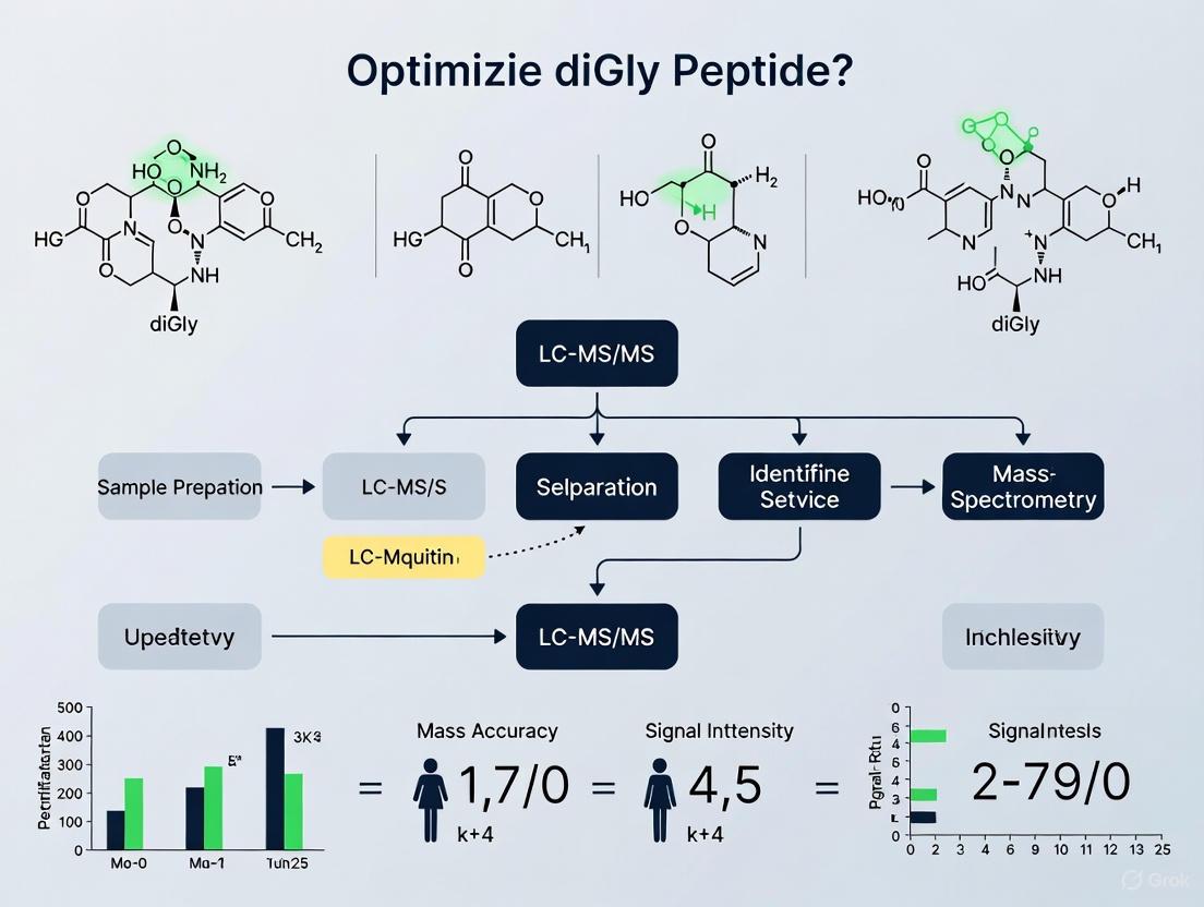
LC-MS/MS Analysis of Ubiquitinated Peptides
Sample Preparation for diGly Peptide Enrichment
The analysis of ubiquitination sites through LC-MS/MS relies on the detection of diGly remnant peptides after tryptic digestion, which leaves a characteristic glycine-glycine modification on the lysine residue where ubiquitin was attached. Sample preparation must be optimized to ensure efficient identification and quantification of these peptides.
Key Considerations:
- Peptide extraction: Use 66.7% ethanol in water for efficient extraction of amphipathic peptides, as this provides significantly greater yields compared to other organic solvents [6].
- Protease selection: Trypsin is preferred as it cleaves after lysine and arginine residues, generating the K-ε-GG remnant that serves as a signature for ubiquitination sites.
- Enrichment strategies: Implement anti-diGly antibody enrichment to selectively isolate ubiquitinated peptides from complex digests, significantly enhancing detection sensitivity.
- LC-MS/MS configuration: Utilize nanoflow liquid chromatography coupled to tandem mass spectrometry for optimal sensitivity in detecting low-abundance diGly peptides.
LC-MS/MS Instrument Configuration for diGly Peptide Detection
Liquid Chromatography Conditions:
- Column: C18 reversed-phase column (150 mm × 0.075 mm, 1.7 μm particles) for high-resolution separation
- Mobile phase: A: 0.1% formic acid in water; B: 0.1% formic acid in acetonitrile
- Gradient: 5-35% B over 120 minutes for complex samples
- Flow rate: 300 nL/min for nanoflow applications
Mass Spectrometry Parameters:
- Ionization: Electrospray ionization in positive mode
- Data acquisition: Data-dependent acquisition (DDA) with survey scans at 60,000 resolution followed by MS/MS of top 15 precursors
- Fragmentation: Higher-energy collisional dissociation (HCD) with normalized collision energy of 28-32%
- Isolation window: 1.4 m/z for precursor selection
Quantification Methods: For label-free quantification of diGly peptides, software tools such as LFQuant and MaxQuant provide robust analysis platforms. LFQuant has demonstrated superior performance in terms of precision and accuracy while consuming significantly less processing time compared to other quantification packages [7]. These tools reconstruct peptide extracted ion chromatograms and enable cross-assignment among different runs to compensate for the random effect of MS/MS sampling [7].
Diagram 2: LC-MS/MS workflow for diGly peptide analysis
Research Reagent Solutions Toolkit
Table 3: Essential Research Reagents for Ubiquitination Studies
| Reagent/Category | Specific Examples | Function/Application | Key Features |
|---|---|---|---|
| E1 Enzymes | Ube1, Uba6 [4] | Ubiquitin activation | ATP-dependent; initiates cascade; high specificity for UB C-terminus |
| E2 Enzymes | Ube2L3 (UbcH7), Ube2O, Ube2W [3] | Ubiquitin conjugation | Determines chain topology; Ube2O functions as E2/E3 hybrid [5] |
| E3 Ligases | SCF complexes, Mdm2, BRCA1 [1] | Substrate recognition & ubiquitin ligation | Provide substrate specificity; RING & HECT types |
| Ubiquitin Variants | Leu73Phe, Leu73Tyr, Gly75Ser/Asp/Asn [4] | Mechanism studies | DUB-resistant; block E2-E3 transfer; study chain assembly |
| LC-MS/MS Tools | LFQuant, MaxQuant [7] [8] | Data analysis | Label-free quantification; visualization; high precision |
| Internal Standards | Isotopically labeled ubiquitin | MS quantification | Normalization; accurate quantification of ubiquitination |
Therapeutic Applications and Drug Discovery
The ubiquitin conjugation cascade represents a promising target for therapeutic intervention, with several components currently under investigation for drug development. The proteasome inhibitor bortezomib (Velcade) was the first FDA-approved drug targeting this pathway, demonstrating the clinical validity of modulating protein degradation for cancer treatment [1] [2]. Current drug discovery efforts are increasingly focused on developing more specific inhibitors that target individual components of the cascade, particularly E3 ligases, which offer the greatest potential for specificity due to their role in substrate recognition [1].
E3 ligases such as Mdm2 (Hdm2 in humans) represent particularly attractive targets, as they regulate key tumor suppressors like p53. Mdm2 is overexpressed in many human cancers, including breast, esophageal, and lung cancers, with high levels associated with poor prognosis [1]. Inhibiting Mdm2's E3 ligase activity can reactivate p53-mediated tumor suppression, providing a promising therapeutic strategy. Similarly, SCF complex components are frequently dysregulated in cancer, with Cul4A gene amplification in breast cancers and Skp2 overexpression in various tumors [1].
Beyond cancer, E3 ligases have been implicated in neurodegenerative disorders (Parkinson's, Alzheimer's, and Huntington's disease), viral diseases (HIV and herpesvirus), cardiovascular diseases, and metabolic disorders including diabetes and obesity [1]. The ongoing development of robust high-throughput screening assays for E1, E2, and E3 enzymes is removing previous technical barriers and accelerating drug discovery efforts in this field [1]. The current climate of ubiquitin drug discovery is highly reminiscent of early kinase drug discovery, suggesting substantial growth potential for this therapeutic approach [1].
Protein ubiquitination is a crucial post-translational modification (PTM) involved in virtually all cellular processes, from proteasomal degradation to kinase signaling and DNA damage response [9]. The ability to study this modification on a large scale was revolutionized by the discovery that tryptic digestion of ubiquitinated proteins generates a characteristic signature—the lysine-ε-glycyl-glycine (K-ε-GG or "diGly") remnant—that can be specifically enriched and detected by mass spectrometry (MS) [10] [11]. This application note details the underlying biochemistry of the diGly remnant and provides optimized protocols for its detection, framed within the context of enhancing sensitivity and reproducibility in LC-MS/MS-based ubiquitinome research. We present standardized methodologies, key performance metrics, and strategic considerations for researchers aiming to implement or improve diGly peptide detection in their experimental workflows.
Ubiquitin is a 76-amino acid protein that is covalently attached to substrate proteins via an isopeptide bond between its C-terminal glycine and the ε-amino group of a lysine residue on the target protein [9]. During proteomic analysis, proteins are typically digested with the protease trypsin to generate peptides amenable to LC-MS/MS analysis. When trypsin digests a ubiquitinated protein, it cleaves after arginine and lysine residues. However, the isopeptide bond between the substrate lysine and the ubiquitin moiety is not a canonical trypsin cleavage site.
This specific cleavage behavior results in a diagnostic signature: the C-terminal two glycine residues of ubiquitin (Leu-Arg-Gly-Gly) remain attached to the modified lysine residue on the substrate peptide, generating a K-ε-GG-modified peptide, commonly referred to as the "diGly remnant" [10] [11] [12]. This remnant serves as a detectable mark of the original ubiquitination site. It is critical to note that while this signature is highly specific for ubiquitin, the ubiquitin-like modifiers NEDD8 and ISG15 also generate an identical diGly remnant upon tryptic digestion, meaning that enrichment of diGly peptides captures a small percentage of peptides modified by these related proteins [10] [13]. Seminal work using antibodies targeting this diGly motif has enabled the immunoaffinity enrichment of these modified peptides from complex biological samples, facilitating the large-scale identification and quantification of ubiquitination sites by mass spectrometry [10] [11] [12].
Key Reagents and Materials for diGly Proteomics
The following table catalogues essential reagents and materials required for successful diGly remnant enrichment and detection.
Table 1: Essential Research Reagents for diGly Proteomics
| Reagent/Material | Function/Application | Key Considerations |
|---|---|---|
| Anti-K-ε-GG Antibody [10] [9] | Immunoaffinity enrichment of diGly-modified peptides | Core reagent for peptide pull-down. Commercial kits are available (e.g., PTMScan). |
| Trypsin [10] [14] | Protein digestion to generate diGly remnants | High specificity for cleavage after Arg and Lys; TPCK-treated is recommended to inhibit chymotrypsin activity. |
| Lys-C Protease [10] [9] | Protein digestion; used prior to trypsin | Efficiently digests proteins in denaturing buffers; used in parallel or prior to trypsin digestion. |
| N-Ethylmaleimide (NEM) [10] | Deubiquitinase (DUB) inhibitor | Preserves ubiquitination signature by inhibiting DUBs during cell lysis. Must be prepared fresh. |
| Urea Lysis Buffer [10] | Protein denaturation and extraction | Standard buffer: 8M Urea, 150mM NaCl, 50mM Tris-HCl, pH 8. |
| SilAC Media Kits [10] [9] | Metabolic labeling for quantitative proteomics | DMEM lacking Lys/Arg, supplemented with heavy ("R10K8") or light isotopes. |
| C18 Reverse-Phase Columns [10] [9] | Peptide desalting and fractionation | Critical for sample cleanup and pre-fractionation to reduce complexity before enrichment. |
Optimized Protocol for diGly Peptide Enrichment and Analysis
This section provides a detailed, step-by-step protocol for the detection of ubiquitination sites via diGly remnant enrichment, incorporating best practices for sample preparation, fractionation, and mass spectrometry analysis to achieve optimal depth of coverage.
Sample Preparation and Protein Digestion
- Cell Lysis: Lyse cells or tissue in a denaturing lysis buffer (e.g., 8M Urea, 150mM NaCl, 50mM Tris-HCl, pH 8) supplemented with complete protease inhibitors and 5mM N-Ethylmaleimide (NEM) to inhibit deubiquitinating enzymes [10]. Boiling the lysate at 95°C for 5 minutes in a buffer containing 0.5% sodium deoxycholate (DOC) is also an effective method to denature proteins and inactivate enzymes [9].
- Protein Quantification: Determine the total protein concentration using a colorimetric assay (e.g., BCA assay). For a successful diGly immunoprecipitation, start with a total protein amount of at least several milligrams [9].
- Reduction and Alkylation: Reduce disulfide bonds with 5mM dithiothreitol (DTT) for 30 minutes at 50°C. Subsequently, alkylate cysteine residues with 10mM iodoacetamide (IAA) for 15 minutes in the dark [9].
- Protein Digestion: First, digest proteins with Lys-C protease (1:200 enzyme-to-substrate ratio) for 4 hours. Then, dilute the sample to reduce urea concentration and perform an overnight digestion with trypsin (1:50 enzyme-to-substrate ratio) at 30°C or room temperature [10] [9].
- Peptide Cleanup: Acidify the digested peptide sample by adding trifluoroacetic acid (TFA) to a final concentration of 0.5%. Centrifuge at 10,000 x g for 10 minutes to precipitate and remove detergents. Collect the supernatant containing the peptides [9]. Desalt the peptides using a C18 reverse-phase Sep-Pak column [10].
Peptide Pre-fractionation and diGly Enrichment
- Offline High-pH Reverse-Phase Fractionation: To significantly increase the depth of analysis, fractionate the tryptic peptides prior to diGly enrichment using a high-pH reverse-phase C18 column.
- diGly Peptide Immunoaffinity Enrichment:
- Reconstitute each peptide fraction in immunoaffinity purification (IAP) buffer (e.g., 50 mM MOPS pH 7.2, 10 mM Na2HPO4, 50 mM NaCl).
- Use the Ubiquitin Remnant Motif (K-ε-GG) Antibody (conjugated to protein A agarose beads) for enrichment. A typical enrichment uses one batch of beads per fraction, as defined by the manufacturer [9].
- Incubate the peptides with the antibody beads for 2 hours at 4°C with gentle agitation.
- Wash the beads multiple times with IAP buffer and then with HPLC-grade water to remove non-specifically bound peptides.
- Elute the bound diGly peptides with 0.2% TFA. Desalt the eluted peptides using C18 StageTips or micro-columns prior to LC-MS/MS analysis [10] [9].
The following workflow diagram summarizes the core experimental protocol.
Diagram 1: Core diGly Peptide Analysis Workflow
Optimizing LC-MS/MS for Maximum diGly Peptide Detection
The unique properties of diGly peptides necessitate specific optimization of mass spectrometry parameters. diGly peptides are often longer and carry higher charge states compared to unmodified peptides due to impeded C-terminal cleavage at the modified lysine [12].
Data Acquisition: DDA vs. DIA
The choice of data acquisition method profoundly impacts the depth and quantitative quality of ubiquitinome analysis. The table below compares the two primary approaches.
Table 2: Quantitative Performance of DDA vs. DIA for diGly Proteomics
| Parameter | Data-Dependent Acquisition (DDA) | Data-Independent Acquisition (DIA) |
|---|---|---|
| Principle | Intensity-based selection of top N precursors for MS/MS [16] | Parallel fragmentation of all precursors in pre-defined m/z windows [12] |
| Typical diGly Peptides ID (Single Shot) | ~20,000 peptides [12] | ~35,000 peptides [12] |
| Quantitative Reproducibility (CV < 20%) | ~15% of peptides [12] | ~45% of peptides [12] |
| Advantages | Well-established, simpler data analysis | Superior sensitivity, quantitative accuracy, and data completeness [12] |
| Disadvantages | Missing values, lower dynamic range | Requires comprehensive spectral library |
For DIA, specific optimizations are critical:
- Isolation Windows: Optimize the width and number of precursor isolation windows to match the unique precursor distribution of diGly peptides, improving identifications by over 10% [12].
- MS2 Resolution: A higher fragment scan resolution (e.g., 30,000) improves identification rates [12].
- Spectral Libraries: Using a project-specific or consolidated spectral library containing over 90,000 diGly peptides is essential for sensitive DIA analysis [12].
Chromatography and Instrument Tuning
- Fast Chromatography: When using fast LC separations with narrow peak widths (a few seconds), ensure that Data-Dependent Acquisition (DDA) settings are optimized to match. This includes adjusting dynamic exclusion and minimum signal thresholds to prevent oversampling of high-intensity peptides and allow acquisition of lower-abundance species [16].
- Peptide Load and Antibody Titration: For a standard experiment using 1 mg of peptide material from untreated cells, optimal results are achieved using approximately 31.25 µg of anti-diGly antibody. With the high sensitivity of optimized DIA methods, injecting only 25% of the total enriched material can be sufficient [12].
The strategic relationship between sample preparation, fractionation, and MS acquisition in achieving optimal depth of analysis is outlined below.
Diagram 2: Strategies for Depth of Analysis in diGly Proteomics
The diGly remnant signature, a direct product of tryptic digestion of ubiquitinated proteins, provides a powerful and specific handle for system-wide ubiquitinome analysis. The robustness of this approach is evidenced by its application across diverse sample types, from cultured cells to complex tissues like mouse brain [15] [9]. As detailed in this application note, the depth and quality of results are highly dependent on a meticulously optimized workflow—from the use of DUB inhibitors during lysis and pre-enrichment fractionation, to the adoption of tailored DIA-based mass spectrometry methods. By implementing the optimized protocols and strategic considerations outlined herein, researchers can reliably uncover the deep ubiquitinome to answer critical biological questions in signaling, proteostasis, and drug mechanism of action.
Ubiquitination is a crucial post-translational modification (PTM) that regulates diverse cellular functions, including protein degradation, signaling, and trafficking [17]. The versatility of ubiquitination stems from its complexity—it can target numerous protein substrates at various lysine residues and form polymers (polyUb chains) with different linkage types, each potentially encoding distinct functional outcomes [18] [17]. The system-wide analysis of protein ubiquitination, however, presents significant challenges due to the low stoichiometry of modified proteins, the dynamic nature of the modification, and the complexity of ubiquitin chain architectures [17].
A major breakthrough in ubiquitin research came with the realization that trypsinolysis of ubiquitinated proteins generates a characteristic "diGly remnant" on modified lysine residues—a consequence of cleavage after the C-terminal Arg-Gly-Gly motif of ubiquitin [18]. This diGly signature, with a mass shift of 114.04 Da on modified lysines, provides a unique handle for proteomic detection [17]. The development of antibodies specifically recognizing this K-ε-GG motif enabled immunoaffinity enrichment of diGly-containing peptides, revolutionizing the field of ubiquitinomics [18] [9]. Despite these advances, significant challenges remain in achieving comprehensive coverage of the "ubiquitinome," including the persistent issues of low stoichiometry, sequence bias in detection, and the considerable "dark ubiquitylome" that remains uncharacterized [17].
Key Challenges in DiGly Proteomics
Low Stoichiometry of Ubiquitination
The low abundance of ubiquitinated peptides relative to their unmodified counterparts presents a fundamental analytical challenge. Unlike phosphorylation or acetylation, which can affect substantial fractions of a target protein population, ubiquitination often occurs at very low stoichiometries under normal physiological conditions [17]. This is particularly true for regulatory ubiquitination events that trigger proteasomal degradation, where the modified proteins are rapidly destroyed, maintaining low steady-state levels of ubiquitinated species [18]. Quality control ubiquitination that targets misfolded or damaged proteins similarly affects only a small fraction of the total protein pool [18].
The low stoichiometry necessitates extensive enrichment prior to mass spectrometric analysis to avoid suppression of diGly peptide signals by unmodified peptides. Even with effective enrichment strategies, the detection of endogenously modified proteins remains challenging without experimental manipulation such as proteasome inhibition to increase the abundance of ubiquitinated substrates [18]. This manipulation, while increasing coverage, may distort the physiological landscape of ubiquitination.
The 'Dark Ubiquitylome' and Technical Limitations
The "dark ubiquitylome" refers to the substantial portion of ubiquitination events that remain undetected by current methodologies. Early proteomic studies identified only hundreds of ubiquitylation sites, but as techniques have improved, this number has expanded dramatically to over 20,000 sites [18] [15] [9]. Nevertheless, the full extent of the ubiquitinome remains uncharted territory.
Technical limitations contributing to the dark ubiquitylome include:
- Inefficient enrichment: Not all diGly peptides immunoprecipitate with equal efficiency using current antibodies [9]
- Dynamic range limitations: Low-abundance ubiquitinated peptides are obscured by high-abundance proteins despite enrichment [17]
- Sample complexity: Interference from non-ubiquitinated peptides co-purifying during enrichment reduces detection sensitivity [17]
- In vivo sampling limitations: Most deep ubiquitinome analyses require cell culture models amenable to genetic manipulation or proteasome inhibition, limiting physiological relevance [9]
Sequence and Context-Dependent Biases
Not all ubiquitination sites are equally detectable by mass spectrometry. Several factors introduce sequence and context-dependent biases in diGly proteomics:
- Trypsin digestion bias: The diGly remnant itself alters the tryptic cleavage pattern, potentially generating peptides with non-optimal properties for LC-MS/MS analysis [18]
- Peptide physicochemical properties: DiGly peptides with favorable hydrophobicity, length, and charge characteristics are more readily detected [19]
- Ionization efficiency: Variations in how different diGly peptides ionize under electrospray conditions create detection biases [19]
- Linkage-type specific biases: Certain ubiquitin linkage types (e.g., K48, K63) may exhibit different enrichment or detection efficiencies, though quantitative studies show distinct dynamics for different linkage types in response to proteasome inhibition [18]
Methodological Advances and Optimization Strategies
Enhanced DiGly Peptide Enrichment Workflows
Recent methodological improvements have significantly increased the depth of ubiquitinome coverage. The key advances include offline high-pH reverse-phase fractionation prior to immunoenrichment, improved wash steps to reduce non-specific binding, and more efficient peptide fragmentation settings in mass spectrometers [15] [9].
The optimized workflow typically involves:
- Denaturing lysis to preserve ubiquitination status and prevent deubiquitination [9]
- Reduction and alkylation followed by tryptic digestion [9]
- Offline high-pH reverse-phase fractionation into a limited number of fractions (e.g., 3 fractions) to reduce sample complexity [15] [9]
- Immunoaffinity enrichment using anti-diGly antibodies [18] [9]
- LC-MS/MS analysis with optimized fragmentation settings [15]
This optimized approach has enabled the identification of over 23,000 diGly peptides from a single sample of HeLa cells treated with proteasome inhibitor, representing a substantial improvement over earlier methods [15] [9].
Liquid Chromatography and Mass Spectrometry Optimization
Optimal LC-MS/MS parameters are critical for comprehensive diGly peptide identification. Key considerations include:
Liquid Chromatography:
- Column selection: C18 columns with appropriate pore size and particle diameter [20]
- Gradient optimization: Shallow gradients improve separation of complex peptide mixtures [20]
- Mobile phase composition: Standard acidified acetonitrile/water gradients are typically employed [20]
Mass Spectrometry:
- Fragmentation techniques: Higher-energy collision dissociation (HCD) provides optimal fragmentation for diGly peptides [15]
- Data acquisition modes: Both data-dependent acquisition (DDA) and data-independent acquisition (DIA) approaches have been applied [21]
- Mass analyzer settings: Orbitrap instruments provide the resolution and mass accuracy needed for confident identifications [15]
The following table summarizes key quantitative improvements achieved through method optimization:
Table 1: Performance Metrics of DiGly Proteomics Methods
| Method Parameter | Early Methods | Optimized Methods | Improvement Factor |
|---|---|---|---|
| DiGly Sites per Experiment | 374-753 sites [18] | 19,000-23,000 sites [18] [15] | ~25-60x |
| Protein Coverage | ~500 proteins [18] | ~5,000 proteins [18] | ~10x |
| Sample Throughput | Low (single samples) | Moderate (fractionated samples) | Improved depth |
| Reproducibility | Moderate between replicates | High correlation between replicates [18] | Significant improvement |
Quantitative DiGly Proteomics Approaches
Stable Isotope Labeling with Amino acids in Cell culture (SILAC) has been successfully applied in diGly proteomics to monitor temporal changes in the ubiquitinome in response to cellular perturbations [18]. In a typical experiment, cells are cultured in "light" or "heavy" media containing normal or stable isotope-labeled lysine (e.g., Lys + 8 Da shift), respectively [9]. After treatment (e.g., with proteasome inhibitors such as bortezomib), light and heavy cells are mixed in a 1:1 ratio based on protein content, followed by digestion, diGly peptide enrichment, and LC-MS/MS analysis [18] [9].
This approach has revealed that approximately 58% of quantified ubiquitination sites increase by more than 2-fold in abundance following proteasome inhibition, while about 13% decrease by more than 2-fold [18]. Interestingly, proteins often contain multiple ubiquitination sites that exhibit distinct regulatory behaviors, suggesting complex regulation of site-specific ubiquitination [18].
Experimental Protocols
Detailed Protocol for DiGly Peptide Enrichment and Analysis
Materials and Reagents:
- Cell line of interest (e.g., HeLa, HCT116, U2OS)
- SILAC media kits (for quantitative experiments)
- Proteasome inhibitors (e.g., bortezomib, epoxomycin)
- Lysis buffer: 50 mM Tris-HCl (pH 8.2) with 0.5% sodium deoxycholate [9]
- Reduction and alkylation reagents: DTT and iodoacetamide
- Proteases: Lys-C and trypsin
- Trifluoroacetic acid (TFA)
- Anti-K-ε-GG antibody-conjugated beads
- C18 reverse-phase columns for fractionation
- LC-MS/MS system (Orbitrap instruments recommended)
Procedure:
Cell Culture and Treatment:
Cell Lysis and Protein Extraction:
- Lyse cells in denaturing lysis buffer (e.g., 50 mM Tris-HCl, pH 8.2, 0.5% sodium deoxycholate) with boiling at 95°C for 5 minutes to inactivate deubiquitinases [9].
- Sonicate lysates to reduce viscosity and complete disruption.
- Quantitate protein concentration using BCA assay.
Protein Digestion:
- Reduce proteins with 5 mM DTT for 30 minutes at 50°C.
- Alkylate with 10 mM iodoacetamide for 15 minutes in the dark.
- Digest first with Lys-C (1:200 enzyme-to-substrate ratio) for 4 hours, then with trypsin (1:50 ratio) overnight at room temperature [9].
- Acidify with TFA to 0.5% final concentration to precipitate detergents.
- Centrifuge at 10,000 × g for 10 minutes and collect supernatant containing peptides.
Peptide Fractionation:
DiGly Peptide Immunoaffinity Enrichment:
- Wash anti-diGly antibody-conjugated beads with PBS.
- Resuspend lyophilized fractions in immunoaffinity purification buffer (IAP buffer: 50 mM MOPS, pH 7.2, 10 mM Na2HPO4, 50 mM NaCl).
- Incubate peptides with antibody beads for 1.5 hours at 4°C with gentle agitation.
- Wash beads 3 times with IAP buffer and twice with water.
- Elute diGly peptides with 0.2% TFA.
LC-MS/MS Analysis:
- Analyze peptides using LC-MS/MS with Orbitrap mass spectrometer.
- Use C18 analytical column with gradient elution (e.g., 5-35% acetonitrile over 2 hours).
- Operate mass spectrometer in data-dependent acquisition mode with HCD fragmentation.
- Set MS1 resolution to 60,000 and MS2 resolution to 15,000.
- Use dynamic exclusion of 30 seconds to increase depth of coverage.
Table 2: Key Research Reagent Solutions for DiGly Proteomics
| Reagent/Category | Specific Examples | Function/Purpose | Considerations |
|---|---|---|---|
| Cell Lines | HCT116, HeLa, U2OS, HEK293T | Model systems for ubiquitinome profiling | Choose based on experimental context; consider genetic manipulability |
| Affinity Tags | His-tag, Strep-tag | Purification of ubiquitinated proteins when overexpressing tagged ubiquitin | May introduce artifacts; Strep-tag offers cleaner purification [17] |
| Ubiquitin Antibodies | Pan-specific (P4D1, FK1/FK2), Linkage-specific (K48, K63) | Enrichment of endogenously ubiquitinated proteins | Linkage-specific antibodies enable chain-type analysis [17] |
| Proteasome Inhibitors | Bortezomib, Epoxomycin | Increase ubiquitinated protein abundance | Different inhibitors have distinct specificities; use consistent concentrations [18] |
| Enrichment Beads | Protein A/G agarose | Immobilization of antibodies for immunopurification | Filter plug systems improve wash efficiency [15] |
| Chromatography Media | C18 reverse-phase, HILIC | Peptide separation and fractionation | High-pH fractionation reduces complexity prior to enrichment [15] |
Visualization of Experimental Workflows and Signaling Pathways
DiGly Proteomics Experimental Workflow
Diagram 1: DiGly Proteomics Workflow
Ubiquitin Signaling and Proteasome Pathway
Diagram 2: Ubiquitin Signaling and diGly Formation
The field of diGly proteomics has made remarkable progress in overcoming the challenges of low stoichiometry, sequence bias, and the dark ubiquitylome. The development of highly specific anti-diGly antibodies, combined with optimized sample preparation workflows and advanced mass spectrometry instrumentation, has enabled the identification of tens of thousands of ubiquitination sites from single experiments [18] [15]. Nevertheless, substantial challenges remain.
Future directions in diGly proteomics will likely focus on improving coverage of low-abundance regulatory ubiquitination events, developing better tools for distinguishing ubiquitin chain linkages, and enabling more robust quantitative analyses across diverse biological systems. The application of diGly proteomics to clinical samples and animal tissues represents another important frontier, as current methods often require genetic manipulation or large sample amounts that limit translational applications [9] [17].
As these methodologies continue to mature, diGly proteomics will provide increasingly powerful insights into the complex landscape of protein ubiquitination and its roles in health and disease. The integration of diGly datasets with other proteomic and functional genomic approaches will be essential for translating ubiquitinome maps into mechanistic understanding of ubiquitin-dependent cellular regulation.
Protein ubiquitination is a dynamic and multifaceted post-translational modification that extends far beyond the well-characterized Lys-48 (K48)-linked chains that target substrates for proteasomal degradation. The ubiquitin code encompasses at least eight distinct chain linkage types, formed through ubiquitin's seven lysine residues (K6, K11, K27, K29, K33, K48, K63) or N-terminal methionine (M1), creating extraordinary signaling diversity [22] [23]. This complexity is further enhanced by the formation of homotypic chains (uniform linkage), mixed chains (multiple linkage types with one modification site per ubiquitin), and branched chains (multiple linkage types with more than one modification site on at least one ubiquitin monomer) [24]. The specific biological outcomes of these diverse ubiquitin modifications—ranging from proteasomal degradation to non-degradative roles in signaling, DNA repair, and endocytosis—are determined by how they are recognized by ubiquitin-binding proteins (UBPs) containing specialized ubiquitin-binding domains (UBDs) [25] [22].
Advances in mass spectrometry, particularly antibody-based enrichment of diGly-containing peptides combined with liquid chromatography-tandem mass spectrometry (LC-MS/MS), have revolutionized our ability to study the ubiquitinome [26] [15]. These technological improvements now enable researchers to decipher the complex ubiquitin code with unprecedented depth and accuracy, revealing new layers of regulation in cellular processes. This application note provides detailed methodologies for analyzing homotypic and heterotypic ubiquitin chains, with a specific focus on optimizing LC-MS/MS settings for superior diGly peptide detection.
Key Biological Insights: Structural Diversity Dictates Functional Outcome
Linkage-Specific Fate Determination
Different ubiquitin linkage types create unique molecular surfaces that are specifically recognized by dedicated receptor proteins, leading to distinct cellular outcomes [27]. While K48-linked chains remain the canonical signal for proteasomal degradation, recent research has revealed unexpected nuances in how chain architecture influences protein fate.
Proteasomal Recognition of K11 Linkages: A pivotal study demonstrated that the proteasome distinguishes between homotypic and heterotypic K11-linked chains. Homotypic K11 chains do not bind strongly to mammalian 26S proteasomes and are inefficient degradation signals. In contrast, heterotypic K11/K48 chains bind effectively to the proteasome and stimulate degradation of cell-cycle regulators like cyclin B1 [25]. This discrimination occurs at the level of ubiquitin receptors Rpn10 and Rpn13 in the 19S regulatory particle, which show preferential binding for K48 linkages [25] [23].
Branched Chain Functions: Branched ubiquitin chains represent an emerging area of research, with specific branched linkages performing specialized functions. For example, K48/K63-branched chains are synthesized by collaborating E3 ligases (TRAF6 and HUWE1) during NF-κB signaling [24]. Similarly, the anaphase-promoting complex/cyclosome (APC/C) collaborates with E2 enzymes UBE2C and UBE2S to form branched K11/K48 chains on mitotic substrates [24]. These architectures potentially allow integration of degradative and non-degradative signals or enhance substrate affinity for proteasomal receptors.
Table 1: Functional Outcomes of Major Ubiquitin Linkage Types
| Linkage Type | Chain Architecture | Primary Functions | Cellular Processes |
|---|---|---|---|
| K48 | Homotypic | Proteasomal degradation [23] | Cell cycle, protein turnover |
| K11 | Homotypic | Proteasome-independent functions [25] | Mitotic regulation, endocytosis |
| K11/K48 | Heterotypic/Branched | Proteasomal degradation [25] [24] | Cell cycle progression |
| K63 | Homotypic | Non-degradative signaling [23] | DNA repair, inflammation, endocytosis |
| K48/K63 | Branched | Signaling & degradation integration [24] | NF-κB signaling, apoptosis |
| M1 (Linear) | Homotypic | NF-κB activation [22] | Inflammation, immunity |
Quantitative Analysis of Ubiquitin Chain Biology
Mass spectrometry-based approaches have enabled quantitative assessment of ubiquitin chain dynamics. Key findings include:
Proteasome Binding Affinities: Competition assays reveal that K48-linked tetraubiquitin (K48-Ub4) binds the proteasome with an approximate affinity constant (Ka) of 70 nM, while K11-Ub4 shows no significant competition even at 300 nM concentrations [25].
Method-Dependent Identification Rates: Data-independent acquisition (DIA) methods identify approximately 35,000 distinct diGly peptides in single measurements of proteasome inhibitor-treated cells, doubling the identification rate compared to data-dependent acquisition (DDA) [26]. The coefficient of variation for DIA quantification is <20% for 45% of diGly peptides and <50% for 77% of peptides, demonstrating superior reproducibility [26].
Cellular Ubiquitin Distribution: Quantitative proteomics indicates K48 linkages constitute >50% of all ubiquitin chains in cells, with K63 being the second most abundant. Treatment with proteasome inhibitor MG132 causes rapid accumulation of K48 linkages, confirming their dominant role in proteasomal targeting [23].
Experimental Protocols: Optimized Methodologies for Ubiquitinome Analysis
Sample Preparation for Deep Ubiquitinome Coverage
Cell Culture and Treatment:
- Culture HEK293 or U2OS cells in standard conditions. For proteasome inhibition, treat with 10 µM MG132 for 4 hours to increase ubiquitinated substrate levels [26].
- Include untreated controls to identify basal ubiquitination patterns without stress-induced perturbations.
Protein Extraction and Digestion:
- Lyse cells in urea-based buffer (8 M urea, 100 mM NH₄HCO₃, pH 8.0) supplemented with protease and phosphatase inhibitors.
- Reduce disulfide bonds with 5 mM dithiothreitol (60 minutes at 37°C) and alkylate with 15 mM iodoacetamide (30 minutes in darkness at room temperature).
- Dilute urea concentration to 2 M and digest with trypsin (1:50 enzyme-to-protein ratio) overnight at 37°C [26].
- Desalt peptides using C18 solid-phase extraction cartridges and lyophilize.
Critical Considerations: For tissue samples, anatomical structure matters (e.g., kidney cortex vs. medulla). Frozen tissues generally yield better protein recovery than FFPE samples, though optimized protocols exist for FFPE material [28].
diGly Peptide Enrichment Protocol
Immunoaffinity Purification:
- Reconstitute dried peptide samples in IAP buffer (50 mM MOPS/NaOH, pH 7.4, 10 mM Na₂HPO₄, 50 mM NaCl).
- Use 1 mg of peptide material per enrichment reaction with 31.25 µg of anti-diGly antibody (PTMScan Ubiquitin Remnant Motif Kit, CST) [26].
- Incubate with rotation for 2 hours at 4°C.
- Wash beads three times with IAP buffer and twice with HPLC-grade water.
- Elute diGly peptides with 0.15% trifluoroacetic acid (2 × 75 µL).
Fractionation Optimization: For ultra-deep coverage, fractionate peptides by basic reversed-phase chromatography (bRP) into 96 fractions before enrichment, then concatenate into 8-12 fractions [26]. Process K48-rich fractions separately to prevent interference with lower-abundance peptides [26].
LC-MS/MS Analysis with Optimized Settings
Liquid Chromatography:
- Use nano-flow LC systems with C18 columns (75 µm × 25 cm, 1.6 µm particle size).
- Employ a 120-minute gradient from 2% to 30% acetonitrile in 0.1% formic acid at 300 nL/min flow rate.
Data-Independent Acquisition (DIA) Parameters:
- MS1: Resolution 120,000, scan range 350-1650 m/z, AGC target 3e6.
- DIA: 46 variable windows covering 400-1000 m/z, higher resolution (30,000) for MS2 scans [26].
- HCD collision energy: 28-32%.
- Maximum injection time: 55 ms for MS2.
Data-Dependent Acquisition (DDA) Alternative:
- MS1: Resolution 120,000, AGC target 3e6.
- MS2: Top 20 most intense precursors, resolution 30,000, AGC target 1e5.
- HCD collision energy: 28%, dynamic exclusion 30 seconds.
Table 2: Optimized LC-MS/MS Parameters for diGly Proteome Analysis
| Parameter | DDA Setting | DIA Setting | Rationale |
|---|---|---|---|
| MS1 Resolution | 120,000 | 120,000 | Precise precursor quantification |
| MS2 Resolution | 30,000 | 30,000 | Improved fragment ion detection |
| Precursor Isolation | Top 20 ions | 46 variable windows | Comprehensive sampling |
| Collision Energy | 28% | 28-32% | Optimal diGly peptide fragmentation |
| Maximum IT | 55 ms | 55 ms | Balance sensitivity & cycle time |
| Peptide Input | 1-4 μg | 0.25-1 μg | Reduced sample requirements |
Data Processing and Analysis
Spectral Library Generation:
- Combine DDA data from multiple samples (different cell lines, treatments) to create a comprehensive library.
- Include direct DIA search results to create a hybrid library for increased identifications [26].
DIA Data Analysis:
- Process using Spectronaut, DIA-NN, or Skyline with default settings.
- Use hybrid library containing >90,000 diGly peptides for optimal matching [26].
- Apply false discovery rate (FDR) threshold of 1% at both peptide and protein levels.
Quality Control Metrics:
- Monitor coefficient of variation (CV) between replicates; target <20% for most peptides.
- Assess enrichment specificity by percentage of diGly peptides in total identifications (>80% expected).
- Evaluate quantitative accuracy using technical replicates.
The Scientist's Toolkit: Essential Research Reagents
Table 3: Key Research Reagents for Ubiquitinome Analysis
| Reagent/Catalog Number | Function | Application Notes |
|---|---|---|
| Anti-K-ε-GG Antibody (CST #5562) | Immunoaffinity enrichment of diGly peptides | Use 31.25 μg per 1 mg peptide input; critical for deep coverage [26] |
| UBE2S (E2 enzyme) | Synthesis of homotypic K11-linked chains | Used with truncated Ube2S (Ube2SΔ) for specific K11 chain formation [25] |
| E6AP (HECT E3 ligase) | Generation of K48-linked ubiquitin chains | For producing reference K48 chains in binding assays [25] |
| MG132 (proteasome inhibitor) | Increases ubiquitinated substrates | 10 μM, 4-hour treatment significantly enhances diGly peptide yield [26] |
| Recombinant 26S Proteasome | Ubiquitin chain binding assays | Used to measure linkage-specific proteasome affinity [25] [23] |
| AQUA Ubiquitin Peptides | Absolute quantification of linkages | Heavy isotope-labeled standards for precise quantification [25] |
| Linkage-Specific DUBs | Validation of chain linkage | Enzymes with defined linkage specificity confirm chain architecture |
Advanced Applications and Future Perspectives
The optimized workflows described here enable researchers to address previously challenging questions in ubiquitin biology. The superior quantitative accuracy and sensitivity of DIA methods make it possible to monitor dynamic changes in ubiquitination during cellular processes like cell cycle progression, circadian regulation, and signal transduction [26]. For example, applying these methods to TNFα signaling has identified novel ubiquitination sites beyond previously known ones [26]. Similarly, analysis across circadian cycles has revealed hundreds of cycling ubiquitination sites, including clusters within individual membrane receptors and transporters [26].
Future directions will likely focus on improving methods to decipher complex ubiquitin architectures, particularly branched chains, and understanding the crosstalk between ubiquitination and other post-translational modifications. The development of improved mass spectrometry instrumentation, enrichment strategies, and computational tools will continue to deepen our understanding of the functional diversity of homotypic and heterotypic ubiquitin chains.
Visualizing Key Concepts and Workflows
Ubiquitin Chain Diversity and Cellular Fates
Ubiquitin Chain Types and Fates Diagram: This visualization illustrates how different ubiquitin chain architectures lead to distinct cellular outcomes, highlighting the critical finding that heterotypic K11/K48-branched chains signal proteasomal degradation while homotypic K11 chains do not [25] [24].
Optimized diGly Proteome Workflow
Optimized diGly Proteome Workflow: This diagram outlines the comprehensive workflow for deep ubiquitinome analysis, highlighting critical optimization points including pre-enrichment fractionation, optimized antibody-to-peptide ratios, and DIA-specific MS settings that collectively enable identification of over 35,000 diGly sites in single measurements [26] [15].
Distinguishing Ubiquitination from NEDD8 and ISG15 Modifications
The ubiquitin-like modifier (Ubl) family, including ubiquitin, NEDD8, and ISG15, regulates virtually every physiological process in eukaryotic cells by post-translationally modifying substrate proteins. These modifications are conjugated via enzymatic cascades to lysine residues on target proteins, and all three can generate a diglycine (diGly) remnant on modified peptides after tryptic digestion. This common signature presents a significant challenge for liquid chromatography-tandem mass spectrometry (LC-MS/MS) analyses aimed at distinguishing the specific modification type. Accurate differentiation is critical because these modifications dictate distinct biological outcomes: ubiquitin primarily targets proteins for proteasomal degradation and regulates signaling, NEDD8 predominantly activates cullin-RING ligases and controls proteotoxic stress responses, and ISG15 serves as a key antiviral effector in the innate immune system. This application note details protocols and strategies for the specific identification and validation of these modifications within the context of optimizing LC-MS/MS for diGly peptide detection.
Biological Functions and Distinguishing Features
Understanding the distinct biological roles and molecular characteristics of ubiquitin, NEDD8, and ISG15 is the foundation for developing specific detection strategies.
- Ubiquitin (Ub): A 76-amino acid protein, ubiquitin is conjugated to substrates via a three-step enzymatic cascade (E1-E2-E3) and can form chains through any of its seven lysine residues or its N-terminus. While best known for targeting proteins for degradation via the 26S proteasome (typically through K48-linked chains), ubiquitination also regulates non-proteolytic processes including endocytosis, kinase signaling, and the DNA damage response (particularly through K63-linked chains). Deubiquitinating enzymes (DUBs) reverse this modification [29] [30].
- NEDD8: This Ubl shares 60% sequence identity with ubiquitin and is conjugated via its own canonical E1 (NAE1-UBA3) and E2 (UBC12) enzymes. Its most characterized function is the activation of cullin-RING E3 ligases (CRLs) via conjugation to cullin proteins. NEDD8 modification can also occur through "atypical" pathways involving enzymes of the ubiquitin system, particularly under proteotoxic stress. Atypical neddylation targets distinct proteomes, such as the ribosome and proteasome, and can form hybrid chains with ubiquitin and SUMO-2. The deneddylase NEDP1 reverses this modification [31].
- ISG15: This unique Ubl consists of two ubiquitin-like domains in tandem. Its expression is strongly induced by type I interferon (IFN) in response to viral or bacterial infection. ISG15 is conjugated by a dedicated cascade (E1: UBE1L; E2: UBCH8; E3: HERC5) to hundreds of target proteins, often co-translationally, to inhibit virus replication. ISG15 can also exist as an unconjugated extracellular cytokine. DeISGylating enzymes (DIGs), such as USP18, reverse this modification [29] [32].
The following diagram summarizes the core conjugation pathways and primary biological roles for each modifier.
Analytical Challenges in diGly-Based Detection
The standard MS-based workflow for identifying these PTMs involves tryptic digestion of modified proteins, enrichment of peptides containing the K-ε-GG remnant using specific antibodies, and subsequent LC-MS/MS analysis. While powerful, this approach faces several key challenges for distinguishing the modifying Ubl.
- Identical Mass Shift: The tryptic diGly remnant left by all three modifiers results in an identical +114.0429 Da mass shift on modified lysines, making them indistinguishable by mass shift alone [15] [9].
- Sequence Homology: The high degree of sequence and structural similarity between ubiquitin and NEDD8 means that many ubiquitin-binding domains (UBDs) can bind NEDD8 promiscuously, complicating affinity-based purification [30].
- Cross-Talk and Hybrid Chains: Biological cross-talk exists where Ubls can modify each other. For instance, ISG15 can form mixed chains with ubiquitin, using Lys29 on ubiquitin as the major acceptor site. Similarly, NEDD8 can form hybrid chains with ubiquitin and SUMO-2 [31] [32]. These hybrid structures can lead to misassignment if not carefully considered.
- Variable Stoichiometry: The stoichiometry of these modifications is often low compared to unmodified peptides, necessitating robust enrichment to avoid missing biologically relevant modifications.
Key Methodological Strategies for Differentiation
To overcome these challenges, researchers must employ specific methodological strategies prior to and during LC-MS/MS analysis.
Genetic and Proteomic Manipulations
- Use of Ubl Mutants: A key strategy for distinguishing NEDD8 modification sites is the use of the NEDD8 R74K mutant. This mutant is processed and conjugated like wild-type NEDD8 but, upon tryptic digestion, generates a K-ε-GG signature with a distinct +383.2281 Da mass shift, allowing it to be differentiated from ubiquitin and ISG15 by a unique mass shift during MS analysis [31].
- Enzymatic Depletion and Inhibition: Treatment with specific deconjugating enzymes (DUBs, DEN1/NEDP1, or DIGs like USP18) prior to analysis can remove specific modifications, allowing for the identification of substrates by comparing treated and untreated samples. Similarly, inhibiting specific E1 activating enzymes (e.g., MLN4924 for NEDD8) can reduce or eliminate specific conjugation cascades.
Affinity Enrichment and Sample Preparation
- Optimized diGly Peptide Immunopurification: The core enrichment protocol for diGly peptides can be significantly improved. Key adaptations include:
- Offline high-pH reverse-phase fractionation of peptides into three fractions (e.g., 7%, 13.5%, and 50% acetonitrile in 10 mM ammonium formate, pH 10) prior to immunopurification to reduce sample complexity [15] [9].
- Efficient sample cleanup using a filter plug to retain antibody beads, which reduces non-specific binding and increases the specificity for true diGly peptides [15].
- The use of cross-linked antibodies to protein A agarose beads improves stability and consistency.
- Leveraging Linkage-Specific Binders: While challenging due to promiscuity, the development of domains that preferentially bind one Ubl is progressing. The CUBAN domain, for example, binds monomeric NEDD8 and neddylated cullins but can also interact with di-ubiquitin chains, requiring careful experimental controls [30].
Mass Spectrometric Acquisition and Data Analysis
- Orbitrap Mass Spectrometry Optimization: For data-dependent acquisition (DDA) on Orbitrap instruments, parameters must be optimized for fast LC separations with narrow peak widths. Critical settings include [15] [16]:
- Dynamic Exclusion: A short duration (e.g., 20-30 seconds) to prevent re-sampling of high-abundance ions.
- Minimum MS Signal Threshold: Set appropriately to trigger MS/MS on lower-abundance glycopeptides.
- Peptide Fragmentation: Using higher-energy collisional dissociation (HCD) with optimized normalized collision energy to generate strong diGly signature ions (e.g., m/z 114.0429) and sequence ions.
- Data Interrogation for Hybrid Chains: Data analysis must account for the possibility of hybrid chains. For example, searching for peptides where a diGly signature is found on known lysines of ubiquitin (e.g., K29) or SUMO-2 (e.g., K11) can identify sites where these proteins themselves are modified by another Ubl [31] [32].
Detailed Experimental Protocol
The following table outlines a comprehensive protocol for the specific identification of NEDD8 modification sites using the R74K mutant strategy, incorporating best practices for deep ubiquitinome analysis.
Table 1: Detailed Protocol for Proteome-wide NEDD8 Site Identification using NEDD8 R74K
| Step | Procedure | Key Parameters & Tips |
|---|---|---|
| 1. Cell Culture & Transfection | Culture HEK293T or HeLa cells. Transfect with plasmid encoding NEDD8-R74K. Optionally, treat with 10 µM proteasome inhibitor (e.g., Bortezomib) for 8h to enhance modification levels. | Use SILAC media for quantitative applications. A control transfection with empty vector is recommended. |
| 2. Cell Lysis & Protein Extraction | Lyse cells in 50 mM Tris-HCl (pH 8.2), 0.5% Sodium Deoxycholate (DOC). Boil lysate at 95°C for 5 min, then sonicate. | Boiling in DOC denatures proteins and inactivates deconjugating enzymes. Avoid deubiquitinase inhibitors like NEM to avoid unwanted side reactions [9]. |
| 3. Protein Digestion | Quantify protein (BCA assay). Reduce with 5 mM DTT (50°C, 30 min), alkylate with 10 mM IAA (15 min, dark). Digest with Lys-C (1:200, 4h) followed by trypsin (1:50, overnight, RT). | A dual-protease approach can increase sequence coverage and digestion efficiency [33]. |
| 4. Peptide Fractionation | Desalt and fractionate peptides using offline high-pH reverse-phase chromatography. Elute into 3 fractions with 7%, 13.5%, and 50% ACN in 10 mM ammonium formate (pH 10). Lyophilize. | Fractionation prior to IP drastically reduces complexity, leading to a 20-30% increase in diGly peptide identifications [15]. |
| 5. diGly Peptide Immunopurification | Reconstitute fractions in IP buffer (50 mM MOPS-NaOH, pH 7.4, 10 mM Na2HPO4, 50 mM NaCl). Incubate with anti-K-ε-GG antibody conjugated to protein A beads for 2h at 4°C. | Use a filter plug for wash steps to retain beads. Wash stringently with IP buffer followed by water to remove non-specific binders [9]. |
| 6. LC-MS/MS Analysis | Elute diGly peptides from beads with 0.1% TFA. Analyze on an Orbitrap mass spectrometer coupled to a UHPLC system. | Critical DDA Settings:• Max AGC Target: 3e6• Max Injection Time: 100 ms• HCD NCE: 28-30%• Dynamic Exclusion: 25 s• Isolation Window: 1.2-1.5 m/z [15] [16] |
| 7. Data Analysis | Search data against a human database using search engines (e.g., MaxQuant, Proteome Discoverer). Include NEDD8-R74K sequence and variable modification of GlyGly (K, +383.2281 Da). | NEDD8 sites are identified by the unique 383.2281 Da mass shift. Ubiquitin/ISG15 sites carry the standard 114.0429 Da shift. Manually inspect spectra for hybrid chain signatures. |
The workflow for this specific protocol, from cell culture to data analysis, is visualized below.
Data Interpretation and Validation
Following LC-MS/MS, rigorous data interpretation is required to confidently assign the modifying Ubl.
- Assigning NEDD8 Sites: Confident NEDD8 site identification relies on the detection of the characteristic +383.2281 Da mass shift on lysines from experiments using the NEDD8-R74K mutant. Bioinformatic analysis can then reveal if the modified proteins are part of canonical (e.g., spliceosome, DNA replication) or atypical (e.g., ribosome, proteasome) neddylation proteomes [31].
- Inferring ISG15 and Ubiquitin Sites: In the same experiment, peptides with the standard +114.0429 Da shift represent ubiquitin, ISG15, or other Ubls. Differentiation here requires additional context:
- Biological Context: ISGylation is highly induced by interferon. Comparing samples with and without IFN treatment can implicate ISG15 targets.
- Validation with DIGs/DUBs: Validation with specific enzymes (e.g., USP18 for deISGylation) is often necessary for confirmation.
- Detection of Hybrid Chains: The identification of a diGly signature on K29 of ubiquitin strongly suggests ISG15-ubiquitin hybrid chain formation, while a signature on K11 of SUMO-2 suggests NEDD8-SUMO-2 chain formation [31] [32].
Table 2: Key Characteristics for Differentiating Ubl Modifications via MS
| Feature | Ubiquitin | NEDD8 | ISG15 |
|---|---|---|---|
| diGly Mass Shift (Standard) | +114.0429 Da | +114.0429 Da | +114.0429 Da |
| diGly Mass Shift (R74K Mutant) | N/A | +383.2281 Da | N/A |
| Key Proteomic Strategy | Standard diGly IP | Mutant NEDD8 (R74K) + diGly IP | IFN-stimulation + diGly IP |
| Typical Chain Type | Mono/Poly (all linkages) | Mono/Poly (canonical), Hybrid (atypical) | Mono, Hybrid with Ub |
| Primary Biological Context | Proteasomal degradation, signaling | Cullin activation, proteotoxic stress | Antiviral response, innate immunity |
The Scientist's Toolkit: Essential Reagents and Materials
Successful differentiation of these modifications relies on key reagents and tools, summarized below.
Table 3: Essential Research Reagents for Differentiating Ubl Modifications
| Reagent / Tool | Function / Utility | Example Use Case |
|---|---|---|
| NEDD8 R74K Plasmid | Genetic tool to introduce a unique mass signature for NEDD8 sites. | Proteome-wide identification of NEDD8 conjugation sites by LC-MS/MS [31]. |
| Anti-K-ε-GG Antibody | Immunoaffinity enrichment of diGly-containing peptides from complex tryptic digests. | Core enrichment step for ubiquitin, NEDD8, and ISG15 modified peptide detection [15] [9]. |
| Recombinant USP18 | DeISGylating enzyme; removes ISG15 from substrates. | Validation of ISG15 modification in western blot or MS experiments by comparing +/- enzyme treatment [29]. |
| Proteasome Inhibitor (e.g., Bortezomib) | Blocks degradation of ubiquitinated proteins, leading to their accumulation. | Enhances signal for ubiquitinated proteins and can also stress cells to induce atypical NEDDylation [31] [9]. |
| Type I Interferon (IFN-α/β) | Potent inducer of ISG15 and its conjugation machinery. | Used to stimulate cells and induce protein ISGylation for subsequent detection and analysis [29]. |
| CUBAN Domain | Binds monomeric NEDD8 and neddylated cullins. | Potential tool for affinity-based enrichment of neddylated proteins, though requires careful control for ubiquitin binding [30]. |
A Step-by-Step Workflow: From Cell Lysis to DiGly Peptide Enrichment and LC-MS/MS Analysis
The reliable detection of endogenous, unstimulated ubiquitylation sites via the enrichment of K-ε-diglycine (diGly) peptides is a cornerstone of modern proteomics. This process is critically dependent on the effective inhibition of deubiquitylating enzymes (DUBs) during the initial cell lysis and sample preparation stages. DUB activity, if not controlled, rapidly reverses protein ubiquitylation, leading to significant underestimation of ubiquitylation events and compromising the depth of ubiquitinome analyses. The inclusion of N-ethylmaleimide (NEM), a cysteine protease inhibitor, in the lysis buffer is a established strategy to irreversibly inhibit a broad range of DUBs. This application note details optimized lysis buffer conditions and protocols, framed within a broader thesis on enhancing LC-MS/MS settings for diGly peptide research, to ensure the preservation of the native ubiquitinome for subsequent mass spectrometric analysis.
Lysis Buffer Composition and Rationale
The primary function of the lysis buffer in diGly peptide research is to rapidly solubilize proteins while completely inactivating cellular enzymes, particularly DUBs, that would otherwise degrade or modify the post-translational modifications of interest.
Table 1: Optimal Lysis Buffer Components for diGly Peptide Research
| Component | Recommended Concentration | Primary Function | Key Considerations |
|---|---|---|---|
| Urea | 6-8 M | Protein denaturant; disrupts non-covalent interactions and inactivates enzymes. | Avoid heating to prevent protein carbamylation. Use high-purity grade. |
| N-Ethylmaleimide (NEM) | 5-20 mM | Irreversible inhibitor of cysteine proteases, including most DUBs. | Critical for protecting poly-ubiquitin chains from deconjugation [34]. |
| Protease Inhibitor Cocktail | As per manufacturer | Broad-spectrum inhibition of serine, cysteine, aspartic, and metallo-proteases. | Prevents general protein degradation. Use EDTA-free formulations if studying metalloproteases. |
| Tris-HCl or HEPES | 20-50 mM, pH 8.0 | Buffering agent to maintain stable pH during lysis. | Slightly alkaline pH favors denaturing conditions. |
| Sodium Chloride (NaCl) | 100-150 mM | Maintains ionic strength, preventing non-specific protein aggregation. | Concentration can be adjusted to optimize specific protein solubilization. |
| EDTA or EGTA | 1-5 mM | Chelating agent for divalent cations; inhibits metalloproteases. |
The synergy between urea and NEM is particularly crucial. Urea denatures proteins, exposing the active site cysteine of DUBs, which is then alkylated and permanently inactivated by NEM. This dual action ensures robust protection of the ubiquitinome. It is noteworthy that while iodoacetamide (IAA) is another common alkylating agent, its use in lysis buffers has been reported to potentially lead to the formation of protein adducts with the same mass signature as a double glycine, which could confound mass spectrometry data interpretation [34]. Therefore, NEM is often the preferred choice for this specific application.
Experimental Protocols for Sample Preparation
Preparation of NEM-Supplemented Lysis Buffer
Materials:
- Ultra-pure Urea
- N-Ethylmaleimide (NEM)
- Commercial, broad-spectrum Protease Inhibitor Cocktail (without DUB inhibitors)
- Tris-HCl, pH 8.0
- Sodium Chloride (NaCl)
- EDTA
- LC-MS grade Water
Method:
- In a fume hood, prepare a 1.0 M NEM stock solution in ethanol or LC-MS grade water. Aliquot and store at -20°C.
- To 10 mL of LC-MS grade water, add the following components while stirring:
- 4.8 g Urea (to achieve ~8 M)
- 2 mL of 1 M Tris-HCl, pH 8.0 (to 200 mM)
- 0.3 mL of 5 M NaCl (to 150 mM)
- 0.2 mL of 0.5 M EDTA (to 10 mM)
- Gently mix until all components are dissolved. Avoid vortexing to prevent introducing air into the urea solution.
- Add 100 µL of the 1.0 M NEM stock solution for a final concentration of 10 mM.
- Add one tablet or the recommended volume of Protease Inhibitor Cocktail.
- Adjust the final volume to 50 mL with LC-MS grade water. The buffer should be prepared fresh for each experiment.
Cell Lysis and Protein Extraction
Materials:
- Cultured cells (e.g., HeLa cells)
- NEM-supplemented Lysis Buffer (as prepared in 2.1)
- Cell scrapers (for adherent cells)
- Refrigerated centrifuge
- Bicinchoninic acid (BCA) or Bradford assay kit for protein quantification
Method:
- Harvest Cells: For adherent cells, place the culture dish on ice. Remove the growth medium and wash cells twice with ice-cold phosphate-buffered saline (PBS).
- Lysate Preparation: For a 10 cm culture dish, add 500 µL to 1 mL of ice-cold NEM-supplemented Lysis Buffer directly to the cells.
- Incubate: Tilt the dish to ensure the buffer covers the entire cell layer. Incubate on ice for 15-20 minutes with occasional gentle rocking.
- Scrape and Collect: Using a cell scraper, dislodge the lysed cells from the dish. Transfer the viscous lysate to a pre-chilled 1.5 mL microcentrifuge tube.
- Clarify Lysate: Centrifuge the lysate at 16,000 × g for 15 minutes at 4°C to pellet insoluble debris, including crosslinked proteins and DNA.
- Quantify Protein: Carefully transfer the supernatant (soluble protein fraction) to a new tube. Determine the protein concentration using a BCA or Bradford assay, following the manufacturer's protocol.
- Proceed or Store: The lysate can be used immediately for downstream digestion and diGly peptide enrichment or aliquoted and snap-frozen in liquid nitrogen for storage at -80°C.
Downstream Processing for diGly Peptide Enrichment
Following lysis, the protein extract is prepared for mass spectrometry analysis. Key improvements to the standard diGly workflow, as demonstrated in studies identifying over 23,000 diGly peptides from a single sample, include fast, offline high-pH reverse-phase fractionation into a minimal number of fractions (e.g., three) prior to immunopurification, and more efficient sample cleanup using a filter plug to retain antibody beads, which results in higher specificity for diGly peptides [15].
The general workflow from lysis to LC-MS/MS analysis is summarized below.
Workflow for diGly Peptide Analysis
The Scientist's Toolkit: Research Reagent Solutions
Table 2: Essential Reagents for Ubiquitinome and diGly Peptide Research
| Reagent / Tool | Function / Application | Example & Notes |
|---|---|---|
| Tandem Ubiquitin Binding Entities (TUBEs) | Affinity purification of poly-ubiquitylated proteins from native cell extracts; protect ubiquitin chains from DUBs and proteasomal degradation [34]. | Based on tandem UBA domains; allows purification under native conditions without the need for NEM/IAA. |
| Anti-diGly Remnant Antibodies | Immunoaffinity enrichment of tryptic peptides containing the K-ε-diGly modification for LC-MS/MS analysis. | Commercial kits available; key for ubiquitin site mapping. |
| N-Ethylmaleimide (NEM) | Cysteine protease/DUB inhibitor for use in lysis buffers to preserve the ubiquitinome. | Preferred over IAA to avoid artefactual adducts with diGly mass signature [34]. |
| Trifluoroacetic Acid (TFA) | Strong acid for efficient lysis of challenging tissues (e.g., skin) and peptide desalting. | SPEED method uses TFA to improve proteome coverage by removing crosslinked matrix proteins [35]. |
| GlycReSoft | Bioinformatics software for identification and quantification of glycopeptides and glycans from LC-MS data. | Freely available; can be adapted for PTM analysis [36]. |
The integrity of ubiquitinome data is fundamentally established at the initial stage of sample lysis. The implementation of a lysis buffer containing 8 M urea and 10 mM NEM, complemented by a broad-spectrum protease inhibitor cocktail, provides a robust foundation for the effective inhibition of DUBs and the preservation of the native ubiquitylation state of the proteome. This optimized protocol, when integrated with advanced downstream fractionation and enrichment strategies [15], enables researchers to achieve unparalleled depth in diGly peptide detection, thereby powering a more comprehensive understanding of the ubiquitinome in health and disease.
In mass spectrometry-based proteomics, the complete and specific proteolytic digestion of protein samples into peptides is a critical step that directly impacts the depth and accuracy of protein identification and quantification. For specialized applications such as the detection of ubiquitination sites via K-ε-diglycine (diGly) remnants, digestion efficiency becomes even more paramount due to the low stoichiometry of this modification. This application note details the comparative performance of three core digestion strategies—Trypsin alone, Lys-C alone, and a Trypsin/Lys-C mix—within the context of optimizing LC-MS/MS settings for diGly peptide detection research. We provide quantitative data and detailed protocols demonstrating that the simultaneous use of Trypsin and Lys-C significantly enhances peptide recovery, improves cleavage efficiency, and increases proteome coverage compared to single protease approaches, thereby enabling more comprehensive ubiquitinome analyses.
Performance Comparison of Digestion Strategies
A large-scale quantitative assessment of different in-solution protein digestion protocols revealed superior cleavage efficiency for the tandem Lys-C/trypsin proteolysis over trypsin digestion alone [37]. The use of a Trypsin/Lys-C Mix under standard non-denaturing digestion conditions improves peptide digestion efficiency compared to trypsin alone, leading to increased peptide recovery, enhanced protein quantitation, and improved reproducibility [38] [39].
Table 1: Comparative Performance of Digestion Strategies
| Digestion Strategy | Cleavage Efficiency | Missed Cleavages | Peptide/Protein ID Increase | Key Advantages |
|---|---|---|---|---|
| Trypsin Alone | Standard | Higher | Baseline | Well-established protocol |
| Lys-C Alone | High for Lys-X bonds | Lower for specific sites | Not specifically quantified | Effective in denaturing conditions |
| Trypsin/Lys-C Mix | Superior | Fewer missed cleavages [38] | ~20% more protein IDs [38]; 3x increase in some systems [40] | Enhanced reproducibility, tolerance to contaminants [38], more accurate quantification [37] |
The mechanism behind this improvement lies in the complementary cleavage specificities of the two enzymes. Trypsin cleaves at the C-terminal side of lysine and arginine residues, while Lys-C specifically cleaves at lysine residues. When used simultaneously, they create a synergistic effect, reducing missed cleavages and improving the overall efficiency of protein digestion into peptides suitable for LC-MS/MS analysis [39]. This is particularly beneficial for diGly peptide detection, as incomplete digestion can lead to longer peptides with missed cleavage sites that may complicate immunopurification and LC-MS/MS analysis.
Detailed Experimental Protocols
Standard In-Solution Trypsin/Lys-C Mix Digestion Protocol
This protocol is adapted for ubiquitinome studies and is designed for ~1-10 mg of protein starting material from cell lines (e.g., HeLa, HEK293, U2OS) or tissues (e.g., mouse brain) [41] [9].
Reagents:
- Lysis Buffer: 50 mM Tris-HCl (pH 8.2), 0.5% Sodium Deoxycholate (DOC) [9] (Alternative for tissue: 100 mM Tris-HCl (pH 8.5), 12 mM DOC, 12 mM Sodium N-lauroylsarcosinate [41])
- Reduction/Alkylation: 1,4-Dithiothreitol (DTT), Iodoacetamide (IAA)
- Enzymes: Lys-C, Trypsin (or commercial Trypsin/Lys-C Mix, Mass Spec Grade [38])
- Solid-Phase Extraction: C18 material
Procedure:
- Lysis and Denaturation: Lyse cell pellet or tissue in ice-cold lysis buffer. Boil the lysate at 95 °C for 5 minutes, then sonicate for 10 minutes at 4 °C [9].
- Protein Quantification: Determine total protein amount using a colorimetric BCA protein assay kit. Several milligrams are recommended for successful diGly peptide immunoprecipitation [41].
- Reduction and Alkylation:
- Proteolytic Digestion:
- Option A (Sequential): Digest first with Lys-C (enzyme-to-substrate ratio 1:200) for 4 hours at room temperature. Follow with overnight digestion using trypsin (enzyme-to-substrate ratio 1:50) at 30 °C or room temperature [9].
- Option B (Simultaneous - Recommended): Digest using a Trypsin/Lys-C Mix under standard non-denaturing conditions overnight [38] [39].
- Digestion Cleanup: Acidify the digest by adding Trifluoroacetic Acid (TFA) to a final concentration of 0.5%. Centrifuge at 10,000 × g for 10 minutes to precipitate and remove detergent. Collect the peptide-containing supernatant for subsequent steps [9].
Advanced Pre-Fractionation and diGly Peptide Enrichment for Deep Ubiquitinome Analysis
For in-depth coverage, the following adaptations to the standard protocol are highly effective [15] [41] [12].
1. Offline High-pH Reverse-Phase Fractionation:
- Column Preparation: Pack an empty 6 mL column cartridge with 0.5 g of C18 polymeric stationary phase material (300 Å, 50 µm) for approximately 10 mg of protein digest [9].
- Loading and Washing: Load the acidified peptide supernatant onto the column. Wash with ~10 column volumes of 0.1% TFA, followed by ~10 column volumes of H₂O.
- Step Elution: Elute peptides into three distinct fractions using 10 column volumes of 10 mM ammonium formate (pH 10) containing 7%, 13.5%, and 50% acetonitrile, respectively [41] [9]. Lyophilize all fractions completely.
2. diGly Peptide Immunoprecipitation:
- Use ubiquitin remnant motif (K-ε-GG) antibodies conjugated to protein A agarose beads.
- Split one batch of bead slurry into multiple equal fractions.
- Re-suspend the three lyophilized peptide fractions in the recommended buffer, spin down debris, and incubate the supernatants with the bead slurry for 2 hours at 4 °C on a rotator.
- For increased yield, transfer the supernatant from the first incubation to a fresh batch of beads and repeat the incubation.
- After incubation, transfer the beads to 200 µL pipette tips equipped with a glass fiber filter (GFF) plug.
- Wash the beads three times with 200 µL of ice-cold Immunoaffinity Purification (IAP) buffer, followed by three washes with ice-cold purified water. Centrifuge at 200 × g for 2 minutes between washes, ensuring the column does not run dry.
- Elute the bound diGly peptides with two cycles of 50 µL of 0.15% TFA.
- Desalt the eluted peptides using a C18 StageTip and dry by vacuum centrifugation prior to LC-MS/MS analysis [41] [9].
Research Reagent Solutions
Table 2: Essential Research Reagents for diGly Peptide Analysis
| Item | Function/Application | Example Usage |
|---|---|---|
| Trypsin/Lys-C Mix, Mass Spec Grade | Simultaneous proteolytic digestion; increases peptide recovery and reduces missed cleavages. | General digestion needs under standard non-denaturing conditions [38] [39]. |
| K-ε-GG Ubiquitin Remnant Motif Antibody | Immunoaffinity enrichment of diGly-containing peptides from complex digests. | Enrichment of ubiquitinated peptides prior to LC-MS/MS [41] [12] [9]. |
| Protein A Agarose Beads | Solid support for antibody conjugation during immunoprecipitation. | Used as a scaffold for diGly antibody beads [41] [9]. |
| Acid-Labile Surfactants (e.g., RapiGest SF) | Improves protein solubilization and digestion efficiency; easily removed by acid hydrolysis. | Efficient lysis and denaturation without interference with downstream MS analysis [40]. |
| Microfluidic UV/VIS Spectrophotometer | Accurate quantification of MS-ready peptides in loading solvent; consumes only 2 µL of sample. | Quality control to ensure optimal peptide amount for LC-MS/MS injection [42]. |
| High-pH Reverse Phase C18 Material | Offline fractionation of complex peptide mixtures to reduce complexity prior to enrichment. | Crude fractionation of peptides into 3 pools to improve depth of analysis [41] [9]. |
LC-MS/MS Configuration for diGly Peptide Detection
The detection of diGly peptides places specific demands on the LC-MS/MS setup. Peptides with C-terminal diglycine-modified lysine residues frequently result in longer sequences with higher charge states, which should be considered when configuring the instrument [12].
Data-Dependent Acquisition (DDA) Settings:
- MS1 Resolution: Collect at high resolution (e.g., 60,000-120,000) [41] [12].
- MS1 AGC Target: 4e5 with a maximum injection time of 50 ms [41].
- Data Acquisition Mode: Employ a multi-mode approach. First, run in "most intense first" mode with a total cycle time of ~3 seconds. Subsequently, perform a second analysis in "least intense first" mode to optimize detection of low-abundance peptides [41].
- Precursor Selection: Isolate peptides with a quadrupole mass filter width of 1.6 Th. Dynamically exclude previously interrogated precursors for 60 seconds [41].
- Fragmentation: Use Higher-Energy Collisional Dissociation (HCD) with collision energy set at 30% [41].
Data-Independent Acquisition (DIA) Settings: DIA has been shown to double diGly peptide identifications in a single-run format compared to DDA, with greatly improved quantitative accuracy [12].
- MS2 Resolution: 30,000.
- Precursor Isolation Windows: 46 windows of optimized width provide superior coverage [12].
- Spectral Libraries: Utilize comprehensive, experimentally derived spectral libraries. A hybrid library approach (merging DDA libraries with direct DIA search results) can identify over 35,000 distinct diGly sites in a single measurement [12].
The integration of a Trypsin/Lys-C Mix into the sample preparation workflow for mass spectrometry-based proteomics represents a significant advancement for routine analyses and specialized applications like ubiquitinome profiling. This strategy reliably enhances digestion efficiency, leading to increased peptide and protein identifications, improved quantitative accuracy, and higher analytical reproducibility. When combined with optimized pre-fractionation, rigorous diGly peptide enrichment, and modern LC-MS/MS acquisition methods—particularly DIA—researchers can achieve unprecedented depth in mapping the ubiquitinome. This application note provides a validated framework that drug development professionals and researchers can implement to push the boundaries of their diGly peptide detection research.
In the pursuit of precision medicine, the detailed analysis of protein post-translational modifications (PTMs) has become indispensable [43]. Among these PTMs, ubiquitination—a process where the small protein ubiquitin is conjugated to target proteins—governs critical cellular processes from proteasomal degradation to kinase signaling and DNA damage response [9] [10]. The detection and quantification of ubiquitination sites, however, present significant analytical challenges due to the low stoichiometry of modified peptides within complex biological samples [9].
Mass spectrometry (MS) has emerged as the primary tool for PTM analysis, but the inherent complexity of proteomic samples often necessitates pre-fractionation to achieve sufficient analytical depth [15]. High-pH reverse-phase (RP) fractionation has proven particularly valuable as a first-dimension separation technique in multi-dimensional proteomic workflows. This method separates peptides based on hydrophobicity under basic conditions (typically pH ~10), offering orthogonality to low-pH RP chromatography coupled directly to MS instrumentation [44] [45] [46]. When applied to ubiquitin proteomics, where tryptic digestion generates peptides with a characteristic K-ε-diglycine (diGly) remnant, high-pH RP fractionation significantly enhances the detection of low-abundance diGly peptides by reducing sample complexity prior to immunoenrichment and final LC-MS/MS analysis [47] [15] [9].
This application note details the implementation of high-pH reverse-phase fractionation to enhance diGly peptide detection, providing optimized protocols, performance metrics, and practical considerations for researchers seeking to deepen their ubiquitinome analyses.
Performance Comparison: With vs. Without Fractionation
The implementation of high-pH reverse-phase fractionation as a pre-enrichment step dramatically improves the depth of ubiquitinome analysis. The table below summarizes key performance metrics observed with and without this crucial fractionation step.
Table 1: Impact of High-pH Reverse-Phase Fractionation on diGly Peptide Detection
| Parameter | Without Fractionation | With High-pH RP Fractionation | Experimental Context |
|---|---|---|---|
| Total diGly Peptides Identified | ~10,000 (untreated HeLa) [9] | >23,000 (HeLa + proteasome inhibition) [47] [15] [9] | HeLa cell lysate |
| Methodology | Direct immunopurification (IP) of diGly peptides [10] | Offline fractionation into 3 fractions + diGly IP [15] [9] | |
| Sample Input | Several milligrams of protein digest [9] | ~10 mg of protein digest [9] | |
| Key Advantage | Simpler, faster workflow [10] | Dramatically increased depth and identification rates [15] |
The data demonstrates that incorporating high-pH RP fractionation can more than double the number of ubiquitination sites identified from a single sample. This enhancement is particularly crucial for detecting low-abundance diGly peptides that would otherwise be masked by the complex peptide mixture [15].
Detailed Experimental Protocol
Sample Preparation Through Tryptic Digestion
The following protocol is optimized for in-depth ubiquitinome analysis from cultured cells or tissue samples [9] [10].
- Cell Lysis: Lyse cells or tissue in an ice-cold buffer containing 50 mM Tris-HCl (pH 8.2) and 0.5% sodium deoxycholate (DOC). Boil the lysate at 95°C for 5 minutes and sonicate. A typical starting point is one 150 cm² culture plate per condition, yielding several milligrams of total protein [9].
- Protein Quantification: Determine protein concentration using a colorimetric assay (e.g., BCA assay). The total protein amount should be at least several milligrams for a successful diGly peptide immunoprecipitation [9].
- Reduction and Alkylation: Reduce disulfide bonds with 5 mM dithiothreitol (DTT) for 30 minutes at 50°C. Alkylate cysteine residues with 10 mM iodoacetamide (IAM) for 15 minutes in the dark [9] [10].
- Protein Digestion: Perform a two-step enzymatic digestion. First, use Lys-C (1:200 enzyme-to-substrate ratio) for 4 hours, followed by an overnight digestion with trypsin (1:50 enzyme-to-substrate ratio) at 30°C or room temperature [9].
- Precipitation of Detergent: Add trifluoroacetic acid (TFA) to the digest to a final concentration of 0.5%. Centrifuge at 10,000 × g for 10 minutes to precipitate and remove the DOC. Collect the supernatant containing the peptides for fractionation [9].
Offline High-pH Reverse-Phase Fractionation
This protocol describes a simple yet highly effective fractionation into three pools, which significantly reduces sample complexity without requiring extensive fraction collection [15] [9].
- Column Preparation: Pack an empty 6 mL column cartridge with 0.5 g of high-pH compatible C18 polymeric stationary phase material (300 Å, 50 μm). Condition the column with approximately 10 column volumes of 0.1% TFA, followed by 10 column volumes of HPLC-grade water [9].
- Sample Loading: Load the clarified peptide supernatant onto the prepared column. A protein digest-to-stationary phase ratio of approximately 1:50 (w/w) is recommended [9].
- Washing: Wash the column with ~10 column volumes of water to remove salts and impurities [9].
- Step-Gradient Elution: Elute the peptides into three distinct fractions using 10 column volumes of each of the following 10 mM ammonium formate (pH 10) solutions:
- Fraction 1: 7% Acetonitrile (ACN)
- Fraction 2: 13.5% ACN
- Fraction 3: 50% ACN [9]
- Lyophilization: Completely dry all three fractions using a vacuum centrifuge or lyophilizer. The dried peptide fractions can be stored at -20°C until the next step [9].
diGly Peptide Immunopurification and LC-MS/MS Analysis
- Immunoaffinity Enrichment: Reconstitute each lyophilized fraction in IP buffer (e.g., 50 mM MOPS pH 7.4, 10 mM Na₂HPO₄, 50 mM NaCl). Incubate each fraction with anti-K-ε-GG antibody-conjugated beads for several hours at 4°C to enrich for diGly-modified peptides [9] [10].
- Sample Cleanup: After enrichment, perform efficient wash steps to remove non-specifically bound peptides. The use of a filter-based plug to retain antibody beads during cleanup enhances specificity for diGly peptides [47] [15].
- LC-MS/MS Analysis: Reconstitute the enriched diGly peptides in a low-percentage ACN solution with 0.1% formic acid for analysis. Utilize advanced peptide fragmentation settings (e.g., in an Orbitrap HCD cell) for improved identification. Data-independent acquisition (DIA) methods like SWATH-MS can be employed for highly reproducible quantification [46] [15].
The Scientist's Toolkit: Essential Research Reagents
Successful implementation of this workflow relies on several key reagents and materials. The following table outlines the essential components and their specific functions within the protocol.
Table 2: Key Research Reagent Solutions for diGly Proteomics
| Reagent/Material | Function/Application | Protocol Notes |
|---|---|---|
| Anti-K-ε-GG Antibody | Immunoaffinity enrichment of diGly peptides; core of detection strategy [10]. | conjugated to protein A agarose beads [9]. |
| High-pH C18 Material | Stationary phase for offline fractionation; separates peptides by hydrophobicity at pH 10 [9]. | Use polymeric-based, 300 Å pore size material for robustness [9]. |
| Ammonium Formate, pH 10 | Elution buffer for high-pH fractionation; volatile for easy removal [9]. | Preferred over other buffers for system stability and lack of precipitation issues [44] [45]. |
| Sodium Deoxycholate (DOC) | Ionic detergent for efficient cell lysis and protein solubilization [9]. | Must be precipitated and removed by acidification (0.5% TFA) after digestion [9]. |
| Lys-C & Trypsin | Enzymes for two-step protein digestion; generate diGly remnant on ubiquitinated peptides [9] [10]. | Two-step protocol (Lys-C followed by trypsin) increases digestion efficiency [9]. |
| N-Ethylmaleimide (NEM) | Deubiquitinase (DUB) inhibitor; preserves ubiquitin signal during lysis [10]. | Note: One protocol advises against DUB inhibitors due to unwanted protein modifications [9]. |
Workflow Visualization
The following diagram illustrates the complete experimental workflow, from sample preparation to data analysis, highlighting the central role of high-pH fractionation in enhancing detection depth.
Integrating high-pH reverse-phase fractionation into the diGly peptide analysis workflow is a powerful strategy to overcome the challenges of sample complexity and low stoichiometry of ubiquitylated peptides. The method robustly enhances detection sensitivity and depth, enabling the identification of over 23,000 ubiquitination sites from a single sample. The protocol detailed herein, utilizing a simple three-fraction approach, provides a practical and highly effective path for researchers to uncover the deep ubiquitinome in cell lines and complex in vivo samples like brain tissue, thereby driving discoveries in fundamental biology and drug development.
Protein ubiquitination is a fundamental post-translational modification (PTM) that regulates virtually all cellular processes, including protein degradation, signal transduction, and circadian biology [10] [12]. The identification of endogenous ubiquitination sites by mass spectrometry (MS) was revolutionized by the commercialization of highly specific antibodies that recognize the lysine-ε-glycyl-glycine (K-ε-GG) remnant left on substrate peptides after tryptic digestion of ubiquitylated proteins [48] [10]. This "diGLY proteomics" approach has enabled researchers to move from identifying only several hundred ubiquitination sites to routinely profiling tens of thousands of distinct sites in a single experiment, dramatically enhancing our understanding of ubiquitin biology [48] [15]. This application note provides detailed protocols for the effective use of K-ε-GG antibodies for the enrichment of ubiquitinated peptides from cell and tissue lysates, framed within the context of optimizing Liquid Chromatography-Tandem Mass Spectrometry (LC-MS/MS) settings for superior diGly peptide detection.
Foundational Principles of diGly Proteomics
The diGLY proteomics approach is based on a key principle: when a ubiquitylated protein is digested with trypsin, a characteristic diglycine (diGLY) remnant remains attached to the modified lysine residue of the substrate peptide [10]. This diGLY-modified peptide serves as a specific marker for a ubiquitination site. Immunoaffinity purification (IAP) using antibodies raised against the K-ε-GG motif enables the selective enrichment of these low-abundance peptides from the complex background of unmodified peptides generated from a whole proteome digest [48] [49]. Following enrichment, the peptides are identified and quantified using LC-MS/MS.
A critical consideration is that the diGLY remnant is identical to the remnants generated by the ubiquitin-like modifiers NEDD8 and ISG15. However, studies have demonstrated that the vast majority (~95%) of diGLY peptides enriched by this method originate from ubiquitination rather than neddylation or ISGylation [10]. The workflow's versatility allows it to be applied to various sample types, including cell lines and primary tissues from humans, mice, and other eukaryotes [10] [15].
Experimental Workflow for K-ε-GG Enrichment
The following section outlines the core and advanced protocols for sample preparation and enrichment, which are foundational to obtaining high-quality data for subsequent LC-MS/MS analysis.
Core Protocol: Cell Culture, Lysis, and Digestion
Cell Culture and Stable Isotope Labeling by Amino Acids in Cell Culture (SILAC) For quantitative experiments, cells can be cultured in SILAC media. Grow cells in arginine- and lysine-deficient media supplemented with dialyzed fetal bovine serum and either "light" (L-lysine and L-arginine) or "heavy" (13C6,15N2 L-lysine and 13C6,15N4 L-arginine) isotopes for 6-7 population doublings to ensure full incorporation [48] [10]. Treatment with proteasome inhibitors (e.g., 2-10 µM MG132 for 4 hours) prior to harvest is common to stabilize ubiquitinated proteins [48] [12].
Cell Lysis Under Denaturing Conditions Lysis is performed under denaturing conditions to preserve PTMs and quench enzymatic activity.
- Lysis Buffer Composition: 8 M Urea, 50 mM Tris-HCl (pH 7.5-8.0), 150 mM NaCl, 1 mM EDTA, and protease inhibitors [48] [10].
- Critical Additives: Include 5-10 mM N-Ethylmaleimide (NEM) to inhibit deubiquitinating enzymes (DUBs) and 50 µM PR-619, a broad-spectrum DUB inhibitor [48] [10].
- Procedure: Pellet cells, resuspend in ice-cold lysis buffer, and incubate at 4°C. Clarify the lysate by centrifugation at 20,000 × g for 15 minutes at 4°C [48].
Protein Digestion and Peptide Cleanup
- Reduction and Alkylation: Reduce proteins with 5 mM dithiothreitol (DTT) for 45 minutes at room temperature, then alkylate with 10 mM iodoacetamide for 30 minutes in the dark [48].
- Digestion: Dilute the lysate to 2 M urea with 50 mM Tris-HCl (pH 7.5). Digest first with LysC (e.g., 1:100 enzyme-to-substrate ratio) for 2-3 hours, followed by overnight digestion with trypsin (1:50 enzyme-to-substrate ratio) at 25-37°C [48] [10].
- Desalting: Acidify peptides with formic acid (FA) or trifluoroacetic acid (TFA) and desalt using a C18 solid-phase extraction cartridge (e.g., Sep-Pak). Condition the cartridge with acetonitrile (ACN) and 0.1% TFA, load the sample, wash with 0.1% TFA, and elute with 50% ACN, 0.1% FA. Dry the eluted peptides completely [48] [10].
Advanced Preparation: Fractionation and Antibody Cross-linking
Off-line Basic Reversed-Phase (bRP) Fractionation To achieve deep ubiquitinome coverage, off-line fractionation is highly recommended prior to immunoprecipitation.
- Method: Resuspend dried peptides in basic RP solvent A (e.g., 2% ACN, 5 mM ammonium formate, pH 10). Separate peptides using a C18 column with a 64-minute gradient from 2% to 60% solvent B (90% ACN, 5 mM ammonium formate, pH 10) [48].
- Non-contiguous Pooling: Collect 80-96 fractions and concatenate them into 8-12 pooled fractions in a non-contiguous manner (e.g., combine fractions 1, 9, 17, ...). This reduces sample complexity and increases the number of identifications by LC-MS/MS [48] [12].
Antibody Bead Cross-linking Cross-linking the antibody to the beads prevents antibody co-elution with peptides, reducing background interference in the MS.
- Procedure: Wash the commercial K-ε-GG antibody beads (e.g., from Cell Signaling Technology) with 100 mM sodium borate (pH 9.0). Resuspend beads in 20 mM dimethyl pimelimidate (DMP) in borate buffer and incubate for 30 minutes at room temperature. Block the reaction with 200 mM ethanolamine (pH 8.0) for 2 hours at 4°C. Wash and store the cross-linked beads in IAP buffer at 4°C [48].
Immunoaffinity Purification (IAP) and Cleanup
Enrichment of diGly Peptides
- Resuspension and Incubation: Resuspend dried peptide fractions or whole peptide digests in 1.5 mL of ice-cold IAP Buffer (50 mM MOPS/HEPES pH 7.2, 10 mM sodium phosphate, 50 mM NaCl). Incubate with cross-linked K-ε-GG antibody beads for 1-2 hours at 4°C with rotation [48] [49]. The optimal antibody amount is typically 31-62 µg per mg of peptide input [48] [12].
- Washing: After incubation, pellet the beads and wash them four times with 1.5 mL of ice-cold IAP buffer or PBS to remove non-specifically bound peptides.
- Elution: Elute the bound diGly peptides twice with 50 µL of 0.15% TFA,每次孵育10 minutes at room temperature. Combine the eluates [48] [49].
Post-Enrichment Cleanup Desalt the eluted peptides using C18 StageTips or micro-columns. Elute with 40-50% ACN, 0.1% FA, and dry the samples completely before LC-MS/MS analysis [48]. For the highest sensitivity, only 25% of the total enriched material may need to be injected [12].
Optimization for LC-MS/MS Analysis
Optimizing the mass spectrometric acquisition is crucial for maximizing the identification and quantification of diGly peptides.
Table 1: Key LC-MS/MS Parameters for diGly Peptide Analysis
| Parameter | Recommended Setting | Rationale |
|---|---|---|
| LC Separation | Nano-flow C18 column, 50-150 min gradient | Provides high-resolution separation of complex peptide mixtures. |
| MS1 Resolution | 120,000 | High resolution enables accurate precursor selection [12]. |
| MS2 Resolution | 30,000-60,000 | Balances spectral quality and scan speed for fragment ions [15] [12]. |
| Fragmentation | Higher-energy Collisional Dissociation (HCD) | Generates clean fragment spectra; optimal collision energy may be slightly higher than for unmodified peptides [15]. |
| Acquisition Mode | Data-Independent Acquisition (DIA) | Superior to Data-Dependent Acquisition (DDA) in quantitative accuracy, sensitivity, and data completeness for diGly peptides [12]. |
Data-Independent Acquisition (DIA) Method DIA has emerged as a powerful alternative for diGly proteomics. The unique characteristics of diGly peptides (often longer and with higher charge states due to impeded cleavage at the modified lysine) require tailored DIA settings [12].
- Window Design: Use 30-46 variable-width windows to cover the precursor range. This can improve identifications by 6-13% compared to standard full proteome methods [12].
- Spectral Libraries: Employ comprehensive, sample-specific spectral libraries. Merging DDA-derived libraries with direct DIA search results can yield libraries covering over 90,000 diGly peptides, enabling the identification of 35,000 distinct diGly sites in a single measurement [12].
The Scientist's Toolkit: Essential Reagent Solutions
Table 2: Key Research Reagents for K-ε-GG Enrichment
| Reagent / Kit | Function | Example Product / Source |
|---|---|---|
| K-ε-GG Motif Antibody | Immunoaffinity enrichment of diGly-containing peptides | PTMScan Ubiquitin Remnant Motif Kit (Cell Signaling Technology) [49] |
| Urea Lysis Buffer | Protein denaturation and inactivation of enzymes | 8 M Urea, 50 mM Tris, 150 mM NaCl, pH 8.0 [48] [10] |
| DUB Inhibitors | Stabilize ubiquitin conjugates during lysis | N-Ethylmaleimide (NEM), PR-619 [48] [10] |
| Trypsin/Lys-C | Proteolytic digestion of proteins | Sequencing-grade Trypsin, LysC [48] [10] |
| C18 Cartridges/StageTips | Peptide desalting and cleanup | Sep-Pak tC18 (Waters), Empore C18 StageTips [48] [10] |
| IAP Buffer | Buffer for immunoaffinity purification | 50 mM MOPS, 10 mM Na Phosphate, 50 mM NaCl, pH 7.2 [48] [49] |
Workflow Visualization
The following diagram illustrates the complete optimized workflow for K-ε-GG-based ubiquitinome analysis, integrating the key steps from sample preparation to data analysis.
Diagram Title: Optimized Workflow for K-ε-GG Ubiquitinome Analysis
Troubleshooting and Best Practices
- Low Yield of diGly Peptides: Ensure fresh DUB inhibitors are used during lysis. Optimize the antibody-to-peptide input ratio (e.g., 31 µg antibody per 1 mg peptide) [48] [12]. Cross-link the antibody to prevent loss.
- High Background in MS: Increase stringency of washes during IP (e.g., use IAP buffer with 1% Nonidet P-40) [50]. Ensure complete desalting after elution.
- Managing Abundant K48 Ubiquitin Peptide: For proteasome-inhibited samples, the K48-linked ubiquitin chain-derived diGly peptide can be extremely abundant and suppress detection of others. Consider separating fractions containing this peptide prior to IP for independent processing [12].
The protocols detailed in this application note provide a robust framework for performing antibody-based enrichment of ubiquitinated peptides using K-ε-GG specific antibodies. The synergy between optimized sample preparation, including cross-linking and fractionation, and tailored LC-MS/MS methods, particularly using DIA, enables the deep, sensitive, and quantitative profiling of the ubiquitinome. By adhering to these guidelines, researchers can systematically investigate the critical roles of ubiquitination in cellular signaling, proteostasis, and disease pathogenesis.
Column and Mobile Phase Selection for Optimal Chromatographic Separation of diGly Peptides
The analysis of protein ubiquitination via the enrichment and detection of K-ε-GG (diGly) remnant peptides has become a cornerstone of proteomics research, enabling the system-wide investigation of this crucial post-translational modification [10] [9]. The successful identification of thousands of ubiquitination sites by liquid chromatography-tandem mass spectrometry (LC-MS/MS) relies heavily on the effective chromatographic separation of complex peptide mixtures prior to mass analysis [15] [12]. This application note details optimized protocols for column and mobile phase selection to achieve high-resolution separation of diGly peptides, directly supporting the broader objective of optimizing LC-MS/MS settings for superior ubiquitinome research. The selection of an appropriate stationary phase and the careful optimization of mobile phase composition, including pH and ion-pairing reagents, are critical parameters that significantly influence peak capacity, selectivity, and overall detection sensitivity in both data-dependent and data-independent acquisition methods [51] [52] [12].
The Scientist's Toolkit: Essential Reagents and Materials
The following table catalogs the key reagents and materials essential for the preparation and chromatographic separation of diGly peptides.
Table 1: Key Research Reagent Solutions for diGly Peptide Analysis
| Item | Function/Application | Example Specifications & Notes |
|---|---|---|
| Ubiquitin Remnant Motif Antibody | Immunoaffinity enrichment of diGly-containing peptides following tryptic digestion. | PTMScan Ubiquitin Remnant Motif (K-ε-GG) Kit; critical for specificity [10] [9]. |
| Ion-Pairing Reagents | Modifies peptide retention and selectivity in reversed-phase chromatography by interacting with charged groups. | Trifluoroacetic Acid (TFA): Common for positive-ion MS, strong ion-pairing [52].Formic Acid (FA): Weaker ion-pairing, superior ESI-MS compatibility [52].Triethylamine (TEA): Used for high-pH separations [51]. |
| Chromatography Columns | Medium for the high-resolution separation of peptides. | C18 Stationary Phase: Standard for RPLC [53].Polymeric Reversed-Phase (e.g., PS-DVB): Stable across wide pH range (pH 2-11) [51].Strong Cation Exchange (SCX): Used for orthogonal 2D separations [54]. |
| Digestion Enzymes | Generation of diGly-modified peptides from ubiquitinated proteins. | Trypsin: Primary enzyme, generates the K-ε-GG remnant [10] [9].LysC: Often used in combination with trypsin for more efficient digestion [10] [9]. |
| Cell Culture Media for SILAC | Enables metabolic labeling for quantitative proteomics. | DMEM lacking Lysine and Arginine, supplemented with "light" or "heavy" (13C6,15N2) Lysine and (13C6,15N4) Arginine [10] [9]. |
Sample Preparation and Pre-Fractionation Protocol
Cell Lysis, Digestion, and diGly Peptide Enrichment
A robust sample preparation protocol is foundational for deep ubiquitinome coverage. The following method is adapted from established workflows [10] [9] [12].
- Cell Lysis and Denaturation: Culture and treat cells (e.g., HeLa, HEK293) as required. Pellet cells and lyse in a denaturing buffer such as 8 M Urea or 0.5% Sodium Deoxycholate (DOC) in 50-100 mM Tris-HCl (pH 8.0-8.5). Include protease inhibitors. For urea buffers, include 5 mM N-Ethylmaleimide (NEM) to inhibit deubiquitinating enzymes. Boil samples for 5 minutes to ensure complete denaturation [10] [9].
- Protein Quantitation and Processing: Determine protein concentration using a colorimetric assay (e.g., BCA). Reduce proteins with 5 mM dithiothreitol (30-50°C, 30 min) and alkylate with 10 mM iodoacetamide (room temperature, 15 min in the dark) [9].
- Proteolytic Digestion: First, digest with Lys-C (enzyme-to-substrate ratio 1:200) for 4 hours. Then, dilute the sample (if urea is used) and perform overnight digestion with trypsin (enzyme-to-substrate ratio 1:50) at 30°C [10] [9].
- Peptide Cleanup: Acidify digested peptides to a final concentration of 0.5-1% Trifluoroacetic Acid (TFA) to precipitate and remove detergents like DOC. Centrifuge at 10,000 × g for 10 minutes and collect the supernatant containing the peptides [9].
- Immunoaffinity Enrichment: Reconstitute the peptide pellet in immunoaffinity purification (IAP) buffer. Enrich diGly peptides using the Ubiquitin Remnant Motif (K-ε-GG) Antibody conjugated to protein A agarose beads. Use approximately 31.25 µg of antibody per 1 mg of total peptide input for optimal results [12]. Wash the beads extensively with IAP buffer and then with water to remove non-specifically bound peptides. Elute diGly peptides with 0.1-0.4% TFA [10] [9].
Offline High-pH Reversed-Phase Fractionation
For in-depth analysis, offline fractionation prior to enrichment dramatically increases coverage by reducing sample complexity [15] [12].
- Column Preparation: Pack an empty column cartridge (e.g., 6 mL) with 0.5 g of C18 polymeric reversed-phase material (300 Å pore size) for ~10 mg of peptide digest [9].
- Sample Loading and Washing: Load the acidified peptide sample onto the column. Wash with 10 column volumes of 0.1% TFA followed by 10 column volumes of water to remove salts and impurities.
- Step-Gradient Elution: Elute peptides into three distinct fractions using 10 column volumes of 10 mM ammonium formate (pH 10) containing increasing concentrations of acetonitrile: 7%, 13.5%, and 50% [9]. This simple three-fraction scheme effectively reduces complexity while maintaining practicality.
- Lyophilization: Completely dry the fractions by lyophilization before proceeding with the diGly immunoaffinity enrichment described in Step 5 above.
Chromatographic Optimization for diGly Peptide Separation
Column Stationary Phase Selection
The chemical stability of the stationary phase is a primary consideration, especially when using high-pH mobile phases for orthogonal separations.
- Bridged Ethylene Hybrid (BEH) C18: A widely used standard for reversed-phase peptide separations at low pH. Offers good efficiency and robustness [53].
- Poly(Styrene-Divinylbenzene) (PS-DVB) Monoliths: This polymeric stationary phase is highly stable across the entire pH range (pH 2-11) and at elevated temperatures (e.g., 50°C). Its use enables high-resolution separations at high pH without column degradation, making it ideal for one- or two-dimensional separation strategies [51].
- Strong Cation Exchange (SCX): Can be used as a first dimension of separation in a 2D-LC setup, offering orthogonality to reversed-phase chromatography based on peptide charge rather than hydrophobicity [54].
Mobile Phase Composition and Ion-Pairing Reagents
The choice of acidic modifier in the mobile phase is a critical factor governing both chromatographic performance and MS detection sensitivity [52]. The following table summarizes the effects of key reagents.
Table 2: Optimization of Mobile Phase Modifiers for diGly Peptide LC-MS
| Modifier | Typical Concentration | Impact on Chromatography | Impact on MS Signal (ESI+) | Primary Use Case |
|---|---|---|---|---|
| Trifluoroacetic Acid (TFA) | 0.05 - 0.1% [51] [52] | Strong ion-pairing; increases retention time; improves peak shape and resolution [52]. | Significant signal suppression (e.g., ~9-fold vs. FA) due to ion-pairing and surface activity [52]. | When chromatographic resolution is the highest priority and sample amount is not limiting. |
| Formic Acid (FA) | 0.1 - 1.0% [51] [52] | Weaker ion-pairing; shorter retention times; slightly broader peaks compared to TFA [52]. | Minimal suppression; superior sensitivity for low-abundance peptides [52]. | Sensitive detection of low-stoichiometry modifications; standard for LC-ESI-MS/MS. |
| Triethylamine-Acetic Acid | ~1.0%, pH 10.6 [51] | Provides ion-pairing at high pH; significantly different selectivity vs. low-pH separations [51]. | Detectable in both positive and negative mode, but sensitivity is 2-3x lower in negative mode [51]. | 2D separations; shifting peptide charge to enhance separation of specific subsets. |
Strategic Application of Mobile Phase Conditions
- Balancing Resolution and Sensitivity: For routine diGly peptide analysis, 0.1% Formic Acid is often the optimal compromise, providing good chromatography with excellent MS sensitivity. If problematic co-elution occurs, testing 0.1% TFA can improve resolution, albeit with a sensitivity cost [52]. Some protocols use a mixed-modifier approach or post-column TFA fixative to mitigate suppression, but these add complexity.
- Leveraging High-pH Separation: The use of triethylamine-based mobile phases at pH 10.6 on a PS-DVB monolithic column provides a highly orthogonal separation to standard low-pH RPLC. This can be exploited in a 2D-LC setup (high-pH × low-pH) to dramatically increase peak capacity for extremely complex samples, such as whole-cell lysates or tissue digests [51].
- Gradient Optimization: Employ a linear gradient from a low to a high organic solvent concentration (e.g., 2-48% acetonitrile over 86 minutes). Coupling this with a linearly decreasing concentration of the ion-pairing reagent (e.g., from 0.1% to 0.08% TFA) can help stabilize the UV baseline and improve peak shape [52].
Workflow Integration and Analysis
The complete optimized workflow, from sample preparation to data acquisition, integrates the above protocols to achieve comprehensive ubiquitinome analysis.
Diagram 1: Comprehensive workflow for diGly peptide analysis, highlighting critical optimization points in sample preparation, enrichment, and LC-MS/MS analysis.
Data Acquisition and Performance Metrics
The final step involves the MS analysis of the separated diGly peptides. The choice of acquisition method profoundly impacts the depth and quality of the data.
Table 3: Comparison of Mass Spectrometry Acquisition Methods for diGly Proteomics
| Parameter | Data-Dependent Acquisition (DDA) | Data-Independent Acquisition (DIA) |
|---|---|---|
| Principle | Intensity-based selection of top N precursors for fragmentation. | Sequential fragmentation of all precursors in pre-defined m/z windows. |
| Typical diGly Peptides ID (Single Run) | ~20,000 peptides [12] | ~35,000 peptides (with hybrid library) [12] |
| Quantitative Reproducibility | Lower; ~15% of peptides with CV <20% [12] | Higher; ~45% of peptides with CV <20% [12] |
| Data Completeness | More missing values across sample sets. | Fewer missing values, greater consistency [12]. |
| Requirement | - | Requires a comprehensive spectral library. |
| Recommended Use | Initial discovery without a library. | High-throughput, reproducible, and quantitative ubiquitinome profiling. |
The implementation of an optimized DIA method, tailored to the unique properties of diGly peptides (e.g., longer length and higher charge states), combined with the chromatographic strategies outlined herein, enables the identification of over 35,000 distinct diGly peptides in a single measurement from proteasome-inhibitor treated cells—significantly outperforming conventional DDA [12]. This integrated approach provides the robustness, depth, and quantitative accuracy required for systems-level investigations of ubiquitin signaling in both cell culture and complex tissue environments [15] [12].
Troubleshooting and Optimization: Maximizing Sensitivity and Specificity in diGly Detection
Within the broader scope of optimizing liquid chromatography-tandem mass spectrometry (LC-MS/MS) for ubiquitination research, the adoption of Data-Independent Acquisition (DIA) represents a paradigm shift. Unlike Data-Dependent Acquisition (DDA), which stochastically selects intense precursors, DIA systematically fragments all ions within predefined isolation windows, leading to superior reproducibility, quantitative accuracy, and data completeness [26] [55]. This is particularly critical for the analysis of endogenously ubiquitinated peptides, which are characterized by low stoichiometry and high dynamic range. The signature of ubiquitination is a diglycine (diGly) remnant left on the modified lysine residue after tryptic digestion [10] [17]. However, the unique physicochemical properties of diGly peptides—often longer and exhibiting higher charge states due to impeded C-terminal cleavage at the modified lysine—necessitate tailored DIA parameter settings [26]. This application note provides a detailed, evidence-based protocol for optimizing key DIA parameters—window widths, number, and fragment scan resolution—specifically for deep and reproducible diGly proteome analysis.
Key DIA Parameters for diGly Peptide Analysis
Optimized DIA Method Configuration
The following table summarizes the core DIA parameters optimized specifically for diGly peptide analysis, as validated in recent literature.
Table 1: Optimized DIA Parameters for diGly Peptide Analysis
| Parameter | Recommended Setting | Impact on diGly Proteome Analysis | Key Experimental Evidence |
|---|---|---|---|
| Precursor m/z Range | ~380-1400 Th [56] | Covers the typical mass range of tryptic peptides. | Standard for proteomic analyses. |
| Number of Windows | 46 [26] | Balances cycle time and chromatographic sampling. | A 13% improvement in diGly peptide identifications compared to standard full proteome methods [26]. |
| Window Width | Variable (optimized based on precursor distribution) [26] | Accommodates the unique precursor distribution of diGly peptides; narrower windows in high-density regions reduce co-fragmentation complexity. | A 6% increase in identified diGly peptides after optimization [26]. |
| Fragment Scan (MS2) Resolution | 30,000 [26] | Provides sufficient resolution to distinguish co-eluting fragment ions from complex diGly peptide spectra. | Part of the optimized method that yielded 35,000+ distinct diGly sites in a single measurement [26]. |
| Cycle Time | Optimized to sufficiently sample chromatographic peaks [26] | Ensures enough data points across eluting peaks for accurate quantification. | Critical for maintaining quantitative accuracy across thousands of diGly peptides. |
Experimental Rationale for Parameter Optimization
The optimization of these parameters is not arbitrary but is driven by the empirical characteristics of diGly peptides. A foundational study systematically tested different window numbers and fragment scan resolutions, finding that a method with a relatively high MS2 resolution of 30,000 and 46 precursor isolation windows performed best, resulting in a 13% improvement in diGly peptide identifications compared to the standard full proteome method used as a starting point [26].
Furthermore, guided by the empirical precursor distribution of diGly peptides, the optimization of DIA window widths alone increased the number of identified diGly peptides by 6% [26]. This approach of using variable window widths ensures that the DIA method is tailored to the specific peptide density across the m/z range, improving sensitivity in crowded regions.
Detailed Experimental Protocol for DIA-based diGly Proteomics
The following workflow diagram outlines the comprehensive protocol from sample preparation to data analysis, with the DIA optimization steps highlighted.
Sample Preparation and diGly Peptide Enrichment
Materials:
- Lysis Buffer: 8 M Urea, 150 mM NaCl, 50 mM Tris-HCl (pH 8.0). Supplement with protease inhibitors (e.g., Complete Protease Inhibitor) and 5 mM N-Ethylmaleimide (NEM) to preserve ubiquitination status by inhibiting deubiquitinases [10].
- Digestion Enzymes: Lys-C and trypsin (sequencing grade). A dual-enzyme approach increases digestion efficiency [57].
- Anti-diGly Antibody: PTMScan Ubiquitin Remnant Motif (K-ε-GG) Kit [26] [10].
Protocol:
- Protein Extraction and Digestion: Homogenize cells or tissues in ice-cold lysis buffer. Quantify protein concentration using a BCA assay. Reduce proteins with 5-10 mM DTT or TCEP at 37°C for 30-60 minutes and alkylate with 10-20 mM iodoacetamide in the dark for 15-20 minutes [10] [57]. Dilute the urea concentration to 2 M and digest proteins first with Lys-C (2-4 hours) followed by trypsin (overnight at 37°C) at an enzyme-to-protein ratio of 1:50-1:100 [10] [55].
- Peptide Cleanup and Pre-fractionation: Desalt the resulting peptides using C18 Solid-Phase Extraction (SPE) cartridges or StageTips [57]. For in-depth spectral library generation, fractionate the peptides using basic reversed-phase (bRP) chromatography. A demonstrated protocol separates peptides into 96 fractions, which are then concatenated into 8-9 pools. This step is crucial for managing the highly abundant K48-linked ubiquitin-chain derived diGly peptide, which can compete for antibody binding sites [26].
- diGly Peptide Immunoprecipitation (IP): For a single-shot DIA analysis, use 1 mg of peptide material and incubate with 1/8 of a vial (31.25 µg) of anti-diGly antibody conjugated to beads. This ratio was determined to be optimal through titration experiments [26]. Incubate for 2 hours at 4°C with rotation. After incubation, wash the beads extensively with ice-cold IP buffer and then with water. Elute the peptides with 0.15% trifluoroacetic acid (TFA) [26] [41]. Desalt the eluted peptides using C18 StageTips.
Spectral Library Generation
A comprehensive, project-specific spectral library is highly beneficial for DIA data extraction.
- Process: Analyze the fractionated and diGly-enriched peptide pools (from Step 2 above) using a standard DDA method on the same instrument platform to be used for DIA.
- Outcome: This approach can generate a deep spectral library containing >90,000 diGly peptides, which allows for the identification of ~35,000 distinct diGly sites in subsequent single DIA measurements [26].
LC-MS/MS Analysis with Optimized DIA
- Chromatography: Use a nanoflow LC system with a long (e.g., 25 cm) C18 column maintained at 50°C. Employ a shallow gradient (e.g., 90-120 minutes) for optimal peptide separation [55].
- Mass Spectrometer: Orbitrap-based mass spectrometer (e.g., Q-Exactive HF or similar).
- DIA Method:
- MS1: Resolution = 120,000; Scan Range = 380-1400 m/z.
- MS2 (DIA): Apply the optimized parameters from Table 1.
- Set the number of windows to 46.
- Use variable window widths optimized based on a pre-acquired diGly peptide precursor distribution.
- Set the MS2 resolution to 30,000.
- With this sensitivity, only 25% of the total enriched diGly peptide material needs to be injected per run [26].
The Scientist's Toolkit: Essential Research Reagents
Table 2: Key Reagents for diGly Proteomics
| Reagent / Kit | Function in Protocol |
|---|---|
| PTMScan Ubiquitin Remnant Motif (K-ε-GG) Kit | Immunoaffinity enrichment of diGly-modified peptides from complex digests. |
| Lys-C & Trypsin (Sequencing Grade) | Dual-enzyme system for efficient and complete protein digestion. |
| N-Ethylmaleimide (NEM) | Deubiquitinase (DUB) inhibitor; preserves ubiquitination status during lysis. |
| C18 Solid-Phase Extraction (SPE) Cartridge / StageTips | Desalting and cleanup of peptides after digestion and after diGly elution. |
| Indexed Retention Time (iRT) Kit | Retention time calibration standard for improved LC consistency and alignment in DIA. |
| Urea / SDC (Sodium Deoxycholate) | MS-compatible chaotropic agent / detergent for efficient protein solubilization during lysis. |
Data Analysis and Performance Metrics
- Data Processing: Process the acquired DIA data using software such as DIA-NN [56] or Spectronaut [55]. These tools perform targeted extraction of fragment ion chromatograms based on the spectral library. Library-free tools like DIAmeter can also be used, which search DIA data directly against a protein sequence database [58].
- Expected Results: The optimized workflow described herein enables the identification of over 35,000 distinct diGly peptides in a single measurement of proteasome inhibitor-treated cells, doubling the number and quantitative accuracy achievable with DDA [26]. The method demonstrates high reproducibility, with 77% of diGly peptides showing coefficients of variation (CVs) below 50% across technical replicates [26].
The strategic optimization of DIA parameters—specifically, implementing variable window widths, a higher number of windows (46), and a fragment scan resolution of 30,000—is paramount for unlocking the full potential of DIA in ubiquitinome research. This tailored approach, integrated with robust sample preparation and enrichment protocols, provides a powerful and reproducible framework for achieving systems-wide depth and precision in quantifying dynamic ubiquitination signaling, thereby directly advancing the frontiers of proteomics and drug development.
In mass spectrometry (MS)-based ubiquitinome analysis, ionization suppression represents a significant matrix effect that compromises the accuracy, precision, and sensitivity of detection for low-abundance ubiquitinated peptides. This interference occurs when co-eluting species compete for charge during ionization, disproportionately affecting the detection of peptides with lower ionization efficiency. In diGly remnant enrichment workflows, the endogenous K48-linked ubiquitin-derived peptide (derived from the K48-GG linkage on ubiquitin itself) presents a particularly challenging interferent due to its exceptional abundance, which can dominate the ionization process and suppress signals from other ubiquitinated peptides of interest [12]. This application note details the sources of this interference and provides optimized protocols to mitigate its effects, thereby enhancing the depth and reliability of ubiquitinome profiling within the broader context of optimizing LC-MS/MS settings for diGly peptide detection research.
The K48 Ubiquitin Peptide: A Major Source of Interference
The K48-linked polyubiquitin chain is one of the most abundant chain types in cells, primarily known for its role in targeting proteins for proteasomal degradation [59] [60]. During tryptic digestion in standard ubiquitinome workflows, a specific diGly-modified peptide stemming from the K48 linkage within ubiquitin polymers is generated. This peptide is present at concentrations orders of magnitude higher than other ubiquitinated peptides.
- Mechanism of Interference: In data-dependent acquisition (DDA), the extreme abundance of the K48 ubiquitin peptide causes it to be selected for fragmentation repeatedly, effectively suppressing the MS/MS sampling of co-eluting, lower-abundance diGly peptides [12].
- Impact on Quantification: The presence of this highly abundant peptide in the liquid chromatography (LC) stream can cause ionization suppression in the mass spectrometer source. Co-eluting peptides compete for protons during electrospray ionization, and the K48 peptide can dominate this process, reducing the ionization efficiency and thus the detected signal of other peptides [61].
The table below summarizes the characteristics and challenges associated with this interfering peptide.
Table 1: The K48 Ubiquitin-Derived DiGly Peptide as a Source of Interference
| Aspect | Description | Impact on Ubiquitinome Analysis |
|---|---|---|
| Origin | K48-linkage within polyubiquitin chains [60] | An inherent byproduct of analyzing ubiquitinated samples. |
| Abundance | Highly abundant; further increased upon proteasome inhibition (e.g., with MG132) [12] | Becomes a major constituent of the enriched diGly peptide pool. |
| Interference Type | Signal suppression during MS/MS acquisition in DDA; potential ionization suppression during ESI [12] [61] | Reduces the number of identifiable and quantifiable ubiquitination sites. |
| Effect on Data | Incomplete ubiquitinome coverage; missing values; reduced quantitative accuracy [12] | Compromises systems-level biological insights. |
Experimental Protocols for Mitigation
Protocol 1: Pre-Enrichment Fractionation to Reduce K48 Peptide Load
This protocol aims to fractionate the complex peptide mixture before diGly immunopurification, separating the highly abundant K48 peptide from the bulk of other diGly peptides.
Materials:
- Sample: Digested peptide lysate (1-10 mg).
- Reagents: Basic reversed-phase (bRP) chromatography system (e.g., high-pH RP column).
- Equipment: HPLC or FPLC system capable of fraction collection.
- Buffers: Solvent A (e.g., 10 mM ammonium formate, pH 10), Solvent B (e.g., acetonitrile).
Method:
- Reconstitute and Load: Reconstitute the tryptic peptide digest in bRP solvent A and load onto the column.
- Fractionate: Perform a gradient elution (e.g., from 5% to 35% solvent B over 60 minutes) and collect 96 fractions.
- Pool Strategy: Use concatenation to pool the 96 fractions into a manageable number (e.g., 8-10). The key step is to identify and isolate the fractions containing the K48 ubiquitin peptide (e.g., via a pilot MS analysis or UV trace) and process them separately from the other pools [12].
- Enrich Separately: Perform separate diGly antibody enrichments on the pool containing the K48 peptide and the other pools. This prevents the K48 peptide from competing for antibody binding sites and saturating the MS signal in subsequent runs.
- MS Analysis: Analyze each enriched fraction by LC-MS/MS.
Protocol 2: Optimization of DiGly Immunopurification
Optimizing the enrichment conditions maximizes the capture of lower-abundance diGly peptides.
Materials:
- Reagents: Anti-K-ε-GG antibody (e.g., PTMScan Ubiquitin Remnant Motif Kit, Cell Signaling Technology).
- Consumables: Protein A/G agarose beads, spin columns with filters to retain beads.
Method:
- Input Titration: For cell lysates not treated with proteasome inhibitors, use 1 mg of peptide material as starting input.
- Antibody Titration: Use 31.25 µg of anti-diGly antibody per 1 mg of peptide input. This ratio was found to optimize peptide yield and coverage depth [12].
- Efficient Wash: After incubation, transfer the bead-antibody-peptide complex to a spin column with an integrated filter. This allows for efficient and stringent washing without bead loss, reducing non-specific binding and improving sample cleanliness [15].
- Elution and Desalting: Elute the diGly peptides from the beads with a low-pH buffer, and desalt using C18 stage tips before MS analysis.
Protocol 3: DIA-MS Acquisition for Comprehensive Detection
Data-independent acquisition (DIA) is a powerful alternative to DDA that fragments all ions within pre-defined m/z windows, thereby reducing the bias towards abundant precursors.
Materials:
- Instrument: Orbitrap mass spectrometer capable of DIA (e.g., Q-Exactive series, Orbitrap Exploris series).
- Software: Software for DIA method building and spectral library generation (e.g., Skyline).
Method:
- Library Generation: Create a project-specific spectral library from data generated by DDA of fractionated, enriched diGly samples [12]. Alternatively, use publicly available libraries.
- DIA Method Setup:
- Set the MS1 resolution to 60,000.
- For the MS2 (DIA) scans, set the resolution to 30,000.
- Define the DIA isolation windows. A method with 46 variable-width windows across a 400-1000 m/z range has been shown to perform well for diGly peptides [12].
- Adjust the collision energy to a normalized value optimized for modified peptides.
- Data Acquisition and Analysis: Inject only 25% of the total enriched material [12]. Acquire data and analyze the DIA files against the spectral library using specialized software to identify and quantify diGly peptides.
Table 2: Key Experimental Parameters for Optimized DIA-based DiGly Analysis
| Parameter | Recommended Setting | Rationale |
|---|---|---|
| Peptide Input | 1 mg (unperturbed system) | Balances depth of coverage with material availability [12] |
| Anti-diGly Antibody | 31.25 µg per 1 mg input | Optimal for yield and specificity [12] |
| MS2 Resolution | 30,000 | Provides a good balance between spectral quality and cycle time [12] |
| Number of DIA Windows | 46 | Improved identification rates over standard proteome methods [12] |
| Injection Amount | 25% of enriched material | Sufficient for high sensitivity while conserving sample [12] |
The Scientist's Toolkit: Research Reagent Solutions
Table 3: Essential Reagents and Materials for DiGly Ubiquitinome Analysis
| Item | Function/Application |
|---|---|
| Anti-K-ε-GG Antibody | Immunoaffinity enrichment of peptides containing the diglycine remnant left after tryptic digestion of ubiquitinated proteins [15] [12]. |
| Proteasome Inhibitor (e.g., MG132) | Used to block degradation of ubiquitinated proteins, thereby increasing their intracellular levels and facilitating detection. Note: this also increases the interfering K48 peptide [12]. |
| Basic Reversed-Phase (bRP) Chromatography | High-pH fractionation of complex peptide mixtures prior to enrichment to reduce dynamic range and separate abundant interferents [12]. |
| Stable Isotope Labeling (e.g., SILAC) | Metabolic labeling for accurate relative quantification of ubiquitination changes across different experimental conditions [59] [15]. |
| Data-Independent Acquisition (DIA) Methods | An MS acquisition mode that fragments all ions in pre-defined m/z windows, reducing bias and improving quantitative accuracy and data completeness [12]. |
Workflow Visualization
The following diagram illustrates the core optimized workflow for deep ubiquitinome analysis, integrating the key protocols described above to address the challenge of ionization suppression.
Optimized Ubiquitinome Workflow
Ionization suppression caused by co-eluting peptides, particularly the hyper-abundant K48 ubiquitin-derived diGly peptide, is a critical yet addressable challenge in ubiquitinome research. By implementing a strategic combination of pre-enrichment fractionation, optimized immunopurification, and a sensitive DIA-MS workflow, researchers can significantly mitigate these interference effects. The protocols detailed herein provide a robust framework for achieving deeper, more accurate, and more reproducible profiling of the ubiquitinome, thereby empowering investigations into the complex roles of ubiquitin signaling in health and disease.
Tuning Source Voltages, Gas Flows, and Temperatures for Robust Electrospray Ionization
Electrospray Ionization (ESI) is a soft ionization technique that is essential for coupling liquid chromatography (LC) with mass spectrometry (MS), especially for the analysis of non-volatile and thermally labile analytes such as peptides and proteins. Its ability to produce multiply charged ions has made it the cornerstone of modern proteomics. However, the ionization process is influenced by a complex interplay of source parameters, including voltages, gas flows, and temperatures. Optimal tuning of these parameters is not a one-size-fits-all process; it is critical for maximizing ion transmission, stabilizing the spray, and ultimately achieving high sensitivity and robust performance, particularly in specialized applications like the detection of diglycine (diGly) remnant peptides in ubiquitination studies. This protocol details a systematic approach for tuning an ESI source to ensure maximum peptide-ion transmission, framed within the context of optimizing LC-MS/MS settings for diGly peptide detection research.
Theoretical Foundations of ESI Optimization
The electrospray process involves the application of a high voltage to a liquid stream, resulting in the formation of a Taylor cone and charged droplets. Through solvent evaporation and Coulomb fissions, these droplets shrink until gas-phase ions are produced via mechanisms such as the Charge Residue Model (CRM) for large biomolecules or the Ion Evaporation Model (IEM) for smaller ions [62] [63]. The goal of source tuning is to shepherd these ions efficiently from atmospheric pressure into the high vacuum of the mass spectrometer.
Key physicochemical properties of the analyte and solvent, including surface tension, conductivity, and molecular volume, significantly influence ionization efficiency [62]. For instance, a strong correlation has been observed between the calculated molecular volume of an analyte and its ESI response [62]. In the context of diGly peptide analysis, the sample undergoes extensive preparation, including tryptic digestion, which leaves a K-ε-GG remnant on ubiquitinated lysines. The subsequent enrichment of these peptides makes the final sample composition unique, necessitating tailored source conditions to maximize the response for this specific class of peptides. Proper tuning is therefore not merely about achieving a strong signal, but about doing so reproducibly and with minimal in-source fragmentation or adduct formation, which could confound the identification and quantification of post-translational modifications.
Critical ESI Source Parameters and Optimization Procedures
Voltage Parameters
Voltage parameters are primary levers for controlling the electrospray process and initial ion formation. The table below summarizes the key voltage parameters and their optimization criteria.
Table 1: Key ESI Voltage Parameters and Optimization Guidelines
| Parameter | Function | Optimization Goal & Method | Typical Range |
|---|---|---|---|
| Sprayer Voltage | Forms Taylor cone and charged aerosol; applied to the ESI capillary [64] [63]. | Achieve stable spray and maximum signal intensity. Use "finder" or infusion of a representative analyte. Start low and increase until signal stabilizes, then stop. Excessive voltage causes discharge and signal instability [64]. | 2-3 kV [62] |
| Capillary Voltage | Guides ions from the atmospheric pressure source region into the vacuum system [65]. | Maximize transmission of desired ions into the first vacuum stage. Part of a multi-parameter optimization (e.g., with gas flows) [65]. | Instrument-dependent |
| Capillary Exit / Cone Voltage / Declustering Potential | Declusters solvent-adducts and ions; can induce in-source fragmentation [64]. | Balance declustering with prevention of unwanted fragmentation. Optimize by infusing analyte and increasing voltage until adducts are minimized but precursor ion intensity is not degraded [64]. | 10-60 V [64] |
Gas Flow and Temperature Parameters
Gas flows and temperatures are crucial for efficient desolvation of the charged droplets and for guiding the resulting ions. The following table outlines these parameters.
Table 2: Gas and Temperature Parameters for Robust ESI
| Parameter | Function | Optimization Goal & Method | Typical Starting Points |
|---|---|---|---|
| Nebulizer Gas | Typically nitrogen; pneumatically assists the formation of a fine aerosol from the liquid stream, creating smaller initial droplets [64]. | Optimize for a stable spray and maximum signal. Titrate flow rate against signal response. Critical for flows > ~10-20 µL/min [64]. | ~0.2 mL/min (pneumatically assisted ESI) [64] |
| Drying Gas | Heated nitrogen that aids in the evaporation of solvent from the charged droplets [65] [64]. | Ensure complete desolvation without premature droplet fission that can reduce sensitivity. Optimize temperature and flow rate simultaneously [65]. | Flow and temperature are instrument-dependent [65] |
| Source Temperature | Heats the entire source region to further assist in solvent evaporation. | Set to aid desolvation without thermally degrading the analyte. | Often set to ~100°C [64] |
Sprayer Positioning
The physical position of the ESI emitter relative to the sampling cone is a critical but often overlooked parameter. Small, polar analytes often benefit from the sprayer being positioned farther from the sampling cone, as they require more time for desolvation. Conversely, larger, more hydrophobic analytes typically yield a better response with the sprayer closer to the cone [64]. One study demonstrated that a shift as small as 0.5 mm in needle position could cause a significant reduction in signal strength [66]. Therefore, the sprayer position should be optimized with a representative standard to find the "sweet spot" for your specific analysis.
Systematic Optimization Protocol Using Design of Experiments (DOE)
A one-factor-at-a-time (OFAT) approach to optimization is inefficient and often fails to reveal important interactions between parameters. Statistical Design of Experiments (DOE) coupled with Response Surface Methodology (RSM) provides a powerful, systematic alternative [65]. The following workflow and diagram illustrate this process for ESI optimization.
Diagram 1: A workflow for systematic ESI optimization using Design of Experiments (DOE).
Protocol: DOE for ESI Source Optimization
This protocol is adapted from a study optimizing ESI for protein-ligand complexes and can be directly applied to tuning for diGly peptide analysis [65].
Define the Objective and Response: Clearly state the goal. For diGly peptide analysis, a suitable response could be the summed signal intensity of several representative diGly peptides or the Signal-to-Noise (S/N) ratio for a low-abundance diGly standard.
Select Key Factors: Choose the ESI parameters that are most likely to influence the response. For a preliminary study, focus on 3-5 factors, such as:
- Capillary Voltage
- Nebulizer Gas Pressure
- Drying Gas Flow Rate
- Drying Gas Temperature
Choose an Experimental Design: An Inscribed Central Composite Design (CCI) is highly effective. This design studies each factor at five levels and requires a manageable number of experiments. For 4 factors, this would be approximately 25-30 individual runs [65].
Execute Experiments and Build Model:
- Prepare a solution of your tuning standard (e.g., a mixture of known diGly peptides at a relevant concentration).
- Infuse the standard directly into the MS, bypassing the LC column to eliminate retention time variability.
- For each run in the experimental design, set the parameters as specified and record the MS response (e.g., the extracted ion chromatogram peak area for the peptides).
- Use RSM software (e.g., the
rsmpackage in R) to fit the data to a quadratic model and generate a response surface.
Identify and Verify the Optimum:
- The software model will predict the parameter settings that maximize your response.
- Configure the source with these predicted optimal settings and perform a final verification experiment to confirm the improvement.
Application to diGly Peptide Analysis
The optimization principles described above must be integrated into the specific workflow for ubiquitination site mapping. The entire process, from cell culture to data acquisition, must be controlled to ensure robust diGly peptide detection.
Integrated Workflow for diGly Peptide Analysis
The following diagram outlines the key steps in a robust diGly analysis workflow, highlighting where ESI optimization fits in.
Diagram 2: An integrated workflow for diGly peptide analysis, emphasizing the role of ESI optimization.
The Scientist's Toolkit: Essential Reagents and Materials
Table 3: Key Research Reagent Solutions for diGly Peptide Analysis
| Item | Function / Role in Workflow | Example from Literature |
|---|---|---|
| Anti-K-ε-GG Antibody | Immunoaffinity enrichment of diGly-containing peptides from complex tryptic digests. | Pan anti-diGly remnant antibody conjugated to agarose beads (PTM Biolabs) used to enrich peptides from Arabidopsis samples [67]. |
| Stable Isotope Labeling (SILAC) | Enables multiplexed, quantitative comparisons of ubiquitination sites across different conditions. | Culturing HeLa cells in "Light" (Lys0/Arg0) and "Heavy" (Lys8/Arg10) media for 6 doublings [9]. |
| Isobaric Tags (e.g., TMT) | Allows for sample multiplexing (up to 18-plex) and improves quantification by reducing missing values [67]. | Used for quantitative profiling of ubiquitinomes from Arabidopsis roots, seedlings, and leaves [67]. |
| Proteasome Inhibitor | Increases the abundance of ubiquitinated proteins by blocking their degradation, thereby enhancing diGly peptide yield. | Treatment of HeLa cells with 10 µM bortezomib for 8 hours prior to lysis [9]. |
| Alkylating Reagent | Modifies cysteine residues to prevent disulfide bond formation. Iodoacetamide (IAM) is standard. | Use of IAM for alkylation; tested and shown to have minimal off-target dialkylation that could mimic diGly mass shift [67]. |
| Ion Mobility Mass Spectrometer | Adds a separation dimension based on ion shape and size, reducing spectral complexity and improving peptide identification. | Orbitrap-based mass spectrometers are commonly used for high-resolution MS/MS analysis of enriched diGly peptides [9] [67]. |
Concluding Remarks
Robust electrospray ionization is not achieved by a single magic setting but through a systematic understanding and careful tuning of multiple interdependent source parameters. For sensitive applications like diGly peptide detection, where analyte abundance is low and sample preparation is lengthy, this optimization is non-negotiable. By leveraging foundational knowledge of the ESI process and adopting a structured optimization protocol like DOE, researchers can significantly enhance the sensitivity, robustness, and overall quality of their LC-MS/MS data, thereby unlocking deeper insights into the cellular ubiquitinome.
The identification of protein ubiquitination sites via mass spectrometry hinges on the effective detection of peptides containing a diglycine (diGly) remnant. This application note provides a detailed protocol for optimizing Higher-energy Collisional Dissociation (HCD) collision energies to improve the fragmentation and confident identification of diGly-modified peptides. We present a comparative analysis of different HCD strategies, including a novel stepped-energy scheme, and provide a complete workflow from sample preparation to data acquisition. Our findings demonstrate that optimized HCD parameters can significantly enhance both the quality of diGly peptide identification and the accuracy of quantification, advancing ubiquitination research in drug discovery and development.
In quantitative proteomics, the analysis of protein ubiquitination has been revolutionized by mass spectrometry-based methods that target the diGly remnant left on trypsinized peptides [10]. The diGly signature serves as a detectable mark for previously ubiquitinated lysine residues, but the low stoichiometry of this modification presents significant analytical challenges [15] [12]. Effective fragmentation of these peptides is paramount for accurate site identification, yet standard mass spectrometry parameters are often suboptimal for diGly-containing species.
Higher-energy Collisional Dissociation (HCD) has emerged as the preferred fragmentation technique for isobaric tag-labeled peptides because it produces accurate reporter ion intensities with minimal loss of low-mass ions [68]. However, the unique characteristics of diGly peptides—including impeded C-terminal cleavage at modified lysines that often generates longer peptides with higher charge states—demand specialized HCD parameters [12]. This application note, framed within a broader thesis on optimizing LC-MS/MS settings for diGly peptide detection, provides detailed protocols for optimizing HCD collision energies to maximize both identification and quantification of diGly-containing peptides.
Key Concepts and Background
The diGly Signature in Ubiquitinome Analysis
Protein ubiquitination involves the covalent attachment of ubiquitin to lysine residues on substrate proteins. When ubiquitinated proteins are digested with trypsin, a characteristic diGly remnant (K-ε-GG) remains attached to the modified lysine [10] [9]. This diGly signature serves as a universal marker for ubiquitination sites, enabling their enrichment and detection through mass spectrometry. Antibodies specifically developed to recognize this motif have enabled large-scale ubiquitinome studies, identifying tens of thousands of ubiquitination sites in human cells [10] [12]. It is important to note that while this diGly remnant is primarily generated from ubiquitin, identical modifications can result from ubiquitin-like proteins such as NEDD8 and ISG15, though these typically constitute a minor fraction (<5-6%) of identified diGly peptides [10] [12].
HCD Fragmentation Fundamentals
Higher-energy Collisional Dissociation (HCD) is a collision-induced dissociation technique that generates fragments in a special collision cell outside the Orbitrap mass analyzer. Compared to other fragmentation techniques, HCD provides several advantages for diGly peptide analysis: (1) it produces low-mass reporter ions with high accuracy, (2) it avoids the loss of low-mass ions typical in other fragmentation methods, and (3) it generates complementary b- and y-ion series that facilitate peptide sequencing [68]. The fragmentation efficiency in HCD is primarily controlled by the Normalized Collision Energy (NCE), which must be carefully optimized to balance the generation of informative fragment ions with the preservation of the labile diGly modification.
Optimization Strategies for HCD Energies
The Energy Balancing Challenge
Optimizing HCD energies for diGly peptides presents a fundamental challenge: higher energies typically produce more intense reporter ions needed for quantification, while lower energies yield better sequence information for peptide identification [68]. This trade-off requires strategic approaches to collision energy selection that can accommodate both requirements.
Stepped HCD: An Optimal Solution
A stepped HCD scheme has demonstrated superior performance for fragmenting modified peptides. Research on intact protein-level TMT labeling revealed that a normalized collision energy (NCE) scheme stepped from 30% to 50% resulted in optimal quantification and identification [68]. This approach allows for the generation of sufficient reporter ion intensity while maintaining comprehensive sequence coverage, effectively resolving the energy optimization dilemma.
Table 1: Comparison of HCD Fragmentation Strategies for Modified Peptides
| Strategy | NCE Range | Advantages | Limitations | Best Applications |
|---|---|---|---|---|
| Fixed Low NCE | 25-30% | Superior sequence information; reduced modification loss | Suboptimal reporter ion intensity | Peptide identification-focused workflows |
| Fixed High NCE | 40-45% | High reporter ion intensity; better quantification | Reduced sequence fragments; potential over-fragmentation | Quantification-focused studies |
| Stepped NCE | 30-50% | Balances identification & quantification; comprehensive coverage | Increased method complexity; longer cycle times | Comprehensive ubiquitinome analysis |
Data-Independent Acquisition (DIA) Optimization
For Data-Independent Acquisition (DIA) methods, specialized HCD parameters have been developed specifically for diGly peptide analysis. Research shows that optimizing DIA window widths and employing a method with 46 precursor isolation windows and MS2 resolution of 30,000 significantly improves diGly peptide identification [12]. This optimized DIA approach has been shown to identify approximately 35,000 distinct diGly peptides in single measurements of MG132-treated cells—nearly double the identification rate of conventional Data-Dependent Acquisition (DDA) methods [12].
Experimental Protocol: diGly Peptide Analysis with Optimized HCD
Sample Preparation
Cell Culture and Lysis:
- Culture cells (e.g., HeLa, HEK293, U2OS) in appropriate medium. For quantitative experiments, use SILAC labeling with heavy lysine (K8) and arginine (R10) for at least six doublings [10] [9].
- Treat cells with proteasome inhibitor (10 µM bortezomib or MG132 for 4-8 hours) to enhance ubiquitinated protein levels [9] [12].
- Lyse cells in ice-cold lysis buffer (e.g., 8M urea, 50mM Tris-HCl, pH 8.0, 150mM NaCl) supplemented with protease inhibitors. Include 5mM N-ethylmaleimide (NEM) to deactivate deubiquitinating enzymes [10].
- Sonicate samples and clarify by centrifugation at 15,000 × g for 15 minutes.
Protein Digestion:
- Quantify protein concentration using BCA assay.
- Reduce proteins with 5mM dithiothreitol (30 minutes, 50°C) and alkylate with 10mM iodoacetamide (15 minutes, dark) [9].
- Perform digestion first with Lys-C (1:200 enzyme-to-substrate ratio, 4 hours) followed by trypsin (1:50 ratio, overnight) [10] [9].
- Acidify with trifluoroacetic acid (TFA) to 0.5% final concentration to precipitate sodium deoxycholate, then centrifuge to collect supernatant.
diGly Peptide Enrichment
Immunoaffinity Purification:
- Use ubiquitin remnant motif (K-ε-GG) antibodies conjugated to protein A agarose beads [10] [9].
- For 1-2 mg of peptide material, use approximately 31.25 µg of anti-diGly antibody [12].
- Incubate peptides with antibody beads for 2 hours at 4°C with gentle agitation.
- Wash beads twice with PBS to remove non-specifically bound peptides.
- Elute diGly peptides with 0.2% TFA or 50% acetonitrile/0.1% TFA.
Peptide Fractionation (Optional):
LC-MS/MS Analysis with Optimized HCD
Chromatographic Separation:
- Use C18 reverse-phase columns (75µm × 25cm) with 2µm particles.
- Employ a 90-120 minute gradient from 2% to 30% acetonitrile in 0.1% formic acid at 300 nL/min flow rate.
Mass Spectrometry Parameters:
- MS1: Resolution = 120,000; Scan Range = 350-1500 m/z; AGC target = 3e6.
- MS2: Resolution = 30,000-60,000; AGC target = 1e5; Maximum IT = 100 ms.
- HCD Activation:
- DIA Settings: When using DIA, implement 46 variable windows with optimized widths based on diGly peptide distribution [12].
The following workflow diagram illustrates the complete experimental procedure:
The Scientist's Toolkit: Essential Research Reagents
Table 2: Key Research Reagent Solutions for diGly Proteomics
| Reagent/Resource | Function | Example Specifications | Supplier Examples |
|---|---|---|---|
| K-ε-GG Antibody | Immunoaffinity enrichment of diGly peptides | PTMScan Ubiquitin Remnant Motif Kit | Cell Signaling Technology |
| SILAC Amino Acids | Metabolic labeling for quantification | L-Lysine:2HCl (13C6, 99%; 15N2, 99%); L-Arginine:HCl (13C6, 99%; 15N4, 99%) | Cambridge Isotope Laboratories |
| Protease Inhibitors | Prevent protein degradation during lysis | Complete Protease Inhibitor Cocktail | Roche |
| N-Ethylmaleimide (NEM) | Deubiquitinase inhibitor | 5mM in ethanol, prepared fresh | Various suppliers |
| Protein A Agarose | Antibody immobilization for immunopurification | Bead suspension for peptide enrichment | Various suppliers |
| Trypsin/Lys-C | Protein digestion | Sequencing grade, 1:50-1:200 enzyme:substrate ratio | Promega, Wako |
| C18 Columns | Peptide desalting and fractionation | SepPak tC18, 500mg for 30mg digest | Waters |
Results and Data Interpretation
Expected Outcomes
With the optimized HCD parameters described in this protocol, researchers can routinely identify >23,000 diGly peptides from a single HeLa cell sample upon proteasome inhibition [15] [9]. When combined with DIA methods, identification rates can exceed 35,000 distinct diGly peptides in single measurements [12]. The stepped HCD approach (30-50% NCE) typically yields average reporter ion intensities of approximately 4×10⁴ and over 1,000 proteoform spectrum matches per run at a 1% false discovery rate cutoff [68].
Data Analysis Considerations
For DDA experiments, database search engines such as MaxQuant or SEQUEST should be configured to account for the diGly modification (K+114.04293 Da). For DIA data, use specialized software like Spectronaut or SkyLine with comprehensive spectral libraries. The improved reproducibility of DIA with optimized HCD results in a significantly higher percentage of diGly peptides with low coefficients of variation (<20% CV for 45% of peptides) compared to DDA (<20% CV for only 15% of peptides) [12].
This application note provides a comprehensive framework for optimizing HCD collision energies to enhance the detection and quantification of diGly-containing peptides. The implementation of stepped HCD energies and DIA methods with customized parameters significantly improves the depth and reliability of ubiquitinome analyses. As ubiquitination continues to be a critical focus in drug development and proteomics research, these optimized protocols offer researchers robust methods to uncover the complex landscape of protein ubiquitination with unprecedented sensitivity and accuracy.
{c:abstract}
In mass spectrometry-based ubiquitinome research, the immunopurification of peptides containing the K-ε-diglycine (diGly) remnant is a critical step for the comprehensive analysis of ubiquitination sites. The efficiency of this enrichment is highly dependent on the precise ratio of antibody to peptide input. This application note details a optimized titration strategy that establishes 31.25 µg of anti-diGly antibody per 1 mg of peptide as the optimal input, enabling the identification of over 35,000 distinct diGly peptides in a single LC-MS/MS run. The provided protocols and data are designed to guide researchers in maximizing yield, improving quantitative accuracy, and achieving deeper coverage of the ubiquitinome.
Ubiquitination is a crucial post-translational modification involved in numerous cellular processes, from protein degradation to signal transduction [9]. For mass spectrometric detection, ubiquitinated proteins are tryptically digested, generating peptides decorated with a diGly remnant on modified lysine residues [12]. The low stoichiometry of these peptides necessitates an enrichment step, typically using antibodies specific for the diGly motif [15] [12]. The antibody-to-peptide input ratio is a fundamental parameter in this enrichment. An insufficient amount of antibody leads to low recovery of diGly peptides, while a vast excess is economically inefficient and can increase non-specific binding. This application note, framed within a broader thesis on optimizing LC-MS/MS for diGly detection, presents a validated titration experiment to determine the optimal ratio, ensuring maximum yield and reliability for drug development research.
Experimental Design and Workflow
The overarching goal of the titration experiment is to systematically vary the amount of anti-diGly antibody while keeping the peptide input constant, then evaluate the yield and specificity of the immunopurification through subsequent LC-MS/MS analysis.
The diagram below illustrates the complete workflow from sample preparation to data analysis, with the core titration step highlighted.
{c:caption} Figure 1. Overall workflow for determining the optimal antibody-to-peptide ratio. {c:caption}
Key Reagent Solutions
The following table catalogues the essential materials required to execute the described protocol successfully.
{c:title} Table 1. Research Reagent Solutions {c:title}
| Reagent / Material | Function / Explanation in Protocol |
|---|---|
| Anti-diGly Antibody | Core enrichment reagent. Specifically binds the K-ε-GG remnant on tryptic peptides from ubiquitinated proteins [12] [9]. |
| Protein A Agarose Beads | Solid support for immobilizing the anti-diGly antibody during the immunopurification step [9]. |
| High pH Reverse-Phase C18 Material | Stationary phase for offline fractionation of complex peptide mixtures prior to enrichment, reducing complexity and increasing depth [41] [15]. |
| Cell/Tissue Lysis Buffer (e.g., with DOC) | Efficiently extracts proteins while being compatible with downstream MS analysis; boiling and sonication ensure complete denaturation and lysis [41] [9]. |
| Stable Isotope-Labeled Peptides | Serve as internal standards for precise quantification via MRM or other targeted MS assays when developing binding assays [69]. |
Protocol for Titration and diGly Peptide Enrichment
Sample Preparation and Peptide Fractionation
- Cell Culture and Lysis: Culture cells (e.g., HEK293 or HeLa) in standard or SILAC media. Treat with a proteasome inhibitor (e.g., 10 µM MG132 for 4-8 hours) to enhance ubiquitinated protein levels [12] [9]. Lyse the cell pellet using an ice-cold buffer such as 50 mM Tris-HCl (pH 8.2) containing 0.5% sodium deoxycholate (DOC). Boil the lysate at 95°C for 5 minutes and sonicate at 4°C for 10 minutes [41] [9].
- Protein Digestion: Quantify protein concentration using a BCA assay. Reduce proteins with 5 mM DTT (30 min, 50°C), alkylate with 10 mM iodoacetamide (15 min, in the dark), and digest sequentially with Lys-C (4 hours) and trypsin (overnight, 30°C) [41] [9].
- Peptide Cleanup and Fractionation: Precipitate detergents by acidifying the digest with TFA to a final concentration of 0.5% and centrifuging at 10,000 × g for 10 minutes [41] [9]. Fractionate the peptides in the supernatant using high-pH reverse-phase chromatography. For ~10 mg of peptide digest, use a column with 0.5 g of C18 material. Elute into three distinct fractions using 10 mM ammonium formate (pH 10) with 7%, 13.5%, and 50% acetonitrile, respectively. Lyophilize all fractions [41] [15]. This crude fractionation is a key improvement that prevents overloading of the antibody beads and significantly increases diGly peptide identifications [15].
Antibody Titration and Immunoenrichment
This section details the core titration experiment. The process of systematically testing different antibody amounts is visualized below.
{c:caption} Figure 2. The titration strategy, testing a constant peptide input against varying antibody amounts. {c:caption}
- Reconstitution and Splitting: Reconstitute the lyophilized peptide fractions from Step 3.1 according to the manuscript directions [41]. For the titration, use a fixed total peptide input (e.g., 1 mg) per antibody amount tested.
- Antibody Bead Preparation: Take a commercial batch of ubiquitin remnant motif antibodies conjugated to protein A agarose beads. Split one batch into several equal fractions for the titration. For example, a single batch might be split into 2, 4, or 8 equal parts, corresponding to different absolute amounts of antibody (e.g., 62.5 µg, 31.25 µg, 15.6 µg) [12].
- Incubation and Binding: Add the fixed amount of peptide sample (1 mg) to each aliquot of antibody beads. Incubate the mixtures for 2 hours at 4°C on a rotator to facilitate binding [41] [9].
- Washing: After incubation, transfer the beads to 200 µL pipette tips equipped with a GFF filter plug. Wash the beads rigorously three times with 200 µL of ice-cold IAP buffer, followed by three washes with ice-cold purified water. Centrifuge the columns at 200 × g for 2 minutes between washes, ensuring the beads do not run dry [41] [15].
- Elution: Elute the bound diGly peptides from the beads with two cycles of 50 µL of 0.15% TFA. Desalt the eluted peptides using a C18 stage tip and dry them via vacuum centrifugation prior to LC-MS/MS analysis [41].
Results and Data Interpretation
The performance of each antibody-to-peptide ratio is evaluated based on the number of unique diGly peptides identified and the quantitative reproducibility of the LC-MS/MS results.
{c:title} Table 2. Expected Performance Metrics from Antibody Titration {c:title}
| Antibody Input (µg) | Antibody : Peptide Ratio | Expected Unique diGly Peptides (from 1 mg input) | Key Observations and Recommendations |
|---|---|---|---|
| 15.6 µg | ~1:64 | < 25,000 | Sub-optimal: Antibody capacity is saturated, leading to lower recovery of low-abundance peptides. |
| 31.25 µg | ~1:32 | ~35,000 | Optimal [12]: Maximizes identifications with high quantitative accuracy (45% of peptides with CV <20%) [12]. Recommended. |
| 62.5 µg | ~1:16 | ~33,000 | Near-optimal: High yield but less cost-effective. Potential for slightly increased non-specific binding. |
The data from the titration experiment will demonstrate that the 31.25 µg antibody per 1 mg peptide condition provides the best balance between depth of coverage and reagent use. This ratio routinely enables the identification of approximately 35,000 distinct diGly peptides in a single measurement from proteasome inhibitor-treated cells, doubling the yield compared to standard DDA methods [12]. Furthermore, experiments performed at this optimal ratio show superior quantitative reproducibility, with 45% of the identified diGly peptides exhibiting a coefficient of variation (CV) below 20% across replicates [12].
Application Notes for LC-MS/MS Analysis
- MS Acquisition: Operate the mass spectrometer in data-dependent acquisition (DDA) or, for superior results, data-independent acquisition (DIA) mode. For DIA, use customized window schemes and a high MS2 resolution (e.g., 30,000) to handle the unique characteristics of diGly peptides [12].
- Data Search: Analyze the raw files using search engines like MaxQuant. Ensure the variable modification "diglycine (K)" is selected. For quantitative SILAC experiments, set the appropriate heavy labels [41].
- Troubleshooting: If the number of identified diGly peptides is low, even at the higher antibody inputs, verify the efficiency of the peptide fractionation step and ensure that the total protein starting material is sufficient (several milligrams) [41] [9].
This application note provides a detailed and practical framework for determining the optimal antibody-to-peptide ratio in diGly enrichment protocols. By implementing the described titration strategy—using 31.25 µg of anti-diGly antibody per 1 mg of peptide input—researchers can significantly enhance the sensitivity and reproducibility of their ubiquitinome studies. This optimization is a critical component in refining LC-MS/MS settings for diGly detection, ultimately contributing to more robust and insightful research in proteomics and drug development.
Validation and Comparative Analysis: Benchmarking Performance of diGly Proteomics Methods
In the field of proteomics, the choice of mass spectrometry data acquisition strategy is pivotal for the success of any study, particularly for challenging applications such as the detection of post-translational modifications like diGly peptides. Data-Dependent Acquisition (DDA) has long been the standard method for discovery proteomics, while Data-Independent Acquisition (DIA) has emerged as a powerful alternative offering enhanced reproducibility and depth [70]. This application note provides a structured, quantitative comparison of these two techniques, focusing on their performance in identification rates, reproducibility, and data completeness. The context is framed within the optimization of Liquid Chromatography-Tandem Mass Spectrometry (LC-MS/MS) settings for diGly peptide research, providing actionable protocols and datasets for researchers, scientists, and drug development professionals seeking to deepen their proteomic coverage and quantitative accuracy.
Key Quantitative Comparisons at a Glance
The following tables summarize core performance metrics from recent studies, offering a direct comparison between DDA and DIA methodologies.
Table 1: Overall Performance Comparison in Tear Fluid and Plasma Proteomics
| Performance Metric | DDA (Data-Dependent Acquisition) | DIA (Data-Independent Acquisition) | Reference Study |
|---|---|---|---|
| Unique Proteins Identified | 396 | 701 | Tear Fluid [71] [72] |
| Unique Peptides Identified | 1,447 | 2,444 | Tear Fluid [71] [72] |
| Protein Data Completeness | 42.0% | 78.7% | Tear Fluid [71] [72] |
| Peptide Data Completeness | 48.0% | 78.5% | Tear Fluid [71] [72] |
| Protein Quantification CV (Median) | 17.3% | 9.8% | Tear Fluid [71] [72] |
| Peptide Quantification CV (Median) | 22.3% | 10.6% | Tear Fluid [71] [72] |
| Technical Reproducibility (CV at Protein Level) | Not specified (lower than DDA) | 3.3% - 9.8% | Plasma, Multicenter Study [73] |
Table 2: Performance in Deep Proteome Profiling (HEK293F Cells)
| Performance Metric | DDA | Overlapping Window DIA (oDIA) | Normal Window DIA (nDIA) |
|---|---|---|---|
| Proteins Identified (200 ng sample) | Lower than DIA methods | 8,509 (with chromatogram library) | 7,020 (with sequence database) |
| Proteins Identified (10 ng sample) | Lower than DIA methods | 5,706 (with chromatogram library) | 4,068 (with sequence database) |
| Quantification Reproducibility (Median CV) | Not specified | 4.3% (in eight replicate analyses) | Not specified |
Experimental Protocols for DDA and DIA
Protocol 1: Standard DDA LC-MS/MS Analysis
This protocol is adapted from a tear fluid proteomics study which provides a benchmarked workflow for typical DDA operation [71].
Sample Preparation:
- Protein Digestion: Use an in-strip digestion method. For Schirmer strips, lyophilize and cut into small pieces (e.g., 5 mm × 2.5 mm). Denature proteins with 120 µL of 8 M urea in 50 mM Tris-HCl (pH 8). Reduce with 10 mM dithiothreitol at 60°C for 30 min and alkylate with 55 mM iodoacetamide at room temperature for 30 min in the dark.
- Digestion: Dilute the urea concentration with 50 mM ammonium bicarbonate buffer. Adjust pH to 8. Add mass spectrometry-grade trypsin at a 1:20 (w/w) trypsin-to-protein ratio. Incubate overnight at 37°C.
- Peptide Clean-up: Quantify peptide concentration using a colorimetric assay. Purify peptides using C18 spin columns, followed by lyophilization. Reconstitute in an appropriate LC-MS loading buffer (e.g., 2% acetonitrile, 0.1% formic acid).
LC-MS/MS Data Acquisition:
- Liquid Chromatography: Utilize a nano-UPLC system with a trap column (e.g., PepMap100 C18, 5 µm, 0.3 × 5.0 mm) and an analytical column (e.g., PepMap100 RSLC C18, 2.0 µm, 75 µm × 150 mm). Employ a multistep acetonitrile gradient for peptide separation.
- Mass Spectrometry: Operate the mass spectrometer in DDA mode. A typical method includes:
- MS1 Survey Scan: Acquire in an Orbitrap mass analyzer with a resolution of 120,000 (at 200 m/z), a scan range of 350-1200 m/z, and an AGC target of
4e5. - MS2 Fragmentation: Select the top N (e.g., 15-20) most intense precursor ions with a charge state of 2-7 for fragmentation per cycle. Fragment ions using Higher-Energy Collisional Dissociation (HCD) with a normalized collision energy of 28-32%. Analyze fragment spectra in the Orbitrap at a resolution of 30,000.
- Dynamic Exclusion: Apply a dynamic exclusion window of 30-60 seconds to prevent repeated sequencing of the same peptides.
- MS1 Survey Scan: Acquire in an Orbitrap mass analyzer with a resolution of 120,000 (at 200 m/z), a scan range of 350-1200 m/z, and an AGC target of
Protocol 2: Optimized DIA LC-MS/MS Analysis
This protocol synthesizes methods from tear fluid [71] and advanced DIA optimization studies [74] [70], which is critical for comprehensive diGly peptide detection.
Sample Preparation:
- The sample preparation steps are identical to the DDA protocol, as the fundamental biochemistry is the same. Consistency in sample preparation is key for a fair comparison between the two acquisition methods.
LC-MS/MS Data Acquisition:
- Liquid Chromatography (Optimized for Sensitivity): For deeper proteome coverage, lower the nanoLC flow rate to 100 nL/min on a conventional 75 µm inner diameter analytical column. This enhances sensitivity and peptide separation [74]. Use a 90-120 minute linear gradient from 2% to 30% acetonitrile in 0.1% formic acid.
- Mass Spectrometry (DIA with Overlapping Windows): Operate the mass spectrometer in DIA mode. An optimized method includes:
- MS1 Survey Scan (Optional): Acquire at a high resolution (e.g., 120,000) for potential library generation or qualitative analysis.
- DIA MS2 Scans: Fragment all precursor ions using sequential, overlapping isolation windows. A highly effective scheme is to use 52 overlapping windows of 4 m/z width, which deconvolves to 300 effective windows of 2 m/z width after data processing [74] [75]. This "oDIA" approach significantly reduces spectral complexity and interference compared to wider windows.
- Fragmentation and Analysis: Use HCD with a normalized collision energy of 28-32%. Analyze fragment ions in the Orbitrap at a resolution of 30,000-45,000. Ensure the total cycle time is kept short (e.g., 1-5 seconds) to provide sufficient data points across chromatographic peaks.
Data Analysis Workflow for DIA
The power of DIA is fully realized through specialized data analysis, which is more complex than for DDA.
Diagram 1: DIA Data Analysis Pathways
- Path A: Library-Based Search: This is a traditional and robust approach. DIA data is queried against a project-specific spectral library, which contains peptide identities and their associated fragmentation patterns and retention times. Libraries can be generated from:
- Gas-phase fractionated DIA runs: As described in Protocol 2, this provides the deepest coverage and is ideal for building a chromatogram library that includes peak shape and interference information [75].
- Fractionated DDA runs: Offline fractionation of a pooled sample analyzed by DDA can also create a comprehensive library.
- Path B: Direct (Library-Free) Search: Advanced software tools like DIA-NN [73] and PECAN (within EncyclopeDIA) [75] can now directly search DIA data against a protein sequence database (FASTA) without an experimentally acquired spectral library, often using predicted spectra. This is highly advantageous for studies where sample is limited for library generation or for maximizing throughput.
The Scientist's Toolkit
Table 3: Essential Reagents and Software for DDA and DIA Proteomics
| Item | Function/Application | Example Products / Methods |
|---|---|---|
| Schirmer Strips | Minimally invasive tear fluid collection. | TearFlo Strips [71] |
| Mass Spectrometry-Grade Trypsin | Proteolytic digestion of proteins into peptides for LC-MS/MS analysis. | Trypsin (Thermo Fisher Scientific #90057) [71] |
| C18 Spin Columns | Desalting and purification of digested peptide samples. | Various suppliers (e.g., Thermo Fisher Scientific) [71] |
| UHPLC System | High-resolution separation of complex peptide mixtures prior to MS injection. | Ultimate 3000 nano-UPLC system [71] |
| High-Resolution Mass Spectrometer | Accurate mass measurement and fragmentation of peptides. | Orbitrap Fusion Tribrid series [71] [70] |
| DDA Analysis Software | Identification and quantification of peptides from DDA data. | MaxQuant [73], Proteome Discoverer |
| DIA Analysis Software | Deconvolution and analysis of complex DIA datasets. | DIA-NN [73], Spectronaut [76], EncyclopeDIA [75] |
The quantitative data and protocols presented herein demonstrate a clear and consistent trend: DIA mass spectrometry outperforms DDA in proteome depth, data completeness, and quantitative reproducibility across diverse sample types, from tear fluid to human plasma. For researchers optimizing LC-MS/MS settings for diGly peptide detection, where comprehensive coverage and high quantitative accuracy are paramount, DIA emerges as the superior technique. Its unbiased acquisition of all detectable peptides minimizes the stochastic data gaps inherent to DDA, making it exceptionally well-suited for large-scale cohort studies and biomarker discovery. The initial investment in mastering DIA's more complex data analysis is offset by the substantial gains in data quality and robustness, ultimately providing a more reliable foundation for scientific and clinical conclusions.
The analysis of protein ubiquitination through the enrichment of characteristic diglycine (diGly) remnants on lysine residues has become a cornerstone of mass spectrometry-based proteomics. This modification serves as a critical signature for investigating the ubiquitin code, a complex post-translational regulatory system governing virtually all cellular processes. The versatility of ubiquitination arises from its ability to form diverse chain architectures through different linkage types, generating a sophisticated language that controls protein stability, activity, and localization [12] [17]. The inherent complexity and low stoichiometry of endogenous ubiquitination events present substantial analytical challenges, requiring highly sensitive and comprehensive methodologies for system-wide investigation.
Traditional data-dependent acquisition (DDA) methods have enabled important discoveries in ubiquitin research but face limitations in quantification accuracy, data completeness, and proteomic depth when analyzing low-abundance diGly peptides. The emergence of data-independent acquisition (DIA) strategies has revolutionized proteomic analysis by fragmenting all eluting peptides within predefined mass windows, thereby improving quantitative precision and reducing missing values across samples [12] [77]. However, DIA typically requires extensive spectral libraries for optimal peptide identification. This application note details an optimized workflow that merges conventional DDA library generation with direct DIA analysis, creating comprehensive spectral libraries containing over 90,000 diGly peptides to enable unprecedented depth and accuracy in ubiquitinome profiling.
Experimental Design and Workflow Optimization
Comprehensive Spectral Library Generation
The foundation of this optimized diGly proteome analysis rests on the construction of extensive spectral libraries through multi-fraction DDA experiments. To achieve maximum coverage, researchers treated two human cell lines (HEK293 and U2OS) with the proteasome inhibitor MG132 (10 µM, 4 hours) to enhance the detection of endogenous ubiquitination events by blocking degradation of ubiquitinated proteins [12]. Following protein extraction and tryptic digestion, the resulting peptides were separated using basic reversed-phase (bRP) chromatography into 96 fractions, which were subsequently concatenated into 8 primary fractions. A critical innovation in this workflow involved isolating fractions containing the highly abundant K48-linked ubiquitin-chain derived diGly peptide and processing them separately to prevent competition for antibody binding sites during enrichment, thereby improving the detection of co-eluting peptides [12].
The concatenated fractions were enriched for diGly peptides using a specific anti-diGly remnant antibody (PTMScan Ubiquitin Remnant Motif Kit), and the immunopurified peptides were analyzed using a standardized DDA method. This approach identified more than 67,000 and 53,000 distinct diGly peptides in MG132-treated HEK293 and U2OS cells, respectively [12]. To ensure comprehensive coverage of ubiquitination events under normal physiological conditions, an additional library was generated from untreated U2OS cells, contributing a further 6,000 unique diGly peptides. In total, this multi-pronged library generation strategy yielded 89,650 diGly sites corresponding to 93,684 unique diGly peptides, with 43,338 peptides detected in at least two libraries, representing the deepest diGly proteome coverage achieved to date [12].
DIA Method Optimization for DiGly Peptides
The unique characteristics of diGly peptides—including impeded C-terminal cleavage of modified lysine residues that often generates longer peptides with higher charge states—necessitated specific optimization of DIA parameters for optimal performance [12]. Through systematic evaluation of DIA method settings, researchers determined that a configuration with 46 precursor isolation windows and high MS2 resolution (30,000) provided optimal performance, resulting in a 13% improvement in diGly peptide identification compared to standard full proteome methods [12].
Critical titration experiments established that enrichment from 1 mg of peptide material using 31.25 µg (1/8th vial) of anti-diGly antibody provided optimal yield and coverage for single DIA experiments [12]. Furthermore, the enhanced sensitivity of the optimized DIA workflow enabled researchers to inject only 25% of the total enriched material while maintaining exceptional depth of coverage, significantly extending the analytical capacity of limited samples [12].
Table 1: Key Optimization Parameters for DIA-based DiGly Analysis
| Parameter | Standard Approach | Optimized Approach | Impact on Performance |
|---|---|---|---|
| MS2 Resolution | 15,000-17,500 | 30,000 | 13% improvement in identifications |
| Precursor Isolation Windows | 32-40 windows | 46 windows | Better coverage of diGly precursor distribution |
| Sample Input | 2-4 mg | 1 mg | Reduced sample requirement without sacrificing depth |
| Antibody Amount | Full vial (250 µg) | 31.25 µg (1/8 vial) | Cost-effective without compromising enrichment efficiency |
| Injection Amount | 100% enriched material | 25% enriched material | Extended analytical capacity for limited samples |
Integrated DDA-DIA Workflow Implementation
Unified Analytical Pipeline
The power of this methodology lies in the strategic integration of DDA-generated spectral libraries with direct DIA analysis, creating a synergistic workflow that leverages the strengths of both acquisition strategies. After generating comprehensive DDA libraries as described in Section 2.1, single-run DIA measurements of biological samples are performed using the optimized parameters outlined in Section 2.2. The resulting DIA data is then analyzed using three complementary approaches: traditional library-based matching against the pre-existing DDA libraries; direct DIA analysis that identifies peptides without library dependence; and a hybrid approach that merges the DDA library with identifications from direct DIA analysis [12].
This integrated strategy demonstrated remarkable performance, identifying 33,409 ± 605 distinct diGly sites in single measurements of MG132-treated HEK293 samples when using the DDA library alone [12]. Notably, even without any pre-existing library, direct DIA analysis identified 26,780 ± 59 diGly sites, highlighting the method's robustness. Most impressively, the hybrid spectral library approach—generated by merging the DDA library with direct DIA search results—yielded 35,111 ± 682 diGly sites in the same samples, effectively doubling the number of diGly peptide identifications achievable in a single-run format compared to previous methodologies [12].
The workflow for building comprehensive spectral libraries and analyzing diGly peptides can be visualized as follows:
Performance Benchmarking and Quality Assessment
Rigorous benchmarking against conventional DDA methodologies demonstrated the superior performance of the integrated DDA-DIA workflow. In replicate analyses of MG132-treated HEK293 cells, the DIA-based approach identified approximately 36,000 distinct diGly peptides across all replicates, with 45% and 77% of peptides exhibiting coefficients of variation (CVs) below 20% and 50%, respectively [12]. In stark contrast, traditional DDA analysis identified only 20,000 diGly peptides with substantially poorer reproducibility—only 15% of peptides had CVs below 20% [12].
The overall depth of coverage achieved through the DIA workflow was particularly impressive, with six DIA experiments yielding nearly 48,000 distinct diGly peptides compared to 24,000 peptides from corresponding DDA analyses [12]. This represents a dramatic improvement in both the comprehensiveness and quantitative reliability of ubiquitinome profiling, addressing critical limitations that have historically hampered systems-wide investigations of ubiquitin signaling dynamics.
Table 2: Performance Comparison Between DDA and Optimized DIA Methods
| Performance Metric | DDA Method | Optimized DIA Method | Improvement Factor |
|---|---|---|---|
| Distinct DiGly Peptides (Single Run) | ~20,000 | ~35,000 | 1.75x |
| Peptides with CV <20% | 15% | 45% | 3.0x |
| Total Distinct Peptides (6 Runs) | 24,000 | 48,000 | 2.0x |
| Data Completeness (Protein Level) | 42% | 78.7% | 1.87x |
| Quantification Precision (Median CV) | 17.3-22.3% | 9.8-10.6% | ~2.0x |
Research Reagent Solutions
Successful implementation of this comprehensive diGly analysis workflow requires several critical reagents and materials. The following table details essential research reagent solutions and their specific functions within the protocol:
Table 3: Essential Research Reagents for Comprehensive DiGly Analysis
| Reagent/Material | Specification | Function in Workflow |
|---|---|---|
| Anti-diGly Remnant Antibody | PTMScan Ubiquitin Remnant Motif Kit (CST) | Immunoaffinity enrichment of diGly-modified peptides from complex digests |
| Cell Lines | HEK293 and U2OS | Provide diverse cellular contexts for comprehensive library generation |
| Proteasome Inhibitor | MG132 (10 µM, 4h treatment) | Enhances detection of ubiquitinated substrates by blocking proteasomal degradation |
| Chromatography Column | Basic reversed-phase (bRP) | High-resolution fractionation (96 fractions) for deep library generation |
| Digestion Enzyme | Sequencing-grade trypsin | Generates diGly-modified peptides with C-terminal lysine residues |
| LC-MS/MS System | Orbitrap-based instrumentation | High-resolution mass analysis; optimized DIA with 46 windows and 30,000 resolution |
Protocol for Library Construction and DiGly Analysis
Sample Preparation and Fractionation
Cell Culture and Treatment: Culture HEK293 and U2OS cells in appropriate media. Treat with 10 µM MG132 for 4 hours to enhance ubiquitinated protein accumulation. Include untreated U2OS cells for physiological context [12].
Protein Extraction and Digestion: Lyse cells in urea-containing buffer (8 M urea, 100 mM Tris pH 8.0). Reduce disulfide bonds with 5 mM dithiothreitol (30 minutes, room temperature) and alkylate with 10 mM iodoacetamide (30 minutes, room temperature in darkness). Digest proteins with sequencing-grade trypsin at an enzyme-to-protein ratio of 1:50 (w/w) for 16 hours at 37°C [12].
High-pH Fractionation: Desalt digested peptides and fractionate using basic reversed-phase chromatography. Separate peptides over a 90-minute gradient using 10 mM ammonium bicarbonate pH 10 and acetonitrile. Collect 96 fractions and concatenate into 8 primary fractions. Isolate fractions containing abundant K48-linked diGly peptides for separate processing to prevent antibody saturation [12].
DiGly Peptide Enrichment
Antibody Binding: Resuspend anti-diGly antibody beads in immunoaffinity purification buffer. Use 31.25 µg antibody per 1 mg peptide input [12]. Incubate peptides with antibody beads for 2 hours at 4°C with gentle rotation.
Washing and Elution: Wash beads three times with ice-cold immunoaffinity purification buffer followed by three washes with deionized water. Elute diGly peptides with 50 µL of 0.15% trifluoroacetic acid with gentle agitation for 10 minutes. Repeat elution once and combine eluates.
Sample Cleanup: Desalt eluted peptides using C18 StageTips. Elute peptides with 50% acetonitrile/0.1% formic acid and dry in a vacuum concentrator. Store dried peptides at -80°C until LC-MS analysis.
LC-MS Analysis and Data Processing
Liquid Chromatography: Reconstitute dried peptides in 2% acetonitrile/0.1% formic acid. Separate peptides using a 90-minute reversed-phase gradient (2-30% acetonitrile over 78 minutes) on a 25-cm C18 column with 1.9 µm particles [12].
DDA Library Generation: Analyze concatenated fractions using DDA on an Orbitrap instrument. Acquire full MS scans at 120,000 resolution, followed by MS/MS scans of the top 20 precursors at 30,000 resolution [12].
DIA Analysis: For single-run samples, use optimized DIA method with 46 variable windows covering 400-1000 m/z range. Acquire MS1 scans at 120,000 resolution and MS2 scans at 30,000 resolution [12].
Data Processing: Process DDA files using database search engines (MaxQuant, Spectronaut) against appropriate protein databases. Build spectral libraries from consolidated DDA results. Process DIA files using both library-based matching and direct DIA analysis. Generate hybrid libraries by merging DDA and direct DIA identifications for final quantification [12].
Applications in Biological Research
Advancing Ubiquitin Signaling Studies
The unprecedented depth and quantitative accuracy of this integrated DDA-DIA workflow has enabled new insights into complex ubiquitin signaling systems. When applied to the well-characterized TNF signaling pathway, the method comprehensively captured known ubiquitination sites while adding numerous novel modifications, demonstrating its utility for expanding our understanding of even extensively studied pathways [12]. The technical advancements in diGly peptide analysis have proven particularly valuable for investigating temporal dynamics of ubiquitination, as evidenced by a systems-wide investigation across the circadian cycle that uncovered hundreds of cycling ubiquitination sites and dozens of cycling ubiquitin clusters within individual membrane protein receptors and transporters [12].
The workflow's capacity to precisely quantify ubiquitination changes has also facilitated discoveries in disease mechanisms, particularly in neurological disorders. For example, researchers have employed similar enrichment strategies to demonstrate that UFMylation, a ubiquitin-like modification, is significantly increased in skeletal muscle biopsies from people living with amyotrophic lateral sclerosis (ALS), with prominent elevation specifically in myosin UFMylation [78]. These findings highlight the utility of comprehensive ubiquitinome profiling for elucidating pathological mechanisms in challenging clinical samples.
Enabling Targeted Protein Degradation Research
The methodology described herein provides essential analytical capabilities for advancing targeted protein degradation (TPD), an emerging therapeutic modality that harnesses cellular ubiquitination machinery to induce selective protein destruction. PROTACs (proteolysis-targeting chimeras) and molecular glue degraders represent promising approaches in this field, but their development faces challenges pertaining to degradation selectivity, efficacy, and understanding of mechanism of action [79]. The integrated DDA-DIA workflow for diGly peptide analysis enables researchers to delineate the specificity of target protein degradation, identify potential off-target effects, and elucidate the biological consequences of protein degradation, thereby accelerating the development of more effective and selective degraders [79].
Technical Considerations and Troubleshooting
Optimization Guidelines
Successful implementation of this comprehensive diGly analysis requires attention to several technical considerations. The optimal antibody-to-peptide ratio (31.25 µg antibody per 1 mg peptides) represents a critical parameter determined through systematic titration experiments [12]. Excessive antibody can increase background, while insufficient antibody reduces enrichment efficiency. For samples with limited material, the demonstrated capability to inject only 25% of enriched material while maintaining excellent coverage provides a valuable strategy for extending analytical capacity [12].
Column longevity represents another practical consideration for large-scale DIA analyses. Researchers have demonstrated that hundreds of LC-MS runs can be performed on a single column without significant degradation in performance when directly injecting complex, unpurified samples [80]. Column failure becomes evident when hydrophilic peptides are no longer retained, a phenomenon that can be easily monitored using standard peptide mixtures for column benchmarking [80].
Data Analysis Recommendations
The data processing strategy significantly influences final results. While traditional library-based approaches using only DDA-generated libraries provide substantial coverage, incorporating direct DIA analysis and creating hybrid libraries boosts identifications by approximately 5% [12]. For studies where library generation is not feasible, direct DIA alone still identifies approximately 80% of the peptides detectable with the hybrid approach, offering a compelling alternative when sample amounts preclude extensive fractionation [12].
The exceptional quantitative accuracy of the DIA workflow (median CVs of 9.8-10.6% for proteins and peptides) enables detection of subtle biological changes in ubiquitination [77]. This precision represents a marked improvement over DDA methodologies (median CVs of 17.3-22.3%) and is essential for reliable quantification of ubiquitination dynamics in time-course experiments or dose-response studies [12] [77].
In the field of quantitative proteomics, particularly in research focused on optimizing Liquid Chromatography-Tandem Mass Spectrometry (LC-MS/MS) for the detection of ubiquitin diGly peptides, assessing the precision and reproducibility of data is paramount. The Coefficient of Variation (CV) serves as a critical statistical metric for this purpose, quantifying the relative variability in data sets and enabling researchers to distinguish true biological changes from technical noise [81] [82]. A thorough understanding of CVs for both technical replicates (repeated measurements of the same sample) and biological replicates (measurements from different individual samples) is essential for rigorous experimental design and credible data interpretation in drug development and basic research [83] [84]. This application note provides detailed protocols and frameworks for calculating and interpreting CVs within the specific context of diGly peptide analysis, supporting scientists in optimizing their LC-MS/MS workflows and validating the accuracy of their quantitative findings.
The Scientist's Toolkit: Key Reagents and Materials
The following table details essential reagents and materials commonly used in sample preparation for ubiquitin diGly proteomics studies [84] [85].
Table 1: Key Research Reagent Solutions for DiGly Peptide Analysis
| Item | Function/Description |
|---|---|
| Lysis Buffer | For cell lysis and protein extraction. Often detergent-based (e.g., from Roche Complete Lysis-M kit) or a precipitation reagent like Trizol [84]. |
| Protease & Phosphatase Inhibitors | Added to lysis buffers to prevent protein degradation and preserve post-translational modifications during sample preparation [84]. |
| Trypsin | Mass spectrometry-grade enzyme used for the enzymatic digestion of proteins into peptides for downstream LC-MS/MS analysis [83] [84]. |
| Immunoaffinity Beads | Anti-diGly remnant motif antibodies (e.g., PTM Scan) immobilized on beads for the specific enrichment of ubiquitinated peptides from complex digests [85]. |
| C18 Solid-Phase Extraction (SPE) Tips/Cartridges | Used for desalting and cleaning up peptide samples after digestion and enrichment, removing contaminants that can suppress ionization in the MS [83] [84]. |
| Mobile Phase Solvents | High-purity solvents for LC-MS/MS, including water and organic phases (acetonitrile or methanol) with modifiers like formic acid, to facilitate chromatographic separation [86] [87]. |
Understanding Variability in LC-MS/MS Experiments
In quantitative LC-MS/MS-based proteomics, the total observed variability stems from multiple sources, which can be broadly categorized into technical and biological variance.
Components of Technical Variance
Technical variance arises from the experimental and instrumental workflow itself. A detailed study dissecting a label-free LC-MS workflow identified the following contributors [83]:
- Extraction Variance: Originating from tissue dissection and homogenization, this was found to be the largest source of technical variability, contributing approximately 72% to the overall technical error [83].
- Digestion Variance: Associated with protein denaturation, tryptic digestion, and sample clean-up. When automated with a liquid-handling robot, this step showed the smallest contribution, at about 3.1% [83].
- Instrumental Variance: Refers to short-term, run-to-run fluctuations in instrument response, contributing roughly 16% to technical variability [83].
- Instrumental Stability: Encompasses long-term drift in quantitative response over days or weeks of continuous analysis, accounting for about 8.4% of technical variance [83].
The Critical Role of Replication
To reliably estimate these different sources of variability and ensure robust findings, a strategic replication approach is mandatory.
- Technical Replicates: These involve the repeated LC-MS/MS analysis of the same biological sample extract. They are essential for quantifying the precision of the entire instrumental and sample preparation pipeline [84]. The inclusion of multiple technical replicates (e.g., 4-6) has been shown to improve peptide identification rates and provide a low percent variance in peptide responses, often around 3% for well-controlled assays [84] [86].
- Biological Replicates: These involve the analysis of samples derived from different biological subjects (e.g., different animals, cell culture flasks, or human patients). Biological replicates are non-negotiable for capturing the inherent heterogeneity of the biological system and for ensuring that observed differential abundances can be generalized beyond the specific samples tested [84].
The relationship between replication levels and the scope of data interpretation is illustrated below.
Experimental Protocol for CV Assessment in DiGly Peptide Research
This protocol outlines the key steps for preparing samples and acquiring data to calculate CVs for technical and biological replicates in a diGly peptide study.
Sample Preparation and Workflow
- Biological Replicate Collection: Collect independent biological samples (e.g., from multiple cell culture plates or animal subjects) under the conditions of interest [84].
- Protein Extraction and Digestion: Lyse cells or tissues using an appropriate buffer. For diGly peptide analysis, a detergent-based lysis (e.g., Roche Complete) followed by nucleic acid removal via ultracentrifugation or SDS-PAGE is effective [84]. Normalize protein concentration, then perform reduction, alkylation, and tryptic digestion according to standardized protocols [83] [85].
- diGly Peptide Enrichment: Enrich ubiquitinated peptides from the complex background using anti-diGly remnant antibody immunoaffinity purification [85].
- Technical Replicate Generation: Split the purified peptide eluent from each biological replicate into equal aliquots to create technical replicates for LC-MS/MS analysis [83] [84].
- LC-MS/MS Analysis: Analyze all technical and biological replicates using the optimized LC-MS/MS method. To control for instrumental drift, randomize the injection order of samples across the entire sequence [83].
Data Analysis and CV Calculation
- Peptide Quantification: Extract quantitative data (e.g., peak areas from extracted ion chromatograms) for the targeted diGly peptides and any internal standards from the LC-MS/MS data.
- Calculate Mean and Standard Deviation: For a given peptide's abundance across a set of replicates, calculate the mean (average) and standard deviation (SD) [82].
- Compute the CV: The CV is calculated as the standard deviation divided by the mean. The result is often multiplied by 100 to be expressed as a percentage [81] [82]. > Formula: CV (%) = (Standard Deviation / Mean) × 100
The complete experimental workflow, from replication to final calculation, is summarized in the following diagram.
Data Presentation and Interpretation
Summarizing Quantitative Data
Structured tables are essential for clear data presentation. The following table provides a template for summarizing CV data from a hypothetical experiment quantifying a diGly peptide under two conditions.
Table 2: Example CV Data for a Target DiGly Peptide Across Replicates
| Experimental Condition | Replicate Level | Peak Area (Mean ± SD) | CV (%) | n |
|---|---|---|---|---|
| Control | Technical | 45,250 ± 1,380 | 3.05 | 4 |
| Biological | 44,900 ± 6,290 | 14.01 | 5 | |
| Treatment | Technical | 82,700 ± 2,480 | 3.00 | 4 |
| Biological | 85,100 ± 9,361 | 11.00 | 5 |
Interpreting CV Values and Benchmarks
Interpreting CVs requires context, but general guidelines exist. A lower CV indicates greater precision and reproducibility.
- Technical CVs: In a well-controlled LC-MS/MS system, technical CVs for peptide quantification should ideally be < 15%, with high-performance methods achieving < 5-10% [84] [86] [87]. The high technical precision shown in Table 2 (~3%) indicates a stable and robust analytical platform.
- Biological CVs: These are typically higher than technical CVs as they reflect true biological heterogeneity. Acceptable ranges depend on the model system but are often in the range of 10-30% [82]. The values in Table 2 fall within this expected range.
- Troubleshooting High CVs: High technical CVs suggest issues with the analytical workflow, such as inconsistent sample preparation, LC column degradation, or MS instrument instability. High biological CVs may indicate an underpowered experiment (too few replicates) or a highly heterogeneous population [83] [84].
The systematic calculation of Coefficients of Variation for technical and biological replicates is a non-negotiable practice in rigorous quantitative LC-MS/MS research, such as profiling the ubiquitinated proteome. By implementing the protocols and guidelines outlined in this document, researchers can effectively monitor the performance of their analytical methods, pinpoint major sources of variability, and ultimately generate high-quality, reliable data. This disciplined approach to assessing quantitative accuracy is foundational for making meaningful biological discoveries and advancing drug development projects.
Within the framework of optimizing LC-MS/MS settings for diGly peptide detection, the use of specific pharmacological and genetic tools is paramount for validating the ubiquitin-proteasome system (UPS). MG132, a potent proteasome inhibitor, and targeted studies on E3 ubiquitin ligases serve as critical perturbations for deepening our understanding of the ubiquitinome. This document provides detailed application notes and protocols for employing these approaches to study ubiquitin signaling, with a focus on sample preparation and mass spectrometric analysis compatible with diGly remnant enrichment.
The Role of Perturbations in Ubiquitinome Research
Pharmacological Perturbation with MG132
MG132 (carbobenzoxy-Leu-Leu-leucinal) is a cell-permeable peptide aldehyde that reversibly inhibits the chymotrypsin-like activity of the 26S proteasome. By blocking the degradation of polyubiquitinated proteins, MG132 causes the accumulation of ubiquitylated substrates, thereby enhancing their detection for subsequent ubiquitinome analysis [88]. This makes it an indispensable tool for capturing transient ubiquitination events and low-abundance substrates of E3 ligases.
Genetic Perturbation of E3 Ubiquitin Ligases
E3 ubiquitin ligases confer substrate specificity to the ubiquitination cascade. Perturbing their expression or function—through techniques such as CRISPR/Cas9-mediated knockout, RNA interference (RNAi), or the use of dominant-negative mutants—allows for the direct investigation of specific ligase-substrate relationships and their functional outcomes in signaling pathways [89]. The integration of genetic perturbation data with drug-induced gene expression profiles can further illuminate cellular mechanisms and drug mechanisms of action [90] [91].
Experimental Protocols
Cell Culture and MG132 Treatment
Materials:
- Cell line of interest (e.g., HeLa, MM.1S, RPMI-8266)
- DMSO (vehicle control)
- MG132 stock solution (typically 10-50 mM in DMSO)
- Appropriate cell culture media and supplements
Procedure:
- Cell Seeding: Seed cells at an appropriate density (e.g., 1-2 x 10^6 cells per 10 cm dish) in complete medium and allow to adhere overnight.
- MG132 Treatment: Prepare a working concentration of MG132 (typically 10-50 µM) in pre-warmed culture medium from the stock solution. Replace the existing medium with the MG132-containing medium.
- Incubation: Incubate cells for the desired duration (commonly 4-8 hours). Include a vehicle control (DMSO only) treated in parallel.
- Harvesting: After treatment, place the culture dish on ice. Wash cells twice with ice-cold phosphate-buffered saline (PBS).
- Cell Lysis: Lyse cells directly in the dish using an appropriate lysis buffer (e.g., RIPA buffer supplemented with protease inhibitors and 10-20 mM N-ethylmaleimide to inhibit deubiquitinases). Scrape the cells and transfer the lysate to a microcentrifuge tube.
- Clarification: Centrifuge the lysate at 14,000-16,000 x g for 15 minutes at 4°C to remove insoluble material. Transfer the supernatant (whole cell lysate) to a new tube.
- Quantification: Determine the protein concentration using a compatible assay (e.g., BCA assay). Snap-freeze aliquots in liquid nitrogen and store at -80°C until further use.
Notes:
- A time-course and dose-response experiment is recommended to establish optimal treatment conditions for your specific cell type and research question.
- The efficacy of MG132 treatment can be monitored by examining the accumulation of a known proteasome substrate (e.g., p53, IκBα) via western blotting.
Genetic Manipulation of E3 Ligases
Materials:
- Plasmid DNA or viral particles for E3 ligase overexpression, shRNA, or CRISPR/Cas9 constructs.
- Transfection or transduction reagents.
- Selection antibiotics (e.g., puromycin for stable cell line generation).
Procedure for Knockdown/Knockout:
- Design: Design and obtain validated shRNA or sgRNA constructs targeting your E3 ligase of interest. Include a non-targeting control (scrambled) sequence.
- Transduction/Transfection: Introduce the genetic material into your target cells using the appropriate method (e.g., lipofection, electroporation, lentiviral transduction).
- Selection: If using constructs with antibiotic resistance, begin selection with the appropriate antibiotic 24-48 hours post-transduction/transfection. Maintain selection pressure for several days to a week.
- Validation: Validate the knockdown or knockout efficiency by quantifying E3 ligase mRNA levels (via qRT-PCR) and/or protein levels (via western blotting) compared to control cells.
Procedure for Overexpression:
- Transduction/Transfection: Introduce a plasmid containing the cDNA of the E3 ligase (wild-type, mutant, or tagged form) into your target cells.
- Expression: Allow 24-72 hours for protein expression.
- Validation: Confirm overexpression via western blotting, optionally using a tag-specific antibody.
Sample Preparation for diGly Peptide Enrichment
This protocol is optimized for LC-MS/MS analysis and is based on established methodologies with key improvements for ubiquitinome studies [15] [92].
Materials:
- Urea Lysis Buffer (8 M Urea, 50 mM Tris-HCl, pH 8.0)
- Volatile buffers (e.g., 50 mM Ammonium Bicarbonate, pH 8.3) [92]
- Tris(2-carboxyethyl)phosphine (TCEP)
- Chloroacetamide (CAA)
- Sequencing-grade modified trypsin
- StageTips (C18 material) or solid-phase extraction cartridges for desalting
- diGly remnant-specific antibody (e.g., PTM Scan Ubiquitin/Remnant Motif (K-ε-GG) Kit)
- Acetonitrile (ACN), HPLC grade
- Trifluoroacetic acid (TFA), MS grade
Procedure:
- Protein Digestion:
- Reduce and alkylate 1-5 mg of protein lysate. Add TCEP to 5 mM and incubate at 25°C for 30 minutes. Then add CAA to 15 mM and incubate in the dark for 30 minutes.
- Dilute the urea concentration to below 2 M using 50 mM ammonium bicarbonate.
- Add trypsin at a 1:50 (w/w) enzyme-to-protein ratio and incubate overnight at 37°C with agitation.
- Acidify the peptide mixture to pH ~2-3 with TFA to stop digestion.
Peptide Cleanup and Fractionation:
- Desalt the peptide mixture using C18 StageTips or a solid-phase extraction cartridge according to the manufacturer's instructions.
- Critical Improvement: For deep ubiquitinome coverage, perform offline, crude high-pH reverse-phase fractionation prior to diGly enrichment [15]. Desalt the tryptic peptides and fractionate them into a limited number of fractions (e.g., 3 fractions) using a step gradient of ACN (e.g., 5%, 12.5%, 25%) in a volatile base (e.g., 0.1% triethylamine, pH 10). Pool fractions as needed, dry down, and reconstitute in Immunoaffinity Purification (IAP) buffer.
diGly Peptide Immunoaffinity Purification:
- Use a commercial kit or conjugate your own diGly-specific antibody to beads.
- Incubate the fractionated or unfractionated peptide samples with the antibody beads in IAP buffer for 1-2 hours at 4°C.
- Critical Improvement: Use a filter plug during wash steps to retain antibody beads more efficiently, reducing non-specific binding [15].
- Wash the beads 3-5 times with ice-cold IAP buffer and twice with LC-MS grade water.
- Elute the bound diGly peptides with 0.1-0.2% TFA.
Final Cleanup for LC-MS/MS:
- Desalt the eluted diGly peptides using C18 StageTips.
- Dry down the peptides completely and reconstitute in a small volume (e.g., 10-20 µL) of 0.1% formic acid for LC-MS/MS analysis.
LC-MS/MS Configuration for diGly Peptide Detection
Optimal mass spectrometer settings are crucial for the confident identification of diGly peptides. The following parameters are based on improvements reported for Orbitrap-based instruments [15].
Table 1: Recommended LC-MS/MS Parameters for diGly Peptide Analysis
| Parameter | Recommended Setting | Notes |
|---|---|---|
| Chromatography | Nano-flow UHPLC system | 75 µm ID x 25 cm C18 column (1.6-2 µm bead size) |
| Gradient | 90-180 min | Shallow gradient from 5% to 30% ACN in 0.1% FA |
| MS1 Resolution | 120,000 @ m/z 200 | |
| Scan Range | 375-1500 m/z | |
| AGC Target | Standard | |
| Data Acquisition | Data-Dependent Acquisition (DDA) | |
| Cycle Time | 3 s | |
| MS2 Resolution | 30,000 @ m/z 200 | Critical Improvement: Higher resolution in HCD cell improves fragment ion detection [15] |
| Fragmentation | HCD (Higher-energy C-trap Dissociation) | |
| Collision Energy | 28-32% | Stepped collision energies can be beneficial |
| AGC Target | 5e4 | |
| Maximum IT | 100 ms |
The Scientist's Toolkit: Essential Research Reagents
Table 2: Key Reagents for Ubiquitin Perturbation and Detection Studies
| Reagent / Solution | Function / Application | Key Considerations |
|---|---|---|
| MG132 | Pharmacological proteasome inhibitor. Accumulates polyubiquitinated proteins. | Reconstitute in DMSO. Use working concentrations of 10-50 µM for 4-8 hours. [88] |
| Protease Inhibitors | Prevent protein degradation during cell lysis. | Include in lysis buffer. Avoid additives incompatible with MS (e.g., AEBSF). |
| N-Ethylmaleimide (NEM) | Deubiquitinase (DUB) inhibitor. Stabilizes the ubiquitinome by preventing deubiquitination. | Typical working concentration is 10-20 mM in lysis buffer. |
| Anti-diGly (K-ε-GG) Antibody | Immunoaffinity enrichment of tryptic peptides containing the diGly remnant. | Crucial for isolating ubiquitylated peptides from complex digests. [15] |
| Trypsin, Sequencing Grade | Proteolytic digestion of proteins for bottom-up proteomics. Generates diGly-containing peptides. | Use a specific protocol for clean digestion [92]. |
| Volatile Buffers | Sample preparation and digestion buffers compatible with MS. | E.g., Ammonium bicarbonate, ammonium acetate. Avoid non-volatile salts (Tris, PBS). [92] |
| Tandem Ubiquitin-Binding Entities (TUBEs) | Reagents to stabilize and purify polyubiquitinated proteins, overcoming transient interactions. | Useful as an alternative or complement to proteasome inhibition. [89] |
| CRISPR/sgRNA or shRNA Constructs | For genetic perturbation (knockout/knockdown) of specific E3 ubiquitin ligases. | Validate perturbation efficiency with qPCR and western blot. [89] |
Workflow and Pathway Diagrams
Experimental Workflow for Perturbation-based Ubiquitinome Analysis
Ubiquitin-Proteasome Pathway and Perturbation Points
The analysis of protein ubiquitination through the enrichment of lysine-ε-diglycine (K-ε-GG or diGly) remnants and subsequent mass spectrometry has become an indispensable tool in functional proteomics. This approach enables systems-wide investigation of post-translational regulation in diverse biological contexts. This application note details optimized methodologies and presents two case studies—TNFα signaling and circadian biology—that demonstrate the power of deep ubiquitinome profiling for uncovering novel regulatory mechanisms. The protocols and data presented are framed within a broader thesis on optimizing Liquid Chromatography-Tandem Mass Spectrometry (LC-MS/MS) settings for enhanced diGly peptide detection, providing researchers with practical guidance for implementing these approaches in their own investigations of cellular signaling pathways.
Experimental Protocols for diGly Peptide Analysis
Sample Preparation and diGly Peptide Enrichment
Cell Culture and Treatment
- Culture cells (e.g., HEK293, U2OS, or HeLa) in appropriate medium. For quantitative experiments, use SILAC (Stable Isotope Labeling with Amino Acids in Cell Culture) media with light (R0K0) or heavy (R10K8) isotopes for at least six population doublings to ensure complete labeling [9].
- Treat cells with biological perturbants (e.g., 10 µM MG132 for 4 hours to inhibit the proteasome, or TNFα at appropriate concentrations and durations to stimulate signaling pathways) [12].
- Wash cells with phosphate-buffered saline (PBS), dissociate with trypsin/EDTA, and pellet by centrifugation.
Protein Extraction and Digestion
- Lyse cell pellets in ice-cold 50 mM Tris-HCl (pH 8.2) containing 0.5% sodium deoxycholate (DOC). Boil lysates at 95°C for 5 minutes and sonicate for 10 minutes at 4°C [9].
- Quantify protein concentration using a BCA assay. Use at least several milligrams of protein for successful diGly immunoprecipitation.
- Reduce proteins with 5 mM dithiothreitol (30 minutes, 50°C) and alkylate with 10 mM iodoacetamide (15 minutes in darkness).
- Digest proteins first with Lys-C (1:200 enzyme-to-substrate ratio, 4 hours) followed by trypsin (1:50 ratio, overnight at 30°C or room temperature) [9].
- Acidify digests with trifluoroacetic acid (TFA) to 0.5% final concentration, centrifuge at 10,000 × g for 10 minutes to precipitate detergents, and collect the peptide-containing supernatant.
Peptide Fractionation and diGly Enrichment
- Fractionate peptides using high-pH reverse-phase chromatography. For ~10 mg of peptide digest, use a column with 0.5 g of C18 material (300 Å, 50 µm) [9].
- Elute peptides stepwise with 10 mM ammonium formate (pH 10) containing 7%, 13.5%, and 50% acetonitrile. Lyophilize fractions completely.
- For diGly peptide enrichment, use ubiquitin remnant motif (K-ε-GG) antibodies conjugated to protein A agarose beads. Wash beads twice with PBS before use [9].
- Incubate peptide fractions with antibody-bound beads. After enrichment, wash beads to remove non-specifically bound peptides.
- Elute diGly peptides from beads with 0.1–0.5% TFA for LC-MS/MS analysis.
Table 1: Key Reagents for diGly Peptide Enrichment
| Reagent | Specification | Function |
|---|---|---|
| Anti-K-ε-GG Antibody | Monoclonal, protein A-conjugated | Immunoaffinity enrichment of diGly-containing peptides |
| Sodium Deoxycholate | 0.5% in lysis buffer | Efficient protein extraction and solubilization |
| Lys-C/Trypsin | Sequencing grade | Parallel digestion to generate diGly remnants |
| C18 Material | 300 Å, 50 µm pore size | High-pH reverse-phase fractionation |
| Trifluoroacetic Acid | MS-grade | Peptide elution and acidification |
Optimized LC-MS/MS Acquisition Methods
Data-Independent Acquisition (DIA) Method
- Utilize an Orbitrap-based DIA method with 46 precursor isolation windows for comprehensive diGly peptide coverage [12].
- Set MS2 resolution to 30,000 with a cycle time that adequately samples eluting chromatographic peaks.
- Employ a hybrid spectral library approach combining data-dependent acquisition (DDA) libraries with direct DIA searches to maximize identifications [12].
- For single-run analyses, inject 25% of total enriched diGly material to maintain sensitivity while preventing column overloading [12].
Data-Dependent Acquisition (DDA) Method
- Use high-resolution MS1 scans (e.g., 120,000 resolution) with automatic gain control target of 3×10^6 ions.
- Select top N precursors (e.g., 10-20) for fragmentation per cycle using higher-energy collisional dissociation.
- Set dynamic exclusion to 30-60 seconds to prevent repeated sequencing of abundant peptides.
Case Study 1: TNFα Signaling Pathway
Biological Context
Tumor necrosis factor alpha (TNFα) is a master cytokine that mediates inflammatory responses and innate immunity through activation of multiple signal transduction pathways, including caspases, NF-κB, and mitogen-activated protein (MAP) kinases [93]. TNFα binding to its receptors engages all three groups of MAP kinases: extracellular-signal-regulated kinases (ERKs), c-Jun N-terminal kinases (JNKs), and p38 MAP kinases [93]. These pathways both regulate and are regulated by ubiquitination events, making diGly proteomics an ideal approach for comprehensive mapping of TNFα signaling networks.
The TNFα-TNFR signaling pathway plays a dual role in cellular responses: while interaction of TNFα with TNFR1 mediates pro-inflammatory effects and cell death, its interaction with TNFR2 mediates anti-inflammatory effects and cell survival [94]. This case study demonstrates how diGly proteomics can elucidate the complex ubiquitination events underlying these distinct signaling outcomes.
Application of diGly Proteomics
Application of the optimized DIA diGly workflow to TNFα signaling enables comprehensive capture of known ubiquitination sites while adding many novel ones [12]. The method reveals extensive ubiquitination of components throughout the TNFα signaling pathway, including receptors, adaptor proteins, and kinases.
The workflow identifies regulatory ubiquitination events on both canonical and non-canonical pathway components, providing insights into feedback mechanisms and crosstalk with other signaling pathways. The quantitative accuracy of the DIA approach enables precise tracking of ubiquitination dynamics following TNFα stimulation, revealing both rapid, transient modifications and sustained ubiquitination events.
Table 2: Key Ubiquitination Events in TNFα Signaling Identified via diGly Proteomics
| Protein | Function in Pathway | Ubiquitination Role | Biological Outcome |
|---|---|---|---|
| RIPK1 | Serine-threonine kinase | K63-linked: Activates NF-κB; K48-linked: Promotes apoptosis | Determines cell survival vs. death |
| TRAF2 | E3 ubiquitin ligase | Multiple sites regulating activity | Modulates downstream JNK and NF-κB signaling |
| IKKγ (NEMO) | Regulatory subunit of IKK complex | K63-linked ubiquitination | Activates NF-κB pathway |
| TRADD | Adaptor protein | Ubiquitination regulates complex formation | Controls TNFα signaling initiation |
Case Study 2: Circadian Biology
Biological Context
The circadian clock in mammals is controlled by a central pacemaker in the suprachiasmatic nucleus (SCN) that synchronizes peripheral biological clocks present in virtually all cells [95]. At the molecular level, the core clock network consists of transcription-translation feedback loops involving PER, CRY, BMAL1, CLOCK, and NPAS2 proteins, which regulate the expression of numerous output genes [95]. The circadian system regulates diverse physiological processes, including immune function, metabolism, and sleep-wake cycles.
Recent evidence indicates bidirectional communication between the circadian clock and the immune system, with TNFα emerging as a crucial intermediary player [95]. Circadian disruption is associated with various diseases, including cancer, metabolic disorders, and inflammatory conditions, highlighting the importance of understanding ubiquitination dynamics in this system.
Application of diGly Proteomics
Application of the optimized diGly workflow to circadian biology enables unprecedented analysis of ubiquitination dynamics across the circadian cycle [12]. This approach has uncovered hundreds of cycling ubiquitination sites and dozens of cycling ubiquitin clusters within individual membrane protein receptors and transporters.
Notably, the method identifies clustered ubiquitination sites with coordinated circadian regulation on individual proteins, particularly in membrane receptors and transporters involved in nutrient uptake and metabolic regulation [12]. These findings highlight new connections between ubiquitination, metabolism, and circadian regulation that were previously inaccessible with conventional approaches.
The depth of coverage achieved with the DIA diGly method enables comprehensive mapping of ubiquitination events on core clock components themselves, revealing complex post-translational regulation of the circadian oscillator.
Table 3: Circadian Ubiquitination Patterns Identified via diGly Proteomics
| Protein Category | Ubiquitination Pattern | Circadian Phase | Functional Significance |
|---|---|---|---|
| Core Clock Components (e.g., PER, CRY, REV-ERBα) | Cyclic ubiquitination with 24-hour periodicity | Various phases relative to expression | Regulates protein turnover and transcriptional activity |
| Membrane Transporters | Clustered ubiquitination sites | Predominantly daytime | Modulates nutrient uptake and metabolic coordination |
| Metabolic Enzymes | Oscillating ubiquitination | Night-phase enriched | Coordinates energy metabolism with sleep-wake cycle |
| Immune Regulators | TNFα-dependent ubiquitination cycles | Interface between circadian and immune systems | Links circadian disruption to inflammatory conditions |
Comparative Data Analysis
Performance of DIA versus DDA for diGly Proteomics
The optimized DIA workflow demonstrates significant advantages over traditional DDA methods for diGly proteomics applications. In single measurements of proteasome inhibitor-treated cells, the DIA method identifies approximately 35,000 diGly peptides—nearly double the number identified by DDA approaches [12].
The DIA method also shows superior quantitative reproducibility, with 45% of diGly peptides having coefficients of variation (CVs) below 20% across replicates, compared to only 15% with DDA methods [12]. This improved reproducibility is critical for detecting subtle but biologically significant changes in ubiquitination in response to pathway stimulation or across circadian cycles.
Table 4: Performance Comparison of DIA vs. DDA for diGly Proteomics
| Parameter | Data-Dependent Acquisition (DDA) | Data-Independent Acquisition (DIA) |
|---|---|---|
| DiGly peptides identified (single run) | ~20,000 | ~35,000 |
| Quantitative reproducibility (% with CV <20%) | 15% | 45% |
| Data completeness across samples | Moderate (frequent missing values) | High (minimal missing values) |
| Required sample amount | Higher (often requires fractionation) | Lower (single-shot analysis sufficient) |
| Spectral libraries | Smaller project-specific libraries | Large comprehensive libraries (~90,000 diGly peptides) |
| Identification of novel sites in TNFα signaling | Limited | Comprehensive coverage of known and novel sites |
| Detection of circadian ubiquitination | Challenging due to missing values | Hundreds of cycling sites identified |
Discussion
The application of optimized diGly proteomics workflows to TNFα signaling and circadian biology demonstrates the power of this approach for uncovering novel regulatory mechanisms in complex biological systems. The enhanced sensitivity and reproducibility of DIA-based methods, particularly when coupled with extensive spectral libraries and optimized LC-MS/MS parameters, enable detection of ubiquitination events that were previously inaccessible.
In TNFα signaling, comprehensive ubiquitinome mapping reveals the complex regulatory landscape that determines signaling outcomes, from cell survival to inflammatory responses and programmed cell death. The ability to capture both known and novel ubiquitination sites provides a more complete picture of pathway regulation and highlights potential therapeutic targets for inflammatory diseases.
In circadian biology, the discovery of extensive cycling ubiquitination patterns, particularly the clustered sites on membrane transporters and receptors, reveals previously unappreciated connections between ubiquitin-mediated proteostasis and circadian regulation of metabolism. These findings have important implications for understanding how circadian disruption contributes to metabolic diseases and how timing of therapies might be optimized based on circadian ubiquitination patterns.
The optimized protocols presented here provide a roadmap for researchers seeking to implement these methods in their investigations of other biological systems. The combination of robust sample preparation, efficient diGly peptide enrichment, and state-of-the-art LC-MS/MS acquisition represents the current gold standard for ubiquitinome analysis, enabling discoveries across diverse fields of biology and medicine.
The Scientist's Toolkit
Table 5: Essential Research Reagent Solutions for diGly Proteomics
| Reagent/Category | Specific Examples | Function in Workflow |
|---|---|---|
| diGly Enrichment Antibodies | PTMScan Ubiquitin Remnant Motif Kit (Cell Signaling Technology) | Immunoaffinity purification of K-ε-GG-containing peptides |
| Protein Digestion Enzymes | Lys-C, Trypsin (sequencing grade) | Parallel digestion to generate diGly remnants while maintaining protein sequence coverage |
| Mass Spectrometry Standards | Heavy labeled diGly peptides, SILAC amino acids (R10K8) | Quantitative accuracy and internal standardization |
| Chromatography Materials | C18 reverse-phase material (300 Å pore size), high-pH compatible | Peptide fractionation and separation prior to MS analysis |
| Proteasome Inhibitors | MG132, Bortezomib | Enhance detection of proteasome-targeted ubiquitinated substrates |
| Pathway Modulators | Recombinant TNFα, TNFR agonists/antagonists | Biological pathway stimulation for dynamic ubiquitination studies |
Conclusion
Optimizing LC-MS/MS for diGly peptide detection is a multi-faceted process that integrates robust sample preparation, strategic pre-fractionation, and meticulously tuned instrument parameters. The shift from Data-Dependent Acquisition (DDA) to optimized Data-Independent Acquisition (DIA) methods represents a significant advancement, enabling the identification of over 35,000 distinct diGly sites in a single run with superior quantitative accuracy. By systematically addressing challenges such as the 'dark ubiquitylome' and ionization suppression from dominant ubiquitin chain peptides, researchers can achieve unprecedented depth in profiling the ubiquitinome. These optimized workflows are poised to accelerate discoveries in disease mechanisms, particularly in cancer and neurodegeneration, by revealing system-wide ubiquitination dynamics in response to cellular signals, circadian cycles, and therapeutic interventions. Future directions will focus on further increasing throughput, spatial resolution in tissues, and integrating ubiquitinomics with other PTM analyses for a holistic view of cellular signaling networks.
References
- 1. Ubiquitination, E3 Ligases and Drug Discovery – Novel ... [https://www.ddw-online.com/ubiquitination-e3-ligases-and-drug-discovery-novel-technologies-for-a-challenging-pathway-757-201208/]
- 2. Ubiquitin-like protein conjugation and the ... - PubMed Central [https://pmc.ncbi.nlm.nih.gov/articles/PMC7097807/]
- 3. E2 enzymes: more than just middle men | Cell Research [https://www.nature.com/articles/cr201635]
- 4. Specificity of the E1-E2-E3 Enzymatic Cascade for ... [https://pmc.ncbi.nlm.nih.gov/articles/PMC4652587/]
- 5. Structural insights into the biochemical mechanism of ... [https://www.sciencedirect.com/science/article/abs/pii/S0969212624005379]
- 6. of a Optimization extraction and peptide - LC for... MS protocol [https://pubmed.ncbi.nlm.nih.gov/30652017/]
- 7. LFQuant: a label-free fast quantitative analysis tool for high -resolution... [https://www.sigmaaldrich.com/BR/pt/tech-docs/paper/1596188]
- 8. of Visualization - LC / MS proteomics MS in MaxQuant data [https://pubmed.ncbi.nlm.nih.gov/25644178/]
- 9. Detection of Protein Ubiquitination Sites by Peptide ... [https://www.jove.com/t/59079/detection-protein-ubiquitination-sites-peptide-enrichment-mass]
- 10. Ubiquitin diGLY proteomics as an approach to identify and ... [https://pmc.ncbi.nlm.nih.gov/articles/PMC6791129/]
- 11. Characterizing Ubiquitination Sites by Peptide-based ... [https://www.sciencedirect.com/science/article/pii/S1535947620334502]
- 12. Data-independent acquisition method for ubiquitinome ... [https://www.nature.com/articles/s41467-020-20509-1]
- 13. Catch and identify your prey | Nature Chemical Biology [https://www.nature.com/articles/s41589-018-0125-1]
- 14. Optimal conditions for carrying out trypsin digestions on ... [https://www.sciencedirect.com/science/article/pii/S1874391924000411]
- 15. Improvement of ubiquitylation site detection by Orbitrap ... [https://www.sciencedirect.com/science/article/pii/S1874391917303718]
- 16. Optimization of Data-Dependent Acquisition Parameters for ... [https://pmc.ncbi.nlm.nih.gov/articles/PMC3605922/]
- 17. Current methodologies in protein ubiquitination characterization [https://cellandbioscience.biomedcentral.com/articles/10.1186/s13578-022-00870-y]
- 18. Systematic and quantitative assessment of the ubiquitin ... [https://pmc.ncbi.nlm.nih.gov/articles/PMC3200427/]
- 19. Assessing MS/MS Search Algorithms for Optimal Peptide ... [https://www.thermofisher.com/blog/proteomics/assessing-msms-search-algorithms-for-optimal-peptide-identification/]
- 20. Optimization of a peptide extraction and LC–MS protocol ... [https://pmc.ncbi.nlm.nih.gov/articles/PMC6331757/]
- 21. Maturing Glycoproteomics Technologies Provide Unique ... [https://www.sciencedirect.com/science/article/pii/S1535947620335362]
- 22. Ubiquitin modifications | Cell Research [https://www.nature.com/articles/cr201639]
- 23. The recognition of ubiquitinated proteins by the proteasome [https://link.springer.com/article/10.1007/s00018-016-2255-5]
- 24. Emerging functions of branched ubiquitin chains [https://www.nature.com/articles/s41421-020-00237-y]
- 25. The Proteasome Distinguishes between Heterotypic and ... [https://pmc.ncbi.nlm.nih.gov/articles/PMC4533228/]
- 26. Data-independent acquisition method for ubiquitinome ... [https://pmc.ncbi.nlm.nih.gov/articles/PMC7801436/]
- 27. The Diversity of Ubiquitin Recognition: Hot Spots and ... [https://www.sciencedirect.com/science/article/pii/S1097276510003655]
- 28. Towards a Consensus on Applying Quantitative LC-MS/MS ... [https://pmc.ncbi.nlm.nih.gov/articles/PMC6692196/]
- 29. DIG-DUBs: mechanisms and functions of ISG15 ... [https://pmc.ncbi.nlm.nih.gov/articles/PMC12410001/]
- 30. Old and New Concepts in Ubiquitin and NEDD8 Recognition [https://pmc.ncbi.nlm.nih.gov/articles/PMC7226360/]
- 31. Proteome-wide identification of NEDD8 modification sites ... [https://www.sciencedirect.com/science/article/pii/S2211124720316247]
- 32. Identification and characterization of a novel ISG15 ... [https://www.nature.com/articles/srep12704]
- 33. The Hitchhiker's guide to glycoproteomics - PMC [https://pmc.ncbi.nlm.nih.gov/articles/PMC8421054/]
- 34. Efficient protection and isolation of ubiquitylated proteins using tandem ubiquitin-binding entities - PMC [https://pmc.ncbi.nlm.nih.gov/articles/PMC2775171/]
- 35. Scalable Acid-Aided Lysis of Skin Samples Improves ... [https://www.sciencedirect.com/science/article/pii/S0022202X25024108]
- 36. Glycoproteomics Sample Processing, LC-MS, and Data ... [https://pmc.ncbi.nlm.nih.gov/articles/PMC8349174/]
- 37. Large-scale quantitative assessment of different in-solution ... [https://pubmed.ncbi.nlm.nih.gov/23017020/]
- 38. Trypsin/Lys-C Mix, Mass Spec Grade [https://www.promega.com.br/en/products/mass-spectrometry/trypsin/trypsin-lys-c-mix-mass-spec-grade/]
- 39. Trypsin/Lys-C protease mix for enhanced protein mass ... [https://www.nature.com/articles/nmeth.f.371]
- 40. Effects of Modified Digestion Schemes on the Identification ... [https://pmc.ncbi.nlm.nih.gov/articles/PMC2535816/]
- 41. Video: Detection of Protein Ubiquitination Sites by Peptide Enrichment... [https://app.jove.com/v/59079/detection-protein-ubiquitination-sites-peptide-enrichment-mass]
- 42. Simple Peptide Quantification Approach for MS -Based Proteomics... [https://pmc.ncbi.nlm.nih.gov/articles/PMC7114614/]
- 43. Optimization of glycopeptide enrichment techniques for the ... [https://pmc.ncbi.nlm.nih.gov/articles/PMC11877277/]
- 44. A micro-flow, high-pH, reversed-phase peptide ... [https://www.sciencedirect.com/science/article/pii/S2215016123003035]
- 45. A micro-flow, high-pH, reversed-phase peptide ... [https://pubmed.ncbi.nlm.nih.gov/37577163/]
- 46. High-pH reversed-phase fractionated neural retina ... [https://www.nature.com/articles/s41597-021-00813-1]
- 47. Detection of Protein Ubiquitination Sites by Peptide ... [https://pubmed.ncbi.nlm.nih.gov/32250355/]
- 48. Refined Preparation and Use of Anti-diglycine Remnant (K- ... [https://pmc.ncbi.nlm.nih.gov/articles/PMC3591673/]
- 49. PTMScan® Ubiquitin Remnant Motif (K-epsilon-GG) Kit [https://www.cellsignal.com/products/proteomic-analysis-products/ubiquitin-remnant-motif-k-epsilon-gg-kit/5562?srsltid=AfmBOoqE8Q3HSlb51ScbmNqTB456OOw9gbv-ZLGYof7oy9Ht8fi9-ueJ]
- 50. Quantitative Profiling of Post-translational Modifications by... [https://pmc.ncbi.nlm.nih.gov/articles/PMC4739682/]
- 51. Separation, detection, and identification of peptides by ion- ... [https://www.sciencedirect.com/science/article/abs/pii/S0021967305007144]
- 52. Optimization of Reversed-Phase Peptide Liquid ... [https://pmc.ncbi.nlm.nih.gov/articles/PMC2291751/]
- 53. HPLC Analysis and Purification of Peptides - PMC [https://pmc.ncbi.nlm.nih.gov/articles/PMC7119934/]
- 54. Optimization of high-performance liquid chromatographic ... [https://www.sciencedirect.com/science/article/pii/S0021967301894942]
- 55. Optimization of Experimental Parameters in Data ... [https://pmc.ncbi.nlm.nih.gov/articles/PMC5724188/]
- 56. Data-Driven Optimization of DIA Mass Spectrometry by DO ... [https://pmc.ncbi.nlm.nih.gov/articles/PMC10591957/]
- 57. Optimizing Sample Preparation for DIA Proteomics [https://www.creative-proteomics.com/ngpro/resource-sample-preparation-for-dia-proteomics.html]
- 58. DIAmeter: matching peptides to data-independent acquisition ... [https://pmc.ncbi.nlm.nih.gov/articles/PMC8686675/]
- 59. Quantitative proteomics to decipher ubiquitin signaling - PMC [https://pmc.ncbi.nlm.nih.gov/articles/PMC3498854/]
- 60. K48-linked ubiquitination and protein degradation regulate ... [https://www.nature.com/articles/cr201258]
- 61. Ion suppression correction and normalization for non- ... [https://www.nature.com/articles/s41467-025-56646-8]
- 62. Physicochemical Parameters Affecting the Electrospray ... [https://pmc.ncbi.nlm.nih.gov/articles/PMC5588090/]
- 63. - Wikipedia Electrospray ionization [https://en.wikipedia.org/wiki/Electrospray_ionization]
- 64. The LCGC Blog: 10 Great Tips for Electrospray Ionization ... [https://www.chromatographyonline.com/view/the-lcgc-blog-10-great-tips-for-electrospray-ionization-lc-ms]
- 65. Optimization of Electrospray Ionization by Statistical Design of ... [https://pmc.ncbi.nlm.nih.gov/articles/PMC4972871/]
- 66. Tuning of an electrospray ionization source for ... - PubMed [https://pubmed.ncbi.nlm.nih.gov/10701263/]
- 67. Quantitative proteomics reveals extensive lysine ubiquitination ... [https://pmc.ncbi.nlm.nih.gov/articles/PMC11663597/]
- 68. Optimization of Higher-Energy Collisional Dissociation ... [https://pubmed.ncbi.nlm.nih.gov/36603205/]
- 69. A new approach to quantification of mAb aggregates using ... [https://www.nature.com/articles/srep42497]
- 70. Data-Independent Acquisition: A Milestone and Prospect in ... [https://pmc.ncbi.nlm.nih.gov/articles/PMC11380018/]
- 71. Tear fluid proteomics: a comparative study of DIA and DDA ... [https://pmc.ncbi.nlm.nih.gov/articles/PMC12554977/]
- 72. a comparative study of DIA and DDA mass spectrometry [https://www.sciencedirect.com/science/article/pii/S2667145X25000240]
- 73. Multicenter evaluation of label-free quantification in human ... [https://www.nature.com/articles/s41467-025-64501-z]
- 74. Optimization of Data-Independent Acquisition Mass ... [https://pmc.ncbi.nlm.nih.gov/articles/PMC6928715/]
- 75. Chromatogram libraries improve peptide detection and ... [https://www.nature.com/articles/s41467-018-07454-w]
- 76. 5 Reasons Why DIA Can Elevate Your Proteomics Research [https://biognosys.com/5-reasons-why-dia-can-elevate-your-proteomics-research/]
- 77. Tear fluid proteomics: a comparative study of DIA and DDA ... [https://pubmed.ncbi.nlm.nih.gov/41158730/]
- 78. Site-specific quantification of the in vivo UFMylome reveals ... [https://www.sciencedirect.com/science/article/pii/S2667237525000840]
- 79. Targeted protein degradation Proteomic approaches ... [https://www.sciencedirect.com/science/article/pii/S0165614723001773]
- 80. A Streamlined High-Throughput LC-MS Assay for Quantifying ... [https://pubmed.ncbi.nlm.nih.gov/39806927/]
- 81. Coefficient of Variation: Mastering Relative Variability in ... [https://www.6sigma.us/six-sigma-in-focus/coefficient-of-variation/]
- 82. How to Calculate and Interpret Coefficient of Variation (2025 ... [https://coefficient.io/excel-tutorials/coefficient-of-variation]
- 83. Sources of Technical Variability in Quantitative LC-MS ... [https://pmc.ncbi.nlm.nih.gov/articles/PMC3695475/]
- 84. The Influence of Sample Preparation and Replicate ... [https://pmc.ncbi.nlm.nih.gov/articles/PMC2517255/]
- 85. Ubiquitin diGly peptide enrichment and LC-MS/MS analysis to characterize the ubiquitinated proteome of mammalian cells - ScienceDirect [https://www.sciencedirect.com/science/article/abs/pii/B9780323903950000061]
- 86. A simple and highly sensitive LC–MS workflow for ... [https://www.nature.com/articles/s41598-024-61522-4]
- 87. Development and validation of a high-throughput LC-MS ... [https://www.sciencedirect.com/science/article/pii/S157002322500248X]
- 88. Identification of berberine as a novel drug for the treatment of multiple... [https://bmcbiol.biomedcentral.com/articles/10.1186/s12915-020-00766-8]
- 89. Using proteomics to identify ubiquitin ligase –substrate pairs: how novel... [https://pmc.ncbi.nlm.nih.gov/articles/PMC11105231/]
- 90. Application of perturbation gene expression profiles in drug ... [https://pmc.ncbi.nlm.nih.gov/articles/PMC12341996/]
- 91. Leveraging drug perturbation to reveal genetic regulators ... [https://www.sciencedirect.com/science/article/pii/S0002929722005390]
- 92. & Protocols | Department of Molecular Biology Guidelines [https://molbio.princeton.edu/core-facilities/shared-research-facilities/proteomics-mass-spec/protocols-guidelines]
- 93. TNF and MAP kinase signaling - PMC pathways [https://pmc.ncbi.nlm.nih.gov/articles/PMC4099309/]
- 94. -TNFR2 TNFα in control of the neural... signaling pathway [https://stemcellres.biomedcentral.com/articles/10.1186/s13287-020-01816-2]
- 95. The reciprocal interplay between TNFα and the circadian ... [https://pmc.ncbi.nlm.nih.gov/articles/PMC6068144/]