Ubiquitin Activation Across Kingdoms: From Evolutionary Origins to Therapeutic Targeting
This review provides a comprehensive analysis of the ubiquitin activation cascade across diverse species, from prokaryotic antecedents to complex eukaryotes.
Ubiquitin Activation Across Kingdoms: From Evolutionary Origins to Therapeutic Targeting
Abstract
This review provides a comprehensive analysis of the ubiquitin activation cascade across diverse species, from prokaryotic antecedents to complex eukaryotes. We explore the foundational evolutionary biology of E1, E2, and E3 enzymes, detail cutting-edge methodological approaches for studying ubiquitination, address common experimental challenges, and present comparative analyses of system conservation and divergence. For researchers and drug development professionals, this synthesis highlights how understanding species-specific variations in ubiquitin activation reveals novel therapeutic targets and intervention strategies for cancer, neurodegenerative disorders, and infectious diseases.
Evolutionary Origins and Core Mechanisms of Ubiquitin Activation
The ubiquitin (Ub)-signaling system, a hallmark of eukaryotic cells that regulates protein degradation, DNA repair, and signaling cascades, has its deepest evolutionary roots not in complex eukaryotes but in the simple, efficient sulfur incorporation pathways of prokaryotes [1]. Systematic analyses reveal that the core components of the eukaryotic Ub-conjugating system were already forming functional associations in bacteria, with key Ub-like proteins and their enzymatic partners predating the emergence of eukaryotes [1]. This evolutionary connection is primarily embodied by two prokaryotic sulfur carrier proteins: ThiS, involved in thiamine biosynthesis, and MoaD, essential for molybdenum cofactor (Moco) biosynthesis [1] [2]. These proteins, though functioning in metabolic pathways, share remarkable structural and mechanistic similarities with eukaryotic ubiquitin, suggesting a direct evolutionary pathway from bacterial sulfur metabolism to eukaryotic protein modification systems. This guide provides a comprehensive comparison of these prokaryotic antecedents, detailing the experimental evidence that links them to the ubiquitin system and framing these findings within the broader context of comparing ubiquitin activation across species.
Comparative Analysis of Ubiquitin-like Proteins and Sulfur Carriers
The structural and functional parallels between eukaryotic ubiquitin and the prokaryotic sulfur carriers ThiS and MoaD provide compelling evidence for an evolutionary relationship. The table below summarizes the key characteristics of these proteins and their activation enzymes.
Table 1: Comparative Analysis of Ubiquitin-like Proteins and Prokaryotic Sulfur Carriers
| Feature | Eukaryotic Ubiquitin | Prokaryotic ThiS | Prokaryotic MoaD |
|---|---|---|---|
| Primary Biological Role | Protein modification signaling [3] | Sulfur carrier in thiamine biosynthesis [1] | Sulfur carrier in molybdenum cofactor biosynthesis [1] |
| Protein Fold | β-grasp fold [1] | β-grasp fold [1] | β-grasp fold [1] |
| C-terminal Motif | Conserved Gly-Gly [1] | Conserved terminal Glycine [1] | Conserved terminal Glycine [1] |
| Activation Enzyme | E1 ubiquitin-activating enzyme [1] | ThiF (E1-like) [1] | MoeB (E1-like) [1] |
| Activation Mechanism | Adenylation → E1 thioester [1] | Adenylation → ThiF persulfide [1] | Adenylation [1] |
| Final Functional State | Ubiquitin-protein isopeptide bond [1] | ThiS-thiocarboxylate [1] | MoaD-thiocarboxylate [1] |
| Downstream Process | Proteasomal degradation, signaling [3] | Thiamine synthesis [1] | Molybdopterin synthesis [1] |
A critical insight from comparative analyses is that despite their different biological endpoints—protein modification in eukaryotes versus cofactor biosynthesis in prokaryotes—the core mechanistic architecture remains conserved. The E1-like enzymes ThiF and MoeB, similar to eukaryotic E1 enzymes, feature an amino-terminal Rossmann-fold nucleotide-binding domain and a carboxyl-terminal β-strand-rich domain containing conserved cysteines [1]. This structural conservation supports the hypothesis that the eukaryotic Ub-signaling apparatus was pieced together from pre-existing bacterial components involved in sulfur transfer.
Key Experimental Evidence and Methodologies
Structural Characterization of the MoeB-MoaD Complex
Experimental Objective: To determine the crystal structure of the native MoeB-MoaD complex from Escherichia coli to elucidate the molecular mechanism of MoaD activation and its relationship to the ubiquitin activation pathway [2].
Methodology:
- Protein Expression and Purification: The MoeB and MoaD proteins were expressed in E. coli BL21 system and subsequently purified [2].
- Crystallization and Data Collection: The complex was crystallized, and X-ray diffraction data were collected to a resolution of 1.70 Å [2].
- Structure Determination: The crystal structure was determined using X-ray diffraction and refined to an R-factor of 0.176 [2]. Multiple structures were analyzed: the apo complex, the ATP-bound form, and the MoaD-adenylate form [2].
Key Findings: The structural analysis revealed that MoeB activates the C-terminus of MoaD to form an acyl-adenylate, mirroring the initial step of ubiquitin activation by E1 enzymes [2]. Despite the lack of significant sequence similarity, MoaD and ubiquitin were found to share the same structural fold, including the conserved C-terminal Gly-Gly motif essential for activation [2]. These findings provided the first structural evidence suggesting that ubiquitin and E1 enzymes are derived from ancestral genes closely related to moaD and moeB [2].
Genomic Analysis of Prokaryotic Ubiquitin-Related Systems
Experimental Objective: To conduct a systematic analysis of prokaryotic Ub-related β-grasp fold proteins to identify novel family members and their functional associations [1].
Methodology:
- Sequence Profile Searches: Sensitive sequence profile searches were performed across prokaryotic genomes to identify Ub-related proteins beyond characterized ThiS, MoaD, TGS, and YukD domains [1].
- Analysis of Conserved Gene Neighborhoods: Genomic contexts were examined to identify conserved gene neighborhoods and domain architectures [1].
- Structural Analysis: Structural comparison using DALI program Z-scores and morphological examination of structures was conducted [1].
Key Findings: This bioinformatic approach revealed several conserved gene neighborhoods in phylogenetically diverse bacteria that combined genes for JAB domains (deubiquitinating isopeptidases), E1-like adenylating enzymes, and different Ub-related proteins [1]. Furthermore, sequence analysis identified Ub-conjugating enzyme/E2-ligase related proteins in these neighborhoods [1]. Most strikingly, genes for a Ub-like protein and a JAB domain peptidase were discovered in the tail assembly gene cluster of certain caudate bacteriophages [1]. These observations suggest that Ub family members had already formed strong functional associations with E1-like proteins, UBC/E2-related proteins, and JAB peptidases in bacteria, potentially functioning together in signaling systems analogous to those in eukaryotes [1].
Table 2: Key Research Reagents and Their Applications in Studying Ubiquitin Antecedents
| Research Reagent | Function/Application | Experimental Context |
|---|---|---|
| MoeB-MoaD Complex | Structural studies of ubiquitin-like activation | X-ray crystallography [2] |
| E. coli BL21 Expression System | Recombinant protein expression | Production of MoeB, MoaD, and encapsulin proteins [2] [4] |
| Cysteine Desulfurase | Sulfur mobilization enzyme | Encapsulin cargo in sulfur metabolism [4] |
| ThiF Enzyme | E1-like adenylating enzyme for ThiS | Studies of thiamine biosynthesis pathway [1] |
| JAB Domain Proteases | Deubiquitinating isopeptidases | Analysis of prokaryotic ubiquitin-like signaling [1] |
| SrpI Encapsulin | Novel prokaryotic nanocompartment | Investigation of sulfur starvation response [4] |
Pathway Integration and Evolutionary Trajectory
The evolutionary relationship between prokaryotic sulfur carriers and the eukaryotic ubiquitin system can be visualized through the following pathway diagram, which highlights the conserved mechanisms and divergent biological functions:
Diagram 1: Evolutionary pathway from prokaryotic sulfur carriers to eukaryotic ubiquitin system
This diagram illustrates how the core molecular mechanism—activation of a β-grasp fold protein by a conserved enzyme—was conserved during evolution, while the biological application diverged significantly from sulfur metabolism for cofactor biosynthesis to targeted protein modification for regulatory purposes.
Implications for Ubiquitin Activation Research Across Species
The comparative analysis of ThiS, MoaD, and ubiquitin reveals fundamental insights with broad implications for understanding ubiquitin activation across species:
Conserved Core Mechanism: The adenylation of the C-terminal glycine by a conserved enzyme represents the central conserved mechanism from prokaryotes to eukaryotes [1] [2]. This suggests that the eukaryotic ubiquitin system evolved by repurposing an ancient biochemical mechanism for a new regulatory function.
Expanded Functional Repertoire: While prokaryotic sulfur carriers like ThiS and MoaD primarily function in specific metabolic pathways, genomic evidence suggests some prokaryotic Ub-like proteins may have already formed proto-signaling systems with E1-like enzymes, E2-related proteins, and JAB peptidases [1], representing an intermediate evolutionary stage.
Structural Conservation Precedes Sequence Conservation: The strong structural similarities between MoaD and ubiquitin despite minimal sequence homology [2] highlight that protein fold and mechanistic architecture are more conserved than primary sequence across evolutionary timescales.
Experimental Paradigms: The study of prokaryotic antecedents provides simplified model systems for understanding fundamental aspects of ubiquitin activation. For instance, the MoeB-MoaD complex offers a minimal two-component system for studying the structural basis of ubiquitin-like protein activation [2].
This evolutionary perspective enriches our understanding of the ubiquitin system's origins and provides researchers with conceptual frameworks and practical model systems for investigating the intricate mechanisms of ubiquitin activation across the spectrum of biological complexity.
Ubiquitin is a small, 76-amino acid protein that serves as a universal post-translational modifier in eukaryotic cells. Its discovery revealed a molecule of extraordinary evolutionary stability—a protein that has maintained virtually identical sequence and structure across eukaryotic organisms ranging from protozoa to humans [5]. This extreme conservation is unparalleled, with human and yeast ubiquitin sharing 96% sequence identity despite over a billion years of evolutionary divergence [6]. The ubiquitin fold represents one of nature's most successful and enduring structural designs, a β-grasp motif that has been repurposed for myriad cellular functions while maintaining its core architectural integrity.
The remarkable preservation of ubiquitin contrasts sharply with the expansive evolution of its regulatory machinery. While ubiquitin itself has remained virtually unchanged, the ubiquitin system has expanded into a sophisticated signaling network comprising hundreds of enzymes and binding partners [5]. This dichotomy highlights the unique evolutionary constraints on the ubiquitin molecule itself—its structure represents an optimal solution for interacting with diverse partners while maintaining stability under varying cellular conditions.
Evolutionary History: From Archaeal Ancestors to Eukaryotic Complexity
Prokaryotic Origins of Ubiquitin-like Signaling
The evolutionary roots of ubiquitin extend beyond eukaryotes into the archaeal and bacterial domains. Evidence reveals that simplified ubiquitin signaling systems exist in certain archaeal species, with Caldiarchaeum subterraneum possessing a minimal, operon-like ubiquitin system containing single copies of ubiquitin, E1, E2, E3, and deubiquitinase genes [5]. This arrangement represents the most simplified genetic architecture encoding a functional ubiquitin signaling pathway known to date.
Structural studies have revealed profound evolutionary connections despite minimal sequence similarity. The sulfur carrier protein ThiS from Escherichia coli, involved in thiamine biosynthesis, shares only 14% sequence identity with ubiquitin yet possesses an nearly identical ubiquitin fold [7]. This structural homology, combined with functional similarities in sulfur chemistry, demonstrates that eukaryotic ubiquitin and prokaryotic ThiS evolved from a common ancestor [7]. Similarly, Small Archaeal Modifier Proteins (SAMPs) in Haloferax volcanii represent ubiquitin-like protein modifiers that function in archaea without requiring the full E2-E3 enzymatic cascade of eukaryotic systems [5].
Expansion and Diversification in Eukaryotes
The transition to eukaryotic cells marked a dramatic expansion of the ubiquitin system. The Last Eukaryotic Common Ancestor (LECA) possessed a surprisingly complex ubiquitin signaling apparatus with essentially all major ubiquitin-related genes, including the SUMO and Ufm1 ubiquitin-like systems [8]. This expansion occurred through massive gene innovation and diversification of protein domain architectures during eukaryogenesis [8].
Unlike the operon organization found in archaea, eukaryotic ubiquitin genes are redundantly encoded in multiple loci, typically as head-to-tail concateners of multiple ubiquitin open reading frames or fusions with ribosomal proteins L40 and S27a [5] [6]. This genetic arrangement facilitates strong concerted evolution that maintains identical ubiquitin copies despite genomic redundancy [5].
Table: Evolutionary Distribution of Ubiquitin System Components
| Organismal Group | Ubiquitin Gene Organization | Key Enzymatic Components | System Complexity |
|---|---|---|---|
| Archaea | Single-copy gene in operon-like clusters | Minimal E1, E2, E3 components | Basic conjugation system |
| Protists | Multiple loci, including polyubiquitin genes | 100+ ubiquitin system genes in Naegleria gruberi | Diverse E2s and E3s |
| Fungi | Polyubiquitin genes and ribosomal fusions | Full E1-E2-E3 cascade | Complete system with specialized functions |
| Plants & Animals | Multiple ubiquitin genes and fusions | Expanded E2 and E3 families | Highly complex with regulatory networks |
Structural Analysis: The Molecular Basis of Extreme Conservation
The Ubiquitin Superfold Architecture
Ubiquitin adopts a compact β-grasp fold that exhibits exceptional stability across extreme conditions. The structure consists of a five-stranded β-sheet (β1-β5) cradling a central α-helix (α1), with a short 3₁₀ helix (η1) completing the architecture [9]. This configuration creates a stable protein core that resists denaturation at temperatures up to 95°C, unfolds only under mechanical forces exceeding 200 pN, and maintains integrity across broad pH ranges [9].
The structural stability derives from several key features:
- Hydrophobic core: A tightly packed interior minimizes solvent-accessible surface area
- Salt bridges: Strategic electrostatic interactions, including K11-E34 and H68-Y59, enhance stability
- Hydrogen bonding network: Extensive main-chain and side-chain H-bonds stabilize secondary structures
- Conserved residues: Critical hydrophobic positions (L8, I44, V70) and polar residues (T9, T14, T22) are maintained [9]
Table: Structural Features Contributing to Ubiquitin Stability
| Structural Element | Key Components | Functional Contribution |
|---|---|---|
| β-sheet (5 strands) | β1 (1-7), β2 (11-17), β3 (40-45), β4 (48-50), β5 (67-71) | Main structural scaffold, resistant to proteolysis |
| α-helix | α1 (23-34) | Stabilized by hydrophobic interactions with β-sheet |
| 3₁₀ helix | η1 (56-59) | Completes compact fold |
| Hydrophobic patch | L8, I44, V70 | Critical for binding interactions |
| Salt bridges | K11-E34, H68-Y59 | Enhanced thermostability |
| C-terminal tail | R74-G76 | Essential for conjugation |
Functional Surfaces and Binding Epitopes
Despite its small size, ubiquitin contains multiple functionally specialized surfaces that mediate specific interactions with diverse binding partners. The I44 hydrophobic patch (L8, I44, H68, V70) serves as the primary recognition site for many ubiquitin-binding domains (UBDs) [9]. Additional interaction surfaces include the TEK-box region around T14 and E34 for proteasome binding, and the N-terminal patch centered on R42 and T66 for specific UBD interactions [9].
Structural analyses of ubiquitin complexes reveal that the same fold can recognize diverse partners through surface plasticity and conformational adaptability. Ubiquitin can sample multiple conformational states in solution, allowing it to adapt to different binding partners while maintaining its core structure [9]. This versatility explains how a single conserved protein can participate in countless specific interactions within the cell.
Comparative Functional Analysis Across Species
Conservation of Functional Capabilities
The extreme sequence conservation of ubiquitin directly correlates with conserved functional capabilities across eukaryotic species. Ubiquitin from diverse organisms can functionally complement ubiquitin-deficient systems, demonstrating that the core biochemical functions have been maintained throughout evolution [5]. This functional conservation extends to:
- Proteasomal targeting: K48-linked polyubiquitin chains target substrates for degradation across eukaryotes
- DNA repair: K63-linked chains function in DNA damage response from yeast to humans
- Endocytic trafficking: Monoubiquitination serves as an endocytosis signal in diverse species
- Inflammatory signaling: Ubiquitin regulates NF-κB pathways through various chain types
Recent research has revealed that ubiquitin's functional repertoire extends beyond protein modification. Ubiquitin can modify non-protein substrates including lipids and sugars, and recent evidence demonstrates direct ubiquitination of small molecules such as the synthetic compound BRD1732 [10]. This expands the potential regulatory scope of ubiquitination beyond the proteome.
Species-Specific Adaptations
Despite overwhelming conservation, subtle species-specific differences exist in ubiquitination pathways. Computational analyses reveal differences in ubiquitination site sequence patterns between species, necessitating species-specific prediction models for accurate ubiquitination site mapping [11]. These differences primarily involve the enzymes that recognize ubiquitin (readers), write ubiquitin codes (writers), or remove ubiquitin (erasers), rather than ubiquitin itself.
The expansion of ubiquitin system components shows lineage-specific patterns, with multicellular lineages exhibiting the most complex ubiquitin systems in terms of protein domain architectures [8]. For example, plants and animals have independently expanded their ubiquitin signaling systems at the origins of multicellularity, developing additional regulatory layers while maintaining the core ubiquitin structure [8].
Experimental Analysis of Ubiquitin Structure and Function
Key Methodologies for Ubiquitin Research
Table: Essential Research Reagents and Methods for Ubiquitin Studies
| Reagent/Method | Specific Example | Application in Ubiquitin Research |
|---|---|---|
| CRISPR-Cas9 screening | Genome-wide knockout | Identification of essential ubiquitin system components [10] |
| Isopeptide linkage detection | Trypsin digestion with GlyGly-remnant mapping | Identification of ubiquitination sites [6] |
| Thioester formation assays | E2∼Ub formation with/without inhibitors | Analysis of ubiquitin conjugation kinetics [12] |
| Structural determination | NMR, X-ray crystallography | High-resolution structure analysis of ubiquitin complexes [7] [13] |
| Proteomic profiling | Ubiquitin remnant immunoaffinity profiling | System-wide identification of ubiquitination sites [11] |
| Species-specific prediction models | SSUbi algorithm | Accurate prediction of ubiquitination sites across species [11] |
Experimental Protocols for Key Ubiquitin Assays
Protocol 1: E2∼Ub Thioester Formation Assay
This assay measures the transfer of ubiquitin from E1 to E2 enzymes, a critical step in the ubiquitination cascade [12].
- Reaction Setup: Combine 100nM E1 enzyme, 1μM E2 enzyme, 10μM ubiquitin, and 2mM ATP in reaction buffer (50mM Tris-HCl pH 7.5, 50mM NaCl, 10mM MgCl₂, 0.1mM DTT)
- Incubation: Conduct reactions at 30°C for precisely 3 minutes to minimize secondary ubiquitin linkages
- Termination and Analysis: Stop reactions with SDS-PAGE loading buffer lacking reducing agents, analyze by non-reducing SDS-PAGE and Western blotting with anti-ubiquitin antibodies
- Inhibition Studies: Include potential inhibitors (e.g., MUB proteins at physiological concentrations) to assess their effect on E2 activation [12]
Protocol 2: Ubiquitination Site Mapping by Mass Spectrometry
This methodology identifies specific ubiquitination sites in substrate proteins [10] [6].
- Sample Preparation: Purify ubiquitinated proteins from cells under denaturing conditions to preserve modifications
- Proteolytic Digestion: Digest with trypsin under nondenaturing conditions, which cleaves ubiquitin after R74 to yield ubiquitin(1-74) while leaving the Gly76-Lys isopeptide bond intact
- GlyGly-Remnant Detection: Identify di-glycine modifications on lysine residues by mass spectrometry (detecting a +114.1 Da mass shift)
- Validation: Confirm sites by mutagenesis of modified lysines to arginine and functional assays
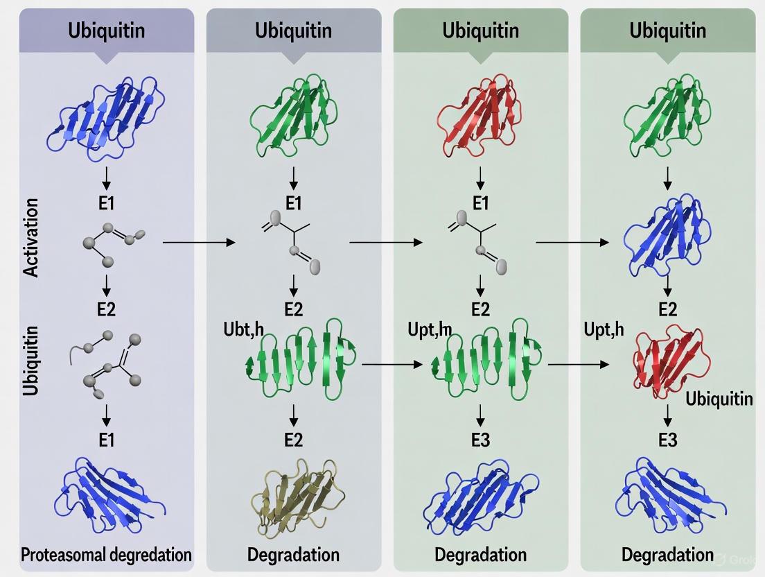
Visualization of Ubiquitin Signaling Pathways
Ubiquitin Conjugation Cascade
Implications for Biomedical Research and Therapeutic Development
The extreme conservation of ubiquitin makes it an attractive target for therapeutic intervention, as mechanisms discovered in model organisms frequently translate to human biology. Recent research has identified specific E2 enzymes, such as UBE2N, as dependencies in certain cancers including acute myeloid leukemia (AML) [14]. UBE2N primarily synthesizes K63-linked ubiquitin chains that stabilize oncoproteins rather than targeting them for degradation, representing a novel therapeutic avenue [14].
The discovery of small molecules that interface with the ubiquitin system, such as BRD1732 that undergoes direct ubiquitination, reveals new mechanistic possibilities for therapeutic intervention [10]. Understanding the structural basis of ubiquitin's conservation provides a framework for developing compounds that target specific aspects of ubiquitin signaling while minimizing off-target effects.
Emerging technologies in ubiquitin research, including species-specific prediction models that integrate both sequence and structural information [11], are enhancing our ability to interpret ubiquitin signaling across different organisms. These advances leverage the conserved nature of ubiquitin while accounting for species-specific adaptations in the broader ubiquitin system.
Ubiquitin represents a paradigm of extreme molecular conservation—a protein that has maintained virtually identical sequence and structure throughout eukaryotic evolution while expanding its functional repertoire. The β-grasp fold exemplifies structural perfection for a signaling molecule: stable yet adaptable, compact yet functionally versatile. Its evolutionary journey from simple archaeal systems to complex eukaryotic networks demonstrates how nature can optimize a successful design while allowing regulatory complexity to expand around it.
The conservation of ubiquitin across species provides tremendous power for biomedical research, enabling discoveries in model organisms to directly inform human therapeutic development. As we continue to unravel the complexities of ubiquitin signaling, the fundamental stability of the ubiquitin fold itself ensures that these insights will apply across biological systems, from the simplest protists to humans.
The ubiquitin-proteasome system (UPS) is a hallmark of eukaryotic cell regulation, controlling virtually all aspects of protein fate, from degradation to localization and activity [5]. At the apex of this system stand the E1 activating enzymes, which initiate the ubiquitination cascade by activating ubiquitin and transferring it to E2 conjugating enzymes [15]. While long considered a eukaryotic-specific innovation, groundbreaking research has revealed that the architectural and mechanistic blueprints of E1 enzymes originated in bacterial biosynthesis pathways [5] [15].
This article provides a comparative analysis of E1 enzyme architecture, tracing the evolutionary path from minimal bacterial ancestors ThiF and MoeB to the multi-domain eukaryotic E1s. We examine structural conservation, functional diversification, and experimental approaches that have elucidated these relationships, providing a comprehensive guide for researchers investigating ubiquitin activation across species.
Structural and Functional Comparison of E1-like Enzymes
Core Architectural Features
The E1 enzyme family shares a conserved catalytic core for ubiquitin-like protein (UBL) activation, with evolution building additional domains onto this foundation to regulate complexity and specificity.
Table 1: Comparative Features of E1-like Enzymes Across Species
| Feature | Bacterial ThiF/MoeB | Eukaryotic E1 (e.g., UBA1) |
|---|---|---|
| Organization | Homodimer [16] | Single polypeptide or heterodimer [16] |
| Molecular Mass | ~27 kDa (monomer) [16] | ~110 kDa [16] |
| Domains | Single adenylation domain [15] | Adenylation domain, Catalytic Cysteine Half-Domains (FCCH/SCCH), Ub-fold Domain (UFD) [17] |
| UBL Cargo | ThiS, MoaD [18] [15] | Ubiquitin, NEDD8, SUMO, etc. [15] |
| Primary Function | Sulfur carrier activation for thiamin/molybdopterin biosynthesis [18] [15] | Protein tag activation for post-translational modification [5] [15] |
| Key Catalytic Residues | ATP-binding Arg finger (from opposite monomer) [15] | Active-site Cysteine for thioester formation [15] [17] |
Evolutionary Trajectory and Functional Diversification
The evolutionary journey from bacterial precursors to eukaryotic E1s is marked by two key developments: domain fusion and functional specialization.
- Bacterial Origins: In E. coli, ThiF and MoeB function as homodimers, each monomer featuring an adenylation domain that recognizes UBL-fold proteins ThiS and MoaD, respectively [15] [16]. They catalyze adenylation of their cargo's C-terminus but do not form a thioester intermediate, instead facilitating sulfur transfer for biosynthesis of essential metabolites like thiamin [18] [15].
- Eukaryotic Expansion: Eukaryotic E1s incorporate the core adenylation domain but have fused additional domains, creating a multi-domain architecture. A critical evolutionary addition is the active-site cysteine domain, which allows the formation of a E1~UBL thioester bond intermediate—a key mechanistic step absent in the bacterial systems [15] [17]. The Ub-fold Domain (UFD) is another eukaryotic innovation that enhances specificity by recruiting cognate E2 enzymes [17].
This progression is exemplified by the catalytic cysteine (e.g., Cys600 in human UBA1), which is absent in ThiF/MoeB and enables the transthioesterification reaction central to the eukaryotic ubiquitination cascade [15] [17].
Diagram 1: Evolution of ubiquitin activation systems from simple bacterial precursors to complex eukaryotic cascades. The system gains complexity through the addition of dedicated E2 and E3 components, and the E1 enzyme evolves from a simple adenylator to a multi-domain orchestrator.
Experimental Approaches for Structural and Functional Analysis
Research in this field relies on structural biology techniques to visualize complexes and detailed biochemical assays to dissect the multi-step activation mechanism.
Structural Biology Workflows
Protein Complex Crystallization (as exemplified by E. coli ThiS-ThiF) [18]
- Cloning & Expression: The thiFS genes are cloned into a plasmid (e.g., pCLK1405) and transformed into an E. coli overexpression strain (e.g., BL834(DE3)).
- Protein Purification: Cells are lysed, and the supernatant is subjected to ammonium sulfate precipitation (50%). Further purification employs Hi-Trap QFF anion-exchange chromatography followed by size-exclusion chromatography (Superdex200).
- Crystallization: The purified ThiS-ThiF complex is concentrated to ~8 mg/mL and crystallized using the hanging-drop vapor diffusion method against a reservoir containing 7-8% PEG 400, 35 mM CaCl₂, and 100 mM Tris-HCl (pH 7.0-7.3).
- Data Collection & Processing: X-ray diffraction data is collected at synchrotron sources (e.g., APS). The crystals typically belong to space group P2₁2₁2₁. Data is integrated and scaled using software like HKL2000, enabling structure determination and refinement.
Functional Biochemical Assays
E1 Activity and Inhibition Profiling [19]
- Multi-turnover Ubiquitination Assay: Reactions contain E1 (UBA1), E2 (UBE2L3 or UBE2D3), E3 (e.g., HUWE1HECT), Ub, ATP, and Mg²⁺, often with a fluorescent Ub variant. Reaction products are visualized by SDS-PAGE to monitor E3 autoubiquitination, E2~Ub thioester formation, and polyUb chain synthesis.
- Single-turnover Assays: To dissect specific catalytic steps, a pre-formed E1~Ub thioester is incubated with E2 in the absence of ATP. This isolates the transthioesterification step from the initial adenylation.
- Inhibitor Mechanism Studies: Compounds are tested in dose-response curves (IC₅₀ determination). To probe if an inhibitor is a substrate, reaction mixtures can be analyzed by LC-MS/MS after protease digestion (e.g., with LysC) to identify Ub-inhibitor conjugates (+ mass of inhibitor).
Table 2: Key Research Reagents and Experimental Tools
| Reagent / Tool | Function / Utility | Example Application |
|---|---|---|
| HUWE1HECT | Isolated catalytic HECT domain of the HUWE1 E3 ligase. | Studying HECT E3-specific mechanics and inhibitor screening [19]. |
| UBE2L3 (E2) | E2 conjugating enzyme with specificity for HECT-type E3s. | In vitro ubiquitination assays to monitor Ub transfer from E1 to E3 [19]. |
| Vinylthioether HUWE1HECT~Ub Proxy | Stable, hydrolysis-resistant mimic of the E3~Ub thioester intermediate. | Probing the structure and dynamics of the key E3~Ub intermediate [19]. |
| Differential Scanning Fluorimetry (DSF) | Measures protein thermal stability changes upon ligand binding. | Detecting potential interactions between an E1/E3 and small molecules [19]. |
| Hydrogen-Deuterium Exchange MS (HDX-MS) | Probes protein conformational dynamics and ligand interactions. | Mapping binding interfaces and conformational changes in E1/E3 enzymes [19]. |
The comparative analysis of E1 enzyme architecture reveals a remarkable evolutionary story: the complex eukaryotic ubiquitin signaling machinery has its roots in fundamental bacterial metabolic pathways. The conserved adenylation domain serves as the universal structural and functional core, while the acquisition of additional domains in eukaryotes enabled the evolution of a sophisticated regulatory network.
Future research will continue to leverage high-resolution structural data and mechanistic biochemistry to fully elucidate the dynamic conformational changes that drive the E1 catalytic cycle [17]. Furthermore, the discovery of minimal, operon-encoded ubiquitination systems in archaea provides a powerful model for understanding the core principles of the UPS [5]. This evolutionary perspective is not merely academic; it informs drug discovery efforts, as evidenced by the ongoing development of E1 inhibitors as potential anticancer therapies [17]. Understanding the detailed architecture of these enzymes, from bacteria to humans, provides the fundamental knowledge required to manipulate this critical cellular machinery for therapeutic benefit.
{ARTICLE CONTENT}
Expansion of the Ubiquitin System: From Archaeal Operons to Eukaryotic Pyramidal Networks
The ubiquitin system represents a quintessential regulatory pathway that underwent profound expansion from a simple archaeal operon-like structure to a complex pyramidal network in eukaryotes. This comparative analysis examines the fundamental architectural differences, functional conservation, and evolutionary trajectory of ubiquitin activation across species. By integrating recent phylogenomic discoveries of Asgard archaea with mechanistic studies on ubiquitin-like protein (Ubl) conjugation, we delineate how a minimal, functionally versatile prokaryotic system transformed into the elaborate eukaryotic ubiquitin-proteasome system (UPS). Experimental data from gene shuffle complementation studies and structural analyses reveal remarkable functional conservation of core conjugation mechanisms despite extensive network diversification. This systematic comparison provides foundational insights for researchers investigating ubiquitin pathway evolution and targeting ubiquitin-related processes in drug development.
Ubiquitin is a small, highly conserved regulatory protein that serves as a central signaling molecule in eukaryotic cells, modulating critical processes including protein degradation, cell cycle control, DNA repair, and immune response [5] [6]. For decades, the ubiquitin system was considered a eukaryotic innovation; however, recent genomic and functional studies have identified ubiquitin-like signaling systems in archaea and bacteria, rewriting our understanding of its evolutionary origins [5] [20]. The discovery of simple, operon-encoded ubiquitin systems in archaea provides a crucial missing link for understanding how the elaborate eukaryotic ubiquitin machinery evolved from prokaryotic precursors.
The core ubiquitination process involves an enzymatic cascade where ubiquitin is activated by E1 enzymes, transferred to E2 conjugating enzymes, and finally attached to substrate proteins via E3 ligases [6]. This review performs a systematic comparison between the architectural organization, functional mechanisms, and evolutionary relationships of the minimal ubiquitin systems found in archaea and the complex pyramidal networks characteristic of eukaryotes. Understanding these systems' comparative biology has significant implications for fundamental cell biology research and drug discovery, particularly in developing therapies that target the ubiquitin-proteasome pathway in cancer and neurodegenerative diseases [21].
Architectural Evolution: From Operons to Pyramidal Networks
Archaeal Operons: Minimal Functional Units
The most streamlined genetic arrangement encoding a ubiquitin signaling system has been identified in archaea such as Caldiarchaeum subterraneum, consisting of just five genes organized in an operon-like cluster [5]. This minimal system includes: (1) a single-copy ubiquitin-like gene, (2) one ubiquitin-activating enzyme (E1), (3) one ubiquitin-conjugating enzyme (E2), (4) one RING-type ubiquitin-protein ligase (E3), and (5) one deubiquitinating enzyme related to the proteasome subunit Rpn11 [5]. This operon topology represents the most simplified ancestral pre-eukaryotic ubiquitin system known to date and demonstrates that the core components of the eukaryotic ubiquitin cascade were already present in prokaryotic organisms.
Similar operonic organizations are found sporadically distributed across diverse bacterial phyla (Actinobacteria, Planctomycetes, and Acidobacteria) and archaea, though they are frequently absent in close relatives, suggesting possible horizontal transfer events [5]. The protein modifiers in these systems, such as SAMPs (Small Archaeal Modifier Proteins) in Haloferax volcanii, function without requiring E2 and E3 conjugating factors in some cases, with the E1 enzyme alone being sufficient for conjugation—a mechanism distinct from the canonical eukaryotic three-enzyme cascade [5].
Eukaryotic Pyramidal Networks: Expanded and Diversified
In stark contrast to the minimalist archaeal systems, eukaryotic ubiquitin signaling is characterized by a vastly expanded pyramidal network with extensive gene duplication and functional specialization [5]. Rather than operon organization, eukaryotic ubiquitin genes are redundantly encoded in multiple loci: as polymeric head-to-tail concatemers of multiple ubiquitin open reading frames (approximately 4 to 15) and as fusions with ribosomal proteins L40 and S27a [5]. The enzymatic cascade follows a pyramidal hierarchy with a limited number of E1 enzymes (2 in humans) activating ubiquitin, which is then transferred to a larger set of E2 enzymes (over 30 in humans), and finally delivered to substrates by hundreds of E3 ligases that provide substrate specificity [5] [6].
This expansion creates a sophisticated regulatory network capable of precise spatiotemporal control over protein modification. For example, the amoebo-flagellate Naegleria gruberi, which diverged from other eukaryotic lineages over 1,000 million years ago, already contains more than 100 ubiquitin signaling system genes, including multiple E2s and E3s, demonstrating the early establishment of this pyramidal architecture in eukaryotic evolution [5].
Table 1: Comparative Architecture of Ubiquitin Systems in Archaea and Eukaryotes
| Feature | Archaeal Systems | Eukaryotic Systems |
|---|---|---|
| Genetic Organization | Operon-like clusters | Dispersed genes; tandem repeats |
| Ubiquitin/UBL Genes | Single copy | Multiple loci (polyubiquitin, fusions) |
| E1 Enzymes | One E1-like | Two E1s (UBA1, UBA6) in humans |
| E2 Enzymes | One E2 | 35 E2s in humans |
| E3 Ligases | One RING-type E3 | Hundreds of E3s |
| Deubiquitinases | One Rpn11-related | ~100 DUBs in humans |
| System Complexity | Minimal, compact | Pyramidal, hierarchical |
Evolutionary Bridging: Urm1 and the Sulfur Transfer Connection
Urm1: At the Crossroads of Evolution
The ubiquitin-related modifier 1 (Urm1) represents a crucial evolutionary link between prokaryotic sulfur carriers and eukaryotic ubiquitin-like modifiers [22] [20]. Urm1 is a unique Ubl protein with dual functionality, operating in both tRNA thiolation and protein urmylation, thereby combining features typical of bacterial sulfur carriers (like ThiS and MoaD) and classical ubiquitin-like modifiers [22] [23]. Phylogenetic analyses reveal that Urm1 proteins from archaea and eukaryotes form a monophyletic clade with solid bootstrap support (BS = 80), suggesting they share a common ancestor and likely represent an ancient Ubl family that predates the eukaryote-archaea divergence [22].
Structural and sequence comparisons show striking similarities between Urm1 from S. cerevisiae (ScUrm1) and S. acidocaldarius (SaciUrm1), particularly in the conserved β-grasp fold and C-terminal di-glycine motif characteristic of Ubl proteins [22]. This structural conservation across domains of life underscores the ancient origin of the β-grasp fold, which has been recruited for diverse biochemical functions including catalytic roles, scaffolding of iron-sulfur clusters, RNA binding, and sulfur transfer [20].
Experimental Evidence from Gene Shuffle Studies
Recent functional studies using URM1 gene shuffle from Sulfolobus acidocaldarius to Saccharomyces cerevisiae have provided direct experimental evidence for the functional conservation of urmylation across archaea and eukaryotes [22] [23]. When expressed in yeast urm1Δ deletion strains, the archaeal SaciUrm1 protein robustly conjugated to the peroxiredoxin Ahp1, a bona fide urmylation target in yeast, despite the evolutionary distance between these organisms [22].
Critical findings from this experimental approach include:
- Conserved Conjugation Specificity: Archaeal SaciUrm1 attached to the same lysine residue (Lys-32) on Ahp1 as yeast Urm1, following identical molecular requirements including dependence on specific cysteine residues (Cys-31, Cys-62) [22].
- Shared Activation Mechanism: Ahp1 conjugation required sulfur transfer onto the archaeal Urm1 modifier from Uba4, the E1-like urmylation activator in yeast, demonstrating conservation of the thioactivation mechanism [22].
- Functional Separation: While archaeal Urm1 supported protein urmylation, it could not rescue tRNA thiolation defects in yeast, indicating that the dual functions of Urm1 can be evolutionarily separated and that archaeal Urm1 may specialize in protein modification [22].
Table 2: Functional Comparison of Urm1 in Sulfolobus acidocaldarius and Saccharomyces cerevisiae
| Functional Aspect | SaciUrm1 (Archaea) | ScUrm1 (Yeast) |
|---|---|---|
| Protein Urmylation | Yes (to Ahp1) | Yes (to Ahp1 and other targets) |
| tRNA Thiolation | No | Yes |
| Thioactivation Required | Yes (by Uba4) | Yes (by Uba4) |
| E1 Activator | Uba4 (yeast) | Uba4 |
| Major Acceptor Site on Ahp1 | Lys-32 | Lys-32 |
| Critical Cysteine Residues | Cys-31, Cys-62 | Cys-31, Cys-62 |
These findings position Urm1 at the evolutionary crossroads of prokaryotic sulfur transfer and eukaryotic protein conjugation pathways, providing a living molecular fossil that bridges these functionally distinct systems [22].
Methodologies for Comparative Ubiquitin System Analysis
Phylogenomic and Genomic Approaches
Advanced phylogenomic analyses leveraging expanded genomic datasets have revolutionized our understanding of ubiquitin system evolution. Recent studies have identified 16 new Asgard archaeal lineages at the genus level or higher, substantially expanding the known phylogenetic diversity of archaea most closely related to eukaryotes [24]. Sophisticated phylogenomic approaches using complex site-heterogeneous evolution models in maximum likelihood and Bayesian inferences with recoded alignments have helped resolve the contentious placement of eukaryotes within the Asgard archaeal lineage [24].
Standardized marker sets have been developed for robust phylogenetic analysis, including:
- GTDB.ar53: 53 archaeal-specific marker proteins from the Genome Taxonomy Database [24]
- S67 Marker Set: 67 markers (39 ribosomal proteins, 28 functional proteins) conserved across all sampled archaeal genomes [24]
- NM200 and NM57: Non-ribosomal protein markers of archaeal origin [24]
These tools enable researchers to reconstruct evolutionary relationships with greater accuracy and assess the functional conservation of ubiquitin system components across domains of life.
Functional Complementation Assays
Gene shuffle complementation experiments, such as those conducted with URM1 from S. acidocaldarius to S. cerevisiae, provide direct functional assessment of conservation [22]. The experimental workflow typically involves:
- Strain Construction: Creating deletion mutants (e.g., urm1Δ) in model eukaryotic organisms
- Heterologous Expression: Introducing archaeal genes under appropriate promoters
- Phenotypic Rescue Assessment: Testing complementation of mutant phenotypes
- Biochemical Validation: Confirming protein conjugation through electrophoretic mobility shift assays (EMSA) and Western blotting with isopeptidase inhibitors
- Mechanistic Analysis: Identifying critical residues through site-directed mutagenesis
These functional assays are complemented by molecular dynamics simulations that reveal how subtle changes in amino acid sequences—such as single aspartic to glutamic acid substitutions—can dramatically alter interaction selectivity between E2 and E3 enzymes by shifting the equilibrium between "open" (binding-competent) and "closed" (binding-incompetent) states [25].
Table 3: Key Research Reagents for Ubiquitin System Studies
| Reagent/Resource | Function/Application | Examples/Sources |
|---|---|---|
| Archaeal Strains | Comparative functional studies | Sulfolobus acidocaldarius, Haloferax volcanii [22] [5] |
| Model Eukaryotes | Genetic manipulation and complementation | Saccharomyces cerevisiae (yeast) [22] |
| Gene Knockout Strains | Functional assessment of specific genes | urm1Δ, uba4Δ, tum1Δ yeast strains [22] |
| Epitope Tags | Protein detection and purification | HA-tag, c-Myc tag [22] |
| Isopeptidase Inhibitors | Stabilization of ubiquitin/Ubl conjugates | N-ethylmaleimide (NEM) [22] |
| Protocol Repositories | Standardized methods for working with archaea | ARCHAEA.bio, Halohandbook [26] |
| Strain Collections | Source of diverse archaeal strains | DSMZ, JCM, ATCC [26] |
| Phylogenomic Markers | Evolutionary analysis | GTDB.ar53, S67 marker sets [24] |
The comparative analysis of ubiquitin system organization from archaeal operons to eukaryotic pyramidal networks reveals fundamental principles of molecular evolution. The conservation of core mechanisms—from Urm1-mediated urmylation to the β-grasp fold structure—highlights the deep evolutionary origins of protein modification systems. Simultaneously, the dramatic expansion in eukaryotic systems demonstrates how gene duplication and functional specialization enabled the development of sophisticated regulatory networks essential for eukaryotic complexity.
For researchers and drug development professionals, these evolutionary insights offer valuable perspectives. The minimal archaeal systems provide simplified models for understanding core ubiquitination mechanisms, while the functional conservation of enzymes like Uba4 across domains suggests ancient, essential processes that might be targeted therapeutically. Furthermore, the evolutionary trajectory of the ubiquitin system illustrates how essential cellular machinery expands and diversifies, offering parallels for understanding other complex biological systems in eukaryotic cells.
Visual Guide: Ubiquitin System Architecture and Experimental Analysis
Ubiquitin System Evolution and Architecture
Diagram 1: Ubiquitin System Architectural Evolution. This schematic illustrates the evolutionary transition from minimal archaeal operons to complex eukaryotic pyramidal networks, with Urm1 representing a key functional bridge between these organizational paradigms.
Experimental Workflow for Cross-Domain Functional Analysis
Diagram 2: Experimental Workflow for Cross-Domain Functional Analysis. This flowchart outlines the key methodological approach for assessing functional conservation of ubiquitin system components between archaea and eukaryotes, incorporating genetic, biochemical, and computational techniques.
Ubiquitination is a crucial post-translational modification that controls a vast array of cellular processes, including protein degradation, DNA repair, and immune signaling. This enzymatic cascade begins with E1 ubiquitin-activating enzymes, which activate and transfer ubiquitin to downstream E2 and E3 enzymes [27]. For years, UBA1 (UBE1) was considered the sole E1 enzyme for ubiquitin activation in eukaryotes. However, the discovery of UBA6 (E1-L2) revealed unexpected complexity in the initiation of ubiquitin signaling [28]. These two E1 enzymes, while performing the same fundamental biochemical reaction, exhibit distinct structural characteristics, E2 specificities, and evolutionary conservation patterns [29] [28].
Understanding the phylogenetic distribution of UBA1 and UBA6 provides valuable insights into the evolution of protein degradation systems and cellular regulatory mechanisms across species. This comparative guide examines the experimental evidence defining the presence, conservation, and functional specialization of these E1 enzymes throughout the animal kingdom, providing researchers with a comprehensive resource for investigating ubiquitin activation pathways in different model organisms and therapeutic contexts.
Comparative Analysis of UBA1 and UBA6
Phylogenetic Distribution Across Species
Table 1: Phylogenetic Distribution of E1 Enzymes Across Species
| Species Group | UBA1 Presence | UBA6 Presence | Key Evidence & Notes |
|---|---|---|---|
| Mammals (Human, Mouse) | Yes | Yes | UBA6 activates both ubiquitin and FAT10; essential for embryonic development in mice [30] [28] |
| Birds | Presumed | Presumed | Likely present given vertebrate conservation |
| Reptiles/Amphibians | Presumed | Presumed | Likely present given vertebrate conservation |
| Sea Urchin | Yes | Yes | Experimental confirmation of UBA6 presence [28] |
| Ascidians (Halocynthia roretzi) | Yes | Yes | cDNA cloning confirms presence of both E1s; involvement in fertilization [31] |
| Insects (D. melanogaster) | Yes | No | UBA6 absent from genome [29] |
| Nematodes (C. elegans) | Yes | No | UBA6 absent from genome [29] |
| Fungi (Yeast) | Yes | No | UBA6 absent from genome [29] |
| Plants | Yes | No | UBA6 absent from genome [29] |
The phylogenetic distribution of UBA1 and UBA6 reveals a striking evolutionary pattern. UBA1 demonstrates universal conservation across eukaryotic organisms, serving as the foundational ubiquitin activation system [29]. In contrast, UBA6 exhibits a restricted phylogenetic presence, appearing only in vertebrates, sea urchin, and ascidians, while being conspicuously absent from insects, nematodes, fungi, and plants [29] [28]. This distribution suggests UBA6 emerged later in evolutionary history to fulfill specialized regulatory functions not required in all eukaryotic lineages.
The conservation of UBA6 in deuterostomes but not in protostomes or simpler eukaryotes indicates this E1 enzyme may have evolved to support increasingly complex cellular regulatory mechanisms in higher organisms. In ascidians such as Halocynthia roretzi, both UBA1 and UBA6 play crucial roles in fertilization, with the 3D protein structures predicted to be very similar to their human counterparts based on AlphaFold2 predictions [31]. The absence of UBA6 from model organisms like Drosophila melanogaster and Caenorhabditis elegans highlights the importance of considering phylogenetic distribution when selecting model systems for studying ubiquitin pathways.
Structural and Functional Characteristics
Table 2: Structural and Functional Comparison of UBA1 and UBA6
| Characteristic | UBA1 | UBA6 |
|---|---|---|
| Ubiquitin Activation | Yes | Yes |
| FAT10 Activation | No | Yes [30] |
| Primary E2 Partners | Charges most E2s (UBE2A, UBE2B, CDC34) | Charges USE1 (UBA6-specific E2) [29] |
| Cellular Localization | Nuclear and cytoplasmic isoforms [27] | Cytoplasmic and mitochondrial association [31] |
| Catalytic Mechanism | Open/closed conformational transition during adenylation/thioester formation | Similar conformational transition; allosterically regulated by InsP6 [30] |
| Inhibitor Sensitivity | Sensitive to TAK-243 [27] | Less sensitive to TAK-243; potentially inhibited by phytic acid [27] |
| Disease Associations | VEXAS syndrome (M41 mutations) [27]; Aortic dissection [32] | Potential therapeutic target in VEXAS syndrome [27] |
Structurally, UBA1 and UBA6 share a similar domain organization including adenylation domains, catalytic cysteine half-domains, and ubiquitin-fold domains, yet they possess distinct features that underlie their functional specialization. UBA1 exists in two naturally occurring isoforms: UBA1a (nuclear) initiated from methionine 1, and UBA1b (cytoplasmic) initiated from methionine 41 [27]. In VEXAS syndrome, mutations at M41 cause an aberrant isoform switch to a nonfunctional UBA1c, demonstrating the critical importance of this regulatory mechanism [27].
UBA6 exhibits dual substrate specificity, uniquely capable of activating both ubiquitin and the ubiquitin-like modifier FAT10 [30] [33]. Structural studies reveal UBA6 undergoes dramatic conformational changes during catalysis, transitioning between "open" and "closed" states during adenylation and thioester bond formation [30]. Surprisingly, UBA6 is allosterically regulated by inositol hexakisphosphate (InsP6), which binds to a conserved basic pocket in the SCCH domain and inhibits enzyme activity by altering conformational equilibria [30]. This represents a previously unknown regulatory mechanism for E1 enzymes.
Functionally, UBA1 and UBA6 operate in parallel pathways with distinct E2 partnerships. UBA1 charges the majority of E2 enzymes, including UBE2A/B and CDC34 family members, while UBA6 specifically charges the USE1 (UBA6-specific E2) enzyme [29] [28]. Despite this separation, both pathways can converge on the same E3 ligases, such as the UBR1-3 family of N-recognins, to mediate substrate ubiquitination in a spatially distinct manner [29].
Experimental Approaches and Methodologies
Phylogenetic Analysis and Gene Identification
Experimental identification of E1 enzymes across species employs complementary bioinformatic and molecular techniques:
Database Mining: Researchers search genomic databases (ANISEED for ascidians) using known E1 sequences to identify candidate gene models [31]. This approach identified seven potential UBA candidates in Halocynthia roretzi before UBA1 and UBA6 were confirmed.
cDNA Cloning: Using degenerate primers or RACE PCR, researchers isolate full-length coding sequences from target species. For ascidian UBA1 and UBA6, this involved cloning from gonad-derived cDNA libraries [31].
Sequence Analysis: Predicted protein sequences are analyzed for characteristic E1 domains (AAD, IAD, FCCH, SCCH, UFD) and key catalytic residues (e.g., Cys625 in human UBA6) [30]. 3D structure prediction tools like AlphaFold2 enable comparative structural analysis between orthologs [31].
Functional Validation: Recombinant enzymes are tested for ubiquitin adenylation, thioester bond formation, and E2 charging capabilities to confirm functional conservation [33].
Functional Characterization Assays
Several established biochemical approaches define E1 enzyme activity and specificity:
ATP-PPᵢ Exchange Assays: Measure the first step of ubiquitin activation where E1 catalyzes ubiquitin adenylation with release of inorganic pyrophosphate. This assay demonstrated UBA6's ability to activate both ubiquitin and FAT10 [33].
Thioester Formation Assays: Detect the formation of covalent E1~ubiquitin intermediates under non-reducing conditions. Immunoblotting of UBA1 in VEXAS models confirmed the aberrant UBA1b-to-UBA1c isoform switch [27].
E2 Charging Assays: Evaluate the transfer of ubiquitin from E1 to candidate E2 enzymes. These assays revealed UBA6's exclusive E2 partnership with USE1, while UBA1 charges multiple E2s [29] [28].
Inhibitor Studies: Compound sensitivity profiles help distinguish E1 activities. TAK-243 preferentially inhibits UBA1, while phytic acid shows selectivity for UBA6 in cellular models [27].
Figure 1: Experimental workflow for phylogenetic and functional analysis of E1 enzymes
Research Reagent Solutions
Table 3: Essential Research Reagents for E1 Enzyme Studies
| Reagent/Category | Specific Examples | Research Applications | Key Characteristics |
|---|---|---|---|
| E1 Inhibitors | TAK-243 (MLN7243) [27] | Selective UBA1 inhibition; studying UBA1-specific functions | First-in-class specific E1 inhibitor; in clinical trials for cancer |
| PYR-41 [31] | General E1 inhibition; fertilization studies | Less specific; inhibits multiple E1 enzymes and deubiquitinases | |
| Phytic acid [27] | Potential UBA6 inhibition; exploring UBA6-specific vulnerabilities | Natural compound; allosterically inhibits UBA6 via InsP6 binding [30] | |
| Cell Models | UBA1M41V THP1 [27] | VEXAS syndrome modeling | Human monocytic cell line with engineered UBA1 M41V mutation |
| BAPN-induced AD mouse [32] | Aortic dissection studies | In vivo model showing UBA1 upregulation in vascular disease | |
| CFBE41o- F508del [34] | Cystic fibrosis research | Airway epithelial cells with F508del-CFTR for ubiquitin-proteasome studies | |
| Antibodies | Anti-UBA1 [34] | Protein expression analysis | Western blot, immunohistochemistry |
| Anti-UBA6 [34] | Protein expression and localization | Western blot, immunocytochemistry | |
| Anti-ubiquitin [27] | Ubiquitination status assessment | Detection of global ubiquitination changes | |
| Biochemical Assays | ATP-PPi Exchange [33] | E1 activation step measurement | Quantifies initial ubiquitin/FAT10 adenylation |
| Thioester Assay [27] | Covalent intermediate detection | Non-reducing Western to detect E1~ubiquitin conjugates | |
| E2 Charging Assay [28] | E1-E2 specificity profiling | Identifies E2 partnerships for UBA1 vs UBA6 |
Research Implications and Future Directions
The phylogenetic restriction of UBA6 to deuterostomes suggests its emergence corresponded with increasing complexity in biological regulatory systems requiring specialized ubiquitination pathways. The differential E2 charging between UBA1 and UBA6 enables cells to maintain parallel ubiquitination cascades that may be activated under distinct physiological conditions or in response to specific cellular stresses [29] [28].
From a therapeutic perspective, the distinct structural features of UBA6, particularly its allosteric InsP6 binding site, offer opportunities for developing specific small-molecule inhibitors that could selectively target UBA6-dependent pathways without disrupting essential UBA1-mediated ubiquitination [27] [30]. The enhanced sensitivity of UBA1-mutant cells in VEXAS syndrome to UBA6 inhibition reveals a promising synthetic lethal relationship that could be exploited therapeutically [27].
Future research should focus on elucidating the complete repertoire of UBA6-specific substrates and pathways, particularly in physiological contexts where its dual specificity for ubiquitin and FAT10 provides unique regulatory potential. The development of more specific pharmacological tools, including conditional genetic models and highly selective inhibitors, will be essential for deciphering the non-redundant functions of these evolutionarily distinct E1 activation systems across different species and tissue types.
Figure 2: UBA6-specific ubiquitination cascade with allosteric regulation
Advanced Techniques and Pharmacological Modulation of Ubiquitin Activation
In Vitro and In Vivo Assays for E1 Enzyme Activity and Thioesterification
Ubiquitin-activating enzymes, known as E1 enzymes, stand at the apex of the ubiquitination cascade, a crucial post-translational modification system in eukaryotic cells. These ATP-dependent enzymes initiate a three-step enzymatic cascade by activating ubiquitin and transferring it to E2 conjugating enzymes, ultimately leading to substrate modification by E3 ligases [35] [36]. The clinical significance of E1 enzymes continues to grow, with research linking their dysfunction to various cancers, neurodegenerative disorders, and autoinflammatory diseases such as VEXAS syndrome, characterized by somatic mutations at methionine 41 in UBA1 [27] [37]. This comparison guide examines the current methodologies for assessing E1 enzyme activity and thioester formation, providing researchers with essential tools for investigating this fundamental biological process and developing targeted therapies.
Comparative Analysis of E1 Activity Assays
The following table summarizes the primary assays used to monitor E1 enzyme activity and thioester bond formation:
Table 1: Comparison of E1 Enzyme Activity and Thioesterification Assays
| Assay Type | Detection Method | Key Readout | Throughput | Biological Context | Key Advantages | Main Limitations |
|---|---|---|---|---|---|---|
| UbiReal (Fluorescence Polarization) | Fluorescence Polarization (FP) | Real-time E1-E2-E3 activity & thioester formation | High | In vitro | Real-time kinetics, universal for Ub/UBLs, suitable for inhibitor screening [36] | Requires fluorescently-labeled ubiquitin |
| Thioester Assay | Western Blot/ Chemiluminescence | E1~Ub and E2~Ub thioester intermediates | Medium | In vitro | Direct detection of covalent thioester linkages [35] | Non-quantitative, endpoint measurement only |
| Radioactive Ub Charging | Radiolabel detection (³²P or ³⁵S) | ATP consumption or ³⁵S-labeled ubiquitin transfer | Low | In vitro | Highly sensitive | Radioactive hazard, specialized facilities required |
| Cell-Based Ubiquitination Monitoring | Immunoblotting | Accumulation of ubiquitin conjugates | Low | In vivo (cellular) | Physiological relevance, can detect endogenous ubiquitination [10] | Indirect measure of E1 activity |
| CRISPR-Cas9 Screening | Next-generation sequencing | Genetic dependencies & resistance mechanisms | Ultra-high | In vivo (cellular) | Unbiased discovery of E1 genetic networks [10] [27] | Indirect, requires validation |
Detailed Experimental Protocols
UbiReal Fluorescence Polarization Assay
The UbiReal platform represents a significant advancement for monitoring the complete ubiquitination cascade in real-time, including the critical E1-catalyzed thioester formation step [36].
Protocol Steps:
- Reaction Setup: Prepare a 20 μL reaction containing 25 mM sodium phosphate (pH 7.4), 150 mM NaCl, 2 mM ATP, 5 mM MgCl₂, 0.1-1 μM E1 enzyme, 100-500 nM fluorescein-labeled ubiquitin (F-Ub), and optional E2/E3 enzymes.
- Instrument Configuration: Use a fluorescence polarization-compatible plate reader with excitation at 485 nm and emission at 528 nm.
- Kinetic Measurement: Monitor polarization values (mP units) immediately after adding E1 enzyme, taking readings every 30-60 seconds for 60-120 minutes.
- Data Analysis: Calculate reaction rates from the initial linear portion of the polarization increase curve. For inhibitor studies, pre-incubate E1 with compound for 15 minutes before adding other reaction components.
Technical Notes: The assay capitalizes on the significant change in molecular rotation when ubiquitin transitions from free diffusion (low polarization) to being covalently bound within the E1~Ub thioester complex (high polarization). For E1-E2 transthiolation monitoring, include appropriate E2 enzymes such as UBE2D3 or UBE2L3 [36].
In Vitro Thioester Assay
This traditional biochemical assay provides direct evidence of E1~Ub and E2~Ub thioester intermediate formation through electrophoretic mobility shift analysis under non-reducing conditions [35].
Protocol Steps:
- Thioester Formation: Incubate 50-100 nM E1 enzyme with 1-5 μM ubiquitin in reaction buffer (20-50 mM Tris/HCl, pH 7.5-8.0, 5 mM MgCl₂, 2 mM ATP) for 5-15 minutes at 30°C.
- E2 Charging: For E2~Ub detection, add 1-5 μM of relevant E2 enzyme (e.g., group IV E2s like SlUBC32/33/34 for plant immunity studies) and continue incubation for additional 5-15 minutes [35].
- Reaction Termination: Add non-reducing Laemmli buffer (lacking β-mercaptoethanol or DTT).
- Electrophoresis: Resolve proteins by SDS-PAGE (6-12% gradient gels) without boiling samples.
- Detection: Transfer to PVDF membrane and immunoblot with anti-ubiquitin antibodies. E1~Ub and E2~Ub thioester complexes appear as higher molecular weight bands that disappear upon DTT treatment.
Technical Notes: The adenylation and thioesterification functions of E1 enzymes involve two intricately connected reactions: ATP hydrolysis coupled with ubiquitin activation, followed by formation of a high-energy E1~ubiquitin thioester linkage, and subsequent transfer to E2 catalytic cysteine [35]. Include DTT-treated controls (50 mM final concentration) to confirm thioester linkage specificity.
Visualization of Ubiquitination Cascade and Assay Workflows
The Ubiquitination Cascade and E1 Function
Diagram 1: The ubiquitination cascade initiated by E1 enzyme. The E1 enzyme activates ubiquitin through ATP-dependent adenylation, forms a thioester bond with ubiquitin, and transfers it to E2 conjugating enzymes, ultimately leading to substrate ubiquitination via E3 ligases [35] [36].
UbiReal Experimental Workflow
Diagram 2: UbiReal assay workflow using fluorescence polarization. The assay monitors the increase in fluorescence polarization as fluorescently-labeled ubiquitin transitions from free diffusion to being covalently bound in the E1~Ub thioester complex, enabling real-time monitoring of ubiquitin activation and transfer [36].
Research Reagent Solutions
Table 2: Essential Reagents for E1 Enzyme Activity Assays
| Reagent Category | Specific Examples | Function in Assays | Application Context |
|---|---|---|---|
| E1 Enzymes | Human UBA1, UBA6, Arabidopsis AtUBA1/2, Tomato SlUBA1/2 | Catalyze ubiquitin activation & thioester formation [35] [27] [37] | Species-specific studies, differential E2 charging |
| E2 Enzymes | UBE2D3, UBE2L3, UBE2N, Tomato group IV E2s (SlUBC32/33/34) | Accept ubiquitin from E1, determine pathway specificity [35] [10] [36] | E1-E2 pairing studies, pathway characterization |
| Ubiquitin Variants | Fluorescein-Ub (F-Ub), TAMRA-Ub (T-Ub), Mutant Ub (K0, K27-only) | E1 substrates for activity monitoring, chain type specificity [10] [36] | FP assays, mechanism studies, chain linkage analysis |
| E1 Inhibitors | TAK-243 (UBA1 inhibitor), Pevonedistat (NAE inhibitor), PYR-41 | Specific E1 inhibition for control experiments & therapeutic studies [27] [37] | Assay validation, drug discovery, pathway modulation |
| Detection Reagents | Anti-ubiquitin antibodies, Fluorescence plate readers, SDS-PAGE systems | Detect ubiquitin conjugates & thioester complexes [35] [10] | All assay formats, endpoint detection |
The expanding toolkit for monitoring E1 enzyme activity and thioesterification provides researchers with complementary approaches spanning from traditional biochemical methods to sophisticated real-time monitoring systems. The UbiReal platform offers distinct advantages for high-throughput screening and kinetic studies, while established thioester assays remain valuable for direct confirmation of covalent intermediate formation. The emerging understanding of differential E2 charging by E1 isoforms across species—from tomato SlUBA1/SlUBA2 with their distinct efficiencies for group IV E2s to human UBA1 and UBA6 with their unique E2 preferences—highlights the importance of enzyme and substrate selection when designing experiments [35] [37]. As research continues to unravel the complexities of E1 biology in health and disease, these assay technologies will play an increasingly vital role in validating new therapeutic targets and developing precision interventions for conditions ranging from VEXAS syndrome to various cancers.
The ubiquitin-proteasome system (UPS) represents a crucial regulatory mechanism for protein homeostasis in eukaryotic cells, controlling the stability, activity, and localization of countless proteins. At the apex of the ubiquitination cascade are ubiquitin-activating enzymes (E1), which initiate the process of ubiquitin transfer to downstream substrates. In humans, two ubiquitin E1 enzymes exist—UBA1 (also known as UBE1) and UBA6—with UBA1 responsible for activating the vast majority (>99%) of cellular ubiquitin [34]. The E1 enzyme mechanism involves a conserved series of steps: first, the E1 binds MgATP and ubiquitin, catalyzing ubiquitin C-terminal acyl-adenylation; second, the catalytic cysteine in the E1 attacks the ubiquitin-adenylate to form a high-energy thioester bond [15]. This activated ubiquitin is then transferred to ubiquitin-conjugating enzymes (E2), which subsequently cooperate with ubiquitin ligases (E3) to modify specific target proteins.
The centrality of E1 enzymes to the UPS makes them attractive therapeutic targets. TAK-243 (previously known as MLN7243) represents the first-in-class inhibitor of ubiquitin-activating enzymes that has progressed to clinical trials for cancer therapy (NCT02045095, NCT03816319) [38] [34]. This compound has emerged as both a valuable tool for probing ubiquitination mechanisms in basic research and a promising therapeutic agent with demonstrated efficacy across multiple disease models. Its development marks a significant advancement over earlier, less specific E1 inhibitors such as PYR-41, which exhibited off-target effects against deubiquitinases and protein kinases [34]. This guide provides a comprehensive comparison of TAK-243's performance relative to other UPS-targeting agents and details experimental approaches for its application in research and drug development.
Comparative Analysis of UPS-Targeting Agents
Mechanism and Specificity Profiles
Table 1: Mechanism of Action Comparison Between UPS-Targeting Agents
| Agent | Primary Target | Mechanistic Action | Key Off-Target Effects | Specificity Advantages |
|---|---|---|---|---|
| TAK-243 | UBA1 (Ubiquitin E1) | Forms TAK-243-Ub adduct, blocking ubiquitin transfer to E2 enzymes [39] | Inhibits UBA6, NAE, and SAE at higher concentrations [34] | High specificity for ubiquitin E1 over other UBL E1s [38] |
| Bortezomib | Proteasome (β5 subunit) | Reversibly inhibits chymotrypsin-like activity of proteasome [38] | Inhibits caspase-like and trypsin-like activities at high concentrations [38] | FDA-approved for multiple myeloma |
| Carfilzomib | Proteasome (β5 subunit) | Irreversibly inhibits chymotrypsin-like activity [38] | Inhibits caspase-like and trypsin-like activities at high concentrations [38] | Reduced peripheral neuropathy vs. bortezomib |
| Ixazomib | Proteasome (β5 subunit) | Reversibly inhibits chymotrypsin-like activity [38] | Shorter recovery half-life (<4 hours) after removal [38] | Oral bioavailability |
| PYR-41 | UBA1 (Ubiquitin E1) | Blocks ubiquitin-thioester formation [40] | Inhibits deubiquitinases and protein kinases [34] | First-generation E1 inhibitor (research tool) |
Therapeutic Efficacy and Selectivity
Table 2: Therapeutic Performance Across Disease Models
| Disease Model | TAK-243 Efficacy | Proteasome Inhibitor Efficacy | Key Resistance Mechanisms | Therapeutic Index |
|---|---|---|---|---|
| Cutaneous SCC | Effective against bortezomib-resistant cells [38] | Variable pattern of sensitivity/resistance [38] | Low UBA1 expression increases TAK-243 susceptibility [38] | Greater selectivity with pulse dosing [38] |
| Triple-Negative Breast Cancer | Tumor regression in PDX models; reduces metastasis [41] | Limited data in search results | SLFN11 expression inversely correlates with sensitivity [42] | Order of magnitude greater sensitivity vs. normal cells [41] |
| Acute Myeloid Leukemia | Sensitivity identified through CRISPR screens [43] | Limited data in search results | ABCB1 transporter mediates efflux [42] | Research ongoing |
| Cystic Fibrosis (F508del) | Boosts elexacaftor/tezacaftor/ivacaftor efficacy [34] | Ineffective as monotherapy [34] | Limited data in search results | Improves rescue of rare misfolded mutants [34] |
| Toxoplasma gondii | Inhibits parasite ubiquitination [39] | Limited data in search results | Limited data in search results | Selective inhibition of TgUAE1 vs. host [39] |
Molecular Mechanisms and Signaling Pathways
Primary Mechanism of Ubiquitin Activation Inhibition
The inhibition of ubiquitin activation by TAK-243 occurs through a well-characterized mechanism. TAK-243 specifically targets the UBA1 enzyme, forming a covalent adduct with ubiquitin (TAK-243-Ub) that blocks the transfer of activated ubiquitin to E2 conjugating enzymes [39]. This mechanism effectively shuts down the majority of cellular ubiquitination events, as UBA1 is responsible for charging over 99% of cellular ubiquitin [34]. The compound exhibits high specificity for ubiquitin E1 enzymes over other ubiquitin-like protein (UBL) E1s, though at higher concentrations it can also inhibit UBA6, NEDD8 E1-activating enzyme (NAE), and the SUMO-activating enzyme (SAE) [34].
Downstream Cellular Consequences
The inhibition of UBA1 by TAK-243 triggers profound cellular stress responses that ultimately lead to cell death, particularly in malignant cells. The primary signaling pathway can be visualized as follows:
Figure 1: TAK-243-induced signaling pathway leading to apoptosis. Inhibition of UBA1 triggers ER stress and the unfolded protein response, culminating in ATF4-mediated NOXA upregulation and apoptosis. c-MYC expression enhances ATF4 activation [41].
The molecular pathway illustrated above demonstrates how TAK-243 inhibition leads to apoptotic cell death. Key steps include:
- UBA1 Inhibition: TAK-243 forms a covalent adduct with ubiquitin, blocking its transfer to E2 enzymes [39]
- Ubiquitination Blockade: Global disruption of protein ubiquitination occurs, affecting numerous cellular processes
- ER Stress Induction: Disruption of protein homeostasis leads to endoplasmic reticulum stress [44] [41]
- UPR Activation: The unfolded protein response is triggered as a compensatory mechanism
- PERK-eIF2α-ATF4 Axis: PERK activation results in eIF2α phosphorylation and selective translation of ATF4 [41]
- NOXA Induction: ATF4 transactivates the pro-apoptotic protein NOXA [41]
- Apoptotic Cell Death: NOXA induction triggers programmed cell death [41]
Notably, c-MYC expression correlates with TAK-243 sensitivity and cooperates with TAK-243 to induce stress response and cell death [41]. This pathway is particularly pronounced in cancer cells with high c-MYC expression, explaining the selective toxicity toward certain malignancies.
Experimental Protocols and Methodologies
In Vitro Assessment of E1 Inhibition
Ubiquitin Thioesterification Assay: This fundamental assay evaluates the formation of ubiquitin-E1 thioester complexes, which is the primary biochemical activity inhibited by TAK-243.
- Reagents: Purified E1 enzyme (UBA1), ubiquitin, ATP, reaction buffer (50 mM Tris-HCl pH 7.5, 50 mM KCl, 5 mM MgCl₂), DTT, TAK-243 in DMSO [39]
- Procedure:
- Prepare reaction mixtures containing 100 nM UBA1, 5 μM ubiquitin, and 2 mM ATP in reaction buffer
- Add TAK-243 across a concentration range (typically 0.1 nM - 10 μM)
- Incubate at 37°C for 30 minutes
- Split reactions: add DTT (final 10 mM) to one set, keep the other set without DTT
- Analyze by non-reducing SDS-PAGE and western blot with anti-ubiquitin antibody [39]
- Expected Results: Without DTT, a high molecular weight band (~140 kDa) representing the UBA1-ubiquitin thioester complex should be visible in controls but diminished in TAK-243-treated samples. DTT treatment eliminates this band due to thioester reduction [39].
E1-to-E2 Ubiquitin Transfer Assay: This assay evaluates the downstream consequences of E1 inhibition on ubiquitin transfer to E2 enzymes.
- Reagents: Purified UBA1, E2 enzyme (e.g., Cdc34), ubiquitin, ATP, reaction buffer, TAK-243 [39]
- Procedure:
- Set up reaction mixtures with UBA1, ubiquitin, and ATP as above
- Include relevant E2 enzyme (e.g., 1 μM Cdc34)
- Treat with TAK-243 across concentration gradient
- Incubate at 37°C for 30 minutes
- Analyze by non-reducing SDS-PAGE and western blot with anti-ubiquitin antibody [39]
- Expected Results: Successful ubiquitin transfer to E2 will produce a band corresponding to ubiquitin-E2 complex (~40 kDa) in control samples, which should be reduced in TAK-243-treated samples in a concentration-dependent manner [39].
Cellular Efficacy Assessment
Cell Viability and Apoptosis Assays: These assays evaluate the functional consequences of E1 inhibition in cellular models.
- Reagents: Cell culture media, MTT reagent or alternative viability dye, Annexin V/PI staining kit, TAK-243 [38] [41] [42]
- Procedure:
- Seed cells at appropriate density (e.g., 5,000 cells/well in 96-well plates)
- After 24 hours, treat with TAK-243 concentration series (typically 1 nM - 10 μM)
- For viability: after 72 hours, add MTT and measure formazan formation [42]
- For apoptosis: after 24-48 hours, harvest cells and stain with Annexin V/PI for flow cytometry [41]
- For clonogenic assays: treat cells for 24 hours, then re-plate at low density and count colonies after 7-14 days [41]
- Expected Results: Cancer cell lines typically show IC50 values in the nanomolar range, with normal cells exhibiting higher resistance [41]. Apoptosis induction should correlate with NOXA upregulation [41].
Western Blot Analysis of Pathway Activation: This method confirms target engagement and downstream pathway modulation.
- Reagents: RIPA buffer, protease and phosphatase inhibitors, SDS-PAGE equipment, antibodies against ubiquitin, UBA1, ATF4, NOXA, PARP cleavage, γH2AX [41] [34]
- Procedure:
- Expected Results: Reduced polyubiquitinated species, increased ATF4 and NOXA protein levels, and PARP cleavage indicating apoptosis [41].
The Scientist's Toolkit: Essential Research Reagents
Table 3: Key Reagents for E1 Inhibition Research
| Reagent/Category | Specific Examples | Research Application | Considerations |
|---|---|---|---|
| E1 Inhibitors | TAK-243/MLN7243, PYR-41, NSC624206 | Mechanistic studies, therapeutic efficacy assessment | TAK-243 has superior specificity; PYR-41 has off-target effects [40] [34] |
| Proteasome Inhibitors | Bortezomib, Carfilzomib, Ixazomib | Comparative studies of UPS inhibition | Different specificity profiles and recovery half-lives [38] |
| Cell Lines | TNBC models (BT-549, MDA-MB-468), cSCC lines, AML lines | Disease-specific efficacy assessment | Varying sensitivity based on c-MYC status and UBA1 expression [38] [41] |
| Antibodies | Anti-ubiquitin, anti-UBA1, anti-ATF4, anti-NOXA, anti-cleaved PARP | Pathway analysis and target engagement verification | Essential for validating mechanism of action [41] |
| ABCB1 Modulators | Tepotinib, CRISPR/Cas9 ABCB1 knockout systems | MDR resistance studies | ABCB1 mediates TAK-243 efflux; knockout reverses resistance [42] |
| Recombinant Proteins | UBA1, UBA6, E2 enzymes (Cdc34), ubiquitin | In vitro biochemical assays | Critical for establishing direct vs. indirect effects [39] |
Cross-Species Conservation and Research Applications
The high conservation of ubiquitin activation mechanisms across species makes TAK-243 a valuable tool for comparative studies. Research has demonstrated its effectiveness in inhibiting E1 enzymes in diverse organisms:
- Toxoplasma gondii: TgUAE1 inhibition by TAK-243 impairs parasite lytic cycle and homeostasis, with the compound forming adducts with ubiquitin similarly to the human system [39]
- Plasmodium falciparum: PfUAE1 is susceptible to TAK-243, blocking schizont to merozoite conversion [39]
- Mammalian cells: The core mechanism is conserved, enabling translational research from cellular models to in vivo applications
This cross-species reactivity enables researchers to use TAK-243 as a probe for ubiquitination-dependent processes in diverse experimental systems, from parasite biology to cancer research. The compound's ability to inhibit E1 enzymes across phylogenetic boundaries underscores the evolutionary conservation of the ubiquitin activation mechanism and enables comparative studies of UPS function.
TAK-243/MLN7243 represents a sophisticated tool for probing ubiquitination mechanisms and a promising therapeutic agent with a unique mechanism of action distinct from proteasome inhibitors. Its high specificity for UBA1, well-characterized mechanism involving adduct formation with ubiquitin, and demonstrated efficacy across multiple disease models make it particularly valuable for both basic research and translational applications. The differential sensitivity observed in various cancer types—often associated with c-MYC expression levels and ER stress response magnitude—suggests opportunities for biomarker-driven therapeutic applications. While ABCB1-mediated resistance represents a potential clinical limitation, this can be addressed through combination approaches or ABCB1 inhibition. As research continues, TAK-243 will undoubtedly remain an essential tool for unraveling the complexities of ubiquitination biology and developing novel therapeutic strategies targeting the UPS.
Ubiquitination is a fundamental post-translational modification that regulates nearly every aspect of cellular function in eukaryotic organisms, with its dysfunction linked to cancer, neurodegenerative diseases, and immunological disorders [45]. This enzymatic cascade involves the sequential action of three enzyme classes: ubiquitin-activating (E1), ubiquitin-conjugating (E2), and ubiquitin-ligating (E3) enzymes [46] [45]. E1 enzymes stand at the apex of this pathway, serving as gatekeepers that coordinate ubiquitin activation with transfer to cognate E2 enzymes, thereby ensuring fidelity in ubiquitin signaling [46] [47]. Understanding the molecular mechanisms of E1-E2 transthioesterification has been a central challenge in the field, with structural biology approaches providing critical insights into the dynamic conformational changes that enable this process. This review comprehensively compares the contributions of X-ray crystallography and computational modeling to our understanding of E1-E2-ubiquitin complexes across species, highlighting how these complementary techniques have revealed conserved and divergent mechanistic principles.
Structural Fundamentals of E1-E2-Ubiquitin Interactions
Ubiquitin E1 enzymes exhibit a multi-domain architecture that includes a pseudo-dimeric adenylation domain responsible for ubiquitin activation, a Cys domain containing the catalytic cysteine residue for thioester bond formation, and a ubiquitin-fold domain (UFD) that facilitates E2 recruitment [46] [47] [48]. During catalysis, E1 enzymes undergo remarkable conformational changes to fulfill their functions [46]. Initially, E1 binds ATP·Mg and ubiquitin to adenylate the ubiquitin C-terminal glycine with its Cys domain in an open conformation, positioning the E1 active site cysteine approximately 35 Å from the adenylation active site [46]. Following adenylation, a 130-degree rotation of the Cys domain brings the catalytic cysteine into proximity with the ubiquitin C-terminus for thioester bond formation [46] [47]. After thioester bond formation and AMP release, the E1 Cys domain rotates back to its open configuration to enable a second round of adenylation, resulting in a "doubly loaded" E1 complex with one ubiquitin linked via thioester bond (Ub(t)) and a second ubiquitin bound as a ubiquitin-adenylate intermediate (Ub(a)) [46] [47].
Table 1: Key Domains of Ubiquitin E1 Enzymes
| Domain | Structural Features | Functional Role |
|---|---|---|
| Adenylation Domain | Pseudo-dimeric structure | Ubiquitin activation through ATP-dependent adenylation |
| Cys Domain | Split into FCCH and SCCH half-domains | Thioester bond formation with ubiquitin |
| Ubiquitin-Fold Domain (UFD) | β-grasp fold | Molecular recognition and recruitment of E2 enzymes |
E2 conjugating enzymes feature a conserved catalytic core domain containing the active site cysteine that receives ubiquitin from E1 [49]. The E1-E2 interaction involves combinatorial recognition of the E2 by both the E1 UFD and Cys domains [46] [48]. Structural studies have revealed that the UFD undergoes significant conformational changes during E2 recruitment, with a transition from distal to proximal conformation that bridges the approximately 25 Å gap between E1 and E2 active sites [47]. This transition brings the E1 and E2 catalytic cysteine residues into proximity for transthioesterification.
X-ray Crystallography Approaches
X-ray crystallography has provided foundational insights into the architecture of E1-E2-ubiquitin complexes, though technical challenges including the lability of thioester bonds and low affinity of E1-E2 interactions have required innovative stabilization strategies [47].
Key Methodological Advances
Several strategic approaches have enabled crystallographic studies of E1-E2-ubiquitin complexes:
Disulfide Trapping: The structure of a Ub E1-E2(Ubc4)/Ub/ATP·Mg complex was determined by inducing a disulfide bond between the E1 and E2 active sites, revealing combinatorial E2 recognition by the E1 UFD and Cys domains [46] [48]. This approach provided the first insights into how E1 and E2 active sites come together during thioester transfer.
Thioester Mimetics: To overcome the lability of thioester bonds, researchers have developed stable mimetics in which the E2 catalytic cysteine is mutated to lysine and subsequently conjugated to ubiquitin [47]. This strategy enabled determination of the first structure of a Ub E1-E2 complex that includes the ubiquitin molecule undergoing transfer.
Cross-linking Strategies: Covalent trapping of E1 and E2-ubiquitin thioester mimetics through their catalytic cysteine residues has allowed stabilization of transient intermediates for crystallographic analysis [47]. This approach revealed two distinct architectures of the E1-E2-ubiquitin complex - an open conformation with ubiquitin thioester (Ub(t)) contacting the E1 FCCH domain, and a closed conformation with Ub(t) contacting the E2.
Table 2: Representative Crystallographic Studies of E1-E2-Ubiquitin Complexes
| Complex | Resolution (Å) | Key Findings | Reference |
|---|---|---|---|
| S. pombe Uba1/Ubc4/Ub/ATP·Mg | 2.9 | Revealed E1 conformational changes and combinatorial E2 recognition | [46] |
| S. cerevisiae Uba1/Cdc34/Ub(t) mimetic | 3.2 | Identified open and closed conformations pre- and post-thioester transfer | [47] |
| Nedd8 E1-E2 complex | 3.5 | Demonstrated UFD unlocking mechanism for E2 recruitment | [46] |
Conserved Mechanisms Across Species
Crystallographic studies across eukaryotic species have revealed both conserved and divergent features of E1-E2 interactions:
Fungal Systems: The structure of Schizosaccharomyces pombe Ub E1 with Ubc4 revealed a 25-degree rotation of the UFD and displacement of E1 residues that mask the E1 catalytic cysteine, enabling contacts between E1 and E2 active sites [46]. Comparison with Saccharomyces cerevisiae structures showed similar topology, particularly regarding the Cys domain in its open configuration relative to the adenylation domains [46].
Plant Systems: Studies in tomato (Solanum lycopersicum) have revealed dual ubiquitin-activating systems (DUAS) with two E1 enzymes, SlUBA1 and SlUBA2, that differentially regulate development and immunity [35]. These E1s exhibit distinct charging efficiencies for different E2 groups, with SlUBA2 showing significantly higher efficiency for group IV E2s (SlUBC32/33/34) implicated in plant immunity [35]. Structural analyses indicated that the C-terminal UFDs play a vital role in governing this differential E2 charging.
Mammalian Systems: Humans possess two ubiquitin E1 activation systems directed by distantly related E1 enzymes UBE1 and UBA6, which display distinct preferences for E2 charging in vitro [35]. The E1-E2 specificity depends partly on their C-terminal UFD, similar to the mechanism observed in yeast E1 [35].
Computational Modeling and Simulation Approaches
Computational approaches have emerged as powerful complementary techniques to crystallography, providing dynamic insights into E1-E2-ubiquitin interactions that static structures cannot capture [45]. Molecular dynamics (MD) simulations allow researchers to explore the conformational energy landscape accessible to biomolecules, connecting three-dimensional structures with their dynamics [45].
Methodological Frameworks
Molecular Dynamics Simulations: MD simulations have been applied to study nearly every aspect of ubiquitin signaling, from ubiquitin flexibility to the dynamics of E1-E2 interactions [45]. These simulations can capture events across microsecond timescales, addressing biological questions inaccessible to experimental techniques [45].
Multiscale Modeling: Combined approaches integrating tethered Brownian dynamics (TBD), protein-protein docking with RosettaDock, flexible loop modeling with ModLoop, and molecular dynamics have proven effective for studying complex systems like ubiquitinated PCNA (Ub-PCNA) [50]. This strategy revealed alternative positions for ubiquitin on the PCNA surface distinct from crystallographic positions.
Ensemble Analysis: Computational techniques like minimal ensemble search (MES) can identify multiple conformations that collectively fit experimental scattering data, revealing dynamic equilibria between different states [50]. For PCNA-Ub, this approach showed ubiquitin transitioning between discrete sites on the PCNA surface.
Key Insights from Modeling Studies
Computational approaches have revealed several fundamental aspects of E1-E2-ubiquitin complexes:
Conformational Flexibility: Studies of the small ubiquitin-like modifier (SUMO) E1 system revealed that a critical E2-binding surface has unusually high populations in both ordered and disordered states [51]. Upon E2 binding, the disordered state converts to the ordered state, providing a mechanism to resolve the "Levinthal Paradox" search problem in folding-upon-binding processes.
Dynamic Complexes: Research on PCNA-Ub demonstrated that ubiquitin dynamically associates with PCNA in solution, transitioning between several discrete sites on the PCNA surface [50]. This positional equilibrium provides insight into previously unexplained biological observations and highlights the limitations of static structural models.
Ternary Complex Prediction: For PROTAC (PROteolysis TArgeting Chimeras) applications, computational models have been developed to predict productive ternary complexes that lead to target ubiquitination [52]. These models classify complexes as "productive" or "unproductive" based on ubiquitin proximity to exposed lysines on target proteins.
Comparative Analysis: Strengths and Limitations
Both X-ray crystallography and computational modeling offer distinct advantages and limitations for studying E1-E2-ubiquitin complexes:
Table 3: Comparison of Structural Biology Approaches for E1-E2-Ubiquitin Complexes
| Parameter | X-ray Crystallography | Computational Modeling |
|---|---|---|
| Resolution | Atomic-level (typically 1.5-3.5 Å) | Dependent on force field accuracy and sampling |
| Timescale | Static snapshots | Picoseconds to microseconds |
| Sample Requirements | High-quality crystals | Atomic coordinates and force field parameters |
| Key Strength | High-resolution structural details | Dynamic processes and conformational flexibility |
| Primary Limitation | Challenges with flexible regions | Validation against experimental data required |
Synergistic Applications
The most powerful insights have emerged from studies that combine crystallographic and computational approaches:
Validated Dynamics: For PCNA-Ub, computational docking identified alternative ubiquitin positions on PCNA, which were subsequently validated against small-angle X-ray scattering (SAXS) data [50]. The ensemble of crystallographic and computationally derived positions provided the best fit to solution scattering data.
Mechanistic Elucidation: In the SUMO system, crystallographic data informed MD simulations that revealed the conformational flexibility of E2-binding surfaces, explaining how molecular recognition occurs despite structural constraints [51].
PROTAC Development: Structural computation models that integrate crystallographic data with MD simulations have successfully predicted productive PROTAC ternary complexes, enabling more rational design of targeted protein degradation therapeutics [52].
Research Reagent Solutions
Table 4: Essential Research Reagents for E1-E2-Ubiquitin Structural Studies
| Reagent | Function | Example Applications |
|---|---|---|
| E1-E2 Disulfide Cross-linking Mutants | Stabilization of E1-E2 complexes for crystallization | Trapping transient E1-E2 interactions [46] [48] |
| E2-Ubiquitin Thioester Mimetics | Stable analogs of labile thioester intermediates | Structural studies of E2~Ub conjugates [47] |
| PROTAC Molecules | Bifunctional molecules inducing target ubiquitination | Studying ternary complex formation [52] |
| NanoBRET Ubiquitination Assay | Monitoring ubiquitination in live cells | Validating computational predictions [52] |
Signaling Pathway and Workflow Diagrams
Diagram 1: Ubiquitination Enzymatic Cascade. The pathway illustrates the sequential transfer of ubiquitin from E1 to E2 to substrate, with ATP-dependent activation as the initial step.
Diagram 2: Integrated Structural Biology Workflow. The diagram shows the complementary application of experimental and computational approaches for studying E1-E2-ubiquitin complexes.
X-ray crystallography and computational modeling have provided complementary insights into the structure and dynamics of E1-E2-ubiquitin complexes across species. Crystallography has delivered high-resolution snapshots of key intermediates, revealing conserved architectural principles and species-specific variations in E1-E2 interactions. Computational approaches have extended these insights by capturing the dynamic nature of these complexes, revealing conformational equilibria and transitions that enable function. The integration of these methods has been particularly powerful, with computational models explaining conformational flexibility observed in crystal structures and experimental data validating computational predictions. This synergistic approach continues to advance our understanding of ubiquitin signaling, with implications for drug development targeting the ubiquitin-proteasome system. As both techniques evolve, particularly with advances in cryo-electron microscopy and machine learning-based modeling, our ability to visualize and comprehend the full dynamic spectrum of E1-E2-ubiquitin interactions will continue to improve, opening new avenues for therapeutic intervention in ubiquitin-related diseases.
CRISPR-Cas9 Screens for Identifying Essential Components of Ubiquitin Pathways
The ubiquitin-proteasome system (UPS) is a critical regulatory mechanism in eukaryotic cells, governing protein degradation, signaling, and homeostasis. This system, involving a cascade of E1 (activating), E2 (conjugating), and E3 (ligating) enzymes, precisely controls cellular processes by targeting specific proteins for ubiquitination. CRISPR-Cas9 screening has emerged as a powerful functional genomics tool for systematically dissecting complex biological systems, including the UPS. By enabling genome-wide loss-of-function studies, it allows researchers to identify essential ubiquitin pathway components with unprecedented precision and scale. The integration of these approaches has accelerated the discovery of novel regulatory mechanisms across diverse biological contexts, from cancer to viral pathogenesis, providing invaluable insights for therapeutic development.
This guide compares experimental platforms and outcomes from recent CRISPR-Cas9 screens focused on ubiquitin pathways, providing researchers with a structured analysis of methodologies, findings, and applications across different biological systems.
Comparative Analysis of Key CRISPR-Cas9 Screens in Ubiquitin Pathways
Table 1: Overview of Major CRISPR-Cas9 Screens Investigating Ubiquitin Pathways
| Study Focus | Biological System | Screening Scale | Key Ubiquitin Regulators Identified | Primary Phenotype Assessed | Citation |
|---|---|---|---|---|---|
| Chemical-Genetic Ubiquitin Profiling | Human cell lines | 41 chemical compounds targeting diverse pathways | FBXO42, RNF25, FBXW7 | Sensitivity/resistance to compounds; mitotic defects | [53] [54] |
| HIV-Host Interactions | Primary human CD4+ T cells | 116 single-subunit E3 ligases | UHRF1 (pro-viral), TRAF2 (anti-viral) | HIV infection efficiency; latency reversal | [55] |
| Autophagy Regulation | Pancreatic cancer cells (AsPC-1) | 660 ubiquitination-related genes | G2E3 | Impaired autophagosome-lysosome fusion | [56] |
| Plant Ubiquitin Profiling | Maize (Zea mays L.) | 45 ubiquitin-specific proteases (UBPs) | ZmUBP15, ZmUBP16, ZmUBP19 | Abiotic stress response; phytohormone signaling | [57] |
Table 2: Quantitative Outcomes from Ubiquitin-Focused CRISPR Screens
| Screening Context | Total Genes Screened | Significant Hits Identified | Hit Rate | Validation Rate | Key Functional Pathways Affected |
|---|---|---|---|---|---|
| Ubiquitin pathway chemical genetics | ~700 E3s/DUBs | 466 gene-compound interactions | 25% of interrogated E3s/DUBs | Multiple E3s validated in functional mitosis assays | Cell cycle progression, genome stability, metabolism [53] |
| HIV infection in primary T cells | 116 E3 ligases | 10 regulators of HIV infection | 8.6% | 2/2 tested (UHRF1, TRAF2) confirmed in latency models | TNF signaling, non-canonical NF-κB pathways, epigenetic regulation [55] |
| Autophagy regulation | 660 ubiquitin-related genes | Multiple novel regulators including top hit G2E3 | Not specified | G2E3 validated in multiple cancer cell lines | Autophagosome-lysosome fusion, GABARAP-dependent mechanisms [56] |
Experimental Protocols and Methodologies
Core CRISPR Screening Workflow for Ubiquitin Pathway Analysis
The standard methodology for ubiquitin-focused CRISPR screens involves several critical stages, each requiring optimization for the specific biological context.
Library Design and Composition: Successful screens employ carefully designed sgRNA libraries targeting relevant gene sets. For example, the HIV-host interaction screen utilized a focused library targeting 116 single-subunit E3 ligases expressed in primary CD4+ T cells, identified through prior proteomic analysis [55]. The autophagy screen employed a larger library of 11,108 sgRNAs targeting 660 ubiquitin-related genes, including E1, E2, E3 ligases, and deubiquitinating enzymes, plus 1,000 non-targeting control sgRNAs [56].
Cell Line Engineering and Selection: The choice of cell system profoundly impacts screening outcomes. Chemical-genetic screens employed human cell lines treated with 41 compounds targeting diverse cellular processes [53] [54], while the HIV study utilized physiologically relevant primary human CD4+ T cells from healthy donors [55]. The autophagy screen engineered pancreatic cancer cells (AsPC-1) to stably express the mCherry-GFP-LC3 autophagy flux reporter, enabling fluorescence-based sorting of autophagy-defective cells [56].
Transduction and Selection: Optimal Cas9 expression and sgRNA delivery are crucial. Studies typically use lentiviral transduction at low multiplicity of infection (MOI ~0.3) to ensure single sgRNA integration per cell, followed by antibiotic selection to eliminate non-transduced cells.
Phenotypic Selection and Analysis: Screens employ various selection methods based on the biological question. The chemical-genetic screen assessed sensitivity/resistance to chemical compounds [53], while the HIV screen measured infection efficiency using NL4-3 GFP reporter HIV-1 [55]. The autophagy screen used FACS to isolate cells with high GFP:mCherry ratios indicating autophagic defects after Torin1 induction [56].
Next-Generation Sequencing and Bioinformatics: Genomic DNA from selected populations undergoes next-generation sequencing of integrated sgRNAs. Bioinformatics tools like MAGeCK-VISPR analyze differential sgRNA enrichment between experimental conditions and controls to identify significant hits [56] [55].
Specialized Methodological Variations
Chemical-Genetic Screening Approach: The comprehensive ubiquitin pathway screen combined CRISPR knockout with small molecule perturbation, treating cells with 41 compounds targeting cell cycle progression, genome stability, metabolism, and vesicular transport. This approach identified 466 gene-compound interactions, revealing functional clustering where inhibitors of related pathways interacted similarly with E3s/DUBs [53].
Primary Cell Screening Technique: The HIV study addressed the challenge of screening in primary human CD4+ T cells by using an arrayed format where each E3 was knocked out individually rather than in a pooled format. This required developing optimized protocols for efficient gene editing in these physiologically relevant but challenging cells [55].
Reporter-Based Screening Method: The autophagy screen employed a dual-fluorescent reporter system (mCherry-GFP-LC3) that exploits pH sensitivity - GFP quenches in acidic lysosomal environments while mCherry remains stable. This enabled FACS-based isolation of autophagy-defective cells based on GFP:mCherry ratio after Torin1 induction [56].
Key Signaling Pathways and Molecular Mechanisms
Ubiquitin-Proteasome System and CRISPR Screening Integration
Mechanistic Insights from Key Screens
Mitotic Regulation through FBXO42: The chemical-genetic screen revealed FBXO42 as a critical regulator of mitosis, with knockout causing pronounced sensitivity to mitotic inhibitors. Functional validation showed that mutation of FBXO42 and other E3s with similar sensitivity profiles led to increased aberrant mitoses, confirming their role in cell cycle regulation [53] [54].
HIV Latency Control via UHRF1 and TRAF2: The primary T cell screen identified UHRF1 as pro-viral and TRAF2 as anti-viral. Knockout of either E3 spontaneously reversed HIV latency in multiple models, including a novel CRISPR-compatible resting primary CD4+ T cell system. Network propagation analysis connected these E3s to TNF and non-canonical NF-κB pathways, known regulators of HIV transcription [55].
GABARAP-Dependent Autophagy Regulation by G2E3: The autophagy screen identified G2E3, a G2/M-phase-specific E3 ubiquitin ligase, as a novel autophagy regulator. Mechanistic studies revealed that G2E3 directly interacts with GABARAP and GABARAPL1 (but not LC3B), positioning it as a specific regulator of autophagosome-lysosome fusion through GABARAPs [56].
Cross-Species Conservation of Ubiquitin Pathways: Research in maize identified 45 ubiquitin-specific proteases (UBPs) with conserved functions in stress response. The evolutionary analysis indicated that ZmUBP genes were present before the divergence of dicotyledons and have been highly conserved under purifying selection [57]. Similar evolutionary conservation was observed in a study of algal spermatangium formation, where the ubiquitin-proteasome pathway played essential regulatory roles during development [58].
The Scientist's Toolkit: Essential Research Reagents
Table 3: Key Research Reagents for Ubiquitin Pathway CRISPR Screens
| Reagent Category | Specific Examples | Function in Screening | Application Context |
|---|---|---|---|
| CRISPR Components | SpCas9, sgRNA libraries | Gene knockout machinery | All screening approaches |
| Cell Systems | Primary CD4+ T cells, Cancer cell lines (AsPC-1, A549), Plant models | Physiological context for screening | HIV infection, autophagy, plant biology [56] [55] [57] |
| Selection Reporters | mCherry-GFP-LC3, NL4-3 GFP reporter HIV-1 | Phenotypic readout | Autophagy flux, viral infection [56] [55] |
| Chemical Modulators | Torin1, Chloroquine, Mitotic inhibitors | Pathway perturbation | Autophagy induction, chemical-genetic screening [53] [56] |
| Analytical Tools | MAGeCK-VISPR, Next-generation sequencing | Hit identification | Bioinformatics analysis [56] [55] |
| Validation Reagents | Antibodies for LC3B, GABARAP, Co-immunoprecipitation kits | Mechanistic follow-up | Pathway characterization [56] |
Cross-Species Comparative Analysis
The application of ubiquitin pathway screening across diverse species reveals both conserved mechanisms and specialized adaptations. In plants, ubiquitin-specific proteases (UBPs) play crucial roles in growth, development, and stress response [57]. Maize UBP genes exhibit different expression levels across tissues and developmental stages, with specific members responding to abiotic stresses and phytohormones. The evolutionary analysis shows that these genes predate the separation of dicotyledons and have been highly conserved under purifying selection [57].
In algal systems, the ubiquitin-proteasome pathway plays essential regulatory roles during spermatangium formation, with 163 differentially expressed genes related to the ubiquitin-proteasome system identified during development. This system appears to control metabolic adjustments by restricting primary metabolisms related to growth while enhancing reproductive metabolism [58].
The conservation of ubiquitin pathways across species enables insights from comparative studies. For instance, the identification of reliable reference genes for cross-species comparison in the Anopheles Hyrcanus Group mosquitoes [59] provides a methodology framework that could be adapted for ubiquitin pathway studies across species. Similarly, the improvement of CRISPR-Cas9 system efficiency using ubiquitin-associated domains in plants [60] demonstrates how understanding ubiquitin mechanisms can enhance screening technologies themselves.
CRISPR-Cas9 screening has dramatically accelerated the functional characterization of ubiquitin pathway components across diverse biological contexts and species. The comparative analysis presented here demonstrates how tailored screening approaches—whether chemical-genetic in human cell lines, focused on pathogen interactions in primary cells, or employing specialized reporters—have identified specific E3 ligases and deubiquitinases regulating critical processes from mitosis to autophagy.
Future directions in this field will likely include the development of more sophisticated screening platforms that incorporate single-cell readouts, inducible CRISPR systems for temporal control, and multi-omics integration. The cross-species conservation of ubiquitin pathways suggests that findings in model systems may frequently translate to other organisms, while species-specific specializations offer opportunities to discover novel regulatory mechanisms. As CRISPR screening technologies continue to evolve, their application to the ubiquitin system will undoubtedly yield further insights with broad basic science and therapeutic implications.
Quantitative Ubiquitin Proteomics and Transcriptomics for Pathway Mapping
The systematic analysis of protein ubiquitination, known as ubiquitinomics, has emerged as a critical discipline for understanding regulatory mechanisms that transcend species boundaries. When integrated with transcriptomic data, quantitative ubiquitin proteomics enables comprehensive mapping of conserved and species-specific pathways governing protein homeostasis, stress responses, and signaling networks. This integration is particularly valuable for comparative biology, as ubiquitin's sequence and core machinery are remarkably conserved across eukaryotes [61] [62]. The ubiquitin-proteasome system regulates virtually all cellular processes through targeted protein degradation and signaling, with implications ranging from plant stress tolerance to human neurodegenerative diseases [63] [64]. Advances in mass spectrometry-based proteomics now allow for system-wide quantification of ubiquitination events, while transcriptomics provides complementary data on regulatory mechanisms at the gene expression level [65] [66]. This multi-omics framework provides unprecedented opportunities to map conserved pathways and identify species-specific adaptations in ubiquitin signaling.
Comparative Analysis of Ubiquitinome and Transcriptome Methodologies
Experimental Designs for Ubiquitinome Profiling
The cornerstone of modern ubiquitinome analysis is the enrichment and identification of peptides containing the di-glycine (diGly) remnant left after tryptic digestion of ubiquitinated proteins. This approach leverages specific antibodies that recognize the K-ε-GG motif, enabling large-scale mapping of ubiquitination sites [67] [66]. Experimental designs must carefully consider perturbation strategies, as treatments with proteasome inhibitors like MG132 (typically 10 μM for 4 hours) significantly enhance detection of ubiquitinated substrates by blocking their degradation, thereby increasing coverage of the ubiquitinome [66]. For cross-species comparisons, researchers must optimize species-specific protocols accounting for physiological differences, while maintaining core methodological consistency to ensure valid comparative analyses.
Critical considerations in experimental design include:
- Treatment Conditions: Proteasome inhibition versus physiological conditions
- Temporal Dynamics: Time-course experiments to capture ubiquitination kinetics
- Multi-Species Sampling: Matched developmental stages and tissues across species
- Replication: Sufficient biological replicates for robust statistical analysis
Quantitative Ubiquitin Proteomics Workflows
Current ubiquitinomics workflows primarily employ mass spectrometry-based approaches with diGly enrichment, though implementation specifics vary significantly across platforms. The table below compares major quantitative methods used in the field:
Table 1: Comparison of Quantitative Ubiquitin Proteomics Methods
| Method Type | Principle | Ubiquitination Sites Identified | Quantitative Accuracy | Best Applications |
|---|---|---|---|---|
| DIA (DiGly) | Data-independent acquisition of all fragments; spectral library matching | ~35,000 sites (single run) [66] | CV <20% for 45% of peptides [66] | Comprehensive ubiquitinome mapping; signaling studies |
| DDA (DiGly) | Data-dependent acquisition of top-intensity precursors | ~20,000 sites (single run) [66] | CV <20% for 15% of peptides [66] | Targeted studies; lower complexity samples |
| Label-Free (DiGly) | Relative quantification based on precursor intensities | Variable based on fractionation | Moderate to high with sufficient replication | Cost-effective larger studies; discovery phase |
| SILAC (DiGly) | Metabolic labeling with heavy amino acids; precise ratio calculation | ~19,000 sites documented [67] | High accuracy for dynamic changes | Temporal studies; precise quantification needs |
The data-independent acquisition (DIA) method has emerged as particularly powerful for ubiquitinome studies, with optimized workflows identifying approximately 35,000 distinct diGly peptides in single measurements of proteasome inhibitor-treated cells—nearly double the identification rate of data-dependent acquisition (DDA) methods [66]. This approach also demonstrates superior quantitative accuracy, with 45% of peptides showing coefficients of variation (CVs) below 20% compared to just 15% for DDA [66].
Transcriptomics Approaches for Complementary Data
Transcriptome analysis provides the essential gene expression context for interpreting ubiquitinome data. For cross-species applications, specialized pipelines like CoRMAP (Comparative RNA-Seq Metadata Analysis Pipeline) facilitate comparisons between divergent taxonomic groups by implementing orthology-based analysis [68]. This approach uses OrthoMCL to create orthologous gene groups, enabling meaningful expression comparisons across species despite technical differences in sequencing platforms, library preparation, and analysis methods [68].
Key transcriptomics considerations for ubiquitin studies include:
- Orthology Mapping: Essential for valid cross-species gene expression comparisons
- Pathway-Centric Analysis: Gene set enrichment focused on ubiquitin-proteasome system components
- Temporal Alignment: Matching expression timepoints with ubiquitination dynamics
- Multi-Omics Integration: Statistical methods for correlating transcript and ubiquitinome data
Experimental Protocols for Integrated Ubiquitinomics and Transcriptomics
Ubiquitinome Analysis Using diGly Antibody Enrichment
The following protocol describes the comprehensive workflow for ubiquitinome analysis that has been successfully applied to multiple species, from plants to mammals:
Table 2: Key Research Reagent Solutions for Ubiquitinome Analysis
| Reagent/Resource | Specifications | Function in Workflow |
|---|---|---|
| Anti-diGly Antibody | Monoclonal, K-ε-GG specific [67] | Immunoaffinity enrichment of ubiquitinated peptides |
| Proteasome Inhibitor | MG132 (10 μM, 4h treatment) [66] | Enhances ubiquitinated substrate detection |
| Mass Spectrometer | Orbitrap-based systems [66] | High-sensitivity detection and quantification |
| Separation Column | C18 reverse-phase [61] | Peptide fractionation prior to MS analysis |
| Trypsin | Sequencing grade | Protein digestion generating diGly remnant |
| LysC Protease | Optional alternative digest | Longer ubiquitin remnant for specificity [66] |
Step-by-Step Protocol:
Sample Preparation and Protein Extraction:
Protein Digestion and Peptide Cleanup:
- Reduce disulfide bonds with 5 mM dithiothreitol (56°C, 30 min)
- Alkylate with 15 mM iodoacetamide (room temperature, 30 min in dark)
- Digest with trypsin (1:50 enzyme-to-substrate ratio, 37°C, 12-16 hours)
- Desalt peptides using C18 solid-phase extraction columns
diGly Peptide Enrichment:
- Incubate peptides with anti-diGly antibody (31.25 μg per 1 mg peptide input) for 2 hours at 4°C with gentle rotation [66]
- Use protein A/G beads for antibody capture (2-hour incubation)
- Wash beads sequentially with ice-cold IAP buffer (3x) and HPLC-grade water (2x)
- Elute diGly peptides with 0.15% trifluoroacetic acid
Mass Spectrometric Analysis:
Transcriptomics Workflow for Cross-Species Comparison
The CoRMAP pipeline provides a standardized framework for comparative transcriptomics applicable to ubiquitination studies [68]:
RNA Extraction and Quality Control:
- Extract RNA using TRIzol or column-based methods
- Verify RNA integrity number (RIN) >8.0 for sequencing
- Quantify using fluorometric methods for accuracy
Library Preparation and Sequencing:
- Use poly-A selection for mRNA enrichment
- Prepare libraries with platform-specific kits (Illumina TruSeq)
- Sequence with minimum 30 million reads per sample at 2×150 bp configuration
Comparative Analysis with CoRMAP:
- Process raw data through Trim Galore! for quality control and adapter removal [68]
- Perform de novo assembly using Trinity (v2.8.6) with default parameters [68]
- Identify orthologous gene groups using OrthoMCL with inflation parameter 1.5 [68]
- Quantify expression as transcripts per million (TPM) for cross-species comparisons
Data Integration and Pathway Mapping
Integrating ubiquitinome and transcriptome data requires specialized bioinformatic approaches:
Orthology-Aware Data Alignment:
- Map ubiquitination sites to orthologous gene groups
- Alter temporal expression and ubiquitination patterns across species
Pathway Enrichment Analysis:
- Identify conserved ubiquitination hotspots in protein complexes
- Detect species-specific ubiquitination events in adapted pathways
Statistical Integration:
- Calculate correlation coefficients between transcript abundance and ubiquitination
- Identify discordant regulations indicating post-transcriptional control
Ubiquitin Activation Pathways Across Species: Conserved and Divergent Mechanisms
Conservation of Core Ubiquitin Machinery
The fundamental components of the ubiquitin-proteasome system exhibit remarkable evolutionary conservation, from the ubiquitin sequence itself to the enzymatic cascade responsible for substrate modification. Research across diverse species reveals that the human genome encodes approximately 2 E1 activating enzymes, 40 E2 conjugating enzymes, 600 E3 ligases, and 100 deubiquitinating enzymes (DUBs) [61], with comparable numbers found in other model organisms. This conservation enables meaningful cross-species comparisons while species-specific expansions in certain enzyme families (particularly E3 ligases) reflect evolutionary adaptations.
Key conserved elements include:
- Ubiquitin Sequence: >95% identity between human and Arabidopsis ubiquitin
- Activation Mechanism: E1-E2-E3 enzymatic cascade conserved in all eukaryotes
- Proteasome Architecture: 26S proteasome structure and function maintained
- Recognition Principles: Ubiquitin-binding domains with conserved specificity
Species-Specific Adaptations in Ubiquitin Signaling
Despite core conservation, significant species-specific adaptations occur in ubiquitin pathway regulation and substrate specificity. In plants, specialized ubiquitination pathways regulate unique processes such as hormone signaling and stress responses not found in mammals. For example, carbon-based nanomaterials activate ubiquitin-dependent salt stress tolerance pathways in tomato plants through restoration of ubiquitinated proteins affected by salt stress [63]. Similarly, maize responds to viral infections through specific ubiquitination of glycolate oxidase 1 (ZmGOX1), a key enzyme in glyoxylate metabolism [69].
Notable species-specific adaptations include:
- Plant Immunity: Unique E3 ligases targeting pathogen effectors
- Mammalian Cell Cycle Control: Specialized ubiquitination of mammalian-specific regulators
- Neural Development: Expanded ubiquitin signaling complexity in vertebrates
- Environmental Adaptation: Stress-responsive ubiquitination in extremophiles
Visualization of Integrated Ubiquitinomics and Transcriptomics Workflow
The following diagram illustrates the complete workflow for integrated ubiquitinomics and transcriptomics analysis, highlighting the parallel processes and integration points for cross-species pathway mapping:
Integrated Ubiquitinomics and Transcriptomics Workflow for Cross-Species Pathway Mapping
Applications in Disease Research and Drug Development
Insights into Neurodegenerative Disorders
Ubiquitin-proteasome system dysfunction represents a common pathological mechanism across multiple neurodegenerative diseases, with integrated omics approaches revealing both conserved and species-specific aspects. Research comparing aging mouse brains demonstrated that 29% of quantified ubiquitylation sites were altered independently of protein abundance changes, indicating specific age-related changes in ubiquitination stoichiometry [62]. These changes particularly affected proteins encoded by neurodegeneration-associated genes including APP and TUBB5, highlighting pathways relevant to human disease mechanisms [62]. The conservation of these ubiquitination changes was further validated in killifish models, establishing an evolutionarily conserved ubiquitin aging signature [62].
Viral Infection Response Mechanisms
Comparative ubiquitinome and transcriptome analyses have revealed how pathogens manipulate host ubiquitination machinery across species. In maize, synergistic infection by sugarcane mosaic virus (SCMV) and maize chlorotic mottle virus (MCMV) significantly increased global ubiquitination levels, with integrated proteome and ubiquitinome analysis identifying key ubiquitination changes in proteins involved in photosynthesis, fructose metabolism, and glyoxylate metabolism [69]. Functional validation demonstrated that glycolate oxidase 1 (ZmGOX1) ubiquitination plays a critical role in maize antiviral defense, with mutation of ubiquitination sites enhancing resistance to viral infection [69].
Cancer and Therapeutic Targeting
Ubiquitination pathways represent promising therapeutic targets in oncology, with comparative studies revealing both conserved regulatory mechanisms and species-specific differences that impact drug development. Research in chordoma identified REGγ as a key regulator of the ubiquitin-independent degradation of RIT1, modulating the RIT1-MAPK pathway to promote tumor progression [64]. This ubiquitin-independent degradation mechanism illustrates the diversity of proteasome-mediated regulation beyond classical ubiquitin signaling, with important implications for therapeutic development [64].
Comparative Performance of Ubiquitinomics Platforms
The selection of appropriate platforms is critical for successful ubiquitinomics studies, particularly for cross-species applications where consistency across experiments is essential. The table below compares the performance characteristics of major ubiquitinomics approaches:
Table 3: Platform Comparison for Ubiquitinome Analysis
| Platform/ Method | Sensitivity (Sites ID) | Quantitative Precision | Cross-Species Compatibility | Throughput | Resource Requirements |
|---|---|---|---|---|---|
| DIA-diGly (Orbitrap) | ~35,000 sites [66] | CV <20% (45% peptides) [66] | High with spectral library adjustment | Medium | High (instrument, expertise) |
| DDA-diGly (Orbitrap) | ~20,000 sites [66] | CV <20% (15% peptides) [66] | High | Medium | High (instrument, expertise) |
| Label-Free (Q-TOF) | 10,000-15,000 sites | Moderate (depends on replication) | Medium | High | Medium |
| SILAC (Orbitrap) | 15,000-25,000 sites | High (metabolic labeling) | Limited to cultivable cells | Low | High (specialized media) |
The DIA-based diGly workflow demonstrates superior performance for comprehensive ubiquitinome mapping, with approximately 35,000 distinct diGly sites identified in single measurements—nearly double the identification rate of DDA methods [66]. This approach also shows significantly better quantitative accuracy, with 45% of peptides having coefficients of variation below 20% compared to just 15% for DDA [66]. For cross-species studies, the consistency and completeness of DIA data make it particularly valuable despite higher resource requirements.
Future Directions in Comparative Ubiquitinomics
The integration of quantitative ubiquitin proteomics with transcriptomics continues to evolve, with several emerging technologies and approaches enhancing cross-species pathway mapping:
- Single-Cell Multi-Omics: Emerging technologies enabling correlated ubiquitination and transcript measurements in individual cells
- Spatial Omics Integration: Mapping ubiquitination patterns within tissue architecture across species
- Structural Ubiquitinomics: Relating ubiquitination sites to protein structural features conserved evolutionarily
- Dynamic Pathway Modeling: Computational approaches integrating time-resolved ubiquitination and expression data
These advancements will further illuminate the conserved principles and adaptive specializations of ubiquitin signaling across the tree of life, providing fundamental insights for basic biology and therapeutic development.
Ubiquitination, once thought to be a modification exclusively reserved for proteins, is now recognized as a versatile regulatory mechanism that extends to various non-protein substrates. This paradigm shift reveals that the ubiquitination machinery can target diverse small molecules and lipids, opening new frontiers in our understanding of cellular regulation and therapeutic development. The discovery that ubiquitin and ubiquitin-like proteins (UBLs) can modify exogenous small molecules and lipid metabolites represents a significant advancement in the field, suggesting an expanded role for this post-translational modification system in cellular homeostasis and disease pathogenesis.
This guide systematically compares the key experimental findings, methodological approaches, and biological implications of unconventional ubiquitination across different substrate classes and biological systems. By objectively examining the current evidence, we provide researchers with a framework for evaluating these novel ubiquitination events within the broader context of ubiquitin activation across species.
Comparative Analysis of Unconventional Ubiquitination Substrates
Table 1: Comparative Analysis of Unconventional Ubiquitination Substrates
| Substrate Category | Specific Example | E3 Ligase(s) Involved | E2 Enzyme | Key Experimental Evidence | Biological Consequences |
|---|---|---|---|---|---|
| Synthetic Small Molecules | BRD1732 (azetidine scaffold) | RNF19A, RNF19B (RBR-type) | UBE2L3 | • Mass shift of +387 Da on ubiquitin• Stereospecific conjugate formation• CRISPR-Cas9 dependency screens | • Accumulation of inactive ubiquitin monomers• Broad UPS inhibition• Cytotoxicity in cancer cells [10] |
| Drug-like Inhibitors | BI8622, BI8626 (HUWE1 inhibitors) | HUWE1 (HECT-type) | UBE2L3, UBE2D3 | • Ubiquitin-compound adduct detection via MS/MS• Primary amine requirement• Competition with protein substrates | • Substrate-competitive inhibition• Reduced ubiquitination at protein sites |
| Natural Polyamines | Spermidine | SUMO (Pmt3 in yeast) | Ubc9 (SUMO E2) | • Antibody enrichment & LC-MS/MS• E1/E2/ATP dependence• Detachment by SUMO isopeptidase | • Conservation from yeast to mammals |
| Lipid Metabolism Regulators | CD36, FASN, other metabolic enzymes | Various E3s (e.g., HUWE1) | Not specified | • Proteomic studies in pediatric tumors• Correlation with lipid metabolic reprogramming | • Altered fatty acid uptake and synthesis• Tumor progression and immune evasion [70] |
Experimental Methodologies for Identification and Validation
Mass Spectrometry-Based Detection of Small Molecule Ubiquitination
Sample Preparation and Enrichment: For identifying small molecule-ubiquitin conjugates, researchers employ specialized sample preparation techniques. For BRD1732-ubiquitin adducts, endogenous ubiquitin was purified from RNF19A-overexpressing Expi293F cells treated with 10 µM compound for 6 hours. The purification involved cation exchange chromatography followed by size-exclusion chromatography (SEC) prior to LC-MS analysis [10]. For spermidine-SUMO conjugates, a method combining antibody enrichment of the UBL C-terminal peptide with subsequent LC-MS/MS analysis proved effective, followed by a blind search using pFind 3 software to identify unexpected modifications [71].
Mass Spectrometry Analysis: Liquid chromatography-mass spectrometry (LC-MS) analysis of ubiquitin from BRD1732-treated cells revealed a mass increase of 387 Da, precisely matching the predicted mass of a covalent adduct between ubiquitin and BRD1732 with loss of water (8,952 Da) [10]. Tryptic digestion under nondenaturing conditions yielded a fragment ion at m/z = 519, corresponding to the predicted mass of Gly-Gly-BRD1732, with further fragmentation confirming the BRD1732 moiety [10]. For HUWE1 substrates, researchers used LysC protease digestion of ubiquitin, which yields C-terminal peptides with monoisotopic masses of 1449.84 Da (no missed cleavage) and 3210.71 Da (one missed cleavage), readily detectable in both unmodified and compound-linked forms [72].
Genetic Screening and Validation Approaches
CRISPR-Cas9 Dependency Screens: Genome-wide CRISPR-Cas9 resistance screens have been instrumental in identifying essential components of small molecule ubiquitination pathways. In the case of BRD1732, sgRNAs targeting RNF19A, RNF19B, and UBE2L3 were significantly enriched following treatment, indicating that loss of these genes confers resistance [10]. Validation involved generating single-knockout and double-knockout clones in HAP1 cells, with DKO of RNF19A and RNF19B resulting in an approximately 10-fold reduction in BRD1732 potency [10].
Expression-Based Profiling: The PRISM platform, which profiles dependency features across approximately 580 cancer cell lines, identified RNF19A expression as strongly associated with sensitivity to BRD1732, with a mean proliferation IC50 of 1.3 µM across all cell lines [10]. This bidirectional approach combining genetic screening and expression profiling strengthens the evidence for specific E3 ligase involvement.
In Vitro Reconstitution Assays
E1-E2-E3 Biochemical Reconstitution: For both HUWE1 and RNF19 ligases, researchers have established in vitro reconstitution systems with purified E1 (UBA1), E2 (UBE2L3 or UBE2D3), E3, ubiquitin, and ATP to demonstrate direct ubiquitination capability [10] [72]. Single-turnover assays confirmed that inhibitors like BI8626 do not obstruct ubiquitin transfer from E2 to HUWE1HECT, but rather inhibit after formation of the thioester-linked intermediate [72].
Functional Selection Platforms: Innovative approaches using DNA-encoded libraries (DELs) have been developed to identify small molecule/protein pairs susceptible to ubiquitination. These systems utilize DNA hybridization to pre-associate DEL ligands with potential protein substrates, enabling functional selection based on ubiquitin transfer ability in reconstituted E3 ligase systems [73].
Table 2: Key Experimental Protocols for Studying Unconventional Ubiquitination
| Methodology | Key Steps | Critical Parameters | Applications |
|---|---|---|---|
| LC-MS/MS Identification of Conjugates | 1. Cell treatment with compound2. Ubiquitin enrichment (cation exchange + SEC)3. Trypsin/LysC digestion4. LC-MS/MS analysis5. Data processing with specialized software (pFind, MaxQuant) | • Nondenaturing digestion conditions• Appropriate controls for adduct detection• High-resolution mass spectrometry | • Detection of small molecule-ubiquitin conjugates• Verification of linkage sites [10] [71] |
| CRISPR-Cas9 Dependency Screens | 1. Genome-wide sgRNA library transduction2. Selection with compound treatment3. Sequencing of enriched sgRNAs4. Validation with knockout clones | • Appropriate selection pressure• Sufficient library coverage• Isogenic knockout controls | • Identification of essential E2/E3 components• Mechanism of resistance studies [10] |
| In Vitro Reconstitution | 1. Purification of E1, E2, E3 components2. Setup of reaction with ATP, ubiquitin, substrate3. Time-course analysis4. Product detection (Western, MS) | • Component purity and activity• Optimal reaction conditions• Appropriate controls without key components | • Demonstration of direct ubiquitination• Kinetic characterization [72] [74] |
| Functional DEL Selections | 1. Preparation of DNA-encoded small molecules2. Hybridization with DNA-tagged proteins3. Incubation with E1-E2-E3 system4. Anti-ubiquitin bead purification5. Sequencing of enriched pairs | • Efficient DNA hybridization• Functional E3 ligase complex• Specific enrichment method | • Discovery of novel substrate-small molecule pairs• Profiling of E3 specificity [73] |
Mechanistic Pathways of Unconventional Ubiquitination
The ubiquitination cascade for non-protein substrates follows the general E1-E2-E3 mechanism but with distinct substrate recognition and conjugation features. The diagram below illustrates the key pathways for small molecule and lipid ubiquitination.
Pathway Diagram: Mechanisms of Small Molecule and Lipid Ubiquitination. This diagram illustrates the enzymatic cascades for unconventional ubiquitination, highlighting the shared E1-E2 activation steps and E3-specific substrate recognition for different substrate classes. The diagram shows how various E3 ligases recognize distinct small molecule and lipid substrates, leading to different cellular consequences.
Key Mechanistic Features
Stereospecificity and Structural Requirements: Small molecule ubiquitination demonstrates remarkable specificity. BRD1732 activity is exclusive to the (2S,3R,4R) stereoisomer, with enantiomers and diastereomers showing 10-40-fold reduced potency [10]. For HUWE1 substrates, a primary amine is essential, as removal or substitution of the amino group abolishes ubiquitination [72]. Site-directed mutagenesis of potential ubiquitination sites on BRD1732 confirmed that the azetidine secondary amine, rather than the primary alcohol, serves as the ubiquitination site [10].
E2-E3 Specificity: Different E3 ligases exhibit preferences for specific E2 enzymes. RNF19A and RNF19B primarily utilize UBE2L3 [10], while HUWE1 can function with both UBE2L3 and UBE2D3 [72]. This specificity influences the efficiency of small molecule ubiquitination and potentially the biological outcomes.
Linkage Specificity: Beyond monoubiquitination, BRD1732 also promotes the formation of diubiquitin-BRD1732 conjugates assembled through noncanonical K27 ubiquitin-ubiquitin linkages [10], a rare linkage type with poorly characterized functions. This suggests that small molecule ubiquitination can influence ubiquitin chain topology with potential signaling implications.
Research Reagent Solutions for Ubiquitination Studies
Table 3: Essential Research Reagents for Studying Unconventional Ubiquitination
| Reagent Category | Specific Examples | Key Function | Application Notes |
|---|---|---|---|
| E1 Activating Enzymes | UBA1 | Activates ubiquitin in ATP-dependent manner | Essential for in vitro reconstitution; commercial sources available [72] [73] |
| E2 Conjugating Enzymes | UBE2L3, UBE2D3, Ubc9 | Accepts ubiquitin from E1 and cooperates with E3 for substrate transfer | UBE2L3 specific for RNF19 and HUWE1; Ubc9 for SUMOylation [10] [72] [71] |
| E3 Ubiquitin Ligases | RNF19A/B (RBR), HUWE1 (HECT), CRL complexes | Confers substrate specificity | Different classes (RBR, HECT, RING) have distinct mechanisms; commercial purified proteins available [10] [72] [74] |
| Ubiquitin and UBLs | Ubiquitin, SUMO (Pmt3), NEDD8 | Protein modifiers conjugated to substrates | Available in wild-type and mutant forms; can be tagged for detection and purification [71] [75] |
| Chemical Inhibitors/Substrates | BRD1732, BI8622, BI8626, Pomalidomide | Small molecule substrates or E3 binders | Used to probe ubiquitination mechanisms; structure-activity relationships important [10] [72] [73] |
| Detection Antibodies | Anti-ubiquitin, anti-SUMO, tag-specific antibodies | Enrichment and detection of conjugates | Critical for Western blot and immunopurification; monoclonal antibodies preferred for specificity [71] |
| Proteases | Trypsin, LysC, SUMO isopeptidase (Ulp1) | Digestion for MS analysis or conjugate cleavage | Specific proteases help identify modification sites; isopeptidases validate linkage specificity [10] [71] |
Cross-Species Conservation and Evolutionary Perspectives
The conservation of unconventional ubiquitination mechanisms across species provides insights into its fundamental biological importance. The SUMO-spermidine conjugation observed in fission yeast (Pmt3) is conserved in mice and humans [71], suggesting an evolutionarily ancient mechanism. Furthermore, spermidine can be conjugated to ubiquitin in vitro by E1 and E2 enzymes in the presence of ATP, indicating potential cross-talk between different UBL modification systems [71].
Plant studies reveal that ubiquitin-conjugating enzymes (UBCs) form multigene families that expand in higher eukaryotes, with UBC genes playing crucial roles in development, stress responses, and metabolic processes [76]. This conservation across kingdoms underscores the fundamental importance of ubiquitination beyond protein substrates.
The systematic comparison of unconventional ubiquitination substrates reveals both shared principles and distinct mechanisms across different substrate classes and biological systems. The emerging pattern suggests that while the core E1-E2-E3 machinery is conserved, specific E3 ligases have evolved to recognize diverse small molecule and lipid substrates, expanding the functional repertoire of ubiquitination beyond protein degradation.
These findings have significant implications for drug discovery, as small molecules that undergo ubiquitination represent novel therapeutic modalities. The ability to selectively target E3 ligases to specific small molecules or lipids opens possibilities for targeted protein degradation, metabolic manipulation, and innovative therapeutic strategies. Furthermore, the conservation of these mechanisms across species suggests fundamental biological principles that can be explored in model organisms and translated to human physiology and disease.
Future research should focus on elucidating the structural basis of small molecule and lipid recognition by E3 ligases, developing more sensitive detection methods for these modifications, and exploring the therapeutic potential of harnessing unconventional ubiquitination pathways for disease treatment.
Addressing Experimental Challenges and System-Specific Complexities
The ubiquitin-proteasome system (UPS) is a master regulator of eukaryotic cellular homeostasis, governing virtually all aspects of protein-mediated cellular functions through the covalent attachment of ubiquitin to substrate proteins [77] [78]. This modification cascade begins with a single class of enzymes—the ubiquitin-activating enzymes (E1)—that function as the essential gatekeepers of the entire ubiquitin system. In humans, only two E1 enzymes, UBA1 and UBA6, initiate the ubiquitination cascade for the dozens of E2 conjugating enzymes and hundreds of E3 ligases that follow [79]. This numerical disparity immediately suggests significant functional redundancy at the E1 level, presenting a substantial research challenge.
The complexity of E1 biology is further heightened by the presence of protein isoforms—structurally similar variants of the same protein that arise from alternative splicing or the use of alternative initiation sites [79]. For instance, UBA1 expresses two major protein isoforms resulting from alternate methionine initiation sites: UBA1a (initiating from methionine 1) typically localizes to the nucleus, while UBA1b (initiating from methionine 41) resides primarily in the cytoplasm [79]. This subcellular compartmentalization suggests that isoform-specific functions may be a mechanism to fine-tune ubiquitination in different cellular contexts, moving beyond simple redundancy toward specialized roles.
This guide objectively compares current methodologies and strategic approaches for dissecting the functional contributions of multiple E1 isoforms, providing researchers with a framework to overcome the challenge of functional redundancy in the ubiquitin system.
E1 Enzyme Landscape and Isoform Complexity
The Core E1 Enzymes and Their Isoforms
The E1 enzyme family represents the smallest component of the ubiquitination cascade, with only two members in human cells responsible for activating all ubiquitin and most ubiquitin-like proteins. UBA1, located on the X-chromosome, is ubiquitously expressed and essential for viability, as complete knockout causes early embryonic lethality across multiple species [77] [79]. UBA6 represents the second E1 enzyme, with distinct yet overlapping functions. The restricted number of E1 enzymes contrasts sharply with the extensive downstream components—approximately 40 E2 conjugating enzymes and over 600 E3 ligases in humans [19] [78].
The isoform complexity within this limited enzyme family is significant. Beyond the two major UBA1 isoforms (UBA1a and UBA1b), additional diversity arises from alternative splicing. For example, significant alternative UBA1 splicing has been identified in colorectal cancers compared to healthy colon tissue, though the clinical implications remain uncharacterized [79]. This tissue-specific and disease-associated isoform expression suggests that E1 isoform variation may represent an underappreciated regulatory layer in ubiquitination signaling.
Table 1: Major E1 Ubiquitin-Activating Enzymes and Characterized Isoforms
| E1 Enzyme | Gene Location | Characterized Isoforms | Subcellular Localization | Functional Specialization |
|---|---|---|---|---|
| UBA1 | X-chromosome | UBA1a (initiating Met1) | Nuclear [79] | Canonical ubiquitination [79] |
| UBA1 | X-chromosome | UBA1b (initiating Met41) | Cytoplasmic [79] | Canonical ubiquitination [79] |
| UBA6 | Not specified | Information limited | Not fully characterized | Activates both Ub and FAT10 [79] |
Functional Consequences of E1 Isoform Variation
The biological significance of E1 isoform variation extends beyond structural differences to impact cellular physiology and disease pathogenesis. Research has revealed that a rare somatic mutation of methionine 41 in UBA1 results in a mutant-specific version of the UBA1b isoform initiated from an alternate methionine (position 67) [79]. This truncated isoform is enzymatically inactive, while the full-length UBA1a form remains functional. The expression of this defective UBA1b isoform induces cellular stress through unfolded protein response, ultimately activating the innate immune system and causing autoimmunity [79]. This example demonstrates how isoform-specific dysfunction can drive human disease without completely abolishing overall E1 function.
The functional redundancy between E1 enzymes presents both challenges and opportunities for therapeutic intervention. While E1 targeting was initially feared to cause nonspecific toxicity due to its central role in ubiquitination, clinical success with proteasome inhibitors and preclinical testing of E1 inhibitors like TAK-243 has demonstrated the therapeutic potential of targeting this level of the ubiquitin cascade [79]. Understanding isoform-specific functions could further refine these therapeutic approaches, potentially enabling more precise manipulation of ubiquitination pathways with reduced off-target effects.
Comparative Methodologies for E1 Isoform Characterization
Structural and Biochemical Approaches
Structural biology provides fundamental insights into E1 enzyme mechanisms and potential isoform-specific functional differences. UBA1 is a large 118 kDa multidomain enzyme with distinct domains playing specific roles in catalyzing adenylation and thioester bond formation [77]. The core consists of two pseudosymmetric adenylation domains (active and inactive), with inserted catalytic cysteine half-domains and a C-terminal ubiquitin-fold domain (UFD) [77]. UBA1 undergoes several distinct conformational states during its catalytic cycle, with the SCCH domain transitioning between "open" and "closed" conformations and the UFD alternating between "distal" and "proximal" positions relative to the SCCH domain [77].
Several specialized biochemical assays have been developed to characterize E1 function:
Nucleotide Exchange Assays: These measure the E1 activation process where ubiquitin is adenylated before thioester bond formation. Specific inhibitors like NSC624206 and PYR-41 have been shown to block ubiquitin-thioester formation without affecting ubiquitin adenylation, demonstrating how specific reaction steps can be selectively targeted [40].
Systematic Mutagenesis Screens: High-throughput approaches have been developed to analyze the effects of all possible point mutations in ubiquitin on E1 activation efficiency. Using yeast display systems with deep sequencing readouts, researchers can quantify how ubiquitin mutations impact E1 reactivity, providing insights into E1-ubiquitin interaction interfaces [80].
Single-Turnover versus Multi-Turnover Assays: These distinct assay formats help pinpoint specific inhibition steps. For example, studies with HUWE1 inhibitors demonstrated that certain compounds don't obstruct Ub transfer from E2 to E1 in single-turnover assays but show broad inhibition in multi-turnover formats, suggesting interference with later catalytic steps [19].
The following diagram illustrates the complex catalytic cycle of E1 enzymes and key conformational changes, highlighting potential points for isoform-specific functional variation:
E1 Enzymatic Activation Cycle. E1 enzymes undergo a multi-step catalytic process involving distinct conformational states that can be experimentally measured using specialized biochemical assays.
Functional Genomics and Proteomic Strategies
Advanced omics technologies enable comprehensive characterization of E1 isoform functions beyond biochemical assays:
Transcriptomic and Proteomic Profiling: Large-scale analyses of gene and protein expression patterns can reveal isoform-specific roles in cellular pathways. For example, transcriptome and proteome analyses of cells with lysosomal damage demonstrated that K63-linked ubiquitin chains activate the TAB-TAK1-IKK-NF-κB pathway, inducing expression of transcription factors and cytokines that promote anti-apoptosis and intercellular signaling [81]. Similar approaches could be applied to identify E1 isoform-specific signaling networks.
AI-Powered Ubiquitination Prediction: Computational tools like EUP (ESM2 based Ubiquitination sites Prediction protocol) leverage pretrained protein language models to predict ubiquitination sites across multiple species [82]. This AI approach extracts lysine site-dependent features from protein sequences and uses conditional variational inference to generate low-dimensional latent representations for ubiquitination prediction [82]. Such tools could be adapted to predict how E1 isoform variation might impact substrate specificity.
Quantitative Mass Spectrometry Approaches: Advanced proteomic methods enable system-wide monitoring of ubiquitination dynamics. As summarized in [78], multiple enrichment strategies exist for ubiquitinated proteins, including:
- Ubiquitin tagging-based approaches (His-tag, Strep-tag)
- Ub antibody-based approaches (pan-specific and linkage-specific)
- Ubiquitin-binding domain (UBD)-based approaches (TUBEs)
Table 2: Methodological Comparison for E1 Isoform Functional Analysis
| Methodology | Key Features | Throughput | Isoform Resolution | Primary Applications |
|---|---|---|---|---|
| Yeast Display + Deep Sequencing [80] | Measures E1 reactivity of ubiquitin mutants | High | Indirect | Mapping functional interfaces, determining residue importance |
| Linkage-Specific Antibody Enrichment [78] | Recognizes specific Ub chain linkages | Medium | Limited | Assessing chain-type specificity of E1 variants |
| - Tandem-repeated Ub-binding Entities (TUBEs) [78] | High-affinity enrichment of polyUb chains | Medium | Limited | Proteome-wide ubiquitination profiling under different E1 conditions |
| Nucleotide Exchange Assays [40] | Measures specific steps in E1 activation cycle | Low | High | Mechanistic studies of isoform-specific catalytic differences |
| AI-Based Prediction (EUP) [82] | Cross-species ubiquitination site prediction | High | Computational | Predicting functional consequences of E1 sequence variation |
Experimental Design for Functional Redundancy Studies
Systematic Approaches to Decouple Redundant Functions
Overcoming functional redundancy requires carefully designed experimental strategies that can distinguish between overlapping and unique functions of E1 isoforms:
Genetic Complementation Systems: Engineered cellular systems where endogenous E1 enzymes can be selectively inhibited or degraded while introducing isoform-specific rescue constructs enable direct functional comparison. These systems allow researchers to test which isoform functions can complement specific phenotypic defects.
Chemical Biology Tools: Selective inhibitors targeting specific steps in the E1 activation cycle provide powerful approaches for functional dissection. For example, NSC624206 and PYR-41 specifically block ubiquitin-thioester formation without affecting ubiquitin adenylation [40]. Similarly, TAK-243 represents a clinical-stage E1 inhibitor that has shown therapeutic potential in preclinical models [79]. These chemical tools can be combined with isoform-specific expression to determine which functional aspects are retained when specific catalytic steps are inhibited.
Cross-Species Comparative Analyses: Evolutionary comparisons can reveal conserved versus species-specific functions. As noted in systematic ubiquitin studies, the structurally characterized ubiquitin-E1 interface encompasses the interfaces of ubiquitin with most other known binding partners, enabling E1 to selectively activate ubiquitin molecules capable of binding to other partners from the cytoplasmic pool [80]. This quality control function may vary between E1 isoforms and across species.
The following workflow diagram illustrates an integrated experimental approach for characterizing redundant E1 isoform functions:
Integrated Workflow for E1 Isoform Characterization. A systematic, multi-stage approach is necessary to comprehensively address functional redundancy between E1 isoforms.
The Scientist's Toolkit: Key Research Reagents
Table 3: Essential Research Reagents for E1 Isoform Studies
| Reagent/Category | Specific Examples | Function/Application | Experimental Considerations |
|---|---|---|---|
| E1 Inhibitors | TAK-243, NSC624206, PYR-41 [40] [79] | Selective blockade of E1 activity; mechanistic studies | Varying specificity; different inhibition mechanisms (e.g., NSC624206 blocks thioester formation only [40]) |
| Tagged Ubiquitin Variants | 6× His-tagged Ub, Strep-tagged Ub [78] | Affinity purification of ubiquitinated substrates | Potential structural perturbation; may not fully mimic endogenous Ub [78] |
| Linkage-Specific Antibodies | K48-specific, K63-specific, M1-linear specific [78] | Enrichment and detection of specific Ub chain types | Variable specificity and affinity; cost considerations [78] |
| Ubiquitin-Binding Entities | Tandem-repeated UBDs (TUBEs) [78] | High-affinity enrichment of polyUb chains | Broader specificity than antibodies; can capture various linkage types [78] |
| Expression Systems | Yeast display [80] | High-throughput screening of ubiquitin variants | May not perfectly mimic intracellular environment [80] |
| Mass Spectrometry Platforms | LC-MS/MS with enrichment strategies [78] | Proteome-wide ubiquitination profiling | Sensitivity challenges due to low stoichiometry; requires specialized sample preparation [78] |
The study of E1 isoforms represents a critical frontier in understanding the complexity of the ubiquitin system. While significant functional redundancy exists between the limited number of E1 enzymes, emerging evidence suggests that isoform-specific functions contribute to the precise spatial and temporal regulation of ubiquitination. The integrated methodological approach outlined in this guide—combining structural biology, biochemical assays, functional genomics, and chemical biology—provides a roadmap for dissecting these complex functional relationships.
Future research directions will likely focus on developing even more precise tools for isoform-specific manipulation, including small molecules that can distinguish between E1 isoforms, targeted protein degradation approaches for specific variants, and single-cell resolution studies of isoform expression and function. Additionally, the expansion of AI-based prediction tools like EUP [82] to include isoform-specific effects may enable computational forecasting of functional consequences when E1 isoforms are dysregulated in disease states.
As our understanding of E1 isoform biology deepens, so too will opportunities for therapeutic intervention. The clinical success of proteasome inhibitors and the preliminary promise of E1 inhibitors like TAK-243 [79] suggest that targeting this level of the ubiquitin cascade has significant potential. By overcoming the challenge of functional redundancy through sophisticated methodological approaches, researchers can unlock new opportunities for manipulating the ubiquitin system in human disease.
Ubiquitination is a crucial post-translational modification that regulates diverse cellular processes, ranging from protein degradation to immune response signaling. This enzymatic cascade begins with ubiquitin activation by E1 enzymes, followed by transfer to E2 conjugating enzymes, and concludes with substrate specificity determined by E3 ligases [83]. While E3 ligases have historically received greater research attention, recent evidence underscores that E1-E2 specificity and E2 charging efficiency are critical determinants of ubiquitination outcomes [35] [84].
The process of "E2 charging" refers to the transfer of activated ubiquitin from the E1 enzyme to the catalytic cysteine of an E2 enzyme via a thioester bond [35]. The efficiency of this charging process varies significantly between different E1-E2 pairs and directly influences downstream biological consequences, including plant immunity against pathogens and developmental regulation [35]. This review examines the molecular mechanisms governing differential E2 charging, comparing E1-E2 specificity across species, and presents experimental approaches for quantifying these interactions.
Comparative Analysis of E1-E2 Specificity Across Species
Table 1: E1 Enzyme Diversity and E2 Charging Specificity Across Species
| Species | E1 Enzymes | E2 Enzymes | Key Findings on E2 Charging Specificity | Biological Consequences |
|---|---|---|---|---|
| Tomato (S. lycopersicum) | SlUBA1, SlUBA2 | ~40 E2s including groups IV, V, VI, XII | SlUBA2 shows significantly higher charging efficiency for group IV E2s (SlUBC32/33/34); UFD domain determines specificity [35] | SlUBA2-group IV E2 module essential for immunity against P. syringae; dual E1 knockdown causes plant death [35] |
| Humans | UBE1 (UBA1), UBA6 | ~40 E2s | UBE1 and UBA6 display distinct E2 charging preferences; UFD domain differences enable E2 discrimination [84] | Determines specificity of ubiquitination pathways; mutations linked to disease [84] [85] |
| N. benthamiana | NbUBA1a, NbUBA1b, NbUBA2a, NbUBA2b | ~40 E2s | Four E1 genes identified; functional specialization predicted [35] | Model for plant immunity studies [35] |
| Arabidopsis | AtUBA1, AtUBA2 | Homologs of tomato group IV E2s (AtUBC32/33/34) | Differential charging of group IV E2 counterparts similar to tomato [35] | Conserved mechanism for E1 functional specialization in plants [35] |
| E2 Group/Class | Representative Members | Preferred E1 Partner | Charging Efficiency | Ubiquitination Outcomes |
|---|---|---|---|---|
| Group IV | SlUBC32/33/34, AtUBC32/33/34 | SlUBA2 (tomato), AtUBA2 (Arabidopsis) | High with SlUBA2/AtUBA2, low with SlUBA1/AtUBA1 [35] | ER-associated degradation (ERAD); plant immunity against bacterial pathogens [35] |
| Group V | SlUBC7/14/35/36 | Both E1s with UFD-dependent differences | Moderate, dependent on conserved 13-aa sequence in E2 [35] | Specific ubiquitination pathways not fully characterized |
| Group VI | SlUBC4/5/6/15 | Both E1s with distinct efficiencies | Variable, UFD swapping reverses efficiency [35] | Diverse cellular functions |
| Group XII | SlUBC22 | SlUBA2 preferred | Higher with SlUBA2 [35] | Not specified in studies |
| UBE2D family | UBE2D1 (UbcH5a) | UBE1 (human) | High efficiency with multiple E3s [86] [83] | Works with broad E3 range; determines ubiquitination products [86] |
| UBE2W | UBE2W | UBE1 (human) | Specialized charging | N-terminal monoubiquitylation [83] |
| UBE2L3 | UBE2L3 (UbcH7) | UBE1 (human) | Standard charging | Prefers HECT and RBR E3s over RING E3s [83] |
Molecular Determinants of E1-E2 Specificity
Structural Basis for Selective E2 Charging
The specificity of E1-E2 interactions is primarily governed by structural elements in both enzymes:
Ubiquitin-Fold Domain (UFD) of E1 Enzymes: The C-terminal UFD of E1 enzymes contains a negatively charged groove that becomes accessible upon ubiquitin activation [84]. This region recognizes two highly conserved lysine residues in α-helix 1 of ubiquitin E2s [84]. In tomato, swapping the UFDs between SlUBA1 and SlUBA2 largely reversed their E2-charging efficiency profiles for groups IV, V, VI, and XII E2s [35].
E2 Conserved Structural Features: All E2s contain a core ubiquitin-conjugating (UBC) domain of approximately 150 amino acids with an α/β-fold comprising four α-helices and a four-stranded β-sheet [83] [85]. Specific loop regions, including loop 7 with its SPA motif in some E2s, contribute to E1 and E3 binding specificity [86].
E2 Sequence Elements: For group V E2s in tomato, a conserved 13-amino-acid sequence is crucial for determining differential charging efficiency by SlUBA1 and SlUBA2 [35]. Similarly, N-terminal extensions in certain E2s like UBE2M and UBE2C modulate their affinity for E1 enzymes [84].
The DUAS Model: Dual Ubiquitin-Activating Systems
Research in tomato has revealed a Dual Ubiquitin-Activating System (DUAS) where two E1 enzymes differentially charge specific E2 subsets to regulate distinct physiological processes [35]. In this model:
- SlUBA1 primarily supports developmental processes
- SlUBA2 preferentially charges immunity-associated E2s (particularly group IV E2s) to combat bacterial pathogens [35]
This division of labor between E1 enzymes represents a conserved mechanism in plants, as evidenced by similar differential charging of group IV E2 homologs by Arabidopsis E1s [35].
Diagram 1: Dual Ubiquitin-Activating Systems (DUAS) Model. E1 enzymes differentially charge specific E2 subsets to regulate distinct physiological processes, as demonstrated in tomato and Arabidopsis [35].
Experimental Approaches for Assessing E2 Charging
In Vitro Thioester Assays
The foundational method for evaluating E2 charging efficiency involves in vitro thioester formation assays [35] [86]:
Protocol:
- Recombinant Protein Purification: Express and purify E1 and E2 enzymes (often using E. coli expression systems with His-tag purification) [86]
- Reaction Setup: Combine 75-150 nM E1, 0.6-2 μM E2, 10 μM ubiquitin in reaction buffer (40 mM Tris pH 7.5, 100 mM NaCl, 5 mM MgCl₂, 1 mM DTT, 5 mM ATP) [86]
- Incubation: Conduct reactions at 30°C for 30-60 minutes
- Detection: Analyze by non-reducing SDS-PAGE and western blotting to detect E2~Ub thioester conjugates, which are sensitive to DTT treatment [35]
Key Controls:
- Include DTT treatment to confirm thioester linkage sensitivity [35]
- Omit ATP to verify energy dependence
- Test E1 and E2 alone to confirm conjugate formation requires both enzymes
Advanced Methodologies for Mechanistic Studies
Diagram 2: Experimental Workflow for E2 Charging Efficiency Studies. Comprehensive approach integrating biochemical, structural, and functional methods [35] [86].
Domain Swapping and Mutagenesis:
- Replace UFD domains between E1 enzymes to assess impact on E2 charging specificity [35]
- Introduce point mutations in critical residues (e.g., SlUBA2Q1009 in tomato) to identify key interaction determinants [35]
- Delete conserved sequences in E2s (e.g., 13-aa segment in group V E2s) to evaluate their role in E1 recognition [35]
Structural Biology Approaches:
- NMR Spectroscopy: Monitor chemical shift perturbations during E1-E2 binding to map interaction interfaces [86]
- X-ray Crystallography: Solve structures of E1-E2 complexes to identify atomic-level interactions [84]
Functional Genetic Validation:
- Gene Silencing: Use VIGS (Virus-Induced Gene Silencing) or RNAi to knock down E1 expression and observe phenotypic consequences [35]
- Mutant Analysis: Characterize developmental and immune phenotypes in E1-deficient plants [35]
Research Reagent Solutions
Table 3: Essential Research Tools for E2 Charging Studies
| Reagent/Tool | Specifications | Research Applications | Key Features |
|---|---|---|---|
| Recombinant E1 Enzymes | Wheat E1, tomato SlUBA1/SlUBA2, human UBE1/UBA6 | In vitro thioester assays, E2 charging efficiency measurements | Catalytically active, often His-tagged for purification [35] [86] |
| Recombinant E2 Enzymes | Group IV (UBC32/33/34), Group V, UBE2D family, etc. | Specificity profiling, structural studies | Full-length or UBC domain, active site cysteine mutants as controls [35] [86] |
| UBE1-Nanobody Fusion | Uba1-VHH05 with 6e-tag recognition | Selective ubiquitin transfer to tagged E2s | Redirects ubiquitin loading to user-defined E2s [87] |
| Activity-Based Probes | Ub-Dha (dehydroalanine) | E2~Ub conjugate trapping and analysis | Forms stable thioether bond with E2 active site [87] |
| NMR Reagents | 15N-labeled CHIPU, 13C-glucose | Protein interaction mapping, structural dynamics | Isotope labeling for chemical shift perturbation studies [86] |
Implications for Therapeutic Development
The specificity of E1-E2 interactions presents promising therapeutic targets:
Cancer Therapeutics: UBE2N has been identified as a dependency in immunoproteasome-positive acute myeloid leukemia, where it modulates proteostasis [88]. Targeting specific E2 enzymes could offer more precise therapeutic interventions with reduced off-target effects compared to general proteasome inhibitors.
Neurodegenerative Disorders: E2 dysregulation contributes to protein aggregation in Alzheimer's, Parkinson's, and Huntington's diseases [85]. Developing small molecule inhibitors that disrupt specific pathogenic E2 interactions represents a promising therapeutic strategy.
Plant Immunity Applications: Engineering crop plants with enhanced E1-E2 combinations could improve disease resistance by optimizing immunity-associated ubiquitination pathways [35].
Differential E2 charging represents a sophisticated regulatory layer in ubiquitination signaling that transcends the traditional view of E2 enzymes as mere ubiquitin carriers. The specificity of E1-E2 interactions, governed primarily by structural determinants in the E1 UFD domain and complementary E2 sequences, enables precise control over diverse physiological processes from plant immunity to human disease pathways.
The emerging DUAS model in plants, coupled with conserved mechanisms in humans, underscores the functional specialization of E1 enzymes in charging distinct E2 subsets. Advanced experimental tools including engineered E1-nanobody fusions and activity-based probes are empowering researchers to dissect these specific interactions with unprecedented precision. As our understanding of E1-E2 specificity deepens, so too does the potential for developing targeted therapeutics that modulate specific ubiquitination pathways while minimizing off-target effects in complex cellular environments.
The ubiquitin-proteasome system (UPS) is a crucial regulatory mechanism in eukaryotic cells, controlling the stability, function, and localization of thousands of proteins. At the heart of this system lies the ubiquitination cascade, wherein three enzyme classes—E1 (activating), E2 (conjugating), and E3 (ligating)—work in concert to attach the small protein ubiquitin to specific substrate proteins. While E1 enzymes initiate the process and E2 enzymes carry ubiquitin, the approximately 600 human E3 ubiquitin ligases are primarily responsible for conferring substrate specificity, ensuring that ubiquitination targets the correct proteins at the appropriate time and cellular context [89] [90].
E3 ligases are categorized into several families based on their structural features and mechanisms, with RING (Really Interesting New Gene), HECT (Homologous to E6AP C Terminus), and RBR (RING-Between-RING) representing the major classes [91]. RING-type E3s constitute the largest family, functioning as scaffolds that directly facilitate ubiquitin transfer from E2 enzymes to substrates without forming a covalent intermediate [90]. Understanding how E3 ligases achieve specificity—both in selecting their partner E2 enzymes and their target substrates—represents a fundamental challenge in cell biology with profound implications for therapeutic development in cancer, neurodegenerative disorders, and inflammatory diseases [89] [92].
This review comprehensively compares the molecular mechanisms underlying E3 ligase specificity, with particular focus on how different E3s differentially activate E2 enzymes and select their substrate targets. We synthesize recent structural and functional insights to provide a framework for understanding this complex regulatory system.
Molecular Determinants of E2-E3 Specificity
The partnership between E2 conjugating enzymes and E3 ligases is highly selective, with specific E3s preferring particular E2s to accomplish diverse biological functions. This specificity arises from precise molecular interactions that govern both binding and catalytic activation.
The RING Domain and E2 Recruitment
RING-type E3s employ a conserved RING domain to recruit E2∼Ub thioester intermediates. Structural studies reveal that RING domains enhance ubiquitin transfer by stabilizing a "closed" conformation of the E2∼Ub complex that primes the thioester bond for nucleophilic attack [93]. A key feature in this process is a conserved cationic "linchpin" (LP) residue (typically arginine) within the RING domain that forms hydrogen bonds with both the E2 and donor ubiquitin (UbD), thereby stabilizing the closed conformation essential for efficient ubiquitin transfer [93].
Table 1: Linchpin Residue Impact on E3 Ligase Activity
| Linchpin Residue | Effect on E2∼Ub Binding | Effect on Ubiquitin Transfer | Experimental System |
|---|---|---|---|
| Wild-type (Arg) | Strong stabilization | High activity | RNF38 RING domain |
| Tyr | Moderate stabilization | Reduced activity | RNF38 mutagenesis |
| His | Low binding affinity | Priming but not elongation | Doa10 E3 ligase |
| Non-functional substitutions | Abolished binding | No activity | RNF38 LP screening |
Recent systematic mutagenesis of the LP residue in RNF38 demonstrates that substituting arginine with other amino acids modulates ubiquitination efficiency to varying degrees, from minor reduction to complete abolition of activity [93]. Notably, the identity of the LP residue influences E2∼Ub binding affinity but does not always directly correlate with catalytic activity, suggesting additional regulatory layers in the E2-E3 interface.
Differential E2 Activation by Homologous E3 Ligases
Strikingly, a single E2 enzyme can be differentially activated by distinct E3 ligases, illustrating sophisticated regulatory mechanisms within the ubiquitination system. A paradigm for this phenomenon comes from the yeast endoplasmic reticulum-associated degradation (ERAD) system, where the E2 enzyme Ubc7 is activated by two homologous E3 ligases—Hrd1 and Doa10—through distinct mechanisms [94].
Ubc7 contains critical residues within helix α2 that interact noncovalently with donor ubiquitin. Mutagenesis of these residues inhibits polyubiquitin chain formation, but this defect can be selectively rescued by the Hrd1 RING domain, not by Doa10 [94]. Consequently, these Ubc7 mutations impair degradation of Doa10 substrates in vivo while having minimal effect on Hrd1 substrates. This represents a novel regulatory mechanism wherein differential activation of a shared E2 by cognate E3s enables pathway-specific substrate targeting.
Figure 1: Differential E2 Activation by Homologous E3 Ligases. The E2 enzyme Ubc7 is differentially activated by Hrd1 versus Doa10 E3 ligases, leading to distinct substrate degradation outcomes.
Mechanisms of Substrate Selection and Specificity
Beyond E2 engagement, E3 ligases must precisely select their substrate proteins from the vast cellular proteome. This occurs through diverse recognition mechanisms and structural features that define E3-substrate relationships.
Substrate Recognition Complexity
The majority of human E3 ligases have no known substrates, highlighting the challenge in mapping E3-substrate relationships [95]. Recent high-throughput approaches like COMET (Combinatorial Mapping of E3 Targets) have begun systematically identifying E3-substrate pairs, revealing that these relationships are often complex rather than simple one-to-one associations [95]. Multiple E3s may target the same substrate, while individual E3s often recognize numerous substrates, creating a sophisticated regulatory network.
Deep learning approaches are now being employed to predict the structural basis of E3-substrate interactions, identifying known and putative degron motifs—short peptide sequences in substrates that are recognized by E3 ligases [95]. These computational models, combined with experimental validation, are accelerating our understanding of substrate selection principles across the E3 ligase family.
Innovative Methods for Substrate Identification
Conventional approaches to identifying E3 substrates have been challenging due to the transient nature of E3-substrate interactions and the dynamic regulation of ubiquitination. The BioE3 method represents a significant technical advance, combining BirA-E3 fusions with bioinylated ubiquitin (bioUb) to enable proximity-dependent labeling and proteomic identification of E3-specific substrates [91].
This method employs an engineered AviTag with lower affinity for BirA (bioGEF) to minimize non-specific labeling, allowing time-limited, proximity-dependent biotinylation of bioGEFUb as it is incorporated by the BirA-E3 fusion onto specific substrates [91]. BioE3 has successfully identified known and novel targets for multiple E3s, including RNF4, MIB1, MARCH5, RNF214, and the HECT-type E3 NEDD4, demonstrating its versatility across E3 families and subcellular localizations.
Table 2: E3 Substrates Identified Through BioE3 Profiling
| E3 Ligase | E3 Type | Known Substrates (Validated) | Novel Substrates Identified | Biological Processes |
|---|---|---|---|---|
| RNF4 | RING | SUMO conjugates | Multiple new targets | DNA damage response, PML bodies |
| MIB1 | RING | Delta ligand | New substrates | Endocytosis, autophagy, centrosome |
| MARCH5 | RING | MITOL, Drp1 | Mitochondrial proteins | Mitochondrial quality control |
| RNF214 | RING | Limited information | Multiple new targets | Cytoplasmic signaling |
| NEDD4 | HECT | Not detailed in source | New targets | Vesicular trafficking |
Experimental Approaches and Methodologies
Studying E3 ligase specificity requires specialized methodologies that can capture transient enzyme-substrate relationships and quantify ubiquitination efficiency. Below we detail key experimental protocols from recent research.
In Vitro Ubiquitination Assay for E2-E3 Specificity
Purpose: To evaluate the functional interaction between specific E2-E3 pairs and quantify ubiquitination efficiency [94].
Procedure:
- E2-E3 Co-expression: Co-express E2 (Ubc7) and its binding partner (Cue1ΔTM) in E. coli and purify the heterodimeric complex using affinity chromatography.
- Reaction Setup: Assemble ubiquitination reactions containing E1 enzyme, ubiquitin, ATP, and the purified E2:Cue1 complex.
- E3 Stimulation: Add purified RING domains of Hrd1 or Doa10 to parallel reactions to assess differential activation.
- Time Course Analysis: Incubate at 30°C and remove aliquots at specified time points (0, 15, 30, 45 min).
- Termination and Analysis: Stop reactions with SDS-PAGE loading buffer with or without DTT. Analyze by immunoblotting using anti-ubiquitin and anti-E2 antibodies.
Key Applications: This protocol enabled the discovery that Hrd1, but not Doa10, could rescue ubiquitin chain formation by mutant Ubc7 defective in helix α2, revealing differential E2 activation mechanisms [94].
BioE3 Protocol for Substrate Identification
Purpose: To identify bona fide substrates of specific E3 ligases in living cells [91].
Procedure:
- Cell Line Engineering: Generate stable cell lines (HEK293FT, U2OS) with doxycycline-inducible expression of bioGEFUb (AviTag with GEF mutation for reduced BirA affinity).
- Biotin Depletion: Culture cells in dialyzed, biotin-depleted serum for 24 hours to minimize background biotinylation.
- BirA-E3 Expression: Introduce BirA-E3 fusion constructs into bioGEFUb cells and induce expression with doxycycline for 24 hours.
- Proximity Labeling: Add exogenous biotin for limited time (2 hours) to allow proximity-dependent labeling of bioGEFUb as it is incorporated by the BirA-E3 onto substrates.
- Streptavidin Capture and Proteomics: Lyse cells, capture biotinylated proteins with streptavidin beads, and identify associated proteins by liquid chromatography-mass spectrometry (LC-MS).
Key Applications: BioE3 has successfully identified specific substrates for RING-type E3s (RNF4, MIB1, MARCH5, RNF214) and HECT-type E3s (NEDD4), revealing new biological functions [91].
Pathological Implications and Therapeutic Targeting
Dysregulation of E3 ligase specificity contributes to numerous human diseases, making these enzymes attractive therapeutic targets.
In oculopharyngeal muscular dystrophy (OPMD), increased ubiquitin-proteasome system activity leads to excessive degradation of myofibrillar proteins and muscle atrophy [96]. Genetic screens in Drosophila OPMD models identified multiple UPS components whose reduction suppressed OPMD defects, confirming the pathway's pathological role [96].
In idiopathic pulmonary fibrosis (IPF), E3 ligases regulate key profibrotic pathways, including TGF-β signaling and epithelial-mesenchymal transition (EMT) [92]. Several E3s modulate the stability of TGF-β receptors and Smad proteins, influencing fibroblast activation and extracellular matrix deposition. Targeting these E3s offers potential therapeutic avenues for IPF treatment.
E3 ligases also play crucial roles in immune regulation and inflammation. In the lysosomal damage response, K63-linked ubiquitin chains accumulate on damaged lysosomes and activate the TAB-TAK1-IKK-NF-κB axis, inducing expression of inflammatory cytokines and transcription factors that promote cell survival [97]. This ubiquitin-regulated signaling pathway represents another potential therapeutic target for inflammatory conditions.
The Scientist's Toolkit: Key Research Reagents
Table 3: Essential Reagents for Studying E3 Ligase Specificity
| Reagent/Tool | Function | Application Examples |
|---|---|---|
| bioGEFUb | Engineered ubiquitin with optimized AviTag for proximity labeling | BioE3 substrate identification [91] |
| BirA-E3 fusions | E3 ligases fused to biotin ligase for proximity-dependent labeling | BioE3 platform [91] |
| UBE2L3 (E2) | E2 conjugating enzyme shared by RNF19A and RNF19B | Studies of small molecule ubiquitination [10] |
| RNF19A/RNF19B | Homologous RBR E3 ubiquitin ligases | Small molecule ubiquitination mechanism [10] |
| Hrd1 & Doa10 RING domains | Minimal E3 constructs for in vitro assays | Differential E2 activation studies [94] |
| LINCHPIN mutant series | RING domain variants with altered LP residues | Mechanistic studies of E2∼Ub stabilization [93] |
| COMET platform | High-throughput E3-substrate pairing system | Mapping E3-substrate interactions [95] |
E3 ligase specificity emerges from sophisticated molecular mechanisms operating at multiple levels—differential E2 activation through precise structural interfaces, diverse substrate recognition strategies, and complex regulatory networks. The developing toolkit of experimental approaches, including high-throughput screening methods like COMET and proximity-dependent labeling techniques like BioE3, is rapidly expanding our understanding of these mechanisms. As structural insights deepen and mapping of E3-substrate relationships becomes more comprehensive, new therapeutic opportunities will emerge for manipulating ubiquitination pathways in disease contexts. The continuing elucidation of E3 ligase specificity promises not only fundamental biological insights but also novel strategies for targeted protein degradation therapeutics.
Ubiquitin-activating enzyme (E1) is the apex enzyme that initiates the ubiquitin-proteasome system (UPS), a central pathway regulating protein degradation and function in eukaryotic cells [15] [75]. The E1 enzyme activates ubiquitin through an ATP-dependent process, creating a thioester bond that enables subsequent transfer to E2 conjugating enzymes and ultimately to protein substrates via E3 ligases [15] [77]. This ubiquitination cascade regulates diverse cellular processes including cell division, immune responses, embryonic development, and stress responses [15] [75]. Given its fundamental position in this pathway, genetic or pharmacological perturbation of E1 function creates widespread pleiotropic effects that complicate phenotypic interpretation in experimental models. Understanding these complex outcomes requires careful dissection of how E1 inhibition affects downstream pathways across different biological contexts and species.
The pleiotropic nature of E1 manipulation stems from its gatekeeper function for the entire ubiquitination cascade. As the primary E1 enzyme, ubiquitin-activating enzyme 1 (UBA1/UAE1) activates over 99% of cellular ubiquitin [39], and its complete knockout causes early embryonic lethality in mammalian models [77]. This essentiality creates significant challenges for studying E1 function, necessitating conditional knockdown approaches, chemical inhibition, or studies in simpler organisms to unravel the specific consequences of E1 perturbation across different biological contexts.
Experimental Approaches for E1 Perturbation Studies
Genetic Manipulation Techniques
CRISPR-Cas9 Screening
Protocol: Genome-wide CRISPR-Cas9 knockout screening for ubiquitin pathway components [53]
- Cell Preparation: Seed appropriate cell lines (e.g., HAP1, cancer cell lines) at optimal density for transfection
- Library Transduction: Transduce cells with genome-wide CRISPR library at low MOI (0.3-0.5) to ensure single guide RNA integration
- Selection: Apply puromycin selection (1-2 μg/mL) for 3-5 days to eliminate non-transduced cells
- Compound Challenge: Treat with E1 inhibitor (e.g., BRD1732) or control DMSO for 7-14 days
- Genomic DNA Extraction: Harvest cells and extract gDNA using standardized protocols
- PCR Amplification: Amplify integrated sgRNA sequences with indexing primers for multiplexing
- Sequencing & Analysis: Perform next-generation sequencing and analyze sgRNA enrichment/depletion using specialized algorithms (e.g., MAGeCK)
This approach has successfully identified E3 ligases and E2 conjugating enzymes whose loss confers resistance or sensitivity to ubiquitin pathway perturbations, validating specific components of the ubiquitination cascade [53] [10].
Conditional Knockdown Models
Protocol: Inducible shRNA-mediated UAE1 knockdown in Toxoplasma gondii [39]
- Vector Construction: Clone UAE1-specific shRNA sequence into tetracycline-inducible plasmid vector
- Parasite Transfection: Transfect T. gondii tachyzoites with constructed vector via electroporation
- Selection: Apply pyrimethamine (1-2 μM) for stable transformant selection over 7-10 days
- Gene Expression Knockdown: Induce shRNA expression with anhydrotetracycline (0.5-1.0 μg/mL) for 24-96 hours
- Phenotypic Assessment: Monitor parasite proliferation, invasion, and replication via plaque assays
- Validation: Confirm UAE1 knockdown via western blotting and quantitative PCR
Pharmacological Inhibition Methods
Protocol: TAK-243 (MLN7243) mediated UAE1 inhibition in mammalian and parasite systems [39]
- Compound Preparation: Prepare fresh TAK-243 stock solution in DMSO (10 mM)
- Cell Treatment: Treat cells with TAK-243 (0.1-5 μM) or vehicle control for 2-24 hours
- Thioesterification Assay: Assess E1-ubiquitin thioester complex formation via non-reducing western blot
- Viability Assessment: Measure cell proliferation and viability via MTT or Alamar Blue assays
- Ubiquitination Profiling: Analyze global ubiquitination patterns via anti-ubiquitin immunoblotting
This protocol has demonstrated effective inhibition of TgUAE1 in T. gondii, causing severe impairments to parasite homeostasis and lytic cycle progression [39].
Biochemical Assessment Techniques
Protocol: In vitro ubiquitin activation assay [39]
- Recombinant Protein Expression: Express and purify GST-tagged E1 enzyme from E. coli
- Reaction Setup: Combine purified E1 (0.5-1 μg), Flag-tagged ubiquitin (2-5 μg), ATP (2 mM), and reaction buffer
- Incubation: Incubate at 30°C for 0-60 minutes
- Thioester Bond Detection: Analyze samples by non-reducing SDS-PAGE and western blotting
- E2 Transfer Assessment: Add E2 conjugating enzyme (e.g., Cdc34) to reaction to confirm functional transfer
- Reduction Control: Include DTT (5-10 mM) to confirm thioester bond disruption
Comparative Phenotypic Outcomes Across Model Systems
Mammalian Systems
Table 1: Phenotypic consequences of E1 perturbation in mammalian models
| Model System | Genetic Manipulation | Key Phenotypic Outcomes | Molecular Consequences |
|---|---|---|---|
| Mouse embryonic | UBA1 complete knockout | Early embryonic lethality [77] | Failure of fundamental developmental processes |
| Conditional knockout (neutrophils) | Tissue-specific UBA1 deletion | Autoinflammatory dermatitis, hair loss [77] | Resembles VEXAS syndrome pathology |
| Human cell lines (HAP1) | UBE2L3 knockout | 10-fold reduced potency to BRD1732 [10] | Resistance to E1-dependent cytotoxin |
| Cancer cell lines | UBA1 inhibition (TAK-243) | Decreased proliferation, cell cycle arrest [77] | Accumulation of ubiquitinated proteins, disrupted proteostasis |
| X-linked infantile spinal muscular atrophy | UBA1 missense mutation | Impaired UBA1 function, motor neuron degeneration [77] | Disrupted protein degradation in neuronal cells |
Parasitic Systems
Table 2: E1 perturbation phenotypes in apicomplexan parasites
| Parasite Species | Genetic/Pharmacological Approach | Phenotypic Outcomes | Therapeutic Implications |
|---|---|---|---|
| Toxoplasma gondii | TgUAE1 knockdown | Severely impaired parasite homeostasis, lytic cycle suppression [39] | Potential novel therapeutic target |
| Toxoplasma gondii | TAK-243 inhibition (3-5 μM) | Decreased parasite ubiquitination, proliferation defects [39] | Validates E1 as drug target |
| Plasmodium falciparum | PfUAE1 inhibition | Impaired schizont to merozoite conversion [39] | Potential antimalarial strategy |
Yeast and Invertebrate Systems
Table 3: E1 perturbation in non-vertebrate models
| Model Organism | Genetic Approach | Phenotypic Outcomes | Experimental Utility |
|---|---|---|---|
| Caenorhabditis elegans | Temperature-sensitive uba1 allele | Embryonic and larval lethality, male-specific paralysis [77] | Tissue-specific vulnerability studies |
| Fission yeast | UBA1 deletion | Non-viable, essential gene [15] | Study of cell cycle defects |
| Saccharomyces cerevisiae | Conditional knockdown | Cell cycle arrest, accumulation of ubiquitinated substrates [15] | Mechanism of proteostasis disruption |
Visualization of E1 Function and Experimental Approaches
Ubiquitin Activation Cascade
Diagram 1: Ubiquitin activation cascade. The E1 enzyme initiates ubiquitination through ATP-dependent adenylation, followed by thioester formation and transfer to E2 conjugating enzymes. E3 ligases facilitate final substrate modification. Inhibition at the E1 level (red) disrupts the entire cascade [15] [77].
E1 Knockdown Experimental Workflow
Diagram 2: Experimental approaches for E1 perturbation. Researchers employ genetic (CRISPR, shRNA) or pharmacological (small molecule inhibitors) methods to manipulate E1 function, followed by validation and comprehensive phenotyping to interpret pleiotropic effects [53] [10] [39].
Research Reagent Solutions for E1 Studies
Table 4: Essential research reagents for E1 perturbation studies
| Reagent Category | Specific Examples | Function/Application | Key Characteristics |
|---|---|---|---|
| E1 Inhibitors | TAK-243 (MLN7243) [39] | Specific UAE1 inhibitor, forms conjugates with activated ubiquitin | Potent, selective, cell-permeable |
| E1 Inhibitors | BRD1732 [10] | Directly ubiquitinated small molecule, causes ubiquitin accumulation | Stereospecific, RNF19A/B dependent |
| Genetic Tools | CRISPR sgRNA libraries [53] | Genome-wide screening for ubiquitin pathway components | Identifies synthetic lethal interactions |
| Genetic Tools | Tetracycline-inducible shRNA [39] | Conditional E1 knockdown | Enables temporal control of gene expression |
| Antibodies | Anti-ubiquitin monoclonal [39] | Detection of global ubiquitination patterns | Identifies changes in ubiquitin homeostasis |
| Antibodies | Anti-E1 specific [39] | E1 expression and thioester formation assessment | Requires non-reducing conditions for thioester |
| Expression Systems | GST-tagged E1 enzymes [39] | Recombinant protein production for biochemical assays | Enables in vitro ubiquitination studies |
| Cell Lines | HAP1 knockout models [10] | Validation of genetic dependencies | Isogenic background controls |
Interpretation Framework for Pleiotropic Phenotypes
The pleiotropic effects observed in E1-perturbation models require careful interpretation through a multidimensional framework that considers both technical and biological variables. The essential nature of E1 enzymes means that even partial inhibition creates cascading effects through multiple downstream pathways, necessitating rigorous controls and validation approaches.
Key Considerations for Phenotypic Interpretation
Temporal Dynamics: Phenotypes evolve over time following E1 inhibition. Early effects may represent primary pathway disruptions, while later phenotypes reflect secondary adaptations and compensatory mechanisms.
Dosage Dependence: Graded responses to E1 inhibition provide important insights. Partial inhibition may reveal tissue-specific vulnerabilities, while complete ablation typically causes systemic failure.
Technical Validation: Multiple orthogonal approaches (genetic and pharmacological) should converge on similar phenotypes to confirm on-target effects rather than off-target consequences.
Pathway Compensation: Related E1 enzymes or ubiquitin-like modifiers may partially compensate for primary E1 loss, particularly in chronic knockdown models [15] [75].
Species-Specific Considerations: Fundamental differences in ubiquitination machinery between model organisms and humans may limit translational relevance, requiring cross-species validation [39].
The integration of phenotypic data from multiple model systems, combined with comprehensive molecular profiling of ubiquitination states and pathway activities, enables researchers to distinguish direct E1-mediated effects from indirect consequences. This systematic approach facilitates the accurate interpretation of pleiotropic phenotypes in E1-perturbation models, advancing both basic understanding of ubiquitin biology and therapeutic development targeting this pathway.
Technical Considerations for Distinguishing Thioester vs. Isopeptide Linkages
Within the intricate processes of cellular signaling and protein regulation, the distinction between thioester and isopeptide linkages represents a fundamental analytical challenge with profound implications for understanding ubiquitin activation across species. These covalent bonds, though similar in energy, serve dramatically different biological functions: thioester bonds serve as transient, energy-rich intermediates in the ubiquitination cascade, whereas isopeptide bonds represent the stable, post-translational modifications that direct protein fate [78]. The accurate discrimination between these linkages is paramount for researchers investigating ubiquitin-like systems in divergent organisms, from canonical eukaryotic pathways to the recently discovered bacterial ubiquitination machinery [98]. This guide provides a comprehensive technical comparison of experimental methodologies for differentiating these linkages, supported by structural insights, quantitative data, and species-specific considerations relevant to drug development professionals seeking to target these pathways.
Structural and Mechanistic Foundations
Defining Characteristics and Biological Roles
The thioester and isopeptide linkages occupy distinct positions in the ubiquitination cascade. Thioester bonds (C-O-P-O-C=O-S-C) form transiently between the C-terminal glycine (G76) of ubiquitin (Ub) or ubiquitin-like proteins (Ubls) and a cysteine residue in the active site of E1 (activating) and E2 (conjugating) enzymes. These are high-energy intermediates that activate the ubiquitin C-terminus for subsequent transfer [78]. In contrast, isopeptide bonds are stable amide linkages between the C-terminal carboxyl group of G76 in Ub/Ubls and the ε-amino group of a lysine residue in a target protein. This constitutes the definitive post-translational modification that regulates substrate function, typically signaling for proteasomal degradation or altered activity [99] [78].
Table 1: Fundamental Characteristics of Thioester and Isopeptide Linkages
| Characteristic | Thioester Linkage | Isopeptide Linkage |
|---|---|---|
| Bond Type | C=O-S-C (Thioester) | C=O-NH (Amide) |
| Formation Stage | E1-E2 activation intermediate | Final substrate modification |
| Stability | Transient, energy-rich | Stable, durable |
| Bond Length | ~1.7 Å (C-S) [99] | ~1.3 Å (C-N) [99] |
| Primary Function | Ubiquitin activation and transfer | Substrate targeting and regulation |
| Cleavage Enzymes | Deubiquitinases (DUBs) | Deubiquitinases (DUBs) |
Architectural Context in Protein Conjugation
The following diagram illustrates the position and role of each linkage type within the core ubiquitination cascade, highlighting the transition from transient thioester to stable isopeptide bond:
Diagram 1: Ubiquitination Cascade with Linkage Types
Comparative Methodologies for Linkage Discrimination
Biochemical and Biophysical Discrimination Techniques
Multiple orthogonal methodologies are required to confidently distinguish thioester from isopeptide linkages, each providing complementary evidence through different analytical principles.
Table 2: Methodologies for Discriminating Thioester vs. Isopeptide Linkages
| Methodology | Thioester Identification | Isopeptide Identification | Key Experimental Considerations |
|---|---|---|---|
| Reducing Agent (DTT/β-ME) Sensitivity | Linkage disrupted (disappearance of band by WB) | Linkage stable (band persists) | Use 10-100 mM DTT; monitor by non-reducing SDS-PAGE [39] |
| Hydroxylamine (NH₂OH) Sensitivity | Linkage cleaved (pH-dependent) | Linkage stable | 0.2-1.0 M NH₂OH; pH 6.0-7.0 for thioesters [78] |
| Mass Spectrometry Analysis | ~114.04 Da shift on modified Cys | ~114.04 Da shift on modified Lys | Tryptic digest; diGly remnant signature after trypsin digestion [78] |
| Bond Length Measurement (X-ray crystallography) | C-S distance ~1.7 Å [99] | C-N distance ~1.3 Å [99] | High-resolution structure (<2.0 Å); evaluate electron density maps [99] |
| Linkage-Specific Antibodies | Limited availability | K48-, K63-, M1-linkage specific antibodies available | Commercial antibodies for specific polyUb chain types [78] |
The experimental workflow for a comprehensive analysis typically integrates multiple of these techniques, as illustrated below:
Diagram 2: Multi-Method Linkage Analysis Workflow
Detailed Experimental Protocols
Thioester Validation Protocol (In Vitro)
This protocol, adapted from characterization of TgUAE1 (Toxoplasma gondii E1 enzyme), validates thioester formation through reducing agent sensitivity [39]:
- Reaction Setup: Combine purified E1 enzyme (e.g., GST-TgUAE1), Flag-tagged ubiquitin (Flag-HsUb), and ATP in reaction buffer.
- Incubation: Allow reaction to proceed for 5-30 minutes at room temperature to enable thioester formation.
- Aliquoting: Split reaction into two equal aliquots.
- DTT Treatment: Add DTT to 10-100 mM final concentration to one aliquot; add buffer only to the control.
- Analysis: Resolve samples by non-reducing SDS-PAGE and perform immunoblotting with anti-Flag antibody.
- Interpretation: Disappearance of the high molecular weight E1-Ub band in the DTT-treated sample indicates thioester linkage, as DTT reduces the cysteine-ubiquitin bond.
Isopeptide Bond Validation Protocol (Electron Density Assessment)
This protocol, based on FirstGlance evaluation methodologies, confirms isopeptide bonds through structural analysis [99]:
- Structure Preparation: Obtain crystal structure (e.g., PDB 2xi9 for isopeptide example) with resolution ≤2.0 Å.
- Crosslink Identification: Use tools like FirstGlance in Jmol to identify putative isopeptide bonds based on atom proximity (N-C distance ≤1.81 Å).
- Bond Length Measurement: Measure the distance between the lysine Nζ atom and the ubiquitin C-terminal carbon.
- Electron Density Validation: Generate and examine the 2Fo-Fc electron density map contoured at 1.0σ; clear continuous density between atoms supports bond presence.
- Difference Map Analysis: Examine Fo-Fc difference map for missing atoms or modeling errors; absence of significant positive density (≥3.0σ) near the bond supports the model.
- Geometry Assessment: Verify appropriate bond angles and planar geometry consistent with amide bond character.
Species-Specific Considerations and Divergent Pathways
Cross-Species Variation in Ubiquitination Machinery
The core ubiquitination machinery exhibits both remarkable conservation and significant divergence across species, with important implications for linkage analysis and drug development.
In Toxoplasma gondii, TgUAE1 (TGGT1_290290) has been identified as the canonical E1 enzyme, possessing the conserved catalytic cysteine (C634) essential for thioester formation with ubiquitin [39]. This enzyme activates ubiquitin through the standard adenylation-thioesterification mechanism and is susceptible to inhibition by TAK-243, demonstrating functional conservation with human E1 enzymes.
However, recent work on bacterial ubiquitination-like (Bub) pathways has revealed striking mechanistic divergence. These systems utilize E1, E2, and Ubl proteins structurally related to their eukaryotic counterparts but likely function through oxyester intermediates rather than the canonical thioester linkages observed in eukaryotic systems [98]. This fundamental biochemical difference underscores the importance of rigorous linkage characterization when studying non-eukaryotic ubiquitination-like pathways.
Methodologically, these species-specific differences necessitate specialized approaches. For example, the SSUbi prediction model has been developed specifically to address species-specific variations in ubiquitination sites by integrating both sequence and structural information, demonstrating improved performance for species with limited training data such as Toxoplasma gondii and Oryza sativa [11].
Analytical Toolkit for Cross-Species Linkage Analysis
Table 3: Research Reagent Solutions for Linkage Characterization
| Reagent/Resource | Function | Application Examples |
|---|---|---|
| TAK-243 (MLN7243) | E1 ubiquitin-activating enzyme inhibitor | Inhibits TgUAE1 in T. gondii; blocks thioester formation [39] |
| Linkage-Specific Antibodies | Enrichment and detection of specific Ub linkages | K48-, K63-, M1-specific antibodies for isopeptide chain typing [78] |
| TUBEs (Tandem-repeated Ub-binding Entities) | High-affinity enrichment of ubiquitinated proteins | Isolation of endogenously ubiquitinated substrates without genetic tags [78] |
| StUbEx (Stable Tagged Ub Exchange) System | Replacement of endogenous Ub with tagged Ub | His/Strep-tagged Ub for affinity purification of ubiquitinated proteins [78] |
| Foldseek | Fast protein structure search and alignment | Identification of structural homologs; 3Di alphabet for efficient comparison [100] [101] |
| FlatProt | 2D protein structure visualization | Comparative analysis of protein structures across species [100] |
| FirstGlance in Jmol | Protein crosslink evaluation | Interactive assessment of putative isopeptide bonds in PDB structures [99] |
The discrimination between thioester and isopeptide linkages requires a multifaceted approach combining biochemical sensitivity assays, structural analysis, and species-contextual interpretation. The methodologies outlined herein provide a robust framework for researchers investigating ubiquitination across diverse biological systems, from canonical pathways to divergent bacterial mechanisms. As drug development increasingly targets ubiquitination machinery, precise understanding of these fundamental chemical linkages becomes not merely an analytical exercise but a crucial foundation for therapeutic innovation. The integration of traditional biochemical methods with emerging computational and structural tools offers an powerful synergistic approach for elucidating these critical protein modifications across the spectrum of biological diversity.
Optimizing E1-E2-E3 Reconstitution Assays for In Vitro Ubiquitination
The E1-E2-E3 enzymatic cascade constitutes a fundamental biological mechanism that regulates protein stability and function through ubiquitination, an essential post-translational modification conserved across eukaryotic organisms and originating from archaeal systems [102]. Research comparing ubiquitin activation across species reveals that modern eukaryotic ubiquitination systems likely evolved from compact archaeal progenitors, with studies demonstrating that the uncultured archaeon Candidatus 'Caldiarchaeum subterraneum' possesses a fully functional, minimal ubiquitylation system operating through a sequential cascade reminiscent of the eukaryotic process [102]. This evolutionary conservation underscores the fundamental importance of robust assay systems for studying ubiquitination mechanisms. The optimization of in vitro reconstitution assays has therefore become a critical focus for researchers investigating basic biochemical mechanisms, disease pathways, and therapeutic interventions targeting the ubiquitin-proteasome system [103] [104].
The strategic optimization of ubiquitination assays enables researchers to address key questions regarding substrate modification patterns, enzyme specificity, and the functional consequences of ubiquitination. Well-optimized assays can determine whether a protein undergoes mono-ubiquitination, poly-ubiquitination, or multi-mono-ubiquitination; identify the specific E2 and E3 enzymes required; and characterize ubiquitin chain linkage types [105]. As the ubiquitination cascade presents an attractive target for therapeutic interventions in cancers, neurodegenerative diseases, and antimicrobial strategies [103] [106], the development of robust, reproducible assay systems has taken on increased importance in both basic research and drug discovery contexts.
Established Methods for Ubiquitination Reconstitution
Traditional Endpoint Detection: Western Blotting
The conventional approach to monitoring in vitro ubiquitination reactions involves endpoint detection through Western blotting. This method utilizes the core ubiquitination reconstitution protocol followed by immunoblotting with ubiquitin-specific antibodies to visualize modified substrates [103] [105]. The protocol requires careful assembly of reaction components including E1 activating enzyme, E2 conjugating enzyme, E3 ligase, ubiquitin, substrate, and an ATP regeneration system in appropriate buffer conditions [105].
Table 1: Core Components for Traditional Ubiquitination Reconstitution Assays
| Component | Stock Concentration | Working Concentration | Function |
|---|---|---|---|
| E1 Enzyme | 5 µM | 100 nM | Activates ubiquitin in ATP-dependent manner |
| E2 Enzyme | 25 µM | 1 µM | Accepts ubiquitin from E1, conjugates to substrate |
| E3 Ligase | 10 µM | 1 µM | Determines substrate specificity, facilitates ubiquitin transfer |
| Ubiquitin | 1.17 mM (10 mg/mL) | ~100 µM | Protein modifier conjugated to substrates |
| MgATP Solution | 100 mM | 10 mM | Energy source for E1-mediated ubiquitin activation |
| 10X E3 Ligase Reaction Buffer | 10X (500 mM HEPES, pH 8.0, 500 mM NaCl, 10 mM TCEP) | 1X | Maintains optimal pH, ionic strength, and reducing conditions |
The standard procedure involves incubating reaction components at 37°C for 30-60 minutes, followed by termination with SDS-PAGE sample buffer (for direct analysis) or EDTA/DTT (for downstream applications) [105]. Reaction products are separated by SDS-PAGE and analyzed through Western blotting with ubiquitin-specific antibodies, substrate-specific antibodies, or E3 ligase antibodies to distinguish between substrate ubiquitination and E3 autoubiquitination [105]. While this endpoint approach provides valuable information about ubiquitination states, it lacks temporal resolution and may miss important kinetic details of the ubiquitination process.
Advanced Real-Time Monitoring: The UbiReal Platform
To address limitations of endpoint assays, the UbiReal platform employs fluorescence polarization (FP) to monitor all stages of ubiquitin conjugation and deconjugation in real time [104] [107]. This innovative approach utilizes fluorescently-labeled ubiquitin (typically TAMRA-Ub labeled at the amino-terminus) to track the sequential transfer of ubiquitin through the E1-E2-E3 cascade by measuring changes in molecular rotation rates as ubiquitin is incorporated into larger protein complexes [104].
The fundamental principle underlying UbiReal is that FP values correlate directly with molecular size - as fluorescent ubiquitin is transferred from E1 to E2 to E3 and eventually incorporated into polyubiquitin chains, the increasing molecular size results in slower rotation and higher FP values [107]. Conversely, deubiquitinating enzyme (DUB) activity can be measured as a decrease in FP values as ubiquitin chains are disassembled into smaller units [104]. This enables researchers to continuously monitor the dynamic process of ubiquitination rather than simply capturing endpoint states.
Table 2: Performance Comparison of Ubiquitination Assay Methods
| Parameter | Traditional Western Blot | UbiReal FP Assay |
|---|---|---|
| Temporal Resolution | Endpoint measurement only | Real-time kinetic monitoring |
| Throughput Capacity | Low to moderate | High-throughput screening compatible |
| Assay Readout | Qualitative/Semi-quantitative | Fully quantitative |
| Information Obtained | Snapshot of final ubiquitination state | Complete kinetic profile of all ubiquitination steps |
| Detection Method | Antibody-based immunodetection | Direct fluorescence polarization measurement |
| Experimental Workflow | Multi-step, discontinuous | Simplified, continuous monitoring |
| Z' Factor (Quality Metric) | Not typically calculated | 0.59 (suitable for HTS) [107] |
| E1 Inhibition Monitoring | Indirect, requires multiple reactions | Direct, real-time IC50 determination [104] |
The UbiReal method demonstrates excellent performance characteristics with a Z' factor of 0.59, indicating robust suitability for high-throughput screening applications [107]. This assay quality metric, combined with the ability to monitor the entire ubiquitination cascade in a single reaction, makes the UbiReal platform particularly valuable for drug discovery efforts targeting specific components of the ubiquitination machinery [104].
Experimental Protocols for Ubiquitination Analysis
Standard In Vitro Ubiquitination Reconstitution Protocol
The following protocol, adapted from established methodologies [105], provides a robust foundation for conducting in vitro ubiquitination assays:
Reaction Assembly: In a microcentrifuge tube, combine components in the order listed for a 25 µL reaction:
- dH₂O to reach 25 µL final volume (accounting for substrate and E3 volumes)
- 2.5 µL 10X E3 Ligase Reaction Buffer (final 1X: 50 mM HEPES, pH 8.0, 50 mM NaCl, 1 mM TCEP)
- 1 µL Ubiquitin (final ~100 µM)
- 2.5 µL MgATP Solution (final 10 mM)
- Substrate protein (final 5-10 µM)
- 0.5 µL E1 Enzyme (final 100 nM)
- 1 µL E2 Enzyme (final 1 µM)
- E3 Ligase (final 1 µM)
For negative controls, replace MgATP solution with dH₂O.
Incubation: Transfer reaction mixture to a 37°C water bath and incubate for 30-60 minutes.
Reaction Termination:
- For direct analysis: Add 25 µL 2X SDS-PAGE sample buffer
- For downstream applications: Add 0.5 µL 500 mM EDTA (final 20 mM) or 1 µL 1 M DTT (final 100 mM)
Analysis:
- Separate reaction products by SDS-PAGE
- Visualize total protein by Coomassie blue staining
- Verify ubiquitination by Western blotting with anti-ubiquitin, anti-substrate, or anti-E3 ligase antibodies
Critical considerations for assay optimization include E2-E3 pairing compatibility, as each E2 enzyme functions with only a subset of E3 ligases [105], and the inclusion of appropriate controls to distinguish between substrate ubiquitination and E3 autoubiquitination [105].
UbiReal Fluorescence Polarization Assay Protocol
For real-time monitoring of ubiquitination kinetics, the UbiReal protocol offers a complementary approach [104] [107]:
Sample Preparation:
- Prepare master solution with final buffer composition: 25 mM sodium phosphate (pH 7.4), 150 mM NaCl, 10 mM MgCl₂, and 100 nM TAMRA-Ub
- Distribute 20 µL aliquots to black, 384-well, small volume microplates
- For inhibition studies, add inhibitor (e.g., PYR-41 at varying concentrations in DMSO) or DMSO control
Reaction Initiation and Monitoring:
- Add E1 enzyme (final 125 nM) to master solution
- For E1 activation assays, monitor FP for 10 cycles (approximately 6-7 minutes) to establish baseline
- Manually pause test and add ATP (final 5 mM) to initiate reaction
- Continue FP monitoring for 70+ minutes
Instrument Settings (CLARIOstar microplate reader):
- Detection mode: Fluorescence polarization, plate mode kinetic
- Filters: Ex: 540/Di: LP566/Em: 590
- Number of flashes: 20 per cycle
- Settling time: 0.1 s
- Kinetic settings: 187 cycles with 40 s cycle time (or 120 cycles with 41 s cycle time)
Data Analysis:
- Calculate FP values using instrument software
- Perform baseline correction using signals from pre-ATP addition cycles
- Determine assay quality using Z' factor calculations
This protocol enables direct monitoring of E1 inhibition through concentration-dependent effects, as demonstrated with the known E1 inhibitor PYR-41 [107].
Evolutionary Context and Enzyme Specificity
Cross-Species Conservation of Ubiquitination Machinery
The evolutionary origins of ubiquitination systems inform assay design and interpretation across model organisms. Research has demonstrated that archaeal ubiquitination systems represent functional precursors to eukaryotic pathways, with the C. subterraneum system comprising homologs of ubiquitin, E1, E2, and small RING finger (srfp) proteins that operate together as a minimal yet bona fide ubiquitination system [102]. Structural analyses reveal remarkable conservation between archaeal and eukaryotic E1 and E2 enzymes, particularly in key catalytic residues and interaction motifs essential for ubiquitin transfer [102].
Phylogenetic analyses confirm the close evolutionary relationship between archaeal and eukaryotic E1 and E2 homologs, supporting the hypothesis that complex eukaryotic ubiquitination signaling pathways developed from compact systems inherited from an archaeal ancestor [102]. This evolutionary conservation enables researchers to apply insights from simplified archaeal systems to more complex eukaryotic pathways, while also highlighting critical specificities that must be considered in cross-species investigations.
Enzyme Specificity and C-Terminal Recognition
The specificity of ubiquitination reactions is critically dependent on precise molecular recognition events, particularly involving the C-terminal sequence of ubiquitin. Structural studies reveal that E1 enzymes recognize the C-terminal peptide of ubiquitin (71LRLRGG76), with Arg72 being absolutely essential for E1 recognition [108]. Interestingly, while E1 enzymes demonstrate substantial promiscuity regarding ubiquitin C-terminal sequences—accepting substitutions at positions 71, 73, and 74 with bulky aromatic side chains, and Gly75 with Ser, Asp, or Asn—this tolerance does not extend throughout the entire cascade [108].
Ubiquitin variants with alternative C-terminal sequences that are efficiently activated by E1 enzymes and transferred to E2 enzymes are frequently blocked from further transfer to E3 enzymes, indicating stricter sequence requirements for E2-to-E3 transfer [108]. This cascade of specificity has important implications for assay design, particularly when engineering ubiquitin variants for specialized applications.
Diagram Title: Ubiquitination Enzymatic Cascade
Specialized Applications and Innovative Approaches
E3-Independent Ubiquitination Systems
Recent research has revealed naturally occurring E3-independent ubiquitination mechanisms that provide alternative strategies for protein ubiquitination. Studies on the human E2 enzyme UBE2E1 have demonstrated its ability to catalyze monoubiquitination of specific substrates, such as SETDB1 at K867, without E3 involvement [109]. Structural analyses of UBE2E1 in complex with SETDB1-derived peptides reveal a unique recognition mechanism where the substrate peptide adopts an L-shaped conformation that positions the target lysine near the UBE2E1 active site [109].
This discovery has enabled the development of SUE1 (sequence-dependent ubiquitination using UBE2E1), an E3-free enzymatic strategy to efficiently generate ubiquitinated proteins with customized modification sites, ubiquitin chain linkages, and lengths [109]. The SUE1 system can also generate site-specific branched ubiquitin chains and NEDD8-modified proteins, significantly expanding the toolbox available for ubiquitination research.
Targeting Bacterial E3 Ligase Effectors
The ubiquitination cascade has emerged as a promising target for antimicrobial strategies, particularly against bacterial pathogens that deploy effector E3 ligases to subvert host ubiquitination systems. Bacterial novel E3 ligases (NELs), such as the SspH subfamily from Salmonella and IpaH proteins from Shigella, are delivered into host cells during infection where they hijack host E2 enzymes and ubiquitin to disrupt immune responses [106].
These bacterial E3 ligases represent attractive drug targets because they lack human homologs and are essential for pathogen virulence [106]. Recent advances include the development of covalent fragment screening platforms to identify inhibitors targeting the catalytic cysteine of NEL E3 ligases, representing a promising approach for developing antimicrobial therapeutics that block bacterial manipulation of host ubiquitination systems [106].
Research Reagent Solutions Toolkit
Table 3: Essential Research Reagents for Ubiquitination Assays
| Reagent Category | Specific Examples | Function/Application | Considerations |
|---|---|---|---|
| E1 Enzymes | Human UBE1, Uba6 | Ubiquitin activation initiation | Two human isoforms with distinct specificities [108] |
| E2 Enzymes | UBE2D3, UBE2E1, UbcH7, UbcH5a | Ubiquitin conjugation and transfer | ~35 human E2s with varying E3 specificities [104] |
| E3 Ligases | RING, HECT, RBR types | Substrate recognition and specificity | Hundreds of human E3s determine substrate specificity [103] |
| Ubiquitin Variants | Wild-type Ub, TAMRA-Ub, Mutant Ub (e.g., K48-only, K63-only) | Modification signal, assay detection | C-terminal sequence critical for enzyme recognition [108] |
| Detection Reagents | Anti-ubiquitin antibodies, Fluorescent ubiquitin (TAMRA-Ub, F-Ub) | Visualization and quantification | Fluorescent tags enable real-time monitoring [104] |
| Inhibitors | PYR-41 (E1 inhibitor), MLN4924 (NAE1 inhibitor), Nutlin (MDM2 inhibitor) | Pathway modulation, mechanistic studies | Specificity varies; use for validation and functional studies [103] [107] |
| Buffer Components | HEPES (pH 8.0), NaCl, TCEP, MgATP | Maintain optimal reaction conditions | ATP regeneration systems may enhance extended reactions |
Diagram Title: Ubiquitination Assay Selection Guide
The optimization of E1-E2-E3 reconstitution assays for in vitro ubiquitination requires careful consideration of experimental goals, available resources, and required throughput. Traditional Western blot-based methods provide accessible, well-established approaches for endpoint analysis of ubiquitination states, while fluorescence polarization-based platforms like UbiReal enable real-time kinetic monitoring suitable for high-throughput screening and mechanistic studies [104] [105] [107]. The evolutionary conservation of ubiquitination systems from archaeal to eukaryotic organisms provides valuable insights for assay design and interpretation, while also highlighting critical specificities in enzyme-substrate recognition [102] [108].
Emerging approaches, including E3-independent ubiquitination systems and targeted inhibition of bacterial effector E3 ligases, continue to expand the experimental toolbox available for ubiquitination research [106] [109]. By selecting appropriate methodologies based on specific research questions and employing well-optimized protocols, researchers can effectively investigate the complex mechanisms and functional consequences of protein ubiquitination across diverse biological systems and disease contexts.
Cross-Species Comparative Analysis and Functional Validation
The ubiquitin-proteasome system is a master regulator of cellular function, controlling protein degradation and influencing nearly every biological process, from immune responses to cell cycle progression. At the apex of this system stands the ubiquitin-activating enzyme (E1), which initiates the ubiquitination cascade by activating ubiquitin in an ATP-dependent manner and transferring it to ubiquitin-conjugating enzymes (E2s) [15]. This initial "charging" step is fundamental to all downstream ubiquitin signaling, yet E1 enzymes across the evolutionary spectrum exhibit both remarkable conservation and critical functional divergence. Understanding these similarities and differences is not merely an academic exercise—it reveals fundamental biological principles and unveils species-specific vulnerabilities that can be exploited therapeutically. This guide provides a systematic comparison of E1 enzymes across three kingdoms: plants, mammals, and parasites, synthesizing structural, functional, and experimental data to illuminate both universal mechanisms and lineage-specific adaptations.
E1 Enzyme Structure and Conserved Activation Mechanics
The core structure and mechanism of E1 enzymes are remarkably conserved across eukaryotes. E1 enzymes characteristically comprise three domains: a pseudo-dimeric adenylation domain responsible for ubiquitin activation, a Cys domain containing the catalytic cysteine residue, and a ubiquitin-fold domain (UFD) that facilitates E2 enzyme recruitment [35] [15]. The activation process follows a conserved two-step mechanism: First, the E1 binds MgATP and ubiquitin, catalyzing ubiquitin C-terminal acyl-adenylation. Second, the catalytic Cys in the E1 attacks the ubiquitin~adenylate to form a high-energy E1 ~ ubiquitin thioester linkage [15].
This mechanistic conservation originates from prokaryotic antecedents. Bacterial proteins such as MoeB and ThiS, which share the UBL fold, activate MoaD and ThiS through C-terminal acyl-adenylation in biosynthetic pathways for molybdopterin and thiamine, respectively [15]. These bacterial systems represent the minimal functional modules from which the eukaryotic E1 enzymes evolved.
The following diagram illustrates this conserved catalytic mechanism:
Figure 1: The conserved catalytic mechanism of E1 enzymes. The process involves adenylation of ubiquitin's C-terminus, formation of a thioester bond with the E1 catalytic cysteine, and final transfer to an E2 conjugating enzyme.
Comparative Analysis of E1 Systems Across Species
E1 Enzyme Diversity and Characteristics
The complement of E1 enzymes varies significantly across species, reflecting evolutionary adaptations to different biological requirements. Humans possess eight E1 enzymes that initiate conjugation of ubiquitin and various ubiquitin-like modifiers (UBLs), including UBE1 and UBA6 for ubiquitin, the SAE1-UBA2 heterodimer for SUMO, and the NAE1-UBA3 heterodimer for NEDD8 [15]. In contrast, plants like tomato (Solanum lycopersicum) encode two ubiquitin E1s (SlUBA1 and SlUBA2) that are homologs of human UBE1, while parasitic protozoa such as Plasmodium falciparum maintain a single essential ubiquitin E1 (PfUBA1) [35] [110] [111].
Table 1: Comparative Overview of E1 Enzymes Across Species
| Organism Category | Representative Species | Key E1 Enzymes | E1 Copy Number (Ubiquitin) | Essential for Viability | Notable Characteristics |
|---|---|---|---|---|---|
| Mammals | Homo sapiens (Human) | UBE1, UBA6 | 2 | Yes [15] | Multiple specialized E1s for UBLs; UBA6 shows distinct E2 charging preferences [15] |
| Plants | Solanum lycopersicum (Tomato) | SlUBA1, SlUBA2 | 2 | Yes (double knockdown lethal) [35] | Form Dual Ubiquitin-Activating Systems (DUAS) with differential E2 charging [35] |
| Parasitic Protozoa | Plasmodium falciparum (Malaria) | PfUBA1 | 1 | Yes [110] | Single, essential E1; potential drug target [110] |
| Trypanosoma, Leishmania spp. | UBA1 (putative) | 1 (predicted) | Likely essential [111] | Likely essential in related trypanosomatids [111] | |
| Model Nematode | Caenorhabditis elegans | UBA-1 | 1 (ubiquitin) | Yes [112] | Required for embryogenesis [112] |
Functional Divergence and E2 Charging Specificity
A key area of functional divergence among E1 enzymes lies in their specificity toward E2 conjugating enzymes. Recent research in tomato has revealed the existence of Dual Ubiquitin-Activating Systems (DUAS), where SlUBA1 and SlUBA2 differentially charge specific E2 groups. SlUBA2 shows significantly higher efficiency than SlUBA1 for charging E2s from groups IV (SlUBC32/33/34), V (SlUBC7/14/35/36), VI (SlUBC4/5/6/15), and XII (SlUBC22) [35]. This functional specialization has profound biological implications, as SlUBA2, but not SlUBA1, is required for immunity against the bacterial pathogen Pseudomonas syringae pv. tomato, likely due to its specific charging of group IV E2s which are established immune regulators [35].
The molecular basis for this differential E2 charging resides in the C-terminal ubiquitin-fold domain (UFD). Swapping the UFDs between SlUBA1 and SlUBA2 largely reversed their E2-charging efficiency profiles. Furthermore, mutating a single key residue (SlUBA2Q1009) in the UFD disrupted this specificity [35]. This UFD-dependent charging specificity appears to be a conserved mechanism, as Arabidopsis E1s (AtUBA1 and AtUBA2) also differentially charge homologs of the tomato group IV E2s [35].
In human systems, UBE1 and UBA6 also display distinct E2 charging preferences in vitro, with the E1-E2 specificity depending partly on their C-terminal UFDs, similar to the mechanism observed in plants [35] [15].
Table 2: E2 Charging Specificity and Functional Consequences of E1 Enzymes
| Organism | E1 Enzyme | E2 Charging Specificity | Biological Role | Key Experimental Findings |
|---|---|---|---|---|
| Tomato | SlUBA2 | High efficiency for E2 groups IV, V, VI, XII [35] | Plant immunity [35] | Silencing compromises immunity to P. syringae; central to SlUBA2-group IV E2 module in host defense [35] |
| SlUBA1 | Lower efficiency for above E2 groups [35] | Development [35] | Silencing causes distinct growth defects but does not compromise immunity [35] | |
| Human | UBE1 | Distinct preferences from UBA6 [15] | Core ubiquitination pathways | Specialized for specific E2 subsets [15] |
| UBA6 | Distinct preferences from UBE1 [15] | Alternative ubiquitination pathways | Specialized for specific E2 subsets [15] | |
| Malaria Parasite | PfUBA1 | Not fully characterized | Schizont to merozoite maturation [110] | Inhibition or genetic deletion blocks nuclear division and merozoite formation [110] |
Experimental Approaches for E1 Functional Analysis
Key Methodologies and Workflows
Research into E1 enzyme function employs a diverse toolkit of biochemical, genetic, and chemical approaches. The following diagram outlines a generalized workflow for comparative E1 functional analysis:
Figure 2: A generalized experimental workflow for the functional analysis of E1 enzymes across species, incorporating phylogenetic, biochemical, genetic, and chemical biological approaches.
Core Experimental Protocols
In Vitro Thioester Assay
This fundamental biochemical assay confirms ubiquitin-activating activity by demonstrating the formation of a covalent thioester bond between E1 and ubiquitin.
Protocol: Purified E1 enzyme is incubated with ubiquitin, MgATP, and an E2 enzyme in reaction buffer. The reaction is terminated with non-reducing Laemmli buffer (without DTT) and analyzed by SDS-PAGE. A shift in E1 mobility indicates ubiquitin thioester formation, which is sensitive to reducing agents like dithiothreitol (DTT) that cleave thioester bonds [35].
Application: This assay confirmed the ubiquitin-activating activity of both tomato SlUBA1 and SlUBA2 when incubated with the tomato E2 enzyme SlUBC3 [35]. Similarly, it demonstrated functional activity in a reconstructed archaeal E1/E2/E3 system [102].
E2 Charging Efficiency Profiling
This methodology quantifies the efficiency with which different E1 enzymes charge various E2s, revealing functional specialization.
Protocol: E1 enzymes are incubated with different E2s in the presence of ubiquitin and ATP. Charging efficiency can be quantified through various methods, including monitoring thioester formation or using swapped domain constructs (e.g., UFD domain swapping) and point mutations to identify specificity determinants [35].
Application: This approach revealed that SlUBA2 has significantly higher efficiency than SlUBA1 for charging E2s from groups IV, V, VI, and XII. Swapping the UFD domains between SlUBA1 and SlUBA2 largely reversed their E2-charging profiles, identifying the UFD as a critical determinant of specificity [35].
Genetic Knockdown/Functional Deletion
Genetic disruption of E1 genes reveals their essentiality and specific physiological roles.
Protocol: In tomato, SlUBA1 and SlUBA2 were silenced using VIGS (Virus-Induced Gene Silencing). In Plasmodium falciparum, a rapamycin-inducible functional deletion of uba1 was created [35] [110].
Application: In tomato, single knockdowns caused distinct developmental defects, while double knockdown resulted in severe abnormalities and plant death within 5-7 weeks, demonstrating essentiality. Crucially, only SlUBA2 silencing compromised immunity [35]. In Plasmodium, UBA1 deletion blocked schizont maturation, preventing formation of infectious merozoites [110].
Inhibitor Studies with E1-Targeting Compounds
Small molecule inhibitors can probe E1 function and therapeutic potential.
Protocol: The E1 inhibitor MLN7243 (TAK-243) was tested in Plasmodium falciparum growth assays and against recombinant PfUBA1 enzyme. A structural homology model of MLN7243 bound to PfUBA1 was generated to identify potential binding interactions [110].
Application: MLN7243 potently inhibited recombinant PfUBA1 and blocked parasite development at the schizont stage, phenocopying the genetic deletion of uba1. This confirmed UBA1 as the intracellular target and highlighted its druggability for anti-malarial development [110].
The Scientist's Toolkit: Essential Research Reagents
Table 3: Key Research Reagents for E1 Enzyme Investigation
| Reagent / Tool | Function/Description | Example Application |
|---|---|---|
| MLN7243 (TAK-243) | Selective ubiquitin E1 inhibitor [110] | Probing E1 essentiality in Plasmodium; potential therapeutic compound [110] |
| VIGS (Virus-Induced Gene Silencing) | Plant gene silencing technique [35] | Tissue-specific knockdown of SlUBA1/SlUBA2 in tomato to study development and immunity [35] |
| In Vitro Thioester Assay Kit | Biochemical components for E1 activity detection | Confirming ubiquitin-activating activity of recombinant E1 enzymes [35] |
| Conditional Knockdown Systems | Rapamycin-inducible gene deletion [110] | Studying essential E1 genes in parasites (e.g., PfUBA1) [110] |
| Activity-Based Probes | Chemical tools for labeling active enzymes [111] | Profiling Ubl enzymatic machinery in parasitic protozoa [111] |
| Structural Modeling Software | Computational prediction of protein-ligand interactions [110] [113] | Guiding mutagenesis studies; inhibitor design (e.g., PfSUMO E1-E2 interface) [113] |
Discussion and Research Implications
The comparative analysis of E1 enzymes reveals a fascinating evolutionary tapestry: a deeply conserved structural and mechanistic core upon which functional specialization has been woven. While all eukaryotic E1s share the fundamental ability to activate ubiquitin through adenylation and thioester formation, they have diversified in their genetic redundancy, E2 charging specificities, and integration into lineage-specific physiological pathways.
This duality of conservation and divergence presents significant research and translational opportunities. The essential nature of E1 activity across species validates it as a potential drug target, while the documented differences in E1-E2 interaction interfaces between human and parasite enzymes suggest that species-specific inhibition is achievable [35] [113]. The discovery of DUAS in plants reveals an unexpected layer of regulation in ubiquitin signaling, where differential E2 charging by distinct E1 enzymes orchestrates the partitioning of ubiquitin flux between developmental and immune signaling pathways [35].
Future research should prioritize the structural elucidation of E1-E2 complexes from diverse species to precisely map the interfaces that dictate charging specificity. Furthermore, expanding the exploration of E1 function in non-model organisms, particularly neglected parasitic protozoa, will likely uncover novel biological mechanisms and vulnerable points for therapeutic intervention. The experimental frameworks and comparative data synthesized in this guide provide a foundation for these continued investigations into the apex regulators of the ubiquitin system.
Ubiquitination is a critical post-translational modification that regulates nearly every aspect of eukaryotic cellular function, from protein degradation to immune signaling. This process is initiated by ubiquitin-activating enzymes (E1s or UBAs), which activate ubiquitin for subsequent transfer through a cascade involving E2 conjugating enzymes and E3 ligases. While plants typically encode multiple E1 enzymes, their functional equivalence or divergence has remained unclear. Recent research has revealed that many species possess Dual Ubiquitin-Activating Systems (DUAS), where distinct E1 enzymes play specialized roles in development and immunity rather than serving redundant functions.
The DUAS hypothesis represents a significant advancement in our understanding of how organisms achieve signaling specificity in ubiquitin-dependent processes. This comparative guide examines experimental evidence supporting functional specialization within DUAS across species, with particular focus on the tomato (Solanum lycopersicum) model system. The unequal roles of E1 enzymes in coordinating growth-defense trade-offs provide a fascinating framework for understanding the evolution of ubiquitination systems and offer potential applications in crop improvement and therapeutic development.
Experimental Evidence: Functional Specialization of Tomato E1 Enzymes
Genetic Evidence for Unequal Physiological Roles
Groundbreaking research on the two tomato E1 enzymes, SlUBA1 and SlUBA2, has provided compelling evidence for their specialized functions. Wang et al. (2025) employed gene silencing approaches to dissect their individual contributions to plant development and immunity [114].
Table 1: Phenotypic Consequences of Silencing Tomato E1 Enzymes
| Gene Silenced | Impact on Development | Impact on Immunity | Lethality Timeline |
|---|---|---|---|
| SlUBA1 | Distinct growth and developmental defects | No compromise against Pst | Not lethal |
| SlUBA2 | Distinct growth and developmental defects | Compromised immunity against Pst | Not lethal |
| Both SlUBA1 & SlUBA2 | Severe abnormalities and rapid etiolation | Not tested | Plant death within 5-7 weeks |
The genetic evidence demonstrates that while both E1 enzymes contribute to development, only SlUBA2 is essential for effective immunity against the bacterial pathogen Pseudomonas syringae pv. tomato (Pst) [114]. This unequal role distribution represents a sophisticated division of labor within the ubiquitin system, challenging previous assumptions about functional redundancy among E1 enzymes.
Biochemical Mechanisms Underlying Functional Divergence
The molecular basis for the specialized functions of SlUBA1 and SlUBA2 lies in their differential efficiency in charging various classes of E2 conjugating enzymes. Wang et al. demonstrated that SlUBA2 shows significantly higher charging efficiency for E2s from groups IV (SlUBC32/33/34), V (SlUBC7/14/35/36), VI (SlUBC4/5/6/15), and XII (SlUBC22) compared to SlUBA1 [114].
Crucially, domain-swapping experiments revealed that the C-terminal ubiquitin-folding domains (UFDs) largely determine this E2-charging specificity. When researchers swapped the UFDs between SlUBA1 and SlUBA2, the E2-charging profiles were substantially reversed [114]. Further mutational analysis identified a key residue (SlUBA2Q1009) in the UFD that is critical for this functional specificity.
Table 2: E2 Charging Efficiency of Tomato E1 Enzymes
| E2 Enzyme Group | Representative Members | SlUBA1 Efficiency | SlUBA2 Efficiency | Proposed Physiological Role |
|---|---|---|---|---|
| Group IV | SlUBC32, SlUBC33, SlUBC34 | Low | High | Host immunity against Pst |
| Group V | SlUBC7, SlUBC14, SlUBC35, SlUBC36 | Low | High | Not specified |
| Group VI | SlUBC4, SlUBC5, SlUBC6, SlUBC15 | Low | High | Not specified |
| Group XII | SlUBC22 | Low | High | Not specified |
The identification of a conserved 13-amino-acid sequence unique to group V E2s that influences charging efficiency further supports the existence of specific molecular determinants governing E1-E2 pairing specificity [114].
Comparative Analysis: DUAS Conservation Across Species
Evolutionary Conservation in Arabidopsis
The DUAS paradigm extends beyond tomato, with evidence of conserved mechanisms in other species. Research has demonstrated that Arabidopsis E1 enzymes, AtUBA1 and AtUBA2, differentially charge homologues of the tomato group IV E2s [114]. This conservation across evolutionarily divergent species suggests that functional specialization of E1 enzymes may be a widespread strategy for regulating ubiquitin signaling in plants.
The conservation of this mechanism highlights the potential evolutionary advantage of maintaining specialized E1 enzymes with distinct E2-charging capabilities. This arrangement likely enables more precise control over specific ubiquitination pathways without compromising the entire ubiquitin system.
Methodological Advances in Ubiquitination Site Prediction
The growing recognition of species-specific differences in ubiquitination systems has driven the development of specialized prediction tools. Recent computational advances include:
SSUbi: A species-specific model based on capsule networks that integrates protein sequence and structural information to predict ubiquitination sites, showing enhanced accuracy for species with small sample sizes [11].
EUP: An AI-powered web server that uses a pretrained protein language model (ESM2) and conditional variational inference to predict ubiquitination sites across multiple species while identifying conserved and species-specific features [82].
These tools address the challenge that ubiquitination site sequences, while evolutionarily conserved, still exhibit significant differences between species [11]. Traditional models that don't account for these species-specific patterns suffer from reduced prediction accuracy.
Experimental Protocols for DUAS Research
Protocol 1: Genetic Analysis of E1 Function
Gene Silencing and Phenotypic Analysis in Tomato
- Design VIGS Vectors: Create Tobacco Rattle Virus (TRV)-based vectors containing specific fragments of SlUBA1 and SlUBA2 genes for virus-induced gene silencing [114].
- Plant Infection: Inoculate tomato seedlings at the 2-3 leaf stage with Agrobacterium tumefaciens carrying the TRV vectors.
- Phenotypic Monitoring: Document growth and developmental phenotypes weekly for 5-7 weeks, including measurements of plant height, leaf size, and morphological abnormalities.
- Pathogen Assays: At 3-4 weeks post-silencing, infect plants with Pseudomonas syringae pv. tomato (Pst) and monitor disease symptoms and bacterial proliferation.
- Validation: Confirm silencing efficiency through quantitative RT-PCR and immunoblotting.
Protocol 2: E2 Charging Efficiency Assays
In Vitro Ubiquitin Charging Assays
- Protein Purification: Express and purify recombinant SlUBA1, SlUBA2, and various E2 enzymes from E. coli or insect cell systems.
- Reaction Setup: Incubate E1 enzymes with E2s in reaction buffer containing ATP, Mg²⁺, and FLAG-tagged ubiquitin.
- Detection: Resolve reactions by non-reducing SDS-PAGE to preserve thioester bonds.
- Transfer and Immunoblotting: Transfer proteins to membranes and detect charged E2~ubiquitin conjugates using anti-FLAG antibodies.
- Quantification: Compare band intensities to determine relative charging efficiencies for different E1-E2 combinations [114].
Protocol 3: Domain Swapping and Mutational Analysis
Structure-Function Studies of E1 Enzymes
- Chimera Construction: Generate SlUBA1-SlUBA2 chimeric proteins using recombinant DNA technology, focusing on swapping the C-terminal UFD domains.
- Site-Directed Mutagenesis: Introduce specific point mutations (e.g., SlUBA2Q1009) to identify critical residues.
- Functional Characterization: Test chimeric and mutant proteins in E2 charging assays to determine how structural changes affect enzyme specificity.
- Structural Modeling: Map functional domains onto homology models based on known E1 structures to interpret results [114].
Visualizing the DUAS Mechanism and Experimental Workflow
Diagram 1: The DUAS mechanism shows specialized E1-E2 partnerships controlling different physiological processes.
Diagram 2: Integrated workflow for genetic and biochemical analysis of DUAS components.
The Scientist's Toolkit: Essential Research Reagents
Table 3: Key Research Reagents for DUAS Investigation
| Reagent / Tool | Type | Specific Function | Example Application |
|---|---|---|---|
| VIGS Vectors | Genetic tool | Gene silencing in plants | Tissue-specific knockdown of SlUBA1/SlUBA2 [114] |
| ESM2-based Prediction Models | Computational tool | Ubiquitination site prediction | Cross-species analysis of conserved motifs [82] |
| Conditional Variational Autoencoder (cVAE) | AI algorithm | Feature reduction and classification | Identifying ubiquitination signatures in ESM2 features [82] |
| SSUbi Model | Species-specific predictor | Ubiquitination site prediction | Handling limited data for non-model species [11] |
| Anti-FLAG Affinity Resins | Biochemical reagent | Ubiquitin conjugate purification | Isolation of E2~Ub thioester complexes [114] |
| Group IV E2 Enzymes | Biological reagent | Specific E2 partners | Testing SlUBA2 charging specificity [114] |
Research Implications and Future Directions
The discovery of DUAS with unequal roles in development and immunity has profound implications for both basic research and applied science. In agriculture, understanding these specialized systems could lead to strategies for enhancing crop disease resistance without penalizing growth and yield. The observed conservation between tomato and Arabidopsis systems suggests that similar mechanisms may operate across diverse plant species [114].
For drug development professionals, the high specificity of E1-E2 interactions revealed by DUAS research presents potential new targets for therapeutic intervention. The recent discovery that small molecules can be directly ubiquitinated by specific E2-E3 pairs (UBE2L3-RNF19A/B) further highlights the potential for manipulating ubiquitination pathways with high precision [10].
Future research directions should include:
- Structural characterization of E1-E2 complexes to understand the molecular basis of charging specificity
- Exploration of DUAS in additional species to determine evolutionary patterns
- Development of small molecule modulators that can selectively target specific E1-E2 partnerships
- Investigation of potential DUAS in mammalian systems and implications for human disease
The integration of experimental and computational approaches will be essential for advancing our understanding of these sophisticated regulatory systems and harnessing their potential for biotechnology and medicine.
The ubiquitin-proteasome system (UPS) is a crucial eukaryotic pathway responsible for post-translational protein modification, regulation, and degradation. In apicomplexan parasites—including Toxoplasma gondii, Plasmodium species (malaria parasites), and Neospora caninum—the UPS plays essential roles in parasite survival, virulence, and completion of complex life cycles [115] [116] [117]. These parasitic organisms possess streamlined versions of the ubiquitin machinery found in their mammalian hosts, with unique components and adaptations that reflect their intracellular lifestyles [115] [116]. The essential nature of these pathways, combined with their parasite-specific characteristics, makes them promising targets for much-needed new therapeutics against diseases that affect millions worldwide [118] [115] [116].
This review provides a comparative analysis of ubiquitin system components across major apicomplexan parasites, with particular focus on Toxoplasma gondii as a model organism. We examine key experimental approaches for characterizing these systems, present visual representations of critical pathways, and compile essential research tools that facilitate ongoing investigation. Understanding these pathogen-specific ubiquitin systems provides not only fundamental biological insights but also reveals potential vulnerabilities that could be exploited for drug development against these significant human and animal pathogens.
Comparative Analysis of Ubiquitin Machinery in Apicomplexan Parasites
The ubiquitination process involves a sequential enzymatic cascade comprising E1 (activating), E2 (conjugating), and E3 (ligating) enzymes that conjugate ubiquitin to target proteins, with deubiquitinases (DUBs) reversing this modification [115] [119]. Comparative genomic analyses reveal that apicomplexan parasites possess complete but streamlined ubiquitin systems compared to their mammalian hosts, with unique adaptations potentially linked to their parasitic lifestyles [115] [116].
Table 1: Ubiquitin System Components in Apicomplexan Parasites and Hosts
| Component | H. sapiens | T. gondii | P. falciparum | T. cruzi | Key Adaptations in Parasites |
|---|---|---|---|---|---|
| E1 Enzymes | 2 | 1 | 1 | 1 | Single E1 for ubiquitin activation [115] [116] |
| E2 Enzymes | ~40 | 11 | 13 | 16 | Reduced diversification [115] [116] |
| E3 Ligases | ~600 | ~200 | ~150 | 149 | Substantial reduction, especially RING-type [115] [116] |
| DUBs | ~100 | 27 | 20 | 27 | OTU family expansions in some species [118] [116] |
| Proteasome | Standard | Unique subunits | Unique subunits | Unique subunits | Potential for selective targeting [116] [117] |
The systematic reduction in ubiquitin system components is particularly evident in E3 ligases, which confer substrate specificity. While humans possess approximately 600 E3s, apicomplexans typically have only 150-200, suggesting either broader substrate recognition or specialization for parasite-specific pathways [115] [116]. Interestingly, certain DUB families show parasite-specific expansions, such as the OTU family in T. gondii, including the recently characterized apicoplast-localized TgOTU7 [118].
Organelle-Specific Ubiquitin Systems: The Apicoplast Example
A remarkable feature of apicomplexan parasites is the apicoplast, a essential plastid-like organelle derived from secondary endosymbiosis. Recent research has revealed that the apicoplast maintains its own specialized ubiquitination system, which is crucial for protein import and organelle biogenesis [118]. The endoplasmic reticulum-associated protein degradation (ERAD) network functions as a translocon across the second outermost membrane of the apicoplast in a ubiquitination-dependent manner [118]. Several apicoplast ERAD-associated ubiquitination components have been identified in T. gondii, with TgOTU7 representing the first characterized apicoplast-localized deubiquitinase [118].
Table 2: Characteristics of TgOTU7 in Toxoplasma gondii
| Property | Characteristics | Functional Significance |
|---|---|---|
| Localization | Specific to apicoplast [118] | Targets ubiquitination machinery to essential organelle |
| Expression Pattern | Cell cycle-regulated [118] | Suggests role in organelle inheritance or biogenesis |
| Deubiquitinase Activity | Linkage-nonspecific, breaks multiple ubiquitin chain types [118] | Broad substrate capability within apicoplast |
| Functional Role | Critical for lytic cycle, apicoplast genome transcription, protein import [118] | Essential for parasite survival and pathogenesis |
| Knockout Phenotype | Defects in apicoplast biogenesis and homeostasis [118] | Validates as potential drug target |
The discovery of TgOTU7 and its essential functions demonstrates how ubiquitin systems can be compartmentalized within parasite-specific organelles, creating potential for highly selective therapeutic interventions that would not affect host ubiquitin pathways [118].
Experimental Approaches for Characterizing Parasite Ubiquitin Systems
Genomic and Bioinformatic Identification Methods
Initial identification of ubiquitin system components in apicomplexan parasites relies heavily on comparative genomic and bioinformatic approaches [115] [116]. The standard methodology involves:
Sequence Database Mining: Using resources like TriTrypDB for trypanosomatids and VEuPathDB for apicomplexans to retrieve protein sequences [118] [116].
Hidden Markov Model (HMM) Searches: Employing Pfam domain profiles (e.g., Ubiquitin, ThiF-MoeB, UQ_con, RING, HECT, UBOX, USP, OTU, UCH, MJD) to identify conserved domains in putative ubiquitin system components [115] [116]. The HMM search parameters typically use e-value cutoffs of 0.1-1.0, with verification that results are robust across threshold variations [116].
Domain Architecture Analysis: Characterizing the arrangement of functional domains within identified proteins to predict function and classify components into families [115] [116].
Phylogenetic Analysis: Constructing phylogenetic trees using neighbor-joining methods with bootstrap validation (typically 1000 replications) to determine evolutionary relationships and identify parasite-specific innovations [118] [115].
These bioinformatic approaches successfully identified 269 putative UPP proteins in T. cruzi and similar numbers in other parasites, providing foundation for experimental validation [116].
Functional Characterization of Ubiquitin System Components
Localization Studies
Determining the subcellular localization of ubiquitin system components is crucial for understanding their function. The standard protocol involves:
Epitope Tagging: Introducing 3×HA or 6×HA tags at the 3' terminus of genes of interest using CRISPR/Cas9-mediated homologous recombination [118]. The repair template contains 40 bp homology regions upstream and downstream of the stop codon.
Transfection and Selection: Electroporation of repair template and CRISPR plasmid into tachyzoites, followed by selection with mycophenolic acid (25 mg/mL) and xanthine (50 mg/mL) [118].
Immunofluorescence Assay (IFA): Infection of HFF monolayers on glass coverslips, fixation with 4% formaldehyde (15 min), permeabilization with 0.25% Triton X-100 (30 min), and blocking with 1% BSA in PBS [118]. Primary antibodies (e.g., anti-HA, 1:1000) and species-appropriate fluorescent secondary antibodies are applied, followed by imaging using confocal microscopy.
Colocalization Analysis: Using organelle-specific markers (e.g., apicoplast markers like ACP) to verify subcellular localization [118].
Functional Validation through Gene Knockout
To establish essentiality of ubiquitin system components:
CRISPR/Cas9 Plasmid Construction: Preparation of sgRNA targeting the gene of interest, cloned into pSAG1::CAS9-U6::sgUPRT vector [118].
Repair Template Design: DNA fragment containing a selection marker (e.g., DHFR for pyrimethamine resistance) flanked by 40 bp homology regions specific to the target gene [118].
Parasite Transfection and Selection: Electroporation of CRISPR plasmid and repair template into tachyzoites, followed by selection with 3 μM pyrimethamine [118].
Phenotypic Characterization: Assessment of knockout parasites for defects in lytic cycle, replication, organelle biogenesis, and stress response [118].
Complementation Assays
To confirm phenotype specificity:
Complementary DNA Amplification: PCR amplification of the coding sequence of the target gene [118].
Vector Construction: Insertion of the coding sequence upstream of a 6×HA tag in complementation vectors [118].
Strain Reconstitution: Transfection of the complementation construct into knockout parasites and selection with mycophenolic acid (25 mg/mL) and xanthine (50 mg/mL) [118].
Rescue Validation: Confirmation that complemented strains restore wild-type phenotype and protein expression [118].
Visualization of Key Pathways and Experimental Workflows
Ubiquitin-Dependent Protein Import in the Apicoplast
Figure 1: Ubiquitin-Dependent Protein Import Pathway in the Apicoplast. Most apicoplast proteins are nuclear-encoded and imported into the organelle via a specialized ubiquitin-dependent system. The ERAD-like machinery facilitates translocation with E1/E2/E3-mediated ubiquitination, regulated by the apicoplast-localized deubiquitinase TgOTU7 [118].
Experimental Workflow for Ubiquitin System Characterization
Figure 2: Experimental Workflow for Characterizing Parasite Ubiquitin Systems. A standardized approach for functional analysis of ubiquitin system components, integrating bioinformatic identification with experimental validation through localization, genetic manipulation, and biochemical assays [118] [115] [116].
Table 3: Key Research Reagents for Studying Parasite Ubiquitin Systems
| Reagent Category | Specific Examples | Application/Function | Experimental Notes |
|---|---|---|---|
| Bioinformatic Tools | SignalP-5.0, ChloroP-1.1, HMMER [118] [116] | Prediction of signal peptides, transit peptides, domain identification | Critical for initial identification of ubiquitin components |
| Genetic Manipulation | pSAG1::CAS9-U6::sgUPRT vector, DHFR selection cassette [118] | CRISPR/Cas9-mediated gene knockout and tagging | Enables precise genetic modifications |
| Epitope Tagging | pLinker-BirA-3×HA-HXGPRT-LoxP, pLinker-6×HA-HXGPRT-LoxP [118] | C-terminal tagging for localization and expression studies | 3×HA sufficient for most IFA applications |
| Selection Agents | Mycophenolic acid (25 mg/mL) + xanthine (50 mg/mL), pyrimethamine (3 μM) [118] | Selection of genetically modified parasites | Concentrations optimized for T. gondii |
| Antibodies | Rabbit monoclonal anti-HA (C29F4) [118] | Detection of epitope-tagged proteins in IFA and Western blot | 1:1000 dilution typically effective |
| Cell Culture | Human foreskin fibroblasts (HFFs) [118] | Host cells for parasite propagation | Maintain confluent monolayers |
| Parasite Isolation | French press, 5-μm syringe filters [118] | Harvesting tachyzoites from host cells | Maintains parasite viability and infectivity |
The systematic comparison of ubiquitin systems in apicomplexan parasites reveals both conserved core components and pathogen-specific adaptations. The streamlined nature of parasite ubiquitin machinery—coupled with essential organelle-specific systems like the apicoplast ubiquitination pathway—provides compelling opportunities for therapeutic intervention [118] [115] [117]. The unique aspects of these systems, including reduced E3 ligase diversity, parasite-specific DUB expansions, and specialized organellar ubiquitination mechanisms, represent potential vulnerabilities that could be exploited without affecting host ubiquitin pathways.
Future research directions should focus on structural characterization of parasite-specific ubiquitin components, high-resolution mapping of ubiquitination sites during different parasite life stages, and development of selective inhibitors targeting essential parasite ubiquitin enzymes. The experimental frameworks and resources compiled in this review provide foundation for these advanced investigations, which hold promise for delivering urgently needed therapeutics against apicomplexan diseases that continue to impose significant global health burdens.
Protein ubiquitination, a crucial post-translational modification in eukaryotes, governs diverse cellular processes ranging from protein degradation to immune signaling [120]. This modification is executed through a sequential enzymatic cascade involving E1 (activating), E2 (conjugating), and E3 (ligating) enzymes. At the heart of this cascade lies a critical specificity challenge: with hundreds of E2 and E3 enzymes working in concert, the system must ensure faithful pairing to maintain cellular homeostasis. The E1 enzyme stands at the apex of this pathway, responsible for the dual function of activating ubiquitin and directing it to appropriate E2 partners [46] [121]. This review focuses on the conserved mechanisms and species-specific variations in how the Ubiquitin-Fold Domain (UFD) of E1 enzymes achieves this remarkable selectivity in E2 engagement, with implications for both basic biology and therapeutic development.
The UFD domain, an integral structural component of E1 enzymes, has emerged as a central player in mediating specific E2 interactions. While all E1 UFD domains share a common β-grasp fold, they have evolved distinct interaction surfaces that enable discrimination between numerous E2 conjugating enzymes [122]. This evolutionary plasticity ensures that each ubiquitin-like modifier pathway maintains fidelity while accommodating organism-specific adaptations. Understanding the structural basis of UFD-E2 interactions provides fundamental insights into how ubiquitination pathways achieve both specificity and versatility—a balance essential for their myriad cellular functions.
Structural Organization of E1 Enzymes and the UFD Domain
E1 activating enzymes exhibit a complex multi-domain architecture that undergoes dramatic conformational changes to perform its functions. The core structure includes the inactive and active adenylation domains (IAD and AAD) that form a pseudodimer, the first and second catalytic cysteine half-domains (FCCH and SCCH) that harbor the catalytic cysteine residue, and the ubiquitin-fold domain (UFD) responsible for E2 recruitment [121]. This sophisticated structural organization enables the E1 to coordinate its dual catalytic activities: ubiquitin activation through adenylation and thioester bond formation, followed by ubiquitin transfer to cognate E2 enzymes.
The UFD domain deserves particular attention for its role in E2 selection. Structural analyses reveal that the UFD adopts a β-grasp fold similar to ubiquitin itself, yet its surface features determine E2 binding specificity [122]. During the ubiquitin transfer process, the UFD domain undergoes significant conformational changes, rotating from a "distal" position to a "proximal" one that brings the E1 and E2 active sites into proximity for efficient thioester transfer [46] [121]. This dynamic repositioning is essential for the catalytic cycle and represents a key regulatory point in the ubiquitination cascade. Comparative structural studies across species indicate that while the overall UFD architecture is conserved, specific elements governing E2 recognition have diverged to accommodate distinct E2 repertoires in different organisms.
Table 1: Core Domains of Ubiquitin E1 Activating Enzymes
| Domain | Structural Features | Functional Role | Conservation Across Species |
|---|---|---|---|
| Active Adenylation Domain (AAD) | Binds Ub, ATP, and Mg²⁺ | Catalyzes Ub adenylation | High conservation of active site residues |
| Inactive Adenylation Domain (IAD) | Pseudodimer with AAD | Structural scaffold | Moderately conserved |
| First Cysteine Half-Domain (FCCH) | Structural element | Supports SCCH positioning | Moderate conservation |
| Second Cysteine Half-Domain (SCCH) | Harbors catalytic cysteine | Forms E1~Ub thioester | High conservation of catalytic cysteine |
| Ubiquitin-Fold Domain (UFD) | β-grasp fold | E2 recruitment and selection | Low sequence conservation but maintained structural fold |
Comparative Analysis of UFD-E2 Interactions Across Species
Structural Basis of UFD-E2 Recognition
The molecular interface between UFD and E2 enzymes represents a remarkable example of evolutionary adaptation maintaining functional specificity. Crystal structures of E1-E2 complexes from multiple species reveal that E2 binding occurs primarily through the same surface of the UFD β-sheet, yet the specific orientation and contact residues vary significantly between organisms and between different ubiquitin-like systems [122]. In humans, the UFD domain interacts with E2 enzymes through a dual-binding mechanism involving both the UFD itself and the catalytic cysteine domain, creating a composite interface that enhances specificity [46].
Notably, the UFD domains from evolutionarily distant species exhibit striking structural conservation despite minimal sequence similarity. For instance, human and yeast SUMO UFD domains share only 17% sequence identity, yet both maintain the characteristic β-grasp fold and utilize equivalent surfaces for E2 binding [122]. This conservation underscores the critical functional importance of this domain across eukaryotes. The E2 binding region of UFD domains shows slightly higher conservation than the rest of the domain among phylogenetically related organisms, reflecting selective pressure to maintain specific interaction capabilities while allowing for evolutionary diversification.
Species-Specific Variations in UFD-E2 Interfaces
Despite the overall structural conservation, significant differences exist in how UFD domains from various species and ubiquitin-like pathways engage their cognate E2 enzymes. Comparative analysis of the human and yeast SUMO E1 UFD complexes with Ubc9 revealed that while both utilize the same general binding surface, they employ largely non-overlapping sets of residues to interact with a conserved surface on Ubc9 [122]. This interface plasticity enables species-specific optimization of E2 interactions while maintaining the core recognition mechanism.
In ubiquitin E1 systems, the UFD domain adopts an "unlocked" configuration even in the absence of E2, unlike SUMO and Nedd8 E1s which require activation to transition from locked to unlocked states [46]. This structural variation reflects adaptation to the broader E2 repertoire of the ubiquitin system, which must engage dozens of different E2s compared to the more restricted SUMO and Nedd8 pathways. The UFD linker regions appear to be key determinants of these conformational differences, highlighting how subtle structural variations can profoundly influence functional properties.
Table 2: Comparison of UFD-E2 Interfaces Across Species and UbL Pathways
| System | Organism | UFD-E2 Interface Area | Key Interaction Features | Sequence Conservation |
|---|---|---|---|---|
| Ubiquitin | S. pombe | ~1,500-1,600 Ų | Dual binding to UFD and Cys domain | Moderate (UFD region) |
| SUMO | Human | 1,493 Ų | Binds α1 helix and β1-β2 loop of Ubc9 | Low (17% human-yeast identity) |
| SUMO | Yeast | 1,557 Ų | Similar binding surface, different residues | Low (17% human-yeast identity) |
| Nedd8 | Human | ~1,500 Ų | Requires UFD unlocking for E2 binding | Moderate |
Experimental Approaches for Studying UFD-E2 Interactions
Structural Biology Techniques
Elucidating the precise molecular details of UFD-E2 interactions has relied heavily on structural biology approaches. X-ray crystallography has been instrumental in revealing the atomic-level details of these complexes. For example, the structure of S. pombe Ub E1 in complex with the E2 Ubc4 was determined by stabilizing the complex through a disulfide bond between the E1 and E2 active sites, allowing crystallization of the transient intermediate [46]. This innovative approach provided the first snapshot of an E1-E2 ubiquitin transfer complex, revealing how conformational changes in both enzymes bring their active sites into proximity.
More recently, structural studies have expanded to include human E1 enzymes. The first crystal structure of human UBA1 in complex with ubiquitin revealed both conserved features and important differences compared to yeast orthologs [121]. These structural insights are crucial for understanding species-specific aspects of UFD-E2 interactions and for designing targeted therapeutic interventions. The experimental protocol for such structural analyses typically involves (1) expression and purification of recombinant E1 and E2 proteins, (2) complex formation under controlled conditions, (3) crystallization screening and optimization, (4) X-ray diffraction data collection, and (5) structural refinement and analysis.
Biochemical and Mutagenesis Approaches
Complementing structural studies, biochemical analyses have been essential for validating UFD-E2 interactions and determining their functional consequences. Mutational coupling experiments, where specific residues at the UFD-E2 interface are systematically altered, have identified key contact points essential for thioester transfer [46]. These experiments typically involve introducing point mutations into putative interaction residues and measuring the impact on ubiquitin transfer efficiency using thioester transfer assays.
For the ubiquitin E1 system, mutagenesis studies targeting both the UFD and Cys domains have demonstrated that both interfaces contribute to E2 recognition and activation [46]. Similar approaches in the SUMO pathway revealed that while the UFD domain provides the primary binding affinity, the Cys domain contributes additional contacts that stabilize the transition state for ubiquitin transfer [122]. These findings support a model where E2 recognition involves combinatorial interactions with multiple E1 domains, allowing both broad specificity and precise discrimination among similar E2 enzymes.
Diagram 1: Experimental workflow for characterizing UFD-E2 interactions
Research Reagent Solutions for UFD-E2 Studies
Table 3: Essential Research Tools for Investigating UFD-E2 Specificity
| Reagent/Category | Specific Examples | Experimental Function | Key Applications |
|---|---|---|---|
| Engineered E1/E2 Pairs | Orthogonal xUB-xE1-xE2 cascades [123] | Enable specific ubiquitination without cross-talk | Mapping E3 substrates; pathway-specific studies |
| Photocaged Ubiquitin Variants | Ub K11pcK, K48pcK, K63pcK [124] | Light-activatable ubiquitin for temporal control | Studying linkage-specific ubiquitination kinetics |
| Structural Biology Tools | Crystallization-stabilized E1-E2 complexes [46] | Facilitate structural determination of transient intermediates | Atomic-level interface analysis; conformational states |
| Mutagenesis Systems | Domain-swap and interface mutants [46] [122] | Functional dissection of specific interactions | Identifying critical residues; specificity determinants |
| Activity Assays | Thioester transfer assays [46] | Quantitative measurement of ubiquitin transfer | Assessing functional consequences of mutations |
| Deubiquitinase Probes | Linkage-specific DUBs (OTUB1, AMSH) [124] | Analysis of ubiquitin chain linkage types | Validating chain topology; linkage specificity |
Implications for Therapeutic Development and Disease
The detailed understanding of UFD-E2 interactions has significant implications for therapeutic development, particularly in diseases characterized by dysregulated ubiquitination such as cancer and neurodegenerative disorders. The UFD domain represents an attractive target for small molecule inhibitors that could selectively modulate specific ubiquitination pathways without globally disrupting ubiquitin signaling [121]. Computational analyses of human UBA1 have identified several potential ligand-binding hot spots on the UFD surface that might be exploited for inhibitor development [121].
In cancer cells, the dependence on specific UFD-E2 interactions can be therapeutically targeted. For example, the development of TAK-243, a specific inhibitor of UBA1, demonstrates the clinical potential of targeting the ubiquitin activation step [121]. As our understanding of UFD-E2 specificity deepens, it may be possible to design even more selective compounds that disrupt specific E1-E2 pairs rather than global ubiquitin activation. This approach could minimize side effects while still achieving therapeutic efficacy in conditions driven by hyperactive ubiquitination of specific substrates.
Furthermore, recent discoveries of non-protein ubiquitination, such as the direct ubiquitination of small molecules like BRD1732 by specific E2-E3 pairs, open new avenues for targeted protein degradation strategies [10]. Understanding how UFD domains contribute to the specificity of such reactions could enable the design of bifunctional molecules that co-opt endogenous ubiquitination machinery for therapeutic purposes. The stereospecificity of BRD1732 ubiquitination, dependent on UBE2L3 and RNF19 ligases, highlights the precision of these interactions and their potential for drug development [10].
The UFD domain of E1 enzymes represents a remarkable evolutionary solution to the specificity challenge in ubiquitin signaling. Through a combination of conserved structural frameworks and species-specific variations, UFD domains enable precise E2 selection while maintaining the flexibility to accommodate diverse E2 repertoires. The dual recognition mechanism involving both the UFD and Cys domains provides a robust yet adaptable framework for ensuring fidelity in ubiquitin transfer.
Future research directions will likely focus on leveraging this knowledge for therapeutic applications, particularly in developing inhibitors that target specific UFD-E2 interfaces. Additionally, the engineering of orthogonal ubiquitin transfer cascades using modified E1-E2 pairs shows promise for dissecting complex ubiquitination networks and developing synthetic biology applications [123]. As structural biology techniques continue to advance, we anticipate more detailed understanding of the dynamic conformational changes that enable UFD domains to interact with multiple E2 partners while maintaining specificity.
The conservation of functional domains across species underscores fundamental principles of ubiquitin signaling while highlighting opportunities for species-specific therapeutic interventions. By continuing to elucidate the structural and mechanistic basis of UFD-E2 interactions, researchers can both advance our basic understanding of cell signaling and develop novel strategies for targeting dysregulated ubiquitination in human disease.
Ubiquitination, the post-translational attachment of ubiquitin to lysine residues on target proteins, is a fundamental regulatory mechanism governing protein stability, function, and localization. The specificity of this modification is determined by the coordinated action of E1 (activating), E2 (conjugating), and E3 (ligase) enzymes, with E3 ligases conferring substrate recognition. The thesis that ubiquitination sites are under significant evolutionary constraint stems from their dual roles in maintaining core cellular functions and adapting to species-specific physiological demands. This guide objectively compares the performance of various experimental and computational methodologies used to decipher the evolutionary pressures acting on ubiquitination sites, framing the analysis within cross-species research on ubiquitin activation. The conservation of the ubiquitin-like protein Urm1 from archaea to yeast underscores the deep evolutionary origins of this system, placing it at the crossroads of prokaryotic sulfur transfer and eukaryotic protein conjugation pathways [125]. The ensuing analysis synthesizes experimental data to evaluate how sequence conservation and functional pressures shape the ubiquitination landscape.
Quantitative Cross-Species Conservation of Ubiquitination Machinery
The evolutionary conservation of the ubiquitination system is evident from its core components to specific modification sites. The following table summarizes quantitative data on the conservation of key elements, derived from cross-species analyses.
Table 1: Quantitative Conservation Metrics of Ubiquitination System Components
| System Component | Organisms Compared | Conservation Metric | Functional Implication |
|---|---|---|---|
| Urm1 Protein | S. acidocaldarius (Archaea) vs. S. cerevisiae (Yeast) | Monophyletic clustering (Bootstrap Support = 80) [125] | Deep evolutionary conservation of urmylation, a ubiquitin-like conjugation pathway. |
| E3 Ligase Substrate Interface | 31 F-box proteins across diverse eukaryotes | Correct identification of 11 known substrate interaction surfaces [126] | Conservation of functionally relevant surfaces for substrate recognition. |
| IAV Polymerase Ubiquitination Sites | Human, avian, and swine influenza A viruses | 59 identified lysines; majority highly conserved across hosts [127] | Conservation suggests essential regulatory roles in viral replication. |
| Cross-Species Ubiquitination Site Prediction (EUP) | H. sapiens, M. musculus, A. thaliana, S. cerevisiae | Superior prediction performance across diverse species [128] | Identifies shared, evolutionarily conserved features governing ubiquitination. |
The data demonstrates that while the core machinery is ancient and highly conserved, the specific surfaces involved in substrate recognition also show significant evolutionary constraint, highlighting their critical functional roles.
Experimental Protocols for Assessing Evolutionary Constraints
Gene Shuffle and Complementation Assay
This protocol tests the functional conservation of ubiquitin-like modifiers by transferring genes between evolutionarily distant species.
- Primary Objective: To determine if an archaeal Urm1-like protein can functionally replace its yeast ortholog [125].
- Methodology Details:
- Gene Shuffle: The URM1 gene from Sulfolobus acidocaldarius (Saci_0669) is cloned and expressed in a Saccharomyces cerevisiae strain where the native URM1 gene is deleted (urm1∆).
- Functional Complementation: The mutant yeast strain is assessed for rescue of two primary Urm1 functions: protein urmylation and tRNA thiolation.
- Conjugation Analysis: Protein extracts are analyzed by electrophoretic mobility shift assays (EMSA) under conditions that preserve isopeptide bonds (e.g., using N-ethylmaleimide, NEM) to detect Urm1-substrate conjugates.
- Site-Directed Mutagenesis: Key acceptor residues (e.g., Lys-32, Cys-31, Cys-62 on the substrate Ahp1) are mutated to establish the molecular criteria for conserved conjugation [125].
- Supporting Data Interpretation: The successful conjugation of archaeal Urm1 to the yeast peroxiredoxin Ahp1, following the same acceptor site requirements, provides direct evidence for the functional conservation of the urmylation pathway over vast evolutionary timescales [125].
Mass Spectrometry-Based Ubiquitinome Mapping
This methodology identifies the specific lysine residues targeted for ubiquitination within a proteome or specific protein of interest.
- Primary Objective: To decipher the landscape of ubiquitination sites on the influenza A virus (IAV) polymerase during infection of human cells [127].
- Methodology Details:
- Cell Infection & Lysis: Human alveolar epithelial (A549) cells are infected with IAV at a high multiplicity of infection (MOI=20) and lysed after 5 hours.
- Protein Digestion: Extracted proteins are subjected to tryptic digestion.
- Di-Glycyl Remnant Immunoaffinity Enrichment: Digested peptides are incubated with antibodies specific for the K-ε-GG remnant, a signature of ubiquitination left after trypsin digestion. This enriches for ubiquitinated peptides.
- Liquid Chromatography-Tandem Mass Spectrometry (LC-MS/MS): Enriched peptides are separated by liquid chromatography and analyzed by MS/MS to determine their sequence and the site of GG modification.
- Bioinformatic Analysis: Identified sites are mapped onto protein structures and their conservation is assessed across viral strains from different hosts [127].
- Supporting Data Interpretation: This workflow identified 59 ubiquitinated lysines across the IAV polymerase. The high conservation of these sites among bird, swine, and human strains implies strong functional pressure, which was confirmed by mutational analysis showing their role in viral RNA synthesis [127].
Computational Projection of Evolutionary Conservation
This protocol uses bioinformatic tools to visualize evolutionary pressure on protein structures.
- Primary Objective: To infer functionally important regions, such as substrate-binding surfaces, on E3 ubiquitin ligases [126].
- Methodology Details:
- Ortholog Sequence Collection: A diverse set of orthologous sequences for the target protein (e.g., an F-box protein) is compiled.
- Multiple Sequence Alignment: The orthologs are aligned using tools like COBALT.
- Conservation Scoring: The degree of evolutionary conservation is calculated for each amino acid position in the alignment.
- 3D Projection: Using a tool like ProteoSync, the conservation scores are projected onto a known or predicted 3D structure of the protein, visualized in molecular graphics software (e.g., PyMOL) [126].
- Supporting Data Interpretation: When applied to 31 F-box proteins, this method correctly identified known substrate interaction surfaces for 11 members, validating its use for predicting functionally constrained interfaces critical for ubiquitin ligase activity [126].
Research Reagent Solutions for Ubiquitination Studies
A successful research program in evolutionary ubiquitination requires a toolkit of reliable reagents and computational resources. The following table catalogs essential solutions used in the featured studies.
Table 2: Key Research Reagents and Tools for Ubiquitination Site Analysis
| Reagent / Tool | Function / Application | Experimental Context |
|---|---|---|
| ProteoSync | Python program that projects evolutionary conservation scores from sequence alignments onto 3D protein structures. | Identifying conserved, functionally relevant surfaces on E3 F-box substrate adapter proteins [126]. |
| EUP Webserver | AI-powered online tool for predicting ubiquitination sites on protein sequences across multiple species. | Cross-species prediction of ubiquitination sites using a conditional variational autoencoder network based on ESM2 [128]. |
| K-ε-GG Remnant Specific Antibodies | Immunoaffinity enrichment of tryptic peptides derived from ubiquitinated proteins for mass spectrometry. | Mapping the ubiquitination landscape of the IAV polymerase in infected human cells [127]. |
| N-Ethylmaleimide (NEM) | Isopeptidase inhibitor that blocks deubiquitinase activity, stabilizing ubiquitin/ubl-conjugates during extraction. | Detection of labile Urm1-substrate conjugates in electrophoretic mobility shift assays [125]. |
| Strep-Tagged RdRP Subunits | Affinity-tagged viral polymerase subunits for purification and analysis of co-post-translational modifications. | Confirming ubiquitination and other UBL modifications on influenza polymerase subunits [127]. |
Signaling Pathways and Experimental Workflows
The following diagrams illustrate the logical flow of key biological processes and experimental methodologies described in this guide.
Evolutionary Conservation of Urmylation Pathway
Ubiquitinome Mapping via Mass Spectrometry
Discussion: Synthesis of Comparative Data and Implications for Drug Development
The comparative data presented in this guide consistently demonstrates that ubiquitination sites and the functional surfaces of the ubiquitination machinery are under significant evolutionary constraint. The functional interchangeability of Urm1 between archaea and yeast, coupled with the identification of conserved substrate-binding surfaces on diverse E3 ligases, indicates that core aspects of the ubiquitin system are under strong purifying selection [125] [126]. Furthermore, the high conservation of ubiquitination sites on a viral polymerase underscores that these modifications are not random but are targeted to functionally critical residues to regulate essential processes like viral replication [127].
For researchers and drug development professionals, these findings have profound implications. First, the success of computational tools like EUP and ProteoSync demonstrates that evolutionary conservation is a powerful predictor of functionally critical ubiquitination sites and protein interfaces [128] [126]. This can guide the prioritization of sites for experimental validation. Second, the understanding that ubiquitination can directly alter the biophysical properties of a target protein, as seen in the destabilization of Ubc7, adds another layer to the "ubiquitin code" beyond mere degradation signaling [129]. This expands the potential mechanisms by which E3 ligases can be targeted therapeutically.
The burgeoning field of targeted protein degradation (TPD), which includes molecular glues and PROTACs, relies on harnessing the ubiquitin-proteasome system. Mapping evolutionarily conserved surfaces on E3 ligases is crucial for designing specific degraders that minimize off-target effects [130]. Moreover, the discovery that E3 ligases like HUWE1 can ubiquitinate not just proteins but also drug-like small molecules opens a new frontier for drug design, where compounds could be engineered to be transformed into novel chemical modalities within cells [19]. Finally, in complex diseases like rheumatoid arthritis, identifying key E3 ligases such as RNF19A that are involved in drug resistance pathways provides new therapeutic targets and a deeper understanding of disease pathogenesis [131]. In conclusion, an evolutionary perspective is indispensable for deciphering the ubiquitin code and translating this knowledge into next-generation therapeutics.
The ubiquitin-activating enzyme E1, situated at the apex of the ubiquitin-proteasome system, is indispensable for initiating all ubiquitin-dependent signaling. Its fundamental role necessitates that complete loss-of-function is embryonically lethal across metazoans. Consequently, validation of its essentiality and functional investigation rely on conditional or partial loss-of-function mutants in model organisms. This guide synthesizes phenotypic data from targeted E1 manipulation in Caenorhabditis elegans, Drosophila melanogaster, and Saccharomyces cerevisiae, providing a comparative resource of organism-specific outcomes, experimental methodologies, and associated research tools for the scientific community.
Ubiquitination is a crucial post-translational modification that regulates diverse cellular processes, including protein degradation, cell cycle progression, and DNA repair [15]. The ubiquitination cascade is initiated by a family of E1 ubiquitin-activating enzymes, which activate ubiquitin (Ub) or ubiquitin-like proteins (UBLs) through a two-step ATP-dependent process involving C-terminal adenylation and thioester bond formation with a catalytic cysteine residue [15] [132] [133]. This activated ubiquitin is then transferred to a cognate E2 conjugating enzyme, ultimately leading to substrate modification by E3 ligases.
Given its position at the apex of the ubiquitin pathway, E1 activity is essential for all downstream ubiquitin signaling. In humans, eight E1 enzymes are known to initiate the conjugation of UBLs, including ubiquitin, NEDD8, and SUMO [15] [132]. This guide focuses specifically on the essential ubiquitin-activating enzymes and their validation through phenotypic analysis across model organisms, providing a comparative framework for researchers investigating ubiquitin activation mechanisms and their physiological consequences.
Comparative Phenotypic Analysis of E1 Manipulation
Manipulation of E1 expression or activity produces distinct, organism-specific phenotypes that reveal both conserved and unique functions of ubiquitin signaling in development and homeostasis. The table below summarizes key phenotypic outcomes from three model organisms.
Table 1: Comparative Phenotypic Analysis of E1 Manipulation Across Model Organisms
| Organism / Gene | Genetic Manipulation | Key Phenotypes | Molecular Readouts |
|---|---|---|---|
| C. elegansuba-1 | Temperature-sensitive allele (it129) [134] | - Embryonic lethality (restrictive temp)- Larval arrest (L2 stage)- Sperm-specific sterility- Reduced body size- Delayed meiotic progression [134] | - Substantially reduced ubiquitin conjugate levels [134] |
| D. melanogasterUba1 | Heterozygous & homozygous hypomorphic alleles [135] | - Reduced adult lifespan (dose-dependent)- Severe motor impairment- Inappropriate Ras activation in adult brain [135] | - Genetic interaction with Ras pathway [135] |
| S. cerevisiaeUBA1 | Comprehensive ubiquitin point mutant libraries [80] | - Non-linear relationship between E1 activation efficiency and growth rate- >50-fold decrease in E1 activation required for 2-fold growth reduction [80] | - E1 reactivity profiling via deep sequencing [80] |
Analysis of Organism-Specific Phenotypes
The phenotypic spectrum observed following E1 manipulation reveals both conserved and specialized biological functions.
Developmental Processes: In C. elegans, the temperature-sensitive uba-1(it129) allele has been instrumental in revealing the enzyme's critical role in embryonic and larval development. Shifting adult hermaphrodites to the restrictive temperature results in 100% embryonic lethality among their progeny, while embryos that are shifted and do hatch uniformly arrest at the L2 larval stage [134]. This highlights the continuous requirement for ubiquitin-mediated proteolysis during metazoan development.
Neurological and Motor Function: Drosophila studies provide compelling evidence for the nervous system's particular susceptibility to impaired ubiquitination. Homozygous hypomorphic E1 mutants that survive to adulthood exhibit severe motor impairment, a phenotype reminiscent of human X-linked Infantile Spinal Muscular Atrophy, which is linked to mutations in the human E1 gene, UBE1 [135]. This establishes Drosophila as a valuable model for studying the role of ubiquitin pathway dysfunction in neurodegeneration.
Cellular Proliferation and Signaling: Beyond protein degradation, E1 activity is crucial for regulating key signaling pathways. In Drosophila, E1 heterozygous mutants exhibit a significantly reduced lifespan, a phenotype that is completely rescued by reducing the gene dosage of Ras, demonstrating a critical genetic interaction between the ubiquitin pathway and Ras signaling [135]. This suggests that unregulated Ras activity contributes to the pathological outcomes of impaired ubiquitination.
Experimental Protocols for E1 Functional Analysis
Temperature-Sensitive Mutant Analysis (C. elegans)
The analysis of the uba-1(it129) temperature-sensitive allele in C. elegans provides a classic method for studying essential genes.
- Strain Maintenance: Maintain mutant strains at the permissive temperature (15°C) to ensure viability and fertility [134].
- Phenotype Induction: Shift age-synchronized populations to the restrictive temperature (25°C) at specific developmental stages to stage-specific requirements for E1 function [134].
- Phenotypic Scoring:
- Embryonic Lethality: Score the number of unhatched eggs versus viable progeny laid by adults shifted to the restrictive temperature.
- Larval Arrest: Monitor larval development following embryonic temperature shifts; uba-1(it129) mutants arrest uniformly at the L2 stage [134].
- Gametogenesis: Assess sperm function by examining fertilization success in sterile adults; the uba-1(it129) mutation causes sperm-specific sterility despite normal sperm morphology [134].
- Molecular Validation: Assess the reduction in global ubiquitin conjugate levels via western blotting to confirm decreased E1 activity in mutant animals [134].
Systematic Ubiquitin Mutagenesis and E1 Reactivity Profiling (S. cerevisiae)
A high-throughput reverse engineering strategy has been developed in yeast to systematically analyze how ubiquitin mutations impact E1 activation.
- Library Construction: Generate comprehensive site-saturation libraries of ubiquitin point mutations, typically organized in pools of 9-10 consecutive amino acids [80].
- Yeast Display: Express ubiquitin mutant libraries on the yeast cell surface with free C-termini, a requirement for E1 recognition and activation [80].
- E1 Reaction & Sorting:
- React display cells with a limiting concentration of purified yeast E1 (Uba1).
- Label cells with fluorescent antibodies against E1 and an epitope tag (e.g., HA) to control for display efficiency.
- Separate E1-reactive cells from non-reactive cells using Fluorescence-Activated Cell Sorting (FACS) [80].
- Deep Sequencing & Data Analysis:
- Recover plasmids from sorted cell populations.
- Sequence the mutated ubiquitin region using high-throughput deep sequencing.
- Calculate E1 reactivity scores based on differences in mutant frequency between E1-reactive cells and the input library [80].
- Fitness Correlation: Compare E1 reactivity data with previously determined yeast growth rates for each ubiquitin mutant to establish the relationship between biochemical efficiency and cellular fitness [80].
Figure 1: Experimental workflow for systematic ubiquitin mutagenesis and E1 reactivity profiling in S. cerevisiae [80].
Table 2: Essential Research Reagents for E1 Functional Studies
| Reagent / Resource | Organism | Key Features & Applications | References |
|---|---|---|---|
| Temperature-sensitive uba-1(it129) | C. elegans | Conditional allele enabling stage-specific E1 inhibition; ideal for studying essential gene function in development. | [134] |
| Hypomorphic Uba1 alleles | D. melanogaster | Partial loss-of-function mutants for studying E1 haploinsufficiency and neurodegenerative phenotypes. | [135] |
| Ubiquitin Saturation Mutagenesis Library | S. cerevisiae | Comprehensive collection of ubiquitin point mutants for high-throughput E1 reactivity and fitness profiling. | [80] |
| E1 Inhibitor (PYR-41) | In vitro / Cell culture | Small molecule inhibitor blocking E1 activity; tool for acute pharmacological inhibition. | [136] |
| Homology Model of Human E1 | In silico | Computational structural model based on yeast E1 for molecular docking and mechanistic studies. | [136] |
Signaling Pathways and Molecular Mechanisms
The E1 enzyme initiates ubiquitin activation through a conserved multi-step mechanism. Understanding this molecular pathway is fundamental to interpreting the phenotypic consequences of its manipulation.
Figure 2: The ubiquitin activation and transfer cascade catalyzed by the E1 enzyme [15] [133].
Key Catalytic Residues and Mechanism
Structural and mechanistic studies have elucidated the critical residues involved in E1 catalysis:
- Adenylation Domain: Binds ATP and ubiquitin, facilitating the initial adenylation reaction. This domain shares homology with bacterial ancestors like MoeB and ThiF [15].
- Catalytic Cysteine: Forms a high-energy thioester bond with the C-terminal glycine of ubiquitin (e.g., Cys600 in yeast E1) [133].
- Oxyanion Hole: Formed by main-chain nitrogen atoms of residues such Asn781 and Asp782 (yeast numbering), stabilizes the negatively charged transition state during thioester bond formation [133].
- Arg Finger Residues: Several arginine residues (e.g., Arg481, Arg603, Arg21) help stabilize reactive intermediates and transition states throughout the catalytic cycle [133].
This mechanistic understanding provides the molecular foundation for interpreting the severe phenotypic consequences observed when E1 function is compromised across model organisms.
The consistent demonstration of E1 essentiality across evolutionarily diverse model organisms underscores its fundamental role in eukaryotic cell biology. The spectrum of phenotypes—from embryonic lethality in C. elegans and reduced lifespan in Drosophila to the precise fitness-biochemistry relationship revealed in yeast—provides a comprehensive validation of E1 as a non-redundant, essential component of the ubiquitin-proteasome system.
These cross-organism studies highlight several critical research applications:
- Model Selection: The choice of organism depends on the research focus—C. elegans for developmental processes, Drosophila for neurological and lifespan studies, and yeast for high-throughput mechanistic dissection.
- Therapeutic Targeting: The vulnerability of cancer cells to E1 inhibition, coupled with the neurological phenotypes of its dysfunction, creates a therapeutic window that could be exploited for drug development.
- Disease Modeling: Drosophila E1 mutants, in particular, offer a promising model for studying human neurodegenerative diseases associated with ubiquitin pathway impairment.
The experimental protocols and reagents detailed in this guide provide a foundation for researchers to further investigate E1 biology, its role in disease pathogenesis, and its potential as a therapeutic target.
Conclusion
The comparative analysis of ubiquitin activation reveals a sophisticated evolutionary trajectory from simple prokaryotic sulfur carriers to complex, multi-tiered eukaryotic cascades. Key takeaways include the extreme conservation of ubiquitin itself contrasted with the functional diversification of E1 and E2 enzymes across species, the emergence of specialized systems like DUAS in plants, and the vulnerability of pathogen-specific ubiquitin pathways. These insights have profound implications for biomedical research, enabling the development of highly specific E1 inhibitors for oncology, targeting essential ubiquitin pathways in infectious parasites like Toxoplasma gondii, and engineering novel degradation platforms. Future research should focus on elucidating the structural basis of E1-E2 specificity across species, exploiting species-specific vulnerabilities for therapeutic gain, and developing next-generation technologies to manipulate the ubiquitin system with unprecedented precision.
References
- 1. The prokaryotic antecedents of the ubiquitin-signaling system ... [https://pmc.ncbi.nlm.nih.gov/articles/PMC1779556/]
- 2. 1JW9: Structure of the Native MoeB-MoaD Protein Complex [https://www.rcsb.org/structure/1jw9]
- 3. E3 ubiquitin ligases: styles, structures and functions - PMC [https://pmc.ncbi.nlm.nih.gov/articles/PMC8607428/]
- 4. Discovery and characterization of a novel family ... [https://elifesciences.org/articles/59288]
- 5. Ubiquitin Signaling: Extreme Conservation as a Source of ... [https://pmc.ncbi.nlm.nih.gov/articles/PMC4197634/]
- 6. Ubiquitin [https://en.wikipedia.org/wiki/Ubiquitin]
- 7. Solution structure of ThiS and implications for the ... [https://pubmed.ncbi.nlm.nih.gov/11135670/]
- 8. The Eukaryotic Ancestor Had a Complex Ubiquitin Signaling ... [https://pmc.ncbi.nlm.nih.gov/articles/PMC4327156/]
- 9. Review Ubiquitin—A structural perspective [https://www.sciencedirect.com/science/article/abs/pii/S1097276524010104]
- 10. Highly specific intracellular ubiquitination of a small molecule [https://www.nature.com/articles/s41589-025-02011-1]
- 11. Species-specific model based on sequence and structural ... [https://www.sciencedirect.com/science/article/abs/pii/S0022283624004017]
- 12. A MUB E2 structure reveals E1 selectivity between cognate ... [https://www.nature.com/articles/ncomms12580]
- 13. The Ubiquitin Domain Superfold: Structure-based ... [https://www.sciencedirect.com/science/article/abs/pii/S0022283605012404]
- 14. Ubiquitin-conjugating enzyme UBE2N modulates ... [https://pmc.ncbi.nlm.nih.gov/articles/PMC12077902/]
- 15. Ubiquitin-like protein activation by E1 enzymes [https://pmc.ncbi.nlm.nih.gov/articles/PMC2712597/]
- 16. Structural Analysis of Escherichia Coli ThiF [https://www.sciencedirect.com/science/article/abs/pii/S0022283605004109]
- 17. Molecular choreography of E1 enzymes in ubiquitin-like ... [https://www.sciencedirect.com/science/article/pii/S0021925825022653]
- 18. Structure of the Escherichia coli ThiS-ThiF complex, a key ... [https://pmc.ncbi.nlm.nih.gov/articles/PMC2566941/]
- 19. Selective ubiquitination of drug-like small molecules by the ... [https://www.nature.com/articles/s41467-025-63442-x]
- 20. Structure and evolution of ubiquitin and ... - PubMed - NIH [https://pubmed.ncbi.nlm.nih.gov/22350875/]
- 21. Protein neddylation and its role in health and diseases [https://www.nature.com/articles/s41392-024-01800-9]
- 22. Evolutionary conservation of ubiquitin-like protein urmylation ... [https://pmc.ncbi.nlm.nih.gov/articles/PMC12645042/]
- 23. Evolutionary conservation of ubiquitin-like protein ... [https://pubmed.ncbi.nlm.nih.gov/41276627/]
- 24. Deep origin of eukaryotes outside Heimdallarchaeia within ... [https://www.nature.com/articles/s41586-025-08955-7]
- 25. Dynamic Control of Selectivity in the Ubiquitination Pathway ... [https://journals.plos.org/ploscompbiol/article?id=10.1371/journal.pcbi.1002754]
- 26. Advancing archaeal research through FAIR resource and ... [https://www.nature.com/articles/s42003-025-07962-8]
- 27. Characterization of E1 enzyme dependencies in mutant ... [https://www.nature.com/articles/s41375-025-02671-x]
- 28. Dual E1 activation systems for ubiquitin differentially regulate ... [https://pubmed.ncbi.nlm.nih.gov/17597759/]
- 29. Alternative Ubiquitin Activation/Conjugation Cascades Interact ... [https://pmc.ncbi.nlm.nih.gov/articles/PMC3151487/]
- 30. Crystal structures reveal catalytic and regulatory ... [https://www.nature.com/articles/s41467-022-32613-5]
- 31. Involvement in Fertilization and Expression of Gamete ... - PMC [https://pmc.ncbi.nlm.nih.gov/articles/PMC10341865/]
- 32. Ubiquitin-like modifier-activating enzyme 1 as a potential ... [https://www.sciencedirect.com/science/article/pii/S1567576924022641]
- 33. Mechanistic Studies on Activation of Ubiquitin and Di- ... [https://www.sciencedirect.com/science/article/pii/S002192582046199X]
- 34. Targeting the E1 ubiquitin-activating enzyme (UBA1) ... [https://link.springer.com/article/10.1007/s00018-022-04215-3]
- 35. The Two Tomato Ubiquitin E1 Enzymes Play Unequal Roles ... [https://pmc.ncbi.nlm.nih.gov/articles/PMC12477439/]
- 36. A High-Throughput Assay for Monitoring Ubiquitination in ... [https://pmc.ncbi.nlm.nih.gov/articles/PMC6904270/]
- 37. E1 Enzymes as Therapeutic Targets in Cancer [https://www.sciencedirect.com/science/article/abs/pii/S0031699724006501]
- 38. Preclinical comparison of proteasome and ubiquitin E1 ... [https://pmc.ncbi.nlm.nih.gov/articles/PMC5945540/]
- 39. Ubiquitin-activating enzyme1 (TgUAE1) acts as a key ... [https://www.nature.com/articles/s42003-025-08149-x]
- 40. Identification and Mechanistic Studies of a Novel Ubiquitin ... [https://www.sciencedirect.com/science/article/pii/S2472555222076365]
- 41. Genomic screening reveals ubiquitin-like modifier ... [https://pmc.ncbi.nlm.nih.gov/articles/PMC9802478/]
- 42. ABCB1 limits the cytotoxic activity of TAK-243, an inhibitor ... [https://www.imrpress.com/journal/FBL/27/1/10.31083/j.fbl2701005/htm]
- 43. A genome-wide CRISPR/Cas9 screen in acute myeloid ... [https://insight.jci.org/articles/view/141518]
- 44. The ubiquitin-proteasome pathway inhibitor TAK-243 has ... [https://www.sciencedirect.com/science/article/pii/S016748892300191X]
- 45. Ubiquitin: Molecular modeling and simulations [https://www.sciencedirect.com/science/article/abs/pii/S1093326313001617]
- 46. Structure of a ubiquitin E1-E2 complex [https://pmc.ncbi.nlm.nih.gov/articles/PMC3625138/]
- 47. Crystal structures of an E1–E2–ubiquitin thioester mimetic ... [https://www.nature.com/articles/s41467-021-22598-y]
- 48. Article Structure of a Ubiquitin E1-E2 Complex [https://www.sciencedirect.com/science/article/pii/S1097276513000464]
- 49. Functional and structural analyses of UbcH5 mutants with ... [https://www.sciencedirect.com/science/article/pii/S0006291X2501589X]
- 50. Solution X-ray scattering combined with computational ... [https://pmc.ncbi.nlm.nih.gov/articles/PMC3203759/]
- 51. Conformational Transition Associated with E1-E2 ... [https://www.sciencedirect.com/science/article/pii/S0021925817493173]
- 52. Using Structural Computation Models to Predict Productive ... [https://www.promegaconnections.com/protac-ternary-complex/]
- 53. A comprehensive phenotypic CRISPR-Cas9 screen of the ... [https://www.sciencedirect.com/science/article/pii/S1097276521000149]
- 54. A comprehensive phenotypic CRISPR-Cas9 screen of the ... [https://pubmed.ncbi.nlm.nih.gov/33539788/]
- 55. CRISPR-Cas9 screen of E3 ubiquitin ligases identifies ... [https://pmc.ncbi.nlm.nih.gov/articles/PMC11005436/]
- 56. CRISPR-Cas9 screening reveals G2E3 as a novel ... [https://www.nature.com/articles/s41420-025-02717-0]
- 57. Genome-wide identification and expression analysis of ... [https://pmc.ncbi.nlm.nih.gov/articles/PMC11097515/]
- 58. Ubiquitin-proteasome pathway plays an essential ... [https://www.sciencedirect.com/science/article/abs/pii/S2211926421004422]
- 59. Validation of reliable reference genes for comparison ... [https://www.nature.com/articles/s41598-024-84361-9]
- 60. The Improvement of CRISPR-Cas9 System With Ubiquitin ... [https://pmc.ncbi.nlm.nih.gov/articles/PMC7253670/]
- 61. Quantitative proteomics to decipher ubiquitin signaling - PMC [https://pmc.ncbi.nlm.nih.gov/articles/PMC3498854/]
- 62. Aging and diet alter the protein ubiquitylation landscape in ... [https://www.nature.com/articles/s41467-025-60542-6]
- 63. Integration of transcriptomics and proteomics data for ... [https://pubs.rsc.org/en/content/articlehtml/2025/en/d5en00327j]
- 64. Ubiquitin-independent pathway regulates the RIT1-MAPK ... [https://www.nature.com/articles/s41419-025-08092-z]
- 65. Proteomic approaches to study ubiquitinomics - PMC [https://pmc.ncbi.nlm.nih.gov/articles/PMC10612121/]
- 66. Data-independent acquisition method for ubiquitinome ... [https://www.nature.com/articles/s41467-020-20509-1]
- 67. Systematic and Quantitative Assessment of the Ubiquitin- ... [https://www.sciencedirect.com/science/article/pii/S1097276511006757]
- 68. Comparative transcriptomics analysis pipeline for the meta- ... [https://pmc.ncbi.nlm.nih.gov/articles/PMC9547434/]
- 69. Proteome and Ubiquitinome Analyses Reveal the ... [https://pmc.ncbi.nlm.nih.gov/articles/PMC12413316/]
- 70. Ubiquitination in lipid metabolism reprogramming [https://www.frontiersin.org/journals/immunology/articles/10.3389/fimmu.2025.1554311/full]
- 71. Global Analysis of Protein and Small-Molecule Substrates ... [https://www.sciencedirect.com/science/article/pii/S1535947625000738]
- 72. Selective ubiquitination of drug-like small molecules by the ... [https://pmc.ncbi.nlm.nih.gov/articles/PMC12405493/]
- 73. A method to identify small molecule/protein pairs susceptible ... [https://pubs.rsc.org/en/content/articlehtml/2025/sc/d5sc01251a]
- 74. Biochemical analysis of PD-L1 ubiquitination by ... [https://www.sciencedirect.com/science/article/abs/pii/S0969212625001832]
- 75. The Role of Ubiquitination and Deubiquitination in ... [https://www.mdpi.com/2227-9059/13/12/2873]
- 76. Genome-wide characterization of ubiquitin-conjugating ... [https://www.frontiersin.org/journals/plant-science/articles/10.3389/fpls.2023.1118339/full]
- 77. Molecular choreography of E1 enzymes in ubiquitin-like ... [https://pmc.ncbi.nlm.nih.gov/articles/PMC12355090/]
- 78. Current methodologies in protein ubiquitination characterization [https://cellandbioscience.biomedcentral.com/articles/10.1186/s13578-022-00870-y]
- 79. Isoform alterations in the ubiquitination machinery ... [https://www.nature.com/articles/s41419-024-06575-z]
- 80. Systematic exploration of ubiquitin sequence, E1 activation ... [https://pmc.ncbi.nlm.nih.gov/articles/PMC4102620/]
- 81. Ubiquitination-activated TAB–TAK1–IKK–NF-κB axis ... [https://elifesciences.org/articles/106901]
- 82. EUP: Enhanced cross-species prediction of ubiquitination ... [https://pmc.ncbi.nlm.nih.gov/articles/PMC12266453/]
- 83. E2 enzymes: more than just middle men | Cell Research [https://www.nature.com/articles/cr201635]
- 84. Building ubiquitin chains: E2 enzymes at work - PMC [https://pmc.ncbi.nlm.nih.gov/articles/PMC3107738/]
- 85. E2 conjugating enzymes: A silent but crucial player in ... [https://www.sciencedirect.com/science/article/abs/pii/S1568163725000868]
- 86. E2 Conjugating Enzyme Selectivity and Requirements for ... [https://pmc.ncbi.nlm.nih.gov/articles/PMC3122187/]
- 87. A Modular Uba1-Nanobody Fusion Enables Selective ... [https://www.sciencedirect.com/science/article/pii/S0021925825027620]
- 88. Ubiquitin-conjugating enzyme UBE2N modulates ... [https://www.jci.org/articles/view/184665]
- 89. Editorial: The role of ubiquitination in disease development ... [https://www.frontiersin.org/journals/immunology/articles/10.3389/fimmu.2025.1595755/full]
- 90. RING-type E3 ligases: Master manipulators of E2 ubiquitin ... [https://www.sciencedirect.com/science/article/pii/S0167488913002164]
- 91. BioE3 identifies specific substrates of ubiquitin E3 ligases [https://www.nature.com/articles/s41467-023-43326-8]
- 92. Targeting E3 ubiquitin ligases: a new frontier in idiopathic ... [https://www.frontiersin.org/journals/immunology/articles/10.3389/fimmu.2025.1618424/full]
- 93. Tuning ubiquitin transfer by RING E3 ubiquitin ligases ... [https://www.life-science-alliance.org/content/8/10/e202503394]
- 94. Distinct activation of an E2 ubiquitin-conjugating enzyme by its ... [https://pmc.ncbi.nlm.nih.gov/articles/PMC4343096/]
- 95. Combinatorial mapping of E3 ubiquitin ligases to their ... [https://www.sciencedirect.com/science/article/pii/S1097276525000516]
- 96. Activation of the ubiquitin-proteasome system contributes to ... [https://pmc.ncbi.nlm.nih.gov/articles/PMC8791501/]
- 97. Ubiquitination-activated TAB–TAK1–IKK–NF-κB axis ... [https://elifesciences.org/reviewed-preprints/106901]
- 98. Mechanistic basis for protein conjugation in a diverged ... [https://pubmed.ncbi.nlm.nih.gov/41184514/]
- 99. FirstGlance/Evaluating Protein Crosslinks [https://proteopedia.org/w/FirstGlance/Evaluating_Protein_Crosslinks]
- 100. FlatProt: 2D visualization eases protein structure comparison [https://pmc.ncbi.nlm.nih.gov/articles/PMC12344939/]
- 101. Fast and accurate protein structure search with Foldseek [https://www.nature.com/articles/s41587-023-01773-0]
- 102. Functional reconstruction of a eukaryotic-like E1/E2/(RING) ... [https://www.nature.com/articles/s41467-017-01162-7]
- 103. Ubiquitination detection techniques - PMC - PubMed Central [https://pmc.ncbi.nlm.nih.gov/articles/PMC10625345/]
- 104. A High-Throughput Assay for Monitoring Ubiquitination in ... [https://www.frontiersin.org/journals/chemistry/articles/10.3389/fchem.2019.00816/full]
- 105. Protocol for In Vitro Ubiquitin Conjugation Reaction [https://www.rndsystems.com/resources/protocols/vitro-ubiquitin-conjugation-reaction]
- 106. Covalent fragment screening to inhibit the E3 ligase activity of ... [https://pubs.rsc.org/en/content/articlehtml/2025/cb/d5cb00177c]
- 107. Ubiquitination assay in real-time with UbiReal [https://www.bmglabtech.com/en/application-notes/ubiquitination-monitoring-in-real-time-the-fluorescence-polarization-based-method-ubireal/]
- 108. Specificity of the E1-E2-E3 Enzymatic Cascade for ... [https://pmc.ncbi.nlm.nih.gov/articles/PMC4652587/]
- 109. Structure-guided engineering enables E3 ligase-free and ... [https://www.nature.com/articles/s41467-024-45635-y]
- 110. Ubiquitin activation is essential for schizont maturation in ... [https://journals.plos.org/plospathogens/article?id=10.1371/journal.ppat.1008640]
- 111. Ubiquitin-Like Modifiers: Emerging Regulators of ... [https://www.mdpi.com/2218-273X/10/10/1403]
- 112. Functional and phylogenetic analysis of the ubiquitylation ... [https://genomebiology.biomedcentral.com/articles/10.1186/gb-2001-3-1-research0002]
- 113. Characterization and Structural Insights into Selective E1 ... [https://pmc.ncbi.nlm.nih.gov/articles/PMC4759166/]
- 114. The Two Tomato Ubiquitin E1 Enzymes Play Unequal in Host... Roles [https://pubmed.ncbi.nlm.nih.gov/41017394/]
- 115. Deciphering the Ubiquitin-Mediated Pathway in Apicomplexan ... [https://pmc.ncbi.nlm.nih.gov/articles/PMC2408969/]
- 116. Ubiquitin Proteasome pathway proteins as potential drug ... [https://www.nature.com/articles/s41598-018-26532-z]
- 117. Review Proteostasis is a key driver of the pathogenesis in ... [https://www.sciencedirect.com/science/article/abs/pii/S0167488924001678]
- 118. An apicoplast-localized deubiquitinase contributes to the cell growth... [https://pmc.ncbi.nlm.nih.gov/articles/PMC10795397/]
- 119. The ubiquitin system: a critical regulator of innate immunity ... [https://pmc.ncbi.nlm.nih.gov/articles/PMC5037286/]
- 120. Ubiquitination in macrophage plasticity: from inflammatory ... [https://www.frontiersin.org/journals/immunology/articles/10.3389/fimmu.2025.1621328/full]
- 121. Crystal structure of a human ubiquitin E1 ... - PubMed Central [https://pmc.ncbi.nlm.nih.gov/articles/PMC6254350/]
- 122. Structural analysis and evolution of specificity of the SUMO UFD ... [https://www.nature.com/articles/srep41998?error=cookies_not_supported&code=021be136-7564-45ca-a1b1-183320ceed0f]
- 123. Orthogonal Ubiquitin Transfer through Engineered E1-E2 ... [https://www.sciencedirect.com/science/article/pii/S1074552112002888]
- 124. Light‐Activatable Ubiquitin for Studying Linkage‐Specific ... [https://pmc.ncbi.nlm.nih.gov/articles/PMC11809417/]
- 125. Evolutionary conservation of ubiquitin-like protein ... [https://www.nature.com/articles/s42003-025-09212-3]
- 126. Analysis of Fbox substrate adapter proteins using ... [https://pubmed.ncbi.nlm.nih.gov/41017827/]
- 127. The ubiquitination landscape of the influenza A virus ... [https://www.nature.com/articles/s41467-023-36389-0]
- 128. EUP: Enhanced cross-species prediction of ubiquitination ... [https://journals.plos.org/ploscompbiol/article?id=10.1371/journal.pcbi.1013268]
- 129. Nonspecific yet decisive: Ubiquitination can affect the native ... [https://pmc.ncbi.nlm.nih.gov/articles/PMC4594657/]
- 130. Opportunities in proximity modulation: Bridging academia ... [https://www.sciencedirect.com/science/article/pii/S1097276525006197]
- 131. Single-cell multi-dimensional data analysis decodes RNF19A ... [https://pmc.ncbi.nlm.nih.gov/articles/PMC12037462/]
- 132. Ubiquitin-like protein activation by E1 enzymes: the apex ... [https://www.nature.com/articles/nrm2673]
- 133. E1 ubiquitin-activating enzyme [https://www.ebi.ac.uk/thornton-srv/m-csa/entry/307/]
- 134. E1 Ubiquitin-Activating Enzyme UBA-1 Plays Multiple ... [https://pmc.ncbi.nlm.nih.gov/articles/PMC2443343/]
- 135. Mutation in E1, the Ubiquitin Activating Enzyme, Reduces ... [https://journals.plos.org/plosone/article?id=10.1371/journal.pone.0032835]
- 136. Homology Modelling of Human E1 Ubiquitin Activating ... [https://pmc.ncbi.nlm.nih.gov/articles/PMC2853970/]