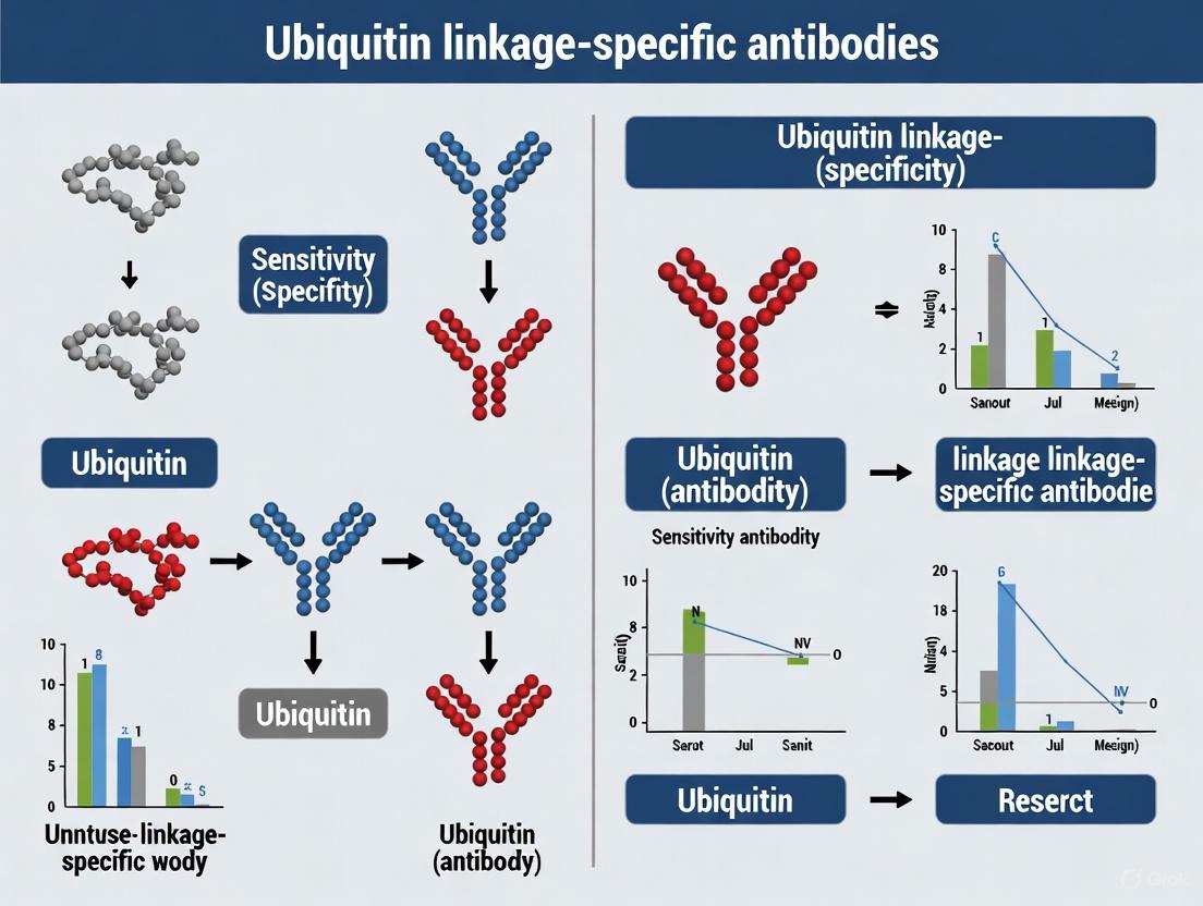
Ubiquitin Linkage-Specific Antibodies: A Comprehensive Guide to Sensitivity, Specificity, and Application
This article provides a critical resource for researchers and drug development professionals navigating the complex landscape of ubiquitin linkage-specific antibodies.
Ubiquitin Linkage-Specific Antibodies: A Comprehensive Guide to Sensitivity, Specificity, and Application
Abstract
This article provides a critical resource for researchers and drug development professionals navigating the complex landscape of ubiquitin linkage-specific antibodies. It establishes the foundational 'ubiquitin code' and the critical functional distinctions between polyubiquitin chain types, such as the proteasomal targeting role of K48-linked chains versus the signaling functions of K63-linked chains. The content details the working mechanisms, key applications, and technical specifications of these essential reagents in methods like Western blotting and immunofluorescence. Furthermore, it offers a rigorous framework for troubleshooting common issues like cross-reactivity and sensitivity limitations, and presents advanced validation strategies and comparative analyses with emerging non-antibody technologies to ensure data accuracy and reproducibility in ubiquitin signaling research.
Decoding the Ubiquitin Code: Why Linkage Specificity Matters
The ubiquitin-proteasome system (UPS) serves as a critical regulator of intracellular protein homeostasis, primarily known for targeting proteins for degradation. However, its functionality extends far beyond this proteolytic role. At the heart of this system lies a sophisticated signaling language known as the ubiquitin code, where the small regulatory protein ubiquitin becomes covalently attached to substrate proteins through a complex enzymatic cascade [1] [2]. This modification is remarkably versatile—ubiquitin itself contains seven lysine residues (K6, K11, K27, K29, K33, K48, K63) and an N-terminal methionine, each capable of serving as an attachment point for additional ubiquitin molecules, thereby forming various chain topologies [1] [3]. These linkage-specific polyubiquitin chains represent a complex post-translational code that governs diverse cellular outcomes, with different chain architectures directing substrates toward distinct fates including proteasomal degradation, altered subcellular localization, modified activity, or participation in signaling assemblies [4] [3].
The process of ubiquitination involves a hierarchical enzymatic cascade. A ubiquitin-activating enzyme (E1) first activates ubiquitin in an ATP-dependent manner, which is then transferred to a ubiquitin-conjugating enzyme (E2). Finally, a ubiquitin ligase (E3) facilitates the transfer of ubiquitin to the target substrate [1] [2]. With hundreds of E3 ligases encoded in the human genome, this system achieves remarkable specificity in substrate selection. The resulting ubiquitin modifications can be homogenous chains (using a single lysine linkage), mixed (incorporating different linkages), or even branched, with each configuration potentially encoding different functional consequences [1]. This intricate system allows cells to regulate virtually every biological process, from cell cycle progression and transcription to synaptic plasticity and immune response, through the dynamic interpretation of the ubiquitin code [1] [4] [3].
Figure 1: The Ubiquitination Cascade and Diverse Chain Outcomes. The enzymatic cascade (E1-E2-E3) conjugates ubiquitin to substrates, generating linkage-specific chains with distinct cellular functions.
Linkage-Specific Functions and Detection Challenges
Different polyubiquitin chain linkages generate specific biological outcomes through their distinct structural properties and recognition by ubiquitin-binding domains (UBDs). K48-linked chains represent the best-characterized ubiquitin signal and primarily target substrates for degradation by the 26S proteasome [5] [3]. Structural studies have revealed that K48-linked chains adopt a "closed" conformation where hydrophobic residues at the interface between adjacent ubiquitin molecules contact each other, creating a compact structure ideally suited for recognition by proteasomal subunits [3] [6]. In contrast, K63-linked chains adopt a more extended, linear conformation that resembles monoubiquitin and functions primarily as a scaffolding signal for protein complexes involved in diverse non-proteolytic processes including DNA repair, kinase activation, endocytosis, and inflammatory signaling [7] [3] [6]. The linear (M1-linked) ubiquitin chain also adopts an extended conformation and plays a specialized role in NF-κB activation and inflammation [6].
The conformational dynamics of ubiquitin chains add a crucial regulatory layer to the ubiquitin code. Single-molecule FRET studies have demonstrated that Lys63- and Met1-linked diubiquitin exist in equilibrium between extended "open" and more compact "closed" conformations in solution, while Lys48-linked diubiquitin adopts predominantly compact conformations [6]. Importantly, ubiquitin-binding proteins and deubiquitinases (DUBs) can select for pre-existing conformational states, suggesting that the conformational equilibria in ubiquitin chains provide an additional mechanism for regulating ubiquitin-dependent signaling [6]. This structural complexity presents significant challenges for biochemical detection, as different chain conformations may be recognized with varying efficiencies by detection reagents, particularly linkage-specific antibodies [8].
Figure 2: Ubiquitin Chain Linkages Dictate Structure and Function. Different ubiquitin linkage types form distinct three-dimensional structures that determine their specific cellular roles.
Comparative Analysis of Detection Methods
Limitations of Conventional Antibody-Based Detection
Western blotting with linkage-specific antibodies has been the most widely used method for detecting polyubiquitin chains, but significant limitations in antibody performance have emerged. Research has demonstrated that conventional anti-ubiquitin antibodies exhibit markedly different affinities for the eight linkage types of ubiquitin chains, with the highest sensitivity for K63-linked chains, lower efficiency for M1 and K48, and very low affinity for the other types of chains [8]. This binding bias creates substantial challenges for accurate ubiquitin chain detection, as it can lead to overestimation of certain chain types while underestimating or completely missing others. For instance, a widely used K48-linkage specific antibody (Cell Signaling Technology #4289) demonstrates slight cross-reactivity with linear polyubiquitin chains despite its primary specificity for K48 linkages [5]. Similarly, a popular K63-linkage specific antibody (Abcam ab179434) shows specific recognition for K63 linkages over other chain types in Western blot applications [7].
The technical limitations of antibody-based detection become particularly problematic when researchers need to compare polyubiquitination signals across different experimental conditions or accurately quantify changes in specific chain types. The variable sensitivity and cross-reactivity profiles of different antibody batches can introduce significant experimental variability and compromise data interpretation. Furthermore, the conformational dynamics of ubiquitin chains—with different linkages adopting distinct conformations in solution—may further complicate antibody recognition, as some epitopes might be inaccessible in certain conformational states [6]. These technical challenges have driven the development of alternative detection methods that can provide more accurate and comprehensive profiling of the cellular ubiquitin code.
TUF-WB: An Advanced Non-Antibody Approach
To address the limitations of antibody-based detection, researchers have developed the tandem hybrid ubiquitin-binding domain (ThUBD)-based far-Western blotting (TUF-WB) method, which utilizes the unbiased affinity of engineered ThUBD to all types of ubiquitin linkages [8]. This innovative approach leverages naturally occurring ubiquitin-binding domains that have been engineered into a tandem hybrid configuration with balanced affinity for diverse chain types. Unlike conventional antibodies, TUF-WB demonstrates equivalent sensitivity across all eight ubiquitin chain linkages, enabling accurate quantification of polyubiquitination signal intensity relative to the mass amounts of different chains [8].
Comparative studies have demonstrated that TUF-WB offers significant advantages over antibody-based methods, with 4-5-fold higher sensitivity when detecting complex ubiquitinated samples and a wider dynamic range for quantification [8]. This enhanced sensitivity is particularly valuable for detecting less abundant chain types that might be missed by conventional antibodies. Additionally, the unbiased nature of ThUBD recognition means that TUF-WB can detect atypical, mixed, or branched ubiquitin chains that may not be recognized efficiently by linkage-specific antibodies. The method provides researchers with a more comprehensive tool for mapping changes in the global ubiquitin landscape in response to cellular stimuli, pharmacological interventions, or in disease states, ultimately enabling more accurate deciphering of the complex ubiquitin code.
Table 1: Comparison of Ubiquitin Chain Detection Methods
| Method | Mechanism | Sensitivity Profile | Key Advantages | Key Limitations |
|---|---|---|---|---|
| Conventional Antibodies | Immunorecognition of linkage-specific epitopes | Variable affinity across linkages (highest for K63, lower for K48/M1, very low for others) [8] | Widely accessible, protocol familiarity | Linkage bias, variable cross-reactivity, limited dynamic range |
| TUF-WB | ThUBD binding to ubiquitin chains | Uniform high sensitivity across all 8 linkage types [8] | Unbiased detection, 4-5x higher sensitivity, wider dynamic range [8] | More specialized reagent required, less established protocols |
| Mass Spectrometry | Analysis of ubiquitin chain composition | High sensitivity with advanced instrumentation | Can identify mixed/branched chains, precise linkage determination | Technically challenging, requires specialized expertise and equipment |
Table 2: Performance Characteristics of Linkage-Specific Antibodies
| Antibody Target | Supplier | Catalog Number | Reported Cross-reactivity | Recommended Applications |
|---|---|---|---|---|
| K48-linkage | Cell Signaling Technology | #4289 | Slight cross-reactivity with linear chains [5] | Western Blot (1:1000 dilution) [5] |
| K63-linkage | Abcam | ab179434 | Specific for K63 in Western blot [7] | Western Blot, IHC-P, Flow Cytometry (Intra) [7] |
Experimental Approaches for Ubiquitin Research
Methodological Details for Key Assays
Western Blotting with Linkage-Specific Antibodies remains a fundamental approach despite its limitations. For K48-linkage detection using Cell Signaling Technology #4289, the recommended protocol includes standard protein extraction and quantification, SDS-PAGE separation, transfer to PVDF membrane, blocking with 5% non-fat dry milk or BSA in TBST, and incubation with primary antibody at 1:1000 dilution in blocking buffer overnight at 4°C [5]. For K63-linkage detection using Abcam ab179434, similar protocols apply with primary antibody dilution at 1:1000-1:5000 in 5% non-fat dry milk/TBST [7]. Critical considerations include optimizing protein loading amounts (typically 20μg of cell lysate), ensuring proper transfer efficiency for high molecular weight ubiquitinated species, and including appropriate controls such as recombinant ubiquitin chains when available to verify specificity.
The TUF-WB Methodology involves several key steps beginning with standard SDS-PAGE and transfer to a solid membrane. The membrane is then blocked and incubated with the recombinant ThUBD probe, which binds to ubiquitin chains with unbiased affinity across linkage types [8]. Detection is achieved using labeled ThUBD or subsequent incubation with anti-tag antibodies if the ThUBD contains an epitope tag. The critical advantage of this method lies in the engineering of the ThUBD, which combines multiple ubiquitin-binding domains with balanced affinities to overcome the linkage bias inherent to conventional antibodies. This approach allows for accurate quantification of polyubiquitination signals across a wider dynamic range and with significantly higher sensitivity compared to antibody-based methods [8].
Biochemical and Cellular Assays for studying ubiquitination often involve immunoprecipitation of target proteins under denaturing conditions to preserve ubiquitin modifications, followed by linkage-specific detection. For studying ubiquitin chain conformation, single-molecule FRET techniques have been employed, requiring specialized instrumentation including confocal microscopy with pulsed interleaved excitation, appropriate fluorophore labeling of ubiquitin, and sophisticated data analysis algorithms to resolve distinct conformational states [6]. These advanced techniques have revealed that ubiquitin chains exist in multiple conformational states in solution and that interacting proteins may select pre-existing conformations rather than inducing conformational changes upon binding.
The Scientist's Toolkit: Essential Research Reagents
Table 3: Essential Reagents for Ubiquitin Code Research
| Reagent Category | Specific Examples | Research Application | Key Considerations |
|---|---|---|---|
| Linkage-Specific Antibodies | Anti-K48 (CST #4289) [5], Anti-K63 (Abcam ab179434) [7] | Western blot, immunohistochemistry, flow cytometry | Validate specificity with recombinant chains; be aware of linkage bias |
| Unbiased Detection Reagents | Tandem hybrid UBD (ThUBD) [8] | Far-Western blotting (TUF-WB) for comprehensive chain detection | Provides uniform sensitivity across linkages; 4-5x more sensitive than antibodies [8] |
| Recombinant Ubiquitin Chains | K48-, K63-, M1-linked diubiquitin and extended chains | Specificity controls, in vitro reconstitution assays | Essential for validating antibody specificity and biochemical characterization |
| Deubiquitinase Enzymes | USP21, OTUB1, AMSH [9] [6] | Chain editing, specificity validation, functional studies | Linkage-specific DUBs can help verify chain identity; used in mechanistic studies |
| Enzymatic Cascade Components | E1 activating enzymes, E2 conjugating enzymes, E3 ligases [4] [2] | In vitro ubiquitination assays, mechanistic studies | Reconstitute minimal systems for specific chain synthesis; study enzyme mechanisms |
| Proteasome Inhibitors | Bortezomib, MG132 | Stabilizing proteasomal substrates, studying degradation-independent functions | Can cause accumulation of ubiquitinated proteins; may indirectl affect various chain types |
Implications for Research and Therapeutic Development
The accurate detection and interpretation of the ubiquitin code has profound implications for both basic research and drug discovery. In neurobiological research, understanding linkage-specific polyubiquitination has become increasingly important, as evidence suggests diverse ubiquitin chains play roles in synaptic plasticity and memory formation [1]. While initial research focused primarily on the proteolytic functions of K48-linked chains, recent studies indicate that non-proteolytic ubiquitin signaling involving K63-linked and other atypical chains contributes significantly to activity-dependent synaptic modification [1]. The technical limitations of antibody-based detection may have previously obscured the full complexity of ubiquitin signaling in neuronal systems, suggesting that more comprehensive profiling with unbiased methods could reveal new dimensions of ubiquitin function in the brain.
In the drug discovery landscape, the ubiquitin system represents a promising but challenging target class, often described as "drugging the undruggables" due to the potential to target proteins previously considered undruggable through modulation of their stability [4]. The success of proteasome inhibitors such as bortezomib in treating multiple myeloma validated the therapeutic potential of targeting the UPS, but current efforts have expanded to more specific interventions. Several companies are now developing small molecules that target specific components of the ubiquitin system, particularly E3 ligases and deubiquitinases [4] [2]. For example, inhibitors of the E3 ligase Mdm2 (Hdm2 in humans) that regulate p53 stability are being investigated as cancer therapeutics, while inhibitors of DUBs such as USP21 show promise in preclinical cancer models [4] [9].
The development of targeted protein degradation (TPD) approaches, including proteolysis-targeting chimeras (PROTACs), represents a particularly innovative application of ubiquitin system knowledge [10]. These strategies harness specific E3 ligases to selectively degrade target proteins of interest, effectively creating pharmacological tools to rewrite the ubiquitin code for therapeutic benefit. The success of these approaches depends critically on understanding the specificity of E3 ligases and the ubiquitin chain types they generate, highlighting the fundamental importance of basic research on ubiquitin chain diversity. As detection methods continue to improve, particularly with the development of unbiased approaches like TUF-WB, researchers will be better equipped to develop and characterize novel therapeutics that modulate the ubiquitin code with greater precision and efficacy.
Protein ubiquitination is a crucial post-translational modification that regulates virtually every cellular process in eukaryotic cells. The versatility of ubiquitin signaling stems from the ability of this 76-amino acid protein to form diverse polyubiquitin chains through its internal lysine residues. Among the eight possible linkage types (M1, K6, K11, K27, K29, K33, K48, and K63), K48- and K63-linked chains represent the most extensively studied and functionally characterized ubiquitin signals. The functional dichotomy between these linkages forms a fundamental paradigm in ubiquitin biology: while K48-linked chains primarily target substrates for proteasomal degradation, K63-linked chains regulate non-proteolytic signaling processes including DNA repair, kinase activation, and intracellular trafficking. This guide provides a comprehensive comparison of linkage-specific antibodies and methodologies essential for dissecting the functional consequences of K48 and K63 ubiquitin linkages in cellular signaling and disease contexts.
Functional Dichotomy of Major Ubiquitin Linkages
K48-Linked Polyubiquitin: The Canonical Degradation Signal
Discovered as the principal signal for proteasomal degradation, K48-linked ubiquitin chains function as the primary degradation signal in the ubiquitin-proteasome system (UPS). These chains are assembled through consecutive linkages between the C-terminal glycine of an incoming ubiquitin and the K48 residue of a substrate-anchored ubiquitin molecule. The molecular recognition of K48-linked chains by proteasomal receptors targets decorated proteins for ATP-dependent degradation, enabling rapid control of protein abundance, cell cycle progression, and stress response. Key substrates include regulatory proteins such as IκB, p53, and Bcl-2, whose degradation must be tightly controlled for cellular homeostasis [5]. Beyond these classical examples, recent research has revealed that K48 linkages can participate in branched ubiquitin chains, particularly in conjunction with K29 linkages, to create potent degradation signals in targeted protein degradation platforms [11].
K63-Linked Polyubiquitin: Versatile Signaling Regulator
In contrast to the degradative function of K48 linkages, K63-linked polyubiquitin serves as a multifunctional signaling scaffold in diverse non-proteolytic pathways. These chains adopt a more open, flexible conformation that facilitates protein-protein interactions without directing substrates to the proteasome. K63 linkages play critical roles in innate immune signaling, where they mediate signal transduction downstream of tumor necrosis factor (TNF) receptor and Toll-like receptor (TLR) activation. Additionally, these chains function in DNA damage repair, endosomal sorting, and selective autophagy processes. The functional versatility of K63 linkages is exemplified by their involvement in the regulation of kinase adaptors such as RIP1 and IRAK1, which undergo temporally controlled "ubiquitin editing"—
Emerging Concepts: Beyond Simple Dichotomy
While the K48-degradation/K63-signaling paradigm provides a useful framework, recent research has revealed more complex scenarios. Under specific conditions, K63 linkages have been demonstrated to signal lysosomal degradation of membrane receptors, as shown for the LDL receptor targeted by the IDOL E3 ligase [12]. Conversely, certain contexts involve K48 linkages in non-degradative functions. Furthermore, the existence of heterotypic branched chains, such as K48/K63-branched ubiquitin, adds another layer of complexity to the ubiquitin code, with emerging evidence suggesting these branched structures can enhance degradation efficiency or create specialized signaling platforms [13] [11].
Comparative Analysis of Linkage-Specific Antibodies
The development of linkage-specific ubiquitin antibodies has revolutionized the study of ubiquitin signaling by enabling researchers to distinguish between different chain types in complex biological samples. The table below provides a comprehensive comparison of commercially available antibodies specific for K48 and K63 linkages.
Table 1: Comparison of Linkage-Specific Polyubiquitin Antibodies
| Parameter | K48-Linkage Specific Antibody | K63-Linkage Specific Antibody |
|---|---|---|
| Commercial Example | Cell Signaling Technology #4289 | Abcam ab179434 [EPR8590-448] |
| Clonality | Polyclonal | Monoclonal (Rabbit) |
| Immunogen | Synthetic peptide corresponding to Lys48 branch of human diubiquitin | Proprietary (Recombinant fragment) |
| Reactivity | All Species Expected | Human, Mouse, Rat |
| Applications | Western Blot (1:1000) | WB (1:1000), IHC-P (1:250-1:500), Flow Cytometry (Intracellular, 1:210) |
| Specificity Validation | Slight cross-reactivity with linear polyubiquitin; no cross-reactivity with monoubiquitin or other linkages | Specific for K63 linkages; tested against K6, K11, K27, K29, K33, K48-linked diUb |
| Key Applications | Detection of proteasome-targeted substrates | Signaling pathway analysis, subcellular localization, protein complex formation |
These linkage-specific antibodies have been instrumental in fundamental discoveries, such as revealing the phenomenon of ubiquitin chain editing, where signaling proteins initially modified with K63 chains later receive K48 chains to terminate their activity [14]. The structural basis for antibody specificity was elucidated through cocrystal structures of antibody-diubiquitin complexes, revealing how complementary determining regions recognize unique conformational epitopes presented by specific linkage types [14].
Experimental Approaches and Methodologies
Western Blotting with Linkage-Specific Antibodies
Protocol Overview:
- Cell Lysis: Prepare RIPA buffer (150mM NaCl, 1% NP-40, 0.5% sodium deoxycholate, 0.1% SDS, 50mM Tris pH 8.0) supplemented with protease inhibitors (e.g., PMSF, leupeptin) and 10-20mM N-ethylmaleimide (NEM) or iodoacetamide to inhibit deubiquitinases [13].
- Protein Separation: Load 20-30μg of total protein per lane on 4-12% Bis-Tris gels for optimal separation of high molecular weight ubiquitin conjugates.
- Transfer: Use standard PVDF transfer protocols; confirm transfer efficiency with Ponceau S staining.
- Blocking: Incubate membrane in 5% non-fat dry milk (NFDM) or BSA in TBST for 1 hour at room temperature.
- Primary Antibody Incubation: Dilute linkage-specific antibodies (1:1000) in 5% NFDM/TBST; incubate overnight at 4°C with gentle agitation [5] [7].
- Detection: Use HRP-conjugated secondary antibodies (1:1000-1:5000) and enhanced chemiluminescence substrates.
Technical Considerations: The observation of smeared bands rather than discrete bands is expected, representing heterogeneous populations of ubiquitinated proteins. Inclusion of linkage-specific diubiquitin standards is recommended for specificity verification [7].
Ubiquitin Interactor Profiling Using Linkage-Specific Reagents
Recent advances in mass spectrometry-based proteomics have enabled systematic mapping of ubiquitin-binding proteins with specific linkage preferences. The methodology below outlines the approach for identifying linkage-specific interactors:
Table 2: Ubiquitin Interactor Pull-Down Protocol
| Step | Description | Key Considerations |
|---|---|---|
| Chain Synthesis | Enzymatic generation of linkage-specific Ub chains using E2 enzymes (CDC34 for K48, Ubc13/Uev1a for K63) | Verify linkage specificity using UbiCRest assay with linkage-specific DUBs (OTUB1 for K48, AMSH for K63) [13] |
| Immobilization | Biotinylation via cysteine-maleimide chemistry and coupling to streptavidin resin | Confirm complete biotin conjugation using intact mass spectrometry |
| Pull-Down | Incubate immobilized chains with cell lysate (HeLa, HEK293) pre-treated with DUB inhibitors | Compare different DUB inhibitors (CAA vs. NEM) as they affect interactor profiles [13] |
| Interactor Identification | LC-MS/MS analysis of specifically bound proteins; statistical enrichment analysis | Use known linkage-specific binders as controls (RAD23B for K48, EPN2 for K63) [13] |
This approach has revealed that chain length and branching patterns significantly influence ubiquitin interactor profiles, with some proteins specifically recognizing K48/K63-branched chains [13]. The workflow for this methodology can be visualized as follows:
Monitoring Ubiquitin Chain Editing in Signaling Pathways
Background: Ubiquitin chain editing describes the dynamic process where proteins undergo sequential modification with different ubiquitin linkage types, typically beginning with K63 chains for activation and concluding with K48 chains for termination [14].
Experimental Workflow:
- Cell Stimulation: Treat appropriate cell models (HEK293, HeLa) with pathway-specific agonists (e.g., TNF-α for NF-κB pathway, IL-1β for inflammatory signaling).
- Time-Course Sampling: Collect samples at multiple time points (0, 5, 15, 30, 60, 120 minutes) post-stimulation.
- Immunoprecipitation: Isolate proteins of interest (e.g., RIP1, IRAK1) using specific antibodies or epitope-tagged constructs.
- Linkage-Specific Detection: Probe immunoprecipitates with K48- and K63-linkage specific antibodies to monitor temporal changes in ubiquitination patterns.
- Functional Validation: Correlate ubiquitination status with functional outputs such as kinase activity, protein-protein interactions, or degradation kinetics.
This approach demonstrated that RIP1, essential for TNF-induced NF-κB activation, initially acquires K63-linked polyubiquitin that facilitates signaling complex assembly, while at later times K48-linked polyubiquitin targets it for proteasomal degradation, effectively attenuating the immune response [14].
The Scientist's Toolkit: Essential Research Reagents
Table 3: Key Reagents for Linkage-Specific Ubiquitin Research
| Reagent Category | Specific Examples | Research Applications | Functional Role |
|---|---|---|---|
| K48-Linkage Antibodies | CST #4289 (Polyclonal) | Western blot analysis of degradation substrates | Detection of K48-linked chains targeting proteins for proteasomal degradation [5] |
| K63-Linkage Antibodies | Abcam ab179434 [EPR8590-448] (Monoclonal) | WB, IHC-P, Flow Cytometry (Intracellular) | Detection of K63 chains in signaling complexes, subcellular localization [7] |
| DUB Inhibitors | N-ethylmaleimide (NEM), Chloroacetamide (CAA) | Ubiquitin interactor pull-downs, stabilization of ubiquitinated species | Prevention of chain disassembly by endogenous deubiquitinases during experiments [13] |
| Linkage-Specific DUBs | OTUB1 (K48-specific), AMSH (K63-specific) | UbiCRest assay for linkage validation | Selective disassembly of specific ubiquitin linkages to confirm chain identity [13] |
| E2 Enzymes | CDC34 (K48-specific), Ubc13/Uev1a (K63-specific) | In vitro ubiquitin chain synthesis | Generation of linkage-defined ubiquitin chains for biochemical studies [13] |
| Branched Chain Reagents | K48/K63-branched Ub3 chains | Study of complex ubiquitin codes | Investigation of branched ubiquitin chain recognition and function [13] |
Advanced Research Applications and Future Directions
Branched Ubiquitin Chains and Therapeutic Applications
Recent research has revealed that branched ubiquitin chains, containing multiple linkage types within a single chain, can function as enhanced degradation signals. Studies have identified that K29/K48-branched ubiquitin chains assembled by the E3 ligase TRIP12 promote efficient degradation of neosubstrates in targeted protein degradation platforms, including PROTAC-based approaches [11]. This discovery has significant implications for therapeutic development, as small molecules that modulate branched chain assembly could enhance the efficacy of targeted protein degraders. The screening approaches outlined in this guide provide methodologies to identify such regulators of branched ubiquitination.
Technological Advances in Linkage-Specific Reagents
While high-quality antibodies exist for K48 and K63 linkages, research on atypical chains (K6, K11, K27, K29, K33) has been hampered by limited reagent availability. Innovative approaches such as affimer technology—
Integration with Functional Genomics
The combination of linkage-specific ubiquitin profiling with CRISPR-based functional genomics represents a powerful future direction. Libraries of E3 ligase and deubiquitinase knockout cells can be screened using linkage-specific antibodies to identify enzymes responsible for writing, erasing, and reading specific ubiquitin codes. This integrated approach will accelerate the functional annotation of the ubiquitin proteome and identify novel regulatory nodes with therapeutic potential.
The functional consequences of ubiquitin linkage type represent a fundamental coding system in cell biology, with K48 and K63 linkages constituting a core dichotomy between degradation and signaling. Linkage-specific antibodies have been indispensable tools in deciphering this code, enabling researchers to distinguish between these functionally distinct modifications in complex biological systems. As research advances, the integration of these reagents with sophisticated proteomic approaches and functional screening platforms will continue to reveal new dimensions of ubiquitin signaling, particularly through the study of branched and mixed chain architectures. These insights will undoubtedly accelerate both fundamental understanding of cellular regulation and the development of novel therapeutic strategies targeting the ubiquitin system.
Ubiquitin linkage-specific antibodies represent a cornerstone of proteomics research, enabling scientists to decipher the complex biological signals encoded by different polyubiquitin chains. These antibodies are engineered to recognize specific isopeptide bonds between ubiquitin molecules, such as those formed through lysine 48 (K48) or lysine 63 (K63) residues, which dictate fundamentally different cellular outcomes for modified proteins. The development of these precise molecular tools presents significant challenges in epitope recognition and antibody generation, demanding sophisticated approaches to achieve the necessary specificity and sensitivity for accurate research. This guide provides an objective comparison of ubiquitin linkage-specific antibody performance, supported by experimental data and detailed methodologies, to assist researchers in selecting appropriate reagents for their specific applications.
The Ubiquitin Code: A Primer on Linkage-Specific Signaling
Ubiquitination represents one of the most pervasive and dynamic post-translational modifications in eukaryotic cells, with more than 110,000 identified ubiquitination sites across over 12,000 human proteins [15]. A defining feature of the ubiquitin system is its ability to form structurally and functionally distinct polyubiquitin chains through different linkage types between ubiquitin molecules. The specific lysine residue used to form these chains—including K6, K11, K27, K29, K33, K48, K63, and N-terminal methionine (M1)—determines the three-dimensional architecture of the resulting chain and consequently its cellular function [15].
Among these linkage types, K48-linked and K63-linked chains are the best characterized. K48-linked chains predominantly target proteins for proteasomal degradation, while K63-linked chains mainly facilitate non-proteolytic signaling functions in DNA damage response, immune signaling, and protein trafficking [5] [15]. The development of linkage-specific antibodies has been instrumental in unraveling these distinct biological functions, with K48 and K63-specific antibodies serving as essential tools for understanding the "ubiquitin code" [14] [15].
Figure 1: Ubiquitin Signaling Pathway. This diagram illustrates the sequential enzymatic cascade (E1-E2-E3) that conjugates ubiquitin to substrate proteins, followed by the formation of linkage-specific polyubiquitin chains that determine distinct cellular fates.
Comparative Analysis of Linkage-Specific Antibodies
The following tables provide a comprehensive comparison of key performance characteristics for commercially available K48 and K63 linkage-specific antibodies, based on manufacturer specifications and independent validation studies.
Table 1: Key Specifications of Ubiquitin Linkage-Specific Antibodies
| Parameter | K48-linkage Specific Antibody (#4289) | K63-linkage Specific Antibody (ab179434) |
|---|---|---|
| Host Species | Rabbit | Rabbit |
| Clonality | Polyclonal | Monoclonal (EPR8590-448) |
| Immunogen | Synthetic peptide corresponding to Lys48 branch of human diubiquitin chain | Proprietary (information not disclosed) |
| Reactivity | All species expected | Human, Mouse, Rat |
| Applications | Western Blot (1:1000) | WB (1:1000), Flow Cytometry (Intracellular, 1:210), IHC-Paraffin (1:250-1:500) |
| Specificity Profile | Detects polyubiquitin chains with Lys48 linkage; slight cross-reactivity with linear polyubiquitin chain; no cross-reactivity with monoubiquitin or other linkage types | Specific for K63-linkage; mass spectrometry studies indicate binding to ubiquitin(60-66) epitope; some cross-reactivity with K6 linkages reported |
Table 2: Experimental Performance Characteristics
| Characteristic | K48-linkage Specific Antibody | K63-linkage Specific Antibody |
|---|---|---|
| Sensitivity | Endogenous detection in Western Blot | Detects endogenous levels across multiple applications |
| Band Pattern | Smear pattern from 16-300 kDa in Western Blot | Smear pattern from 16-300 kDa in Western Blot |
| Validation Methods | Protein A and peptide affinity purification; specificity testing against various linkage types | Epitope excision/exraction mass spectrometry; dot blot; testing against comprehensive panel of linkage types |
| Key Applications | Western Blot analysis of proteasomal targeting | Multiplex analysis of DNA repair, kinase activation, and immune signaling pathways |
Methodologies for Antibody Validation and Specificity Assessment
Mass Spectrometry-Based Epitope Mapping
Recent advances in mass spectrometry have enabled precise characterization of antibody epitopes. In a 2023 study, researchers employed affinity-mass spectrometry (Affinity-MS) to identify the exact binding epitope of a K63-linkage specific ubiquitin antibody [16]. The methodology involved:
Epitope Excision: The K63-ubiquitin antibody was immobilized and incubated with ubiquitin building blocks containing K63 residues. Proteolytic cleavage was then performed using pronase, and the resulting epitope and non-epitope fractions were analyzed by matrix-assisted laser desorption/ionization-time of flight (MALDI-TOF) analysis.
Epitope Extraction: The antibody-antigen complex was proteolytically cleaved while intact, followed by extraction and identification of the bound epitope fragments.
This approach revealed that the K63-linkage specific antibody recognizes an epitope located within the ubiquitin(60-66) sequence, while also demonstrating some cross-reactivity with K6-linked ubiquitin peptides [16]. This level of detailed epitope mapping provides researchers with critical information for experimental design and data interpretation.
Western Blot Specificity Testing
Comprehensive specificity profiling remains essential for validating linkage-specific ubiquitin antibodies. The standard protocol involves:
Membrane Preparation: Separate 20μg of cell lysates or 0.02-20μg of recombinant diubiquitin proteins of various linkage types (K6, K11, K27, K29, K33, K48, K63) by SDS-PAGE and transfer to nitrocellulose or PVDF membranes.
Blocking and Incubation: Block membranes with 5% non-fat dry milk/TBST for 1 hour at room temperature. Incubate with primary antibody at the recommended dilution (typically 1:1000) in blocking buffer overnight at 4°C.
Detection: Incubate with appropriate HRP-conjugated secondary antibody (1:1000 dilution) for 1 hour at room temperature. Develop using enhanced chemiluminescence substrate and visualize.
The K63-linkage specific antibody (ab179434) demonstrates strong specificity for K63-linked diubiquitin with minimal cross-reactivity against other linkage types, though some cross-reactivity with K6-linked chains has been observed in certain assays [16] [7].
Figure 2: Antibody Validation Workflow. This diagram outlines the comprehensive multi-platform approach required to establish antibody specificity, including mass spectrometry-based epitope mapping, Western blot analysis against various linkage types, and application-specific testing.
Advanced Applications in Ubiquitin Research
Studying Polyubiquitin Chain Editing
Linkage-specific antibodies have been instrumental in identifying the phenomenon of polyubiquitin chain editing, a regulatory mechanism that attenuates innate immune signaling. Research using these tools has demonstrated that kinase adaptors such as RIP1 (essential for tumor necrosis factor-induced NF-κB activation) and IRAK1 (involved in interleukin-1β and Toll-like receptor signaling) initially acquire K63-linked polyubiquitin chains to activate signaling pathways, while at later time points undergo editing to K48-linked chains that target them for proteasomal degradation [14]. This dynamic process represents a crucial regulatory mechanism for controlling the duration and intensity of immune responses.
Disease Association Studies
Linkage-specific ubiquitin antibodies have proven valuable in characterizing pathological inclusions associated with neurodegenerative diseases. Studies have shown that neurofibrillary tangles in Alzheimer's disease and Lewy bodies in Parkinson's disease are heavily ubiquitinated and can be readily visualized using specific ubiquitin antibodies [16]. The ability to distinguish between different ubiquitin linkage types in these pathological structures may provide insights into disease mechanisms and potential therapeutic approaches.
The Scientist's Toolkit: Essential Research Reagents
Table 3: Key Research Reagent Solutions for Ubiquitin Studies
| Reagent Category | Specific Examples | Function and Application |
|---|---|---|
| Linkage-Specific Antibodies | K48-linkage Specific (CST #4289), K63-linkage Specific (Abcam ab179434) | Detection and quantification of specific polyubiquitin chain types in various experimental applications |
| Recombinant Ubiquitin Proteins | K6-, K11-, K27-, K29-, K33-, K48-, K63-linked diubiquitin proteins | Specificity controls for antibody validation; substrate for in vitro assays |
| Cell Lines | HEK-293, HeLa, ID8, DF-1, Vero | Model systems for studying ubiquitin signaling in different cellular contexts |
| Detection Systems | HRP-conjugated secondary antibodies, Alexa Fluor dye conjugates, Quantum MESF Bead Kits | Signal amplification and quantification in immunoassays and flow cytometry |
| Proteasome Inhibitors | MG-132, Bortezomib, Carfilzomib | Tools to accumulate ubiquitinated proteins by blocking proteasomal degradation |
| Deubiquitinase Enzymes | Catalytically inactive DUB mutants, DUB inhibitors | Tools for studying ubiquitin chain dynamics and editing |
The development and validation of ubiquitin linkage-specific antibodies represents a significant achievement in molecular recognition technology, enabling precise dissection of the complex ubiquitin signaling code. While both K48 and K63-linkage specific antibodies demonstrate high specificity and sensitivity in their intended applications, researchers must remain aware of their distinct performance characteristics and potential limitations. The slight cross-reactivity of K48 antibodies with linear chains and the recently identified K6 cross-reactivity of some K63 antibodies highlight the importance of appropriate controls and validation in specific experimental systems. As mass spectrometry and other analytical techniques continue to advance our understanding of antibody epitopes, researchers will be better equipped to select optimal reagents and interpret experimental results in the context of the dynamic and multifaceted ubiquitin signaling network.
Protein ubiquitination is a fundamental post-translational modification that regulates a vast array of cellular processes, extending far beyond its well-established role in protein degradation via the proteasome. This 76-amino acid polypeptide operates as a sophisticated signaling system that influences protein-protein interactions, DNA repair mechanisms, transcriptional regulation, and chromatin dynamics [17]. The ubiquitin code is remarkably complex, involving different forms of ubiquitination—from single ubiquitin moieties attached to a substrate (monoubiquitination) to polyubiquitin chains connected through different lysine residues (e.g., K48, K63) or the N-terminal methionine (M1)—each capable of generating distinct functional outcomes within the cell [17]. Understanding the mechanistic basis and functional consequences of these specific ubiquitination events requires research tools of exceptional quality, particularly linkage-specific antibodies capable of distinguishing between these structurally similar modifications.
The development and selection of appropriate ubiquitin linkage-specific antibodies present significant challenges for researchers. The large size of ubiquitin compared to other post-translational modifications, the instability of the native isopeptide linkage, which can be cleaved by deubiquitinating enzymes, and the high degree of structural similarity between different ubiquitin linkages collectively create a scenario where antibody specificity becomes difficult to achieve [17]. Furthermore, as evidenced by a systematic evaluation of SUMO monoclonal antibodies, substantial variability exists between different antibody clones in their sensitivity, specificity, and ability to detect different conjugation states, even when targeting the same modification [18]. This variability underscores the critical importance of rigorous validation and informed selection of immunological reagents for ubiquitin research. The landscape of available reagents is divided primarily between monoclonal and polyclonal antibody platforms, each with distinct characteristics that determine their suitability for specific applications in both basic research and drug development contexts.
Fundamental Distinctions: Monoclonal vs. Polyclonal Antibody Platforms
Core Characteristics and Production Methodologies
Monoclonal and polyclonal antibodies represent fundamentally different immunological reagents with distinct production workflows and biochemical properties. Monoclonal antibodies (mAbs) are defined as antibodies produced by a single clone of B cells, resulting in a homogeneous population of immunoglobulin molecules that demonstrate identical specificity toward a single epitope on the target antigen [19] [20] [21]. Their production typically employs hybridoma technology, which involves fusing antigen-specific B cells isolated from immunized animals with immortal myeloma cells to create stable hybridoma cell lines [19] [20]. These cell lines can be indefinitely cultured to produce consistent, renewable quantities of antibodies with uniform characteristics, making them particularly valuable for applications requiring standardized reagents.
In contrast, polyclonal antibodies (pAbs) are heterogeneous mixtures of antibodies produced by multiple different B-cell clones within an immunized host animal [19] [21]. This diverse antibody population recognizes multiple different epitopes on the target antigen, resulting in a reagent with broad epitope coverage but inherent variability between production batches [19] [20]. The production of polyclonal antibodies involves immunizing host animals with the target antigen and harvesting the antibody-rich serum after a sufficient immune response has developed [20]. While this process is generally less complex and time-consuming than monoclonal antibody development, the resulting antisera contain a complex mixture of antibodies with varying affinities and specificities, and the supply is finite unless the immunization process is repeated in additional animals.
Table 1: Fundamental Characteristics of Monoclonal vs. Polyclonal Antibodies
| Characteristic | Monoclonal Antibodies | Polyclonal Antibodies |
|---|---|---|
| Composition | Homogeneous antibody population [19] | Heterogeneous antibody mixture [19] [21] |
| Epitope Recognition | Single, specific epitope [19] [20] [21] | Multiple epitopes across the antigen [19] [21] |
| Production Process | Hybridoma technology & cell culture [19] [20] | Animal immunization & serum collection [19] [20] |
| Production Timeline | Long (6+ months) [19] | Short (3-4 months) [19] |
| Batch-to-Batch Consistency | High [19] [20] [21] | Variable [19] [20] [21] |
| Specificity | High for a single epitope [19] | Broad for multiple epitopes [19] |
| Cross-Reactivity Potential | Low [20] | Increased likelihood [20] |
Key Advantages and Limitations for Research Applications
The structural homogeneity of monoclonal antibodies translates to several key advantages in research and diagnostic applications. Their uniform composition ensures high specificity for a defined molecular target, minimal lot-to-lot variability, and reduced background noise in detection assays [20]. These characteristics make monoclonal antibodies particularly suitable for applications requiring precise target quantification, standardized diagnostic tests, and therapeutic development where consistency is paramount [19] [20]. However, this high specificity can also represent a limitation, as monoclonal antibodies may fail to detect target proteins that have undergone slight conformational changes, genetic polymorphisms, or post-translational modifications that alter the specific epitope they recognize [20]. Additionally, the initial development process for monoclonal antibodies is typically more costly and time-consuming than for polyclonal antibodies [19] [21].
Polyclonal antibodies offer complementary advantages stemming from their ability to recognize multiple epitopes on a target antigen. This broad recognition profile enhances their sensitivity for detecting low-abundance targets, makes them more tolerant of minor alterations in antigen structure, and allows for stronger signal amplification in various detection systems [20]. These characteristics make polyclonal antibodies particularly valuable for detecting denatured proteins in Western blots, capturing target antigens in immunoprecipitation protocols, and detecting unknown protein isoforms [20]. The primary limitations of polyclonal antibodies include significant batch-to-batch variability, higher potential for cross-reactivity with structurally similar proteins, and the finite nature of each production series [19] [20] [21].
Application-Based Selection Guide for Ubiquitin Research
The choice between monoclonal and polyclonal antibody platforms depends heavily on the specific research application and the particular characteristics of the ubiquitination event under investigation. Different experimental approaches impose distinct requirements on the binding properties, specificity, and signal amplification capabilities of the immunological reagents used. The table below summarizes the recommended antibody types for common applications in ubiquitin research, based on the inherent strengths of each platform.
Table 2: Application-Based Recommendations for Antibody Selection
| Application | Recommended Type | Rationale |
|---|---|---|
| Western Blotting | Both suitable [19] | Monoclonal for specific band detection; polyclonal for denatured proteins [20] |
| ELISA | Both suitable [19] | Monoclonal for precise quantification; polyclonal for broad detection |
| Immunohistochemistry (IHC) | Polyclonal recommended [19] | Broader specificity, stronger signal, better for complex tissue samples [19] |
| Immunofluorescence (IF) | Polyclonal recommended [19] | Stronger signal amplification, tolerance to antigen variability [19] |
| Flow Cytometry | Monoclonal recommended [19] | High specificity, linear fluorescence correlation with antigen expression [19] |
| Immunoprecipitation (IP) | Polyclonal recommended [19] | Recognition of multiple epitopes enhances target capture efficiency [19] |
| Therapeutic Development | Monoclonal recommended [19] | High specificity, minimal cross-reactivity, consistent batch production [19] |
For ubiquitin research specifically, the application requirements become particularly nuanced. The detection of specific ubiquitin linkages—such as K48-linked versus K63-linked polyubiquitin chains—demands antibodies with exquisite specificity, favoring well-validated monoclonal antibodies [17]. However, the development of such specific reagents faces substantial challenges due to the large size of ubiquitin and the structural similarity between different linkage types. Research indicates that successful development of site-specific ubiquitin antibodies requires specialized antigen design, often incorporating proteolytically stable ubiquitin-peptide conjugates that mimic the native isopeptide linkage while resisting cleavage by deubiquitinating enzymes during immunization [17].
The following workflow diagram illustrates the decision process for selecting appropriate antibody types based on research goals and experimental requirements in ubiquitin research:
Experimental Data: Performance Variation in Ubiquitin/SUMO Antibodies
Empirical Evidence of Antibody Variability
Systematic evaluations of monoclonal antibodies targeting ubiquitin-like modifiers reveal substantial variability in performance characteristics, highlighting the critical importance of empirical validation for research reagents. In a comprehensive study examining twenty-four monoclonal antibodies directed against SUMO family members (SUMO1-4), researchers observed marked differences in specificity, sensitivity, and ability to detect distinct conjugation states across various experimental platforms [18]. The antibodies demonstrated substantial variability in their capacity to detect increased SUMOylation in response to thirteen different cellular stress agents, and their effectiveness as enrichment reagents for specific SUMOylated targets like RanGAP1 or KAP1 varied significantly between clones [18]. Particularly noteworthy was the finding that all four anti-SUMO4 monoclonal antibodies tested exhibited cross-reactivity with SUMO2/3, while several SUMO2/3 monoclonal antibodies cross-reacted with SUMO4, underscoring the challenge of achieving absolute specificity for highly homologous targets [18].
These findings have direct relevance to ubiquitin research, where the high degree of structural conservation between different ubiquitin linkages creates similar challenges for antibody specificity. The study highlights that monoclonal antibodies, while theoretically offering perfect specificity, in practice demonstrate varying degrees of cross-reactivity that must be empirically characterized for each research application. Furthermore, the performance of these antibodies differed significantly across experimental platforms including dot blots, immunoblots, immunofluorescence, and immunoprecipitation, emphasizing that validation in one application does not guarantee equivalent performance in another [18]. This evidence-based approach to antibody characterization provides a valuable framework for the ubiquitin research community in assessing reagent suitability for specific experimental needs.
Specialized Methodologies for Site-Specific Ubiquitin Antibody Development
The development of antibodies capable of distinguishing specific ubiquitination events requires specialized methodologies that address the unique challenges posed by the ubiquitin system. Traditional immunization approaches using short peptides containing ubiquitinated lysine residues have met with limited success, prompting the development of more sophisticated antigen design strategies [17]. One advanced approach involves the chemical synthesis of full-length ubiquitin derivatives that can be attached to target peptides of choice using chemical ligation technologies [17]. These strategies include the synthesis of well-defined ubiquitin-modified polypeptides with native isopeptide linkages using thiolysine-mediated ligation, or alternatively, the creation of proteolytically stable analogs using click chemistry that replace the native isopeptide bond with a stable amide triazole isostere while preserving the overall structural environment around the ubiquitin-lysine interface [17].
The successful generation of a monoclonal antibody specific for ubiquitin on lysine 123 of yeast histone H2B (yH2B-K123ub1) demonstrates the effectiveness of this approach [17]. This antibody was obtained using antigens featuring the complete ubiquitin protein in a proteolytically stable form, which significantly increased the probability of generating antibodies recognizing the site-specific epitope rather than just ubiquitin or the target protein alone [17]. The resulting monoclonal antibody has been successfully deployed in both immunoblotting and chromatin immunoprecipitation assays, enabling mechanistic studies of the bidirectional regulatory relationships between histone ubiquitination and methylation [17]. This methodological framework provides a roadmap for developing additional site-specific ubiquitin antibodies, which remain scarce despite their critical importance for advancing understanding of ubiquitin signaling in normal physiology and disease states.
Advanced Reagent Solutions: Recombinant and Superclonal Antibodies
Recombinant Monoclonal Antibodies
Recent technological advances have introduced recombinant antibody platforms that offer distinct advantages over traditional hybridoma-derived monoclonals and polyclonal sera. Recombinant monoclonal antibodies are produced using in vitro expression systems by cloning antibody DNA sequences from immunoreactive animals into stable production cell lines [22]. This platform offers several significant benefits: superior lot-to-lot consistency, animal origin-free production processes, and the ability to mine broader immune repertoires for challenging targets [22]. For ubiquitin research, where subtle differences in linkage specificity are critical, recombinant monoclonal antibodies provide a renewable resource with perfectly defined characteristics, making them particularly valuable for long-term studies requiring standardized reagents.
Commercial examples include recombinant rabbit monoclonal antibodies against ubiquitin, such as clone 10H4L21, which has been successfully used to detect ubiquitination of specific targets like GLUT1 in transfected HeLa cells [22]. These reagents demonstrate the practical application of recombinant technology for producing highly specific detection tools for ubiquitination research. The defined molecular nature of recombinant antibodies also facilitates engineering approaches to enhance their performance characteristics, such as modifying affinity, stability, or species cross-reactivity patterns to better suit specific research applications.
Superclonal Antibody Technology
A hybrid approach that combines advantages of both polyclonal and monoclonal systems has emerged with the development of recombinant Superclonal antibodies. These reagents comprise a defined mixture of multiple different recombinant monoclonal antibodies, creating a preparation that recognizes multiple epitopes on the target antigen while maintaining the consistency of a recombinant product [23]. Functionally, Superclonal antibodies deliver the sensitivity typically associated with polyclonal antibodies—particularly beneficial for detecting low-abundance targets—while providing the specificity and batch-to-batch consistency normally exclusive to monoclonal antibodies [23].
This technology addresses a fundamental limitation of conventional polyclonal antibodies: their inherent variability between production batches. As noted in the search results, while polyclonal antibodies may be functionally equivalent to Superclonal antibodies in recognizing multiple epitopes, the exact composition of a Superclonal antibody can be reproduced in every manufacturing lot, circumventing the biological variability that plagues traditional polyclonal production [23]. For ubiquitin research applications where detection of multiple ubiquitination states or forms is desirable, but experimental reproducibility is essential, Superclonal antibodies represent a promising technological solution that merges the key advantages of both conventional platforms.
Essential Research Reagent Solutions for Ubiquitin Studies
The experimental investigation of protein ubiquitination requires a specialized toolkit of reagents and methodologies designed to address the unique challenges of working with this modification. The following table summarizes key reagents and their specific functions in ubiquitin research, particularly in the context of developing and validating linkage-specific antibodies.
Table 3: Essential Research Reagent Solutions for Ubiquitin Studies
| Reagent / Methodology | Function in Ubiquitin Research |
|---|---|
| Proteolytically Stable Ubiquitin-Peptide Conjugates | Serve as immunization antigens resistant to deubiquitinating enzyme activity [17] |
| Chemical Ligation Technologies | Enable synthesis of well-defined Ub-peptide conjugates with native or stable isopeptide linkages [17] |
| Hybridoma Screening with Extended Native Ub-Conjugates | Identifies clones recognizing the complete ubiquitination epitope rather than ubiquitin alone [17] |
| Recombinant Superclonal Antibodies | Provide multiple epitope recognition with monoclonal consistency [23] |
| Orthogonal Validation Methods (LC-MS/MS) | Confirms antibody specificity and detects ubiquitin in HCP analysis where ELISA may fail [24] |
| Linkage-Specific Ubiquitin Binding Domains | Serve as complementary tools to antibodies for detecting specific polyubiquitin chains [17] |
| Deubiquitinase Inhibitors | Preserve ubiquitination states during cell lysis and protein preparation [17] |
The following diagram illustrates the specialized workflow for developing site-specific ubiquitin antibodies, highlighting the critical steps that differentiate this process from conventional antibody generation:
The landscape of available reagents for detecting ubiquitin linkages presents researchers with multiple options, each with distinct advantages and limitations. Monoclonal antibodies offer unparalleled specificity for defined epitopes, making them indispensable for distinguishing between highly similar ubiquitin linkages and providing consistent, reproducible results across experimental batches [19] [20]. Polyclonal antibodies deliver superior sensitivity and broad epitope recognition, advantageous for capturing diverse ubiquitination states and detecting low-abundance modifications [19] [20] [21]. Emerging technologies, particularly recombinant monoclonal and Superclonal antibodies, bridge these platforms by offering defined specificity with enhanced consistency and engineering capabilities [23] [22].
The development of high-quality linkage-specific ubiquitin antibodies remains challenging due to the structural complexity and similarity between different ubiquitin modifications [17]. Success in this endeavor requires sophisticated antigen design strategies incorporating full-length ubiquitin in proteolytically stable forms and rigorous validation across multiple experimental platforms [17] [18]. As the field advances, the strategic selection and development of these reagents will continue to drive discoveries in ubiquitin biology, enabling researchers to decipher the complex regulatory networks controlled by this versatile post-translational modification in health and disease.
A Practical Guide to Using Linkage-Specific Ubiquitin Antibodies in the Lab
Protein analysis techniques form the cornerstone of modern biological research and drug development, enabling scientists to detect, quantify, and characterize proteins in complex biological systems. This guide provides an objective comparison of four core techniques—Western blot, immunoprecipitation, immunohistochemistry, and immunocytochemistry/immunofluorescence—with particular emphasis on their performance in evaluating ubiquitin linkage-specific antibodies. Understanding the strengths, limitations, and appropriate applications of each method is essential for researchers designing experiments to study protein expression, modifications, and cellular localization, especially in the context of ubiquitin signaling pathways that regulate critical cellular processes.
The selection of an appropriate protein analysis technique depends on various factors including the research question, sample type, and required throughput. The table below provides a systematic comparison of the core techniques to guide researchers in making informed methodological choices.
Table 1: Performance Comparison of Core Protein Analysis Techniques
| Technique | Primary Applications | Throughput | Sensitivity | Specificity | Quantitative Capability | Spatial Context | Key Limitations |
|---|---|---|---|---|---|---|---|
| Western Blot | Protein detection, size determination, modification analysis [25] | Medium (10-15 samples/gel) [25] | Less sensitive than ELISA [25] | High (size-based separation) [25] | Semi-quantitative [25] | No | Limited throughput, semi-quantitative [25] |
| Immunoprecipitation | Protein complex isolation, co-immunoprecipitation, ubiquitination studies [14] | Low | High (concentrates target) | Dependent on antibody quality | Semi-quantitative | No | Technically demanding, requires optimization |
| IHC | Protein localization in tissue context, diagnostic pathology [26] [27] | Medium to High | Varies with protocol | High (morphological context) | Semi-quantitative to quantitative | Yes (tissue architecture) | Subject to pre-analytical variables [27] |
| ICC/IF | Protein localization in cells, subcellular distribution [28] | Medium to High | High (fluorescence amplification) | High (subcellular context) | Quantitative with proper controls | Yes (cellular and subcellular) | Limited to cultured cells |
Detailed Methodological Protocols
Western Blotting Protocol
Western blotting enables protein detection through size-based separation followed by antibody-mediated identification, providing information about protein presence, relative abundance, and molecular weight [25].
Sample Preparation:
- Lyse cells or tissues in appropriate buffer (e.g., RIPA buffer) containing protease and phosphatase inhibitors
- Quantify protein concentration using BCA or Bradford assay
- Denature samples in SDS-PAGE loading buffer at 95-100°C for 5-10 minutes
Gel Electrophoresis:
- Prepare SDS-polyacrylamide gel appropriate for target protein size
- Load equal protein amounts (20-50 μg) into wells alongside molecular weight markers
- Run electrophoresis at constant voltage (100-150V) until dye front reaches bottom
Protein Transfer:
- Activate PVDF membrane in methanol or hydrate NC membrane in transfer buffer
- Assemble transfer stack following manufacturer recommendations
- Transfer proteins using wet or semi-dry systems (PVDF membranes may offer superior sensitivity for certain applications [29])
Immunoblotting:
- Block membrane with 5% BSA or non-fat dry milk in TBST for 1 hour
- Incubate with primary antibody diluted in blocking buffer overnight at 4°C
- Wash membrane 3× with TBST, 5 minutes each
- Incubate with HRP-conjugated secondary antibody for 1 hour at room temperature
- Wash membrane 3× with TBST, 5 minutes each
- Develop with ECL substrate and image
Western Blot Experimental Workflow
Immunoprecipitation Protocol
Immunoprecipitation enables isolation of specific proteins or protein complexes from cell lysates, useful for studying protein interactions and post-translational modifications including ubiquitination [14].
Lysate Preparation:
- Lyse cells in appropriate IP buffer (e.g., with mild detergents like NP-40)
- Clarify lysate by centrifugation at 14,000×g for 15 minutes
- Pre-clear lysate with protein A/G beads for 30 minutes
Antibody-Bead Complex Formation:
- Incubate antibody with protein A/G beads for 1-2 hours
- Wash beads to remove unbound antibody
Immunoprecipitation:
- Incubate pre-cleared lysate with antibody-bead complex overnight at 4°C
- Wash beads 3-5 times with IP wash buffer
- Elute bound proteins with SDS sample buffer or low pH elution buffer
Downstream Analysis:
- Analyze by Western blotting (for co-IP)
- For ubiquitination studies, use linkage-specific antibodies to detect ubiquitin chains [14]
IHC Protocol
Immunohistochemistry enables protein localization within tissue architecture, preserving spatial context [26] [27].
Tissue Preparation:
- Fix tissue in formalin (typically 10% neutral buffered) for 24 hours
- Process through graded alcohols and embed in paraffin
- Section at 4-5μm thickness and mount on charged slides
Deparaffinization and Antigen Retrieval:
- Deparaffinize in xylene and rehydrate through graded alcohols
- Perform antigen retrieval using heat-induced (citrate buffer, pH 6.0) or enzyme-mediated methods
- Block endogenous peroxidase with 3% H₂O₂ if using HRP detection
Staining:
- Block non-specific binding with serum or protein block
- Incubate with primary antibody for 1 hour at room temperature or overnight at 4°C
- Apply labeled polymer or secondary antibody
- Develop with chromogen (DAB, AEC)
- Counterstain, dehydrate, and mount
ICC/IF Protocol
Immunocytochemistry and immunofluorescence enable protein visualization in cultured cells with subcellular resolution.
Cell Preparation:
- Culture cells on glass coverslips or chamber slides
- Fix with 4% paraformaldehyde for 15 minutes or ice-cold methanol for 10 minutes
- Permeabilize with 0.1% Triton X-100 (for intracellular targets)
Staining:
- Block with serum or BSA for 1 hour
- Incubate with primary antibody for 1-2 hours at room temperature or overnight at 4°C
- Wash and incubate with fluorophore-conjugated secondary antibody
- Counterstain nuclei with DAPI or Hoechst
- Mount with anti-fade mounting medium
Application in Ubiquitin Linkage-Specific Antibody Research
The development of ubiquitin linkage-specific antibodies has revolutionized the study of ubiquitin signaling, enabling researchers to distinguish between different polyubiquitin chain types that determine protein fate [14]. These antibodies have revealed dynamic ubiquitin editing processes in signaling pathways, such as the transition from K63-linked to K48-linked chains on RIP1 and IRAK1 in innate immune signaling attenuation [14].
Table 2: Technique Selection for Ubiquitin Research Applications
| Research Goal | Recommended Technique | Key Considerations | Supporting Data |
|---|---|---|---|
| Ubiquitin chain type determination | Western blot with linkage-specific antibodies [14] | Requires validated linkage-specific antibodies | K63 and K48 linkage-specific antibodies enabled discovery of ubiquitin editing [14] |
| Protein complex ubiquitination | IP followed by Western blot | Co-IP can preserve protein complexes | Linkage-specific antibodies used after IP to characterize chain types [14] |
| Spatial distribution in tissues | IHC | Limited by antibody specificity for modified proteins | Technical specificity assessed with tissue panels [27] |
| Dynamic ubiquitination in cells | ICC/IF | Can reveal subcellular localization | Requires high-specificity antibodies with minimal background |
Research Reagent Solutions
Selecting appropriate reagents is critical for successful protein detection experiments. The following table outlines essential materials and their functions.
Table 3: Essential Research Reagents for Protein Detection Techniques
| Reagent Category | Specific Examples | Function | Technical Notes |
|---|---|---|---|
| Membranes | PVDF, nitrocellulose (NC) [29] | Bind transferred proteins for detection | PVDF may offer superior sensitivity for certain applications; requires methanol activation [29] |
| Detection Enzymes | Horseradish peroxidase (HRP), alkaline phosphatase (AP) [25] | Convert substrates to detectable signals | HRP most common with ECL; AP with colorimetric |
| Blocking Agents | BSA, non-fat dry milk, serum | Reduce non-specific antibody binding | Milk may interfere with phospho-specific antibodies; BSA preferred |
| Chromogens/Substrates | DAB, TMB, ECL | Generate visible or detectable signal | DAB for IHC; ECL for Western; choice affects sensitivity |
| Fixation Agents | Formalin, paraformaldehyde, methanol | Preserve tissue/cell structure and antigens | Formalin fixation can mask epitopes requiring antigen retrieval |
Quality Control and Validation
Robust quality control measures are essential for generating reliable data across all protein analysis techniques. For IHC, external controls should include both high-expressor and low-expressor tissues to properly monitor technical sensitivity [27]. In Western blotting, inclusion of positive and negative controls alongside molecular weight markers validates target identity. For ubiquitin linkage-specific antibodies, rigorous validation using defined ubiquitin chains is critical, as demonstrated by the cocrystal structure confirmation of anti-K63 linkage Fab bound to K63-linked diubiquitin [14].
Statistical considerations for assay validation include appropriate sample size calculations to ensure adequate statistical power. Studies evaluating diagnostic performance should calculate sample sizes based on expected sensitivity and specificity values, while reliability studies using Cohen's κ or intraclass correlation coefficients require different statistical approaches [28].
Ubiquitin Linkage-Specific Antibody Specificity
Western blot, immunoprecipitation, IHC, and ICC/IF each offer unique capabilities for protein analysis with specific strengths and limitations. Western blot provides size-based protein identification with semi-quantitative data, while immunoprecipitation enables isolation of specific proteins and complexes. IHC and ICC/IF preserve spatial context at tissue and cellular levels, respectively. For ubiquitin research, linkage-specific antibodies have enabled sophisticated studies of ubiquitin chain functions, with technique selection dependent on the specific research question. Proper implementation of these techniques, with attention to validation and controls, provides powerful approaches for advancing research in protein function and signaling pathways.
Western blot analysis of ubiquitinated proteins presents unique challenges and considerations for researchers studying protein regulation. The characteristic smeared appearance of ubiquitin blots, far from being an artifact, contains valuable biological information about the complexity of ubiquitin chains. This guide provides a comprehensive overview of methodologies for interpreting western blot data for ubiquitinated proteins, with particular focus on validating antibody specificity, optimizing detection protocols, and understanding the molecular basis of ubiquitin signaling. We compare the performance of various detection strategies and provide experimental protocols for researchers working in ubiquitin proteomics and drug development.
The Complexity of Protein Ubiquitination
Protein ubiquitination represents one of the most versatile post-translational modifications in eukaryotic cells, regulating diverse fundamental features of protein substrates including stability, activity, and localization [30]. Unlike simple binary modifications, ubiquitination creates a complex signature on target proteins that manifests as the characteristic smear on western blots. This complexity arises from several factors:
Ubiquitin Chain Diversity: Ubiquitin can form polymers through its own lysine residues (K6, K11, K27, K29, K33, K48, K63) or the N-terminal methionine (M1), creating homotypic chains, heterotypic chains, and branched chains with distinct biological functions [30]. K48-linked chains typically target substrates for proteasomal degradation, while K63-linked chains often regulate protein-protein interactions in signaling pathways such as NF-κB activation and autophagy [30].
Dynamic Modification States: Proteins can undergo mono-ubiquitination, multi-monoubiquitination (multiple single ubiquitins on different lysines), or polyubiquitination (ubiquitin chains on one or more lysines) [30]. Each state produces different migration patterns and potentially different biological outcomes.
Stoichiometric Challenges: The stoichiometry of protein ubiquitination is typically low under normal physiological conditions, increasing the difficulty of detecting endogenous ubiquitinated species amid abundant unmodified proteins [30]. This necessitates sophisticated enrichment strategies and sensitive detection methods.
Understanding this complexity is fundamental to proper interpretation of ubiquitin western blot data, as the smear pattern provides information about the distribution, extent, and possibly the linkage types of ubiquitin modifications.
Methodological Approaches for Ubiquitin Detection
Enrichment Strategies for Ubiquitinated Proteins
Due to the low abundance of ubiquitinated species, effective enrichment is essential prior to western blot analysis. The table below compares the primary methodologies used for enriching ubiquitinated proteins.
Table 1: Comparison of Ubiquitinated Protein Enrichment Methods
| Method | Principle | Advantages | Limitations | Typical Applications |
|---|---|---|---|---|
| Ubiquitin Tagging | Expression of affinity-tagged ubiquitin (His, Strep, FLAG) in cells [30] | Easy implementation; relatively low cost; compatible with various detection methods | May not mimic endogenous ubiquitin; genetic manipulation required; potential artifacts | Large-scale profiling of ubiquitination in cultured cells [30] |
| Antibody-Based Enrichment | Use of anti-ubiquitin antibodies (P4D1, FK1/FK2) or linkage-specific antibodies to immunoprecipitate ubiquitinated proteins [30] | Works with endogenous ubiquitin; applicable to tissues and clinical samples; linkage-specific options available | High cost of quality antibodies; non-specific binding; limited availability for rare linkages | Targeted studies; clinical samples; linkage-specific analysis [30] |
| UBD-Based Approaches | Tandem-repeated Ub-binding entities (TUBEs) with high affinity for ubiquitin chains [30] | High affinity; protects ubiquitin chains from deubiquitinases; recognizes various linkage types | Limited commercial availability; requires optimization; may have linkage preferences | Studies requiring preservation of labile ubiquitin modifications; proteomics studies [30] |
Antibody Selection and Validation
Antibody performance is arguably the most critical factor in obtaining reliable ubiquitin western blot data. Selection and validation should follow rigorous protocols:
Specificity Validation: Antibody specificity must be demonstrated using genetic controls such as ubiquitin knockout cells or CRISPR-mediated gene editing [31]. For ubiquitin-specific antibodies, testing in cells treated with proteasome inhibitors (e.g., MG132) that accumulate ubiquitinated proteins provides additional validation.
Selectivity Assessment: Determine whether antibodies recognize all ubiquitin linkages (pan-specific) or specific linkage types (linkage-specific) [30]. Linkage-specific antibodies (e.g., for K48 or K63 chains) require additional validation with defined ubiquitin chains.
Lot-to-Lot Consistency: Documented batch variation presents significant reproducibility challenges [31]. Recombinant antibodies offer superior lot-to-lot consistency compared to polyclonal antibodies derived from animal immunizations [32].
Table 2: Antibody Types for Ubiquitin Detection
| Antibody Type | Consistency | Sensitivity | Specificity | Recommended Applications |
|---|---|---|---|---|
| Polyclonal | Variable between lots and immunizations [32] | High (recognizes multiple epitopes) [32] | Moderate (potential cross-reactivity) [32] | Initial detection; low-abundance targets |
| Monoclonal | Good (hybridoma-derived) [32] | Moderate (single epitope) [32] | High (single epitope) [32] | Reproducible assays; specific modifications |
| Recombinant | Excellent (sequence-defined) [32] | Configurable | High (defined specificity) [32] | Quantitative studies; reproducible workflows |
Experimental Workflow for Ubiquitin Western Blotting
The following diagram illustrates the comprehensive workflow for analyzing ubiquitinated proteins by western blot, from sample preparation to data interpretation:
Sample Preparation and Enrichment Protocols
Denaturing Lysis: Use strong denaturing buffers (e.g., containing SDS) to preserve ubiquitination status by inhibiting deubiquitinating enzymes (DUBs) [33]. Immediately heat samples to 95°C for 5-10 minutes to denature proteins and inactivate DUBs.
Enrichment Methods:
- TUBE-Based Enrichment: Incubate cell lysates with Tandem Ubiquitin Binding Entities (TUBEs) immobilized on beads for 2-4 hours at 4°C [30]. Wash with mild buffers to reduce non-specific binding while maintaining ubiquitin chain integrity.
- Immunoaffinity Enrichment: For antibody-based enrichment, use linkage-specific or pan-ubiquitin antibodies cross-linked to protein A/G beads to prevent antibody leaching. Typical incubation times range from 4 hours to overnight at 4°C [30].
Competitive Elution: Elute enriched ubiquitinated proteins using free ubiquitin peptides (0.2-0.5 mg/mL) or low pH buffers (0.1 M glycine, pH 2.5-3.0) followed by neutralization [30].
Gel Electrophoresis and Transfer Optimization
Gel Selection: For resolving high molecular weight ubiquitinated species, use gradient gels (4-12% or 4-20% acrylamide) to achieve optimal separation across a broad molecular weight range [34]. Low-percentage acrylamide gels (6-8%) better resolve high molecular weight smears.
Transfer Conditions: For high molecular weight ubiquitin conjugates, use wet transfer systems with extended transfer times (2-3 hours for proteins >150 kDa) and low methanol content (<5%) in transfer buffer to maintain protein solubility [34]. For small proteins (<30 kDa), include 20% methanol and reduce transfer time to prevent blow-through.
Membrane Selection: PVDF membranes generally provide better protein retention for hydrophobic ubiquitinated species, while nitrocellulose may offer lower background for some applications [34]. Activate PVDF membranes in methanol before use.
Immunodetection and Imaging
Blocking Optimization: Test multiple blocking buffers (BSA, non-fat milk, commercial blocking reagents) to identify conditions that minimize background while maintaining signal [34]. For phosphorylated ubiquitin species, avoid milk-based blockers due to phosphatase activity.
Antibody Incubation:
- Primary antibodies: Incubate with anti-ubiquitin antibodies (dilutions typically 1:500 to 1:2000) overnight at 4°C with gentle agitation [32].
- Secondary antibodies: Use fluorescently-labeled secondary antibodies for multiplex detection or HRP-conjugated antibodies for chemiluminescent detection. Incubate for 1-2 hours at room temperature [32].
Detection Method Selection: Fluorescent detection offers a wider linear range for quantification, while chemiluminescent detection may provide higher sensitivity for low-abundance targets [35]. For quantitative comparisons, ensure signals fall within the linear range of detection by creating dilution series of samples [36].
Data Interpretation and Analysis
Understanding Ubiquitin Blot Patterns
The following diagram illustrates the relationship between ubiquitination states and their appearance on western blots:
Discrete Bands vs. Smears: Discrete bands above the expected molecular weight may represent mono-ubiquitination or specific polyubiquitinated species, while continuous smears indicate heterogeneous ubiquitination states with varying chain lengths and linkage types [33]. The absence of a smear does not necessarily indicate lack of ubiquitination - it may reflect synchronized chain lengths or specific ubiquitination patterns.
Molecular Weight Shifts: Each ubiquitin moiety adds approximately 8.5 kDa to the protein's apparent molecular weight [33]. Significant upward shifts (e.g., >50 kDa) suggest extensive polyubiquitination. Compare experimental molecular weight with theoretical weight to confirm ubiquitination status [33].
Technical Artifacts: Distinguish specific smears from non-specific background by including appropriate controls (untreated cells, ubiquitin knockdown, DUB inhibition). Vertical streaking may indicate transfer issues or protein aggregation.
Quantification and Normalization Strategies
Linear Range Determination: Establish the linear range for quantification by running a dilution series of samples and determining where signal intensity deviates from linearity [36] [37]. Signal saturation leads to underestimation of differences between samples.
Normalization Methods:
- Total Protein Normalization (TPN): Increasingly considered the gold standard, TPN uses total protein stain (e.g., No-Stain Protein Labeling Reagent) to normalize for loading variations [35]. This method accounts for uneven transfer and loading across lanes.
- Housekeeping Proteins (HKP): Traditional method using proteins like GAPDH, actin, or tubulin, but falling out of favor due to expression variability under experimental conditions [35].
- Ponceau S Staining: Rapid reversible stain for approximate normalization before immunodetection.
Quantitative Analysis: For quantitative comparisons, ensure that: (1) signals are within linear detection range, (2) appropriate normalization method is used, and (3) statistical analysis includes multiple biological replicates [37]. Use software that can quantify smeared regions rather than just discrete bands.
Research Reagent Solutions
The table below outlines essential reagents and their applications in ubiquitin western blotting:
Table 3: Essential Research Reagents for Ubiquitin Western Blotting
| Reagent Category | Specific Examples | Function & Application | Key Considerations |
|---|---|---|---|
| Ubiquitin Antibodies | P4D1, FK1, FK2 [30] | Pan-ubiquitin detection; recognizes various linkage types | Verify specificity with ubiquitin knockdown; lot-to-lot variability [31] |
| Linkage-Specific Antibodies | K48-specific, K63-specific [30] | Detection of specific ubiquitin chain linkages | Requires additional validation; may have cross-reactivity with similar linkages |
| Enrichment Reagents | TUBEs, Ubiquitin Binding Entities [30] | Affinity purification of ubiquitinated proteins | Preserves labile ubiquitination; protects from DUBs [30] |
| Positive Controls | MG132-treated cell lysates [31] | Verify detection system functionality | Confirms protocol working; establishes expected signal |
| Deubiquitinase Inhibitors | PR-619, N-ethylmaleimide | Preserve ubiquitination during preparation | Include in lysis buffers to prevent deubiquitination |
| Normalization Reagents | No-Stain Protein Labeling Reagents [35] | Total protein normalization for quantification | Superior to housekeeping proteins for quantitative work [35] |
Publication Guidelines for Ubiquitin Western Blots
Adhering to journal guidelines is essential for publishing ubiquitin western blot data. Key requirements include:
Image Presentation:
- Maintain original, unprocessed images as supplementary data [38].
- Include molecular weight markers on all blot images [39].
- Minimize cropping to show relevant portions of the blot while maintaining context [39].
- Avoid excessive brightness/contrast adjustments that may misrepresent data [38].
Methodology Reporting:
- Document antibody information (catalog numbers, RRIDs, dilutions) [39] [31].
- Describe enrichment methods and specific conditions [30].
- Detail normalization strategies and quantification methods [35].
- Report protein loading amounts and gel percentages [39].
Data Transparency:
- Provide source data for all blots during manuscript submission [38].
- Clearly indicate lane splicing or gel rearrangements [38].
- Disclose image processing steps in figure legends [38].
Major journals including Nature, Cell Press, and Elsevier have specific requirements for western blot presentation, so consult target journal guidelines early in experimental design [38] [35].
Troubleshooting Common Issues
High Background: Optimize blocking conditions (try different blockers or combinations), increase wash stringency (increase salt concentration or add detergent), titrate antibody concentrations to optimal dilution, and ensure sufficient washing between steps [34].
Weak or No Signal: Verify antibody specificity using positive controls, check antigen integrity (avoid repeated freeze-thaw cycles), optimize enrichment efficiency, increase protein loading, try different detection methods (e.g., switch from chemiluminescence to fluorescence), and extend exposure times appropriately [32].
Non-Specific Bands: Include proper controls (knockdown, knockout, or competitive peptides), pre-clear lysates with non-specific IgG, use more specific antibodies (monoclonal or recombinant rather than polyclonal), and ensure sufficient blocking [31].
Smear Interpretation Challenges: Distinguish specific smears from degradation by including protease inhibitors, compare with positive controls showing expected patterns, use linkage-specific antibodies to characterize smear composition, and consider that some proteins naturally show heterogeneous migration [33].
Western blot analysis of ubiquitinated proteins requires specialized methodologies and careful interpretation of the characteristic smear patterns that reflect the biological complexity of ubiquitin signaling. Successful detection and interpretation depend on appropriate enrichment strategies, validated antibodies, optimized electrophoretic conditions, and rigorous quantification methods. As ubiquitin research continues to evolve, particularly in drug development targeting ubiquitin pathways, standardized approaches for ubiquitin western blotting will enhance reproducibility and reliability across the scientific community. By following the methodologies and guidelines outlined in this document, researchers can more accurately interpret ubiquitin western blot data and advance our understanding of this crucial regulatory pathway.
Ubiquitination is a crucial post-translational modification that regulates virtually all aspects of eukaryotic cell biology. The specificity of this regulation is encoded in the diverse architectures of polyubiquitin chains, where ubiquitin molecules are connected through different lysine residues. K48- and K63-linked polyubiquitin chains represent two of the most abundant and functionally distinct chain types, with K48-linked chains primarily targeting proteins for proteasomal degradation, while K63-linked chains mainly mediate non-proteolytic signaling functions in processes such as DNA damage repair, kinase activation, and protein trafficking [15]. The ability to distinguish between these chain types is therefore fundamental to understanding their distinct cellular roles.
Linkage-specific antibodies serve as essential tools for deciphering this "ubiquitin code" by enabling researchers to detect, quantify, and characterize specific polyubiquitin chain types amidst the complex background of cellular ubiquitination. This guide provides a detailed comparison of commercially available K48- and K63-linkage specific antibodies, presenting experimental data and case studies to objectively evaluate their performance across various applications. By framing this comparison within the broader context of ubiquitin research methodologies, we aim to provide researchers, scientists, and drug development professionals with the practical information needed to select appropriate reagents for their specific experimental requirements.
Product Specifications and Comparative Analysis
Key Commercial Antibodies for K48 and K63 Linkage Detection
Table 1: Comparison of Key Commercial K48 and K63 Linkage-Specific Antibodies
| Target | Product Name | Supplier | Clonality | Applications | Species Reactivity | Recommended Dilution |
|---|---|---|---|---|---|---|
| K48-linkage | K48-linkage Specific Polyubiquitin Antibody #4289 | Cell Signaling Technology | Polyclonal | Western Blot | All Species Expected | 1:1000 [5] |
| K63-linkage | K63-linkage Specific Polyubiquitin (D7A11) Rabbit mAb #5621 | Cell Signaling Technology | Monoclonal | Western Blot | All Species Expected | 1:1000 [40] |
| K63-linkage | Anti-Ubiquitin (linkage-specific K63) antibody [EPR8590-448] | Abcam | Monoclonal | WB, IHC-P, Flow Cytometry | Human, Mouse, Rat | 1:1000 (WB) [7] |
Specificity and Cross-Reactivity Profiles
The specificity of linkage-specific antibodies is paramount for accurate data interpretation. The K48-linkage Specific Polyubiquitin Antibody (#4289) demonstrates slight cross-reactivity with linear polyubiquitin chains but shows no cross-reactivity with monoubiquitin or polyubiquitin chains formed by linkage to different lysine residues [5]. This antibody was generated using a synthetic peptide corresponding to the Lys48 branch of the human diubiquitin chain, with purification through protein A and peptide affinity chromatography to enhance specificity.
In comparison, the K63-linkage Specific Polyubiquitin Antibody (#5621) from Cell Signaling Technology shows no detectable reactivity with monoubiquitin or polyubiquitin chains formed by linkage to different lysine residues, indicating high linkage specificity [40]. The monoclonal nature of this antibody (clone D7A11) contributes to superior lot-to-lot consistency, which is crucial for experimental reproducibility.
The K63-linkage specific antibody from Abcam (ab179434) has been extensively validated across multiple applications including Western blot, immunohistochemistry (IHC-P), and flow cytometry. Specificity data demonstrates strong reactivity with K63-linked diubiquitin while showing minimal cross-reactivity with other linkage types (K6, K11, K29, K33, K48) under optimized conditions [7]. This antibody has been referenced in over 80 publications, supporting its utility in diverse research contexts.
Experimental Applications and Methodologies
Western Blot Protocols for Linkage-Specific Detection
Western blotting remains the most common application for linkage-specific ubiquitin antibodies. The standard protocol for detecting polyubiquitin chains using these antibodies typically involves:
Cell Lysis and Protein Extraction:
- Use RIPA or SDS-containing lysis buffers to disrupt non-covalent protein interactions
- Include proteasome inhibitors (e.g., MG132) and deubiquitinase inhibitors (e.g., N-ethylmaleimide) to preserve ubiquitin chains
- Boil samples for 5-10 minutes to ensure complete denaturation
Electrophoresis and Transfer:
- Separate proteins using 4-12% Bis-Tris gels for optimal resolution of high molecular weight polyubiquitin chains
- Transfer to PVDF membranes using standard wet or semi-dry transfer systems
Immunoblotting:
- Block membranes with 5% non-fat dry milk or BSA in TBST for 1 hour
- Incubate with primary antibody at recommended dilutions (typically 1:1000) overnight at 4°C
- Perform secondary antibody incubation with HRP-conjugated reagents for 1 hour at room temperature
- Develop using enhanced chemiluminescence substrates [5] [40] [7]
Table 2: Observed Band Patterns in Western Blot Applications
| Antibody | Predicted Band Size | Observed Band Pattern | Noteworthy Characteristics |
|---|---|---|---|
| K48-specific (#4289) | Not specified | Broad smearing pattern | Represents heterogeneous populations of K48-linked polyubiquitinated proteins [5] |
| K63-specific (#5621) | Not specified | Broad smearing pattern | Characteristic pattern for K63-linked polyubiquitin chains of varying lengths [40] |
| K63-specific (ab179434) | 26 kDa (monomeric ubiquitin) | 16-300 kDa range | Pattern reflects diverse K63-ubiquitinated proteins; specificity confirmed using linkage-defined ubiquitin chains [7] |
Advanced Applications and Techniques
Beyond standard Western blotting, linkage-specific ubiquitin antibodies have been adapted for diverse methodological approaches:
Immunohistochemistry: The Abcam K63-linkage specific antibody (ab179434) has been successfully used for IHC on formalin-fixed, paraffin-embedded tissues at dilutions ranging from 1:100 to 1:500. Optimal results require heat-mediated antigen retrieval using Tris/EDTA buffer (pH 9.0) [7]. This application enables spatial resolution of K63-linked ubiquitination in tissue contexts, particularly valuable for clinical samples.
Flow Cytometry: For intracellular flow cytometry, the same antibody has been used at a dilution of 1:210, with cell fixation using 4% paraformaldehyde and permeabilization with 90% methanol [7]. This approach enables analysis of K63-linked ubiquitination at single-cell resolution, particularly useful for heterogeneous cell populations.
Live-Cell Imaging and Reporter Systems: Innovative approaches have emerged that complement antibody-based methods. Engineered reporters such as the K63UbR enable real-time, quantitative imaging of AKT-directed K63-polyubiquitination in living cells [41]. This reporter system utilizes a split luciferase complementation approach that decreases bioluminescence when K63-linked ubiquitination occurs, allowing dynamic monitoring of ubiquitination events without cell lysis.
Biological Context and Signaling Pathways
Distinct Cellular Functions of K48 vs. K63 Linkages
The functional divergence between K48- and K63-linked ubiquitination represents a fundamental principle in ubiquitin biology. K48-linked polyubiquitin chains constitute approximately 40% of cellular ubiquitin linkages and primarily target substrate proteins for degradation by the 26S proteasome [15] [42]. This degradation pathway regulates the abundance of key cellular proteins such as IκB, p53, and Bcl-2, thereby influencing cell cycle progression, stress responses, and apoptosis [5].
In contrast, K63-linked polyubiquitin chains represent approximately 30% of cellular ubiquitin linkages and mainly mediate non-proteolytic signaling functions [15] [42]. These chains regulate diverse processes including protein trafficking, kinase/phosphatase activation, DNA damage repair, and immune signaling [40]. Recent research has revealed more nuanced functions, demonstrating that K63-linked chains can under specific conditions also signal lysosomal degradation, challenging the strict functional dichotomy previously assumed [42].
Diagram 1: Ubiquitination Cascade and Functional Divergence of K48 vs K63 Linkages
Experimental Evidence for Functional Overlap
While the functional distinction between K48 and K63 linkages is well-established, research has revealed surprising overlap under certain biological contexts. A notable example comes from studies of the LDL receptor (LDLR) degradation pathway, where both K48 and K63 ubiquitin linkages were found to mediate lysosomal degradation, contrary to the expected exclusive role of K63 chains in this process [42]. This demonstrates the importance of using well-validated linkage-specific antibodies to uncover unexpected biological mechanisms.
Mitochondrial quality control provides another compelling case study. Research using an engineered ubiquitin ligase (ProxE3) that specifically generates K63-linked chains on mitochondria demonstrated that K63 ubiquitination alone is sufficient to induce mitochondrial sequestration and recruitment of p62, but not mitophagy [43]. This suggests that K63 chains provide a necessary signal for mitochondrial quality control, but require additional signals for complete autophagic degradation.
The Scientist's Toolkit: Essential Research Reagents
Table 3: Key Research Reagent Solutions for Ubiquitin Linkage Studies
| Reagent Category | Specific Examples | Function/Application | Experimental Notes |
|---|---|---|---|
| Linkage-Specific Antibodies | CST #4289 (K48), CST #5621 (K63), Abcam ab179434 (K63) | Detection and quantification of specific polyubiquitin chains | Validate specificity using linkage-defined ubiquitin standards; optimize blocking conditions to reduce background [5] [40] [7] |
| Activity-Based Probes | Catalytically inactive DUBs, Ubiquitin-binding domains | Enrichment and characterization of linkage-specific ubiquitin chains | Useful for proteomic applications; can be coupled to mass spectrometry for system-wide analyses [15] |
| Engineered Reporter Systems | K63UbR reporter | Real-time imaging of K63-polyubiquitination in live cells | Enables dynamic monitoring of ubiquitination; suitable for high-throughput screening applications [41] |
| Ubiquitin Mutants | K48R, K63R, K48-only, K63-only ubiquitin mutants | Functional studies of linkage-specific ubiquitination | Critical for determining requirement of specific linkages in degradation pathways [42] |
| Specialized E3 Ligases | ProxE3 (engineered K63-specific ligase) | Inducible formation of K63-linked chains on specific substrates | Enables precise manipulation of ubiquitination states without confounding damage signals [43] |
The expanding toolbox for linkage-specific ubiquitin research, particularly antibodies targeting K48 and K63 linkages, has dramatically enhanced our ability to decipher the complex ubiquitin code. The antibodies compared in this guide demonstrate high specificity and utility across multiple experimental platforms, enabling researchers to investigate the distinct functions of these polyubiquitin signals in health and disease. As research progresses, the integration of these reagents with innovative approaches such as engineered ligases and live-cell reporters will continue to advance our understanding of ubiquitin signaling complexity, potentially revealing new therapeutic opportunities for diseases characterized by ubiquitin pathway dysregulation.
Antibody conjugates are indispensable tools in modern biological research, providing the combined power of specific target recognition with detectable labels for visualization and quantification. The process of antibody conjugation involves chemically linking an antibody to another molecule, such as a fluorescent dye, enzyme, or other marker, which dramatically enhances our ability to detect specific targets in complex biological samples [44]. In flow cytometry, these conjugated antibodies enable researchers to analyze cell populations at the single-cell level, characterizing numerous parameters simultaneously through multiplexed assays. The strategic application of conjugated antibodies has revolutionized our understanding of cellular functions, particularly in specialized fields such as ubiquitin signaling, where detecting specific protein post-translational modifications requires exceptional sensitivity and specificity.
The fundamental advantage of conjugated antibodies lies in their ability to facilitate high-throughput analysis while maintaining excellent specificity. Flow cytometry platforms leverage the unique spectral properties of various fluorophores to differentiate multiple cellular targets in a single sample, making it possible to dissect complex cellular phenotypes and functional states. This capability is particularly valuable when studying intricate signaling systems like the ubiquitin-proteasome pathway, where different ubiquitin linkage types control diverse cellular outcomes—from protein degradation to DNA repair and immune signaling [15]. The expanding toolbox of conjugated antibodies continues to push the boundaries of what we can detect and analyze, bringing previously obscure biological mechanisms into clear focus.
Types of Antibody Conjugates and Their Properties
Fluorophore Conjugates
Fluorophore-conjugated antibodies represent the most widely used conjugate type for flow cytometry applications due to their versatility, high sensitivity, and suitability for multiplexing [44]. These conjugates incorporate various fluorescent dyes that emit light at specific wavelengths when excited by lasers in flow cytometers. The selection of appropriate fluorophores depends on multiple factors, including the instrument's laser and filter configuration, the abundance of the target antigen, and the requirements for multicolor panels.
The table below summarizes key characteristics of common fluorophores used in antibody conjugates for flow cytometry:
Table 1: Characteristics of Common Fluorophore Conjugates for Flow Cytometry
| Fluorophore | Color Range | Max Excitation (nm) | Max Emission (nm) | Extinction Coefficient (M⁻¹cm⁻¹) | Primary Applications |
|---|---|---|---|---|---|
| FITC | Green | 495 | 519 | 70,000 | Flow cytometry, immunofluorescence |
| PE | Orange | 480-565 | 575-590 | 1,960,000 | Flow cytometry (high brightness) |
| APC | Red | 650 | 660 | 700,000 | Flow cytometry (high quantum yield) |
| Cy5 | Red | 650 | 670 | 250,000 | Flow cytometry, imaging, FRET |
| DyLight 594 | Red | 593 | 618 | 115,000 | Multicolor imaging, flow cytometry |
| iFluor 647 | Far-red | 650 | 665 | 250,000 | Flow cytometry, super-resolution microscopy |
| Alexa Fluor 488 | Green | 495 | 519 | ~73,000 | Flow cytometry, replacement for FITC |
Among these options, phycoerythrin (PE) stands out for its exceptional brightness due to its extremely high extinction coefficient, making it ideal for detecting low-abundance targets [44]. Allophycocyanin (APC) offers excellent performance in the red spectrum with high quantum yield. The newer iFluor and Alexa Fluor dyes provide enhanced photostability and brightness compared to traditional dyes like FITC, along with reduced susceptibility to photobleaching during prolonged analysis [44]. For multiplexed panels requiring many parameters, the availability of fluorophores spanning the entire visible to near-infrared spectrum (such as DyLight 650, DyLight 800, and iFluor 750) enables comprehensive profiling of complex cellular systems [44].
Other Antibody Conjugate Types
Beyond fluorescent tags, antibodies can be conjugated to various other molecules that enable detection through different mechanisms:
Enzyme conjugates, such as horseradish peroxidase (HRP) and alkaline phosphatase (AP), are primarily used in colorimetric or chemiluminescent detection systems like ELISA and Western blotting [44]. While not directly used for flow cytometry, these conjugates play important roles in complementary assays that validate flow cytometry findings.
Biotin conjugates leverage the strong non-covalent interaction between biotin and streptavidin, which can be further conjugated to fluorophores or enzymes [44]. This approach provides signal amplification, as multiple streptavidin molecules can bind to a single biotinylated antibody, enhancing detection sensitivity for low-abundance targets.
Metal conjugates have gained prominence with the advent of mass cytometry (CyTOF), which uses antibodies tagged with rare earth metal isotopes instead of fluorophores [44]. This technology eliminates spectral overlap issues inherent in fluorescence-based flow cytometry, enabling the measurement of over 40 parameters simultaneously—a significant advantage for comprehensive immune profiling or signaling network analysis.
Direct vs. Indirect Staining Methods
Comparison of Staining Approaches
Flow cytometry experiments can employ either direct or indirect staining methods, each with distinct advantages and limitations. Direct staining uses primary antibodies that are already conjugated to fluorophores, while indirect staining employs an unlabeled primary antibody followed by a fluorophore-conjugated secondary antibody that recognizes the primary [45].
Table 2: Comparison of Direct and Indirect Staining Methods
| Parameter | Direct Staining | Indirect Staining |
|---|---|---|
| Protocol Complexity | Simple one-step procedure | Multi-step process requiring additional incubations and washes |
| Time Requirement | Faster (30-60 minutes) | Slower (2+ hours with incubation steps) |
| Signal Amplification | No amplification | Yes, multiple secondary antibodies bind to each primary |
| Multiplexing Capacity | Excellent - multiple conjugates from same host species | Challenging - requires primary antibodies from different hosts |
| Background Signal | Lower (no secondary antibody cross-reactivity) | Higher potential due to secondary antibody non-specific binding |
| Flexibility | Low (conjugate fixed) | High (can pair with different secondaries) |
| Cost | Higher for conjugated primaries | Lower (unconjugated primaries + reusable secondaries) |
Practical Applications and Considerations
Direct staining is particularly advantageous for multiplexed panels where researchers need to detect multiple targets simultaneously. Since directly conjugated antibodies can all be raised in the same host species without cross-reactivity concerns, this method enables the design of complex panels with 10+ parameters [46]. The simplified protocol also reduces hands-on time and minimizes potential errors, while the absence of secondary antibodies decreases background signal [45]. However, the lack of signal amplification may limit sensitivity for low-abundance targets, and the fixed fluorophore configuration offers less flexibility once panels are established.
Indirect staining provides superior sensitivity through signal amplification, as multiple secondary antibodies can bind to a single primary antibody [45]. This makes it ideal for detecting targets with low expression levels or when working with precious samples where maximum signal detection is critical. The method is also more cost-effective, as a single conjugated secondary antibody can be used with various primary antibodies from the same host species [45]. However, the increased background potential and multiplexing limitations represent significant drawbacks for complex phenotyping panels.
In practice, many advanced flow cytometry applications employ a hybrid approach, using direct conjugation for highly expressed targets and indirect methods for low-abundance targets within the same experiment [45]. This strategy balances the benefits of both methods while mitigating their respective limitations.
Experimental Protocols for Flow Cytometry
Multiplex Flow Cytometry for Antibody Detection
The following protocol adapts a high-throughput, multiplex flow cytometry-based assay for detecting and quantifying isotype-specific antibody responses, particularly relevant for immunotherapy studies and ubiquitin signaling research [47] [48]:
Sample Preparation:
- Blood Collection: Collect plasma using heparinized microhematocrit capillary tubes via retro-orbital bleeding under anesthesia. Alternatively, collect serum from clotted blood, though plasma enables simultaneous cellular analyses.
- Target Cell Culture: Maintain relevant cell lines (e.g., DF-1, Vero, ID8, or cell lines expressing specific ubiquitin linkages) in appropriate complete media. Culture cells to 80-90% confluency in T75 cm² flasks, confirming mycoplasma-free status using detection kits.
- Cell Harvesting: Wash adherent cells with PBS and detach using 0.25% EDTA. Wash cells with FACS buffer (PBS + 0.5% BSA) and resuspend at 1×10⁵ cells per well in 96-well U-bottom plates.
Staining Procedure:
- Primary Antibody Incubation: Incubate target cells with sample plasma/serum or primary antibodies (e.g., ubiquitin linkage-specific antibodies) at optimized dilutions for 30 minutes at 4°C. Include appropriate controls: isotype controls, unstained cells, and single-stain compensation controls.
- Washing: Centrifuge plates at 300 × g for 5 minutes, discard supernatant, and resuspend cells in FACS buffer. Repeat twice.
- Secondary Antibody Detection (if using indirect staining): For unconjugated primary antibodies, incubate with fluorophore-conjugated secondary antibodies (e.g., anti-rabbit, anti-mouse) for 30 minutes at 4°C, protected from light.
- Washing: Repeat washing steps as above.
- Fixation: Resuspend cells in fixation buffer (e.g., 4% paraformaldehyde) and incubate for 15-20 minutes at room temperature if intracellular staining is required. For intracellular targets like ubiquitin, permeabilize cells using permeabilization wash buffer after fixation.
- Final Resuspension: Resuspend fixed cells in FACS buffer for immediate acquisition or in PBS with 1% formaldehyde for longer storage at 4°C.
Flow Cytometry Acquisition and Analysis:
- Instrument Setup: Calibrate the flow cytometer using standardized beads (e.g., Quantum MESF Bead Kit) according to manufacturer instructions. Create appropriate compensation controls using single-stain samples.
- Data Acquisition: Acquire data for all samples using consistent instrument settings. Collect a minimum of 10,000 events per sample for statistical relevance.
- Data Analysis: Use flow cytometry analysis software (e.g., FlowJo) to gate on viable cells, eliminate doublets, and analyze fluorescence intensity. Quantify results using standardized beads to convert fluorescence intensity to molecules of equivalent soluble fluorophore (MESF) units for cross-experiment comparisons.
Figure 1: Experimental Workflow for Multiplex Flow Cytometry
Protocol Optimization Tips
- Antibody Titration: Always titrate antibodies to determine optimal concentrations that provide the best signal-to-noise ratio [45].
- Multiplex Panel Design: When designing multicolor panels, account for spectral overlap and spread signals across different laser lines when possible [44].
- Viability Staining: Incorporate viability dyes (e.g., propidium iodide, DAPI) to exclude dead cells from analysis.
- FC Receptor Blocking: When working with immune cells, include FC receptor blocking steps to reduce non-specific antibody binding.
- Reproducibility: Use consistent staining protocols, incubation times, and lot-matched reagents across experiments to ensure reproducible results.
Application in Ubiquitin Linkage-Specific Research
Specificity of Ubiquitin Linkage Antibodies
The ubiquitin system represents a particularly challenging and relevant application for conjugated antibodies in flow cytometry. Ubiquitin can form at least 12 structurally and functionally distinct polyubiquitin chain types through different linkage points (K6, K11, K27, K29, K33, K48, K63, M1, and ester linkages via Thr12, Thr14, Ser20, and Thr22) [15]. Each linkage type adopts a unique architecture and mediates specific cellular functions, from proteasomal degradation (K48-linked) to DNA repair and immune signaling (K63-linked) [15] [5]. The development of linkage-specific ubiquitin antibodies has been crucial for deciphering this complex "ubiquitin code."
Advanced conjugated antibodies targeting specific ubiquitin linkages enable researchers to investigate ubiquitin signaling dynamics in cellular processes using flow cytometry. For example, the K48-linkage Specific Polyubiquitin Antibody (#4289) detects polyubiquitin chains formed by Lys48 linkage with minimal cross-reactivity to monoubiquitin or polyubiquitin chains formed by different lysine residues [5]. Similarly, the Anti-Ubiquitin (linkage-specific K63) antibody (ab179434) specifically recognizes K63-linked chains with demonstrated specificity against other linkage types in Western blot validation [7]. These antibodies are available conjugated to various fluorophores, including Alexa Fluor 488, Alexa Fluor 647, PE, and APC, making them suitable for flow cytometry applications [7].
Table 3: Commercially Available Ubiquitin Linkage-Specific Antibodies
| Target | Product Code | Host Species | Clonality | Conjugates Available | Applications |
|---|---|---|---|---|---|
| K48-linkage | #4289 | Rabbit | Polyclonal | Unconjugated | WB, Flow Cytometry |
| K63-linkage | ab179434 | Rabbit | Monoclonal | Alexa Fluor 488, 647, PE, APC, HRP | WB, IHC-P, Flow Cytometry |
| K63-linkage | EPR8590-448 | Rabbit | Monoclonal | Multiple fluorophores | WB, IHC-P, Flow Cytometry |
Experimental Design for Ubiquitin Flow Cytometry
When applying conjugated antibodies to study ubiquitin signaling via flow cytometry, several specialized considerations are necessary:
Sample Preparation:
- Cell Stimulation: Treat cells with relevant stimuli (e.g., DNA damage agents, proteasome inhibitors, cytokines) to modulate ubiquitin signaling pathways.
- Permeabilization: Use robust permeabilization protocols to ensure antibody access to intracellular ubiquitin conjugates.
- Protease Inhibition: Include protease inhibitors in lysis and staining buffers to preserve ubiquitin modifications.
Controls:
- Linkage Specificity: Include cells expressing different ubiquitin linkage types to verify antibody specificity.
- Competition Controls: Pre-incubate antibodies with their cognate peptides to demonstrate binding specificity.
- Isotype Controls: Use matched isotype controls at the same concentration as specific antibodies.
Multiplexing Strategies:
- Combine ubiquitin linkage-specific antibodies with phenotyping markers to correlate ubiquitin signaling with specific cell populations.
- Use directly conjugated primary antibodies to avoid cross-reactivity issues in complex panels.
- Incorporate cell cycle or proliferation markers to investigate cell cycle-dependent ubiquitination.
Figure 2: Ubiquitin Linkage-Specific Detection Strategy
Research Reagent Solutions
The following table details essential reagents and tools for implementing conjugated antibody protocols in flow cytometry, particularly for ubiquitin signaling research:
Table 4: Essential Research Reagents for Flow Cytometry with Conjugated Antibodies
| Reagent Category | Specific Examples | Function/Purpose | Considerations |
|---|---|---|---|
| Fluorophore-Conjugated Antibodies | Anti-Ubiquitin (K63) Alexa Fluor 488 [7], IgG1-APC [47], IgG2b-PE-Cy7 [47] | Direct target detection with minimal processing | Match fluorophore brightness to target abundance; consider spectral overlap in panels |
| Secondary Antibodies | Goat anti-rabbit IgG Alexa Fluor 488, Donkey anti-mouse IgG PE | Signal amplification for indirect detection | Must match host species of primary antibody; can increase sensitivity |
| Cell Preparation Reagents | FACS buffer (PBS + 0.5% BSA) [47], Fixation Buffer [47], Permeabilization Wash Buffer [47] | Maintain cell viability and enable intracellular staining | Optimization required for different cell types; affects antibody accessibility |
| Validation Tools | Quantum MESF Bead Kit [47], Compensation Beads | Instrument calibration and signal quantification | Essential for cross-experiment comparison and panel optimization |
| Ubiquitin Specific Reagents | K48-linkage Specific Antibody [5], K63-linkage Specific Antibody [7] | Detection of specific ubiquitin chain types | Require thorough validation of linkage specificity; intracellular application needed |
| Cell Culture Materials | DMEM complete media [47], Fetal Bovine Serum, Penicillin/Streptomycin | Maintenance of target cells for analysis | Use consistent lots throughout experiments; monitor mycoplasma contamination |
Comparative Performance Data
Sensitivity and Specificity Assessments
Rigorous validation of conjugated antibody performance is essential, particularly for challenging applications like ubiquitin linkage detection. The following data summarizes performance characteristics for key reagents:
Ubiquitin Linkage-Specific Antibodies:
- The K63-linkage specific antibody (ab179434) shows no cross-reactivity with K6-, K11-, K29-, K33-, or K48-linked diubiquitin in Western blot assays, demonstrating excellent linkage specificity [7].
- In flow cytometry applications, this antibody detects endogenous K63-linked ubiquitin chains in human cell lines (HeLa, HEK-293) at dilutions up to 1:210 [7].
- The K48-linkage specific antibody (#4289) detects endogenous polyubiquitin chains across multiple species at 1:1000 dilution in Western blot, with slight cross-reactivity to linear polyubiquitin chains but not to monoubiquitin or other linkage types [5].
Fluorophore Performance:
- PE and its derivatives (R-PE, PE-Cy7) provide the highest brightness for low-abundance targets due to multiple chromophores and high extinction coefficients [44].
- APC demonstrates superior performance in the red spectrum with high quantum yield and photostability [44].
- The newer iFluor and Alexa Fluor dyes offer improved photostability and brightness compared to traditional dyes like FITC, with reduced pH sensitivity [44].
Multiplexing Capacity Assessment
The capacity for multiplexing varies significantly between direct and indirect staining approaches:
Direct Staining Multiplexing:
- Enables detection of 8-10+ targets simultaneously using directly conjugated primary antibodies [46].
- Allows use of antibodies from the same host species without cross-reactivity concerns.
- Limited mainly by flow cytometer configuration (lasers, detectors) and spectral overlap.
Indirect Staining Multiplexing:
- Typically limited to 2-3 targets unless primary antibodies from different host species are available.
- Secondary antibody cross-reactivity can create significant challenges in complex panels.
- Signal amplification beneficial for low-abundance targets but can cause oversaturation for abundant targets.
For ubiquitin research, direct staining approaches with conjugated linkage-specific antibodies enable researchers to simultaneously monitor multiple ubiquitin linkage types alongside cell surface markers, providing comprehensive insights into ubiquitin signaling dynamics in different cell populations.
Solving Common Problems: Cross-Reactivity, Sensitivity, and Reproducibility
Identifying and Mitigating Cross-Reactivity with Atypical Linkages and Monoubiquitin
The ubiquitin code, a complex post-translational modification system, regulates nearly all essential eukaryotic cellular processes. Deciphering this code requires tools that can specifically recognize distinct ubiquitin chain topologies. Among the eight possible ubiquitin linkage types, the "atypical" linkages (K6, K11, K27, K29, and K33) have proven particularly challenging to study due to historically limited reagent availability. A significant obstacle in this field has been antibody cross-reactivity—both with monoubiquitin and between different polyubiquitin linkages—which can generate misleading experimental results. This guide provides a systematic comparison of contemporary linkage-specific binding reagents, their documented specificities, and experimental methodologies to validate their performance in ubiquitin research.
Comparative Analysis of Ubiquitin Linkage-Specific Reagents
The development of specific binders has been crucial for advancing the study of atypical ubiquitin chains. The table below summarizes key performance characteristics of several well-characterized reagents.
Table 1: Specificity Profile of Ubiquitin Linkage-Specific Reagents
| Reagent Name | Target Linkage | Reported Cross-Reactivity | Experimental Applications | Key Validation Data |
|---|---|---|---|---|
| sAB-K29 [49] | K29 | Specific; no cross-reactivity with other linkages reported. | Pull-down assays, immunofluorescent imaging, mass spectrometry [49] | Crystal structure with K29-diUb; nanomolar affinity; used to uncover role in proteotoxic stress and cell cycle [49] |
| K6/Lys6 Affimer [50] | K6 | Specific for K6-linked ubiquitin chains. | Western blotting, confocal fluorescence microscopy, pull-down assays [50] | Crystal structure with K6-diUb; identified HUWE1 as a main E3 ligase for K6 chains on mitofusin-2 [50] |
| K33/Lys33 Affimer [50] | K33 | Original version showed K11 cross-reactivity; improved version developed [50] | Western blotting, confocal fluorescence microscopy, pull-down assays [50] | Structure-guided improvements yielded a superior affinity reagent without K11 cross-reactivity [50] |
| K48-linkage Specific Antibody #4289 [5] | K48 | Slight cross-reactivity with linear (M1) polyubiquitin chains. No reactivity with monoubiquitin or other linkages [5]. | Western Blotting [5] | Antibodies raised against a synthetic peptide corresponding to the Lys48 branch of human diubiquitin [5]. |
| P4D1 Mouse mAb [51] [52] | Pan-ubiquitin (broad) | Detects ubiquitin, polyubiquitin, and ubiquitinated proteins. May cross-react with recombinant NEDD8 [51] [52]. | Western Blotting, Immunohistochemistry [52] | Raised against full-length bovine ubiquitin; detects a broad range of ubiquitinated proteins and free ubiquitin [51]. |
Experimental Protocols for Determining Specificity
A multi-faceted experimental approach is essential for rigorously determining binder specificity and mitigating cross-reactivity concerns. The following protocols are adapted from methodologies used to characterize the reagents in Table 1.
Phage Display Selection with Competitive Elution
The sAB-K29 binder was developed using this method to ensure linkage specificity [49].
- Procedure:
- Immobilization: Chemically synthesize biotinylated K29-linked diubiquitin (K29-diUb) with high purity and immobilize it on a streptavidin-coated surface.
- Library Screening: Incubate a synthetic phage display library (e.g., a humanized Fab library) with the immobilized antigen.
- Competitive Elution: Add a large excess of free monoubiquitin and other linkage types (e.g., K48-diUb, K63-diUb) to the solution during the washing and elution steps. This competitively elutes phages displaying binders that recognize common epitopes on monoubiquitin or non-K29 linkages.
- Amplification and Iteration: Elute and amplify the specifically bound phages, repeating the process over several rounds under increasingly stringent conditions to enrich for clones with high specificity for K29-linked chains. -Mitigation Function: This method directly selects against binders that recognize monoubiquitin or shared ubiquitin surfaces, forcing the selection of clones whose binding is dependent on the unique topology around the K29 isopeptide bond [49].
Crystallographic Structural Analysis
Determining the crystal structure of a binder in complex with its target diubiquitin provides the definitive molecular basis for its specificity.
- Procedure:
- Complex Formation: Purify the specific binder (e.g., sAB-K29) and enzymatically prepare or synthesize its target diubiquitin. Mix them at an optimal molar ratio (e.g., 1:1.5) to form a stable complex [49].
- Crystallization and Data Collection: Screen for crystallization conditions of the complex. Collect high-resolution X-ray diffraction data (e.g., 2.9 Å) [49].
- Structure Determination: Solve the crystal structure to visualize the binding interfaces. For sAB-K29, the structure revealed three distinct binding interfaces with the distal ubiquitin, proximal ubiquitin, and the K29 linker region itself, explaining its high specificity [49].
- Mitigation Function: Structural data confirms whether the binding mechanism is dependent on the unique linkage and identifies potential off-target interaction surfaces. It also guides engineering efforts to remove residual cross-reactivity, as demonstrated with the K33-specific Affimer [50].
Specificity Profiling by Western Blot
A standard method to visually confirm linkage specificity using a panel of different ubiquitin chains.
- Procedure:
- Prepare a Specificity Panel: Load purified samples of different linkage types (monoUb, K6-, K11-, K29-, K33-, K48-, K63-diUb, etc.) onto an SDS-PAGE gel. The K48-specific antibody, for example, was validated against monoubiquitin and other polyubiquitin chains [5].
- Western Blotting: Transfer the proteins to a membrane and probe with the linkage-specific binder under optimized conditions.
- Analysis: A specific binder will produce a strong signal only at the molecular weight corresponding to its target linkage, with no detectable signal in other lanes. Any cross-reactivity, such as the slight linear chain reactivity noted for the K48 antibody, should be clearly documented [5].
The following diagram illustrates the logical workflow for developing and validating a specific reagent.
The Scientist's Toolkit: Key Research Reagents
Successful experimentation in ubiquitin linkage research depends on a core set of reagents and tools.
Table 2: Essential Reagents for Ubiquitin Linkage Research
| Reagent / Tool | Function / Description | Example Use Case |
|---|---|---|
| Synthetic Diubiquitin | Chemically synthesized diubiquitin of defined linkage, ensuring absolute purity and no contamination with other chain types [49]. | Used as the ideal antigen for selecting and characterizing highly specific binders like sAB-K29 [49]. |
| Linkage-Specific E2/E3 Enzymes | Recombinant enzymes that catalyze the formation of a specific ubiquitin linkage in vitro [53]. | Generating pure or enriched samples of a specific polyubiquitin chain for specificity profiling [49]. |
| Deubiquitinases (DUBs) | Linkage-specific proteases that cleave particular ubiquitin chains. E.g., vOTU does not cleave K29-linked chains [49]. | Used to remove unwanted linkages from enzymatically prepared polyubiquitin mixtures, enriching for the target chain (e.g., K29) [49]. |
| Pan-Ubiquitin Antibodies | Antibodies that bind a common epitope on ubiquitin (e.g., P4D1 clone) [51] [52]. | Useful as a positive control to total ubiquitin levels but cannot distinguish linkage types. |
| Ubi-Tagging System | A novel protein conjugation technique exploiting the ubiquitination enzymatic cascade for site-specific labeling [53]. | Generating defined antibody conjugates and multimers; a potential alternative to traditional cross-linking methods [53]. |
Biological Context and Signaling Pathways
Understanding the cellular functions of atypical ubiquitin chains provides context for why specificity is critical. K29-linked ubiquitination, specifically detected by sAB-K29, is involved in cellular proteotoxic stress response. It forms puncta under stresses like unfolded protein response, oxidative stress, and heat shock. Furthermore, it is enriched in the midbody during mitosis, and its downregulation can arrest the cell cycle at the G1/S phase [49]. K6-linked ubiquitination, studied with specific Affimers, plays a role in mitophagy, and mitofusin-2 has been identified as a substrate modified with K6-linked chains in a HUWE1-dependent manner [50]. In innate immunity, atypical chains (K11, K27, K29) are important regulators, balancing the activation and inhibition of signaling pathways like NFκB and IRF3 [54].
The diagram below summarizes the roles of different atypical ubiquitin chains in key cellular pathways.
The journey to identify and mitigate cross-reactivity in ubiquitin linkage research is a multi-step process requiring rigorous validation. As demonstrated by the developers of sAB-K29 and linkage-specific Affimers, a combination of advanced selection techniques, high-resolution structural analysis, and comprehensive biochemical profiling is essential to generate reliable reagents. The experimental protocols outlined herein provide a framework for scientists to critically evaluate the tools they use. The ongoing development and refinement of these specific binders, including novel systems like ubi-tagging, continue to unlock the functional complexity of the ubiquitin code, offering fresh insights into cell signaling, cancer biology, and targeted therapeutic development [49] [50] [53].
In the specialized field of ubiquitin biology, the ability to accurately detect specific polyubiquitin linkages is fundamental to deciphering the ubiquitin code, which governs critical cellular processes from protein degradation to DNA repair. Linkage-specific ubiquitin antibodies serve as essential tools in this endeavor, yet their effectiveness is heavily dependent on rigorous protocol optimization. The specificity of an antibody is an intrinsic property, but the signal-to-noise ratio achieved in applications like Western blot (WB) or immunohistochemistry (IHC) is profoundly influenced by three extrinsic factors: antibody dilution, antigen retrieval methods, and blocking conditions. Failure to optimize these parameters can lead to false positives, obscured results, and a failure to detect biologically significant ubiquitination events. This guide provides a structured, evidence-based comparison of optimization strategies for these critical reagents, equipping researchers with the methodologies to maximize the reliability and reproducibility of their ubiquitin signaling research.
Comparative Analysis of Ubiquitin Linkage-Specific Antibodies
The core of linkage-specific ubiquitin research relies on antibodies that can distinguish between polyubiquitin chains connected via different lysine residues. The following table summarizes key performance data for commercially available reagents, which form the basis for subsequent optimization.
Table 1: Performance Comparison of Ubiquitin Linkage-Specific Reagents
| Target Linkage | Reagent Type | Supplier / Reference | Recommended Dilution (WB) | Key Specificity Notes | Common Applications |
|---|---|---|---|---|---|
| K48-linked | Polyclonal Antibody | Cell Signaling Technology (#4289) [5] | 1:1000 [5] | Detects K48-linked chains; slight cross-reactivity with linear chains; no reactivity with monoUb or other linkages [5] | Western Blot (WB) [5] |
| K63-linked | Monoclonal Antibody (RabMAb) | Abcam (ab179434) [7] | 1:1000 [7] | Specific for K63 linkage; data shows no cross-reactivity with K6, K11, K29, K33, or K48 linkages [7] | WB, IHC-P, Flow Cytometry (Intra) [7] |
| K6-linked | Affimer Reagent | Michel et al., 2017 [50] | N/A (Used for blotting, microscopy, pull-downs) | High-affinity, linkage-specific non-antibody binding protein; crystal structure confirms specificity [50] | WB, Confocal Fluorescence Microscopy, Pull-downs [50] |
| K33-linked | Affimer Reagent | Michel et al., 2017 [50] | N/A (Used for blotting, microscopy, pull-downs) | High-affinity interactor; initial version showed K11 cross-reactivity, improved via structure-guided engineering [50] | WB, Confocal Fluorescence Microscopy, Pull-downs [50] |
Experimental Protocols for Key Methodologies
Optimizing signal-to-noise requires standardized, reproducible protocols for the most common applications. The following sections detail foundational methodologies cited in ubiquitination research.
Standard Immunohistochemistry (IHC) Protocol
IHC is a powerful technique for localizing specific antigens, such as ubiquitinated proteins, within cells and tissue, and is widely used in anatomic pathology [55]. The sequential steps are critical for achieving high signal-to-noise.
- Step-by-Step Guide:
- Slide Preparation: Cut 4-7 μm thick sections from a formalin-fixed, paraffin-embedded (FFPE) tissue block and mount on adhesion-treated slides. Dry overnight, then place in a 60°C oven for at least 2 hours [55].
- Deparaffinization and Rehydration:
- Immerse slides in 3 washes of xylene, 10 minutes each.
- Dip slides in a graded series of alcohols: 100%, 100%, 80%, to 70%.
- Immerse in two changes of deionized water and let sit for 5 minutes [55].
- Antigen Retrieval: This is a critical step to unmask epitopes cross-linked by formalin fixation. The most common method is Heat-Induced Epitope Retrieval (HIER).
- Place slides in a retrieval buffer (e.g., 10 mM citrate buffer pH 6 or Tris-EDTA buffer pH 9).
- Heat in a microwave at 100°C for 5-10 minutes, ensuring the buffer does not run dry.
- Cool slides for 15 minutes [55].
- Blocking: Incubate tissue sections with a blocking reagent (e.g., Background Sniper, normal serum, or a commercially available universal blocker) to decrease nonspecific background staining. This step is typically performed for 10-30 minutes at room temperature [55].
- Primary Antibody Incubation: Apply the optimized dilution of the linkage-specific ubiquitin primary antibody and incubate as required (often 60-90 minutes at room temperature or overnight at 4°C) [55].
- Detection: Apply a labeled secondary antibody that binds the primary antibody, followed by a detection reagent (e.g., HRP or alkaline phosphatase with a chromogen like DAB) to localize the primary antibody [55].
Ubiquitinated Protein Enrichment using OtUBD
For proteomic or biochemical analysis, enriching ubiquitinated proteins from complex lysates is a crucial first step. The OtUBD protocol offers a high-affinity, versatile tool for this purpose, capable of enriching both mono- and polyubiquitinated proteins [56].
- Workflow Overview:
- Lysate Preparation: Prepare lysates from yeast or mammalian cells in an appropriate lysis buffer (e.g., RIPA) supplemented with protease inhibitors and N-ethylmaleimide (NEM) to inhibit deubiquitinases (DUBs) [56].
- Resin Preparation: The recombinant OtUBD polypeptide is purified and coupled to a resin, such as SulfoLink, to create an affinity matrix [56].
- Enrichment/Pull-down: Incubate the clarified cell lysate with the OtUBD affinity resin. This can be performed under native conditions to pull down ubiquitinated proteins and their interactors, or under denaturing conditions (e.g., with SDS) to enrich only covalently ubiquitinated proteins [56].
- Washing and Elution: Wash the resin thoroughly to remove non-specifically bound proteins. Elute the bound ubiquitinated proteins using a buffer containing SDS or by competing with free ubiquitin [56].
- Downstream Analysis: The eluate can be analyzed by immunoblotting with linkage-specific antibodies or by liquid chromatography–tandem mass spectrometry (LC–MS/MS) for proteomic profiling [56].
Optimization Strategies for Signal-to-Noise Ratio
The performance data and standard protocols provide a starting point, but achieving optimal results requires systematic optimization of key parameters.
Antibody Dilution and Titration
A critical balance must be struck between sufficient signal and minimal background. The recommended dilution is a starting point for further titration [55].
- Optimal Dilution Ranges:
- Primary Antibody: A concentration of 1 to 5 μg/mL is usually recommended for initial titration [55]. For the K63-linkage specific antibody (ab179434), dilutions of 1/1000 for WB and 1/250-1/500 for IHC (purified) have been successfully used [7].
- Secondary Antibody: Titration of the secondary antibody should be combined with various dilutions of the primary antibody to produce optimum staining [55].
- Titration Protocol: Test a series of dilutions flanking the manufacturer's recommendation (e.g., a more concentrated and a less concentrated dilution) on a series of tissues or cell lysates with the appropriate positive control [55].
Antigen Retrieval Methods
Antigen retrieval is one of the most important factors affecting IHC results, often more critical than the detection system itself [57]. The goal is to break protein cross-links caused by fixation, making epitopes accessible.
Table 2: Comparison of Antigen Retrieval Methods
| Method | Type | Typical Conditions | Advantages | Disadvantages |
|---|---|---|---|---|
| Heat-Induced Epitope Retrieval (HIER) [55] [57] | Physical | pH 6-9, 95-120°C, 10-20 min [55] [57] | Gentler; more definable parameters; highly effective for many antigens [58] | Can damage tissue morphology; uneven heating (microwave) [58] |
| Proteolytic-Induced Epitope Retrieval (PIER) [55] [57] | Chemical (Enzymatic) | Enzymes (e.g., trypsin, pepsin), 37°C, 5-30 min [55] [58] | Useful for epitopes resistant to heat retrieval [58] | May destroy epitopes and tissue morphology; difficult to standardize [55] [57] |
- Choosing a Method: There is no single optimal method for all antibodies and epitopes [57]. For linkage-specific ubiquitin antibodies, HIER is most common. For example, the K63-specific antibody (ab179434) recommends performing heat-mediated antigen retrieval with Tris/EDTA buffer pH 9.0 for IHC [7]. A "test-battery" approach using different retrieval solutions (e.g., citrate pH 6 and Tris-EDTA pH 9) and heating conditions is advised to find the maximal retrieval for a given antibody [57].
Blocking and Reduction of Background Staining
Background staining undermines assay specificity and can originate from multiple sources.
- Nonspecific Antibody Binding: More common with polyclonal antibodies. Can be decreased by preincubation with normal serum from the same species as the secondary antibody or with a commercially available universal blocking agent [55].
- Endogenous Enzyme Activity: For peroxidase-based detection systems, endogenous peroxidase activity (abundant in hematopoietic tissues) can be inhibited by pretreating the tissue with 3% hydrogen peroxide for 5-10 minutes prior to antibody application [55].
- Endogenous Biotin: The avidin-biotin-peroxidase method suffers from high background due to endogenous biotin and is now largely obsolete. Polymer-based methods, which utilize many peroxidase molecules and secondary antibodies attached to a dextran backbone, are preferred for increased sensitivity and lower background [55].
Essential Research Reagent Solutions
A successful experiment relies on a toolkit of reliable reagents and materials. The following table outlines key solutions used in the protocols described above.
Table 3: Key Research Reagent Solutions and Their Functions
| Reagent / Material | Function / Application | Examples / Notes |
|---|---|---|
| Linkage-Specific Antibodies | Detect and localize specific polyubiquitin chain types (e.g., K48, K63) in complex samples. | CST #4289 (K48-specific) [5]; Abcam ab179434 (K63-specific) [7] |
| Adhesion Slides | Microscope slides coated to promote tissue retention during harsh antigen retrieval steps. | Poly-l-lysine coated, APES coated, or positively-charged slides [57] |
| Antigen Retrieval Buffers | Solutions used in HIER to unmask epitopes cross-linked by formalin fixation. | 10 mM Citrate Buffer (pH 6), Tris-EDTA Buffer (pH 9) [55] [7] |
| Universal Blocking Reagent | A protein solution used to occupy nonspecific binding sites on tissues, reducing background. | Normal serum, commercial blockers (e.g., Background Sniper) [55] |
| Polymer-Based Detection System | A detection method that offers increased sensitivity and lower background than avidin-biotin systems. | Multiple peroxidase molecules and secondary antibodies attached to a dextran polymer backbone [55] |
| OtUBD Affinity Resin | A high-affinity ubiquitin-binding domain used to enrich mono- and poly-ubiquitinated proteins from lysates. | Useful for proteomics and immunoblotting; works with all ubiquitin conjugate types [56] |
| Protease Inhibitors & NEM | Added to lysis buffers to prevent protein degradation and inhibit deubiquitinase (DUB) activity, preserving the ubiquitination state. | e.g., cOmplete EDTA-free protease inhibitor cocktail; N-Ethylmaleimide (NEM) [56] |
Experimental Workflow and Pathway Visualization
The process of optimizing and executing an experiment for detecting linkage-specific ubiquitination can be conceptualized as a pathway where choices at one node influence subsequent outcomes. The following diagram illustrates this workflow and the logical relationships between key steps.
Diagram 1: Experimental optimization workflow for high signal-to-noise detection.
Discussion and Concluding Remarks
The journey to achieving superior signal-to-noise in ubiquitin linkage-specific detection is one of systematic optimization rather than a one-size-fits-all protocol. As the data and methodologies presented illustrate, the specificity of a reagent is merely the starting point. The K48-specific antibody's slight cross-reactivity with linear chains [5] and the initial K33-specific Affimer's K11 cross-reactivity [50] underscore that even well-characterized tools have unique profiles that must be accounted for during experimental design.
The most significant gains in assay quality often come from tailoring antigen retrieval and blocking conditions to the specific biological sample and antibody pair. The recommendation to employ a "test-battery" approach for antigen retrieval [57] is particularly salient, as the optimal pH and method can vary dramatically. Furthermore, the move towards polymer-based detection systems has been a key advancement in mitigating background issues associated with endogenous biotin [55].
For researchers, the implications are clear: investing time in preliminary titration and retrieval tests is not optional but essential for generating reliable, publication-quality data. This is especially true as the field moves beyond the well-studied K48 and K63 linkages to characterize the functions of atypical chains (K6, K11, K27, K29, K33, M1) [15] [30], where signal may be weaker and specificity even more critical. By adhering to the comparative and methodological framework outlined in this guide, scientists and drug developers can confidently optimize their protocols, ensuring that their findings on the complex roles of ubiquitin signaling are both accurate and significant.
In the complex landscape of ubiquitin signaling, the specific linkage type connecting polyubiquitin chains dictates their cellular function, with K48-linked chains primarily targeting substrates for proteasomal degradation and K63-linked chains regulating non-proteolytic signaling events [5] [15]. However, the ubiquitin field faces a significant sensitivity gap: while K48 and K63 linkages constitute approximately 40% and 30% of cellular ubiquitin chains respectively and are readily detectable, the remaining "atypical" linkages (M1, K6, K11, K27, K29, K33) and newly discovered ester-linked chains exist at substantially lower abundances, creating formidable detection challenges [15]. This sensitivity gap impedes comprehensive understanding of the ubiquitin code, as these low-abundance chains play crucial roles in cell cycle regulation, proteotoxic stress, and immune signaling [15]. This guide objectively compares the performance of current linkage-specific antibodies and the experimental methodologies designed to push detection boundaries.
Comparative Analysis of Linkage-Specific Antibodies
The cornerstone of linkage-specific ubiquitin research lies with affinity reagents, particularly antibodies. The table below summarizes key performance characteristics of commercially available linkage-specific antibodies based on manufacturer specifications and independent research applications.
Table 1: Performance Comparison of Linkage-Specific Ubiquitin Antibodies
| Antibody Target | Clone/Product Name | Host Species & Clonality | Key Applications | Recommended Dilution | Specificity Profile |
|---|---|---|---|---|---|
| K48-linkage Polyubiquitin | #4289 | Rabbit Polyclonal | Western Blot | 1:1000 | Specific for K48-linked chains; slight cross-reactivity with linear chains [5] |
| K63-linkage Polyubiquitin | EPR8590-448 (ab179434) | Rabbit Monoclonal | WB, IHC-P, Flow Cytometry | 1:1000 (WB) | Specific for K63-linked chains; no cross-reactivity with other linkage types in validation blots [7] |
| Pan-Ubiquitin (All Linkages) | VU-1 (VU101) | Mouse Monoclonal | WB, Immunostaining | 1:1000-1:10,000 (WB) | Recognizes mono-ubiquitin and all ubiquitin linkages (K6, K11, K27, K29, K33, K48, K63, linear) [59] |
| K48-linkage Polyubiquitin | Apu2 | Rabbit Monoclonal | Immunofluorescence, WB | 1:100 (IF) | Specific for K48-linked chains; used to implicate rhTRIM5α cytoplasmic bodies in proteasomal degradation [60] |
| K63-linkage Polyubiquitin | HWA4C4 | Mouse Monoclonal | Immunofluorescence, WB | 1:100 (IF) | Specific for K63-linked chains; used to distinguish from K48-linked signaling [60] |
The specificity data for the K63-linkage specific antibody (ab179434) is particularly compelling, as demonstrated in a comprehensive Western blot showing no cross-reactivity with K6, K11, K29, K33, or K48-linked diubiquitin chains, highlighting exceptional linkage discrimination [7]. Similarly, the K48-specific antibody (#4289) demonstrates minimal cross-reactivity, though researchers should note its slight recognition of linear polyubiquitin chains [5]. The pan-ubiquitin antibody VU-1 serves as a useful control for total ubiquitin detection but cannot discriminate linkage-specific functions [59].
Experimental Protocols for Enhanced Detection Sensitivity
Protocol 1: Linkage-Specific Immunoblotting for Low-Abundance Chains
The standard protocol for detecting ubiquitin chains via Western blot requires specific optimizations to enhance sensitivity for low-abundance linkages.
Materials & Reagents:
- Primary Antibodies: Linkage-specific antibodies (see Table 1)
- Cell Lysis Buffer: RIPA buffer supplemented with proteasome inhibitors (e.g., MG132) and deubiquitinase (DUB) inhibitors (e.g., PR-619) to preserve endogenous ubiquitin chains [60]
- Blocking Buffer: 5% non-fat dry milk (NFDM) in TBST [7]
- Gel Electrophoresis System: Standard SDS-PAGE setup
- Membrane: PVDF preferred for better protein retention
Methodology:
- Sample Preparation: Lyse cells directly in 2X Laemmli buffer and boil immediately to minimize deubiquitination. Protein concentration should be increased (20-40 μg per lane) for low-abundance chain detection [7].
- Gel Electrophoresis: Use 4-12% gradient gels to resolve polyubiquitin smears. Run at constant voltage until adequate separation is achieved.
- Membrane Transfer: Transfer to PVDF membrane using standard wet or semi-dry transfer systems.
- Blocking: Incubate membrane with 5% NFDM/TBST for 1 hour at room temperature with gentle agitation.
- Primary Antibody Incubation: Incubate with linkage-specific antibody at recommended dilution (typically 1:1000) in blocking buffer overnight at 4°C [5] [7].
- Secondary Antibody Incubation: Incubate with species-appropriate HRP-conjugated secondary antibody (1:1000-1:5000 dilution) for 1 hour at room temperature [7].
- Detection: Use enhanced chemiluminescence (ECL) substrate with extended film exposure or digital imaging to capture weak signals.
Critical Optimization Notes: For the VU-1 pan-ubiquitin antibody, pre-treatment of the membrane with 0.5% glutaraldehyde before antibody exposure significantly enhances sensitivity by cross-linking ubiquitin to the membrane [59].
Protocol 2: Immunofluorescence and Microscopy for Subcellular Localization
This protocol, adapted from research on rhTRIM5α cytoplasmic bodies, enables visualization of linkage-specific ubiquitin chains in their cellular context [60].
Materials & Reagents:
- Fixative: 3.7% formaldehyde in PIPES buffer [60]
- Permeabilization Buffer: 0.1-0.5% Triton X-100 in PBS
- Blocking Solution: 1-5% BSA in PBS
- Primary Antibodies: Linkage-specific antibodies (e.g., Anti-K48-Ub clone Apu2, Anti-K63-Ub clone HWA4C4) [60]
- Secondary Antibodies: Fluorescently conjugated species-specific antibodies
- Mounting Medium: Anti-fade mounting medium (e.g., FluoroGel)
Methodology:
- Cell Culture and Treatment: Culture cells on glass coverslips. Treat with proteasome inhibitor (e.g., 1 μg/mL MG132) for 1-4 hours to accumulate ubiquitinated proteins if studying degradation pathways [60].
- Fixation: Aspirate media and fix cells with 3.7% formaldehyde in PIPES buffer for 5 minutes at room temperature.
- Permeabilization: Incubate with permeabilization buffer for 10 minutes.
- Blocking: Incubate with blocking solution for 30-60 minutes.
- Primary Antibody Staining: Incubate with linkage-specific antibody (typically 1:100 dilution) in blocking solution for 60 minutes [60].
- Secondary Antibody Staining: Incubate with fluorescently conjugated secondary antibody (dilution determined empirically) for 15-60 minutes, protected from light.
- Mounting and Imaging: Mount coverslips and image using fluorescence or super-resolution structured illumination microscopy [60].
Critical Optimization Notes: Super-resolution microscopy techniques, such as structured illumination microscopy (SIM), have revealed finer details of ubiquitin chain localization within subcellular structures like cytoplasmic bodies, overcoming diffraction limits of conventional fluorescence microscopy [60].
Visualizing Ubiquitin Signaling and Experimental Workflows
Diagram: Ubiquitination Cascade and Linkage-Specific Fates. This diagram illustrates the sequential E1-E2-E3 enzymatic cascade that conjugates ubiquitin to substrate proteins, forming either monoubiquitination or polyubiquitination with specific linkages that determine functional outcomes—K48-linked chains target substrates for proteasomal degradation, while K63-linked chains mediate non-proteolytic signaling functions [5] [15].
Diagram: Linkage-Specific Immunoblotting Workflow. This experimental workflow outlines the key steps for detecting ubiquitin chains with linkage-specific antibodies, highlighting critical optimization points including the use of deubiquitinase (DUB) inhibitors during sample preparation and extended detection times to enhance sensitivity for low-abundance chains [60] [7].
The Scientist's Toolkit: Essential Research Reagents
Table 2: Essential Research Reagents for Linkage-Specific Ubiquitin Studies
| Reagent/Resource | Function/Application | Example Products/Suppliers |
|---|---|---|
| Linkage-Specific Antibodies | Detection and quantification of specific ubiquitin chain types | Cell Signaling Technology #4289 (K48) [5]; Abcam ab179434 (K63) [7] |
| Pan-Ubiquitin Antibodies | Total ubiquitin detection; positive control | LifeSensors VU101 (clone VU-1) [59] |
| Proteasome Inhibitors | Stabilize proteasome-targeted ubiquitin chains | MG132 (Sigma-Aldrich) [60] |
| Deubiquitinase (DUB) Inhibitors | Prevent chain disassembly during processing | PR-619 (LifeSensors) [60] |
| Ubiquitin-Activating Enzyme (E1) Inhibitor | Blocks ubiquitination cascade; negative control | PYR-41 (Sigma-Aldrich) |
| Recombinant Ubiquitin Chains | Positive controls for antibody specificity | K48- and K63-linked diubiquitin (available from various suppliers) [7] |
The detection of low-abundance ubiquitin chain types remains technically challenging but essential for comprehensive understanding of ubiquitin signaling. Current linkage-specific antibodies demonstrate exceptional specificity for their target linkages, particularly for K48 and K63 chains, but sensitivity limitations persist for the less abundant atypical linkages. Methodological optimizations—including strategic use of protease and deubiquitinase inhibitors, enhanced detection methods, and advanced imaging techniques—can partially bridge this sensitivity gap. Future directions will likely involve developing next-generation affinity reagents with improved affinity for low-abundance chains and employing signal amplification strategies that push detection boundaries without compromising specificity. As these tools evolve, they will illuminate the functional roles of the more enigmatic ubiquitin chain types in cellular regulation and disease pathogenesis.
Ubiquitin linkage-specific antibodies are indispensable tools for deciphering the complex biological signals encoded by different polyubiquitin chains. These reagents enable researchers to distinguish between the various functions of ubiquitin modifications, from targeting proteins for proteasomal degradation (primarily K48-linked chains) to regulating non-proteolytic signaling pathways (such as K63-linked chains) [5] [15]. However, the reproducibility of research findings depends critically on recognizing and mitigating sources of variability in these reagents, particularly lot-to-lot inconsistencies. This guide objectively compares the performance of commercially available ubiquitin linkage-specific antibodies and provides experimental protocols to validate their sensitivity and specificity, supporting rigorous and reproducible ubiquitin research.
The Challenge of Antibody Specificity in Ubiquitin Research
Generating specific antibodies against ubiquitin linkages presents unique challenges not encountered with most other post-translational modifications. The large size of ubiquitin (76 amino acids) compared to typical modified peptides complicates antigen preparation, and the native isopeptide linkage between ubiquitin molecules is highly susceptible to cleavage by deubiquitinating enzymes present in biological systems [17] [61]. These technical hurdles have resulted in a limited availability of well-validated linkage-specific ubiquitin antibodies, particularly for the less common "atypical" linkages such as K6, K11, K27, K29, and K33 [15] [62].
The commercial sector offers various linkage-specific antibodies, but their documentation often lacks comprehensive validation data. As evidenced by similar challenges in the SUMO antibody field, where extensive testing revealed significant variability in sensitivity, specificity, and ability to detect different conjugation states among 24 monoclonal antibodies, rigorous independent validation is essential [63].
Comparative Performance of Linkage-Specific Ubiquitin Antibodies
Commercially Available Antibodies and Their Documented Specificity
The table below summarizes key performance characteristics of representative linkage-specific ubiquitin antibodies based on manufacturer specifications and independent research:
Table 1: Comparison of Ubiquitin Linkage-Specific Antibodies
| Linkage Type | Product Name/Clone | Host Species | Applications | Documented Cross-Reactivity | Recommended Dilutions |
|---|---|---|---|---|---|
| K48-linked | K48-linkage Specific Polyubiquitin Antibody #4289 [5] | Rabbit | WB | Slight reactivity with linear polyubiquitin chains [5] | WB: 1:1000 [5] |
| K48-linked | Anti-Ubiquitin (linkage-specific K48) [EP8589] (ab140601) [64] | Rabbit | WB, ICC/IF, IHC-P, Flow Cytometry | Specific for K48 linkages; no cross-reactivity with K6, K11, K27, K29, K33, K63, or monoUb based on vendor data [64] | WB: 1:1000; IHC-P: 1:250; ICC/IF: 1:500 [64] |
| K63-linked | Anti-Ubiquitin (linkage-specific K63) [EPR8590-448] (ab179434) [7] | Rabbit | WB, IHC-P, Flow Cytometry | Specific for K63 linkages; minimal cross-reactivity with other linkage types based on vendor data [7] | WB: 1:1000; IHC-P: 1:250-1:500; Flow Cytometry: 1:210 [7] |
Quantitative Assessment of Antibody Performance
Independent studies across related fields reveal substantial variability in antibody performance. In systematic testing of SUMO antibodies, researchers observed significant differences in sensitivity, with some antibodies failing to detect their targets even at high concentrations of recombinant protein, while others produced saturating signals with minimal antigen [63]. Similarly, cross-reactivity profiling showed that several antibodies raised against SUMO4 cross-reacted with SUMO2/3 due to sequence similarity in the immunogen region [63]. These findings highlight the importance of empirical validation for ubiquitin antibodies as well.
Experimental Protocols for Antibody Validation
Implementing standardized validation protocols is essential for establishing antibody specificity and sensitivity, particularly when lot-to-lot variability may affect performance.
Protocol 1: Specificity Testing Using Recombinant Ubiquitin Chains
Purpose: To determine linkage specificity by testing antibody reactivity against all possible ubiquitin linkage types.
Materials:
- Recombinant di-ubiquitin or polyubiquitin chains of known linkages (K6, K11, K27, K29, K33, K48, K63, M1)
- Linkage-specific antibody to be validated
- Standard Western blotting equipment and reagents
- Chemiluminescence detection system
Procedure:
- Dilute recombinant ubiquitin chains to 0.01-0.02 µg/µL in loading buffer [64].
- Separate 20-50 ng of each chain type by SDS-PAGE (4-20% gradient gels recommended) [7].
- Transfer to PVDF membrane and block with 5% non-fat dry milk/TBST.
- Incubate with primary antibody at recommended dilution (typically 1:1000) in blocking buffer overnight at 4°C [5].
- Incubate with appropriate HRP-conjugated secondary antibody (1:2000-1:10000 dilution) for 1 hour at room temperature [64].
- Develop with chemiluminescent substrate and image.
- Analyze signal intensity to identify any cross-reactivity with non-cognate linkage types.
Validation Criteria: A specific antibody should produce strong signal only with its cognate linkage type, with minimal to no detection of other linkages [64].
Protocol 2: Peptide Competition Assay
Purpose: To confirm antibody specificity through competitive binding with defined antigens.
Materials:
- Biotinylated peptide corresponding to the target epitope
- Streptavidin-conjugated beads
- Cell lysates containing ubiquitinated proteins
- Standard immunoprecipitation and Western blotting reagents
Procedure:
- Incubate antibody with increasing concentrations of competing peptide (0-100 µM) for 1 hour at 4°C.
- Use peptide-saturated antibody for Western blotting as described in Protocol 1.
- Compare signal intensity with and without peptide competition.
- Include control peptides with sequences of non-cognate linkages to confirm specificity.
Validation Criteria: Specific signal should decrease in a dose-dependent manner with the cognate peptide, but not with control peptides [63].
Emerging Alternatives to Traditional Antibodies
While antibodies remain widely used, new classes of affinity reagents offer potential advantages for linkage-specific ubiquitin detection:
Affimers: These small (12-kDa) non-antibody scaffolds based on the cystatin fold can be selected for high affinity and specificity to particular ubiquitin linkages. Crystal structures have revealed that affimers achieve linkage specificity by dimerizing to create two binding sites with defined distance and orientation that match the geometry of specific diubiquitin linkages [62]. Improved K6-specific affimers have demonstrated utility in Western blotting, confocal microscopy, and pull-down applications [62].
Engineered Ubiquitin-Binding Domains (UBDs): Naturally occurring UBDs with inherent linkage specificity can be engineered for enhanced affinity and incorporated into tandem-repeated Ub-binding entities (TUBEs) that show significantly higher affinity (low nanomolar range) than single domains [30].
Catalytically Inactive Deubiquitinases (DUBs): These can serve as highly specific recognition elements for particular linkage types, leveraging the natural specificity of DUB substrate preferences [15].
The Scientist's Toolkit: Essential Research Reagent Solutions
Table 2: Key Reagents for Ubiquitin Research
| Reagent Type | Specific Examples | Research Application | Function in Experiments |
|---|---|---|---|
| Linkage-Specific Antibodies | Anti-K48 (#4289), Anti-K63 (ab179434) [5] [7] | Western Blot, Immunohistochemistry, Flow Cytometry | Detect specific polyubiquitin chain types in complex samples |
| Recombinant Ubiquitin Chains | K48-linked-Ub2-7, K63-linked-Ub2-7 [64] | Antibody Validation, Specificity Controls | Provide defined positive controls for assay development |
| Affinity Reagents | K6- and K33-linkage-specific affimers [62] | Pull-downs, Enrichment, Microscopy | Alternative recognition elements with potentially superior specificity |
| Ubiquitin-Binding Domains | Tandem-repeated UBDs (TUBEs) [30] | Enrichment of Ubiquitinated Proteins | High-affinity capture of polyubiquitinated substrates from lysates |
| Activity-Based Probes | Ubiquitin-based probes with warhead groups | DUB Activity Profiling | Identify active deubiquitinating enzymes in complex mixtures |
Visualizing Experimental Workflows and Ubiquitin Signaling
Ubiquitin Linkage Specificity Validation Workflow
Ubiquitin Signaling Pathway Architecture
Best Practices for Ensuring Reproducibility
Addressing Lot-to-Lot Variability
Comprehensive Documentation: Maintain detailed records of antibody lot numbers, storage conditions, and validation results for each new lot received.
Side-by-Side Comparison: When a new lot is acquired, perform parallel testing with the previous lot using standardized reference samples to detect any shifts in sensitivity or specificity.
Standardized Validation Panels: Create aliquots of well-characterized cell lysates or recombinant ubiquitin chains to serve as internal controls for all validation experiments.
Multi-Method Validation: Confirm antibody performance across multiple applications (e.g., Western blot, immunohistochemistry, flow cytometry) as specificity can vary by technique [7] [64].
Implementation in Research Workflows
For drug development professionals and researchers requiring high confidence in ubiquitination data, implementing a tiered validation approach is recommended:
Primary Validation: Establish baseline specificity using recombinant ubiquitin chains as described in Protocol 1.
Secondary Validation: Confirm cellular context performance using techniques such as siRNA knockdown or CRISPR-mediated gene editing to reduce specific ubiquitin linkages, then test antibody signal reduction [63].
Orthogonal Validation: Verify critical findings using alternative methods, such as mass spectrometry-based ubiquitin proteomics or independent antibody reagents.
The expanding toolbox of ubiquitin linkage-specific detection reagents, including traditional antibodies, affimers, and engineered binding domains, offers researchers multiple paths for investigating ubiquitin signaling. However, the demonstrated variability in these reagents necessitates rigorous, standardized validation protocols to ensure research reproducibility. By implementing the comparative frameworks and experimental best practices outlined in this guide, researchers can navigate the challenges of lot-to-lot variability and generate reliable, reproducible data on ubiquitin linkage-specific signaling in health and disease.
Beyond Antibodies: Validation Strategies and Emerging Competing Technologies
Protein ubiquitination is a versatile post-translational modification that regulates virtually all cellular processes, with functional diversity originating from polyubiquitin chains that can be linked in eight distinct ways. The combinatorial complexity of homotypic and heterotypic chains poses significant challenges for biochemical analysis. Among the various techniques developed, the Deubiquitinase-Based Ubiquitin Chain Restriction (UbiCRest) method has emerged as a powerful tool for elucidating ubiquitin chain architecture. This review objectively compares UbiCRest's performance against alternative methodologies including linkage-specific antibodies and mass spectrometry-based approaches, with particular focus on sensitivity and specificity within ubiquitin linkage analysis research. We provide experimental data and protocols to guide researchers in selecting appropriate methods for their specific applications in drug development and basic research.
Ubiquitination represents a crucial regulatory mechanism in cellular homeostasis, with specificity determined by the architecture of polyubiquitin chains. These chains can be homotypic (containing a single linkage type), mixed (multiple linkage types in sequence), or branched (multiple linkages on a single ubiquitin molecule), creating a "mind-boggling" complexity that challenges conventional biochemical analysis [65]. The eight possible linkage types (Lys6, Lys11, Lys27, Lys29, Lys33, Lys48, Lys63, and Met1) confer distinct structural properties and biological functions, ranging from proteasomal degradation to signaling activation [65] [66].
The versatility of ubiquitin signals necessitates sophisticated analytical approaches capable of discriminating between linkage types and architectural patterns. Traditional methods, including ubiquitin mutants and linkage-specific antibodies, have provided valuable insights but face limitations in specificity and architectural resolution [65]. This review examines the current methodological landscape for ubiquitin chain analysis, focusing on the comparative performance of UbiCRest against established and emerging alternatives.
Methodological Comparison: UbiCRest vs. Alternative Approaches
We systematically evaluated three primary methodologies for ubiquitin chain analysis based on sensitivity, specificity, architectural insight, throughput, and implementation requirements. The following table summarizes the key characteristics of each approach:
Table 1: Comparison of Ubiquitin Chain Analysis Methods
| Method | Sensitivity | Linkage Specificity | Architectural Insight | Throughput | Key Limitations |
|---|---|---|---|---|---|
| UbiCRest | Western blotting quantities of endogenously ubiquitinated proteins [65] | High (defined by characterized DUB specificity) [65] | High (can distinguish homotypic vs. heterotypic chains) [65] [67] | Medium (parallel reactions with gel-based readout) [65] | Qualitative; requires DUB specificity profiling [65] |
| Linkage-Specific Antibodies | Variable; depends on antibody affinity [65] | Moderate (potential cross-reactivity) [65] | Limited (cannot resolve chain architecture) [65] | High (direct immunoblotting) [65] | Limited to characterized linkages; cannot detect novel architectures [65] |
| Mass Spectrometry | High (femtomole range for purified chains) [65] | High (direct linkage identification) [65] [66] | Limited in complex mixtures [65] | Low to medium (instrument-intensive) [65] | Requires specialized expertise; difficult for heterotypic chains [65] |
| Fluorescence Polarization (IsoMim) | Suitable for HTS [68] | Context-dependent | None (activity-based only) [68] | Very high [68] | Limited to DUB inhibition studies, not chain analysis [68] |
Critical Analysis of Sensitivity and Specificity Parameters
In diagnostic methodology evaluation, sensitivity measures the ability to correctly identify true positives (e.g., presence of a specific ubiquitin linkage), while specificity measures the ability to correctly identify true negatives (e.g., absence of that linkage) [69]. These parameters create a fundamental trade-off in methodological optimization [69] [70] [71].
For ubiquitin chain analysis, UbiCRest demonstrates high effective specificity through the use of deubiquitinating enzymes (DUBs) with defined linkage preferences, such as OTUB1 (Lys48-specific) and OTUD1 (Lys63-specific) [65]. However, this specificity is concentration-dependent, as many DUBs exhibit reduced linkage selectivity at high concentrations [65]. In contrast, mass spectrometry provides absolute specificity through direct identification of linkage types but with sensitivity limitations for low-abundance modifications in complex samples [65].
Linkage-specific antibodies represent a middle ground, with commercially available reagents for Lys11, Lys48, Lys63, and Met1 linkages, but their utility is constrained by potential cross-reactivity and inability to resolve complex chain architectures [65]. The recent development of the IsoMim fluorescence polarization assay offers excellent sensitivity for high-throughput screening of DUB inhibitors but provides no direct information on ubiquitin chain architecture [68].
UbiCRest Methodology: Principles and Protocols
Fundamental Principles
UbiCRest exploits the intrinsic linkage specificity of deubiquitinating enzymes to cleave particular ubiquitin chain types [65] [67]. The method involves treating ubiquitinated substrates or purified polyubiquitin chains with a panel of linkage-specific DUBs in parallel reactions, followed by gel-based analysis to determine cleavage patterns [65]. By comparing the digestion profiles across different DUB treatments, researchers can deduce the linkage types present and infer chain architecture, including heterotypic branched chains [65].
The workflow involves three key steps: (1) preparation of ubiquitinated substrates, (2) parallel DUB digestion reactions, and (3) analysis by SDS-PAGE and immunoblotting with ubiquitin antibodies [65] [72]. The pattern of cleavage products reveals the linkage composition, with complete digestion indicating the presence of a specific linkage type and partial digestion suggesting complex architectural features [65].
Detailed Experimental Protocol
Sample Preparation:
- Isolate ubiquitinated proteins of interest using Tandem Ubiquitin Binding Entities (TUBEs) to preserve endogenous ubiquitin modifications and protect against non-specific deubiquitination [72].
- Alternatively, immunopurify the target protein using specific antibodies under denaturing conditions to co-precipitate associated ubiquitin chains.
- Use approximately 1-5 μg of ubiquitinated protein per DUB reaction, adjusting based on ubiquitination levels detectable by western blotting [65].
DUB Panel Preparation:
- Prepare a toolkit of purified DUBs with characterized linkage specificities as listed in Table 2.
- Dilute each DUB to appropriate working concentrations in reaction buffer (50 mM Tris-HCl pH 7.5, 50 mM NaCl, 1 mM DTT, 0.1 mg/mL BSA) [65].
- Include positive control DUBs (USP21 or vOTU) that cleave most linkage types to confirm substrate accessibility [65].
Table 2: Essential DUB Toolkit for UbiCRest Analysis
| Linkage Type | Recommended DUB | Typical Working Concentration | Specificity Notes |
|---|---|---|---|
| All linkages | USP21 | 1-5 μM | Positive control; cleaves all linkages including proximal ubiquitin [65] |
| All except Met1 | vOTU | 0.5-3 μM | Positive control; does not cleave Met1 linkages [65] |
| Lys6 | OTUD3 | 1-20 μM | Also cleaves Lys11 chains; may target other linkages at high concentrations [65] |
| Lys11 | Cezanne | 0.1-2 μM | Very active; non-specific at very high concentrations [65] |
| Lys27 | OTUD2 | 1-20 μM | Also cleaves Lys11, Lys29, Lys33; prefers longer Lys11 chains [65] |
| Lys29/Lys33 | TRABID | 0.5-10 μM | Cleaves Lys29 and Lys33 equally well; lower activity toward Lys63 [65] |
| Lys48 | OTUB1 | 1-20 μM | Highly Lys48-specific; not very active [65] |
| Lys63 | OTUD1 | 0.1-2 μM | Very active; non-specific at high concentrations [65] |
Reaction Setup and Conditions:
- Set up 20-50 μL reactions for each DUB containing ubiquitinated substrate and 1x reaction buffer.
- Incubate at 37°C for 1-2 hours, optimizing time and enzyme concentration to avoid non-specific cleavage.
- Terminate reactions by adding SDS-PAGE sample buffer and boiling at 95°C for 5 minutes.
- Analyze cleavage products by SDS-PAGE followed by western blotting with ubiquitin antibodies or antibodies specific to the protein of interest [65].
Data Interpretation:
- Complete digestion with a linkage-specific DUB indicates the presence of that linkage type in the sample.
- Partial digestion suggests the linkage is present but may be protected in complex architectures.
- Comparison across multiple DUBs can reveal heterotypic chains; for example, resistance to vOTU but sensitivity to USP21 suggests Met1-linkage involvement [65].
- Branched chains may show incomplete digestion with single DUBs but complete digestion with combinations of DUBs [65].
Figure 1: UbiCRest Experimental Workflow. Ubiquitinated samples are purified and treated with a panel of linkage-specific DUBs in parallel reactions, followed by gel analysis to determine cleavage patterns indicative of specific ubiquitin linkages [65] [72].
Applications and Validation Studies
Case Study: Analysis of Heterotypic Ubiquitin Chains
UbiCRest has been instrumental in identifying and characterizing heterotypic ubiquitin chains, which contain multiple linkage types within a single polymeric structure. In one seminal application, researchers employed UbiCRest to demonstrate the existence of K63/Met1-linked hybrid ubiquitin chains that activate the canonical IKK complex in NF-κB signaling [67]. The method revealed that these chains were resistant to digestion with single DUBs but completely disassembled when treated with combinations of linkage-specific DUBs, providing compelling evidence for their heterotypic architecture [65] [67].
This application highlights UbiCRest's unique strength in resolving complex ubiquitin chain architectures that are difficult to analyze with other methods. Mass spectrometry approaches struggle with heterotypic chains due to the combinatorial complexity of possible peptides, while linkage-specific antibodies cannot distinguish between homotypic chains and heterotypic chains containing the same linkage type [65].
Integration with TUBE Technology
The combination of Tandem Ubiquitin Binding Entities (TUBE) with UbiCRest has significantly enhanced the method's sensitivity and specificity for analyzing endogenous ubiquitination [72]. TUBEs protect ubiquitin chains from disassembly by cellular DUBs during extraction and facilitate the enrichment of ubiquitinated proteins without requiring overexpression of ubiquitin or the target protein [72].
This integrated approach (TUBE-UbiCRest) has been successfully applied to profile ubiquitin modifications in NOD2 signaling pathways, demonstrating its robustness for elucidating endogenous ubiquitin modifications in native protein complexes [72]. The protocol involves: (1) TUBE-based affinity purification of ubiquitinated conjugates from cell lysates, (2) extensive washing to remove non-specifically bound proteins, (3) in vitro DUB treatment of bound complexes, and (4) analysis by immunoblotting with specific antibodies [72].
The Scientist's Toolkit: Essential Research Reagents
Successful implementation of UbiCRest requires access to well-characterized reagents and tools. The following table details essential research solutions for ubiquitin chain analysis:
Table 3: Essential Research Reagent Solutions for Ubiquitin Chain Analysis
| Reagent Category | Specific Examples | Function and Application | Commercial Sources |
|---|---|---|---|
| Linkage-Specific DUBs | OTUB1 (K48), OTUD1 (K63), Cezanne (K11) | Cleavage of specific ubiquitin linkages in UbiCRest; define linkage composition | Boston Biochem / Biotechne [65] |
| Pan-Specific DUBs | USP21, USP2, vOTU | Positive controls; cleave most linkage types to confirm substrate accessibility | Various commercial suppliers [65] |
| Ubiquitin Binding Reagents | TUBEs (Tandem Ubiquitin Binding Entities) | Affinity purification of ubiquitinated conjugates; protection from DUBs | Available from specialized ubiquitin reagent providers [72] |
| Linkage-Specific Antibodies | Anti-K48, Anti-K63, Anti-K11, Anti-Met1 | Immunodetection of specific linkage types; validation of UbiCRest results | Multiple commercial sources [65] |
| Activity-Based Probes | Ubiquitin-based fluorescent probes (IsoMim) | DUB activity measurement; inhibitor screening | Recently developed research tools [68] |
| Ubiquitin Chains | Di-ubiquitin, tetra-ubiquitin of defined linkages | DUB specificity profiling; positive controls for assays | Boston Biochem, UBPBio, others [66] |
Comparative Performance in Drug Development Contexts
In pharmaceutical research and development, the choice of ubiquitin analysis method depends heavily on the specific application. For high-throughput screening of DUB inhibitors, fluorescence polarization assays like the recently developed IsoMim approach offer significant advantages in throughput and quantitation [68]. This method uses an isopeptide bond substrate mimetic that can be produced recombinantly in high yields, enabling efficient screening of compound libraries [68].
However, for mechanistic studies investigating how potential therapeutics affect ubiquitin chain architecture on specific protein targets, UbiCRest provides unparalleled insights. The method has been used to characterize the specificity of DUB inhibitors in development, such as PR-619, and to validate their effects on cellular ubiquitin signaling [68] [66]. This application is particularly relevant given the growing interest in DUBs as drug targets for cancer, neurodegenerative diseases, and infectious diseases [66].
Figure 2: Method Selection Guide for Ubiquitin Analysis. Decision tree illustrating appropriate methodology selection based on research questions and practical constraints [65] [68] [72].
UbiCRest represents a robust, accessible methodology for qualitative analysis of ubiquitin chain linkage types and architecture. While mass spectrometry approaches offer superior potential for quantification and linkage identification, and antibody-based methods provide greater throughput for routine analysis, UbiCRest fills a unique niche in its ability to resolve complex chain architectures including heterotypic and branched chains. The method's compatibility with endogenously ubiquitinated proteins and its relatively simple implementation make it particularly valuable for researchers exploring the ubiquitin code in physiological contexts.
For drug development professionals, UbiCRest serves as a crucial validation tool for characterizing how candidate compounds affect ubiquitin signaling pathways, complementing higher-throughput screening approaches. As the field advances, integration of UbiCRest with emerging technologies such as improved mass spectrometry methods and next-generation DUB inhibitors will further enhance our ability to decipher the complex language of ubiquitin signaling in health and disease.
Protein ubiquitination is a crucial post-translational modification that regulates a vast array of cellular processes, from protein degradation to kinase signaling and DNA repair. The versatility of ubiquitin signaling originates from its ability to form polyubiquitin chains through different linkage types, primarily via lysine residues (K6, K11, K27, K29, K33, K48, K63) or the N-terminal methionine (M1). Each linkage type can encode distinct functional outcomes for the modified substrate. Among these, K48-linked chains primarily target proteins for proteasomal degradation, while K63-linked chains often function in non-proteolytic processes such as intracellular trafficking, kinase signaling, and the DNA damage response. The less abundant "atypical" chains (K6, K11, K27, K29, K33) are increasingly recognized for their specialized roles, though their study has been hampered by a lack of specific detection tools [5] [62].
Understanding the dynamics and functional relevance of specific ubiquitin linkages requires reagents and methodologies capable of precise detection and quantification. Two primary technological approaches have emerged to address this challenge: linkage-specific affinity reagents (including antibodies and alternative scaffolds) and mass spectrometry (MS)-based proteomics. This guide provides an objective comparison of these core methodologies, detailing their performance characteristics, experimental applications, and appropriate protocols to inform research and drug development in ubiquitin signaling.
The direct detection of polyubiquitin chains on cellular substrates was historically impractical, forcing researchers to rely on indirect methods. Advances in linkage-specific affinity reagents and mass spectrometry have overcome this limitation, enabling direct analysis of ubiquitin chain linkages [73]. The following table summarizes the core technologies used for linkage-specific ubiquitin detection.
Table 1: Core Technologies for Linkage-Specific Ubiquitin Detection
| Technology | Principle of Specificity | Key Applications | Recognized Linkages (Examples) |
|---|---|---|---|
| Linkage-Specific Antibodies [14] [5] [7] | High-affinity binding to unique conformational epitopes presented by specific diubiquitin linkages. | Western Blot (WB), Immunohistochemistry (IHC), Immunoprecipitation (IP), Flow Cytometry (FC). | K48, K63, K11, K29, M1 (availability varies). |
| Linkage-Specific Affimers [62] | Non-antibody protein scaffolds (based on cystatin fold) with randomized surface loops selected for linkage-specific binding; often dimerize to bind diUb. | Western Blot, Confocal Microscopy, Pull-downs/Enrichment. | K6, K33/K11 (specificity can be engineered). |
| Mass Spectrometry (Ub-AQUA, Proteomics) [73] | Physical separation and identification of ubiquitin-derived peptides or chains based on mass-to-charge ratio, often using signature peptides or diagnostic ions. | Identification, relative/absolute quantification, system-wide profiling (discovery). | All linkage types. |
The performance of these technologies can be evaluated based on critical parameters such as sensitivity, specificity, and throughput. The table below provides a structured comparison to guide method selection.
Table 2: Performance Comparison of Ubiquitin Detection Methods
| Performance Parameter | Linkage-Specific Antibodies | Linkage-Specific Affimers | Mass Spectrometry Proteomics |
|---|---|---|---|
| Sensitivity | High (e.g., WB at 1:1,000 dilution for K63 Ab) [7]; detects endogenous levels. | High (e.g., K6 affimer useful in WB, microscopy) [62]. | Variable; can be highly sensitive but depends on instrumentation and sample preparation. |
| Specificity / Cross-Reactivity | Generally high, but must be validated. e.g., K48 Ab shows slight cross-reactivity with linear chains [5]. | Can be engineered for high specificity. e.g., K6 affimer highly specific; K33 affimer shows K11 cross-reactivity [62]. | High inherent specificity based on mass; can distinguish all linkage types. |
| Quantitative Capabilities | Semi-quantitative (e.g., band intensity in WB). | Semi-quantitative. | Excellent; enables absolute quantification (e.g., AQUA). |
| Throughput | High (suitable for screening). | High. | Lower; requires significant instrument time and data analysis. |
| Key Advantage | Accessibility, well-established protocols, wide application range. | Potential for linkages lacking good antibodies; engineerable. | Unbiased discovery, comprehensive data, no prior reagent needed. |
| Key Limitation | Limited availability for some linkages; potential for lot-to-lot variation. | Relatively new technology; limited commercial availability. | High cost, technical complexity, requires specialized expertise. |
Experimental Protocols for Key Methodologies
Protocol for Western Blotting with Linkage-Specific Antibodies
Western blotting is a foundational application for linkage-specific antibodies, allowing for the detection and semi-quantification of polyubiquitin chains in complex lysates.
- Sample Preparation: Lyse cells or tissues in an appropriate RIPA buffer supplemented with 1-10 mM N-Ethylmaleimide (NEM) and protease inhibitors. NEM is critical as it inhibits deubiquitinating enzymes (DUBs), preserving the native ubiquitin modification landscape [17].
- Gel Electrophoresis and Transfer: Separate 20-30 µg of total protein lysate using SDS-PAGE (e.g., 4-12% Bis-Tris gels). Subsequently, transfer proteins to a PVDF or nitrocellulose membrane using standard wet or semi-dry transfer methods.
- Blocking and Antibody Incubation: Block the membrane with 5% non-fat dry milk (NFDM) in TBST for 1 hour at room temperature. Incubate with the primary linkage-specific antibody (e.g., Anti-K63 antibody [EPR8590-448] at 1/1000 dilution in 5% NFDM/TBST [7]) overnight at 4°C with gentle agitation.
- Detection: Wash the membrane and incubate with an HRP-conjugated secondary antibody (e.g., goat anti-rabbit at 1/1000 dilution) for 1 hour at room temperature. After further washing, detect the signal using a chemiluminescent substrate and image the blot.
Validation: Specificity should be confirmed using cell lines or recombinant proteins expressing the linkage of interest versus other linkage types. For instance, ab179434 (K63-specific) shows strong signal for K63-linked diUb but no cross-reactivity with K6, K11, K29, K33, or K48 linkages [7].
Protocol for Ubiquitin Linkage Analysis by Mass Spectrometry
Mass spectrometry provides an unbiased approach for identifying and quantifying ubiquitin linkages. The following outlines a standard proteomics workflow.
- Protein Digestion and Peptide Preparation: Denature and digest protein extracts from cells or tissues with trypsin. For Ub-AQUA (Absolute Quantification) analysis, heavy isotope-labeled synthetic peptides corresponding to specific ubiquitin linkage signatures are added at this stage as internal standards [73].
- Liquid Chromatography-Tandem Mass Spectrometry (LC-MS/MS): Separate the complex peptide mixture using reverse-phase liquid chromatography coupled online to a high-resolution mass spectrometer (e.g., Q-Exactive, Orbitrap series).
- Data Acquisition: Operate the mass spectrometer in data-dependent acquisition (DDA) mode. Full MS scans are collected, followed by fragmentation (MS/MS) of the most intense ions. For targeted analysis, methods like Selected Reaction Monitoring (SRM) or Parallel Reaction Monitoring (PRM) can be used to specifically monitor signature peptides for each ubiquitin linkage.
- Data Analysis: Process the raw MS data using software tools (e.g., MaxQuant, Skyline, or tools from the Trans-Proteomic Pipeline). Search MS/MS spectra against a protein database to identify peptides. Ubiquitin-derived peptides containing a remnant of the modified lysine (e.g., Gly-Gly) or specific diubiquitin-derived peptides are used to infer the presence and abundance of specific ubiquitin linkages [73] [74].
Data Handling: The large volume of MS data requires specialized open data formats for storage and processing. The mzML format is the current unified, open standard for MS data representation, developed to replace earlier formats like mzData and mzXML [75] [76]. Conversion from proprietary vendor formats to mzML is typically performed using tools like msConvert (part of ProteoWizard) [75] [74].
Signaling Pathways and Experimental Workflows
The discovery of "polyubiquitin chain editing" exemplifies how these tools elucidate complex ubiquitin signaling dynamics. This process, revealed using K48- and K63-linkage-specific antibodies, is a key attenuation mechanism in innate immune signaling [14].
Diagram 1: Ubiquitin Chain Editing in Immune Signaling.
The experimental workflow for a typical study integrating linkage-specific reagents and mass spectrometry to validate and explore ubiquitin signaling is shown below.
Diagram 2: Integrated Workflow for Ubiquitin Analysis.
The Scientist's Toolkit: Essential Research Reagents and Materials
Successful investigation of ubiquitin signaling relies on a suite of specific reagents and tools. The following table details key solutions used in the featured experiments and the broader field.
Table 3: Key Research Reagent Solutions for Ubiquitin Signaling Studies
| Reagent / Material | Function / Application | Example Product / Specification |
|---|---|---|
| K63-Linkage Specific Antibody | Detection of K63-linked polyubiquitin chains in Western Blot (WB), IHC, and Flow Cytometry. | Abcam ab179434 [EPR8590-448]; Rabbit monoclonal; WB dilution 1:1,000 [7]. |
| K48-Linkage Specific Antibody | Detection of K48-linked polyubiquitin chains, primarily for studying proteasomal targeting. | Cell Signaling Technology #4289; Rabbit polyclonal; WB dilution 1:1,000 [5]. |
| K6-Linkage Specific Affimer | Detection and pull-down of K6-linked polyubiquitin chains; useful for under-studied linkages. | Avacta-derived K6 affimer; used in WB, confocal microscopy, and enrichment [62]. |
| Deubiquitinase (DUB) Inhibitor | Preserves the native ubiquitome during sample preparation by inhibiting ubiquitin cleavage. | N-Ethylmaleimide (NEM); added to lysis buffers [17]. |
| Synthetic Ubiquitin Chains (diUb, triUb) | Essential controls for validating antibody/affimer specificity and for in vitro assays. | Recombinant K6-, K11-, K48-, K63-linked diUb proteins (e.g., from Abcam, UBPBio). |
| LC-MS/MS System | High-sensitivity identification and quantification of ubiquitin linkages and modified substrates. | High-resolution mass spectrometer (e.g., Orbitrap) coupled to nano-flow UHPLC. |
| mzML Data Converter | Converts proprietary mass spectrometer data to an open, community-standard format for analysis. | ProteoWizard's msConvert tool [75] [74]. |
| Ubiquitin-AQUA Peptides | Heavy isotope-labeled internal standards for absolute quantification of ubiquitin linkages by MS. | Synthetic peptides with C-terminal Gly-Gly modification on specific lysine residues [73]. |
The independent verification of ubiquitin signaling events is best achieved through a synergistic approach that leverages the complementary strengths of linkage-specific affinity reagents and mass spectrometry. Linkage-specific antibodies and affimers offer high sensitivity, accessibility, and are ideal for initial detection, validation, and cellular localization studies across multiple samples. Conversely, mass spectrometry provides an unbiased, discovery-oriented platform capable of identifying and quantifying all linkage types simultaneously, without the need for pre-defined reagents.
The emerging trend is to use these methods in tandem: linkage-specific reagents can rapidly identify a linkage of interest and enrich for modified proteins, while mass spectrometry can unequivocally confirm the identity of the linkage and identify the specific substrate proteins. This integrated methodology, as demonstrated in the study of K6-linked ubiquitination by HUWE1 on Mitofusin-2 [62] and the dynamics of K48/K63 chain editing [14], provides a powerful and robust framework for advancing our understanding of the complex language of polyubiquitin in health and disease.
The ubiquitin-proteasome system represents a crucial regulatory mechanism in eukaryotic cells, controlling protein degradation, DNA repair, cell cycle progression, and signal transduction pathways. Ubiquitin itself is a small regulatory protein that can be covalently attached to substrate proteins as a monomer or as polyubiquitin chains with diverse topologies. The biological outcome of ubiquitination depends critically on the linkage type through which ubiquitin molecules are connected. For instance, K48-linked chains typically target proteins for proteasomal degradation, while K63-linked chains mediate non-proteolytic signaling in DNA repair, endocytosis, and kinase activation [77]. The accurate detection and quantification of these specific ubiquitin linkages is therefore paramount for understanding fundamental cellular processes and their dysregulation in diseases ranging from cancer to neurodegenerative disorders.
Traditional antibody-based methods, particularly Western blotting, have been the cornerstone of ubiquitin detection for decades. However, emerging evidence suggests significant limitations in these reagents, especially concerning their linkage specificity and affinity profiles. Meanwhile, novel non-antibody approaches have been developed to address these shortcomings. This comparison guide provides an objective assessment of antibody-based detection versus the tandem hybrid ubiquitin-binding domain (ThUBD)-based far-Western blotting (TUF-WB) method, focusing on their respective capabilities for unbiased ubiquitin chain detection.
Antibody-Based Detection Systems
Antibody-based detection relies on immunoglobulin molecules generated through immunization with ubiquitin-derived immunogens. These reagents include:
- Polyclonal antibodies: Produced by immunizing animals with synthetic peptides or purified ubiquitin proteins, resulting in a heterogeneous mixture recognizing multiple epitopes [78]. Examples include Proteintech's 10201-2-AP, generated against recombinant human ubiquitin protein [79].
- Monoclonal antibodies: Derived from single B-cell clones, offering superior lot-to-lot consistency but potentially narrower epitope recognition. Examples include Cell Signaling Technology's Ubiquitin Antibody #3933, a rabbit polyclonal that detects ubiquitin, polyubiquitin, and ubiquitinated proteins across all species [80].
- Linkage-specific antibodies: Engineered to recognize particular ubiquitin linkage types. For instance, Abcam's anti-Ubiquitin (linkage-specific K63) antibody [EPR8590-448] specifically detects K63-linked chains [7].
These antibodies are used across various applications including Western blotting, immunohistochemistry, immunoprecipitation, and flow cytometry, with vendors typically providing recommended dilution protocols for each application [81] [79] [7].
TUF-Based Non-Antibody Approach
The TUF (tandem hybrid ubiquitin-binding domain) approach represents a paradigm shift in ubiquitin detection. This method utilizes engineered protein probes comprising multiple ubiquitin-binding domains (UBDs) fused in tandem to create a high-affinity ubiquitin sensor. The latest iteration, TUF-WB Plus (TUF-WB+), employs chemically labeled ThUBD with reporter groups such as horseradish peroxidase (ThUBD-HRP), significantly enhancing detection sensitivity and reducing experimental time [82]. Unlike antibodies that recognize specific epitopes, ThUBD probes interact with the ubiquitin molecule through natural ubiquitin-UBD interactions, providing broader recognition across linkage types.
Comparative Performance Analysis: Specificity and Sensitivity
Specificity Profiles Across Ubiquitin Linkage Types
Table 1: Linkage Specificity Comparison Between Antibody and TUF-Based Detection Methods
| Detection Method | K63 Linkage | K48 Linkage | M1 Linkage | K11, K29, K33, K6 Linkages | Specificity Mechanism |
|---|---|---|---|---|---|
| Traditional Anti-Ubiquitin Antibodies | Strong preference | Moderate affinity | Lower efficiency | Very low affinity [8] | Epitope recognition of specific ubiquitin configurations |
| K63 Linkage-Specific Antibodies | Highly specific | Minimal cross-reactivity | Not detected | Not detected [7] [83] | Engineered to recognize K63-linked diubiquitin conformation |
| TUF-WB Method | Balanced detection | Balanced detection | Balanced detection | Balanced detection across all eight linkage types [8] | Natural UBD-ubiquitin interactions without linkage bias |
The specificity analysis reveals a fundamental limitation of conventional antibody reagents: their strong preference for K63-linked chains, with moderate efficiency for M1 and K48 linkages, and remarkably low affinity for other chain types [8]. This bias creates significant blind spots in ubiquitin research, potentially missing biologically important signaling through less-studied linkages. Linkage-specific antibodies address this partially by focusing on particular chain types, but their narrow recognition spectrum necessitates multiple reagents for comprehensive ubiquitin profiling.
In contrast, the TUF-WB approach demonstrates balanced affinity across all eight ubiquitin linkage types, enabled by the natural ubiquitin-binding domains that recognize common structural features in polyubiquitin chains regardless of linkage specificity [8]. This unbiased recognition profile allows researchers to capture the full complexity of ubiquitin signaling without predetermined linkage preferences.
Sensitivity and Dynamic Range Comparison
Table 2: Sensitivity and Performance Metrics of Ubiquitin Detection Methods
| Performance Parameter | Traditional Antibody Detection | TUF-WB Method | TUF-WB+ (Enhanced Version) |
|---|---|---|---|
| Relative Sensitivity | Baseline (1x) | 4-5 times higher than antibodies [8] | Further improved vs. standard TUF-WB [82] |
| Dynamic Range | Limited | Wider dynamic range [8] | Not specified |
| Experimental Time | Standard protocol (~2 days) | Not specified | Significant reduction vs. standard methods [82] |
| Detection Consistency | Variable between lots [78] | High consistency | High consistency |
| Linear Quantification | Limited by linkage bias | Accurate quantification across all chain types [8] | Enhanced accuracy |
Sensitivity analyses demonstrate clear advantages for TUF-based methods. The standard TUF-WB approach shows 4-5-fold higher sensitivity compared to antibody detection, while the enhanced TUF-WB+ method further improves detection limits and reduces experimental time [8] [82]. This enhanced sensitivity is particularly valuable for detecting low-abundance ubiquitinated species or working with limited sample material.
The dynamic range of TUF-WB also surpasses antibody-based detection, allowing accurate quantification across a broader concentration range of ubiquitinated proteins [8]. This characteristic is essential for comparative studies examining ubiquitination changes under different physiological conditions or drug treatments.
Technical Reproducibility and Lot Consistency
Antibody reproducibility represents a significant challenge in ubiquitin research. Studies have documented concerning lot-to-lot variations even with the same antibody clone. A striking example involves the Met tyrosine kinase receptor antibody, where different lots produced completely different staining patterns—one nuclear and one membranous/cytoplasmic—with virtually no correlation (R² = 0.038) [78]. Similar issues have been reported with antibodies targeting G protein-coupled receptors and cannabinoid receptors, where knockout controls revealed unexpected specificities [78].
TUF-based methods, being recombinant protein-based probes, offer superior lot-to-lot consistency due to their defined composition and production process [82]. This reproducibility ensures more reliable long-term studies and better experimental comparability across research groups.
Experimental Protocols and Methodologies
Standard Antibody-Based Western Blot Protocol
The conventional protocol for ubiquitin detection via Western blot typically involves:
- Sample Preparation: Cells or tissues are lysed using RIPA or similar buffers containing protease inhibitors and deubiquitinase inhibitors to preserve ubiquitination states.
- Electrophoresis: Proteins are separated by SDS-PAGE (typically 4-12% Bis-tris gels) under reducing conditions [77].
- Transfer: Proteins are transferred to nitrocellulose or PVDF membranes using standard transfer systems.
- Blocking: Membranes are blocked with 5% non-fat dry milk or BSA in TBST to minimize non-specific binding [7].
- Primary Antibody Incubation: Ubiquitin antibodies are applied at optimized dilutions (typically 1:1,000 for antibodies like Cell Signaling #3933 [80] or Abcam's K63-linkage specific antibody [7]) and incubated overnight at 4°C.
- Secondary Antibody Incubation: HRP-conjugated species-specific secondary antibodies are applied (1:2,000-1:10,000 dilution) for 1 hour at room temperature [77].
- Detection: Chemiluminescent substrates are applied, and signals are captured using imaging systems.
This process typically requires 1-2 days to complete and is susceptible to the specificity limitations discussed previously.
TUF-WB and TUF-WB+ Protocols
The TUF-WB methodology follows a similar initial workflow with key modifications:
- Sample Preparation and Separation: Identical to standard Western blotting through the transfer step.
- Blocking: Membranes are blocked with standard blocking buffers.
- ThUBD Probe Incubation: Instead of primary antibodies, the ThUBD probe is applied to detect polyubiquitin chains. This probe consists of tandem hybrid ubiquitin-binding domains that recognize ubiquitin moieties without linkage bias.
- Detection: For standard TUF-WB, secondary detection may be needed, while TUF-WB+ uses directly conjugated ThUBD-HRP, eliminating the secondary antibody step and reducing procedure time [82].
- Signal Visualization: Chemiluminescent or fluorescent detection systems are employed.
The TUF-WB+ method significantly streamlines the protocol by combining recognition and detection into a single step, reducing total experimental time and potential variability [82].
Pathway Diagrams and Experimental Workflows
Ubiquitin Signaling Pathway and Detection Implications
This diagram illustrates how different ubiquitin linkage types trigger distinct cellular responses and the detection challenges associated with them. While K48 and K63-linked chains are well-detected by conventional antibodies, the less common linkage types (K6, K11, K27, K29, K33) with specialized functions are often poorly recognized by antibody-based methods but effectively captured by TUF-based detection.
Experimental Workflow Comparison
The workflow comparison highlights the procedural efficiency of TUF-WB+, which eliminates the secondary antibody incubation step through direct HRP conjugation to the ThUBD probe. This streamlined process reduces total experimental time and potential sources of variability.
Research Reagent Solutions Toolkit
Table 3: Essential Reagents for Ubiquitin Detection Research
| Reagent Category | Specific Examples | Key Features & Applications | Considerations |
|---|---|---|---|
| General Ubiquitin Antibodies | Cell Signaling #3933 [80], Proteintech 10201-2-AP [79] | Detect ubiquitin, polyubiquitin, ubiquitinated proteins; multiple applications (WB, IHC, IP) | Potential cross-reactivity with NEDD8; species variations in reactivity |
| Linkage-Specific Antibodies | Abcam anti-Ubiquitin K63-linkage [EPR8590-448] [7] | Specific for K63-linked chains; validated for WB, IHC-P, flow cytometry | Narrow specificity spectrum; may miss other biologically relevant linkages |
| TUF-Based Detection Reagents | ThUBD-HRP probe [82] | Unbiased detection across all linkage types; enhanced sensitivity; reduced experimental time | Novel methodology with less established protocols; limited commercial availability |
| Positive Control Reagents | Recombinant ubiquitin chains (K6, K11, K48, K63-linked) [7] | Essential for method validation and specificity testing | Quality and linkage fidelity vary between suppliers |
| Specialized Buffers | Tris-EDTA buffer (pH 9.0), citrate buffer (pH 6.0) [79] [77] | Critical for antigen retrieval in IHC applications | Optimal buffer varies by antibody and tissue type |
The comparative analysis reveals a nuanced landscape for ubiquitin detection methodologies. Antibody-based approaches offer well-established protocols and commercial availability, with linkage-specific reagents providing targeted insights for particular signaling pathways. However, their inherent linkage biases, variable lot-to-lot consistency, and limited sensitivity present significant constraints for comprehensive ubiquitin research.
TUF-based methods address these limitations through unbiased linkage detection, superior sensitivity, and enhanced reproducibility. The streamlined TUF-WB+ protocol offers additional practical advantages for laboratory workflow efficiency.
For researchers designing ubiquitin detection strategies, the following recommendations emerge:
For targeted studies of specific pathways known to involve well-characterized linkages (e.g., K63-linked signaling in NF-κB activation), linkage-specific antibodies remain valuable tools when validated with appropriate controls.
For exploratory research aiming to characterize global ubiquitination changes or discover novel ubiquitin-dependent processes, TUF-based methods provide more comprehensive and unbiased detection.
For quantitative comparative studies requiring high sensitivity and broad dynamic range, TUF-WB approaches offer significant advantages over conventional antibody detection.
Regardless of methodology, rigorous validation using genetic controls (knockout cells), recombinant ubiquitin chains, and pharmacological perturbations remains essential for generating reliable data.
As the ubiquitin field continues to evolve, the ideal detection platform would combine the specificity of well-validated antibodies with the unbiased recognition profile of TUF-based methods, potentially through the development of expanded linkage-specific reagent panels or improved engineered detection domains.
Comparison of Ubiquitin Linkage-Specific Reagents
The development of reagents that can specifically recognize distinct polyubiquitin chain linkages is critical for deciphering the ubiquitin code. This guide compares the performance of three emerging, non-antibody reagents—Affimers, engineered Deubiquitinases (DUBs), and Macrocyclic Peptides—against traditional antibodies.
Table 1: Performance Comparison in Linkage-Specific Detection
| Reagent Type | Target Linkage | Sensitivity (LoD) | Specificity (Cross-Reactivity) | Assay Type | Key Advantage |
|---|---|---|---|---|---|
| Traditional Antibody | K48 | ~1-5 ng (Western Blot) | High for K48, but variable lot-to-lot | WB, IP, IF | Well-established protocols |
| Affimer | K63 | < 0.5 nM (SPR) | <2% to other linkages (K11, K48, M1) | ELISA, SPR, IF | Rapid in vitro selection, high stability |
| Engineered DUB | K48 | ~10 nM (Activity Assay) | Catalytic specificity for K48 chains | Activity Assay, IP | Catalytic confirmation of linkage |
| Macrocyclic Peptide | M1 | ~2 nM (FP) | <5% to K63 linkage | Fluorescence Polarization, Cell Imaging | Cell permeability for intracellular targets |
Experimental Protocols for Key Assays
Protocol 1: Surface Plasmon Resonance (SPR) for Affimer Specificity
- Objective: Determine the binding kinetics and specificity of an anti-K63 Affimer.
- Methodology:
- Immobilization: A biotinylated K63-linked di-ubiquitin is captured on a streptavidin-coated (SA) sensor chip.
- Binding Analysis: Purified Affimer at varying concentrations (0-100 nM) is injected over the chip surface in HBS-EP buffer (10 mM HEPES, 150 mM NaCl, 3 mM EDTA, 0.05% v/v Surfactant P20, pH 7.4).
- Specificity Test: The same Affimer concentration is flowed over channels immobilized with K48-, K11-, and M1-linked di-ubiquitin.
- Regeneration: The surface is regenerated with a 10 mM Glycine-HCl (pH 2.0) pulse.
- Data Analysis: Sensorgrams are fitted using a 1:1 Langmuir binding model to calculate association (ka) and dissociation (kd) rate constants.
Protocol 2: Engineered DUB Linkage-Selectivity Activity Assay
- Objective: Validate the linkage-specific hydrolysis activity of an engineered K48-specific DUB.
- Methodology:
- Substrate Incubation: 1 µM of various tetra-ubiquitin chains (K48, K63, K11) are incubated with 50 nM engineered DUB in reaction buffer (50 mM Tris-HCl, pH 7.5, 150 mM NaCl, 5 mM DTT).
- Reaction Control: A catalytically inactive mutant (Cys-to-Ala) of the DUB is used as a negative control.
- Time Course: The reaction is carried out at 37°C and stopped at 0, 15, 30, and 60 minutes by adding SDS-PAGE loading buffer.
- Analysis: Products are resolved by 4-12% Bis-Tris SDS-PAGE and visualized by Coomassie Blue staining or Western blotting with a pan-ubiquitin antibody.
- Data Analysis: The disappearance of tetra-Ub and appearance of di-Ub/mono-Ub bands are quantified to confirm selective hydrolysis of K48 chains.
Visualization of Concepts and Workflows
Diagram 1: Ubiquitin Proteasome Pathway
Diagram 2: Reagent Binding Mechanism
Diagram 3: SPR Workflow for Affimer Validation
The Scientist's Toolkit
Table 2: Essential Research Reagent Solutions
| Reagent/Material | Function in Ubiquitin Research |
|---|---|
| Linkage-Specific Di-/Tetra-Ubiquitin | Defined substrates for specificity assays (SPR, activity). |
| SPR Sensor Chip (SA or CM5) | Solid support for immobilizing ubiquitin ligands. |
| Activity Assay Buffer (Tris, DTT) | Maintains reducing environment for DUB enzyme activity. |
| Pan-Ubiquitin Antibody | Control antibody to detect total ubiquitin in Western blots. |
| Proteasome Inhibitor (e.g., MG132) | Used in cell-based assays to accumulate ubiquitinated proteins. |
Conclusion
Ubiquitin linkage-specific antibodies are indispensable yet imperfect tools for deciphering the complex language of ubiquitin signaling. Their successful application hinges on a deep understanding of their defined sensitivity and specificity profiles, which vary significantly between products and linkage types. While K48- and K63-specific antibodies are well-characterized, researchers must exercise caution with reagents for atypical linkages due to potential cross-reactivity and lower affinity. The future of ubiquitin research lies in a multi-faceted approach that combines these antibodies with orthogonal validation methods like UbiCRest and mass spectrometry. Emerging technologies, such as tandem ubiquitin-binding domains (TUF-WB) and other non-antibody affinity reagents, promise a new era of unbiased, highly sensitive detection. For biomedical and clinical research, overcoming current limitations in antibody specificity is paramount to accurately identify novel disease-specific ubiquitin signatures and develop targeted therapeutics that modulate the ubiquitin-proteasome system.
References
- 1. The diversity of linkage-specific polyubiquitin and their role in... chains [https://pmc.ncbi.nlm.nih.gov/articles/PMC7484030/]
- 2. Ubiquitination, E3 Ligases and Drug Discovery - Novel technologies for a challenging pathway - Drug Discovery World (DDW) [https://www.ddw-online.com/ubiquitination-e3-ligases-and-drug-discovery-novel-technologies-for-a-challenging-pathway-757-201208/]
- 3. : a signalling problem | Cell Division | Full Text The ubiquitination code [https://celldiv.biomedcentral.com/articles/10.1186/1747-1028-2-11]
- 4. Drugging the undruggables: exploring the ubiquitin system for drug development | Cell Research [https://www.nature.com/articles/cr201631]
- 5. K48- linkage Polyubiquitin Specific Antibody [https://www.cellsignal.com/products/primary-antibodies/k48-linkage-specific-polyubiquitin-antibody/4289]
- 6. conformation regulates recognition and... | Nature Ubiquitin chain [https://www.nature.com/articles/nature11722?error=cookies_not_supported&code=986da12f-4f45-4d6c-a673-67e93a5dc57f]
- 7. Anti- Ubiquitin ( linkage - specific K63) antibody ... | Abcam [https://www.abcam.com/en-us/products/primary-antibodies/ubiquitin-linkage-specific-k63-antibody-epr8590-448-ab179434]
- 8. and Unbiased Detection of Polyubiquitination via a Specific ... Sensitive [https://pubmed.ncbi.nlm.nih.gov/31820937/]
- 9. Exploring bioactive phytoconstituents as USP21 inhibitors for therapeutic development against cancer | Scientific Reports [https://www.nature.com/articles/s41598-025-94825-1]
- 10. Solutions for Targeted Protein Degradation | Cell Signaling Technology [https://www.cellsignal.com/research/solutions-for-target-protein-degradation]
- 11. Intrinsic signaling pathways modulate targeted protein degradation [https://www.nature.com/articles/s41467-024-49519-z?error=cookies_not_supported&code=a4a8faec-b9ed-4362-a567-54158ad718ec]
- 12. Both K and 63 lysosomal K ... 48 ubiquitin linkages signal degradation [https://pubmed.ncbi.nlm.nih.gov/23419260/]
- 13. K48- and K63-linked ubiquitin chain interactome reveals branch- and length-specific ubiquitin interactors - PMC [https://pmc.ncbi.nlm.nih.gov/articles/PMC11109483/]
- 14. Ubiquitin chain editing revealed by polyubiquitin linkage-specific antibodies - PubMed [https://pubmed.ncbi.nlm.nih.gov/18724939/]
- 15. The Molecular Toolbox for Linkage Type‐ Specific Analysis of... [https://pmc.ncbi.nlm.nih.gov/articles/PMC12118340/]
- 16. identification of a Lys63 Epitope by mass... linkage ubiquitin antibody [https://pubmed.ncbi.nlm.nih.gov/37724023/]
- 17. Strategy for Development of Site-Specific Ubiquitin Antibodies - PMC [https://www.ncbi.nlm.nih.gov/pmc/articles/PMC7047734/]
- 18. SUMO monoclonal vary in antibodies , sensitivity , and... specificity [https://pubmed.ncbi.nlm.nih.gov/36494414/]
- 19. : Key Differences... | Boster Bio Monoclonal vs Polyclonal Antibodies [https://www.bosterbio.com/blog/post/monoclonal-vs-polyclonal-antibodies-how-to-choose]
- 20. The Differences Between Monoclonal and Polyclonal Antibodies [https://www.news-medical.net/whitepaper/20200128/The-Differences-Between-Monoclonal-and-Polyclonal-Antibodies.aspx]
- 21. . Monoclonal - ProteoGenix US vs Polyclonal Antibodies [https://us.proteogenix.science/scientific-corner/custom-antibody/monoclonal-vs-polyclonal-antibodies/]
- 22. Recombinant Ubiquitin (10H4L21) (701339) Monoclonal Antibody [https://www.thermofisher.com/antibody/product/Ubiquitin-Antibody-clone-10H4L21-Recombinant-Monoclonal/701339]
- 23. Ubiquitin Recombinant Superclonal Antibody (10HCLC) (710362) [https://www.thermofisher.com/antibody/product/Ubiquitin-Antibody-clone-10HCLC-Recombinant-Superclonal/710362]
- 24. Ubiquitin in purified mAb products - Problematic or harmless? [https://alphalyse.com/investigation-of-ubiquitin-in-purified-antibody-products/]
- 25. vs. ELISA | Abcam Western blotting [https://www.abcam.com/en-us/knowledge-center/western-blot/elisa-vs-western-blot]
- 26. Guidance for Submission of Immunohistochemistry Applications to the Food and Drug Administration - Final Guidance for Industry | FDA [https://www.fda.gov/medical-devices/guidance-documents-medical-devices-and-radiation-emitting-products/guidance-submission-immunohistochemistry-applications-food-and-drug-administration-final-guidance]
- 27. Getting controls under control: the time is now for immunohistochemistry - PMC [https://www.ncbi.nlm.nih.gov/pmc/articles/PMC4680144/]
- 28. Sample size calculation for data reliability and diagnostic performance: a go-to review | European Radiology Experimental | Full Text [https://eurradiolexp.springeropen.com/articles/10.1186/s41747-024-00474-w]
- 29. of the Comparison of sensitivity between PVDF... Western blotting [https://www.nature.com/articles/s41598-021-91521-8?error=cookies_not_supported&code=9215b03f-45f7-4898-b632-6f1a30c29f07]
- 30. Current methodologies in protein ubiquitination characterization: from... [https://cellandbioscience.biomedcentral.com/articles/10.1186/s13578-022-00870-y]
- 31. Antibody validation for Western blot: By the user, for the user - PMC [https://pmc.ncbi.nlm.nih.gov/articles/PMC6983856/]
- 32. Antibodies for Western Blotting | Thermo Fisher Scientific - US [https://www.thermofisher.com/us/en/home/life-science/protein-biology/protein-assays-analysis/western-blotting/detect-proteins-western-blot/western-blot-detection-reagents/western-blot-antibodies.html]
- 33. Systematic approach for validating the ubiquitinated proteome [https://pubmed.ncbi.nlm.nih.gov/18433149/]
- 34. Developing a 3-Color Western Blot [https://www.licorbio.com/support/contents/applications/western-blots/developing-a-3-color-western.html]
- 35. 2024 Western Blot Publication Guide - Life in the Lab [https://www.thermofisher.com/blog/life-in-the-lab/2023-guide-to-quantitative-western-blot-publication/]
- 36. How To Read a Western : A Blot to Understanding Results Guide [https://www.excedr.com/resources/how-to-read-a-western-blot]
- 37. Validating Antibodies for Quantitative Western Blot Measurements with Microwestern Array | Scientific Reports [https://www.nature.com/articles/s41598-018-29436-0]
- 38. 2024 Scientific Publication Requirements for Western Blots and Gels - Azure Biosystems [https://azurebiosystems.com/blog/publication-requirements-for-western-blots-and-gels/]
- 39. Blind spots on western blots: Assessment of common problems in western blot figures and methods reporting with recommendations to improve them - PMC [https://pmc.ncbi.nlm.nih.gov/articles/PMC9518894/]
- 40. -linkage K Polyubiquitin... | Cell Signaling Technology 63 Specific [https://www.cellsignal.com/products/primary-antibodies/k63-linkage-specific-polyubiquitin-d7a11-rabbit-mab/5621]
- 41. A novel reporter for real-time, quantitative imaging of... | Oncotarget [https://www.oncotarget.com/article/24323/text/]
- 42. Both K63 and K48 ubiquitin linkages signal lysosomal degradation of the LDL receptor - PMC [https://pmc.ncbi.nlm.nih.gov/articles/PMC3653405/]
- 43. K63-linked ubiquitylation induces global sequestration of mitochondria | Scientific Reports [https://www.nature.com/articles/s41598-020-78845-7]
- 44. Common Antibody & Their Research... | Boster Bio Conjugates [https://www.bosterbio.com/blog/post/common-antibody-conjugates-for-research]
- 45. Direct vs. Indirect Staining for Flow | Fortis Life Sciences Cytometry [https://www.fortislife.com/resources/antibody-resources/direct-vs-indirect-staining-for-flow-cytometry]
- 46. Primary Conjugated | Thermo Fisher Scientific - DE Antibodies [https://www.thermofisher.com/de/de/home/life-science/antibodies/primary-antibodies/conjugated-primary-antibodies.html]
- 47. Frontiers | Multiplex flow cytometry-based assay for quantifying tumor- and virus-associated antibodies induced by immunotherapies [https://www.frontiersin.org/journals/immunology/articles/10.3389/fimmu.2022.1038340/full]
- 48. Multiplex flow cytometry-based assay for quantifying tumor- and virus-associated antibodies induced by immunotherapies - PMC [https://pmc.ncbi.nlm.nih.gov/articles/PMC9708883/]
- 49. K29-Linked Ubiquitin Signaling Regulates Proteotoxic Stress Response... [https://pmc.ncbi.nlm.nih.gov/articles/PMC8717942/]
- 50. - Ubiquitin Affimers Reveal Insights into K6-Linked... Linkage Specific [https://pubmed.ncbi.nlm.nih.gov/28943312/]
- 51. Anti-Ubiquitin-HRP Monoclonal Antibody from Cytoskeleton, Inc. [https://www.cytoskeleton.com/ubiquitin-antibody-aub01-hrp]
- 52. Ubiquitin (P4D1) Mouse mAb | Cell Signaling Technology [https://www.cellsignal.com/products/primary-antibodies/ubiquitin-p4d1-mouse-mab/3936]
- 53. Site-directed multivalent conjugation of antibodies to ubiquitinated... [https://www.nature.com/articles/s41551-024-01342-z?error=cookies_not_supported&code=1a230677-ffc2-4752-b32a-a49e8590033b]
- 54. Frontiers | The Role of Atypical Ubiquitin Chains in the Regulation of... [https://www.frontiersin.org/journals/cell-and-developmental-biology/articles/10.3389/fcell.2019.00392/full]
- 55. An Introduction to the Performance of Immunohistochemistry - PMC [https://pmc.ncbi.nlm.nih.gov/articles/PMC6749998/]
- 56. Use of a High-Affinity Ubiquitin -Binding Domain to Detect and Purify... [https://bio-protocol.org/en/bpdetail?id=5426&type=0]
- 57. Antigen Retrieval - an overview | ScienceDirect Topics [https://www.sciencedirect.com/topics/medicine-and-dentistry/antigen-retrieval]
- 58. Antigen retrieval and permeabilization for IHC | Abcam [https://www.abcam.com/en-us/technical-resources/guides/ihc-guide/antigen-retrieval-and-permeabilization]
- 59. Anti-Ubiquitin Antibody - VU101 (MAb; clone VU-1) - LifeSensors [https://lifesensors.com/product/vu101-anti-ubiquitin-antibody-mab-clone-vu-1/]
- 60. Using Antiubiquitin Antibodies to Probe the Ubiquitination State Within rhTRIM5α Cytoplasmic Bodies - PMC [https://pmc.ncbi.nlm.nih.gov/articles/PMC3785812/]
- 61. Frontiers | Strategy for Development of Site- Specific ... Ubiquitin [https://www.frontiersin.org/journals/chemistry/articles/10.3389/fchem.2020.00111/full]
- 62. - Ubiquitin Affimers Reveal Insights into K6-Linked... Linkage Specific [https://pmc.ncbi.nlm.nih.gov/articles/PMC5640506/]
- 63. SUMO monoclonal antibodies vary in sensitivity, specificity, and ability to detect types of SUMO conjugate | Scientific Reports [https://www.nature.com/articles/s41598-022-25665-6]
- 64. ( Ubiquitin - linkage K48)抗体[EP8589] (ab140601) specific [https://www.abcam.cn/products/primary-antibodies/ubiquitin-linkage-specific-k48-antibody-ep8589-ab140601]
- 65. Deubiquitinase–based analysis of ubiquitin chain architecture using... [https://pmc.ncbi.nlm.nih.gov/articles/PMC5011418/]
- 66. Assay Systems for Profiling Deubiquitinating Activity - PMC [https://pmc.ncbi.nlm.nih.gov/articles/PMC7460613/]
- 67. Deubiquitinase-based analysis of ubiquitin chain architecture using Ubiquitin Chain Restriction (UbiCRest) - PubMed [https://pubmed.ncbi.nlm.nih.gov/25633630/]
- 68. A versatile fluorescence polarization-based deubiquitination assay using an isopeptide bond substrate mimetic (IsoMim) - PubMed [https://pubmed.ncbi.nlm.nih.gov/40482917/]
- 69. and Sensitivity - Wikipedia specificity [https://en.wikipedia.org/wiki/Sensitivity_and_specificity]
- 70. , Sensitivity , and Accuracy: Understanding Model... Specificity [https://www.analyticsvidhya.com/blog/2021/06/classification-problem-relation-between-sensitivity-specificity-and-accuracy/]
- 71. Precision, Recall, Sensitivity and Specificity [https://iq.opengenus.org/precision-recall-sensitivity-specificity/]
- 72. TUBE and UbiCRest assays for elucidating polyubiquitin modifications in protein complexes - PubMed [https://pubmed.ncbi.nlm.nih.gov/31455535/]
- 73. Interpreting the Language of Polyubiquitin with Linkage - Specific ... [https://pubmed.ncbi.nlm.nih.gov/30242722/]
- 74. File Formats Commonly Used in Mass Spectrometry Proteomics - PMC [https://pmc.ncbi.nlm.nih.gov/articles/PMC3518119/]
- 75. Mass spectrometry data format - Wikipedia [https://en.wikipedia.org/wiki/Mass_spectrometry_data_format]
- 76. A common open representation of mass spectrometry data and its application to proteomics research - PubMed [https://pubmed.ncbi.nlm.nih.gov/15529173/]
- 77. Anti- Ubiquitin [EPR8590] (ab137025) | Abcam antibody [https://www.abcam.com/en-us/products/primary-antibodies/ubiquitin-antibody-epr8590-ab137025]
- 78. Antibody validation - PMC [https://pmc.ncbi.nlm.nih.gov/articles/PMC3891910/]
- 79. (10201-2-AP) | Proteintech ubiquitin antibody [https://www.ptglab.com/products/ubiquitin-Antibody-10201-2-AP.htm]
- 80. | Cell Signaling Technology Ubiquitin Antibody [https://www.cellsignal.com/products/primary-antibodies/ubiquitin-antibody/3933]
- 81. Anti- Ubiquitin | Invitrogen Antibodies [https://www.thermofisher.com/antibody/primary/target/ubiquitin]
- 82. Chemically labeled ThUBD permits rapid and super- sensitive imaging... [https://pubs.rsc.org/en/content/articlelanding/2022/an/d2an00577h]
- 83. Ubiquitin Chain Editing Revealed by Polyubiquitin Linkage-Specific Antibodies - ScienceDirect [https://www.sciencedirect.com/science/article/pii/S0092867408009598]